US4983580A - Methods and materials for use in corneal wound healing - Google Patents
Methods and materials for use in corneal wound healing Download PDFInfo
- Publication number
- US4983580A US4983580A US07/467,116 US46711690A US4983580A US 4983580 A US4983580 A US 4983580A US 46711690 A US46711690 A US 46711690A US 4983580 A US4983580 A US 4983580A
- Authority
- US
- United States
- Prior art keywords
- corneal
- fibronectin
- mortar composition
- chondroitin sulfate
- carrier material
- Prior art date
- Legal status (The legal status is an assumption and is not a legal conclusion. Google has not performed a legal analysis and makes no representation as to the accuracy of the status listed.)
- Expired - Fee Related
Links
- 238000000034 method Methods 0.000 title claims description 42
- 230000029663 wound healing Effects 0.000 title abstract description 15
- 239000000463 material Substances 0.000 title description 24
- 239000000203 mixture Substances 0.000 claims abstract description 116
- 239000004570 mortar (masonry) Substances 0.000 claims abstract description 75
- 102000010834 Extracellular Matrix Proteins Human genes 0.000 claims abstract description 29
- 108010037362 Extracellular Matrix Proteins Proteins 0.000 claims abstract description 29
- 230000035876 healing Effects 0.000 claims abstract description 29
- 210000002744 extracellular matrix Anatomy 0.000 claims abstract description 27
- 239000012876 carrier material Substances 0.000 claims abstract description 23
- 239000011159 matrix material Substances 0.000 claims abstract description 11
- 210000003683 corneal stroma Anatomy 0.000 claims abstract description 4
- 108010067306 Fibronectins Proteins 0.000 claims description 51
- 102000016359 Fibronectins Human genes 0.000 claims description 51
- SQDAZGGFXASXDW-UHFFFAOYSA-N 5-bromo-2-(trifluoromethoxy)pyridine Chemical compound FC(F)(F)OC1=CC=C(Br)C=N1 SQDAZGGFXASXDW-UHFFFAOYSA-N 0.000 claims description 36
- 229920001287 Chondroitin sulfate Polymers 0.000 claims description 36
- 229940059329 chondroitin sulfate Drugs 0.000 claims description 36
- 238000001356 surgical procedure Methods 0.000 claims description 29
- 230000000860 keratorefractive effect Effects 0.000 claims description 23
- VBEQCZHXXJYVRD-GACYYNSASA-N uroanthelone Chemical group C([C@@H](C(=O)N[C@H](C(=O)N[C@@H](CS)C(=O)N[C@@H](CC(N)=O)C(=O)N[C@@H](CS)C(=O)N[C@H](C(=O)N[C@@H]([C@@H](C)CC)C(=O)NCC(=O)N[C@@H](CC=1C=CC(O)=CC=1)C(=O)N[C@@H](CO)C(=O)NCC(=O)N[C@@H](CC(O)=O)C(=O)N[C@@H](CCCNC(N)=N)C(=O)N[C@@H](CS)C(=O)N[C@@H](CCC(N)=O)C(=O)N[C@@H]([C@@H](C)O)C(=O)N[C@@H](CCCNC(N)=N)C(=O)N[C@@H](CC(O)=O)C(=O)N[C@@H](CC(C)C)C(=O)N[C@@H](CCCNC(N)=N)C(=O)N[C@@H](CC=1C2=CC=CC=C2NC=1)C(=O)N[C@@H](CC=1C2=CC=CC=C2NC=1)C(=O)N[C@@H](CCC(O)=O)C(=O)N[C@@H](CC(C)C)C(=O)N[C@@H](CCCNC(N)=N)C(O)=O)C(C)C)[C@@H](C)O)NC(=O)[C@H](CO)NC(=O)[C@H](CC(O)=O)NC(=O)[C@H](CC(C)C)NC(=O)[C@H](CO)NC(=O)[C@H](CCC(O)=O)NC(=O)[C@@H](NC(=O)[C@H](CC=1NC=NC=1)NC(=O)[C@H](CCSC)NC(=O)[C@H](CS)NC(=O)[C@@H](NC(=O)CNC(=O)CNC(=O)[C@H](CC(N)=O)NC(=O)[C@H](CC(C)C)NC(=O)[C@H](CS)NC(=O)[C@H](CC=1C=CC(O)=CC=1)NC(=O)CNC(=O)[C@H](CC(O)=O)NC(=O)[C@H](CC=1C=CC(O)=CC=1)NC(=O)[C@H](CO)NC(=O)[C@H](CO)NC(=O)[C@H]1N(CCC1)C(=O)[C@H](CS)NC(=O)CNC(=O)[C@H]1N(CCC1)C(=O)[C@H](CC=1C=CC(O)=CC=1)NC(=O)[C@H](CO)NC(=O)[C@@H](N)CC(N)=O)C(C)C)[C@@H](C)CC)C1=CC=C(O)C=C1 VBEQCZHXXJYVRD-GACYYNSASA-N 0.000 claims description 23
- 101800003838 Epidermal growth factor Proteins 0.000 claims description 22
- 229940116977 epidermal growth factor Drugs 0.000 claims description 21
- 239000003102 growth factor Substances 0.000 claims description 20
- 108010035532 Collagen Proteins 0.000 claims description 19
- 102000008186 Collagen Human genes 0.000 claims description 19
- 229920001436 collagen Polymers 0.000 claims description 19
- 239000002953 phosphate buffered saline Substances 0.000 claims description 11
- QAOWNCQODCNURD-UHFFFAOYSA-L Sulfate Chemical compound [O-]S([O-])(=O)=O QAOWNCQODCNURD-UHFFFAOYSA-L 0.000 claims description 6
- 239000000243 solution Substances 0.000 claims description 5
- LOKCTEFSRHRXRJ-UHFFFAOYSA-I dipotassium trisodium dihydrogen phosphate hydrogen phosphate dichloride Chemical compound P(=O)(O)(O)[O-].[K+].P(=O)(O)([O-])[O-].[Na+].[Na+].[Cl-].[K+].[Cl-].[Na+] LOKCTEFSRHRXRJ-UHFFFAOYSA-I 0.000 claims description 4
- 230000002708 enhancing effect Effects 0.000 claims description 4
- 102000008946 Fibrinogen Human genes 0.000 claims description 3
- 108010049003 Fibrinogen Proteins 0.000 claims description 3
- 102000011782 Keratins Human genes 0.000 claims description 3
- 108010076876 Keratins Proteins 0.000 claims description 3
- 108010085895 Laminin Proteins 0.000 claims description 3
- 102000007547 Laminin Human genes 0.000 claims description 3
- 108010031318 Vitronectin Proteins 0.000 claims description 3
- 102100035140 Vitronectin Human genes 0.000 claims description 3
- 229940012952 fibrinogen Drugs 0.000 claims description 3
- KIUKXJAPPMFGSW-DNGZLQJQSA-N (2S,3S,4S,5R,6R)-6-[(2S,3R,4R,5S,6R)-3-Acetamido-2-[(2S,3S,4R,5R,6R)-6-[(2R,3R,4R,5S,6R)-3-acetamido-2,5-dihydroxy-6-(hydroxymethyl)oxan-4-yl]oxy-2-carboxy-4,5-dihydroxyoxan-3-yl]oxy-5-hydroxy-6-(hydroxymethyl)oxan-4-yl]oxy-3,4,5-trihydroxyoxane-2-carboxylic acid Chemical compound CC(=O)N[C@H]1[C@H](O)O[C@H](CO)[C@@H](O)[C@@H]1O[C@H]1[C@H](O)[C@@H](O)[C@H](O[C@H]2[C@@H]([C@@H](O[C@H]3[C@@H]([C@@H](O)[C@H](O)[C@H](O3)C(O)=O)O)[C@H](O)[C@@H](CO)O2)NC(C)=O)[C@@H](C(O)=O)O1 KIUKXJAPPMFGSW-DNGZLQJQSA-N 0.000 claims description 2
- 102000007469 Actins Human genes 0.000 claims description 2
- 108010085238 Actins Proteins 0.000 claims description 2
- 108010014258 Elastin Proteins 0.000 claims description 2
- 102000016942 Elastin Human genes 0.000 claims description 2
- HTTJABKRGRZYRN-UHFFFAOYSA-N Heparin Chemical compound OC1C(NC(=O)C)C(O)OC(COS(O)(=O)=O)C1OC1C(OS(O)(=O)=O)C(O)C(OC2C(C(OS(O)(=O)=O)C(OC3C(C(O)C(O)C(O3)C(O)=O)OS(O)(=O)=O)C(CO)O2)NS(O)(=O)=O)C(C(O)=O)O1 HTTJABKRGRZYRN-UHFFFAOYSA-N 0.000 claims description 2
- 229920002549 elastin Polymers 0.000 claims description 2
- 229920000669 heparin Polymers 0.000 claims description 2
- 229960002897 heparin Drugs 0.000 claims description 2
- 229920002674 hyaluronan Polymers 0.000 claims description 2
- 229960003160 hyaluronic acid Drugs 0.000 claims description 2
- 102000009024 Epidermal Growth Factor Human genes 0.000 claims 1
- 206010052428 Wound Diseases 0.000 abstract description 36
- 208000027418 Wounds and injury Diseases 0.000 abstract description 36
- 230000006872 improvement Effects 0.000 abstract description 3
- 210000004087 cornea Anatomy 0.000 description 30
- 102400001368 Epidermal growth factor Human genes 0.000 description 21
- FAPWRFPIFSIZLT-UHFFFAOYSA-M Sodium chloride Chemical compound [Na+].[Cl-] FAPWRFPIFSIZLT-UHFFFAOYSA-M 0.000 description 14
- 210000001519 tissue Anatomy 0.000 description 14
- 238000011282 treatment Methods 0.000 description 12
- 241000282326 Felis catus Species 0.000 description 11
- 239000011780 sodium chloride Substances 0.000 description 11
- 238000012937 correction Methods 0.000 description 8
- 239000000126 substance Substances 0.000 description 7
- 241001465754 Metazoa Species 0.000 description 6
- 210000000981 epithelium Anatomy 0.000 description 6
- NOESYZHRGYRDHS-UHFFFAOYSA-N insulin Chemical compound N1C(=O)C(NC(=O)C(CCC(N)=O)NC(=O)C(CCC(O)=O)NC(=O)C(C(C)C)NC(=O)C(NC(=O)CN)C(C)CC)CSSCC(C(NC(CO)C(=O)NC(CC(C)C)C(=O)NC(CC=2C=CC(O)=CC=2)C(=O)NC(CCC(N)=O)C(=O)NC(CC(C)C)C(=O)NC(CCC(O)=O)C(=O)NC(CC(N)=O)C(=O)NC(CC=2C=CC(O)=CC=2)C(=O)NC(CSSCC(NC(=O)C(C(C)C)NC(=O)C(CC(C)C)NC(=O)C(CC=2C=CC(O)=CC=2)NC(=O)C(CC(C)C)NC(=O)C(C)NC(=O)C(CCC(O)=O)NC(=O)C(C(C)C)NC(=O)C(CC(C)C)NC(=O)C(CC=2NC=NC=2)NC(=O)C(CO)NC(=O)CNC2=O)C(=O)NCC(=O)NC(CCC(O)=O)C(=O)NC(CCCNC(N)=N)C(=O)NCC(=O)NC(CC=3C=CC=CC=3)C(=O)NC(CC=3C=CC=CC=3)C(=O)NC(CC=3C=CC(O)=CC=3)C(=O)NC(C(C)O)C(=O)N3C(CCC3)C(=O)NC(CCCCN)C(=O)NC(C)C(O)=O)C(=O)NC(CC(N)=O)C(O)=O)=O)NC(=O)C(C(C)CC)NC(=O)C(CO)NC(=O)C(C(C)O)NC(=O)C1CSSCC2NC(=O)C(CC(C)C)NC(=O)C(NC(=O)C(CCC(N)=O)NC(=O)C(CC(N)=O)NC(=O)C(NC(=O)C(N)CC=1C=CC=CC=1)C(C)C)CC1=CN=CN1 NOESYZHRGYRDHS-UHFFFAOYSA-N 0.000 description 6
- 230000037390 scarring Effects 0.000 description 6
- 208000002847 Surgical Wound Diseases 0.000 description 5
- 210000004027 cell Anatomy 0.000 description 5
- 238000009472 formulation Methods 0.000 description 5
- 239000004615 ingredient Substances 0.000 description 5
- 230000004379 myopia Effects 0.000 description 5
- 208000001491 myopia Diseases 0.000 description 5
- 230000004304 visual acuity Effects 0.000 description 5
- 102000003886 Glycoproteins Human genes 0.000 description 4
- 108090000288 Glycoproteins Proteins 0.000 description 4
- 102000004877 Insulin Human genes 0.000 description 4
- 108090001061 Insulin Proteins 0.000 description 4
- 241000288906 Primates Species 0.000 description 4
- 230000000694 effects Effects 0.000 description 4
- 230000004438 eyesight Effects 0.000 description 4
- 241000283973 Oryctolagus cuniculus Species 0.000 description 3
- 230000009286 beneficial effect Effects 0.000 description 3
- 230000008901 benefit Effects 0.000 description 3
- 210000004045 bowman membrane Anatomy 0.000 description 3
- 230000010261 cell growth Effects 0.000 description 3
- 230000008021 deposition Effects 0.000 description 3
- 150000002016 disaccharides Chemical group 0.000 description 3
- 210000003038 endothelium Anatomy 0.000 description 3
- 210000002919 epithelial cell Anatomy 0.000 description 3
- 210000002950 fibroblast Anatomy 0.000 description 3
- 229940125396 insulin Drugs 0.000 description 3
- 230000002297 mitogenic effect Effects 0.000 description 3
- 230000002980 postoperative effect Effects 0.000 description 3
- 108090000765 processed proteins & peptides Proteins 0.000 description 3
- 230000000717 retained effect Effects 0.000 description 3
- 108010025020 Nerve Growth Factor Proteins 0.000 description 2
- 102000015336 Nerve Growth Factor Human genes 0.000 description 2
- 108010009583 Transforming Growth Factors Proteins 0.000 description 2
- 102000009618 Transforming Growth Factors Human genes 0.000 description 2
- 201000009310 astigmatism Diseases 0.000 description 2
- 230000004663 cell proliferation Effects 0.000 description 2
- 239000002537 cosmetic Substances 0.000 description 2
- 230000007547 defect Effects 0.000 description 2
- 210000002555 descemet membrane Anatomy 0.000 description 2
- 239000012530 fluid Substances 0.000 description 2
- 230000004313 glare Effects 0.000 description 2
- 239000007788 liquid Substances 0.000 description 2
- 238000000386 microscopy Methods 0.000 description 2
- 229940053128 nerve growth factor Drugs 0.000 description 2
- 230000001737 promoting effect Effects 0.000 description 2
- 102000004169 proteins and genes Human genes 0.000 description 2
- 108090000623 proteins and genes Proteins 0.000 description 2
- 239000013589 supplement Substances 0.000 description 2
- 230000000699 topical effect Effects 0.000 description 2
- 230000000007 visual effect Effects 0.000 description 2
- 239000003357 wound healing promoting agent Substances 0.000 description 2
- PXGPLTODNUVGFL-BRIYLRKRSA-N (E,Z)-(1R,2R,3R,5S)-7-(3,5-Dihydroxy-2-((3S)-(3-hydroxy-1-octenyl))cyclopentyl)-5-heptenoic acid Chemical compound CCCCC[C@H](O)C=C[C@H]1[C@H](O)C[C@H](O)[C@@H]1CC=CCCCC(O)=O PXGPLTODNUVGFL-BRIYLRKRSA-N 0.000 description 1
- 241000283690 Bos taurus Species 0.000 description 1
- 208000032544 Cicatrix Diseases 0.000 description 1
- 206010010984 Corneal abrasion Diseases 0.000 description 1
- 206010061788 Corneal infection Diseases 0.000 description 1
- 208000028006 Corneal injury Diseases 0.000 description 1
- 206010011039 Corneal perforation Diseases 0.000 description 1
- 108050007372 Fibroblast Growth Factor Proteins 0.000 description 1
- 102000018233 Fibroblast Growth Factor Human genes 0.000 description 1
- 102100037362 Fibronectin Human genes 0.000 description 1
- 101001027128 Homo sapiens Fibronectin Proteins 0.000 description 1
- 108010003272 Hyaluronate lyase Proteins 0.000 description 1
- 206010061218 Inflammation Diseases 0.000 description 1
- 108090000723 Insulin-Like Growth Factor I Proteins 0.000 description 1
- 102000018697 Membrane Proteins Human genes 0.000 description 1
- 108010052285 Membrane Proteins Proteins 0.000 description 1
- 101710135467 Outer capsid protein sigma-1 Proteins 0.000 description 1
- 102000015731 Peptide Hormones Human genes 0.000 description 1
- 108010038988 Peptide Hormones Proteins 0.000 description 1
- 108010038512 Platelet-Derived Growth Factor Proteins 0.000 description 1
- 102000010780 Platelet-Derived Growth Factor Human genes 0.000 description 1
- 229920002385 Sodium hyaluronate Polymers 0.000 description 1
- 102000013275 Somatomedins Human genes 0.000 description 1
- 230000004913 activation Effects 0.000 description 1
- 230000004075 alteration Effects 0.000 description 1
- 150000001413 amino acids Chemical group 0.000 description 1
- 230000003444 anaesthetic effect Effects 0.000 description 1
- 239000003242 anti bacterial agent Substances 0.000 description 1
- 229940088710 antibiotic agent Drugs 0.000 description 1
- 239000000427 antigen Substances 0.000 description 1
- 102000036639 antigens Human genes 0.000 description 1
- 108091007433 antigens Proteins 0.000 description 1
- 210000001742 aqueous humor Anatomy 0.000 description 1
- 239000000607 artificial tear Substances 0.000 description 1
- 210000002469 basement membrane Anatomy 0.000 description 1
- XEGGRYVFLWGFHI-UHFFFAOYSA-N bendiocarb Chemical compound CNC(=O)OC1=CC=CC2=C1OC(C)(C)O2 XEGGRYVFLWGFHI-UHFFFAOYSA-N 0.000 description 1
- 230000005540 biological transmission Effects 0.000 description 1
- 239000000872 buffer Substances 0.000 description 1
- 150000001720 carbohydrates Chemical class 0.000 description 1
- 230000021164 cell adhesion Effects 0.000 description 1
- 230000012292 cell migration Effects 0.000 description 1
- 239000003795 chemical substances by application Substances 0.000 description 1
- KXKPYJOVDUMHGS-OSRGNVMNSA-N chondroitin sulfate Chemical compound CC(=O)N[C@H]1[C@H](O)O[C@H](OS(O)(=O)=O)[C@H](O)[C@@H]1O[C@H]1[C@H](O)[C@@H](O)[C@H](O)[C@@H](C(O)=O)O1 KXKPYJOVDUMHGS-OSRGNVMNSA-N 0.000 description 1
- 239000011248 coating agent Substances 0.000 description 1
- 238000000576 coating method Methods 0.000 description 1
- 210000002808 connective tissue Anatomy 0.000 description 1
- 238000007796 conventional method Methods 0.000 description 1
- 210000004748 cultured cell Anatomy 0.000 description 1
- 230000003247 decreasing effect Effects 0.000 description 1
- AVJBPWGFOQAPRH-FWMKGIEWSA-L dermatan sulfate Chemical compound CC(=O)N[C@H]1[C@H](O)O[C@H](CO)[C@H](OS([O-])(=O)=O)[C@@H]1O[C@H]1[C@H](O)[C@@H](O)[C@H](O)[C@H](C([O-])=O)O1 AVJBPWGFOQAPRH-FWMKGIEWSA-L 0.000 description 1
- 239000003085 diluting agent Substances 0.000 description 1
- 239000000539 dimer Substances 0.000 description 1
- 239000012153 distilled water Substances 0.000 description 1
- 238000001493 electron microscopy Methods 0.000 description 1
- 210000005081 epithelial layer Anatomy 0.000 description 1
- 229940126864 fibroblast growth factor Drugs 0.000 description 1
- 108010034120 galactoproteins Proteins 0.000 description 1
- 229920000159 gelatin Polymers 0.000 description 1
- 239000008273 gelatin Substances 0.000 description 1
- 150000004676 glycans Chemical class 0.000 description 1
- 208000015181 infectious disease Diseases 0.000 description 1
- 230000004054 inflammatory process Effects 0.000 description 1
- 238000002347 injection Methods 0.000 description 1
- 239000007924 injection Substances 0.000 description 1
- 238000007689 inspection Methods 0.000 description 1
- 238000010255 intramuscular injection Methods 0.000 description 1
- 239000007927 intramuscular injection Substances 0.000 description 1
- 230000002262 irrigation Effects 0.000 description 1
- 238000003973 irrigation Methods 0.000 description 1
- 230000000670 limiting effect Effects 0.000 description 1
- 229920002521 macromolecule Polymers 0.000 description 1
- 238000005259 measurement Methods 0.000 description 1
- 230000007246 mechanism Effects 0.000 description 1
- 210000004379 membrane Anatomy 0.000 description 1
- 239000012528 membrane Substances 0.000 description 1
- 108700005457 microfibrillar Proteins 0.000 description 1
- 230000005012 migration Effects 0.000 description 1
- 238000013508 migration Methods 0.000 description 1
- 230000001662 opsonic effect Effects 0.000 description 1
- 230000003287 optical effect Effects 0.000 description 1
- 230000000149 penetrating effect Effects 0.000 description 1
- 239000000813 peptide hormone Substances 0.000 description 1
- 229920000642 polymer Polymers 0.000 description 1
- 229920001184 polypeptide Polymers 0.000 description 1
- 229920001282 polysaccharide Polymers 0.000 description 1
- 239000005017 polysaccharide Substances 0.000 description 1
- 230000008092 positive effect Effects 0.000 description 1
- 230000008569 process Effects 0.000 description 1
- 102000004196 processed proteins & peptides Human genes 0.000 description 1
- 230000002829 reductive effect Effects 0.000 description 1
- 238000011160 research Methods 0.000 description 1
- 230000002441 reversible effect Effects 0.000 description 1
- 238000012552 review Methods 0.000 description 1
- 239000010979 ruby Substances 0.000 description 1
- 229910001750 ruby Inorganic materials 0.000 description 1
- 231100000241 scar Toxicity 0.000 description 1
- 230000037387 scars Effects 0.000 description 1
- 230000028327 secretion Effects 0.000 description 1
- 229940010747 sodium hyaluronate Drugs 0.000 description 1
- 159000000000 sodium salts Chemical class 0.000 description 1
- YWIVKILSMZOHHF-QJZPQSOGSA-N sodium;(2s,3s,4s,5r,6r)-6-[(2s,3r,4r,5s,6r)-3-acetamido-2-[(2s,3s,4r,5r,6r)-6-[(2r,3r,4r,5s,6r)-3-acetamido-2,5-dihydroxy-6-(hydroxymethyl)oxan-4-yl]oxy-2-carboxy-4,5-dihydroxyoxan-3-yl]oxy-5-hydroxy-6-(hydroxymethyl)oxan-4-yl]oxy-3,4,5-trihydroxyoxane-2- Chemical compound [Na+].CC(=O)N[C@H]1[C@H](O)O[C@H](CO)[C@@H](O)[C@@H]1O[C@H]1[C@H](O)[C@@H](O)[C@H](O[C@H]2[C@@H]([C@@H](O[C@H]3[C@@H]([C@@H](O)[C@H](O)[C@H](O3)C(O)=O)O)[C@H](O)[C@@H](CO)O2)NC(C)=O)[C@@H](C(O)=O)O1 YWIVKILSMZOHHF-QJZPQSOGSA-N 0.000 description 1
- 210000002536 stromal cell Anatomy 0.000 description 1
- 230000002195 synergetic effect Effects 0.000 description 1
- 230000008467 tissue growth Effects 0.000 description 1
- 230000009466 transformation Effects 0.000 description 1
- 230000032258 transport Effects 0.000 description 1
- 239000004034 viscosity adjusting agent Substances 0.000 description 1
- 239000011345 viscous material Substances 0.000 description 1
- XLYOFNOQVPJJNP-UHFFFAOYSA-N water Chemical compound O XLYOFNOQVPJJNP-UHFFFAOYSA-N 0.000 description 1
Images


Classifications
-
- A—HUMAN NECESSITIES
- A61—MEDICAL OR VETERINARY SCIENCE; HYGIENE
- A61L—METHODS OR APPARATUS FOR STERILISING MATERIALS OR OBJECTS IN GENERAL; DISINFECTION, STERILISATION OR DEODORISATION OF AIR; CHEMICAL ASPECTS OF BANDAGES, DRESSINGS, ABSORBENT PADS OR SURGICAL ARTICLES; MATERIALS FOR BANDAGES, DRESSINGS, ABSORBENT PADS OR SURGICAL ARTICLES
- A61L26/00—Chemical aspects of, or use of materials for, wound dressings or bandages in liquid, gel or powder form
- A61L26/0009—Chemical aspects of, or use of materials for, wound dressings or bandages in liquid, gel or powder form containing macromolecular materials
-
- A—HUMAN NECESSITIES
- A61—MEDICAL OR VETERINARY SCIENCE; HYGIENE
- A61F—FILTERS IMPLANTABLE INTO BLOOD VESSELS; PROSTHESES; DEVICES PROVIDING PATENCY TO, OR PREVENTING COLLAPSING OF, TUBULAR STRUCTURES OF THE BODY, e.g. STENTS; ORTHOPAEDIC, NURSING OR CONTRACEPTIVE DEVICES; FOMENTATION; TREATMENT OR PROTECTION OF EYES OR EARS; BANDAGES, DRESSINGS OR ABSORBENT PADS; FIRST-AID KITS
- A61F9/00—Methods or devices for treatment of the eyes; Devices for putting in contact-lenses; Devices to correct squinting; Apparatus to guide the blind; Protective devices for the eyes, carried on the body or in the hand
- A61F9/007—Methods or devices for eye surgery
-
- A—HUMAN NECESSITIES
- A61—MEDICAL OR VETERINARY SCIENCE; HYGIENE
- A61F—FILTERS IMPLANTABLE INTO BLOOD VESSELS; PROSTHESES; DEVICES PROVIDING PATENCY TO, OR PREVENTING COLLAPSING OF, TUBULAR STRUCTURES OF THE BODY, e.g. STENTS; ORTHOPAEDIC, NURSING OR CONTRACEPTIVE DEVICES; FOMENTATION; TREATMENT OR PROTECTION OF EYES OR EARS; BANDAGES, DRESSINGS OR ABSORBENT PADS; FIRST-AID KITS
- A61F2/00—Filters implantable into blood vessels; Prostheses, i.e. artificial substitutes or replacements for parts of the body; Appliances for connecting them with the body; Devices providing patency to, or preventing collapsing of, tubular structures of the body, e.g. stents
- A61F2/02—Prostheses implantable into the body
- A61F2/14—Eye parts, e.g. lenses or corneal implants; Artificial eyes
- A61F2/142—Cornea, e.g. artificial corneae, keratoprostheses or corneal implants for repair of defective corneal tissue
-
- A—HUMAN NECESSITIES
- A61—MEDICAL OR VETERINARY SCIENCE; HYGIENE
- A61F—FILTERS IMPLANTABLE INTO BLOOD VESSELS; PROSTHESES; DEVICES PROVIDING PATENCY TO, OR PREVENTING COLLAPSING OF, TUBULAR STRUCTURES OF THE BODY, e.g. STENTS; ORTHOPAEDIC, NURSING OR CONTRACEPTIVE DEVICES; FOMENTATION; TREATMENT OR PROTECTION OF EYES OR EARS; BANDAGES, DRESSINGS OR ABSORBENT PADS; FIRST-AID KITS
- A61F2/00—Filters implantable into blood vessels; Prostheses, i.e. artificial substitutes or replacements for parts of the body; Appliances for connecting them with the body; Devices providing patency to, or preventing collapsing of, tubular structures of the body, e.g. stents
- A61F2/02—Prostheses implantable into the body
- A61F2/14—Eye parts, e.g. lenses or corneal implants; Artificial eyes
- A61F2/147—Implants to be inserted in the stroma for refractive correction, e.g. ring-like implants
-
- A—HUMAN NECESSITIES
- A61—MEDICAL OR VETERINARY SCIENCE; HYGIENE
- A61K—PREPARATIONS FOR MEDICAL, DENTAL OR TOILETRY PURPOSES
- A61K9/00—Medicinal preparations characterised by special physical form
- A61K9/0012—Galenical forms characterised by the site of application
- A61K9/0048—Eye, e.g. artificial tears
- A61K9/0051—Ocular inserts, ocular implants
-
- A—HUMAN NECESSITIES
- A61—MEDICAL OR VETERINARY SCIENCE; HYGIENE
- A61L—METHODS OR APPARATUS FOR STERILISING MATERIALS OR OBJECTS IN GENERAL; DISINFECTION, STERILISATION OR DEODORISATION OF AIR; CHEMICAL ASPECTS OF BANDAGES, DRESSINGS, ABSORBENT PADS OR SURGICAL ARTICLES; MATERIALS FOR BANDAGES, DRESSINGS, ABSORBENT PADS OR SURGICAL ARTICLES
- A61L26/00—Chemical aspects of, or use of materials for, wound dressings or bandages in liquid, gel or powder form
- A61L26/0009—Chemical aspects of, or use of materials for, wound dressings or bandages in liquid, gel or powder form containing macromolecular materials
- A61L26/0023—Polysaccharides
-
- A—HUMAN NECESSITIES
- A61—MEDICAL OR VETERINARY SCIENCE; HYGIENE
- A61L—METHODS OR APPARATUS FOR STERILISING MATERIALS OR OBJECTS IN GENERAL; DISINFECTION, STERILISATION OR DEODORISATION OF AIR; CHEMICAL ASPECTS OF BANDAGES, DRESSINGS, ABSORBENT PADS OR SURGICAL ARTICLES; MATERIALS FOR BANDAGES, DRESSINGS, ABSORBENT PADS OR SURGICAL ARTICLES
- A61L26/00—Chemical aspects of, or use of materials for, wound dressings or bandages in liquid, gel or powder form
- A61L26/0061—Use of materials characterised by their function or physical properties
- A61L26/0066—Medicaments; Biocides
-
- A—HUMAN NECESSITIES
- A61—MEDICAL OR VETERINARY SCIENCE; HYGIENE
- A61F—FILTERS IMPLANTABLE INTO BLOOD VESSELS; PROSTHESES; DEVICES PROVIDING PATENCY TO, OR PREVENTING COLLAPSING OF, TUBULAR STRUCTURES OF THE BODY, e.g. STENTS; ORTHOPAEDIC, NURSING OR CONTRACEPTIVE DEVICES; FOMENTATION; TREATMENT OR PROTECTION OF EYES OR EARS; BANDAGES, DRESSINGS OR ABSORBENT PADS; FIRST-AID KITS
- A61F13/00—Bandages or dressings; Absorbent pads
- A61F13/15—Absorbent pads, e.g. sanitary towels, swabs or tampons for external or internal application to the body; Supporting or fastening means therefor; Tampon applicators
- A61F13/84—Accessories, not otherwise provided for, for absorbent pads
- A61F13/8405—Additives, e.g. for odour, disinfectant or pH control
-
- A—HUMAN NECESSITIES
- A61—MEDICAL OR VETERINARY SCIENCE; HYGIENE
- A61F—FILTERS IMPLANTABLE INTO BLOOD VESSELS; PROSTHESES; DEVICES PROVIDING PATENCY TO, OR PREVENTING COLLAPSING OF, TUBULAR STRUCTURES OF THE BODY, e.g. STENTS; ORTHOPAEDIC, NURSING OR CONTRACEPTIVE DEVICES; FOMENTATION; TREATMENT OR PROTECTION OF EYES OR EARS; BANDAGES, DRESSINGS OR ABSORBENT PADS; FIRST-AID KITS
- A61F13/00—Bandages or dressings; Absorbent pads
- A61F2013/00089—Wound bandages
- A61F2013/00217—Wound bandages not adhering to the wound
- A61F2013/00221—Wound bandages not adhering to the wound biodegradable, non-irritating
-
- A—HUMAN NECESSITIES
- A61—MEDICAL OR VETERINARY SCIENCE; HYGIENE
- A61L—METHODS OR APPARATUS FOR STERILISING MATERIALS OR OBJECTS IN GENERAL; DISINFECTION, STERILISATION OR DEODORISATION OF AIR; CHEMICAL ASPECTS OF BANDAGES, DRESSINGS, ABSORBENT PADS OR SURGICAL ARTICLES; MATERIALS FOR BANDAGES, DRESSINGS, ABSORBENT PADS OR SURGICAL ARTICLES
- A61L2300/00—Biologically active materials used in bandages, wound dressings, absorbent pads or medical devices
- A61L2300/40—Biologically active materials used in bandages, wound dressings, absorbent pads or medical devices characterised by a specific therapeutic activity or mode of action
- A61L2300/412—Tissue-regenerating or healing or proliferative agents
- A61L2300/414—Growth factors
Definitions
- This invention relates to methods and materials which have beneficial effects in promoting the healing of wounds of the eye.
- the methods and materials of the invention are particularly useful in promoting the healing of corneal incisions made during keratorefractive surgical procedures such as radial keratotamy.
- the methods and materials of the invention can improve the degree of refractive correction and/or provide for greater control and predictability of the results, as well as reduce scarring and improve cosmetic appearance.
- Ophthalmologists have long been concerned with the treatment of vision problems caused by defects in the geometry of the eye.
- the most common of these problems include myopia (nearsightedness) caused by excessive corneal curvature and astigmatism, a refractive problem caused by corneal asymmetry.
- myopia nearsightedness
- corneal curvature and astigmatism
- corneal asymmetry a refractive problem caused by corneal asymmetry.
- keratorefractive surgeries which correct these conditions by surgically altering corneal geometry.
- corrective lenses such as eyeglasses or contact lenses.
- Corrective lenses are often inconvenient or uncomfortable to wear and are subject to loss or breakage.
- Contact lenses present a risk of corneal abrasion and/or infection.
- Radial keratotamy is a keratorefractive surgical procedure which is employed to correct myopia caused by excessive corneal curvature.
- a series of incisions is made in the cornea, usually penetrating about 90 to 95% of the thickness of the cornea.
- the incisions which are usually about 3 mm in length, extend along lines which radiate outwardly from the corneal center.
- the number of incisions may vary from as few as four to as many as 16, with 8 to 12 being commonly employed.
- the incisions allow the cornea to relax and to flatten out somewhat, thereby reducing or eliminating nearsightedness. Similar procedures, in which corneal incisions in directions other than radial directions, have been employed to correct some astigmatisms.
- the degree of correction measured in diopters
- the degree of correction is not well controlled and may be more or less than is needed by the particular individual, so that the operation may have to be repeated or corrective lenses may still be needed.
- the healing process usually takes from 12 to 24 months, during which time some patients experience instability in visual acuity; that is, the cornea begins to reacquire some of the curvature lost as a result of the operation.
- Maximum flattening of the cornea usually occurs about 2 days after surgery, with a gradual increase in curvature occurring thereafter until the incisions have healed.
- Some keratotamy patients have also encountered post-operative vision problems related to scarring.
- scars at the healed incision sites cause light to be reflected within the eye, resulting in a perceived glare, particularly at night. Fluctuations in visual acuity throughout the day may also result.
- Fibronectin a plasma and extracellular matrix glycoprotein
- Fibronectin has been applied as a topical wound-healing agent in the treatment of wounds or defects of the epithelial layer of the cornea (see Phan, T.M. et al., ARVO 1985 Supplement to Investigative Ophthalmology & Visual Science, Vol. 26, No. 3, p. 92 (1985); Nishida et al., Arch. Ophthalmol., 101:1046-1048 (1983); Nishida et al., Ophthalmology, 92, 2, 213-216 (1985)).
- the appearance of fibronectin at the edges of stromal wounds in rabbit eyes was reported by Suda and coworkers.
- the mechanisms of healing of deep stromal wounds are considerably more complex than those involved in epithelial wound healing and are generally not as well understood.
- the incisions which are made during a keratotamy exhibit V-shaped cross-sectional configurations. They penetrate through the epithelium (outer corneal layer), the basement membrane, Bowman's membrane and most of the thickness of the stroma (the thick structural layer of the cornea), leaving only Descemet's membrane and the endothelium completely intact.
- This invention provides methods and compositions for enhancing the healing of wounds of the corneal stroma.
- the methods and compositions of the invention can substantially enhance the results obtainable in keratorefractive surgery by altering the course of healing of surgical incisions of the corneal stroma.
- substantial improvements can be obtained in the degree of refractive correction obtainable in keratorefractive surgery.
- the degree of refractive correction obtained in a given patient is much more controllable and predictable than it is using prior art procedures.
- the methods and compositions of the invention can also promote more controlled healing of the surgical incisions, reduce glare caused by scarring and improve cosmetic results.
- a corneal mortar composition is placed into a wound which extends into the stromal tissue in order to enhance wound healing.
- the corneal mortar composition of the invention serves the function of providing a matrix for the migration of keratocytes and for the deposition of wound healing substances in the wound site.
- the corneal mortar composition serves to help maintain the original spatial relationship between the walls of the incision while wound healing occurs. That is, the corneal mortar which is deposited in the incisions prevents the incision walls from drawing back together during healing, thereby partially reversing the effect of the procedure. Consequently, the cornea tends to retain the geometric alterations imparted by the incisions and visual acuity tends to remain stable through the course of healing.
- the corneal mortar composition which is employed in the practice of the invention, comprises an extracellular matrix (ECM) material, such as fibronectin, and an ophthalmologically compatible carrier material having a sufficiently high viscosity to cause the ECM material to be retained within the wound during healing.
- ECM extracellular matrix
- the corneal mortar composition contains two ECM materials, fibronectin and chondroitin sulfate, and a growth factor such as epidermal growth factor.
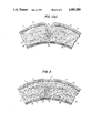
- FIG. 1(a) illustrates a cross-section of a portion of an intact cornea.
- FIG. 1(b) illustrates a cross-section of a portion of a cornea immediately after keratorefractive surgery.
- FIG. 1(c) illustrates a cross-section of a portion of a cornea approximately 3-5 days after keratorefractive surgery in which the method and composition of the invention were not employed.
- FIG. 1(d) illustrates a cross-section of a portion of a cornea approximately 28 days after keratorefractive surgery in which the method and composition of the invention were not employed.
- FIG. 2 illustrates a cross-section of a portion of a cornea approximately 28 days after keratorefractive surgery in which the corneal mortar composition of the invention has been inserted into the incisions.
- FIG. 3 is a graph presenting plots of corneal flattening versus post-surgical time for radial keratotamies in primates.
- One plot represents corneal flattening in primates in which corneal mortar composition of the invention was placed in the surgical incisions.
- the other plot represents controls in which only the saline carrier vehicle was placed in the incisions.
- the corneal mortar composition of the invention comprises at least one ECM material and an ophthalmologically compatible carrier material, the composition having a sufficiently high viscosity to retain the ECM material within the wound during healing
- ECMs are materials which can be found in extracellular matrix structures laid down by cultured cells.
- ECM materials include extracellular matrix proteins and extracellular ground substances.
- the former are generally high molecular weight (>150,000 daltons) fibrinous glycoproteins, which include fibronectin, collagens, vitronectin, elastin, laminin, actin and fibrinogen.
- the latter are polysaccharides, glycosylaminoglycans, which include chondroitin sulfate, heparin, keratin sulfate and hyaluronic acid or its sodium salt.
- a preferred ECM material for use in the corneal mortar composition is fibronectin.
- fibronectin is present in the corneal mortar composition even if other ECM materials are also employed
- Fibronectin can be present in the corneal mortar composition in amounts from about 0.5% to about 90%, preferably from about 2% to about 40% by weight of the composition.
- Fibronectin is a glycoprotein (4-5% carbohydrate) having a molecular weight of about 220,000 daltons, which exists in the form of a 440,000-dalton dimer. Fibronectin exists in a plasma associated form and a cell associated form. It can conveniently be isolated from plasma by the procedure described by Nishida et al., Jap. J. Ophth., Vol. 26, pp.
- Fibronectin is also known by various other names, including cold-insoluble globulin, surface fibroblast antigen, cell surface protein, band 1, L1 band, band I, zeta-protein, major fibroblast glycoprotein, galactoprotein A, large external transformation sensitive protein (LETS), micro-fibrillar protein, cell attachment protein, cell adhesion factor, anti-gelatin factor, cell spreading factor and opsonic factor.
- ECM proteins having a high degree of amino acid sequence homology with fibronectin such as vitronectin (Suzuki, S., J. Biol. Chem., 259:15307-15314 (1984) can be used in the preferred embodiment.
- the corneal mortar composition contains both fibronectin and chondroitin sulfate.
- Chondroitin sulfate is a glycosylaminoglycan found in the ECM's of animal connective tissues. It is a polymer formed of repeating disaccharide units. Each repeating disaccharide unit contains one sulfate group. Chondroitin sulfate has three isomers (chondroitin sulfate A, chondroitin sulfate B and chondroitin sulfate C), which differ in the position of the sulfate group in the disaccharide unit. All three isomers are useful in the corneal mortar compositions of the invention. Chondroitin sulfate can be obtained from commercial sources. Chondroitin sulfate can be present in the corneal mortar composition in amounts from about 0.5% to about 75% by weight of the composition.
- the corneal mortar composition can also contain collagen, an ECM material which is present in normal stromal tissue.
- collagen an ECM material which is present in normal stromal tissue.
- the amount of collagen, if present, does not exceed about 50 weight percent of the composition. While any type of collagen is suitable for use in the corneal mortar composition, Type I bovine collagen is preferred.
- Laminin which is another ECM material present in normal stromal tissue, can also be present in the corneal mortar composition in amounts up to about 75% by weight of the composition.
- Fibrinogen which is also an ECM material, can be present in the corneal mortar composition in amounts up to about 40% by weight thereof.
- the corneal mortar composition of the invention also contains a growth factor such as epidermal growth factor.
- Growth factors are mitogenic proteins or polypeptides which promote cell proliferation.
- a number of growth factors are known. These include epidermal growth factor (EGF), transforming growth factors (TGF's) and nerve growth factor (NGF).
- Insulin a polypeptide hormone, has mitogenic activity and can be used in conjunction with prostaglandin F 2 ⁇ , a non-peptide which has been shown to increase greatly the mitogenic activity of insulin (see Jimenez de Asua, L. et al., Cold Spring Harbor Conf. Cell Proliferation, Vol. 6, Sato, ed., Cold Spring Harbor Labs., New York [1979], at 403-424).
- fibroblast growth factor Similar activation of insulin has been reported with fibroblast growth factor by Rudland, P.S. et al., Proc. Natl. Acad. Sci., U.S.A., 76:1279-1293 (1974). Positive effects on cell growth have been demonstrated for platelet-derived growth factor or fibroblast-derived growth factor in combination with members of the insulin family such as somatomedins A and C (Stiles, C.D. et al., Proc. Natl. Acad. Sci., U.S.A., 76:1279-1283 [1979]). Additionally, many new peptide growth factors have been isolated and characterized recently as indicated in Tissue Growth Factors, R. Baserga, ed., Springer-Verlag pub., New York (1981). The present invention contemplates the use of any of the known growth factors, alone or in combination, in conjunction with ECM materials in the corneal mortar compositions of the invention.
- a preferred growth factor for use in the corneal mortar compositions of the invention is epidermal growth factor.
- EGF can be obtained from human tissues by the procedure described by Urdea et al., PNAS (USA), Vol. 80, p. 7461.
- the growth factor is employed in an amount which is effective to promote stromal cell growth at the wound site.
- the growth factor can be present in the corneal mortar composition at a concentration from about 0.01 ⁇ g/ml to about 100 ⁇ g/ml, preferably from about 0.1 ⁇ g/ml to about 10 ⁇ g/ml, although there is no strict upper limit to the concentration of growth factor.
- the carrier material is selected to act as a viscosity-adjusting agent, normally a diluent, to produce the desired viscosity in the corneal mortar composition.
- the carrier is normally a solution which is buffered to physiological pH, i.e., from about 6.5 to about 7.8.
- Phosphate buffered saline solution (PBS) is a preferred carrier material.
- Other suitable carrier materials include distilled water, ophthalmic saline solutions and other ophthalmic buffers, artificial tear materials, and viscoelastic agents such as sodium hyaluronate.
- the corneal mortar composition of the invention has a viscosity sufficiently high that the ECM material is retained within the wound during wound healing. That is, at least a sufficient amount of ECM material is retained in the wound to establish a matrix for healing. Since ECM materials bind to the stromal surfaces and establish a matrix rather quickly, it is sufficient if the composition has the consistency of a viscous fluid, so that it is not washed out of the wound by lacrimal secretions.
- the composition has a thick, pastelike viscosity. Since fibronectin and chondroitin sulfate are viscous materials, they are capable of imparting the desired viscosity to the corneal mortar compositions even at low concentrations.
- Fibronectin by itself begins to impart the desired viscosity when dissolved in saline solutions at concentrations of about 2% or higher. Chondroitin sulfate by itself begins to impart the desired viscosity when dissolved in saline at levels as low as about 1%. While there is no strict upper limit on the viscosity of the composition, it should not be so viscous that it cannot be inserted into the wound by the physician.
- ophthalmologically compatible substances which optionally can be present in the corneal mortar composition include substances which are known to promote wound healing or combat infection or inflammation.
- antibiotics can be present in the compositions in known effective amounts.
- the corneal mortar composition comprises fibronectin and an ophthalmologically compatible carrier material, the composition having a viscosity sufficiently high to retain the composition in the wound.
- the following formulation is exemplary of this embodiment:
- the corneal mortar composition comprises fibronectin, chondroitin sulfate and an ophthalmologically compatible carrier material, the composition having a viscosity sufficiently high to retain the composition in the wound.
- the following formulation is exemplary of this embodiment:
- the corneal mortar composition comprises fibronectin, a growth factor and an ophthalmologically compatible carrier material, the composition having a sufficiently high viscosity to retain the composition in the wound.
- the following formulation is exemplary of this embodiment:
- the corneal mortar composition comprises fibronectin, chondroitin sulfate, collagen and an ophthalmologically compatible carrier material, the composition having a sufficiently high viscosity to retain the composition in the wound.
- the following formulation is exemplary of this embodiment:
- the corneal mortar composition comprises fibronectin, chondroitin sulfate, a growth factor and an ophthalmologically acceptable carrier material, the composition having a sufficiently high viscosity to retain the composition in the wound.
- the following formulation is exemplary of this embodiment:
- compositions of the invention which contained chondroitin sulfate or fibronectin as the sole ECM material did not result in improved flattening following radial keratotamy in cat studies. Histological studies, however, showed that the use of chondroitin sulfate or fibronectin as the sole ECM material resulted in reduced scarring as a result of improved organizational integrity of the healed tissue.
- epidermal growth factor in combination with chondroitin sulfate and the use of epidermal growth factor in combination with fibronectin each provided a synergistic effect in the enhancement of corneal flattening.
- EGF is known to promote wound healing generally, the use of EGF alone as a wound healing agent following radial keratotamy tends to reverse the beneficial effect of the surgery on visual acuity even though it speeds healing of the incisions.
- chondroitin sulfate or fibronectin as a sole ECM material each resulted in decreased flattening as compared with controls in cat studies.
- EGF was used in conjunction with fibronectin or chondroitin sulfate, improved corneal flattening was obtained.
- the methods of the invention will be described below with specific reference to the use of the corneal mortar composition to treat keratorefractive incisions, thereby enhancing the improvement in visual acuity and/or reducing scarring. It is to be understood, however, that the corneal mortar compositions can also be used in a similar manner to treat corneal wounds of a non-surgical nature which extend into the stromal tissue and that the corneal mortar composition will have beneficial effects in the healing of such wounds.
- corneal mortar composition of the invention in keratorefractive surgery can be understood with reference to the figures.
- FIG. 1(a) illustrates a cross-section of a portion of an intact cornea
- the outer layer i.e., the layer on the convex surface of the cornea
- the epithelium 10 Under the epithelium 10 is the Bowman's membrane 14 (present only in primates).
- the Bowman's membrane 14 separates the epithelium 10 from the stroma 16, the relatively thick structural layer of the cornea.
- the stroma 16 is comprised of macromolecules, including collagen, chondroitin sulfate and keratin sulfate, as well as cells.
- Descemet's membrane 17 separates the stroma 16 from the endothelium 18.
- the endothelium 18 is a membrane of single-cell thickness which separates the stroma 16 from the aqueous humor (not shown) and serves to regulate fluid transport to and from the stroma 16. Keratocytes 20 are distributed throughout the stroma 16.
- FIG. 1(b) illustrates a cross-section of a portion of a cornea immediately after keratorefractive surgery in which an incision having a V-shaped cross section has been made into the stroma 16. The incision has allowed the cornea to relax and flatten out somewhat, thereby changing the refraction of the cornea to reduce or eliminate myopia.
- FIG. 1(c) illustrates the same cross-sectional portion of the cornea as it would appear about 3-5 days after surgery without the use of the corneal mortar composition of the invention. The position of the stromal tissue surfaces forming the original incision walls 22 is indicated in FIGS. 1(c) and 1(d) by a dashed line.
- the healing process can only occur along the surfaces of the walls 22 of the incision and only after those surfaces have been epithelialized; that is, epithelial cells must grow down from the epithelium 10 to cover the entire surface of the walls 22 of the incision.
- the epithelium 10 must extend itself down to the bottom of the "V" of the incision in order for healing to take place.
- the closing of the incision wound begins at the bottom of the "V” and works its way upward.
- the epithelial cells which have extended into the wound must be pushed out of the wound in a direction indicated by arrows in FIG. 1(c).
- the ability to displace the epithelial cells from the wound site may be a rate-limiting factor in wound healing.
- FIG. 1(d) illustrates the typical condition of the incision wound about 28 days after surgery.
- the stromal tissue surfaces which formed the original walls 22 of the incision have been drawn somewhat closer together, i.e., in the direction indicated by the arrows 26 in FIG. 1(d).
- the corneal mortar composition is inserted into the incisions during keratorefractive surgery.
- the corneal mortar composition can be inserted into the wound as a coating on the walls 22 of the incisions.
- the amount of corneal mortar composition which is placed in the incision is sufficient not only to coat the walls 22 of the incision but also to fill in at least a portion of the space between the walls 22 of the incision.
- FIG. 2 illustrates a cross-section of a portion of a cornea approximately 28 days after keratorefractive surgery in which the corneal mortar composition of the invention has been deposited into the incision wound. It is preferred that at least about 1% of the space between the walls 22 of the incision be filled. It is particularly desirable that the posterior portion of the incision, i.e., the portion of the incision at the bottom of the "V", be completely filled.
- the corneal mortar composition provides a matrix for the deposition of wound-healing substances and for cell migration and growth.
- the wound-healing process is no longer constrained to take place at the surfaces of the incision, but rather, it can take place concurrently throughout the volume of space occupied by the corneal mortar composition. Placing the corneal mortar composition into the incision induces keratocytes 20 to migrate into the space between the walls 22 of the incision where they grow and deposit wound-healing substances such as collagen.
- the corneal mortar composition in the incision maintains space between the walls 22 of the incision throughout the healing process, i.e., it prevents the stromal tissue surfaces which formed the original incision walls 22 from being drawn together in the manner illustrated in FIGS. 1(c) and 1(d). This is illustrated in FIG. 2 by the position of the dashed lines representing the original walls 22, which have not drawn together following surgery, and have moved apart somewhat at the base of the incision. Consequently, the effect of the surgery in adjusting the curvature of the cornea is not reversed by the healing process.
- a further advantage of using the corneal mortar composition of the invention relates to the organizational integrity of the healed tissue. Keratocytes, which are somewhat disc-shaped, are oriented in the plane of the "grain" in normal stromal tissue. Consequently, when viewed microscopically in a cross-section of normal cornea, they are seen on edge and appear relatively narrow as seen in FIG. 1(a).
- keratocytes 20 are distributed within the healed area in a random orientation so that some of them appear round on microscopic inspection. This random orientation results in collagen being laid down from the edges of the keratocytes 20 in a swirling manner, rather than aligned with the grain of the stromal tissue.
- the corneal mortar composition of the invention when deposited in the wound, it provides a matrix which properly orients the keratocytes 20, as shown in FIG. 2, so that collagen is laid down with the grain of the stromal tissue.
- the lack of orientation of keratocytes 20 in control animals was associated with increased scarring and cosmetically poor healing.
- the use of the corneal mortar composition of the invention may also speed the healing process. As previously mentioned, use of the composition frees the healing process from the geometric constraints of the wound surfaces. Moreover, the composition appears to promote epithelialization of the incision surfaces which is necessary for healing to occur.
- the corneal mortar composition can be placed into the surgical incision by the surgeon using any convenient means, such as by injection through a large-bore needle or by the use of any suitable trowel-like tool.
- Any convenient means such as by injection through a large-bore needle or by the use of any suitable trowel-like tool.
- the particular method which is best will depend largely on the viscosity of the corneal mortar composition.
- a soft contact lens which is permeable to gas and moisture may be placed over the cornea post-operatively in order to allow moisture transmission while insuring that the corneal mortar composition remains in the incision.
- a hard contact lens which forces the cornea to conform to the contact lens geometry, may be placed over the cornea in order to fix the desired shape of the cornea during the healing process.
- a substantially increased degree of refractive correction can be obtained in many instances.
- radial keratotamies were performed in rabbits in which one eye was a control which received no corneal mortar while the incisions in the other eye were packed with a corneal mortar composition containing 50 mg fibronectin, 2.6 gm, chondroitin sulfate and 13-15 mg collagen in phosphate buffered saline.
- corneascopic examination revealed that the corneas which received the corneal mortar composition exhibited from 12 to 15 diopters of flattening, compared with only 3 to 4 diopters for the control eyes.
- the degree of corneal flattening gradually lessened in the control eyes after the second day of healing, whereas it underwent a slight increase in the eyes which received the corneal mortar composition. Because of the significant increase in the degree of flattening obtainable with the method of the invention, it may be possible in many instances to reduce the number of incisions required to obtain the desired degree of refractive correction and/or to reduce the depth of the incisions. Reducing the depth of the incisions in turn reduces the danger of corneal perforation during surgery.
- Radial keratotamies were performed on a number of cats. Preoperative treatment consisted of weight measurement, slit lamp examination, specular microscopy and corneascope examination. A tattoo was placed at the center of the cornea.
- each animal was sedated with an intramuscular injection of ketaminexylazine and each eye was then treated with a topical anesthetic.
- the eye was irrigated with preservativefree ophthalmic saline.
- the optical zone was set by a 3-mm trephine at the central corneal tatoo.
- an incision was made to 90% of the depth of the lowest corneal thickness based on pachometry readings taken prior to cutting.
- Radial keratotamy incisions were made at 12, 3, 6 and 9 o'clock and extended from the end of the 3-mm zone to the limbus.
- CM corneal mortar composition of the invention
- Corneal flattening was measured at 7-day intervals, using a corneascope. The average flattening, in diopters, was determined for the treated eyes and the control eyes.
- FIG. 3 is a plot of diopters of flattening versus post-operative time. It can be seen from FIG. 3 that the treated eyes maintained a greater degree of corneal flattening throughout the post-operative period than the control eyes. After 77 days, the control eyes exhibited only an average of 0.1 diopters of flattening, whereas the treated eyes exhibited an average of 2.2 diopters of flattening.
Landscapes
- Health & Medical Sciences (AREA)
- General Health & Medical Sciences (AREA)
- Veterinary Medicine (AREA)
- Public Health (AREA)
- Life Sciences & Earth Sciences (AREA)
- Animal Behavior & Ethology (AREA)
- Engineering & Computer Science (AREA)
- Ophthalmology & Optometry (AREA)
- Epidemiology (AREA)
- Chemical & Material Sciences (AREA)
- Biomedical Technology (AREA)
- Vascular Medicine (AREA)
- Heart & Thoracic Surgery (AREA)
- Materials Engineering (AREA)
- Transplantation (AREA)
- Cardiology (AREA)
- Oral & Maxillofacial Surgery (AREA)
- Surgery (AREA)
- Nuclear Medicine, Radiotherapy & Molecular Imaging (AREA)
- Medicinal Chemistry (AREA)
- Pharmacology & Pharmacy (AREA)
- Materials For Medical Uses (AREA)
Abstract
Description
______________________________________
       Ingredient
               Amount*
______________________________________
       Fibronectin
               2.0-40%
       PBS      60-98%
______________________________________
 *Percentages based on total composition weight
______________________________________
Ingredient       Amount
______________________________________
Fibronectin      0.5-40%
Chondroitin sulfate
                 0.5-75%
PBS               25-99%
______________________________________
______________________________________
Ingredient         Amount
______________________________________
Fibronectin         0.5-40%
PBS                  60-99.5%
EGF                0.01-100 μg/ml
______________________________________
______________________________________
Ingredient       Amount
______________________________________
Fibronectin      0.5-40%
Chondroitin sulfate
                 0.5-75%
Collagen         0.5-50%
PBS                25-98.5%
______________________________________
______________________________________
Ingredient           Amount
______________________________________
Fibronectin           0.5-40%
Chondroitin sulfate   0.5-75%
PBS                    25-99%
EGF                  0.01-100 μg/ml
______________________________________
              TABLE 1
______________________________________
Treatment Matrix for Cats
No. of Cats
         Left Eye (OS) Right Eye (OD)
______________________________________
4 Cats   Control       Fn
         RK            0.25 mg Fn + 1.5 ml Saline
         No Treatment  liquid
4 Cats   Fn + CS       Fn + CS + EGF
         25 mg Fn      1/3 g. CS
         1/3 gm CS     25 mg Fn
         0.7 ml saline 15 mg EGF
         Very thick    0.7 ml saline
                       Very thick
4 Cats   CS + EGF      EGF
         15 μg EGF  15 μg EGF
         1.3 gm CS     1.5 ml saline
         1.1-1.2 ml saline
                       liquid
         thick paste
4 Cats, one
         CS
eye only 1.3 gm CS
         1.3 ml saline
         thick paste
3 Cats, one
         Control
eye only RK
______________________________________
TABLE 2______________________________________ Day 56 Corneal Flattening* Treatment Group Diopters of Flattening ______________________________________ Epidermal Growth Factor (EGF) 1.5 Fibronectin 1.5 Chonodroitin Sulfate, 1.9 Fibronectin and EGF Chondroitin Sulfate 2.7 Control 3.1 Chondroitin Sulfate & EGF 3.8 Chondroitin Sulfate & 3.8 Fibronectin ______________________________________ *Average value all eyes in each respective group
Claims (15)
Priority Applications (1)
| Application Number | Priority Date | Filing Date | Title |
|---|---|---|---|
| US07/467,116 US4983580A (en) | 1986-04-04 | 1990-03-07 | Methods and materials for use in corneal wound healing |
Applications Claiming Priority (2)
| Application Number | Priority Date | Filing Date | Title |
|---|---|---|---|
| US84827986A | 1986-04-04 | 1986-04-04 | |
| US07/467,116 US4983580A (en) | 1986-04-04 | 1990-03-07 | Methods and materials for use in corneal wound healing |
Related Parent Applications (1)
| Application Number | Title | Priority Date | Filing Date |
|---|---|---|---|
| US84827986A Division | 1986-04-04 | 1986-04-04 |
Publications (1)
| Publication Number | Publication Date |
|---|---|
| US4983580A true US4983580A (en) | 1991-01-08 |
Family
ID=27041919
Family Applications (1)
| Application Number | Title | Priority Date | Filing Date |
|---|---|---|---|
| US07/467,116 Expired - Fee Related US4983580A (en) | 1986-04-04 | 1990-03-07 | Methods and materials for use in corneal wound healing |
Country Status (1)
| Country | Link |
|---|---|
| US (1) | US4983580A (en) |
Cited By (49)
| Publication number | Priority date | Publication date | Assignee | Title |
|---|---|---|---|---|
| US5219576A (en) * | 1988-06-30 | 1993-06-15 | Collagen Corporation | Collagen wound healing matrices and process for their production |
| US5234914A (en) * | 1991-06-11 | 1993-08-10 | Patent Biopharmaceutics, Inc. | Methods of treating hemorrhoids and anorecial disease |
| US5360611A (en) * | 1988-10-03 | 1994-11-01 | Alcon Laboratories, Inc. | Pharmaceutical compositions and methods of treatment of the cornea following ultraviolet laser irradiation |
| US5364845A (en) * | 1993-03-31 | 1994-11-15 | Nutramax Laboratories, Inc. | Glucosamine, chondroitin and manganese composition for the protection and repair of connective tissue |
| US5411940A (en) * | 1993-09-29 | 1995-05-02 | Alcon Laboratories, Inc. | Use of TGF-β3 to reduce the formation of scar tissue in response to corneal trauma |
| US5487889A (en) * | 1992-06-03 | 1996-01-30 | The Metrohealth System | Bandage for continuous application of biologicals |
| US5562946A (en) * | 1994-11-02 | 1996-10-08 | Tissue Engineering, Inc. | Apparatus and method for spinning and processing collagen fiber |
| US5604200A (en) * | 1994-05-02 | 1997-02-18 | Taylor-Mccord; Darlene | Wound therapeutic mixture containing medical grade hyaluronic acid and tissue culture grade plasma-fibronectin in a delivery system that creates a moist environment which simulates in utero healing |
| US5610148A (en) * | 1991-01-18 | 1997-03-11 | University College London | Macroscopically oriented cell adhesion protein for wound treatment |
| US5624893A (en) * | 1993-10-14 | 1997-04-29 | Alcon Laboratories, Inc. | Pharmaceutical compositions and methods of treatment of the cornea following laser irradiation |
| US5629287A (en) * | 1991-01-18 | 1997-05-13 | University College London | Depot formulations |
| US5654267A (en) * | 1988-12-20 | 1997-08-05 | La Jolla Cancer Research Center | Cooperative combinations of ligands contained within a matrix |
| US5668119A (en) * | 1996-02-22 | 1997-09-16 | Medenica; Rajko D. | Topical pharmaceutical containing heparin and method of treatment |
| US5709934A (en) * | 1994-11-22 | 1998-01-20 | Tissue Engineering, Inc. | Bipolymer foams having extracellular matrix particulates |
| US5714463A (en) * | 1993-09-29 | 1998-02-03 | Alcon Laboratories, Inc. | Use of growth factor and antimetabolite combination to prevent or retard fistula closure following glaucoma filtration surgery |
| US5767079A (en) * | 1992-07-08 | 1998-06-16 | Celtrix Pharmaceuticals, Inc. | Method of treating ophthalmic disorders using TGF -β |
| WO1998029069A3 (en) * | 1997-01-02 | 1998-10-08 | Allergan Inc | Method for changing the refractive power of an eye |
| US5863892A (en) * | 1992-02-26 | 1999-01-26 | Allergan Inc. | Use of platelet derived growth factor in ophthalmic wound healing |
| US5874500A (en) * | 1995-12-18 | 1999-02-23 | Cohesion Technologies, Inc. | Crosslinked polymer compositions and methods for their use |
| US5891558A (en) * | 1994-11-22 | 1999-04-06 | Tissue Engineering, Inc. | Biopolymer foams for use in tissue repair and reconstruction |
| US5911942A (en) * | 1995-11-02 | 1999-06-15 | Tissue Engineering, Inc. | Method for spinning and processing collagen fiber |
| US5929050A (en) * | 1998-02-27 | 1999-07-27 | Petito; George D. | Chondroitin sulfate composition and method for wound treatment |
| US5955436A (en) * | 1990-11-20 | 1999-09-21 | Alcon Laboratories, Inc. | Use of platelet derived growth factor to enhance wound healing |
| US6110487A (en) * | 1997-11-26 | 2000-08-29 | Keraplast Technologies Ltd. | Method of making porous keratin scaffolds and products of same |
| US6255295B1 (en) | 1996-12-23 | 2001-07-03 | Nutramax Laboratories, Inc. | Aminosugar, glycosaminoglycan or glycosaminoglycan-like compounds, and s-adenosylmethionine composition for the protection, treatment, repair, and reduction of inflammation of connective tissue |
| US6270791B1 (en) * | 1999-06-11 | 2001-08-07 | Keraplast Technologies, Ltd. | Soluble keratin peptide |
| US6271213B1 (en) | 1996-12-23 | 2001-08-07 | Nutramax Laboratories, Inc. | Aminosugar, glycosaminoglycan, and S-adenosylmethionine composition for the treatment and repair of connective tissue |
| US6274163B1 (en) | 1998-04-08 | 2001-08-14 | Keraplast Technologies, Ltd. | Keratinous protein material for wound healing applications and method |
| US6458889B1 (en) | 1995-12-18 | 2002-10-01 | Cohesion Technologies, Inc. | Compositions and systems for forming crosslinked biomaterials and associated methods of preparation and use |
| US6528483B2 (en) | 1995-06-07 | 2003-03-04 | André Beaulieu | Method of producing concentrated non-buffered solutions of fibronectin |
| US20030119985A1 (en) * | 1995-12-18 | 2003-06-26 | Sehl Louis C. | Methods for tissue repair using adhesive materials |
| US20030203001A1 (en) * | 2002-04-25 | 2003-10-30 | Schultz Clyde L. | Growth factor delivery system for the healing of wounds and the prevention of inflammation and disease |
| US20030211793A1 (en) * | 2001-03-05 | 2003-11-13 | Eugene Bell | Injectable bio-compatible material and methods of use |
| US20040009893A1 (en) * | 2000-12-20 | 2004-01-15 | Pao-Li Wang | Ophthalmic lubricating solution adapted for use in lasik surgery |
| US20040167480A1 (en) * | 2003-02-21 | 2004-08-26 | Advanced Medical Optics, Inc. | Administration of multiple viscoelastic solutions with a multi-compartment syringe |
| US20040219214A1 (en) * | 2002-12-30 | 2004-11-04 | Angiotech International Ag | Tissue reactive compounds and compositions and uses thereof |
| US20050074497A1 (en) * | 2003-04-09 | 2005-04-07 | Schultz Clyde L. | Hydrogels used to deliver medicaments to the eye for the treatment of posterior segment diseases |
| US20050085758A1 (en) * | 2002-04-25 | 2005-04-21 | Schultz Clyde L. | Growth factor delivery system for the healing of wounds and the prevention of inflammation and disease |
| US20050147679A1 (en) * | 1998-03-24 | 2005-07-07 | Petito George D. | Composition and method for healing tissues |
| US20050175665A1 (en) * | 2003-11-20 | 2005-08-11 | Angiotech International Ag | Polymer compositions and methods for their use |
| US20050208114A1 (en) * | 1998-03-24 | 2005-09-22 | Petito George D | Composition and method for healing tissues |
| US20050255144A1 (en) * | 2003-04-09 | 2005-11-17 | Directcontact Llc | Methods and articles for the delivery of medicaments to the eye for the treatment of posterior segment diseases |
| US7112320B1 (en) | 1995-06-07 | 2006-09-26 | Andre Beaulieu | Solid wound healing formulations containing fibronectin |
| US20090192214A1 (en) * | 2002-12-30 | 2009-07-30 | Angiotech International Ag | Drug delivery from rapid gelling polymer composition |
| US7883693B2 (en) | 1995-12-18 | 2011-02-08 | Angiodevice International Gmbh | Compositions and systems for forming crosslinked biomaterials and methods of preparation of use |
| US8067031B2 (en) | 2004-04-28 | 2011-11-29 | Angiodevice International Gmbh | Compositions and systems for forming crosslinked biomaterials and associated methods of preparation and use |
| US9216106B2 (en) | 2003-04-09 | 2015-12-22 | Directcontact Llc | Device and method for the delivery of drugs for the treatment of posterior segment disease |
| US9353218B2 (en) | 2004-09-17 | 2016-05-31 | Angiotech Pharmaceuticals, Inc. | Kit for multifunctional compounds forming crosslinked biomaterials |
| DE102018107230A1 (en) | 2018-03-27 | 2019-10-02 | Universität Zu Lübeck | Ocular surface reconstruction device |
Citations (3)
| Publication number | Priority date | Publication date | Assignee | Title |
|---|---|---|---|---|
| US4458678A (en) * | 1981-10-26 | 1984-07-10 | Massachusetts Institute Of Technology | Cell-seeding procedures involving fibrous lattices |
| US4486416A (en) * | 1981-03-02 | 1984-12-04 | Soll David B | Protection of human and animal cells subject to exposure to trauma |
| EP0190018A2 (en) * | 1985-01-29 | 1986-08-06 | Oncogen | Use of transforming growth factor to promote wound healing and composition therefor |
-
1990
- 1990-03-07 US US07/467,116 patent/US4983580A/en not_active Expired - Fee Related
Patent Citations (3)
| Publication number | Priority date | Publication date | Assignee | Title |
|---|---|---|---|---|
| US4486416A (en) * | 1981-03-02 | 1984-12-04 | Soll David B | Protection of human and animal cells subject to exposure to trauma |
| US4458678A (en) * | 1981-10-26 | 1984-07-10 | Massachusetts Institute Of Technology | Cell-seeding procedures involving fibrous lattices |
| EP0190018A2 (en) * | 1985-01-29 | 1986-08-06 | Oncogen | Use of transforming growth factor to promote wound healing and composition therefor |
Non-Patent Citations (6)
| Title |
|---|
| Kawaba et al., "Effect of Human EGF and Plasma Fibronectin on Corneal Epithelial Regeneration", Nippon Ganka Gakkai Zasshi, 88(9) 1237-44, 1984. |
| Kawaba et al., Effect of Human EGF and Plasma Fibronectin on Corneal Epithelial Regeneration , Nippon Ganka Gakkai Zasshi, 88(9) 1237 44, 1984. * |
| Nishida et al., Arch Ophthalmol , 101:1046 1048, Jul. 1983. * |
| Nishida et al., Arch Ophthalmol, 101:1046-1048, Jul. 1983. |
| Nishida et al., Ophthalmology , 92(2):213 216, Feb. 1985. * |
| Nishida et al., Ophthalmology, 92(2):213-216, Feb. 1985. |
Cited By (97)
| Publication number | Priority date | Publication date | Assignee | Title |
|---|---|---|---|---|
| US5219576A (en) * | 1988-06-30 | 1993-06-15 | Collagen Corporation | Collagen wound healing matrices and process for their production |
| US5580570A (en) * | 1988-10-03 | 1996-12-03 | Alcon Laboratories, Inc. | Pharmaceutical compositions and methods of treatment of the cornea following laser irradiation |
| US5360611A (en) * | 1988-10-03 | 1994-11-01 | Alcon Laboratories, Inc. | Pharmaceutical compositions and methods of treatment of the cornea following ultraviolet laser irradiation |
| US5589184A (en) * | 1988-10-03 | 1996-12-31 | Alcon Laboratories, Inc. | Pharmaceutical compositions and methods of treatment of the cornea following laser treatment |
| US5654267A (en) * | 1988-12-20 | 1997-08-05 | La Jolla Cancer Research Center | Cooperative combinations of ligands contained within a matrix |
| US5830504A (en) * | 1988-12-20 | 1998-11-03 | La Jolla Cancer Research Foundation | Cooperative combinations of ligands contained within a matrix |
| US5955436A (en) * | 1990-11-20 | 1999-09-21 | Alcon Laboratories, Inc. | Use of platelet derived growth factor to enhance wound healing |
| US5610148A (en) * | 1991-01-18 | 1997-03-11 | University College London | Macroscopically oriented cell adhesion protein for wound treatment |
| US5629287A (en) * | 1991-01-18 | 1997-05-13 | University College London | Depot formulations |
| US5234914A (en) * | 1991-06-11 | 1993-08-10 | Patent Biopharmaceutics, Inc. | Methods of treating hemorrhoids and anorecial disease |
| US5863892A (en) * | 1992-02-26 | 1999-01-26 | Allergan Inc. | Use of platelet derived growth factor in ophthalmic wound healing |
| US5487889A (en) * | 1992-06-03 | 1996-01-30 | The Metrohealth System | Bandage for continuous application of biologicals |
| US5767079A (en) * | 1992-07-08 | 1998-06-16 | Celtrix Pharmaceuticals, Inc. | Method of treating ophthalmic disorders using TGF -β |
| US6492349B1 (en) | 1993-03-31 | 2002-12-10 | Nutramax Laboratories, Inc. | Aminosugar and glycosaminoglycan composition for the treatment and repair of connective tissue |
| US5587363A (en) * | 1993-03-31 | 1996-12-24 | Nutramax Laboratories, Inc. | Aminosugar and glycosaminoglycan composition for the treatment and repair of connective tissue |
| US5364845A (en) * | 1993-03-31 | 1994-11-15 | Nutramax Laboratories, Inc. | Glucosamine, chondroitin and manganese composition for the protection and repair of connective tissue |
| US5714463A (en) * | 1993-09-29 | 1998-02-03 | Alcon Laboratories, Inc. | Use of growth factor and antimetabolite combination to prevent or retard fistula closure following glaucoma filtration surgery |
| US5411940A (en) * | 1993-09-29 | 1995-05-02 | Alcon Laboratories, Inc. | Use of TGF-β3 to reduce the formation of scar tissue in response to corneal trauma |
| US5624893A (en) * | 1993-10-14 | 1997-04-29 | Alcon Laboratories, Inc. | Pharmaceutical compositions and methods of treatment of the cornea following laser irradiation |
| US5604200A (en) * | 1994-05-02 | 1997-02-18 | Taylor-Mccord; Darlene | Wound therapeutic mixture containing medical grade hyaluronic acid and tissue culture grade plasma-fibronectin in a delivery system that creates a moist environment which simulates in utero healing |
| US5851290A (en) * | 1994-11-02 | 1998-12-22 | Tissue Engineering, Inc. | Apparatus for spinning and processing collagen fiber |
| US5562946A (en) * | 1994-11-02 | 1996-10-08 | Tissue Engineering, Inc. | Apparatus and method for spinning and processing collagen fiber |
| US5709934A (en) * | 1994-11-22 | 1998-01-20 | Tissue Engineering, Inc. | Bipolymer foams having extracellular matrix particulates |
| US5891558A (en) * | 1994-11-22 | 1999-04-06 | Tissue Engineering, Inc. | Biopolymer foams for use in tissue repair and reconstruction |
| US5948429A (en) * | 1994-11-22 | 1999-09-07 | Tissue Engineering, Inc. | Methods for preparing biopolymer foams |
| US7112320B1 (en) | 1995-06-07 | 2006-09-26 | Andre Beaulieu | Solid wound healing formulations containing fibronectin |
| US6528483B2 (en) | 1995-06-07 | 2003-03-04 | André Beaulieu | Method of producing concentrated non-buffered solutions of fibronectin |
| US6323278B2 (en) | 1995-10-05 | 2001-11-27 | Cohesion Technologies, Inc. | Method of making crosslinked polymer matrices in tissue treatment applications |
| US5911942A (en) * | 1995-11-02 | 1999-06-15 | Tissue Engineering, Inc. | Method for spinning and processing collagen fiber |
| US6969400B2 (en) | 1995-12-18 | 2005-11-29 | Cohesion Technologies, Inc. | Synthetic implant with nonimmunogenicity coating |
| US5874500A (en) * | 1995-12-18 | 1999-02-23 | Cohesion Technologies, Inc. | Crosslinked polymer compositions and methods for their use |
| US20040235708A1 (en) * | 1995-12-18 | 2004-11-25 | Rhee Woonza M. | Method for preventing the formation of adhesions following surgery or injury |
| US20050159544A1 (en) * | 1995-12-18 | 2005-07-21 | Rhee Woonza M. | Crosslinked polymer compositions |
| US6166130A (en) * | 1995-12-18 | 2000-12-26 | Cohesion Technologies, Inc. | Method of using crosslinked polymer compositions in tissue treatment applications |
| US7151135B2 (en) | 1995-12-18 | 2006-12-19 | Angiotech Pharmaceuticals (Us), Inc. | Crosslinked polymer compositions |
| US8617584B2 (en) | 1995-12-18 | 2013-12-31 | Angiodevice International Gmbh | Adhesive tissue repair patch and collagen sheets |
| US7176256B2 (en) | 1995-12-18 | 2007-02-13 | Angiotech Pharmaceuticals (Us), Inc. | Biocompatible crosslinked composition |
| US8377466B2 (en) | 1995-12-18 | 2013-02-19 | Angiotech Pharmaceuticals (Us), Inc. | Adhesive tissue repair patch |
| US6051648A (en) * | 1995-12-18 | 2000-04-18 | Cohesion Technologies, Inc. | Crosslinked polymer compositions and methods for their use |
| US6911496B2 (en) | 1995-12-18 | 2005-06-28 | Cohesion Technologies, Inc. | Composition for administration of a biologically active compound |
| US6458889B1 (en) | 1995-12-18 | 2002-10-01 | Cohesion Technologies, Inc. | Compositions and systems for forming crosslinked biomaterials and associated methods of preparation and use |
| US20100233246A1 (en) * | 1995-12-18 | 2010-09-16 | Angiotech Pharmaceuticals (Us), Inc. | Adhesive tissue repair patch and collagen sheets |
| US6833408B2 (en) | 1995-12-18 | 2004-12-21 | Cohesion Technologies, Inc. | Methods for tissue repair using adhesive materials |
| US6534591B2 (en) | 1995-12-18 | 2003-03-18 | Cohesion Technologies, Inc. | Cross-linked polymer compositions and methods for their use |
| US20050054771A1 (en) * | 1995-12-18 | 2005-03-10 | Sehl Louis C. | Adhesive tissue repair patch |
| US20030119985A1 (en) * | 1995-12-18 | 2003-06-26 | Sehl Louis C. | Methods for tissue repair using adhesive materials |
| US8197802B2 (en) | 1995-12-18 | 2012-06-12 | Angiodevice International Gmbh | Method for treating or inhibiting the formation of adhesions following surgery or injury |
| US20110195040A1 (en) * | 1995-12-18 | 2011-08-11 | Angiodevice International Gmbh | Method for preventing the formation of adhesions following surgery or injury |
| US20050027070A1 (en) * | 1995-12-18 | 2005-02-03 | Rhee Woonza M. | Method for preparing a biocompatible crosslinked matrix and matrix provided thereby |
| US20110159075A1 (en) * | 1995-12-18 | 2011-06-30 | Angiodevice International Gmbh | Compositions and systems for forming crosslinked biomaterials and methods of preparation and use |
| US7883694B2 (en) | 1995-12-18 | 2011-02-08 | Angiodevice International Gmbh | Method for preventing the formation of adhesions following surgery or injury |
| US20040185084A1 (en) * | 1995-12-18 | 2004-09-23 | Rhee Woonza M. | Synthetic implant with nonimmunogenicity coating |
| US20040186230A1 (en) * | 1995-12-18 | 2004-09-23 | Rhee Woonza M. | Composition for administration of a biologically active compound |
| US20040186231A1 (en) * | 1995-12-18 | 2004-09-23 | Rhee Woonza M. | Dehydrated, shaped matrix and use thereof in the treatment of vascular malformation |
| US7883693B2 (en) | 1995-12-18 | 2011-02-08 | Angiodevice International Gmbh | Compositions and systems for forming crosslinked biomaterials and methods of preparation of use |
| US5668119A (en) * | 1996-02-22 | 1997-09-16 | Medenica; Rajko D. | Topical pharmaceutical containing heparin and method of treatment |
| US20030216348A1 (en) * | 1996-12-23 | 2003-11-20 | Nutramax Laboratories, Inc. | Aminosugar, glycosaminoglycan, and S-Adenosylmethionine composition for the treatment and repair of connective tissue |
| US6583123B2 (en) | 1996-12-23 | 2003-06-24 | Nutramax Laboratories, Inc. | Aminosugar, glycosaminoglycan, and S-adenosylmethionine composition for the treatment and repair of connective tissue |
| US6271213B1 (en) | 1996-12-23 | 2001-08-07 | Nutramax Laboratories, Inc. | Aminosugar, glycosaminoglycan, and S-adenosylmethionine composition for the treatment and repair of connective tissue |
| US6255295B1 (en) | 1996-12-23 | 2001-07-03 | Nutramax Laboratories, Inc. | Aminosugar, glycosaminoglycan or glycosaminoglycan-like compounds, and s-adenosylmethionine composition for the protection, treatment, repair, and reduction of inflammation of connective tissue |
| WO1998029069A3 (en) * | 1997-01-02 | 1998-10-08 | Allergan Inc | Method for changing the refractive power of an eye |
| US6110487A (en) * | 1997-11-26 | 2000-08-29 | Keraplast Technologies Ltd. | Method of making porous keratin scaffolds and products of same |
| US6432435B1 (en) | 1997-11-26 | 2002-08-13 | Keraplast Technologies, Ltd. | Keratin-based tissue engineering scaffold |
| US6159495A (en) * | 1997-11-26 | 2000-12-12 | Keraplast Technologies, Ltd. | Porous and bulk keratin bio-polymers |
| US6124265A (en) * | 1997-11-26 | 2000-09-26 | Keraplast Technologies, Ltd. | Method of making and cross-linking keratin-based films and sheets |
| US5929050A (en) * | 1998-02-27 | 1999-07-27 | Petito; George D. | Chondroitin sulfate composition and method for wound treatment |
| US20100173868A1 (en) * | 1998-03-24 | 2010-07-08 | Petito George D | Composition and method for healing tissues |
| US7691829B2 (en) | 1998-03-24 | 2010-04-06 | Petito George D | Composition and method for healing tissues |
| US20050208114A1 (en) * | 1998-03-24 | 2005-09-22 | Petito George D | Composition and method for healing tissues |
| US8168599B2 (en) | 1998-03-24 | 2012-05-01 | Petito George D | Composition and method for healing tissues |
| US20050147679A1 (en) * | 1998-03-24 | 2005-07-07 | Petito George D. | Composition and method for healing tissues |
| US6274163B1 (en) | 1998-04-08 | 2001-08-14 | Keraplast Technologies, Ltd. | Keratinous protein material for wound healing applications and method |
| US6270791B1 (en) * | 1999-06-11 | 2001-08-07 | Keraplast Technologies, Ltd. | Soluble keratin peptide |
| US6919321B2 (en) * | 2000-12-20 | 2005-07-19 | Alcon, Inc. | Ophthalmic lubricating solution adapted for use in lasik surgery |
| US20040009893A1 (en) * | 2000-12-20 | 2004-01-15 | Pao-Li Wang | Ophthalmic lubricating solution adapted for use in lasik surgery |
| US20030211793A1 (en) * | 2001-03-05 | 2003-11-13 | Eugene Bell | Injectable bio-compatible material and methods of use |
| US7169406B2 (en) | 2002-04-25 | 2007-01-30 | Directcontact Llc | Growth factor delivery systems for the healing of wounds and the prevention of inflammation and disease |
| US20030203001A1 (en) * | 2002-04-25 | 2003-10-30 | Schultz Clyde L. | Growth factor delivery system for the healing of wounds and the prevention of inflammation and disease |
| US20050085758A1 (en) * | 2002-04-25 | 2005-04-21 | Schultz Clyde L. | Growth factor delivery system for the healing of wounds and the prevention of inflammation and disease |
| US7618643B2 (en) | 2002-04-25 | 2009-11-17 | Directcontact Llc | Growth factor delivery system for the healing of wounds and the prevention of inflammation and disease |
| US20050196428A1 (en) * | 2002-04-25 | 2005-09-08 | Schultz Clyde L. | Growth factor delivery system for the healing of wounds and the prevention of inflammation and disease |
| US20090192214A1 (en) * | 2002-12-30 | 2009-07-30 | Angiotech International Ag | Drug delivery from rapid gelling polymer composition |
| US9326934B2 (en) | 2002-12-30 | 2016-05-03 | Angiotech International Ag | Drug delivery from rapid gelling polymer composition |
| US20040219214A1 (en) * | 2002-12-30 | 2004-11-04 | Angiotech International Ag | Tissue reactive compounds and compositions and uses thereof |
| US20040167480A1 (en) * | 2003-02-21 | 2004-08-26 | Advanced Medical Optics, Inc. | Administration of multiple viscoelastic solutions with a multi-compartment syringe |
| US20050255144A1 (en) * | 2003-04-09 | 2005-11-17 | Directcontact Llc | Methods and articles for the delivery of medicaments to the eye for the treatment of posterior segment diseases |
| US9216106B2 (en) | 2003-04-09 | 2015-12-22 | Directcontact Llc | Device and method for the delivery of drugs for the treatment of posterior segment disease |
| US20050074497A1 (en) * | 2003-04-09 | 2005-04-07 | Schultz Clyde L. | Hydrogels used to deliver medicaments to the eye for the treatment of posterior segment diseases |
| US20050187140A1 (en) * | 2003-11-20 | 2005-08-25 | Angiotech International Ag | Polymer compositions and methods for their use |
| US20050175665A1 (en) * | 2003-11-20 | 2005-08-11 | Angiotech International Ag | Polymer compositions and methods for their use |
| US20050208095A1 (en) * | 2003-11-20 | 2005-09-22 | Angiotech International Ag | Polymer compositions and methods for their use |
| US8067031B2 (en) | 2004-04-28 | 2011-11-29 | Angiodevice International Gmbh | Compositions and systems for forming crosslinked biomaterials and associated methods of preparation and use |
| US8460708B2 (en) | 2004-04-28 | 2013-06-11 | Angiodevice International Gmbh | Compositions and systems for forming crosslinked biomaterials and associated methods of preparation and use |
| US8481073B2 (en) | 2004-04-28 | 2013-07-09 | Angiodevice International Gmbh | Compositions and systems for forming crosslinked biomaterials and associated methods of preparation and use |
| US9353218B2 (en) | 2004-09-17 | 2016-05-31 | Angiotech Pharmaceuticals, Inc. | Kit for multifunctional compounds forming crosslinked biomaterials |
| DE102018107230A1 (en) | 2018-03-27 | 2019-10-02 | Universität Zu Lübeck | Ocular surface reconstruction device |
| WO2019185085A1 (en) | 2018-03-27 | 2019-10-03 | Universität Zu Lübeck | Eye surface reconstruction device |
Similar Documents
| Publication | Publication Date | Title |
|---|---|---|
| US4983580A (en) | Methods and materials for use in corneal wound healing | |
| US4981841A (en) | Methods and materials for use in corneal wound healing | |
| EP0240031B1 (en) | Mortar composition for use in corneal wounds | |
| US5124155A (en) | Fibronectin wound-healing dressings | |
| US4973466A (en) | Wound-healing dressings and methods | |
| US5053388A (en) | Wound healing composition and method | |
| Keivyon et al. | Limbal autograft transplantation for ocular surface disorders | |
| Alio et al. | Use of autologous platelet-rich plasma in the treatment of dormant corneal ulcers | |
| US5171318A (en) | Treated corneal prosthetic device | |
| RU2114579C1 (en) | Prosthesis for carrying out subepithelial implantation operations | |
| US5103840A (en) | Viscoelastic collagen gel for ophthalmic surgery | |
| JP2008509748A (en) | Visual enhancement ophthalmic device and related methods and compositions | |
| MXPA04006954A (en) | Methods for producing epithelial flaps on the cornea and for placement of ocular devices and lenses beneath an epithelial flap or membrane, epithelial delaminating devices, and structures of epithelium and ocular devices and lenses. | |
| Holmberg et al. | Sodium hyaluronate in cataract surgery: I. Report on the use of Healon® in two different types of intracapsular cataract surgery | |
| Cagatay et al. | Long–Term Comparison of Fibrin Tissue Glue and Vicryl Suture in Conjunctival Autografting for Pterygium Surgery | |
| Waring III et al. | Slit-lamp microscopic appearance of corneal wound healing after radial keratotomy | |
| Marmer | Therapeutic and protective properties of the corneal collagen shield | |
| US20030203032A1 (en) | Growth factor delivery system for the healing of wounds and the prevention of inflammation and disease | |
| JP2005529122A (en) | Growth factor delivery system for wound healing and methods of preventing inflammation and disease | |
| Aquavella et al. | Therapeutic applications of a collagen bandage lens: A preliminary report | |
| US5411940A (en) | Use of TGF-β3 to reduce the formation of scar tissue in response to corneal trauma | |
| Yaqub et al. | The use of autogenous auricular cartilage in the management of upper eyelid entropion | |
| Lindstrom et al. | Cataract surgery and astigmatic keratotomy | |
| Ehrlich et al. | Epikeratophakia for the treatment of hyperopia | |
| WO1989004172A1 (en) | Wound healing composition and method |
Legal Events
| Date | Code | Title | Description |
|---|---|---|---|
| FEPP | Fee payment procedure |
Free format text: PAYOR NUMBER ASSIGNED (ORIGINAL EVENT CODE: ASPN); ENTITY STATUS OF PATENT OWNER: LARGE ENTITY |
|
| FPAY | Fee payment |
Year of fee payment: 4 |
|
| AS | Assignment |
Owner name: ALLERGAN, TEXAS Free format text: ASSIGNMENT OF ASSIGNORS INTEREST;ASSIGNOR:ALLERGAN, INC.;REEL/FRAME:008239/0239 Effective date: 19960117 |
|
| FPAY | Fee payment |
Year of fee payment: 8 |
|
| REMI | Maintenance fee reminder mailed | ||
| LAPS | Lapse for failure to pay maintenance fees | ||
| STCH | Information on status: patent discontinuation |
Free format text: PATENT EXPIRED DUE TO NONPAYMENT OF MAINTENANCE FEES UNDER 37 CFR 1.362 |
|
| FP | Lapsed due to failure to pay maintenance fee |
Effective date: 20030108 |