US4904867A - Radiation image read-out method and apparatus - Google Patents
Radiation image read-out method and apparatus Download PDFInfo
- Publication number
- US4904867A US4904867A US07/061,366 US6136687A US4904867A US 4904867 A US4904867 A US 4904867A US 6136687 A US6136687 A US 6136687A US 4904867 A US4904867 A US 4904867A
- Authority
- US
- United States
- Prior art keywords
- image
- image signals
- read
- value
- recording medium
- Prior art date
- Legal status (The legal status is an assumption and is not a legal conclusion. Google has not performed a legal analysis and makes no representation as to the accuracy of the status listed.)
- Expired - Lifetime
Links
Images

Classifications
-
- G—PHYSICS
- G01—MEASURING; TESTING
- G01T—MEASUREMENT OF NUCLEAR OR X-RADIATION
- G01T1/00—Measuring X-radiation, gamma radiation, corpuscular radiation, or cosmic radiation
- G01T1/16—Measuring radiation intensity
- G01T1/20—Measuring radiation intensity with scintillation detectors
- G01T1/2012—Measuring radiation intensity with scintillation detectors using stimulable phosphors, e.g. stimulable phosphor sheets
- G01T1/2014—Reading out of stimulable sheets, e.g. latent image
Definitions
- This invention relates to a method of reading out a radiation image wherein final read-out is carried out for detecting image signals for use in reproduction of a visible image from a recording medium such as a stimulable phosphor sheet carrying a radiation image recorded thereon, and an apparatus for carrying out the method.
- This invention particularly relates to a method of reading out a radiation image so that the density of an object region concerned in a visible image reproduced based on the read-out image signals is prevented from differing between images of the object recorded in different image recording directions, and an apparatus for carrying out the method.
- phosphors When certain kinds of phosphors are exposed to a radiation such as X-rays, ⁇ -rays, ⁇ -rays, ⁇ -rays, cathode rays or ultraviolet rays, they store a part of the energy of the radiation. Then, when the phosphor which has been exposed to the radiation is exposed to stimulating rays such as visible light, light is emitted by the phosphor in proportion to the stored energy of the radiation. A phosphor exhibiting such properties is referred to as a stimulable phosphor.
- a stimulable phosphor sheet As disclosed in U.S. Pat. No. 4,258,264 and Japanese Unexamined Patent Publication No. 56(1981)-11395, it has been proposed to use a stimulable phosphor in a radiation image recording and reproducing system. Specifically, a sheet provided with a layer of the stimulable phosphor (hereinafter referred to as a stimulable phosphor sheet) is first exposed to a radiation passing through an object such as the human body to have a radiation image of the object stored thereon.
- a stimulable phosphor sheet a sheet provided with a layer of the stimulable phosphor
- final read-out is carried out by exposing the stimulable phosphor sheet carrying the radiation image stored thereon to stimulating rays such as a laser beam which cause the stimulable phosphor sheet to emit light in proportion to the stored radiation energy, and photoelectrically detecting the emitted light to obtain electric image signals for use in reproduction of a visible image.
- An image processing is carried out on the final read-out image signals obtained by the final read-out, and the radiation image of the object is reproduced as a visible image by use of the processed image signals on a recording medium such as a photographic film, a display device such as a cathode ray tube (CRT), or the like.
- the range of the level of the radiation energy stored on the stimulable phosphor sheet is caused to fluctuate among radiation images by changes in the object, the image recording portion thereof, radiation dose, or the like.
- the recorded image information, particularly the range of the level of the radiation energy or the like, of each radiation image stored on the stimulable phosphor sheet may be ascertained in advance, and the final read-out may be carried out by use of read-out conditions such as a read-out gain and a scale factor adjusted to appropriate values in accordance with the recorded image information of each radiation image.
- read-out conditions such as a read-out gain and a scale factor adjusted to appropriate values in accordance with the recorded image information of each radiation image.
- the image processing of the final read-out image signals obtained by the final read-out is carried out by use of image processing conditions such as gradation processing conditions adjusted for each radiation image based on the image recording portion of the object such as the head, the chest or the abdomen, and/or the image recording method such as plain image recording or contrasted image recording so that a visible image suitable for viewing, particularly for diagnostic purposes, can be obtained.
- image processing conditions such as gradation processing conditions adjusted for each radiation image based on the image recording portion of the object such as the head, the chest or the abdomen
- the image recording method such as plain image recording or contrasted image recording so that a visible image suitable for viewing, particularly for diagnostic purposes, can be obtained.
- the image processing conditions should preferably be adjusted by considering the recorded image information of each radiation image, which has been ascertained in advance, besides the image recording portion of the object and/or the image recording method. In this manner, it becomes possible to obtain a visible image suitable for viewing, particularly for diagnostic purposes, wherein the necessary object image information is expressed within the correct density range.
- Ascertaining of the image information recorded on the stimulable phosphor sheet prior to the final read-out and the image processing may be carried out by use of the method as disclosed in Japanese Unexamined Patent Publication No. 58(1983)-67240.
- preliminary read-out for ascertaining the recorded image information of a radiation image stored on the stimulable phosphor sheet is carried out in advance by use of stimulating rays having stimulation energy of a level lower than the level of the stimulation energy of stimulating rays used in the final read-out for detecting the image signals for use in reproduction of a visible image for viewing, particularly for diagnostic purposes, and thereafter the final read-out is carried out.
- the read-out conditions for the final read-out and/or the image processing conditions are adjusted based on the recorded image information ascertained by the preliminary read-out, i.e. the preliminary read-out image signals obtained by the preliminary read-out.
- the level of the stimulating rays used in the preliminary read-out is lower than the level of the stimulating rays used in the final read out.
- the effective energy of the stimulating rays which the stimulable phosphor sheet receives per unit area in the preliminary read-out should be lower than the effective energy of the stimulating rays used in the final read-out.
- the image processing conditions are to be adjusted based on the recorded image information, it is only necessary that the recorded image information be ascertained prior to the image processing. In this case, since the final read-out image signals representing the recorded image information have already been detected, the image processing conditions can be adjusted based on the final read-out image signals, and the preliminary read-out as mentioned above need not necessarily be carried out.
- FIG. 2A shows the case where an image of the chest is recorded from the front of the object for diagnosis of the thoracic vertebrae
- FIG. 2B shows the case where an image of the chest is recorded from the side of the object.
- the amount of radiation stored at the thoracic vertebra image portion on a stimulable phosphor sheet 103 is large, and a large amount of light is emitted by the thoracic vertebra image portion on the stimulable phosphor sheet 103 as shown by the histogram of the read-out image signals in FIG. 3B when the sheet 103 is exposed to stimulating rays.
- the read-out conditions for the final read-out and/or the image processing conditions adjusted by a method of adjustment based on the maximum value Smax and the minimum value Smin of the read-out image signals as one of the conventional methods of adjustment based on the recorded image information become approximately equal between the front image recording and the side image recording.
- the density of the thoracic vertebra image portion becomes comparatively low in the reproduced visible image obtained in the case of the front image recording, and becomes comparatively high in the case of the side image recording.
- the primary object of the present invention is to provide a method of reading out a radiation image wherein, in the course of reproducing a visible radiation image by reading out a radiation image from a recording medium such as a stimulable phosphor sheet carrying the radiation image of an object recorded thereon in a different image recording direction, the density of a region concerned in the reproduced visible image is maintained constant regardless of the image recording direction.
- Another object of the present invention is to provide a method of reading out a radiation image, which is suitable for obtaining a reproduced visible image having an improved image quality, particularly a high diagnostic efficiency and accuracy.
- the specific object of the present invention is to provide an apparatus for carrying out the method of reading out a radiation image.
- a further object of the present invention is to provide a method of discriminating the image recording direction of the object.
- the present invention provides a method of reading out a radiation image in which final read-out is carried out for detecting image signals for use in reproduction of a visible image from a recording medium carrying a radiation image recorded thereon, and read-out conditions for the final read-out and/or image processing conditions for an image processing of final read-out image signals detected by the final read-out are adjusted based on image signals for condition adjustment detected by reading out the image information recorded on the recording medium,
- the present invention also provides a radiation image read-out apparatus comprising a final read-out system provided with a means for emitting light to a recording medium carrying a radiation image recorded thereon, a means for moving said recording medium, and a light detection means for photoelectrically detecting an amount of light emission by said recording medium upon exposure to said light and obtaining image signals for use in reproduction of a visible image, and a system for adjusting read-out conditions for the final read-out and/or image processing conditions for an image processing of final read-out image signals, which are detected by the final read-out, based on image signals for condition adjustment detected by reading out the image information recorded on the recording medium,
- the improvement comprises constituting said system for adjusting said read-out conditions for the final read-out and/or said image processing conditions by:
- the representative value of the image signals in the partial region is calculated, and the read-out conditions for the final read-out and/or the image processing conditions are adjusted based on the predetermined discrimination value representing the relationship between the representative value and a characteristic value of the image signals for condition adjustment, it is possible to make constant the density of the image region concerned in the reproduced visible image regardless of the image recording direction of the object.
- the present invention further provides a method of discriminating the image recording direction of the object in a radiation image read-out method comprising the steps of extracting image signals in a partial region at a predetermined position on a recording medium from said image signals, and calculating a representative value of the image signal for said partial region, and calculating a predetermined discrimination value representing a relationship between a characteristic value of said image signals and said representative value.
- the mean value, the maximum value, the minimum value or the like of the image signals in the partial region it is possible to use the mean value, the maximum value, the minimum value or the like of the image signals in the partial region.
- the mean value Save of the image signals in the partial region is used as the representative value and the maximum value Smax and the minimum value Smin are used as the characteristic values of the image signals for condition adjustment, a value Q expressed as
- recording medium a medium capable of recording a radiation image thereon, such as a stimulable phosphor sheet.
- the recording medium is not limited to the stimulable phosphor sheet.
- final read-out is meant the operation of detecting (reading out) the image signals for use in reproduction of a visible image from the recording medium, and this term is used in this sense also for the recording medium other than the stimulable phosphor sheet.
- read-out conditions various conditions affecting the relationship between the input and output of a read-out means, for example, the relationship between the input to a photoelectric read-out means (i.e. the amount of light emitted by the stimulable phosphor sheet in the read-out step) and the output of tee photoelectric read-out means (i.e. the levels of the image signals) in the case where the recording medium is the stimulable phosphor sheet.
- the read-out conditions mean the read-out gain (sensitivity), the scale factor (latitude) or the like.
- image processing conditions are meant various conditions affecting the relationship between the input and output of an image processing means, for example, gradation processing conditions, frequency response processing conditions, or the like.
- image signals for condition adjustment means the image signals which are utilized for adjusting the read-out conditions for the final read-out and/or the image processing conditions and which are obtained by reading out the image information recorded on the recording medium.
- the aforesaid preliminary read-out image signals or the final read-out image signals may be utilized as the image signals for condition adjustment.
- the image signals for condition adjustment are not limited to the preliminary read-out image signals or the final read-out image signals.
- the read-out conditions for the final read-out and/or the image processing conditions can be adjusted based thereon.
- the final read-out image signals are used as the image signals for condition adjustment, only the image processing conditions can be adjusted based thereon.
- adjusting based on a discrimination value embraces the case where the read-out conditions for the final read-out and/or the image processing conditions are adjusted on the basis of only the discrimination value and the case where the conditions are adjusted on the basis of the discrimination value and other factors such as the image recording portion of the object, the image recording method, or an appropriate characteristic value of the recorded image information.
- Adjustment of the read-out conditions for the final read-out and/or the image processing conditions may be carried out in any manner insofar as the conditions are adjusted based on the discrimination value, i.e. by the utilization of the discrimination value.
- the method of adjusting the conditions is not limited to a specific one.
- images including a specific region and recorded in different image recording directions have patterns different from each other.
- the thoracic vertebrae K overlap the mediastinum portion including the heart and the main arteries through which radiation cannot readily pass, and the thoracic vertebra image portion constitutes a portion of a small light emission amount in the histogram of the read-out image signals within the irradiation field as shown in FIG. 3A.
- the image recorded by side image recording as shown in FIG.
- the thoracic vertebrae K do not overlap the heart and the main artery, but instead overlap the lungs through which radiation can readily pass, and the thoracic vertebra image portion exhibits a light emission amount larger than at the diaphragm and the apexes of the lungs (i.e. the lung portions close to the shoulder) in the histogram as shown in FIG. 3B.
- the diaphragm portion and the lung apex portions are disposed at positions deviated from the partial region P shown. Therefore, in this case, when the partial region P for extraction of the image signals for condition adjustment is disposed approximately at the center of the image, the amounts of light emission by the partial region P become as a whole larger, i.e.
- the mean value Save becomes larger, in the image shown in FIG. 2B than in the image shown in FIG. 2A. Accordingly, for example, the aforesaid discrimination value Q becomes larger in the case of the image shown in FIG. 2B than in the case of the image shown in FIG. 2A, and front image recording and side image recording can be indirectly discriminated from each other by calculating the discrimination value Q.
- the read-out conditions for the final read-out and/or the image processing conditions are adjusted to decrease the density of the reproduced visible image when the discrimination value Q is comparatively large, and to increase the density of the visible image when the discrimination value Q is comparatively small.
- the density of the thoracic vertebra image portion as the region concerned can be maintained identical between the case of front image recording and the case of the side image recording.
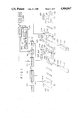
- FIG. 1 is a schematic view showing an example of the radiation image recording and reproducing system wherein an embodiment of the method of reading out a radiation image in accordance with the present invention is employed
- FIGS. 2A and 2B are schematic views showing examples of radiation images recorded in different image recording directions of an object
- FIGS. 3A and 3B are graphic examples of histograms of the read-out image signals detected from stimulable phosphor sheets on which image recording has been carried out in different image recording directions of the object, and
- FIG. 4 is a block diagram showing in detail a part of the radiation image recording and reproducing system shown in FIG. 1.
- a radiation image recording and reproducing system basically comprises a radiation image recording section 20, a preliminary read-out section 30, a final read-out section 40, and an image reproducing section 50.
- radiation 102 is emitted by a radiation source 100 constituted by an X-ray tube or the like toward an object 101.
- a stimulable phosphor sheet 103 for storing radiation energy thereon is placed at the position exposed to the radiation 102 passing through the object 101, and a radiation image of the object 101 is stored on the stimulable phosphor sheet 103.
- the stimulable phosphor sheet 103 carrying the radiation image of the object 101 stored thereon is sent to the preliminary read-out section 30 by a sheet conveyance means 110 constituted by a conveyor roller or the like.
- a laser beam 202 produced by a laser beam source 201 is first passed through a filter 203 for cutting off light having a wavelength within a range identical with the range of the wavelength of the light emitted by the stimulable phosphor sheet 103 upon stimulation thereof by the laser beam 202.
- the laser beam 202 is one-dimensionally deflected by a light deflector 204 such as a galvanometer mirror and directed onto the stimulable phosphor sheet 103 by a plane reflection mirror 205.
- the laser beam source 201 is selected so that the laser beam 202 emanated thereby has a wavelength distribution different from and far apart from the wavelength distribution of the light emitted by the stimulable phosphor sheet 103 upon stimulation thereof. While the laser beam 202 impinges upon the stimulable phosphor sheet 103, the stimulable phosphor sheet 103 is moved in the direction as indicated by the arrow 206 (i.e. in the sub-scanning direction) by a sheet conveyance means 210 constituted by conveyor rollers or the like, and thus the overall surface of the stimulable phosphor sheet 103 is exposed to and scanned by the laser beam 202.
- the power of the laser beam source 201, the beam diameter of the laser beam 202, the scanning speed of the laser beam 202, and the moving speed of the stimulable phosphor sheet 103 are selected so that the level of the stimulation energy of the laser beam 202 for preliminary read-out is lower than the level of the stimulation energy of the laser beam for the final read-out carried out at the final read-out section 40.
- the stimulable phosphor sheet 103 When exposed to the laser beam 202 as mentioned above, the stimulable phosphor sheet 103 emits light in proportion to the radiation energy stored thereon, and the emitted light enters a light guide member 207 which may be of a shape and a material as disclosed in U.S. Pat. No. 4,346,295.
- the light is guided inside of the light guide member 207 through total reflection, projected from a light output face of the light guide member 207 and received by a photodetector 208 constituted by a photomultiplier or the like.
- the light receiving face of the photodetector 208 is closely contacted with a filter for transmitting only light having the wavelength distribution of the light emitted by the stimulable phosphor sheet 103 and cutting off the light having the wavelength distribution of the stimulating rays, so that the photodetector 208 can detect only the light emitted by the stimulable phosphor sheet 103 upon stimulation thereof.
- the light detected by the photodetector 208 is converted into electric signals carrying the image information stored on the stimulable phosphor sheet 103, and amplified by an amplifier 209.
- the signals generated by the amplifier 209 are digitized by an A/D converter 211, and sent as preliminary read-out image signals Sp to a final read-out control circuit 314 at the final read-out section 40.
- the final read-out control circuit 314 calculates a read-out gain setting value (a), a scale factor setting value (b), and a gradation processing condition setting value (c) as one of image processing condition setting values by means of histogram analysis or the like.
- the stimulable phosphor sheet 103 is sent to the final read-out section 40.
- a laser beam 302 produced by a laser beam source 301 is first passed through a filter 303 for cutting off light having a wavelength within the range identical with the range of the wavelength of the light emitted by the stimulable phosphor sheet 103 upon stimulation thereof by the laser beam 302.
- the beam diameter of the laser beam 302 is strictly adjusted by a beam expander 304.
- the laser beam 302 is then deflected by a light deflector 305 formed of a galvanometer mirror or the like, and is made to impinge upon the stimulable phosphor sheet 103 by a plane reflection mirror 306.
- an f ⁇ lens 307 for maintaining the beam diameter of the laser beam 302 uniform in the course of the scanning of the laser beam 302 on the stimulable phosphor sheet 103.
- the stimulable phosphor sheet 103 is moved in the direction as indicated by the arrow 308 (i.e. in the sub-scanning direction) by a sheet conveyance means 320 constituted by conveyor rollers or the like and, consequently, the overall area of the stimulable phosphor sheet 103 is exposed to and scanned by the laser beam 302.
- the stimulable phosphor sheet 103 Upon exposure to the laser beam 302, the stimulable phosphor sheet 103 emits light in proportion to the radiation energy stored thereon, and the light emitted enters a light guide member 309 which is made of the same material and has the same configuration as the light guide member 207 used for the preliminary read-out.
- the light emitted by the stimulable phosphor sheet 103 is guided inside of the light guide member 309 through total reflection, projected from the light output face of the light guide member 309 and received by a photodetector 310 constituted by a photomultiplier or the like.
- the light receiving face of the photodetector 310 is closely contacted with a filter for selectively transmitting only the light having the wavelength distribution of the light emitted by the stimulable phosphor sheet 103, so that the photodetector 310 can detect only the light emitted thereby.
- the output of the photodetector 310 photoelectrically detecting the light emission representing the radiation image stored on the stimulable phosphor sheet 103 is amplified to an appropriate level by an amplifier 311 the read-out gain of which has been adjusted by the read-out gain setting value (a) calculated by the control circuit 314.
- the amplified electric signals are fed to an A/D converter 312 which converts the electric signals into digital signals by use of a scale factor which has been adjusted by the scale factor setting value (b) to suit the width of signal fluctuation.
- the digital signals i.e.
- the final read-out image signals thus obtained are fed to a signal processing circuit 313, in which they are subjected to a gradation processing (signal processing) based on the gradation processing condition setting value (c) so as to obtain a visible radiation image suitable for viewing, particularly for diagnostic purposes, and are output as read-out image signals (final read-out image signals) So.
- a gradation processing signal processing
- c gradation processing condition setting value
- the final read-out image signals So generated by the signal processing circuit 313 are fed to a light modulator 401 at the image reproducing section 50.
- a laser beam 403 produced by a reproducing laser beam source 402 is modulated by the light modulator 401 on the basis of the final read-out image signals So received from the signal processing circuit 313, and is made to impinge upon a photosensitive material 405 such as a photographic film by a scanning mirror 404 for scanning the photosensitive material 405 by the laser beam 403.
- the photosensitive material 405 is moved normal to the aforesaid scanning direction, i.e. in the direction as indicated by the arrow 406. Accordingly, the radiation image represented by the final read-out image signals So is recorded on the photosensitive material 405.
- any other appropriate method such as the aforesaid display on a CRT.
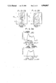
- FIG. 4 shows in detail the configuration of the density correction control circuit 500.
- a signal extracting section 501 of the density correction control circuit 500 extracts only the image signals in a partial region P at the predetermined position on the stimulable phosphor sheet 103 from the fed preliminary read-out image signals Sp.
- the partial region P is specified by a region setting section 502.
- the partial region P is specified to a size of approximately, 5cm ⁇ 5cm or 7cm ⁇ 7cm at the center of the stimulable phosphor sheet 103 as shown in FIGS. 2A and 2B.
- the case of reading out the thoracic vertebra image will hereinbelow be described by way of example.
- Preliminary read-out image signals Sp' extracted as mentioned above only in the partial region P are sent to an analysis section 503.
- the analysis section 503 creates a histogram of the thus extracted preliminary read-out image signals Sp', and calculates a mean value Save thereof.
- the mean value Save as a representative value indicating the amount of light emission by the partial region P is sent to an operating section 504.
- the operating section 504 also receives the maximum value Smax and the minimum value Smin of the whole preliminary read-out image signals Sp which are calculated by the final read-out control circuit 314.
- the operating section 504 calculates a discrimination value Q expressed as
- the discrimination value Q is fed to a comparing section 505, which compares the discrimination value Q with a reference value R received from a reference value setting section 506. When the discrimination value Q is larger than the reference value R, the comparing section 505 feeds a correction signal T to a read-out gain correcting circuit 507. Upon receiving the correction signal T, the read-out gain correcting circuit 507 corrects the read-out gain setting value (a), which has been calculated as mentioned above by the final read-out control circuit 314, so as to decrease the read-out gain.
- the discrimination value Q is comparatively larger in the case of side image recording than in the case of front image recording.
- the read-out conditions for the final read-out and/or the image processing conditions are constant, the density at the image portion of the thoracic vertebrae K in the reproduced visible image becomes higher in the case of side image recording than in the case of front image recording. To eliminate this problem, the read-out gain is decreased by correcting as mentioned above in the case of side image recording.
- the levels of the final read-out image signals So become generally low, and the density of the visible radiation image reproduced on the photosensitive material 405 becomes low as a whole. Accordingly, the density of the image portion of the thoracic vertebrae K in the visible image reproduced in the case of side image recording is adjusted to be equal to the density of the image portion of the thoracic vertebrae K in the visible image reproduced in the case of front image recording.
- the appropriate correction amount of the read-out gain can be determined experimentally.
- the final read-out for the radiation image recorded by front image recording is carried out without changing the read-out gain as adjusted by the final read-out control circuit 314, and the read-out gain is corrected to a smaller value when reading out the radiation image recorded by side image recording.
- the final read out for the radiation image recorded by side image recording may be carried out without changing the read-out gain as adjusted by the final read-out control circuit 314, and the read-out gain may be corrected to a larger value when reading out the radiation image recorded by front image recording.
- the read-out gain setting value (a) calculated by the final read-out control circuit 314 may be increased or decreased in accordance with the aforesaid discrimination value Q.
- the conditions of the scale factor in the A/D converter 312 may be changed, or the gradation processing conditions in the signal processing circuit 313 may be changed. Or, these methods of adjusting the density may be employed in combination with each other.
- the gradation processing conditions are usually expressed by a non-linear gradation curve.
- the gradation curve may be shifted vertically or horizontally in accordance with the correction signal T, or may be rotated around a predetermined position on the gradation curve, so that the density of the region concerned in the reproduced visible image is made constant.
- the density of the thoracic vertebra image portion i.e the image-recording region, is made constant in the manner as mentioned above between the cases of front image recording and side image recording of the thoracic vertebra image.
- the method of reading out a radiation image in accordance with the present invention is applicable in the same manner also to the case where a radiation image is read out from the stimulable phosphor sheet carrying a radiation image of the other object portions such as the lumbar vertebrae recorded thereon, or the stimulable phosphor sheets on which image recording has been carried out in various different image recording directions of the object, for example, by front image recording and oblique image recording.
- the position and the size of the partial region P and the reference value R may be adjusted to appropriate values in each case.
- the position and the size of the partial region P and the reference value R may be adjusted manually in accordance with the image recording portion and the image recording direction of the image which is to be read out.
- the position and the size of the partial region P and the reference value R may be set automatically based on the entered information at the region setting section 502 and the reference value setting section 506.
- the preliminary read-out section and the final read-out section are disposed independently.
- a single read-out system may be used for the preliminary read-out and the final read-out.
- the stimulable phosphor sheet is returned to the read-out system by a sheet conveyance means and then the final read-out is carried out.
- the stimulation energy of the stimulating rays is adjusted by a stimulating ray energy adjusting means to be lower than the stimulation energy of the stimulating rays used in the final read-out.
- the present invention is also applicable to such a case.
- the present invention is applicable also to the case of subdivision image recording wherein a single stimulable phosphor sheet is divided into, for example, two subdivisions and image recording is carried out at each subdivision.
- the present invention may be applied to each of the subdivisions.
- the image signals for condition adjustment are obtained by the preliminary read-out, and the read-out conditions for the final read-out and/or the image processing conditions are adjusted based on the discrimination value representing the relationship between the characteristic value of the image signals for condition adjustment and the aforesaid representative value of the image signals in the partial region extracted from the image signals for condition adjustment.
- the final read-out image signals may be utilized as the image signals for condition adjustment.
- the final read-out image signals may be stored in a storage means, and the image processing conditions may be adjusted based on the discrimination value calculated in the same manner as mentioned above. Then, the final read-out image signals may be read from the storage means, and the image processing of the final read-out image signals may be carried out by use of the thus adjusted image processing conditions.
Landscapes
- Physics & Mathematics (AREA)
- Health & Medical Sciences (AREA)
- Life Sciences & Earth Sciences (AREA)
- General Physics & Mathematics (AREA)
- High Energy & Nuclear Physics (AREA)
- Molecular Biology (AREA)
- Spectroscopy & Molecular Physics (AREA)
- Radiography Using Non-Light Waves (AREA)
- Apparatus For Radiation Diagnosis (AREA)
- Image Analysis (AREA)
Abstract
Description
Q=(Save-Smin)/(Smax-Smin)
Q=(Save-Smin)/(Smax-Smin)
Q=(Save-Smin)/(Smax-Smin),
(Smax-Save)/(Smax-Smin),
Claims (13)
(Save-Smin)/(Smax-Smin).
(Smax-Save)/(Smax-Smin).
(Save-Smin)/(Smax-Smin).
(Smax-Save)/(Smax-Smin).
(Save-Smin)/(Smax-Smin).
(Smax-Save)/(Smax-Smin).
Applications Claiming Priority (4)
| Application Number | Priority Date | Filing Date | Title |
|---|---|---|---|
| JP61-137660 | 1986-06-13 | ||
| JP13766086 | 1986-06-13 | ||
| JP62-17038 | 1987-01-27 | ||
| JP62017038A JP2717653B2 (en) | 1986-06-13 | 1987-01-27 | Radiation image information reading method |
Publications (1)
| Publication Number | Publication Date |
|---|---|
| US4904867A true US4904867A (en) | 1990-02-27 |
Family
ID=26353493
Family Applications (1)
| Application Number | Title | Priority Date | Filing Date |
|---|---|---|---|
| US07/061,366 Expired - Lifetime US4904867A (en) | 1986-06-13 | 1987-06-15 | Radiation image read-out method and apparatus |
Country Status (1)
| Country | Link |
|---|---|
| US (1) | US4904867A (en) |
Cited By (2)
| Publication number | Priority date | Publication date | Assignee | Title |
|---|---|---|---|---|
| US5006707A (en) * | 1989-01-24 | 1991-04-09 | Fuji Photo Film Co., Ltd. | Electron microscope image recording and read-out method |
| EP3260845A3 (en) * | 2016-06-21 | 2018-05-02 | Fujifilm Corporation | Image reader using a photomultiplier, in which the optimal value for the voltage controlling the gain is calculated from a selected region of a prescan |
Citations (2)
| Publication number | Priority date | Publication date | Assignee | Title |
|---|---|---|---|---|
| US4620097A (en) * | 1983-11-17 | 1986-10-28 | Fuji Photo Film Co., Ltd. | Method of adjusting scale factor for radiation image |
| US4682028A (en) * | 1984-01-26 | 1987-07-21 | Fuji Photo Film Co., Ltd. | Method of adjusting radiation image read-out conditions |
-
1987
- 1987-06-15 US US07/061,366 patent/US4904867A/en not_active Expired - Lifetime
Patent Citations (2)
| Publication number | Priority date | Publication date | Assignee | Title |
|---|---|---|---|---|
| US4620097A (en) * | 1983-11-17 | 1986-10-28 | Fuji Photo Film Co., Ltd. | Method of adjusting scale factor for radiation image |
| US4682028A (en) * | 1984-01-26 | 1987-07-21 | Fuji Photo Film Co., Ltd. | Method of adjusting radiation image read-out conditions |
Cited By (2)
| Publication number | Priority date | Publication date | Assignee | Title |
|---|---|---|---|---|
| US5006707A (en) * | 1989-01-24 | 1991-04-09 | Fuji Photo Film Co., Ltd. | Electron microscope image recording and read-out method |
| EP3260845A3 (en) * | 2016-06-21 | 2018-05-02 | Fujifilm Corporation | Image reader using a photomultiplier, in which the optimal value for the voltage controlling the gain is calculated from a selected region of a prescan |
Similar Documents
| Publication | Publication Date | Title |
|---|---|---|
| US4682028A (en) | Method of adjusting radiation image read-out conditions | |
| US4804842A (en) | Radiation image read-out method and apparatus | |
| US4887305A (en) | Method of adjusting read-out conditions for radiation image | |
| US4903310A (en) | Method of automatically determining imaged body posture in medical image display | |
| US4950894A (en) | Radiation image read-out method | |
| US4638162A (en) | Method of adjusting radiation image read-out condition | |
| US5028782A (en) | Method of adjusting image processing conditions | |
| US4999497A (en) | Radiation image read-out and reproducing method and apparatus | |
| US4859850A (en) | Irradiation field recognizing method, and method of adjusting image processing conditions using the same | |
| EP0177801B1 (en) | Radiation image read-out method and apparatus | |
| US4994662A (en) | Radiation image read-out apparatus and method for operating the same | |
| US4861993A (en) | Radiation image read-out method | |
| US4896038A (en) | Radiation image read-out method and apparatus | |
| US4851701A (en) | Radiation image read-out method and apparatus | |
| EP0145982B1 (en) | Method of adjusting scale factor for radiation image | |
| US4804841A (en) | Radiation image read-out method | |
| US5764791A (en) | Method for determining the shape and location of an irradiation field | |
| US4904867A (en) | Radiation image read-out method and apparatus | |
| US20020136441A1 (en) | Energy subtraction processing method and apparatus | |
| US4873437A (en) | Radiation image read-out method and apparatus | |
| US4877958A (en) | Radiation image read-out method and apparatus | |
| US4943723A (en) | Radiation image read-out method | |
| US4851675A (en) | Radiation image read-out method and apparatus | |
| US4810887A (en) | Radiation image read-out method and apparatus | |
| US4861994A (en) | Method of measuring after-glow of stimulable phosphor sheet, and method of adjusting radiation image read-out conditions |
Legal Events
| Date | Code | Title | Description |
|---|---|---|---|
| AS | Assignment |
Owner name: FUJI PHOTO FILM CO., LTD. Free format text: ASSIGNMENT OF ASSIGNORS INTEREST.;ASSIGNOR:ADACHI, YUUMA;REEL/FRAME:005199/0164 Effective date: 19870609 |
|
| STCF | Information on status: patent grant |
Free format text: PATENTED CASE |
|
| FEPP | Fee payment procedure |
Free format text: PAYOR NUMBER ASSIGNED (ORIGINAL EVENT CODE: ASPN); ENTITY STATUS OF PATENT OWNER: LARGE ENTITY |
|
| FPAY | Fee payment |
Year of fee payment: 4 |
|
| FEPP | Fee payment procedure |
Free format text: PAYER NUMBER DE-ASSIGNED (ORIGINAL EVENT CODE: RMPN); ENTITY STATUS OF PATENT OWNER: LARGE ENTITY Free format text: PAYOR NUMBER ASSIGNED (ORIGINAL EVENT CODE: ASPN); ENTITY STATUS OF PATENT OWNER: LARGE ENTITY |
|
| FPAY | Fee payment |
Year of fee payment: 8 |
|
| FEPP | Fee payment procedure |
Free format text: PAYER NUMBER DE-ASSIGNED (ORIGINAL EVENT CODE: RMPN); ENTITY STATUS OF PATENT OWNER: LARGE ENTITY Free format text: PAYOR NUMBER ASSIGNED (ORIGINAL EVENT CODE: ASPN); ENTITY STATUS OF PATENT OWNER: LARGE ENTITY |
|
| FPAY | Fee payment |
Year of fee payment: 12 |
|
| AS | Assignment |
Owner name: FUJIFILM CORPORATION, JAPAN Free format text: ASSIGNMENT OF ASSIGNORS INTEREST;ASSIGNOR:FUJIFILM HOLDINGS CORPORATION (FORMERLY FUJI PHOTO FILM CO., LTD.);REEL/FRAME:020817/0190 Effective date: 20080225 Owner name: FUJIFILM CORPORATION,JAPAN Free format text: ASSIGNMENT OF ASSIGNORS INTEREST;ASSIGNOR:FUJIFILM HOLDINGS CORPORATION (FORMERLY FUJI PHOTO FILM CO., LTD.);REEL/FRAME:020817/0190 Effective date: 20080225 |