US20030232350A1 - Methods of diagnosis of cancer, compositions and methods of screening for modulators of cancer - Google Patents
Methods of diagnosis of cancer, compositions and methods of screening for modulators of cancerInfo
- Publication number
- US20030232350A1 US20030232350A1 US10/295,027 US29502702A US2003232350A1 US 20030232350 A1 US20030232350 A1 US 20030232350A1 US 29502702 A US29502702 A US 29502702A US 2003232350 A1 US2003232350 A1 US 2003232350A1
- Authority
- US
- United States
- Prior art keywords
- cancer
- sequence
- protein
- nucleic acid
- proteins
- Prior art date
- Legal status (The legal status is an assumption and is not a legal conclusion. Google has not performed a legal analysis and makes no representation as to the accuracy of the status listed.)
- Abandoned
Links
- 206010028980 Neoplasm Diseases 0.000 title claims abstract description 503
- 238000000034 method Methods 0.000 title claims abstract description 175
- 201000011510 cancer Diseases 0.000 title claims description 471
- 239000000203 mixture Substances 0.000 title abstract description 42
- 238000003745 diagnosis Methods 0.000 title abstract description 20
- 238000012216 screening Methods 0.000 title description 30
- 108090000623 proteins and genes Proteins 0.000 claims abstract description 470
- 230000014509 gene expression Effects 0.000 claims abstract description 144
- 238000011282 treatment Methods 0.000 claims abstract description 30
- 102000004169 proteins and genes Human genes 0.000 claims description 366
- 150000007523 nucleic acids Chemical class 0.000 claims description 224
- 102000039446 nucleic acids Human genes 0.000 claims description 198
- 108020004707 nucleic acids Proteins 0.000 claims description 198
- 108090000765 processed proteins & peptides Proteins 0.000 claims description 149
- 102000004196 processed proteins & peptides Human genes 0.000 claims description 124
- 229920001184 polypeptide Polymers 0.000 claims description 105
- 239000000523 sample Substances 0.000 claims description 89
- 150000001875 compounds Chemical class 0.000 claims description 85
- 230000000694 effects Effects 0.000 claims description 65
- 102000040430 polynucleotide Human genes 0.000 claims description 36
- 108091033319 polynucleotide Proteins 0.000 claims description 36
- 239000002157 polynucleotide Substances 0.000 claims description 36
- 238000012360 testing method Methods 0.000 claims description 36
- 239000013604 expression vector Substances 0.000 claims description 35
- 239000000126 substance Substances 0.000 claims description 34
- 108020004999 messenger RNA Proteins 0.000 claims description 26
- 238000000018 DNA microarray Methods 0.000 claims description 20
- 241000124008 Mammalia Species 0.000 claims description 20
- 108020004711 Nucleic Acid Probes Proteins 0.000 claims description 20
- 239000002853 nucleic acid probe Substances 0.000 claims description 20
- 239000012472 biological sample Substances 0.000 claims description 18
- 230000007170 pathology Effects 0.000 claims description 18
- 239000012636 effector Substances 0.000 claims description 12
- 230000001575 pathological effect Effects 0.000 claims description 12
- 210000002966 serum Anatomy 0.000 claims description 12
- 210000004369 blood Anatomy 0.000 claims description 8
- 239000008280 blood Substances 0.000 claims description 8
- 239000007850 fluorescent dye Substances 0.000 claims description 8
- 210000000056 organ Anatomy 0.000 claims description 8
- 108010021625 Immunoglobulin Fragments Proteins 0.000 claims description 7
- 102000008394 Immunoglobulin Fragments Human genes 0.000 claims description 6
- 230000008685 targeting Effects 0.000 claims description 6
- 238000007878 drug screening assay Methods 0.000 claims description 4
- 231100000433 cytotoxic Toxicity 0.000 claims description 3
- 230000001472 cytotoxic effect Effects 0.000 claims description 3
- 238000011285 therapeutic regimen Methods 0.000 claims description 2
- 210000002700 urine Anatomy 0.000 claims description 2
- 208000037265 diseases, disorders, signs and symptoms Diseases 0.000 abstract description 38
- 201000010099 disease Diseases 0.000 abstract description 32
- 230000001105 regulatory effect Effects 0.000 abstract description 21
- 238000004393 prognosis Methods 0.000 abstract description 14
- 235000018102 proteins Nutrition 0.000 description 351
- 210000004027 cell Anatomy 0.000 description 195
- 230000027455 binding Effects 0.000 description 82
- 238000009739 binding Methods 0.000 description 80
- 210000001519 tissue Anatomy 0.000 description 75
- 238000003556 assay Methods 0.000 description 65
- 239000003795 chemical substances by application Substances 0.000 description 52
- 235000001014 amino acid Nutrition 0.000 description 50
- 229940024606 amino acid Drugs 0.000 description 44
- 150000001413 amino acids Chemical class 0.000 description 44
- 102000048850 Neoplasm Genes Human genes 0.000 description 39
- 108700019961 Neoplasm Genes Proteins 0.000 description 39
- 238000009396 hybridization Methods 0.000 description 36
- 125000003729 nucleotide group Chemical group 0.000 description 36
- 239000002773 nucleotide Substances 0.000 description 35
- 125000003275 alpha amino acid group Chemical group 0.000 description 34
- 241000282414 Homo sapiens Species 0.000 description 32
- -1 antibodies Chemical class 0.000 description 32
- 239000012634 fragment Substances 0.000 description 30
- 230000000692 anti-sense effect Effects 0.000 description 29
- 230000001225 therapeutic effect Effects 0.000 description 26
- 108020004414 DNA Proteins 0.000 description 25
- 238000003752 polymerase chain reaction Methods 0.000 description 25
- 239000013598 vector Substances 0.000 description 25
- 239000000427 antigen Substances 0.000 description 24
- 108091007433 antigens Proteins 0.000 description 24
- 102000036639 antigens Human genes 0.000 description 24
- 239000003814 drug Substances 0.000 description 24
- 239000000047 product Substances 0.000 description 23
- 239000007787 solid Substances 0.000 description 21
- 108091028043 Nucleic acid sequence Proteins 0.000 description 20
- 239000003153 chemical reaction reagent Substances 0.000 description 20
- 238000006467 substitution reaction Methods 0.000 description 20
- 230000003834 intracellular effect Effects 0.000 description 19
- 108091032973 (ribonucleotides)n+m Proteins 0.000 description 18
- 108091034117 Oligonucleotide Proteins 0.000 description 18
- 230000012010 growth Effects 0.000 description 18
- 235000002639 sodium chloride Nutrition 0.000 description 18
- 230000000295 complement effect Effects 0.000 description 17
- 239000003446 ligand Substances 0.000 description 17
- 239000002671 adjuvant Substances 0.000 description 16
- 238000004458 analytical method Methods 0.000 description 16
- 238000004422 calculation algorithm Methods 0.000 description 16
- 238000001514 detection method Methods 0.000 description 16
- 238000013518 transcription Methods 0.000 description 16
- 230000035897 transcription Effects 0.000 description 16
- 229960005486 vaccine Drugs 0.000 description 16
- 238000000338 in vitro Methods 0.000 description 15
- 150000003839 salts Chemical class 0.000 description 15
- 239000000758 substrate Substances 0.000 description 15
- 108091005461 Nucleic proteins Proteins 0.000 description 14
- 230000003321 amplification Effects 0.000 description 14
- 229940079593 drug Drugs 0.000 description 14
- 238000005516 engineering process Methods 0.000 description 14
- 230000006870 function Effects 0.000 description 14
- 239000003112 inhibitor Substances 0.000 description 14
- 239000000463 material Substances 0.000 description 14
- 238000003199 nucleic acid amplification method Methods 0.000 description 14
- 108090000994 Catalytic RNA Proteins 0.000 description 13
- 102000053642 Catalytic RNA Human genes 0.000 description 13
- 108060003951 Immunoglobulin Proteins 0.000 description 13
- 230000015572 biosynthetic process Effects 0.000 description 13
- 230000000875 corresponding effect Effects 0.000 description 13
- 238000013537 high throughput screening Methods 0.000 description 13
- 102000018358 immunoglobulin Human genes 0.000 description 13
- 108091092562 ribozyme Proteins 0.000 description 13
- 102000035160 transmembrane proteins Human genes 0.000 description 13
- 108091005703 transmembrane proteins Proteins 0.000 description 13
- 108020004705 Codon Proteins 0.000 description 12
- 241001465754 Metazoa Species 0.000 description 12
- 241000699666 Mus <mouse, genus> Species 0.000 description 12
- 238000013459 approach Methods 0.000 description 12
- 239000003102 growth factor Substances 0.000 description 12
- 102000005962 receptors Human genes 0.000 description 12
- JLCPHMBAVCMARE-UHFFFAOYSA-N [3-[[3-[[3-[[3-[[3-[[3-[[3-[[3-[[3-[[3-[[3-[[5-(2-amino-6-oxo-1H-purin-9-yl)-3-[[3-[[3-[[3-[[3-[[3-[[5-(2-amino-6-oxo-1H-purin-9-yl)-3-[[5-(2-amino-6-oxo-1H-purin-9-yl)-3-hydroxyoxolan-2-yl]methoxy-hydroxyphosphoryl]oxyoxolan-2-yl]methoxy-hydroxyphosphoryl]oxy-5-(5-methyl-2,4-dioxopyrimidin-1-yl)oxolan-2-yl]methoxy-hydroxyphosphoryl]oxy-5-(6-aminopurin-9-yl)oxolan-2-yl]methoxy-hydroxyphosphoryl]oxy-5-(6-aminopurin-9-yl)oxolan-2-yl]methoxy-hydroxyphosphoryl]oxy-5-(6-aminopurin-9-yl)oxolan-2-yl]methoxy-hydroxyphosphoryl]oxy-5-(6-aminopurin-9-yl)oxolan-2-yl]methoxy-hydroxyphosphoryl]oxyoxolan-2-yl]methoxy-hydroxyphosphoryl]oxy-5-(5-methyl-2,4-dioxopyrimidin-1-yl)oxolan-2-yl]methoxy-hydroxyphosphoryl]oxy-5-(4-amino-2-oxopyrimidin-1-yl)oxolan-2-yl]methoxy-hydroxyphosphoryl]oxy-5-(5-methyl-2,4-dioxopyrimidin-1-yl)oxolan-2-yl]methoxy-hydroxyphosphoryl]oxy-5-(5-methyl-2,4-dioxopyrimidin-1-yl)oxolan-2-yl]methoxy-hydroxyphosphoryl]oxy-5-(6-aminopurin-9-yl)oxolan-2-yl]methoxy-hydroxyphosphoryl]oxy-5-(6-aminopurin-9-yl)oxolan-2-yl]methoxy-hydroxyphosphoryl]oxy-5-(4-amino-2-oxopyrimidin-1-yl)oxolan-2-yl]methoxy-hydroxyphosphoryl]oxy-5-(4-amino-2-oxopyrimidin-1-yl)oxolan-2-yl]methoxy-hydroxyphosphoryl]oxy-5-(4-amino-2-oxopyrimidin-1-yl)oxolan-2-yl]methoxy-hydroxyphosphoryl]oxy-5-(6-aminopurin-9-yl)oxolan-2-yl]methoxy-hydroxyphosphoryl]oxy-5-(4-amino-2-oxopyrimidin-1-yl)oxolan-2-yl]methyl [5-(6-aminopurin-9-yl)-2-(hydroxymethyl)oxolan-3-yl] hydrogen phosphate Polymers Cc1cn(C2CC(OP(O)(=O)OCC3OC(CC3OP(O)(=O)OCC3OC(CC3O)n3cnc4c3nc(N)[nH]c4=O)n3cnc4c3nc(N)[nH]c4=O)C(COP(O)(=O)OC3CC(OC3COP(O)(=O)OC3CC(OC3COP(O)(=O)OC3CC(OC3COP(O)(=O)OC3CC(OC3COP(O)(=O)OC3CC(OC3COP(O)(=O)OC3CC(OC3COP(O)(=O)OC3CC(OC3COP(O)(=O)OC3CC(OC3COP(O)(=O)OC3CC(OC3COP(O)(=O)OC3CC(OC3COP(O)(=O)OC3CC(OC3COP(O)(=O)OC3CC(OC3COP(O)(=O)OC3CC(OC3COP(O)(=O)OC3CC(OC3COP(O)(=O)OC3CC(OC3COP(O)(=O)OC3CC(OC3COP(O)(=O)OC3CC(OC3CO)n3cnc4c(N)ncnc34)n3ccc(N)nc3=O)n3cnc4c(N)ncnc34)n3ccc(N)nc3=O)n3ccc(N)nc3=O)n3ccc(N)nc3=O)n3cnc4c(N)ncnc34)n3cnc4c(N)ncnc34)n3cc(C)c(=O)[nH]c3=O)n3cc(C)c(=O)[nH]c3=O)n3ccc(N)nc3=O)n3cc(C)c(=O)[nH]c3=O)n3cnc4c3nc(N)[nH]c4=O)n3cnc4c(N)ncnc34)n3cnc4c(N)ncnc34)n3cnc4c(N)ncnc34)n3cnc4c(N)ncnc34)O2)c(=O)[nH]c1=O JLCPHMBAVCMARE-UHFFFAOYSA-N 0.000 description 11
- 238000007792 addition Methods 0.000 description 11
- 230000008859 change Effects 0.000 description 11
- 230000007423 decrease Effects 0.000 description 11
- 238000001727 in vivo Methods 0.000 description 11
- 230000001965 increasing effect Effects 0.000 description 11
- 230000004048 modification Effects 0.000 description 11
- 238000012986 modification Methods 0.000 description 11
- 108020003175 receptors Proteins 0.000 description 11
- YBJHBAHKTGYVGT-ZKWXMUAHSA-N (+)-Biotin Chemical compound N1C(=O)N[C@@H]2[C@H](CCCCC(=O)O)SC[C@@H]21 YBJHBAHKTGYVGT-ZKWXMUAHSA-N 0.000 description 10
- 108091026890 Coding region Proteins 0.000 description 10
- 108091093037 Peptide nucleic acid Proteins 0.000 description 10
- 239000002253 acid Substances 0.000 description 10
- 239000012190 activator Substances 0.000 description 10
- 150000001720 carbohydrates Chemical class 0.000 description 10
- 230000001413 cellular effect Effects 0.000 description 10
- 238000006243 chemical reaction Methods 0.000 description 10
- 238000001415 gene therapy Methods 0.000 description 10
- 230000013595 glycosylation Effects 0.000 description 10
- 238000006206 glycosylation reaction Methods 0.000 description 10
- 230000003211 malignant effect Effects 0.000 description 10
- 239000003053 toxin Substances 0.000 description 10
- 102000004190 Enzymes Human genes 0.000 description 9
- 108090000790 Enzymes Proteins 0.000 description 9
- 241000699670 Mus sp. Species 0.000 description 9
- 239000002246 antineoplastic agent Substances 0.000 description 9
- 230000001580 bacterial effect Effects 0.000 description 9
- 229940088598 enzyme Drugs 0.000 description 9
- 230000002401 inhibitory effect Effects 0.000 description 9
- 239000002777 nucleoside Substances 0.000 description 9
- 231100000765 toxin Toxicity 0.000 description 9
- 108700012359 toxins Proteins 0.000 description 9
- 108010090804 Streptavidin Proteins 0.000 description 8
- 238000012217 deletion Methods 0.000 description 8
- 230000037430 deletion Effects 0.000 description 8
- 230000002255 enzymatic effect Effects 0.000 description 8
- 238000011156 evaluation Methods 0.000 description 8
- 238000003780 insertion Methods 0.000 description 8
- 230000037431 insertion Effects 0.000 description 8
- 238000012544 monitoring process Methods 0.000 description 8
- 238000011002 quantification Methods 0.000 description 8
- 150000003384 small molecules Chemical class 0.000 description 8
- 241000894007 species Species 0.000 description 8
- 238000003786 synthesis reaction Methods 0.000 description 8
- 230000004614 tumor growth Effects 0.000 description 8
- 238000005406 washing Methods 0.000 description 8
- 108010041986 DNA Vaccines Proteins 0.000 description 7
- 229940021995 DNA vaccine Drugs 0.000 description 7
- 150000007513 acids Chemical class 0.000 description 7
- 230000004075 alteration Effects 0.000 description 7
- 238000003491 array Methods 0.000 description 7
- 230000004071 biological effect Effects 0.000 description 7
- 230000010261 cell growth Effects 0.000 description 7
- 229940127089 cytotoxic agent Drugs 0.000 description 7
- 229940000406 drug candidate Drugs 0.000 description 7
- 230000028993 immune response Effects 0.000 description 7
- 229940124452 immunizing agent Drugs 0.000 description 7
- 238000011534 incubation Methods 0.000 description 7
- 230000001939 inductive effect Effects 0.000 description 7
- 125000005647 linker group Chemical group 0.000 description 7
- 239000002502 liposome Substances 0.000 description 7
- 239000003550 marker Substances 0.000 description 7
- 229920000642 polymer Polymers 0.000 description 7
- 238000002360 preparation method Methods 0.000 description 7
- 238000000746 purification Methods 0.000 description 7
- 238000011160 research Methods 0.000 description 7
- 238000002560 therapeutic procedure Methods 0.000 description 7
- 230000009466 transformation Effects 0.000 description 7
- 229920001817 Agar Polymers 0.000 description 6
- 241000894006 Bacteria Species 0.000 description 6
- 206010006187 Breast cancer Diseases 0.000 description 6
- ZHNUHDYFZUAESO-UHFFFAOYSA-N Formamide Chemical compound NC=O ZHNUHDYFZUAESO-UHFFFAOYSA-N 0.000 description 6
- 241000238631 Hexapoda Species 0.000 description 6
- 206010027476 Metastases Diseases 0.000 description 6
- 108091030071 RNAI Proteins 0.000 description 6
- 241000700159 Rattus Species 0.000 description 6
- 241000283984 Rodentia Species 0.000 description 6
- 240000004808 Saccharomyces cerevisiae Species 0.000 description 6
- 235000014680 Saccharomyces cerevisiae Nutrition 0.000 description 6
- 230000004913 activation Effects 0.000 description 6
- 239000008272 agar Substances 0.000 description 6
- 125000000539 amino acid group Chemical group 0.000 description 6
- 125000003277 amino group Chemical group 0.000 description 6
- 238000011161 development Methods 0.000 description 6
- 230000018109 developmental process Effects 0.000 description 6
- 208000035475 disorder Diseases 0.000 description 6
- 125000000524 functional group Chemical group 0.000 description 6
- 230000004927 fusion Effects 0.000 description 6
- 239000000499 gel Substances 0.000 description 6
- 230000009368 gene silencing by RNA Effects 0.000 description 6
- 230000002068 genetic effect Effects 0.000 description 6
- 238000003018 immunoassay Methods 0.000 description 6
- 230000002163 immunogen Effects 0.000 description 6
- 230000003993 interaction Effects 0.000 description 6
- 210000004962 mammalian cell Anatomy 0.000 description 6
- 108010082117 matrigel Proteins 0.000 description 6
- 230000001404 mediated effect Effects 0.000 description 6
- 230000009401 metastasis Effects 0.000 description 6
- 150000003833 nucleoside derivatives Chemical class 0.000 description 6
- 238000007423 screening assay Methods 0.000 description 6
- 238000003860 storage Methods 0.000 description 6
- 230000002103 transcriptional effect Effects 0.000 description 6
- 210000004881 tumor cell Anatomy 0.000 description 6
- 230000003612 virological effect Effects 0.000 description 6
- 102000004127 Cytokines Human genes 0.000 description 5
- 108090000695 Cytokines Proteins 0.000 description 5
- 238000002965 ELISA Methods 0.000 description 5
- 241000282412 Homo Species 0.000 description 5
- 241000288906 Primates Species 0.000 description 5
- FAPWRFPIFSIZLT-UHFFFAOYSA-M Sodium chloride Chemical compound [Na+].[Cl-] FAPWRFPIFSIZLT-UHFFFAOYSA-M 0.000 description 5
- IQFYYKKMVGJFEH-XLPZGREQSA-N Thymidine Chemical compound O=C1NC(=O)C(C)=CN1[C@@H]1O[C@H](CO)[C@@H](O)C1 IQFYYKKMVGJFEH-XLPZGREQSA-N 0.000 description 5
- 150000001412 amines Chemical group 0.000 description 5
- 210000004102 animal cell Anatomy 0.000 description 5
- 238000010171 animal model Methods 0.000 description 5
- 230000001093 anti-cancer Effects 0.000 description 5
- 229960002685 biotin Drugs 0.000 description 5
- 235000020958 biotin Nutrition 0.000 description 5
- 239000011616 biotin Substances 0.000 description 5
- 239000002299 complementary DNA Substances 0.000 description 5
- 239000000470 constituent Substances 0.000 description 5
- 230000001186 cumulative effect Effects 0.000 description 5
- 239000002254 cytotoxic agent Substances 0.000 description 5
- 231100000599 cytotoxic agent Toxicity 0.000 description 5
- 230000003828 downregulation Effects 0.000 description 5
- 239000003623 enhancer Substances 0.000 description 5
- 108020001507 fusion proteins Proteins 0.000 description 5
- 102000037865 fusion proteins Human genes 0.000 description 5
- 210000004408 hybridoma Anatomy 0.000 description 5
- 229910052739 hydrogen Inorganic materials 0.000 description 5
- 239000001257 hydrogen Substances 0.000 description 5
- 238000003384 imaging method Methods 0.000 description 5
- 230000003053 immunization Effects 0.000 description 5
- 238000002649 immunization Methods 0.000 description 5
- 230000005764 inhibitory process Effects 0.000 description 5
- 238000002372 labelling Methods 0.000 description 5
- 230000003902 lesion Effects 0.000 description 5
- 238000004519 manufacturing process Methods 0.000 description 5
- 230000007246 mechanism Effects 0.000 description 5
- 239000002609 medium Substances 0.000 description 5
- 206010061289 metastatic neoplasm Diseases 0.000 description 5
- 239000008194 pharmaceutical composition Substances 0.000 description 5
- 230000005855 radiation Effects 0.000 description 5
- 230000028327 secretion Effects 0.000 description 5
- 239000000725 suspension Substances 0.000 description 5
- 238000013519 translation Methods 0.000 description 5
- 241000283690 Bos taurus Species 0.000 description 4
- 108091003079 Bovine Serum Albumin Proteins 0.000 description 4
- 208000026310 Breast neoplasm Diseases 0.000 description 4
- 108010001857 Cell Surface Receptors Proteins 0.000 description 4
- 108010037362 Extracellular Matrix Proteins Proteins 0.000 description 4
- 102000010834 Extracellular Matrix Proteins Human genes 0.000 description 4
- 108010091358 Hypoxanthine Phosphoribosyltransferase Proteins 0.000 description 4
- XEEYBQQBJWHFJM-UHFFFAOYSA-N Iron Chemical compound [Fe] XEEYBQQBJWHFJM-UHFFFAOYSA-N 0.000 description 4
- 241000764238 Isis Species 0.000 description 4
- QNAYBMKLOCPYGJ-REOHCLBHSA-N L-alanine Chemical compound C[C@H](N)C(O)=O QNAYBMKLOCPYGJ-REOHCLBHSA-N 0.000 description 4
- FFEARJCKVFRZRR-BYPYZUCNSA-N L-methionine Chemical group CSCC[C@H](N)C(O)=O FFEARJCKVFRZRR-BYPYZUCNSA-N 0.000 description 4
- 206010058467 Lung neoplasm malignant Diseases 0.000 description 4
- KDXKERNSBIXSRK-UHFFFAOYSA-N Lysine Natural products NCCCCC(N)C(O)=O KDXKERNSBIXSRK-UHFFFAOYSA-N 0.000 description 4
- 239000004472 Lysine Substances 0.000 description 4
- 108700026244 Open Reading Frames Proteins 0.000 description 4
- 102000035195 Peptidases Human genes 0.000 description 4
- 108091005804 Peptidases Proteins 0.000 description 4
- 108010076504 Protein Sorting Signals Proteins 0.000 description 4
- 108700008625 Reporter Genes Proteins 0.000 description 4
- VYPSYNLAJGMNEJ-UHFFFAOYSA-N Silicium dioxide Chemical compound O=[Si]=O VYPSYNLAJGMNEJ-UHFFFAOYSA-N 0.000 description 4
- 235000004279 alanine Nutrition 0.000 description 4
- 230000033115 angiogenesis Effects 0.000 description 4
- 239000005557 antagonist Substances 0.000 description 4
- 229940049706 benzodiazepine Drugs 0.000 description 4
- 125000003310 benzodiazepinyl group Chemical class N1N=C(C=CC2=C1C=CC=C2)* 0.000 description 4
- 229940098773 bovine serum albumin Drugs 0.000 description 4
- 239000000872 buffer Substances 0.000 description 4
- 235000014633 carbohydrates Nutrition 0.000 description 4
- 125000003178 carboxy group Chemical group [H]OC(*)=O 0.000 description 4
- 230000004663 cell proliferation Effects 0.000 description 4
- 238000010367 cloning Methods 0.000 description 4
- 238000004590 computer program Methods 0.000 description 4
- 235000018417 cysteine Nutrition 0.000 description 4
- 230000003247 decreasing effect Effects 0.000 description 4
- 230000029087 digestion Effects 0.000 description 4
- 239000002552 dosage form Substances 0.000 description 4
- 238000004520 electroporation Methods 0.000 description 4
- 210000002744 extracellular matrix Anatomy 0.000 description 4
- 239000011521 glass Substances 0.000 description 4
- 150000004676 glycans Chemical class 0.000 description 4
- 230000002209 hydrophobic effect Effects 0.000 description 4
- FDGQSTZJBFJUBT-UHFFFAOYSA-N hypoxanthine Chemical compound O=C1NC=NC2=C1NC=N2 FDGQSTZJBFJUBT-UHFFFAOYSA-N 0.000 description 4
- 238000009169 immunotherapy Methods 0.000 description 4
- 238000011065 in-situ storage Methods 0.000 description 4
- 230000006698 induction Effects 0.000 description 4
- 201000005202 lung cancer Diseases 0.000 description 4
- 208000020816 lung neoplasm Diseases 0.000 description 4
- 238000007726 management method Methods 0.000 description 4
- 239000011159 matrix material Substances 0.000 description 4
- 238000005259 measurement Methods 0.000 description 4
- 239000012528 membrane Substances 0.000 description 4
- 230000001394 metastastic effect Effects 0.000 description 4
- 229930182817 methionine Chemical group 0.000 description 4
- 102000035118 modified proteins Human genes 0.000 description 4
- 108091005573 modified proteins Proteins 0.000 description 4
- 230000035772 mutation Effects 0.000 description 4
- 125000003835 nucleoside group Chemical group 0.000 description 4
- 230000003287 optical effect Effects 0.000 description 4
- 210000002381 plasma Anatomy 0.000 description 4
- 239000004033 plastic Substances 0.000 description 4
- 229920003023 plastic Polymers 0.000 description 4
- 229920001282 polysaccharide Polymers 0.000 description 4
- 239000005017 polysaccharide Substances 0.000 description 4
- 238000012545 processing Methods 0.000 description 4
- 235000013930 proline Nutrition 0.000 description 4
- 238000000159 protein binding assay Methods 0.000 description 4
- 230000002285 radioactive effect Effects 0.000 description 4
- 230000010076 replication Effects 0.000 description 4
- 230000004044 response Effects 0.000 description 4
- 230000001177 retroviral effect Effects 0.000 description 4
- 238000000926 separation method Methods 0.000 description 4
- 235000004400 serine Nutrition 0.000 description 4
- 239000000243 solution Substances 0.000 description 4
- 235000000346 sugar Nutrition 0.000 description 4
- 230000009261 transgenic effect Effects 0.000 description 4
- 235000002374 tyrosine Nutrition 0.000 description 4
- 125000001493 tyrosinyl group Chemical class [H]OC1=C([H])C([H])=C(C([H])=C1[H])C([H])([H])C([H])(N([H])[H])C(*)=O 0.000 description 4
- 230000003827 upregulation Effects 0.000 description 4
- MTCFGRXMJLQNBG-REOHCLBHSA-N (2S)-2-Amino-3-hydroxypropansäure Chemical compound OC[C@H](N)C(O)=O MTCFGRXMJLQNBG-REOHCLBHSA-N 0.000 description 3
- QTBSBXVTEAMEQO-UHFFFAOYSA-N Acetic acid Chemical compound CC(O)=O QTBSBXVTEAMEQO-UHFFFAOYSA-N 0.000 description 3
- 239000004475 Arginine Substances 0.000 description 3
- OYPRJOBELJOOCE-UHFFFAOYSA-N Calcium Chemical compound [Ca] OYPRJOBELJOOCE-UHFFFAOYSA-N 0.000 description 3
- OKTJSMMVPCPJKN-UHFFFAOYSA-N Carbon Chemical compound [C] OKTJSMMVPCPJKN-UHFFFAOYSA-N 0.000 description 3
- 102000000844 Cell Surface Receptors Human genes 0.000 description 3
- 206010009944 Colon cancer Diseases 0.000 description 3
- 241000699800 Cricetinae Species 0.000 description 3
- 241000588724 Escherichia coli Species 0.000 description 3
- DHMQDGOQFOQNFH-UHFFFAOYSA-N Glycine Chemical compound NCC(O)=O DHMQDGOQFOQNFH-UHFFFAOYSA-N 0.000 description 3
- 102100029098 Hypoxanthine-guanine phosphoribosyltransferase Human genes 0.000 description 3
- 108700005091 Immunoglobulin Genes Proteins 0.000 description 3
- 229930010555 Inosine Natural products 0.000 description 3
- UGQMRVRMYYASKQ-KQYNXXCUSA-N Inosine Chemical compound O[C@@H]1[C@H](O)[C@@H](CO)O[C@H]1N1C2=NC=NC(O)=C2N=C1 UGQMRVRMYYASKQ-KQYNXXCUSA-N 0.000 description 3
- XUJNEKJLAYXESH-REOHCLBHSA-N L-Cysteine Chemical compound SC[C@H](N)C(O)=O XUJNEKJLAYXESH-REOHCLBHSA-N 0.000 description 3
- ONIBWKKTOPOVIA-BYPYZUCNSA-N L-Proline Chemical compound OC(=O)[C@@H]1CCCN1 ONIBWKKTOPOVIA-BYPYZUCNSA-N 0.000 description 3
- HNDVDQJCIGZPNO-YFKPBYRVSA-N L-histidine Chemical compound OC(=O)[C@@H](N)CC1=CN=CN1 HNDVDQJCIGZPNO-YFKPBYRVSA-N 0.000 description 3
- ROHFNLRQFUQHCH-YFKPBYRVSA-N L-leucine Chemical compound CC(C)C[C@H](N)C(O)=O ROHFNLRQFUQHCH-YFKPBYRVSA-N 0.000 description 3
- COLNVLDHVKWLRT-QMMMGPOBSA-N L-phenylalanine Chemical compound OC(=O)[C@@H](N)CC1=CC=CC=C1 COLNVLDHVKWLRT-QMMMGPOBSA-N 0.000 description 3
- QIVBCDIJIAJPQS-VIFPVBQESA-N L-tryptophane Chemical compound C1=CC=C2C(C[C@H](N)C(O)=O)=CNC2=C1 QIVBCDIJIAJPQS-VIFPVBQESA-N 0.000 description 3
- ROHFNLRQFUQHCH-UHFFFAOYSA-N Leucine Natural products CC(C)CC(N)C(O)=O ROHFNLRQFUQHCH-UHFFFAOYSA-N 0.000 description 3
- 241000699660 Mus musculus Species 0.000 description 3
- 101710163270 Nuclease Proteins 0.000 description 3
- 239000004677 Nylon Substances 0.000 description 3
- MUBZPKHOEPUJKR-UHFFFAOYSA-N Oxalic acid Chemical compound OC(=O)C(O)=O MUBZPKHOEPUJKR-UHFFFAOYSA-N 0.000 description 3
- 241000276498 Pollachius virens Species 0.000 description 3
- 239000002202 Polyethylene glycol Substances 0.000 description 3
- ONIBWKKTOPOVIA-UHFFFAOYSA-N Proline Natural products OC(=O)C1CCCN1 ONIBWKKTOPOVIA-UHFFFAOYSA-N 0.000 description 3
- OFOBLEOULBTSOW-UHFFFAOYSA-N Propanedioic acid Natural products OC(=O)CC(O)=O OFOBLEOULBTSOW-UHFFFAOYSA-N 0.000 description 3
- 239000004365 Protease Substances 0.000 description 3
- 102000001253 Protein Kinase Human genes 0.000 description 3
- 108020004511 Recombinant DNA Proteins 0.000 description 3
- 102000014400 SH2 domains Human genes 0.000 description 3
- 108050003452 SH2 domains Proteins 0.000 description 3
- 238000012300 Sequence Analysis Methods 0.000 description 3
- MTCFGRXMJLQNBG-UHFFFAOYSA-N Serine Natural products OCC(N)C(O)=O MTCFGRXMJLQNBG-UHFFFAOYSA-N 0.000 description 3
- 208000000453 Skin Neoplasms Diseases 0.000 description 3
- 230000005867 T cell response Effects 0.000 description 3
- 108700019146 Transgenes Proteins 0.000 description 3
- ZMANZCXQSJIPKH-UHFFFAOYSA-N Triethylamine Chemical compound CCN(CC)CC ZMANZCXQSJIPKH-UHFFFAOYSA-N 0.000 description 3
- QIVBCDIJIAJPQS-UHFFFAOYSA-N Tryptophan Natural products C1=CC=C2C(CC(N)C(O)=O)=CNC2=C1 QIVBCDIJIAJPQS-UHFFFAOYSA-N 0.000 description 3
- 102000044209 Tumor Suppressor Genes Human genes 0.000 description 3
- 108700025716 Tumor Suppressor Genes Proteins 0.000 description 3
- 241000700605 Viruses Species 0.000 description 3
- 238000000137 annealing Methods 0.000 description 3
- ODKSFYDXXFIFQN-UHFFFAOYSA-N arginine Natural products OC(=O)C(N)CCCNC(N)=N ODKSFYDXXFIFQN-UHFFFAOYSA-N 0.000 description 3
- 108010005774 beta-Galactosidase Proteins 0.000 description 3
- 239000011230 binding agent Substances 0.000 description 3
- 239000011575 calcium Substances 0.000 description 3
- 229910052791 calcium Inorganic materials 0.000 description 3
- 229910052799 carbon Inorganic materials 0.000 description 3
- 239000003183 carcinogenic agent Substances 0.000 description 3
- 230000003833 cell viability Effects 0.000 description 3
- KRKNYBCHXYNGOX-UHFFFAOYSA-N citric acid Chemical compound OC(=O)CC(O)(C(O)=O)CC(O)=O KRKNYBCHXYNGOX-UHFFFAOYSA-N 0.000 description 3
- 238000003776 cleavage reaction Methods 0.000 description 3
- 230000005757 colony formation Effects 0.000 description 3
- 230000030944 contact inhibition Effects 0.000 description 3
- XUJNEKJLAYXESH-UHFFFAOYSA-N cysteine Natural products SCC(N)C(O)=O XUJNEKJLAYXESH-UHFFFAOYSA-N 0.000 description 3
- 238000013500 data storage Methods 0.000 description 3
- 230000001419 dependent effect Effects 0.000 description 3
- 238000006073 displacement reaction Methods 0.000 description 3
- 239000001963 growth medium Substances 0.000 description 3
- 235000014304 histidine Nutrition 0.000 description 3
- 238000002744 homologous recombination Methods 0.000 description 3
- 230000006801 homologous recombination Effects 0.000 description 3
- 239000005556 hormone Substances 0.000 description 3
- 229940088597 hormone Drugs 0.000 description 3
- 229940072221 immunoglobulins Drugs 0.000 description 3
- 230000001024 immunotherapeutic effect Effects 0.000 description 3
- 238000010348 incorporation Methods 0.000 description 3
- 229960003786 inosine Drugs 0.000 description 3
- 150000002611 lead compounds Chemical class 0.000 description 3
- 238000007834 ligase chain reaction Methods 0.000 description 3
- 230000004807 localization Effects 0.000 description 3
- 210000004072 lung Anatomy 0.000 description 3
- 210000004698 lymphocyte Anatomy 0.000 description 3
- 230000036210 malignancy Effects 0.000 description 3
- 239000000178 monomer Substances 0.000 description 3
- 238000002703 mutagenesis Methods 0.000 description 3
- 231100000350 mutagenesis Toxicity 0.000 description 3
- 230000007935 neutral effect Effects 0.000 description 3
- 229920001778 nylon Polymers 0.000 description 3
- 230000008520 organization Effects 0.000 description 3
- 239000002245 particle Substances 0.000 description 3
- 238000002823 phage display Methods 0.000 description 3
- COLNVLDHVKWLRT-UHFFFAOYSA-N phenylalanine Natural products OC(=O)C(N)CC1=CC=CC=C1 COLNVLDHVKWLRT-UHFFFAOYSA-N 0.000 description 3
- 229920001223 polyethylene glycol Polymers 0.000 description 3
- 230000008569 process Effects 0.000 description 3
- 238000011321 prophylaxis Methods 0.000 description 3
- 235000019419 proteases Nutrition 0.000 description 3
- 230000004952 protein activity Effects 0.000 description 3
- 108060006633 protein kinase Proteins 0.000 description 3
- 238000010791 quenching Methods 0.000 description 3
- 230000000171 quenching effect Effects 0.000 description 3
- 230000007017 scission Effects 0.000 description 3
- 230000003248 secreting effect Effects 0.000 description 3
- 230000035945 sensitivity Effects 0.000 description 3
- 201000000849 skin cancer Diseases 0.000 description 3
- 239000011780 sodium chloride Substances 0.000 description 3
- 238000010561 standard procedure Methods 0.000 description 3
- 239000013589 supplement Substances 0.000 description 3
- RYYWUUFWQRZTIU-UHFFFAOYSA-K thiophosphate Chemical compound [O-]P([O-])([O-])=S RYYWUUFWQRZTIU-UHFFFAOYSA-K 0.000 description 3
- 125000000341 threoninyl group Chemical group [H]OC([H])(C([H])([H])[H])C([H])(N([H])[H])C(*)=O 0.000 description 3
- 238000001890 transfection Methods 0.000 description 3
- 238000011830 transgenic mouse model Methods 0.000 description 3
- HZAXFHJVJLSVMW-UHFFFAOYSA-N 2-Aminoethan-1-ol Chemical compound NCCO HZAXFHJVJLSVMW-UHFFFAOYSA-N 0.000 description 2
- VPFUWHKTPYPNGT-UHFFFAOYSA-N 3-(3,4-dihydroxyphenyl)-1-(5-hydroxy-2,2-dimethylchromen-6-yl)propan-1-one Chemical compound OC1=C2C=CC(C)(C)OC2=CC=C1C(=O)CCC1=CC=C(O)C(O)=C1 VPFUWHKTPYPNGT-UHFFFAOYSA-N 0.000 description 2
- LRFVTYWOQMYALW-UHFFFAOYSA-N 9H-xanthine Chemical compound O=C1NC(=O)NC2=C1NC=N2 LRFVTYWOQMYALW-UHFFFAOYSA-N 0.000 description 2
- 108010088751 Albumins Proteins 0.000 description 2
- 102000009027 Albumins Human genes 0.000 description 2
- 102000002260 Alkaline Phosphatase Human genes 0.000 description 2
- 108020004774 Alkaline Phosphatase Proteins 0.000 description 2
- 108700028369 Alleles Proteins 0.000 description 2
- QGZKDVFQNNGYKY-UHFFFAOYSA-O Ammonium Chemical compound [NH4+] QGZKDVFQNNGYKY-UHFFFAOYSA-O 0.000 description 2
- 244000063299 Bacillus subtilis Species 0.000 description 2
- 235000014469 Bacillus subtilis Nutrition 0.000 description 2
- UXVMQQNJUSDDNG-UHFFFAOYSA-L Calcium chloride Chemical compound [Cl-].[Cl-].[Ca+2] UXVMQQNJUSDDNG-UHFFFAOYSA-L 0.000 description 2
- 241000282472 Canis lupus familiaris Species 0.000 description 2
- 108010078791 Carrier Proteins Proteins 0.000 description 2
- 102000014914 Carrier Proteins Human genes 0.000 description 2
- 241000557626 Corvus corax Species 0.000 description 2
- 239000004971 Cross linker Substances 0.000 description 2
- BWGNESOTFCXPMA-UHFFFAOYSA-N Dihydrogen disulfide Chemical compound SS BWGNESOTFCXPMA-UHFFFAOYSA-N 0.000 description 2
- 102000001301 EGF receptor Human genes 0.000 description 2
- 108060006698 EGF receptor Proteins 0.000 description 2
- 101150029707 ERBB2 gene Proteins 0.000 description 2
- ULGZDMOVFRHVEP-RWJQBGPGSA-N Erythromycin Chemical compound O([C@@H]1[C@@H](C)C(=O)O[C@@H]([C@@]([C@H](O)[C@@H](C)C(=O)[C@H](C)C[C@@](C)(O)[C@H](O[C@H]2[C@@H]([C@H](C[C@@H](C)O2)N(C)C)O)[C@H]1C)(C)O)CC)[C@H]1C[C@@](C)(OC)[C@@H](O)[C@H](C)O1 ULGZDMOVFRHVEP-RWJQBGPGSA-N 0.000 description 2
- 241000282326 Felis catus Species 0.000 description 2
- VZCYOOQTPOCHFL-OWOJBTEDSA-N Fumaric acid Chemical compound OC(=O)\C=C\C(O)=O VZCYOOQTPOCHFL-OWOJBTEDSA-N 0.000 description 2
- 108090000045 G-Protein-Coupled Receptors Proteins 0.000 description 2
- 102000003688 G-Protein-Coupled Receptors Human genes 0.000 description 2
- WHUUTDBJXJRKMK-UHFFFAOYSA-N Glutamic acid Natural products OC(=O)C(N)CCC(O)=O WHUUTDBJXJRKMK-UHFFFAOYSA-N 0.000 description 2
- 239000004471 Glycine Substances 0.000 description 2
- AEMRFAOFKBGASW-UHFFFAOYSA-N Glycolic acid Chemical compound OCC(O)=O AEMRFAOFKBGASW-UHFFFAOYSA-N 0.000 description 2
- 108010031186 Glycoside Hydrolases Proteins 0.000 description 2
- 102000005744 Glycoside Hydrolases Human genes 0.000 description 2
- 108010001336 Horseradish Peroxidase Proteins 0.000 description 2
- VEXZGXHMUGYJMC-UHFFFAOYSA-N Hydrochloric acid Chemical compound Cl VEXZGXHMUGYJMC-UHFFFAOYSA-N 0.000 description 2
- UGQMRVRMYYASKQ-UHFFFAOYSA-N Hypoxanthine nucleoside Natural products OC1C(O)C(CO)OC1N1C(NC=NC2=O)=C2N=C1 UGQMRVRMYYASKQ-UHFFFAOYSA-N 0.000 description 2
- DGAQECJNVWCQMB-PUAWFVPOSA-M Ilexoside XXIX Chemical compound C[C@@H]1CC[C@@]2(CC[C@@]3(C(=CC[C@H]4[C@]3(CC[C@@H]5[C@@]4(CC[C@@H](C5(C)C)OS(=O)(=O)[O-])C)C)[C@@H]2[C@]1(C)O)C)C(=O)O[C@H]6[C@@H]([C@H]([C@@H]([C@H](O6)CO)O)O)O.[Na+] DGAQECJNVWCQMB-PUAWFVPOSA-M 0.000 description 2
- 102000000588 Interleukin-2 Human genes 0.000 description 2
- 108010002350 Interleukin-2 Proteins 0.000 description 2
- ODKSFYDXXFIFQN-BYPYZUCNSA-P L-argininium(2+) Chemical compound NC(=[NH2+])NCCC[C@H]([NH3+])C(O)=O ODKSFYDXXFIFQN-BYPYZUCNSA-P 0.000 description 2
- CKLJMWTZIZZHCS-REOHCLBHSA-N L-aspartic acid Chemical compound OC(=O)[C@@H](N)CC(O)=O CKLJMWTZIZZHCS-REOHCLBHSA-N 0.000 description 2
- AGPKZVBTJJNPAG-WHFBIAKZSA-N L-isoleucine Chemical compound CC[C@H](C)[C@H](N)C(O)=O AGPKZVBTJJNPAG-WHFBIAKZSA-N 0.000 description 2
- KDXKERNSBIXSRK-YFKPBYRVSA-N L-lysine Chemical compound NCCCC[C@H](N)C(O)=O KDXKERNSBIXSRK-YFKPBYRVSA-N 0.000 description 2
- LRQKBLKVPFOOQJ-YFKPBYRVSA-N L-norleucine Chemical group CCCC[C@H]([NH3+])C([O-])=O LRQKBLKVPFOOQJ-YFKPBYRVSA-N 0.000 description 2
- OUYCCCASQSFEME-QMMMGPOBSA-N L-tyrosine Chemical compound OC(=O)[C@@H](N)CC1=CC=C(O)C=C1 OUYCCCASQSFEME-QMMMGPOBSA-N 0.000 description 2
- KZSNJWFQEVHDMF-BYPYZUCNSA-N L-valine Chemical compound CC(C)[C@H](N)C(O)=O KZSNJWFQEVHDMF-BYPYZUCNSA-N 0.000 description 2
- 108060001084 Luciferase Proteins 0.000 description 2
- 239000005089 Luciferase Substances 0.000 description 2
- 108010052285 Membrane Proteins Proteins 0.000 description 2
- AFVFQIVMOAPDHO-UHFFFAOYSA-N Methanesulfonic acid Chemical compound CS(O)(=O)=O AFVFQIVMOAPDHO-UHFFFAOYSA-N 0.000 description 2
- 239000000020 Nitrocellulose Substances 0.000 description 2
- 241000283973 Oryctolagus cuniculus Species 0.000 description 2
- 241001494479 Pecora Species 0.000 description 2
- 108010067902 Peptide Library Proteins 0.000 description 2
- NBIIXXVUZAFLBC-UHFFFAOYSA-N Phosphoric acid Chemical compound OP(O)(O)=O NBIIXXVUZAFLBC-UHFFFAOYSA-N 0.000 description 2
- 206010035226 Plasma cell myeloma Diseases 0.000 description 2
- 102000001938 Plasminogen Activators Human genes 0.000 description 2
- 108010001014 Plasminogen Activators Proteins 0.000 description 2
- 239000004698 Polyethylene Substances 0.000 description 2
- 239000004793 Polystyrene Substances 0.000 description 2
- ZLMJMSJWJFRBEC-UHFFFAOYSA-N Potassium Chemical compound [K] ZLMJMSJWJFRBEC-UHFFFAOYSA-N 0.000 description 2
- WCUXLLCKKVVCTQ-UHFFFAOYSA-M Potassium chloride Chemical compound [Cl-].[K+] WCUXLLCKKVVCTQ-UHFFFAOYSA-M 0.000 description 2
- LCTONWCANYUPML-UHFFFAOYSA-N Pyruvic acid Chemical compound CC(=O)C(O)=O LCTONWCANYUPML-UHFFFAOYSA-N 0.000 description 2
- 108010008281 Recombinant Fusion Proteins Proteins 0.000 description 2
- 102000007056 Recombinant Fusion Proteins Human genes 0.000 description 2
- 241000700584 Simplexvirus Species 0.000 description 2
- 108020004459 Small interfering RNA Proteins 0.000 description 2
- FKNQFGJONOIPTF-UHFFFAOYSA-N Sodium cation Chemical compound [Na+] FKNQFGJONOIPTF-UHFFFAOYSA-N 0.000 description 2
- 206010068771 Soft tissue neoplasm Diseases 0.000 description 2
- PPBRXRYQALVLMV-UHFFFAOYSA-N Styrene Chemical compound C=CC1=CC=CC=C1 PPBRXRYQALVLMV-UHFFFAOYSA-N 0.000 description 2
- QAOWNCQODCNURD-UHFFFAOYSA-N Sulfuric acid Chemical compound OS(O)(=O)=O QAOWNCQODCNURD-UHFFFAOYSA-N 0.000 description 2
- ISAKRJDGNUQOIC-UHFFFAOYSA-N Uracil Chemical compound O=C1C=CNC(=O)N1 ISAKRJDGNUQOIC-UHFFFAOYSA-N 0.000 description 2
- 241000700618 Vaccinia virus Species 0.000 description 2
- 206010046865 Vaccinia virus infection Diseases 0.000 description 2
- KZSNJWFQEVHDMF-UHFFFAOYSA-N Valine Natural products CC(C)C(N)C(O)=O KZSNJWFQEVHDMF-UHFFFAOYSA-N 0.000 description 2
- 108700005077 Viral Genes Proteins 0.000 description 2
- HCHKCACWOHOZIP-UHFFFAOYSA-N Zinc Chemical compound [Zn] HCHKCACWOHOZIP-UHFFFAOYSA-N 0.000 description 2
- 230000001594 aberrant effect Effects 0.000 description 2
- RJURFGZVJUQBHK-UHFFFAOYSA-N actinomycin D Natural products CC1OC(=O)C(C(C)C)N(C)C(=O)CN(C)C(=O)C2CCCN2C(=O)C(C(C)C)NC(=O)C1NC(=O)C1=C(N)C(=O)C(C)=C2OC(C(C)=CC=C3C(=O)NC4C(=O)NC(C(N5CCCC5C(=O)N(C)CC(=O)N(C)C(C(C)C)C(=O)OC4C)=O)C(C)C)=C3N=C21 RJURFGZVJUQBHK-UHFFFAOYSA-N 0.000 description 2
- 230000009471 action Effects 0.000 description 2
- OIRDTQYFTABQOQ-KQYNXXCUSA-N adenosine Chemical compound C1=NC=2C(N)=NC=NC=2N1[C@@H]1O[C@H](CO)[C@@H](O)[C@H]1O OIRDTQYFTABQOQ-KQYNXXCUSA-N 0.000 description 2
- 238000001042 affinity chromatography Methods 0.000 description 2
- 239000000556 agonist Substances 0.000 description 2
- 230000005875 antibody response Effects 0.000 description 2
- 239000004599 antimicrobial Substances 0.000 description 2
- 239000008365 aqueous carrier Substances 0.000 description 2
- 235000003704 aspartic acid Nutrition 0.000 description 2
- 230000008901 benefit Effects 0.000 description 2
- WPYMKLBDIGXBTP-UHFFFAOYSA-N benzoic acid Chemical compound OC(=O)C1=CC=CC=C1 WPYMKLBDIGXBTP-UHFFFAOYSA-N 0.000 description 2
- WQZGKKKJIJFFOK-FPRJBGLDSA-N beta-D-galactose Chemical compound OC[C@H]1O[C@@H](O)[C@H](O)[C@@H](O)[C@H]1O WQZGKKKJIJFFOK-FPRJBGLDSA-N 0.000 description 2
- OQFSQFPPLPISGP-UHFFFAOYSA-N beta-carboxyaspartic acid Natural products OC(=O)C(N)C(C(O)=O)C(O)=O OQFSQFPPLPISGP-UHFFFAOYSA-N 0.000 description 2
- 230000001588 bifunctional effect Effects 0.000 description 2
- 239000012867 bioactive agent Substances 0.000 description 2
- 230000000975 bioactive effect Effects 0.000 description 2
- 230000008827 biological function Effects 0.000 description 2
- 230000008512 biological response Effects 0.000 description 2
- 230000000903 blocking effect Effects 0.000 description 2
- 230000037396 body weight Effects 0.000 description 2
- 210000000988 bone and bone Anatomy 0.000 description 2
- 239000001110 calcium chloride Substances 0.000 description 2
- 229910001628 calcium chloride Inorganic materials 0.000 description 2
- 235000011148 calcium chloride Nutrition 0.000 description 2
- 239000001506 calcium phosphate Substances 0.000 description 2
- 229910000389 calcium phosphate Inorganic materials 0.000 description 2
- 235000011010 calcium phosphates Nutrition 0.000 description 2
- 230000000711 cancerogenic effect Effects 0.000 description 2
- 231100000357 carcinogen Toxicity 0.000 description 2
- 230000015556 catabolic process Effects 0.000 description 2
- 238000004113 cell culture Methods 0.000 description 2
- 230000032823 cell division Effects 0.000 description 2
- 238000012512 characterization method Methods 0.000 description 2
- 230000002925 chemical effect Effects 0.000 description 2
- 238000004587 chromatography analysis Methods 0.000 description 2
- 230000014107 chromosome localization Effects 0.000 description 2
- 230000001268 conjugating effect Effects 0.000 description 2
- 108091036078 conserved sequence Proteins 0.000 description 2
- 238000007596 consolidation process Methods 0.000 description 2
- 230000002596 correlated effect Effects 0.000 description 2
- 230000008878 coupling Effects 0.000 description 2
- 238000010168 coupling process Methods 0.000 description 2
- 238000005859 coupling reaction Methods 0.000 description 2
- 238000004132 cross linking Methods 0.000 description 2
- 230000009260 cross reactivity Effects 0.000 description 2
- OPTASPLRGRRNAP-UHFFFAOYSA-N cytosine Chemical compound NC=1C=CNC(=O)N=1 OPTASPLRGRRNAP-UHFFFAOYSA-N 0.000 description 2
- 210000001151 cytotoxic T lymphocyte Anatomy 0.000 description 2
- 230000003013 cytotoxicity Effects 0.000 description 2
- 231100000135 cytotoxicity Toxicity 0.000 description 2
- 238000007418 data mining Methods 0.000 description 2
- 230000002950 deficient Effects 0.000 description 2
- 239000003599 detergent Substances 0.000 description 2
- 238000002405 diagnostic procedure Methods 0.000 description 2
- 239000000539 dimer Substances 0.000 description 2
- XBDQKXXYIPTUBI-UHFFFAOYSA-N dimethylselenoniopropionate Natural products CCC(O)=O XBDQKXXYIPTUBI-UHFFFAOYSA-N 0.000 description 2
- NAGJZTKCGNOGPW-UHFFFAOYSA-K dioxido-sulfanylidene-sulfido-$l^{5}-phosphane Chemical compound [O-]P([O-])([S-])=S NAGJZTKCGNOGPW-UHFFFAOYSA-K 0.000 description 2
- 229940042399 direct acting antivirals protease inhibitors Drugs 0.000 description 2
- 210000001671 embryonic stem cell Anatomy 0.000 description 2
- 230000002124 endocrine Effects 0.000 description 2
- 230000007613 environmental effect Effects 0.000 description 2
- 150000002148 esters Chemical class 0.000 description 2
- 210000003527 eukaryotic cell Anatomy 0.000 description 2
- 239000000284 extract Substances 0.000 description 2
- 239000012530 fluid Substances 0.000 description 2
- 239000006260 foam Substances 0.000 description 2
- 235000013922 glutamic acid Nutrition 0.000 description 2
- 229930004094 glycosylphosphatidylinositol Natural products 0.000 description 2
- 239000005090 green fluorescent protein Substances 0.000 description 2
- UYTPUPDQBNUYGX-UHFFFAOYSA-N guanine Chemical compound O=C1NC(N)=NC2=C1N=CN2 UYTPUPDQBNUYGX-UHFFFAOYSA-N 0.000 description 2
- 238000003306 harvesting Methods 0.000 description 2
- 208000014829 head and neck neoplasm Diseases 0.000 description 2
- 230000036541 health Effects 0.000 description 2
- HNDVDQJCIGZPNO-UHFFFAOYSA-N histidine Natural products OC(=O)C(N)CC1=CN=CN1 HNDVDQJCIGZPNO-UHFFFAOYSA-N 0.000 description 2
- 210000005260 human cell Anatomy 0.000 description 2
- 125000002887 hydroxy group Chemical group [H]O* 0.000 description 2
- 230000001900 immune effect Effects 0.000 description 2
- 238000003119 immunoblot Methods 0.000 description 2
- 238000007901 in situ hybridization Methods 0.000 description 2
- 229910052742 iron Inorganic materials 0.000 description 2
- DRAVOWXCEBXPTN-UHFFFAOYSA-N isoguanine Chemical compound NC1=NC(=O)NC2=C1NC=N2 DRAVOWXCEBXPTN-UHFFFAOYSA-N 0.000 description 2
- AGPKZVBTJJNPAG-UHFFFAOYSA-N isoleucine Natural products CCC(C)C(N)C(O)=O AGPKZVBTJJNPAG-UHFFFAOYSA-N 0.000 description 2
- 229960000310 isoleucine Drugs 0.000 description 2
- 208000032839 leukemia Diseases 0.000 description 2
- 210000004185 liver Anatomy 0.000 description 2
- 230000005381 magnetic domain Effects 0.000 description 2
- 238000012423 maintenance Methods 0.000 description 2
- 210000001161 mammalian embryo Anatomy 0.000 description 2
- 238000004949 mass spectrometry Methods 0.000 description 2
- 238000001819 mass spectrum Methods 0.000 description 2
- 238000002844 melting Methods 0.000 description 2
- 230000008018 melting Effects 0.000 description 2
- 229910052751 metal Inorganic materials 0.000 description 2
- 239000002184 metal Substances 0.000 description 2
- 208000037819 metastatic cancer Diseases 0.000 description 2
- 208000011575 metastatic malignant neoplasm Diseases 0.000 description 2
- TWXDDNPPQUTEOV-FVGYRXGTSA-N methamphetamine hydrochloride Chemical compound Cl.CN[C@@H](C)CC1=CC=CC=C1 TWXDDNPPQUTEOV-FVGYRXGTSA-N 0.000 description 2
- 238000000520 microinjection Methods 0.000 description 2
- 239000004005 microsphere Substances 0.000 description 2
- 230000003278 mimic effect Effects 0.000 description 2
- 238000010369 molecular cloning Methods 0.000 description 2
- 229940035032 monophosphoryl lipid a Drugs 0.000 description 2
- 201000000050 myeloid neoplasm Diseases 0.000 description 2
- 239000013642 negative control Substances 0.000 description 2
- 229920001220 nitrocellulos Polymers 0.000 description 2
- 230000009871 nonspecific binding Effects 0.000 description 2
- 238000011275 oncology therapy Methods 0.000 description 2
- 230000036961 partial effect Effects 0.000 description 2
- 239000013610 patient sample Substances 0.000 description 2
- 239000000137 peptide hydrolase inhibitor Substances 0.000 description 2
- 230000000144 pharmacologic effect Effects 0.000 description 2
- 150000004713 phosphodiesters Chemical group 0.000 description 2
- 150000003904 phospholipids Chemical class 0.000 description 2
- PTMHPRAIXMAOOB-UHFFFAOYSA-L phosphoramidate Chemical compound NP([O-])([O-])=O PTMHPRAIXMAOOB-UHFFFAOYSA-L 0.000 description 2
- 230000026731 phosphorylation Effects 0.000 description 2
- 238000006366 phosphorylation reaction Methods 0.000 description 2
- 230000000704 physical effect Effects 0.000 description 2
- 230000004962 physiological condition Effects 0.000 description 2
- 230000035790 physiological processes and functions Effects 0.000 description 2
- 239000013612 plasmid Substances 0.000 description 2
- 229940127126 plasminogen activator Drugs 0.000 description 2
- 230000008488 polyadenylation Effects 0.000 description 2
- 229920002223 polystyrene Polymers 0.000 description 2
- 239000011591 potassium Substances 0.000 description 2
- 229910052700 potassium Inorganic materials 0.000 description 2
- 230000035755 proliferation Effects 0.000 description 2
- 229940076155 protein modulator Drugs 0.000 description 2
- 238000001742 protein purification Methods 0.000 description 2
- 230000004850 protein–protein interaction Effects 0.000 description 2
- 150000003212 purines Chemical class 0.000 description 2
- 238000001959 radiotherapy Methods 0.000 description 2
- 102000027426 receptor tyrosine kinases Human genes 0.000 description 2
- 108091008598 receptor tyrosine kinases Proteins 0.000 description 2
- 238000010188 recombinant method Methods 0.000 description 2
- 230000009467 reduction Effects 0.000 description 2
- 238000012552 review Methods 0.000 description 2
- 150000003290 ribose derivatives Chemical group 0.000 description 2
- YGSDEFSMJLZEOE-UHFFFAOYSA-N salicylic acid Chemical compound OC(=O)C1=CC=CC=C1O YGSDEFSMJLZEOE-UHFFFAOYSA-N 0.000 description 2
- 238000012163 sequencing technique Methods 0.000 description 2
- 239000000377 silicon dioxide Substances 0.000 description 2
- 239000011734 sodium Substances 0.000 description 2
- 229910052708 sodium Inorganic materials 0.000 description 2
- 229910001415 sodium ion Inorganic materials 0.000 description 2
- 238000007619 statistical method Methods 0.000 description 2
- 210000002784 stomach Anatomy 0.000 description 2
- 238000004114 suspension culture Methods 0.000 description 2
- 208000024891 symptom Diseases 0.000 description 2
- 229940124597 therapeutic agent Drugs 0.000 description 2
- 235000008521 threonine Nutrition 0.000 description 2
- RWQNBRDOKXIBIV-UHFFFAOYSA-N thymine Chemical compound CC1=CNC(=O)NC1=O RWQNBRDOKXIBIV-UHFFFAOYSA-N 0.000 description 2
- JOXIMZWYDAKGHI-UHFFFAOYSA-N toluene-4-sulfonic acid Chemical compound CC1=CC=C(S(O)(=O)=O)C=C1 JOXIMZWYDAKGHI-UHFFFAOYSA-N 0.000 description 2
- 231100000200 toxicological information Toxicity 0.000 description 2
- VZCYOOQTPOCHFL-UHFFFAOYSA-N trans-butenedioic acid Natural products OC(=O)C=CC(O)=O VZCYOOQTPOCHFL-UHFFFAOYSA-N 0.000 description 2
- 238000012546 transfer Methods 0.000 description 2
- QORWJWZARLRLPR-UHFFFAOYSA-H tricalcium bis(phosphate) Chemical compound [Ca+2].[Ca+2].[Ca+2].[O-]P([O-])([O-])=O.[O-]P([O-])([O-])=O QORWJWZARLRLPR-UHFFFAOYSA-H 0.000 description 2
- GETQZCLCWQTVFV-UHFFFAOYSA-N trimethylamine Chemical compound CN(C)C GETQZCLCWQTVFV-UHFFFAOYSA-N 0.000 description 2
- OUYCCCASQSFEME-UHFFFAOYSA-N tyrosine Natural products OC(=O)C(N)CC1=CC=C(O)C=C1 OUYCCCASQSFEME-UHFFFAOYSA-N 0.000 description 2
- 241000701161 unidentified adenovirus Species 0.000 description 2
- 208000007089 vaccinia Diseases 0.000 description 2
- 239000004474 valine Substances 0.000 description 2
- 238000011179 visual inspection Methods 0.000 description 2
- 238000001262 western blot Methods 0.000 description 2
- 239000011701 zinc Substances 0.000 description 2
- 229910052725 zinc Inorganic materials 0.000 description 2
- HDTRYLNUVZCQOY-UHFFFAOYSA-N α-D-glucopyranosyl-α-D-glucopyranoside Natural products OC1C(O)C(O)C(CO)OC1OC1C(O)C(O)C(O)C(CO)O1 HDTRYLNUVZCQOY-UHFFFAOYSA-N 0.000 description 1
- FXYPGCIGRDZWNR-UHFFFAOYSA-N (2,5-dioxopyrrolidin-1-yl) 3-[[3-(2,5-dioxopyrrolidin-1-yl)oxy-3-oxopropyl]disulfanyl]propanoate Chemical compound O=C1CCC(=O)N1OC(=O)CCSSCCC(=O)ON1C(=O)CCC1=O FXYPGCIGRDZWNR-UHFFFAOYSA-N 0.000 description 1
- QBYIENPQHBMVBV-HFEGYEGKSA-N (2R)-2-hydroxy-2-phenylacetic acid Chemical compound O[C@@H](C(O)=O)c1ccccc1.O[C@@H](C(O)=O)c1ccccc1 QBYIENPQHBMVBV-HFEGYEGKSA-N 0.000 description 1
- MZOFCQQQCNRIBI-VMXHOPILSA-N (3s)-4-[[(2s)-1-[[(2s)-1-[[(1s)-1-carboxy-2-hydroxyethyl]amino]-4-methyl-1-oxopentan-2-yl]amino]-5-(diaminomethylideneamino)-1-oxopentan-2-yl]amino]-3-[[2-[[(2s)-2,6-diaminohexanoyl]amino]acetyl]amino]-4-oxobutanoic acid Chemical compound OC[C@@H](C(O)=O)NC(=O)[C@H](CC(C)C)NC(=O)[C@H](CCCN=C(N)N)NC(=O)[C@H](CC(O)=O)NC(=O)CNC(=O)[C@@H](N)CCCCN MZOFCQQQCNRIBI-VMXHOPILSA-N 0.000 description 1
- UKAUYVFTDYCKQA-UHFFFAOYSA-N -2-Amino-4-hydroxybutanoic acid Natural products OC(=O)C(N)CCO UKAUYVFTDYCKQA-UHFFFAOYSA-N 0.000 description 1
- WBYWAXJHAXSJNI-VOTSOKGWSA-M .beta-Phenylacrylic acid Natural products [O-]C(=O)\C=C\C1=CC=CC=C1 WBYWAXJHAXSJNI-VOTSOKGWSA-M 0.000 description 1
- 125000003287 1H-imidazol-4-ylmethyl group Chemical group [H]N1C([H])=NC(C([H])([H])[*])=C1[H] 0.000 description 1
- 150000003923 2,5-pyrrolediones Chemical class 0.000 description 1
- XQCZBXHVTFVIFE-UHFFFAOYSA-N 2-amino-4-hydroxypyrimidine Chemical compound NC1=NC=CC(O)=N1 XQCZBXHVTFVIFE-UHFFFAOYSA-N 0.000 description 1
- JRYMOPZHXMVHTA-DAGMQNCNSA-N 2-amino-7-[(2r,3r,4s,5r)-3,4-dihydroxy-5-(hydroxymethyl)oxolan-2-yl]-1h-pyrrolo[2,3-d]pyrimidin-4-one Chemical compound C1=CC=2C(=O)NC(N)=NC=2N1[C@@H]1O[C@H](CO)[C@@H](O)[C@H]1O JRYMOPZHXMVHTA-DAGMQNCNSA-N 0.000 description 1
- OZDAOHVKBFBBMZ-UHFFFAOYSA-N 2-aminopentanedioic acid;hydrate Chemical compound O.OC(=O)C(N)CCC(O)=O OZDAOHVKBFBBMZ-UHFFFAOYSA-N 0.000 description 1
- ZOOGRGPOEVQQDX-UUOKFMHZSA-N 3',5'-cyclic GMP Chemical compound C([C@H]1O2)OP(O)(=O)O[C@H]1[C@@H](O)[C@@H]2N1C(N=C(NC2=O)N)=C2N=C1 ZOOGRGPOEVQQDX-UUOKFMHZSA-N 0.000 description 1
- BMYNFMYTOJXKLE-UHFFFAOYSA-N 3-azaniumyl-2-hydroxypropanoate Chemical compound NCC(O)C(O)=O BMYNFMYTOJXKLE-UHFFFAOYSA-N 0.000 description 1
- BIGBDMFRWJRLGJ-UHFFFAOYSA-N 3-benzyl-1,5-didiazoniopenta-1,4-diene-2,4-diolate Chemical compound [N-]=[N+]=CC(=O)C(C(=O)C=[N+]=[N-])CC1=CC=CC=C1 BIGBDMFRWJRLGJ-UHFFFAOYSA-N 0.000 description 1
- CYDQOEWLBCCFJZ-UHFFFAOYSA-N 4-(4-fluorophenyl)oxane-4-carboxylic acid Chemical compound C=1C=C(F)C=CC=1C1(C(=O)O)CCOCC1 CYDQOEWLBCCFJZ-UHFFFAOYSA-N 0.000 description 1
- TVZGACDUOSZQKY-LBPRGKRZSA-N 4-aminofolic acid Chemical compound C1=NC2=NC(N)=NC(N)=C2N=C1CNC1=CC=C(C(=O)N[C@@H](CCC(O)=O)C(O)=O)C=C1 TVZGACDUOSZQKY-LBPRGKRZSA-N 0.000 description 1
- NLPWSMKACWGINL-UHFFFAOYSA-N 4-azido-2-hydroxybenzoic acid Chemical compound OC(=O)C1=CC=C(N=[N+]=[N-])C=C1O NLPWSMKACWGINL-UHFFFAOYSA-N 0.000 description 1
- QRXMUCSWCMTJGU-UHFFFAOYSA-N 5-bromo-4-chloro-3-indolyl phosphate Chemical compound C1=C(Br)C(Cl)=C2C(OP(O)(=O)O)=CNC2=C1 QRXMUCSWCMTJGU-UHFFFAOYSA-N 0.000 description 1
- 108010066676 Abrin Proteins 0.000 description 1
- 241000251468 Actinopterygii Species 0.000 description 1
- 206010000830 Acute leukaemia Diseases 0.000 description 1
- 229930024421 Adenine Natural products 0.000 description 1
- GFFGJBXGBJISGV-UHFFFAOYSA-N Adenine Chemical compound NC1=NC=NC2=C1N=CN2 GFFGJBXGBJISGV-UHFFFAOYSA-N 0.000 description 1
- 108020005544 Antisense RNA Proteins 0.000 description 1
- 241000203069 Archaea Species 0.000 description 1
- DCXYFEDJOCDNAF-UHFFFAOYSA-N Asparagine Natural products OC(=O)C(N)CC(N)=O DCXYFEDJOCDNAF-UHFFFAOYSA-N 0.000 description 1
- 102100022717 Atypical chemokine receptor 1 Human genes 0.000 description 1
- 102100022005 B-lymphocyte antigen CD20 Human genes 0.000 description 1
- 241000193738 Bacillus anthracis Species 0.000 description 1
- 108020004513 Bacterial RNA Proteins 0.000 description 1
- 239000005711 Benzoic acid Substances 0.000 description 1
- DWRXFEITVBNRMK-UHFFFAOYSA-N Beta-D-1-Arabinofuranosylthymine Natural products O=C1NC(=O)C(C)=CN1C1C(O)C(O)C(CO)O1 DWRXFEITVBNRMK-UHFFFAOYSA-N 0.000 description 1
- 206010005003 Bladder cancer Diseases 0.000 description 1
- 208000003174 Brain Neoplasms Diseases 0.000 description 1
- 206010055113 Breast cancer metastatic Diseases 0.000 description 1
- 125000001433 C-terminal amino-acid group Chemical group 0.000 description 1
- 239000002126 C01EB10 - Adenosine Substances 0.000 description 1
- 229940124292 CD20 monoclonal antibody Drugs 0.000 description 1
- 241000222122 Candida albicans Species 0.000 description 1
- 241000222128 Candida maltosa Species 0.000 description 1
- 241000283707 Capra Species 0.000 description 1
- 241000700198 Cavia Species 0.000 description 1
- 241000700199 Cavia porcellus Species 0.000 description 1
- 206010008342 Cervix carcinoma Diseases 0.000 description 1
- 206010008805 Chromosomal abnormalities Diseases 0.000 description 1
- 208000031404 Chromosome Aberrations Diseases 0.000 description 1
- WBYWAXJHAXSJNI-SREVYHEPSA-N Cinnamic acid Chemical compound OC(=O)\C=C/C1=CC=CC=C1 WBYWAXJHAXSJNI-SREVYHEPSA-N 0.000 description 1
- 102100038385 Coiled-coil domain-containing protein R3HCC1L Human genes 0.000 description 1
- 102000029816 Collagenase Human genes 0.000 description 1
- 108060005980 Collagenase Proteins 0.000 description 1
- 208000001333 Colorectal Neoplasms Diseases 0.000 description 1
- 108091035707 Consensus sequence Proteins 0.000 description 1
- RYGMFSIKBFXOCR-UHFFFAOYSA-N Copper Chemical compound [Cu] RYGMFSIKBFXOCR-UHFFFAOYSA-N 0.000 description 1
- 108700032819 Croton tiglium crotin II Proteins 0.000 description 1
- 101710095468 Cyclase Proteins 0.000 description 1
- IGXWBGJHJZYPQS-SSDOTTSWSA-N D-Luciferin Chemical compound OC(=O)[C@H]1CSC(C=2SC3=CC=C(O)C=C3N=2)=N1 IGXWBGJHJZYPQS-SSDOTTSWSA-N 0.000 description 1
- 102000004163 DNA-directed RNA polymerases Human genes 0.000 description 1
- 108010092160 Dactinomycin Proteins 0.000 description 1
- CYCGRDQQIOGCKX-UHFFFAOYSA-N Dehydro-luciferin Natural products OC(=O)C1=CSC(C=2SC3=CC(O)=CC=C3N=2)=N1 CYCGRDQQIOGCKX-UHFFFAOYSA-N 0.000 description 1
- 241000702421 Dependoparvovirus Species 0.000 description 1
- 229920002307 Dextran Polymers 0.000 description 1
- FEWJPZIEWOKRBE-JCYAYHJZSA-N Dextrotartaric acid Chemical compound OC(=O)[C@H](O)[C@@H](O)C(O)=O FEWJPZIEWOKRBE-JCYAYHJZSA-N 0.000 description 1
- SHIBSTMRCDJXLN-UHFFFAOYSA-N Digoxigenin Natural products C1CC(C2C(C3(C)CCC(O)CC3CC2)CC2O)(O)C2(C)C1C1=CC(=O)OC1 SHIBSTMRCDJXLN-UHFFFAOYSA-N 0.000 description 1
- 108010016626 Dipeptides Proteins 0.000 description 1
- 206010013710 Drug interaction Diseases 0.000 description 1
- 101710202200 Endolysin A Proteins 0.000 description 1
- 102000004533 Endonucleases Human genes 0.000 description 1
- 108010042407 Endonucleases Proteins 0.000 description 1
- 102100030013 Endoribonuclease Human genes 0.000 description 1
- 101710199605 Endoribonuclease Proteins 0.000 description 1
- 241000283086 Equidae Species 0.000 description 1
- 241000283073 Equus caballus Species 0.000 description 1
- 208000000461 Esophageal Neoplasms Diseases 0.000 description 1
- 208000006168 Ewing Sarcoma Diseases 0.000 description 1
- 108091029865 Exogenous DNA Proteins 0.000 description 1
- 101710082714 Exotoxin A Proteins 0.000 description 1
- 206010053487 Exposure to toxic agent Diseases 0.000 description 1
- 206010016654 Fibrosis Diseases 0.000 description 1
- 241000192125 Firmicutes Species 0.000 description 1
- BJGNCJDXODQBOB-UHFFFAOYSA-N Fivefly Luciferin Natural products OC(=O)C1CSC(C=2SC3=CC(O)=CC=C3N=2)=N1 BJGNCJDXODQBOB-UHFFFAOYSA-N 0.000 description 1
- GHASVSINZRGABV-UHFFFAOYSA-N Fluorouracil Chemical compound FC1=CNC(=O)NC1=O GHASVSINZRGABV-UHFFFAOYSA-N 0.000 description 1
- 208000000666 Fowlpox Diseases 0.000 description 1
- 241000233866 Fungi Species 0.000 description 1
- 206010017993 Gastrointestinal neoplasms Diseases 0.000 description 1
- 208000032612 Glial tumor Diseases 0.000 description 1
- 206010018338 Glioma Diseases 0.000 description 1
- 102000042092 Glucose transporter family Human genes 0.000 description 1
- 108091052347 Glucose transporter family Proteins 0.000 description 1
- SXRSQZLOMIGNAQ-UHFFFAOYSA-N Glutaraldehyde Chemical compound O=CCCCC=O SXRSQZLOMIGNAQ-UHFFFAOYSA-N 0.000 description 1
- 244000068988 Glycine max Species 0.000 description 1
- 235000010469 Glycine max Nutrition 0.000 description 1
- 108010017213 Granulocyte-Macrophage Colony-Stimulating Factor Proteins 0.000 description 1
- 102100039620 Granulocyte-macrophage colony-stimulating factor Human genes 0.000 description 1
- 108010043121 Green Fluorescent Proteins Proteins 0.000 description 1
- 102000004144 Green Fluorescent Proteins Human genes 0.000 description 1
- 241000941423 Grom virus Species 0.000 description 1
- HVLSXIKZNLPZJJ-TXZCQADKSA-N HA peptide Chemical compound C([C@@H](C(=O)N[C@@H](CC(O)=O)C(=O)N[C@@H](C(C)C)C(=O)N1[C@@H](CCC1)C(=O)N[C@@H](CC(O)=O)C(=O)N[C@@H](CC=1C=CC(O)=CC=1)C(=O)N[C@@H](C)C(O)=O)NC(=O)[C@H]1N(CCC1)C(=O)[C@@H](N)CC=1C=CC(O)=CC=1)C1=CC=C(O)C=C1 HVLSXIKZNLPZJJ-TXZCQADKSA-N 0.000 description 1
- 101150105462 HIS6 gene Proteins 0.000 description 1
- 229920000209 Hexadimethrine bromide Polymers 0.000 description 1
- LYCVKHSJGDMDLM-LURJTMIESA-N His-Gly Chemical compound OC(=O)CNC(=O)[C@@H](N)CC1=CN=CN1 LYCVKHSJGDMDLM-LURJTMIESA-N 0.000 description 1
- 102000008949 Histocompatibility Antigens Class I Human genes 0.000 description 1
- 108010088652 Histocompatibility Antigens Class I Proteins 0.000 description 1
- 101000678879 Homo sapiens Atypical chemokine receptor 1 Proteins 0.000 description 1
- 101000897405 Homo sapiens B-lymphocyte antigen CD20 Proteins 0.000 description 1
- 101000743767 Homo sapiens Coiled-coil domain-containing protein R3HCC1L Proteins 0.000 description 1
- 101001074035 Homo sapiens Zinc finger protein GLI2 Proteins 0.000 description 1
- 108090000144 Human Proteins Proteins 0.000 description 1
- 102000003839 Human Proteins Human genes 0.000 description 1
- PMMYEEVYMWASQN-DMTCNVIQSA-N Hydroxyproline Chemical compound O[C@H]1CN[C@H](C(O)=O)C1 PMMYEEVYMWASQN-DMTCNVIQSA-N 0.000 description 1
- 102000009786 Immunoglobulin Constant Regions Human genes 0.000 description 1
- 108010009817 Immunoglobulin Constant Regions Proteins 0.000 description 1
- 102000001706 Immunoglobulin Fab Fragments Human genes 0.000 description 1
- 108010054477 Immunoglobulin Fab Fragments Proteins 0.000 description 1
- 102000012745 Immunoglobulin Subunits Human genes 0.000 description 1
- 108010079585 Immunoglobulin Subunits Proteins 0.000 description 1
- 108010067060 Immunoglobulin Variable Region Proteins 0.000 description 1
- 206010061218 Inflammation Diseases 0.000 description 1
- 108010001127 Insulin Receptor Proteins 0.000 description 1
- 102100036721 Insulin receptor Human genes 0.000 description 1
- 102000000589 Interleukin-1 Human genes 0.000 description 1
- 108010002352 Interleukin-1 Proteins 0.000 description 1
- 102000019223 Interleukin-1 receptor Human genes 0.000 description 1
- 108050006617 Interleukin-1 receptor Proteins 0.000 description 1
- 102000013462 Interleukin-12 Human genes 0.000 description 1
- 108010065805 Interleukin-12 Proteins 0.000 description 1
- 102000010789 Interleukin-2 Receptors Human genes 0.000 description 1
- 108010038453 Interleukin-2 Receptors Proteins 0.000 description 1
- 102000000704 Interleukin-7 Human genes 0.000 description 1
- 108010002586 Interleukin-7 Proteins 0.000 description 1
- 208000008839 Kidney Neoplasms Diseases 0.000 description 1
- 244000285963 Kluyveromyces fragilis Species 0.000 description 1
- 235000014663 Kluyveromyces fragilis Nutrition 0.000 description 1
- 241001138401 Kluyveromyces lactis Species 0.000 description 1
- 241000235058 Komagataella pastoris Species 0.000 description 1
- DCXYFEDJOCDNAF-REOHCLBHSA-N L-asparagine Chemical compound OC(=O)[C@@H](N)CC(N)=O DCXYFEDJOCDNAF-REOHCLBHSA-N 0.000 description 1
- UKAUYVFTDYCKQA-VKHMYHEASA-N L-homoserine Chemical group OC(=O)[C@@H](N)CCO UKAUYVFTDYCKQA-VKHMYHEASA-N 0.000 description 1
- QEFRNWWLZKMPFJ-ZXPFJRLXSA-N L-methionine (R)-S-oxide Chemical group C[S@@](=O)CC[C@H]([NH3+])C([O-])=O QEFRNWWLZKMPFJ-ZXPFJRLXSA-N 0.000 description 1
- QEFRNWWLZKMPFJ-UHFFFAOYSA-N L-methionine sulphoxide Chemical group CS(=O)CCC(N)C(O)=O QEFRNWWLZKMPFJ-UHFFFAOYSA-N 0.000 description 1
- FBOZXECLQNJBKD-ZDUSSCGKSA-N L-methotrexate Chemical compound C=1N=C2N=C(N)N=C(N)C2=NC=1CN(C)C1=CC=C(C(=O)N[C@@H](CCC(O)=O)C(O)=O)C=C1 FBOZXECLQNJBKD-ZDUSSCGKSA-N 0.000 description 1
- 108010001831 LDL receptors Proteins 0.000 description 1
- 102000000853 LDL receptors Human genes 0.000 description 1
- 241000194034 Lactococcus lactis subsp. cremoris Species 0.000 description 1
- GUBGYTABKSRVRQ-QKKXKWKRSA-N Lactose Natural products OC[C@H]1O[C@@H](O[C@H]2[C@H](O)[C@@H](O)C(O)O[C@@H]2CO)[C@H](O)[C@@H](O)[C@H]1O GUBGYTABKSRVRQ-QKKXKWKRSA-N 0.000 description 1
- 241000270322 Lepidosauria Species 0.000 description 1
- 102000004882 Lipase Human genes 0.000 description 1
- 108090001060 Lipase Proteins 0.000 description 1
- 239000004367 Lipase Substances 0.000 description 1
- WHXSMMKQMYFTQS-UHFFFAOYSA-N Lithium Chemical compound [Li] WHXSMMKQMYFTQS-UHFFFAOYSA-N 0.000 description 1
- DDWFXDSYGUXRAY-UHFFFAOYSA-N Luciferin Natural products CCc1c(C)c(CC2NC(=O)C(=C2C=C)C)[nH]c1Cc3[nH]c4C(=C5/NC(CC(=O)O)C(C)C5CC(=O)O)CC(=O)c4c3C DDWFXDSYGUXRAY-UHFFFAOYSA-N 0.000 description 1
- 206010025323 Lymphomas Diseases 0.000 description 1
- 102000004083 Lymphotoxin-alpha Human genes 0.000 description 1
- 108090000542 Lymphotoxin-alpha Proteins 0.000 description 1
- FYYHWMGAXLPEAU-UHFFFAOYSA-N Magnesium Chemical compound [Mg] FYYHWMGAXLPEAU-UHFFFAOYSA-N 0.000 description 1
- 102000018697 Membrane Proteins Human genes 0.000 description 1
- 208000024556 Mendelian disease Diseases 0.000 description 1
- 229920000168 Microcrystalline cellulose Polymers 0.000 description 1
- 241000713333 Mouse mammary tumor virus Species 0.000 description 1
- 208000003445 Mouth Neoplasms Diseases 0.000 description 1
- 241001529936 Murinae Species 0.000 description 1
- 101710135898 Myc proto-oncogene protein Proteins 0.000 description 1
- 102100038895 Myc proto-oncogene protein Human genes 0.000 description 1
- 241000187479 Mycobacterium tuberculosis Species 0.000 description 1
- NQTADLQHYWFPDB-UHFFFAOYSA-N N-Hydroxysuccinimide Chemical class ON1C(=O)CCC1=O NQTADLQHYWFPDB-UHFFFAOYSA-N 0.000 description 1
- 108091061960 Naked DNA Proteins 0.000 description 1
- 229930193140 Neomycin Natural products 0.000 description 1
- 206010029113 Neovascularisation Diseases 0.000 description 1
- 108010025020 Nerve Growth Factor Proteins 0.000 description 1
- 102000007072 Nerve Growth Factors Human genes 0.000 description 1
- 241000221960 Neurospora Species 0.000 description 1
- 101100395023 Neurospora crassa (strain ATCC 24698 / 74-OR23-1A / CBS 708.71 / DSM 1257 / FGSC 987) his-7 gene Proteins 0.000 description 1
- GRYLNZFGIOXLOG-UHFFFAOYSA-N Nitric acid Chemical compound O[N+]([O-])=O GRYLNZFGIOXLOG-UHFFFAOYSA-N 0.000 description 1
- 208000015914 Non-Hodgkin lymphomas Diseases 0.000 description 1
- 238000000636 Northern blotting Methods 0.000 description 1
- 108010077850 Nuclear Localization Signals Proteins 0.000 description 1
- 108091007494 Nucleic acid- binding domains Proteins 0.000 description 1
- 206010030155 Oesophageal carcinoma Diseases 0.000 description 1
- 241000320412 Ogataea angusta Species 0.000 description 1
- 108020005187 Oligonucleotide Probes Proteins 0.000 description 1
- 108010038807 Oligopeptides Proteins 0.000 description 1
- 102000015636 Oligopeptides Human genes 0.000 description 1
- 241000283283 Orcinus orca Species 0.000 description 1
- 206010033128 Ovarian cancer Diseases 0.000 description 1
- 206010061535 Ovarian neoplasm Diseases 0.000 description 1
- 238000012408 PCR amplification Methods 0.000 description 1
- 241000282577 Pan troglodytes Species 0.000 description 1
- 206010061902 Pancreatic neoplasm Diseases 0.000 description 1
- 102000057297 Pepsin A Human genes 0.000 description 1
- 108090000284 Pepsin A Proteins 0.000 description 1
- 108010043958 Peptoids Proteins 0.000 description 1
- 201000005702 Pertussis Diseases 0.000 description 1
- 102000045595 Phosphoprotein Phosphatases Human genes 0.000 description 1
- 108700019535 Phosphoprotein Phosphatases Proteins 0.000 description 1
- 241000235648 Pichia Species 0.000 description 1
- 102100038124 Plasminogen Human genes 0.000 description 1
- 239000004743 Polypropylene Substances 0.000 description 1
- 208000032236 Predisposition to disease Diseases 0.000 description 1
- 102100033237 Pro-epidermal growth factor Human genes 0.000 description 1
- 206010036790 Productive cough Diseases 0.000 description 1
- 206010060862 Prostate cancer Diseases 0.000 description 1
- 208000000236 Prostatic Neoplasms Diseases 0.000 description 1
- 238000012341 Quantitative reverse-transcriptase PCR Methods 0.000 description 1
- IWYDHOAUDWTVEP-UHFFFAOYSA-N R-2-phenyl-2-hydroxyacetic acid Natural products OC(=O)C(O)C1=CC=CC=C1 IWYDHOAUDWTVEP-UHFFFAOYSA-N 0.000 description 1
- 108020004518 RNA Probes Proteins 0.000 description 1
- 238000012228 RNA interference-mediated gene silencing Methods 0.000 description 1
- 239000003391 RNA probe Substances 0.000 description 1
- 206010038389 Renal cancer Diseases 0.000 description 1
- 102000004167 Ribonuclease P Human genes 0.000 description 1
- 108090000621 Ribonuclease P Proteins 0.000 description 1
- 102000006382 Ribonucleases Human genes 0.000 description 1
- 108010083644 Ribonucleases Proteins 0.000 description 1
- 108091028664 Ribonucleotide Proteins 0.000 description 1
- 108010039491 Ricin Proteins 0.000 description 1
- 238000011579 SCID mouse model Methods 0.000 description 1
- 102000000395 SH3 domains Human genes 0.000 description 1
- 108050008861 SH3 domains Proteins 0.000 description 1
- 241000293871 Salmonella enterica subsp. enterica serovar Typhi Species 0.000 description 1
- 108010084592 Saporins Proteins 0.000 description 1
- 206010039491 Sarcoma Diseases 0.000 description 1
- 241000235347 Schizosaccharomyces pombe Species 0.000 description 1
- 101710113029 Serine/threonine-protein kinase Proteins 0.000 description 1
- 241000719193 Seriola rivoliana Species 0.000 description 1
- 108010071390 Serum Albumin Proteins 0.000 description 1
- 102000007562 Serum Albumin Human genes 0.000 description 1
- XUIMIQQOPSSXEZ-UHFFFAOYSA-N Silicon Chemical compound [Si] XUIMIQQOPSSXEZ-UHFFFAOYSA-N 0.000 description 1
- VMHLLURERBWHNL-UHFFFAOYSA-M Sodium acetate Chemical compound [Na+].CC([O-])=O VMHLLURERBWHNL-UHFFFAOYSA-M 0.000 description 1
- 208000021712 Soft tissue sarcoma Diseases 0.000 description 1
- 108010017622 Somatomedin Receptors Proteins 0.000 description 1
- 102000004584 Somatomedin Receptors Human genes 0.000 description 1
- 241000251131 Sphyrna Species 0.000 description 1
- 229920002472 Starch Polymers 0.000 description 1
- 208000005718 Stomach Neoplasms Diseases 0.000 description 1
- 241000194017 Streptococcus Species 0.000 description 1
- 235000014962 Streptococcus cremoris Nutrition 0.000 description 1
- 238000000692 Student's t-test Methods 0.000 description 1
- KDYFGRWQOYBRFD-UHFFFAOYSA-N Succinic acid Natural products OC(=O)CCC(O)=O KDYFGRWQOYBRFD-UHFFFAOYSA-N 0.000 description 1
- 241000282887 Suidae Species 0.000 description 1
- 101000930762 Sulfolobus acidocaldarius (strain ATCC 33909 / DSM 639 / JCM 8929 / NBRC 15157 / NCIMB 11770) Signal recognition particle receptor FtsY Proteins 0.000 description 1
- NINIDFKCEFEMDL-UHFFFAOYSA-N Sulfur Chemical compound [S] NINIDFKCEFEMDL-UHFFFAOYSA-N 0.000 description 1
- 241000282898 Sus scrofa Species 0.000 description 1
- 108091008874 T cell receptors Proteins 0.000 description 1
- 102000016266 T-Cell Antigen Receptors Human genes 0.000 description 1
- FEWJPZIEWOKRBE-UHFFFAOYSA-N Tartaric acid Natural products [H+].[H+].[O-]C(=O)C(O)C(O)C([O-])=O FEWJPZIEWOKRBE-UHFFFAOYSA-N 0.000 description 1
- 208000024313 Testicular Neoplasms Diseases 0.000 description 1
- 206010057644 Testis cancer Diseases 0.000 description 1
- 239000004098 Tetracycline Substances 0.000 description 1
- RYYWUUFWQRZTIU-UHFFFAOYSA-N Thiophosphoric acid Chemical class OP(O)(S)=O RYYWUUFWQRZTIU-UHFFFAOYSA-N 0.000 description 1
- AYFVYJQAPQTCCC-UHFFFAOYSA-N Threonine Natural products CC(O)C(N)C(O)=O AYFVYJQAPQTCCC-UHFFFAOYSA-N 0.000 description 1
- 239000004473 Threonine Substances 0.000 description 1
- 108010034949 Thyroglobulin Proteins 0.000 description 1
- 102000009843 Thyroglobulin Human genes 0.000 description 1
- 108091023040 Transcription factor Proteins 0.000 description 1
- 102000040945 Transcription factor Human genes 0.000 description 1
- 101710150448 Transcriptional regulator Myc Proteins 0.000 description 1
- 108010033576 Transferrin Receptors Proteins 0.000 description 1
- 102000007238 Transferrin Receptors Human genes 0.000 description 1
- HDTRYLNUVZCQOY-WSWWMNSNSA-N Trehalose Natural products O[C@@H]1[C@@H](O)[C@@H](O)[C@@H](CO)O[C@@H]1O[C@@H]1[C@H](O)[C@@H](O)[C@@H](O)[C@@H](CO)O1 HDTRYLNUVZCQOY-WSWWMNSNSA-N 0.000 description 1
- 229940122618 Trypsin inhibitor Drugs 0.000 description 1
- 101710162629 Trypsin inhibitor Proteins 0.000 description 1
- 102000004243 Tubulin Human genes 0.000 description 1
- 108090000704 Tubulin Proteins 0.000 description 1
- 108060008682 Tumor Necrosis Factor Proteins 0.000 description 1
- 102000000852 Tumor Necrosis Factor-alpha Human genes 0.000 description 1
- 208000007097 Urinary Bladder Neoplasms Diseases 0.000 description 1
- 208000006105 Uterine Cervical Neoplasms Diseases 0.000 description 1
- 208000002495 Uterine Neoplasms Diseases 0.000 description 1
- 241000251539 Vertebrata <Metazoa> Species 0.000 description 1
- JXLYSJRDGCGARV-WWYNWVTFSA-N Vinblastine Natural products O=C(O[C@H]1[C@](O)(C(=O)OC)[C@@H]2N(C)c3c(cc(c(OC)c3)[C@]3(C(=O)OC)c4[nH]c5c(c4CCN4C[C@](O)(CC)C[C@H](C3)C4)cccc5)[C@@]32[C@H]2[C@@]1(CC)C=CCN2CC3)C JXLYSJRDGCGARV-WWYNWVTFSA-N 0.000 description 1
- 208000036142 Viral infection Diseases 0.000 description 1
- 206010052428 Wound Diseases 0.000 description 1
- 208000027418 Wounds and injury Diseases 0.000 description 1
- 241000235015 Yarrowia lipolytica Species 0.000 description 1
- 240000008042 Zea mays Species 0.000 description 1
- 235000005824 Zea mays ssp. parviglumis Nutrition 0.000 description 1
- 235000002017 Zea mays subsp mays Nutrition 0.000 description 1
- 102100035558 Zinc finger protein GLI2 Human genes 0.000 description 1
- UZQJVUCHXGYFLQ-AYDHOLPZSA-N [(2s,3r,4s,5r,6r)-4-[(2s,3r,4s,5r,6r)-4-[(2r,3r,4s,5r,6r)-4-[(2s,3r,4s,5r,6r)-3,5-dihydroxy-6-(hydroxymethyl)-4-[(2s,3r,4s,5s,6r)-3,4,5-trihydroxy-6-(hydroxymethyl)oxan-2-yl]oxyoxan-2-yl]oxy-3,5-dihydroxy-6-(hydroxymethyl)oxan-2-yl]oxy-3,5-dihydroxy-6-(hy Chemical compound O([C@H]1[C@H](O)[C@@H](CO)O[C@H]([C@@H]1O)O[C@H]1[C@H](O)[C@@H](CO)O[C@H]([C@@H]1O)O[C@H]1CC[C@]2(C)[C@H]3CC=C4[C@@]([C@@]3(CC[C@H]2[C@@]1(C=O)C)C)(C)CC(O)[C@]1(CCC(CC14)(C)C)C(=O)O[C@H]1[C@@H]([C@@H](O[C@H]2[C@@H]([C@@H](O[C@H]3[C@@H]([C@@H](O[C@H]4[C@@H]([C@@H](O[C@H]5[C@@H]([C@@H](O)[C@H](O)[C@@H](CO)O5)O)[C@H](O)[C@@H](CO)O4)O)[C@H](O)[C@@H](CO)O3)O)[C@H](O)[C@@H](CO)O2)O)[C@H](O)[C@@H](CO)O1)O)[C@@H]1O[C@H](CO)[C@@H](O)[C@H](O)[C@H]1O UZQJVUCHXGYFLQ-AYDHOLPZSA-N 0.000 description 1
- 230000002159 abnormal effect Effects 0.000 description 1
- 238000002835 absorbance Methods 0.000 description 1
- 238000009825 accumulation Methods 0.000 description 1
- 230000021736 acetylation Effects 0.000 description 1
- 238000006640 acetylation reaction Methods 0.000 description 1
- 238000005903 acid hydrolysis reaction Methods 0.000 description 1
- 230000002378 acidificating effect Effects 0.000 description 1
- 229920006397 acrylic thermoplastic Polymers 0.000 description 1
- RJURFGZVJUQBHK-IIXSONLDSA-N actinomycin D Chemical compound C[C@H]1OC(=O)[C@H](C(C)C)N(C)C(=O)CN(C)C(=O)[C@@H]2CCCN2C(=O)[C@@H](C(C)C)NC(=O)[C@H]1NC(=O)C1=C(N)C(=O)C(C)=C2OC(C(C)=CC=C3C(=O)N[C@@H]4C(=O)N[C@@H](C(N5CCC[C@H]5C(=O)N(C)CC(=O)N(C)[C@@H](C(C)C)C(=O)O[C@@H]4C)=O)C(C)C)=C3N=C21 RJURFGZVJUQBHK-IIXSONLDSA-N 0.000 description 1
- 230000036982 action potential Effects 0.000 description 1
- 230000003213 activating effect Effects 0.000 description 1
- 239000013543 active substance Substances 0.000 description 1
- 229960000643 adenine Drugs 0.000 description 1
- 229960005305 adenosine Drugs 0.000 description 1
- 108060000200 adenylate cyclase Proteins 0.000 description 1
- 102000030621 adenylate cyclase Human genes 0.000 description 1
- 238000001261 affinity purification Methods 0.000 description 1
- PPQRONHOSHZGFQ-LMVFSUKVSA-N aldehydo-D-ribose 5-phosphate Chemical group OP(=O)(O)OC[C@@H](O)[C@@H](O)[C@@H](O)C=O PPQRONHOSHZGFQ-LMVFSUKVSA-N 0.000 description 1
- 125000000217 alkyl group Chemical group 0.000 description 1
- HDTRYLNUVZCQOY-LIZSDCNHSA-N alpha,alpha-trehalose Chemical compound O[C@@H]1[C@@H](O)[C@H](O)[C@@H](CO)O[C@@H]1O[C@@H]1[C@H](O)[C@@H](O)[C@H](O)[C@@H](CO)O1 HDTRYLNUVZCQOY-LIZSDCNHSA-N 0.000 description 1
- 229940037003 alum Drugs 0.000 description 1
- AZDRQVAHHNSJOQ-UHFFFAOYSA-N alumane Chemical class [AlH3] AZDRQVAHHNSJOQ-UHFFFAOYSA-N 0.000 description 1
- WNROFYMDJYEPJX-UHFFFAOYSA-K aluminium hydroxide Chemical compound [OH-].[OH-].[OH-].[Al+3] WNROFYMDJYEPJX-UHFFFAOYSA-K 0.000 description 1
- ILRRQNADMUWWFW-UHFFFAOYSA-K aluminium phosphate Chemical compound O1[Al]2OP1(=O)O2 ILRRQNADMUWWFW-UHFFFAOYSA-K 0.000 description 1
- 229910000147 aluminium phosphate Inorganic materials 0.000 description 1
- 229940024545 aluminum hydroxide Drugs 0.000 description 1
- 229940024546 aluminum hydroxide gel Drugs 0.000 description 1
- SMYKVLBUSSNXMV-UHFFFAOYSA-K aluminum;trihydroxide;hydrate Chemical compound O.[OH-].[OH-].[OH-].[Al+3] SMYKVLBUSSNXMV-UHFFFAOYSA-K 0.000 description 1
- 230000009435 amidation Effects 0.000 description 1
- 238000007112 amidation reaction Methods 0.000 description 1
- 229960003896 aminopterin Drugs 0.000 description 1
- 238000012870 ammonium sulfate precipitation Methods 0.000 description 1
- 229960000723 ampicillin Drugs 0.000 description 1
- AVKUERGKIZMTKX-NJBDSQKTSA-N ampicillin Chemical compound C1([C@@H](N)C(=O)N[C@H]2[C@H]3SC([C@@H](N3C2=O)C(O)=O)(C)C)=CC=CC=C1 AVKUERGKIZMTKX-NJBDSQKTSA-N 0.000 description 1
- 239000002870 angiogenesis inducing agent Substances 0.000 description 1
- 230000008485 antagonism Effects 0.000 description 1
- 239000003242 anti bacterial agent Substances 0.000 description 1
- 229940088710 antibiotic agent Drugs 0.000 description 1
- 239000000074 antisense oligonucleotide Substances 0.000 description 1
- 238000012230 antisense oligonucleotides Methods 0.000 description 1
- 206010003246 arthritis Diseases 0.000 description 1
- 125000003118 aryl group Chemical group 0.000 description 1
- 235000009582 asparagine Nutrition 0.000 description 1
- 229960001230 asparagine Drugs 0.000 description 1
- 125000000613 asparagine group Chemical group N[C@@H](CC(N)=O)C(=O)* 0.000 description 1
- 230000002238 attenuated effect Effects 0.000 description 1
- 239000005667 attractant Substances 0.000 description 1
- 108010044540 auristatin Proteins 0.000 description 1
- 230000003305 autocrine Effects 0.000 description 1
- 230000008267 autocrine signaling Effects 0.000 description 1
- 230000035578 autophosphorylation Effects 0.000 description 1
- 238000011888 autopsy Methods 0.000 description 1
- 210000003719 b-lymphocyte Anatomy 0.000 description 1
- 230000004888 barrier function Effects 0.000 description 1
- 239000011324 bead Substances 0.000 description 1
- 235000013405 beer Nutrition 0.000 description 1
- 235000010233 benzoic acid Nutrition 0.000 description 1
- WQZGKKKJIJFFOK-VFUOTHLCSA-N beta-D-glucose Chemical compound OC[C@H]1O[C@@H](O)[C@H](O)[C@@H](O)[C@@H]1O WQZGKKKJIJFFOK-VFUOTHLCSA-N 0.000 description 1
- 102000005936 beta-Galactosidase Human genes 0.000 description 1
- IQFYYKKMVGJFEH-UHFFFAOYSA-N beta-L-thymidine Natural products O=C1NC(=O)C(C)=CN1C1OC(CO)C(O)C1 IQFYYKKMVGJFEH-UHFFFAOYSA-N 0.000 description 1
- 238000001574 biopsy Methods 0.000 description 1
- 230000001851 biosynthetic effect Effects 0.000 description 1
- 230000006696 biosynthetic metabolic pathway Effects 0.000 description 1
- 238000001815 biotherapy Methods 0.000 description 1
- 239000002981 blocking agent Substances 0.000 description 1
- 238000006664 bond formation reaction Methods 0.000 description 1
- 210000004556 brain Anatomy 0.000 description 1
- 210000004958 brain cell Anatomy 0.000 description 1
- 210000000481 breast Anatomy 0.000 description 1
- 238000009395 breeding Methods 0.000 description 1
- 230000001488 breeding effect Effects 0.000 description 1
- 239000007975 buffered saline Substances 0.000 description 1
- 239000006172 buffering agent Substances 0.000 description 1
- KDYFGRWQOYBRFD-NUQCWPJISA-N butanedioic acid Chemical compound O[14C](=O)CC[14C](O)=O KDYFGRWQOYBRFD-NUQCWPJISA-N 0.000 description 1
- 210000004899 c-terminal region Anatomy 0.000 description 1
- 238000004364 calculation method Methods 0.000 description 1
- 229940095731 candida albicans Drugs 0.000 description 1
- 239000002775 capsule Substances 0.000 description 1
- 125000002915 carbonyl group Chemical group [*:2]C([*:1])=O 0.000 description 1
- UHBYWPGGCSDKFX-UHFFFAOYSA-N carboxyglutamic acid Chemical compound OC(=O)C(N)CC(C(O)=O)C(O)=O UHBYWPGGCSDKFX-UHFFFAOYSA-N 0.000 description 1
- 231100000315 carcinogenic Toxicity 0.000 description 1
- 239000005018 casein Substances 0.000 description 1
- BECPQYXYKAMYBN-UHFFFAOYSA-N casein, tech. Chemical compound NCCCCC(C(O)=O)N=C(O)C(CC(O)=O)N=C(O)C(CCC(O)=N)N=C(O)C(CC(C)C)N=C(O)C(CCC(O)=O)N=C(O)C(CC(O)=O)N=C(O)C(CCC(O)=O)N=C(O)C(C(C)O)N=C(O)C(CCC(O)=N)N=C(O)C(CCC(O)=N)N=C(O)C(CCC(O)=N)N=C(O)C(CCC(O)=O)N=C(O)C(CCC(O)=O)N=C(O)C(COP(O)(O)=O)N=C(O)C(CCC(O)=N)N=C(O)C(N)CC1=CC=CC=C1 BECPQYXYKAMYBN-UHFFFAOYSA-N 0.000 description 1
- 235000021240 caseins Nutrition 0.000 description 1
- 230000001364 causal effect Effects 0.000 description 1
- 230000033366 cell cycle process Effects 0.000 description 1
- 230000024245 cell differentiation Effects 0.000 description 1
- 230000003915 cell function Effects 0.000 description 1
- 238000002701 cell growth assay Methods 0.000 description 1
- 210000000170 cell membrane Anatomy 0.000 description 1
- 238000001516 cell proliferation assay Methods 0.000 description 1
- 230000019522 cellular metabolic process Effects 0.000 description 1
- 230000004640 cellular pathway Effects 0.000 description 1
- 230000033077 cellular process Effects 0.000 description 1
- 230000036755 cellular response Effects 0.000 description 1
- 201000007455 central nervous system cancer Diseases 0.000 description 1
- 208000025997 central nervous system neoplasm Diseases 0.000 description 1
- 201000010881 cervical cancer Diseases 0.000 description 1
- 230000003196 chaotropic effect Effects 0.000 description 1
- 239000002738 chelating agent Substances 0.000 description 1
- 230000009920 chelation Effects 0.000 description 1
- 238000009614 chemical analysis method Methods 0.000 description 1
- 238000010382 chemical cross-linking Methods 0.000 description 1
- 150000005829 chemical entities Chemical class 0.000 description 1
- 125000003636 chemical group Chemical group 0.000 description 1
- 239000013626 chemical specie Substances 0.000 description 1
- 239000002975 chemoattractant Substances 0.000 description 1
- 230000031902 chemoattractant activity Effects 0.000 description 1
- 238000002512 chemotherapy Methods 0.000 description 1
- 229940044683 chemotherapy drug Drugs 0.000 description 1
- 229960005091 chloramphenicol Drugs 0.000 description 1
- WIIZWVCIJKGZOK-RKDXNWHRSA-N chloramphenicol Chemical compound ClC(Cl)C(=O)N[C@H](CO)[C@H](O)C1=CC=C([N+]([O-])=O)C=C1 WIIZWVCIJKGZOK-RKDXNWHRSA-N 0.000 description 1
- 238000011098 chromatofocusing Methods 0.000 description 1
- 235000013985 cinnamic acid Nutrition 0.000 description 1
- 229930016911 cinnamic acid Natural products 0.000 description 1
- DQLATGHUWYMOKM-UHFFFAOYSA-L cisplatin Chemical compound N[Pt](N)(Cl)Cl DQLATGHUWYMOKM-UHFFFAOYSA-L 0.000 description 1
- 229960004316 cisplatin Drugs 0.000 description 1
- 235000015165 citric acid Nutrition 0.000 description 1
- 229960002424 collagenase Drugs 0.000 description 1
- 210000001072 colon Anatomy 0.000 description 1
- 239000003086 colorant Substances 0.000 description 1
- 238000004891 communication Methods 0.000 description 1
- 238000012875 competitive assay Methods 0.000 description 1
- 230000009137 competitive binding Effects 0.000 description 1
- 230000002860 competitive effect Effects 0.000 description 1
- 239000003184 complementary RNA Substances 0.000 description 1
- 230000000536 complexating effect Effects 0.000 description 1
- 238000013329 compounding Methods 0.000 description 1
- 230000021615 conjugation Effects 0.000 description 1
- 239000000356 contaminant Substances 0.000 description 1
- 229920001577 copolymer Polymers 0.000 description 1
- 229910052802 copper Inorganic materials 0.000 description 1
- 239000010949 copper Substances 0.000 description 1
- 235000005822 corn Nutrition 0.000 description 1
- 239000003431 cross linking reagent Substances 0.000 description 1
- 210000004748 cultured cell Anatomy 0.000 description 1
- 238000012258 culturing Methods 0.000 description 1
- 238000011461 current therapy Methods 0.000 description 1
- 150000001945 cysteines Chemical class 0.000 description 1
- 108010057085 cytokine receptors Proteins 0.000 description 1
- 102000003675 cytokine receptors Human genes 0.000 description 1
- 210000000805 cytoplasm Anatomy 0.000 description 1
- 229940104302 cytosine Drugs 0.000 description 1
- 229960000640 dactinomycin Drugs 0.000 description 1
- 238000007405 data analysis Methods 0.000 description 1
- 230000006240 deamidation Effects 0.000 description 1
- 230000034994 death Effects 0.000 description 1
- 231100000517 death Toxicity 0.000 description 1
- 230000022811 deglycosylation Effects 0.000 description 1
- 238000006731 degradation reaction Methods 0.000 description 1
- 230000002939 deleterious effect Effects 0.000 description 1
- 108010005905 delta-hGHR Proteins 0.000 description 1
- 238000004925 denaturation Methods 0.000 description 1
- 230000036425 denaturation Effects 0.000 description 1
- 238000001212 derivatisation Methods 0.000 description 1
- 238000013461 design Methods 0.000 description 1
- 230000000368 destabilizing effect Effects 0.000 description 1
- 230000009025 developmental regulation Effects 0.000 description 1
- 229950006137 dexfosfoserine Drugs 0.000 description 1
- 235000005911 diet Nutrition 0.000 description 1
- 230000037213 diet Effects 0.000 description 1
- 235000014113 dietary fatty acids Nutrition 0.000 description 1
- HPNMFZURTQLUMO-UHFFFAOYSA-N diethylamine Chemical compound CCNCC HPNMFZURTQLUMO-UHFFFAOYSA-N 0.000 description 1
- 238000003748 differential diagnosis Methods 0.000 description 1
- QONQRTHLHBTMGP-UHFFFAOYSA-N digitoxigenin Natural products CC12CCC(C3(CCC(O)CC3CC3)C)C3C11OC1CC2C1=CC(=O)OC1 QONQRTHLHBTMGP-UHFFFAOYSA-N 0.000 description 1
- SHIBSTMRCDJXLN-KCZCNTNESA-N digoxigenin Chemical compound C1([C@@H]2[C@@]3([C@@](CC2)(O)[C@H]2[C@@H]([C@@]4(C)CC[C@H](O)C[C@H]4CC2)C[C@H]3O)C)=CC(=O)OC1 SHIBSTMRCDJXLN-KCZCNTNESA-N 0.000 description 1
- 230000003292 diminished effect Effects 0.000 description 1
- 206010013023 diphtheria Diseases 0.000 description 1
- PMMYEEVYMWASQN-UHFFFAOYSA-N dl-hydroxyproline Natural products OC1C[NH2+]C(C([O-])=O)C1 PMMYEEVYMWASQN-UHFFFAOYSA-N 0.000 description 1
- 239000003937 drug carrier Substances 0.000 description 1
- 238000012377 drug delivery Methods 0.000 description 1
- 238000009510 drug design Methods 0.000 description 1
- 238000009509 drug development Methods 0.000 description 1
- 238000007876 drug discovery Methods 0.000 description 1
- 239000003596 drug target Substances 0.000 description 1
- 238000003255 drug test Methods 0.000 description 1
- 230000000235 effect on cancer Effects 0.000 description 1
- 238000001962 electrophoresis Methods 0.000 description 1
- 238000002337 electrophoretic mobility shift assay Methods 0.000 description 1
- 210000002257 embryonic structure Anatomy 0.000 description 1
- 239000000839 emulsion Substances 0.000 description 1
- 238000005538 encapsulation Methods 0.000 description 1
- 230000012202 endocytosis Effects 0.000 description 1
- 210000002889 endothelial cell Anatomy 0.000 description 1
- 108010028531 enomycin Proteins 0.000 description 1
- 230000029578 entry into host Effects 0.000 description 1
- 230000007071 enzymatic hydrolysis Effects 0.000 description 1
- 238000006047 enzymatic hydrolysis reaction Methods 0.000 description 1
- 239000002532 enzyme inhibitor Substances 0.000 description 1
- 229940125532 enzyme inhibitor Drugs 0.000 description 1
- 229960003276 erythromycin Drugs 0.000 description 1
- 201000004101 esophageal cancer Diseases 0.000 description 1
- CCIVGXIOQKPBKL-UHFFFAOYSA-M ethanesulfonate Chemical compound CCS([O-])(=O)=O CCIVGXIOQKPBKL-UHFFFAOYSA-M 0.000 description 1
- 229930195729 fatty acid Natural products 0.000 description 1
- 239000000194 fatty acid Substances 0.000 description 1
- 150000004665 fatty acids Chemical class 0.000 description 1
- 230000004761 fibrosis Effects 0.000 description 1
- 239000000945 filler Substances 0.000 description 1
- 239000000796 flavoring agent Substances 0.000 description 1
- MHMNJMPURVTYEJ-UHFFFAOYSA-N fluorescein-5-isothiocyanate Chemical compound O1C(=O)C2=CC(N=C=S)=CC=C2C21C1=CC=C(O)C=C1OC1=CC(O)=CC=C21 MHMNJMPURVTYEJ-UHFFFAOYSA-N 0.000 description 1
- 235000013355 food flavoring agent Nutrition 0.000 description 1
- 235000003599 food sweetener Nutrition 0.000 description 1
- 238000009472 formulation Methods 0.000 description 1
- 238000013467 fragmentation Methods 0.000 description 1
- 238000006062 fragmentation reaction Methods 0.000 description 1
- 239000001530 fumaric acid Substances 0.000 description 1
- 235000011087 fumaric acid Nutrition 0.000 description 1
- 238000002825 functional assay Methods 0.000 description 1
- 230000002538 fungal effect Effects 0.000 description 1
- 206010017758 gastric cancer Diseases 0.000 description 1
- 230000002496 gastric effect Effects 0.000 description 1
- 238000001502 gel electrophoresis Methods 0.000 description 1
- 238000003209 gene knockout Methods 0.000 description 1
- 238000010363 gene targeting Methods 0.000 description 1
- 210000004602 germ cell Anatomy 0.000 description 1
- 210000004907 gland Anatomy 0.000 description 1
- 208000005017 glioblastoma Diseases 0.000 description 1
- 239000004220 glutamic acid Substances 0.000 description 1
- 125000000291 glutamic acid group Chemical group N[C@@H](CCC(O)=O)C(=O)* 0.000 description 1
- ZDXPYRJPNDTMRX-UHFFFAOYSA-N glutamine Natural products OC(=O)C(N)CCC(N)=O ZDXPYRJPNDTMRX-UHFFFAOYSA-N 0.000 description 1
- 125000000404 glutamine group Chemical group N[C@@H](CCC(N)=O)C(=O)* 0.000 description 1
- 229930182470 glycoside Natural products 0.000 description 1
- 150000002338 glycosides Chemical class 0.000 description 1
- 201000010536 head and neck cancer Diseases 0.000 description 1
- 210000002216 heart Anatomy 0.000 description 1
- 208000019622 heart disease Diseases 0.000 description 1
- 125000004404 heteroalkyl group Chemical group 0.000 description 1
- 125000000623 heterocyclic group Chemical group 0.000 description 1
- 238000004128 high performance liquid chromatography Methods 0.000 description 1
- 238000012203 high throughput assay Methods 0.000 description 1
- 150000002411 histidines Chemical class 0.000 description 1
- 230000002962 histologic effect Effects 0.000 description 1
- 150000001469 hydantoins Chemical class 0.000 description 1
- 125000004435 hydrogen atom Chemical group [H]* 0.000 description 1
- 125000001165 hydrophobic group Chemical group 0.000 description 1
- 230000033444 hydroxylation Effects 0.000 description 1
- 238000005805 hydroxylation reaction Methods 0.000 description 1
- 229960002591 hydroxyproline Drugs 0.000 description 1
- 150000002463 imidates Chemical class 0.000 description 1
- 230000003100 immobilizing effect Effects 0.000 description 1
- 230000036039 immunity Effects 0.000 description 1
- 230000000984 immunochemical effect Effects 0.000 description 1
- 230000009851 immunogenic response Effects 0.000 description 1
- 230000005847 immunogenicity Effects 0.000 description 1
- 230000016784 immunoglobulin production Effects 0.000 description 1
- 238000001114 immunoprecipitation Methods 0.000 description 1
- 238000011293 immunotherapeutic strategy Methods 0.000 description 1
- 230000001976 improved effect Effects 0.000 description 1
- 238000000099 in vitro assay Methods 0.000 description 1
- 238000005462 in vivo assay Methods 0.000 description 1
- 230000000415 inactivating effect Effects 0.000 description 1
- 230000002779 inactivation Effects 0.000 description 1
- 208000015181 infectious disease Diseases 0.000 description 1
- 230000004054 inflammatory process Effects 0.000 description 1
- 230000000977 initiatory effect Effects 0.000 description 1
- 239000007924 injection Substances 0.000 description 1
- 238000002347 injection Methods 0.000 description 1
- 150000007529 inorganic bases Chemical class 0.000 description 1
- 230000008611 intercellular interaction Effects 0.000 description 1
- 102000002467 interleukin receptors Human genes 0.000 description 1
- 108010093036 interleukin receptors Proteins 0.000 description 1
- 238000002697 interventional radiology Methods 0.000 description 1
- 239000007928 intraperitoneal injection Substances 0.000 description 1
- 238000001990 intravenous administration Methods 0.000 description 1
- 238000005342 ion exchange Methods 0.000 description 1
- 239000003456 ion exchange resin Substances 0.000 description 1
- 229920003303 ion-exchange polymer Polymers 0.000 description 1
- 238000001155 isoelectric focusing Methods 0.000 description 1
- JJWLVOIRVHMVIS-UHFFFAOYSA-N isopropylamine Chemical compound CC(C)N JJWLVOIRVHMVIS-UHFFFAOYSA-N 0.000 description 1
- 230000000155 isotopic effect Effects 0.000 description 1
- 229960000318 kanamycin Drugs 0.000 description 1
- 229930027917 kanamycin Natural products 0.000 description 1
- SBUJHOSQTJFQJX-NOAMYHISSA-N kanamycin Chemical compound O[C@@H]1[C@@H](O)[C@H](O)[C@@H](CN)O[C@@H]1O[C@H]1[C@H](O)[C@@H](O[C@@H]2[C@@H]([C@@H](N)[C@H](O)[C@@H](CO)O2)O)[C@H](N)C[C@@H]1N SBUJHOSQTJFQJX-NOAMYHISSA-N 0.000 description 1
- 229930182823 kanamycin A Natural products 0.000 description 1
- 108010045069 keyhole-limpet hemocyanin Proteins 0.000 description 1
- 210000003734 kidney Anatomy 0.000 description 1
- 201000010982 kidney cancer Diseases 0.000 description 1
- 238000011005 laboratory method Methods 0.000 description 1
- 210000004561 lacrimal apparatus Anatomy 0.000 description 1
- 239000008101 lactose Substances 0.000 description 1
- 210000002429 large intestine Anatomy 0.000 description 1
- 230000002045 lasting effect Effects 0.000 description 1
- 102000005861 leptin receptors Human genes 0.000 description 1
- 108010019813 leptin receptors Proteins 0.000 description 1
- 210000000265 leukocyte Anatomy 0.000 description 1
- 238000000670 ligand binding assay Methods 0.000 description 1
- 230000000670 limiting effect Effects 0.000 description 1
- 208000012987 lip and oral cavity carcinoma Diseases 0.000 description 1
- 235000019421 lipase Nutrition 0.000 description 1
- GZQKNULLWNGMCW-PWQABINMSA-N lipid A (E. coli) Chemical compound O1[C@H](CO)[C@@H](OP(O)(O)=O)[C@H](OC(=O)C[C@@H](CCCCCCCCCCC)OC(=O)CCCCCCCCCCCCC)[C@@H](NC(=O)C[C@@H](CCCCCCCCCCC)OC(=O)CCCCCCCCCCC)[C@@H]1OC[C@@H]1[C@@H](O)[C@H](OC(=O)C[C@H](O)CCCCCCCCCCC)[C@@H](NC(=O)C[C@H](O)CCCCCCCCCCC)[C@@H](OP(O)(O)=O)O1 GZQKNULLWNGMCW-PWQABINMSA-N 0.000 description 1
- 150000002632 lipids Chemical class 0.000 description 1
- 239000007788 liquid Substances 0.000 description 1
- 229910052744 lithium Inorganic materials 0.000 description 1
- 244000144972 livestock Species 0.000 description 1
- 230000007774 longterm Effects 0.000 description 1
- 231100000053 low toxicity Toxicity 0.000 description 1
- 239000007937 lozenge Substances 0.000 description 1
- 208000037841 lung tumor Diseases 0.000 description 1
- 210000001165 lymph node Anatomy 0.000 description 1
- 239000012139 lysis buffer Substances 0.000 description 1
- 230000002101 lytic effect Effects 0.000 description 1
- 229920002521 macromolecule Polymers 0.000 description 1
- 210000002540 macrophage Anatomy 0.000 description 1
- 239000011777 magnesium Substances 0.000 description 1
- 229910052749 magnesium Inorganic materials 0.000 description 1
- 159000000003 magnesium salts Chemical class 0.000 description 1
- VZCYOOQTPOCHFL-UPHRSURJSA-N maleic acid Chemical compound OC(=O)\C=C/C(O)=O VZCYOOQTPOCHFL-UPHRSURJSA-N 0.000 description 1
- 239000011976 maleic acid Substances 0.000 description 1
- 208000015486 malignant pancreatic neoplasm Diseases 0.000 description 1
- 210000005075 mammary gland Anatomy 0.000 description 1
- 229960002510 mandelic acid Drugs 0.000 description 1
- 150000002739 metals Chemical class 0.000 description 1
- 238000003358 metastasis assay Methods 0.000 description 1
- 229940098779 methanesulfonic acid Drugs 0.000 description 1
- 229960000485 methotrexate Drugs 0.000 description 1
- YCXSYMVGMXQYNT-UHFFFAOYSA-N methyl 3-[(4-azidophenyl)disulfanyl]propanimidate Chemical compound COC(=N)CCSSC1=CC=C(N=[N+]=[N-])C=C1 YCXSYMVGMXQYNT-UHFFFAOYSA-N 0.000 description 1
- WBYWAXJHAXSJNI-UHFFFAOYSA-N methyl p-hydroxycinnamate Natural products OC(=O)C=CC1=CC=CC=C1 WBYWAXJHAXSJNI-UHFFFAOYSA-N 0.000 description 1
- 230000011987 methylation Effects 0.000 description 1
- 238000007069 methylation reaction Methods 0.000 description 1
- LSDPWZHWYPCBBB-UHFFFAOYSA-O methylsulfide anion Chemical compound [SH2+]C LSDPWZHWYPCBBB-UHFFFAOYSA-O 0.000 description 1
- 239000010445 mica Substances 0.000 description 1
- 229910052618 mica group Inorganic materials 0.000 description 1
- 235000019813 microcrystalline cellulose Nutrition 0.000 description 1
- 239000008108 microcrystalline cellulose Substances 0.000 description 1
- 229940016286 microcrystalline cellulose Drugs 0.000 description 1
- 238000000386 microscopy Methods 0.000 description 1
- 239000002480 mineral oil Substances 0.000 description 1
- 235000010446 mineral oil Nutrition 0.000 description 1
- 150000007522 mineralic acids Chemical class 0.000 description 1
- 239000003226 mitogen Substances 0.000 description 1
- 238000002156 mixing Methods 0.000 description 1
- 108091005601 modified peptides Proteins 0.000 description 1
- 125000004573 morpholin-4-yl group Chemical group N1(CCOCC1)* 0.000 description 1
- 230000000877 morphologic effect Effects 0.000 description 1
- 210000003097 mucus Anatomy 0.000 description 1
- 238000002887 multiple sequence alignment Methods 0.000 description 1
- 210000003205 muscle Anatomy 0.000 description 1
- 230000000869 mutational effect Effects 0.000 description 1
- 210000000822 natural killer cell Anatomy 0.000 description 1
- 229960004927 neomycin Drugs 0.000 description 1
- 230000001613 neoplastic effect Effects 0.000 description 1
- 210000000653 nervous system Anatomy 0.000 description 1
- 239000003900 neurotrophic factor Substances 0.000 description 1
- 239000002547 new drug Substances 0.000 description 1
- 229910017604 nitric acid Inorganic materials 0.000 description 1
- JPXMTWWFLBLUCD-UHFFFAOYSA-N nitro blue tetrazolium(2+) Chemical compound COC1=CC(C=2C=C(OC)C(=CC=2)[N+]=2N(N=C(N=2)C=2C=CC=CC=2)C=2C=CC(=CC=2)[N+]([O-])=O)=CC=C1[N+]1=NC(C=2C=CC=CC=2)=NN1C1=CC=C([N+]([O-])=O)C=C1 JPXMTWWFLBLUCD-UHFFFAOYSA-N 0.000 description 1
- 230000036963 noncompetitive effect Effects 0.000 description 1
- 230000000683 nonmetastatic effect Effects 0.000 description 1
- 231100000252 nontoxic Toxicity 0.000 description 1
- 230000003000 nontoxic effect Effects 0.000 description 1
- 238000007826 nucleic acid assay Methods 0.000 description 1
- 239000002751 oligonucleotide probe Substances 0.000 description 1
- 239000013307 optical fiber Substances 0.000 description 1
- 238000005457 optimization Methods 0.000 description 1
- 210000003463 organelle Anatomy 0.000 description 1
- 150000007524 organic acids Chemical class 0.000 description 1
- 235000005985 organic acids Nutrition 0.000 description 1
- 150000002894 organic compounds Chemical class 0.000 description 1
- 239000003960 organic solvent Substances 0.000 description 1
- 235000006408 oxalic acid Nutrition 0.000 description 1
- 125000004043 oxo group Chemical group O=* 0.000 description 1
- 239000003002 pH adjusting agent Substances 0.000 description 1
- 238000004806 packaging method and process Methods 0.000 description 1
- 201000002528 pancreatic cancer Diseases 0.000 description 1
- 208000008443 pancreatic carcinoma Diseases 0.000 description 1
- 210000000277 pancreatic duct Anatomy 0.000 description 1
- FJKROLUGYXJWQN-UHFFFAOYSA-N papa-hydroxy-benzoic acid Natural products OC(=O)C1=CC=C(O)C=C1 FJKROLUGYXJWQN-UHFFFAOYSA-N 0.000 description 1
- 230000003076 paracrine Effects 0.000 description 1
- 102000045222 parkin Human genes 0.000 description 1
- 230000007310 pathophysiology Effects 0.000 description 1
- 230000037361 pathway Effects 0.000 description 1
- 230000035515 penetration Effects 0.000 description 1
- 229940111202 pepsin Drugs 0.000 description 1
- 239000000816 peptidomimetic Substances 0.000 description 1
- 210000005105 peripheral blood lymphocyte Anatomy 0.000 description 1
- 210000001322 periplasm Anatomy 0.000 description 1
- 230000002974 pharmacogenomic effect Effects 0.000 description 1
- 239000002831 pharmacologic agent Substances 0.000 description 1
- 108010076042 phenomycin Proteins 0.000 description 1
- 238000003566 phosphorylation assay Methods 0.000 description 1
- 230000002186 photoactivation Effects 0.000 description 1
- 230000035479 physiological effects, processes and functions Effects 0.000 description 1
- 239000006187 pill Substances 0.000 description 1
- 210000002826 placenta Anatomy 0.000 description 1
- 229920003229 poly(methyl methacrylate) Polymers 0.000 description 1
- 229920002627 poly(phosphazenes) Polymers 0.000 description 1
- 238000002264 polyacrylamide gel electrophoresis Methods 0.000 description 1
- 229920001748 polybutylene Polymers 0.000 description 1
- 229920000573 polyethylene Polymers 0.000 description 1
- 210000004896 polypeptide structure Anatomy 0.000 description 1
- 229920001155 polypropylene Polymers 0.000 description 1
- 229920001451 polypropylene glycol Polymers 0.000 description 1
- 229920002635 polyurethane Polymers 0.000 description 1
- 239000004814 polyurethane Substances 0.000 description 1
- 239000013641 positive control Substances 0.000 description 1
- 230000029279 positive regulation of transcription, DNA-dependent Effects 0.000 description 1
- 230000001323 posttranslational effect Effects 0.000 description 1
- 239000001103 potassium chloride Substances 0.000 description 1
- 235000011164 potassium chloride Nutrition 0.000 description 1
- 239000000843 powder Substances 0.000 description 1
- 238000001556 precipitation Methods 0.000 description 1
- 239000002243 precursor Substances 0.000 description 1
- 150000003141 primary amines Chemical class 0.000 description 1
- 125000002924 primary amino group Chemical group [H]N([H])* 0.000 description 1
- 210000001236 prokaryotic cell Anatomy 0.000 description 1
- 150000003148 prolines Chemical class 0.000 description 1
- 125000001500 prolyl group Chemical group [H]N1C([H])(C(=O)[*])C([H])([H])C([H])([H])C1([H])[H] 0.000 description 1
- 230000000069 prophylactic effect Effects 0.000 description 1
- 235000019260 propionic acid Nutrition 0.000 description 1
- 235000019833 protease Nutrition 0.000 description 1
- 239000003223 protective agent Substances 0.000 description 1
- 108020001580 protein domains Proteins 0.000 description 1
- 230000004853 protein function Effects 0.000 description 1
- 229940023143 protein vaccine Drugs 0.000 description 1
- 210000001938 protoplast Anatomy 0.000 description 1
- 208000005069 pulmonary fibrosis Diseases 0.000 description 1
- 239000012264 purified product Substances 0.000 description 1
- 150000003230 pyrimidines Chemical class 0.000 description 1
- 150000003235 pyrrolidines Chemical class 0.000 description 1
- 229940107700 pyruvic acid Drugs 0.000 description 1
- IUVKMZGDUIUOCP-BTNSXGMBSA-N quinbolone Chemical compound O([C@H]1CC[C@H]2[C@H]3[C@@H]([C@]4(C=CC(=O)C=C4CC3)C)CC[C@@]21C)C1=CCCC1 IUVKMZGDUIUOCP-BTNSXGMBSA-N 0.000 description 1
- 238000002708 random mutagenesis Methods 0.000 description 1
- 238000003753 real-time PCR Methods 0.000 description 1
- 230000008707 rearrangement Effects 0.000 description 1
- 102000027427 receptor guanylyl cyclases Human genes 0.000 description 1
- 108091008596 receptor guanylyl cyclases Proteins 0.000 description 1
- 238000003259 recombinant expression Methods 0.000 description 1
- 230000002829 reductive effect Effects 0.000 description 1
- 238000003571 reporter gene assay Methods 0.000 description 1
- 230000001850 reproductive effect Effects 0.000 description 1
- 239000011347 resin Substances 0.000 description 1
- 229920005989 resin Polymers 0.000 description 1
- 238000010839 reverse transcription Methods 0.000 description 1
- 238000004007 reversed phase HPLC Methods 0.000 description 1
- PYWVYCXTNDRMGF-UHFFFAOYSA-N rhodamine B Chemical compound [Cl-].C=12C=CC(=[N+](CC)CC)C=C2OC2=CC(N(CC)CC)=CC=C2C=1C1=CC=CC=C1C(O)=O PYWVYCXTNDRMGF-UHFFFAOYSA-N 0.000 description 1
- 239000002336 ribonucleotide Substances 0.000 description 1
- 125000002652 ribonucleotide group Chemical group 0.000 description 1
- 210000003705 ribosome Anatomy 0.000 description 1
- 229960004641 rituximab Drugs 0.000 description 1
- 239000011435 rock Substances 0.000 description 1
- 229960004889 salicylic acid Drugs 0.000 description 1
- 238000005464 sample preparation method Methods 0.000 description 1
- 210000001732 sebaceous gland Anatomy 0.000 description 1
- 150000003335 secondary amines Chemical class 0.000 description 1
- 150000003355 serines Chemical class 0.000 description 1
- 230000019491 signal transduction Effects 0.000 description 1
- 230000008054 signal transmission Effects 0.000 description 1
- 230000007781 signaling event Effects 0.000 description 1
- 229910052710 silicon Inorganic materials 0.000 description 1
- 239000010703 silicon Substances 0.000 description 1
- 150000003376 silicon Chemical class 0.000 description 1
- 239000002356 single layer Substances 0.000 description 1
- 238000002741 site-directed mutagenesis Methods 0.000 description 1
- 210000000813 small intestine Anatomy 0.000 description 1
- 239000001632 sodium acetate Substances 0.000 description 1
- 235000017281 sodium acetate Nutrition 0.000 description 1
- 239000001540 sodium lactate Substances 0.000 description 1
- 235000011088 sodium lactate Nutrition 0.000 description 1
- 229940005581 sodium lactate Drugs 0.000 description 1
- 239000002195 soluble material Substances 0.000 description 1
- 238000000638 solvent extraction Methods 0.000 description 1
- 230000009870 specific binding Effects 0.000 description 1
- 210000000952 spleen Anatomy 0.000 description 1
- 210000004989 spleen cell Anatomy 0.000 description 1
- 230000002269 spontaneous effect Effects 0.000 description 1
- 239000007921 spray Substances 0.000 description 1
- 210000003802 sputum Anatomy 0.000 description 1
- 208000024794 sputum Diseases 0.000 description 1
- 235000019698 starch Nutrition 0.000 description 1
- 210000000130 stem cell Anatomy 0.000 description 1
- 230000001954 sterilising effect Effects 0.000 description 1
- 238000004659 sterilization and disinfection Methods 0.000 description 1
- 150000003431 steroids Chemical class 0.000 description 1
- 230000004936 stimulating effect Effects 0.000 description 1
- 201000011549 stomach cancer Diseases 0.000 description 1
- 238000002563 stool test Methods 0.000 description 1
- 238000005556 structure-activity relationship Methods 0.000 description 1
- 230000004960 subcellular localization Effects 0.000 description 1
- 238000007920 subcutaneous administration Methods 0.000 description 1
- 239000007929 subcutaneous injection Substances 0.000 description 1
- 125000000547 substituted alkyl group Chemical group 0.000 description 1
- 150000008163 sugars Chemical class 0.000 description 1
- 239000011593 sulfur Substances 0.000 description 1
- 229910052717 sulfur Inorganic materials 0.000 description 1
- 238000001356 surgical procedure Methods 0.000 description 1
- 230000004083 survival effect Effects 0.000 description 1
- 210000000106 sweat gland Anatomy 0.000 description 1
- 239000003765 sweetening agent Substances 0.000 description 1
- 230000009885 systemic effect Effects 0.000 description 1
- 239000003826 tablet Substances 0.000 description 1
- 239000011975 tartaric acid Substances 0.000 description 1
- 235000002906 tartaric acid Nutrition 0.000 description 1
- 230000002123 temporal effect Effects 0.000 description 1
- 208000001608 teratocarcinoma Diseases 0.000 description 1
- 150000003505 terpenes Chemical class 0.000 description 1
- ISXSCDLOGDJUNJ-UHFFFAOYSA-N tert-butyl prop-2-enoate Chemical compound CC(C)(C)OC(=O)C=C ISXSCDLOGDJUNJ-UHFFFAOYSA-N 0.000 description 1
- 150000003512 tertiary amines Chemical class 0.000 description 1
- 201000003120 testicular cancer Diseases 0.000 description 1
- 229960002180 tetracycline Drugs 0.000 description 1
- 229930101283 tetracycline Natural products 0.000 description 1
- 235000019364 tetracycline Nutrition 0.000 description 1
- 150000003522 tetracyclines Chemical class 0.000 description 1
- 229940126585 therapeutic drug Drugs 0.000 description 1
- 125000003396 thiol group Chemical group [H]S* 0.000 description 1
- 210000000115 thoracic cavity Anatomy 0.000 description 1
- 229940104230 thymidine Drugs 0.000 description 1
- 229940113082 thymine Drugs 0.000 description 1
- 229960002175 thyroglobulin Drugs 0.000 description 1
- 238000011200 topical administration Methods 0.000 description 1
- 230000001988 toxicity Effects 0.000 description 1
- 231100000419 toxicity Toxicity 0.000 description 1
- 230000002110 toxicologic effect Effects 0.000 description 1
- 231100000759 toxicological effect Toxicity 0.000 description 1
- FGMPLJWBKKVCDB-UHFFFAOYSA-N trans-L-hydroxy-proline Natural products ON1CCCC1C(O)=O FGMPLJWBKKVCDB-UHFFFAOYSA-N 0.000 description 1
- 230000005030 transcription termination Effects 0.000 description 1
- 230000001131 transforming effect Effects 0.000 description 1
- 230000014616 translation Effects 0.000 description 1
- 230000005945 translocation Effects 0.000 description 1
- 229960000575 trastuzumab Drugs 0.000 description 1
- 238000011269 treatment regimen Methods 0.000 description 1
- YFTHZRPMJXBUME-UHFFFAOYSA-N tripropylamine Chemical compound CCCN(CCC)CCC YFTHZRPMJXBUME-UHFFFAOYSA-N 0.000 description 1
- 239000002753 trypsin inhibitor Substances 0.000 description 1
- 230000005747 tumor angiogenesis Effects 0.000 description 1
- 231100000588 tumorigenic Toxicity 0.000 description 1
- 230000000381 tumorigenic effect Effects 0.000 description 1
- 238000013060 ultrafiltration and diafiltration Methods 0.000 description 1
- 210000003606 umbilical vein Anatomy 0.000 description 1
- 230000036967 uncompetitive effect Effects 0.000 description 1
- 241000701447 unidentified baculovirus Species 0.000 description 1
- 241001515965 unidentified phage Species 0.000 description 1
- 229940035893 uracil Drugs 0.000 description 1
- 201000005112 urinary bladder cancer Diseases 0.000 description 1
- 206010046766 uterine cancer Diseases 0.000 description 1
- 238000010200 validation analysis Methods 0.000 description 1
- 210000005166 vasculature Anatomy 0.000 description 1
- 238000003026 viability measurement method Methods 0.000 description 1
- 229960003048 vinblastine Drugs 0.000 description 1
- JXLYSJRDGCGARV-XQKSVPLYSA-N vincaleukoblastine Chemical compound C([C@@H](C[C@]1(C(=O)OC)C=2C(=CC3=C([C@]45[C@H]([C@@]([C@H](OC(C)=O)[C@]6(CC)C=CCN([C@H]56)CC4)(O)C(=O)OC)N3C)C=2)OC)C[C@@](C2)(O)CC)N2CCC2=C1NC1=CC=CC=C21 JXLYSJRDGCGARV-XQKSVPLYSA-N 0.000 description 1
- 230000009385 viral infection Effects 0.000 description 1
- 239000013603 viral vector Substances 0.000 description 1
- XLYOFNOQVPJJNP-UHFFFAOYSA-N water Substances O XLYOFNOQVPJJNP-UHFFFAOYSA-N 0.000 description 1
- 229940075420 xanthine Drugs 0.000 description 1
- 210000005253 yeast cell Anatomy 0.000 description 1
Classifications
-
- G—PHYSICS
- G01—MEASURING; TESTING
- G01N—INVESTIGATING OR ANALYSING MATERIALS BY DETERMINING THEIR CHEMICAL OR PHYSICAL PROPERTIES
- G01N33/00—Investigating or analysing materials by specific methods not covered by groups G01N1/00 - G01N31/00
- G01N33/48—Biological material, e.g. blood, urine; Haemocytometers
- G01N33/50—Chemical analysis of biological material, e.g. blood, urine; Testing involving biospecific ligand binding methods; Immunological testing
- G01N33/53—Immunoassay; Biospecific binding assay; Materials therefor
- G01N33/574—Immunoassay; Biospecific binding assay; Materials therefor for cancer
-
- C—CHEMISTRY; METALLURGY
- C12—BIOCHEMISTRY; BEER; SPIRITS; WINE; VINEGAR; MICROBIOLOGY; ENZYMOLOGY; MUTATION OR GENETIC ENGINEERING
- C12Q—MEASURING OR TESTING PROCESSES INVOLVING ENZYMES, NUCLEIC ACIDS OR MICROORGANISMS; COMPOSITIONS OR TEST PAPERS THEREFOR; PROCESSES OF PREPARING SUCH COMPOSITIONS; CONDITION-RESPONSIVE CONTROL IN MICROBIOLOGICAL OR ENZYMOLOGICAL PROCESSES
- C12Q1/00—Measuring or testing processes involving enzymes, nucleic acids or microorganisms; Compositions therefor; Processes of preparing such compositions
- C12Q1/68—Measuring or testing processes involving enzymes, nucleic acids or microorganisms; Compositions therefor; Processes of preparing such compositions involving nucleic acids
- C12Q1/6876—Nucleic acid products used in the analysis of nucleic acids, e.g. primers or probes
- C12Q1/6883—Nucleic acid products used in the analysis of nucleic acids, e.g. primers or probes for diseases caused by alterations of genetic material
- C12Q1/6886—Nucleic acid products used in the analysis of nucleic acids, e.g. primers or probes for diseases caused by alterations of genetic material for cancer
-
- C—CHEMISTRY; METALLURGY
- C12—BIOCHEMISTRY; BEER; SPIRITS; WINE; VINEGAR; MICROBIOLOGY; ENZYMOLOGY; MUTATION OR GENETIC ENGINEERING
- C12Q—MEASURING OR TESTING PROCESSES INVOLVING ENZYMES, NUCLEIC ACIDS OR MICROORGANISMS; COMPOSITIONS OR TEST PAPERS THEREFOR; PROCESSES OF PREPARING SUCH COMPOSITIONS; CONDITION-RESPONSIVE CONTROL IN MICROBIOLOGICAL OR ENZYMOLOGICAL PROCESSES
- C12Q2600/00—Oligonucleotides characterized by their use
- C12Q2600/156—Polymorphic or mutational markers
Definitions
- the invention relates to the identification of nucleic acid and protein expression profiles and nucleic acids, products, and antibodies thereto that are involved in cancer and other diseases; and to the use of such expression profiles and compositions in the diagnosis, prognosis, and therapy of these conditions.
- the invention further relates to methods for identifying and using agents and/or targets that modulate these conditions.
- Cancer is a major cause of morbidity in the United States.
- the American Cancer Society estimated that 1 , 359 , 150 people were diagnosed with a malignant neoplasm and 554,740 died from one of these diseases. Cancer is responsible for 23.9 percent of all American deaths and is exceeded only by heart disease as a cause of mortality (33 percent).
- cancer mortality is increasing and sometime early in this century, cancer is expected to become the leading cause of mortality in the United States as it already is in Japan.
- Cancers share the charactaristic of disordered control over normal cell division, growth, and differentiation. Their initial clinical manifestations are extremely heterogeneous, with over 70 types of cancer arising in virtually every organ and tissue of the body. Moreover, some of those similarly classified cancer types may represent multiple different molecular diseases. Unfortunately, some cancers may be virtually asymptomatic until late in the disease course, when treatment is more difficult, and prognosis grim.
- Treatment for cancer typically includes surgery, chemotherapy, and/or radiation therapy. Although nearly 50 percent of cancer patients can be effectively treated using these methods, the current therapies all induce serious side effects which diminish quality of life. The identification of novel therapeutic targets and diagnostic markers will be important for improving the diagnosis, prognosis, and treatment of cancer patients.
- Antigens suitable for immunotherapeutic strategies should be highly expressed in cancer tissues, preferably accessible from the vasculature and at the cell surface, and ideally not expressed in normal adult tissues. Expression in tissues that are dispensable for life, however, may be tolerated, e.g., reproductive organs, especially those absent in one sex.
- Examples of antigens that are currently available for the detection and treatment of certain cancers include Her2/neu and the B-cell antigen CD20. Humanized monclonal antibodies directed to Her2/neu (Hercepting®/trastuzumab) are currently in use for the treatment of metastatic breast cancer.
- anti-CD20 monoclonal antibodies are used to effectively treat non-Hodgkin's lymphoma. See Maloney, et al. (1997) Blood 90:2188-2195; Leget and Czuczman (1998) Curr. Opin. Oncol. 10:548-551.
- the present invention provides methods for determining the presence or absence of a pathological cell in a patient, the method comprising detecting a nucleic acid comprising a sequence at least 80% identical to a sequence as described in Tables 2A-80 in a biological sample from the patient, thereby determining the presence or absence of the pathological cell.
- the pathology is described in Table 1, including a cancer;
- the biological sample comprises isolated nucleic acids;
- the nucleic acids are mRNA;
- the biological sample is tissue from an organ which is affected by the pathology of Table 1, including a cancer;
- a further step is used of amplifying nucleic acids before the step of detecting the nucleic acid;
- the detecting is of a protein encoded by the nucleic acid;
- the nucleic acid comprises a sequence as described in Tables 2A-80;
- the detecting step is carried out by using a labeled nucleic acid probe, utilizing a biochip comprising at sequence at least 80% identical to a sequence as described in Tables 2A-80, or detecting a polypeptide encoded by the nucleic acid; or the patient is undergoing a therapeutic regimen to treat the pathology of Table 1, or is suspected of having the pathology or cancer.
- compositions are also provided, e.g., an isolated nucleic acid molecule comprising a sequence as described in Tables 2A-80, including, e.g., those which are labeled; an expression vector comprising such nucleic acid; a host cell comprising such expression vector; an isolated polypeptide which is encoded by such a nucleic acid molecule comprising a sequence as described in Tables 2A-80; or an antibody that specifically binds the polypeptide.
- the antibody is: conjugated to an effector component, is conjugated to a detectable label (including, e.g., a fluorescent label, a radioisotope, or a cytotoxic chemical), an antibody fragment, or is a humanized antibody.
- Additional methods are provided, including methods for specifically targeting a compound to a pathological cell in a patient, the method comprising administering to the patient an antibody, as described, thereby providing the targetting.
- Others include, e.g., methods for determining the presence or absence of a pathological cell in a patient, the methods comprising contacting a biological sample with an antibody, as described.
- the antibody is: conjugated to an effector component, or to a fluorescent label; or the biological sample is a blood, serum, urine, or stool sample.
- Further methods include those for identifying a compound that modulates a pathology-associated polypeptide, the method comprising steps of: contacting the compound with a pathology-associated polypeptide, the polypeptide encoded by a polynucleotide that selectively hybridizes to a sequence at least 80% identical to a sequence as described in Tables 2A-80; and determining the functional effect of the compound upon the polypeptide.
- Another drug screening assay method comprises steps of: administering a test compound to a mammal having a pathology of Table 1 or a cell isolated therefrom; and comparing the level of gene expression of a polynucleotide that selectively hybridizes to a sequence at least 80% identical to a sequence as described in Tables 2A-80 in a treated cell or mammal with the level of gene expression of the polynucleotide in a control cell or mammal, wherein a test compound that modulates the level of expression of the polynucleotide is a candidate for the treatment of the pathology.
- the present invention provides novel methods for diagnosis and prognosis evaluation for various disorders, e.g., angiogenesis, fibrosis, and various defined forms of cancer, including metastatic cancer, as well as methods for screening for compositions which modulate such conditions. Also provided are methods for treating such disorders or cancers. See, e.g., American Society of Clinical Oncology (ed. 2001) ASCO Curriculum: Symptom Management Kendall/Hunt , ISBN: 0787277851; Bonadonna, et al. (2001) Textbook of Breast Cancer (2d ed.) Dunitz Martin, ISBN: 1853178241; Devita and Hellman (eds.
- identification of markers selectively expressed on defined cancers allows for use of that expression in diagnostic, prognostic, or therapeutic methods.
- the invention defines various compositions, e.g., nucleic acids, polypeptides, antibodies, and small molecule agonists/antagonists, which will be useful to selectively identify those markers.
- therapeutic methods may take the form of protein therapeutics which use the marker expression for selective localization or modulation of function (for those markers which have a causative disease effect), for vaccines, identification of binding partners, or antagonism, e.g., using antisense or RNAi.
- the markers may be useful for molecular characterization of subsets of the diseases, e.g., as provided in Table 1, which subsets may actually require very different treatments. Moreover, the markers may also be important in related diseases to the specific disorders and cancers, e.g., which affect similar tissues in non-malignant diseases, or have similar mechanisms of induction/maintenance. Metastatic processes or characteristics may also be targeted. Diagnostic and prognostic uses are made available, e.g., to subset related but distinct diseases, or to determine treatment strategy. The detection methods may be based upon nucleic acid, e.g., PCR or hybridization techniques, or protein, e.g., ELISA, imaging, IHC, etc. The diagnosis may be qualitative or quantitative, and may detect increases or decreases in expression levels.
- Tables 2B-76B provide unigene cluster identification numbers for the nucleotide sequence of genes that exhibit increased or decreased expression in diseased samples (see Tables 1-3), particularly sequences involved in angiogenesis, arthritis, prostate cancer, breast cancer, colorectal cancer, cervical cancer, bladder cancer, head and neck cancer, esophageal cancer, lung cancer, ovarian cancer, pancreatic cancer, renal cancer, stomach cancer, skin cancer, testicular cancer, uterine cancer, glioblastoma, Ewing sarcoma, soft tissue sarcoma, and lung fibrosis.
- Tables 2A-80 also provide an exemplar accession number that provides a nucleotide sequence that is part of the unigene cluster.
- cancer protein or “cancer polynucleotide” or “cancer-associated transcript” refers to nucleic acid and polypeptide polymorphic variants, alleles, mutants, and interspecies homologues that: (1) have a nucleotide sequence that has greater than about 60% nucleotide sequence identity, 65%, 70%, 75%, 80%, 85%, 90%, preferably about 92%, 94%, 96%, 97% 98%, or 99% or greater nucleotide sequence identity, preferably over a region of over a region of at least about 25, 50, 100, 200, 500, 1000, or more nucleotides, to a nucleotide sequence of or associated with a gene of Tables 1-80; (2) bind to antibodies, e.g., polyclonal antibodies, raised against an immunogen comprising an amino acid sequence encoded by a nucleotide sequence of or associated with a gene of Tables 1-80, and conservatively modified variants thereof; (3) specifically hybridize
- a polynucleotide or polypeptide sequence is typically from a mammal including, but not limited to, primate, e.g., human; rodent, e.g., rat, mouse, hamster; cow, pig, horse, sheep, or other mammal.
- primate e.g., human
- rodent e.g., rat, mouse, hamster
- a “cancer polypeptide” and a “cancer polynucleotide,” include both naturally occurring or recombinant forms.
- a “full length” cancer protein or nucleic acid refers to a cancer polypeptide or polynucleotide sequence, or a variant thereof, that contains elements normally contained in one or more naturally occurring, wild type cancer polynucleotide or polypeptide sequences.
- the “full length” may be prior to, or after, various stages of post-translational processing or splicing, including alternative splicing.
- Biological sample as used herein is a sample of biological tissue or fluid that contains nucleic acids or polypeptides, e.g., of a cancer protein, polynucleotide, or transcript.
- samples include, but are not limited to, tissue isolated from primates, e.g., humans, or rodents, e.g., mice, and rats.
- Biological samples may also include sections of tissues such as biopsy and autopsy samples, frozen sections taken for histologic purposes, archival samples, blood, plasma, serum, sputum, stool, tears, mucus, hair, skin, etc.
- Biological samples also include explants and primary and/or transformed cell cultures derived from patient tissues.
- a biological sample is typically obtained from a eukaryotic organism, most preferably a mammal such as a primate, e.g., chimpanzee or human; cow; dog; cat; a rodent, e.g., guinea pig, rat, mouse; rabbit; or a bird; reptile; or fish. Livestock and domestic animals are of interest.
- Providing a biological sample means to obtain a biological sample for use in methods described in this invention. Most often, this will be done by removing a sample of cells from an animal, but can also be accomplished by using previously isolated cells (e.g., isolated by another person, at another time, and/or for another purpose), or by performing the methods of the invention in vivo. Archival tissues or materials, having treatment or outcome history, will be particularly useful.
- nucleic acids or polypeptide sequences refer to two or more sequences or subsequences that are the same or have a specified percentage of amino acid residues or nucleotides that are the same (e.g., about 70% identity, preferably 75%, 80%, 85%, 90%, 91%, 93%, 95%, 97%, 98%, 99%, or higher identity over a specified region, when compared and aligned for maximum correspondence over a comparison window or designated region) as measured using, e.g., a BLAST or BLAST 2.0 sequence comparison algorithms with default parameters described below, or by manual alignment and visual inspection (see, e.g., NCBI web site http://www.ncbi.nlm.nih.gov/BLAST/ or the like).
- sequences are then said to be “substantially identical.”
- This definition also refers to, or may be applied to, the complement of a test sequence.
- the definition also includes sequences that have deletions and/or insertions, substitutions, and naturally occurring, e.g., polymorphic or allelic variants, and man-made variants.
- the preferred algorithms can account for gaps and the like.
- identity exists over a region that is at least about 25 amino acids or nucleotides in length, or more preferably over a region that is about 50-100 amino acids or nucleotides in length.
- sequence comparison typically one sequence acts as a reference sequence, to which test sequences are compared.
- test and reference sequences are entered into a computer, subsequence coordinates are designated, if necessary, and sequence algorithm program parameters are designated.
- sequence algorithm program parameters Preferably, default program parameters can be used, or alternative parameters can be designated.
- sequence comparison algorithm then calculates the percent sequence identities for the test sequences relative to the reference sequence, based on the program parameters.
- a “comparison window”, as used herein, includes reference to a segment of contiguous positions selected from the group consisting typically of from about 20 to 600, usually about 50 to 200, more usually about 100 to 150, in which a sequence may be compared to a reference sequence of the same number of contiguous positions after the two sequences are optimally aligned.
- Methods of alignment of sequences for comparison are well-known. Optimal alignment of sequences for comparison can be conducted, e.g., by the local homology algorithm of Smith and Waterman (1981) Adv. Appl. Math. 2:482-489, by the homology alignment algorithm of Needleman and Wunsch (1970) J. Mol. Biol.
- BLAST and BLAST 2.0 are used, with the parameters described herein, to determine percent sequence identity for the nucleic acids and proteins of the invention.
- Software for performing BLAST analyses is publicly available through the National Center for Biotechnology Information (http://www.ncbi.nlm.nih.gov/).
- This algorithm involves first identifying high scoring sequence pairs (HSPs) by identifying short words of length W in the query sequence, which either match or satisfy some positive-valued threshold score T when aligned with a word of the same length in a database sequence.
- T is referred to as the neighborhood word score threshold (Altschul, et al., supra).
- a scoring matrix is used to calculate the cumulative score. Extension of the word hits in each direction are halted when: the cumulative alignment score falls off by the quantity X from its maximum achieved value; the cumulative score goes to zero or below, due to the accumulation of one or more negative-scoring residue alignments; or the end of either sequence is reached.
- the BLAST algorithm parameters W, T, and X determine the sensitivity and speed of the alignment.
- the BLAST algorithm also performs a statistical analysis of the similarity between two sequences. See, e.g., Karlin and Altschul (1993) Proc. Nat'l. Acad. Sci. USA 90:5873-5787.
- One measure of similarity provided by the BLAST algorithm is the smallest sum probability (P(N)), which provides an indication of the probability by which a match between two nucleotide or amino acid sequences would occur by chance.
- P(N) the smallest sum probability
- a nucleic acid is considered similar to a reference sequence if the smallest sum probability in a comparison of the test nucleic acid to the reference nucleic acid is less than about 0.2, more preferably less than about 0.01, and most preferably less than about 0.001.
- Log values may be negative large numbers, e.g., 5, 10,20, 30,40,40, 70,90, 110, 150, 170, etc.
- nucleic acid sequences are substantially identical.
- a polypeptide is typically substantially identical to a second polypeptide, e.g., where the two peptides differ only by conservative substitutions.
- Another indication that two nucleic acid sequences are substantially identical is that the two molecules or their complements hybridize to each other under stringent conditions.
- Yet another indication that two nucleic acid sequences are substantially identical is that the same primers can be used to amplify the sequences.
- a “host cell” is a naturally occurring cell or a transformed cell that contains an expression vector and supports the replication or expression of the expression vector.
- Host cells may be cultured cells, explants, cells in vivo, and the like.
- Host cells may be prokaryotic cells such as E. coli , or eukaryotic cells such as yeast, insect, amphibian, or mammalian cells such as CHO, HeLa, and the like (see, e.g., the American Type Culture Collection catalog or web site, www.atcc.org).
- isolated refers to material that is substantially or essentially free from components that normally accompany it as found in its native state. Purity and homogeneity are typically determined using analytical chemistry techniques such as polyacrylamide gel electrophoresis or high performance liquid chromatography. A protein or nucleic acid that is the predominant species present in a preparation is substantially purified. In particular, an isolated nucleic acid is separated from some open reading frames that naturally flank the gene and encode proteins other than protein encoded by the gene.
- purified in some embodiments denotes that a nucleic acid or protein gives rise to essentially one band in an electrophoretic gel.
- nucleic acid or protein is at least about 85% pure, more preferably at least 95% pure, and most preferably at least 99% pure.
- “Purify” or “purification” in other embodiments means removing at least one contaminant or component from the composition to be purified. In this sense, purification does not require that the purified compound be homogeneous, e.g., 100% pure.
- polypeptide “peptide,” and “protein” are used interchangeably herein to refer to a polymer of amino acid residues.
- the terms apply to amino acid polymers in which one or more amino acid residue is an artificial chemical mimetic of a corresponding naturally occurring amino acid, as well as to naturally occurring amino acid polymers, those containing modified residues, and non-naturally occurring amino acid polymers.
- amino acid refers to naturally occurring and synthetic amino acids, as well as amino acid analogs and amino acid mimetics that function similarly to the naturally occurring amino acids.
- Naturally occurring amino acids are those encoded by the genetic code, as well as those amino acids that are later modified, e.g., hydroxyproline, ⁇ -carboxyglutamate, and ⁇ -phosphoserine.
- Amino acid analogs refers to compounds that have the same basic chemical structure as a naturally occurring amino acid, e.g., an ⁇ carbon that is bound to a hydrogen, a carboxyl group, an amino group, and an R group, e.g., homoserine, norleucine, methionine sulfoxide, methionine methyl sulfonium. Such analogs may have modified R groups (e.g., norleucine) or modified peptide backbones, but retain somebasic chemical structure as a naturally occurring amino acid.
- Amino acid mimetic refers to a chemical compound that has a structure that is different from the general chemical structure of an amino acid, but that functions similarly to another amino acid.
- Amino acids may be referred to herein by either their commonly known three letter symbols or by the one-letter symbols recommended by the IUPAC-IUB Biochemical Nomenclature Commission. Nucleotides, likewise, may be referred to by their commonly accepted single-letter codes.
- Constantly modified variant applies to both amino acid and nucleic acid sequences.
- conservatively modified variants refers to those nucleic acids which encode identical or essentially identical amino acid sequences, or where the nucleic acid does not encode an amino acid sequence, to essentially identical or associated, e.g., naturally contiguous, sequences.
- the codons GCA, GCC, GCG, and GCU each encode the amino acid alanine.
- nucleic acid variations are “silent variations,” which are one species of conservatively modified variations. Every nucleic acid sequence herein which encodes a polypeptide also describes silent variations of the nucleic acid. In certain contexts each codon in a nucleic acid (except AUG, which is ordinarily the only codon for methionine, and TGG, which is ordinarily the only codon for tryptophan) can be modified to yield a functionally similar molecule. Accordingly, a silent variation of a nucleic acid which encodes a polypeptide is implicit in a described sequence with respect to the expression product, but not necessarily with respect to actual probe sequences.
- amino acid sequences one of skill will recognize that individual substitutions, deletions, or additions to a nucleic acid, peptide, polypeptide, or protein sequence which alters, adds, or deletes a single amino acid or a small percentage of amino acids in the encoded sequence is a “conservatively modified variant” where the alteration results in the substitution of an amino acid with a chemically similar amino acid. Conservative substitution tables providing functionally similar amino acids are well known. Such conservatively modified variants are in addition to and do not exclude polymorphic variants, interspecies homologs, and alleles of the invention.
- conservative substitutions include for one another: 1) Alanine (A), Glycine (G); 2) Aspartic acid (D), Glutamic acid (E); 3) Asparagine (N), Glutamine (Q); 4) Arginine (R), Lysine (K); 5) Isoleucine (I), Leucine (L), Methionine (M), Valine (V); 6) Phenylalanine (F), Tyrosine (Y), Tryptophan (W); 7) Serine (S), Threonine (T); and 8) Cysteine (C), Methionine (M) (see, e.g., Creighton (1984) Proteins: Structure and Molecular Properties Freeman).
- Macromolecular structures such as polypeptide structures can be described in terms of various levels of organization. For a general discussion of this organization, see, e.g., Alberts, et al. (eds. 2001) Molecular Biology of the Cell (4th ed.) Garland; and Cantor and Schimmel (1980) Biophysical Chemistry Part I: The Conformation of Biological Macromolecules Freeman.
- Primary structure refers to the amino acid sequence of a particular peptide.
- “Secondary structure” refers to locally ordered, three dimensional structures within a polypeptide. These structures are commonly known as domains.
- Domains are portions of a polypeptide that often form a compact unit of the polypeptide and are typically 25 to approximately 500 amino acids long. Typical domains are made up of sections of lesser organization such as stretches of 62 -sheet and ⁇ -helices. “Tertiary structure” refers to the complete three dimensional structure of a polypeptide monomer. “Quaternary structure” refers to the three dimensional structure formed, usually by the noncovalent association of independent tertiary units. Anisotropic terms are also known as energy terms.
- Nucleic acid or “oligonucleotide” or “polynucleotide” or grammatical equivalents used herein means at least two nucleotides covalently linked together. Oligonucleotides are typically from about 5, 6, 7, 8, 9, 10, 12, 15, 25, 30, 40, 50, or more nucleotides in length, up to about 100 nucleotides in length. Nucleic acids and polynucleotides are a polymers of any length, including longer lengths, e.g., 200, 300, 500, 1000, 2000, 3000, 5000, 7000, 10,000, etc.
- a nucleic acid of the present invention will generally contain phosphodiester bonds, although in some cases, nucleic acid analogs are included that may have at least one different linkahge, e.g., phosphoramidate, phosphorothioate, phosphorodithioate, or ⁇ -methylphophoroamidite linkages (see Eckstein (1992) Oligonucleotides and Analogues: A Practical Approach Oxford Univ. Press); and peptide nucleic acid backbones and linkages.
- Other analog nucleic acids include those with positive backbones; non-ionic backbones, and non-ribose backbones, including those described in U.S. Pat. Nos.
- nucleic acids containing one or more carbocyclic sugars are also included within one definition of nucleic acids. Modifications of the ribose-phosphate backbone may be done for a variety of reasons, e.g., to increase the stability and half-life of such molecules in physiological environments or as probes on a biochip. Mixtures of naturally occurring nucleic acids and analogs can be made; alternatively, mixtures of different nucleic acid analogs, and mixtures of naturally occurring nucleic acids and analogs may be made.
- nucleic acid analogs include, e.g., phosphoramidate (Beaucage, et al. (1993) Tetrahedron 49:1925-1963 and references therein; Letsinger (1970) J. Org. Chem. 35:3800-3803; Sblul, et al. (1977) Eur. J. Biochem. 81:579-589; Letsinger, et al. (1986) Nucl. Acids Res. 14:3487-499; Sawai, et al. (1984) Chem. Lett. 805, Letsinger, et al. (1988) J. Am. Chem. Soc.
- PNA peptide nucleic acids
- These backbones are substantially non-ionic under neutral conditions, in contrast to the highly charged phosphodiester backbone of naturally occurring nucleic acids. This results in at least two advantages.
- the PNA backbone exhibits improved hybridization kinetics. PNAs have larger changes in the melting temperature (T m ) for mismatched versus perfectly matched basepairs. DNA and RNA typically exhibit a 2-4° C. drop in T m for an internal mismatch. With the non-ionic PNA backbone, the drop is closer to 7-9° C. Similarly, due to their non-ionic nature, hybridization of the bases attached to these backbones is relatively insensitive to salt concentration. In addition, PNAs are not degraded by cellular enzymes, and thus can be more stable.
- the nucleic acids may be single stranded or double stranded, as specified, or contain portions of both double stranded or single stranded sequence.
- the depiction of a single strand also defines the sequence of the complementary strand; thus the sequences described herein also provide the complement of the sequence.
- the nucleic acid may be DNA, both genomic and cDNA, RNA, or a hybrid, where the nucleic acid may contain combinations of deoxyribo- and ribo-nucleotides, and combinations of bases, including uracil, adenine, thymine, cytosine, guanine, inosine, xanthine hypoxanthine, isocytosine, isoguanine, etc.
- Transcript typically refers to a naturally occurring RNA, e.g., a pre-mRNA, hnRNA, or mRNA.
- nucleoside includes nucleotides and nucleoside and nucleotide analogs, and modified nucleosides such as amino modified nucleosides.
- nucleoside includes non-naturally occurring analog structures. Thus, e.g., the individual units of a peptide nucleic acid, each containing a base, are referred to herein as a nucleoside.
- a “label” or a “detectable moiety” is a composition detectable by spectroscopic, photochemical, biochemical, immunochemical, physiological, chemical, or other physical means.
- labels fall into three classes: a) isotopic labels, which may be radioactive or heavy isotopes; b) immune labels, which may be antibodies, antigens, or epitope tags; and c) colored or fluorescent dyes.
- the labels may be incorporated into the cancer nucleic acids, proteins, and antibodies.
- the label should be capable of producing, either directly or indirectly, a detectable signal.
- the detectable moiety may be a radioisotope, such as 3 H, 14 c, 32 p, 35 S, or 125 I, electron-dense reagents, a fluorescent or chemiluminescent compound, such as fluorescein isothiocyanate, rhodamine, or luciferin, or an enzyme (e.g., as commonly used in an ELISA), biotin, digoxigenin, or haptens and proteins or other entities which can be made detectable such as alkaline phosphatase, beta-galactosidase, or horseradish peroxidase. Methods are known for conjugating the antibody to the label. See, e.g., Hunter, et al.
- effector or “effector moiety” or “effector component” is a molecule that is bound (or linked, or conjugated), either covalently, through a linker or a chemical bond, or noncovalently, through ionic, van der Waals, electrostatic, or hydrogen bonds, to an antibody.
- the “effector” can be a variety of molecules including, e.g., detection moieties including radioactive compounds, fluorescent compounds, enzymes or substrates, tags such as epitope tags, toxins; activatable moieties, chemotherapeutic agents; lipases; antibiotics; chemoattracting moieties, immune modulators (micA/B), or radioisotopes, e.g., emitting “hard” beta, radiation.
- a “labeled nucleic acid probe or oligonucleotide” is one that is bound, e.g., covalently, through a linker or a chemical bond, or noncovalently, through ionic, van der Waals, electrostatic, or hydrogen bonds to a label such that the presence of the probe may be detected by detecting the presence of the label bound to the probe.
- methods using high affinity interactions may achieve the same results where one of a pair of binding partners binds to the other, e.g., biotin, streptavidin.
- nucleic acid probe or oligonucleotide is a nucleic acid capable of binding to a target nucleic acid of complementary sequence through one or more types of chemical bonds, usually through complementary base pairing, e.g., through hydrogen bond formation.
- a probe may include natural (e.g., A, G, C, or T) or modified bases (7-deazaguanosine, inosine, etc.).
- the bases in a probe may be joined by a linkage other than a phosphodiester bond, preferably one that does not functionally interfere with hybridization.
- probes may be peptide nucleic acids in which the constituent bases are joined by peptide bonds rather than phosphodiester linkages. Probes may bind target sequences lacking complete complementarity with the probe sequence depending upon the stringency of the hybridization conditions.
- the probes are preferably directly labeled, e.g., with isotopes, chromophores, lumiphores, chromogens, or indirectly labeled, e.g., with biotin to which a streptavidin complex may later bind.
- By assaying for the presence or absence of the probe one can detect the presence or absence of the select sequence or subsequence. Diagnosis or prognosis may be based at the genomic level, or at the level of RNA or protein expression.
- recombinant when used with reference, e.g., to a cell, or nucleic acid, protein, or vector, indicates that the cell, nucleic acid, protein, or vector, has been modified by the introduction of a heterologous nucleic acid or protein or the alteration of a native nucleic acid or protein, or that the cell is derived from a cell so modified.
- recombinant cells express genes that are not found within the native (non-recombinant) form of the cell or express native genes that are otherwise abnormally expressed, under expressed, or not expressed at all.
- nucleic acid By the term “recombinant nucleic acid” herein is meant nucleic acid, originally formed in vitro, in general, by the manipulation of nucleic acid, e.g., using polymerases and endonucleases, in a form not normally found in nature. In this manner, operably linkage of different sequences is achieved.
- an isolated nucleic acid, in a linear form, or an expression vector formed in vitro by ligating DNA molecules that are not normally joined are both considered recombinant for the purposes of this invention.
- nucleic acid once a recombinant nucleic acid is made and reintroduced into a host cell or organism, it will replicate non-recombinantly, e.g., using the in vivo cellular machinery of the host cell rather than in vitro manipulations; however, such nucleic acids, once produced recombinantly, although subsequently replicated non-recombinantly, are still considered recombinant for the purposes of the invention.
- a “recombinant protein” is a protein made using recombinant techniques, e.g., through the expression of a recombinant nucleic acid as depicted above.
- a recombinant protein is distinguished from naturally occurring protein by at least one or more characteristics.
- the protein may be isolated or purified away from some or most of the proteins and compounds with which it is normally associated in its wild type host, and thus may be substantially pure.
- An isolated protein is unaccompanied by at least some of the material with which it is normally associated in its natural state, preferably constituting at least about 0.5%, more preferably at least about 5% by weight of the total protein in a given sample.
- a substantially pure protein comprises at least about 75% by weight of the total protein, with at least about 80% being preferred, and at least about 90% being particularly preferred.
- the definition includes the production of a cancer protein from one organism in a different organism or host cell.
- the protein may be made at a significantly higher concentration than is normally seen, through the use of an inducible promoter or high expression promoter, such that the protein is made at increased concentration levels.
- the protein may be in a form not normally found in nature, as in the addition of an epitope tag or amino acid substitutions, insertions and deletions, as discussed below.
- heterologous when used with reference to portions of a nucleic acid indicates that the nucleic acid comprises two or more subsequences that are not normally found in the same relationship to each other in nature.
- the nucleic acid is typically recombinantly produced, having two or more sequences, e.g., from unrelated genes arranged to make a new functional nucleic acid, e.g., a promoter from one source and a coding region from another source.
- a heterologous protein will often refer to two or more subsequences that are not found in the same relationship to each other in nature (e.g., a fusion protein).
- a “promoter” is typically an array of nucleic acid control sequences that direct transcription of a nucleic acid.
- a promoter includes necessary nucleic acid sequences near the start site of transcription, such as, in the case of a polymerase II type promoter, a TATA element.
- a promoter also optionally includes distal enhancer or repressor elements, which can be located as much as several thousand base pairs from the start site of transcription.
- a “constitutive” promoter is a promoter that is active under most environmental and developmental conditions.
- An “inducible” promoter is active under environmental or developmental regulation.
- operably linked refers to a functional linkage between a nucleic acid expression control sequence (such as a promoter, or array of transcription factor binding sites) and a second nucleic acid sequence, e.g., wherein the expression control sequence directs transcription of the nucleic acid corresponding to the second sequence.
- a nucleic acid expression control sequence such as a promoter, or array of transcription factor binding sites
- An “expression vector” is a nucleic acid construct, generated recombinantly or synthetically, with a series of specified nucleic acid elements that permit transcription of a particular nucleic acid in a host cell.
- the expression vector can be part of a plasmid, virus, or nucleic acid fragment.
- the expression vector includes a nucleic acid to be transcribed in operable linkage to a promoter.
- the phrase “selectively (or specifically) hybridizes to” refers to the binding, duplexing, or hybridizing of a molecule selectively to a particular nucleotide sequence under stringent hybridization conditions when that sequence is present in a complex mixture (e.g., total cellular or library DNA or RNA).
- a complex mixture e.g., total cellular or library DNA or RNA
- stringent hybridization conditions refers to conditions under which a probe will hybridize to its target subsequence, typically in a complex mixture of nucleic acids, but to no other sequences. Stringent conditions are sequence-dependent and will be different in different circumstances. Longer sequences hybridize specifically at higher temperatures. An extensive guide to the hybridization of nucleic acids is found in “Overview of principles of hybridization and the strategy of nucleic acid assays” in Tijssen (1993) Hybridization with Nucleic Probes (Laboratory Techniques in Biochemistry and Molecular Biology ) (vol. 24) Elsevier. Generally, stringent conditions are selected to be about 5-10° C.
- T m thermal melting point
- the T m is the temperature (under defined ionic strength, pH, and nucleic concentration) at which 50% of the probes complementary to the target hybridize to the target sequence at equilibrium (as the target sequences are present in excess, at T m , 50% of the probes are occupied at equilibrium).
- Stringent conditions will be those in which the salt concentration is less than about 1.0 M sodium ion, typically about 0.01-1.0 M sodium ion concentration (or other salts) at pH 7.0 to 8.3 and the temperature is at least about 30° C. for short probes (e.g., about 10-50 nucleotides) and at least about 60° C.
- Stringent conditions may also be achieved with the addition of destabilizing agents such as formamide.
- a positive signal is typically at least two times background, preferably 10 times background hybridization.
- Exemplary stringent hybridization conditions can be as following: 50% formamide, 5 ⁇ SSC, and 1% SDS, incubating at 42° C., or, 5 ⁇ SSC, 1% SDS, incubating at 65° C., with wash in 0.2 ⁇ SSC, and 0.1% SDS at 65° C.
- a temperature of about 36° C. is typical for low stringency amplification, although annealing temperatures may vary between about 32°- 48° C. depending on primer length.
- a temperature of about 62° C. is typical, although high stringency annealing temperatures can range from about 50-65° C., depending on the primer length and specificity.
- Typical cycle conditions for both high and low stringency amplifications include a denaturation phase of 90-95° C. for 30-120 sec, an annealing phase lasting 30-120 sec, and an extension phase of about 72° C. for 1-2 min. Protocols and guidelines for low and high stringency amplification reactions are provided, e.g., in Innis, et al. (1990) PCR Protocols: A Guide to Methods and Applications Academic Press, NY.
- Nucleic acids that do not hybridize to each other under stringent conditions are still substantially identical if the polypeptides which they encode are substantially identical. This occurs, e.g., when a copy of a nucleic acid is created using the maximum codon degeneracy permitted by the genetic code. In such cases, the nucleic acids typically hybridize under moderately stringent hybridization conditions.
- Exemplary “moderately stringent hybridization conditions” include a hybridization in a buffer of 40% fornamide, 1 M NaCl, 1% SDS at 37°C., and a wash in 1 ⁇ SSC at 45° C. A positive hybridization is typically at least twice background. Alternative hybridization and wash conditions can be utilized to provide conditions of similar stringency. Additional guidelines for determining hybridization parameters are provided in numerous references, e.g., Ausubel, et al. (eds. 1991 and supplements) Current Protocols in Molecular Biology Wiley.
- the phrase “functional effects” in the context of assays for testing compounds that modulate activity of a cancer protein includes the determination of a parameter that is indirectly or directly under the influence of the cancer protein or nucleic acid, e.g., a physiological, functional, physical, or chemical effect, such as the ability to decrease cancer. It includes ligand binding activity; cell viability; cell growth on soft agar; anchorage dependence; contact inhibition and density limitation of growth; cellular proliferation; cellular transformation; growth factor or serum dependence; tumor specific marker levels; invasiveness into Matrigel; tumor growth and metastasis in vivo; mRNA and protein expression in cells undergoing metastasis; and other characteristics of cancer cells. “Functional effects” include in vitro, in vivo, and ex vivo activities.
- determining the functional effect is meant assaying for a compound that increases or decreases a parameter that is indirectly or directly under the influence of a cancer protein sequence, e.g., physiological, functional, enzymatic, physical, or chemical effects.
- Such functional effects can be measured, e.g., changes in spectroscopic characteristics (e.g., fluorescence, absorbance, refractive index), hydrodynamic (e.g., shape), chromatographic, or solubility properties for the protein, measuring inducible markers or transcriptional activation of the cancer protein, measuring binding activity or binding assays, e.g., binding to antibodies or other ligands, and measuring growth, cellular proliferation, cell viability, cellular transformation, growth factor or serum dependence, tumor specific marker levels, invasiveness into Matrigel, tumor growth and metastasis in vivo, MRNA and protein expression, and other characteristics of cancer cells.
- spectroscopic characteristics e.g., fluorescence, absorbance, refractive index
- hydrodynamic e.g., shape
- chromatographic, or solubility properties for the protein measuring inducible markers or transcriptional activation of the cancer protein
- binding activity or binding assays e.g., binding to antibodies or other ligands
- the functional effects can be evaluated by many means, e.g., microscopy for quantitative or qualitative measures of alterations in morphological features, measurement of changes in RNA or protein levels for cancer-associated sequences, measurement of RNA stability, identification of downstream or reporter gene expression (CAT, luciferase, ⁇ -gal, GFP, and the like), e.g., via chemiluminescence, fluorescence, colorimetric reactions, antibody binding, inducible markers, and ligand binding assays.
- microscopy for quantitative or qualitative measures of alterations in morphological features, measurement of changes in RNA or protein levels for cancer-associated sequences, measurement of RNA stability, identification of downstream or reporter gene expression (CAT, luciferase, ⁇ -gal, GFP, and the like), e.g., via chemiluminescence, fluorescence, colorimetric reactions, antibody binding, inducible markers, and ligand binding assays.
- Inhibitors are used to refer to activating, inhibitory, or modulating molecules or compounds identified using in vitro and in vivo assays of cancer polynucleotide and polypeptide sequences.
- Inhibitors are compounds that, e.g., bind to, partially or totally block activity, decrease, prevent, delay activation, inactivate, desensitize, or down regulate the activity or expression of cancer proteins, e.g., antagonists.
- Antisense or inhibitory nucleic acids may seem to inhibit expression and subsequent function of the protein.
- Activators are compounds that increase, open, activate, facilitate, enhance activation, sensitize, agonize, or up regulate cancer protein activity.
- Inhibitors, activators, or modulators also include genetically modified versions of cancer proteins, e.g., versions with altered activity, as well as naturally occurring and synthetic ligands, antagonists, agonists, antibodies, small chemical molecules, and the like.
- Such assays for inhibitors and activators include, e.g., expressing the cancer protein in vitro, in cells, or cell membranes, applying putative modulator compounds, and then determining the functional effects on activity, as described above.
- Activators and inhibitors of cancer can also be identified by incubating cancer cells with the test compound and determining increases or decreases in the expression of 1 or more cancer proteins, e.g., 1, 2, 3, 4, 5, 10, 15, 20, 25, 30, 40, 50, or more cancer proteins, such as cancer proteins encoded by the sequences set out in Tables 2A-80.
- 1 or more cancer proteins e.g., 1, 2, 3, 4, 5, 10, 15, 20, 25, 30, 40, 50, or more cancer proteins, such as cancer proteins encoded by the sequences set out in Tables 2A-80.
- Samples or assays comprising cancer proteins that are treated with a potential activator, inhibitor, or modulator are compared to control samples without the inhibitor, activator, or modulator to examine the extent of inhibition.
- Control samples (untreated with inhibitors) are assigned a relative protein activity value of 100%.
- Inhibition of a polypeptide is achieved when the activity value relative to the control is about 80%, preferably 50%, more preferably 25-0%.
- Activation of a cancer polypeptide is achieved when the activity value relative to the control (untreated with activators) is about 110%, more preferably 150%, more preferably 200-500% (e.g., two to five fold higher relative to the control), more preferably 1000-3000% higher.
- change in cell growth refers to any change in cell growth and proliferation characteristics in vitro or in vivo, such as cell viability, formation of foci, anchorage independence, semi-solid or soft agar growth, changes in contact inhibition and density limitation of growth, loss of growth factor or serum requirements, changes in cell morphology, gaining or losing immortalization, gaining or losing tumor specific markers, ability to form or suppress tumors when injected into suitable animal hosts, and/or immortalization of the cell. See, e.g., pp. 231-241 in Freshney (1994) Culture of Animal Cells a Manual of Basic Technique (2d ed.) Wiley-Liss.
- Tumor cell refers to precancerous, cancerous, and normal cells in a tumor.
- “Cancer cells,” “transformed” cells or “transformation” in tissue culture refers to spontaneous or induced phenotypic changes that do not necessarily involve the uptake of new genetic material. Although transformation can arise from infection with a transforming virus and incorporation of new genomic DNA, or uptake of exogenous DNA, it can also arise spontaneously or following exposure to a carcinogen, thereby mutating an endogenous gene. Transformation is associated with phenotypic changes, such as immortalization of cells, aberrant growth control, nonmorphological changes, and/or malignancy. See, Freshney (2000) Culture of Animal Cells: A Manual of Basic Technique (4th ed.) Wiley-Liss.
- Antibody refers to a polypeptide comprising a framework region from an immunoglobulin gene or fragments thereof that specifically binds and recognizes an antigen.
- the recognized immunoglobulin genes include the kappa, lambda, alpha, gamma, delta, epsilon, and mu constant region genes, as well as the myriad immunoglobulin variable region genes.
- Light chains are classified as either kappa or lambda.
- Heavy chains are classified as gamma, mu, alpha, delta, or epsilon, which in turn define the immunoglobulin classes, IgG, IgM, IgA, IgD, and IgE, respectively.
- the antigen-binding region of an antibody or its functional equivalent will be most critical in specificity and affinity of binding. See Paul (ed. 1999) Fundamental Immunology (4th ed.) Raven.
- An exemplary immunoglobulin (antibody) structural unit comprises a tetramer.
- Each tetramer is composed of two identical pairs of polypeptide chains, each pair having one “light” (about 25 kD) and one “heavy” chain (about 50-70 kD).
- the N-terminus of each chain defines a variable region of about 100 to 110 or more amino acids primarily responsible for antigen recognition.
- the terms variable light chain (V L ) and variable heavy chain (V H ) refer to these light and heavy chains respectively.
- Antibodies exist, e.g., as intact immunoglobulins or as a number of well-characterized fragments produced by digestion with various peptidases.
- pepsin digests an antibody below the disulfide linkages in the hinge region to produce F(ab)′ 2 , a dimer of Fab which itself is a light chain joined to V H -C H 1 by a disulfide bond.
- the F(ab)′ 2 may be reduced under mild conditions to break the disulfide linkage in the hinge region, thereby converting the F(ab)′ 2 dimer into an Fab′ monomer.
- the Fab′ monomer is essentially Fab with part of the hinge region (see Paul (ed.
- antibody fragments are defined in terms of the digestion of an intact antibody, one of skill will appreciate that such fragments may be synthesized de novo either chemically or by using recombinant DNA methodology.
- the term antibody also includes antibody fragments either produced by the modification of whole antibodies, or those synthesized de novo using recombinant DNA methodologies (e.g., single chain Fv) or those identified using phage display libraries (see, e.g., McCafferty, et al. (1990) Nature 348:552-554).
- a “chimeric antibody” is an antibody molecule in which (a) the constant region, or a portion thereof, is altered, replaced, or exchanged so that the antigen binding site (variable region) is linked to a constant region of a different or altered class, and/or species, or an entirely different molecule which confers new properties to the chimeric antibody, e.g., an enzyme, toxin, hormone, growth factor, drug, effector function, chemoattractant, immune modulator, etc.; or (b) the variable region, or a portion thereof, is altered, replaced, or exchanged with a variable region having a different or altered antigen specificity. Identification of cancer-associated sequences
- the expression levels of genes are determined in different patient samples for which diagnosis information is desired, to provide expression profiles.
- An expression profile of a particular sample is essentially a “fingerprint” of the state of the sample; while two states may have any particular gene similarly expressed, the evaluation of a number of genes simultaneously allows the generation of a gene expression profile that is characteristic of the state of the cell. That is, normal tissue may be distinguished from cancerous or metastatic cancerous tissue, or cancer tissue or metastatic cancerous tissue can be compared with tissue from surviving cancer patients. By comparing expression profiles of tissue in known different cancer states, information regarding which genes are important (including both up-and down-regulation of genes) in each of these states is obtained. Molecular profiling may distinguish subtypes of a currently collective disease designation, e.g., different forms of a cancer.
- sequences that are differentially expressed in cancer versus non-cancer tissue allows the use of this information in a number of ways. For example, a particular treatment regime may be evaluated: does a chemotherapeutic drug act to down-regulate cancer, and thus tumor growth or recurrence, in a particular patient. Alternatively, a treatment step may induce other markers which may be used as targets to destroy tumor cells. Similarly, diagnosis and treatment outcomes may be done or confirmed by comparing patient samples with the known expression profiles. Maliganant disease may be compared to non-malignant conditions. Metastatic tissue can also be analyzed to determine the stage of cancer in the tissue, or origin of primary tumor, e.g., metastasis from a remote primary site.
- these gene expression profiles allow screening of drug candidates with an eye to mimicking or altering a particular expression profile; e.g., screening can be done for drugs that suppress the cancer expression profile. This may be done by making biochips comprising sets of the important cancer genes, which can then be used in these screens. These methods can also be done on the protein basis; that is, protein expression levels of the cancer proteins can be evaluated for diagnostic purposes or to screen candidate agents.
- the cancer nucleic acid sequences can be administered for gene therapy purposes, including the administration of antisense nucleic acids, or the cancer proteins (including antibodies and other modulators thereof) administered as therapeutic drugs.
- cancer sequences include those that are up-regulated (e.g., expressed at a higher level) in cancer, as well as those that are down-regulated (e.g., expressed at a lower level).
- the cancer sequences are from humans; however, cancer sequences from other organisms may be useful in animal models of disease and drug evaluation; thus, other cancer sequences are provided, from vertebrates, including mammals, including rodents (rats, mice, hamsters, guinea pigs, etc.), primates, farm animals (including sheep, goats, pigs, cows, horses, etc.) and pets (e.g., dogs, cats, etc.). Cancer sequences from other organisms may be obtained using the techniques outlined below.
- Cancer sequences can include both nucleic acid and amino acid sequences.
- the skin cancer sequences are recombinant nucleic acids. These nucleic acid sequences are useful in a variety of applications, including diagnostic applications, which will detect naturally occurring nucleic acids, as well as screening applications; e.g., biochips comprising nucleic acid probes or PCR microtiter plates with selected probes to the cancer sequences.
- a cancer sequence can be initially identified by substantial nucleic acid and/or amino acid sequence homology to the cancer sequences outlined herein. Such homology can be based upon the overall nucleic acid or amino acid sequence, and is generally determined as outlined below, e.g., using homology programs or hybridization conditions.
- the cancer screen typically includes comparing genes identified in different tissues, e.g., normal and cancerous tissues, cancer and non-malignant conditions, non-malignant conditions and normal tissues, or tumor tissue samples from patients who have metastatic disease vs. non metastatic tissue.
- Other suitable tissue comparisons include comparing cancer samples with metastatic cancer samples from other cancers, such as lung, stomach, gastrointestinal cancers, etc.
- Samples of different stages of cancer e.g., survivor tissue, drug resistant states, and tissue undergoing metastasis, are applied to biochips comprising nucleic acid probes. The samples are first microdissected, if applicable, and treated for preparation of mRNA. Suitable biochips are commercially available, e.g., from Affymetrix, Santa Clara, Calif. Gene expression profiles as described herein are generated and the data analyzed.
- the genes showing changes in expression as between normal and disease states are compared to genes expressed in other normal tissues, including, and not limited to lung, heart, brain, liver, stomach, kidney, muscle, colon, small intestine, large intestine, spleen, bone, and/or placenta.
- those genes identified during the cancer screen that are expressed in a significant amount in other tissues are removed from the profile, although in some embodiments, this is not necessary (e.g., where organs may be dispensible, e.g., female or male specific). That is, when screening for drugs, it is usually preferable that the target expression be disease specific, to minimize possible side effects on other organs were there expression.
- cancer sequences are those that are up-regulated in cancer; that is, the expression of these genes is higher in the cancer tissue as compared to non-cancer or non-malignant tissue.
- Up-regulation as used herein often means at least about a two-fold change, preferably at least about a three fold change, with at least about five-fold or higher being preferred.
- Another embodiment is directed to sequences up-regulated in non-malignant conditions relative to normal. Uniformity among relevant samples is also preferred.
- Unigene cluster identification numbers and accession numbers herein are for the GenBank sequence database and the sequences of the accession numbers are hereby expressly incorporated by reference.
- GenBank is available, see, e.g., Benson, et al. (1998) Nuc. Acids Res. 26:1-7; and http://www.ncbi.nlm.nih.gov/.
- Sequences are also available in other databases, e.g., European Molecular Biology Laboratory (EMBL) and DNA Database of Japan (DDBJ).
- the sequences may be derived from assembly of available sequences or be predicted from genomic DNA using exon prediction algorithms, such as FGENESH. See Salamov and Solovyev (2000) Genome Res. 10:516-522.
- sequences have been derived from cloning and sequencing of isolated nucleic acids.
- cancer sequences are those that are down-regulated in the cancer; that is, the expression of these genes is lower in cancer tissue as compared to non-cancerous tissue.
- Down-regulation as used herein often means at least about a two-fold change, preferably at least about a three fold change, with at least about five-fold or higher being preferred.
- the ability to identify genes that are over or under expressed in cancer can additionally provide high-resolution, high-sensitivity datasets which can be used in the areas of diagnostics, therapeutics, drug development, pharmacogenetics, protein structure, biosensor development, and other related areas.
- the expression profiles can be used in diagnostic or prognostic evaluation of patients with cancer or related diseases. See Tables 1-3.
- subcellular toxicological information can be generated to better direct drug structure and activity correlation (see Anderson (Jun. 11-12, 1998) Pharmaceutical Proteomics: Targets, Mechanism, and Function , paper presented at the IBC Proteomics conference, Coronado, Calif.).
- Subcellular toxicological information can also be utilized in a biological sensor device to predict the likely toxicological effect of chemical exposures and likely tolerable exposure thresholds (see U.S. Pat. No. 5,811,231). Similar advantages accrue from datasets relevant to other biomolecules and bioactive agents (e.g., nucleic acids, saccharides, lipids, drugs, and the like).
- bioactive agents e.g., nucleic acids, saccharides, lipids, drugs, and the like.
- the present invention provides a database that includes at least one set of assay data.
- the data contained in the database is acquired, e.g., using array analysis either singly or in a library format.
- the database can be in a form in which data can be maintained and transmitted, but is preferably an electronic database.
- the electronic database of the invention can be maintained on any electronic device allowing for the storage of and access to the database, such as a personal computer, but is preferably distributed on a wide area network, such as the World Wide Web.
- compositions and methods for identifying and/or quantitating the relative and/or absolute abundance of a variety of molecular and macromolecular species from a biological sample representing cancer e.g., the identification of cancer-associated sequences described herein, provide an abundance of information which can be correlated with pathological conditions, predisposition to disease, drug testing, therapeutic monitoring, gene-disease causal linkages, identification of correlates of immunity and physiological status, among others.
- data generated from the assays of the invention is suited for manual review and analysis, in a preferred embodiment, data processing using high-speed computers is utilized.
- U.S. Pat. Nos. 6,023,659 and 5,966,712 disclose a relational database system for storing biomolecular sequence information in a manner that allows sequences to be catalogued and searched according to one or more protein function hierarchies.
- U.S. Pat. No. 5,953,727 discloses a relational database having sequence records containing information in a format that allows a collection of partial-length DNA sequences to be catalogued and searched according to association with one or more sequencing projects for obtaining full-length sequences from the collection of partial length sequences.
- 5,706,498 discloses a gene database retrieval system for making a retrieval of a gene sequence similar to a sequence data item in a gene database based on the degree of similarity between a key sequence and a target sequence.
- U.S. Pat. No. 5,538,897 discloses a method using mass spectroscopy fragmentation patterns of peptides to identify amino acid sequences in computer databases by comparison of predicted mass spectra with experimentally-derived mass spectra using a closeness-of-fit measure.
- U.S. Pat. No. 5,926,818 discloses a multi-dimensional database comprising a functionality for multi-dimensional data analysis described as on-line analytical processing (OLAP), which entails the consolidation of projected and actual data according to more than one consolidation path or dimension.
- OLAP on-line analytical processing
- U.S. Pat. No. 5,295,261 reports a hybrid database structure in which the fields of each database record are divided into two classes, navigational and informational data, with navigational fields stored in a hierarchical topological map which can be viewed as a tree structure or as the merger of two or more such tree structures. See also Baxevanis, et al. (2001) Bioinformatics: A Practical Guuide to the Analysis of Genes and Proteins Wiley; Mount (2001) Bioinformatics: Sequence and Genome Analysis CSH Press, NY; Durbin, et al. (eds. 1999) Biological Sequence Analysis: Probabilistic Models of Proteins and Nucleic Acids Cambridge University Press; Baxevanis and Oeullette (eds.
- the present invention provides a computer database comprising a computer and software for storing in computer-retrievable form assay data records cross-tabulated, e.g., with data specifying the source of the target-containing sample from which each sequence specificity record was obtained.
- At least one of the sources of target-containing sample is from a control tissue sample known to be free of pathological disorders.
- at least one of the sources is a known pathological tissue specimen, e.g., a neoplastic lesion or another tissue specimen to be analyzed for cancer.
- the assay records cross-tabulate one or more of the following parameters for each target species in a sample: (1) a unique identification code, which can include, e.g., a target molecular structure and/or characteristic separation coordinate (e.g., electrophoretic coordinates); (2) sample source; and (3) absolute and/or relative quantity of the target species present in the sample.
- the invention also provides for the storage and retrieval of a collection of target data in a computer data storage apparatus, which can include magnetic disks, optical disks, magneto-optical disks, DRAM, SRAM, SGRAM, SDRAM, RDRAM, DDR RAM, magnetic bubble memory devices, and other data storage devices, including CPU registers and on-CPU data storage arrays.
- the target data records are stored as a bit pattern in an array of magnetic domains on a magnetizable medium or as an array of charge states or transistor gate states, such as an array of cells in a DRAM device (e.g., each cell comprised of a transistor and a charge storage area, which may be on the transistor).
- the invention provides such storage devices, and computer systems built therewith, comprising a bit pattern encoding a protein expression fingerprint record comprising unique identifiers for at least 10 target data records cross-tabulated with target source.
- the invention preferably provides a method for identifying related peptide or nucleic acid sequences, comprising performing a computerized comparison between a peptide or nucleic acid sequence assay record stored in or retrieved from a computer storage device or database and at least one other sequence.
- the comparison can include a sequence analysis or comparison algorithm or computer program embodiment thereof (e.g., FASTA, TFASTA, GAP, BESTFIT) and/or the comparison may be of the relative amount of a peptide or nucleic acid sequence in a pool of sequences determined from a polypeptide or nucleic acid sample of a specimen.
- the invention also preferably provides a magnetic disk, such as an IBM-compatible (DOS, Windows, Windows95/98/2000, Windows NT, OS/2) or other format (e.g., Linux, SunOS, Solaris, AIX, SCO Unix, VMS, MV, Macintosh, etc.) floppy diskette or hard (fixed, Winchester) disk drive, comprising a bit pattern encoding data from an assay of the invention in a file format suitable for retrieval and processing in a computerized sequence analysis, comparison, or relative quantitation method.
- a magnetic disk such as an IBM-compatible (DOS, Windows, Windows95/98/2000, Windows NT, OS/2) or other format (e.g., Linux, SunOS, Solaris, AIX, SCO Unix, VMS, MV, Macintosh, etc.) floppy diskette or hard (fixed, Winchester) disk drive, comprising a bit pattern encoding data from an assay of the invention in a file format suitable for retrieval and processing
- the invention also provides a network, comprising a plurality of computing devices linked via a data link, such as an Ethernet cable (coax or 10BaseT), telephone line, ISDN line, wireless network, optical fiber, or other suitable signal transmission medium, whereby at least one network device (e.g., computer, disk array, etc.) comprises a pattern of magnetic domains (e.g., magnetic disk) and/or charge domains (e.g., an array of DRAM cells) composing a bit pattern encoding data acquired from an assay of the invention.
- a network device e.g., computer, disk array, etc.
- a pattern of magnetic domains e.g., magnetic disk
- charge domains e.g., an array of DRAM cells
- the invention also provides a method for transmitting assay data that includes generating an electronic signal on an electronic communications device, such as a modem, ISDN terminal adapter, DSL, cable modem, ATM switch, or the like, wherein the signal includes (in native or encrypted format) a bit pattern encoding data from an assay or a database comprising a plurality of assay results obtained by the method of the invention.
- an electronic communications device such as a modem, ISDN terminal adapter, DSL, cable modem, ATM switch, or the like
- the signal includes (in native or encrypted format) a bit pattern encoding data from an assay or a database comprising a plurality of assay results obtained by the method of the invention.
- the invention provides a computer system for comparing a query target to a database containing an array of data structures, such as an assay result obtained by the method of the invention, and ranking database targets based on the degree of identity and gap weight to the target data.
- a central processor is preferably initialized to load and execute the computer program for alignment and/or comparison of the assay results.
- Data for a query target is entered into the central processor via an I/O device.
- Execution of the computer program results in the central processor retrieving the assay data from the data file, which comprises a binary description of an assay result.
- the target data or record and the computer program can be transferred to secondary memory, which is typically random access memory (e.g., DRAM, SRAM, SGRAM, or SDRAM).
- Targets are ranked according to the degree of correspondence between a selected assay characteristic (e.g., binding to a selected affinity moiety) and the same characteristic of the query target and results are output via an I/O device.
- a central processor can be a conventional computer (e.g., Intel Pentium, PowerPC, Alpha, PA-8000, SPARC, MIPS 4400, MIPS 10000, VAX, etc.);
- a program can be a commercial or public domain molecular biology software package (e.g., UWGCG Sequence Analysis Software, Darwin);
- a data file can be an optical or magnetic disk, a data server, a memory device (e.g., DRAM, SRAM, SGRAM, SDRAM, EPROM, bubble memory, flash memory, etc.);
- an I/O device can be a terminal comprising a video display and a keyboard, a modem, an ISDN terminal adapter, an Ethernet port, a punched card reader, a magnetic strip reader, or other suitable I/O device.
- the invention also preferably provides the use of a computer system, such as that described above, which comprises: (1) a computer; (2) a stored bit pattern encoding a collection of peptide sequence specificity records obtained by the methods of the invention, which may be stored in the computer; (3) a comparison target, such as a query target; and (4) a program for alignment and comparison, typically with rank-ordering of comparison results on the basis of computed similarity values.
- a computer system such as that described above, which comprises: (1) a computer; (2) a stored bit pattern encoding a collection of peptide sequence specificity records obtained by the methods of the invention, which may be stored in the computer; (3) a comparison target, such as a query target; and (4) a program for alignment and comparison, typically with rank-ordering of comparison results on the basis of computed similarity values.
- a computer system such as that described above, which comprises: (1) a computer; (2) a stored bit pattern encoding a collection of peptide sequence specificity records obtained by the methods of the invention,
- Cancer proteins of the present invention may be classified as secreted proteins, transmembrane proteins, or intracellular proteins.
- the cancer protein is an intracellular protein.
- Intracellular proteins may be found in the cytoplasm and/or in the nucleus. Intracellular proteins are involved in all aspects of cellular function and replication (including, e.g., signaling pathways); aberrant expression of such proteins often results in unregulated or disregulated cellular processes (see, e.g., Alberts, et al. (eds. 1994) Molecular Biology of the Cell (3d ed.) Garland).
- intracellular proteins have enzymatic activity such as protein kinase activity, protein phosphatase activity, protease activity, nucleotide cyclase activity, polymerase activity, and the like.
- Intracellular proteins also serve as docking proteins that are involved in organizing complexes of proteins, or targeting proteins to various subcellular localizations, and are involved in maintaining the structural integrity of organelles.
- Src-homology-2 (SH2) domains bind tyrosine-phosphorylated targets in a sequence dependent manner.
- PTB domains which are distinct from SH2 domains, also bind tyrosine phosphorylated targets.
- SH3 domains bind to proline-rich targets.
- PH domains, tetratricopeptide repeats and WD domains have been shown to mediate protein-protein interactions.
- Pfam protein families
- Pfam protein families
- Pfam protein families
- Protein families which is a large collection of multiple sequence alignments and hidden Markov models covering many common protein domains. Versions are available via the internet from Washington University in St. Louis, the Sanger Center in England, and the Karolinska Institute in Sweden. See, e.g., Bateman, et al. (2000) Nuc. Acids Res. 28:263-266; Sonnhammer, et al. (1997) Proteins 28:405-420; Bateman, et al. (1999) Nuc. Acids Res. 27:260-262; and Sonnhammer, et al. (1998) Nuc. Acids Res. 26:320-322.
- the cancer sequences are transmembrane proteins.
- Transmembrane proteins are molecules that span a phospholipid bilayer of a cell. They may have an intracellular domain, an extracellular domain, or both.
- the intracellular domains of such proteins may have a number of functions including those already described for intracellular proteins.
- the intracellular domain may have enzymatic activity and/or may serve as a binding site for additional proteins.
- the intracellular domain of transmembrane proteins serves both roles.
- certain receptor tyrosine kinases have both protein kinase activity and SH2 domains.
- autophosphorylation of tyrosines on the receptor molecule itself creates binding sites for additional SH2 domain containing proteins.
- Transmembrane proteins may contain from one to many transmembrane domains.
- receptor tyrosine kinases certain cytokine receptors, receptor guanylyl cyclases and receptor serine/threonine protein kinases contain a single transmembrane domain.
- various other proteins including channels and adenylyl cyclases contain numerous transmembrane domains.
- Many important cell surface receptors such as G protein coupled receptors (GPCRs) are classified as “seven transmembrane domain” proteins, as they contain 7 membrane spanning regions. Characteristics of transmembrane domains include approximately 17 consecutive hydrophobic amino acids that may be followed by charged amino acids.
- transmembrane protein receptors include, but are not limited to the insulin receptor, insulin-like growth factor receptor, human growth hormone receptor, glucose transporters, transferrin receptor, epidermal growth factor receptor, low density lipoprotein receptor, epidermal growth factor receptor, leptin receptor, and interleukin receptors, e.g., IL-1 receptor, IL-2 receptor, etc.
- extracellular domains of transmembrane proteins are diverse; however, conserved motifs are found repeatedly among various extracellular domains. conserveed structure and/or functions have been ascribed to different extracellular motifs. Many extracellular domains are involved in binding to other molecules. In one aspect, extracellular domains are found on receptors. Factors that bind the receptor domain include circulating ligands, which may be peptides, proteins, or small molecules such as adenosine and the like. For example, growth factors such as EGF, FGF, and PDGF are circulating growth factors that bind to their cognate receptors to initiate a variety of cellular responses. Other factors include cytokines, mitogenic factors, neurotrophic factors, and the like.
- Extracellular domains also bind to cell-associated molecules. In this respect, they may mediate cell-cell interactions.
- Cell-associated ligands can be tethered to the cell, e.g., via a glycosylphosphatidylinositol (GPI) anchor, or may themselves be transmembrane proteins.
- Extracellular domains may also associate with the extracellular matrix and contribute to the maintenance of the cell structure.
- transmembrane proteins are particularly preferred in the present invention as they are readily accessible targets for immunotherapeutics, as are described herein.
- transmembrane proteins can be also useful in imaging modalities.
- Antibodies may be used to label such readily accessible proteins in situ.
- antibodies can also label intracellular proteins, in which case samples are typically permeablized to provide access to intracellular proteins.
- some membrane proteins can be processed to release a soluble protein, or to expose a residual fragment. Released soluble proteins may be useful diagnostic markers, processed residual protein fragments may be useful lung markers of disease.
- transmembrane protein can be made soluble by removing transmembrane sequences, e.g., through recombinant methods. Furthermore, transmembrane proteins that have been made soluble can be made to be secreted through recombinant means by adding an appropriate signal sequence.
- the cancer proteins are secreted proteins; the secretion of which can be either constitutive or regulated. These proteins may have a signal peptide or signal sequence that targets the molecule to the secretory pathway. Secreted proteins are involved in numerous physiological events; e.g., if circulating, they often serve to transmit signals to various other cell types.
- the secreted protein may function in an autocrine manner (acting on the cell that secreted the factor), a paracrine manner (acting on cells in close proximity to the cell that secreted the factor), an endocrine manner (acting on cells at a distance, e.g, secretion into the blood stream), or exocrine (secretion, e.g., through a duct or to adjacent epithelial surface as sweat glands, sebaceous glands, pancreatic ducts, lacrimal glands, mammary glands, wax producing glands of the ear, etc.).
- secreted molecules often find use in modulating or altering numerous aspects of physiology.
- Cancer proteins that are secreted proteins are particularly preferred in the present invention as they serve as good targets for diagnostic markers, e.g., for blood, plasma, serum, or stool tests. Those which are enzymes may be antibody or small molecule targets. Others may be useful as vaccine targets, e.g., via CTL mechanisms.
- cancer sequence is initially identified by substantial nucleic acid and/or amino acid sequence homology or linkage to the cancer sequences outlined herein.
- homology can be based upon the overall nucleic acid or amino acid sequence, and is generally determined as outlined below, using either homology programs or hybridization conditions.
- linked sequences on a mRNA are found on the same molecule.
- percent identity can be determined using an algorithm such as BLAST.
- a preferred method utilizes the BLASTN module of WU-BLAST-2 set to the default parameters, with overlap span and overlap fraction set to 1 and 0.125, respectively.
- Alignment may include the introduction of gaps in the sequences to be aligned.
- the percentage of homology may be determined based on the number of homologous nucleosides in relation to the total number of nucleosides. Thus, e.g., homology of sequences shorter than those of the sequences identified will be determined using the number of nucleosides in the shorter sequence.
- the nucleic acid homology is determined through hybridization studies.
- nucleic acids which hybridize under high stringency to a described nucleic acid, or its complement, or is also found on naturally occurring mRNAs is considered a cancer sequence.
- less stringent hybridization conditions are used; e.g., moderate or low stringency conditions may be used; see Ausubel, supra, and Tijssen, supra.
- the cancer nucleic acid sequences of the invention can be fragments of larger genes, e.g., they are nucleic acid segments. “Genes” in this context includes coding regions, non-coding regions, and mixtures of coding and non-coding regions. Accordingly, using the sequences provided herein, extended sequences, in either direction, of the cancer genes can be obtained, using techniques well known for cloning either longer sequences or the full length sequences; see Ausubel, et al., supra.
- a cancer nucleic acid Once a cancer nucleic acid is identified, it can be cloned and, if necessary, its constituent parts recombined to form the entire cancer nucleic acid coding regions or the entire mRNA sequence.
- the recombinant cancer nucleic acid Once isolated from its natural source, e.g., contained within a plasmid or other vector or excised therefrom as a linear nucleic acid segment, the recombinant cancer nucleic acid can be further used as a probe to identify and isolate other cancer nucleic acids, e.g., extended coding regions. It can also be used as a “precursor” nucleic acid to make modified or variant cancer nucleic acids and proteins.
- nucleic acid probes to the cancer nucleic acids are made and attached to biochips to be used in screening and diagnostic methods, as outlined below, or for administration, e.g., for gene therapy, vaccine, RNAi, and/or antisense applications.
- cancer nucleic acids that include coding regions of cancer proteins can be put into expression vectors for the expression of cancer proteins, again for screening purposes or for administration to a patient.
- nucleic acid probes to cancer nucleic acids are made.
- the nucleic acid probes attached to the biochip are designed to be substantially complementary to the cancer nucleic acids, e.g., the target sequence (either the target sequence of the sample or to other probe sequences, e.g., in sandwich assays), such that hybridization of the target sequence and the probes of the present invention occurs.
- this complementarity need not be perfect; there may be any number of base pair mismatches which will interfere with hybridization between the target sequence and the single stranded nucleic acids of the present invention.
- the sequence is not a complementary target sequence.
- substantially complementary herein is meant that the probes are sufficiently complementary to the target sequences to hybridize under normal reaction conditions, particularly high stringency conditions, as outlined herein.
- a nucleic acid probe is generally single stranded but can be partially single and partially double stranded.
- the strandedness of the probe is dictated by the structure, composition, and properties of the target sequence.
- the nucleic acid probes range from about 8-100 bases long, with from about 10-80 bases being preferred, and from about 30-50 bases being particularly preferred. That is, generally whole genes are not used. In some embodiments, much longer nucleic acids can be used, up to hundreds of bases.
- more than one probe per sequence is used, with either overlapping probes or probes to different sections of the target being used. That is, two, three, four or more probes, with three being preferred, are used to build in a redundancy for a particular target.
- the probes can be overlapping (e.g., have some sequence in common), or separate.
- PCR primers may be used to amplify signal for higher sensitivity.
- Nucleic acids can be attached or immobilized to a solid support in a wide variety of ways.
- immobilized and grammatical equivalents herein is meant the association or binding between the nucleic acid probe and the solid support is sufficient to be stable under the conditions of binding, washing, analysis, and removal as outlined.
- the binding can typically be covalent or non-covalent.
- non-covalent binding and grammatical equivalents herein is meant one or more of electrostatic, hydrophilic, and hydrophobic interactions. Included in noncovalent binding is the covalent attachment of a molecule, e.g., streptavidin to the support and the non-covalent binding of the biotinylated probe to the streptavidin.
- covalent binding and grammatical equivalents herein is meant that the two moieties, the solid support and the probe, are attached by at least one bond, including sigma bonds, pi bonds, and coordination bonds. Covalent bonds can be formed directly between the probe and the solid support or can be formed by a cross linker or by inclusion of a specific reactive group on either the solid support or the probe or both molecules. Immobilization may also involve a combination of covalent and non-covalent interactions.
- the probes are attached to the biochip in a wide variety of ways.
- the nucleic acids can either be synthesized first, with subsequent attachment to the biochip, or can be directly synthesized on the biochip.
- the biochip comprises a suitable solid substrate.
- substrate or “solid support” or other grammatical equivalents herein is meant a material that can be modified for the attachment or association of the nucleic acid probes and is amenable to at least one detection method. Often, the substrate may contain discrete individual sites appropriate for individual partitioning and identification.
- the number of possible substrates is very large, and include, but are not limited to, glass and modified or functionalized glass, plastics (including acrylics, polystyrene and copolymers of styrene and other materials, polypropylene, polyethylene, polybutylene, polyurethanes, TeflonJ, etc.), polysaccharides, nylon or nitrocellulose, resins, silica or silica-based materials including silicon and modified silicon, carbon, metals, inorganic glasses, plastics, etc.
- the substrates allow optical detection and do not appreciably fluoresce. See WO 0055627.
- the substrate is planar, although other configurations of substrates may be used as well.
- the probes may be placed on the inside surface of a tube for flow-through sample analysis to minimize sample volume.
- the substrate may be flexible, such as a flexible foam, including closed cell foams made of particular plastics.
- the surface of the biochip and the probe may be derivatized with chemical functional groups for subsequent attachment of the two.
- the biochip is derivatized with a chemical functional group including, but not limited to, amino groups, carboxy groups, oxo groups, and thiol groups, with amino groups being particularly preferred.
- the probes can be attached using functional groups on the probes.
- nucleic acids containing amino groups can be attached to surfaces comprising amino groups, e.g., using linkers; e.g., homo-or hetero-bifunctional linkers as are well known (see 1994 Pierce Chemical Company catalog, technical section on cross-linkers, pages 155-200).
- additional linkers such as alkyl groups (including substituted and heteroalkyl groups) may be used.
- oligonucleotides are synthesized, and then attached to the surface of the solid support. Either the 5′ or 3′ terminus may be attached to the solid support, or attachment may be via linkage to an internal nucleoside.
- the immobilization to the solid support may be very strong, yet non-covalent.
- biotinylated oligonucleotides can be made, which bind to surfaces covalently coated with streptavidin, resulting in attachment.
- the oligonucleotides may be synthesized on the surface.
- photoactivation techniques utilizing photopolymerization compounds and techniques are used.
- the nucleic acids can be synthesized in situ, using known photolithographic techniques, such as those described in WO 95/25116; WO 95/35505; U.S. Pat. Nos. 5,700,637 and 5,445,934; and references cited within, all of which are expressly incorporated by reference; these methods of attachment form the basis of the Affymetrix GeneChipTM technology.
- amplification-based assays are performed to measure the expression level of cancer-associated sequences. These assays are typically performed in conjunction with reverse transcription.
- a cancer-associated nucleic acid sequence acts as a template in an amplification reaction (e.g., Polymerase Chain Reaction, or PCR).
- an amplification reaction e.g., Polymerase Chain Reaction, or PCR.
- the amount of amplification product will be proportional to the amount of template in the original sample. Comparison to appropriate controls provides a measure of the amount of cancer-associated RNA.
- Methods of quantitative amplification are well known. Detailed protocols for quantitative PCR are provided, e.g., in Innis, et al. (1990) PCR Protocols: A Guide to Methods and Applications Academic Press.
- a TaqMan based assay is used to measure expression.
- TaqMan based assays use a fluorogenic oligonucleotide probe that contains a 5′ fluorescent dye and a 3′ quenching agent. The probe hybridizes to a PCR product, but cannot itself be extended due to a blocking agent at the 3′ end.
- the 5′ nuclease activity of the polymerase e.g., AmpliTaq
- This cleavage separates the 5′ fluorescent dye and the 3′ quenching agent, thereby resulting in an increase in fluorescence as a function of amplification (see, e.g., literature provided by Perkin-Elmer, e.g., www2.perkin-elmer.com).
- LCR ligase chain reaction
- Genomics 4:560-569 Landegren, et al. (1988) Science 241:1077-1080, and Barringer, et al. (1990) Gene 89:117-122
- transcription amplification Kwoh, et al. (1989) Proc. Natl. Acad. Sci. USA 86:1173-1177
- self-sustained sequence replication Guatelli, et al. (1990) Proc. Natl. Acad. Sci. USA 87:1874-1878
- dot PCR linker adapter PCR, etc.
- cancer nucleic acids e.g., encoding cancer proteins
- expression vectors are well known (see, e.g., Ausubel, supra, and Fernandez and Hoeffler (eds. 1999) Gene Expression Systems Academic Press) to express proteins.
- the expression vectors may be either self-replicating extrachromosomal vectors or vectors which integrate into a host genome.
- these expression vectors include transcriptional and translational regulatory nucleic acid operably linked to the nucleic acid encoding the cancer protein.
- control sequences refers to DNA sequences used for the expression of an operably linked coding sequence in a particular host organism.
- Control sequences that are suitable for prokaryotes include a promoter, optionally an operator sequence, and a ribosome binding site.
- Eukaryotic cells are known to utilize promoters, polyadenylation signals, and enhancers.
- Nucleic acid is “operably linked” when it is placed into a functional relationship with another nucleic acid sequence.
- DNA for a presequence or secretory leader is operably linked to DNA for a polypeptide if it is expressed as a preprotein that participates in the secretion of the polypeptide;
- a promoter or enhancer is operably linked to a coding sequence if it affects the transcription of the sequence; or
- a ribosome binding site is operably linked to a coding sequence if it is positioned so as to facilitate translation.
- “operably linked” means that the DNA sequences being linked are contiguous, and, in the case of a secretory leader, contiguous and in reading phase.
- Enhancers do not have to be contiguous. Linking is typically accomplished by ligation at convenient restriction sites. If such sites do not exist, synthetic oligonucleotide adaptors or linkers are used in accordance with conventional practice. Transcriptional and translational regulatory nucleic acid will generally be appropriate to the host cell used to express the cancer protein. Numerous types of appropriate expression vectors and suitable regulatory sequences are known for a variety of host cells.
- transcriptional and translational regulatory sequences may include, but are not limited to, promoter sequences, ribosomal binding sites, transcriptional start and stop sequences, translational start and stop sequences, and enhancer or activator sequences.
- the regulatory sequences include a promoter and transcriptional start and stop sequences.
- Promoter sequences may be either constitutive or inducible promoters.
- the promoters may be either naturally occurring promoters or hybrid promoters.
- Hybrid promoters which combine elements of more than one promoter, are also known, and are useful in the present invention.
- An expression vector may comprise additional elements.
- the expression vector may have two replication systems, thus allowing it to be maintained in two organisms, e.g., in mammalian or insect cells for expression and in a prokaryotic host for cloning and amplification.
- the expression vector often contains at least one sequence homologous to the host cell genome, and preferably two homologous sequences which flank the expression construct.
- the integrating vector may be directed to a specific locus in the host cell by selecting the appropriate homologous sequence for inclusion in the vector. Constructs for integrating vectors are available. See, e.g., Fernandez and Hoeffler, supra; and Kitamura, et al. (1995) Proc. Nat'l Acad. Sci. USA 92:9146-9150.
- the expression vector contains a selectable marker gene to allow the selection of transformed host cells. Selection genes are well known and will vary with the host cell used.
- the cancer proteins of the present invention are usually produced by culturing a host cell transformed with an expression vector containing nucleic acid encoding a cancer protein, under the appropriate conditions to induce or cause expression of the cancer protein.
- Conditions appropriate for cancer protein expression will vary with the choice of the expression vector and the host cell, and will be easily ascertained through routine experimentation or optimization.
- the use of constitutive promoters in the expression vector will require optimizing the growth and proliferation of the host cell, while the use of an inducible promoter requires the appropriate growth conditions for induction.
- the timing of the harvest is important.
- the baculoviral systems used in insect cell expression are lytic viruses, and thus harvest time selection can be crucial for product yield.
- Appropriate host cells include yeast, bacteria, archaebacteria, fungi, and insect and animal cells, including mammalian cells. Of particular interest are Saccharomyces cerevisiae and other yeasts, E. coli, Bacillus subtilis , Sf9 cells, C129 cells, 293 cells, Neurospora, BHK, CHO, COS, HeLa cells, HUVEC (human umbilical vein endothelial cells), THP1 cells (a macrophage cell line), and various other human cells and cell lines.
- the cancer proteins are expressed in mammalian cells.
- Mammalian expression systems may be used, and include retroviral and adenoviral systems.
- One expression vector system is a retroviral vector system such as is generally described in PCT/US97/01019 and PCT/US97/01048.
- mammalian promoters are the promoters from mammalian viral genes, since the viral genes are often highly expressed and have a broad host range. Examples include the SV40 early promoter, mouse mammary tumor virus LTR promoter, adenovirus major late promoter, herpes simplex virus promoter, and the CMV promoter (see, e.g., Fernandez and Hoeffler, supra).
- transcription termination and polyadenylation sequences recognized by mammalian cells are regulatory regions located 3′ to the translation stop codon and thus, together with the promoter elements, flank the coding sequence.
- transcription terminator and polyadenlyation signals include those derived from SV40.
- Methods of introducing exogenous nucleic acid into mammalian hosts, as well as other hosts, are available, and will vary with the host cell used. Techniques include dextran-mediated transfection, calcium phosphate precipitation, polybrene mediated transfection, protoplast fusion, electroporation, viral infection, encapsulation of the polynucleotide(s) in liposomes, and direct microinjection of the DNA into nuclei.
- cancer proteins are expressed in bacterial systems. Promoters from bacteriophage may also be used. In addition, synthetic promoters and hybrid promoters are also useful; e.g., the tac promoter is a hybrid of the trp and lac promoter sequences. Furthermore, a bacterial promoter can include naturally occurring promoters of non-bacterial origin that have the ability to bind bacterial RNA polymerase and initiate transcription. In addition to a functioning promoter sequence, an efficient ribosome binding site is desirable.
- the expression vector may also include a signal peptide sequence that provides for secretion of the cancer protein in bacteria.
- the protein is either secreted into the growth media (gram-positive bacteria) or into the periplasmic space, located between the inner and outer membrane of the cell (gram-negative bacteria).
- the bacterial expression vector may also include a selectable marker gene to allow for the selection of bacterial strains that have been transformed. Suitable selection genes include genes which render the bacteria resistant to drugs such as ampicillin, chloramphenicol, erythromycin, kanamycin, neomycin, and tetracycline. Selectable markers also include biosynthetic genes, such as those in the histidine, tryptophan, and leucine biosynthetic pathways. These components are assembled into expression vectors. Expression vectors for bacteria are well known, and include vectors for Bacillus subtilis, E.
- the bacterial expression vectors are transformed into bacterial host cells using techniques such as calcium chloride treatment, electroporation, and others.
- cancer proteins are produced in insect cells using, e.g., expression vectors for the transformation of insect cells, and in particular, baculovirus-based expression vectors.
- a cancer protein is produced in yeast cells.
- Yeast expression systems are well known, and include expression vectors for Saccharomyces cerevisiae, Candida albicans and C. maltosa, Hansenula polymorpha, Kluyveromyces fragilis and K. lactis, Pichia guillerimondii and P. pastoris, Schizosaccharomyces pombe , and Yarrowia lipolytica.
- the cancer protein may also be made as a fusion protein, using available techniques.
- the cancer protein may be fused to a carrier protein to form an immunogen.
- the cancer protein may be made as a fusion protein to increase expression, or for other reasons.
- the cancer protein is a cancer peptide
- the nucleic acid encoding the peptide may be linked to other nucleic acid for expression purposes. Fusion with detection epitope tags can be made, e.g., with FLAG, His6, myc, HA, etc.
- the cancer protein is purified or isolated after expression.
- Cancer proteins may be isolated or purified in a variety of ways depending on what other components are present in the sample and the requirements for purified product, e.g., natural conformation or denatured.
- Standard purification methods include ammonium sulfate precipitations, electrophoretic, molecular, immunological, and chromatographic techniques, including ion exchange, hydrophobic, affinity, and reverse-phase HPLC chromatography, and chromatofocusing.
- the cancer protein may be purified using a standard anti-cancer protein antibody column. Ultrafiltration and diafiltration techniques, in conjunction with protein concentration, are also useful.
- the cancer proteins and nucleic acids are useful in a number of applications. They may be used as immunoselection reagents, as vaccine reagents, as screening agents, therapeutic entities, for production of antibodies, as transcription or translation inhibitors, etc.
- variants of the naturally occurring sequences are preferably greater than about 75% homologous to the wild-type sequence, more preferably greater than about 80%, even more preferably greater than about 85%, and most preferably greater than 90%.
- homology will be as high as about 93-95% or 98%.
- nucleic acids homology in this context means sequence similarity or identity, with identity being preferred. This homology will be determined using standard techniques, as are outlined above for nucleic acid homologies.
- Cancer proteins of the present invention may be shorter or longer than the wild type amino acid sequences. Thus, in a preferred embodiment, included within the definition of cancer proteins are portions or fragments of the wild type sequences herein. In addition, as outlined above, the cancer nucleic acids of the invention may be used to obtain additional coding regions, and thus additional protein sequence.
- the cancer proteins are derivative or variant cancer proteins as compared to the wild-type sequence. That is, as outlined more fully below, the derivative cancer peptide will often contain at least one amino acid substitution, deletion, or insertion, with amino acid substitutions being particularly preferred. The amino acid substitution, insertion, or deletion may occur at many residue positions within the cancer peptide.
- variants typically fall into one or more of three classes: substitutional, insertional, or deletional variants. These variants ordinarily are prepared by site specific mutagenesis of nucleotides in the DNA encoding the cancer protein, using cassette or PCR mutagenesis or other techniques, to produce DNA encoding the variant, and thereafter expressing the DNA in recombinant cell culture as outlined above.
- variant cancer protein fragments having up to about 100-150 residues may be prepared by in vitro synthesis using established techniques.
- Amino acid sequence variants are characterized by the predetermined nature of the variation, a feature that sets them apart from naturally occurring allelic or interspecies variation of the cancer protein amino acid sequence.
- the variants typically exhibit a similar qualitative biological activity as a naturally occurring analogue, although variants can also be selected which have modified characteristics.
- the site or region for introducing an amino acid sequence variation is often predetermined, the mutation per se need not be predetermined.
- random mutagenesis may be conducted at the target codon or region and the expressed cancer variants screened for the optimal combination of desired activity.
- Techniques for making substitution mutations at predetermined sites in DNA having a known sequence are well known, e.g., M13 primer mutagenesis and PCR mutagenesis. Screening of mutants is often done using assays of cancer protein activities.
- Amino acid substitutions are typically of single residues; insertions usually will be on the order of from about 1-20 amino acids, although considerably larger insertions may be tolerated. Deletions generally range from about 1-20 residues, although in some cases deletions may be much larger.
- substitutions, deletions, insertions, or combination thereof may be used to arrive at a final derivative. Generally these changes are done on a few amino acids to minimize the alteration of the molecule. However, larger changes may be tolerated in certain circumstances. When small alterations in the characteristics of the cancer protein are desired, substitutions are generally made in accordance with the amino acid substitution relationships described.
- the variants typically exhibit essentially the same qualitative biological activity and will elicit the same immune response as a naturally-occurring analog, although variants also are selected to modify the characteristics of cancer proteins as needed.
- the variant may be designed such that a biological activity of the cancer protein is altered. For example, glycosylation sites may be added, altered, or removed.
- substitutions that are less conservative than those described above.
- substitutions may be made which more significantly affect: the structure of the polypeptide backbone in the area of the alteration, for example the alpha-helical or beta-sheet structure; the charge or hydrophobicity of the molecule at the target site; or the bulk of the side chain.
- substitutions which generally are expected to produce the greatest changes in the polypeptide's properties are those in which (a) a hydrophilic residue, e.g., serine or threone is substituted for (or by) a hydrophobic residue, e.g., leucine, isoleucine, phenylalanine, valine, or alanine; (b) a cysteine or proline is substituted for (or by) another residue; (c) a residue having an electropositive side chain, e.g., lysine, arginine, or histidine, is substituted for (or by) an electronegative residue, e.g., glutamic or aspartic acid; (d) a residue having a bulky side chain, e.g., phenylalanine, is substituted for (or by) one not having a side chain, e.g., glycine; or (e) a proline residue is incorporated or substituted, which changes the degree of rotational
- Variants typically exhibit a similar qualitative biological activity and will elicit the same immune response as the naturally-occurring analog, although variants also are selected to modify the characteristics of the skin cancer proteins as needed. Alternatively, the variant may be designed such that the biological activity of the cancer protein is altered. For example, glycosylation sites may be altered or removed.
- Covalent modifications of cancer polypeptides are included within the scope of this invention.
- One type of covalent modification includes reacting targeted amino acid residues of a cancer polypeptide with an organic derivatizing agent that is capable of reacting with selected side chains or the N-or C-terminal residues of a cancer polypeptide.
- Derivatization with bifunctional agents is useful, for instance, for crosslinking cancer polypeptides to a water-insoluble support matrix or surface for use in a method for purifying anti-cancer polypeptide antibodies or screening assays, as is more fully described below.
- crosslinking agents include, e.g., 1,1-bis(diazoacetyl)-2-phenylethane, glutaraldehyde, N-hydroxysuccinimide esters, e.g., esters with 4-azidosalicylic acid, homobifunctional imidoesters, including disuccinimidyl esters such as 3,3′-dithiobis(succinimidylpropionate), bifunctional maleimides such as bis-N-maleimido-1,8-octane and agents such as methyl-3-((p-azidophenyl)dithio)propioimidate.
- 1,1-bis(diazoacetyl)-2-phenylethane glutaraldehyde
- N-hydroxysuccinimide esters e.g., esters with 4-azidosalicylic acid
- homobifunctional imidoesters including disuccinimidyl esters such as 3,3′-d
- Another type of covalent modification of the cancer polypeptide included within the scope of this invention comprises altering the native glycosylation pattern of the polypeptide.
- “Altering the native glycosylation pattern” is intended for purposes herein to mean deleting one or more carbohydrate moieties found in native sequence cancer polypeptide, and/or adding one or more glycosylation sites that are not present in the native sequence cancer polypeptide.
- Glycosylation patterns can be altered in many ways. Different cell types to express cancer-associated sequences can result in different glycosylation patterns.
- Addition of glycosylation sites to cancer polypeptides may also be accomplished by altering the amino acid sequence thereof.
- the alteration may be made, e.g., by the addition of, or substitution by, one or more serine or threonine residues to the native sequence cancer polypeptide (for ⁇ -linked glycosylation sites).
- the cancer amino acid sequence may optionally be altered through changes at the DNA level, particularly by mutating the DNA encoding the cancer polypeptide at preselected bases such that codons are generated that will translate into the desired amino acids.
- Another means of increasing the number of carbohydrate moieties on the cancer polypeptide is by chemical or enzymatic coupling of glycosides to the polypeptide. See, e.g., WO 87/05330; pp. 259-306 in Aplin and Wriston (1981) CRC Crit. Rev. Biochem.
- Removal of carbohydrate moieties present on the cancer polypeptide may be accomplished chemically or enzymatically or by mutational substitution of codons encoding for amino acid residues that serve as targets for glycosylation.
- Chemical deglycosylation techniques are applicable. See, e.g., Sojar and Bahl (1987) Arch. Biochem. Biophys. 259:52-57 and Edge, et al. (1981) Anal. Biochem. 118:131-137.
- Enzymatic cleavage of carbohydrate moieties on polypeptides can be achieved by the use of a variety of endo- and exo-glycosidases. See, e.g., Thotakura, et al. (1987) Meth. Enzymol. 138:350-359.
- Another type of covalent modification of cancer comprises linking the cancer polypeptide to one of a variety of nonproteinaceous polymers, e.g., polyethylene glycol, polypropylene glycol, or polyoxyalkylenes, in the manner set forth in U.S. Pat. Nos. 4,640,835; 4,496,689; 4,301,144; 4,670,417; 4,791,192, or 4,179,337.
- nonproteinaceous polymers e.g., polyethylene glycol, polypropylene glycol, or polyoxyalkylenes
- Cancer polypeptides of the present invention may also be modified in a way to form chimeric molecules comprising a cancer polypeptide fused to another heterologous polypeptide or amino acid sequence.
- a chimeric molecule comprises a fusion of a cancer polypeptide with a tag polypeptide which provides an epitope to which an anti-tag antibody can selectively bind.
- the epitope tag is generally placed at the amino-or carboxyl-terminus of the cancer polypeptide. The presence of such epitope-tagged forms of a cancer polypeptide can be detected using an antibody against the tag polypeptide.
- the epitope tag enables the cancer polypeptide to be readily purified by affinity purification using an anti-tag antibody or another type of affinity matrix that binds to the epitope tag.
- the chimeric molecule may comprise a fusion of a cancer polypeptide with an immunoglobulin or a particular region of an immunoglobulin. For a bivalent form of the chimeric molecule, such a fusion could be to the Fc region of an IgG molecule.
- tag polypeptides and their respective antibodies are available. Examples include poly-histidine (poly-his) or poly-histidine-glycine (poly-his-gly) tags; HIS6 and metal chelation tags, the flu HA tag polypeptide and its antibody 12CA5 (Field, et al. (1988) Mol. Cell. Biol. 8:2159-2165); the c-myc tag and the 8F9, 3C7, 6E10, G4, B7, and 9E10 antibodies thereto (Evan, et al. (1985) Molecular and Cellular Biology 5:3610-3616); and the Herpes Simplex virus glycoprotein D (gD) tag and its antibody (Paborsky, et al.
- tag polypeptides include the Flag-peptide (Hopp, et al. (1988) BioTechnology 6:1204-1210); the KT3 epitope peptide (Martin, et al. (1992) Science 255:192-194); tubulin epitope peptide (Skinner, et al. (1991) J. Biol. Chem. 266:15163-15166); and the T7 gene 10 protein peptide tag (Lutz-Freyermuth, et al. (1990) Proc. Natl. Acad. Sci. USA 87:6393-6397).
- probe or degenerate polymerase chain reaction (PCR) primer sequences may be used to find other related cancer proteins from humans or other organisms.
- Particularly useful probe and/or PCR primer sequences include the unique areas of the cancer nucleic acid sequence.
- Preferred PCR primers are from about 15-35 nucleotides in length, with from about 20-30 being preferred, and may contain inosine as needed. The conditions for PCR reaction have been well described (e.g., Innis, PCR Protocols, supra).
- cancer proteins can be made that are longer than those encoded by the nucleic acids of the Tables, e.g., by the elucidation of extended sequences, the addition of epitope or purification tags, the addition of other fusion sequences, etc.
- Cancer proteins may also be identified as being encoded by cancer nucleic acids. Thus, cancer proteins are encoded by nucleic acids that will hybridize to the sequences of the sequence listings, or their complements, as outlined herein.
- the cancer protein when the cancer protein is to be used to generate antibodies, e.g., for immunotherapy or immunodiagnosis, the cancer protein should share at least one epitope or determinant with the full length protein.
- epitope or determinant herein is typically meant a portion of a protein which will generate and/or bind an antibody or T-cell receptor in the context of MHC.
- the epitope is unique; that is, antibodies generated to a unique epitope show little or no cross-reactivity.
- the epitope is selected from a protein sequence set out in the tables.
- polyclonal antibodies can be raised in a mammal, e.g., by one or more injections of an immunizing agent and, if desired, an adjuvant.
- the immunizing agent and/or adjuvant will be injected in the mammal by multiple subcutaneous or intraperitoneal injections.
- the immunizing agent may include a protein encoded by a nucleic acid of Tables 2A-80 or fragment thereof or a fusion protein thereof. It may be useful to conjugate the immunizing agent to a protein known to be immunogenic in the mammal being immunized.
- immunogenic proteins include but are not limited to keyhole limpet hemocyanin, serum albumin, bovine thyroglobulin, and soybean trypsin inhibitor.
- adjuvants which may be employed include Freund's complete adjuvant and MPL-TDM adjuvant (monophosphoryl Lipid A, synthetic trehalose dicorynomycolate).
- Various immunization protocols may be used.
- the antibodies may, alternatively, be monoclonal antibodies.
- Monoclonal antibodies may be prepared using hybridoma methods, such as those described by Kohler and Milstein (1975) Nature 256:495.
- a hybridoma method a mouse, hamster, or other appropriate host animal, is typically immunized with an immunizing agent to elicit lymphocytes that produce or are capable of producing antibodies that will specifically bind to the immunizing agent.
- the lymphocytes may be immunized in vitro.
- the immunizing agent will typically include a polypeptide encoded by a nucleic acid of the tables or fragment thereof, or a fusion protein thereof.
- peripheral blood lymphocytes are used if cells of human origin are desired, or spleen cells or lymph node cells are used if non-human mammalian sources are desired.
- the lymphocytes are then fused with an immortalized cell line using a suitable fusing agent, such as polyethylene glycol, to form a hybridoma cell (e.g., pp. 59-103 in Goding (1986) Monoclonal Antibodies: Principles and Practice Academic Press).
- Immortalized cell lines are usually transformed mammalian cells, particularly myeloma cells of rodent, bovine, or human origin. Usually, rat or mouse myeloma cell lines are employed.
- the hybridoma cells may be cultured in a suitable culture medium that preferably contains one or more substances that inhibit the growth or survival of the unfused, immortalized cells.
- a suitable culture medium that preferably contains one or more substances that inhibit the growth or survival of the unfused, immortalized cells.
- the culture medium for the hybridomas typically will include hypoxanthine, aminopterin, and thymidine (“HAT medium”), which substances prevent the growth of HGPRT-deficient cells.
- the antibodies are bispecific antibodies.
- Bispecific antibodies are monoclonal, preferably human or humanized, antibodies that have binding specificities for at least two different antigens or that have binding specificities for two epitopes on the same antigen.
- one of the binding specificities is for a protein encoded by a nucleic acid of the tables or a fragment thereof, the other one is for another antigen, and preferably for a cell-surface protein or receptor or receptor subunit, preferably one that is tumor specific.
- tetramer-type technology may create multivalent reagents.
- the antibodies to cancer protein are capable of reducing or eliminating a biological function of a cancer protein, in a naked form or conjugated to an effector moiety, as is described below. That is, the addition of anti-cancer protein antibodies (either polyclonal or preferably monoclonal) to cancer tissue (or cells containing cancer) may reduce or eliminate the cancer. Generally, at least a 25% decrease in activity, growth, size, or the like is preferred, with at least about 50% being particularly preferred and about a 95-100% decrease being especially preferred.
- the antibodies to the cancer proteins are humanized antibodies (e.g., Xenerex Biosciences, Medarex, Inc., Abgenix, Inc., Protein Design Labs, Inc.)
- Humanized forms of non-human (e.g., murine) antibodies are chimeric molecules of immunoglobulins, immunoglobulin chains or fragments thereof (such as Fv, Fab, Fab′, F(ab′)2 or other antigen-binding subsequences of antibodies) which contain minimal sequence derived from non-human immunoglobulin.
- Humanized antibodies include human immunoglobulins (recipient antibody) in which residues from a complementary determining region (CDR) of the recipient are replaced by residues from a CDR of a non-human species (donor antibody) such as mouse, rat, or rabbit having the desired specificity, affinity, and capacity.
- CDR complementary determining region
- donor antibody such as mouse, rat, or rabbit having the desired specificity, affinity, and capacity.
- Fv framework residues of a human immunoglobulin are replaced by corresponding non-human residues.
- Humanized antibodies may also comprise residues which are found neither in the recipient antibody nor in the imported CDR or framework sequences.
- a humanized antibody will comprise substantially all of at least one, and typically two, variable domains, in which all or substantially all of the CDR regions correspond to those of a non-human immunoglobulin and all or substantially all of the framework (FR) regions are those of a human immunoglobulin consensus sequence.
- the humanized antibody optimally also will typically comprise at least a portion of an immunoglobulin constant region (Fc), typically that of a human immunoglobulin (Jones, et al. (1986) Nature 321:522-525; Riechmann, et al. (1988) Nature 332:323-329; and Presta (1992) Curr. Op. Struct. Biol. 2:593-596).
- Humanization can be essentially performed following the method of Winter and co-workers (Jones, et al. (1986) Nature 321:522-525; Riechmann, et al. (1988) Nature 332:323-327; Verhoeyen, et al. (1988) Science 239:1534-1536), by substituting rodent CDRs or CDR sequences for corresponding sequences of a human antibody. Accordingly, such humanized antibodies are chimeric antibodies (U.S. Pat. No. 4,816,567), wherein substantially less than an intact human variable domain has been substituted by corresponding sequence from a non-human species.
- Human antibodies can also be produced using phage display libraries (Hoogenboom and Winter (1992) J. Mol. Biol. 227:381-388; Marks, et al. (1991) J. Mol. Biol. 222:581-597) or human monoclonal antibodies (e.g., p. 77, Cole, et al. in Reisfeld and Sell (1985) Monoclonal Antibodies and Cancer Therapy Liss; and Boemer, et al. (1991) J. Immunol. 147:86-95).
- human antibodies can be made by introducing human immunoglobulin loci into transgenic animals, e.g., mice in which the endogenous immunoglobulin genes have been partially or completely inactivated.
- immunotherapy is meant treatment of cancer with an antibody raised against cancer proteins.
- immunotherapy can be passive or active.
- Passive immunotherapy as defined herein is the passive transfer of antibody to a recipient (patient).
- Active immunization is the induction of antibody and/or T-cell responses in a recipient (patient).
- Induction of an immune response is the result of providing the recipient with an antigen to which antibodies are raised.
- the antigen may be provided by injecting a polypeptide against which antibodies are desired to be raised into a recipient, or contacting the recipient with a nucleic acid capable of expressing the antigen and under conditions for expression of the antigen, leading to an immune response.
- the cancer proteins against which antibodies are raised are secreted proteins as described above.
- antibodies used for treatment may bind and prevent the secreted protein from binding to its receptor, thereby inactivating the secreted cancer protein, e.g., in autocrine signaling.
- the cancer protein to which antibodies are raised is a transmembrane protein.
- antibodies used for treatment may bind the extracellular domain of the cancer protein and prevent it from binding to other proteins, such as circulating ligands or cell-associated molecules.
- the antibody may cause down-regulation of the transmembrane cancer protein.
- the antibody may be a competitive, non-competitive or uncompetitive inhibitor of protein binding to the extracellular domain of the cancer protein.
- the antibody may also be an antagonist of the cancer protein.
- the antibody may prevent activation of the transmembrane cancer protein, or may induce or suppress a particular cellular pathway. In one aspect, when the antibody prevents the binding of other molecules to the cancer protein, the antibody prevents growth of the cell.
- the antibody may also be used to target or sensitize the cell to cytotoxic agents, including, but not limited to TNF- ⁇ , TNF- ⁇ , IL-1, INF- ⁇ , and IL-2, or chemotherapeutic agents including 5FU, vinblastine, actinomycin D, cisplatin, methotrexate, and the like.
- cytotoxic agents including, but not limited to TNF- ⁇ , TNF- ⁇ , IL-1, INF- ⁇ , and IL-2, or chemotherapeutic agents including 5FU, vinblastine, actinomycin D, cisplatin, methotrexate, and the like.
- the antibody may belong to a sub-type that activates serum complement when complexed with the transmembrane protein thereby mediating cytotoxicity or antigen-dependent cytotoxicity (ADCC).
- ADCC antigen-dependent cytotoxicity
- cancer may be treated by administering to a patient antibodies directed against the transmembrane cancer protein.
- Antibody-labeling may activate
- the antibody is conjugated to an effector moiety.
- the effector moiety can be various molecules, including labeling moieties such as radioactive labels or fluorescent labels, or can be a therapeutic moiety.
- the therapeutic moiety is a small molecule that modulates the activity of a cancer protein.
- the therapeutic moiety may modulate the activity of molecules associated with or in close proximity to a cancer protein.
- the therapeutic moiety may inhibit enzymatic or signaling activity such as protease or collagenase or protein kinase activity associated with cancer, or be an attractant of other cells, such as NK cells. See, e.g., U.S. Ser. No. 09/544,494.
- the therapeutic moiety can also be a cytotoxic agent.
- targeting the cytotoxic agent to cancer tissue or cells results in a reduction in the number of afflicted cells, thereby reducing symptoms associated with cancer.
- Cytotoxic agents are numerous and varied and include, but are not limited to, cytotoxic drugs or toxins or active fragments of such toxins. Suitable toxins and their corresponding fragments include diphtheria A chain, exotoxin A chain, ricin A chain, abrin A chain, curcin, crotin, phenomycin, enomycin, saporin, auristatin, and the like.
- Cytotoxic agents also include radiochemicals made by conjugating radioisotopes to antibodies raised against cancer proteins, or binding of a radionuclide to a chelating agent that has been covalently attached to the antibody.
- Targeting the therapeutic moiety to transmembrane cancer proteins not only serves to increase the local concentration of therapeutic moiety in the cancer afflicted area, but also serves to reduce deleterious side effects that may be associated with the untargeted therapeutic moiety.
- Antibody fragments may be used to target toxin loaded liposomes.
- the cancer protein against which the antibodies are raised is an intracellular protein.
- the antibody may be conjugated to a protein which facilitates entry into the cell.
- the antibody enters the cell by endocytosis.
- a nucleic acid encoding the antibody is administered to the individual or cell.
- an antibody thereto may contain a signal for that target localization, e.g., a nuclear localization signal.
- the cancer antibodies of the invention specifically bind to cancer proteins.
- specifically bind herein is meant that the antibodies bind to the protein with a K d of at least about 0.1 mM, more usually at least about 1 ⁇ M, preferably at least about 0.1 ⁇ M or better, and most preferably, 0.01 ⁇ M or better. Selectivity of binding to the specific target and not to related sequences is often also important.
- the RNA expression levels of genes are determined for different cellular states in the cancer phenotype. Expression levels of genes in normal tissue (e.g., not undergoing cancer) and in cancer tissue (and in some cases, for varying severities of cancer that relate to prognosis, as outlined below), or in non-malignant disease are evaluated to provide expression profiles.
- a gene expression profile of a particular cell state or point of development is essentially a “fingerprint” of the state of the cell. While two states may have a particular gene similarly expressed, the evaluation of a number of genes simultaneously allows the generation of a gene expression profile that is reflective of the state of the cell.
- differential expression refers to qualitative or quantitative differences in the temporal and/or cellular gene expression patterns within and among cells and tissue.
- a differentially expressed gene can qualitatively have its expression altered, including an activation or inactivation, in, e.g., normal versus cancer tissue.
- Genes may be turned on or turned off in a particular state, relative to another state thus permitting comparison of two or more states.
- a qualitatively regulated gene will exhibit an expression pattern within a state or cell type which is detectable by standard techniques. Some genes will be expressed in one state or cell type, but not in both.
- the difference in expression may be quantitative, e.g., in that expression is increased or decreased; e.g., gene expression is either upregulated, resulting in an increased amount of transcript, or downregulated, resulting in a decreased amount of transcript.
- the degree to which expression differs need only be large enough to quantify via standard characterization techniques as outlined below, such as by use of Affymetrix GeneChipTM expression arrays. See, Lockhart (1996) Nature Biotechnology 14:1675-1680.
- Other techniques include, but are not limited to, quantitative reverse transcriptase PCR, northern analysis, and RNase protection.
- the change in expression is at least about 50%, more preferably at least about 100%, more preferably at least about 150%, more preferably at least about 200%, with from 300 to at least 1000% being especially preferred.
- Evaluation may be at the gene transcript or the protein level.
- the amount of gene expression may be monitored using nucleic acid probes to the RNA or DNA equivalent of the gene transcript, and the quantification of gene expression levels, or, alternatively, the final gene product itself (protein) can be monitored, e.g., with antibodies to the cancer protein and standard immunoassays (ELISAs, etc.) or other techniques, including mass spectroscopy assays, 2D gel electrophoresis assays, etc.
- Proteins corresponding to cancer genes e.g., those identified as being important in a cancer or disease phenotype, can be evaluated in a cancer diagnostic test.
- gene expression monitoring is performed simultaneously on a number of genes. Multiple protein expression monitoring can be performed as well.
- the cancer nucleic acid probes are attached to biochips as outlined herein for the detection and quantification of cancer sequences in a particular cell.
- the assays are further described below in the example. PCR techniques can be used to provide greater sensitivity.
- nucleic acids encoding the cancer protein are detected.
- DNA or RNA encoding the cancer protein may be detected, of particular interest are methods wherein an MRNA encoding a cancer protein is detected.
- Probes to detect mRNA can be a nucleotide/deoxynucleotide probe that is complementary to and hybridizes with the MRNA and includes, but is not limited to, oligonucleotides, cDNA, or RNA. Probes also should contain a detectable label, as defined herein.
- the mRNA is detected after immobilizing the nucleic acid to be examined on a solid support such as nylon membranes and hybridizing the probe with the sample.
- RNA probe digoxygenin labeled riboprobe
- RNA probe that is complementary to the mRNA encoding a cancer protein is detected by binding the digoxygenin with an anti-digoxygenin secondary antibody and developed with nitro blue tetrazolium and 5-bromo-4-chloro-3-indoyl phosphate.
- various proteins from the three classes of proteins as described herein are used in diagnostic assays.
- the cancer proteins, antibodies, nucleic acids, modified proteins, and cells containing cancer sequences are used in diagnostic assays. This can be performed on an individual gene or corresponding polypeptide level.
- the expression profiles are used, preferably in conjunction with high throughput screening techniques to allow monitoring for expression profile genes and/or corresponding polypeptides.
- cancer proteins including intracellular, transmembrane, or secreted proteins, find use as markers of cancer, e.g., for prognostic or diagnostic purposes. Detection of these proteins in putative cancer tissue allows for detection, prognosis, or diagnosis of cancer or similar disease, and for selection of therapeutic strategy.
- antibodies are used to detect cancer proteins.
- a preferred method separates proteins from a sample by electrophoresis on a gel (typically a denaturing and reducing protein gel, but may be another type of gel, including isoelectric focusing gels and the like). Following separation of proteins, the cancer protein is detected, e.g., by immunoblotting with antibodies raised against the cancer protein.
- antibodies to the cancer protein find use in situ imaging techniques, e.g., in histology. See, e.g., Asai, et al. (eds. 1993) Methods in Cell Biology: Antibodies in Cell Biology (vol. 37) Academic Press.
- cells are contacted with from one to many antibodies to the cancer protein(s). Following washing to remove nonspecific antibody binding, the presence of the antibody or antibodies is detected.
- the antibody is detected by incubating with a secondary antibody that contains a detectable label.
- the primary antibody to the cancer protein(s) contains a detectable label, e.g., an enzyme marker that can act on a substrate.
- each one of multiple primary antibodies contains a distinct and detectable label. This method finds particular use in simultaneous screening for a plurality of cancer proteins. Many other histological imaging techniques are also provided by the invention.
- the label is detected in a fluorometer which has the ability to detect and distinguish emissions of different wavelengths.
- a fluorescence activated cell sorter FACS
- FACS fluorescence activated cell sorter
- antibodies find use in diagnosing cancer from blood, serum, plasma, stool, and other samples. Such samples, therefore, are useful as samples to be probed or tested for the presence of cancer proteins.
- Antibodies can be used to detect a cancer protein by previously described immunoassay techniques including ELISA, immunoblotting (western blotting), immunoprecipitation, BIACORE technology and the like. Conversely, the presence of antibodies may indicate an immune response against an endogenous cancer protein.
- in situ hybridization of labeled cancer nucleic acid probes to tissue arrays is done. For example, arrays of tissue samples, including cancer tissue and/or normal tissue, are made. In situ hybridization (see, e.g., Ausubel, supra) is then performed. When comparing the fingerprints between an individual and a standard, a diagnosis, a prognosis, or a prediction may be based on the findings. It is further understood that the genes which indicate the diagnosis may differ from those which indicate the prognosis and molecular profiling of the condition of the cells may lead to distinctions between responsive or refractory conditions or may be predictive of outcomes.
- the cancer proteins, antibodies, nucleic acids, modified proteins, and cells containing cancer sequences are used in prognosis assays.
- gene expression profiles can be generated that correlate to cancer, clinical, pathological, or other information, in terms of long term prognosis. Again, this may be done on either a protein or gene level, with the use of genes being preferred. Single or multiple genes may be useful in various combinations.
- cancer probes may be attached to biochips for the detection and quantification of cancer sequences in a tissue or patient. The assays proceed as outlined above for diagnosis. PCR method may provide more sensitive and accurate quantification.
- the proteins, nucleic acids, and antibodies as described herein are used in drug screening assays.
- the cancer proteins, antibodies, nucleic acids, modified proteins, and cells containing cancer sequences are used in drug screening assays or by evaluating the effect of drug candidates on a “gene expression profile” or expression profile of polypeptides.
- the expression profiles are used, preferably in conjunction with high throughput screening techniques, to allow monitoring for expression profile genes after treatment with a candidate agent (e.g., Zlokamik, et al. (1998) Science 279:84-88; Heid (1996) Genome Res. 6:986-994.
- the cancer proteins, antibodies, nucleic acids, modified proteins and cells containing the native or modified cancer proteins are used in screening assays. That is, the present invention provides novel methods for screening for compositions which modulate the cancer phenotype or an identified physiological function of a cancer protein. As above, this can be done on an individual gene level or by evaluating the effect of drug candidates on a “gene expression profile”.
- the expression profiles are used, preferably in conjunction with high throughput screening techniques, to allow monitoring for expression profile genes after treatment with a candidate agent, see Zlokamik, supra.
- assays may be run on an individual gene or protein level. That is, having identified a particular gene as up regulated in cancer, test compounds can be screened for the ability to modulate gene expression or for binding to the cancer protein. “Modulation” thus includes both an increase and a decrease in gene expression. The preferred amount of modulation will depend on the original change of the gene expression in normal versus tissue undergoing cancer, with changes of at least 10%, preferably 50%, more preferably 100-300%, and in some embodiments 300-1000% or greater.
- a gene exhibits a 4-fold increase in cancer tissue compared to normal tissue, a decrease of about four-fold is often desired; similarly, a 10-fold decrease in cancer tissue compared to normal tissue often provides a target value of a 10-fold increase in expression to be induced by the test compound.
- the amount of gene expression may be monitored using nucleic acid probes and the quantification of gene expression levels, or, alternatively, the gene product itself can be monitored, e.g., through the use of antibodies to the cancer protein and standard immunoassays. Proteomics and separation techniques may also allow quantification of expression.
- gene expression or protein monitoring of a number of entities is monitored simultaneously.
- Such profiles will typically involve a plurality of those entities described herein.
- the cancer nucleic acid probes are attached to biochips as outlined herein for the detection and quantification of cancer sequences in a particular cell.
- PCR may be used.
- a series e.g., of microtiter plate, may be used with dispensed primers in desired wells. A PCR reaction can then be performed and analyzed for each well.
- Expression monitoring can be performed to identify compounds that modify the expression of one or more cancer-associated sequences, e.g., a polynucleotide sequence set out in the tables.
- a test modulator is added to the cells prior to analysis.
- screens are also provided to identify agents that modulate cancer, modulate cancer proteins, bind to a cancer protein, or interfere with the binding of a cancer protein and an antibody or other binding partner.
- test compound or “drug candidate” or “modulator” or grammatical equivalents as used herein describes a molecule, e.g., protein, oligopeptide, small organic molecule, polysaccharide, polynucleotide, etc., to be tested for the capacity to directly or indirectly alter the cancer phenotype or the expression of a cancer sequence, e.g., a nucleic acid or protein sequence.
- modulators alter expression profiles, or expression profile nucleic acids or proteins provided herein.
- the modulator suppresses a cancer phenotype, e.g., to a normal or non-malignant tissue fingerprint.
- a modulator induced a cancer phenotype.
- a plurality of assay mixtures are run in parallel with different agent concentrations to obtain a differential response to the various concentrations.
- one of these concentrations serves as a negative control, e.g., at zero concentration or below the level of detection.
- Drug candidates encompass numerous chemical classes, though typically they are organic molecules, preferably small organic compounds having a molecular weight of more than 100 and less than about 2,500 daltons. Preferred small molecules are less than 2000, or less than 1500, or less than 1000, or less than 500 D.
- Candidate agents comprise functional groups necessary for structural interaction with proteins, particularly hydrogen bonding, and typically include at least an amine, carbonyl, hydroxyl or carboxyl group, preferably at least two of the functional chemical groups. The candidate agents often comprise cyclical carbon or heterocyclic structures and/or aromatic or polyaromatic structures substituted with one or more of the above functional groups.
- Candidate agents are also found among biomolecules including peptides, saccharides, fatty acids, steroids, purines, pyrimidines, derivatives, structural analogs, or combinations thereof. Particularly preferred are peptides.
- a modulator will neutralize the effect of a cancer protein.
- neutralize is meant that activity of a protein is inhibited or blocked and the consequent effect on the cell.
- combinatorial libraries of potential modulators will be screened for an ability to bind to a cancer polypeptide or to modulate activity.
- new chemical entities with useful properties are generated by identifying a chemical compound (called a “lead compound”) with some desirable property or activity, e.g., inhibiting activity, creating variants of the lead compound, and evaluating the property and activity of those variant compounds.
- high throughput screening (HTS) methods are employed for such an analysis. See, e.g., Janzen (2002) High Throughput Screening: Methods and Protocols Humana; Devlin (ed. 1997) High Throughput Screening: The Discovery of Bioactive Substances Dekker; and Mei and Czamik (eds. 2002) Integrated Drug Discovery Techniques Dekker.
- high throughput screening methods involve providing a library containing a large number of potential therapeutic compounds (candidate compounds). Such “combinatorial chemical libraries” are then screened in one or more assays to identify those library members (particular chemical species or subclasses) that display a desired characteristic activity. The compounds thus identified can serve as conventional “lead compounds” or can themselves be used as potential or actual therapeutics.
- a combinatorial chemical library is a collection of diverse chemical compounds generated by either chemical synthesis or biological synthesis by combining a number of chemical “building blocks” such as reagents.
- a linear combinatorial chemical library such as a polypeptide (e.g., mutein) library, is formed by combining a set of chemical building blocks called amino acids in every possible way for a given compound length (e.g., the number of amino acids in a polypeptide compound). Millions of chemical compounds can be synthesized through such combinatorial mixing of chemical building blocks (Gallop, et al. (1994) J. Med. Chem. 37:1233-1251).
- combinatorial chemical libraries include, but are not limited to, peptide libraries (see, e.g., U.S. Pat. No. 5,010,175, Furka (1991) Pept. Prot. Res. 37:487-493, Houghton, et al. (1991) Nature 354:84-88), peptoids (PCT Publication No WO 91/19735), encoded peptides (PCT Publication WO 93/20242), random bio-oligomers (PCT Publication WO 92/00091), benzodiazepines (U.S. Pat. No.
- a number of well known robotic systems have also been developed for solution phase chemistries. These systems include automated workstations like the automated synthesis apparatus developed by Takeda Chemical Industries, LTD. (Osaka, Japan) and many robotic systems utilizing robotic arms (Zymate II, Zymark Corporation, Hopkinton, Mass.; Orca, Hewlett-Packard, Palo Alto, Calif.), which mimic manual synthetic operations performed by a chemist.
- the above devices are suitable for use with the present invention. The nature and implementation of modifications to these devices (if any) so that they can operate as discussed herein will be apparent.
- the assays to identify modulators are amenable to high throughput screening. Preferred assays thus detect enhancement or inhibition of cancer gene transcription, inhibition, or enhancement of polypeptide expression, and inhibition or enhancement of polypeptide activity.
- high throughput screening systems are commercially available (see, e.g., Zymark Corp., Hopkinton, Mass.; Air Technical Industries, Mentor, Ohio.; Beckman Instruments, Inc. Fullerton, Calif.; Precision Systems, Inc., Natick, Mass., etc.). These systems typically automate entire procedures, including sample and reagent pipetting, liquid dispensing, timed incubations, and final readings of the microplate in detector(s) appropriate for the assay.
- These configurable systems provide high throughput and rapid start up as well as a high degree of flexibility and customization. The manufacturers of such systems provide detailed protocols for various high throughput systems.
- Zymark Corp. provides technical bulletins describing screening systems for detecting the modulation of gene transcription, ligand binding, and the like.
- modulators are proteins, often naturally occurring proteins or fragments of naturally occurring proteins.
- cellular extracts containing proteins, or random or directed digests of proteinaceous cellular extracts may be used.
- libraries of proteins may be made for screening in the methods of the invention.
- Particularly preferred in this embodiment are libraries of bacterial, fungal, viral, and mammalian proteins, with the latter being preferred, and human proteins being especially preferred.
- Particularly useful test compound will be directed to the class of proteins to which the target belongs, e.g., substrates for enzymes or ligands and receptors.
- modulators are peptides of from about 5-30 amino acids, with from about 5-20 amino acids being preferred, and from about 7-15 being particularly preferred.
- the peptides may be digests of naturally occurring proteins, random peptides, or “biased” random peptides.
- randomized or grammatical equivalents herein is meant that each nucleic acid and peptide consists of essentially random nucleotides and amino acids, respectively. Since generally these random peptides (or nucleic acids, discussed below) are chemically synthesized, they may incorporate a nucleotide or amino acid at any position.
- the synthetic process can be designed to generate randomized proteins or nucleic acids, to allow the formation of all or most of the possible combinations over the length of the sequence, thus forming a library of randomized candidate bioactive proteinaceous agents.
- the library is fully randomized, with no sequence preferences or constants at any position.
- the library is biased. That is, some positions within the sequence are either held constant, or are selected from a limited number of possibilities.
- the nucleotides or amino acid residues are randomized within a defined class, e.g., of hydrophobic amino acids, hydrophilic residues, sterically biased (either small or large) residues, towards the creation of nucleic acid binding domains, the creation of cysteines, for cross-linking, prolines for SH-3 domains, serines, threonines, tyrosines, or histidines for phosphorylation sites, etc., or to purines, etc.
- Modulators of cancer can also be nucleic acids, as defined above.
- nucleic acid modulating agents may be naturally occurring nucleic acids, random nucleic acids, or “biased” random nucleic acids.
- digests of prokaryotic or eukaryotic genomes may be used as is outlined above for proteins.
- the candidate compounds are organic chemical moieties, a wide variety of which are available in the literature.
- the sample containing a target sequence to be analyzed is added to the biochip.
- the target sequence is prepared using known techniques.
- the sample may be treated to lyse the cells, using known lysis buffers, electroporation, etc., with purification and/or amplification such as PCR performed as appropriate.
- an in vitro transcription with labels covalently attached to the nucleotides is performed.
- the nucleic acids are labeled with biotin-FITC or PE, or with cy3 or cy5.
- the target sequence is labeled with, e.g., a fluorescent, a chemiluminescent, a chemical, or a radioactive signal, to provide a means of detecting the target sequence's specific binding to a probe.
- the label also can be an enzyme, such as, alkaline phosphatase or horseradish peroxidase, which when provided with an appropriate substrate produces a product that can be detected.
- the label can be a labeled compound or small molecule, such as an enzyme inhibitor, that binds but is not catalyzed or altered by the enzyme.
- the label also can be a moiety or compound, such as, an epitope tag or biotin which specifically binds to streptavidin.
- the streptavidin is labeled as described above, thereby, providing a detectable signal for the bound target sequence. Unbound labeled streptavidin is typically removed prior to analysis.
- these assays can be direct hybridization assays or can comprise “sandwich assays”, which include the use of multiple probes, as is generally outlined in U.S. Pat. Nos. 5,681,702, 5,597,909, 5,545,730, 5,594,117, 5,591,584, 5,571,670, 5,580,731, 5,571,670, 5,591,584, 5,624,802, 5,635,352, 5,594,118, 5,359,100, 5,124,246, and 5,681,697, all of which are hereby incorporated by reference.
- the target nucleic acid is prepared as outlined above, and then added to the biochip comprising a plurality of nucleic acid probes, under conditions that allow the formation of a hybridization complex.
- hybridization conditions may be used in the present invention, including high, moderate, and low stringency conditions as outlined above.
- the assays are generally run under stringency conditions which allows formation of the label probe hybridization complex only in the presence of target.
- Stringency can be controlled by altering a step parameter that is a thermodynamic variable, including, but not limited to, temperature, formamide concentration, salt concentration, chaotropic salt concentration, pH, organic solvent concentration, etc.
- reaction may be accomplished in a variety of ways. Components of the reaction may be added simultaneously, or sequentially, in different orders, with preferred embodiments outlined below.
- the reaction may include a variety of other reagents. These include salts, buffers, neutral proteins, e.g., albumin, detergents, etc. which may be used to facilitate optimal hybridization and detection, and/or reduce non-specific or background interactions. Reagents that otherwise improve the efficiency of the assay, such as protease inhibitors, nuclease inhibitors, anti-microbial agents, etc., may also be used as appropriate, depending on the sample preparation methods and purity of the target.
- the assay data are analyzed to determine the expression levels, and changes in expression levels as between states of individual genes, forming a gene expression profile.
- Screens are performed to identify modulators of the cancer phenotype.
- screening is performed to identify modulators that can induce or suppress a particular expression profile, thus preferably generating the associated phenotype.
- screens can be performed to identify modulators that alter expression of individual genes.
- screening is performed to identify modulators that alter a biological function of the expression product of a differentially expressed gene. Again, having identified the importance of a gene in a particular state, screens are performed to identify agents that bind and/or modulate the biological activity of the gene product.
- screens can be done for genes that are induced in response to a candidate agent or treatment process. After identifying a modulator based upon its ability to suppress a cancer expression pattern leading to a normal expression pattern (or its converse), or to modulate a single cancer gene expression profile so as to mimic the expression of the gene from normal tissue, a screen as described above can be performed to identify genes that are specifically modulated in response to the agent. Comparing expression profiles between normal tissue and agent treated cancer tissue reveals genes that are not expressed in normal tissue or cancer tissue, but are expressed in agent treated tissue.
- agent-specific sequences can be identified and used by methods described herein for cancer genes or proteins. In particular, these sequences and the proteins they encode find use in marking or identifying agent treated cells.
- antibodies can be raised against the agent induced proteins and used to target novel therapeutics, e.g., toxin loaded liposomes, to the treated cancer tissue sample.
- a test compound is administered to a population of cancer cells that have an associated cancer expression profile.
- administration or “contacting” herein is meant that the candidate agent is added to the cells in such a manner as to allow the agent to act upon the cell, whether by uptake and intracellular action, or by action at the cell surface.
- nucleic acid encoding a proteinaceous candidate agent e.g., a peptide
- a viral construct such as an adenoviral or retroviral construct
- expression of the peptide agent is accomplished, e.g., PCT US97/01019.
- Regulatable gene therapy systems can also be used.
- the cells can be washed if desired and are allowed to incubate under preferably physiological conditions for some period of time. The cells are then harvested and a new gene expression profile is generated, as outlined herein.
- cancer or non-malignant tissue may be screened for agents that modulate, e.g., induce or suppress a cancer phenotype.
- a change in at least one gene, preferably many, of the expression profile indicates that the agent has an effect on cancer activity.
- screens may be done on individual genes and gene products (proteins). That is, having identified a particular differentially expressed gene as important in a particular state, screening of modulators of either the expression of the gene or the gene product itself can be done.
- the gene products of differentially expressed genes are sometimes referred to herein as “cancer proteins” or a “cancer modulatory protein”.
- the cancer modulatory protein may be a fragment, or alternatively, be the full length protein to the fragment encoded by the nucleic acids of the Tables.
- the cancer modulatory protein is a fragment.
- the cancer amino acid sequence which is used to determine sequence identity or similarity is encoded by a nucleic acid of the Tables.
- the sequences are naturally occurring allelic variants of a protein encoded by a nucleic acid of the Tables.
- the sequences are sequence variants as further described herein.
- the cancer modulatory protein is a fragment of about 14-24 amino acids long. More preferably the fragment is a soluble fragment. Preferably, the fragment includes a non-transmembrane region. In a preferred embodiment, the fragment has an N-terminal Cys to aid in solubility. In one embodiment, the C-terminus of the fragment is kept as a free acid and the N-terminus is a free amine to aid in coupling, e.g., to cysteine.
- cancer proteins are conjugated to an immunogenic agent as discussed herein. In one embodiment the cancer protein is conjugated to BSA.
- Measurements of cancer polypeptide activity, or of cancer or the cancer phenotype can be performed using a variety of assays.
- the effects of the test compounds upon the function of the cancer polypeptides can be measured by examining parameters described above.
- a suitable physiological change that affects activity can be used to assess the influence of a test compound on the polypeptides of this invention.
- cancer polypeptide is typically used, e.g., mouse, preferably human.
- Assays to identify compounds with modulating activity can be performed in vitro. For example, a cancer polypeptide is first contacted with a potential modulator and incubated for a suitable amount of time, e.g., from 0.5-48 hours. In one embodiment, the cancer polypeptide levels are determined in vitro by measuring the level of protein or MRNA. The level of protein is typically measured using immunoassays such as western blotting, ELISA, and the like with an antibody that selectively binds to the cancer polypeptide or a fragment thereof.
- amplification e.g., using PCR, LCR, or hybridization assays, e.g., northern hybridization, RNAse protection, dot blotting
- hybridization assays e.g., northern hybridization, RNAse protection, dot blotting
- the level of protein or mRNA is typically detected using directly or indirectly labeled detection agents, e.g., fluorescently or radioactively labeled nucleic acids, radioactively or enzymatically labeled antibodies, and the like, as described herein.
- a reporter gene system can be devised using a cancer protein promoter operably linked to a reporter gene such as luciferase, green fluorescent protein, CAT, or ⁇ -gal.
- a reporter gene such as luciferase, green fluorescent protein, CAT, or ⁇ -gal.
- the reporter construct is typically transfected into a cell. After treatment with a potential modulator, the amount of reporter gene transcription, translation, or activity is measured according to standard techniques.
- screens may be done on individual genes and gene products (proteins). That is, having identified a particular differentially expressed gene as important in a particular state, screening of modulators of the expression of the gene or the gene product itself can be done.
- the gene products of differentially expressed genes are sometimes referred to herein as “cancer proteins.”
- the cancer protein may be a fragment, or alternatively, the full length protein to a fragment shown herein.
- screening for modulators of expression of specific genes is performed. Typically, the expression of only one or a few genes are evaluated.
- screens are designed to first find compounds that bind to differentially expressed proteins. These compounds are then evaluated for the ability to modulate differentially expressed activity. Moreover, once initial candidate compounds are identified, variants can be further screened to better evaluate structure activity relationships.
- binding assays are done.
- purified or isolated gene product is used; that is, the gene products of one or more differentially expressed nucleic acids are made.
- antibodies are generated to the protein gene products, and standard immunoassays are run to determine the amount of protein present.
- cells comprising the cancer proteins can be used in the assays.
- the methods comprise combining a cancer protein and a candidate compound, and determining the binding of the compound to the cancer protein.
- Preferred embodiments utilize the human cancer protein, although other mammalian proteins may also be used, e.g., for the development of animal models of human disease.
- variant or derivative cancer proteins may be used.
- the cancer protein or the candidate agent is non-diffusably bound to an insoluble support, preferably having isolated sample receiving areas (e.g., a microtiter plate, an array, etc.).
- the insoluble supports may be made of a composition to which the compositions can be bound, is readily separated from soluble material, and is otherwise compatible with the overall method of screening.
- the surface of such supports may be solid or porous and of a convenient shape.
- suitable insoluble supports include microtiter plates, arrays, membranes, and beads. These are typically made of glass, plastic (e.g., polystyrene), polysaccharides, nylon or nitrocellulose, teflonTM, etc.
- Microtiter plates and arrays are especially convenient because a large number of assays can be carried out simultaneously, using small amounts of reagents and samples.
- the particular manner of binding of the composition is typically not crucial so long as it is compatible with the reagents and overall methods of the invention, maintains the activity of the composition, and is nondiffusable.
- Preferred methods of binding include the use of antibodies (which do not sterically block either the ligand binding site or activation sequence when the protein is bound to the support), direct binding to “sticky” or ionic supports, chemical crosslinking, the synthesis of the protein or agent on the surface, etc. Following binding of the protein or agent, excess unbound material is removed by washing. The sample receiving areas may then be blocked through incubation with bovine serum albumin (BSA), casein, or other innocuous protein or other moiety.
- BSA bovine serum albumin
- the cancer protein is bound to the support, and a test compound is added to the assay.
- the candidate agent is bound to the support and the cancer protein is added.
- Novel binding agents include specific antibodies, non-natural binding agents identified in screens of chemical libraries, peptide analogs, etc. Of particular interest are screening assays for agents that have a low toxicity for human cells. A wide variety of assays may be used for this purpose, including labeled in vitro protein-protein binding assays, electrophoretic mobility shift assays, immunoassays for protein binding, functional assays (phosphorylation assays, etc.), and the like.
- the determination of the binding of the test modulating compound to the cancer protein may be done in a number of ways.
- the compound is labeled, and binding determined directly, e.g., by attaching all or a portion of the cancer protein to a solid support, adding a labeled candidate agent (e.g., a fluorescent label), washing off excess reagent, and determining whether the label is present on the solid support.
- a labeled candidate agent e.g., a fluorescent label
- washing off excess reagent e.g., a fluorescent label
- Various blocking and washing steps may be utilized as appropriate.
- only one of the components is labeled, e.g., the proteins (or proteinaceous candidate compounds) can be labeled.
- more than one component can be labeled with different labels, e.g., 125 I for the proteins and a fluorophor for the compound.
- Proximity reagents e.g., quenching or energy transfer reagents are also useful.
- the binding of the test compound is determined by competitive binding assay.
- the competitor may be a binding moiety known to bind to the target molecule (e.g., a cancer protein), such as an antibody, peptide, binding partner, ligand, etc. Under certain circumstances, there may be competitive binding between the compound and the binding moiety, with the binding moiety displacing the compound.
- the test compound is labeled. Either the compound, or the competitor, or both, is added first to the protein for a time sufficient to allow binding, if present. Incubations may be performed at a temperature which facilitates optimal activity, typically between about 4-40° C. Incubation periods are typically optimized, e.g., to facilitate rapid high throughput screening. Typically between 0.1-1 hour will be sufficient. Excess reagent is generally removed or washed away. The second component is then added, and the presence or absence of the labeled component is followed, to indicate binding.
- the competitor is added first, followed by a test compound.
- Displacement of the competitor is an indication that the test compound is binding to the cancer protein and thus is capable of binding to, and potentially modulating, the activity of the cancer protein.
- either component can be labeled.
- the presence of label in the wash solution indicates displacement by the agent.
- the test compound is labeled, the presence of the label on the support indicates displacement.
- test compound is added first, with incubation and washing, followed by the competitor.
- the absence of binding by the competitor may indicate that the test compound is bound to the cancer protein with a higher affinity.
- the presence of the label on the support, coupled with a lack of competitor binding may indicate that the test compound is capable of binding to the cancer protein.
- the methods comprise differential screening to identity agents that are capable of modulating the activity of the cancer proteins.
- the methods comprise combining a cancer protein and a competitor in a first sample.
- a second sample comprises a test compound, a cancer protein, and a competitor.
- the binding of the competitor is determined for both samples, and a change, or difference in binding between the two samples indicates the presence of an agent capable of binding to the cancer protein and potentially modulating its activity. That is, if the binding of the competitor is different in the second sample relative to the first sample, the agent is capable of binding to the cancer protein.
- differential screening is used to identify drug candidates that bind to the native cancer protein, but cannot bind to modified cancer proteins.
- the structure of the cancer protein may be modeled, and used in rational drug design to synthesize agents that interact with that site.
- Drug candidates that affect the activity of a cancer protein are also identified by screening drugs for the ability to either enhance or reduce the activity of the protein.
- Positive controls and negative controls may be used in the assays.
- control and test samples are performed in at least triplicate to obtain statistically significant results. Incubation of all samples is for a time sufficient for the binding of the agent to the protein. Following incubation, samples are washed free of non-specifically bound material and the amount of bound, generally labeled agent determined. For example, where a radiolabel is employed, the samples may be counted in a scintillation counter to determine the amount of bound compound.
- a variety of other reagents may be included in the screening assays. These include reagents like salts, neutral proteins, e.g., albumin, detergents, etc., which may be used to facilitate optimal protein-protein binding and/or reduce non-specific or background interactions. Also reagents that otherwise improve the efficiency of the assay, such as protease inhibitors, nuclease inhibitors, anti-microbial agents, etc., may be used. The mixture of components may be added in an order that provides for the requisite binding.
- the invention provides methods for screening for a compound capable of modulating the activity of a cancer protein.
- the methods comprise adding a test compound, as defined above, to a cell comprising cancer proteins.
- Preferred cell types include almost any cell.
- the cells contain a recombinant nucleic acid that encodes a cancer protein.
- a library of candidate agents are tested on a plurality of cells.
- the assays are evaluated in the presence or absence or previous or subsequent exposure of physiological signals, e.g., hormones, antibodies, peptides, antigens, cytokines, growth factors, action potentials, pharmacological agents including chemotherapeutics, radiation, carcinogenics, or other cells (e.g., cell-cell contacts).
- physiological signals e.g., hormones, antibodies, peptides, antigens, cytokines, growth factors, action potentials, pharmacological agents including chemotherapeutics, radiation, carcinogenics, or other cells (e.g., cell-cell contacts).
- the determinations are determined at different stages of the cell cycle process.
- a method of inhibiting cancer cell division comprises administration of a cancer inhibitor.
- a method of inhibiting cancer is provided.
- the method may comprise administration of a cancer inhibitor.
- methods of treating cells or individuals with cancer are provided, e.g., comprising administration of a cancer inhibitor.
- a cancer inhibitor is an antibody as discussed above. In another embodiment, the cancer inhibitor is an antisense molecule.
- Normal cells require a solid substrate to attach and grow. When the cells are transformed, they lose this phenotype and grow detached from the substrate.
- transformed cells can grow in stirred suspension culture or suspended in semi-solid media, such as semi-solid or soft agar.
- the transformed cells when transfected with tumor suppressor genes, regenerate normal phenotype and require a solid substrate to attach and grow.
- Soft agar growth or colony formation in suspension assays can be used to identify modulators of cancer sequences, which when expressed in host cells, inhibit abnormal cellular proliferation and transformation.
- a therapeutic compound would reduce or eliminate the host cells' ability to grow in stirred suspension culture or suspended in semi-solid media, such as semi-solid or soft.
- Normal cells typically grow in a flat and organized pattern in a petri dish until they touch other cells. When the cells touch one another, they are contact inhibited and stop growing. When cells are transformed, however, the cells are not contact inhibited and continue to grow to high densities in disorganized foci. Thus, the transformed cells grow to a higher saturation density than normal cells. This can be detected morphologically by the formation of a disoriented monolayer of cells or rounded cells in foci within the regular pattern of normal surrounding cells. Alternatively, labeling index with ( 3 H)-thymidine at saturation density can be used to measure density limitation of growth. See Freshney (2000), supra. The transformed cells, when transfected with tumor suppressor genes, regenerate a normal phenotype and become contact inhibited and would grow to a lower density.
- labeling index with ( 3 H)-thymidine at saturation density is a preferred method of measuring density limitation of growth.
- Transformed host cells are transfected with a cancer-associated sequence and are grown for 24 hours at saturation density in non-limiting medium conditions.
- the percentage of cells labeling with ( 3 H)-thymidine is determined autoradiographically. See, Freshney (1998), supra.
- Transformed cells typically have a lower serum dependence than their normal counterparts (see, e.g., Temin (1966) J. Natl. Cancer Insti. 37:167-175; Eagle, et al.(1970) J. Exp. Med. 131:836-879); Freshney, supra. This is in part due to release of various growth factors by the transformed cells. Growth factor or serum dependence of transformed host cells can be compared with that of control.
- Tumor cells release an increased amount of certain factors (hereinafter “tumor specific markers”) than their normal counterparts.
- tumor specific markers plasminogen activator (PA) is released from human glioma at a higher level than from normal brain cells (see, e.g., Gullino “Angiogenesis, tumor vascularization, and potential interference with tumor growth” pp. 178-184 in Mihich (ed. 1985) Biological Responses in Cancer Plenum.
- tumor angiogenesis factor TAF is released at a higher level in tumor cells than their normal counterparts. See, e.g., Folkman (1992) Sem. Cancer Biol. 3:89-96.
- the degree of invasiveness into Matrigel or some other extracellular matrix constituent can be used as an assay to identify compounds that modulate cancer-associated sequences.
- Tumor cells exhibit a good correlation between malignancy and invasiveness of cells into Matrigel or some other extracellular matrix constituent.
- tumorigenic cells are typically used as host cells. Expression of a tumor suppressor gene in these host cells would decrease invasiveness of the host cells.
- the level of invasion of host cells can be measured by using filters coated with Matrigel or some other extracellular matrix constituent. Penetration into the gel, or through to the distal side of the filter, is rated as invasiveness, and rated histologically by number of cells and distance moved, or by prelabeling the cells with 125 I and counting the radioactivity on the distal side of the filter or bottom of the dish. See, e.g., Freshney (1984), supra.
- Knock-out transgenic mice can be made, in which the cancer gene is disrupted or in which a cancer gene is inserted.
- Knock-out transgenic mice can be made by insertion of a marker gene or other heterologous gene into the endogenous cancer gene site in the mouse genome via homologous recombination.
- Such mice can also be made by substituting the endogenous cancer gene with a mutated version of the cancer gene, or by mutating the endogenous cancer gene, e.g., by exposure to carcinogens.
- a DNA construct is introduced into the nuclei of embryonic stem cells.
- Cells containing the newly engineered genetic lesion are injected into a host mouse embryo, which is reimplanted into a recipient female. Some of these embryos develop into chimeric mice that possess germ cells partially derived from the mutant cell line. Therefore, by breeding the chimeric mice it is possible to obtain a new line of mice containing the introduced genetic lesion (see, e.g., Capecchi, et al. (1989) Science 244:1288-1292).
- Chimeric targeted mice can be derived according to Hogan, et al. (1988) Manipulating the Mouse Embryo: A Laboratory Manual CSH Press; and Robertson (ed. 1987) Teratocarcinomas and Embryonic Stem Cells: A Practical Approach IRL Press, Washington, D.C.
- various immune-suppressed or immune-deficient host animals can be used.
- genetically athymic “nude” mouse see, e.g., Giovanella, et al. (1974) J. Natl. Cancer Inst. 52:921-930
- SCID mouse a SCID mouse
- thymectomized mouse a thymectomized mouse
- irradiated mouse see, e.g., Bradley, et al. (1978) Br. J. Cancer 38:263-272; Selby, et al. (1980) Br. J. Cancer 41:52-61
- irradiated mouse see, e.g., Bradley, et al. (1978) Br. J. Cancer 38:263-272; Selby, et al. (1980) Br. J. Cancer 41:52-61
- Transplantable tumor cells typically about 10 6 cells
- injected into isogenic hosts will produce invasive tumors in a high proportions of cases, while normal cells of similar origin will not.
- cells expressing a cancer-associated sequences are injected subcutaneously.
- tumor growth is measured (e.g., by volume or by its two largest dimensions) and compared to the control. Tumors that have statistically significant reduction (using, e.g., Student's T test) are said to have inhibited growth.
- the activity of a cancer-associated protein is down-regulated, or entirely inhibited, by the use of an inhibitory or antisense polynucleotide, e.g., a nucleic acid complementary to, and which can preferably hybridize specifically to, a coding MRNA nucleic acid sequence, e.g., a cancer protein mRNA, or a subsequence thereof. Binding of the antisense polynucleotide to the mRNA reduces the translation and/or stability of the mRNA.
- an inhibitory or antisense polynucleotide e.g., a nucleic acid complementary to, and which can preferably hybridize specifically to, a coding MRNA nucleic acid sequence, e.g., a cancer protein mRNA, or a subsequence thereof. Binding of the antisense polynucleotide to the mRNA reduces the translation and/or stability of the mRNA.
- antisense polynucleotides can comprise naturally-occurring nucleotides, or synthetic species formed from naturally-occurring subunits or their close homologs. Antisense polynucleotides may also have altered sugar moieties or inter-sugar linkages. Exemplary among these are the phosphorothioate and other sulfur containing species. Analogs are comprehended by this invention so long as they function effectively to hybridize with the cancer protein mRNA. See, e.g., Isis Pharmaceuticals, Carlsbad, Calif.; Sequitor, Inc., Natick, Mass.
- antisense polynucleotides can readily be synthesized using recombinant means, or can be synthesized in vitro. Equipment for such synthesis is sold by several vendors, including Applied Biosystems. The preparation of other oligonucleotides such as phosphorothioates and alkylated derivatives is also well known.
- Antisense molecules as used herein include antisense or sense oligonucleotides.
- Sense oligonucleotides can,-e.g., be employed to block transcription by binding to the anti-sense strand.
- the antisense and sense oligonucleotide comprise a single-stranded nucleic acid sequence (either RNA or DNA) capable of binding to target mRNA (sense) or DNA (antisense) sequences for cancer molecules.
- a preferred antisense molecule is for a cancer sequences in the Tables, or for a ligand or activator thereof.
- Antisense or sense oligonucleotides comprise a fragment generally at least about 14 nucleotides, preferably from about 14-30 nucleotides.
- the ability to derive an antisense or a sense oligonucleotide, based upon a cDNA sequence encoding a given protein is described in, e.g., Stein and Cohen (1988) Cancer Res. 48:2659-2668; and van der Krol, et al. (1988) BioTechniques 6:958-976.
- RNA interference is a mechanism to suppress gene expression in a sequence specific manner. See, e.g., Brumelkamp, et al. (2002) Sciencexpress (Mar. 21, 2002); Sharp (1999) Genes Dev. 13:139-141; and Cathew (2001) Curr. Op. Cell Biol. 13:244-248.
- short e.g., 21 nt
- double stranded small interfering RNAs siRNA
- the mechanism may be used to downregulate expression levels of identified genes, e.g., treatment of or validation of relevance to disease.
- ribozymes can be used to target and inhibit transcription of cancer-associated nucleotide sequences.
- a ribozyme is an RNA molecule that catalytically cleaves other RNA molecules.
- Different kinds of ribozymes have been described, including group I ribozymes, hammerhead ribozymes, hairpin ribozymes, RNase P, and axhead ribozymes (see, e.g., Castanotto, et al. (1994) Adv. in Pharmacology 25: 289-317 for a general review of the properties of different ribozymes).
- hairpin ribozymes The general features of hairpin ribozymes are described, e.g., in Hampel, et al. (1990) Nucl. Acids Res. 18:299-304; European Patent Publication No. 0 360 257; U.S. Pat. No. 5,254,678. Methods of preparation are described in, e.g., WO 94/26877; Ojwang, et al. (1993) Proc. Natl. Acad. Sci. USA 90:6340-6344; Yamada, et al. (1994) Human Gene Therapy 1:3945; Leavitt, et al. (1995) Proc. Natl. Acad. Sci. USA 92:699-703; Leavitt, et al. (1994) Human Gene Therapy 5:1151-120; and Yamada, et al. (1994) Virology 205: 121-126.
- Polynucleotide modulators of cancer may be introduced into a cell containing the target nucleotide sequence by formation of a conjugate with a ligand binding molecule, as described in WO 91/04753.
- Suitable ligand binding molecules include, but are not limited to, cell surface receptors, growth factors, other cytokines, or other ligands that bind to cell surface receptors.
- conjugation of the ligand binding molecule does not substantially interfere with the ability of the ligand binding molecule to bind to its corresponding molecule or receptor, or block entry of the sense or antisense oligonucleotide or its conjugated version into the cell.
- a polynucleotide modulator of cancer may be introduced into a cell containing the target nucleic acid sequence, e.g., by formation of an polynucleotide-lipid complex, as described in WO 90/10448. It is understood that the use of antisense molecules or knock out and knock in models may also be used in screening assays as discussed above, in addition to methods of treatment.
- methods of modulating cancer in cells or organisms comprise administering to a cell an anti-cancer antibody that reduces or eliminates the biological activity of an endogenous cancer protein.
- the methods comprise administering to a cell or organism a recombinant nucleic acid encoding a cancer protein. This may be accomplished in any number of ways. In a preferred embodiment, e.g., when the cancer sequence is down-regulated in cancer, such state may be reversed by increasing the amount of cancer gene product in the cell. This can be accomplished, e.g., by overexpressing the endogenous cancer gene or administering a gene encoding the cancer sequence, using known gene-therapy techniques.
- the gene therapy techniques include the incorporation of the exogenous gene using enhanced homologous recombination (EHR), e.g., as described in PCT/US93/0386.
- EHR enhanced homologous recombination
- the activity of the endogenous cancer gene is decreased, e.g., by the administration of a cancer antisense or other inhibitor, e.g., RNAi.
- the cancer proteins of the present invention may be used to generate polyclonal and monoclonal antibodies to cancer proteins.
- the cancer proteins can be coupled, using standard technology, to affinity chromatography columns. These columns may then be used to purify cancer antibodies useful for production, diagnostic, or therapeutic purposes.
- the antibodies are generated to epitopes unique to a cancer protein; that is, the antibodies show little or no cross-reactivity to other proteins.
- the cancer antibodies may be coupled to standard affinity chromatography columns and used to purify cancer proteins.
- the antibodies may also be used as blocking polypeptides, as outlined above, since they will specifically bind to the cancer protein.
- the invention provides methods for identifying cells containing variant cancer genes, e.g., determining all or part of the sequence of at least one endogenous cancer gene in a cell.
- the invention provides methods of identifying the cancer genotype of an individual, e.g., determining all or part of the sequence of at least one cancer gene of the individual. This is generally done in at least one tissue of the individual, and may include the evaluation of a number of tissues or different samples of the same tissue.
- the method may include comparing the sequence of the sequenced cancer gene to a known cancer gene, e.g., a wild-type gene.
- the sequence of all or part of the cancer gene can then be compared to the sequence of a known cancer gene to determine if any differences exist. This can be done using known homology programs, such as Bestfit, etc.
- the presence of a difference in the sequence between the cancer gene of the patient and the known cancer gene correlates with a disease state or a propensity for a disease state, as outlined herein.
- the cancer genes are used as probes to determine the number of copies of the cancer gene in the genome.
- the cancer genes are used as probes to determine the chromosomal localization of the cancer genes.
- Information such as chromosomal localization finds use in providing a diagnosis or prognosis in particular when chromosomal abnormalities such as translocations, and the like are identified in the cancer gene locus.
- a therapeutically effective dose of a cancer protein or modulator thereof is administered to a patient.
- therapeutically effective dose herein is meant a dose that produces effects for which it is administered. The exact dose will depend on the purpose of the treatment, and will be ascertainable using known techniques. See, e.g., Ansel, et al. (1999) Pharmaceutical Dosage Forms and Drug Delivery Lippincott; Lieberman (1992) Pharmaceutical Dosage Forms (vols. 1-3) Dekker, ISBN 0824770846, 082476918X, 0824712692, 0824716981; Lloyd (1999) The Art, Science and Technology of Pharmaceutical Compounding Amer. Pharmaceut.
- a “patient” for the purposes of the present invention includes both humans and other animals, particularly mammals. Thus the methods are applicable to both human therapy and veterinary applications.
- the patient is a mammal, preferably a primate, and in the most preferred embodiment the patient is human.
- cancer proteins and modulators thereof of the present invention can be done in a variety of ways, including, but not limited to, orally, subcutaneously, intravenously, intranasally, transdermally, intraperitoneally, intramuscularly, intrapulmonary, vaginally, rectally, or intraocularly.
- the cancer proteins and modulators may be directly applied as a solution or spray.
- compositions of the present invention comprise a cancer protein in a form suitable for administration to a patient.
- the pharmaceutical compositions are in a water soluble form, such as being present as pharmaceutically acceptable salts, which is meant to include both acid and base addition salts.
- “Pharmaceutically acceptable acid addition salt” refers to those salts that retain the biological effectiveness of the free bases and that are not biologically or otherwise undesirable, formed with inorganic acids such as hydrochloric acid, hydrobromic acid, sulfuric acid, nitric acid, phosphoric acid, and the like, and organic acids such as acetic acid, propionic acid, glycolic acid, pyruvic acid, oxalic acid, maleic acid, malonic acid, succinic acid, fumaric acid, tartaric acid, citric acid, benzoic acid, cinnamic acid, mandelic acid, methanesulfonic acid, ethanesulfonic acid, p-toluenesulfonic acid, salicylic acid, and the like.
- inorganic acids such as hydrochloric acid, hydrobromic acid, sulfuric acid, nitric acid, phosphoric acid, and the like
- organic acids such as acetic acid, propionic acid, glycolic acid, pyruvic
- “Pharmaceutically acceptable base addition salts” include those derived from inorganic bases such as sodium, potassium, lithium, ammonium, calcium, magnesium, iron, zinc, copper, manganese, aluminum salts, and the like. Particularly preferred are the ammonium, potassium, sodium, calcium, and magnesium salts. Salts derived from pharmaceutically acceptable organic non-toxic bases include salts of primary, secondary, and tertiary amines, substituted amines including naturally occurring substituted amines, cyclic amines and basic ion exchange resins, such as isopropylamine, trimethylamine, diethylamine, triethylamine, tripropylamine, and ethanolamine.
- compositions may also include one or more of the following: carrier proteins such as serum albumin; buffers; fillers such as microcrystalline cellulose, lactose, corn and other starches; binding agents; sweeteners and other flavoring agents; coloring agents; and polyethylene glycol.
- compositions can be administered in a variety of unit dosage forms depending upon the method of administration.
- unit dosage forms suitable for oral administration include, but are not limited to, powder, tablets, pills, capsules and lozenges.
- cancer protein modulators e.g., antibodies, antisense constructs, ribozymes, small organic molecules, etc.
- This is typically accomplished either by complexing the molecule(s) with a composition to render it resistant to acidic and enzymatic hydrolysis, or by packaging the molecule(s) in an appropriately resistant carrier, such as a liposome or a protection barrier. Means of protecting agents from digestion are available.
- compositions for administration will commonly comprise a cancer protein modulator dissolved in a pharmaceutically acceptable carrier, preferably an aqueous carrier.
- a pharmaceutically acceptable carrier preferably an aqueous carrier.
- aqueous carriers can be used, e.g., buffered saline and the like. These solutions are sterile and generally free of undesirable matter.
- These compositions may be sterilized by conventional, well known sterilization techniques.
- the compositions may contain pharmaceutically acceptable auxiliary substances as required to approximate physiological conditions such as pH adjusting and buffering agents, toxicity adjusting agents, and the like, e.g., sodium acetate, sodium chloride, potassium chloride, calcium chloride, sodium lactate, and the like.
- the concentration of active agent in these formulations can vary widely, and will be selected primarily based on fluid volumes, viscosities, body weight, and the like in accordance with the particular mode of administration selected and the patient's needs (e.g., (1980) Remington's Pharmaceutical Science (18th ed.) Mack, and Hardman and Limbird (eds. 2001) Goodman and Gilman: The Pharmacological Basis of Therapeutics (10th ed.) McGraw-Hill.
- a typical pharmaceutical composition for intravenous administration would be about 0.1 to 10 mg per patient per day. Dosages from 0.1 up to about 100 mg per patient per day may be used, particularly when the drug is administered to a secluded site and not into the blood stream, such as into a body cavity or into a lumen of an organ. Substantially higher dosages are possible in topical administration. Actual methods for preparing parenterally administrable compositions will be known or apparent.
- compositions containing modulators of cancer proteins can be administered for therapeutic or prophylactic treatments.
- compositions are administered to a patient suffering from a disease (e.g., a cancer) in an amount sufficient to cure or at least partially arrest the disease and its complications.
- An amount adequate to accomplish this is defined as a “therapeutically effective dose.” Amounts effective for this use will depend upon the severity of the disease and the general state of the patient's health.
- Single or multiple administrations of the compositions may be administered depending on the dosage and frequency as required and tolerated by the patient. In any event, the composition should provide a sufficient quantity of the agents of this invention to effectively treat the patient.
- prophylactically effective dose An amount of modulator that is capable of preventing or slowing the development of cancer in a mammal is referred to as a “prophylactically effective dose.”
- the particular dose required for a prophylactic treatment will depend upon the medical condition and history of the mammal, the particular cancer being prevented, as well as other factors such as age, weight, gender, administration route, efficiency, etc.
- prophylactic treatments may be used, e.g., in a mammal who has previously had cancer to prevent a recurrence of the cancer, or in a mammal who is suspected of having a significant likelihood of developing cancer based, at least in part, upon gene expression profiles.
- Vaccine strategies may be used, in either a DNA vaccine form, or protein vaccine.
- cancer protein-modulating compounds can be administered alone or in combination with additional cancer modulating compounds or with other therapeutic agent, e.g., other ;anti-cancer agents or treatments.
- one or more nucleic acids e.g., polynucleotides comprising nucleic acid sequences set forth in the Tables, such as RNAi, antisense polynucleotides or ribozymes, will be introduced into cells, in vitro or in vivo.
- the present invention provides methods, reagents, vectors, and cells useful for expression of cancer-associated polypeptides and nucleic acids using in vitro (cell-free), ex vivo or in vivo (cell or organism-based) recombinant expression systems.
- nucleic acids into a host cell for expression of a protein or nucleic acid
- Many procedures for introducing foreign nucleotide sequences into host cells may be used. These include the use of calcium phosphate transfection, spheroplasts, electroporation, liposomes, microinjection, plasma vectors, viral vectors, and other well known methods for introducing cloned genomic DNA, cDNA, synthetic DNA, or other foreign genetic material into a host cell (see, e.g., Berger and Kimmel (1987) Guide to Molecular Cloning Techniques from Methods in Enzymology (vol. 152) Academic Press; Ausubel, et al. (eds. 1999 and supplements) Current Protocols Lippincott; and Sambrook, et al. (2001) Molecular Cloning: A Laboratory Manual (3d ed., Vol. 1-3) CSH Press.
- cancer proteins and modulators are administered as therapeutic agents, and can be formulated as outlined above.
- cancer genes (including both the full-length sequence, partial sequences, or regulatory sequences of the cancer coding regions) can be administered in a gene therapy application. These cancer genes can include inhibitory applications, e.g., as inhibitory RNA, gene therapy (e.g., for incorporation into the genome), or antisense compositions.
- Cancer polypeptides and polynucleotides can also be administered as vaccine compositions to stimulate HTL, CTL, and antibody responses.
- vaccine compositions can include, e.g., lipidated peptides (see, e.g., Vitiello, et al. (1995) J. Clin. Invest. 95:341-349), peptide compositions encapsulated in poly(DL-lactide-co-glycolide) (“PLG”) microspheres (see, e.g., Eldridge, et al. (1991) Molec. Immunol. 28:287-294; Alonso, et al. (1994) Vaccine 12:299-306; Jones, et al.
- Vaccine 13:675-681 peptide compositions contained in immune stimulating complexes (ISCOMS) (see, e.g., Takahashi, et al. (1990) Nature 344:873875; Hu, et al. (1998) Clin Exp Immunol. 113:235-243), multiple antigen peptide systems (MAPs) (see, e.g., Tam (1988) Proc. Natl. Acad. Sci. USA 85:5409-5413; Tam (1996) J. Immunol.
- ISCOMS immune stimulating complexes
- MAPs multiple antigen peptide systems
- Vaccine compositions often include adjuvants.
- Many adjuvants contain a substance designed to protect the antigen from rapid catabolism, such as aluminum hydroxide or mineral oil, and a stimulator of immune responses, such as lipid A, Bortadella pertussis , or Mycobacterium tuberculosis derived proteins.
- adjuvants are commercially available as, e.g., Freund's Incomplete Adjuvant and Complete Adjuvant (Difco Laboratories, Detroit, Mich.); Merck Adjuvant 65 (Merck and Company, Inc., Rahway, N.J.); AS-2 (SmithKline Beecham, Philadelphia, Pa.); aluminum salts such as aluminum hydroxide gel (alum) or aluminum phosphate; salts of calcium, iron, or zinc; an insoluble suspension of acylated tyrosine; acylated sugars; cationically or anionically derivatized polysaccharides; polyphosphazenes; biodegradable microspheres; monophosphoryl lipid A and quil A. Cytokines, such as GM-CSF, interleukin-2, -7, -12, and other like growth factors, may also be used as adjuvants.
- GM-CSF interleukin-2, -7, -12, and other like growth factors
- Vaccines can be administered as nucleic acid compositions wherein DNA or RNA encoding one or more of the polypeptides, or a fragment thereof, is administered to a patient.
- This approach is described, for instance, in Wolff et. al. (1990) Science 247:1465-1468, as well as U.S. Pat. Nos. 5,580,859; 5,589,466; 5,804,566; 5,739,118; 5,736,524; 5,679,647; WO 98/04720; and in more detail below.
- DNA-based delivery technologies include “naked DNA”, facilitated (bupivicaine, polymers, peptide-mediated) delivery, cationic lipid complexes, and particle-mediated (“gene gun”) or pressure-mediated delivery (see, e.g., U.S. Pat. No. 5,922,687).
- the peptides of the invention can be expressed by viral or bacterial vectors.
- expression vectors include attenuated viral hosts, such as vaccinia or fowlpox. This approach involves the use of vaccinia virus, e.g., as a vector to express nucleotide sequences that encode cancer polypeptides or polypeptide fragments. Upon introduction into a host, the recombinant vaccinia virus expresses the immunogenic peptide, and thereby elicits an immune response.
- Vaccinia vectors and methods useful in immunization protocols are described in, e.g., U.S. Pat. No. 4,722,848.
- BCG Bacille Calmette Guerin
- BCG vectors are described in Stover, et al. (1991) Nature 351:456-460.
- a wide variety of other vectors are availablel for therapeutic administration or immunization, e.g., adeno and adeno-associated virus vectors, retroviral vectors, Salmonella typhi vectors, detoxified anthrax toxin vectors, and the like. See, e.g., Shata, et al. (2000) Mol Med Today 6:66-71; Shedlock, et al. (2000) J. Leukoc. Biol. 68:793-806; Hipp, et al. (2000) In Vivo 14:571-85.
- Methods for the use of genes as DNA vaccines are well known, and include placing a cancer gene or portion of a cancer gene under the control of a regulatable promoter or a tissue-specific promoter for expression in a cancer patient.
- the cancer gene used for DNA vaccines can encode full-length cancer proteins, but more preferably encodes portions of the cancer proteins including peptides derived from the cancer protein.
- a patient is immunized with a DNA vaccine comprising a plurality of nucleotide sequences derived from a cancer gene.
- cancer-associated genes or sequence encoding subfragrnents of a cancer protein are introduced into expression vectors and tested for their immunogenicity in the context of Class I MHC and an ability to generate cytotoxic T cell responses. This procedure provides for production of cytotoxic T cell responses against cells which present antigen, including intracellular epitopes.
- DNA vaccines include a gene encoding an adjuvant molecule with the DNA vaccine.
- adjuvant molecules include cytokines that increase the immunogenic response to the cancer polypeptide encoded by the DNA vaccine. Additional or alternative adjuvants are available.
- cancer genes find use in generating animal models of cancer.
- gene therapy technology e.g., wherein inhibitory or antisense RNA directed to the cancer gene will also diminish or repress expression of the gene.
- Animal models of cancer find use in screening for modulators of a cancer-associated sequence or modulators of cancer.
- transgenic animal technology including gene knockout technology, e.g., as a result of homologous recombination with an appropriate gene targeting vector, will result in the absence or increased expression of the cancer protein.
- tissue-specific expression or knockout of the cancer protein may be necessary.
- the cancer protein is overexpressed in cancer.
- transgenic animals can be generated that overexpress the cancer protein.
- promoters of various strengths can be employed to express the transgene.
- the number of copies of the integrated transgene can be determined and compared for a determination, of the expression level of the transgene. Animals generated by such methods will find use as animal models of cancer and are additionally useful in screening for modulators to treat cancer.
- kits are also provided by the invention.
- such kits may include at least one of the following: assay reagents, buffers, cancer-specific nucleic acids or antibodies, hybridization probes and/or primers, antisense polynucleotides, ribozymes, dominant negative cancer polypeptides or polynucleotides, small molecule inhibitors of cancer-associated sequences etc.
- a therapeutic product may include sterile saline or another pharmaceutically acceptable emulsion and suspension base.
- kits may include instructional materials containing instructions (e.g., protocols) for the practice of the methods of this invention. While the instructional materials typically comprise written or printed materials, they are not limited to such. A medium capable of storing such instructions and communicating them to an end user is contemplated by this invention. Such media include, but are not limited to, electronic storage media (e.g., magnetic discs, tapes, cartridges, chips), optical media (e.g., CD ROM), and the like. Such media may include addresses to internet sites that provide such instructional materials.
- electronic storage media e.g., magnetic discs, tapes, cartridges, chips
- optical media e.g., CD ROM
- kits for screening for modulators of cancer-associated sequences can be prepared from readily available materials and reagents.
- such kits can comprise one or more of the following materials: a cancer-associated polypeptide or polynucleotide, reaction tubes, and instructions for testing cancer-associated activity.
- the kit contains biologically active cancer protein.
- kits and components can be prepared according to the present invention, depending upon the intended user of the kit and the particular needs of the user. Diagnosis would typically involve evaluation of a plurality of genes or products. The genes will typically be selected based on correlations with important parameters in disease which may be identified in historical or outcome data.
Abstract
Described herein are genes whose expression are up-regulated or down-regulated in specific cancers or other diseases, or are otherwise regulated in disease. Related methods and compositions that can be used for diagnosis, prognosis, and treatment of those medical conditions are disclosed. Also described herein are methods that can be used to identify modulators of these selected conditions.
Description
- This application claims priority to U.S. Ser. No. 60/340,376, filed Dec. 14, 2001; Attorney Docket Number 018501-006400US, filed Feb. 8, 2002; U.S. Ser. No. 60/347,211, filed Jan. 8, 2002; U.S. Ser. No. 60/334,393, filed Nov. 29, 2001; U.S. Ser. No. 60/335,394, filed Nov. 15, 2001; U.S. Ser. No. 60/347,349, filed Jan. 10, 2002; U.S. Ser. No. 60/368,809, filed Mar. 29, 2002; U.S. Ser. No. 60/409,450, filed Sep. 9, 2002; U.S. Ser. No. 60/359,077, filed Feb. 20, 2002; U.S. Ser. No. 60/386,614, filed Jun. 5, 2002; U.S. Ser. No. 60/356,714, filed Feb. 13, 2002; U.S. Ser. No. 60/397,775 filed Jul. 22, 2002; U.S. Ser. No. 60/332,464, filed Nov. 21, 2001; U.S. Ser. No. 60/397,845, filed Jul. 22, 2002; U.S. Ser. No. 60/370,110, filed Apr. 4, 2002; U.S. Ser. No. 60/396,839, filed Jul. 16, 2002; U.S. Ser. No. 60/350,666, filed Nov. 13, 2001; and U.S. Ser. No. 60/372,246, filed Apr. 12, 2002; each of which is incorporated herein by reference for all purposes. The application also incorporates by reference PCT/US02/29560; PCT/US02/02242; and PCT/US02/17594.
- The invention relates to the identification of nucleic acid and protein expression profiles and nucleic acids, products, and antibodies thereto that are involved in cancer and other diseases; and to the use of such expression profiles and compositions in the diagnosis, prognosis, and therapy of these conditions. The invention further relates to methods for identifying and using agents and/or targets that modulate these conditions.
- Cancer is a major cause of morbidity in the United States. For example, in 1996, the American Cancer Society estimated that 1,359,150 people were diagnosed with a malignant neoplasm and 554,740 died from one of these diseases. Cancer is responsible for 23.9 percent of all American deaths and is exceeded only by heart disease as a cause of mortality (33 percent). Unfortunately, cancer mortality is increasing and sometime early in this century, cancer is expected to become the leading cause of mortality in the United States as it already is in Japan.
- Cancers share the charactaristic of disordered control over normal cell division, growth, and differentiation. Their initial clinical manifestations are extremely heterogeneous, with over 70 types of cancer arising in virtually every organ and tissue of the body. Moreover, some of those similarly classified cancer types may represent multiple different molecular diseases. Unfortunately, some cancers may be virtually asymptomatic until late in the disease course, when treatment is more difficult, and prognosis grim.
- Treatment for cancer typically includes surgery, chemotherapy, and/or radiation therapy. Although nearly 50 percent of cancer patients can be effectively treated using these methods, the current therapies all induce serious side effects which diminish quality of life. The identification of novel therapeutic targets and diagnostic markers will be important for improving the diagnosis, prognosis, and treatment of cancer patients.
- Recent advances in molecular medicine have increased the interest in tumor-specific antigens that could serve as targets for various immunotherapeutic or small molecule strategies. Antigens suitable for immunotherapeutic strategies should be highly expressed in cancer tissues, preferably accessible from the vasculature and at the cell surface, and ideally not expressed in normal adult tissues. Expression in tissues that are dispensable for life, however, may be tolerated, e.g., reproductive organs, especially those absent in one sex. Examples of antigens that are currently available for the detection and treatment of certain cancers include Her2/neu and the B-cell antigen CD20. Humanized monclonal antibodies directed to Her2/neu (Hercepting®/trastuzumab) are currently in use for the treatment of metastatic breast cancer. See Ross and Fletcher (1998) Stem Cells 16:413-428. Similarly, anti-CD20 monoclonal antibodies (Rituxin®/rituximab) are used to effectively treat non-Hodgkin's lymphoma. See Maloney, et al. (1997) Blood 90:2188-2195; Leget and Czuczman (1998) Curr. Opin. Oncol. 10:548-551.
- The elucidation of a role for novel proteins and compounds in disease states for identification of therapeutic targets and diagnostic markers is valuable for improving the current treatment of cancer patients. Accordingly, provided herein are molecular targets for therapeutic intervention in various defined cancers. Additionally, provided herein are methods that can be used in diagnosis and prognosis of cancer. Further provided are methods that can be used to screen candidate bioactive agents for the ability to modulate cancer.
- The present invention provides methods for determining the presence or absence of a pathological cell in a patient, the method comprising detecting a nucleic acid comprising a sequence at least 80% identical to a sequence as described in Tables 2A-80 in a biological sample from the patient, thereby determining the presence or absence of the pathological cell. In certain embodiments of the method, the pathology is described in Table 1, including a cancer; the biological sample comprises isolated nucleic acids; the nucleic acids are mRNA; the biological sample is tissue from an organ which is affected by the pathology of Table 1, including a cancer; a further step is used of amplifying nucleic acids before the step of detecting the nucleic acid; the detecting is of a protein encoded by the nucleic acid; the nucleic acid comprises a sequence as described in Tables 2A-80; the detecting step is carried out by using a labeled nucleic acid probe, utilizing a biochip comprising at sequence at least 80% identical to a sequence as described in Tables 2A-80, or detecting a polypeptide encoded by the nucleic acid; or the patient is undergoing a therapeutic regimen to treat the pathology of Table 1, or is suspected of having the pathology or cancer.
- Compositions are also provided, e.g., an isolated nucleic acid molecule comprising a sequence as described in Tables 2A-80, including, e.g., those which are labeled; an expression vector comprising such nucleic acid; a host cell comprising such expression vector; an isolated polypeptide which is encoded by such a nucleic acid molecule comprising a sequence as described in Tables 2A-80; or an antibody that specifically binds the polypeptide. In particular embodiments, the antibody is: conjugated to an effector component, is conjugated to a detectable label (including, e.g., a fluorescent label, a radioisotope, or a cytotoxic chemical), an antibody fragment, or is a humanized antibody.
- Additional methods are provided, including methods for specifically targeting a compound to a pathological cell in a patient, the method comprising administering to the patient an antibody, as described, thereby providing the targetting. Others include, e.g., methods for determining the presence or absence of a pathological cell in a patient, the methods comprising contacting a biological sample with an antibody, as described. In more particular methods, the antibody is: conjugated to an effector component, or to a fluorescent label; or the biological sample is a blood, serum, urine, or stool sample.
- Further methods include those for identifying a compound that modulates a pathology-associated polypeptide, the method comprising steps of: contacting the compound with a pathology-associated polypeptide, the polypeptide encoded by a polynucleotide that selectively hybridizes to a sequence at least 80% identical to a sequence as described in Tables 2A-80; and determining the functional effect of the compound upon the polypeptide. Another drug screening assay method comprises steps of: administering a test compound to a mammal having a pathology of Table 1 or a cell isolated therefrom; and comparing the level of gene expression of a polynucleotide that selectively hybridizes to a sequence at least 80% identical to a sequence as described in Tables 2A-80 in a treated cell or mammal with the level of gene expression of the polynucleotide in a control cell or mammal, wherein a test compound that modulates the level of expression of the polynucleotide is a candidate for the treatment of the pathology.
- In accordance with the objects outlined above, the present invention provides novel methods for diagnosis and prognosis evaluation for various disorders, e.g., angiogenesis, fibrosis, and various defined forms of cancer, including metastatic cancer, as well as methods for screening for compositions which modulate such conditions. Also provided are methods for treating such disorders or cancers. See, e.g., American Society of Clinical Oncology (ed. 2001) ASCO Curriculum: Symptom Management Kendall/Hunt, ISBN: 0787277851; Bonadonna, et al. (2001) Textbook of Breast Cancer (2d ed.) Dunitz Martin, ISBN: 1853178241; Devita and Hellman (eds. 2001) Cancer Principles and Practice of Oncology (2 vols.), Lippincott Williams, ISBN: 0781723876; Howell, et al. (2001) Breast Cancer Isis Medical Media, ISBN: 1901865584; Kaye and Laws (2001) Brain Tumours: An Encyclopedic Approach (2d ed.) Churchill Livingstone, ISBN: 0443064261; Mihm, et al. (2001) The Melanocytic Proliferation: A Comprehensive Textbook of Pigmented Lesions Wiley-Liss, ISBN: 0471252719; Montgomery and Aaron (2001) Clinical Pathology of Soft-Tissue Tumors Marcel Dekker, ISBN: 0824702905; Petrovich, et al. (eds. 2001) Combined Modality of Central Nervous System Tumors (Medical Radiology) Springer Verlag, ISBN: 3540660534; Rosen (2001) Rosen's Breast Pathology Lippincott Williams and Wilkins, ISBN: 0781723795; Shah, et al. (2001) Oral Cancer Isis Medical Media, ISBN: 189906687X; Weiss and Goldblum (2001) Enzinger and Weiss's Soft Tissue Tumors (4th ed.) Mosby, ISBN: 0323012000; Abeloff, et al. (eds. 2000) Clinical Oncology (2d ed.) Churchill Livingstone, ISBN: 044307545X; American Society of Clinical Oncology (ed. 2000) Cancer Genetics and Cancer Predisposition Testing Kendall/Hunt, ISBN: 0787276154; Fletcher (2000) Diagnostic Histopathology of Tumors (2 vols. 2d ed.) Churchill Livingstone, ISBN: 0443079927; Vogelzang (ed. 2000) Comprehensive Textbook of Genitourinary Oncology (2d ed.) Lippincott Williams and Wilkins, ISBN: 0683306456; Holland, et al. (eds. 2000) Holland-Frei Cancer Medicine (Book with CD-ROM 5th ed.) Decker, ISBN: 1550091131; Turrisi, et al. (2000) Lung Cancer Isis Medical Media, ISBN: 1901865428; Bartolozzi and Lencioni (eds. 1999) Liver Malignancies: Diagnostic and Interventional Radiology (Medical Radiology) Springer Verlag, ISBN: 3540647562; Gasparini (ed. 1999) Prognostic Variables in Node-Negative and Node-Positive Breast Cancer Kluwer, ISBN: 0792384474; Hansen (ed. 1999) The LASLC Textbook of Lung Cancer: International Association for the Study of Lung Cancer Dunitz Martin, ISBN: 1853177083; Raghavan, et al. (eds. 1999) Textbook of Uncommon Cancer (2nd ed.) Wiley, ISBN: 0471929212; Thawley, et al. (eds. 1999) Comprehensive Management of Head and Neck Tumors (2 vols.) Saunders, ISBN: 0721655823; Whittaker and Holmes (eds. 1999) Leukemia and Related Disorders (3d ed.) Blackwell Science, ISBN: 0865426074; Aapro (ed. 1998) OncoMedia: Medical Oncology (CD-ROM) Elsevier Science, ISBN: 0080427480; Abeloff (1998) Clinical Oncology (Library Version 2 CD-ROM Individual Version 2.0 Windows and Macintosh) Harcourt Brace, ISBN: 0443075557; Benson (ed. 1998) Gastrointestinal Oncology (Cancer Treatment and Research, CTAR 98) Kluwer, ISBN: 0792382056; Brambilla and Brambilla (eds. 1998) Lung Tumors: Fundamental Biology and Clinical Management (Vol 124) Marcel Dekker, ISBN: 0824701607; Canellos, et al. (eds. 1998) The Lymphomas Saunders, ISBN: 0721650309; Greenspan and Remagen (1998) Differential Diagnosis of Tumors and Tumor-Like Lesions of Bones and Joints Lippincott Williams and Wilkins Publishers, ISBN: 0397517106; Hiddemann (ed. 1998) Acute Leukemias VII: Experimental Approaches and Novel Therapies (Haematologic Und Bluttransfusion, Vol 39), Springer Verlag, ISBN: 3540635041; Husband and Reznek (1998) Imaging in Oncology (2 vols.) Mosby, ISBN: 1899066489; Leibel and Phillips (eds. 1998) Textbook of Radiation Oncology Saunders, ISBN: 0721653367; Maloney and Miller (eds. 1998) Cutaneous Oncology: Pathophysiology, Diagnosis, and Management Blackwell Science, ISBN: 0865425175; Mittal, et al. (eds. 1998) Advances in Radiation Therapy Kluwe, ISBN: 0792399811; Oldham (ed. 1998) Principles of Cancer Biotherapy (3d ed.) Kluwer, ISBN: 0792335074; Ozols (ed. 1998) Gynecologic Oncology Kluwer, ISBN: 0792380703; Parkin, et al. (eds. 1998) Cancer Incidence in Five Continents (Iarc Scientific Publications, No 143) Oxford University Press, ISBN: 9283221435; Perez and Brady (eds. 1998) Principles and Practice of Radiation Oncology Lippincott Williams and Wilkins, ISBN: 0397584164; Black, et al. (eds. 1997) Cancer of the Nervous System Blackwell Science, ISBN: 0865423849; Bonadonna, et al. (1997) Textbook of Breast Cancer: A Clinical Guide to Therapy Blackwell Science, ISBN: 1853173487; Pollock (ed. 1997) Surgical Oncology Kluwer, ISBN: 0792399005; Sheaves, et al. (eds. 1997) Clinical Endocrine Oncology Blackwell Science, ISBN: 086542862 X; Vahrson (1997) Radiation Oncology of Gynecological Cancers Springer Verlag, ISBN: 0387567682; Walterhouse and Cohn (eds. 1997) Diagnostic and Therapeutic Advances in Pediatric Oncology Kluwer, ISBN: 0792399781; Aisner (ed. 1996) Comprehensive Textbook of Thoracic Oncology Lippincott, Williams and Wilkins, ISBN: 0683000624; Bertino, et al. (eds. 1996) Encyclopedia of Cancer (3 vols.) Academic, ISBN: 012093230X; Cavalli, et al. (1996) Textbook of Medical Oncology Dunitz Martin, ISBN: 1853172901; Peckham, et al. (eds. 1995) Oxford Textbook of Oncology (2-Vols.) Oxford University Press, ISBN: 0192616854; and Freireich and Kantarjian (eds. 1996) Molecular Genetics and Therapy of Leukemia (Cancer Treatment and Research, V. 84) Kluwer, ISBN: 0792339126.
- In particular, identification of markers selectively expressed on defined cancers allows for use of that expression in diagnostic, prognostic, or therapeutic methods. As such, the invention defines various compositions, e.g., nucleic acids, polypeptides, antibodies, and small molecule agonists/antagonists, which will be useful to selectively identify those markers. For example, therapeutic methods may take the form of protein therapeutics which use the marker expression for selective localization or modulation of function (for those markers which have a causative disease effect), for vaccines, identification of binding partners, or antagonism, e.g., using antisense or RNAi. The markers may be useful for molecular characterization of subsets of the diseases, e.g., as provided in Table 1, which subsets may actually require very different treatments. Moreover, the markers may also be important in related diseases to the specific disorders and cancers, e.g., which affect similar tissues in non-malignant diseases, or have similar mechanisms of induction/maintenance. Metastatic processes or characteristics may also be targeted. Diagnostic and prognostic uses are made available, e.g., to subset related but distinct diseases, or to determine treatment strategy. The detection methods may be based upon nucleic acid, e.g., PCR or hybridization techniques, or protein, e.g., ELISA, imaging, IHC, etc. The diagnosis may be qualitative or quantitative, and may detect increases or decreases in expression levels.
- Tables 2B-76B provide unigene cluster identification numbers for the nucleotide sequence of genes that exhibit increased or decreased expression in diseased samples (see Tables 1-3), particularly sequences involved in angiogenesis, arthritis, prostate cancer, breast cancer, colorectal cancer, cervical cancer, bladder cancer, head and neck cancer, esophageal cancer, lung cancer, ovarian cancer, pancreatic cancer, renal cancer, stomach cancer, skin cancer, testicular cancer, uterine cancer, glioblastoma, Ewing sarcoma, soft tissue sarcoma, and lung fibrosis. Tables 2A-80 also provide an exemplar accession number that provides a nucleotide sequence that is part of the unigene cluster.
- Definitions
- The term “cancer protein” or “cancer polynucleotide” or “cancer-associated transcript” refers to nucleic acid and polypeptide polymorphic variants, alleles, mutants, and interspecies homologues that: (1) have a nucleotide sequence that has greater than about 60% nucleotide sequence identity, 65%, 70%, 75%, 80%, 85%, 90%, preferably about 92%, 94%, 96%, 97% 98%, or 99% or greater nucleotide sequence identity, preferably over a region of over a region of at least about 25, 50, 100, 200, 500, 1000, or more nucleotides, to a nucleotide sequence of or associated with a gene of Tables 1-80; (2) bind to antibodies, e.g., polyclonal antibodies, raised against an immunogen comprising an amino acid sequence encoded by a nucleotide sequence of or associated with a gene of Tables 1-80, and conservatively modified variants thereof; (3) specifically hybridize under stringent hybridization conditions to a nucleic acid sequence, or the complement thereof of Tables 1-80 and conservatively modified variants thereof; or (4) have an amino acid sequence that has greater than about 60% amino acid sequence identity, 65%, 70%, 75%, 80%, 85%, preferably 90%, 91%, 93%, 95%, 97%, 98%, or 99% or greater amino sequence identity, preferably over a region of over a region of at least about 25, 50, 100, 200, 500, 1000, or more amino acids, to an amino acid sequence encoded by a nucleotide sequence of or associated with a gene of Tables 1-80. A polynucleotide or polypeptide sequence is typically from a mammal including, but not limited to, primate, e.g., human; rodent, e.g., rat, mouse, hamster; cow, pig, horse, sheep, or other mammal. A “cancer polypeptide” and a “cancer polynucleotide,” include both naturally occurring or recombinant forms.
- A “full length” cancer protein or nucleic acid refers to a cancer polypeptide or polynucleotide sequence, or a variant thereof, that contains elements normally contained in one or more naturally occurring, wild type cancer polynucleotide or polypeptide sequences. The “full length” may be prior to, or after, various stages of post-translational processing or splicing, including alternative splicing.
- “Biological sample” as used herein is a sample of biological tissue or fluid that contains nucleic acids or polypeptides, e.g., of a cancer protein, polynucleotide, or transcript. Such samples include, but are not limited to, tissue isolated from primates, e.g., humans, or rodents, e.g., mice, and rats. Biological samples may also include sections of tissues such as biopsy and autopsy samples, frozen sections taken for histologic purposes, archival samples, blood, plasma, serum, sputum, stool, tears, mucus, hair, skin, etc. Biological samples also include explants and primary and/or transformed cell cultures derived from patient tissues. A biological sample is typically obtained from a eukaryotic organism, most preferably a mammal such as a primate, e.g., chimpanzee or human; cow; dog; cat; a rodent, e.g., guinea pig, rat, mouse; rabbit; or a bird; reptile; or fish. Livestock and domestic animals are of interest.
- “Providing a biological sample” means to obtain a biological sample for use in methods described in this invention. Most often, this will be done by removing a sample of cells from an animal, but can also be accomplished by using previously isolated cells (e.g., isolated by another person, at another time, and/or for another purpose), or by performing the methods of the invention in vivo. Archival tissues or materials, having treatment or outcome history, will be particularly useful.
- The terms “identical” or percent “identity,” in the context of two or more nucleic acids or polypeptide sequences, refer to two or more sequences or subsequences that are the same or have a specified percentage of amino acid residues or nucleotides that are the same (e.g., about 70% identity, preferably 75%, 80%, 85%, 90%, 91%, 93%, 95%, 97%, 98%, 99%, or higher identity over a specified region, when compared and aligned for maximum correspondence over a comparison window or designated region) as measured using, e.g., a BLAST or BLAST 2.0 sequence comparison algorithms with default parameters described below, or by manual alignment and visual inspection (see, e.g., NCBI web site http://www.ncbi.nlm.nih.gov/BLAST/ or the like). Such sequences are then said to be “substantially identical.” This definition also refers to, or may be applied to, the complement of a test sequence. The definition also includes sequences that have deletions and/or insertions, substitutions, and naturally occurring, e.g., polymorphic or allelic variants, and man-made variants. As described below, the preferred algorithms can account for gaps and the like. Preferably, identity exists over a region that is at least about 25 amino acids or nucleotides in length, or more preferably over a region that is about 50-100 amino acids or nucleotides in length.
- For sequence comparison, typically one sequence acts as a reference sequence, to which test sequences are compared. When using a sequence comparison algorithm, test and reference sequences are entered into a computer, subsequence coordinates are designated, if necessary, and sequence algorithm program parameters are designated. Preferably, default program parameters can be used, or alternative parameters can be designated. The sequence comparison algorithm then calculates the percent sequence identities for the test sequences relative to the reference sequence, based on the program parameters.
- A “comparison window”, as used herein, includes reference to a segment of contiguous positions selected from the group consisting typically of from about 20 to 600, usually about 50 to 200, more usually about 100 to 150, in which a sequence may be compared to a reference sequence of the same number of contiguous positions after the two sequences are optimally aligned. Methods of alignment of sequences for comparison are well-known. Optimal alignment of sequences for comparison can be conducted, e.g., by the local homology algorithm of Smith and Waterman (1981) Adv. Appl. Math. 2:482-489, by the homology alignment algorithm of Needleman and Wunsch (1970) J. Mol. Biol. 48:443-453, by the search for similarity method of Pearson and Lipman (1988) Proc. Nat'l. Acad. Sci. USA 85:2444-2448, by computerized implementations of these algorithms (GAP, BESTFIT, FASTA, and TFASTA in the Wisconsin Genetics Software Package, Genetics Computer Group, 575 Science Dr., Madison, Wis.), or by manual alignment and visual inspection (see, e.g., Ausubel, et al. (eds. 1995 and supplements) Current Protocols in Molecular Biology Wiley).
- Preferred examples of algorithms that are suitable for determining percent sequence identity and sequence similarity include the BLAST and BLAST 2.0 algorithms, which are described in Altschul, et al. (1977) Nuc. Acids Res. 25:3389-3402 and Altschul, et al. (1990) J. Mol. Biol. 215:403-410. BLAST and BLAST 2.0 are used, with the parameters described herein, to determine percent sequence identity for the nucleic acids and proteins of the invention. Software for performing BLAST analyses is publicly available through the National Center for Biotechnology Information (http://www.ncbi.nlm.nih.gov/). This algorithm involves first identifying high scoring sequence pairs (HSPs) by identifying short words of length W in the query sequence, which either match or satisfy some positive-valued threshold score T when aligned with a word of the same length in a database sequence. T is referred to as the neighborhood word score threshold (Altschul, et al., supra). These initial neighborhood word hits act as seeds for initiating searches to find longer HSPs containing them. The word hits are extended in both directions along each sequence for as far as the cumulative alignment score can be increased. Cumulative scores are calculated using, e.g., for nucleotide sequences, the parameters M (reward score for a pair of matching residues; always >0) and N (penalty score for mismatching residues; always <0). For amino acid sequences, a scoring matrix is used to calculate the cumulative score. Extension of the word hits in each direction are halted when: the cumulative alignment score falls off by the quantity X from its maximum achieved value; the cumulative score goes to zero or below, due to the accumulation of one or more negative-scoring residue alignments; or the end of either sequence is reached. The BLAST algorithm parameters W, T, and X determine the sensitivity and speed of the alignment. The BLASTN program (for nucleotide sequences) uses as defaults a wordlength (W) of 11, an expectation (E) of 10, M=5, N=−4 and a comparison of both strands. For amino acid sequences, the BLASTP program uses as defaults a wordlength of 3, and expectation (E) of 10, and the BLOSUM62 scoring matrix (see Henikoff and Henikoff (1992) Proc. Natl. Acad. Sci. USA 89:10915-919) alignments (B) of 50, expectation (E) of 10, M=5, N=−4, and a comparison of both strands.
- The BLAST algorithm also performs a statistical analysis of the similarity between two sequences. See, e.g., Karlin and Altschul (1993) Proc. Nat'l. Acad. Sci. USA 90:5873-5787. One measure of similarity provided by the BLAST algorithm is the smallest sum probability (P(N)), which provides an indication of the probability by which a match between two nucleotide or amino acid sequences would occur by chance. For example, a nucleic acid is considered similar to a reference sequence if the smallest sum probability in a comparison of the test nucleic acid to the reference nucleic acid is less than about 0.2, more preferably less than about 0.01, and most preferably less than about 0.001. Log values may be negative large numbers, e.g., 5, 10,20, 30,40,40, 70,90, 110, 150, 170, etc.
- An indication that two nucleic acid sequences are substantially identical is that the polypeptide encoded by the first nucleic acid is immunologically cross reactive with the antibodies raised against the polypeptide encoded by the second nucleic acid. Thus, a polypeptide is typically substantially identical to a second polypeptide, e.g., where the two peptides differ only by conservative substitutions. Another indication that two nucleic acid sequences are substantially identical is that the two molecules or their complements hybridize to each other under stringent conditions. Yet another indication that two nucleic acid sequences are substantially identical is that the same primers can be used to amplify the sequences.
- A “host cell” is a naturally occurring cell or a transformed cell that contains an expression vector and supports the replication or expression of the expression vector. Host cells may be cultured cells, explants, cells in vivo, and the like. Host cells may be prokaryotic cells such as E. coli, or eukaryotic cells such as yeast, insect, amphibian, or mammalian cells such as CHO, HeLa, and the like (see, e.g., the American Type Culture Collection catalog or web site, www.atcc.org).
- The terms “isolated,” “purified,” or “biologically pure” refer to material that is substantially or essentially free from components that normally accompany it as found in its native state. Purity and homogeneity are typically determined using analytical chemistry techniques such as polyacrylamide gel electrophoresis or high performance liquid chromatography. A protein or nucleic acid that is the predominant species present in a preparation is substantially purified. In particular, an isolated nucleic acid is separated from some open reading frames that naturally flank the gene and encode proteins other than protein encoded by the gene. The term “purified” in some embodiments denotes that a nucleic acid or protein gives rise to essentially one band in an electrophoretic gel. Preferably, it means that the nucleic acid or protein is at least about 85% pure, more preferably at least 95% pure, and most preferably at least 99% pure. “Purify” or “purification” in other embodiments means removing at least one contaminant or component from the composition to be purified. In this sense, purification does not require that the purified compound be homogeneous, e.g., 100% pure.
- The terms “polypeptide,” “peptide,” and “protein” are used interchangeably herein to refer to a polymer of amino acid residues. The terms apply to amino acid polymers in which one or more amino acid residue is an artificial chemical mimetic of a corresponding naturally occurring amino acid, as well as to naturally occurring amino acid polymers, those containing modified residues, and non-naturally occurring amino acid polymers.
- The term “amino acid” refers to naturally occurring and synthetic amino acids, as well as amino acid analogs and amino acid mimetics that function similarly to the naturally occurring amino acids. Naturally occurring amino acids are those encoded by the genetic code, as well as those amino acids that are later modified, e.g., hydroxyproline, γ-carboxyglutamate, and ο-phosphoserine. Amino acid analogs refers to compounds that have the same basic chemical structure as a naturally occurring amino acid, e.g., an α carbon that is bound to a hydrogen, a carboxyl group, an amino group, and an R group, e.g., homoserine, norleucine, methionine sulfoxide, methionine methyl sulfonium. Such analogs may have modified R groups (e.g., norleucine) or modified peptide backbones, but retain somebasic chemical structure as a naturally occurring amino acid. Amino acid mimetic refers to a chemical compound that has a structure that is different from the general chemical structure of an amino acid, but that functions similarly to another amino acid.
- Amino acids may be referred to herein by either their commonly known three letter symbols or by the one-letter symbols recommended by the IUPAC-IUB Biochemical Nomenclature Commission. Nucleotides, likewise, may be referred to by their commonly accepted single-letter codes.
- “Conservatively modified variant” applies to both amino acid and nucleic acid sequences. With respect to particular nucleic acid sequences, conservatively modified variants refers to those nucleic acids which encode identical or essentially identical amino acid sequences, or where the nucleic acid does not encode an amino acid sequence, to essentially identical or associated, e.g., naturally contiguous, sequences. Because of the degeneracy of the genetic code, a large number of functionally identical nucleic acids encode most proteins. For instance, the codons GCA, GCC, GCG, and GCU each encode the amino acid alanine. Thus, at each position where an alanine is specified by a codon, the codon can be altered to another of the corresponding codons described without altering the encoded polypeptide. Such nucleic acid variations are “silent variations,” which are one species of conservatively modified variations. Every nucleic acid sequence herein which encodes a polypeptide also describes silent variations of the nucleic acid. In certain contexts each codon in a nucleic acid (except AUG, which is ordinarily the only codon for methionine, and TGG, which is ordinarily the only codon for tryptophan) can be modified to yield a functionally similar molecule. Accordingly, a silent variation of a nucleic acid which encodes a polypeptide is implicit in a described sequence with respect to the expression product, but not necessarily with respect to actual probe sequences.
- As to amino acid sequences, one of skill will recognize that individual substitutions, deletions, or additions to a nucleic acid, peptide, polypeptide, or protein sequence which alters, adds, or deletes a single amino acid or a small percentage of amino acids in the encoded sequence is a “conservatively modified variant” where the alteration results in the substitution of an amino acid with a chemically similar amino acid. Conservative substitution tables providing functionally similar amino acids are well known. Such conservatively modified variants are in addition to and do not exclude polymorphic variants, interspecies homologs, and alleles of the invention. Typically conservative substitutions include for one another: 1) Alanine (A), Glycine (G); 2) Aspartic acid (D), Glutamic acid (E); 3) Asparagine (N), Glutamine (Q); 4) Arginine (R), Lysine (K); 5) Isoleucine (I), Leucine (L), Methionine (M), Valine (V); 6) Phenylalanine (F), Tyrosine (Y), Tryptophan (W); 7) Serine (S), Threonine (T); and 8) Cysteine (C), Methionine (M) (see, e.g., Creighton (1984) Proteins: Structure and Molecular Properties Freeman).
- Macromolecular structures such as polypeptide structures can be described in terms of various levels of organization. For a general discussion of this organization, see, e.g., Alberts, et al. (eds. 2001) Molecular Biology of the Cell (4th ed.) Garland; and Cantor and Schimmel (1980) Biophysical Chemistry Part I: The Conformation of Biological Macromolecules Freeman. “Primary structure” refers to the amino acid sequence of a particular peptide. “Secondary structure” refers to locally ordered, three dimensional structures within a polypeptide. These structures are commonly known as domains. Domains are portions of a polypeptide that often form a compact unit of the polypeptide and are typically 25 to approximately 500 amino acids long. Typical domains are made up of sections of lesser organization such as stretches of 62 -sheet and α-helices. “Tertiary structure” refers to the complete three dimensional structure of a polypeptide monomer. “Quaternary structure” refers to the three dimensional structure formed, usually by the noncovalent association of independent tertiary units. Anisotropic terms are also known as energy terms.
- “Nucleic acid” or “oligonucleotide” or “polynucleotide” or grammatical equivalents used herein means at least two nucleotides covalently linked together. Oligonucleotides are typically from about 5, 6, 7, 8, 9, 10, 12, 15, 25, 30, 40, 50, or more nucleotides in length, up to about 100 nucleotides in length. Nucleic acids and polynucleotides are a polymers of any length, including longer lengths, e.g., 200, 300, 500, 1000, 2000, 3000, 5000, 7000, 10,000, etc. A nucleic acid of the present invention will generally contain phosphodiester bonds, although in some cases, nucleic acid analogs are included that may have at least one different linkahge, e.g., phosphoramidate, phosphorothioate, phosphorodithioate, or ο-methylphophoroamidite linkages (see Eckstein (1992) Oligonucleotides and Analogues: A Practical Approach Oxford Univ. Press); and peptide nucleic acid backbones and linkages. Other analog nucleic acids include those with positive backbones; non-ionic backbones, and non-ribose backbones, including those described in U.S. Pat. Nos. 5,235,033 and 5,034,506, and Chapters 6 and 7 of Sanghvi and Cook (eds. 1994) Carbohydrate Modifications in Antisense Research ACS Symposium Series 580. Nucleic acids containing one or more carbocyclic sugars are also included within one definition of nucleic acids. Modifications of the ribose-phosphate backbone may be done for a variety of reasons, e.g., to increase the stability and half-life of such molecules in physiological environments or as probes on a biochip. Mixtures of naturally occurring nucleic acids and analogs can be made; alternatively, mixtures of different nucleic acid analogs, and mixtures of naturally occurring nucleic acids and analogs may be made.
- A variety of references disclose such nucleic acid analogs, including, e.g., phosphoramidate (Beaucage, et al. (1993) Tetrahedron 49:1925-1963 and references therein; Letsinger (1970) J. Org. Chem. 35:3800-3803; Sprinzl, et al. (1977) Eur. J. Biochem. 81:579-589; Letsinger, et al. (1986) Nucl. Acids Res. 14:3487-499; Sawai, et al. (1984) Chem. Lett. 805, Letsinger, et al. (1988) J. Am. Chem. Soc. 110:4470-4471; and Pauwels, et al. (1986) Chemica Scripta 26:141-149), phosphorothioate (Mag, et al. (1991) Nucleic Acids Res. 19:1437-441; and U.S. Pat. No. 5,644,048), phosphorodithioate (Brill, et al. (1989) J. Am. Chem. Soc. 111:2321-2322), ο-methylphophoroamidite linkages (see Eckstein (1992) Oligonucleotides and Analogues: A Practical Approach, Oxford Univ. Press), and peptide nucleic acid backbones and linkages (see Egholm (1992) J. Am. Chem. Soc. 114:1895-1897; Meier, et al. (1992) Chem. Int. Ed. Engl. 31:1008-1010; Nielsen (1993) Nature 365:566-568; Carlsson, et al. (1996) Nature 380:207, all of which are incorporated by reference). Other analog nucleic acids include those with positive backbones (Denpcy, et al. (1995) Proc. Natl. Acad. Sci. USA 92:6097-101; non-ionic backbones (U.S. Pat. Nos. 5,386,023, 5,637,684, 5,602,240, 5,216,141, and 4,469,863; Kiedrowski, et al. (1991) Angew. Chem. Intl. Ed. English 30:423-426; Letsinger, et al. (1988) J. Am. Chem. Soc. 110:4470-4471; Letsinger, et al. (1994) Nucleoside and Nucleotide 13:1597; Chapters 2 and 3 in Sanghvi and Cook (eds. 1994) Carbohydrate Modifications in Antisense Research ACS Symposium Series 580; Mesmaeker, et al. (1994) Bioorganic and Medicinal Chem. Lett. 4:395-398; Jeffs, et al. (1994) J. Biomolecular NMR 34:17; Horn, et al. (1996) Tetrahedron Lett. 37:743) and non-ribose backbones, including those described in U.S. Pat. Nos. 5,235,033 and 5,034,506, and Chapters 6 and 7 in Sanghvi and Cook (eds. 1994) Carbohydrate Modifications in Antisense Research ACS Symposium Series 580. Nucleic acids containing one or more carbocyclic sugars are also included within one definition of nucleic acids (see Jenkins, et al. (1995) Chem. Soc. Rev. pp 169-176). Several nucleic acid analogs are described in Rawls (page 35, Jun. 2, 1997) C&E News.
- Particularly preferred are peptide nucleic acids (PNA) which includes peptide nucleic acid analogs. These backbones are substantially non-ionic under neutral conditions, in contrast to the highly charged phosphodiester backbone of naturally occurring nucleic acids. This results in at least two advantages. The PNA backbone exhibits improved hybridization kinetics. PNAs have larger changes in the melting temperature (T m) for mismatched versus perfectly matched basepairs. DNA and RNA typically exhibit a 2-4° C. drop in Tm for an internal mismatch. With the non-ionic PNA backbone, the drop is closer to 7-9° C. Similarly, due to their non-ionic nature, hybridization of the bases attached to these backbones is relatively insensitive to salt concentration. In addition, PNAs are not degraded by cellular enzymes, and thus can be more stable.
- The nucleic acids may be single stranded or double stranded, as specified, or contain portions of both double stranded or single stranded sequence. The depiction of a single strand also defines the sequence of the complementary strand; thus the sequences described herein also provide the complement of the sequence. The nucleic acid may be DNA, both genomic and cDNA, RNA, or a hybrid, where the nucleic acid may contain combinations of deoxyribo- and ribo-nucleotides, and combinations of bases, including uracil, adenine, thymine, cytosine, guanine, inosine, xanthine hypoxanthine, isocytosine, isoguanine, etc. “Transcript” typically refers to a naturally occurring RNA, e.g., a pre-mRNA, hnRNA, or mRNA. As used herein, the term “nucleoside” includes nucleotides and nucleoside and nucleotide analogs, and modified nucleosides such as amino modified nucleosides. In addition, “nucleoside” includes non-naturally occurring analog structures. Thus, e.g., the individual units of a peptide nucleic acid, each containing a base, are referred to herein as a nucleoside.
- A “label” or a “detectable moiety” is a composition detectable by spectroscopic, photochemical, biochemical, immunochemical, physiological, chemical, or other physical means. In general, labels fall into three classes: a) isotopic labels, which may be radioactive or heavy isotopes; b) immune labels, which may be antibodies, antigens, or epitope tags; and c) colored or fluorescent dyes. The labels may be incorporated into the cancer nucleic acids, proteins, and antibodies. For example, the label should be capable of producing, either directly or indirectly, a detectable signal. The detectable moiety may be a radioisotope, such as 3H, 14c, 32p, 35S, or 125I, electron-dense reagents, a fluorescent or chemiluminescent compound, such as fluorescein isothiocyanate, rhodamine, or luciferin, or an enzyme (e.g., as commonly used in an ELISA), biotin, digoxigenin, or haptens and proteins or other entities which can be made detectable such as alkaline phosphatase, beta-galactosidase, or horseradish peroxidase. Methods are known for conjugating the antibody to the label. See, e.g., Hunter, et al. (1962) Nature 144:945; David, et al. (1974) Biochemistry 13:1014-1021; Pain, et al. (1981) J. Immunol. Meth. 40:219-230; and Nygren (1982) J. Histochem. and Cytochem. 30:407-412.
- An “effector” or “effector moiety” or “effector component” is a molecule that is bound (or linked, or conjugated), either covalently, through a linker or a chemical bond, or noncovalently, through ionic, van der Waals, electrostatic, or hydrogen bonds, to an antibody. The “effector” can be a variety of molecules including, e.g., detection moieties including radioactive compounds, fluorescent compounds, enzymes or substrates, tags such as epitope tags, toxins; activatable moieties, chemotherapeutic agents; lipases; antibiotics; chemoattracting moieties, immune modulators (micA/B), or radioisotopes, e.g., emitting “hard” beta, radiation.
- A “labeled nucleic acid probe or oligonucleotide” is one that is bound, e.g., covalently, through a linker or a chemical bond, or noncovalently, through ionic, van der Waals, electrostatic, or hydrogen bonds to a label such that the presence of the probe may be detected by detecting the presence of the label bound to the probe. Alternatively, methods using high affinity interactions may achieve the same results where one of a pair of binding partners binds to the other, e.g., biotin, streptavidin.
- As used herein a “nucleic acid probe or oligonucleotide” is a nucleic acid capable of binding to a target nucleic acid of complementary sequence through one or more types of chemical bonds, usually through complementary base pairing, e.g., through hydrogen bond formation. As used herein, a probe may include natural (e.g., A, G, C, or T) or modified bases (7-deazaguanosine, inosine, etc.). In addition, the bases in a probe may be joined by a linkage other than a phosphodiester bond, preferably one that does not functionally interfere with hybridization. Thus, e.g., probes may be peptide nucleic acids in which the constituent bases are joined by peptide bonds rather than phosphodiester linkages. Probes may bind target sequences lacking complete complementarity with the probe sequence depending upon the stringency of the hybridization conditions. The probes are preferably directly labeled, e.g., with isotopes, chromophores, lumiphores, chromogens, or indirectly labeled, e.g., with biotin to which a streptavidin complex may later bind. By assaying for the presence or absence of the probe, one can detect the presence or absence of the select sequence or subsequence. Diagnosis or prognosis may be based at the genomic level, or at the level of RNA or protein expression.
- The term “recombinant” when used with reference, e.g., to a cell, or nucleic acid, protein, or vector, indicates that the cell, nucleic acid, protein, or vector, has been modified by the introduction of a heterologous nucleic acid or protein or the alteration of a native nucleic acid or protein, or that the cell is derived from a cell so modified. Thus, e.g., recombinant cells express genes that are not found within the native (non-recombinant) form of the cell or express native genes that are otherwise abnormally expressed, under expressed, or not expressed at all. By the term “recombinant nucleic acid” herein is meant nucleic acid, originally formed in vitro, in general, by the manipulation of nucleic acid, e.g., using polymerases and endonucleases, in a form not normally found in nature. In this manner, operably linkage of different sequences is achieved. Thus an isolated nucleic acid, in a linear form, or an expression vector formed in vitro by ligating DNA molecules that are not normally joined, are both considered recombinant for the purposes of this invention. It is understood that once a recombinant nucleic acid is made and reintroduced into a host cell or organism, it will replicate non-recombinantly, e.g., using the in vivo cellular machinery of the host cell rather than in vitro manipulations; however, such nucleic acids, once produced recombinantly, although subsequently replicated non-recombinantly, are still considered recombinant for the purposes of the invention.
- Similarly, a “recombinant protein” is a protein made using recombinant techniques, e.g., through the expression of a recombinant nucleic acid as depicted above. A recombinant protein is distinguished from naturally occurring protein by at least one or more characteristics. The protein may be isolated or purified away from some or most of the proteins and compounds with which it is normally associated in its wild type host, and thus may be substantially pure. An isolated protein is unaccompanied by at least some of the material with which it is normally associated in its natural state, preferably constituting at least about 0.5%, more preferably at least about 5% by weight of the total protein in a given sample. A substantially pure protein comprises at least about 75% by weight of the total protein, with at least about 80% being preferred, and at least about 90% being particularly preferred. The definition includes the production of a cancer protein from one organism in a different organism or host cell. Alternatively, the protein may be made at a significantly higher concentration than is normally seen, through the use of an inducible promoter or high expression promoter, such that the protein is made at increased concentration levels. Alternatively, the protein may be in a form not normally found in nature, as in the addition of an epitope tag or amino acid substitutions, insertions and deletions, as discussed below.
- The term “heterologous” when used with reference to portions of a nucleic acid indicates that the nucleic acid comprises two or more subsequences that are not normally found in the same relationship to each other in nature. For instance, the nucleic acid is typically recombinantly produced, having two or more sequences, e.g., from unrelated genes arranged to make a new functional nucleic acid, e.g., a promoter from one source and a coding region from another source. Similarly, a heterologous protein will often refer to two or more subsequences that are not found in the same relationship to each other in nature (e.g., a fusion protein).
- A “promoter” is typically an array of nucleic acid control sequences that direct transcription of a nucleic acid. As used herein, a promoter includes necessary nucleic acid sequences near the start site of transcription, such as, in the case of a polymerase II type promoter, a TATA element. A promoter also optionally includes distal enhancer or repressor elements, which can be located as much as several thousand base pairs from the start site of transcription. A “constitutive” promoter is a promoter that is active under most environmental and developmental conditions. An “inducible” promoter is active under environmental or developmental regulation. The term “operably linked” refers to a functional linkage between a nucleic acid expression control sequence (such as a promoter, or array of transcription factor binding sites) and a second nucleic acid sequence, e.g., wherein the expression control sequence directs transcription of the nucleic acid corresponding to the second sequence.
- An “expression vector” is a nucleic acid construct, generated recombinantly or synthetically, with a series of specified nucleic acid elements that permit transcription of a particular nucleic acid in a host cell. The expression vector can be part of a plasmid, virus, or nucleic acid fragment. Typically, the expression vector includes a nucleic acid to be transcribed in operable linkage to a promoter.
- The phrase “selectively (or specifically) hybridizes to” refers to the binding, duplexing, or hybridizing of a molecule selectively to a particular nucleotide sequence under stringent hybridization conditions when that sequence is present in a complex mixture (e.g., total cellular or library DNA or RNA).
- The phrase “stringent hybridization conditions” refers to conditions under which a probe will hybridize to its target subsequence, typically in a complex mixture of nucleic acids, but to no other sequences. Stringent conditions are sequence-dependent and will be different in different circumstances. Longer sequences hybridize specifically at higher temperatures. An extensive guide to the hybridization of nucleic acids is found in “Overview of principles of hybridization and the strategy of nucleic acid assays” in Tijssen (1993) Hybridization with Nucleic Probes (Laboratory Techniques in Biochemistry and Molecular Biology) (vol. 24) Elsevier. Generally, stringent conditions are selected to be about 5-10° C. lower than the thermal melting point (Tm) for the specific sequence at a defined ionic strength pH. The Tm is the temperature (under defined ionic strength, pH, and nucleic concentration) at which 50% of the probes complementary to the target hybridize to the target sequence at equilibrium (as the target sequences are present in excess, at Tm, 50% of the probes are occupied at equilibrium). Stringent conditions will be those in which the salt concentration is less than about 1.0 M sodium ion, typically about 0.01-1.0 M sodium ion concentration (or other salts) at pH 7.0 to 8.3 and the temperature is at least about 30° C. for short probes (e.g., about 10-50 nucleotides) and at least about 60° C. for long probes (e.g., greater than about 50 nucleotides). Stringent conditions may also be achieved with the addition of destabilizing agents such as formamide. For selective or specific hybridization, a positive signal is typically at least two times background, preferably 10 times background hybridization. Exemplary stringent hybridization conditions can be as following: 50% formamide, 5×SSC, and 1% SDS, incubating at 42° C., or, 5×SSC, 1% SDS, incubating at 65° C., with wash in 0.2×SSC, and 0.1% SDS at 65° C. For PCR, a temperature of about 36° C. is typical for low stringency amplification, although annealing temperatures may vary between about 32°-48° C. depending on primer length. For high stringency PCR amplification, a temperature of about 62° C. is typical, although high stringency annealing temperatures can range from about 50-65° C., depending on the primer length and specificity. Typical cycle conditions for both high and low stringency amplifications include a denaturation phase of 90-95° C. for 30-120 sec, an annealing phase lasting 30-120 sec, and an extension phase of about 72° C. for 1-2 min. Protocols and guidelines for low and high stringency amplification reactions are provided, e.g., in Innis, et al. (1990) PCR Protocols: A Guide to Methods and Applications Academic Press, NY.
- Nucleic acids that do not hybridize to each other under stringent conditions are still substantially identical if the polypeptides which they encode are substantially identical. This occurs, e.g., when a copy of a nucleic acid is created using the maximum codon degeneracy permitted by the genetic code. In such cases, the nucleic acids typically hybridize under moderately stringent hybridization conditions. Exemplary “moderately stringent hybridization conditions” include a hybridization in a buffer of 40% fornamide, 1 M NaCl, 1% SDS at 37°C., and a wash in 1×SSC at 45° C. A positive hybridization is typically at least twice background. Alternative hybridization and wash conditions can be utilized to provide conditions of similar stringency. Additional guidelines for determining hybridization parameters are provided in numerous references, e.g., Ausubel, et al. (eds. 1991 and supplements) Current Protocols in Molecular Biology Wiley.
- The phrase “functional effects” in the context of assays for testing compounds that modulate activity of a cancer protein includes the determination of a parameter that is indirectly or directly under the influence of the cancer protein or nucleic acid, e.g., a physiological, functional, physical, or chemical effect, such as the ability to decrease cancer. It includes ligand binding activity; cell viability; cell growth on soft agar; anchorage dependence; contact inhibition and density limitation of growth; cellular proliferation; cellular transformation; growth factor or serum dependence; tumor specific marker levels; invasiveness into Matrigel; tumor growth and metastasis in vivo; mRNA and protein expression in cells undergoing metastasis; and other characteristics of cancer cells. “Functional effects” include in vitro, in vivo, and ex vivo activities.
- By “determining the functional effect” is meant assaying for a compound that increases or decreases a parameter that is indirectly or directly under the influence of a cancer protein sequence, e.g., physiological, functional, enzymatic, physical, or chemical effects. Such functional effects can be measured, e.g., changes in spectroscopic characteristics (e.g., fluorescence, absorbance, refractive index), hydrodynamic (e.g., shape), chromatographic, or solubility properties for the protein, measuring inducible markers or transcriptional activation of the cancer protein, measuring binding activity or binding assays, e.g., binding to antibodies or other ligands, and measuring growth, cellular proliferation, cell viability, cellular transformation, growth factor or serum dependence, tumor specific marker levels, invasiveness into Matrigel, tumor growth and metastasis in vivo, MRNA and protein expression, and other characteristics of cancer cells. The functional effects can be evaluated by many means, e.g., microscopy for quantitative or qualitative measures of alterations in morphological features, measurement of changes in RNA or protein levels for cancer-associated sequences, measurement of RNA stability, identification of downstream or reporter gene expression (CAT, luciferase, β-gal, GFP, and the like), e.g., via chemiluminescence, fluorescence, colorimetric reactions, antibody binding, inducible markers, and ligand binding assays.
- “Inhibitors”, “activators,” and “modulators” of cancer polynucleotide and polypeptide sequences are used to refer to activating, inhibitory, or modulating molecules or compounds identified using in vitro and in vivo assays of cancer polynucleotide and polypeptide sequences. Inhibitors are compounds that, e.g., bind to, partially or totally block activity, decrease, prevent, delay activation, inactivate, desensitize, or down regulate the activity or expression of cancer proteins, e.g., antagonists. Antisense or inhibitory nucleic acids may seem to inhibit expression and subsequent function of the protein. “Activators” are compounds that increase, open, activate, facilitate, enhance activation, sensitize, agonize, or up regulate cancer protein activity. Inhibitors, activators, or modulators also include genetically modified versions of cancer proteins, e.g., versions with altered activity, as well as naturally occurring and synthetic ligands, antagonists, agonists, antibodies, small chemical molecules, and the like. Such assays for inhibitors and activators include, e.g., expressing the cancer protein in vitro, in cells, or cell membranes, applying putative modulator compounds, and then determining the functional effects on activity, as described above. Activators and inhibitors of cancer can also be identified by incubating cancer cells with the test compound and determining increases or decreases in the expression of 1 or more cancer proteins, e.g., 1, 2, 3, 4, 5, 10, 15, 20, 25, 30, 40, 50, or more cancer proteins, such as cancer proteins encoded by the sequences set out in Tables 2A-80.
- Samples or assays comprising cancer proteins that are treated with a potential activator, inhibitor, or modulator are compared to control samples without the inhibitor, activator, or modulator to examine the extent of inhibition. Control samples (untreated with inhibitors) are assigned a relative protein activity value of 100%. Inhibition of a polypeptide is achieved when the activity value relative to the control is about 80%, preferably 50%, more preferably 25-0%. Activation of a cancer polypeptide is achieved when the activity value relative to the control (untreated with activators) is about 110%, more preferably 150%, more preferably 200-500% (e.g., two to five fold higher relative to the control), more preferably 1000-3000% higher.
- The phrase “changes in cell growth” refers to any change in cell growth and proliferation characteristics in vitro or in vivo, such as cell viability, formation of foci, anchorage independence, semi-solid or soft agar growth, changes in contact inhibition and density limitation of growth, loss of growth factor or serum requirements, changes in cell morphology, gaining or losing immortalization, gaining or losing tumor specific markers, ability to form or suppress tumors when injected into suitable animal hosts, and/or immortalization of the cell. See, e.g., pp. 231-241 in Freshney (1994) Culture of Animal Cells a Manual of Basic Technique (2d ed.) Wiley-Liss.
- “Tumor cell” refers to precancerous, cancerous, and normal cells in a tumor.
- “Cancer cells,” “transformed” cells or “transformation” in tissue culture, refers to spontaneous or induced phenotypic changes that do not necessarily involve the uptake of new genetic material. Although transformation can arise from infection with a transforming virus and incorporation of new genomic DNA, or uptake of exogenous DNA, it can also arise spontaneously or following exposure to a carcinogen, thereby mutating an endogenous gene. Transformation is associated with phenotypic changes, such as immortalization of cells, aberrant growth control, nonmorphological changes, and/or malignancy. See, Freshney (2000) Culture of Animal Cells: A Manual of Basic Technique (4th ed.) Wiley-Liss.
- “Antibody” refers to a polypeptide comprising a framework region from an immunoglobulin gene or fragments thereof that specifically binds and recognizes an antigen. The recognized immunoglobulin genes include the kappa, lambda, alpha, gamma, delta, epsilon, and mu constant region genes, as well as the myriad immunoglobulin variable region genes. Light chains are classified as either kappa or lambda. Heavy chains are classified as gamma, mu, alpha, delta, or epsilon, which in turn define the immunoglobulin classes, IgG, IgM, IgA, IgD, and IgE, respectively. Typically, the antigen-binding region of an antibody or its functional equivalent will be most critical in specificity and affinity of binding. See Paul (ed. 1999) Fundamental Immunology (4th ed.) Raven.
- An exemplary immunoglobulin (antibody) structural unit comprises a tetramer. Each tetramer is composed of two identical pairs of polypeptide chains, each pair having one “light” (about 25 kD) and one “heavy” chain (about 50-70 kD). The N-terminus of each chain defines a variable region of about 100 to 110 or more amino acids primarily responsible for antigen recognition. The terms variable light chain (V L) and variable heavy chain (VH) refer to these light and heavy chains respectively.
- Antibodies exist, e.g., as intact immunoglobulins or as a number of well-characterized fragments produced by digestion with various peptidases. Thus, e.g., pepsin digests an antibody below the disulfide linkages in the hinge region to produce F(ab)′ 2, a dimer of Fab which itself is a light chain joined to VH-CH1 by a disulfide bond. The F(ab)′2 may be reduced under mild conditions to break the disulfide linkage in the hinge region, thereby converting the F(ab)′2 dimer into an Fab′ monomer. The Fab′ monomer is essentially Fab with part of the hinge region (see Paul (ed. 1999) Fundamental Immunology (4th ed.) Raven. While various antibody fragments are defined in terms of the digestion of an intact antibody, one of skill will appreciate that such fragments may be synthesized de novo either chemically or by using recombinant DNA methodology. Thus, the term antibody, as used herein, also includes antibody fragments either produced by the modification of whole antibodies, or those synthesized de novo using recombinant DNA methodologies (e.g., single chain Fv) or those identified using phage display libraries (see, e.g., McCafferty, et al. (1990) Nature 348:552-554).
- For preparation of antibodies, e.g., recombinant, monoclonal, or polyclonal antibodies, many techniques known. See, e.g., Kohler and Milstein (1975) Nature 256:495-497; Kozbor, et al. (1983) Immunology Today 4:72; Cole, et al. (1985) pp. 77-96 in Reisfeld and Sell (1985) Monoclonal Antibodies and Cancer Therapy Liss; Coligan (1991) Current Protocols in Immunology Lippincott; Harlow and Lane (1988) Antibodies: A Laboratory Manual CSH Press; and Goding (1986) Monoclonal Antibodies: Principles and Practice (2d ed.) Academic Press. Techniques for the production of single chain antibodies (U.S. Pat. No. 4,946,778) can be adapted to produce antibodies to polypeptides of this invention. Also, transgenic mice, or other organisms such as other mammals, may be used to express humanized antibodies. Alternatively, phage display technology can be used to identify antibodies and heteromeric Fab fragments that specifically bind to selected antigens. See, e.g., McCafferty, et al. (1990) Nature 348:552-554; Marks, et al. (1992) Biotechnology 10:779-783.
- A “chimeric antibody” is an antibody molecule in which (a) the constant region, or a portion thereof, is altered, replaced, or exchanged so that the antigen binding site (variable region) is linked to a constant region of a different or altered class, and/or species, or an entirely different molecule which confers new properties to the chimeric antibody, e.g., an enzyme, toxin, hormone, growth factor, drug, effector function, chemoattractant, immune modulator, etc.; or (b) the variable region, or a portion thereof, is altered, replaced, or exchanged with a variable region having a different or altered antigen specificity. Identification of cancer-associated sequences
- In one aspect, the expression levels of genes are determined in different patient samples for which diagnosis information is desired, to provide expression profiles. An expression profile of a particular sample is essentially a “fingerprint” of the state of the sample; while two states may have any particular gene similarly expressed, the evaluation of a number of genes simultaneously allows the generation of a gene expression profile that is characteristic of the state of the cell. That is, normal tissue may be distinguished from cancerous or metastatic cancerous tissue, or cancer tissue or metastatic cancerous tissue can be compared with tissue from surviving cancer patients. By comparing expression profiles of tissue in known different cancer states, information regarding which genes are important (including both up-and down-regulation of genes) in each of these states is obtained. Molecular profiling may distinguish subtypes of a currently collective disease designation, e.g., different forms of a cancer.
- The identification of sequences that are differentially expressed in cancer versus non-cancer tissue allows the use of this information in a number of ways. For example, a particular treatment regime may be evaluated: does a chemotherapeutic drug act to down-regulate cancer, and thus tumor growth or recurrence, in a particular patient. Alternatively, a treatment step may induce other markers which may be used as targets to destroy tumor cells. Similarly, diagnosis and treatment outcomes may be done or confirmed by comparing patient samples with the known expression profiles. Maliganant disease may be compared to non-malignant conditions. Metastatic tissue can also be analyzed to determine the stage of cancer in the tissue, or origin of primary tumor, e.g., metastasis from a remote primary site. Furthermore, these gene expression profiles (or individual genes) allow screening of drug candidates with an eye to mimicking or altering a particular expression profile; e.g., screening can be done for drugs that suppress the cancer expression profile. This may be done by making biochips comprising sets of the important cancer genes, which can then be used in these screens. These methods can also be done on the protein basis; that is, protein expression levels of the cancer proteins can be evaluated for diagnostic purposes or to screen candidate agents. In addition, the cancer nucleic acid sequences can be administered for gene therapy purposes, including the administration of antisense nucleic acids, or the cancer proteins (including antibodies and other modulators thereof) administered as therapeutic drugs.
- Thus the present invention provides nucleic acid and protein sequences that are differentially expressed in cancer relative to normal tissues and/or non-malignant disease, or in different types of related diseases, herein termed “cancer sequences.” As outlined below, cancer sequences include those that are up-regulated (e.g., expressed at a higher level) in cancer, as well as those that are down-regulated (e.g., expressed at a lower level). In a preferred embodiment, the cancer sequences are from humans; however, cancer sequences from other organisms may be useful in animal models of disease and drug evaluation; thus, other cancer sequences are provided, from vertebrates, including mammals, including rodents (rats, mice, hamsters, guinea pigs, etc.), primates, farm animals (including sheep, goats, pigs, cows, horses, etc.) and pets (e.g., dogs, cats, etc.). Cancer sequences from other organisms may be obtained using the techniques outlined below.
- Cancer sequences can include both nucleic acid and amino acid sequences. In a preferred embodiment, the skin cancer sequences are recombinant nucleic acids. These nucleic acid sequences are useful in a variety of applications, including diagnostic applications, which will detect naturally occurring nucleic acids, as well as screening applications; e.g., biochips comprising nucleic acid probes or PCR microtiter plates with selected probes to the cancer sequences.
- A cancer sequence can be initially identified by substantial nucleic acid and/or amino acid sequence homology to the cancer sequences outlined herein. Such homology can be based upon the overall nucleic acid or amino acid sequence, and is generally determined as outlined below, e.g., using homology programs or hybridization conditions.
- For identifying cancer-associated sequences, the cancer screen typically includes comparing genes identified in different tissues, e.g., normal and cancerous tissues, cancer and non-malignant conditions, non-malignant conditions and normal tissues, or tumor tissue samples from patients who have metastatic disease vs. non metastatic tissue. Other suitable tissue comparisons include comparing cancer samples with metastatic cancer samples from other cancers, such as lung, stomach, gastrointestinal cancers, etc. Samples of different stages of cancer, e.g., survivor tissue, drug resistant states, and tissue undergoing metastasis, are applied to biochips comprising nucleic acid probes. The samples are first microdissected, if applicable, and treated for preparation of mRNA. Suitable biochips are commercially available, e.g., from Affymetrix, Santa Clara, Calif. Gene expression profiles as described herein are generated and the data analyzed.
- In one embodiment, the genes showing changes in expression as between normal and disease states are compared to genes expressed in other normal tissues, including, and not limited to lung, heart, brain, liver, stomach, kidney, muscle, colon, small intestine, large intestine, spleen, bone, and/or placenta. In a preferred embodiment, those genes identified during the cancer screen that are expressed in a significant amount in other tissues (e.g., essential organs) are removed from the profile, although in some embodiments, this is not necessary (e.g., where organs may be dispensible, e.g., female or male specific). That is, when screening for drugs, it is usually preferable that the target expression be disease specific, to minimize possible side effects on other organs were there expression.
- In a preferred embodiment, cancer sequences are those that are up-regulated in cancer; that is, the expression of these genes is higher in the cancer tissue as compared to non-cancer or non-malignant tissue. “Up-regulation” as used herein often means at least about a two-fold change, preferably at least about a three fold change, with at least about five-fold or higher being preferred. Another embodiment is directed to sequences up-regulated in non-malignant conditions relative to normal. Uniformity among relevant samples is also preferred.
- Unigene cluster identification numbers and accession numbers herein are for the GenBank sequence database and the sequences of the accession numbers are hereby expressly incorporated by reference. GenBank is available, see, e.g., Benson, et al. (1998) Nuc. Acids Res. 26:1-7; and http://www.ncbi.nlm.nih.gov/. Sequences are also available in other databases, e.g., European Molecular Biology Laboratory (EMBL) and DNA Database of Japan (DDBJ). In some situations, the sequences may be derived from assembly of available sequences or be predicted from genomic DNA using exon prediction algorithms, such as FGENESH. See Salamov and Solovyev (2000) Genome Res. 10:516-522. In other situations, sequences have been derived from cloning and sequencing of isolated nucleic acids.
- In another preferred embodiment, cancer sequences are those that are down-regulated in the cancer; that is, the expression of these genes is lower in cancer tissue as compared to non-cancerous tissue. “Down-regulation” as used herein often means at least about a two-fold change, preferably at least about a three fold change, with at least about five-fold or higher being preferred.
- Informatics
- The ability to identify genes that are over or under expressed in cancer can additionally provide high-resolution, high-sensitivity datasets which can be used in the areas of diagnostics, therapeutics, drug development, pharmacogenetics, protein structure, biosensor development, and other related areas. For example, the expression profiles can be used in diagnostic or prognostic evaluation of patients with cancer or related diseases. See Tables 1-3. Or as another example, subcellular toxicological information can be generated to better direct drug structure and activity correlation (see Anderson (Jun. 11-12, 1998) Pharmaceutical Proteomics: Targets, Mechanism, and Function, paper presented at the IBC Proteomics conference, Coronado, Calif.). Subcellular toxicological information can also be utilized in a biological sensor device to predict the likely toxicological effect of chemical exposures and likely tolerable exposure thresholds (see U.S. Pat. No. 5,811,231). Similar advantages accrue from datasets relevant to other biomolecules and bioactive agents (e.g., nucleic acids, saccharides, lipids, drugs, and the like).
- Thus, in another embodiment, the present invention provides a database that includes at least one set of assay data. The data contained in the database is acquired, e.g., using array analysis either singly or in a library format. The database can be in a form in which data can be maintained and transmitted, but is preferably an electronic database. The electronic database of the invention can be maintained on any electronic device allowing for the storage of and access to the database, such as a personal computer, but is preferably distributed on a wide area network, such as the World Wide Web.
- The focus of the present section on databases that include peptide sequence data is for clarity of illustration only. Similar databases can be assembled for assay data acquired using an assay of the invention.
- The compositions and methods for identifying and/or quantitating the relative and/or absolute abundance of a variety of molecular and macromolecular species from a biological sample representing cancer, e.g., the identification of cancer-associated sequences described herein, provide an abundance of information which can be correlated with pathological conditions, predisposition to disease, drug testing, therapeutic monitoring, gene-disease causal linkages, identification of correlates of immunity and physiological status, among others. Although the data generated from the assays of the invention is suited for manual review and analysis, in a preferred embodiment, data processing using high-speed computers is utilized.
- An array of methods for indexing and retrieving biomolecular information is available. For example, U.S. Pat. Nos. 6,023,659 and 5,966,712 disclose a relational database system for storing biomolecular sequence information in a manner that allows sequences to be catalogued and searched according to one or more protein function hierarchies. U.S. Pat. No. 5,953,727 discloses a relational database having sequence records containing information in a format that allows a collection of partial-length DNA sequences to be catalogued and searched according to association with one or more sequencing projects for obtaining full-length sequences from the collection of partial length sequences. U.S. Pat. No. 5,706,498 discloses a gene database retrieval system for making a retrieval of a gene sequence similar to a sequence data item in a gene database based on the degree of similarity between a key sequence and a target sequence. U.S. Pat. No. 5,538,897 discloses a method using mass spectroscopy fragmentation patterns of peptides to identify amino acid sequences in computer databases by comparison of predicted mass spectra with experimentally-derived mass spectra using a closeness-of-fit measure. U.S. Pat. No. 5,926,818 discloses a multi-dimensional database comprising a functionality for multi-dimensional data analysis described as on-line analytical processing (OLAP), which entails the consolidation of projected and actual data according to more than one consolidation path or dimension. U.S. Pat. No. 5,295,261 reports a hybrid database structure in which the fields of each database record are divided into two classes, navigational and informational data, with navigational fields stored in a hierarchical topological map which can be viewed as a tree structure or as the merger of two or more such tree structures. See also Baxevanis, et al. (2001) Bioinformatics: A Practical Guuide to the Analysis of Genes and Proteins Wiley; Mount (2001) Bioinformatics: Sequence and Genome Analysis CSH Press, NY; Durbin, et al. (eds. 1999) Biological Sequence Analysis: Probabilistic Models of Proteins and Nucleic Acids Cambridge University Press; Baxevanis and Oeullette (eds. 1998) Bioinformatics: A Practical Guide to the Analysis of Genes and Proteins (2d. ed.) Wiley-Liss; Rashidi and Buehler (1999) Bioinformatics: Basic Applications in Biological Science and Medicine CRC Press; Setubal, et al. (eds. 1997) Introduction to Computational Molecular Biology Brooks/Cole; Misener and Krawetz (eds. 2000) Bioinformatics: Methods and Protocols Humana Press; Higgins and Taylor (eds. 2000) Bioinformatics: Sequence, Structure, and Databanks: A Practical Approach Oxford University Press; Brown (2001) Bioinformatics: A Biologist's Guide to Biocomputing and the Internet Eaton Pub.; Han and Kamber (2000) Data Mining: Concepts and Techniques Kaufmann Pub.; and Waterman (1995) Introduction to Computational Biology: Maps, Sequences, and Genomes Chap and Hall.
- The present invention provides a computer database comprising a computer and software for storing in computer-retrievable form assay data records cross-tabulated, e.g., with data specifying the source of the target-containing sample from which each sequence specificity record was obtained.
- In an exemplary embodiment, at least one of the sources of target-containing sample is from a control tissue sample known to be free of pathological disorders. In a variation, at least one of the sources is a known pathological tissue specimen, e.g., a neoplastic lesion or another tissue specimen to be analyzed for cancer. In another variation, the assay records cross-tabulate one or more of the following parameters for each target species in a sample: (1) a unique identification code, which can include, e.g., a target molecular structure and/or characteristic separation coordinate (e.g., electrophoretic coordinates); (2) sample source; and (3) absolute and/or relative quantity of the target species present in the sample.
- The invention also provides for the storage and retrieval of a collection of target data in a computer data storage apparatus, which can include magnetic disks, optical disks, magneto-optical disks, DRAM, SRAM, SGRAM, SDRAM, RDRAM, DDR RAM, magnetic bubble memory devices, and other data storage devices, including CPU registers and on-CPU data storage arrays. Typically, the target data records are stored as a bit pattern in an array of magnetic domains on a magnetizable medium or as an array of charge states or transistor gate states, such as an array of cells in a DRAM device (e.g., each cell comprised of a transistor and a charge storage area, which may be on the transistor). In one embodiment, the invention provides such storage devices, and computer systems built therewith, comprising a bit pattern encoding a protein expression fingerprint record comprising unique identifiers for at least 10 target data records cross-tabulated with target source.
- When the target is a peptide or nucleic acid, the invention preferably provides a method for identifying related peptide or nucleic acid sequences, comprising performing a computerized comparison between a peptide or nucleic acid sequence assay record stored in or retrieved from a computer storage device or database and at least one other sequence. The comparison can include a sequence analysis or comparison algorithm or computer program embodiment thereof (e.g., FASTA, TFASTA, GAP, BESTFIT) and/or the comparison may be of the relative amount of a peptide or nucleic acid sequence in a pool of sequences determined from a polypeptide or nucleic acid sample of a specimen.
- The invention also preferably provides a magnetic disk, such as an IBM-compatible (DOS, Windows, Windows95/98/2000, Windows NT, OS/2) or other format (e.g., Linux, SunOS, Solaris, AIX, SCO Unix, VMS, MV, Macintosh, etc.) floppy diskette or hard (fixed, Winchester) disk drive, comprising a bit pattern encoding data from an assay of the invention in a file format suitable for retrieval and processing in a computerized sequence analysis, comparison, or relative quantitation method.
- The invention also provides a network, comprising a plurality of computing devices linked via a data link, such as an Ethernet cable (coax or 10BaseT), telephone line, ISDN line, wireless network, optical fiber, or other suitable signal transmission medium, whereby at least one network device (e.g., computer, disk array, etc.) comprises a pattern of magnetic domains (e.g., magnetic disk) and/or charge domains (e.g., an array of DRAM cells) composing a bit pattern encoding data acquired from an assay of the invention.
- The invention also provides a method for transmitting assay data that includes generating an electronic signal on an electronic communications device, such as a modem, ISDN terminal adapter, DSL, cable modem, ATM switch, or the like, wherein the signal includes (in native or encrypted format) a bit pattern encoding data from an assay or a database comprising a plurality of assay results obtained by the method of the invention.
- In a preferred embodiment, the invention provides a computer system for comparing a query target to a database containing an array of data structures, such as an assay result obtained by the method of the invention, and ranking database targets based on the degree of identity and gap weight to the target data. A central processor is preferably initialized to load and execute the computer program for alignment and/or comparison of the assay results. Data for a query target is entered into the central processor via an I/O device. Execution of the computer program results in the central processor retrieving the assay data from the data file, which comprises a binary description of an assay result.
- The target data or record and the computer program can be transferred to secondary memory, which is typically random access memory (e.g., DRAM, SRAM, SGRAM, or SDRAM). Targets are ranked according to the degree of correspondence between a selected assay characteristic (e.g., binding to a selected affinity moiety) and the same characteristic of the query target and results are output via an I/O device. For example, a central processor can be a conventional computer (e.g., Intel Pentium, PowerPC, Alpha, PA-8000, SPARC, MIPS 4400, MIPS 10000, VAX, etc.); a program can be a commercial or public domain molecular biology software package (e.g., UWGCG Sequence Analysis Software, Darwin); a data file can be an optical or magnetic disk, a data server, a memory device (e.g., DRAM, SRAM, SGRAM, SDRAM, EPROM, bubble memory, flash memory, etc.); an I/O device can be a terminal comprising a video display and a keyboard, a modem, an ISDN terminal adapter, an Ethernet port, a punched card reader, a magnetic strip reader, or other suitable I/O device.
- The invention also preferably provides the use of a computer system, such as that described above, which comprises: (1) a computer; (2) a stored bit pattern encoding a collection of peptide sequence specificity records obtained by the methods of the invention, which may be stored in the computer; (3) a comparison target, such as a query target; and (4) a program for alignment and comparison, typically with rank-ordering of comparison results on the basis of computed similarity values. See, e.g., Ewens and Grant (2001) Statistical Methods in Bioinformatics: An Introduction Springer-Verlag. Mathematical approaches can also be used to conclude whether similarities or differences in the gene expression exhibited by different samples are significant. See, e.g., Golub, et al. (1999) Science 286:531-537; Duda, et al. (2001) Pattern Classification Wiley; and Hastie, et al. (2001) The Elements of Statistical Learning: Data Mining, Inference, and Prediction Springer-Verlag. One approach to determine whether a sample is more similar to or has maximum similarity with a given condition between the sample and one or more pools representing different conditions for comparison; the pool with the smallest vector angle is then chosen as the most similar to the biological sample among the pools compared.
- Characteristics of Cancer-Associated Proteins
- Cancer proteins of the present invention may be classified as secreted proteins, transmembrane proteins, or intracellular proteins. In one embodiment, the cancer protein is an intracellular protein. Intracellular proteins may be found in the cytoplasm and/or in the nucleus. Intracellular proteins are involved in all aspects of cellular function and replication (including, e.g., signaling pathways); aberrant expression of such proteins often results in unregulated or disregulated cellular processes (see, e.g., Alberts, et al. (eds. 1994) Molecular Biology of the Cell (3d ed.) Garland). For example, many intracellular proteins have enzymatic activity such as protein kinase activity, protein phosphatase activity, protease activity, nucleotide cyclase activity, polymerase activity, and the like. Intracellular proteins also serve as docking proteins that are involved in organizing complexes of proteins, or targeting proteins to various subcellular localizations, and are involved in maintaining the structural integrity of organelles.
- An increasingly appreciated concept in characterizing proteins is the presence in the proteins of one or more structural motifs for which defined functions have been attributed. In addition to the highly conserved sequences found in the enzymatic domain of proteins, highly conserved sequences have been identified in proteins that are involved in protein-protein interaction. For example, Src-homology-2 (SH2) domains bind tyrosine-phosphorylated targets in a sequence dependent manner. PTB domains, which are distinct from SH2 domains, also bind tyrosine phosphorylated targets. SH3 domains bind to proline-rich targets. In addition, PH domains, tetratricopeptide repeats and WD domains to name only a few, have been shown to mediate protein-protein interactions. Some of these may also be involved in binding to phospholipids or other second messengers. These motifs can be identified on the basis of amino acid sequence; thus, an analysis of the sequence of proteins may provide insight into both the enzymatic potential of the molecule and/or molecules with which the protein may associate. One useful database is Pfam (protein families), which is a large collection of multiple sequence alignments and hidden Markov models covering many common protein domains. Versions are available via the internet from Washington University in St. Louis, the Sanger Center in England, and the Karolinska Institute in Sweden. See, e.g., Bateman, et al. (2000) Nuc. Acids Res. 28:263-266; Sonnhammer, et al. (1997) Proteins 28:405-420; Bateman, et al. (1999) Nuc. Acids Res. 27:260-262; and Sonnhammer, et al. (1998) Nuc. Acids Res. 26:320-322.
- In another embodiment, the cancer sequences are transmembrane proteins. Transmembrane proteins are molecules that span a phospholipid bilayer of a cell. They may have an intracellular domain, an extracellular domain, or both. The intracellular domains of such proteins may have a number of functions including those already described for intracellular proteins. For example, the intracellular domain may have enzymatic activity and/or may serve as a binding site for additional proteins. Frequently the intracellular domain of transmembrane proteins serves both roles. For example certain receptor tyrosine kinases have both protein kinase activity and SH2 domains. In addition, autophosphorylation of tyrosines on the receptor molecule itself, creates binding sites for additional SH2 domain containing proteins.
- Transmembrane proteins may contain from one to many transmembrane domains. For example, receptor tyrosine kinases, certain cytokine receptors, receptor guanylyl cyclases and receptor serine/threonine protein kinases contain a single transmembrane domain. However, various other proteins including channels and adenylyl cyclases contain numerous transmembrane domains. Many important cell surface receptors such as G protein coupled receptors (GPCRs) are classified as “seven transmembrane domain” proteins, as they contain 7 membrane spanning regions. Characteristics of transmembrane domains include approximately 17 consecutive hydrophobic amino acids that may be followed by charged amino acids. Therefore, upon analysis of the amino acid sequence of a particular protein, the localization and number of transmembrane domains within the protein may be predicted (see, e.g., PSORT web site http://psort.nibb.ac.jp/). Important transmembrane protein receptors include, but are not limited to the insulin receptor, insulin-like growth factor receptor, human growth hormone receptor, glucose transporters, transferrin receptor, epidermal growth factor receptor, low density lipoprotein receptor, epidermal growth factor receptor, leptin receptor, and interleukin receptors, e.g., IL-1 receptor, IL-2 receptor, etc.
- The extracellular domains of transmembrane proteins are diverse; however, conserved motifs are found repeatedly among various extracellular domains. Conserved structure and/or functions have been ascribed to different extracellular motifs. Many extracellular domains are involved in binding to other molecules. In one aspect, extracellular domains are found on receptors. Factors that bind the receptor domain include circulating ligands, which may be peptides, proteins, or small molecules such as adenosine and the like. For example, growth factors such as EGF, FGF, and PDGF are circulating growth factors that bind to their cognate receptors to initiate a variety of cellular responses. Other factors include cytokines, mitogenic factors, neurotrophic factors, and the like. Extracellular domains also bind to cell-associated molecules. In this respect, they may mediate cell-cell interactions. Cell-associated ligands can be tethered to the cell, e.g., via a glycosylphosphatidylinositol (GPI) anchor, or may themselves be transmembrane proteins. Extracellular domains may also associate with the extracellular matrix and contribute to the maintenance of the cell structure.
- Cancer proteins that are transmembrane are particularly preferred in the present invention as they are readily accessible targets for immunotherapeutics, as are described herein. In addition, as outlined below, transmembrane proteins can be also useful in imaging modalities. Antibodies may be used to label such readily accessible proteins in situ. Alternatively, antibodies can also label intracellular proteins, in which case samples are typically permeablized to provide access to intracellular proteins. In addition, some membrane proteins can be processed to release a soluble protein, or to expose a residual fragment. Released soluble proteins may be useful diagnostic markers, processed residual protein fragments may be useful lung markers of disease.
- It will also be appreciated that a transmembrane protein can be made soluble by removing transmembrane sequences, e.g., through recombinant methods. Furthermore, transmembrane proteins that have been made soluble can be made to be secreted through recombinant means by adding an appropriate signal sequence.
- In another embodiment, the cancer proteins are secreted proteins; the secretion of which can be either constitutive or regulated. These proteins may have a signal peptide or signal sequence that targets the molecule to the secretory pathway. Secreted proteins are involved in numerous physiological events; e.g., if circulating, they often serve to transmit signals to various other cell types. The secreted protein may function in an autocrine manner (acting on the cell that secreted the factor), a paracrine manner (acting on cells in close proximity to the cell that secreted the factor), an endocrine manner (acting on cells at a distance, e.g, secretion into the blood stream), or exocrine (secretion, e.g., through a duct or to adjacent epithelial surface as sweat glands, sebaceous glands, pancreatic ducts, lacrimal glands, mammary glands, wax producing glands of the ear, etc.). Thus secreted molecules often find use in modulating or altering numerous aspects of physiology. Cancer proteins that are secreted proteins are particularly preferred in the present invention as they serve as good targets for diagnostic markers, e.g., for blood, plasma, serum, or stool tests. Those which are enzymes may be antibody or small molecule targets. Others may be useful as vaccine targets, e.g., via CTL mechanisms.
- Use of Cancer Nucleic Acids
- As described above, cancer sequence is initially identified by substantial nucleic acid and/or amino acid sequence homology or linkage to the cancer sequences outlined herein. Such homology can be based upon the overall nucleic acid or amino acid sequence, and is generally determined as outlined below, using either homology programs or hybridization conditions. Typically, linked sequences on a mRNA are found on the same molecule.
- As detailed elsewhere, percent identity can be determined using an algorithm such as BLAST. A preferred method utilizes the BLASTN module of WU-BLAST-2 set to the default parameters, with overlap span and overlap fraction set to 1 and 0.125, respectively. Alignment may include the introduction of gaps in the sequences to be aligned. In addition, for sequences which contain either more or fewer nucleotides than those of the nucleic acids described, the percentage of homology may be determined based on the number of homologous nucleosides in relation to the total number of nucleosides. Thus, e.g., homology of sequences shorter than those of the sequences identified will be determined using the number of nucleosides in the shorter sequence.
- In one embodiment, the nucleic acid homology is determined through hybridization studies. Thus, e.g., nucleic acids which hybridize under high stringency to a described nucleic acid, or its complement, or is also found on naturally occurring mRNAs is considered a cancer sequence. In another embodiment, less stringent hybridization conditions are used; e.g., moderate or low stringency conditions may be used; see Ausubel, supra, and Tijssen, supra.
- The cancer nucleic acid sequences of the invention, e.g., the sequences in Tables 1-80, can be fragments of larger genes, e.g., they are nucleic acid segments. “Genes” in this context includes coding regions, non-coding regions, and mixtures of coding and non-coding regions. Accordingly, using the sequences provided herein, extended sequences, in either direction, of the cancer genes can be obtained, using techniques well known for cloning either longer sequences or the full length sequences; see Ausubel, et al., supra. Much can be done by informatics and many sequences can be clustered to include multiple sequences corresponding to a single gene, e.g., systems such as UniGene (see, http://www.ncbi.nlm.nih.gov/UniGene/).
- Once a cancer nucleic acid is identified, it can be cloned and, if necessary, its constituent parts recombined to form the entire cancer nucleic acid coding regions or the entire mRNA sequence. Once isolated from its natural source, e.g., contained within a plasmid or other vector or excised therefrom as a linear nucleic acid segment, the recombinant cancer nucleic acid can be further used as a probe to identify and isolate other cancer nucleic acids, e.g., extended coding regions. It can also be used as a “precursor” nucleic acid to make modified or variant cancer nucleic acids and proteins.
- The cancer nucleic acids of the present invention are used in several ways. In one embodiment, nucleic acid probes to the cancer nucleic acids are made and attached to biochips to be used in screening and diagnostic methods, as outlined below, or for administration, e.g., for gene therapy, vaccine, RNAi, and/or antisense applications. Alternatively, cancer nucleic acids that include coding regions of cancer proteins can be put into expression vectors for the expression of cancer proteins, again for screening purposes or for administration to a patient.
- In a preferred embodiment, nucleic acid probes to cancer nucleic acids (both the nucleic acid sequences outlined in the figures and/or the complements thereof) are made. The nucleic acid probes attached to the biochip are designed to be substantially complementary to the cancer nucleic acids, e.g., the target sequence (either the target sequence of the sample or to other probe sequences, e.g., in sandwich assays), such that hybridization of the target sequence and the probes of the present invention occurs. As outlined below, this complementarity need not be perfect; there may be any number of base pair mismatches which will interfere with hybridization between the target sequence and the single stranded nucleic acids of the present invention. However, if the number of mutations is so great that no hybridization can occur under even the least stringent of hybridization conditions, the sequence is not a complementary target sequence. Thus, by “substantially complementary” herein is meant that the probes are sufficiently complementary to the target sequences to hybridize under normal reaction conditions, particularly high stringency conditions, as outlined herein.
- A nucleic acid probe is generally single stranded but can be partially single and partially double stranded. The strandedness of the probe is dictated by the structure, composition, and properties of the target sequence. In general, the nucleic acid probes range from about 8-100 bases long, with from about 10-80 bases being preferred, and from about 30-50 bases being particularly preferred. That is, generally whole genes are not used. In some embodiments, much longer nucleic acids can be used, up to hundreds of bases.
- In a preferred embodiment, more than one probe per sequence is used, with either overlapping probes or probes to different sections of the target being used. That is, two, three, four or more probes, with three being preferred, are used to build in a redundancy for a particular target. The probes can be overlapping (e.g., have some sequence in common), or separate. In some cases, PCR primers may be used to amplify signal for higher sensitivity.
- Nucleic acids can be attached or immobilized to a solid support in a wide variety of ways. By “immobilized” and grammatical equivalents herein is meant the association or binding between the nucleic acid probe and the solid support is sufficient to be stable under the conditions of binding, washing, analysis, and removal as outlined. The binding can typically be covalent or non-covalent. By “non-covalent binding” and grammatical equivalents herein is meant one or more of electrostatic, hydrophilic, and hydrophobic interactions. Included in noncovalent binding is the covalent attachment of a molecule, e.g., streptavidin to the support and the non-covalent binding of the biotinylated probe to the streptavidin. By “covalent binding” and grammatical equivalents herein is meant that the two moieties, the solid support and the probe, are attached by at least one bond, including sigma bonds, pi bonds, and coordination bonds. Covalent bonds can be formed directly between the probe and the solid support or can be formed by a cross linker or by inclusion of a specific reactive group on either the solid support or the probe or both molecules. Immobilization may also involve a combination of covalent and non-covalent interactions.
- In general, the probes are attached to the biochip in a wide variety of ways. As described herein, the nucleic acids can either be synthesized first, with subsequent attachment to the biochip, or can be directly synthesized on the biochip.
- The biochip comprises a suitable solid substrate. By “substrate” or “solid support” or other grammatical equivalents herein is meant a material that can be modified for the attachment or association of the nucleic acid probes and is amenable to at least one detection method. Often, the substrate may contain discrete individual sites appropriate for individual partitioning and identification. The number of possible substrates is very large, and include, but are not limited to, glass and modified or functionalized glass, plastics (including acrylics, polystyrene and copolymers of styrene and other materials, polypropylene, polyethylene, polybutylene, polyurethanes, TeflonJ, etc.), polysaccharides, nylon or nitrocellulose, resins, silica or silica-based materials including silicon and modified silicon, carbon, metals, inorganic glasses, plastics, etc. In general, the substrates allow optical detection and do not appreciably fluoresce. See WO 0055627.
- Generally the substrate is planar, although other configurations of substrates may be used as well. For example, the probes may be placed on the inside surface of a tube for flow-through sample analysis to minimize sample volume. Similarly, the substrate may be flexible, such as a flexible foam, including closed cell foams made of particular plastics.
- In a preferred embodiment, the surface of the biochip and the probe may be derivatized with chemical functional groups for subsequent attachment of the two. Thus, e.g., the biochip is derivatized with a chemical functional group including, but not limited to, amino groups, carboxy groups, oxo groups, and thiol groups, with amino groups being particularly preferred. Using these functional groups, the probes can be attached using functional groups on the probes. For example, nucleic acids containing amino groups can be attached to surfaces comprising amino groups, e.g., using linkers; e.g., homo-or hetero-bifunctional linkers as are well known (see 1994 Pierce Chemical Company catalog, technical section on cross-linkers, pages 155-200). In addition, in some cases, additional linkers, such as alkyl groups (including substituted and heteroalkyl groups) may be used.
- In this embodiment, oligonucleotides are synthesized, and then attached to the surface of the solid support. Either the 5′ or 3′ terminus may be attached to the solid support, or attachment may be via linkage to an internal nucleoside. In another embodiment, the immobilization to the solid support may be very strong, yet non-covalent. For example, biotinylated oligonucleotides can be made, which bind to surfaces covalently coated with streptavidin, resulting in attachment.
- Alternatively, the oligonucleotides may be synthesized on the surface. For example, photoactivation techniques utilizing photopolymerization compounds and techniques are used. In a preferred embodiment, the nucleic acids can be synthesized in situ, using known photolithographic techniques, such as those described in WO 95/25116; WO 95/35505; U.S. Pat. Nos. 5,700,637 and 5,445,934; and references cited within, all of which are expressly incorporated by reference; these methods of attachment form the basis of the Affymetrix GeneChip™ technology.
- Often, amplification-based assays are performed to measure the expression level of cancer-associated sequences. These assays are typically performed in conjunction with reverse transcription. In such assays, a cancer-associated nucleic acid sequence acts as a template in an amplification reaction (e.g., Polymerase Chain Reaction, or PCR). In a quantitative amplification, the amount of amplification product will be proportional to the amount of template in the original sample. Comparison to appropriate controls provides a measure of the amount of cancer-associated RNA. Methods of quantitative amplification are well known. Detailed protocols for quantitative PCR are provided, e.g., in Innis, et al. (1990) PCR Protocols: A Guide to Methods and Applications Academic Press.
- In some embodiments, a TaqMan based assay is used to measure expression. TaqMan based assays use a fluorogenic oligonucleotide probe that contains a 5′ fluorescent dye and a 3′ quenching agent. The probe hybridizes to a PCR product, but cannot itself be extended due to a blocking agent at the 3′ end. When the PCR product is amplified in subsequent cycles, the 5′ nuclease activity of the polymerase, e.g., AmpliTaq, results in the cleavage of the TaqMan probe. This cleavage separates the 5′ fluorescent dye and the 3′ quenching agent, thereby resulting in an increase in fluorescence as a function of amplification (see, e.g., literature provided by Perkin-Elmer, e.g., www2.perkin-elmer.com).
- Other suitable amplification methods include, but are not limited to, ligase chain reaction (LCR) (see Wu and Wallace (1989) Genomics 4:560-569, Landegren, et al. (1988) Science 241:1077-1080, and Barringer, et al. (1990) Gene 89:117-122), transcription amplification (Kwoh, et al. (1989) Proc. Natl. Acad. Sci. USA 86:1173-1177), self-sustained sequence replication (Guatelli, et al. (1990) Proc. Natl. Acad. Sci. USA 87:1874-1878), dot PCR, linker adapter PCR, etc.
- Expression of Cancer Proteins from Nucleic Acids
- In a preferred embodiment, cancer nucleic acids, e.g., encoding cancer proteins, are used to make a variety of expression vectors to express cancer proteins which can then be used in screening assays, as described below. Expression vectors and recombinant DNA technology are well known (see, e.g., Ausubel, supra, and Fernandez and Hoeffler (eds. 1999) Gene Expression Systems Academic Press) to express proteins. The expression vectors may be either self-replicating extrachromosomal vectors or vectors which integrate into a host genome. Generally, these expression vectors include transcriptional and translational regulatory nucleic acid operably linked to the nucleic acid encoding the cancer protein. The term “control sequences” refers to DNA sequences used for the expression of an operably linked coding sequence in a particular host organism. Control sequences that are suitable for prokaryotes, e.g., include a promoter, optionally an operator sequence, and a ribosome binding site. Eukaryotic cells are known to utilize promoters, polyadenylation signals, and enhancers.
- Nucleic acid is “operably linked” when it is placed into a functional relationship with another nucleic acid sequence. For example, DNA for a presequence or secretory leader is operably linked to DNA for a polypeptide if it is expressed as a preprotein that participates in the secretion of the polypeptide; a promoter or enhancer is operably linked to a coding sequence if it affects the transcription of the sequence; or a ribosome binding site is operably linked to a coding sequence if it is positioned so as to facilitate translation. Generally, “operably linked” means that the DNA sequences being linked are contiguous, and, in the case of a secretory leader, contiguous and in reading phase. However, enhancers do not have to be contiguous. Linking is typically accomplished by ligation at convenient restriction sites. If such sites do not exist, synthetic oligonucleotide adaptors or linkers are used in accordance with conventional practice. Transcriptional and translational regulatory nucleic acid will generally be appropriate to the host cell used to express the cancer protein. Numerous types of appropriate expression vectors and suitable regulatory sequences are known for a variety of host cells.
- In general, transcriptional and translational regulatory sequences may include, but are not limited to, promoter sequences, ribosomal binding sites, transcriptional start and stop sequences, translational start and stop sequences, and enhancer or activator sequences. In a preferred embodiment, the regulatory sequences include a promoter and transcriptional start and stop sequences.
- Promoter sequences may be either constitutive or inducible promoters. The promoters may be either naturally occurring promoters or hybrid promoters. Hybrid promoters, which combine elements of more than one promoter, are also known, and are useful in the present invention.
- An expression vector may comprise additional elements. For example, the expression vector may have two replication systems, thus allowing it to be maintained in two organisms, e.g., in mammalian or insect cells for expression and in a prokaryotic host for cloning and amplification. Furthermore, for integrating expression vectors, the expression vector often contains at least one sequence homologous to the host cell genome, and preferably two homologous sequences which flank the expression construct. The integrating vector may be directed to a specific locus in the host cell by selecting the appropriate homologous sequence for inclusion in the vector. Constructs for integrating vectors are available. See, e.g., Fernandez and Hoeffler, supra; and Kitamura, et al. (1995) Proc. Nat'l Acad. Sci. USA 92:9146-9150.
- In addition, in a preferred embodiment, the expression vector contains a selectable marker gene to allow the selection of transformed host cells. Selection genes are well known and will vary with the host cell used.
- The cancer proteins of the present invention are usually produced by culturing a host cell transformed with an expression vector containing nucleic acid encoding a cancer protein, under the appropriate conditions to induce or cause expression of the cancer protein. Conditions appropriate for cancer protein expression will vary with the choice of the expression vector and the host cell, and will be easily ascertained through routine experimentation or optimization. For example, the use of constitutive promoters in the expression vector will require optimizing the growth and proliferation of the host cell, while the use of an inducible promoter requires the appropriate growth conditions for induction. In addition, in some embodiments, the timing of the harvest is important. For example, the baculoviral systems used in insect cell expression are lytic viruses, and thus harvest time selection can be crucial for product yield.
- Appropriate host cells include yeast, bacteria, archaebacteria, fungi, and insect and animal cells, including mammalian cells. Of particular interest are Saccharomyces cerevisiae and other yeasts, E. coli, Bacillus subtilis, Sf9 cells, C129 cells, 293 cells, Neurospora, BHK, CHO, COS, HeLa cells, HUVEC (human umbilical vein endothelial cells), THP1 cells (a macrophage cell line), and various other human cells and cell lines.
- In a preferred embodiment, the cancer proteins are expressed in mammalian cells. Mammalian expression systems may be used, and include retroviral and adenoviral systems. One expression vector system is a retroviral vector system such as is generally described in PCT/US97/01019 and PCT/US97/01048. Of particular use as mammalian promoters are the promoters from mammalian viral genes, since the viral genes are often highly expressed and have a broad host range. Examples include the SV40 early promoter, mouse mammary tumor virus LTR promoter, adenovirus major late promoter, herpes simplex virus promoter, and the CMV promoter (see, e.g., Fernandez and Hoeffler, supra). Typically, transcription termination and polyadenylation sequences recognized by mammalian cells are regulatory regions located 3′ to the translation stop codon and thus, together with the promoter elements, flank the coding sequence. Examples of transcription terminator and polyadenlyation signals include those derived from SV40.
- Methods of introducing exogenous nucleic acid into mammalian hosts, as well as other hosts, are available, and will vary with the host cell used. Techniques include dextran-mediated transfection, calcium phosphate precipitation, polybrene mediated transfection, protoplast fusion, electroporation, viral infection, encapsulation of the polynucleotide(s) in liposomes, and direct microinjection of the DNA into nuclei.
- In a preferred embodiment, cancer proteins are expressed in bacterial systems. Promoters from bacteriophage may also be used. In addition, synthetic promoters and hybrid promoters are also useful; e.g., the tac promoter is a hybrid of the trp and lac promoter sequences. Furthermore, a bacterial promoter can include naturally occurring promoters of non-bacterial origin that have the ability to bind bacterial RNA polymerase and initiate transcription. In addition to a functioning promoter sequence, an efficient ribosome binding site is desirable. The expression vector may also include a signal peptide sequence that provides for secretion of the cancer protein in bacteria. The protein is either secreted into the growth media (gram-positive bacteria) or into the periplasmic space, located between the inner and outer membrane of the cell (gram-negative bacteria). The bacterial expression vector may also include a selectable marker gene to allow for the selection of bacterial strains that have been transformed. Suitable selection genes include genes which render the bacteria resistant to drugs such as ampicillin, chloramphenicol, erythromycin, kanamycin, neomycin, and tetracycline. Selectable markers also include biosynthetic genes, such as those in the histidine, tryptophan, and leucine biosynthetic pathways. These components are assembled into expression vectors. Expression vectors for bacteria are well known, and include vectors for Bacillus subtilis, E. coli, Streptococcus cremoris, and Streptococcus lividans, among others (e.g., Fernandez and Hoeffler, supra). The bacterial expression vectors are transformed into bacterial host cells using techniques such as calcium chloride treatment, electroporation, and others.
- In one embodiment, cancer proteins are produced in insect cells using, e.g., expression vectors for the transformation of insect cells, and in particular, baculovirus-based expression vectors.
- In a preferred embodiment, a cancer protein is produced in yeast cells. Yeast expression systems are well known, and include expression vectors for Saccharomyces cerevisiae, Candida albicans and C. maltosa, Hansenula polymorpha, Kluyveromyces fragilis and K. lactis, Pichia guillerimondii and P. pastoris, Schizosaccharomyces pombe, and Yarrowia lipolytica.
- The cancer protein may also be made as a fusion protein, using available techniques. Thus, e.g., for the creation of monoclonal antibodies, if the desired epitope is small, the cancer protein may be fused to a carrier protein to form an immunogen. Alternatively, the cancer protein may be made as a fusion protein to increase expression, or for other reasons. For example, when the cancer protein is a cancer peptide, the nucleic acid encoding the peptide may be linked to other nucleic acid for expression purposes. Fusion with detection epitope tags can be made, e.g., with FLAG, His6, myc, HA, etc.
- In a preferred embodiment, the cancer protein is purified or isolated after expression. Cancer proteins may be isolated or purified in a variety of ways depending on what other components are present in the sample and the requirements for purified product, e.g., natural conformation or denatured. Standard purification methods include ammonium sulfate precipitations, electrophoretic, molecular, immunological, and chromatographic techniques, including ion exchange, hydrophobic, affinity, and reverse-phase HPLC chromatography, and chromatofocusing. For example, the cancer protein may be purified using a standard anti-cancer protein antibody column. Ultrafiltration and diafiltration techniques, in conjunction with protein concentration, are also useful. See, e.g., Walsh (2002) Proteins: Biochemistry and Biotechnology Wiley; Hardin, et al. (eds. 2001) Cloning, Gene Expression and Protein Purification Oxford Univ. Press; Wilson, et al. (eds. 2000) Encyclopedia of Separation Science Academic Press; and Scopes (1993) Protein Purification Springer-Verlag. The degree of purification necessary will vary depending on the use of the cancer protein. In some instances no purification will be necessary.
- Once expressed and purified if necessary, the cancer proteins and nucleic acids are useful in a number of applications. They may be used as immunoselection reagents, as vaccine reagents, as screening agents, therapeutic entities, for production of antibodies, as transcription or translation inhibitors, etc.
- Variants of Cancer Proteins
- Also included within one embodiment of cancer proteins are amino acid variants of the naturally occurring sequences, as determined herein. Preferably, the variants are preferably greater than about 75% homologous to the wild-type sequence, more preferably greater than about 80%, even more preferably greater than about 85%, and most preferably greater than 90%. In some embodiments the homology will be as high as about 93-95% or 98%. As for nucleic acids, homology in this context means sequence similarity or identity, with identity being preferred. This homology will be determined using standard techniques, as are outlined above for nucleic acid homologies.
- Cancer proteins of the present invention may be shorter or longer than the wild type amino acid sequences. Thus, in a preferred embodiment, included within the definition of cancer proteins are portions or fragments of the wild type sequences herein. In addition, as outlined above, the cancer nucleic acids of the invention may be used to obtain additional coding regions, and thus additional protein sequence.
- In one embodiment, the cancer proteins are derivative or variant cancer proteins as compared to the wild-type sequence. That is, as outlined more fully below, the derivative cancer peptide will often contain at least one amino acid substitution, deletion, or insertion, with amino acid substitutions being particularly preferred. The amino acid substitution, insertion, or deletion may occur at many residue positions within the cancer peptide.
- Also included within one embodiment of cancer proteins of the present invention are amino acid sequence variants. These variants typically fall into one or more of three classes: substitutional, insertional, or deletional variants. These variants ordinarily are prepared by site specific mutagenesis of nucleotides in the DNA encoding the cancer protein, using cassette or PCR mutagenesis or other techniques, to produce DNA encoding the variant, and thereafter expressing the DNA in recombinant cell culture as outlined above. However, variant cancer protein fragments having up to about 100-150 residues may be prepared by in vitro synthesis using established techniques. Amino acid sequence variants are characterized by the predetermined nature of the variation, a feature that sets them apart from naturally occurring allelic or interspecies variation of the cancer protein amino acid sequence. The variants typically exhibit a similar qualitative biological activity as a naturally occurring analogue, although variants can also be selected which have modified characteristics.
- While the site or region for introducing an amino acid sequence variation is often predetermined, the mutation per se need not be predetermined. For example, in order to optimize the performance of a mutation at a given site, random mutagenesis may be conducted at the target codon or region and the expressed cancer variants screened for the optimal combination of desired activity. Techniques for making substitution mutations at predetermined sites in DNA having a known sequence are well known, e.g., M13 primer mutagenesis and PCR mutagenesis. Screening of mutants is often done using assays of cancer protein activities.
- Amino acid substitutions are typically of single residues; insertions usually will be on the order of from about 1-20 amino acids, although considerably larger insertions may be tolerated. Deletions generally range from about 1-20 residues, although in some cases deletions may be much larger.
- Substitutions, deletions, insertions, or combination thereof may be used to arrive at a final derivative. Generally these changes are done on a few amino acids to minimize the alteration of the molecule. However, larger changes may be tolerated in certain circumstances. When small alterations in the characteristics of the cancer protein are desired, substitutions are generally made in accordance with the amino acid substitution relationships described.
- The variants typically exhibit essentially the same qualitative biological activity and will elicit the same immune response as a naturally-occurring analog, although variants also are selected to modify the characteristics of cancer proteins as needed. Alternatively, the variant may be designed such that a biological activity of the cancer protein is altered. For example, glycosylation sites may be added, altered, or removed.
- Substantial changes in function or immunological identity are sometimes made by selecting substitutions that are less conservative than those described above. For example, substitutions may be made which more significantly affect: the structure of the polypeptide backbone in the area of the alteration, for example the alpha-helical or beta-sheet structure; the charge or hydrophobicity of the molecule at the target site; or the bulk of the side chain. Substitutions which generally are expected to produce the greatest changes in the polypeptide's properties are those in which (a) a hydrophilic residue, e.g., serine or threone is substituted for (or by) a hydrophobic residue, e.g., leucine, isoleucine, phenylalanine, valine, or alanine; (b) a cysteine or proline is substituted for (or by) another residue; (c) a residue having an electropositive side chain, e.g., lysine, arginine, or histidine, is substituted for (or by) an electronegative residue, e.g., glutamic or aspartic acid; (d) a residue having a bulky side chain, e.g., phenylalanine, is substituted for (or by) one not having a side chain, e.g., glycine; or (e) a proline residue is incorporated or substituted, which changes the degree of rotational freedom of the peptidyl bond.
- Variants typically exhibit a similar qualitative biological activity and will elicit the same immune response as the naturally-occurring analog, although variants also are selected to modify the characteristics of the skin cancer proteins as needed. Alternatively, the variant may be designed such that the biological activity of the cancer protein is altered. For example, glycosylation sites may be altered or removed.
- Covalent modifications of cancer polypeptides are included within the scope of this invention. One type of covalent modification includes reacting targeted amino acid residues of a cancer polypeptide with an organic derivatizing agent that is capable of reacting with selected side chains or the N-or C-terminal residues of a cancer polypeptide. Derivatization with bifunctional agents is useful, for instance, for crosslinking cancer polypeptides to a water-insoluble support matrix or surface for use in a method for purifying anti-cancer polypeptide antibodies or screening assays, as is more fully described below. Commonly used crosslinking agents include, e.g., 1,1-bis(diazoacetyl)-2-phenylethane, glutaraldehyde, N-hydroxysuccinimide esters, e.g., esters with 4-azidosalicylic acid, homobifunctional imidoesters, including disuccinimidyl esters such as 3,3′-dithiobis(succinimidylpropionate), bifunctional maleimides such as bis-N-maleimido-1,8-octane and agents such as methyl-3-((p-azidophenyl)dithio)propioimidate.
- Other modifications include deamidation of glutaminyl and asparaginyl residues to the corresponding glutamyl and aspartyl residues, respectively, hydroxylation of proline and lysine, phosphorylation of hydroxyl groups of serinyl, threonyl, or tyrosyl residues, methylation of the amino groups of the lysine, arginine, and histidine side chains (e.g., pp. 79-86, Creighton (1992) Proteins: Structure and Molecular Properties Freeman), acetylation of the N-terminal amine, and amidation of a C-terminal carboxyl group.
- Another type of covalent modification of the cancer polypeptide included within the scope of this invention comprises altering the native glycosylation pattern of the polypeptide. “Altering the native glycosylation pattern” is intended for purposes herein to mean deleting one or more carbohydrate moieties found in native sequence cancer polypeptide, and/or adding one or more glycosylation sites that are not present in the native sequence cancer polypeptide. Glycosylation patterns can be altered in many ways. Different cell types to express cancer-associated sequences can result in different glycosylation patterns.
- Addition of glycosylation sites to cancer polypeptides may also be accomplished by altering the amino acid sequence thereof. The alteration may be made, e.g., by the addition of, or substitution by, one or more serine or threonine residues to the native sequence cancer polypeptide (for ο-linked glycosylation sites). The cancer amino acid sequence may optionally be altered through changes at the DNA level, particularly by mutating the DNA encoding the cancer polypeptide at preselected bases such that codons are generated that will translate into the desired amino acids.
- Another means of increasing the number of carbohydrate moieties on the cancer polypeptide is by chemical or enzymatic coupling of glycosides to the polypeptide. See, e.g., WO 87/05330; pp. 259-306 in Aplin and Wriston (1981) CRC Crit. Rev. Biochem.
- Removal of carbohydrate moieties present on the cancer polypeptide may be accomplished chemically or enzymatically or by mutational substitution of codons encoding for amino acid residues that serve as targets for glycosylation. Chemical deglycosylation techniques are applicable. See, e.g., Sojar and Bahl (1987) Arch. Biochem. Biophys. 259:52-57 and Edge, et al. (1981) Anal. Biochem. 118:131-137. Enzymatic cleavage of carbohydrate moieties on polypeptides can be achieved by the use of a variety of endo- and exo-glycosidases. See, e.g., Thotakura, et al. (1987) Meth. Enzymol. 138:350-359.
- Another type of covalent modification of cancer comprises linking the cancer polypeptide to one of a variety of nonproteinaceous polymers, e.g., polyethylene glycol, polypropylene glycol, or polyoxyalkylenes, in the manner set forth in U.S. Pat. Nos. 4,640,835; 4,496,689; 4,301,144; 4,670,417; 4,791,192, or 4,179,337.
- Cancer polypeptides of the present invention may also be modified in a way to form chimeric molecules comprising a cancer polypeptide fused to another heterologous polypeptide or amino acid sequence. In one embodiment, such a chimeric molecule comprises a fusion of a cancer polypeptide with a tag polypeptide which provides an epitope to which an anti-tag antibody can selectively bind. The epitope tag is generally placed at the amino-or carboxyl-terminus of the cancer polypeptide. The presence of such epitope-tagged forms of a cancer polypeptide can be detected using an antibody against the tag polypeptide. Also, provision of the epitope tag enables the cancer polypeptide to be readily purified by affinity purification using an anti-tag antibody or another type of affinity matrix that binds to the epitope tag. In an alternative embodiment, the chimeric molecule may comprise a fusion of a cancer polypeptide with an immunoglobulin or a particular region of an immunoglobulin. For a bivalent form of the chimeric molecule, such a fusion could be to the Fc region of an IgG molecule.
- Various tag polypeptides and their respective antibodies are available. Examples include poly-histidine (poly-his) or poly-histidine-glycine (poly-his-gly) tags; HIS6 and metal chelation tags, the flu HA tag polypeptide and its antibody 12CA5 (Field, et al. (1988) Mol. Cell. Biol. 8:2159-2165); the c-myc tag and the 8F9, 3C7, 6E10, G4, B7, and 9E10 antibodies thereto (Evan, et al. (1985) Molecular and Cellular Biology 5:3610-3616); and the Herpes Simplex virus glycoprotein D (gD) tag and its antibody (Paborsky, et al. (1990) Protein Engineering 3(6):547-553). Other tag polypeptides include the Flag-peptide (Hopp, et al. (1988) BioTechnology 6:1204-1210); the KT3 epitope peptide (Martin, et al. (1992) Science 255:192-194); tubulin epitope peptide (Skinner, et al. (1991) J. Biol. Chem. 266:15163-15166); and the T7 gene 10 protein peptide tag (Lutz-Freyermuth, et al. (1990) Proc. Natl. Acad. Sci. USA 87:6393-6397).
- Also included are other cancer proteins of the cancer family, and cancer proteins from other organisms, which are cloned and expressed as outlined below. Thus, probe or degenerate polymerase chain reaction (PCR) primer sequences may be used to find other related cancer proteins from humans or other organisms. Particularly useful probe and/or PCR primer sequences include the unique areas of the cancer nucleic acid sequence. Preferred PCR primers are from about 15-35 nucleotides in length, with from about 20-30 being preferred, and may contain inosine as needed. The conditions for PCR reaction have been well described (e.g., Innis, PCR Protocols, supra).
- In addition, cancer proteins can be made that are longer than those encoded by the nucleic acids of the Tables, e.g., by the elucidation of extended sequences, the addition of epitope or purification tags, the addition of other fusion sequences, etc.
- Cancer proteins may also be identified as being encoded by cancer nucleic acids. Thus, cancer proteins are encoded by nucleic acids that will hybridize to the sequences of the sequence listings, or their complements, as outlined herein.
- Antibodies to Cancer Proteins
- In a preferred embodiment, when the cancer protein is to be used to generate antibodies, e.g., for immunotherapy or immunodiagnosis, the cancer protein should share at least one epitope or determinant with the full length protein. By “epitope” or “determinant” herein is typically meant a portion of a protein which will generate and/or bind an antibody or T-cell receptor in the context of MHC. Thus, in most instances, antibodies made to a smaller cancer protein will be able to bind to the full-length protein, particularly linear epitopes. In a preferred embodiment, the epitope is unique; that is, antibodies generated to a unique epitope show little or no cross-reactivity. In a preferred embodiment, the epitope is selected from a protein sequence set out in the tables.
- Methods of preparing polyclonal antibodies exist (e.g., Coligan, supra; and Harlow and Lane, supra). Polyclonal antibodies can be raised in a mammal, e.g., by one or more injections of an immunizing agent and, if desired, an adjuvant. Typically, the immunizing agent and/or adjuvant will be injected in the mammal by multiple subcutaneous or intraperitoneal injections. The immunizing agent may include a protein encoded by a nucleic acid of Tables 2A-80 or fragment thereof or a fusion protein thereof. It may be useful to conjugate the immunizing agent to a protein known to be immunogenic in the mammal being immunized. Examples of such immunogenic proteins include but are not limited to keyhole limpet hemocyanin, serum albumin, bovine thyroglobulin, and soybean trypsin inhibitor. Examples of adjuvants which may be employed include Freund's complete adjuvant and MPL-TDM adjuvant (monophosphoryl Lipid A, synthetic trehalose dicorynomycolate). Various immunization protocols may be used.
- The antibodies may, alternatively, be monoclonal antibodies. Monoclonal antibodies may be prepared using hybridoma methods, such as those described by Kohler and Milstein (1975) Nature 256:495. In a hybridoma method, a mouse, hamster, or other appropriate host animal, is typically immunized with an immunizing agent to elicit lymphocytes that produce or are capable of producing antibodies that will specifically bind to the immunizing agent. Alternatively, the lymphocytes may be immunized in vitro. The immunizing agent will typically include a polypeptide encoded by a nucleic acid of the tables or fragment thereof, or a fusion protein thereof. Generally, either peripheral blood lymphocytes (“PBLs”) are used if cells of human origin are desired, or spleen cells or lymph node cells are used if non-human mammalian sources are desired. The lymphocytes are then fused with an immortalized cell line using a suitable fusing agent, such as polyethylene glycol, to form a hybridoma cell (e.g., pp. 59-103 in Goding (1986) Monoclonal Antibodies: Principles and Practice Academic Press). Immortalized cell lines are usually transformed mammalian cells, particularly myeloma cells of rodent, bovine, or human origin. Usually, rat or mouse myeloma cell lines are employed. The hybridoma cells may be cultured in a suitable culture medium that preferably contains one or more substances that inhibit the growth or survival of the unfused, immortalized cells. For example, if the parental cells lack the enzyme hypoxanthine guanine phosphoribosyl transferase (HGPRT or HPRT), the culture medium for the hybridomas typically will include hypoxanthine, aminopterin, and thymidine (“HAT medium”), which substances prevent the growth of HGPRT-deficient cells.
- In one embodiment, the antibodies are bispecific antibodies. Bispecific antibodies are monoclonal, preferably human or humanized, antibodies that have binding specificities for at least two different antigens or that have binding specificities for two epitopes on the same antigen. In one embodiment, one of the binding specificities is for a protein encoded by a nucleic acid of the tables or a fragment thereof, the other one is for another antigen, and preferably for a cell-surface protein or receptor or receptor subunit, preferably one that is tumor specific. Alternatively, tetramer-type technology may create multivalent reagents.
- In a preferred embodiment, the antibodies to cancer protein are capable of reducing or eliminating a biological function of a cancer protein, in a naked form or conjugated to an effector moiety, as is described below. That is, the addition of anti-cancer protein antibodies (either polyclonal or preferably monoclonal) to cancer tissue (or cells containing cancer) may reduce or eliminate the cancer. Generally, at least a 25% decrease in activity, growth, size, or the like is preferred, with at least about 50% being particularly preferred and about a 95-100% decrease being especially preferred.
- In a preferred embodiment the antibodies to the cancer proteins are humanized antibodies (e.g., Xenerex Biosciences, Medarex, Inc., Abgenix, Inc., Protein Design Labs, Inc.) Humanized forms of non-human (e.g., murine) antibodies are chimeric molecules of immunoglobulins, immunoglobulin chains or fragments thereof (such as Fv, Fab, Fab′, F(ab′)2 or other antigen-binding subsequences of antibodies) which contain minimal sequence derived from non-human immunoglobulin. Humanized antibodies include human immunoglobulins (recipient antibody) in which residues from a complementary determining region (CDR) of the recipient are replaced by residues from a CDR of a non-human species (donor antibody) such as mouse, rat, or rabbit having the desired specificity, affinity, and capacity. In some instances, Fv framework residues of a human immunoglobulin are replaced by corresponding non-human residues. Humanized antibodies may also comprise residues which are found neither in the recipient antibody nor in the imported CDR or framework sequences. In general, a humanized antibody will comprise substantially all of at least one, and typically two, variable domains, in which all or substantially all of the CDR regions correspond to those of a non-human immunoglobulin and all or substantially all of the framework (FR) regions are those of a human immunoglobulin consensus sequence. The humanized antibody optimally also will typically comprise at least a portion of an immunoglobulin constant region (Fc), typically that of a human immunoglobulin (Jones, et al. (1986) Nature 321:522-525; Riechmann, et al. (1988) Nature 332:323-329; and Presta (1992) Curr. Op. Struct. Biol. 2:593-596). Humanization can be essentially performed following the method of Winter and co-workers (Jones, et al. (1986) Nature 321:522-525; Riechmann, et al. (1988) Nature 332:323-327; Verhoeyen, et al. (1988) Science 239:1534-1536), by substituting rodent CDRs or CDR sequences for corresponding sequences of a human antibody. Accordingly, such humanized antibodies are chimeric antibodies (U.S. Pat. No. 4,816,567), wherein substantially less than an intact human variable domain has been substituted by corresponding sequence from a non-human species.
- Human antibodies can also be produced using phage display libraries (Hoogenboom and Winter (1992) J. Mol. Biol. 227:381-388; Marks, et al. (1991) J. Mol. Biol. 222:581-597) or human monoclonal antibodies (e.g., p. 77, Cole, et al. in Reisfeld and Sell (1985) Monoclonal Antibodies and Cancer Therapy Liss; and Boemer, et al. (1991) J. Immunol. 147:86-95). Similarly, human antibodies can be made by introducing human immunoglobulin loci into transgenic animals, e.g., mice in which the endogenous immunoglobulin genes have been partially or completely inactivated. Upon challenge, human antibody production is observed, which closely resembles that seen in humans in nearly all respects, including gene rearrangement, assembly, and antibody repertoire. This approach is described, e.g., in U.S. Pat. Nos. 5,545,807; 5,545,806; 5,569,825; 5,625,126; 5,633,425; 5,661,016, and in the following scientific publications: Marks, et al. (1992) Bio/Technology 10:779-783; Lonberg, et al. (1994) Nature 368:856-859; Morrison (1994) Nature 368:812-13; Fishwild, et al. (1996) Nature Biotechnology 14:845-851; Neuberger (1996) Nature Biotechnology 14:826; and Lonberg and Huszar (1995) Intern. Rev. Immunol. 13:65-93.
- By immunotherapy is meant treatment of cancer with an antibody raised against cancer proteins. As used herein, immunotherapy can be passive or active. Passive immunotherapy as defined herein is the passive transfer of antibody to a recipient (patient). Active immunization is the induction of antibody and/or T-cell responses in a recipient (patient). Induction of an immune response is the result of providing the recipient with an antigen to which antibodies are raised. The antigen may be provided by injecting a polypeptide against which antibodies are desired to be raised into a recipient, or contacting the recipient with a nucleic acid capable of expressing the antigen and under conditions for expression of the antigen, leading to an immune response.
- In a preferred embodiment the cancer proteins against which antibodies are raised are secreted proteins as described above. Without being bound by theory, antibodies used for treatment may bind and prevent the secreted protein from binding to its receptor, thereby inactivating the secreted cancer protein, e.g., in autocrine signaling.
- In another preferred embodiment, the cancer protein to which antibodies are raised is a transmembrane protein. Without being bound by theory, antibodies used for treatment may bind the extracellular domain of the cancer protein and prevent it from binding to other proteins, such as circulating ligands or cell-associated molecules. The antibody may cause down-regulation of the transmembrane cancer protein. The antibody may be a competitive, non-competitive or uncompetitive inhibitor of protein binding to the extracellular domain of the cancer protein. The antibody may also be an antagonist of the cancer protein. Further, the antibody may prevent activation of the transmembrane cancer protein, or may induce or suppress a particular cellular pathway. In one aspect, when the antibody prevents the binding of other molecules to the cancer protein, the antibody prevents growth of the cell. The antibody may also be used to target or sensitize the cell to cytotoxic agents, including, but not limited to TNF-α, TNF-β, IL-1, INF-γ, and IL-2, or chemotherapeutic agents including 5FU, vinblastine, actinomycin D, cisplatin, methotrexate, and the like. In some instances the antibody may belong to a sub-type that activates serum complement when complexed with the transmembrane protein thereby mediating cytotoxicity or antigen-dependent cytotoxicity (ADCC). Thus, cancer may be treated by administering to a patient antibodies directed against the transmembrane cancer protein. Antibody-labeling may activate a co-toxin, localize a toxin payload, target a drug loaded liposome, or otherwise provide means to locally ablate cells.
- In another preferred embodiment, the antibody is conjugated to an effector moiety. The effector moiety can be various molecules, including labeling moieties such as radioactive labels or fluorescent labels, or can be a therapeutic moiety. In one aspect the therapeutic moiety is a small molecule that modulates the activity of a cancer protein. In another aspect the therapeutic moiety may modulate the activity of molecules associated with or in close proximity to a cancer protein. The therapeutic moiety may inhibit enzymatic or signaling activity such as protease or collagenase or protein kinase activity associated with cancer, or be an attractant of other cells, such as NK cells. See, e.g., U.S. Ser. No. 09/544,494.
- In a preferred embodiment, the therapeutic moiety can also be a cytotoxic agent. In this method, targeting the cytotoxic agent to cancer tissue or cells results in a reduction in the number of afflicted cells, thereby reducing symptoms associated with cancer. Cytotoxic agents are numerous and varied and include, but are not limited to, cytotoxic drugs or toxins or active fragments of such toxins. Suitable toxins and their corresponding fragments include diphtheria A chain, exotoxin A chain, ricin A chain, abrin A chain, curcin, crotin, phenomycin, enomycin, saporin, auristatin, and the like. Cytotoxic agents also include radiochemicals made by conjugating radioisotopes to antibodies raised against cancer proteins, or binding of a radionuclide to a chelating agent that has been covalently attached to the antibody. Targeting the therapeutic moiety to transmembrane cancer proteins not only serves to increase the local concentration of therapeutic moiety in the cancer afflicted area, but also serves to reduce deleterious side effects that may be associated with the untargeted therapeutic moiety. Antibody fragments may be used to target toxin loaded liposomes.
- In another preferred embodiment, the cancer protein against which the antibodies are raised is an intracellular protein. In this case, the antibody may be conjugated to a protein which facilitates entry into the cell. In one case, the antibody enters the cell by endocytosis. In another embodiment, a nucleic acid encoding the antibody is administered to the individual or cell. Moreover, wherein the cancer protein can be targeted within a cell, e.g., the nucleus, an antibody thereto may contain a signal for that target localization, e.g., a nuclear localization signal.
- The cancer antibodies of the invention specifically bind to cancer proteins. By “specifically bind” herein is meant that the antibodies bind to the protein with a K d of at least about 0.1 mM, more usually at least about 1 μM, preferably at least about 0.1 μM or better, and most preferably, 0.01 μM or better. Selectivity of binding to the specific target and not to related sequences is often also important.
- Detection of Cancer Sequence for Diagnostic and Therapeutic Applications
- In one aspect, the RNA expression levels of genes are determined for different cellular states in the cancer phenotype. Expression levels of genes in normal tissue (e.g., not undergoing cancer) and in cancer tissue (and in some cases, for varying severities of cancer that relate to prognosis, as outlined below), or in non-malignant disease are evaluated to provide expression profiles. A gene expression profile of a particular cell state or point of development is essentially a “fingerprint” of the state of the cell. While two states may have a particular gene similarly expressed, the evaluation of a number of genes simultaneously allows the generation of a gene expression profile that is reflective of the state of the cell. By comparing expression profiles of cells in different states, information regarding which genes are important (including both up- and down-regulation of genes) in each of these states is obtained. Then, diagnosis may be performed or confirmed to determine whether a tissue sample has the gene expression profile of normal or cancerous tissue. This will provide for molecular diagnosis of related conditions.
- “Differential expression,” or grammatical equivalents as used herein, refers to qualitative or quantitative differences in the temporal and/or cellular gene expression patterns within and among cells and tissue. Thus, a differentially expressed gene can qualitatively have its expression altered, including an activation or inactivation, in, e.g., normal versus cancer tissue. Genes may be turned on or turned off in a particular state, relative to another state thus permitting comparison of two or more states. A qualitatively regulated gene will exhibit an expression pattern within a state or cell type which is detectable by standard techniques. Some genes will be expressed in one state or cell type, but not in both. Alternatively, the difference in expression may be quantitative, e.g., in that expression is increased or decreased; e.g., gene expression is either upregulated, resulting in an increased amount of transcript, or downregulated, resulting in a decreased amount of transcript. The degree to which expression differs need only be large enough to quantify via standard characterization techniques as outlined below, such as by use of Affymetrix GeneChip™ expression arrays. See, Lockhart (1996) Nature Biotechnology 14:1675-1680. Other techniques include, but are not limited to, quantitative reverse transcriptase PCR, northern analysis, and RNase protection. As outlined above, preferably the change in expression (e.g., upregulation or downregulation) is at least about 50%, more preferably at least about 100%, more preferably at least about 150%, more preferably at least about 200%, with from 300 to at least 1000% being especially preferred.
- Evaluation may be at the gene transcript or the protein level. The amount of gene expression may be monitored using nucleic acid probes to the RNA or DNA equivalent of the gene transcript, and the quantification of gene expression levels, or, alternatively, the final gene product itself (protein) can be monitored, e.g., with antibodies to the cancer protein and standard immunoassays (ELISAs, etc.) or other techniques, including mass spectroscopy assays, 2D gel electrophoresis assays, etc. Proteins corresponding to cancer genes, e.g., those identified as being important in a cancer or disease phenotype, can be evaluated in a cancer diagnostic test. In a preferred embodiment, gene expression monitoring is performed simultaneously on a number of genes. Multiple protein expression monitoring can be performed as well.
- In this embodiment, the cancer nucleic acid probes are attached to biochips as outlined herein for the detection and quantification of cancer sequences in a particular cell. The assays are further described below in the example. PCR techniques can be used to provide greater sensitivity.
- In a preferred embodiment nucleic acids encoding the cancer protein are detected. Although DNA or RNA encoding the cancer protein may be detected, of particular interest are methods wherein an MRNA encoding a cancer protein is detected. Probes to detect mRNA can be a nucleotide/deoxynucleotide probe that is complementary to and hybridizes with the MRNA and includes, but is not limited to, oligonucleotides, cDNA, or RNA. Probes also should contain a detectable label, as defined herein. In one method the mRNA is detected after immobilizing the nucleic acid to be examined on a solid support such as nylon membranes and hybridizing the probe with the sample. Following washing to remove the non-specifically bound probe, the label is detected. In another method, detection of the mRNA is performed in situ. In this method permeabilized cells or tissue samples are contacted with a detectably labeled nucleic acid probe for sufficient time to allow the probe to hybridize with the target mRNA. Following washing to remove the non-specifically bound probe, the label is detected. For example a digoxygenin labeled riboprobe (RNA probe) that is complementary to the mRNA encoding a cancer protein is detected by binding the digoxygenin with an anti-digoxygenin secondary antibody and developed with nitro blue tetrazolium and 5-bromo-4-chloro-3-indoyl phosphate.
- In a preferred embodiment, various proteins from the three classes of proteins as described herein (secreted, transmembrane, or intracellular proteins) are used in diagnostic assays. The cancer proteins, antibodies, nucleic acids, modified proteins, and cells containing cancer sequences are used in diagnostic assays. This can be performed on an individual gene or corresponding polypeptide level. In a preferred embodiment, the expression profiles are used, preferably in conjunction with high throughput screening techniques to allow monitoring for expression profile genes and/or corresponding polypeptides.
- As described and defined herein, cancer proteins, including intracellular, transmembrane, or secreted proteins, find use as markers of cancer, e.g., for prognostic or diagnostic purposes. Detection of these proteins in putative cancer tissue allows for detection, prognosis, or diagnosis of cancer or similar disease, and for selection of therapeutic strategy. In one embodiment, antibodies are used to detect cancer proteins. A preferred method separates proteins from a sample by electrophoresis on a gel (typically a denaturing and reducing protein gel, but may be another type of gel, including isoelectric focusing gels and the like). Following separation of proteins, the cancer protein is detected, e.g., by immunoblotting with antibodies raised against the cancer protein.
- In another preferred method, antibodies to the cancer protein find use in situ imaging techniques, e.g., in histology. See, e.g., Asai, et al. (eds. 1993) Methods in Cell Biology: Antibodies in Cell Biology (vol. 37) Academic Press. In this method, cells are contacted with from one to many antibodies to the cancer protein(s). Following washing to remove nonspecific antibody binding, the presence of the antibody or antibodies is detected. In one embodiment the antibody is detected by incubating with a secondary antibody that contains a detectable label. In another method the primary antibody to the cancer protein(s) contains a detectable label, e.g., an enzyme marker that can act on a substrate. In another preferred embodiment each one of multiple primary antibodies contains a distinct and detectable label. This method finds particular use in simultaneous screening for a plurality of cancer proteins. Many other histological imaging techniques are also provided by the invention.
- In a preferred embodiment the label is detected in a fluorometer which has the ability to detect and distinguish emissions of different wavelengths. In addition, a fluorescence activated cell sorter (FACS) can be used in the method.
- In another preferred embodiment, antibodies find use in diagnosing cancer from blood, serum, plasma, stool, and other samples. Such samples, therefore, are useful as samples to be probed or tested for the presence of cancer proteins. Antibodies can be used to detect a cancer protein by previously described immunoassay techniques including ELISA, immunoblotting (western blotting), immunoprecipitation, BIACORE technology and the like. Conversely, the presence of antibodies may indicate an immune response against an endogenous cancer protein.
- In a preferred embodiment, in situ hybridization of labeled cancer nucleic acid probes to tissue arrays is done. For example, arrays of tissue samples, including cancer tissue and/or normal tissue, are made. In situ hybridization (see, e.g., Ausubel, supra) is then performed. When comparing the fingerprints between an individual and a standard, a diagnosis, a prognosis, or a prediction may be based on the findings. It is further understood that the genes which indicate the diagnosis may differ from those which indicate the prognosis and molecular profiling of the condition of the cells may lead to distinctions between responsive or refractory conditions or may be predictive of outcomes.
- In a preferred embodiment, the cancer proteins, antibodies, nucleic acids, modified proteins, and cells containing cancer sequences are used in prognosis assays. As above, gene expression profiles can be generated that correlate to cancer, clinical, pathological, or other information, in terms of long term prognosis. Again, this may be done on either a protein or gene level, with the use of genes being preferred. Single or multiple genes may be useful in various combinations. As above, cancer probes may be attached to biochips for the detection and quantification of cancer sequences in a tissue or patient. The assays proceed as outlined above for diagnosis. PCR method may provide more sensitive and accurate quantification.
- Assays for Therapeutic Compounds
- In a preferred embodiment, the proteins, nucleic acids, and antibodies as described herein are used in drug screening assays. The cancer proteins, antibodies, nucleic acids, modified proteins, and cells containing cancer sequences are used in drug screening assays or by evaluating the effect of drug candidates on a “gene expression profile” or expression profile of polypeptides. In a preferred embodiment, the expression profiles are used, preferably in conjunction with high throughput screening techniques, to allow monitoring for expression profile genes after treatment with a candidate agent (e.g., Zlokamik, et al. (1998) Science 279:84-88; Heid (1996) Genome Res. 6:986-994.
- In a preferred embodiment, the cancer proteins, antibodies, nucleic acids, modified proteins and cells containing the native or modified cancer proteins are used in screening assays. That is, the present invention provides novel methods for screening for compositions which modulate the cancer phenotype or an identified physiological function of a cancer protein. As above, this can be done on an individual gene level or by evaluating the effect of drug candidates on a “gene expression profile”. In a preferred embodiment, the expression profiles are used, preferably in conjunction with high throughput screening techniques, to allow monitoring for expression profile genes after treatment with a candidate agent, see Zlokamik, supra.
- Having identified the differentially expressed genes herein, a variety of assays may be performed. In a preferred embodiment, assays may be run on an individual gene or protein level. That is, having identified a particular gene as up regulated in cancer, test compounds can be screened for the ability to modulate gene expression or for binding to the cancer protein. “Modulation” thus includes both an increase and a decrease in gene expression. The preferred amount of modulation will depend on the original change of the gene expression in normal versus tissue undergoing cancer, with changes of at least 10%, preferably 50%, more preferably 100-300%, and in some embodiments 300-1000% or greater. Thus, if a gene exhibits a 4-fold increase in cancer tissue compared to normal tissue, a decrease of about four-fold is often desired; similarly, a 10-fold decrease in cancer tissue compared to normal tissue often provides a target value of a 10-fold increase in expression to be induced by the test compound.
- The amount of gene expression may be monitored using nucleic acid probes and the quantification of gene expression levels, or, alternatively, the gene product itself can be monitored, e.g., through the use of antibodies to the cancer protein and standard immunoassays. Proteomics and separation techniques may also allow quantification of expression.
- In a preferred embodiment, gene expression or protein monitoring of a number of entities, e.g., an expression profile, is monitored simultaneously. Such profiles will typically involve a plurality of those entities described herein.
- In this embodiment, the cancer nucleic acid probes are attached to biochips as outlined herein for the detection and quantification of cancer sequences in a particular cell. Alternatively, PCR may be used. Thus, a series, e.g., of microtiter plate, may be used with dispensed primers in desired wells. A PCR reaction can then be performed and analyzed for each well.
- Modulators of Cancer
- Expression monitoring can be performed to identify compounds that modify the expression of one or more cancer-associated sequences, e.g., a polynucleotide sequence set out in the tables. Generally, in a preferred embodiment, a test modulator is added to the cells prior to analysis. Moreover, screens are also provided to identify agents that modulate cancer, modulate cancer proteins, bind to a cancer protein, or interfere with the binding of a cancer protein and an antibody or other binding partner.
- The term “test compound” or “drug candidate” or “modulator” or grammatical equivalents as used herein describes a molecule, e.g., protein, oligopeptide, small organic molecule, polysaccharide, polynucleotide, etc., to be tested for the capacity to directly or indirectly alter the cancer phenotype or the expression of a cancer sequence, e.g., a nucleic acid or protein sequence. In preferred embodiments, modulators alter expression profiles, or expression profile nucleic acids or proteins provided herein. In one embodiment, the modulator suppresses a cancer phenotype, e.g., to a normal or non-malignant tissue fingerprint. In another embodiment, a modulator induced a cancer phenotype. Generally, a plurality of assay mixtures are run in parallel with different agent concentrations to obtain a differential response to the various concentrations. Typically, one of these concentrations serves as a negative control, e.g., at zero concentration or below the level of detection.
- Drug candidates encompass numerous chemical classes, though typically they are organic molecules, preferably small organic compounds having a molecular weight of more than 100 and less than about 2,500 daltons. Preferred small molecules are less than 2000, or less than 1500, or less than 1000, or less than 500 D. Candidate agents comprise functional groups necessary for structural interaction with proteins, particularly hydrogen bonding, and typically include at least an amine, carbonyl, hydroxyl or carboxyl group, preferably at least two of the functional chemical groups. The candidate agents often comprise cyclical carbon or heterocyclic structures and/or aromatic or polyaromatic structures substituted with one or more of the above functional groups. Candidate agents are also found among biomolecules including peptides, saccharides, fatty acids, steroids, purines, pyrimidines, derivatives, structural analogs, or combinations thereof. Particularly preferred are peptides.
- In one aspect, a modulator will neutralize the effect of a cancer protein. By “neutralize” is meant that activity of a protein is inhibited or blocked and the consequent effect on the cell.
- In certain embodiments, combinatorial libraries of potential modulators will be screened for an ability to bind to a cancer polypeptide or to modulate activity. Conventionally, new chemical entities with useful properties are generated by identifying a chemical compound (called a “lead compound”) with some desirable property or activity, e.g., inhibiting activity, creating variants of the lead compound, and evaluating the property and activity of those variant compounds. Often, high throughput screening (HTS) methods are employed for such an analysis. See, e.g., Janzen (2002) High Throughput Screening: Methods and Protocols Humana; Devlin (ed. 1997) High Throughput Screening: The Discovery of Bioactive Substances Dekker; and Mei and Czamik (eds. 2002) Integrated Drug Discovery Techniques Dekker.
- In one preferred embodiment, high throughput screening methods involve providing a library containing a large number of potential therapeutic compounds (candidate compounds). Such “combinatorial chemical libraries” are then screened in one or more assays to identify those library members (particular chemical species or subclasses) that display a desired characteristic activity. The compounds thus identified can serve as conventional “lead compounds” or can themselves be used as potential or actual therapeutics.
- A combinatorial chemical library is a collection of diverse chemical compounds generated by either chemical synthesis or biological synthesis by combining a number of chemical “building blocks” such as reagents. For example, a linear combinatorial chemical library, such as a polypeptide (e.g., mutein) library, is formed by combining a set of chemical building blocks called amino acids in every possible way for a given compound length (e.g., the number of amino acids in a polypeptide compound). Millions of chemical compounds can be synthesized through such combinatorial mixing of chemical building blocks (Gallop, et al. (1994) J. Med. Chem. 37:1233-1251).
- Preparation and screening of combinatorial chemical libraries is well known. Such combinatorial chemical libraries include, but are not limited to, peptide libraries (see, e.g., U.S. Pat. No. 5,010,175, Furka (1991) Pept. Prot. Res. 37:487-493, Houghton, et al. (1991) Nature 354:84-88), peptoids (PCT Publication No WO 91/19735), encoded peptides (PCT Publication WO 93/20242), random bio-oligomers (PCT Publication WO 92/00091), benzodiazepines (U.S. Pat. No. 5,288,514), diversomers such as hydantoins, benzodiazepines and dipeptides (Hobbs, et al. (1993) Proc. Natl. Acad. Sci. USA 90:6909-6913, vinylogous polypeptides (Hagihara, et al. (1992) J. Amer. Chem. Soc. 114:6568-570), nonpeptidal peptidomimetics with a Beta-D-Glucose scaffolding (Hirschmann, et al. (1992) J. Amer. Chem. Soc. 114:9217-9218), analogous organic syntheses of small compound libraries (Chen, et al. (1994) J. Amer. Chem. Soc. 116:2661-662), oligocarbamates (Cho, et al. (1993) Science 261:1303-1305), and/or peptidyl phosphonates (Campbell, et al. (1994) J. Org. Chem. 59:658). See, generally, Gordon, et al. (1994) J. Med. Chem. 37:1385-1401, nucleic acid libraries (see, e.g., Stratagene, Corp.), peptide nucleic acid libraries (see, e.g., U.S. Pat. No. 5,539,083), antibody libraries (see, e.g., Vaughn, et al. (1996) Nature Biotechnology 14(3):309-314, and PCT/US96/10287), carbohydrate libraries (see, e.g., Liang, et al. (1996) Science 274:1520-1522, and U.S. Pat. No. 5,593,853), and small organic molecule libraries (see, e.g., benzodiazepines, page 33 Baum (Jan. 18, 1993) C&EN; isoprenoids, U.S. Pat. No. 5,569,588; thiazolidinones and metathiazanones, U.S. Pat. No. 5,549,974; pyrrolidines, U.S. Pat. Nos. 5,525,735 and 5,519,134; morpholino compounds, U.S. Pat. No. 5,506,337; benzodiazepines, U.S. Pat. No. 5,288,514; and the like).
- Devices for the preparation of combinatorial libraries are commercially available (see, e.g., 357 MPS, 390 mMPS, Advanced Chem Tech, Louisville Ky., Symphony, Rainin, Woburn, Mass., 433A Applied Biosystems, Foster City, Calif., 9050 Plus, Millipore, Bedford, Mass.).
- A number of well known robotic systems have also been developed for solution phase chemistries. These systems include automated workstations like the automated synthesis apparatus developed by Takeda Chemical Industries, LTD. (Osaka, Japan) and many robotic systems utilizing robotic arms (Zymate II, Zymark Corporation, Hopkinton, Mass.; Orca, Hewlett-Packard, Palo Alto, Calif.), which mimic manual synthetic operations performed by a chemist. The above devices are suitable for use with the present invention. The nature and implementation of modifications to these devices (if any) so that they can operate as discussed herein will be apparent. In addition, numerous combinatorial libraries are themselves commercially available (see, e.g., ComGenex, Princeton, N.J., Asinex, Moscow, Ru, Tripos, Inc., St. Louis, Mo., ChemStar, Ltd, Moscow, RU, 3D Pharmaceuticals, Exton, Pa., Martek Biosciences, Columbia, Md., etc.).
- The assays to identify modulators are amenable to high throughput screening. Preferred assays thus detect enhancement or inhibition of cancer gene transcription, inhibition, or enhancement of polypeptide expression, and inhibition or enhancement of polypeptide activity.
- High throughput assays for the presence, absence, quantification, or other properties of particular nucleic acids or protein products are well known. Similarly, binding assays and reporter gene assays are similarly well known. Thus, e.g., U.S. Pat. No. 5,559,410 discloses high throughput screening methods for proteins, U.S. Pat. No. 5,585,639 discloses high throughput screening methods for nucleic acid binding (e.g., in arrays), while U.S. Pat. Nos. 5,576,220 and 5,541,061 disclose high throughput methods of screening for ligand/antibody binding.
- In addition, high throughput screening systems are commercially available (see, e.g., Zymark Corp., Hopkinton, Mass.; Air Technical Industries, Mentor, Ohio.; Beckman Instruments, Inc. Fullerton, Calif.; Precision Systems, Inc., Natick, Mass., etc.). These systems typically automate entire procedures, including sample and reagent pipetting, liquid dispensing, timed incubations, and final readings of the microplate in detector(s) appropriate for the assay. These configurable systems provide high throughput and rapid start up as well as a high degree of flexibility and customization. The manufacturers of such systems provide detailed protocols for various high throughput systems. Thus, e.g., Zymark Corp. provides technical bulletins describing screening systems for detecting the modulation of gene transcription, ligand binding, and the like.
- In one embodiment, modulators are proteins, often naturally occurring proteins or fragments of naturally occurring proteins. Thus, e.g., cellular extracts containing proteins, or random or directed digests of proteinaceous cellular extracts, may be used. In this way libraries of proteins may be made for screening in the methods of the invention. Particularly preferred in this embodiment are libraries of bacterial, fungal, viral, and mammalian proteins, with the latter being preferred, and human proteins being especially preferred. Particularly useful test compound will be directed to the class of proteins to which the target belongs, e.g., substrates for enzymes or ligands and receptors.
- In a preferred embodiment, modulators are peptides of from about 5-30 amino acids, with from about 5-20 amino acids being preferred, and from about 7-15 being particularly preferred. The peptides may be digests of naturally occurring proteins, random peptides, or “biased” random peptides. By “randomized” or grammatical equivalents herein is meant that each nucleic acid and peptide consists of essentially random nucleotides and amino acids, respectively. Since generally these random peptides (or nucleic acids, discussed below) are chemically synthesized, they may incorporate a nucleotide or amino acid at any position. The synthetic process can be designed to generate randomized proteins or nucleic acids, to allow the formation of all or most of the possible combinations over the length of the sequence, thus forming a library of randomized candidate bioactive proteinaceous agents.
- In one embodiment, the library is fully randomized, with no sequence preferences or constants at any position. In a preferred embodiment, the library is biased. That is, some positions within the sequence are either held constant, or are selected from a limited number of possibilities. For example, in a preferred embodiment, the nucleotides or amino acid residues are randomized within a defined class, e.g., of hydrophobic amino acids, hydrophilic residues, sterically biased (either small or large) residues, towards the creation of nucleic acid binding domains, the creation of cysteines, for cross-linking, prolines for SH-3 domains, serines, threonines, tyrosines, or histidines for phosphorylation sites, etc., or to purines, etc.
- Modulators of cancer can also be nucleic acids, as defined above.
- As described above generally for proteins, nucleic acid modulating agents may be naturally occurring nucleic acids, random nucleic acids, or “biased” random nucleic acids. For example, digests of prokaryotic or eukaryotic genomes may be used as is outlined above for proteins.
- In a preferred embodiment, the candidate compounds are organic chemical moieties, a wide variety of which are available in the literature.
- After the candidate agent has been added and the cells allowed to incubate for some period of time, the sample containing a target sequence to be analyzed is added to the biochip. If required, the target sequence is prepared using known techniques. For example, the sample may be treated to lyse the cells, using known lysis buffers, electroporation, etc., with purification and/or amplification such as PCR performed as appropriate. For example, an in vitro transcription with labels covalently attached to the nucleotides is performed. Generally, the nucleic acids are labeled with biotin-FITC or PE, or with cy3 or cy5.
- In a preferred embodiment, the target sequence is labeled with, e.g., a fluorescent, a chemiluminescent, a chemical, or a radioactive signal, to provide a means of detecting the target sequence's specific binding to a probe. The label also can be an enzyme, such as, alkaline phosphatase or horseradish peroxidase, which when provided with an appropriate substrate produces a product that can be detected. Alternatively, the label can be a labeled compound or small molecule, such as an enzyme inhibitor, that binds but is not catalyzed or altered by the enzyme. The label also can be a moiety or compound, such as, an epitope tag or biotin which specifically binds to streptavidin. For the example of biotin, the streptavidin is labeled as described above, thereby, providing a detectable signal for the bound target sequence. Unbound labeled streptavidin is typically removed prior to analysis.
- These assays can be direct hybridization assays or can comprise “sandwich assays”, which include the use of multiple probes, as is generally outlined in U.S. Pat. Nos. 5,681,702, 5,597,909, 5,545,730, 5,594,117, 5,591,584, 5,571,670, 5,580,731, 5,571,670, 5,591,584, 5,624,802, 5,635,352, 5,594,118, 5,359,100, 5,124,246, and 5,681,697, all of which are hereby incorporated by reference. In this embodiment, in general, the target nucleic acid is prepared as outlined above, and then added to the biochip comprising a plurality of nucleic acid probes, under conditions that allow the formation of a hybridization complex.
- A variety of hybridization conditions may be used in the present invention, including high, moderate, and low stringency conditions as outlined above. The assays are generally run under stringency conditions which allows formation of the label probe hybridization complex only in the presence of target. Stringency can be controlled by altering a step parameter that is a thermodynamic variable, including, but not limited to, temperature, formamide concentration, salt concentration, chaotropic salt concentration, pH, organic solvent concentration, etc.
- These parameters may also be used to control non-specific binding, as is generally outlined in U.S. Pat. No. 5,681,697. Thus it may be desirable to perform certain steps at higher stringency conditions to reduce non-specific binding.
- The reactions outlined herein may be accomplished in a variety of ways. Components of the reaction may be added simultaneously, or sequentially, in different orders, with preferred embodiments outlined below. In addition, the reaction may include a variety of other reagents. These include salts, buffers, neutral proteins, e.g., albumin, detergents, etc. which may be used to facilitate optimal hybridization and detection, and/or reduce non-specific or background interactions. Reagents that otherwise improve the efficiency of the assay, such as protease inhibitors, nuclease inhibitors, anti-microbial agents, etc., may also be used as appropriate, depending on the sample preparation methods and purity of the target.
- The assay data are analyzed to determine the expression levels, and changes in expression levels as between states of individual genes, forming a gene expression profile.
- Screens are performed to identify modulators of the cancer phenotype. In one embodiment, screening is performed to identify modulators that can induce or suppress a particular expression profile, thus preferably generating the associated phenotype. In another embodiment, e.g., for diagnostic applications, having identified differentially expressed genes important in a particular state, screens can be performed to identify modulators that alter expression of individual genes. In an another embodiment, screening is performed to identify modulators that alter a biological function of the expression product of a differentially expressed gene. Again, having identified the importance of a gene in a particular state, screens are performed to identify agents that bind and/or modulate the biological activity of the gene product.
- In addition, screens can be done for genes that are induced in response to a candidate agent or treatment process. After identifying a modulator based upon its ability to suppress a cancer expression pattern leading to a normal expression pattern (or its converse), or to modulate a single cancer gene expression profile so as to mimic the expression of the gene from normal tissue, a screen as described above can be performed to identify genes that are specifically modulated in response to the agent. Comparing expression profiles between normal tissue and agent treated cancer tissue reveals genes that are not expressed in normal tissue or cancer tissue, but are expressed in agent treated tissue. These agent-specific sequences can be identified and used by methods described herein for cancer genes or proteins. In particular, these sequences and the proteins they encode find use in marking or identifying agent treated cells. In addition, antibodies can be raised against the agent induced proteins and used to target novel therapeutics, e.g., toxin loaded liposomes, to the treated cancer tissue sample.
- Thus, in one embodiment, a test compound is administered to a population of cancer cells that have an associated cancer expression profile. By “administration” or “contacting” herein is meant that the candidate agent is added to the cells in such a manner as to allow the agent to act upon the cell, whether by uptake and intracellular action, or by action at the cell surface. In some embodiments, nucleic acid encoding a proteinaceous candidate agent (e.g., a peptide) may be put into a viral construct such as an adenoviral or retroviral construct, and added to the cell, such that expression of the peptide agent is accomplished, e.g., PCT US97/01019. Regulatable gene therapy systems can also be used.
- Once a test compound has been administered to the cells, the cells can be washed if desired and are allowed to incubate under preferably physiological conditions for some period of time. The cells are then harvested and a new gene expression profile is generated, as outlined herein.
- Thus, e.g., cancer or non-malignant tissue may be screened for agents that modulate, e.g., induce or suppress a cancer phenotype. A change in at least one gene, preferably many, of the expression profile indicates that the agent has an effect on cancer activity. By defining such a signature for the cancer phenotype, screens for new drugs that alter the phenotype can be devised. With this approach, the drug target need not be known and need not be represented in the original expression screening platform, nor does the level of transcript for the target protein need to change.
- In a preferred embodiment, as outlined above, screens may be done on individual genes and gene products (proteins). That is, having identified a particular differentially expressed gene as important in a particular state, screening of modulators of either the expression of the gene or the gene product itself can be done. The gene products of differentially expressed genes are sometimes referred to herein as “cancer proteins” or a “cancer modulatory protein”. The cancer modulatory protein may be a fragment, or alternatively, be the full length protein to the fragment encoded by the nucleic acids of the Tables. Preferably, the cancer modulatory protein is a fragment. In a preferred embodiment, the cancer amino acid sequence which is used to determine sequence identity or similarity is encoded by a nucleic acid of the Tables. In another embodiment, the sequences are naturally occurring allelic variants of a protein encoded by a nucleic acid of the Tables. In another embodiment, the sequences are sequence variants as further described herein.
- Preferably, the cancer modulatory protein is a fragment of about 14-24 amino acids long. More preferably the fragment is a soluble fragment. Preferably, the fragment includes a non-transmembrane region. In a preferred embodiment, the fragment has an N-terminal Cys to aid in solubility. In one embodiment, the C-terminus of the fragment is kept as a free acid and the N-terminus is a free amine to aid in coupling, e.g., to cysteine.
- In one embodiment the cancer proteins are conjugated to an immunogenic agent as discussed herein. In one embodiment the cancer protein is conjugated to BSA.
- Measurements of cancer polypeptide activity, or of cancer or the cancer phenotype can be performed using a variety of assays. For example, the effects of the test compounds upon the function of the cancer polypeptides can be measured by examining parameters described above. A suitable physiological change that affects activity can be used to assess the influence of a test compound on the polypeptides of this invention. When the functional consequences are determined using intact cells or animals, one can also measure a variety of effects such as, in the case of cancer associated with tumors, tumor growth, tumor metastasis, neovascularization, hormone release, transcriptional changes to both known and uncharacterized genetic markers (e.g., northern blots), changes in cell metabolism such as cell growth or pH changes, and changes in intracellular second messengers such as cGMP. In the assays of the invention, mammalian cancer polypeptide is typically used, e.g., mouse, preferably human.
- Assays to identify compounds with modulating activity can be performed in vitro. For example, a cancer polypeptide is first contacted with a potential modulator and incubated for a suitable amount of time, e.g., from 0.5-48 hours. In one embodiment, the cancer polypeptide levels are determined in vitro by measuring the level of protein or MRNA. The level of protein is typically measured using immunoassays such as western blotting, ELISA, and the like with an antibody that selectively binds to the cancer polypeptide or a fragment thereof. For measurement of mRNA, amplification, e.g., using PCR, LCR, or hybridization assays, e.g., northern hybridization, RNAse protection, dot blotting, are preferred. The level of protein or mRNA is typically detected using directly or indirectly labeled detection agents, e.g., fluorescently or radioactively labeled nucleic acids, radioactively or enzymatically labeled antibodies, and the like, as described herein.
- Alternatively, a reporter gene system can be devised using a cancer protein promoter operably linked to a reporter gene such as luciferase, green fluorescent protein, CAT, or β-gal. The reporter construct is typically transfected into a cell. After treatment with a potential modulator, the amount of reporter gene transcription, translation, or activity is measured according to standard techniques.
- In a preferred embodiment, as outlined above, screens may be done on individual genes and gene products (proteins). That is, having identified a particular differentially expressed gene as important in a particular state, screening of modulators of the expression of the gene or the gene product itself can be done. The gene products of differentially expressed genes are sometimes referred to herein as “cancer proteins.” The cancer protein may be a fragment, or alternatively, the full length protein to a fragment shown herein.
- In one embodiment, screening for modulators of expression of specific genes is performed. Typically, the expression of only one or a few genes are evaluated. In another embodiment, screens are designed to first find compounds that bind to differentially expressed proteins. These compounds are then evaluated for the ability to modulate differentially expressed activity. Moreover, once initial candidate compounds are identified, variants can be further screened to better evaluate structure activity relationships.
- In a preferred embodiment, binding assays are done. In general, purified or isolated gene product is used; that is, the gene products of one or more differentially expressed nucleic acids are made. For example, antibodies are generated to the protein gene products, and standard immunoassays are run to determine the amount of protein present. Alternatively, cells comprising the cancer proteins can be used in the assays.
- Thus, in a preferred embodiment, the methods comprise combining a cancer protein and a candidate compound, and determining the binding of the compound to the cancer protein. Preferred embodiments utilize the human cancer protein, although other mammalian proteins may also be used, e.g., for the development of animal models of human disease. In some embodiments, as outlined herein, variant or derivative cancer proteins may be used.
- Generally, in a preferred embodiment of the methods herein, the cancer protein or the candidate agent is non-diffusably bound to an insoluble support, preferably having isolated sample receiving areas (e.g., a microtiter plate, an array, etc.). The insoluble supports may be made of a composition to which the compositions can be bound, is readily separated from soluble material, and is otherwise compatible with the overall method of screening. The surface of such supports may be solid or porous and of a convenient shape. Examples of suitable insoluble supports include microtiter plates, arrays, membranes, and beads. These are typically made of glass, plastic (e.g., polystyrene), polysaccharides, nylon or nitrocellulose, teflon™, etc. Microtiter plates and arrays are especially convenient because a large number of assays can be carried out simultaneously, using small amounts of reagents and samples. The particular manner of binding of the composition is typically not crucial so long as it is compatible with the reagents and overall methods of the invention, maintains the activity of the composition, and is nondiffusable. Preferred methods of binding include the use of antibodies (which do not sterically block either the ligand binding site or activation sequence when the protein is bound to the support), direct binding to “sticky” or ionic supports, chemical crosslinking, the synthesis of the protein or agent on the surface, etc. Following binding of the protein or agent, excess unbound material is removed by washing. The sample receiving areas may then be blocked through incubation with bovine serum albumin (BSA), casein, or other innocuous protein or other moiety.
- In a preferred embodiment, the cancer protein is bound to the support, and a test compound is added to the assay. Alternatively, the candidate agent is bound to the support and the cancer protein is added. Novel binding agents include specific antibodies, non-natural binding agents identified in screens of chemical libraries, peptide analogs, etc. Of particular interest are screening assays for agents that have a low toxicity for human cells. A wide variety of assays may be used for this purpose, including labeled in vitro protein-protein binding assays, electrophoretic mobility shift assays, immunoassays for protein binding, functional assays (phosphorylation assays, etc.), and the like.
- The determination of the binding of the test modulating compound to the cancer protein may be done in a number of ways. In a preferred embodiment, the compound is labeled, and binding determined directly, e.g., by attaching all or a portion of the cancer protein to a solid support, adding a labeled candidate agent (e.g., a fluorescent label), washing off excess reagent, and determining whether the label is present on the solid support. Various blocking and washing steps may be utilized as appropriate.
- In some embodiments, only one of the components is labeled, e.g., the proteins (or proteinaceous candidate compounds) can be labeled. Alternatively, more than one component can be labeled with different labels, e.g., 125I for the proteins and a fluorophor for the compound. Proximity reagents, e.g., quenching or energy transfer reagents are also useful.
- In one embodiment, the binding of the test compound is determined by competitive binding assay. The competitor may be a binding moiety known to bind to the target molecule (e.g., a cancer protein), such as an antibody, peptide, binding partner, ligand, etc. Under certain circumstances, there may be competitive binding between the compound and the binding moiety, with the binding moiety displacing the compound. In one embodiment, the test compound is labeled. Either the compound, or the competitor, or both, is added first to the protein for a time sufficient to allow binding, if present. Incubations may be performed at a temperature which facilitates optimal activity, typically between about 4-40° C. Incubation periods are typically optimized, e.g., to facilitate rapid high throughput screening. Typically between 0.1-1 hour will be sufficient. Excess reagent is generally removed or washed away. The second component is then added, and the presence or absence of the labeled component is followed, to indicate binding.
- In a preferred embodiment, the competitor is added first, followed by a test compound. Displacement of the competitor is an indication that the test compound is binding to the cancer protein and thus is capable of binding to, and potentially modulating, the activity of the cancer protein. In this embodiment, either component can be labeled. Thus, e.g., if the competitor is labeled, the presence of label in the wash solution indicates displacement by the agent. Alternatively, if the test compound is labeled, the presence of the label on the support indicates displacement.
- In an alternative embodiment, the test compound is added first, with incubation and washing, followed by the competitor. The absence of binding by the competitor may indicate that the test compound is bound to the cancer protein with a higher affinity. Thus, if the test compound is labeled, the presence of the label on the support, coupled with a lack of competitor binding, may indicate that the test compound is capable of binding to the cancer protein.
- In a preferred embodiment, the methods comprise differential screening to identity agents that are capable of modulating the activity of the cancer proteins. In one embodiment, the methods comprise combining a cancer protein and a competitor in a first sample. A second sample comprises a test compound, a cancer protein, and a competitor. The binding of the competitor is determined for both samples, and a change, or difference in binding between the two samples indicates the presence of an agent capable of binding to the cancer protein and potentially modulating its activity. That is, if the binding of the competitor is different in the second sample relative to the first sample, the agent is capable of binding to the cancer protein.
- Alternatively, differential screening is used to identify drug candidates that bind to the native cancer protein, but cannot bind to modified cancer proteins. The structure of the cancer protein may be modeled, and used in rational drug design to synthesize agents that interact with that site. Drug candidates that affect the activity of a cancer protein are also identified by screening drugs for the ability to either enhance or reduce the activity of the protein.
- Positive controls and negative controls may be used in the assays. Preferably control and test samples are performed in at least triplicate to obtain statistically significant results. Incubation of all samples is for a time sufficient for the binding of the agent to the protein. Following incubation, samples are washed free of non-specifically bound material and the amount of bound, generally labeled agent determined. For example, where a radiolabel is employed, the samples may be counted in a scintillation counter to determine the amount of bound compound.
- A variety of other reagents may be included in the screening assays. These include reagents like salts, neutral proteins, e.g., albumin, detergents, etc., which may be used to facilitate optimal protein-protein binding and/or reduce non-specific or background interactions. Also reagents that otherwise improve the efficiency of the assay, such as protease inhibitors, nuclease inhibitors, anti-microbial agents, etc., may be used. The mixture of components may be added in an order that provides for the requisite binding.
- In a preferred embodiment, the invention provides methods for screening for a compound capable of modulating the activity of a cancer protein. The methods comprise adding a test compound, as defined above, to a cell comprising cancer proteins. Preferred cell types include almost any cell. The cells contain a recombinant nucleic acid that encodes a cancer protein. In a preferred embodiment, a library of candidate agents are tested on a plurality of cells.
- In one aspect, the assays are evaluated in the presence or absence or previous or subsequent exposure of physiological signals, e.g., hormones, antibodies, peptides, antigens, cytokines, growth factors, action potentials, pharmacological agents including chemotherapeutics, radiation, carcinogenics, or other cells (e.g., cell-cell contacts). In another example, the determinations are determined at different stages of the cell cycle process.
- In this way, compounds that modulate cancer agents are identified. Compounds with pharmacological activity are able to enhance or interfere with the activity of the cancer protein. Once identified, similar structures are evaluated to identify critical structural feature of the compound.
- In one embodiment, a method of inhibiting cancer cell division is provided. The method comprises administration of a cancer inhibitor. In another embodiment, a method of inhibiting cancer is provided. The method may comprise administration of a cancer inhibitor. In a further embodiment, methods of treating cells or individuals with cancer are provided, e.g., comprising administration of a cancer inhibitor.
- In one embodiment, a cancer inhibitor is an antibody as discussed above. In another embodiment, the cancer inhibitor is an antisense molecule.
- A variety of cell growth, proliferation, viability, and metastasis assays are available, as described below.
- Soft Agar Growth or Colony Formation in Suspension
- Normal cells require a solid substrate to attach and grow. When the cells are transformed, they lose this phenotype and grow detached from the substrate. For example, transformed cells can grow in stirred suspension culture or suspended in semi-solid media, such as semi-solid or soft agar. The transformed cells, when transfected with tumor suppressor genes, regenerate normal phenotype and require a solid substrate to attach and grow. Soft agar growth or colony formation in suspension assays can be used to identify modulators of cancer sequences, which when expressed in host cells, inhibit abnormal cellular proliferation and transformation. A therapeutic compound would reduce or eliminate the host cells' ability to grow in stirred suspension culture or suspended in semi-solid media, such as semi-solid or soft.
- Techniques for soft agar growth or colony formation in suspension assays are described, e.g., in Freshney (1998) Culture of Animal Cells: A Manual of Basic Technique (3d ed.) Wiley-Liss; Freshney (2000) Culture of Animal Cells: A Manual of Basic Technique (4th ed.) Wiley-Liss; and Garkavtsev, et al. (1996) Nature Genet. 14:415-20.
- Contact Inhibition and Density Limitation of Growth
- Normal cells typically grow in a flat and organized pattern in a petri dish until they touch other cells. When the cells touch one another, they are contact inhibited and stop growing. When cells are transformed, however, the cells are not contact inhibited and continue to grow to high densities in disorganized foci. Thus, the transformed cells grow to a higher saturation density than normal cells. This can be detected morphologically by the formation of a disoriented monolayer of cells or rounded cells in foci within the regular pattern of normal surrounding cells. Alternatively, labeling index with ( 3H)-thymidine at saturation density can be used to measure density limitation of growth. See Freshney (2000), supra. The transformed cells, when transfected with tumor suppressor genes, regenerate a normal phenotype and become contact inhibited and would grow to a lower density.
- In this assay, labeling index with ( 3H)-thymidine at saturation density is a preferred method of measuring density limitation of growth. Transformed host cells are transfected with a cancer-associated sequence and are grown for 24 hours at saturation density in non-limiting medium conditions. The percentage of cells labeling with (3H)-thymidine is determined autoradiographically. See, Freshney (1998), supra.
- Growth Factor or Serum Dependence
- Transformed cells typically have a lower serum dependence than their normal counterparts (see, e.g., Temin (1966) J. Natl. Cancer Insti. 37:167-175; Eagle, et al.(1970) J. Exp. Med. 131:836-879); Freshney, supra. This is in part due to release of various growth factors by the transformed cells. Growth factor or serum dependence of transformed host cells can be compared with that of control.
- Tumor Specific Markers Levels
- Tumor cells release an increased amount of certain factors (hereinafter “tumor specific markers”) than their normal counterparts. For example, plasminogen activator (PA) is released from human glioma at a higher level than from normal brain cells (see, e.g., Gullino “Angiogenesis, tumor vascularization, and potential interference with tumor growth” pp. 178-184 in Mihich (ed. 1985) Biological Responses in Cancer Plenum. Similarly, tumor angiogenesis factor (TAF) is released at a higher level in tumor cells than their normal counterparts. See, e.g., Folkman (1992) Sem. Cancer Biol. 3:89-96.
- Various techniques which measure the release of these factors are described in Freshney (1998), supra. Also, see, Unkeless, et al. (1974) J. Biol. Chem. 249:4295-4305; Strickland and Beers (1976) J. Biol. Chem. 251:5694-5702; Whur, et al. (1980) Br. J. Cancer 42:305-312; Gullino “Angiogenesis, tumor vascularization, and potential interference with tumor growth” pp. 178-184 in Mihich (ed. 1985) Biological Responses in Cancer Plenum; Freshney (1985) Anticancer Res. 5:111-130.
- Invasiveness into Matrigel
- The degree of invasiveness into Matrigel or some other extracellular matrix constituent can be used as an assay to identify compounds that modulate cancer-associated sequences. Tumor cells exhibit a good correlation between malignancy and invasiveness of cells into Matrigel or some other extracellular matrix constituent. In this assay, tumorigenic cells are typically used as host cells. Expression of a tumor suppressor gene in these host cells would decrease invasiveness of the host cells.
- Techniques described in Freshney (1994), supra, can be used. Briefly, the level of invasion of host cells can be measured by using filters coated with Matrigel or some other extracellular matrix constituent. Penetration into the gel, or through to the distal side of the filter, is rated as invasiveness, and rated histologically by number of cells and distance moved, or by prelabeling the cells with 125I and counting the radioactivity on the distal side of the filter or bottom of the dish. See, e.g., Freshney (1984), supra.
- Tumor Growth in Vivo
- Effects of cancer-associated sequences on cell growth can be tested in transgenic or immune-suppressed mice. Knock-out transgenic mice can be made, in which the cancer gene is disrupted or in which a cancer gene is inserted. Knock-out transgenic mice can be made by insertion of a marker gene or other heterologous gene into the endogenous cancer gene site in the mouse genome via homologous recombination. Such mice can also be made by substituting the endogenous cancer gene with a mutated version of the cancer gene, or by mutating the endogenous cancer gene, e.g., by exposure to carcinogens.
- A DNA construct is introduced into the nuclei of embryonic stem cells. Cells containing the newly engineered genetic lesion are injected into a host mouse embryo, which is reimplanted into a recipient female. Some of these embryos develop into chimeric mice that possess germ cells partially derived from the mutant cell line. Therefore, by breeding the chimeric mice it is possible to obtain a new line of mice containing the introduced genetic lesion (see, e.g., Capecchi, et al. (1989) Science 244:1288-1292). Chimeric targeted mice can be derived according to Hogan, et al. (1988) Manipulating the Mouse Embryo: A Laboratory Manual CSH Press; and Robertson (ed. 1987) Teratocarcinomas and Embryonic Stem Cells: A Practical Approach IRL Press, Washington, D.C.
- Alternatively, various immune-suppressed or immune-deficient host animals can be used. For example, genetically athymic “nude” mouse (see, e.g., Giovanella, et al. (1974) J. Natl. Cancer Inst. 52:921-930), a SCID mouse, a thymectomized mouse, or an irradiated mouse (see, e.g., Bradley, et al. (1978) Br. J. Cancer 38:263-272; Selby, et al. (1980) Br. J. Cancer 41:52-61) can be used as a host. Transplantable tumor cells (typically about 106 cells) injected into isogenic hosts will produce invasive tumors in a high proportions of cases, while normal cells of similar origin will not. In hosts which developed invasive tumors, cells expressing a cancer-associated sequences are injected subcutaneously. After a suitable length of time, preferably about 4-8 weeks, tumor growth is measured (e.g., by volume or by its two largest dimensions) and compared to the control. Tumors that have statistically significant reduction (using, e.g., Student's T test) are said to have inhibited growth.
- Polynucleotide Modulators of Cancer
- Antisense and RNAi Polynucleotides
- In certain embodiments, the activity of a cancer-associated protein is down-regulated, or entirely inhibited, by the use of an inhibitory or antisense polynucleotide, e.g., a nucleic acid complementary to, and which can preferably hybridize specifically to, a coding MRNA nucleic acid sequence, e.g., a cancer protein mRNA, or a subsequence thereof. Binding of the antisense polynucleotide to the mRNA reduces the translation and/or stability of the mRNA.
- In the context of this invention, antisense polynucleotides can comprise naturally-occurring nucleotides, or synthetic species formed from naturally-occurring subunits or their close homologs. Antisense polynucleotides may also have altered sugar moieties or inter-sugar linkages. Exemplary among these are the phosphorothioate and other sulfur containing species. Analogs are comprehended by this invention so long as they function effectively to hybridize with the cancer protein mRNA. See, e.g., Isis Pharmaceuticals, Carlsbad, Calif.; Sequitor, Inc., Natick, Mass.
- Such antisense polynucleotides can readily be synthesized using recombinant means, or can be synthesized in vitro. Equipment for such synthesis is sold by several vendors, including Applied Biosystems. The preparation of other oligonucleotides such as phosphorothioates and alkylated derivatives is also well known.
- Antisense molecules as used herein include antisense or sense oligonucleotides. Sense oligonucleotides can,-e.g., be employed to block transcription by binding to the anti-sense strand. The antisense and sense oligonucleotide comprise a single-stranded nucleic acid sequence (either RNA or DNA) capable of binding to target mRNA (sense) or DNA (antisense) sequences for cancer molecules. A preferred antisense molecule is for a cancer sequences in the Tables, or for a ligand or activator thereof. Antisense or sense oligonucleotides, according to the present invention, comprise a fragment generally at least about 14 nucleotides, preferably from about 14-30 nucleotides. The ability to derive an antisense or a sense oligonucleotide, based upon a cDNA sequence encoding a given protein is described in, e.g., Stein and Cohen (1988) Cancer Res. 48:2659-2668; and van der Krol, et al. (1988) BioTechniques 6:958-976.
- RNA interference is a mechanism to suppress gene expression in a sequence specific manner. See, e.g., Brumelkamp, et al. (2002) Sciencexpress (Mar. 21, 2002); Sharp (1999) Genes Dev. 13:139-141; and Cathew (2001) Curr. Op. Cell Biol. 13:244-248. In mammalian cells, short, e.g., 21 nt, double stranded small interfering RNAs (siRNA) have been shown to be effective at inducing an RNAi response. See, e.g., Elbashir, et al. (2001) Nature 411:494-498. The mechanism may be used to downregulate expression levels of identified genes, e.g., treatment of or validation of relevance to disease.
- Ribozymes
- In addition to antisense polynucleotides, ribozymes can be used to target and inhibit transcription of cancer-associated nucleotide sequences. A ribozyme is an RNA molecule that catalytically cleaves other RNA molecules. Different kinds of ribozymes have been described, including group I ribozymes, hammerhead ribozymes, hairpin ribozymes, RNase P, and axhead ribozymes (see, e.g., Castanotto, et al. (1994) Adv. in Pharmacology 25: 289-317 for a general review of the properties of different ribozymes).
- The general features of hairpin ribozymes are described, e.g., in Hampel, et al. (1990) Nucl. Acids Res. 18:299-304; European Patent Publication No. 0 360 257; U.S. Pat. No. 5,254,678. Methods of preparation are described in, e.g., WO 94/26877; Ojwang, et al. (1993) Proc. Natl. Acad. Sci. USA 90:6340-6344; Yamada, et al. (1994) Human Gene Therapy 1:3945; Leavitt, et al. (1995) Proc. Natl. Acad. Sci. USA 92:699-703; Leavitt, et al. (1994) Human Gene Therapy 5:1151-120; and Yamada, et al. (1994) Virology 205: 121-126.
- Polynucleotide modulators of cancer may be introduced into a cell containing the target nucleotide sequence by formation of a conjugate with a ligand binding molecule, as described in WO 91/04753. Suitable ligand binding molecules include, but are not limited to, cell surface receptors, growth factors, other cytokines, or other ligands that bind to cell surface receptors. Preferably, conjugation of the ligand binding molecule does not substantially interfere with the ability of the ligand binding molecule to bind to its corresponding molecule or receptor, or block entry of the sense or antisense oligonucleotide or its conjugated version into the cell. Alternatively, a polynucleotide modulator of cancer may be introduced into a cell containing the target nucleic acid sequence, e.g., by formation of an polynucleotide-lipid complex, as described in WO 90/10448. It is understood that the use of antisense molecules or knock out and knock in models may also be used in screening assays as discussed above, in addition to methods of treatment.
- Thus, in one embodiment, methods of modulating cancer in cells or organisms are provided. In one embodiment, the methods comprise administering to a cell an anti-cancer antibody that reduces or eliminates the biological activity of an endogenous cancer protein. Alternatively, the methods comprise administering to a cell or organism a recombinant nucleic acid encoding a cancer protein. This may be accomplished in any number of ways. In a preferred embodiment, e.g., when the cancer sequence is down-regulated in cancer, such state may be reversed by increasing the amount of cancer gene product in the cell. This can be accomplished, e.g., by overexpressing the endogenous cancer gene or administering a gene encoding the cancer sequence, using known gene-therapy techniques. In a preferred embodiment, the gene therapy techniques include the incorporation of the exogenous gene using enhanced homologous recombination (EHR), e.g., as described in PCT/US93/0386. Alternatively, e.g., when the cancer sequence is up-regulated in cancer, the activity of the endogenous cancer gene is decreased, e.g., by the administration of a cancer antisense or other inhibitor, e.g., RNAi.
- In one embodiment, the cancer proteins of the present invention may be used to generate polyclonal and monoclonal antibodies to cancer proteins. Similarly, the cancer proteins can be coupled, using standard technology, to affinity chromatography columns. These columns may then be used to purify cancer antibodies useful for production, diagnostic, or therapeutic purposes. In a preferred embodiment, the antibodies are generated to epitopes unique to a cancer protein; that is, the antibodies show little or no cross-reactivity to other proteins. The cancer antibodies may be coupled to standard affinity chromatography columns and used to purify cancer proteins. The antibodies may also be used as blocking polypeptides, as outlined above, since they will specifically bind to the cancer protein. Methods of identifying variant cancer-associated sequences
- Without being bound by theory, expression of various cancer sequences is correlated with cancer. Accordingly, disorders based on mutant or variant cancer genes may be determined. In one embodiment, the invention provides methods for identifying cells containing variant cancer genes, e.g., determining all or part of the sequence of at least one endogenous cancer gene in a cell. In a preferred embodiment, the invention provides methods of identifying the cancer genotype of an individual, e.g., determining all or part of the sequence of at least one cancer gene of the individual. This is generally done in at least one tissue of the individual, and may include the evaluation of a number of tissues or different samples of the same tissue. The method may include comparing the sequence of the sequenced cancer gene to a known cancer gene, e.g., a wild-type gene.
- The sequence of all or part of the cancer gene can then be compared to the sequence of a known cancer gene to determine if any differences exist. This can be done using known homology programs, such as Bestfit, etc. In a preferred embodiment, the presence of a difference in the sequence between the cancer gene of the patient and the known cancer gene correlates with a disease state or a propensity for a disease state, as outlined herein.
- In a preferred embodiment, the cancer genes are used as probes to determine the number of copies of the cancer gene in the genome.
- In another preferred embodiment, the cancer genes are used as probes to determine the chromosomal localization of the cancer genes. Information such as chromosomal localization finds use in providing a diagnosis or prognosis in particular when chromosomal abnormalities such as translocations, and the like are identified in the cancer gene locus. Administration of pharmaceutical and vaccine compositions
- In one embodiment, a therapeutically effective dose of a cancer protein or modulator thereof, is administered to a patient. By “therapeutically effective dose” herein is meant a dose that produces effects for which it is administered. The exact dose will depend on the purpose of the treatment, and will be ascertainable using known techniques. See, e.g., Ansel, et al. (1999) Pharmaceutical Dosage Forms and Drug Delivery Lippincott; Lieberman (1992) Pharmaceutical Dosage Forms (vols. 1-3) Dekker, ISBN 0824770846, 082476918X, 0824712692, 0824716981; Lloyd (1999) The Art, Science and Technology of Pharmaceutical Compounding Amer. Pharmaceut. Assn.; and Pickar (1998) Dosage Calculations Thomson. Adjustments for cancer degradation, systemic versus localized delivery, and rate of new protease synthesis, as well as the age, body weight, general health, sex, diet, time of administration, drug interaction, and the severity of the condition may be necessary. U.S. patent application Ser. No. 09/687,576, further discloses the use of compositions and methods of diagnosis and treatment in cancer.
- A “patient” for the purposes of the present invention includes both humans and other animals, particularly mammals. Thus the methods are applicable to both human therapy and veterinary applications. In the preferred embodiment the patient is a mammal, preferably a primate, and in the most preferred embodiment the patient is human.
- The administration of the cancer proteins and modulators thereof of the present invention can be done in a variety of ways, including, but not limited to, orally, subcutaneously, intravenously, intranasally, transdermally, intraperitoneally, intramuscularly, intrapulmonary, vaginally, rectally, or intraocularly. In some instances, e.g., in the treatment of wounds and inflammation, the cancer proteins and modulators may be directly applied as a solution or spray.
- The pharmaceutical compositions of the present invention comprise a cancer protein in a form suitable for administration to a patient. In the preferred embodiment, the pharmaceutical compositions are in a water soluble form, such as being present as pharmaceutically acceptable salts, which is meant to include both acid and base addition salts. “Pharmaceutically acceptable acid addition salt” refers to those salts that retain the biological effectiveness of the free bases and that are not biologically or otherwise undesirable, formed with inorganic acids such as hydrochloric acid, hydrobromic acid, sulfuric acid, nitric acid, phosphoric acid, and the like, and organic acids such as acetic acid, propionic acid, glycolic acid, pyruvic acid, oxalic acid, maleic acid, malonic acid, succinic acid, fumaric acid, tartaric acid, citric acid, benzoic acid, cinnamic acid, mandelic acid, methanesulfonic acid, ethanesulfonic acid, p-toluenesulfonic acid, salicylic acid, and the like. “Pharmaceutically acceptable base addition salts” include those derived from inorganic bases such as sodium, potassium, lithium, ammonium, calcium, magnesium, iron, zinc, copper, manganese, aluminum salts, and the like. Particularly preferred are the ammonium, potassium, sodium, calcium, and magnesium salts. Salts derived from pharmaceutically acceptable organic non-toxic bases include salts of primary, secondary, and tertiary amines, substituted amines including naturally occurring substituted amines, cyclic amines and basic ion exchange resins, such as isopropylamine, trimethylamine, diethylamine, triethylamine, tripropylamine, and ethanolamine.
- The pharmaceutical compositions may also include one or more of the following: carrier proteins such as serum albumin; buffers; fillers such as microcrystalline cellulose, lactose, corn and other starches; binding agents; sweeteners and other flavoring agents; coloring agents; and polyethylene glycol.
- The pharmaceutical compositions can be administered in a variety of unit dosage forms depending upon the method of administration. For example, unit dosage forms suitable for oral administration include, but are not limited to, powder, tablets, pills, capsules and lozenges. It is recognized that cancer protein modulators (e.g., antibodies, antisense constructs, ribozymes, small organic molecules, etc.) when administered orally, should be protected from digestion. This is typically accomplished either by complexing the molecule(s) with a composition to render it resistant to acidic and enzymatic hydrolysis, or by packaging the molecule(s) in an appropriately resistant carrier, such as a liposome or a protection barrier. Means of protecting agents from digestion are available.
- The compositions for administration will commonly comprise a cancer protein modulator dissolved in a pharmaceutically acceptable carrier, preferably an aqueous carrier. A variety of aqueous carriers can be used, e.g., buffered saline and the like. These solutions are sterile and generally free of undesirable matter. These compositions may be sterilized by conventional, well known sterilization techniques. The compositions may contain pharmaceutically acceptable auxiliary substances as required to approximate physiological conditions such as pH adjusting and buffering agents, toxicity adjusting agents, and the like, e.g., sodium acetate, sodium chloride, potassium chloride, calcium chloride, sodium lactate, and the like. The concentration of active agent in these formulations can vary widely, and will be selected primarily based on fluid volumes, viscosities, body weight, and the like in accordance with the particular mode of administration selected and the patient's needs (e.g., (1980) Remington's Pharmaceutical Science (18th ed.) Mack, and Hardman and Limbird (eds. 2001) Goodman and Gilman: The Pharmacological Basis of Therapeutics (10th ed.) McGraw-Hill.
- Thus, a typical pharmaceutical composition for intravenous administration would be about 0.1 to 10 mg per patient per day. Dosages from 0.1 up to about 100 mg per patient per day may be used, particularly when the drug is administered to a secluded site and not into the blood stream, such as into a body cavity or into a lumen of an organ. Substantially higher dosages are possible in topical administration. Actual methods for preparing parenterally administrable compositions will be known or apparent.
- The compositions containing modulators of cancer proteins can be administered for therapeutic or prophylactic treatments. In therapeutic applications, compositions are administered to a patient suffering from a disease (e.g., a cancer) in an amount sufficient to cure or at least partially arrest the disease and its complications. An amount adequate to accomplish this is defined as a “therapeutically effective dose.” Amounts effective for this use will depend upon the severity of the disease and the general state of the patient's health. Single or multiple administrations of the compositions may be administered depending on the dosage and frequency as required and tolerated by the patient. In any event, the composition should provide a sufficient quantity of the agents of this invention to effectively treat the patient. An amount of modulator that is capable of preventing or slowing the development of cancer in a mammal is referred to as a “prophylactically effective dose.” The particular dose required for a prophylactic treatment will depend upon the medical condition and history of the mammal, the particular cancer being prevented, as well as other factors such as age, weight, gender, administration route, efficiency, etc. Such prophylactic treatments may be used, e.g., in a mammal who has previously had cancer to prevent a recurrence of the cancer, or in a mammal who is suspected of having a significant likelihood of developing cancer based, at least in part, upon gene expression profiles. Vaccine strategies may be used, in either a DNA vaccine form, or protein vaccine.
- It will be appreciated that the present cancer protein-modulating compounds can be administered alone or in combination with additional cancer modulating compounds or with other therapeutic agent, e.g., other ;anti-cancer agents or treatments.
- In numerous embodiments, one or more nucleic acids, e.g., polynucleotides comprising nucleic acid sequences set forth in the Tables, such as RNAi, antisense polynucleotides or ribozymes, will be introduced into cells, in vitro or in vivo. The present invention provides methods, reagents, vectors, and cells useful for expression of cancer-associated polypeptides and nucleic acids using in vitro (cell-free), ex vivo or in vivo (cell or organism-based) recombinant expression systems.
- The particular procedure used to introduce the nucleic acids into a host cell for expression of a protein or nucleic acid is application specific. Many procedures for introducing foreign nucleotide sequences into host cells may be used. These include the use of calcium phosphate transfection, spheroplasts, electroporation, liposomes, microinjection, plasma vectors, viral vectors, and other well known methods for introducing cloned genomic DNA, cDNA, synthetic DNA, or other foreign genetic material into a host cell (see, e.g., Berger and Kimmel (1987) Guide to Molecular Cloning Techniques from Methods in Enzymology (vol. 152) Academic Press; Ausubel, et al. (eds. 1999 and supplements) Current Protocols Lippincott; and Sambrook, et al. (2001) Molecular Cloning: A Laboratory Manual (3d ed., Vol. 1-3) CSH Press.
- In a preferred embodiment, cancer proteins and modulators are administered as therapeutic agents, and can be formulated as outlined above. Similarly, cancer genes (including both the full-length sequence, partial sequences, or regulatory sequences of the cancer coding regions) can be administered in a gene therapy application. These cancer genes can include inhibitory applications, e.g., as inhibitory RNA, gene therapy (e.g., for incorporation into the genome), or antisense compositions.
- Cancer polypeptides and polynucleotides can also be administered as vaccine compositions to stimulate HTL, CTL, and antibody responses. Such vaccine compositions can include, e.g., lipidated peptides (see, e.g., Vitiello, et al. (1995) J. Clin. Invest. 95:341-349), peptide compositions encapsulated in poly(DL-lactide-co-glycolide) (“PLG”) microspheres (see, e.g., Eldridge, et al. (1991) Molec. Immunol. 28:287-294; Alonso, et al. (1994) Vaccine 12:299-306; Jones, et al. (1995) Vaccine 13:675-681), peptide compositions contained in immune stimulating complexes (ISCOMS) (see, e.g., Takahashi, et al. (1990) Nature 344:873875; Hu, et al. (1998) Clin Exp Immunol. 113:235-243), multiple antigen peptide systems (MAPs) (see, e.g., Tam (1988) Proc. Natl. Acad. Sci. USA 85:5409-5413; Tam (1996) J. Immunol. Methods 196:17-32), peptides formulated as multivalent peptides; peptides for use in ballistic delivery systems, typically crystallized peptides, viral delivery vectors (Perkus, et al., p. 379, in Kaufmann (ed. 1996) Concepts in Vaccine Development de Gruyter; Chakrabarti, et al. (1986) Nature 320:535-537; Hu, et al. (1986) Nature 320:537-540; Kieny, et al. (1986) Bio/Technology 4:790-795; Top, et al. (1971) J. Infect. Dis. 124:148-154; Chanda, et al. (1990) Virology 175:535-547), particles of viral or synthetic origin (see, e.g., Kofler, et al. (1996) J. Immunol. Methods 192:25-35; Eldridge, et al. (1993) Sem. Hematol. 30:16-24; Falo, et al. (1995) Nature Med. 1:649-653), adjuvants (Warren, et al. (1986) Annu. Rev. Immunol. 4:369-388; Gupta, et al. (1993) Vaccine 11:293-306), liposomes (Reddy, et al. (1992) J. Immunol. 148:1585-1589; Rock (1996) Immunol. Today 17:131-137), or, naked or particle absorbed cDNA (Ulmer, et al. (1993) Science 259:1745-1749; Robinson, et al. (1993) Vaccine 11:957-960; Shiver, et al., p 423, in Kaufmann (ed. 1996) Concepts in Vaccine Development de Gruyter; Cease and Berzofsky (1994) Annu. Rev. Immunol. 12:923-989; and Eldridge, et al. (1993) Sem. Hematol. 30:16-24). Toxin-targeted delivery technologies, also known as receptor mediated targeting, such as those of Avant Immunotherapeutics, Inc. (Needham, Mass.) may also be used.
- Vaccine compositions often include adjuvants. Many adjuvants contain a substance designed to protect the antigen from rapid catabolism, such as aluminum hydroxide or mineral oil, and a stimulator of immune responses, such as lipid A, Bortadella pertussis, or Mycobacterium tuberculosis derived proteins. Certain adjuvants are commercially available as, e.g., Freund's Incomplete Adjuvant and Complete Adjuvant (Difco Laboratories, Detroit, Mich.); Merck Adjuvant 65 (Merck and Company, Inc., Rahway, N.J.); AS-2 (SmithKline Beecham, Philadelphia, Pa.); aluminum salts such as aluminum hydroxide gel (alum) or aluminum phosphate; salts of calcium, iron, or zinc; an insoluble suspension of acylated tyrosine; acylated sugars; cationically or anionically derivatized polysaccharides; polyphosphazenes; biodegradable microspheres; monophosphoryl lipid A and quil A. Cytokines, such as GM-CSF, interleukin-2, -7, -12, and other like growth factors, may also be used as adjuvants.
- Vaccines can be administered as nucleic acid compositions wherein DNA or RNA encoding one or more of the polypeptides, or a fragment thereof, is administered to a patient. This approach is described, for instance, in Wolff et. al. (1990) Science 247:1465-1468, as well as U.S. Pat. Nos. 5,580,859; 5,589,466; 5,804,566; 5,739,118; 5,736,524; 5,679,647; WO 98/04720; and in more detail below. Examples of DNA-based delivery technologies include “naked DNA”, facilitated (bupivicaine, polymers, peptide-mediated) delivery, cationic lipid complexes, and particle-mediated (“gene gun”) or pressure-mediated delivery (see, e.g., U.S. Pat. No. 5,922,687).
- For therapeutic or prophylactic immunization purposes, the peptides of the invention can be expressed by viral or bacterial vectors. Examples of expression vectors include attenuated viral hosts, such as vaccinia or fowlpox. This approach involves the use of vaccinia virus, e.g., as a vector to express nucleotide sequences that encode cancer polypeptides or polypeptide fragments. Upon introduction into a host, the recombinant vaccinia virus expresses the immunogenic peptide, and thereby elicits an immune response. Vaccinia vectors and methods useful in immunization protocols are described in, e.g., U.S. Pat. No. 4,722,848. Another vector is BCG (Bacille Calmette Guerin). BCG vectors are described in Stover, et al. (1991) Nature 351:456-460. A wide variety of other vectors are availablel for therapeutic administration or immunization, e.g., adeno and adeno-associated virus vectors, retroviral vectors, Salmonella typhi vectors, detoxified anthrax toxin vectors, and the like. See, e.g., Shata, et al. (2000) Mol Med Today 6:66-71; Shedlock, et al. (2000) J. Leukoc. Biol. 68:793-806; Hipp, et al. (2000) In Vivo 14:571-85.
- Methods for the use of genes as DNA vaccines are well known, and include placing a cancer gene or portion of a cancer gene under the control of a regulatable promoter or a tissue-specific promoter for expression in a cancer patient. The cancer gene used for DNA vaccines can encode full-length cancer proteins, but more preferably encodes portions of the cancer proteins including peptides derived from the cancer protein. In one embodiment, a patient is immunized with a DNA vaccine comprising a plurality of nucleotide sequences derived from a cancer gene. For example, cancer-associated genes or sequence encoding subfragrnents of a cancer protein are introduced into expression vectors and tested for their immunogenicity in the context of Class I MHC and an ability to generate cytotoxic T cell responses. This procedure provides for production of cytotoxic T cell responses against cells which present antigen, including intracellular epitopes.
- In a preferred embodiment, DNA vaccines include a gene encoding an adjuvant molecule with the DNA vaccine. Such adjuvant molecules include cytokines that increase the immunogenic response to the cancer polypeptide encoded by the DNA vaccine. Additional or alternative adjuvants are available.
- In another preferred embodiment, cancer genes find use in generating animal models of cancer. When the cancer gene identified is repressed or diminished in cancer tissue, gene therapy technology, e.g., wherein inhibitory or antisense RNA directed to the cancer gene will also diminish or repress expression of the gene. Animal models of cancer find use in screening for modulators of a cancer-associated sequence or modulators of cancer. Similarly, transgenic animal technology, including gene knockout technology, e.g., as a result of homologous recombination with an appropriate gene targeting vector, will result in the absence or increased expression of the cancer protein. When desired, tissue-specific expression or knockout of the cancer protein may be necessary.
- It is also possible that the cancer protein is overexpressed in cancer. As such, transgenic animals can be generated that overexpress the cancer protein. Depending on the desired expression level, promoters of various strengths can be employed to express the transgene. Also, the number of copies of the integrated transgene can be determined and compared for a determination, of the expression level of the transgene. Animals generated by such methods will find use as animal models of cancer and are additionally useful in screening for modulators to treat cancer.
- Kits for Use in Diagnostic and/or Prognostic Applications
- For use in diagnostic, research, and therapeutic applications suggested above, kits are also provided by the invention. In diagnostic and research applications, such kits may include at least one of the following: assay reagents, buffers, cancer-specific nucleic acids or antibodies, hybridization probes and/or primers, antisense polynucleotides, ribozymes, dominant negative cancer polypeptides or polynucleotides, small molecule inhibitors of cancer-associated sequences etc. A therapeutic product may include sterile saline or another pharmaceutically acceptable emulsion and suspension base.
- In addition, the kits may include instructional materials containing instructions (e.g., protocols) for the practice of the methods of this invention. While the instructional materials typically comprise written or printed materials, they are not limited to such. A medium capable of storing such instructions and communicating them to an end user is contemplated by this invention. Such media include, but are not limited to, electronic storage media (e.g., magnetic discs, tapes, cartridges, chips), optical media (e.g., CD ROM), and the like. Such media may include addresses to internet sites that provide such instructional materials.
- The present invention also provides for kits for screening for modulators of cancer-associated sequences. Such kits can be prepared from readily available materials and reagents. For example, such kits can comprise one or more of the following materials: a cancer-associated polypeptide or polynucleotide, reaction tubes, and instructions for testing cancer-associated activity. Optionally, the kit contains biologically active cancer protein. A wide variety of kits and components can be prepared according to the present invention, depending upon the intended user of the kit and the particular needs of the user. Diagnosis would typically involve evaluation of a plurality of genes or products. The genes will typically be selected based on correlations with important parameters in disease which may be identified in historical or outcome data.
- Gene Chip Analysis
- Molecular profiles of various normal and cancerous tissues were determined and analyzed using gene chips. RNA was isolated and gene chip analysis was performed as described (Glynne, et al. (2000) Nature 403:672-676; Zhao, et al. (2000) Genes Dev. 14:981-993).


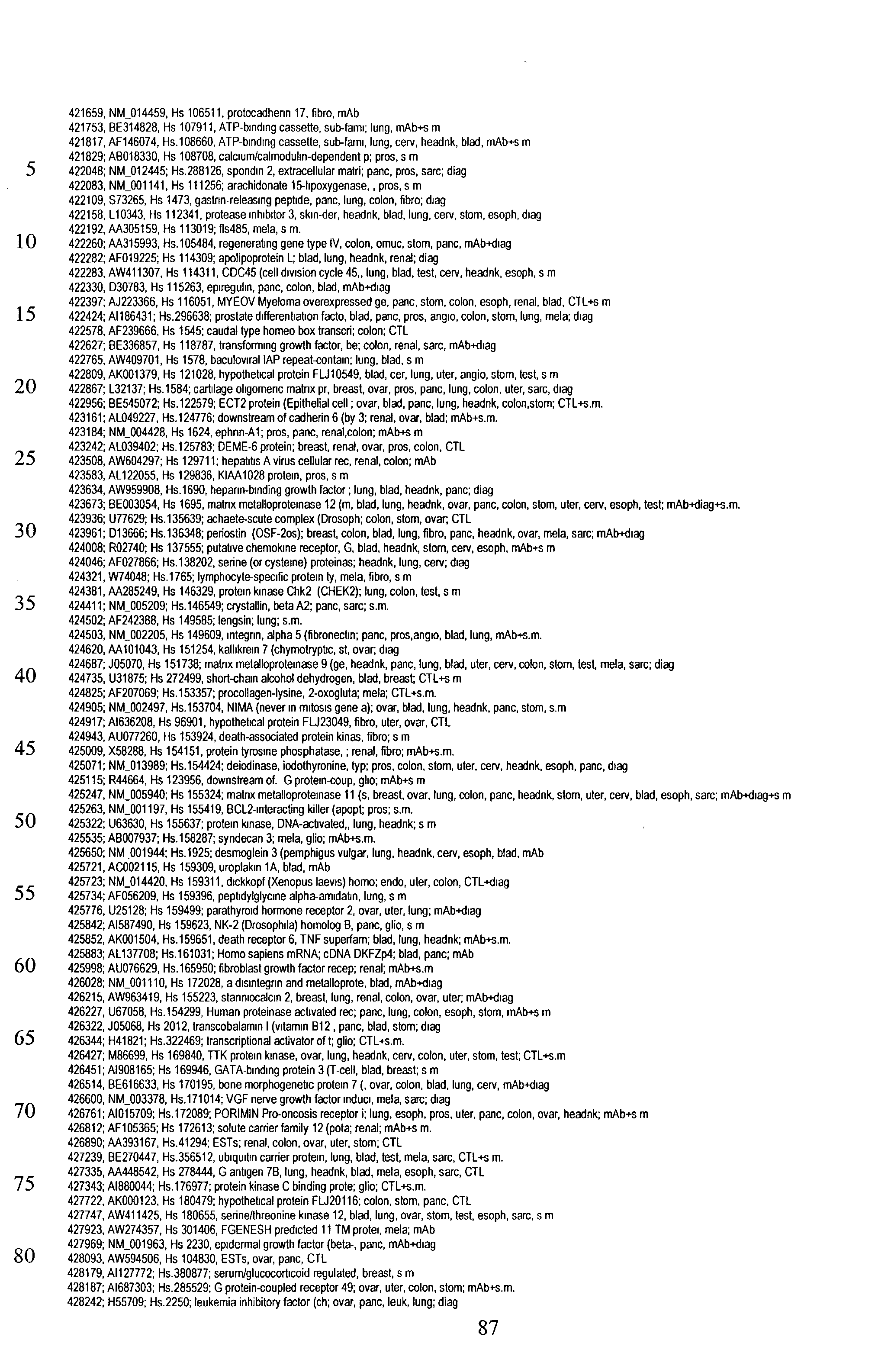


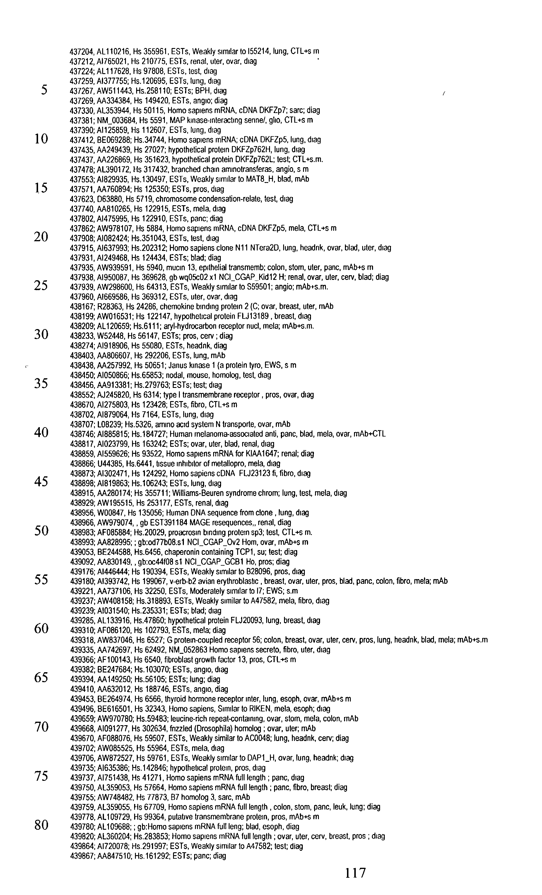


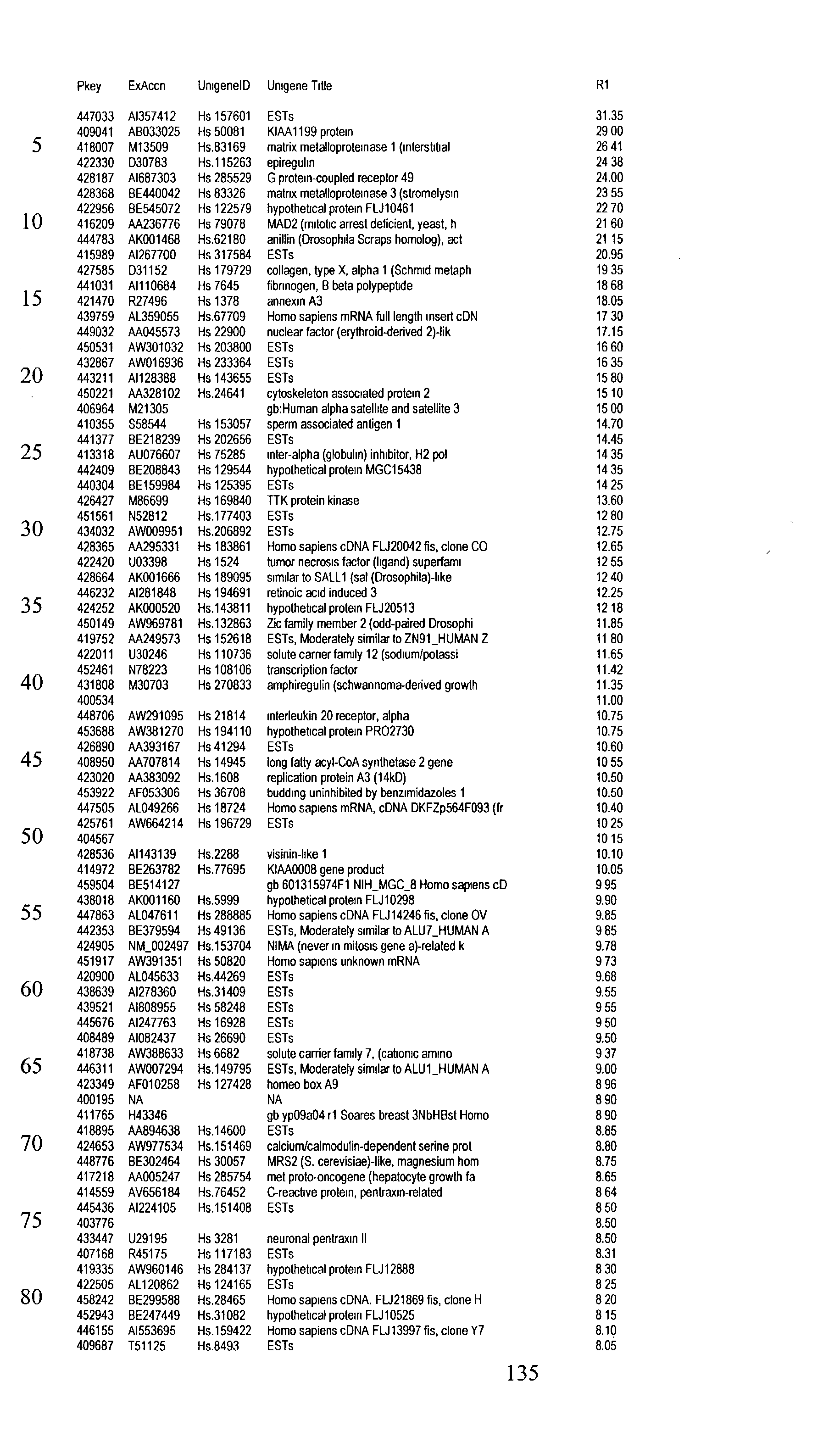


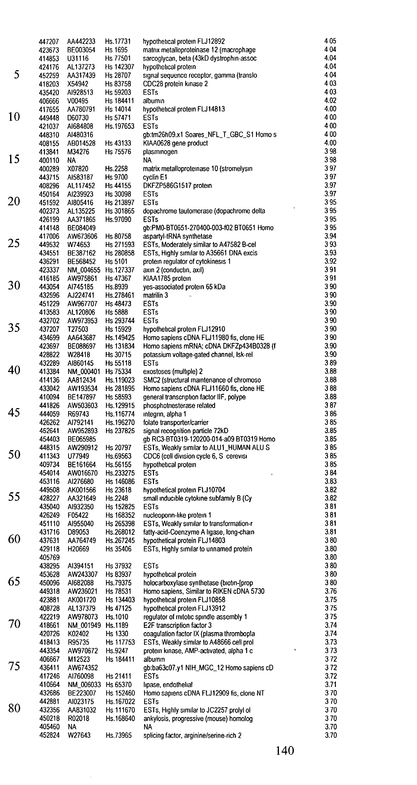


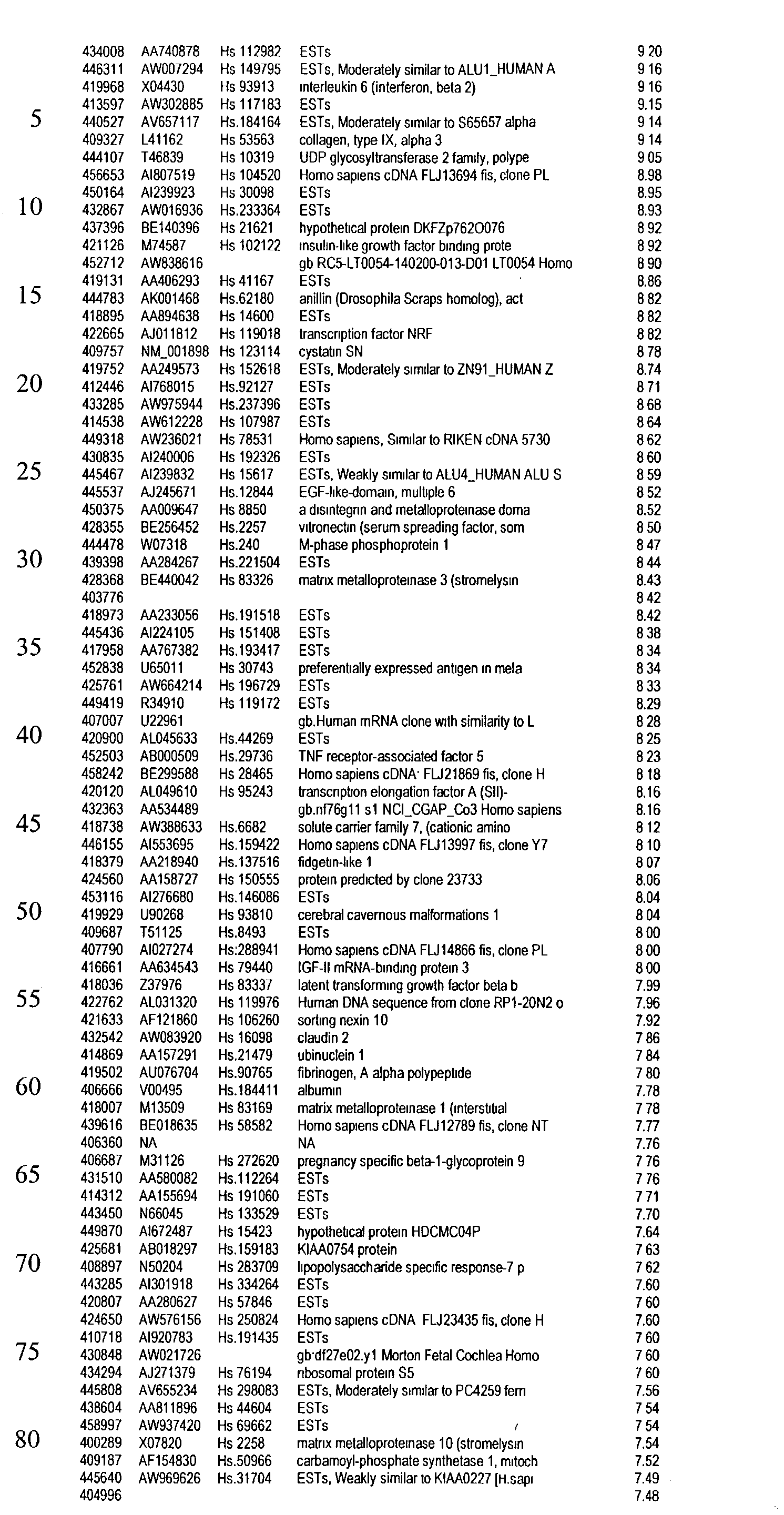


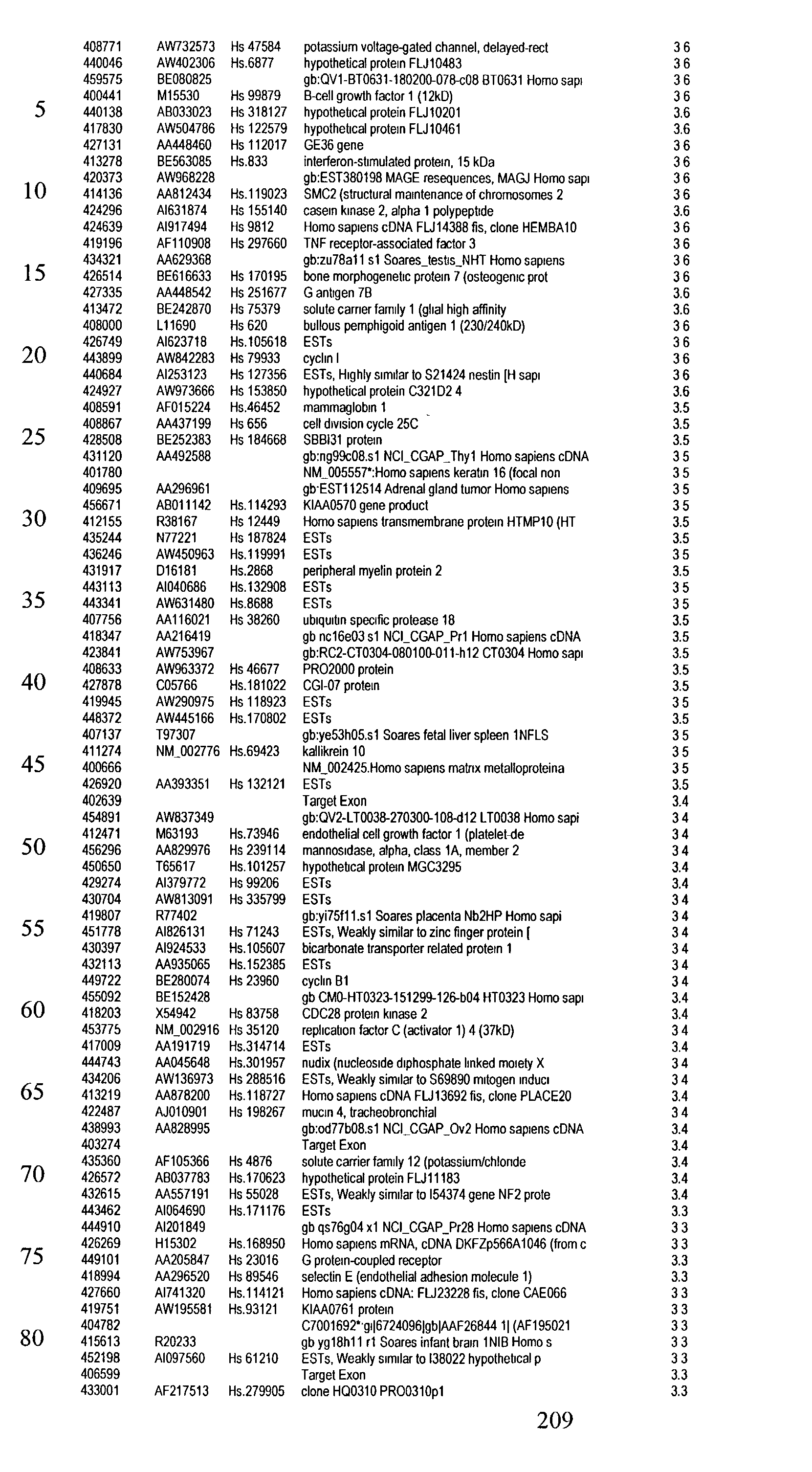


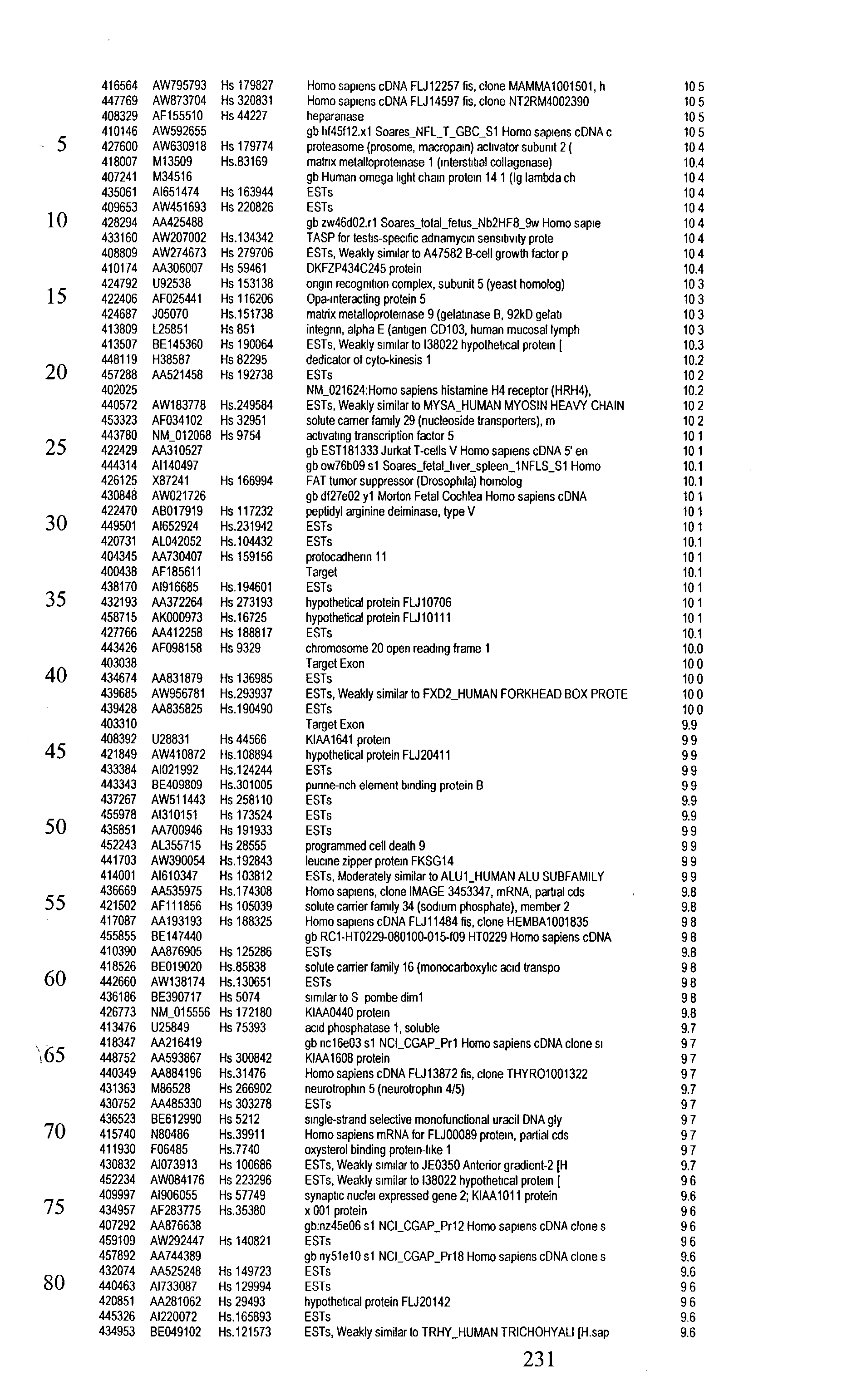


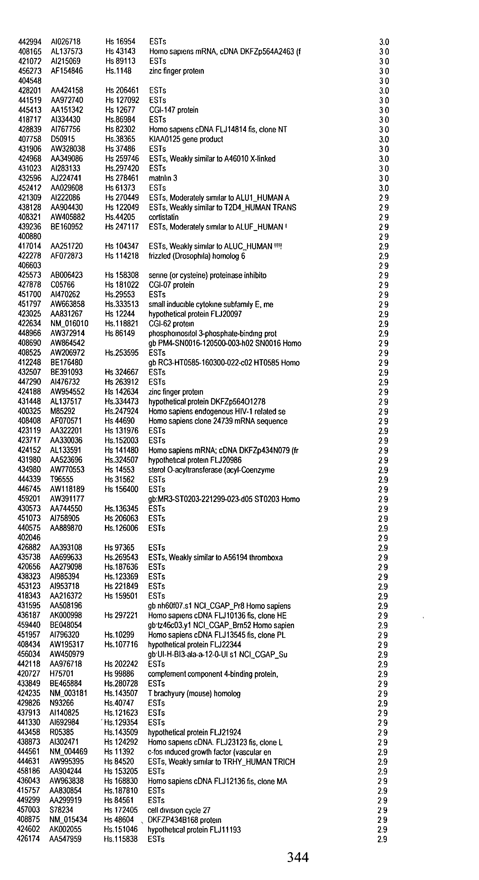


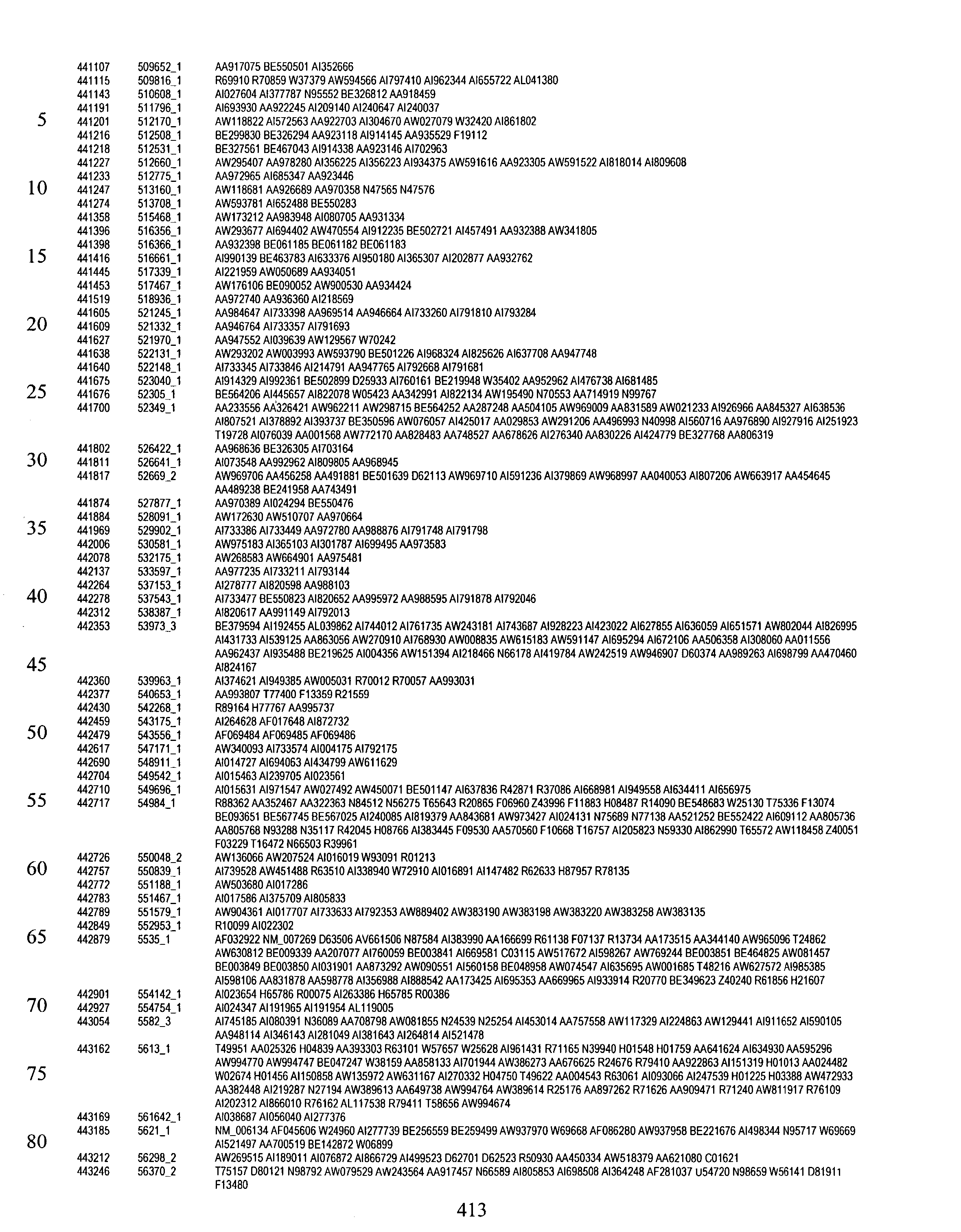


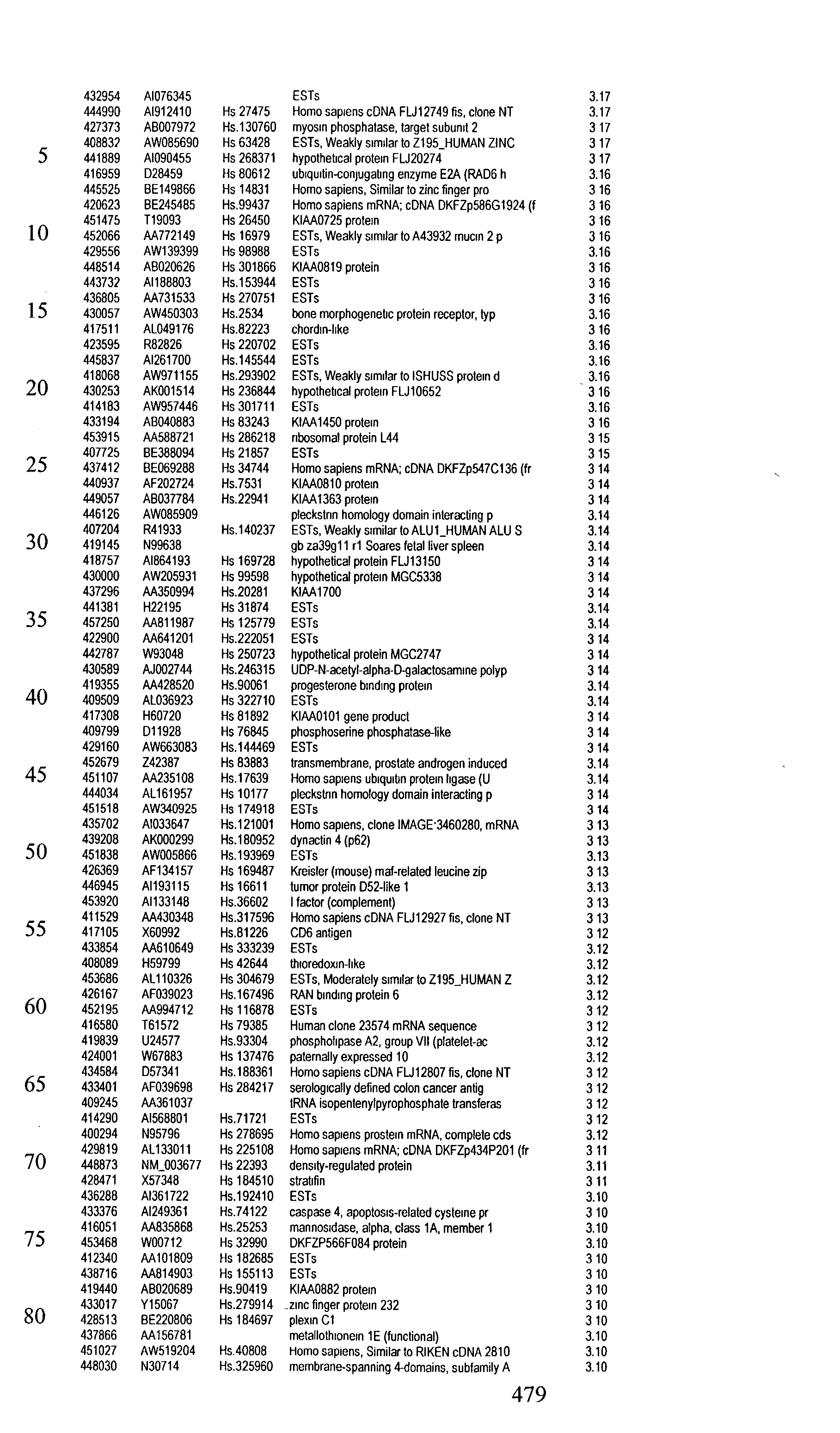


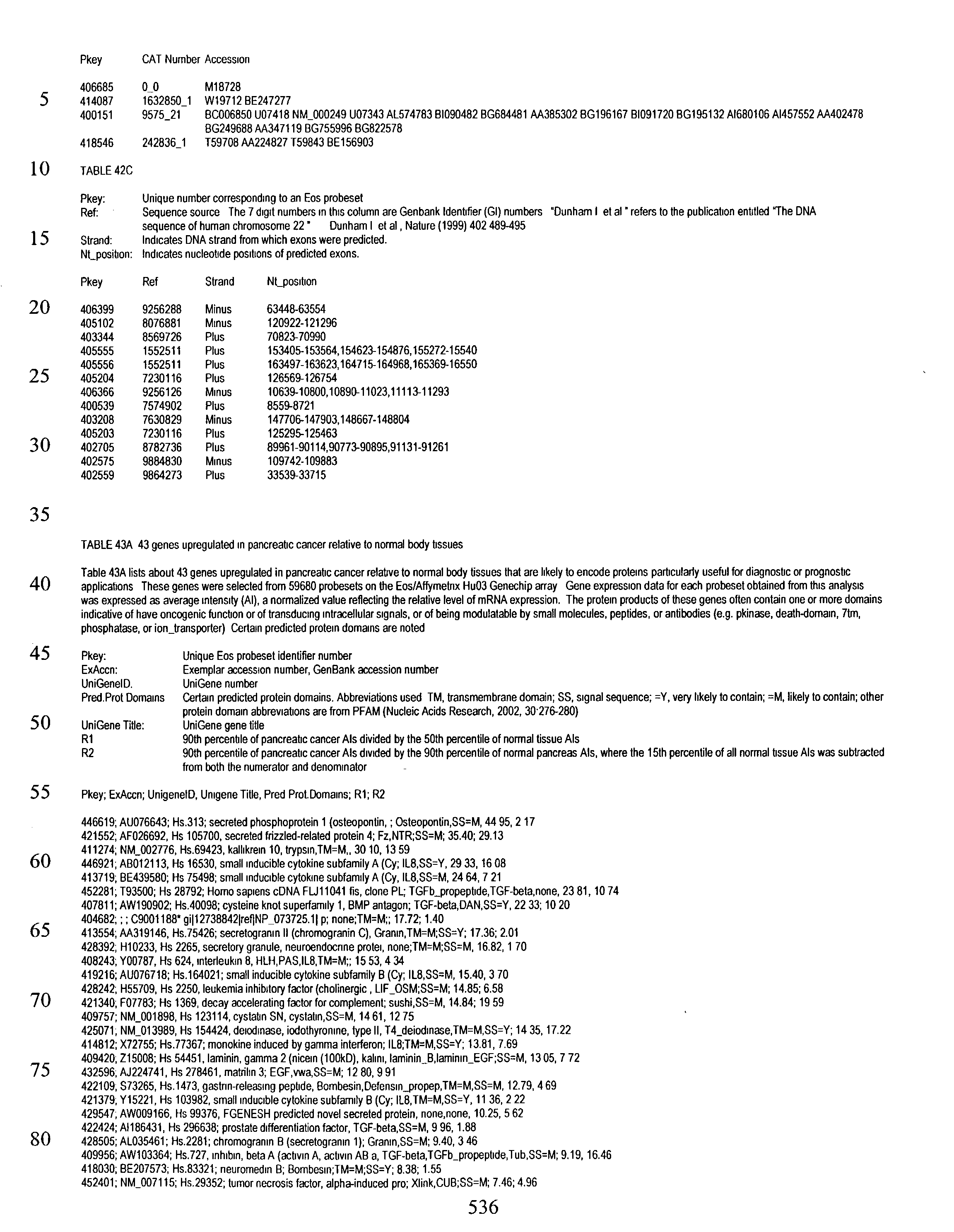


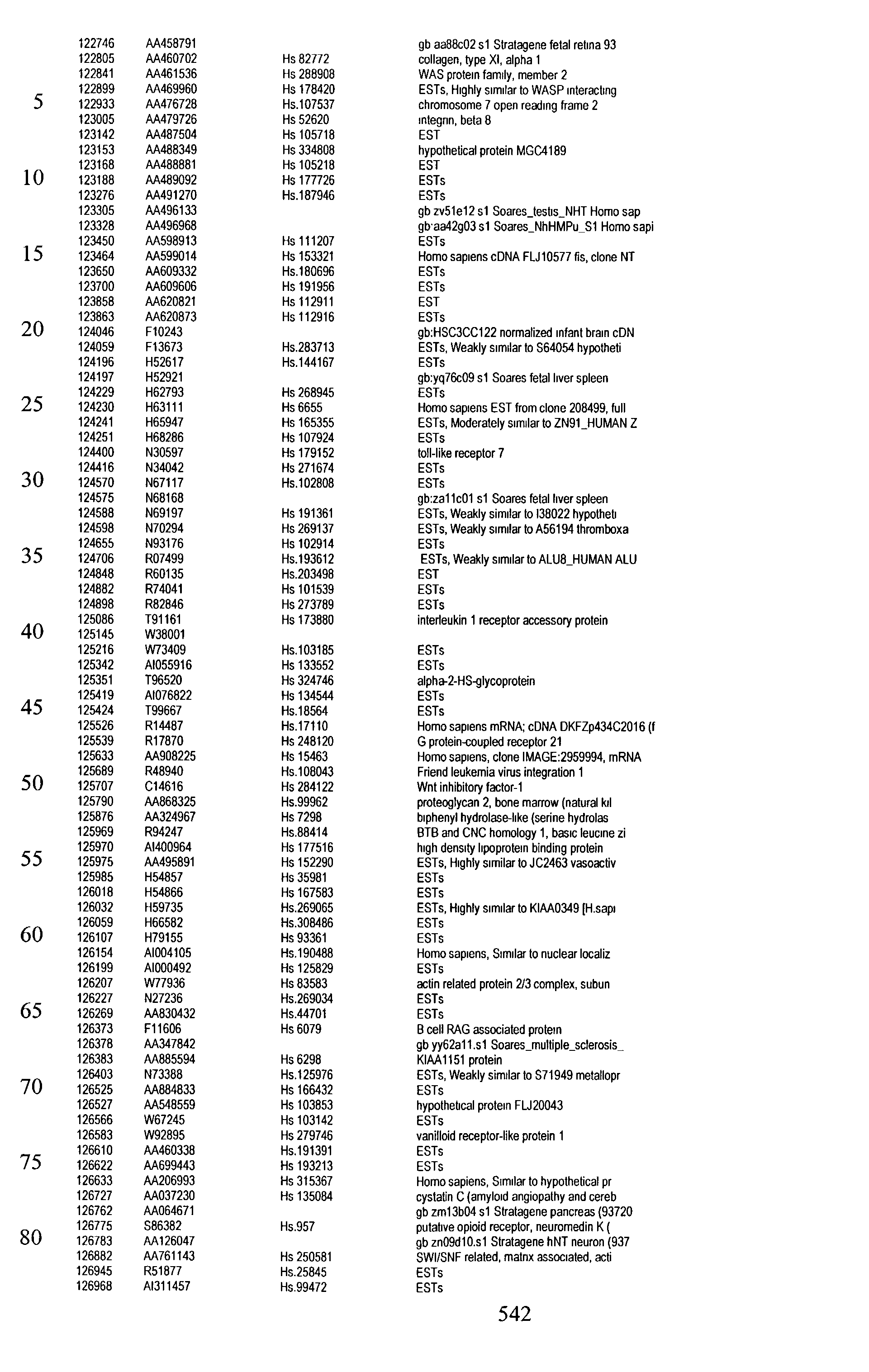


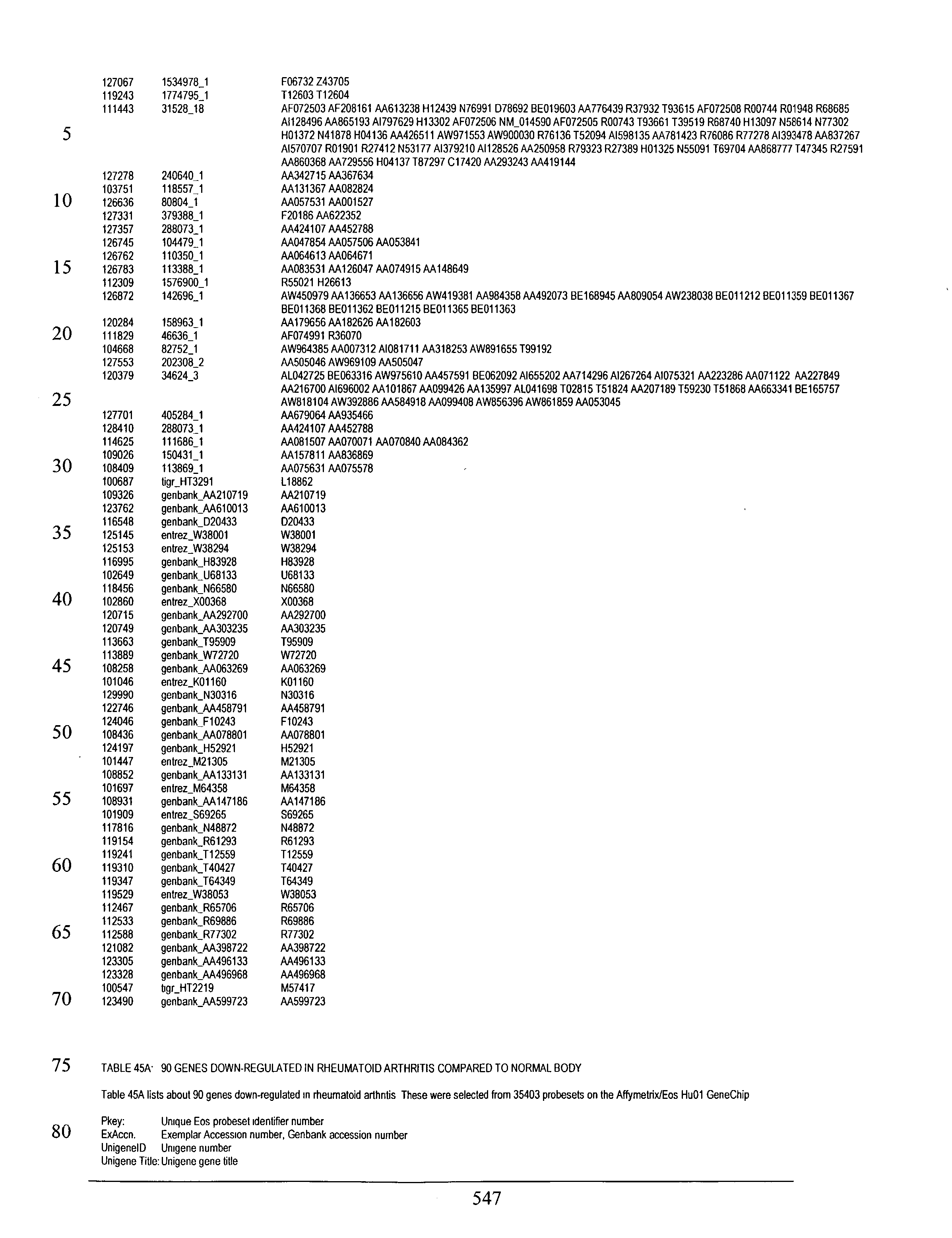


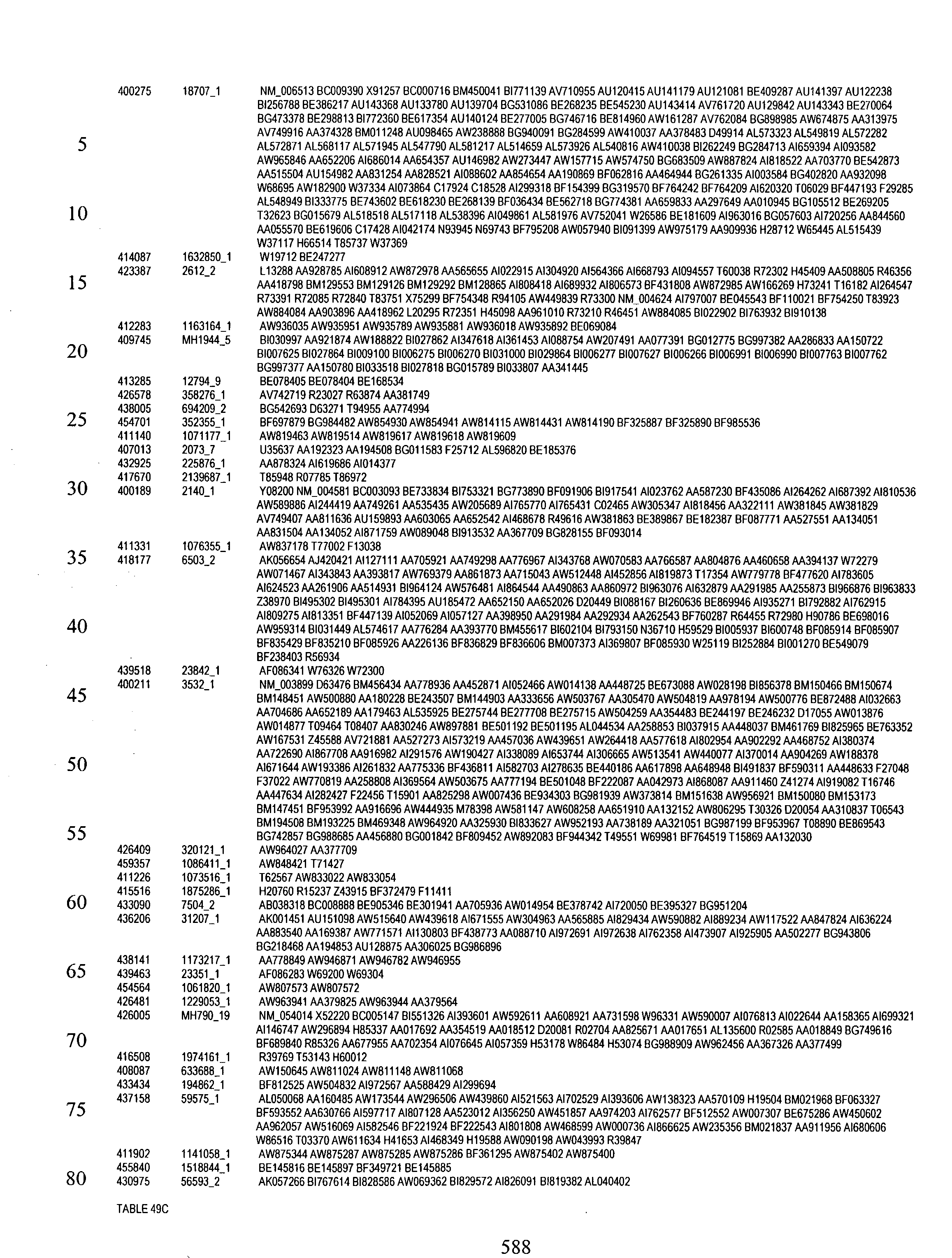


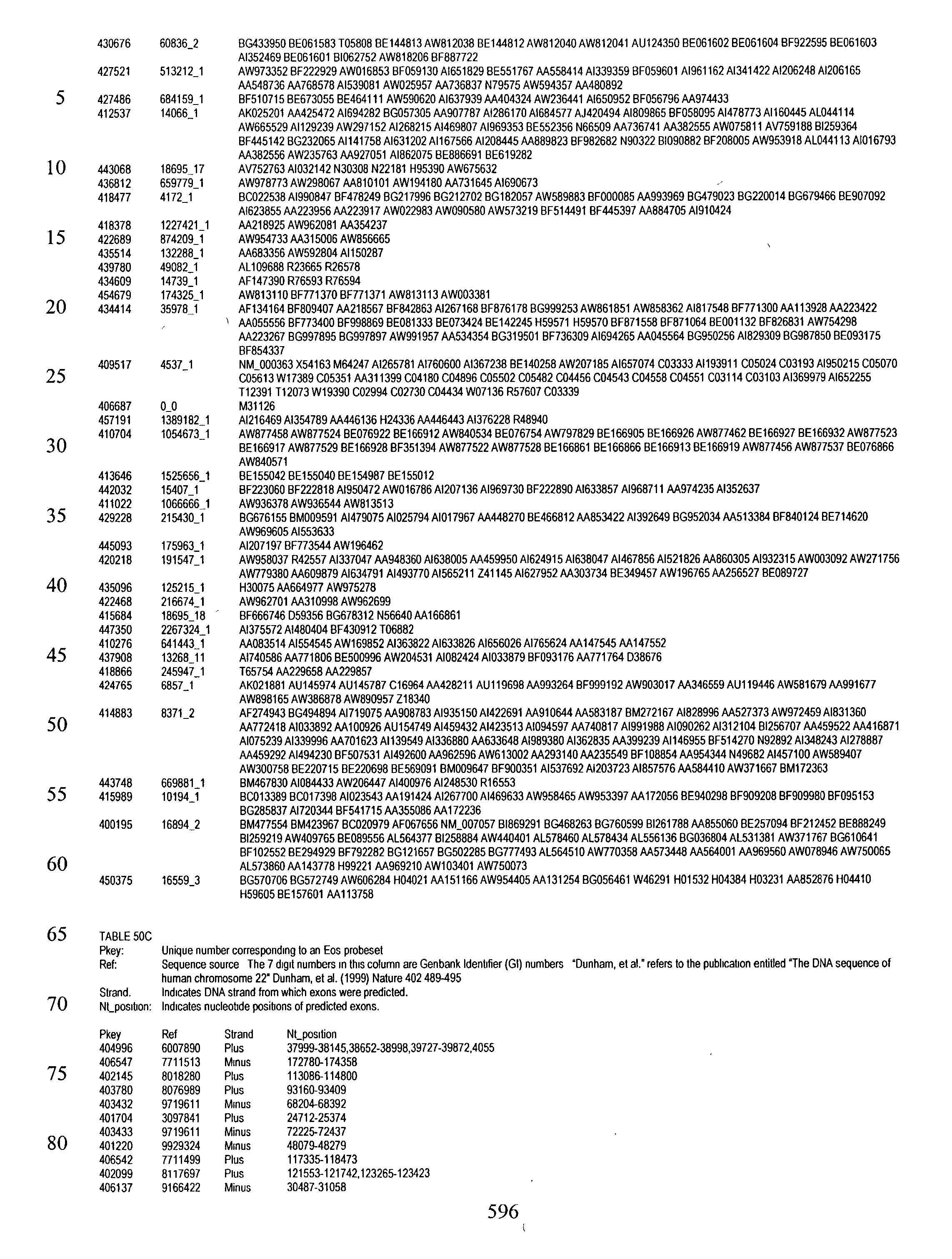


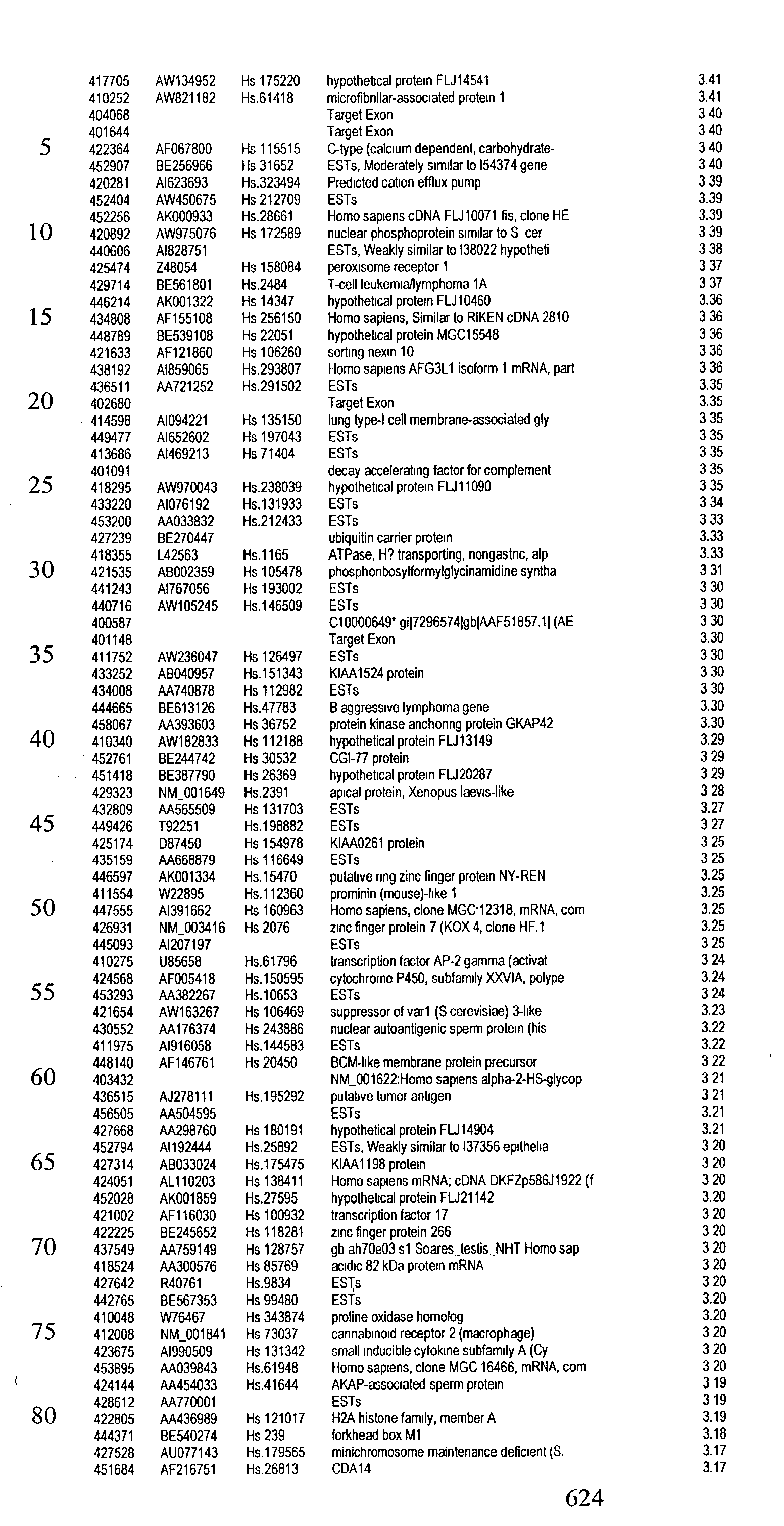


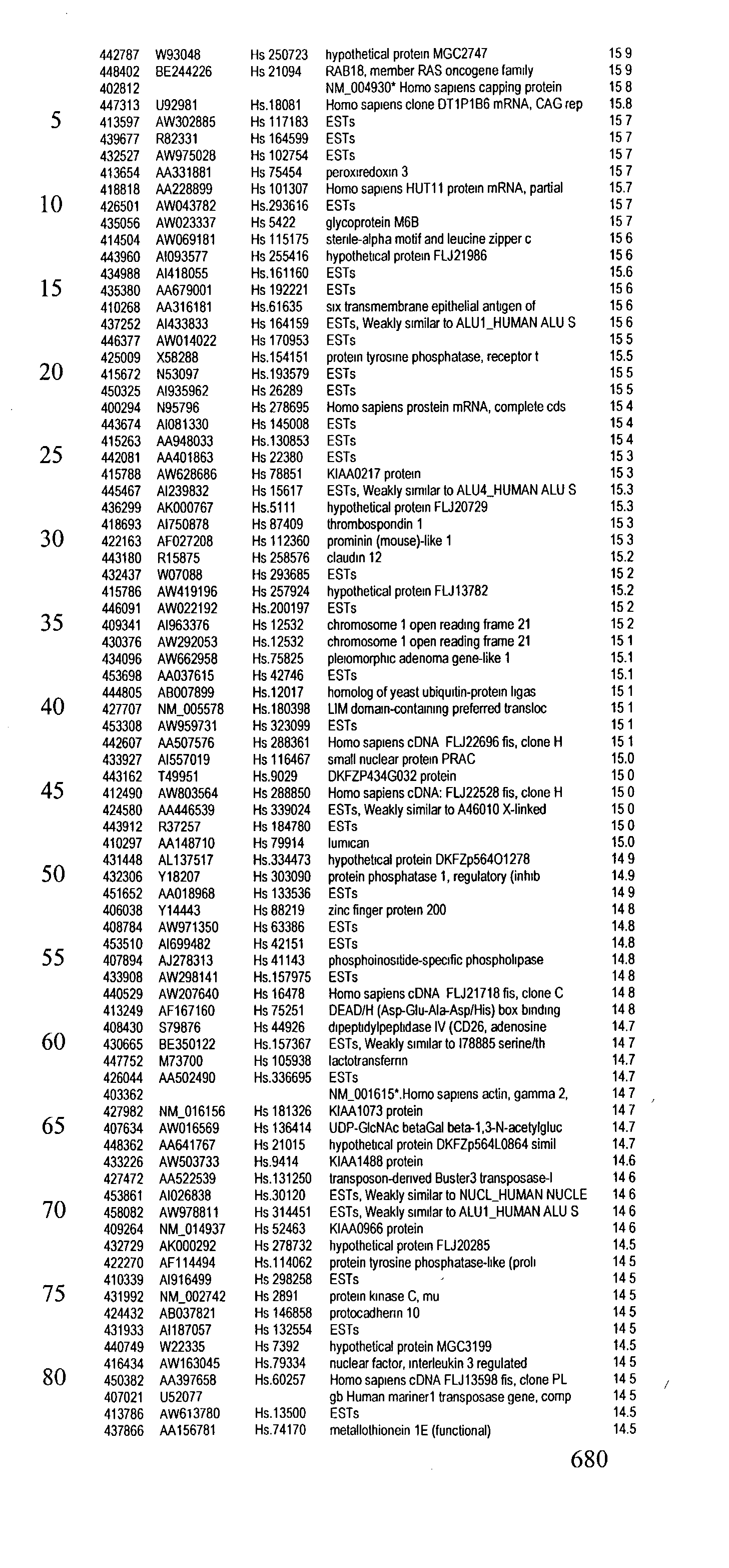


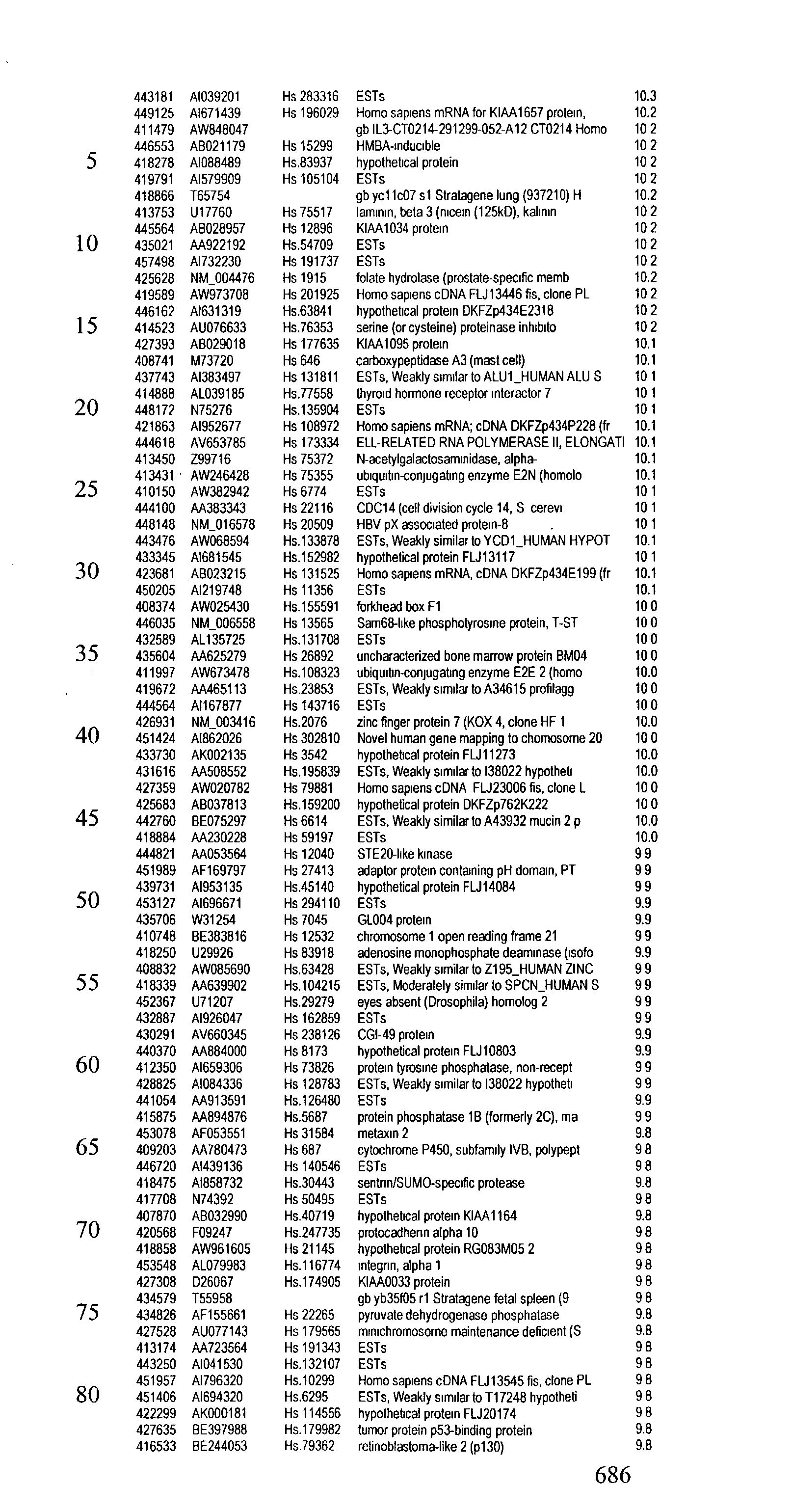


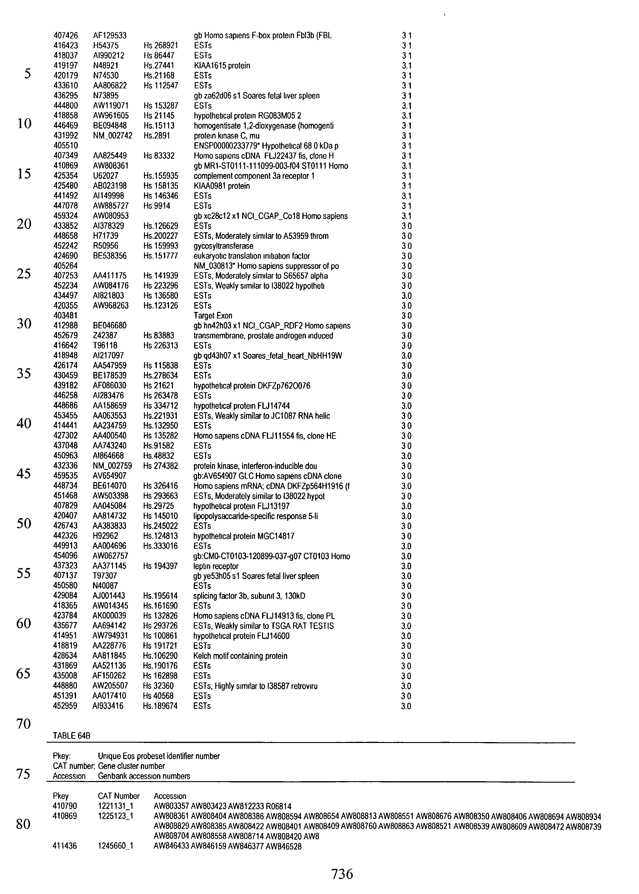


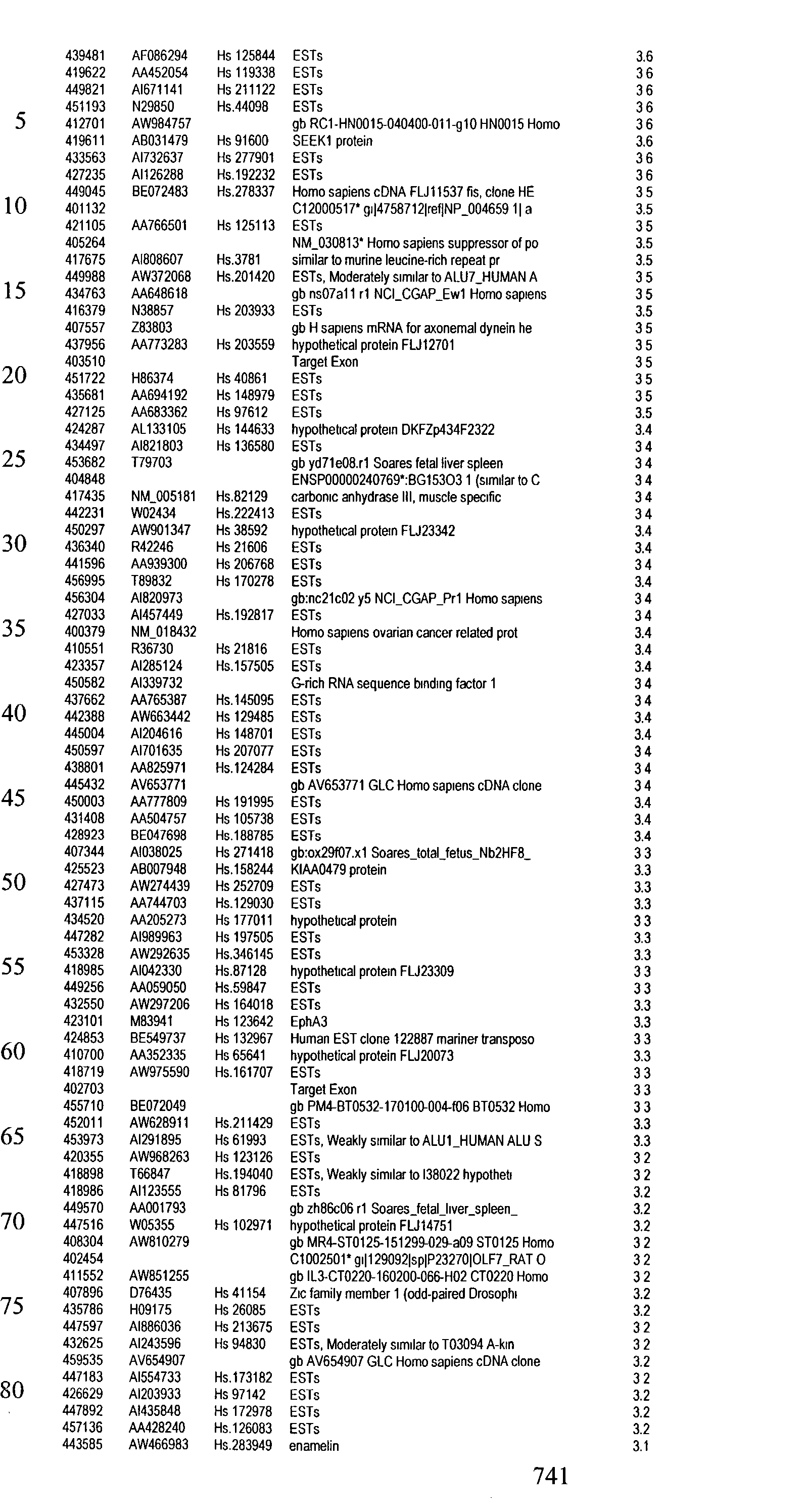


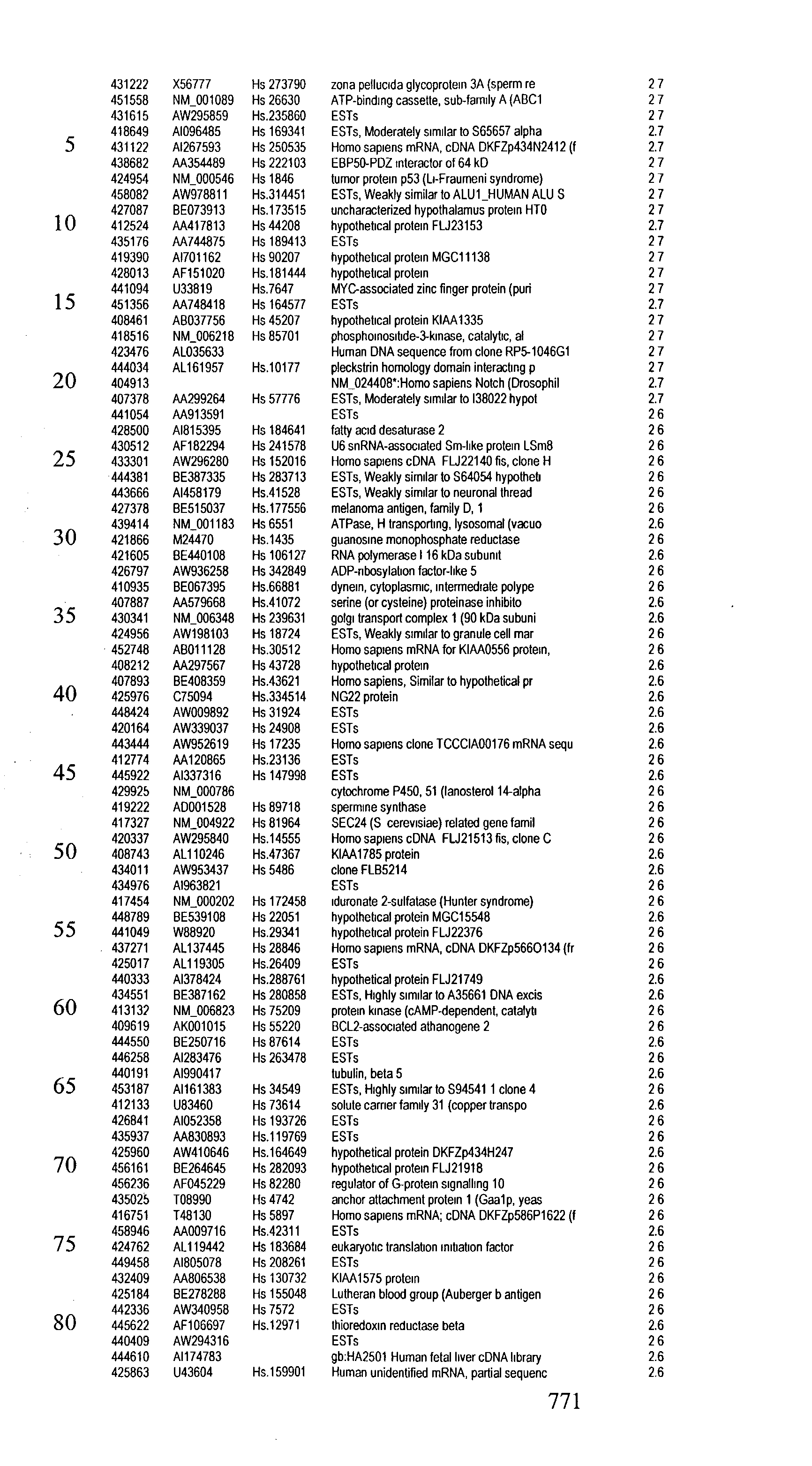


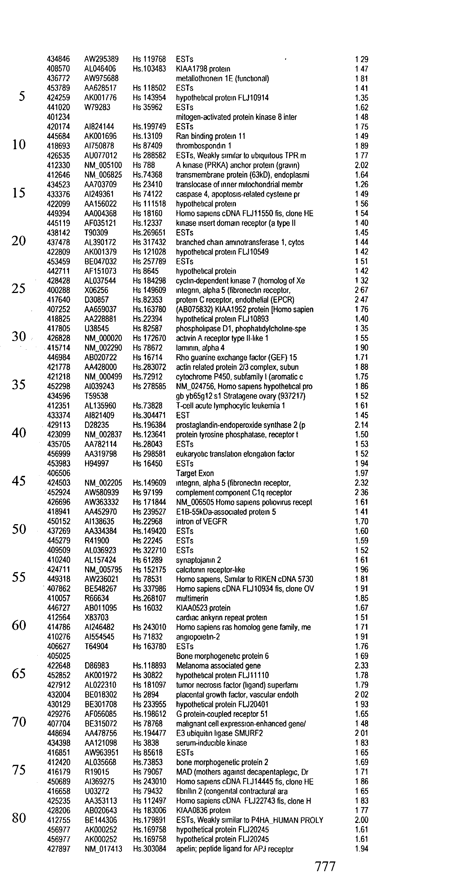


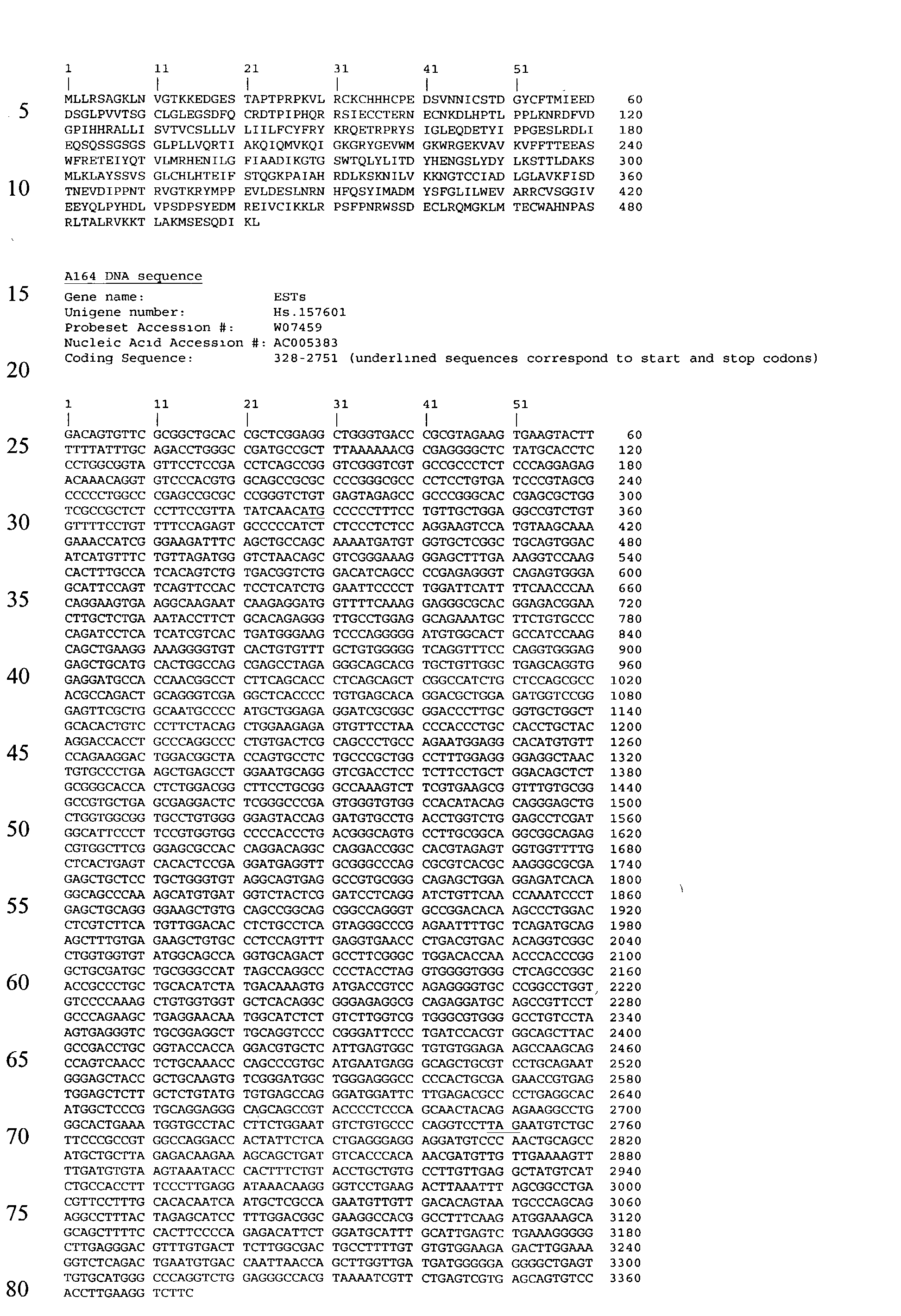


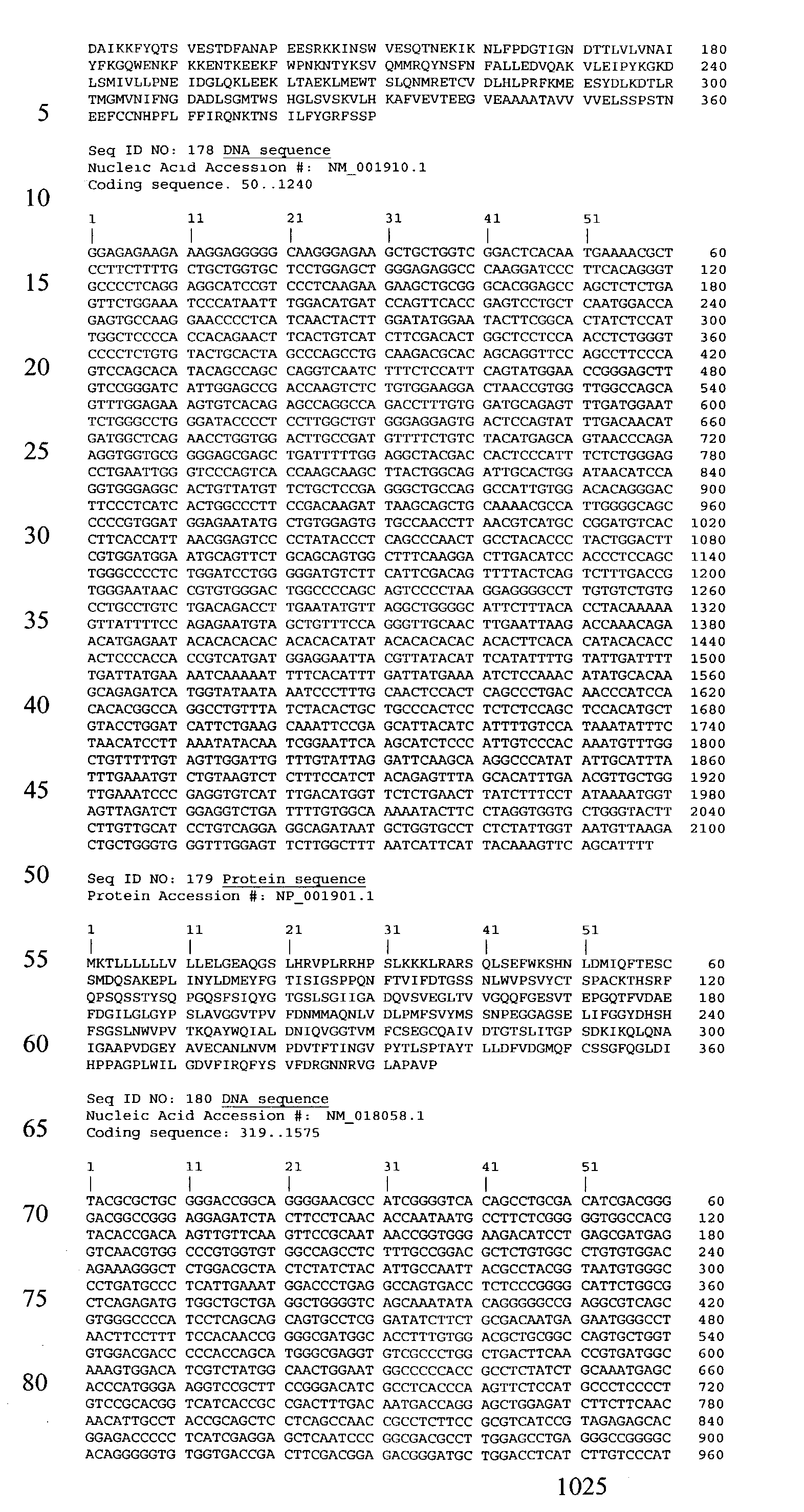


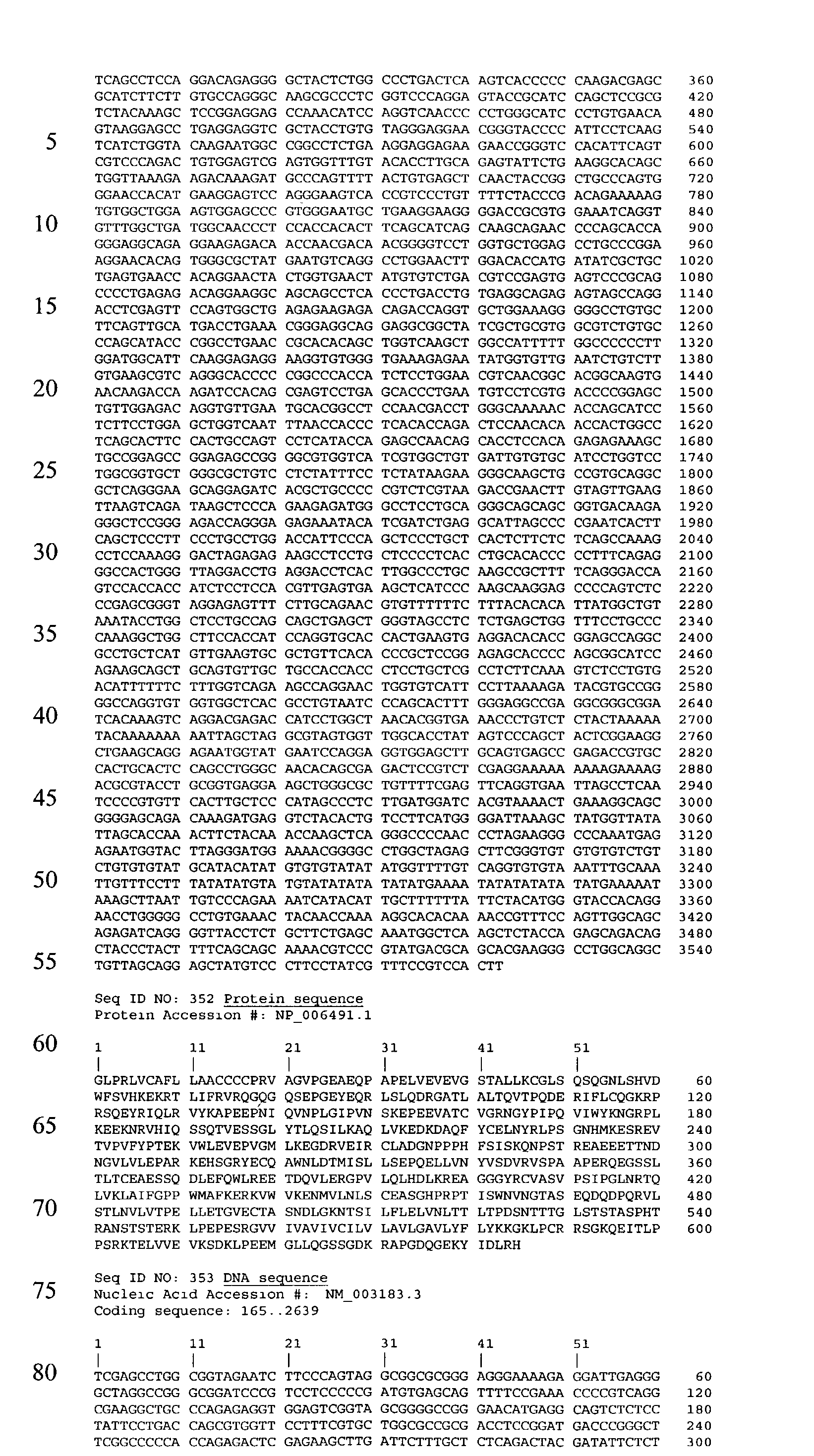


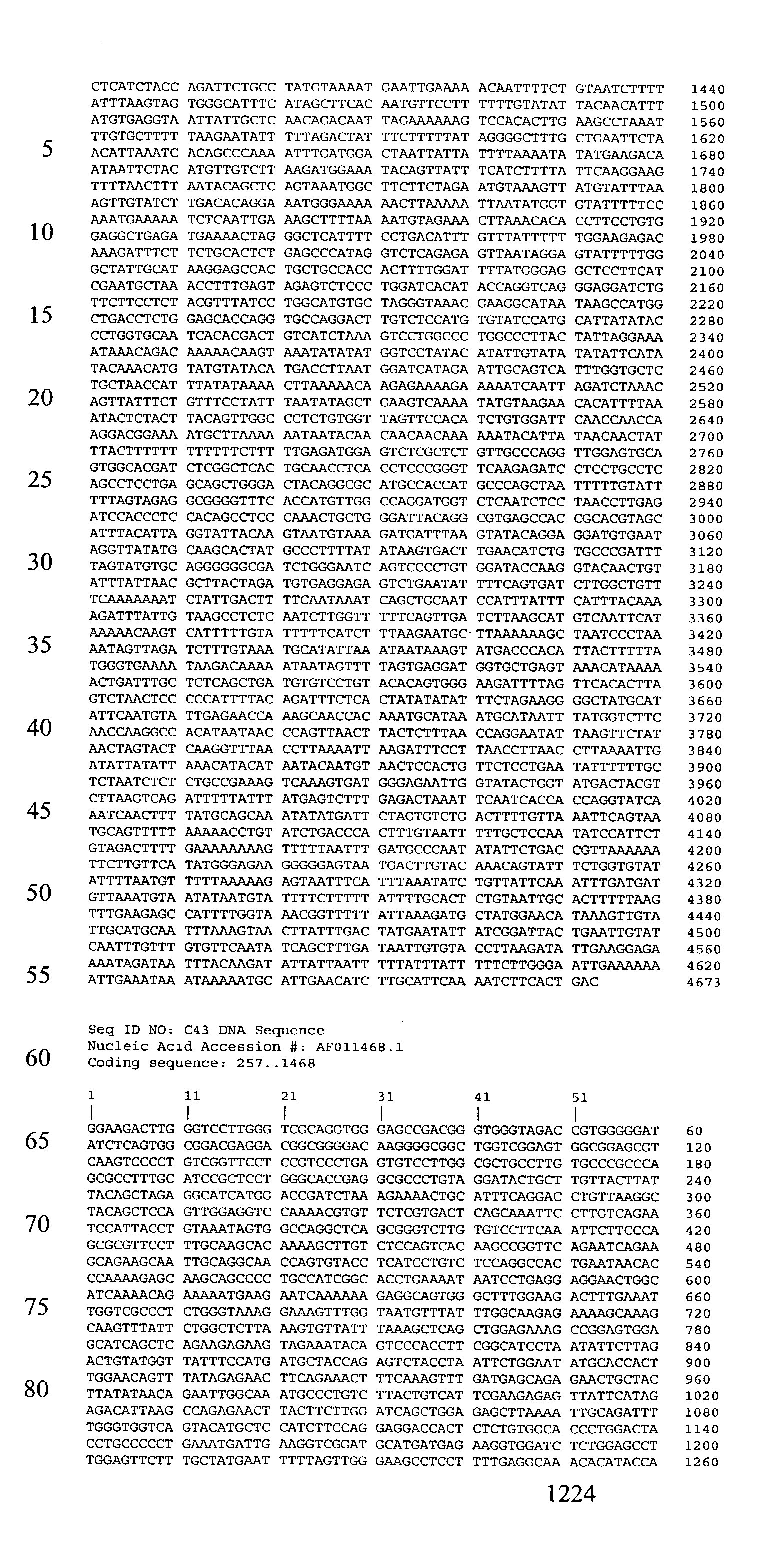


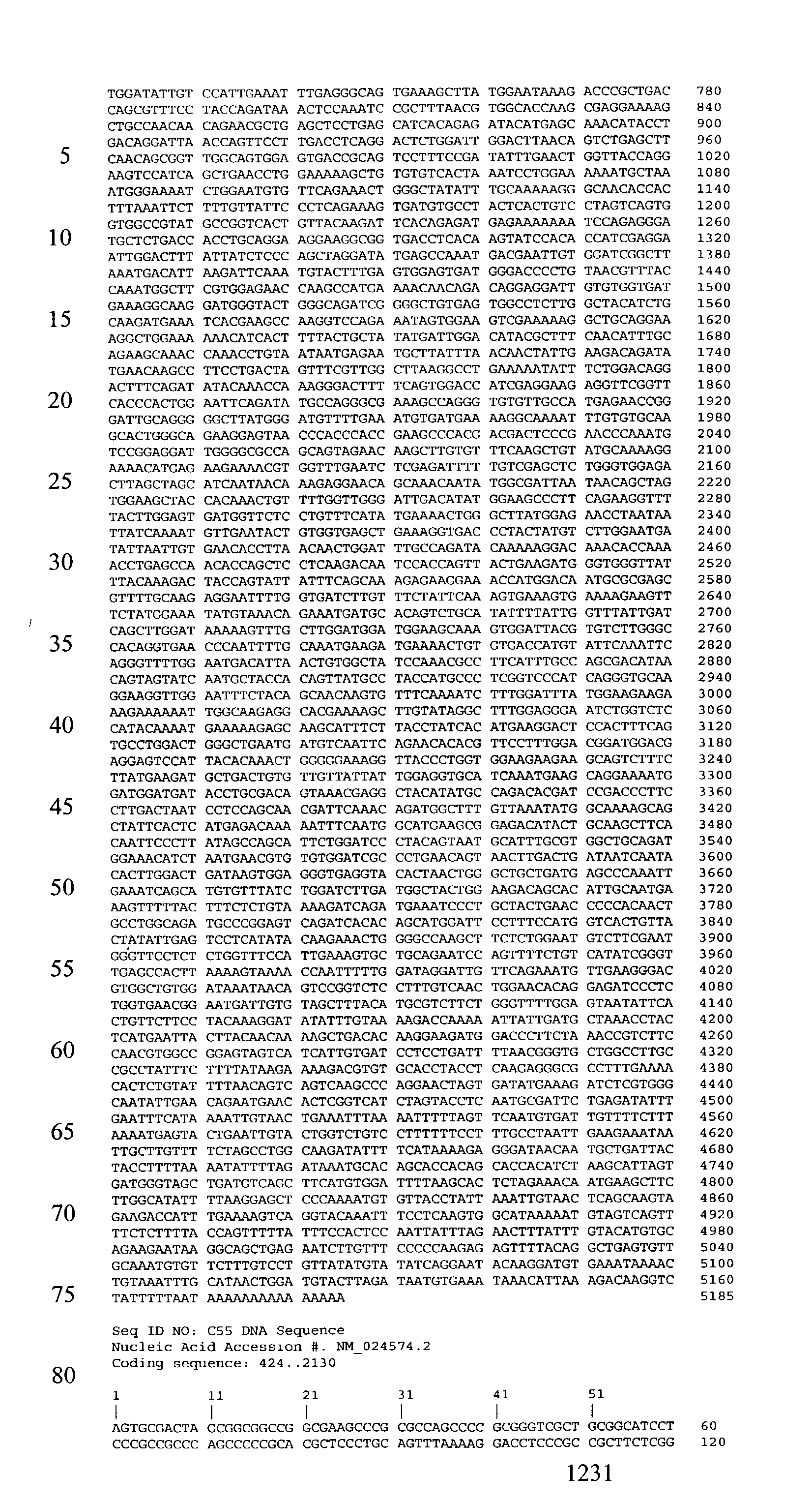


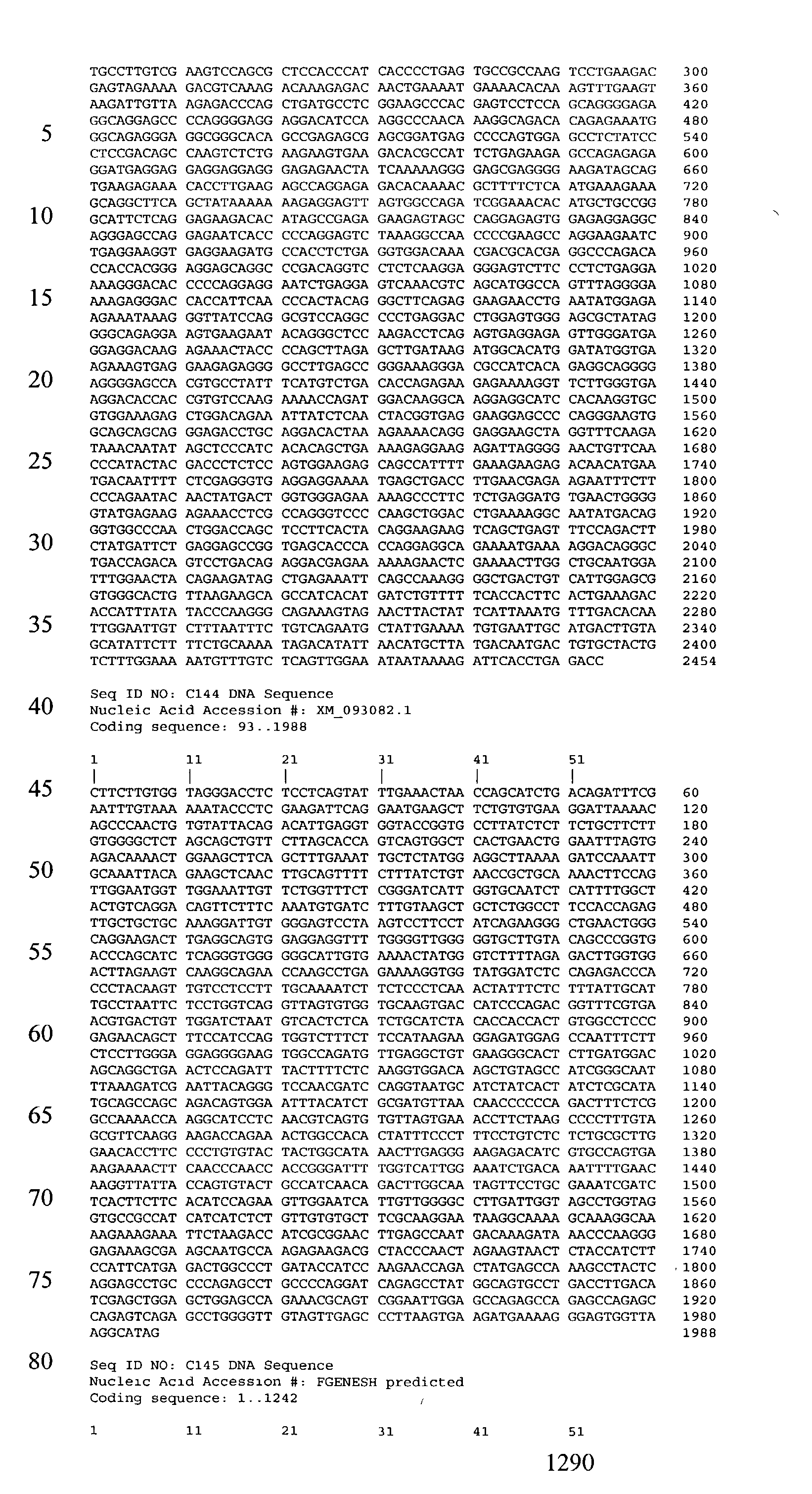


- It is understood that the examples described above in no way serve to limit the true scope of this invention, but rather are presented for illustrative purposes. All publications, sequences of accession numbers, and patent applications cited in this specification are herein incorporated by reference as if each individual publication, accession number, or patent application were specifically and individually indicated to be incorporated by reference.
Claims (19)
1. A method for determining the presence or absence of a pathological cell in a patient, said method comprising detecting a nucleic acid comprising a sequence at least 80% identical to a sequence as described in Tables 2A-80 in a biological sample from said patient, thereby determining the presence or absence of said pathological cell.
2. The method of claim 1 , wherein:
a) said pathology is described in Table 1, including a cancer; and/or
b) said biological sample comprises isolated nucleic acids.
3. The method of claim 1 , wherein said biological sample is tissue from an organ which is affected by said pathology of Table 1, including a cancer.
4. The method of claim 2 , wherein said nucleic acids are mRNA
5. The method of claim 2:
a) further comprising a step of amplifying nucleic acids before said step of detecting said nucleic acid; or
b) where said detecting is of a protein encoded by said nucleic acid.
6. The method of claim 1 , wherein said nucleic acid comprises a sequence as described in Tables 2A-80.
7. The method of claim 2 , wherein:
a) said detecting step is carried out by:
i) using a labeled nucleic acid probe;
ii) utilizing a biochip comprising a sequence at least 80% identical to a sequence as described in Tables 2A-80; or
iii) detecting a polypeptide encoded by said nucleic acid; or
b) said patient is:
i) undergoing a therapeutic regimen to treat said pathology of Table 1; or
ii) is suspected of having said pathology or cancer.
8. An isolated nucleic acid molecule comprising a sequence as described in Tables 2A-80.
9. The nucleic acid molecule of claim 8 , which is labeled.
10. An expression vector comprising the nucleic acid of claim 8 .
11. A host cell comprising the expression vector of claim 10 .
12. An isolated polypeptide which is encoded by a nucleic acid molecule comprising a sequence as described in Tables 2A-80.
13. An antibody that specifically binds a polypeptide of claim 12 .
14. The antibody of claim 13:
a) conjugated to an effector component;
b) conjugated to a detectable label, including a fluorescent label, a radioisotope, or a cytotoxic chemical;
c) which is an antibody fragment; or
d) which is a humanized antibody.
15. A method for specifically targeting a compound to a pathological cell in a patient, said method comprising administering to said patient an antibody of claim 13 , thereby providing said targetting.
16. A method for determining the presence or absence of a pathological cell in a patient, said method comprising contacting a biological sample with an antibody of claim 13 .
17. The method of claim 16 , wherein:
a) said antibody is conjugated to:
i) an effector component; or
ii) a fluorescent label; or
b) said biological sample is a blood, serum, urine, or stool sample.
18. A method for identifying a compound that modulates a pathology-associated polypeptide, said method comprising the steps of:
a) contacting said compound with a pathology-associated polypeptide, said polypeptide encoded by a polynucleotide that selectively hybridizes to a sequence at least 80% identical to a sequence as described in Tables 2A-80; and
b) determining the functional effect of said compound upon said polypeptide.
19. A drug screening assay comprising the steps of:
a) administering a test compound to a mammal having a pathology of Table 1 or a cell isolated therefrom; and
b) comparing the level of gene expression of a polynucleotide that selectively hybridizes to a sequence at least 80% identical to a sequence as described in Tables 2A-80 in a treated cell or mammal with the level of gene expression of said polynucleotide in a control cell or mammal, wherein a test compound that modulates said level of expression of the polynucleotide is a candidate for the treatment of said pathology.
Priority Applications (1)
| Application Number | Priority Date | Filing Date | Title |
|---|---|---|---|
| US10/295,027 US20030232350A1 (en) | 2001-11-13 | 2002-11-13 | Methods of diagnosis of cancer, compositions and methods of screening for modulators of cancer |
Applications Claiming Priority (18)
| Application Number | Priority Date | Filing Date | Title |
|---|---|---|---|
| US35066601P | 2001-11-13 | 2001-11-13 | |
| US33246401P | 2001-11-21 | 2001-11-21 | |
| US33439301P | 2001-11-29 | 2001-11-29 | |
| US33539401P | 2001-12-03 | 2001-12-03 | |
| US34037601P | 2001-12-14 | 2001-12-14 | |
| US34721102P | 2002-01-08 | 2002-01-08 | |
| US34734902P | 2002-01-10 | 2002-01-10 | |
| US35671402P | 2002-02-13 | 2002-02-13 | |
| US35907702P | 2002-02-20 | 2002-02-20 | |
| US36880902P | 2002-03-29 | 2002-03-29 | |
| US37011002P | 2002-04-04 | 2002-04-04 | |
| US37224602P | 2002-04-12 | 2002-04-12 | |
| US38661402P | 2002-06-05 | 2002-06-05 | |
| US39683902P | 2002-07-16 | 2002-07-16 | |
| US39777502P | 2002-07-22 | 2002-07-22 | |
| US39784502P | 2002-07-22 | 2002-07-22 | |
| US40945002P | 2002-09-09 | 2002-09-09 | |
| US10/295,027 US20030232350A1 (en) | 2001-11-13 | 2002-11-13 | Methods of diagnosis of cancer, compositions and methods of screening for modulators of cancer |
Publications (1)
| Publication Number | Publication Date |
|---|---|
| US20030232350A1 true US20030232350A1 (en) | 2003-12-18 |
Family
ID=29741293
Family Applications (1)
| Application Number | Title | Priority Date | Filing Date |
|---|---|---|---|
| US10/295,027 Abandoned US20030232350A1 (en) | 2001-11-13 | 2002-11-13 | Methods of diagnosis of cancer, compositions and methods of screening for modulators of cancer |
Country Status (1)
| Country | Link |
|---|---|
| US (1) | US20030232350A1 (en) |
Cited By (121)
| Publication number | Priority date | Publication date | Assignee | Title |
|---|---|---|---|---|
| US20030211039A1 (en) * | 2001-05-29 | 2003-11-13 | Macina Roberto A. | Method of diagnosing, monitoring, staging, imaging and treating colon cancer |
| US20030224976A1 (en) * | 2001-07-17 | 2003-12-04 | Yale University | Compositions, methods and kits relating to behab and primary CNS tumors |
| US20040038225A1 (en) * | 2002-08-26 | 2004-02-26 | Markowitz Sanford D. | Methods and compositions for categorizing patients |
| US20040038220A1 (en) * | 2002-08-26 | 2004-02-26 | Markowitz Sanford D. | Methods for categorizing patients |
| WO2004041076A2 (en) * | 2002-11-04 | 2004-05-21 | Protein Design Labs, Inc. | Methods of detecting colorectal cancer |
| US20040110712A1 (en) * | 2002-08-26 | 2004-06-10 | Markowitz Sanford D. | Methods for treating patients and identifying therapeutics |
| US20040265230A1 (en) * | 2003-01-06 | 2004-12-30 | Martinez Robert Vincent | Compositions and methods for diagnosing and treating colon cancers |
| US20050124012A1 (en) * | 2003-08-15 | 2005-06-09 | Iris Simon | Pro108 antibody compositions and methods of use and use of Pro108 to assess cancer risk |
| US20050186610A1 (en) * | 2004-02-20 | 2005-08-25 | Yeon-Su Lee | Breast cancer related protein, gene encoding the same, and method of diagnosing breast cancer using the protein and gene |
| US20050191312A1 (en) * | 2001-04-10 | 2005-09-01 | Raitano Arthur B. | Nucleic acids and corresponding proteins entitled 158P3D2 useful in treatment and detection of cancer |
| US20050233353A1 (en) * | 2001-02-27 | 2005-10-20 | Markowitz Sanford D | Methods and compositions for categorizing patients |
| WO2005118851A1 (en) * | 2004-06-02 | 2005-12-15 | Diagenic As | Oligonucleotides for cancer diagnosis |
| US20060035237A1 (en) * | 2002-08-26 | 2006-02-16 | Markowitz Sanford D | Methods and compositions for categorizing patients |
| WO2006060533A2 (en) | 2004-12-01 | 2006-06-08 | Genentech, Inc. | Conjugates of 1, 8-bis-naphthalimides with an antibody |
| US20060160090A1 (en) * | 2003-04-11 | 2006-07-20 | Macina Robert A | Composition splice variants and methods relating to cancer specific genes and proteins |
| WO2006078780A2 (en) * | 2005-01-19 | 2006-07-27 | Genzyme Corporation | Rdc1 antibodies for the diagnosis of nsclc |
| US20070184439A1 (en) * | 2003-07-17 | 2007-08-09 | Guilford Parry J | Markers for detection of gastric cancer |
| US20080050726A1 (en) * | 2005-09-19 | 2008-02-28 | Yixin Wang | Methods for diagnosing pancreatic cancer |
| US20080138838A1 (en) * | 2002-07-31 | 2008-06-12 | Cedars-Sinai Medical Center | Diagnosis of zd1839 resistant tumors |
| US20080286386A1 (en) * | 2005-12-30 | 2008-11-20 | Hiroaki Nitta | Na+, K+-Atpase Expression in Cervical Dysplasia and Cancer |
| US7488813B2 (en) | 2005-02-24 | 2009-02-10 | Compugen, Ltd. | Diagnostic markers, especially for in vivo imaging, and assays and methods of use thereof |
| US20090054630A1 (en) * | 2001-11-30 | 2009-02-26 | Chemocentryx, Inc. | Compositions and methods for detecting and treating diseases and conditions related to chemokine receptors |
| US20090068690A1 (en) * | 2006-01-27 | 2009-03-12 | Tripath Imaging, Inc. | Methods for identifying patients with an increased likelihood of having ovarian cancer and compositions therefor |
| US20090169575A1 (en) * | 2006-06-06 | 2009-07-02 | Christian Rohlff | Proteins |
| US20090202576A1 (en) * | 2005-02-25 | 2009-08-13 | Oncotherapy Science, Inc. | Peptide vaccines for lung cancers expressing ttk, urlc10 or koc1 polypeptides |
| US20090220504A1 (en) * | 2006-03-21 | 2009-09-03 | Anan Chuntharapai | Combinatorial therapy |
| US20090232813A1 (en) * | 2007-06-25 | 2009-09-17 | Beauchamp R Daniel | Four-Jointed Box (FJX1) In Cancer Diagnosis and Treatment |
| US20100247540A1 (en) * | 2003-10-30 | 2010-09-30 | Chemocentryx, Inc. | Methods and Compositions For Modulating Angiogenesis |
| US20100291091A1 (en) * | 2007-07-30 | 2010-11-18 | ONCOTHERAPY SCIENCE ,inc. | Cancer associated gene ly6k |
| US20100297669A1 (en) * | 2002-08-16 | 2010-11-25 | Agensys, Inc. | NUCLEIC ACIDS AND CORRESPONDING PROTEINS ENTITLED 191P4D12(b) USEFUL IN TREATMENT AND DETECTION OF CANCER |
| US20100297152A1 (en) * | 2000-10-13 | 2010-11-25 | Cedars-Sinai Medical Center | Methods of diagnosis of prostate cancer, compositions and methods of screening for modulators of prostate cancer |
| US20110027283A1 (en) * | 2007-09-06 | 2011-02-03 | Case Western Reserve University | Methods for diagnosing and treating cancers |
| EP2286844A2 (en) | 2004-06-01 | 2011-02-23 | Genentech, Inc. | Antibody-drug conjugates and methods |
| WO2011031870A1 (en) | 2009-09-09 | 2011-03-17 | Centrose, Llc | Extracellular targeted drug conjugates |
| WO2011056983A1 (en) | 2009-11-05 | 2011-05-12 | Genentech, Inc. | Zirconium-radiolabeled, cysteine engineered antibody conjugates |
| US8021614B2 (en) | 2005-04-05 | 2011-09-20 | The General Hospital Corporation | Devices and methods for enrichment and alteration of cells and other particles |
| WO2011130598A1 (en) | 2010-04-15 | 2011-10-20 | Spirogen Limited | Pyrrolobenzodiazepines and conjugates thereof |
| WO2011156328A1 (en) | 2010-06-08 | 2011-12-15 | Genentech, Inc. | Cysteine engineered antibodies and conjugates |
| US8124740B2 (en) | 2009-03-25 | 2012-02-28 | Genentech, Inc. | Anti- α5 β1 antibodies and uses thereof |
| US8137912B2 (en) | 2006-06-14 | 2012-03-20 | The General Hospital Corporation | Methods for the diagnosis of fetal abnormalities |
| US8168389B2 (en) | 2006-06-14 | 2012-05-01 | The General Hospital Corporation | Fetal cell analysis using sample splitting |
| US8173434B2 (en) | 2006-04-04 | 2012-05-08 | Diadexus, Inc. | PCan065 antibody compositions and methods of use |
| US8195415B2 (en) | 2008-09-20 | 2012-06-05 | The Board Of Trustees Of The Leland Stanford Junior University | Noninvasive diagnosis of fetal aneuploidy by sequencing |
| WO2012074757A1 (en) | 2010-11-17 | 2012-06-07 | Genentech, Inc. | Alaninyl maytansinol antibody conjugates |
| EP2518166A2 (en) | 2005-05-20 | 2012-10-31 | Veridex, LLC | Thyroid fine needle aspiration molecular assay |
| US8304230B2 (en) | 2002-09-27 | 2012-11-06 | The General Hospital Corporation | Microfluidic device for cell separation and uses thereof |
| WO2012155019A1 (en) | 2011-05-12 | 2012-11-15 | Genentech, Inc. | Multiple reaction monitoring lc-ms/ms method to detect therapeutic antibodies in animal samples using framework signature pepides |
| WO2013130093A1 (en) | 2012-03-02 | 2013-09-06 | Genentech, Inc. | Biomarkers for treatment with anti-tubulin chemotherapeutic compounds |
| US8623829B2 (en) | 2007-02-21 | 2014-01-07 | Oncotherapy Science, Inc. | Peptide vaccines for cancers expressing tumor-associated antigens |
| WO2014057074A1 (en) | 2012-10-12 | 2014-04-17 | Spirogen Sàrl | Pyrrolobenzodiazepines and conjugates thereof |
| US20140178391A1 (en) * | 2004-04-30 | 2014-06-26 | Celera Corporation | Colon disease targets and uses thereof |
| WO2014140862A2 (en) | 2013-03-13 | 2014-09-18 | Spirogen Sarl | Pyrrolobenzodiazepines and conjugates thereof |
| WO2014140174A1 (en) | 2013-03-13 | 2014-09-18 | Spirogen Sàrl | Pyrrolobenzodiazepines and conjugates thereof |
| US8840887B2 (en) | 2007-09-26 | 2014-09-23 | Genentech, Inc. | Antibodies |
| WO2014159981A2 (en) | 2013-03-13 | 2014-10-02 | Spirogen Sarl | Pyrrolobenzodiazepines and conjugates thereof |
| WO2013110030A3 (en) * | 2012-01-19 | 2014-12-18 | Duke University | Vaccines against antigens involved in therapy resistance and methods of using same |
| US8921102B2 (en) | 2005-07-29 | 2014-12-30 | Gpb Scientific, Llc | Devices and methods for enrichment and alteration of circulating tumor cells and other particles |
| WO2015023355A1 (en) | 2013-08-12 | 2015-02-19 | Genentech, Inc. | 1-(chloromethyl)-2,3-dihydro-1h-benzo[e]indole dimer antibody-drug conjugate compounds, and methods of use and treatment |
| WO2015095212A1 (en) | 2013-12-16 | 2015-06-25 | Genentech, Inc. | 1-(chloromethyl)-2,3-dihydro-1h-benzo[e]indole dimer antibody-drug conjugate compounds, and methods of use and treatment |
| WO2015095227A2 (en) | 2013-12-16 | 2015-06-25 | Genentech, Inc. | Peptidomimetic compounds and antibody-drug conjugates thereof |
| WO2015095223A2 (en) | 2013-12-16 | 2015-06-25 | Genentech, Inc. | Peptidomimetic compounds and antibody-drug conjugates thereof |
| US9078931B2 (en) | 2010-09-29 | 2015-07-14 | Agensys, Inc. | Antibody drug conjugates (ADC) that bind to 191P4D12 proteins |
| US9119800B2 (en) | 2008-08-19 | 2015-09-01 | Oncotherapy Science, Inc. | HIG2 and URLC10 epitope peptide and vaccines containing the same |
| WO2016037644A1 (en) | 2014-09-10 | 2016-03-17 | Medimmune Limited | Pyrrolobenzodiazepines and conjugates thereof |
| WO2016040825A1 (en) | 2014-09-12 | 2016-03-17 | Genentech, Inc. | Anthracycline disulfide intermediates, antibody-drug conjugates and methods |
| WO2016040856A2 (en) | 2014-09-12 | 2016-03-17 | Genentech, Inc. | Cysteine engineered antibodies and conjugates |
| WO2016090050A1 (en) | 2014-12-03 | 2016-06-09 | Genentech, Inc. | Quaternary amine compounds and antibody-drug conjugates thereof |
| EP3088004A1 (en) | 2004-09-23 | 2016-11-02 | Genentech, Inc. | Cysteine engineered antibodies and conjugates |
| US9562049B2 (en) | 2012-12-21 | 2017-02-07 | Medimmune Limited | Pyrrolobenzodiazepines and conjugates thereof |
| US9567340B2 (en) | 2012-12-21 | 2017-02-14 | Medimmune Limited | Unsymmetrical pyrrolobenzodiazepines-dimers for use in the treatment of proliferative and autoimmune diseases |
| WO2017059289A1 (en) | 2015-10-02 | 2017-04-06 | Genentech, Inc. | Pyrrolobenzodiazepine antibody drug conjugates and methods of use |
| WO2017064675A1 (en) | 2015-10-16 | 2017-04-20 | Genentech, Inc. | Hindered disulfide drug conjugates |
| WO2017068511A1 (en) | 2015-10-20 | 2017-04-27 | Genentech, Inc. | Calicheamicin-antibody-drug conjugates and methods of use |
| WO2017165734A1 (en) | 2016-03-25 | 2017-09-28 | Genentech, Inc. | Multiplexed total antibody and antibody-conjugated drug quantification assay |
| EP3235820A1 (en) | 2014-09-17 | 2017-10-25 | Genentech, Inc. | Pyrrolobenzodiazepines and antibody disulfide conjugates thereof |
| WO2017201449A1 (en) | 2016-05-20 | 2017-11-23 | Genentech, Inc. | Protac antibody conjugates and methods of use |
| WO2017205741A1 (en) | 2016-05-27 | 2017-11-30 | Genentech, Inc. | Bioanalytical method for the characterization of site-specific antibody-drug conjugates |
| WO2017214024A1 (en) | 2016-06-06 | 2017-12-14 | Genentech, Inc. | Silvestrol antibody-drug conjugates and methods of use |
| WO2018031662A1 (en) | 2016-08-11 | 2018-02-15 | Genentech, Inc. | Pyrrolobenzodiazepine prodrugs and antibody conjugates thereof |
| US9919056B2 (en) | 2012-10-12 | 2018-03-20 | Adc Therapeutics S.A. | Pyrrolobenzodiazepine-anti-CD22 antibody conjugates |
| US9931415B2 (en) | 2012-10-12 | 2018-04-03 | Medimmune Limited | Pyrrolobenzodiazepine-antibody conjugates |
| US9931414B2 (en) | 2012-10-12 | 2018-04-03 | Medimmune Limited | Pyrrolobenzodiazepine-antibody conjugates |
| WO2018065501A1 (en) | 2016-10-05 | 2018-04-12 | F. Hoffmann-La Roche Ag | Methods for preparing antibody drug conjugates |
| US9950078B2 (en) | 2013-10-11 | 2018-04-24 | Medimmune Limited | Pyrrolobenzodiazepine-antibody conjugates |
| US9956299B2 (en) | 2013-10-11 | 2018-05-01 | Medimmune Limited | Pyrrolobenzodiazepine—antibody conjugates |
| US10010624B2 (en) | 2013-10-11 | 2018-07-03 | Medimmune Limited | Pyrrolobenzodiazepine-antibody conjugates |
| US10029018B2 (en) | 2013-10-11 | 2018-07-24 | Medimmune Limited | Pyrrolobenzodiazepines and conjugates thereof |
| WO2019060398A1 (en) | 2017-09-20 | 2019-03-28 | Ph Pharma Co., Ltd. | Thailanstatin analogs |
| US10392393B2 (en) | 2016-01-26 | 2019-08-27 | Medimmune Limited | Pyrrolobenzodiazepines |
| US10420777B2 (en) | 2014-09-12 | 2019-09-24 | Medimmune Limited | Pyrrolobenzodiazepines and conjugates thereof |
| US10487143B2 (en) | 2016-10-05 | 2019-11-26 | Duke University | Vaccines against HER3 antigens and methods of using the same |
| US10543279B2 (en) | 2016-04-29 | 2020-01-28 | Medimmune Limited | Pyrrolobenzodiazepine conjugates and their use for the treatment of cancer |
| US10544223B2 (en) | 2017-04-20 | 2020-01-28 | Adc Therapeutics Sa | Combination therapy with an anti-axl antibody-drug conjugate |
| WO2020049286A1 (en) | 2018-09-03 | 2020-03-12 | Femtogenix Limited | Polycyclic amides as cytotoxic agents |
| US10591391B2 (en) | 2006-06-14 | 2020-03-17 | Verinata Health, Inc. | Diagnosis of fetal abnormalities using polymorphisms including short tandem repeats |
| WO2020086858A1 (en) | 2018-10-24 | 2020-04-30 | Genentech, Inc. | Conjugated chemical inducers of degradation and methods of use |
| WO2020123275A1 (en) | 2018-12-10 | 2020-06-18 | Genentech, Inc. | Photocrosslinking peptides for site specific conjugation to fc-containing proteins |
| US10695439B2 (en) | 2016-02-10 | 2020-06-30 | Medimmune Limited | Pyrrolobenzodiazepine conjugates |
| US10695433B2 (en) | 2012-10-12 | 2020-06-30 | Medimmune Limited | Pyrrolobenzodiazepine-antibody conjugates |
| US10704090B2 (en) | 2006-06-14 | 2020-07-07 | Verinata Health, Inc. | Fetal aneuploidy detection by sequencing |
| WO2020157491A1 (en) | 2019-01-29 | 2020-08-06 | Femtogenix Limited | G-a crosslinking cytotoxic agents |
| US10736903B2 (en) | 2012-10-12 | 2020-08-11 | Medimmune Limited | Pyrrolobenzodiazepine-anti-PSMA antibody conjugates |
| US10751346B2 (en) | 2012-10-12 | 2020-08-25 | Medimmune Limited | Pyrrolobenzodiazepine—anti-PSMA antibody conjugates |
| US10780096B2 (en) | 2014-11-25 | 2020-09-22 | Adc Therapeutics Sa | Pyrrolobenzodiazepine-antibody conjugates |
| US10799595B2 (en) | 2016-10-14 | 2020-10-13 | Medimmune Limited | Pyrrolobenzodiazepine conjugates |
| US10842857B2 (en) | 2014-07-07 | 2020-11-24 | Duke University | Vaccines against an oncogenic isoform of HER2 (ErbB2) and methods of using the same |
| US10842855B2 (en) | 2014-07-07 | 2020-11-24 | Duke University | Vaccines against an oncogenic isoform of ESR1 and methods of using the same |
| US11059893B2 (en) | 2015-04-15 | 2021-07-13 | Bergenbio Asa | Humanized anti-AXL antibodies |
| US11135303B2 (en) | 2011-10-14 | 2021-10-05 | Medimmune Limited | Pyrrolobenzodiazepines and conjugates thereof |
| US11160872B2 (en) | 2017-02-08 | 2021-11-02 | Adc Therapeutics Sa | Pyrrolobenzodiazepine-antibody conjugates |
| US11224665B2 (en) | 2016-10-05 | 2022-01-18 | Duke University | Mitochondrial antiviral signaling (MAVS) protein compositions and methods of using the same |
| WO2022023735A1 (en) | 2020-07-28 | 2022-02-03 | Femtogenix Limited | Cytotoxic agents |
| US11253580B2 (en) | 2016-01-07 | 2022-02-22 | Duke University | Cancer vaccines and methods of delivery |
| US11318211B2 (en) | 2017-06-14 | 2022-05-03 | Adc Therapeutics Sa | Dosage regimes for the administration of an anti-CD19 ADC |
| US11352324B2 (en) | 2018-03-01 | 2022-06-07 | Medimmune Limited | Methods |
| US11370801B2 (en) | 2017-04-18 | 2022-06-28 | Medimmune Limited | Pyrrolobenzodiazepine conjugates |
| US11517626B2 (en) | 2016-02-10 | 2022-12-06 | Medimmune Limited | Pyrrolobenzodiazepine antibody conjugates |
| US11524969B2 (en) | 2018-04-12 | 2022-12-13 | Medimmune Limited | Pyrrolobenzodiazepines and conjugates thereof as antitumour agents |
| US11612665B2 (en) | 2017-02-08 | 2023-03-28 | Medimmune Limited | Pyrrolobenzodiazepine-antibody conjugates |
| US11649250B2 (en) | 2017-08-18 | 2023-05-16 | Medimmune Limited | Pyrrolobenzodiazepine conjugates |
| US11702473B2 (en) | 2015-04-15 | 2023-07-18 | Medimmune Limited | Site-specific antibody-drug conjugates |
-
2002
- 2002-11-13 US US10/295,027 patent/US20030232350A1/en not_active Abandoned
Cited By (208)
| Publication number | Priority date | Publication date | Assignee | Title |
|---|---|---|---|---|
| US20100297152A1 (en) * | 2000-10-13 | 2010-11-25 | Cedars-Sinai Medical Center | Methods of diagnosis of prostate cancer, compositions and methods of screening for modulators of prostate cancer |
| US20050233353A1 (en) * | 2001-02-27 | 2005-10-20 | Markowitz Sanford D | Methods and compositions for categorizing patients |
| US20060134668A1 (en) * | 2001-02-27 | 2006-06-22 | Markowitz Sanford D | Methods for treating patients and identifying therapeutics |
| US20130005668A1 (en) * | 2001-04-10 | 2013-01-03 | Agensys, Inc. | Nucleic acids and corresponding proteins entitled 158p3d2 useful in treatment and detection of cancer |
| US8524872B2 (en) * | 2001-04-10 | 2013-09-03 | Agensys, Inc. | Nucleic acids and corresponding proteins entitled 158P3D2 useful in treatment and detection of cancer |
| US20110082095A1 (en) * | 2001-04-10 | 2011-04-07 | Agensys, Inc. | Nucleic acids and corresponding proteins entitled 158p3d2 useful in treatment and detection of cancer |
| US20050191312A1 (en) * | 2001-04-10 | 2005-09-01 | Raitano Arthur B. | Nucleic acids and corresponding proteins entitled 158P3D2 useful in treatment and detection of cancer |
| US7811575B2 (en) * | 2001-04-10 | 2010-10-12 | Agensys, Inc. | Nucleic acids and corresponding proteins entitled 158P3D2 useful in treatment and detection of cancer |
| US8212017B2 (en) | 2001-04-10 | 2012-07-03 | Agensys, Inc. | Nucleic acids and corresponding proteins entitled 158P3D2 useful in treatment and detection of cancer |
| US20030211039A1 (en) * | 2001-05-29 | 2003-11-13 | Macina Roberto A. | Method of diagnosing, monitoring, staging, imaging and treating colon cancer |
| US20030224976A1 (en) * | 2001-07-17 | 2003-12-04 | Yale University | Compositions, methods and kits relating to behab and primary CNS tumors |
| US7777009B2 (en) * | 2001-11-30 | 2010-08-17 | Chemocentryx, Inc. | Compositions for detecting and treating diseases and conditions related to chemokine receptors |
| US20110052589A1 (en) * | 2001-11-30 | 2011-03-03 | Chemocentryx, Inc. | Compositions and methods for detecting and treating diseases and conditions related to chemokine receptors |
| US20090054630A1 (en) * | 2001-11-30 | 2009-02-26 | Chemocentryx, Inc. | Compositions and methods for detecting and treating diseases and conditions related to chemokine receptors |
| US20080138838A1 (en) * | 2002-07-31 | 2008-06-12 | Cedars-Sinai Medical Center | Diagnosis of zd1839 resistant tumors |
| US20100297669A1 (en) * | 2002-08-16 | 2010-11-25 | Agensys, Inc. | NUCLEIC ACIDS AND CORRESPONDING PROTEINS ENTITLED 191P4D12(b) USEFUL IN TREATMENT AND DETECTION OF CANCER |
| US20100297006A1 (en) * | 2002-08-16 | 2010-11-25 | Agensys, Inc. | NUCLEIC ACIDS AND CORRESPONDING PROTEINS ENTITLED 191P4D12(b) USEFUL IN TREATMENT AND DETECTION OF CANCER |
| US8722350B2 (en) | 2002-08-26 | 2014-05-13 | Case Western Reserve University | Methods and compositions for categorizing patients |
| US7081516B2 (en) * | 2002-08-26 | 2006-07-25 | Case Western Reserve University | Methods for categorizing patients |
| US20040038225A1 (en) * | 2002-08-26 | 2004-02-26 | Markowitz Sanford D. | Methods and compositions for categorizing patients |
| US7118912B2 (en) * | 2002-08-26 | 2006-10-10 | Case Western Reserve University | Methods and compositions for categorizing patients |
| US20060035237A1 (en) * | 2002-08-26 | 2006-02-16 | Markowitz Sanford D | Methods and compositions for categorizing patients |
| US20040038220A1 (en) * | 2002-08-26 | 2004-02-26 | Markowitz Sanford D. | Methods for categorizing patients |
| US20140220679A1 (en) * | 2002-08-26 | 2014-08-07 | Case Western Reserve University | Methods and compositions for categorizing patients |
| US8268568B2 (en) | 2002-08-26 | 2012-09-18 | Case Western Reserve University | Methods and compositions for categorizing patients |
| US20090311726A1 (en) * | 2002-08-26 | 2009-12-17 | Markowitz Sanford D | Methods and compositions for categorizing patients |
| US20040110712A1 (en) * | 2002-08-26 | 2004-06-10 | Markowitz Sanford D. | Methods for treating patients and identifying therapeutics |
| US10081014B2 (en) | 2002-09-27 | 2018-09-25 | The General Hospital Corporation | Microfluidic device for cell separation and uses thereof |
| US8304230B2 (en) | 2002-09-27 | 2012-11-06 | The General Hospital Corporation | Microfluidic device for cell separation and uses thereof |
| US11052392B2 (en) | 2002-09-27 | 2021-07-06 | The General Hospital Corporation | Microfluidic device for cell separation and uses thereof |
| US8986966B2 (en) | 2002-09-27 | 2015-03-24 | The General Hospital Corporation | Microfluidic device for cell separation and uses thereof |
| US8895298B2 (en) | 2002-09-27 | 2014-11-25 | The General Hospital Corporation | Microfluidic device for cell separation and uses thereof |
| US8372579B2 (en) | 2002-09-27 | 2013-02-12 | The General Hospital Corporation | Microfluidic device for cell separation and uses thereof |
| WO2004041076A2 (en) * | 2002-11-04 | 2004-05-21 | Protein Design Labs, Inc. | Methods of detecting colorectal cancer |
| WO2004041076A3 (en) * | 2002-11-04 | 2004-07-15 | Protein Design Labs Inc | Methods of detecting colorectal cancer |
| US20040241710A1 (en) * | 2002-11-04 | 2004-12-02 | Gish Kurt C. | Methods of detecting colorectal cancer |
| US20040265230A1 (en) * | 2003-01-06 | 2004-12-30 | Martinez Robert Vincent | Compositions and methods for diagnosing and treating colon cancers |
| US20060160090A1 (en) * | 2003-04-11 | 2006-07-20 | Macina Robert A | Composition splice variants and methods relating to cancer specific genes and proteins |
| US20070184439A1 (en) * | 2003-07-17 | 2007-08-09 | Guilford Parry J | Markers for detection of gastric cancer |
| US10179935B2 (en) * | 2003-07-17 | 2019-01-15 | Pacific Edge Limited | Markers for detection of gastric cancer |
| US10689707B2 (en) * | 2003-07-17 | 2020-06-23 | Pacific Edge Limited | Treatment of recurrent gastric cancer identified using genetic biomarkers |
| US20160369355A1 (en) * | 2003-07-17 | 2016-12-22 | Pacific Edge Limited | PCR-Based Assays for Nucleic Acids |
| US20050124012A1 (en) * | 2003-08-15 | 2005-06-09 | Iris Simon | Pro108 antibody compositions and methods of use and use of Pro108 to assess cancer risk |
| US8148093B2 (en) | 2003-08-15 | 2012-04-03 | Diadexus, Inc. | Pro108 antibody compositions and methods of use and use of Pro108 to assess cancer risk |
| US20100247540A1 (en) * | 2003-10-30 | 2010-09-30 | Chemocentryx, Inc. | Methods and Compositions For Modulating Angiogenesis |
| US7666603B2 (en) | 2004-02-20 | 2010-02-23 | Samsung Electronics Co., Ltd. | Breast cancer related protein, gene encoding the same, and method of diagnosing breast cancer using the protein and gene |
| US20050186610A1 (en) * | 2004-02-20 | 2005-08-25 | Yeon-Su Lee | Breast cancer related protein, gene encoding the same, and method of diagnosing breast cancer using the protein and gene |
| US20070128676A1 (en) * | 2004-02-20 | 2007-06-07 | Samsung Electronics Co., Ltd. | Breast cancer related protein, gene encoding the same, and method of diagnosing breast cancer using the protein and gene |
| US20090162865A1 (en) * | 2004-02-20 | 2009-06-25 | Samsung Elecronics Co., Ltd | Breast cancer related protein, gene encoding the same, and method of diagnosing breast cancer using the protein and gene |
| US20140178391A1 (en) * | 2004-04-30 | 2014-06-26 | Celera Corporation | Colon disease targets and uses thereof |
| EP2286844A2 (en) | 2004-06-01 | 2011-02-23 | Genentech, Inc. | Antibody-drug conjugates and methods |
| US20080026385A1 (en) * | 2004-06-02 | 2008-01-31 | Diagenic As | Oligonucleotides For Cancer Diagnosis |
| JP2008501332A (en) * | 2004-06-02 | 2008-01-24 | ダイアゲニック エイエス | Oligonucleotides for cancer diagnosis |
| WO2005118851A1 (en) * | 2004-06-02 | 2005-12-15 | Diagenic As | Oligonucleotides for cancer diagnosis |
| US8105773B2 (en) | 2004-06-02 | 2012-01-31 | Diagenic As | Oligonucleotides for cancer diagnosis |
| EP3088004A1 (en) | 2004-09-23 | 2016-11-02 | Genentech, Inc. | Cysteine engineered antibodies and conjugates |
| WO2006060533A2 (en) | 2004-12-01 | 2006-06-08 | Genentech, Inc. | Conjugates of 1, 8-bis-naphthalimides with an antibody |
| WO2006078780A2 (en) * | 2005-01-19 | 2006-07-27 | Genzyme Corporation | Rdc1 antibodies for the diagnosis of nsclc |
| WO2006078780A3 (en) * | 2005-01-19 | 2007-07-26 | Genzyme Corp | Rdc1 antibodies for the diagnosis of nsclc |
| US7488813B2 (en) | 2005-02-24 | 2009-02-10 | Compugen, Ltd. | Diagnostic markers, especially for in vivo imaging, and assays and methods of use thereof |
| US20090202991A1 (en) * | 2005-02-24 | 2009-08-13 | Sarah Pollock | Novel diagnostic markers, especially for in vivo imaging and assays and methods of use thereof |
| US7741433B2 (en) | 2005-02-24 | 2010-06-22 | Compugen Ltd. | Diagnostic markers, especially for in vivo imaging and assays and methods of use thereof |
| US20110027302A1 (en) * | 2005-02-25 | 2011-02-03 | Oncotherapy Science, Inc. | Peptide vaccines for lung cancers expressing ttk, urlc10 or koc1 polypeptides |
| US7847060B2 (en) * | 2005-02-25 | 2010-12-07 | Oncotherapy Science, Inc. | Peptide vaccines for lung cancers expressing TTK, URLC10 or KOC1 polypeptides |
| US8614176B2 (en) | 2005-02-25 | 2013-12-24 | Oncotherapy Science, Inc. | Peptide vaccines for lung cancers expressing TTK, URLC10 or KOC1 polypeptides |
| US20090202576A1 (en) * | 2005-02-25 | 2009-08-13 | Oncotherapy Science, Inc. | Peptide vaccines for lung cancers expressing ttk, urlc10 or koc1 polypeptides |
| US9956562B2 (en) | 2005-04-05 | 2018-05-01 | The General Hospital Corporation | Devices and method for enrichment and alteration of cells and other particles |
| US10786817B2 (en) | 2005-04-05 | 2020-09-29 | The General Hospital Corporation | Devices and method for enrichment and alteration of cells and other particles |
| US8021614B2 (en) | 2005-04-05 | 2011-09-20 | The General Hospital Corporation | Devices and methods for enrichment and alteration of cells and other particles |
| US8585971B2 (en) | 2005-04-05 | 2013-11-19 | The General Hospital Corporation | Devices and method for enrichment and alteration of cells and other particles |
| US9174222B2 (en) | 2005-04-05 | 2015-11-03 | The General Hospital Corporation | Devices and method for enrichment and alteration of cells and other particles |
| EP2518166A2 (en) | 2005-05-20 | 2012-10-31 | Veridex, LLC | Thyroid fine needle aspiration molecular assay |
| US8921102B2 (en) | 2005-07-29 | 2014-12-30 | Gpb Scientific, Llc | Devices and methods for enrichment and alteration of circulating tumor cells and other particles |
| EP2402758A2 (en) | 2005-09-19 | 2012-01-04 | Veridex, LLC | Methods and materials for identifying the origin of a carcinoma of unknown primary origin |
| US20080050726A1 (en) * | 2005-09-19 | 2008-02-28 | Yixin Wang | Methods for diagnosing pancreatic cancer |
| US20080286386A1 (en) * | 2005-12-30 | 2008-11-20 | Hiroaki Nitta | Na+, K+-Atpase Expression in Cervical Dysplasia and Cancer |
| US7851145B2 (en) | 2005-12-30 | 2010-12-14 | Ventana Medical Systems, Inc. | Na+, K+-ATPase expression in cervical dysplasia and cancer |
| US20090068690A1 (en) * | 2006-01-27 | 2009-03-12 | Tripath Imaging, Inc. | Methods for identifying patients with an increased likelihood of having ovarian cancer and compositions therefor |
| US8350010B2 (en) | 2006-03-21 | 2013-01-08 | Genentech, Inc. | Anti-alpha5/beta1 antibody |
| US20090220504A1 (en) * | 2006-03-21 | 2009-09-03 | Anan Chuntharapai | Combinatorial therapy |
| US8173434B2 (en) | 2006-04-04 | 2012-05-08 | Diadexus, Inc. | PCan065 antibody compositions and methods of use |
| US20090169575A1 (en) * | 2006-06-06 | 2009-07-02 | Christian Rohlff | Proteins |
| US11781187B2 (en) | 2006-06-14 | 2023-10-10 | The General Hospital Corporation | Rare cell analysis using sample splitting and DNA tags |
| US8168389B2 (en) | 2006-06-14 | 2012-05-01 | The General Hospital Corporation | Fetal cell analysis using sample splitting |
| US8137912B2 (en) | 2006-06-14 | 2012-03-20 | The General Hospital Corporation | Methods for the diagnosis of fetal abnormalities |
| US10155984B2 (en) | 2006-06-14 | 2018-12-18 | The General Hospital Corporation | Rare cell analysis using sample splitting and DNA tags |
| US11674176B2 (en) | 2006-06-14 | 2023-06-13 | Verinata Health, Inc | Fetal aneuploidy detection by sequencing |
| US9347100B2 (en) | 2006-06-14 | 2016-05-24 | Gpb Scientific, Llc | Rare cell analysis using sample splitting and DNA tags |
| US9017942B2 (en) | 2006-06-14 | 2015-04-28 | The General Hospital Corporation | Rare cell analysis using sample splitting and DNA tags |
| US8372584B2 (en) | 2006-06-14 | 2013-02-12 | The General Hospital Corporation | Rare cell analysis using sample splitting and DNA tags |
| US10591391B2 (en) | 2006-06-14 | 2020-03-17 | Verinata Health, Inc. | Diagnosis of fetal abnormalities using polymorphisms including short tandem repeats |
| US9273355B2 (en) | 2006-06-14 | 2016-03-01 | The General Hospital Corporation | Rare cell analysis using sample splitting and DNA tags |
| US10704090B2 (en) | 2006-06-14 | 2020-07-07 | Verinata Health, Inc. | Fetal aneuploidy detection by sequencing |
| US9284349B2 (en) | 2007-02-21 | 2016-03-15 | Oncotherapy Science, Inc. | Peptide vaccines for cancers expressing tumor-associated antigens |
| US9067973B2 (en) | 2007-02-21 | 2015-06-30 | Oncotherapy Science, Inc. | Peptide vaccines for cancers expressing tumor-associated antigens |
| US8759481B2 (en) | 2007-02-21 | 2014-06-24 | Oncotherapy Science, Inc. | Peptide vaccines for cancers expressing tumor-associated antigens |
| US8623829B2 (en) | 2007-02-21 | 2014-01-07 | Oncotherapy Science, Inc. | Peptide vaccines for cancers expressing tumor-associated antigens |
| US8153368B2 (en) * | 2007-06-25 | 2012-04-10 | Vanderbilt University | Four-jointed box (FJX1) in cancer diagnosis and treatment |
| US20090232813A1 (en) * | 2007-06-25 | 2009-09-17 | Beauchamp R Daniel | Four-Jointed Box (FJX1) In Cancer Diagnosis and Treatment |
| US20100291091A1 (en) * | 2007-07-30 | 2010-11-18 | ONCOTHERAPY SCIENCE ,inc. | Cancer associated gene ly6k |
| US20110027283A1 (en) * | 2007-09-06 | 2011-02-03 | Case Western Reserve University | Methods for diagnosing and treating cancers |
| US9134314B2 (en) | 2007-09-06 | 2015-09-15 | Case Western Reserve University | Methods for diagnosing and treating cancers |
| US9284376B2 (en) | 2007-09-26 | 2016-03-15 | Genentech, Inc. | Antibodies |
| US8840887B2 (en) | 2007-09-26 | 2014-09-23 | Genentech, Inc. | Antibodies |
| US9119800B2 (en) | 2008-08-19 | 2015-09-01 | Oncotherapy Science, Inc. | HIG2 and URLC10 epitope peptide and vaccines containing the same |
| US8195415B2 (en) | 2008-09-20 | 2012-06-05 | The Board Of Trustees Of The Leland Stanford Junior University | Noninvasive diagnosis of fetal aneuploidy by sequencing |
| US9353414B2 (en) | 2008-09-20 | 2016-05-31 | The Board Of Trustees Of The Leland Stanford Junior University | Noninvasive diagnosis of fetal aneuploidy by sequencing |
| US8682594B2 (en) | 2008-09-20 | 2014-03-25 | The Board Of Trustees Of The Leland Stanford Junior University | Noninvasive diagnosis of fetal aneuploidy by sequencing |
| US8296076B2 (en) | 2008-09-20 | 2012-10-23 | The Board Of Trustees Of The Leland Stanford Junior University | Noninvasive diagnosis of fetal aneuoploidy by sequencing |
| US9404157B2 (en) | 2008-09-20 | 2016-08-02 | The Board Of Trustees Of The Leland Stanford Junior University | Noninvasive diagnosis of fetal aneuploidy by sequencing |
| US10669585B2 (en) | 2008-09-20 | 2020-06-02 | The Board Of Trustees Of The Leland Stanford Junior University | Noninvasive diagnosis of fetal aneuploidy by sequencing |
| US8124740B2 (en) | 2009-03-25 | 2012-02-28 | Genentech, Inc. | Anti- α5 β1 antibodies and uses thereof |
| US8962275B2 (en) | 2009-03-25 | 2015-02-24 | Genentech, Inc. | Anti-α5β1 antibodies and uses thereof |
| WO2011031870A1 (en) | 2009-09-09 | 2011-03-17 | Centrose, Llc | Extracellular targeted drug conjugates |
| WO2011056983A1 (en) | 2009-11-05 | 2011-05-12 | Genentech, Inc. | Zirconium-radiolabeled, cysteine engineered antibody conjugates |
| WO2011130598A1 (en) | 2010-04-15 | 2011-10-20 | Spirogen Limited | Pyrrolobenzodiazepines and conjugates thereof |
| WO2011156328A1 (en) | 2010-06-08 | 2011-12-15 | Genentech, Inc. | Cysteine engineered antibodies and conjugates |
| US9078931B2 (en) | 2010-09-29 | 2015-07-14 | Agensys, Inc. | Antibody drug conjugates (ADC) that bind to 191P4D12 proteins |
| US10894090B2 (en) | 2010-09-29 | 2021-01-19 | Agensys, Inc. | Antibody drug conjugates (ADC) that bind to 191P4D12 proteins |
| US9314538B2 (en) | 2010-09-29 | 2016-04-19 | Agensys, Inc. | Nucleic acid molecules encoding antibody drug conjugates (ADC) that bind to 191P4D12 proteins |
| US9962454B2 (en) | 2010-09-29 | 2018-05-08 | Agensys, Inc. | Antibody drug conjugates (ADC) that bind to 191P4D12 proteins |
| USRE48389E1 (en) | 2010-09-29 | 2021-01-12 | Agensys, Inc. | Antibody drug conjugates (ADC) that bind to 191P4D12 proteins |
| US11559582B2 (en) | 2010-09-29 | 2023-01-24 | Agensys, Inc. | Antibody drug conjugates (ADC) that bind to 191P4D12 proteins |
| WO2012074757A1 (en) | 2010-11-17 | 2012-06-07 | Genentech, Inc. | Alaninyl maytansinol antibody conjugates |
| WO2012155019A1 (en) | 2011-05-12 | 2012-11-15 | Genentech, Inc. | Multiple reaction monitoring lc-ms/ms method to detect therapeutic antibodies in animal samples using framework signature pepides |
| US11135303B2 (en) | 2011-10-14 | 2021-10-05 | Medimmune Limited | Pyrrolobenzodiazepines and conjugates thereof |
| WO2013110030A3 (en) * | 2012-01-19 | 2014-12-18 | Duke University | Vaccines against antigens involved in therapy resistance and methods of using same |
| US9956276B2 (en) | 2012-01-19 | 2018-05-01 | Duke University | Vaccines against antigens involved in therapy resistance and methods of using same |
| US11235043B2 (en) | 2012-01-19 | 2022-02-01 | Duke University | Vaccines against antigens involved in therapy resistance and methods of using same |
| WO2013130093A1 (en) | 2012-03-02 | 2013-09-06 | Genentech, Inc. | Biomarkers for treatment with anti-tubulin chemotherapeutic compounds |
| EP2839860A1 (en) | 2012-10-12 | 2015-02-25 | Spirogen Sàrl | Pyrrolobenzodiazepines and conjugates thereof |
| US10335497B2 (en) | 2012-10-12 | 2019-07-02 | Medimmune Limited | Pyrrolobenzodiazepines and conjugates thereof |
| US10646584B2 (en) | 2012-10-12 | 2020-05-12 | Medimmune Limited | Pyrrolobenzodiazepines and conjugates thereof |
| US10799596B2 (en) | 2012-10-12 | 2020-10-13 | Adc Therapeutics S.A. | Pyrrolobenzodiazepine-antibody conjugates |
| US9889207B2 (en) | 2012-10-12 | 2018-02-13 | Medimmune Limited | Pyrrolobenzodiazepines and conjugates thereof |
| WO2014057074A1 (en) | 2012-10-12 | 2014-04-17 | Spirogen Sàrl | Pyrrolobenzodiazepines and conjugates thereof |
| US9919056B2 (en) | 2012-10-12 | 2018-03-20 | Adc Therapeutics S.A. | Pyrrolobenzodiazepine-anti-CD22 antibody conjugates |
| US9931415B2 (en) | 2012-10-12 | 2018-04-03 | Medimmune Limited | Pyrrolobenzodiazepine-antibody conjugates |
| US9931414B2 (en) | 2012-10-12 | 2018-04-03 | Medimmune Limited | Pyrrolobenzodiazepine-antibody conjugates |
| US10780181B2 (en) | 2012-10-12 | 2020-09-22 | Medimmune Limited | Pyrrolobenzodiazepine-antibody conjugates |
| US10695433B2 (en) | 2012-10-12 | 2020-06-30 | Medimmune Limited | Pyrrolobenzodiazepine-antibody conjugates |
| US11690918B2 (en) | 2012-10-12 | 2023-07-04 | Medimmune Limited | Pyrrolobenzodiazepine-anti-CD22 antibody conjugates |
| US10722594B2 (en) | 2012-10-12 | 2020-07-28 | Adc Therapeutics S.A. | Pyrrolobenzodiazepine-anti-CD22 antibody conjugates |
| US11701430B2 (en) | 2012-10-12 | 2023-07-18 | Medimmune Limited | Pyrrolobenzodiazepines and conjugates thereof |
| US11771775B2 (en) | 2012-10-12 | 2023-10-03 | Medimmune Limited | Pyrrolobenzodiazepine-antibody conjugates |
| US10994023B2 (en) | 2012-10-12 | 2021-05-04 | Medimmune Limited | Pyrrolobenzodiazepines and conjugates thereof |
| US10736903B2 (en) | 2012-10-12 | 2020-08-11 | Medimmune Limited | Pyrrolobenzodiazepine-anti-PSMA antibody conjugates |
| US11779650B2 (en) | 2012-10-12 | 2023-10-10 | Medimmune Limited | Pyrrolobenzodiazepine-antibody conjugates |
| US10751346B2 (en) | 2012-10-12 | 2020-08-25 | Medimmune Limited | Pyrrolobenzodiazepine—anti-PSMA antibody conjugates |
| US9562049B2 (en) | 2012-12-21 | 2017-02-07 | Medimmune Limited | Pyrrolobenzodiazepines and conjugates thereof |
| US9567340B2 (en) | 2012-12-21 | 2017-02-14 | Medimmune Limited | Unsymmetrical pyrrolobenzodiazepines-dimers for use in the treatment of proliferative and autoimmune diseases |
| WO2014159981A2 (en) | 2013-03-13 | 2014-10-02 | Spirogen Sarl | Pyrrolobenzodiazepines and conjugates thereof |
| WO2014140174A1 (en) | 2013-03-13 | 2014-09-18 | Spirogen Sàrl | Pyrrolobenzodiazepines and conjugates thereof |
| WO2014140862A2 (en) | 2013-03-13 | 2014-09-18 | Spirogen Sarl | Pyrrolobenzodiazepines and conjugates thereof |
| WO2015023355A1 (en) | 2013-08-12 | 2015-02-19 | Genentech, Inc. | 1-(chloromethyl)-2,3-dihydro-1h-benzo[e]indole dimer antibody-drug conjugate compounds, and methods of use and treatment |
| US10029018B2 (en) | 2013-10-11 | 2018-07-24 | Medimmune Limited | Pyrrolobenzodiazepines and conjugates thereof |
| US10010624B2 (en) | 2013-10-11 | 2018-07-03 | Medimmune Limited | Pyrrolobenzodiazepine-antibody conjugates |
| US9956299B2 (en) | 2013-10-11 | 2018-05-01 | Medimmune Limited | Pyrrolobenzodiazepine—antibody conjugates |
| US9950078B2 (en) | 2013-10-11 | 2018-04-24 | Medimmune Limited | Pyrrolobenzodiazepine-antibody conjugates |
| WO2015095227A2 (en) | 2013-12-16 | 2015-06-25 | Genentech, Inc. | Peptidomimetic compounds and antibody-drug conjugates thereof |
| WO2015095212A1 (en) | 2013-12-16 | 2015-06-25 | Genentech, Inc. | 1-(chloromethyl)-2,3-dihydro-1h-benzo[e]indole dimer antibody-drug conjugate compounds, and methods of use and treatment |
| WO2015095223A2 (en) | 2013-12-16 | 2015-06-25 | Genentech, Inc. | Peptidomimetic compounds and antibody-drug conjugates thereof |
| US10842855B2 (en) | 2014-07-07 | 2020-11-24 | Duke University | Vaccines against an oncogenic isoform of ESR1 and methods of using the same |
| US10842857B2 (en) | 2014-07-07 | 2020-11-24 | Duke University | Vaccines against an oncogenic isoform of HER2 (ErbB2) and methods of using the same |
| US10188746B2 (en) | 2014-09-10 | 2019-01-29 | Medimmune Limited | Pyrrolobenzodiazepines and conjugates thereof |
| WO2016037644A1 (en) | 2014-09-10 | 2016-03-17 | Medimmune Limited | Pyrrolobenzodiazepines and conjugates thereof |
| WO2016040825A1 (en) | 2014-09-12 | 2016-03-17 | Genentech, Inc. | Anthracycline disulfide intermediates, antibody-drug conjugates and methods |
| WO2016040856A2 (en) | 2014-09-12 | 2016-03-17 | Genentech, Inc. | Cysteine engineered antibodies and conjugates |
| US10420777B2 (en) | 2014-09-12 | 2019-09-24 | Medimmune Limited | Pyrrolobenzodiazepines and conjugates thereof |
| EP3235820A1 (en) | 2014-09-17 | 2017-10-25 | Genentech, Inc. | Pyrrolobenzodiazepines and antibody disulfide conjugates thereof |
| US10780096B2 (en) | 2014-11-25 | 2020-09-22 | Adc Therapeutics Sa | Pyrrolobenzodiazepine-antibody conjugates |
| WO2016090050A1 (en) | 2014-12-03 | 2016-06-09 | Genentech, Inc. | Quaternary amine compounds and antibody-drug conjugates thereof |
| US11059893B2 (en) | 2015-04-15 | 2021-07-13 | Bergenbio Asa | Humanized anti-AXL antibodies |
| US11702473B2 (en) | 2015-04-15 | 2023-07-18 | Medimmune Limited | Site-specific antibody-drug conjugates |
| WO2017059289A1 (en) | 2015-10-02 | 2017-04-06 | Genentech, Inc. | Pyrrolobenzodiazepine antibody drug conjugates and methods of use |
| WO2017064675A1 (en) | 2015-10-16 | 2017-04-20 | Genentech, Inc. | Hindered disulfide drug conjugates |
| WO2017068511A1 (en) | 2015-10-20 | 2017-04-27 | Genentech, Inc. | Calicheamicin-antibody-drug conjugates and methods of use |
| US11253580B2 (en) | 2016-01-07 | 2022-02-22 | Duke University | Cancer vaccines and methods of delivery |
| US10392393B2 (en) | 2016-01-26 | 2019-08-27 | Medimmune Limited | Pyrrolobenzodiazepines |
| US10695439B2 (en) | 2016-02-10 | 2020-06-30 | Medimmune Limited | Pyrrolobenzodiazepine conjugates |
| US11517626B2 (en) | 2016-02-10 | 2022-12-06 | Medimmune Limited | Pyrrolobenzodiazepine antibody conjugates |
| WO2017165734A1 (en) | 2016-03-25 | 2017-09-28 | Genentech, Inc. | Multiplexed total antibody and antibody-conjugated drug quantification assay |
| EP4273551A2 (en) | 2016-03-25 | 2023-11-08 | F. Hoffmann-La Roche AG | Multiplexed total antibody and antibody-conjugated drug quantification assay |
| US10543279B2 (en) | 2016-04-29 | 2020-01-28 | Medimmune Limited | Pyrrolobenzodiazepine conjugates and their use for the treatment of cancer |
| WO2017201449A1 (en) | 2016-05-20 | 2017-11-23 | Genentech, Inc. | Protac antibody conjugates and methods of use |
| WO2017205741A1 (en) | 2016-05-27 | 2017-11-30 | Genentech, Inc. | Bioanalytical method for the characterization of site-specific antibody-drug conjugates |
| WO2017214024A1 (en) | 2016-06-06 | 2017-12-14 | Genentech, Inc. | Silvestrol antibody-drug conjugates and methods of use |
| WO2018031662A1 (en) | 2016-08-11 | 2018-02-15 | Genentech, Inc. | Pyrrolobenzodiazepine prodrugs and antibody conjugates thereof |
| WO2018065501A1 (en) | 2016-10-05 | 2018-04-12 | F. Hoffmann-La Roche Ag | Methods for preparing antibody drug conjugates |
| US11224665B2 (en) | 2016-10-05 | 2022-01-18 | Duke University | Mitochondrial antiviral signaling (MAVS) protein compositions and methods of using the same |
| US10487143B2 (en) | 2016-10-05 | 2019-11-26 | Duke University | Vaccines against HER3 antigens and methods of using the same |
| US10799595B2 (en) | 2016-10-14 | 2020-10-13 | Medimmune Limited | Pyrrolobenzodiazepine conjugates |
| US11813335B2 (en) | 2017-02-08 | 2023-11-14 | Medimmune Limited | Pyrrolobenzodiazepine-antibody conjugates |
| US11612665B2 (en) | 2017-02-08 | 2023-03-28 | Medimmune Limited | Pyrrolobenzodiazepine-antibody conjugates |
| US11160872B2 (en) | 2017-02-08 | 2021-11-02 | Adc Therapeutics Sa | Pyrrolobenzodiazepine-antibody conjugates |
| US11370801B2 (en) | 2017-04-18 | 2022-06-28 | Medimmune Limited | Pyrrolobenzodiazepine conjugates |
| US10544223B2 (en) | 2017-04-20 | 2020-01-28 | Adc Therapeutics Sa | Combination therapy with an anti-axl antibody-drug conjugate |
| US11318211B2 (en) | 2017-06-14 | 2022-05-03 | Adc Therapeutics Sa | Dosage regimes for the administration of an anti-CD19 ADC |
| US11938192B2 (en) | 2017-06-14 | 2024-03-26 | Medimmune Limited | Dosage regimes for the administration of an anti-CD19 ADC |
| US11649250B2 (en) | 2017-08-18 | 2023-05-16 | Medimmune Limited | Pyrrolobenzodiazepine conjugates |
| WO2019060398A1 (en) | 2017-09-20 | 2019-03-28 | Ph Pharma Co., Ltd. | Thailanstatin analogs |
| US11352324B2 (en) | 2018-03-01 | 2022-06-07 | Medimmune Limited | Methods |
| US11524969B2 (en) | 2018-04-12 | 2022-12-13 | Medimmune Limited | Pyrrolobenzodiazepines and conjugates thereof as antitumour agents |
| WO2020049286A1 (en) | 2018-09-03 | 2020-03-12 | Femtogenix Limited | Polycyclic amides as cytotoxic agents |
| WO2020086858A1 (en) | 2018-10-24 | 2020-04-30 | Genentech, Inc. | Conjugated chemical inducers of degradation and methods of use |
| WO2020123275A1 (en) | 2018-12-10 | 2020-06-18 | Genentech, Inc. | Photocrosslinking peptides for site specific conjugation to fc-containing proteins |
| WO2020157491A1 (en) | 2019-01-29 | 2020-08-06 | Femtogenix Limited | G-a crosslinking cytotoxic agents |
| WO2022023735A1 (en) | 2020-07-28 | 2022-02-03 | Femtogenix Limited | Cytotoxic agents |
Similar Documents
| Publication | Publication Date | Title |
|---|---|---|
| US20030232350A1 (en) | Methods of diagnosis of cancer, compositions and methods of screening for modulators of cancer | |
| US20070059748A1 (en) | Methods of diagnosis of cancer, compositions and methods of screening for modulators of cancer | |
| US20040253606A1 (en) | Methods of detecting soft tissue sarcoma, compositions and methods of screening for soft tissue sarcoma modulators | |
| US20100297152A1 (en) | Methods of diagnosis of prostate cancer, compositions and methods of screening for modulators of prostate cancer | |
| JP2005518782A (en) | Cancer diagnosis method, cancer modulator screening composition and method | |
| JP2005514908A (en) | Bladder cancer diagnostic method, bladder cancer modulator composition and screening method | |
| US6682890B2 (en) | Methods of diagnosing and determining prognosis of colorectal cancer | |
| US7435589B2 (en) | Methods of diagnosis of ovarian cancer, compositions and methods of screening for modulators of ovarian cancer | |
| JP2004532622A (en) | Novel diagnostic methods and compositions for metastatic colorectal cancer and methods for screening modulators of metastatic colorectal cancer | |
| JP2005503760A (en) | Breast cancer diagnosis method, composition and breast cancer modulator screening method | |
| JP2005506033A (en) | Prostate cancer diagnostic method, prostate cancer modulator screening composition and method | |
| JP2005508144A (en) | Ovarian cancer diagnostic method, composition and method for screening ovarian cancer modulator | |
| US20030124579A1 (en) | Methods of diagnosis of ovarian cancer, compositions and methods of screening for modulators of ovarian cancer | |
| JP2005527180A (en) | Lung cancer diagnosis method, composition of lung cancer modifier and screening method | |
| WO2004073657A2 (en) | Methods of diagnosis of cancer and other diseases, composition and methods of screening for modulators of cancer and other diseases | |
| US6750013B2 (en) | Methods for detection and diagnosing of breast cancer | |
| JP2004531249A (en) | Method for diagnosing angiogenesis, composition, and method for screening angiogenesis modulator | |
| US20030077568A1 (en) | Methods of diagnosis of colorectal cancer, compositions and methods of screening for colorectal cancer modulators | |
| CA2369319A1 (en) | Novel methods of diagnosing colorectal cancer, compositions, and methods of screening for colorectal cancer modulators | |
| US20040146862A1 (en) | Methods of diagnosis of breast cancer, compositions and methods of screening for modulators of breast cancer | |
| WO2002067771A2 (en) | Chemokine receptors and disease | |
| US20080138838A1 (en) | Diagnosis of zd1839 resistant tumors | |
| US6455668B1 (en) | Methods of diagnosing colorectal cancer, compositions, and methods of screening for colorectal cancer modulators | |
| US20020068036A1 (en) | Novel methods of diagnosis of prostate cancer and/or breast cancer, compositions, and methods of screening for prostate cancer and /or breast cancer modulators | |
| JP2005525789A (en) | Cancer diagnosis method, cancer modulator screening composition and method |
Legal Events
| Date | Code | Title | Description |
|---|---|---|---|
| AS | Assignment |
Owner name: EOS BIOTECHNOLOGY, INC., CALIFORNIA Free format text: ASSIGNMENT OF ASSIGNORS INTEREST;ASSIGNORS:AFAR, DANIEL;AZIZ, NATASHA;GINSBURG, WENDY M.;AND OTHERS;REEL/FRAME:014140/0973;SIGNING DATES FROM 20021204 TO 20030113 |
|
| STCB | Information on status: application discontinuation |
Free format text: EXPRESSLY ABANDONED -- DURING EXAMINATION |