US12343159B2 - Systems and methods for modeling the breast using spherical harmonics - Google Patents
Systems and methods for modeling the breast using spherical harmonics Download PDFInfo
- Publication number
- US12343159B2 US12343159B2 US17/605,720 US202017605720A US12343159B2 US 12343159 B2 US12343159 B2 US 12343159B2 US 202017605720 A US202017605720 A US 202017605720A US 12343159 B2 US12343159 B2 US 12343159B2
- Authority
- US
- United States
- Prior art keywords
- breast
- image
- spharm
- coefficients
- operative
- Prior art date
- Legal status (The legal status is an assumption and is not a legal conclusion. Google has not performed a legal analysis and makes no representation as to the accuracy of the status listed.)
- Active, expires
Links
Images


Classifications
-
- A—HUMAN NECESSITIES
- A61—MEDICAL OR VETERINARY SCIENCE; HYGIENE
- A61B—DIAGNOSIS; SURGERY; IDENTIFICATION
- A61B34/00—Computer-aided surgery; Manipulators or robots specially adapted for use in surgery
- A61B34/10—Computer-aided planning, simulation or modelling of surgical operations
-
- A—HUMAN NECESSITIES
- A61—MEDICAL OR VETERINARY SCIENCE; HYGIENE
- A61B—DIAGNOSIS; SURGERY; IDENTIFICATION
- A61B5/00—Measuring for diagnostic purposes; Identification of persons
- A61B5/103—Measuring devices for testing the shape, pattern, colour, size or movement of the body or parts thereof, for diagnostic purposes
- A61B5/107—Measuring physical dimensions, e.g. size of the entire body or parts thereof
- A61B5/1079—Measuring physical dimensions, e.g. size of the entire body or parts thereof using optical or photographic means
-
- A—HUMAN NECESSITIES
- A61—MEDICAL OR VETERINARY SCIENCE; HYGIENE
- A61B—DIAGNOSIS; SURGERY; IDENTIFICATION
- A61B5/00—Measuring for diagnostic purposes; Identification of persons
- A61B5/43—Detecting, measuring or recording for evaluating the reproductive systems
- A61B5/4306—Detecting, measuring or recording for evaluating the reproductive systems for evaluating the female reproductive systems, e.g. gynaecological evaluations
- A61B5/4312—Breast evaluation or disorder diagnosis
-
- G—PHYSICS
- G16—INFORMATION AND COMMUNICATION TECHNOLOGY [ICT] SPECIALLY ADAPTED FOR SPECIFIC APPLICATION FIELDS
- G16H—HEALTHCARE INFORMATICS, i.e. INFORMATION AND COMMUNICATION TECHNOLOGY [ICT] SPECIALLY ADAPTED FOR THE HANDLING OR PROCESSING OF MEDICAL OR HEALTHCARE DATA
- G16H30/00—ICT specially adapted for the handling or processing of medical images
- G16H30/40—ICT specially adapted for the handling or processing of medical images for processing medical images, e.g. editing
-
- G—PHYSICS
- G16—INFORMATION AND COMMUNICATION TECHNOLOGY [ICT] SPECIALLY ADAPTED FOR SPECIFIC APPLICATION FIELDS
- G16H—HEALTHCARE INFORMATICS, i.e. INFORMATION AND COMMUNICATION TECHNOLOGY [ICT] SPECIALLY ADAPTED FOR THE HANDLING OR PROCESSING OF MEDICAL OR HEALTHCARE DATA
- G16H50/00—ICT specially adapted for medical diagnosis, medical simulation or medical data mining; ICT specially adapted for detecting, monitoring or modelling epidemics or pandemics
- G16H50/70—ICT specially adapted for medical diagnosis, medical simulation or medical data mining; ICT specially adapted for detecting, monitoring or modelling epidemics or pandemics for mining of medical data, e.g. analysing previous cases of other patients
-
- A—HUMAN NECESSITIES
- A61—MEDICAL OR VETERINARY SCIENCE; HYGIENE
- A61B—DIAGNOSIS; SURGERY; IDENTIFICATION
- A61B34/00—Computer-aided surgery; Manipulators or robots specially adapted for use in surgery
- A61B34/10—Computer-aided planning, simulation or modelling of surgical operations
- A61B2034/101—Computer-aided simulation of surgical operations
- A61B2034/105—Modelling of the patient, e.g. for ligaments or bones
-
- A—HUMAN NECESSITIES
- A61—MEDICAL OR VETERINARY SCIENCE; HYGIENE
- A61B—DIAGNOSIS; SURGERY; IDENTIFICATION
- A61B5/00—Measuring for diagnostic purposes; Identification of persons
- A61B5/0033—Features or image-related aspects of imaging apparatus, e.g. for MRI, optical tomography or impedance tomography apparatus; Arrangements of imaging apparatus in a room
- A61B5/004—Features or image-related aspects of imaging apparatus, e.g. for MRI, optical tomography or impedance tomography apparatus; Arrangements of imaging apparatus in a room adapted for image acquisition of a particular organ or body part
Definitions
- the present application relates to systems and methods for the modeling of breasts, and in particular, to the modeling of breasts using spherical harmonics.
- This disclosure relates to systems and methods for the modeling of breasts using spherical harmonics.
- a computer implemented method of modeling breast shape includes receiving a three-dimensional (3D) image including a breast, identifying the breast in the 3D image, extracting 3D image data of the breast from the 3D image, forming a closed object using the 3D image data of the breast to create a zero-genus surface, mapping the 3D image data of the breast to a predefined template using spherical coordinates, and determining a 3D spherical harmonic descriptor of the 3D image data of the breast based on a least squares estimation.
- 3D three-dimensional
- the method further includes identifying parameters of the 3D spherical harmonic descriptor that represent anatomical breast parameters including at least one of a height, a width, a depth, or ptosis.
- the method further includes identifying different types of breast shapes, including at least one of a natural breast shape, a surgically altered breast shape, an autologous breast, an implant reconstructed breast, and/or a combination of autologous and implant breasts, based on spherical harmonic (SPHARM) coefficients.
- SPHARM spherical harmonic
- the predicting may include searching a database for a 3D image of at least one second patient with similar demographics and/or medical history, to the patient of the received 3D image.
- the database may include pre-operative and post-operative 3D images.
- the different types of breast shapes may include natural, unnatural, surgically altered, and/or aged.
- the forming of a closed object may include identifying holes in a first mesh by finding boundary edges, which are edges that are not shared by two faces, calculating the angle between adjacent boundary edges at a vertex, and locating the smallest angle and creating a new triangle at the vertex. Creating a second mesh to substantially fill the identified holes. A location of a second vertex may be determined by an average edge length and the shortest direction to close a gap across the two meshes.
- Forming of a closed object may further include computing a distance between every newly created vertex and every related boundary vertex, and in a case where the distance between them is less than a predetermined threshold they are merged.
- Forming of a closed object may further include updating the mesh based on the computed distance.
- the 3D image may be a patient's preoperative image.
- the instructions when executed, may further cause the system to: predict a post-operative breast shape from the 3D image based on the 3D spherical harmonic (SPHARM) model and output a predicted 3D image based on the predicted post-operative breast shape.
- SPHARM 3D spherical harmonic
- a system for modeling a breast shape includes a processor and a memory.
- the memory includes instructions, which when executed by the processor, cause the system to receive a 3D image including a breast, identify the breast in the 3D image, extract 3D image data of the breast from the 3D image, form a closed object using the 3D image data of the breast to create a zero-genus surface, map the 3D image data of the breast to a predefined template using spherical coordinates, and determine a 3D spherical harmonic descriptor of the 3D image data of the breast.
- the instructions when executed, may further cause the system to identify parameters of the 3D spherical harmonic descriptor that represent anatomical breast parameters including a height, a width, a depth, and/or ptosis.
- the instructions when executed, may further cause the system to identify different types of breast shapes, including natural breast shape, cosmetically altered breast shape, surgically reconstructed breast shape, reduction mammoplasty, reduction mastopexy, augmentation mammoplasty, augmentation mastopexy, or correction of any breast shape deformities, based on spherical harmonic coefficients.
- the 3D image may be a patient's preoperative image.
- the instructions when executed, may further cause the system to: predict a post-operative breast shape from the 3D image based on the 3D SPHARM model and output a predicted 3D image based on the predicted post-operative breast shape.
- the instructions when executed, may further cause the system to search a database for a 3D image of at least one second patient with similar demographics or medical history, to the received the patient of the 3D image, wherein the database includes pre-operative and post-operative 3D images, determine SPHARM coefficients of the received 3D image, locate a pre-operative 3D image of a second patient with a similar age, breast size, and/or breast shape based on the SPHARM coefficients, locate a post-operative 3D image of the second patient, generate an average pre-operative 3D image based on the pre-operative 3D images, generate an average post-operative 3D image based on the post-operative 3D images, determine SPHARM coefficients of at least one of the average pre-operative 3D image, determine SPHARM coefficients of the average post-operative 3D image and/or a located post-operative 3D image, determine a difference between SPHARM coefficients of the received 3D image and/or the average pre-operative image and S
- the instructions when executed, may further cause the system to identify, in a database, a post-op 3D image of at least one second patient with similar demographics or medical history, or breast shape to the patient of the received 3D image.
- the database may include post-operative 3D images of breasts.
- the instructions may further cause the system to generate a template post-operative 3D image based on the identified post-operative images to represent a particular outcome, determine SPHARM coefficients of the received 3D image and the SPHARM coefficients of the template, determine a difference between the SPHARM coefficients of the received 3D image and the SPHARM coefficients of the template, apply the difference in SPHARM coefficients to the received 3D image, and morph the breast of the received 3D image based on the determined SPHARM coefficients.
- the predicting may include using a machine learning algorithm, where training data inputs include at least one of pre and post operation image data or patient demographic data, wherein the machine learning algorithm includes a neural network, random forest regression, linear regression (LR), ridge regression (RR), least-angle regression (LARS), and/or least absolute shrinkage and selection operator regression (LASSO).
- a machine learning algorithm includes a neural network, random forest regression, linear regression (LR), ridge regression (RR), least-angle regression (LARS), and/or least absolute shrinkage and selection operator regression (LASSO).
- a non-transitory storage medium that stores a program causing a computer to execute a method for modeling a breast shape.
- the method includes receiving a 3D image including a breast, identifying the breast in the 3D image, extracting 3D image data of the breast from the 3D image, forming a closed object using the 3D image data of the breast to create a zero-genus surface, mapping the 3D image data of the breast to a predefined template using spherical coordinates, and determining a 3D spherical harmonic descriptor of the 3D image data of the breast.
- FIGS. 1 A-B are diagrams of autologous reconstruction and implant reconstruction, in accordance with aspects of the present disclosure
- FIGS. 2 A-C are examples of a 3D image of the front torso, in accordance with aspects of the present disclosure
- FIG. 3 A is an image of a medical imaging system, in accordance with aspects of the present disclosure.
- FIG. 3 B is diagram of an exemplary controller, in accordance with aspects of the present disclosure.
- FIG. 4 A is an image of linear and contour distances, in accordance with aspects of the present disclosure.
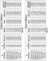
- FIG. 6 B is a table of the baseline clinical characteristics of the patients' breasts, in accordance with aspects of the present disclosure.
- FIG. 20 A is a table of different height, width, and projection parameter settings, in accordance with aspects of the present disclosure
- FIGS. 21 A-D are bar plots of the coefficient differences for height, width, depth, and ptosis, in accordance with aspects of the present disclosure
- FIGS. 22 A-D are graphs of the coefficient differences for height, width, depth and ptosis linearly related to their corresponding parameters, in accordance with aspects of the present disclosure
- FIGS. 23 A-B are boxplots of the ptosis coefficients versus the ptosis grade rating, in accordance with aspects of the present disclosure
- FIGS. 24 A-B are tables of the P-values, in accordance with aspects of the present disclosure.
- FIGS. 25 A-D are scatterplots of selected parameter coefficients vs. the parameter, in accordance with aspects of the present disclosure.
- FIG. 26 A is a table of the statistics of the RMSE between the ground truth data set and the reconstructed SPHARM models, in accordance with aspects of the present disclosure
- FIG. 26 B is a table of the statistics of the Hausdorff distance between the ground truth data set and the reconstructed SPHARM models, in accordance with aspects of the present disclosure
- FIGS. 27 A-C are diagrams of example SPHARM models based on degree 1, 20, and 50 using level 4 icosahedral subdivision, in accordance with aspects of the present disclosure
- FIG. 28 is a table of the magnitude of the SPHARM coefficients for different degrees, in accordance with aspects of the present disclosure.
- FIG. 29 is a table of the P-values from the Wilcoxon rank-sum test comparing the five coefficient values between different degrees, in accordance with aspects of the present disclosure.
- FIGS. 31 A-C are diagrams of the selection area of the transition point and lateral point, in accordance with aspects of the present disclosure.
- FIG. 32 is a diagram of examples of breast cropping selecting the extreme corners for the transition and lateral points, in accordance with aspects of the present disclosure
- FIG. 33 is a table of the rate of successful processing at each step of the algorithm for different landmark positions, in accordance with aspects of the present disclosure
- FIG. 35 is a table of the Euclidean distance of the corner selections relative to the ground truth selection, in accordance with aspects of the present disclosure.
- FIG. 36 is a diagram of the lateral point location of the different sets relative to the ground truth, in accordance with aspects of the present disclosure.
- FIG. 37 is a table of the rate of successful processing at each step of the algorithm for different lateral point locations based on the 161 breasts, in accordance with aspects of the present disclosure
- FIG. 38 is a diagram of the transition point location of the different sets relative to the ground truth, in accordance with aspects of the present disclosure.
- FIG. 39 is a table of the rate of successful processing at each step of the algorithm for different transition point locations based on 161 breasts, in accordance with aspects of the present disclosure.
- FIG. 40 is a table depicting statistics on the projection percent difference between the ground truth selection and shifting the transition point (TP) 3 and 6 mm in different directions, in accordance with aspects of the present disclosure
- FIG. 41 is a table depicting statistics on the volume percent difference between the ground truth selection and shifting the transition point (TP) 3 and 6 mm in different directions, in accordance with aspects of the present disclosure
- FIG. 42 is a table depicting statistics on the projection percent difference between the ground truth selection and shifting the lateral point (LP) 3 and 6 mm in different directions, in accordance with aspects of the present disclosure
- FIG. 46 B is a set of confusion matrices for k-nearest neighbor classification, quadratic discriminate analysis, and Na ⁇ ve Bayes classifier using BMI, breast volume, and breast dimensions, in accordance with aspects of the present disclosure
- FIG. 52 D is a table of the Hausdorff distance between the true postoperative models and the predicted models organized into three BMI groups, in accordance with aspects of the present disclosure
- FIG. 53 is a table depicting examples of predicted postoperative models, in accordance with aspects of the present disclosure.
- FIG. 54 is a diagram of the best prediction out of 53 reconstructed breasts, in accordance with aspects of the present disclosure.
- FIG. 55 is a diagram of the worst prediction out of 53 reconstructed breasts, in accordance with aspects of the present disclosure.
- FIG. 56 is a table of RMSD comparisons between the pre-op breast shape SPHARM coefficients, the transformed pre-op breast coefficient, and actual post—op coefficients for three patients, in accordance with aspects of the present disclosure
- FIG. 58 is a block diagram of a method for modeling a breast, in accordance with the present disclosure.
- This disclosure relates to the modeling of breasts using spherical harmonics.
- FIGS. 1 A-B two general types of cosmetic and reconstructive breast procedures are shown. Two common procedures performed are autologous reconstruction using a transverse rectus abdominis (TRAM) flap is shown in FIG. 1 A and implant reconstruction is shown in 1 B.
- TAM transverse rectus abdominis
- FIG. 1 A two common procedures performed are autologous reconstruction using a transverse rectus abdominis (TRAM) flap is shown in FIG. 1 A and implant reconstruction is shown in 1 B.
- TAM transverse rectus abdominis
- FIG. 1 B implant reconstruction
- computational three-dimensional breast model may be used. According the present disclosure, a method using spherical harmonics is employed to produce the three-dimensional breast models.
- Spherical harmonics are a complete series of orthogonal functions defined on the surface of a sphere.
- the spherical harmonic (SPHARM) method converts a 3D object of spherical topology into three sets of SPHARM coefficients that describe its overall shape in terms of three sets of spherical harmonics basis functions (one set for each dimension).
- SPHARM has applicability to several fields including computer vision and computer graphics but has been most aptly applied in studying medical images, particularly in brain morphometry. There are several inherent properties of this shape descriptor that make it an advantageous method for medical image analysis. It can be compactly represented, allows for efficient shape comparison, incorporates implicit interpolation since the spherical domain is continuous, and can be used to establish surface correspondence. In addition, it can be processed in both the spatial and frequency domain.
- a 3D imaging system may be used to record 3D images of the frontal portion of female torsos.
- the imaging system may represent a 3D image in the form of a triangular mesh that contains, for example about 75,000 vertices and 140,000 faces for each patient image. Each face also has texture associated with it, so a 3D texture image can also be viewed.
- the vertices may be spaced about 3 mm apart from each other and the stated error is less than 0.5 mm.
- the 3D surface image can be viewed as a 3D point cloud ( FIG. 2 A ), a triangular mesh ( FIG. 2 B ), or a 3D texture image overlaid on the triangular mesh ( FIG. 2 C ).
- the exemplary imaging system 300 may include six modular camera units 302 and may use, for example, stereophotogrammetry to estimate a 3D surface image from pairs of 2D photographs. These camera units 302 may be effectively positioned around a person (top, middle, and bottom) to improve the 3D surface coverage of the front torso.
- Each camera unit 302 may be equipped with a pair of stereo cameras and a color camera. The stereo cameras may be synchronized to initiate together and has a 1.5 millisecond capture speed. A half millisecond later, the color cameras may be triggered to capture 2D photographs from six different viewpoints. The system then employs a sophisticated software algorithm to unify the images from the six camera units.
- the color photographs are mapped to the generated triangular mesh.
- the illustrated example uses stereophotogrammetry, however, other forms of 3D imaging (such as a portable handheld 3D scanner, or IPAD Pro, or others) are contemplated.
- the controller 200 includes, for example, a database 210 , one or more processors 220 , at least one memory 230 , and a network interface 240 .
- the database 210 can be located in a storage.
- Storage may refer to any device or material from which information may be capable of being accessed, reproduced, and/or held in an electromagnetic or optical form for access by a computer processor.
- a storage may be, for example, volatile memory such as RAM, non-volatile memory, which permanently hold digital data until purposely erased, such as flash memory, magnetic devices such as hard disk drives, and optical media such as a CD, DVD, Blu-ray disc, cloud storage, or the like.
- Tissue expanders or temporary saline implants, are sometimes used to slowly stretch the skin and pectoralis muscle (large chest muscle). They may be later replaced with tissue or a permanent implant.
- the average age of the patients was 49.9 ⁇ 10.3 years (range: 24 to 75), and BMI was 28.0 ⁇ 5.5 (range: 18.1 to 69.8).
- patients may be generally in a standing position with their hands on their hips. Images may be taken, typically but not consistently, at three-month intervals (for up to a period of two years) during the reconstructive process. Some, but not all, patients may have had a preoperative image.
- a Java-based visualization and analysis tool may be used to manipulate 3D images of the female torso (translate, rotate, scale, and crop), manually mark fiducial points, such as the sternal notch, nipples, lateral points, inframammary fold (IMF) points, midline point, transition points, and umbilicus, and extract linear, contour, and volume measurements.
- Customized software may be used to align the images in the xyz coordinate space, manually mark fiducial points, and calculate breast volumes.
- FIG. 4 A is an example image of customized software showing linear and contour distances.
- FIG. 4 B is an example image of customized software showing volume measurements of a front female torso.
- a breast model for simulating, evaluating, and interactively adjusting breast shape will be a tool for surgeons in surgical planning and for clinical consultations with patients in shared decision making.
- a parametric breast model proposed by Chen may be used either to create a breast model or fit a model to breast data. They initially create an asymmetric superquadric and then perform five global deformations to model five major features of breast shape.
- FIGS. 5 A-G these five global deformations/features are called lower pole deformation, upper pole deformation, horizontal deviation deformation, medial deformation, and axillary tail deformation.
- FIG. 5 A and FIG. 5 B show the front and side views of a breast model without any deformations applied.
- FIG. 5 C shows a front view example of lower pole deformation, which models breast sagging.
- FIG. 5 D is a side view example of upper pole deformation, which controls the slope and curvature of the upper half of the breast.
- FIG. 5 E a front view example is shown of horizontal deviation deformation, which controls the turn of the breast whether to the right and left.
- FIG. 5 E a front view example is shown of horizontal deviation deformation, which controls the turn of the breast whether to the right and left.
- FIG. 5 F is a front view example of medial deformation, which flattens out the sides of the breast.
- FIG. 5 G is a front view example of axillary tail deformation, which adjusts the top half of the breast to point toward the shoulder.
- the 17 parameters that control the breast size and shape can be used to quantitatively analyze the degrees of key shape variables of the breast.
- Chen's model had limited capability in accurately fitting to breast data due to its basic design. Breasts contain many different curves and shape features that an asymmetric superquadric and five global deformations is unable to fully capture.
- the modeling technique that is introduced in this disclosure may allow for computing the breast shape “distance” based on coefficients, and the coefficients can be related to specific breast shapes.
- the breast shapes may include for example, reduction mammoplasty, reduction mastopexy, augmentation mammoplasty, augmentation mastopexy, and/or correction of any breast shape deformities, based on spherical harmonic coefficients.
- the disclosed method allows for modeling the original breast shape (accuracy of fitted model), can be used to modify breast shape (shape modulation), establishes correspondence between different breast shapes and sizes (cross model alignment), can be used to compute shape distance between different breasts (parametric shape comparison), and can be used to predict breast shapes.
- Surgeons may evaluate breast shape by taking measurements in person using a tape measure in the clinic or using rulers on standard photographs of the frontal and lateral views. These measurements include the linear distance between fiducial points, including the sternal notch, nipples, lateral points, mid-clavicle points, midline, and inframammary fold.
- Another measurement to describe breast shape is ptosis, which is used to describe sagging of the breasts.
- the inframammary fold is often designated as a reference point for evaluating the degree of ptosis.
- grading ptotic breasts can be difficult as the inframammary fold is hidden when the woman is standing in an upright position.
- the breasts may be assigned a grade ranging from 0 to 3, where Grade 0 represents no ptosis and Grade 3 represents extreme ptosis.
- Grade 0 represents no ptosis
- Grade 3 represents extreme ptosis.
- One of skill in the art would know what Regnault's classification is and how to implement it. With the advent of three-dimensional imaging technology, additional objective measurements, such as surface contours and curvature, surface area, volume, and even ptosis can now be quantitatively assessed.
- the SPHARM modeling method was first tested on three-dimensional preoperative torso surface images of a number of women scheduled to undergo mastectomy for the treatment or prevention of breast cancer and other abnormalities. None of them have previous breast surgeries but may have had a biopsy that did not affect the breast appearance as determined by an experienced plastic surgeon. Patients with rare congenital breast abnormalities, previous radiation therapy, or previous major breast surgeries were excluded.
- FIGS. 6 A-E tables of patient demographics, clinical characteristics of the patients' breasts, patient variables, procedure types, and the procedures underwent are shown.
- FIG. 6 A is a table of the percentages of the patient demographics and characteristics describing the age, BMI, tumor size, race, ethnicity, diagnosis and number treated with pre-operative chemotherapy.
- FIG. 6 C is a table of the demographics of 32 patients who underwent TRAM flap and/or implant reconstruction.
- FIG. 6 D is a table of the procedure used for each breast.
- FIG. 6 E is a table of the patient demographics at the time of their preoperative image.
- the breasts may be extracted from the torso images by identifying the borders of each breast.
- the fiducial locations at the top, bottom, left, and right sides of each breast that would delineate how the breast would be segmented from the torso images may be identified.
- the images had to be manually aligned so that the height of a patient from head to foot aligned with the y axis, the width from the right shoulder to the left shoulder aligned with the x axis, and the body faced the positive z direction.
- four fiducial locations were found along the border of each breast from the 3D torso image ( FIG. 9 A ). Two locations were automatically detected: the midline point and inframammary fold. The other two locations, the transition point and the lateral point, may be manually selected. These points may be manually selected as the breast is usually relatively smooth in these regions.
- the graphical models of the breasts may be made by finding the number of vertices along the x and y axes ( 7 A-B) and the midline point ( 7 C-E) to show the inframammary folds ( 7 F).
- Manual fiducial point selection may be conducted in customized software (a customized Java-based visualization tool), and the automatically detected points may be found using code developed in MATLAB 2015a.
- the head, arms, and the torso below the breasts may be cropped out first. The head and arms contained less points than the torso, and the number of points peaked on the sides of the torso.
- the vertices with surface normal within 18.2° (acos(0.95)) of the negative y direction were sub-selected. Then the vertex with the lowest y value was designated as the lowest visible point of the breast and was used as the bottom cutoff point. If there were no valid vertices with a y surface normal within 18.2° (acos(0.95)) of the negative y direction, excluding the nipple, then the bottom cutoff point was set to 33 centimeters below the top cutoff point. It may be determined that the breasts were within 33 centimeters from the top cutoff point. This mostly occurred for relatively small, non-ptotic breasts. Then Gaussian curvature was computed on the remaining mesh contained within the designated borders as shown in FIG.
- points may be estimated along the IMF (inferior breast-chest contour) of the left and right breasts.
- the red line in FIG. 7 F delineates the estimated IMF.
- the partial torso image in FIG. 7 F is colored by the shape index for each vertex in the mesh, which was calculated from the contour detection algorithm.
- the IMF was detected by following the negative curvature path along the underside of the breast.
- the algorithm selected the underside of the nipple as the inframammary fold, hence providing incorrect fiducial points.
- the algorithm was modified to ignore the curvature values of the nipple area.
- the nipple area was experimentally determined to be within 2.5 centimeters of the most projected points along the z axis on the left and right half of the torso.
- the nipple diameter for women aged 20-64 years may range from for example, 1 cm to 2.75 cm.
- the ellipse formula (using only x and y coordinates assuming that the x axis aligns with the width of the breast and the y axis aligns with the breast height):
- SPHARM may require a genus zero surface and a relatively dense mesh to accurately model an object. Since the cropped breast was an unclosed surface patch, a method to patch the back hole in order to form a closed surface may be used. First, the cropped breast mesh may be pre-processed to clean up non-manifold vertices (i.e., edges that are shared by more than two faces and isolated pieces (disconnected vertices and edges)). Then the advancing front mesh (AFM) technique may be used to fill any small holes that were created due to the removal of the non-manifold vertices following the rules for creating triangles as shown in FIGS. 10 C-E . FIG.
- AFM advancing front mesh
- the location of the new vertices is determined by the average edge length and the shortest direction to close the gap across the two meshes. Next, computing the distance between every newly created vertex and every related boundary vertex; if the distance between them is less than the given threshold (such as the average edge length), they are merged. Next, update the front. Next, repeat until all holes are filled.
- a straight line (CN) extending from the centroid of the breast boundary points (backside of the breast) to the nipple, or most projecting point may be generated.
- Each point (x, y, z) on the breast surface was then projected onto the CN line, to generate its position (y 1 , z 1 ) on the line, such that x 1 , the x-coordinate of the point on the CN line, is equivalent to the x-coordinate of point PP.
- the angle ⁇ can be calculated based on the (y, z) breast point coordinate and point (y 1 , z 1 ) on the CN line.
- the set of all the points on the breast surface having the same ⁇ , on the positive (front) side of the breast are used to trace its angular path along the surface, and its length is determined.
- the relative surface distance of the point P with respect to this length is scaled by ⁇ /2 and is assigned to ⁇ accordingly.
- the front side of the breast was assigned positive values for ⁇ , and the back side was assigned negative values for ⁇ .
- the surface of different objects are aligned through spherical parameterization, achieving correspondence across two different breast models is feasible, which allows for comparisons of local and global changes in breast shape and size.
- the Fourier spherical harmonics Y( ⁇ , ⁇ ) (or SPHARM functions) of degree l and order m can be defined by
- L max is a user-specified degree.
- the function v( ⁇ , ⁇ ) can be independently decomposed into three functions for the three coordinates:
- FIG. 42 is a table depicting statistics on the projection percent difference between the ground truth selection and shifting the LP three (3) and six (6) millimeters in different directions including down, up, forward and backward.
- FIG. 43 is a table depicting statistics on the volume percent difference between the ground truth selection and shifting the LP three (3) and six (6) millimeters in different directions including down, up, forward and backward
- a program may be used to adjust the height, width, depth, and ptosis to any loaded breast data.
- a MATLAB application was created that allows any user to easily apply different settings for adjusting height, width, projection, and ptosis to any loaded breast data that has SPHARM coefficients computed for it.
- the original texture or a generic texture can be mapped to the breast model.
- the application takes less than half a second to process the input parameters and display the modified breast model.
- TRAM flaps tend to give breasts a smooth teardrop shape that looks more natural ( FIG. 45 A ), while implants create round and protruded shaped breasts ( FIG. 45 B ).
- FIG. 48 a set of images of true versus predicted class reconstructed breasts according to a TRAM flap or implant are shown.
- a template breast model can be created from a set of breast models that are similar in shape using the RMSD. Two examples of the average breast object are shown below. One TRAM flap reconstructed breast model was selected, and four other breast models that were similar in shape (out of 28 TRAM reconstructed breasts) based on the coefficients were also selected. The five breast models shown in FIGS. 49 A-B were averaged together to form an average breast model shown in FIG. 51 A . The original images are shown in FIG. 49 A and the SPHARM models with the original texture applied are shown in FIG. 49 B . All right breasts were mirrored in order to compare with the left breasts. The RMSDs were between 5.51 and 7.16 for the first TRAM flap reconstructed breast versus the other four breasts.
- the RMSDs of the five breasts were between 3.83 and 4.66.
- an implant reconstructed breast model and its four nearest implant reconstructed breast models (out of 23 implant breasts) were averaged together ( FIGS. 50 A-B ) to form an average breast model ( FIG. 51 B ).
- FIG. 50 A shows the original images
- FIG. 50 B shows the SPHARM models with the original texture applied.
- the RMSDs were between 3.89 and 5.54 for the implant reconstructed breast versus the other four implant reconstructed breasts.
- the RMSDs of the five breasts were between 2.57 and 4.38.
- FIGS. 52 A-D tables of data for a set of patients are shown describing the types of operations undergone ( FIG. 52 A ), the number of patients, the RMSD of various preoperative and postoperative breast comparisons ( FIG. 52 B ), the RMSD for various BMI ( FIG. 52 C ), and the HD for various BMI ( FIG. 52 D ).
- FIG. 52 A is a table of the procedures conducted on the left and right breast of each patient and the average RMSD for each set of patients with the same procedures.
- FIG. 52 B is a table of the statistics on the RMSD comparing preoperative, postoperative, and predicted breast shapes.
- FIG. 52 B is a table of the statistics on the RMSD comparing preoperative, postoperative and predicted breast shapes.
- FIG. 52 C is a table of the RMSD between the true postoperative models and the predicted models organized into three BMI groups.
- FIG. 52 D is a table of the HD between the true postoperative models and the predicted models organized into three BMI groups.
- postoperative models may be predicted.
- individualistic features may be averaged and/or smoothed out, such as the nipple.
- a depression does not get added in the predicted breast, as shown in row 3 of FIG. 53 .
- the RMSD between the predicted and the actual postoperative model also improved. Only the first 27 coefficients were added in order to avoid the depression but found that the RMSD was lower when using the average object approach, as presented in FIG. 52 B and exemplified in FIG. 53 .
- a prediction had a RMSD of 6.17 between the true postoperative model and the predicted model as shown in FIG. 54 .
- the five closest breast models had RMSDs between 6.83 and 9.35, so there were relatively similar shaped breasts in the database.
- the reference image's closest breast models in the database had RMSDs between 14.99 and 19.04, so there just wasn't a similar breast shape available in the database to make a more accurate prediction ( FIG. 55 ).
- the prediction models were separated by BMI, the RMSDs between the true postoperative model and the predicted model were lower for BMI less than 25 and higher for BMI greater than 30 ( FIG. 52 C ).
- the average Hausdorff distance was also larger for patients with BMI greater than 30 than the patients with lower BMI ( FIG. 52 D ).
- SPHARM models may be used to predict outcomes, which may be improved with a larger dataset and matching by age, BMI, breast volume, reconstruction type, smoking history, and other factors including patient preferences. All of these factors are discussed during patient consultations, which can be used to help predict the model.
- a method to model the breast that can be used to analyze, compare, and modify its shape, is shown.
- the algorithm may be robust to small differences in the point selection of the transition point and lateral point.
- Results on classification for different breast reconstruction types are shown, creating average breast objects that can represent a particular shape, and predictive modeling.
- the three-dimensional model based on SPHARM and its further development will provide a state-of-the-art surgical planning tool for surgeons to visualize and interactively evaluate the morphology of the breast. It will also help patients in making more informed decisions.
- her breasts can be shape matched to the preoperative breasts of previous reconstruction patients and then shown their post-surgical outcomes, which may help the patient mentally prepare for possible outcomes.
- a standard may be developed for automatically detecting the lateral and transition points to maintain consistency (increase precision), if not accuracy, across different time points in the reconstructive process (as the patient is imaged every three months before and after mastectomy and reconstruction) as well as across different patients.
- the model may also be reconnected with the torso to evaluate the overall appearance of the breast in relation to the human body.
- the SPHARM coefficients has potential for classifying different breast shapes. There are several natural breast shapes that have been identified for women. They may serve as a starting point for helping to objectively categorize the shape of a woman's breasts in order to select the right bra size and type that would fit comfortably.
- a method to predict surgical outcome may include acquiring a 3D pre-op image of a patient, looking at database for similar demographics (e.g., age etc.) and breast size and shape, where the database also includes pre-op and post-op 3D images, find post operation image of those patients, determine new SPHARM coefficients, applying the SPHARM coefficients to the 3D pre-op image of the patient, and morphing the breast based on the new SPHARM coefficients.
- similar demographics e.g., age etc.
- the database also includes pre-op and post-op 3D images, find post operation image of those patients, determine new SPHARM coefficients, applying the SPHARM coefficients to the 3D pre-op image of the patient, and morphing the breast based on the new SPHARM coefficients.
- a method to predict surgical outcome may include the generation of template breast shapes that can be then used to predict and/or visualize the breast shape for women seeking a particular option, or to compare different options.
- FIGS. 57 A-D images of frontal and lateral views of pre-op (P 1 ), post-op (P 2 ), estimate (E) and overlay of estimate on the post-op breast for four breasts in the input test set are shown.
- the post-op images in FIGS. 57 A-D are actual reconstruction results obtained by imaging the patient after the surgery at 18 months in a consultation timeline.
- deep learning/AI/machine learning algorithms may be used in the above method to predict surgical outcomes.
- the predicting may include using a machine learning algorithm, where training data inputs include for example, pre and post operation image data and/or patient demographic data.
- Machine learning algorithms may include, for example, a neural network, random forest regression, linear regression (LR), ridge regression (RR), least-angle regression (LARS), and/or least absolute shrinkage and selection operator regression (LASSO).
- the machine learning algorithms may be executed on the controller (see FIG. 3 B ), and/or on a remote computing system.
- a 3D surface image of pre-op breast is available before the surgery is scheduled and the surgical option under consideration is known, or to be determined.
- the shape change of breasts pre- and post-surgery is dependent on surgery type and other medical parameters such as ptosis grade, implant size and weight, skin elasticity. It is not feasible to compute a single generalized transformation for pre-op breast shape to the expected post-op shape for all surgery types.
- a data-driven approach may be employed to estimate the transformation vector using nonlinear regression for any changes in breast shape, including natural (e.g. aging, pregnancy, or other deformities), or surgical.”
- Random regression forest is made of several individual regression trees.
- a regression tree is recursively constructed such that at each node the training data is split on a randomly chosen feature variable so that entropy at the node is minimized.
- the entropy of the feature densities associated with different nodes decreases when going from the root towards the leaves.
- the random forest simply averages the results from individual regression trees to predict the output.
- the transformation obtained from the regression to the pre-op (P 1 ) breast coefficients to obtain the estimation of post-op shape (see FIG. 57 D ) was applied.
- the method further includes identifying different types of natural breast shapes and contours, shapes related to breast diseases, and outcomes of surgical procedures, including reconstructed breasts (at least one of autologous or implant reconstructed breasts), and cosmetic procedures (augmentation, reduction, mastopexy), based on spherical harmonic coefficients.
- a method of predicting includes creating a general shape template from images of several women.
- the general template may be created using images of other breasts from a group of women (similar in demographics such as BMI, age, etc.).
- the general template may be created for specific breast conditions using data from large groups of women (i.e. not the few that have the most similar shape).
- the database for 3D images of patient with similar demographics are not limited to age, breast size, or breast shape, and may contain other demographic data.
- the method receives a 3D image (e.g., a pre-operative image), which includes a breast.
- a 3D image e.g., a pre-operative image
- the method identifies the breast in the 3D image.
- the method extracts 3D image data of the breast from the 3D image.
- the method forms a closed object using 3D image data of the breast to create a zero-genus surface.
Landscapes
- Health & Medical Sciences (AREA)
- Life Sciences & Earth Sciences (AREA)
- Engineering & Computer Science (AREA)
- Public Health (AREA)
- Medical Informatics (AREA)
- General Health & Medical Sciences (AREA)
- Biomedical Technology (AREA)
- Surgery (AREA)
- Pathology (AREA)
- Heart & Thoracic Surgery (AREA)
- Molecular Biology (AREA)
- Animal Behavior & Ethology (AREA)
- Veterinary Medicine (AREA)
- Biophysics (AREA)
- Physics & Mathematics (AREA)
- Nuclear Medicine, Radiotherapy & Molecular Imaging (AREA)
- Radiology & Medical Imaging (AREA)
- Primary Health Care (AREA)
- Epidemiology (AREA)
- Data Mining & Analysis (AREA)
- Reproductive Health (AREA)
- Gynecology & Obstetrics (AREA)
- Dentistry (AREA)
- Oral & Maxillofacial Surgery (AREA)
- Robotics (AREA)
- Databases & Information Systems (AREA)
- Apparatus For Radiation Diagnosis (AREA)
Abstract
Description
P faces ∧(x′, y′, z′)=Σl=0 L
β=[β1β2 . . . β1319 β1320]T.
AW=B
min/β∥AW−B∥ 2
S i =C i if (C i /k>C j /k).
p(x i |C k x l , . . . , x i+1. . . , x n)=p(x i |C k),
-
- Set 0: Ground truth (Manually selected TP and LP)
- Set 1: Top-medial TP and top-front LP
- Set 2: Top-lateral TP and top-back LP
- Set 3: Bottom-lateral TP and bottom-back LP
- Set 4: Bottom-medial TP and bottom-front LP
Claims (20)
Priority Applications (1)
| Application Number | Priority Date | Filing Date | Title |
|---|---|---|---|
| US17/605,720 US12343159B2 (en) | 2019-04-26 | 2020-04-24 | Systems and methods for modeling the breast using spherical harmonics |
Applications Claiming Priority (3)
| Application Number | Priority Date | Filing Date | Title |
|---|---|---|---|
| US201962838997P | 2019-04-26 | 2019-04-26 | |
| PCT/US2020/029783 WO2020219856A1 (en) | 2019-04-26 | 2020-04-24 | Systems and methods for modeling the breast using spherical harmonics |
| US17/605,720 US12343159B2 (en) | 2019-04-26 | 2020-04-24 | Systems and methods for modeling the breast using spherical harmonics |
Related Parent Applications (1)
| Application Number | Title | Priority Date | Filing Date |
|---|---|---|---|
| PCT/US2020/029783 A-371-Of-International WO2020219856A1 (en) | 2019-04-26 | 2020-04-24 | Systems and methods for modeling the breast using spherical harmonics |
Related Child Applications (1)
| Application Number | Title | Priority Date | Filing Date |
|---|---|---|---|
| US19/218,940 Continuation US20250281107A1 (en) | 2019-04-26 | 2025-05-27 | Systems and methods for modeling the breast using spherical harmonics |
Publications (2)
| Publication Number | Publication Date |
|---|---|
| US20220304619A1 US20220304619A1 (en) | 2022-09-29 |
| US12343159B2 true US12343159B2 (en) | 2025-07-01 |
Family
ID=72941359
Family Applications (2)
| Application Number | Title | Priority Date | Filing Date |
|---|---|---|---|
| US17/605,720 Active 2042-09-20 US12343159B2 (en) | 2019-04-26 | 2020-04-24 | Systems and methods for modeling the breast using spherical harmonics |
| US19/218,940 Pending US20250281107A1 (en) | 2019-04-26 | 2025-05-27 | Systems and methods for modeling the breast using spherical harmonics |
Family Applications After (1)
| Application Number | Title | Priority Date | Filing Date |
|---|---|---|---|
| US19/218,940 Pending US20250281107A1 (en) | 2019-04-26 | 2025-05-27 | Systems and methods for modeling the breast using spherical harmonics |
Country Status (2)
| Country | Link |
|---|---|
| US (2) | US12343159B2 (en) |
| WO (1) | WO2020219856A1 (en) |
Families Citing this family (5)
| Publication number | Priority date | Publication date | Assignee | Title |
|---|---|---|---|---|
| US11759151B2 (en) * | 2019-05-03 | 2023-09-19 | The Board Of Trustees Of The University Of Alabama | Body composition assessment using two-dimensional digital image analysis |
| US12179060B2 (en) | 2021-03-11 | 2024-12-31 | The Board Of Trustees Of The University Of Alabama | Automated aerobic fitness measurement from smartphone technology |
| US12254562B2 (en) | 2021-08-30 | 2025-03-18 | Proxamama LLC | Method for nipple replication |
| EP4460281A1 (en) * | 2022-01-03 | 2024-11-13 | Proxamama LLC | Method for nipple replication |
| CN115984229B (en) * | 2023-01-10 | 2023-09-05 | 北京医准智能科技有限公司 | Model training method, breast measurement device, electronic equipment and medium |
Citations (3)
| Publication number | Priority date | Publication date | Assignee | Title |
|---|---|---|---|---|
| US20130267850A1 (en) | 2010-12-06 | 2013-10-10 | Michael Berman | System and method for ultrasonic examination of the breast |
| US20140254910A1 (en) * | 2013-03-11 | 2014-09-11 | Siemens Aktiengesellschaft | Imaging device, assignment system and method for assignment of localization data |
| US20150286785A1 (en) * | 2012-11-21 | 2015-10-08 | The Trustees Of Columbia University In The City Of New York | Systems, methods, and devices for image reconstruction using combined pde-constrained and simplified spherical harmonics algorithm |
-
2020
- 2020-04-24 US US17/605,720 patent/US12343159B2/en active Active
- 2020-04-24 WO PCT/US2020/029783 patent/WO2020219856A1/en not_active Ceased
-
2025
- 2025-05-27 US US19/218,940 patent/US20250281107A1/en active Pending
Patent Citations (3)
| Publication number | Priority date | Publication date | Assignee | Title |
|---|---|---|---|---|
| US20130267850A1 (en) | 2010-12-06 | 2013-10-10 | Michael Berman | System and method for ultrasonic examination of the breast |
| US20150286785A1 (en) * | 2012-11-21 | 2015-10-08 | The Trustees Of Columbia University In The City Of New York | Systems, methods, and devices for image reconstruction using combined pde-constrained and simplified spherical harmonics algorithm |
| US20140254910A1 (en) * | 2013-03-11 | 2014-09-11 | Siemens Aktiengesellschaft | Imaging device, assignment system and method for assignment of localization data |
Non-Patent Citations (2)
| Title |
|---|
| International Preliminary Report on Patentability issued by The International Bureau of WIPO in connection with International Application No. PCT/US20/29783, dated Sep. 28, 2021. |
| International Search Report and Written Opinion issued by the International Searching Authority/US in connection with International Application No. PCT/US20/29783, dated Jul. 23, 2020. |
Also Published As
| Publication number | Publication date |
|---|---|
| WO2020219856A1 (en) | 2020-10-29 |
| US20250281107A1 (en) | 2025-09-11 |
| US20220304619A1 (en) | 2022-09-29 |
Similar Documents
| Publication | Publication Date | Title |
|---|---|---|
| US20250281107A1 (en) | Systems and methods for modeling the breast using spherical harmonics | |
| US9345551B2 (en) | Implant design analysis suite | |
| US8571278B2 (en) | System and methods for multi-object multi-surface segmentation | |
| Kelemen et al. | Elastic model-based segmentation of 3-D neuroradiological data sets | |
| Golland et al. | Deformation analysis for shape based classification | |
| US20100239147A1 (en) | Method and System for Dynamic Pulmonary Trunk Modeling and Intervention Planning | |
| Vezzetti et al. | Geometry-based 3D face morphology analysis: soft-tissue landmark formalization | |
| JP2006518886A (en) | Analysis method of geometric surface by conformal structure | |
| CN106780518A (en) | A kind of MR image three-dimensional interactive segmentation methods of the movable contour model cut based on random walk and figure | |
| Cheema et al. | Image-aligned dynamic liver reconstruction using intra-operative field of views for minimal invasive surgery | |
| Nguyen et al. | A statistical shape modeling approach for predicting subject-specific human skull from head surface | |
| Hobbs et al. | Quad-mesh based radial distance biomarkers for Alzheimer's disease | |
| Broadhurst et al. | Histogram statistics of local model-relative image regions | |
| US20210383915A1 (en) | Systems and methods for processing electronic images to determine a modified electronic image for breast procedures | |
| Cheong | Computational modeling of breast shape using spherical harmonics | |
| Timoner | Compact representations for fast nonrigid registration of medical images | |
| De Oliveira | An affordable and practical 3d solution for the aesthetic evaluation of breast cancer conservative treatment | |
| Golland | Statistical shape analysis of anatomical structures | |
| Zsemlye | Shape prediction from partial information | |
| Kolagunda | Towards computer aided guidance system for robot assisted laparoscopic radical prostatectomy | |
| Marinotto | Development of a 3D digital tool for breast reconstruction simulation in post-mastectomy planning | |
| Baudin | De la segmentation au moyen de graphes d’images de muscles striés squelettiques acquises par RMN | |
| Zhou et al. | Automatic segmentation for medical image with the optimized tree structured part model | |
| Zolfagharnasab | Toward a 3D Planning Approach for Breast Conserving Surgery | |
| Leymarie et al. | A general approach to model biomedical data from 3D unorganised point clouds with medial scaffolds |
Legal Events
| Date | Code | Title | Description |
|---|---|---|---|
| FEPP | Fee payment procedure |
Free format text: ENTITY STATUS SET TO UNDISCOUNTED (ORIGINAL EVENT CODE: BIG.); ENTITY STATUS OF PATENT OWNER: SMALL ENTITY |
|
| AS | Assignment |
Owner name: UNIVERSITY OF HOUSTON SYSTEM, TEXAS Free format text: ASSIGNMENT OF ASSIGNORS INTEREST;ASSIGNORS:MERCHANT, FATIMA;CHEONG, AUDREY;SIGNING DATES FROM 20211015 TO 20211022;REEL/FRAME:057945/0346 |
|
| FEPP | Fee payment procedure |
Free format text: ENTITY STATUS SET TO SMALL (ORIGINAL EVENT CODE: SMAL); ENTITY STATUS OF PATENT OWNER: SMALL ENTITY |
|
| STPP | Information on status: patent application and granting procedure in general |
Free format text: DOCKETED NEW CASE - READY FOR EXAMINATION |
|
| STPP | Information on status: patent application and granting procedure in general |
Free format text: NON FINAL ACTION MAILED |
|
| STPP | Information on status: patent application and granting procedure in general |
Free format text: RESPONSE TO NON-FINAL OFFICE ACTION ENTERED AND FORWARDED TO EXAMINER |
|
| STPP | Information on status: patent application and granting procedure in general |
Free format text: NOTICE OF ALLOWANCE MAILED -- APPLICATION RECEIVED IN OFFICE OF PUBLICATIONS |
|
| STCF | Information on status: patent grant |
Free format text: PATENTED CASE |