US12331359B2 - Neoantigens and uses thereof for treating cancer - Google Patents
Neoantigens and uses thereof for treating cancer Download PDFInfo
- Publication number
- US12331359B2 US12331359B2 US16/478,818 US201816478818A US12331359B2 US 12331359 B2 US12331359 B2 US 12331359B2 US 201816478818 A US201816478818 A US 201816478818A US 12331359 B2 US12331359 B2 US 12331359B2
- Authority
- US
- United States
- Prior art keywords
- neoantigen
- cancer
- clone
- subject
- neoantigens
- Prior art date
- Legal status (The legal status is an assumption and is not a legal conclusion. Google has not performed a legal analysis and makes no representation as to the accuracy of the status listed.)
- Active, expires
Links
Images


Classifications
-
- A—HUMAN NECESSITIES
- A61—MEDICAL OR VETERINARY SCIENCE; HYGIENE
- A61P—SPECIFIC THERAPEUTIC ACTIVITY OF CHEMICAL COMPOUNDS OR MEDICINAL PREPARATIONS
- A61P35/00—Antineoplastic agents
-
- A—HUMAN NECESSITIES
- A61—MEDICAL OR VETERINARY SCIENCE; HYGIENE
- A61K—PREPARATIONS FOR MEDICAL, DENTAL OR TOILETRY PURPOSES
- A61K35/00—Medicinal preparations containing materials or reaction products thereof with undetermined constitution
- A61K35/12—Materials from mammals; Compositions comprising non-specified tissues or cells; Compositions comprising non-embryonic stem cells; Genetically modified cells
- A61K35/14—Blood; Artificial blood
- A61K35/15—Cells of the myeloid line, e.g. granulocytes, basophils, eosinophils, neutrophils, leucocytes, monocytes, macrophages or mast cells; Myeloid precursor cells; Antigen-presenting cells, e.g. dendritic cells
-
- A—HUMAN NECESSITIES
- A61—MEDICAL OR VETERINARY SCIENCE; HYGIENE
- A61K—PREPARATIONS FOR MEDICAL, DENTAL OR TOILETRY PURPOSES
- A61K35/00—Medicinal preparations containing materials or reaction products thereof with undetermined constitution
- A61K35/12—Materials from mammals; Compositions comprising non-specified tissues or cells; Compositions comprising non-embryonic stem cells; Genetically modified cells
- A61K35/14—Blood; Artificial blood
- A61K35/17—Lymphocytes; B-cells; T-cells; Natural killer cells; Interferon-activated or cytokine-activated lymphocytes
-
- A—HUMAN NECESSITIES
- A61—MEDICAL OR VETERINARY SCIENCE; HYGIENE
- A61K—PREPARATIONS FOR MEDICAL, DENTAL OR TOILETRY PURPOSES
- A61K39/00—Medicinal preparations containing antigens or antibodies
-
- A—HUMAN NECESSITIES
- A61—MEDICAL OR VETERINARY SCIENCE; HYGIENE
- A61K—PREPARATIONS FOR MEDICAL, DENTAL OR TOILETRY PURPOSES
- A61K39/00—Medicinal preparations containing antigens or antibodies
- A61K39/0005—Vertebrate antigens
- A61K39/0011—Cancer antigens
-
- A—HUMAN NECESSITIES
- A61—MEDICAL OR VETERINARY SCIENCE; HYGIENE
- A61K—PREPARATIONS FOR MEDICAL, DENTAL OR TOILETRY PURPOSES
- A61K39/00—Medicinal preparations containing antigens or antibodies
- A61K39/0005—Vertebrate antigens
- A61K39/0011—Cancer antigens
- A61K39/001169—Tumor associated carbohydrates
- A61K39/00117—Mucins, e.g. MUC-1
-
- C—CHEMISTRY; METALLURGY
- C12—BIOCHEMISTRY; BEER; SPIRITS; WINE; VINEGAR; MICROBIOLOGY; ENZYMOLOGY; MUTATION OR GENETIC ENGINEERING
- C12Q—MEASURING OR TESTING PROCESSES INVOLVING ENZYMES, NUCLEIC ACIDS OR MICROORGANISMS; COMPOSITIONS OR TEST PAPERS THEREFOR; PROCESSES OF PREPARING SUCH COMPOSITIONS; CONDITION-RESPONSIVE CONTROL IN MICROBIOLOGICAL OR ENZYMOLOGICAL PROCESSES
- C12Q1/00—Measuring or testing processes involving enzymes, nucleic acids or microorganisms; Compositions therefor; Processes of preparing such compositions
- C12Q1/68—Measuring or testing processes involving enzymes, nucleic acids or microorganisms; Compositions therefor; Processes of preparing such compositions involving nucleic acids
- C12Q1/6876—Nucleic acid products used in the analysis of nucleic acids, e.g. primers or probes
- C12Q1/6883—Nucleic acid products used in the analysis of nucleic acids, e.g. primers or probes for diseases caused by alterations of genetic material
- C12Q1/6886—Nucleic acid products used in the analysis of nucleic acids, e.g. primers or probes for diseases caused by alterations of genetic material for cancer
-
- G—PHYSICS
- G01—MEASURING; TESTING
- G01N—INVESTIGATING OR ANALYSING MATERIALS BY DETERMINING THEIR CHEMICAL OR PHYSICAL PROPERTIES
- G01N33/00—Investigating or analysing materials by specific methods not covered by groups G01N1/00 - G01N31/00
- G01N33/48—Biological material, e.g. blood, urine; Haemocytometers
- G01N33/50—Chemical analysis of biological material, e.g. blood, urine; Testing involving biospecific ligand binding methods; Immunological testing
- G01N33/53—Immunoassay; Biospecific binding assay; Materials therefor
- G01N33/569—Immunoassay; Biospecific binding assay; Materials therefor for microorganisms, e.g. protozoa, bacteria, viruses
- G01N33/56966—Animal cells
- G01N33/56977—HLA or MHC typing
-
- G01N33/57438—
-
- G—PHYSICS
- G01—MEASURING; TESTING
- G01N—INVESTIGATING OR ANALYSING MATERIALS BY DETERMINING THEIR CHEMICAL OR PHYSICAL PROPERTIES
- G01N33/00—Investigating or analysing materials by specific methods not covered by groups G01N1/00 - G01N31/00
- G01N33/48—Biological material, e.g. blood, urine; Haemocytometers
- G01N33/50—Chemical analysis of biological material, e.g. blood, urine; Testing involving biospecific ligand binding methods; Immunological testing
- G01N33/53—Immunoassay; Biospecific binding assay; Materials therefor
- G01N33/575—Immunoassay; Biospecific binding assay; Materials therefor for cancer
- G01N33/57525—Immunoassay; Biospecific binding assay; Materials therefor for cancer of the liver or pancreas
-
- G—PHYSICS
- G16—INFORMATION AND COMMUNICATION TECHNOLOGY [ICT] SPECIALLY ADAPTED FOR SPECIFIC APPLICATION FIELDS
- G16B—BIOINFORMATICS, i.e. INFORMATION AND COMMUNICATION TECHNOLOGY [ICT] SPECIALLY ADAPTED FOR GENETIC OR PROTEIN-RELATED DATA PROCESSING IN COMPUTATIONAL MOLECULAR BIOLOGY
- G16B20/00—ICT specially adapted for functional genomics or proteomics, e.g. genotype-phenotype associations
- G16B20/10—Ploidy or copy number detection
-
- G—PHYSICS
- G16—INFORMATION AND COMMUNICATION TECHNOLOGY [ICT] SPECIALLY ADAPTED FOR SPECIFIC APPLICATION FIELDS
- G16B—BIOINFORMATICS, i.e. INFORMATION AND COMMUNICATION TECHNOLOGY [ICT] SPECIALLY ADAPTED FOR GENETIC OR PROTEIN-RELATED DATA PROCESSING IN COMPUTATIONAL MOLECULAR BIOLOGY
- G16B20/00—ICT specially adapted for functional genomics or proteomics, e.g. genotype-phenotype associations
- G16B20/20—Allele or variant detection, e.g. single nucleotide polymorphism [SNP] detection
-
- G—PHYSICS
- G16—INFORMATION AND COMMUNICATION TECHNOLOGY [ICT] SPECIALLY ADAPTED FOR SPECIFIC APPLICATION FIELDS
- G16B—BIOINFORMATICS, i.e. INFORMATION AND COMMUNICATION TECHNOLOGY [ICT] SPECIALLY ADAPTED FOR GENETIC OR PROTEIN-RELATED DATA PROCESSING IN COMPUTATIONAL MOLECULAR BIOLOGY
- G16B20/00—ICT specially adapted for functional genomics or proteomics, e.g. genotype-phenotype associations
- G16B20/30—Detection of binding sites or motifs
-
- G—PHYSICS
- G16—INFORMATION AND COMMUNICATION TECHNOLOGY [ICT] SPECIALLY ADAPTED FOR SPECIFIC APPLICATION FIELDS
- G16B—BIOINFORMATICS, i.e. INFORMATION AND COMMUNICATION TECHNOLOGY [ICT] SPECIALLY ADAPTED FOR GENETIC OR PROTEIN-RELATED DATA PROCESSING IN COMPUTATIONAL MOLECULAR BIOLOGY
- G16B30/00—ICT specially adapted for sequence analysis involving nucleotides or amino acids
- G16B30/10—Sequence alignment; Homology search
-
- G—PHYSICS
- G16—INFORMATION AND COMMUNICATION TECHNOLOGY [ICT] SPECIALLY ADAPTED FOR SPECIFIC APPLICATION FIELDS
- G16B—BIOINFORMATICS, i.e. INFORMATION AND COMMUNICATION TECHNOLOGY [ICT] SPECIALLY ADAPTED FOR GENETIC OR PROTEIN-RELATED DATA PROCESSING IN COMPUTATIONAL MOLECULAR BIOLOGY
- G16B30/00—ICT specially adapted for sequence analysis involving nucleotides or amino acids
- G16B30/20—Sequence assembly
-
- G—PHYSICS
- G16—INFORMATION AND COMMUNICATION TECHNOLOGY [ICT] SPECIALLY ADAPTED FOR SPECIFIC APPLICATION FIELDS
- G16B—BIOINFORMATICS, i.e. INFORMATION AND COMMUNICATION TECHNOLOGY [ICT] SPECIALLY ADAPTED FOR GENETIC OR PROTEIN-RELATED DATA PROCESSING IN COMPUTATIONAL MOLECULAR BIOLOGY
- G16B50/00—ICT programming tools or database systems specially adapted for bioinformatics
-
- A—HUMAN NECESSITIES
- A61—MEDICAL OR VETERINARY SCIENCE; HYGIENE
- A61K—PREPARATIONS FOR MEDICAL, DENTAL OR TOILETRY PURPOSES
- A61K39/00—Medicinal preparations containing antigens or antibodies
- A61K2039/80—Vaccine for a specifically defined cancer
- A61K2039/852—Pancreas
-
- C—CHEMISTRY; METALLURGY
- C07—ORGANIC CHEMISTRY
- C07K—PEPTIDES
- C07K16/00—Immunoglobulins [IG], e.g. monoclonal or polyclonal antibodies
- C07K16/18—Immunoglobulins [IG], e.g. monoclonal or polyclonal antibodies against material from animals or humans
- C07K16/28—Immunoglobulins [IG], e.g. monoclonal or polyclonal antibodies against material from animals or humans against receptors, cell surface antigens or cell surface determinants
- C07K16/2803—Immunoglobulins [IG], e.g. monoclonal or polyclonal antibodies against material from animals or humans against receptors, cell surface antigens or cell surface determinants against the immunoglobulin superfamily
- C07K16/2818—Immunoglobulins [IG], e.g. monoclonal or polyclonal antibodies against material from animals or humans against receptors, cell surface antigens or cell surface determinants against the immunoglobulin superfamily against CD28 or CD152
-
- C—CHEMISTRY; METALLURGY
- C12—BIOCHEMISTRY; BEER; SPIRITS; WINE; VINEGAR; MICROBIOLOGY; ENZYMOLOGY; MUTATION OR GENETIC ENGINEERING
- C12Q—MEASURING OR TESTING PROCESSES INVOLVING ENZYMES, NUCLEIC ACIDS OR MICROORGANISMS; COMPOSITIONS OR TEST PAPERS THEREFOR; PROCESSES OF PREPARING SUCH COMPOSITIONS; CONDITION-RESPONSIVE CONTROL IN MICROBIOLOGICAL OR ENZYMOLOGICAL PROCESSES
- C12Q2600/00—Oligonucleotides characterized by their use
- C12Q2600/106—Pharmacogenomics, i.e. genetic variability in individual responses to drugs and drug metabolism
-
- C—CHEMISTRY; METALLURGY
- C12—BIOCHEMISTRY; BEER; SPIRITS; WINE; VINEGAR; MICROBIOLOGY; ENZYMOLOGY; MUTATION OR GENETIC ENGINEERING
- C12Q—MEASURING OR TESTING PROCESSES INVOLVING ENZYMES, NUCLEIC ACIDS OR MICROORGANISMS; COMPOSITIONS OR TEST PAPERS THEREFOR; PROCESSES OF PREPARING SUCH COMPOSITIONS; CONDITION-RESPONSIVE CONTROL IN MICROBIOLOGICAL OR ENZYMOLOGICAL PROCESSES
- C12Q2600/00—Oligonucleotides characterized by their use
- C12Q2600/156—Polymorphic or mutational markers
-
- G—PHYSICS
- G01—MEASURING; TESTING
- G01N—INVESTIGATING OR ANALYSING MATERIALS BY DETERMINING THEIR CHEMICAL OR PHYSICAL PROPERTIES
- G01N2800/00—Detection or diagnosis of diseases
- G01N2800/52—Predicting or monitoring the response to treatment, e.g. for selection of therapy based on assay results in personalised medicine; Prognosis
-
- G—PHYSICS
- G01—MEASURING; TESTING
- G01N—INVESTIGATING OR ANALYSING MATERIALS BY DETERMINING THEIR CHEMICAL OR PHYSICAL PROPERTIES
- G01N33/00—Investigating or analysing materials by specific methods not covered by groups G01N1/00 - G01N31/00
- G01N33/48—Biological material, e.g. blood, urine; Haemocytometers
- G01N33/50—Chemical analysis of biological material, e.g. blood, urine; Testing involving biospecific ligand binding methods; Immunological testing
- G01N33/5005—Chemical analysis of biological material, e.g. blood, urine; Testing involving biospecific ligand binding methods; Immunological testing involving human or animal cells
- G01N33/5008—Chemical analysis of biological material, e.g. blood, urine; Testing involving biospecific ligand binding methods; Immunological testing involving human or animal cells for testing or evaluating the effect of chemical or biological compounds, e.g. drugs, cosmetics
- G01N33/5044—Chemical analysis of biological material, e.g. blood, urine; Testing involving biospecific ligand binding methods; Immunological testing involving human or animal cells for testing or evaluating the effect of chemical or biological compounds, e.g. drugs, cosmetics involving specific cell types
- G01N33/5047—Cells of the immune system
- G01N33/505—Cells of the immune system involving T-cells
-
- G01N33/574—
-
- G—PHYSICS
- G01—MEASURING; TESTING
- G01N—INVESTIGATING OR ANALYSING MATERIALS BY DETERMINING THEIR CHEMICAL OR PHYSICAL PROPERTIES
- G01N33/00—Investigating or analysing materials by specific methods not covered by groups G01N1/00 - G01N31/00
- G01N33/48—Biological material, e.g. blood, urine; Haemocytometers
- G01N33/50—Chemical analysis of biological material, e.g. blood, urine; Testing involving biospecific ligand binding methods; Immunological testing
- G01N33/53—Immunoassay; Biospecific binding assay; Materials therefor
- G01N33/575—Immunoassay; Biospecific binding assay; Materials therefor for cancer
-
- G—PHYSICS
- G16—INFORMATION AND COMMUNICATION TECHNOLOGY [ICT] SPECIALLY ADAPTED FOR SPECIFIC APPLICATION FIELDS
- G16B—BIOINFORMATICS, i.e. INFORMATION AND COMMUNICATION TECHNOLOGY [ICT] SPECIALLY ADAPTED FOR GENETIC OR PROTEIN-RELATED DATA PROCESSING IN COMPUTATIONAL MOLECULAR BIOLOGY
- G16B30/00—ICT specially adapted for sequence analysis involving nucleotides or amino acids
Definitions
- the present disclosure relates generally to systems and methods for determining a likelihood that a human subject afflicted with a cancer will be responsive to a treatment regimen, where the treatment regimen that comprises administering a checkpoint blockade immunotherapy directed to the cancer to the subject.
- Pancreatic ductal adenocarcinoma will be diagnosed in approximately 53,000 patients in the United States in 2016, and an estimated 41,000 will die from its effects 1 .
- PDAC is one of the most lethal forms of cancer, less than 7% of PDAC patients survive 5 years after diagnosis 2 .
- Research aims to identify new therapeutic agents and effective combinations of existing therapies for PDAC patients have not yet significantly improved patient survival 4 .
- the immune system plays an important role in controlling and eradicating cancer. Nevertheless, in the setting of malignancy, multiple mechanisms of immune suppression can exist that prevent effective antitumor immunity.
- Antibody therapy directed against several negative immunologic regulators (checkpoints) is demonstrating significant success and is likely to be a major component of treatment for patients with a variety of malignancies.
- Immunologic checkpoint blockade with antibodies that target cytotoxic T lymphocyte-associated antigen 4 (CTLA-4) and the programmed cell death protein 1 pathway (PD-1/PD-L1) have demonstrated promise in a variety of malignancies.
- CTLA-4 cytotoxic T lymphocyte-associated antigen 4
- PD-1/PD-L1 programmed cell death protein 1 pathway
- these immune checkpoint inhibitors have demonstrated limited impact on overall patient survival in PDAC 5 . A possible reason for this can be the relatively low mutational load observed in PDAC 6 .
- Checkpoint blockade immunotherapies enable the host immune system to recognize and destroy tumor cells.
- immune checkpoint blocking antibodies such as anti-cytotoxic T-lymphocyte-associated protein 4 (anti-CTLA4), or anti-programmed cell death protein-1 (anti-PD-1)
- anti-CTLA4 anti-cytotoxic T-lymphocyte-associated protein 4
- anti-PD-1 anti-programmed cell death protein-1
- Their clinical activity depends on activated T-cell recognition of neoantigens, which are tumor-specific, mutated peptides presented on the surface of cancer cells.
- neoantigen burden is a coarse-grained proxy for whether a tumor is likely to respond to therapy.
- TCR T-cell receptor
- a heterogeneous tumor may have immunogenic neoantigens present only in certain subclones.
- therapies targeting only a fraction of the tumor could disrupt clonal competitive balance and inadvertently stimulate growth of untargeted clones (Fisher et al., 2015, “The value of monitoring to control evolving populations,” Proc. Natl. Acad. Sci. 112(4), pp. 1007-1012; and Anagnostu et al., 2016, “Evolution of neoantigen landscape during immune checkpoint blockade in non-small cell lung cancer,” Cancer Discov., 7(3), pp. 264-276).
- the present disclosure addresses the need in the art for systems and methods for determining the likely responsiveness of a human cancer subject to a checkpoint blockade immunotherapy regimen.
- Such a mathematical model using genomic data has the advantage of broad consideration of neoantigen space.
- the disclosed recognition potential fitness model of immune interactions is used to describe the evolutionary dynamics of cancer cell populations under checkpoint-blockade immunotherapy.
- sequencing reads e.g., whole genome sequencing reads, exome sequencing reads, targeted sequencing reads, etc.
- a human leukocyte antigen (HLA) type and a plurality of clones is determined (e.g., from the sequencing reads).
- HLA human leukocyte antigen
- Each such fitness score is computed by identifying neoantigens in the respective clone, computing a recognition potential for each neoantigen, and determining the corresponding clone fitness score of the respective clone as an aggregate of these recognition potentials.
- a total fitness, quantifying the likely responsiveness of the subject to the regimen, is then computed by summing the clone fitness scores across the plurality of clones.
- one aspect of the present disclosure provides a method for determining a likelihood that a human subject afflicted with a cancer will be responsive to a treatment regimen, where the treatment regimen comprises administering a checkpoint blockade immunotherapy directed to the cancer to the subject.
- the checkpoint blockade immunotherapy comprises administering an anti-CTLA-4, anti-PD1, anti-PD-L1, anti-LAG3, anti-TIM-3, anti-GITR, anti-OX40, anti-CD40, anti-TIGIT, anti4-1BB, anti-B7-H3, anti-B7-H4, or anti-BTLA compound to the cancer subject.
- the cancer is a carcinoma, a melanoma, a lymphoma/leukemia, a sarcoma, or a neuro-glial tumor.
- the cancer is lung cancer, pancreatic cancer, colon cancer, stomach or esophagus cancer, breast cancer, ovary cancer, prostate cancer, or liver cancer.
- a plurality of sequencing reads (e.g., whole genome sequencing reads, exome sequencing reads, targeted sequencing reads, etc.) is obtained from one or more samples from the human cancer subject that is representative of the cancer.
- the plurality of sequencing reads exhibits an average read depth of less than 40. In some embodiments, the plurality of sequencing reads exhibits an average read depth of between 25 and 60.
- a human leukocyte antigen (HLA) type of the human cancer subject is determined.
- the HLA type of the human cancer subject is determined from the plurality of sequencing reads.
- the determining the HLA type of the human cancer subject is determined using a polymerase chain reaction using a biological sample from the cancer subject.
- a plurality of clones is determined from the plurality of sequencing reads. For each respective clone ⁇ in the plurality of clones, an initial frequency X, of the respective clone ⁇ in the one or more samples is determined.
- each clone ⁇ in the plurality of clones is uniquely defined by a unique set of somatic mutations (e.g., single nucleotide variant or an indel).
- the plurality of clones is determined by a variant allele frequency of each respective somatic mutation in a plurality of somatic mutations determined from the whole-genome sequencing data.
- the plurality of clones is determined by identifying a plurality of inferred copy number variations using the whole-genome sequencing data.
- each clone ⁇ in the plurality of clones is uniquely defined by a unique set of somatic mutations.
- the plurality of clones is determined by a combination of (i) a variant allele frequency of each respective somatic mutation in the plurality of somatic mutations determined from the whole-genome sequencing data and (ii) an identification of a plurality of inferred copy number variations using the whole-genome sequencing data ( 330 ).
- the plurality of clones consists of two clones ( 332 ). In some embodiments, the plurality of clones consists of between two clones and ten clones ( 334 ). In some embodiments, the initial frequency X ⁇ of the respective clone ⁇ in the one or more samples is determined using the plurality of sequencing reads from the one or more samples from the human cancer subject ( 336 ).
- a corresponding clone fitness score of the respective clone is computed, thereby computing a plurality of clone fitness scores, each corresponding clone fitness score computed for a respective clone ⁇ by a first procedure.
- each neoantigen in the plurality of neoantigens of a clone in the plurality of clones is a nonamer peptide.
- each neoantigen in the plurality of neoantigens of a clone in the plurality of clones is a peptide that is eight, nine, ten, or eleven residues in length.
- each neoantigen in the plurality of neoantigens of a clone in the plurality of clones is a peptide that is 3-30 amino acids, e.g., about 3-5, about 5-15 (e.g., about 8-11, about 5-10, or about 10-15), about 15-20, about 20-25, or about 20-30 amino acids, in length.
- each neoantigen in the plurality of neoantigens of a clone in the plurality of clones is a peptide that is about 8-11 amino acids in length.
- each neoantigen in the plurality of neoantigens of a clone in the plurality of clones is a peptide that is about 3, about 4, about 5, about 6, about 7, about 8, about 9, about 10, about 11, about 12, about 13, about 14, about 15, about 16, about 17, about 18, about 19, about 20, about 21, about 22, about 23, about 24, about 25, about 26, about 27, about 28, about 29, or about 30 amino acids in length.
- each neoantigen in the plurality of neoantigens of a clone in the plurality of clones is a peptide that is at least about 3, at least about 5, or at least about 8 amino acids in length.
- each neoantigen in the plurality of neoantigens of a clone in the plurality of clones is a peptide is less than about 30, less than about 20, less than about 15, or less than about 10 amino acids in length.
- the method further comprises identifying a population of neoantigens present in the one or more samples by a procedure comprising: determining a plurality of somatic single nucleotide polymorphisms (SNPs) in the plurality of sequencing reads by comparison of the plurality of sequencing reads to a reference human genome, evaluating each respective somatic SNP in the plurality of SNPs as a neoantigen candidate by evaluation of a peptide encoded by a portion of one or more sequencing reads in the sequencing reads that includes the respective somatic SNP against a classifier that has been trained to predict peptide binding to class 1 MHC of the HLA type of the cancer subject, where a neoantigen candidate having a binding score below a threshold value is deemed to be a neoantigen in the population of neoantigens.
- SNPs somatic single nucleotide polymorphisms
- the identifying the plurality of neoantigens in the respective clone ⁇ comprises matching the SNPs in the respective clone ⁇ to respective neoantigens in the population of neoantigens.
- the threshold value is 500 nM.
- a recognition potential of each respective neoantigen in the plurality of neoantigens in the respective clone ⁇ is computed by a second procedure.
- an amplitude A of the respective neoantigen is computed as a function of the relative major histocompatibility complex (MHC) affinity of the respective neoantigen and the wildtype counterpart of the respective neoantigen given the HLA type of the subject.
- MHC major histocompatibility complex
- the function of the relative class I MHC affinity of the respective neoantigen and the wildtype counterpart of the respective neoantigen given the HLA type of the human cancer subject is a ratio of the relative class I MHC affinity of the respective neoantigen and the wildtype counterpart of the respective neoantigen given the HLA type of the subject.
- the function of the relative class I MHC affinity of the respective neoantigen and the wildtype counterpart of the respective neoantigen given the HLA type of the subject is a ratio of: (1) a dissociation constant between the respective neoantigen and the class I MHC presented by the cancer subject given the HLA type of the cancer subject, and (2) a dissociation constant between the wildtype counterpart of the respective neoantigen and the class I MHC presented by the cancer subject given the HLA type of the cancer subject.
- the dissociation constant between the respective neoantigen and the class I MHC presented by the cancer subject is obtained as output from a first classifier upon inputting into the first classifier the amino acid sequence of the neoantigen.
- the dissociation constant between the wildtype counterpart of the respective neoantigen and the class I MHC presented by the cancer subject of the HLA type of the subject is obtained as output from the first classifier upon inputting into the first classifier the amino acid sequence of the respective wildtype counterpart of the neoantigen (e.g., the first classifier is specific to the HLA type of the cancer subject and has been trained with the respective class I MHC binding coefficient and sequence data of each peptide epitope in a plurality of epitopes presented by class I MHC in a training population having the HLA type of the subject).
- a probability of T-cell receptor recognition R of the respective neoantigen is computed as a probability that the respective neoantigen binds one or more epitopes that are positively recognized by T-cells after class I MHC presentation.
- the probability that the respective neoantigen binds one or more epitopes that are positively recognized by T-cells after class I MHC presentation is determined by a third procedure that comprises (a) selecting a respective epitope e from an epitope database IEDB, where the respective epitope e is positively recognized by T-cells after class I MHC presentation, (b) computing, for the respective epitope e, the probability
- is computed as an alignment (e.g., gapless, or an alignment that allows gaps with suitable gap introduction and extension penalties) between the sequence of the respective neoantigen and the sequence of the respective epitope using an amino-acid similarity matrix.
- the amino-acid similarity matrix is a BLOSUM62 matrix.
- sequence alignments of IEDB sequences and the patient's neoantigen sequences is performed.
- BLAST and a blastp program with BLOSUM62 matrix and a strong gap penalty ⁇ 11 is used to prevent gapped alignments.
- the gap extension cost is set to the default value ⁇ 1.
- a threshold on alignment E-values is not imposed and all alignments are considered.
- alignment scores for these identified alignments are then computed with Biopython Bio.pairwise2 package.
- the recognition potential of the respective neoantigen is computed as a function (e.g., product) of the amplitude A of the respective neoantigen and the probability of T-cell receptor recognition R of the respective neoantigen.
- the method further comprises determining the corresponding clone fitness score of the respective clone ⁇ as an aggregate of the neoantigen recognition potential across the plurality of neoantigens in the respective clone ⁇ .
- the aggregate of the neoantigen recognition potentials across the plurality of neoantigens in the respective clone ⁇ is computed as:
- F ⁇ - max i ⁇ Clone ⁇ ⁇ ( A i ⁇ R i ) where i is an index iterating over each neoantigen in the plurality of neoantigens in the respective clone ⁇ .
- the aggregate of the neoantigen recognition potentials across the plurality of neoantigens in the respective clone ⁇ is computed as a summation of the recognition potential of each respective neoantigen in the plurality of neoantigens. In some embodiments, the aggregate of the neoantigen recognition potentials across the plurality of neoantigens in the respective clone ⁇ is computed as a summation of the recognition potentials of a subset of the neoantigens in the plurality of neoantigens.
- the subset of the neoantigens in the plurality of neoantigens constitutes a predetermined number of neoantigens in the plurality of neoantigens that have the top recognition potential for the respective clone ⁇ .
- the aggregate of the neoantigen recognition potentials across the plurality of neoantigens in the respective clone ⁇ is computed as a nonlinear combination of the recognition potential of all or a subset of the neoantigens in the plurality of neoantigens.
- the method further comprises computing a total fitness for the one or more samples as a sum of the clone fitness scores across the plurality of clones, where each clone fitness score is weighted by the initial frequency X ⁇ of the corresponding clone ⁇ , and the total fitness quantifies the likelihood that the human subject afflicted with the cancer will be responsive to the treatment regimen.
- ⁇ is between 0.0 and 0.5. In some such embodiments, ⁇ is 0.06. In some such embodiments, ⁇ is 0.09. In some such embodiments, ⁇ is between 0.01 and 1.0. In some such embodiments, a lower total fitness score is associated with (a) a higher likelihood that the cancer subject will be responsive to the immunotherapy and (b) a longer term survival of the cancer patient.
- a plurality of sequencing reads is obtained from one or more samples from a human cancer subject that is representative of the cancer.
- a human leukocyte antigen (HLA) type of the human cancer subject is determined from the plurality of sequencing reads.
- a plurality of clones is determined. For each respective clone ⁇ in the plurality of clones, an initial frequency X ⁇ of the respective clone ⁇ in the one or more samples is determined from the plurality of sequencing reads.
- a corresponding clone fitness score of the respective clone is computed, thereby computing a plurality of clone fitness scores, each corresponding clone fitness score computed for a respective clone ⁇ by a first procedure.
- a recognition potential of each respective neoantigen in the plurality of neoantigens in the respective clone ⁇ is then computed by a second procedure.
- an amplitude A of the respective neoantigen as a function of the relative major histocompatibility complex (MHC) affinity of the respective neoantigen and the wildtype counterpart of the respective neoantigen given the HLA type of the subject is computed.
- MHC major histocompatibility complex
- a probability of T-cell receptor recognition R of the respective neoantigen as a probability that the respective neoantigen binds one or more epitopes that are positively recognized by T-cells after class I MHC presentation is computed.
- the recognition potential of the respective neoantigen is computed as a function of (e.g., the product of) the amplitude A of the respective neoantigen and the probability of T-cell receptor recognition R of the respective neoantigen.
- the corresponding clone fitness score of the respective clone ⁇ is determined as an aggregate of the neoantigen recognition potentials across the plurality of neoantigens in the respective clone ⁇ .
- a first neoantigen is selected from a plurality of neoantigens for a respective clone ⁇ in the plurality of respective clones based upon the recognition potential of the first neoantigen as the immunotherapy for the cancer.
- the first procedure is repeated for a plurality of human cancer subjects across a plurality of HLA types and the first neoantigen is selected on the basis of the recognition potential of the first neoantigen across the plurality of HLA types.
- the first procedure is repeated for a plurality of human cancer subjects and the first neoantigen is selected on the basis of the recognition potential of the first neoantigen across the plurality of human cancer subject.
- the cancer is a carcinoma, a melanoma, a lymphoma/leukemia, a sarcoma, or a neuro-glial tumor.
- the cancer is lung cancer, pancreatic cancer, colon cancer, stomach or esophagus cancer, breast cancer, ovary cancer, prostate cancer, or liver cancer.
- each clone ⁇ in the plurality of clones is uniquely defined by a unique set of somatic mutations, and the plurality of clones is determined by a variant allele frequency of each respective somatic mutation in a plurality of somatic mutations determined from the whole-genome sequencing data.
- the somatic mutation is a single nucleotide variant or an indel.
- the plurality of clones is determined by identifying a plurality of inferred copy number variations using the whole-genome sequencing data.
- each clone ⁇ in the plurality of clones is uniquely defined by a unique set of somatic mutations, and the plurality of clones is determined by a combination of (i) a variant allele frequency of each respective somatic mutation in the plurality of somatic mutations determined from the whole-genome sequencing data and (ii) an identification of a plurality of inferred copy number variations using the whole-genome sequencing data.
- the plurality of sequencing reads exhibits an average read depth of less than 40. In some embodiments, the plurality of sequencing reads exhibits an average read depth of between 25 and 60.
- each neoantigen in the plurality of neoantigens of a clone in the plurality of clones is a nonamer peptide. In some embodiments, each neoantigen in the plurality of neoantigens of a clone in the plurality of clones is a peptide that is eight, nine, ten, or eleven residues in length.
- each neoantigen in the plurality of neoantigens of a clone in the plurality of clones is a peptide that is 3-30 amino acids, e.g., about 3-5, about 5-15 (e.g., about 8-11, about 5-10, or about 10-15), about 15-20, about 20-25, or about 20-30 amino acids, in length.
- each neoantigen in the plurality of neoantigens of a clone in the plurality of clones is a peptide that is about 8-11 amino acids in length.
- each neoantigen in the plurality of neoantigens of a clone in the plurality of clones is a peptide that is about 3, about 4, about 5, about 6, about 7, about 8, about 9, about 10, about 11, about 12, about 13, about 14, about 15, about 16, about 17, about 18, about 19, about 20, about 21, about 22, about 23, about 24, about 25, about 26, about 27, about 28, about 29, or about 30 amino acids in length.
- each neoantigen in the plurality of neoantigens of a clone in the plurality of clones is a peptide that is at least about 3, at least about 5, or at least about 8 amino acids in length.
- each neoantigen in the plurality of neoantigens of a clone in the plurality of clones is a peptide is less than about 30, less than about 20, less than about 15, or less than about 10 amino acids in length.
- the method further comprises identifying a population of neoantigens present in the one or more samples by a third procedure in which a plurality of somatic single nucleotide polymorphisms (SNPs) in the plurality of sequencing reads is determined by comparison of the plurality of sequencing reads to a reference human genome.
- SNPs somatic single nucleotide polymorphisms
- each respective somatic SNP in the plurality of SNPs is evaluated as a neoantigen candidate by evaluation of a peptide encoded by a portion of one or more sequencing reads in the sequencing reads that includes the respective somatic SNP against a classifier that has been trained to predict peptide binding to class 1 MHC of the HLA type of the cancer subject, where a neoantigen candidate having a binding score below a threshold value (e.g., 500 nM) is deemed to be a neoantigen in the population of neoantigens.
- a threshold value e.g. 500 nM
- the identifying the plurality of neoantigens in the respective clone ⁇ comprises matching the SNPs in the respective clone ⁇ to respective neoantigens in the population of neoantigens.
- the HLA type determination of the human cancer subject is made from the plurality of sequencing reads. In some embodiments, the HLA type determination of the human cancer subject is made using a polymerase chain reaction using a biological sample from the cancer subject.
- the plurality of clones consists of two clones. In some embodiments, the plurality of clones consists of between two clones and ten clones. In some embodiments, the initial frequency X ⁇ of the respective clone ⁇ in the one or more samples is determined using the plurality of sequencing reads from the one or more samples from the human cancer subject.
- the function of the relative class I MHC affinity of the respective neoantigen and the wildtype counterpart of the respective neoantigen given the HLA type of the subject is a ratio of the relative class I MHC affinity of the respective neoantigen and the wildtype counterpart of the respective neoantigen given the HLA type of the subject.
- the function of the relative class I MHC affinity of the respective neoantigen and the wildtype counterpart of the respective neoantigen given the HLA type of the human cancer subject is a ratio of: (1) a dissociation constant between the respective neoantigen and the class I MHC presented by the cancer subject given the HLA type of the cancer subject, and (2) a dissociation constant between the wildtype counterpart of the respective neoantigen and the class I MHC presented by the cancer subject given the HLA type of the cancer subject.
- the dissociation constant between the respective neoantigen and the class I MHC presented by the cancer subject is obtained as output from a first classifier upon inputting into the first classifier the amino acid sequence of the neoantigen
- the dissociation constant between the wildtype counterpart of the respective neoantigen and the class I MHC presented by the cancer subject of the HLA type of the subject is obtained as output from the first classifier upon inputting into the first classifier the amino acid sequence of the respective wildtype counterpart of the neoantigen.
- the first classifier is specific to the HLA type of the cancer subject and has been trained with the respective class I MHC binding coefficient and sequence data of each peptide epitope in a plurality of epitopes presented by class I MHC in a training population having the HLA type of the subject.
- the probability that the respective neoantigen binds one or more epitopes that are positively recognized by T-cells after class I MHC presentation is determined by a third procedure that comprises (a) selecting a respective epitope e from an epitope database IEDB, where the respective epitope e is positively recognized by T-cells after class I MHC presentation, (b) computing, for the respective epitope e, the probability
- a is set to 23 and k is set to 1.
- is computed as an alignment (e.g., gapless, or an alignment that allows gaps with suitable gap introduction and extension penalties) between the sequence of the respective neoantigen and the sequence of the respective epitope using an amino-acid similarity matrix (e.g., a BLOSUM62 matrix).
- the first neoantigen from a plurality of neoantigens for a respective clone ⁇ in the plurality of respective clones is selected when it has a recognition potential that is lower than the recognition potential of other neoantigens in each plurality of neoantigens for each respective clone ⁇ in the plurality of respective clones of the subject.
- the presently disclosed subject matter further provides neoantigens, methods for detecting neoantigens, uses of the neoantigens for identifying cancer subjects as candidates for an immunotherapy, and uses of the neoantigens for predicting responsiveness of cancer subjects to an immunotherapy.
- the presently disclosed subject matter provides a population of T cells that target one or more of the presently disclosed neoantigens, and vaccines comprising one or more of the presently disclosed neoantigens.
- the presently disclosed subject matter provides a method for identifying a subject having a cancer as a candidate for treatment with an immunotherapy.
- the method comprises (a) obtaining a biological sample from the subject; (b) measuring the number of neoantigens (neoantigen number) in the biological sample; and (c) measuring the homology between each of the neoantigen and a microbial epitope (neoantigen-microbial homology); wherein a neoantigen number higher than the median neoantigen number obtained from a population of subjects having the cancer and a neoantigen-microbial homology higher than the median neoantigen-microbial homology obtained from subjects having the cancer indicate that the subject is a candidate for an immunotherapy.
- the neoantigen-microbial homology is measured by calculating a recognition potential score between each neoantigen and microbial epitope.
- the method for identifying a subject having a cancer as a candidate for treatment with an immunotherapy comprises: (a) measuring the number of neoantigens (neoantigen number) in a biological sample of the subject; and (b) measuring the number of activated T cells (activated T cell number) in a biological sample of the subject; wherein a neoantigen number higher than the median neoantigen number obtained from a population of subjects having the cancer and an activated T cell number higher than the median activated T cell number obtained from subjects having the cancer indicate that the subject is a candidate for an immunotherapy.
- the presently disclosed subject matter further provides a method of predicting the responsiveness of a subject having a cancer to an immunotherapy.
- the method comprises: (a) obtaining a biological sample from the subject; (b) measuring the number of neoantigens (neoantigen number) in the biological sample; and (c) measuring the homology between each of the neoantigen and a microbial epitope (neoantigen-microbial homology); wherein a neoantigen number higher than the median neoantigen number obtained from a population of subjects having the cancer and a neoantigen-microbial homology higher than the median neoantigen-microbial homology obtained from subjects having the cancer indicate that the subject is likely to be responsive to an immunotherapy.
- the neoantigen-microbial homology is measured by calculating a recognition potential score between each neoantigen and microbial epitope.
- the method of predicting the responsiveness of a subject having a cancer to an immunotherapy comprises: (a) measuring the number of neoantigens (neoantigen number) in a biological sample of the subject; and (b) measuring the number of activated T cells (activated T cell number) in a biological sample of the subject; wherein a neoantigen number higher than the median neoantigen number obtained from a population of subjects having the cancer and an activated T cell number higher than the median activated T cell number obtained from subjects having the cancer indicate that the subject is likely to be responsive to an immunotherapy.
- the subject's neoantigen number, the neoantigen-microbial homology, and activated T cell numbers are at least about 1%, at least about 2%, at least about 3%, at least about 4%, at least about 5%, at least about 6%, at least about 7%, at least about 8%, at least about 9%, at least about 10%, or at least about 20%, or at least about 30%, higher than the median values.
- the activated T cells are T cells expressing one or more T cell activation marker.
- the one or more T cell activation marker is selected from the group consisting of CD3, CD8, PD-1, 4-1BB, CD69, CD107a, Granzyme B, and combinations thereof.
- the activated T cells are selected from the group consisting of CD3 + CD8 + T cells, CD3 + CD8 + Granzyme-B + T cells, and polyclonal activated T cells.
- the cancer is a solid tumor. In certain embodiments, the cancer is pancreatic cancer. In certain embodiments, the pancreatic cancer is pancreatic ductal adenocarcinoma (PDAC). In certain embodiments, the one or more neoantigen is selected from the neoantigenic peptides listed in Table 2.
- the presently disclosed subject matter also provides a method for identifying a subject having pancreatic cancer as a candidate for treatment with an immunotherapy.
- the method comprises: (a) obtaining a biological sample from the subject; and (b) detecting one or more neoantigen of MUC16 in the biological sample; wherein the presence of one or more neoantigen of MUC16 in the biological sample indicates that the subject is a candidate for an immunotherapy.
- the method comprises detecting a plurality of neoantigens of MUC16.
- the one or more neoantigen is selected from the neoantigenic peptides listed in Table 1.
- the presently disclosed subject matter provides a method for predicting the responsiveness of a subject having pancreatic cancer to an immunotherapy.
- the method comprises: (a) obtaining a biological sample from the subject; and (b) detecting one or more neoantigen in MUC16 in the biological sample; wherein the presence of one or more neoantigen, and preferably a plurality of neoantigens, in MUC16 in the biological sample indicates that the subject is likely to be responsive to an immunotherapy.
- the method comprises detecting a plurality of neoantigens of MUC16.
- the one or more neoantigen is selected from the neoantigenic peptides listed in Table 1.
- the neoantigen is a peptide, for example a peptide incorporated into a larger protein. In certain embodiments, the neoantigen is about 8 to 11 amino acids in length. In certain embodiments each neoantigen in the plurality of neoantigens of a clone in the plurality of clones is a peptide that is 3-30 amino acids, e.g., about 3-5, about 5-15 (e.g., about 8-11, about 5-10, or about 10-15), about 15-20, about 20-25, or about 20-30 amino acids, in length.
- each neoantigen in the plurality of neoantigens of a clone in the plurality of clones is a peptide that is about 8-11 amino acids in length. In certain embodiments, each neoantigen in the plurality of neoantigens of a clone in the plurality of clones is a peptide that is about 3, about 4, about 5, about 6, about 7, about 8, about 9, about 10, about 11, about 12, about 13, about 14, about 15, about 16, about 17, about 18, about 19, about 20, about 21, about 22, about 23, about 24, about 25, about 26, about 27, about 28, about 29, or about 30 amino acids in length.
- each neoantigen in the plurality of neoantigens of a clone in the plurality of clones is a peptide that is at least about 3, at least about 5, or at least about 8 amino acids in length. In certain embodiments, each neoantigen in the plurality of neoantigens of a clone in the plurality of clones is a peptide is less than about 30, less than about 20, less than about 15, or less than about 10 amino acids in length.
- the neoantigen is identified by exome sequencing.
- the immunotherapy is selected from the group consisting of therapies comprising one or more immune checkpoint-blocking antibody, adoptive T cell therapies, and combinations thereof.
- the one or more immune checkpoint-blocking antibody is selected from the group consisting of anti-CTLA-4 antibodies, anti-PD-1 antibodies, anti-PD-L1 antibodies, anti-TIM3 antibodies, anti-LAG3 antibodies, anti-GITR antibodies, anti-OX40 antibodies, anti-CD40 antibodies, anti-TIGIT antibodies, and anti-4-1BB antibodies, anti-B7-H3 antibodies, anti-B7-H4 antibodies, anti-BTLA antibodies.
- the presently disclosed subject matter provides a population of neoantigen-specific T cells.
- the T cells target one or more neoantigen in MUC16.
- the one or more neoantigen in MUC16 is selected from the neoantigenic peptides listed in Table 1.
- the T cells target one or more neoantigen associated with a cancer, said one or more neoantigen correlating with a neoantigen-microbial homology that is higher than the median neoantigen-microbial homology occurring in subjects with the cancer.
- the T cells target one or more neoantigen associated with a cancer, said neoantigen correlating with an activated T cell number that is higher than the median activated T cell number occurring in subjects with the cancer.
- the T cells are selectively expanded to target the one or more neoantigen in MUC16.
- the one or more neoantigen in MUC16 is selected from the neoantigenic peptides listed in Table 1.
- the neoantigen to be targeted is selected based, at least in part, on predicted immunogenicity.
- a neoantigen may be selectively targeted based on one or more of the following: (i) homology to an epitope of a known pathogen or microbe; and/or (ii) ability to activate T cells, e.g. in an in vitro assay.
- the T cells are selectively expanded to target the one or more neoantigen associated with a cancer, said one or more neoantigen correlating with a neoantigen-microbial homology that is higher than the median neoantigen-microbial homology occurring in subjects with the cancer, where said neoantigen less frequently occurs in subjects with the cancer and having neoantigen-microbial homology at or less than the median value.
- the T cells are selectively expanded to target a neoantigen occurring in a subject with a cancer where said subject has an activated T cell number that is higher than the median activated T cell number of subjects with the cancer, where said neoantigen less frequently occurs in subjects with the cancer and having activated T cell numbers at or less than the median value.
- the T cells comprise a recombinant antigen receptor that specifically binds to one or more neoantigen of MUC16.
- the one or more neoantigen of MUC16 is selected from the neoantigenic peptides listed in Table 1.
- the T cells comprise a recombinant antigen receptor that specifically binds to the one or more neoantigen associated with a cancer, said one or more neoantigen correlating with a neoantigen-microbial homology that is higher than the median neoantigen-microbial homology occurring in subjects with the cancer.
- the T cells comprise a recombinant antigen receptor that specifically binds to a neoantigen associated with a cancer, said neoantigen correlating with an activated T cell number that is higher than the median activated T cell number occurring in subjects with the cancer.
- the one or more neoantigen is selected from the neoantigenic peptides listed in Table 2.
- the recombinant antigen receptor is a T cell receptor (TCR). In certain embodiments, the recombinant antigen receptor is a chimeric antigen receptor (CAR).
- TCR T cell receptor
- CAR chimeric antigen receptor
- the presently disclosed subject further provides a vaccine comprising one or more neoantigen described herein or a polynucleotide encoding said neoantigen or a protein or peptide comprising said neoantigen.
- the vaccine comprises one or more neoantigen of MUC16, or a polynucleotide encoding said neoantigen or a protein or peptide comprising said neoantigen.
- the one or more neoantigen of MUC16 is selected from the neoantigenic peptides listed in Table 1.
- the vaccine is comprised in a vector.
- the vector is a viral vector.
- the polynucleotide is an RNA or a DNA.
- compositions comprising the T cell population described herein.
- the presently disclosed subject also provides compositions comprising the vaccine described herein.
- the composition is a pharmaceutical composition that comprises a pharmaceutically acceptable carrier.
- the presently disclosed subject provides methods of treating pancreatic cancer.
- the method comprises administering to the subject the T cell population or the composition comprising the T cell population as described herein.
- the method comprises administering to the subject the vaccine or the composition comprising the vaccine as described herein.
- FIG. 1 illustrates an exemplary system topology for determining a likelihood that a human subject afflicted with a cancer will be responsive to a treatment regimen that comprises administering a checkpoint blockade immunotherapy directed to the cancer to the subject, in accordance with an embodiment of the present disclosure.
- FIG. 2 illustrates a device for determining a likelihood that a human subject afflicted with a cancer will be responsive to a treatment regimen that comprises administering a checkpoint blockade immunotherapy directed to the cancer to the subject in accordance with an embodiment of the present disclosure.
- FIGS. 3 A, 3 B, 3 C, 3 D, 3 E, and 3 F collectively provide a flow chart of processes and features for determining a likelihood that a human subject afflicted with a cancer will be responsive to a treatment regimen that comprises administering a checkpoint blockade immunotherapy directed to the cancer to the subject, in accordance with some embodiments of the present disclosure.
- FIG. 4 illustrates evolutionary tumor dynamics under strong immune selection and a neoantigen recognition potential fitness model based on immune interactions.
- clones are inferred from a tumor's genealogical tree.
- Application of therapy can decrease fitness of clones depending on their neoantigens. Clones with strongly negative fitness have greater loss of population size than more fit ones.
- the disclosed model accounts for the presence of dominant neoantigens within a clone, ⁇ , by modeling presentation and recognition of inferred neoantigens, assigning fitness to a clone, F ⁇ .
- FIG. 5 illustrates a neoantigen recognition potential fitness model based on immune interactions that accounts for the presence of dominant neoantigens within a clone, ⁇ , by modeling the presentation and recognition of inferred neoantigens and assigning a fitness to a clone, F ⁇ in accordance with some embodiments of the present disclosure.
- FIG. 6 illustrates (A) how positions 2 and 9 in neoantigens are of less predictive value in some embodiments of the present disclosure, with neoantigens with mutations at anchor residues at position 2 and 9 have highly diverging amplitude values and are of less overall predictive value than neoantigens at other positions; and (B) how patients classified in studies as responders are marked with solid circles and non-responders are marked with hollow circles. Positions 2 and 9 are highly constrained by a bias to be hydrophobic. Their Shannon entropy is lower than that of other residues, across all three datasets regardless of classification of their neoantigens in those datasets.
- the locally smoothed landscape is plotted for the Van Allen et al., 2015, “Genomic correlates of response to CTLA-4 blockade in metastatic melanoma,” Science 350, pp.
- 207-211 dataset as a function of the model parameters for the logistic curve midpoint (a) and steepness (k) and (B) a logistic binding curve at inferred midpoint and steepness parameters used across all three datasets from parameters in Van Allen et al., 2015, “Genomic correlates of response to CTLA-4 blockade in metastatic melanoma,” Science 350, pp. 207-211.
- the curve represents the binding probability of a neoantigen to a T-cell receptor associated with an IEDB epitope as a function of its alignment score to that epitope, in accordance with an embodiment of the present disclosure.
- FIG. 9 illustrates distribution of predicted relative population size n( ⁇ ) for responders and non-responders at consistent parameters across the Snyder et al., 2014, “Genetic Basis for Clinical Response to CTLA-4 Blockade in Melanoma,” N. Engl. J. Med. 371, pp. 2189-2199 cohort.
- Responders and non-responders are as defined in Synder et al.
- Error bars are 95% confidence intervals around the population average.
- FIG. 10 illustrates distribution of predicted relative population size n( ⁇ ) for responders and non-responders at consistent parameters across the Rizvi et al., 2015, “Mutational landscape determines sensitivity to PD-1 blockade in non-small cell lung cancer,” Science 348, pp. 124-128 cohort.
- Responders and non-responders are as defined in Synder et al. Error bars are 95% confidence intervals around the population average.
- FIG. 11 illustrates how a disclosed neoantigen recognition potential fitness model is predictive of patient survival after checkpoint blockade immunotherapy.
- Kaplan-Meier survival curves are calculated across a melanoma patient dataset treated with anti-CTLA4 antibodies (Van Allen et al., 2015, “Genomic correlates of response to CTLA-4 blockade in metastatic melanoma,” Science 350, pp. 207-211).
- curve 1102 represents low fitness tumors while curve 1104 represents high fitness tumors.
- Error bars represent standard error due to sample size and were calculated in GraphPad Prism 7 and are defined by the standard error of the Kaplan-Meier estimator using Greenwood's formula (Greenwood, 1926, “The natural duration of cancer. Rep Public Health and Related Subjects. 33). The p-values from log-rank test comparing the two KM curves are shown above each plot.
- FIG. 12 illustrates how a disclosed neoantigen recognition potential fitness model is predictive of patient survival after checkpoint blockade immunotherapy.
- Kaplan-Meier survival curves are calculated across a melanoma patient dataset treated with anti-CTLA4 antibodies (See, Snyder et al., 2014, “Genetic Basis for Clinical Response to CTLA-4 Blockade in Melanoma,” N. Engl. J. Med. 371, pp. 2189-2199).
- Curve 1202 represents low fitness tumors while curve 1204 represents high fitness tumors.
- Error bars represent standard error due to sample size and were calculated in GraphPad Prism 7 and are defined by the standard error of the Kaplan-Meier estimator using Greenwood's formula (Greenwood, 1926, “The natural duration of cancer. Rep Public Health and Related Subjects. 33). The p-values from log-rank test comparing the two KM curves are shown above each plot.
- FIG. 13 illustrates how a disclosed neoantigen recognition potential fitness model is predictive of patient survival after checkpoint blockade immunotherapy.
- Kaplan-Meier survival curves are calculated across a melanoma patient dataset treated with anti-CTLA4 antibodies (Rizvi et al., 2015, “Mutational landscape determines sensitivity to PD-1 blockade in non-small cell lung cancer,” Science 348, pp. 124-128).
- curve 1302 represents low fitness tumors while curve 1304 represents high fitness tumors.
- Error bars represent standard error due to sample size and were calculated in GraphPad Prism 7 and are defined by the standard error of the Kaplan-Meier estimator using Greenwood's formula (Greenwood, 1926, “The natural duration of cancer. Rep Public Health and Related Subjects. 33). The p-values from log-rank test comparing the two KM curves are shown above each plot.
- FIG. 14 illustrates the log-rank test score (higher is better) for the model of the Van Allen et al., 2015, “Genomic correlates of response to CTLA-4 blockade in metastatic melanoma,” Science 350, pp. 207-211 cohort of FIG. 11 , which accounts for removal of one feature of the model: full model ( 1401 ), an MHC-presentability only model in which the recognition factor is ignored and fitness is assumed to be determined only by MHC-amplitude of neoantigens in accordance with the equation
- n ⁇ ( ⁇ ) ⁇ ⁇ X ⁇ ⁇ exp [ - max i ⁇ Clone ⁇ ⁇ A i ⁇ ⁇ ] ( 1402 ) and a TCR-recognition only model in which the MHC-presentation factor is ignored and fitness is assumed to be determined only by TCR-recognition of neoantigens as given by
- n ⁇ ( ⁇ ) ⁇ ⁇ X ⁇ ⁇ exp [ - max i ⁇ Clone ⁇ ⁇ R i ⁇ ⁇ ] ( 1404 ).
- FIG. 15 illustrates the log-rank test score (higher is better) for the model of the Snyder et al., 2014, “Genetic Basis for Clinical Response to CTLA-4 Blockade in Melanoma,” N. Engl. J. Med. 371, pp. 2189-2199 cohort of FIG. 12 , which accounts for removal of one feature of the model: full model ( 1501 ), an MHC-presentability only model ( 1502 ) and a TCR-recognition only model ( 1504 ).
- the dashed line 1512 marks the score value corresponding to the significance threshold of 5%.
- FIG. 16 illustrates the log-rank test score (higher is better) for the model of the Rizvi et al., 2015, “Mutational landscape determines sensitivity to PD-1 blockade in non-small cell lung cancer,” Science 348, pp. 124-128 cohort of FIG. 13 , which accounts for removal of one feature of the model: full model ( 1601 ), an MHC-presentability only model ( 1602 ) and a TCR-recognition only model ( 1604 ).
- the dashed line 1612 marks the score value corresponding to the significance threshold of 5%.
- FIG. 17 illustrates a survival landscape for Snyder et al., 2014, “Genetic Basis for Clinical Response to CTLA-4 Blockade in Melanoma,” N. Engl. J. Med. 371, pp. 2189-2199 (A) and Rizvi et al., 2015, “Mutational landscape determines sensitivity to PD-1 blockade in non-small cell lung cancer, Science 348, pp. 124-128 (B) cohorts in accordance with an embodiment of the present disclosure.
- FIG. 18 illustrates the distribution of characteristic times scales of samples with clonal fitness heterogeneity for the three patient cohorts (Van Allen et al., 2015, “Genomic correlates of response to CTLA-4 blockade in metastatic melanoma,” Science 350, pp. 207-211, Snyder et al., 2014, “Genetic Basis for Clinical Response to CTLA-4 Blockade in Melanoma,” N. Engl. J. Med. 371, pp. 2189-2199, and Rizvi et al., 2015, “Mutational landscape determines sensitivity to PD-1 blockade in non-small cell lung cancer, Science 348, pp. 124-128). These distributions consistently define the interval for relevant time scales of ⁇ , in all datasets investigated ⁇ [0,0.5], in accordance with an embodiment of the present disclosure.
- FIG. 19 illustrates significance of survival analysis reported as the result of the log-rank test on the Van Allen et al., 2015, “Genomic correlates of response to CTLA-4 blockade in metastatic melanoma,” Science 350, pp. 207-211 dataset with sample split at a median value n( ⁇ ) plotted as a function of ⁇ , in accordance with an embodiment of the present disclosure.
- FIG. 20 illustrates significance of survival analysis reported as the result of the log-rank test on the Snyder et al., 2014, “Genetic Basis for Clinical Response to CTLA-4 Blockade in Melanoma,” N. Engl. J. Med. 371, pp. 2189-2199 dataset with sample split at a median value n( ⁇ ) plotted as a function of ⁇ , in accordance with an embodiment of the present disclosure.
- FIG. 21 illustrates significance of survival analysis reported as the result of the log-rank test on the Rizvi et al., 2015, “Mutational landscape determines sensitivity to PD-1 blockade in non-small cell lung cancer, Science 348, pp. 124-128 dataset with sample split at a median value n( ⁇ ) plotted as a function of ⁇ , in accordance with an embodiment of the present disclosure.
- FIG. 22 illustrates how word usage in a proteome is exhausted between 5 and 6 letter words.
- the expected number of words of a given length in the proteome is calculated as a function of word length. This is compared that to the number of unique words in the proteome of a given length. Between 5 and 6 letters the two curves diverge due to the finite size of the genome. By the time one reaches 9 letter nonamers (the length of a neoantigen) this divergence is of several orders of magnitude.
- FIG. 26 illustrates inferred MHC binding affinities of mutant versus wildtype peptides in the Van Allen et al., 2015, “Genomic correlates of response to CTLA-4 blockade in metastatic melanoma,” Science 350, pp. 207-211 dataset in accordance with an embodiment of the present disclosure.
- FIG. 27 illustrates inferred MHC binding affinities of mutant versus wildtype peptides in the Snyder et al., 2014, “Genetic Basis for Clinical Response to CTLA-4 Blockade in Melanoma,” N. Engl. J. Med. 371, pp. 2189-2199 dataset in accordance with an embodiment of the present disclosure.
- FIG. 28 illustrates inferred MHC binding affinities of mutant versus wildtype peptides in the Rizvi et al., 2015, “Mutational landscape determines sensitivity to PD-1 blockade in non-small cell lung cancer, Science 348, pp. 124-128 dataset in accordance with an embodiment of the present disclosure.
- FIG. 29 illustrates alignments to IEDB epitopes in which the TCR recognition probability for a neoantigen is a sigmoidal function of the neoantigen's alignment scores with IEDB epitopes, here shown as evaluated for the set of neoantigens from Van Allen et al. cohort patients, using a consistent set of parameters in accordance with an embodiment of the present disclosure.
- FIGS. 30 A, 30 B, 30 C, 30 D, 30 E, and 30 F illustrate the effect of IEDB sequence content on predictive power of neoantigen recognition potential fitness model. Predictions were performed using subsampled IEDB epitope sequences, with subsampling rate varying between 0.1 and 0.9. For each rate, 10,000 iterations were performed to obtain a distribution of log-rank test scores.
- FIGS. 30 A, 30 B, 30 C, 30 D, 30 E, and 30 F illustrate the effect of IEDB sequence content on predictive power of neoantigen recognition potential fitness model. Predictions were performed using subsampled IEDB epitope sequences, with subsampling rate varying between 0.1 and 0.9. For each rate, 10,000 iterations
- FIGS. 30 D, 30 E, and 30 F analogous subsampling procedure was repeated on IEDB sequences not supported by positive T-cell assays. For Van Allen et al. and Snyder et al., model performance is substantially lowered.
- FIGS. 31 A, 31 B, and 31 C illustrate how neoantigen residue positions 2 and 9 provide less predictive value.
- the violin plots represent data density at a given value on a vertical axis.
- neoantigens coming from mutations at position 2 or 9 tend to have wildtype peptides with larger predicted affinities. In particular, this is magnified if the corresponding wildtype residue is non-hydrophobic.
- FIG. 31 B those biases are reflected in a wider distribution of amplitudes
- FIG. 31 C illustrates the Shannon entropy of amino acid diversity by position in neoantigens, shown for all distinct HLA-types and computed based on neoantigens across all datasets. Positions 2 and 9 have lower entropy than other residues.
- FIGS. 32 A, 32 B, and 32 C illustrate ranking of fitness models disregarding subclonal composition of tumors.
- parameters used for predictions and error bars for these parameters are reported.
- Parameter ⁇ is not a free parameter when disregarding subclonal composition of tumors and is not reported.
- Log-rank test scores are reported for all models and the log-rank test p-value for models with significant patient segregation (p ⁇ 0.05). The significant models are highlighted: models significant in a single cohort are shown with a solid line box, and models significant across two cohorts are shown with a dashed line box.
- FIGS. 33 A, 33 B, 33 C, 33 D, 33 E, and 33 F illustrate survival analysis score landscape as a function of model parameters.
- FIGS. 33 D, 33 E, and 33 F log-rank score for fitness model at consistent binding function parameters, plotted as a function of ⁇ .
- FIGS. 34 A, 34 B, and 34 C illustrate how cytolytic score improves prediction quality.
- FIG. 34 B illustrates that the model optimized for cytolytic score significantly separates patients as described in further detail in Example IV.
- FIG. 34 C illustrates how inclusion of cytolytic score in the model improves prediction on 40 patient subset.
- FIG. 35 illustrates how the reshuffling of patient HLA-types reduces predictive power of the disclosed neoantigen recognition potential fitness model.
- ten iterations of reshuffling patient HLA-types, followed by computational neoantigen prediction, recognition potential fitness model calculation and survival analysis is performed.
- the score values for the model on original data are marked with squares.
- FIG. 36 illustrates a multivariate analysis with a Cox proportional hazards model that was performed to adjust for clinical covariates, while assessing for the predictive value of n( ⁇ ) values.
- Stage IIIC and IVa are combined together, as both of these stages had limited number of patients in either cohort.
- Stage IIIc/IVa serve as the reference in the table.
- n( ⁇ ) predictions are independently associated with overall survival after anti-CTLA4 therapy.
- n( ⁇ ) predictions are independently associated with overall survival after anti-PD1 therapy.
- FIG. 37 illustrates a script for computing neoantigen fitness scores (recognition potentials) for the Van Allen et al., Snyder et al., and Rizvi et al., datasets in accordance with an embodiment of the present disclosure.
- FIGS. 38 A, 38 B and 38 C collectively illustrate the “main.py” python script that is called by the script of FIG. 37 in order to compute neoantigen fitness scores (recognition potentials) for the Van Allen et al., Snyder et al., and Rizvi et al., datasets in accordance with an embodiment of the present disclosure.
- FIGS. 39 A and 39 B collectively illustrate the “aligner.py” python script that is called by the “main.py” script of FIG. 38 in order to compute neoantigen fitness scores (recognition potentials) for the Van Allen et al., Snyder et al., and Rizvi et al., datasets in accordance with an embodiment of the present disclosure.
- FIGS. 40 A, 40 B, and 40 C collectively illustrate the “neoantigen.py” python script that is called by the “main.py” script of FIG. 38 in order to compute neoantigen fitness scores (recognition potentials) for the Van Allen et al., Snyder et al., and Rizvi et al., datasets in accordance with an embodiment of the present disclosure.
- FIGS. 41 A and 41 B collectively depict overall Survival and patient overlap of short and long term survivors in tissue microarray, whole exome sequencing, TCR sequencing, and bulk tumor transcriptomic profiling cohorts.
- FIGS. 42 A, 42 B, 42 C, 42 D, 42 E, 42 F, 42 G, 42 H, 42 K, 42 L, and 42 M collectively present data showing that long term pancreatic cancer survivors displayed enhanced intratumoral T cell immunity.
- A Overall survival of short (>3 m, ⁇ 1 yr) and long term (>3 yr) survivor cohorts.
- red rectangular sections are enlarged to 50 ⁇ .
- CK19 stains tumor cells.
- Arrows indicate CD3 + CD8 + Granzyme-B + T cells.
- Data in (E) indicates log 2(cells/mm2). Highlighted boxes indicated significantly increased parameters in long term survivors.
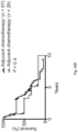
- K-M Depict overall survival of patients who did or did not receive adjuvant chemotherapy (adjuvant chemotherapy +/ ⁇ respectively, top left), and of patients with tumors harboring greater or less than the median number of CD3-CD8Granzyme B triple positive cells (CD3-CD8-GranzymeB Hi/Low respectively, top right). Overall survival of all four groups shown in bottom. Table shows univariate and multivariate Cox regression analysis of clinicopathologic features, adjuvant chemotherapy, and CD3-CD8-GranzymeB density association with overall survival.
- FIGS. 43 A and 43 B collectively depict representative sequential immunohistochemistical staining of a single short term and a single long term core tumor section. Sections bounded by black rectangles (100 ⁇ ) are magnified to 275 ⁇ (right) for each core section. Merged images are shown in FIG. 42 B .
- FIG. 44 depicts immunofluorescent quantification of CD8 + and CD4 + cells in tumor tissue microarray of short and long term survivors. Slides used above were cut from separate sections of the block as those used for sequential immunohistochemistry ( FIG. 42 B- 42 E ). *P ⁇ 0.05.
- FIG. 45 depicts the number of T cell clones in matched tumor and adjacent non-tumor pancreatic tissue. ****P ⁇ 0.0001.
- FIGS. 46 A- 46 D collectively depict a representative flow cytometric gating strategy to phenotype human T cells.
- Single cell suspensions from peripheral blood, tumor draining lymph nodes, and tumor tissue were subjected to flow cytometric analysis.
- First plot is pre-gated on live cells, followed by CD45 + , and CD3 + CD56 ⁇ cells. Values indicate percentage of cells within the red boxes, and are gated based on isotype controls.
- FIG. 47 depicts an oncoprint demonstrating the frequency of oncogenic driver gene mutations in the MSKCC PDAC cohort.
- FIGS. 48 A , 48 B 1 , 48 B 2 , and 48 C collectively depict data showing that neoantigen quantity and cytotoxic CD8 + T cell infiltrate identified long term pancreatic cancer survivors.
- A Number of nonsynonymous, missense, and neoantigenic mutations per tumor.
- B1-B2 Overall survival of patients with tumors harboring greater than the median number of neoantigens (NeoantigenHi), CD3-CD8 double positive cells (CD3-CD8Hi), CD3-CD8-Granzyme-B triple positive cells (CD3-CD8-Granzyme-BHi), or median polyclonality (PolyclonalHi), compared to all other patients (Rest).
- Neoantigens were determined using the MSKCC (left) and the pVAC-Seq (right) neoantigen prediction pipelines.
- C Unsupervised hierarchical clusters (cluster 1-4) of bulk tumor whole transcriptomic profiling in short and long term survivors (left). Each column represents a patient, each row a gene. Gene list available in methods. Overlap of NeoantigenHiCD8Hi, NeoantigenHiCD3-CD8-Granzyme-BHi, or all other tumors (Rest) with transcriptionally defined clusters (right). Each bar in (A) represents data from one patient's tumor. Mutations in (A), (B1-B2), and (C) were determined by whole exome sequencing.
- Polyclonality in (B1-B2) was calculated by quantitative TCR V ⁇ sequencing.
- Number of immune cells in (B1-B2) and (C) were quantified by immunophenotyping as the median of three cores per tumor in short and long term survivor tumor tissue microarrays. *P ⁇ 0.05.
- FIGS. 50 A, 50 B, and 50 C collectively depict survival of patients relative to number of neoantigens.
- A depicts overall survival of patients with tumors harbouring more or fewer than the median number of neoantigens (neoantigen hi/low ) and CD4 single positive cells (CD4 hi/low ) compared to all other patients (Rest).
- B depicts overall survival of patients with tumors harbouring CD3-CD8 double-positive cells (CD3-CD8 hi/low ) compared to all other patients (Rest).
- C depicts overall survival of patients with tumors harbouring polyclonality (polyclonal hi/low ) and mutations (mutation hi/low ), compared to all other patients (Rest).
- FIG. 51 depicts an oncoprint demonstrating the frequency of oncogenic driver mutations in short and long term tumors.
- FIG. 52 depicts the number of nonsynonymous, missense, and immunogenic mutations (neoantigens) in short and long term PDAC tumors.
- FIG. 53 depicts overall survival stratified by mutations in ARID1A, KRASQ61H, RBM10, and MLL related genes (MLL, MLL2, MLL3, MLL5) in accordance with an embodiment of the present disclosure.
- FIGS. 54 A, 54 B, 54 C, 54 D , 54 E 1 , 54 E 2 , 54 E 3 , 54 F, 54 G 1 , 54 G 2 , 54 G 3 , 54 G 4 , 54 H 1 , and 54 H 2 collectively depict neoantigens with microbial homology stratify long term pancreatic cancer survivors.
- A Schematic of neoantigen immune fitness models. Each circle represents a tumor clone in an evolutionary tree. Clones in both models are identical with respect to the number of mutations and neoantigens. Numbers represent hypothetical neoantigens gained in a successive tumor clone.
- neoantigen quality recognition potential model or quality model
- neoantigen quantity neoantigen load model or quantity model.
- B Parameters defining the recognition potential score in the recognition potential model.
- amino acid sequences of a hypothetical wild type (WT) epitope, tumor neoepitope, and a homologous microbial epitope are shown. Yellow highlights the changing amino acid between the WT and tumor sequence as a consequence of a tumor specific mutation.
- the amino acids in red indicate homology between the tumor neoepitope and the microbial epitope.
- Neoantigen quality is independently associated with long term survival. Overall survival of patients whose tumors displayed high compared to low neoantigen recognition potential (Neoantigen QualityHi/Low) to microbial epitopes (E1 top). Overall survival of patient who did or did not receive adjuvant chemotherapy (E2 top). Overall survival of all four groups is shown at the bottom. Neoantigen quality defined by neoantigen fitness modeling of sequence homology to microbial epitopes. Table shows univariate and multivariate Cox regression analysis of the associations of clinicopathologic features, adjuvant chemotherapy, and neoantigen quality with overall survival. Data include all patients in the whole exome sequencing MSKCC cohort.
- Neoantigen quality defined by neoantigen fitness modeling of sequence homology to microbial epitopes. Table shows univariate and multivariate Cox regression analysis of the associations of clinicopathologic features, adjuvant chemotherapy, and neoantigen quality with overall survival in the ICGC cohort.
- (H) depicts parameters of the neoantigen recognition potential fitness (quality model) for a, the MSKCC cohort b, and the ICGC cohort.
- (top) Log-rank test score landscape as a function of the model parameters, the horizontal alignment score displacement a, and the characterist time T, the significance of the score is denoted in the legend.
- (bottom) Two dimensional histograms showing distributions of optimal parameters obtained on subsampled datasets with 50, 70, and 80% of patients left, over 500 iterations of subsampling at each frequency.
- FIGS. 55 A, 55 B, 55 C, 55 D, 55 E, 55 F, 55 G, 55 H , 55 I 1 , 55 I 2 , 55 J 1 , 55 J 2 , 55 K, and 55 L collectively depict MUC16 to be a neoantigen enriched immunogenic hotspot in long term pancreatic cancer survivors.
- A Frequency (left) and distribution (right) of patients with neoantigens. Genes harboring neoantigens in >15% of patients are indicated.
- B Frequency of patients with MUC16 neoantigens and
- C the number of MUC16 neoantigens per tumor.
- FIG. 56 depicts the recognition potential load fitness model parameter stability analysis.
- the boxplots (top) and the histograms (bottom) illustrate distributions of the difference between the optimal shift parameter obtained on the full dataset and the subsampled datasets, with frequencies 0.8, 0.7, 0.6, 0.5, and 0.4.
- FIG. 57 depicts the frequency of MUC16 mutations in short and long term PDAC tumors.
- FIG. 58 depicts a lollipop plot showing location of MUC16 mutations in short and long term pancreatic cancer survivors.
- FIG. 59 depicts a lollipop plot showing location of MUC16 neoantigens in short and long term pancreatic cancer survivors.
- FIG. 60 depicts genes most frequently generating neoantigens as determined by pVACSeq. Frequency of patients and raw numbers are indicated.
- FIG. 61 depicts MUC16 mRNA and protein expression in MUC16 non-mutated (WT) and mutated (mutant) tumors.
- FIG. 62 depicts mRNA expression of transcriptional activators of MUC16.
- FIG. 63 depicts mRNA expression of mediators implicated in MUC16 dependent tumor progression in short and long term tumors.
- FIG. 64 depicts mRNA expression of tissue expression antigens MUC1, MUC4, WT1, mesothelin, and Annexin A2 in short and long term tumors.
- FIG. 65 depicts quantified protein expression of tissue expression antigens MUC1, mesothelin, and Annexin A2 in short and long term tumors. WT1 protein was undetectable in tumors of both short and long term survivors.
- FIGS. 69 A, 69 B , 69 C 1 , 69 C 2 , 69 D 1 , 69 D 2 , 69 D 3 , and 69 D 4 collectively depict neoantigen and cross-reactive microbial peptide T cells detected in blood and tumours.
- A Left, gene expression in the presence (red) or absence (grey) of high-quality neoantigenic mutations.
- Right median non-neoantigenic and neoantigenic expression. All high-quality neoantigenic genes with available mRNA expression are shown.
- n is the number of biologically independent samples in individual patients in (A) and (C). P values were determined using a two-tailed Student's t-test (A), a two-tailed Mann-Whitney U-test (B), a one-way analysis of variance (ANOVA) with Tukey's multiple comparison test (C) and as described in Example X (D).
- FIGS. 70 A 1 , 70 A 2 , 70 B 1 , 70 B 2 , and 70 C collectively show decreased MUC16 protein expression in long term survivors is not a consequence of differential antibody binding to neoantigenic MUC16 mutations.
- A representative immunohistochemical statining and quantification of MUC16 expression in tissue microarrays of short and long term pancreatic cancer survivors as assese using three independent anti MUC16 antibodies Ab #1-clone EPSISR23, purchased from Abcam; Ab #2-polyclonal, purchased from Abcam ab1333419; Ab #3-clone 4H11.
- B Western blot (B1) and immunocytochemistry (B2) of untransfected ( ⁇ ), empty vector (vector), MUC16 wild type (MUC16WT), and MUC16 mutant (MUC16 R15C) HEK293T cells.
- the top left blot was probed with anti MUC16 specific antibody (clone 4H11) and the right blot with anti b-actin. Red rectandly indicates MUC16 specific band. All bottom cells were probed with anti MUC16 antibody (clone 4H11).
- the inserted mutation was identical to a neoantigenic MUC16 mutation.
- C MUC16 immunohistochemistry on two long term pancreatic cancer survivors with MUC16 neoepitopes in primary resected tumors. Areas in rectangular low power fields are magnified on the right.
- FIG. 71 depicts a comprehensive flowchart of neoantigen fitness modelling pipeline.
- Software programs utilized for each step are indicated in bold, colored text.
- Mathematical formulae for calculation of individual components of neoantigen quality are shown above and further defined in Methods. All software components of the pipeline are published and/or publically available.
- the presently disclosed subject matter provides strategies for improving cancer therapy (e.g., immunotherapy) by selection of particular patients to receive therapy based on the neoantigen portfolio of the patients (e.g., immunogenic hotspot (e.g., MUC16 for pancreatic cancer), neoantigen quantity and neoantigen immunogenicity).
- the presently disclosed subject matter also provides methods for predicting responsiveness of cancer patients to immunotherapy based on the patients' neoantigen portfolio. Furthermore, based on the fact that, at least in tumors with high mutational loads, the amount of DNA damage is sufficient for the immune system to perceive one or more neoantigens as foreign, it becomes of interest to stimulate neoantigen-specific T cell responses in cancer patients.
- the presently disclosed subject matter further provides vaccines comprising one or more neoantigen, and/or therapeutic T cell preparations comprising neoantigen-specific T cells.
- the present disclosure further provides a recognition potential fitness model for tumors based on the immune interactions of dominant neoantigens predicts response to immunotherapy.
- the disclosed recognition potential fitness model associates each neoantigen with a fitness cost, which is termed the recognition potential of a neoantigen.
- the recognition potential of a neoantigen is the likelihood it is productively recognized by the TCR repertoire. It is defined by two components. The first is the amplitude A, which is given by the relative probability that a neoantigen will be presented by on class I major histocompatibility complex (MHC) and the relative probability that its wildtype counterpart will not be presented.
- MHC major histocompatibility complex
- the second component is the probability R that a presented neoantigen will be recognized by the TCR repertoire, which is inferred from its sequence homology to known antigens. For a given neoantigen their product defines its recognition potential, A ⁇ R. In some embodiments, this recognition potential is computed using the scripts illustrated in FIGS. 37 through 40 in accordance with the present disclosure.
- the term “about” or “approximately” means within an acceptable error range for the particular value as determined by one of ordinary skill in the art, which will depend in part on how the value is measured or determined, i.e., the limitations of the measurement system. For example, “about” can mean within 3 or more than 3 standard deviations, per the practice in the art. Alternatively, “about” can mean a range of up to 20%, e.g., up to 10%, up to 5%, or up to 1% of a given value. Alternatively, particularly with respect to biological systems or processes, the term can mean within an order of magnitude, e.g., within 5-fold, or within 2-fold, of a value.
- neoantigen As used herein, the term “neoantigen”, “neoepitope” or “neopeptide” refers to a tumor-specific antigen that arises from one or more tumor-specific mutation, which alters the amino acid sequence of genome encoded proteins.
- neoantigen number or “neoantigen burden” refers to the number of neoantigen(s) measured, detected or predicted in a sample (e.g., a biological sample from a subject). In certain embodiments, the neoantigen number is measured by using whole exome sequencing and in silico prediction.
- mutations refers to permanent change in the DNA sequence that makes up a gene.
- mutations range in size from a single DNA building block (DNA base) to a large segment of a chromosome.
- mutations can include missense mutations, frameshift mutations, duplications, insertions, nonsense mutation, deletions and repeat expansions.
- a missense mutation is a change in one DNA base pair that results in the substitution of one amino acid for another in the protein made by a gene.
- a nonsense mutation is also a change in one DNA base pair. Instead of substituting one amino acid for another, however, the altered DNA sequence prematurely signals the cell to stop building a protein.
- an insertion changes the number of DNA bases in a gene by adding a piece of DNA.
- a deletion changes the number of DNA bases by removing a piece of DNA.
- small deletions can remove one or a few base pairs within a gene, while larger deletions can remove an entire gene or several neighboring genes.
- a duplication consists of a piece of DNA that is abnormally copied one or more times.
- frameshift mutations occur when the addition or loss of DNA bases changes a gene's reading frame.
- a reading frame consists of groups of 3 bases that each code for one amino acid.
- a frameshift mutation shifts the grouping of these bases and changes the code for amino acids.
- insertions, deletions, and duplications can all be frameshift mutations.
- a repeat expansion is another type of mutation.
- nucleotide repeats are short DNA sequences that are repeated a number of times in a row.
- a trinucleotide repeat is made up of 3-base-pair sequences
- a tetranucleotide repeat is made up of 4-base-pair sequences.
- a repeat expansion is a mutation that increases the number of times that the short DNA sequence is repeated.
- the “median” value e.g., median neoantigen number, median neoantigen-microbial homology—e.g., median cross-reactivity score, median recognition potential score), or median activated T cell number—refers to the median value obtained from a population of subjects having a cancer (e.g., pancreatic cancer, e.g., PDAC).
- the median values may be previously determined reference values, or may be contemporaneously determined values.
- a cell population refers to a group of at least two cells expressing similar or different phenotypes.
- a cell population can include at least about 10, at least about 100, at least about 200, at least about 300, at least about 400, at least about 500, at least about 600, at least about 700, at least about 800, at least about 900, or at least about 1000 cells expressing similar or different phenotypes.
- antibody refers to antigen-binding proteins of the immune system.
- antibody includes whole, full length antibodies having an antigen-binding region, and any fragment thereof in which the “antigen-binding portion” or “antigen-binding region” is retained, or single chains, for example, single chain variable fragment (scFv), thereof.
- scFv single chain variable fragment
- antibody means not only intact antibody molecules, but also fragments of antibody molecules that retain immunogen-binding ability. Such fragments are also well known in the art and are regularly employed both in vitro and in vivo.
- an antibody means not only intact immunoglobulin molecules but also the well-known active fragments F(ab′)2, and Fab.
- F(ab′)2, and Fab fragments that lack the Fc fragment of intact antibody clear more rapidly from the circulation, and can have less non-specific tissue binding of an intact antibody (Wahl et al., J. Nucl. Med. 24:316-325 (1983).
- an antibody is a glycoprotein comprising at least two heavy (H) chains and two light (L) chains inter-connected by disulfide bonds. Each heavy chain is comprised of a heavy chain variable region (abbreviated herein as V H ) and a heavy chain constant (C H ) region.
- V H heavy chain variable region
- C H heavy chain constant
- the heavy chain constant region is comprised of three domains, C H 1, C H 2 and C H 3.
- Each light chain is comprised of a light chain variable region (abbreviated herein as V L ) and a light chain constant C L region.
- the light chain constant region is comprised of one domain, C L .
- the V H and V L regions can be further sub-divided into regions of hypervariability, termed complementarity determining regions (CDR), interspersed with regions that are more conserved, termed framework regions (FR).
- CDR complementarity determining regions
- FR framework regions
- Each VH and V L is composed of three CDRs and four FRs arranged from amino-terminus to carboxy-terminus in the following order: FR1, CDR1, FR2, CDR2, FR3, CDR3, FR4.
- variable regions of the heavy and light chains contain a binding domain that interacts with an antigen.
- the constant regions of the antibodies can mediate the binding of the immunoglobulin to host tissues or factors, including various cells of the immune system (e.g., effector cells) and the first component (C1 q) of the classical complement system.
- antigen-binding portion refers to that region or portion of an antibody that binds to the antigen and which confers antigen specificity to the antibody; fragments of antigen-binding proteins. It has been shown that the antigen-binding function of an antibody can be performed by fragments of a full-length antibody.
- antibody fragments examples include a Fab fragment, a monovalent fragment consisting of the V L , V H , C L and C H 1 domains; a F(ab)2 fragment, a bivalent fragment comprising two Fab fragments linked by a disulfide bridge at the hinge region; a Fd fragment consisting of the V H and CH1 domains; a Fv fragment consisting of the V L and V H domains of a single arm of an antibody; a dAb fragment (Ward et al., 1989 Nature 341:544-546), which consists of a V H domain; and an isolated complementarity determining region (CDR).
- Fab fragment a monovalent fragment consisting of the V L , V H , C L and C H 1 domains
- F(ab)2 fragment a bivalent fragment comprising two Fab fragments linked by a disulfide bridge at the hinge region
- a Fd fragment consisting of the V H and CH1 domains
- a Fv fragment consisting of
- single-chain variable fragment is a fusion protein of the variable regions of the heavy (V H ) and light chains (V L ) of an immunoglobulin (e.g., mouse or human) covalently linked to form a V H ::V L heterodimer.
- the heavy (V H ) and light chains (V L ) are either joined directly or joined by a peptide-encoding linker (e.g., 10, 15, 20, 25 amino acids), which connects the N-terminus of the V H with the C-terminus of the V L , or the C-terminus of the V H with the N-terminus of the V L .
- the linker is usually rich in glycine for flexibility, as well as serine or threonine for solubility. Despite removal of the constant regions and the introduction of a linker, scFv proteins retain the specificity of the original immunoglobulin.
- Single chain Fv polypeptide antibodies can be expressed from a nucleic acid comprising V H - and V L -encoding sequences as described by Huston et al. (Proc. Nat. Acad. Sci. USA, 85:5879-5883, 1988). See, also, U.S. Pat. Nos. 5,091,513, 5,132,405 and 4,956,778; and U.S. Patent Publication Nos. 20050196754 and 20050196754.
- Antagonistic scFvs having inhibitory activity have been described (see, e.g., Zhao et al. Hyrbidoma (Larchmt) 2008 27(6):455-51; Peter et al., J Cachexia Sarcopenia Muscle 2012 Aug. 12; Shieh et al., J Imunol 2009 183(4):2277-85; Giomarelli et al., Thromb Haemost 2007 97(6):955-63; Fife eta., J Clin Invst 2006 116(8):2252-61; Brocks et al., Immunotechnology 1997 3(3):173-84; Mooscaner et al., Ther Immunol 1995 2(10:31-40).
- F(ab) refers to a fragment of an antibody structure that binds to an antigen but is monovalent and does not have a Fc portion, for example, an antibody digested by the enzyme papain yields two F(ab) fragments and an Fc fragment (e.g., a heavy (H) chain constant region; Fc region that does not bind to an antigen).
- an antibody digested by the enzyme papain yields two F(ab) fragments and an Fc fragment (e.g., a heavy (H) chain constant region; Fc region that does not bind to an antigen).
- F(ab′) 2 refers to an antibody fragment generated by pepsin digestion of whole IgG antibodies, wherein this fragment has two antigen binding (ab′) (bivalent) regions, wherein each (ab′) region comprises two separate amino acid chains, a part of a H chain and a light (L) chain linked by an S—S bond for binding an antigen and where the remaining H chain portions are linked together.
- a “F(ab′) 2 ” fragment can be split into two individual Fab′ fragments.
- antigen-binding protein refers to a protein or polypeptide that comprises an antigen-binding region or antigen-binding portion, that is, has a strong affinity to another molecule to which it binds.
- Antigen-binding proteins encompass antibodies, chimeric antigen receptors (CARs) and fusion proteins.
- treating refers to clinical intervention in an attempt to alter the disease course of the individual or cell being treated, and can be performed either for prophylaxis or during the course of clinical pathology.
- Therapeutic effects of treatment include, without limitation, preventing occurrence or recurrence of disease, alleviation of symptoms, diminishment of any direct or indirect pathological consequences of the disease, preventing metastases, decreasing the rate of disease progression, amelioration or palliation of the disease state, and remission or improved prognosis.
- a treatment can prevent deterioration due to a disorder in an affected or diagnosed subject or a subject suspected of having the disorder, but also a treatment may prevent the onset of the disorder or a symptom of the disorder in a subject at risk for the disorder or suspected of having the disorder.
- the term “subject” refers to any animal (e.g., a mammal), including, but not limited to, humans, and non-human animals (including, but not limited to, non-human primates, dogs, cats, rodents, horses, cows, pigs, mice, rats, hamsters, rabbits, and the like (e.g., which is to be the recipient of a particular treatment, or from whom cells are harvested).
- the subject is a human.
- an “effective amount” or “therapeutically effective amount” is an amount sufficient to affect a beneficial or desired clinical result upon treatment.
- An effective amount can be administered to a subject in one or more doses.
- an effective amount is an amount that is sufficient to palliate, ameliorate, stabilize, reverse or slow the progression of the disease, or otherwise reduce the pathological consequences of the disease.
- the effective amount is generally determined by the physician on a case-by-case basis and is within the skill of one in the art. Several factors are typically taken into account when determining an appropriate dosage to achieve an effective amount. These factors include age, sex and weight of the subject, the condition being treated, the severity of the condition and the form and effective concentration of the immunoresponsive cells administered.
- a response refers to an alteration in a subject's condition that occurs as a result of or correlates with treatment.
- a response is a beneficial response.
- a beneficial response can include stabilization of the condition (e.g., prevention or delay of deterioration expected or typically observed to occur absent the treatment), amelioration (e.g., reduction in frequency and/or intensity) of one or more symptoms of the condition, and/or improvement in the prospects for cure of the condition, etc.
- “response” can refer to response of an organism, an organ, a tissue, a cell, or a cell component or in vitro system.
- a response is a clinical response.
- presence, extent, and/or nature of response can be measured and/or characterized according to particular criteria.
- criteria can include clinical criteria and/or objective criteria.
- techniques for assessing response can include, but are not limited to, clinical examination, positron emission tomography, chest X-ray CT scan, MRI, ultrasound, endoscopy, laparoscopy, presence or level of a particular marker in a sample, cytology, and/or histology.
- a response of interest is a response of a tumor to a therapy
- predictive features identified herein e.g., one or more MUC16 neoantigen, number of neoantigens, number of activated T cells
- a similarly situated second subject e.g., a second subject having the same type cancer, optionally the same type of cancer with similar characteristics (e.g. stage and/or location/distribution)
- group of subjects that lack one or more predictive feature.
- sample refers to a biological sample obtained or derived from a source of interest, as described herein.
- a source of interest comprises an organism, such as an animal or human.
- a biological sample is a biological tissue or fluid.
- Non-limiting biological samples include bone marrow, blood, blood cells, ascites, (tissue or fine needle) biopsy samples, cell-containing body fluids, free floating nucleic acids, sputum, saliva, urine, cerebrospinal fluid, peritoneal fluid, pleural fluid, feces, lymph, gynecological fluids, swabs (e.g., skin swabs, vaginal swabs, oral swabs, and nasal swabs), washings or lavages such as a ductal lavages or broncheoalveolar lavages, aspirates, scrapings, specimens (e.g., bone marrow specimens, tissue biopsy specimens, and surgical specimens), feces, other body fluids, secretions, and/or excretions, and cells therefrom, etc.
- swabs e.g., skin swabs, vaginal swabs, oral swabs, and nasal swabs
- the term “substantially” refers to the qualitative condition of exhibiting total or near-total extent or degree of a characteristic or property of interest.
- One of ordinary skill in the art will understand that biological and chemical phenomena rarely, if ever, go to completion and/or proceed to completeness or achieve or avoid an absolute result.
- the term “substantially” is therefore used herein to capture the potential lack of completeness inherent in many biological and chemical phenomena.
- vaccine refers to a composition for generating immunity for the prophylaxis and/or treatment of diseases (e.g., neoplasia/tumor).
- vaccines are medicaments that comprise antigens and are intended to be used in humans or animals for generating specific defense and protective substance by vaccination.
- first, second, etc. may be used herein to describe various elements, these elements should not be limited by these terms. These terms are only used to distinguish one element from another. For example, a first subject could be termed a second subject, and, similarly, a second subject could be termed a first subject, without departing from the scope of the present disclosure. The first subject and the second subject are both subjects, but they are not the same subject. Furthermore, the terms “subject,” “user,” and “patient” are used interchangeably herein.
- the term “if” may be construed to mean “when” or “upon” or “in response to determining” or “in response to detecting,” depending on the context.
- the phrase “if it is determined” or “if [a stated condition or event] is detected” may be construed to mean “upon determining” or “in response to determining” or “upon detecting [the stated condition or event]” or “in response to detecting [the stated condition or event],” depending on the context.
- pancreatic cancer e.g., pancreatic ductal adenocarcinoma (PDAC)
- PDAC pancreatic ductal adenocarcinoma
- Neoantigen is a tumor-specific antigen that arises from one or more tumor-specific mutation.
- the neoantigen is a tumor-specific antigen that arises from a tumor-specific mutation.
- a neoantigen is not expressed by healthy cells (e.g., non-tumor cells or non-cancer cells) in a subject.
- neoantigens are self-antigens which are selectively expressed or overexpressed on the cancer cells. These self-antigens are difficult to target with immunotherapy because they require overcoming both central tolerance (whereby autoreactive T cells are deleted in the thymus during development) and peripheral tolerance (whereby mature T cells are suppressed by regulatory mechanisms). Targeting neoantigens can abrogate these tolerance mechanisms.
- a neoantigen is recognized by cells of the immune system of a subject (e.g., T cells) as “non-self”. Neoantigens are not recognized as “self-antigens” by immune system, T cells that are capable of targeting neoantigens are not subject to central and peripheral tolerance mechanisms to the same extent as T cells which recognize self-antigens.
- the tumor-specific mutation that results in a neoantigen is a somatic mutation.
- Somatic mutations comprise DNA alterations in non-germline cells and commonly occur in cancer cells.
- Certain somatic mutations in cancer cells result in the expression of neoantigens, that in certain embodiments, transform a stretch of amino acids from being recognized as “self” to “non-self”.
- Human tumors without a viral etiology can accumulate tens to hundreds of fold somatic mutations in tumor genes during neoplastic transformation, and some of these somatic mutations can occur in protein-coding regions and result in the formation of neoantigens.
- the exome is the protein-encoding part of the genome.
- exome sequencing is performed in a biological sample (e.g., a tumor sample) obtained from a subject having cancer.
- a neoantigen is a neoantigenic peptide comprising a tumor specific mutation.
- the neoantigenic peptide can be a peptide that is incorporated into a larger protein.
- a neoantigenic peptide is a series of residues, typically L-amino acids, connected one to the other, typically by peptide bonds between the ⁇ -amino and carboxyl groups of adjacent amino acids.
- a neoantigenic peptide can be a variety of lengths, either in their neutral (uncharged) forms or in forms which are salts, and either free of modifications such as glycosylation, side chain oxidation, or phosphorylation.
- the size of the neoantigenic peptide is about 3-30 amino acids, e.g., about 3-5, about 5-15 (e.g., about 8-11, about 5-10, or about 10-15), about 15-20, about 20-25, or about 20-30 amino acids, in length.
- the neoantigenic peptide is about 8-11 amino acids in length.
- the neoantigenic peptide is about 3, about 4, about 5, about 6, about 7, about 8, about 9, about 10, about 11, about 12, about 13, about 14, about 15, about 16, about 17, about 18, about 19, about 20, about 21, about 22, about 23, about 24, about 25, about 26, about 27, about 28, about 29, or about 30 amino acids in length.
- the neoantigenic peptide is at least about 3, at least about 5, or at least about 8 amino acids in length. In certain embodiments, the neoantigenic peptide is less than about 30, less than about 20, less than about 15, or less than about 10 amino acids in length.
- a neoantigenic peptide is selected from the neoantigenic peptides listed in Table 1.
- a neoantigenic peptide is selected from the neoantigenic peptides listed in Table 2.
- Neoantigens may vary in different subjects, e.g., different subjects may have different combinations of neoantigens, also referred to as “neoantigen signatures”. For example, each subject may have a unique neoantigen signature. In certain embodiments, a neoantigen signature is a combination of one or more neoantigen listed in Tables 1 and 2.
- compositions comprising one or more presently disclosed neoantigen.
- the compositions are pharmaceutical compositions comprising pharmaceutically acceptable carriers.
- the composition comprises two or more neoantigens. The two or more can be linked, e.g., by any biochemical strategy to link two proteins.
- Cancers can be screened to detect neoantigens using any of a variety of known technologies.
- neoantigens or expression thereof is detected at the nucleic acid level (e.g., in DNA or RNA).
- neoantigens or expression thereof is detected at the protein level (e.g., in a sample comprising polypeptides from cancer cells, which sample can be or comprise polypeptide complexes or other higher order structures including but not limited to cells, tissues, or organs).
- a neoantigen is detected by the method selected from the group consisting of whole exome sequencing, immunoassay, microarray, genome sequencing, RNA sequencing, ELISA, Western Transfer, DNA or RNA sequencing, mass spectrometry, and combinations thereof.
- one or more neoantigen is detected by whole exome sequencing.
- one or more neoantigen is detected by the immunogenicity analysis method of somatic mutations described in Snyder et al. Engl J Med 371, 2189-2199 (2014).
- one or more neoantigen is detected by the pVAC-Seq method described in Hndal et al., Genome Med (2016); 8:11.
- one or more neoantigen is detected by the in silico neoantigen prediction pipeline method described in Rizvi et al., Science 348, 124-128 (2015). In certain embodiments, one or more neoantigen is detected by any of the methods described in WO2015/103037 and WO2016/081947.
- the neoantigens of the presently disclosed subject matter can be used to identify cancer subjects as candidates for immunotherapies, and to predict responsiveness of cancer subjects to immunotherapies.
- the presently disclosed subject matter provides methods of identifying subjects (e.g., cancer subjects) as candidates for treatment with an immunotherapy (hereinafter “the patient selection method”).
- the presently disclosed subject matter provides methods of predicting the responsiveness of subjects (e.g., cancer subjects) to an immunotherapy (hereinafter “the responsiveness prediction method”).
- immunogenic hotspot refers to a genetic locus that is enriched with neoantigens (e.g., a genetic locus that frequently generates neoantigens).
- the immunogenic hotspot can vary depending on the type of disease (e.g., tumor). Detecting one or more neoantigen of an immunogenic hotspot can be used to identify cancer subjects as candidates for immunotherapies, and to predict responsiveness of cancer subjects to immunotherapies.
- immunotherapies e.g., vaccines, T cells (including modified T cells, e.g., T cells comprising a T cell receptor (TCR) or a chimeric antigen receptor (CAR)
- TCR T cell receptor
- CAR chimeric antigen receptor
- MUC16 is an immunogenic hotspot for pancreatic cancer, more specifically, for pancreatic ductal adenocarcinoma (PDAC).
- PDAC pancreatic ductal adenocarcinoma
- MUC16 is an immunogenic hotspot for checkpoint blockade refractory tumor (e.g., PDAC).
- MUC16-neoantigen specific T cell immunity induces immunoediting of MUC16-expressing clones in primary tumors, curtails the development of metastases, and prolongs survival, given the cell-autonomous roles of MUC16 in promoting metastases.
- the patient selection method comprises obtaining a biological sample from the subject, and detecting one or more neoantigen of MUC16 in the biological sample, wherein the presence of one or more neoantigen of MUC16 in the biological sample indicates that the subject is a candidate for an immunotherapy.
- the responsiveness prediction method comprises obtaining a biological sample from the subject, and detecting one or more neoantigen of MUC16 in the biological sample, wherein the presence of one or more neoantigen of MUC16 in the biological sample indicates that the subject is likely to be responsive to an immunotherapy.
- the patient selection method and responsiveness prediction method comprise detecting a plurality of neoantigens of MUC16.
- the methods comprise detecting one or more of the neoantigenic peptides listed in Table 1.
- the methods comprise detecting one or more of the neoantigenic peptides having the amino acid sequences set forth in SEQ ID NOS: 1-18.
- the subject has pancreatic cancer.
- the pancreatic cancer is pancreatic ductal adenocacinma (PDAC).
- neoantigens of MUC16 are detected by any of the methods for detecting neoantigens, e.g., those described in Section 2.2. In certain embodiments, the neoantigens of MUC16 are detected by whole exome sequencing the biological sample. In certain embodiments, the neoantigens of MUC16 are detected by the immunogenicity analysis method of somatic mutations described in Snyder et al. Engl J Med 371, 2189-2199 (2014). In certain embodiments, the neoantigens of MUC16 are detected by the pVAC-Seq method described in Hndal et al., Genome Med (2016); 8:11.
- the neoantigens of MUC16 are detected by the in silico neoantigen prediction pipeline described in Rizvi et al., 2015 , Science 348, 124-128. In certain embodiments, the neoantigens of MUC16 are detected by any of the methods described in WO2015/103037 and WO2016/081947.
- the patient selection method and responsiveness prediction method relate to the quantity of the neoantigens (e.g., the number of neoantigens (hereinafter “neoantigen number”) in the subject.
- the neoantigen number includes but is not limited to MUC16 neoantigens.
- neoantigen number is measured by any methods for detecting neoantigens, e.g., those described in Section 2.2. In certain embodiments, the neoantigen number is measured by whole exome sequencing the biological sample. In certain embodiments, the neoantigen number is measured by the immunogenicity analysis method of somatic mutations described in Snyder et al., 204 , Engl J Med 371, 2189-2199. In certain embodiments, the neoantigen number is measured by the pVAC-Seq method described in Hndal et al., 2016 , Genome Med 8, 11.
- the neoantigen number is measured by the in silico neoantigen prediction pipeline described in Rizvi et al., 2015 Science 348, 124-128. In certain embodiments, the neoantigen number is measured by any of the methods described in WO2015/103037 and WO2016/081947.
- the neoantigen number obtained from a population of subjects having the cancer may vary depending on the type of the cancer, therapy (e.g., immunotherapy, chemotherapy), the caner histology, and/or the site of disease (tumor).
- the median neoantigen number obtained from a population of subjects having cancer is between 10 and 10,000, between 10 and 50, between 30 and 50, between 30 and 40, between 50 and 100, between 100 and 200, between 200 and 300, between 300 and 400, between 400 and 500, between 500 and 600, between 600 and 700, between 700 and 800, between 800 and 900, between 900 and 1,000, between 1,000 and 2,000, between 2,000 and 3,000, between 3,000 and 4,000, between 4,000 and 5,000, between 5,000 and 6,000, between 6,000 and 7,000, between 7,000 and 8,000, between 8,000 and 9,000, or between 9,000 and 10,000.
- the median neoantigen number obtained from a population of subjects having cancer is about 40. In certain embodiments, the median neoantigen number obtained from a population of subjects having cancer is about 38. In certain embodiments, the cancer is pancreatic cancer, e.g., PDAC.
- the neoantigens can be located in any genes in a subject. In certain embodiments, the neoantigen is located in MUC16. In certain embodiments, the neoantigen is located in any of the genes listed in Table 2.
- the patient selection method and responsiveness prediction method relate to the quantity of the neoantigens and the immunogenicity of the neoantigens (hereinafter “neoantigen immunogenicity”) in the subject.
- the patient selection method and responsiveness prediction method comprise assessing the neoantigen immunogenicity. Immunogenicity is the ability of a particular substance, such as an antigen (e.g., a neoantigen), to induce or stimulate an immune response in the cells expressing such antigen.
- assessing the neoantigen immunogenicity comprises measuring one or more surrogate for the neoantigen immunogenicity.
- the surrogate is the homology between a neoantigen and a microbial epitope (hereinafter “neoantigen-microbial homology”).
- the patient selection method comprises obtaining a biological sample from the subject, measuring the neoantigen number in the biological sample; and measuring the homology between each of the neoantigen and a microbial epitope (neoantigen-microbial homology), where a neoantigen number higher than the median neoantigen number obtained from a population of subjects having the cancer (e.g., referred to as “neoantigen number high ”) and a neoantigen-microbial homology higher than the median neoantigen-microbial homology obtained from subjects having the cancer (e.g., referred to as “neoantigen-microbial homology high ”) indicate that the subject is a candidate for an immunotherapy.
- the responsiveness prediction method comprises obtaining a biological sample from the subject, measuring the neoantigen number in the biological sample, and measuring the neoantigen-microbial homology, where a neoantigen number higher than the median neoantigen number obtained from a population of subjects having the cancer (e.g., referred to as “neoantigen number high ”) and a neoantigen-microbial homology higher than the median neoantigen-microbial homology obtained from subjects having the cancer (e.g., referred to as “neoantigen-microbial homology high ”) indicate that the subject is likely to be responsive to an immunotherapy.
- a neoantigen number higher than the median neoantigen number obtained from a population of subjects having the cancer e.g., referred to as “neoantigen number high
- neoantigen-microbial homology high a neoantigen-microbial homology higher than the median neo
- the surrogate is the number of activated T cells (hereinafter “activated T cell number”).
- activated T cell number The immune system plays an important role in controlling and eradicating cancer. The number and frequency of the activated T cells present in the tumor can affect the fraction of tumor cells eventually killed in vitro. Therefore, the baseline infiltration by activated T cells in the tumor can have important prognostic implications for solid tumors.
- the subject's neoantigen number, the neoantigen-microbial homology, and activated T cell numbers are at least about 1%, at least about 2%, at least about 3%, at least about 4%, at least about 5%, at least about 6%, at least about 7%, at least about 8%, at least about 9%, at least about 10%, or at least about 20%, or at least about 30%, higher than the median values.
- the one or more neoantigen is selected from the neoantigenic peptides listed in Tables 1 and 2.
- activated T cells are T cells expressing one or more T cell activation marker.
- T cell activation markers include CD3, CD8, PD-1, 4-1BB, CD69, CD107a, and Granzyme B.
- the activated T cells are selected from the group consisting of CD3 + CD8 + T cells, CD3 + CD8 + Granzyme-B + T cells, polyclonal activated T cells, and combinations thereof.
- the patient selection method comprises measuring the neoantigen number in a biological sample of the subject, and measuring the number of activated T cells (hereinafter “activated T cell number”) in a biological sample of the subject.
- activated T cell number a neoantigen number higher than the median neoantigen number obtained from a population of subjects having the cancer
- activated T cell number higher than the median activated T cell number obtained from subjects having the cancer e.g., referred to as “activated T cell number high ” indicate that the subject is a candidate for an immunotherapy.
- the responsiveness prediction method comprises measuring the neoantigen number in a biological sample of the subject; and measuring the activated T cell number a biological sample of the subject.
- a neoantigen number higher than the median neoantigen number obtained from a population of subjects having the cancer e.g., referred to as “neoantigen number high ”
- an activated T cell number higher than the median activated T cell number obtained from subjects having the cancer e.g., referred to as “activated T cell number high ” indicate that the subject is likely to be responsive to an immunotherapy.
- the patient selection method and responsiveness prediction method further comprise obtaining one or more biological sample from the subject.
- the biological sample used to measure the neoantigen number, the biological sample used to measure the neoantigen-microbial homology, and the biological sample used to measure the activated T cell numbers can be the same or different.
- the biological sample can be a tumor tissue.
- the biological sample is a blood sample.
- the neoantigen-microbial homology is measured by calculating a recognition potential (cross reactivity) score between each neoantigen and microbial epitope.
- a recognition potential cross reactivity
- calculating a recognition potential score between each neoantigen and microbial epitope is performed by a recognition potential model.
- recognition potential model the terms “recognition potential model,” “neoantigen recognition potential model,” and “cross reactivity model” are used interchangeably.
- the neoantigen recognition potential model comprises measuring sequence alignment scores for each neoantigen to a microbial epitope (e.g., with positive immune assays from the Immune Epitope Database).
- the neoantigen recognition potential model can also comprise scaling the alignment scores to binding probability of a T cell to cross reacting microbial epitope by fitting a sigmoid function.
- the binding probabilities are amplified by relative wild type (“WT”) and neoantigenic peptide (“Mutant”).
- WT relative wild type
- Motant neoantigenic peptide
- a neoantigen recognition potential score for a given neoantigen is a function of the alignment score and the amplitude, K d WT /K d Mutant .
- the calculation of a recognition potential score for a neoantigen is done using any of the fitness models disclosed in Luksza et al., 2017, “A neoantigen fitness model predicts tumour response to checkpoint blockade immunotherapy,” Nature 551, 517-520. In some embodiments, the calculation of a recognition potential score for a neoantigen is done using any of the fitness models disclosed in Luksza et al., 2017, “A neoantigen fitness model predicts tumour response to checkpoint blockade immunotherapy,” Nature 551, 517-520. In some embodiments, the calculation of a recognition potential score for a neoantigen is done in accordance with any of the models disclosed below in conjunction with blocks 346 through 358 of FIG. 3 .
- the neoantigen-microbial homology high neoantigen is a cross-reactivity score high neoantigen, e.g., a neoantigen having a recognition potential score (e.g., calculated by i) a recognition potential model described above, ii) any of the fitness models disclosed in Luksza et al., 2017, “A neoantigen fitness model predicts tumour response to checkpoint blockade immunotherapy,” Nature 551, 517-520, and/or iii) any of the models disclosed below in conjunction with blocks 346 through 358 of FIG. 3 ) higher than the median recognition potential obtained from a population of subjects with the cancer.
- a recognition potential model e.g., calculated by i) a recognition potential model described above, ii) any of the fitness models disclosed in Luksza et al., 2017, “A neoantigen fitness model predicts tumour response to checkpoint blockade immunotherapy,” Nature 551, 517-520, and/
- the median recognition potential score obtained from a population of subjects having the cancer may vary depending on the type of the cancer, the type of therapy (e.g., immunotherapy, chemotherapy), the caner histology, and/or presence/absence of neoantigens.
- the median recognition potential score (e.g., calculated by i) a neoantigen recognition potential model described above, ii) any of the fitness models disclosed in Luksza et al., 2017, “A neoantigen fitness model predicts tumour response to checkpoint blockade immunotherapy,” Nature 551, 517-520, and/or iii) any of the models disclosed below in conjunction with blocks 346 through 358 of FIG.
- 3 obtained from a population of subjects having a cancer is between about 0 and 1, between about 0 and 0.5, between about 0 and 0.1, between about 0.1 and 0.2, between about 0.2 and 0.3, between about 0.3 and 0.4, between about 0.4 and 0.5, between about 0.5 and 1, between about 0.5 and 0.6, between about 0.6 and 0.7, between about 0.7 and 0.8, between about 0.8 and 0.9, or between about 0.9 and 0.1.
- the median neoantigen number (e.g., calculated by i) a recognition potential model described above, ii) any of the fitness models disclosed in Luksza et al., 2017, “A neoantigen fitness model predicts tumour response to checkpoint blockade immunotherapy,” Nature 551, 517-520, and/or iii) any of the models disclosed below in conjunction with blocks 346 through 358 of FIG. 3 ) obtained from a population of subjects having cancer is about 0.8.
- the median neoantigen number (e.g., calculated by i) a recognition potential model described above, ii) any of the fitness models disclosed in Luksza et al., 2017, “A neoantigen fitness model predicts tumour response to checkpoint blockade immunotherapy,” Nature 551, 517-520, and/or iii) any of the models disclosed below in conjunction with blocks 346 through 358 of FIG. 3 ) obtained from a population of subjects having cancer is about 0.9.
- the cancer is pancreatic cancer, e.g., PDAC.
- the median activated T cell number obtained from subjects having the cancer varies depending on the type of the cancer, the type of therapy (e.g., immunotherapy, chemotherapy), the caner histology, presence/absence of ongoing immune response, and/or presence/absence of neoantigens.
- the median activated T cell number obtained from a population of subjects having a cancer is between 0 and 2,000 cells/mm 2 , between 0 and 10 cells/mm 2 , between 10 and 20 cells/mm 2 , between 20 and 30 cells/mm 2 , between 30 and 40 cells/mm 2 , between 40 and 50 cells/mm 2 , between 10 and 50 cells/mm 2 , between 50 and 100 cells/mm 2 , between 50 and 60 cells/mm 2 , between 60 and 70 cells/mm 2 , between 70 and 80 cells/mm 2 , between 80 and 90 cells/mm 2 , between 90 and 100 cells/mm 2 , between 100 and 200 cells/mm 2 , between 200 and 500 cells/mm 2 , between 500 and 1,000 cells/mm 2 , between 1,000 and 1,500 cells/mm 2 , or between 1,500 and 2,000 cells/mm 2 .
- the median activated T cell number obtained from a population of subjects having a cancer is about 60 cells/mm 2 . In certain embodiments, the median activated T cell number obtained from a population of subjects having a cancer is about 58 cells/mm 2 . In certain embodiments, the cancer is pancreatic cancer, e.g., PDAC.
- the cancer is a solid tumor. In some embodiments, the cancer is a liquid tumor.
- solid tumor include pancreatic cancer, gastric cancer, bile duct cancer (e.g., cholangiocarcinoma), liver cancer, colorectal cancer, melanoma, lung cancer, and breast cancer.
- liquid tumor include acute leukemia and chronic leukemia.
- the cancer is pancreatic cancer. In certain embodiments, the pancreatic cancer is pancreatic ductal adenocacinma (PDAC).
- PDAC pancreatic ductal adenocacinma
- Immunotherapies that boost the ability of endogenous T cells to destroy cancer cells have demonstrated therapeutic efficacy in a variety of human malignancies. However, some cancer patients have resistance to certain immunotherapies.
- the presently disclosed subject matter provides methods for identifying cancer patients who would be candidates and/or who would likely to respond to an immunotherapy.
- Non-limiting examples of immunotherapies include therapies comprising one or more immune checkpoint blocking antibody, adoptive T cell therapies, non-checkpoint blocking antibody based immunotherapies, small molecule inhibitors, cancer vaccines, and combinations thereof.
- Non-limiting examples of immune checkpoint blocking antibodies include antibodies against CTLA cytotoxic T-lymphocyte antigen 4 (anti-CTLA4 antibodies), antibodies against programmed death 1 (anti-PD-1 antibodies), antibodies against Programmed death-ligand 1 (anti-PD-L1 antibodies), antibodies against lymphocyte activation gene-3 (anti-LAG3 antibodies), antibodies against T cell immunoglobulin and mucin domain-containing protein 3 (anti-TIM-3 antibodies), antibodies against glucocorticoid-induced TNFR-related protein (GITR), antibodies against OX40, antibodies against CD40, antibodies against T cell immunoreceptor with Ig and ITIM domains (TIGIT), antibodies against 4-1BB, antibodies against B7 homolog 3 (anti-B7-H3 antibodies), antibodies against B7 homolog 4 (anti-B7-H4 antibodies), and antibodies against B- and T-lymphocyte attenuator (anti-BTLA antibodies).
- CTLA4 antibodies CTLA cytotoxic T-lymphocyte antigen 4
- anti-PD-1 antibodies antibodies against programmed death 1 (
- Adoptive T cell therapy involves the isolation and ex vivo expansion of tumor specific T cells to achieve greater number of T cells.
- the tumor specific T cells are infused into cancer patients to give their immune system the ability to overwhelm remaining tumor via T cells which can attack and kill cancer.
- Non-limiting examples of adoptive T cell therapy include tumor-infiltrating lymphocyte (TIL) cell therapies, therapies comprising engineered or modified T cells, e.g., T cells engineered or modified with T cell receptor (TCR-transduced T cells), or T cells engineered or modified with chimeric antigen receptor (CAR-transduced T cells). These engineered or modified T cells recognize specific antigens associated with the cancers and attack cancers.
- TIL tumor-infiltrating lymphocyte
- TCR-transduced T cells T cell receptor
- CAR-transduced T cells chimeric antigen receptor
- the neoantigens of the present disclosure are used for vaccine therapy and/or adoptive T cell therapies.
- Neoantigens can be an attractive source of targets for vaccine therapy.
- Neoantigen-based cancer vaccine can induce more robust and specific anti-tumor T-cell responses compared with conventional shared-antigen-targeted vaccine.
- Certain genetic loci also known as immunogenic hotspots, can be preferentially enriched for neoantigens in specific tumors that display great T cell infiltration and adaptive immune activation.
- Vaccine targeting tumor-specific immunogenic hotspot generated neoantigen can induce robust and specific anti-tumor T cell response against the tumor cell.
- the vaccine can be used along or in combination with other cancer treatment, e.g., immunotherapy.
- the present disclosure provides a vaccine comprising one or more of the presently disclosed neoantigens, or a polynucleotide encoding the neoantigen, or a protein or peptide comprising the neoantigen.
- the neoantigen is selected based, at least in part, on predicted immunogenicity, for example, in silico.
- the predicted immunogenicity is analyzed using computational algorithms for MHC class I and class II binding as well as use of tandem minigene libraries for class II epitope screening.
- neoantigen specific T cell assays are used to differentiate true immunogenic neoepitopes from putative ones (see Kvistborg et. al., 2016, J. ImmunoTherapy of Cancer 4:22 for detailed review).
- any methods and tools known in the art are used to predict immunogenicity of a neoantigen.
- the Immune Epitope Database (IEDB) T Cell Epitope-MHC Binding Prediction Tool disclosed in Brown et. al. 2010, Nucleic Acids Res. January; 38 (Database issue):D854-62
- IEDB Immune Epitope Database
- a neoantigen is selectively targeted based on one or more of the following: (i) homology to an epitope of a known pathogen or microbe and/or (ii) ability to activate T cells, e.g. in an in vitro assay.
- the present disclosure provides a vaccine comprising one or more neoantigens identified using any of the neoantigen identification methods disclosed herein.
- the vaccine comprises one or more neoantigen of MUC16, or a polynucleotide encoding said neoantigen or a protein or peptide comprising said neoantigen.
- the one or more neoantigen of MUC16 is selected from the neoantigenic peptides listed in Table 1.
- the vaccine comprises one or more neoantigen associated with a cancer, the one or more neoantigen correlating with a neoantigen-microbial homology that is higher than the median neoantigen-microbial homology occurring in subjects with the cancer, or a polynucleotide encoding the neoantigen or a protein or peptide comprising the neoantigen.
- the vaccine comprises one or more neoantigen associated with a cancer, the neoantigen correlating with an activated T cell number that is higher than the median activated T cell number occurring in subjects with a cancer, or a polynucleotide encoding the neoantigen or a protein or peptide comprising the neoantigen.
- the vaccine comprises a neoantigen that occurs in a subject with a cancer, where this subject has an activated T cell number that is higher than the median activated T cell number of a population of subjects with the cancer.
- the neoantigen less frequently occurs in subjects with the cancer and having activated T cell numbers at or less than the median activated T cell number.
- the number of different neoantigens in the vaccine varies, for example, in some embodiments the vaccine comprises 2, 3, 4, 5, 6, 7, 8, 9, 10 or more different neoantigens.
- the neoantigens of a given vaccine are linked, e.g., by any biochemical strategy to link two proteins. In some embodiments, the neoantigens of a given vaccine are not linked.
- the vaccine comprises one or more polynucleotides.
- the one or more polynucleotides are RNA, DNA, or a mixture thereof.
- the vaccine is in the form of DNA or RNA vaccines relating to neoantigens.
- the one or more neoantigen are delivered via a bacterial or viral vector containing DNA or RNA sequences that encode one or more neoantigen.
- Non-limiting examples of vaccines of the present disclosure include tumor cell vaccines, antigen vaccines, and dendritic cell vaccines, RNA vaccines, and DNA vaccines.
- Tumor cell vaccines are made from cancer cells removed from the patient. In some such embodiments, the cells are altered (and killed) to make them more likely to be attacked by the immune system and then injected back into the patient.
- the tumor cell vaccines are autologous, e.g., the vaccine is made from killed tumor cells taken from the same person who receives the vaccine.
- the tumor cell vaccines are allogeneic, e.g., the cells for the vaccine come from someone other than the patient being treated.
- the vaccine is an antigen presenting cell vaccine, e.g., a dendritic cell vaccine.
- Dendritic cells are special immune cells in the body that help the immune system recognize cancer cells. They break down cancer cells into smaller pieces (including antigens), and then hold out these antigens so T cells can see them. The T cells then start an immune reaction against any cells in the body that contain these antigens.
- the antigen presenting cell such as a dendritic cell is pulsed or loaded with the neoantigen, or genetically modified (via DNA or RNA transfer) to express one or more neoantigen (see, e.g., Butterfield, 2015, BMJ. 22, 350; and Palucka, 2013, Immunity 39, 38-48).
- the dendritic cell is genetically modified to express one or more neoantigen peptide.
- any suitable method known in the art is used for preparing dendritic cell vaccines of the present disclosure.
- immune cells are removed from the patient's blood and exposed to cancer cells or cancer antigens, as well as to other chemicals that turn the immune cells into dendritic cells and help them grow.
- the dendritic cells are then injected back into the patient, where they can cause an immune response to cancer cells in the body.
- the presently disclosed subject matter provides methods for treating pancreatic cancer in a subject.
- the method comprises administering to the subject a presently disclosed vaccine.
- the vaccination is therapeutic vaccination, administered to a subject who has pancreatic cancer. In certain embodiments, the vaccination is prophylactic vaccination, administered to a subject who can be at risk of developing pancreatic cancer. In certain embodiments, the vaccine is administered to a subject who has previously had cancer and in whom there is a risk of the cancer recurring.
- Vaccines can be administered in any suitable way as known in the art.
- the vaccine is delivered using a vector delivery system.
- the vector delivery system is viral, bacterial or makes use of liposomes.
- a listeria vaccine or electroporation is used to deliver the vaccine.
- composition comprising a presently disclosed vaccine.
- the composition is a pharmaceutical composition.
- the pharmaceutical composition further comprises a pharmaceutically acceptable carrier, diluent or excipient.
- the vaccine leads to generation of an immune response in a subject.
- the immune response is humoral and/or cell-mediated immunity, for example the stimulation of antibody production, or the stimulation of cytotoxic or killer cells, which can recognize and destroy (or otherwise eliminate) cells (e. g., tumor cells) expressing antigens (e.g., neoantigen) corresponding to the antigens in the vaccine on their surface.
- inducing or stimulating an immune response includes all types of immune responses and mechanisms for stimulating them.
- the induced immune response comprises expansion and/or activation of Cytotoxic T Lymphocytes (CTLs).
- the induced immune response comprises expansion and/or activation of CD8 + T cells.
- the induced immune response comprises expansion and/or activation of helper CD4 + T Cells.
- the extent of an immune response is assessed by production of cytokines, including, but not limited to, IL-2, IFN- ⁇ , and/or TNF ⁇ .
- the neoantigens of the present disclosure are used in adoptive T cell therapy.
- the presently disclosed subject matter provides a population of T cells that target one or more of the presently disclosed neoantigens.
- the neoantigen are selected based, at least in part, on predicted immunogenicity, for example, in silico.
- the predicted immunogenicity is analyzed using computational algorithms for MHC class I and class II binding as well as use of tandem minigene libraries for class II epitope screening.
- neoantigen specific T cell assays are used to differentiate true immunogenic neoepitopes from putative ones (see, Kvistborg et. al., 2016, J. ImmunoTherapy of Cancer 4:22 for a detailed review).
- any methods and tools known in the art are used to predict immunogenicity of a neoantigen.
- IEDB Immune Epitope Database
- neoantigen is selectively targeted based on one or more of the following: (i) homology to an epitope of a known pathogen or microbe; and/or (ii) ability to activate T cells, e.g. in an in vitro assay.
- the population of T cells target one or more neoantigen of MUC16.
- the one or more neoantigen of MUC16 is selected from the neoantigenic peptides listed in Table 1.
- the population of T cells target one or more neoantigen associated with a cancer, the one or more neoantigen correlating with a neoantigen-microbial homology that is higher than the median neoantigen-microbial homology occurring in subjects with the cancer.
- the population of T cells target one or more neoantigen associated with a cancer, the neoantigen correlating with an activated T cell number that is higher than the median activated T cell number occurring in subjects with the cancer.
- the neoantigen comprised in the vaccine occurs in a subject with a cancer, where the subject has an activated T cell number that is higher than the median activated T cell number of a population of subjects with the cancer.
- the neoantigen occurs less frequently in subjects with the cancer and having activated T cell numbers at or less than the median activated T cell number.
- the T cells are selectively expanded to target the one or more neoantigen.
- T cells are lymphocytes that mature in the thymus and are chiefly responsible for cell-mediated immunity. T cells are involved in the adaptive immune system.
- the T cells of the presently disclosed subject matter are any type of T cells, including, but not limited to, helper T cells, cytotoxic T cells, memory T cells (including central memory T cells, stem-cell-like memory T cells (or stem-like memory T cells), and two types of effector memory T cells: e.g., T EM cells and T EMRA cells, regulatory T cells (also known as suppressor T cells), natural killer T cells, mucosal associated invariant T cells, and ⁇ T cells.
- Cytotoxic T cells CTL or killer T cells
- the T cells that specifically target one or more neoantigen are engineered or modified T cells.
- the engineered T cells comprise a recombinant antigen receptor that specifically targets or binds to one or more of the presently disclose neoantigens.
- the recombinant antigen receptor specifically targets one or more neoantigen of MUC16.
- the recombinant antigen receptor specifically targets one or more neoantigen associated with a cancer, the one or more neoantigen correlating with a neoantigen-microbial homology that is higher than the median neoantigen-microbial homology occurring in subjects with the cancer.
- the recombinant antigen receptor specifically targets one or more neoantigen associated with a cancer, the neoantigen correlating with an activated T cell number that is higher than the median activated T cell number occurring in subjects with the cancer.
- the recombinant antigen receptor is a chimeric antigen receptor (CAR).
- the recombinant antigen receptor is a T cell receptor (TCR).
- the CAR comprises an extracellular antigen-binding domain that specifically binds to one or more neoantigen, a transmembrane domain, and an intracellular signaling domain. CARs can activate the T-cell in response to recognition by the extracellular antigen-binding domain of its target. When T cells express such a CAR, they recognize and kill cells that express one or more neoantigen.
- Affinity-enhanced TCRs are generated by identifying a T cell clone from which the TCR ⁇ and ⁇ chains with the desired target specificity are cloned.
- the candidate TCR then undergoes PCR directed mutagenesis at the complimentary determining regions (“CDR”) of the ⁇ and ⁇ chains.
- CDR complimentary determining regions
- the mutations in each CDR region are screened to select for mutants with enhanced affinity over the native TCR.
- lead candidates are cloned into vectors to allow functional testing in T cells expressing the affinity-enhanced TCR.
- the T cell population is enriched with T cells that are specific to one or more neoantigen, e.g., having an increased number of T cells that target one or more neoantigen. Therefore, the T cell population differs from a naturally occurring T cell population, in that the percentage or proportion of T cells that target a neoantigen is increased.
- the T cell population comprises at least about 10%, about 20%, about 30%, about 40%, about 50%, about 60%, about 70%, about 80%, about 90%, about 95%, or about 100% T cells that target one or more neoantigen. In certain embodiments, the T cell population comprises no more than about 5%, about 10%, about 15%, about 20%, about 25%, about 30%, about 35%, about 40%, about 45%, or about 50% T cells that do not target one or more neoantigen.
- the T cell population is generated from T cells isolated from a subject with cancer.
- the T cell population is generated from T cells in a biological sample isolated from a subject with cancer.
- the biological sample is a tumor sample, a peripheral blood sample, or a sample from a tissue of the subject.
- the T cell population is generated from a biological sample in which the one or more neoantigen is identified or detected.
- the presently disclosed subject matter further provides a composition comprising such T cell populations as described herein.
- the composition is a pharmaceutical composition that comprises a pharmaceutically acceptable carrier.
- the presently disclosed subject matter provides a method of treating cancer in a subject, comprising administering to the subject a composition comprising such T cell population as described herein.
- the cancer is any of the cancers enumerated in the present disclosure.
- the cancer is pancreatic cancer.
- the composition comprises a population of T cells that target one or more neoantigen of MUC16.
- the one or more neoantigen of MUC16 is selected from the neoantigenic peptides listed in Table 1.
- the methods are used in vitro, ex vivo or in vivo, for example, either for in situ treatment or for ex vivo treatment followed by the administration of the treated cells to the subject.
- the T cell population or composition is reinfused into the subject, for example following T cell isolation and expansion to target the one or more MUC16 neoantigen.
- Neoantigen Fitness Models for Determining a Likelihood that a Human Subject Afflicted with a Cancer Will be Responsive to a Treatment Regimen that Comprises Administering a Checkpoint Blockade Immunotherapy and/or for Identifying an Immunotherapy for a Cancer
- neoantigen recognition potential model predicts survival in anti-CTLA4 treated melanoma patients (Snyder et al., 2014, “Genetic Basis for Clinical Response to CTLA-4 Blockade in Melanoma,” N. Engl. J. Med. 371, pp. 2189-2199; and Van Allen et al., 2015, “Genomic correlates of response to CTLA-4 blockade in metastatic melanoma,” Science 350, pp.
- FIG. 1 illustrates an example of an integrated system 502 for the acquisition of such data
- FIG. 2 provides more details of such a system 502 .
- the integrated system 502 obtains sequencing data (e.g., whole genome sequencing reads, exome sequencing reads, targeted sequencing reads, etc.) from one or more samples 102 from a human cancer subject that is representative of the cancer, one or more glucose monitors 102 , and a data collection device 250 .
- FIGS. 1 and 2 collectively illustrate the topology of the system in accordance with the present disclosure.
- a data collection device 250 for determining a likelihood that a human subject afflicted with a cancer will be responsive to a treatment regimen and/or for identifying an immunotherapy for a cancer using a neoantigen fitness model.
- the data collection device 250 determines a likelihood that a human subject afflicted with a cancer will be responsive to a treatment regimen and/or for identifying an immunotherapy for a cancer using a neoantigen fitness model. To do this, the data collection device 250 , receives sequencing reads (e.g., whole genome sequencing reads, exome sequencing reads, targeted sequencing reads, etc.) originating from one or more biological samples (e.g., biopsies) 102 of a subject. In some embodiments, the data collection device 250 receives such data directly from nucleic acid sequencers. For instance, in some embodiments the data collection device 250 receives this data wirelessly through radio-frequency signals.
- sequencing reads e.g., whole genome sequencing reads, exome sequencing reads, targeted sequencing reads, etc.
- biological samples e.g., biopsies
- such signals are in accordance with an 802.11 (WiFi), Bluetooth, or ZigBee standard.
- WiFi 802.11
- Bluetooth Bluetooth
- ZigBee ZigBee standard.
- the data collection device 250 receives such data directly. In some embodiments the data collection device 250 receives this data across a communications networks.
- networks 106 include, but are not limited to, the World Wide Web (WWW), an intranet and/or a wireless network, such as a cellular telephone network, a wireless local area network (LAN) and/or a metropolitan area network (MAN), and other devices by wireless communication.
- WWW World Wide Web
- LAN wireless local area network
- MAN metropolitan area network
- the wireless communication optionally uses any of a plurality of communications standards, protocols and technologies, including but not limited to Global System for Mobile Communications (GSM), Enhanced Data GSM Environment (EDGE), high-speed downlink packet access (HSDPA), high-speed uplink packet access (HSUPA), Evolution, Data-Only (EV-DO), HSPA, HSPA+, Dual-Cell HSPA (DC-HSPDA), long term evolution (LTE), near field communication (NFC), wideband code division multiple access (W-CDMA), code division multiple access (CDMA), time division multiple access (TDMA), Bluetooth, Wireless Fidelity (Wi-Fi) (e.g., IEEE 802.11a, IEEE 802.11ac, IEEE 802.11ax, IEEE 802.11b, IEEE 802.11g and/or IEEE 802.11n), voice over Internet Protocol (VoIP), Wi-MAX, a protocol for e-mail (e.g., Internet message access protocol (IMAP) and/or post office protocol (POP)), instant messaging (e.g.
- FIG. 1 merely serves to describe the features of an embodiment of the present disclosure in a manner that will be readily understood to one of skill in the art.
- the data collection device 250 comprises one or more computers.
- the data collection device 250 is represented as a single computer that includes all of the functionality for determining a likelihood that a human subject afflicted with a cancer will be responsive to a treatment regimen and/or for identifying an immunotherapy for a cancer using a neoantigen fitness model.
- the disclosure is not so limited.
- the functionality for determining a likelihood that a human subject afflicted with a cancer will be responsive to a treatment regimen and/or for identifying an immunotherapy for a cancer using a neoantigen fitness model is spread across any number of networked computers and/or resides on each of several networked computers and/or is hosted on one or more virtual machines at a remote location accessible across the communications network 106 .
- a neoantigen fitness model is spread across any number of networked computers and/or resides on each of several networked computers and/or is hosted on one or more virtual machines at a remote location accessible across the communications network 106 .
- an exemplary data collection device 250 for determining a likelihood that a human subject afflicted with a cancer will be responsive to a treatment regimen and/or for identifying an immunotherapy for a cancer using a neoantigen fitness model comprises one or more processing units (CPU's) 274 , a network or other communications interface 284 , a memory 192 (e.g., random access memory), one or more magnetic disk storage and/or persistent devices 290 optionally accessed by one or more controllers 288 , one or more communication busses 213 for interconnecting the aforementioned components, a user interface 278 , the user interface 278 including a display 282 and input 280 (e.g., keyboard, keypad, touch screen), and a power supply 276 for powering the aforementioned components.
- CPU's processing units
- memory 192 e.g., random access memory
- magnetic disk storage and/or persistent devices 290 optionally accessed by one or more controllers 288
- communication busses 213 for interconnecting
- the input 280 is a touch-sensitive display, such as a touch-sensitive surface.
- the user interface 278 includes one or more soft keyboard embodiments.
- the soft keyboard embodiments may include standard (QWERTY) and/or non-standard configurations of symbols on the displayed icons.
- data in memory 192 is seamlessly shared with non-volatile memory 290 using known computing techniques such as caching.
- memory 192 and/or memory 290 includes mass storage that is remotely located with respect to the central processing unit(s) 274 .
- some data stored in memory 192 and/or memory 290 may in fact be hosted on computers that are external to the data collection device 250 but that can be electronically accessed by the data collection device 250 over an Internet, intranet, or other form of network or electronic cable (illustrated as element 106 in FIG. 2 ) using network interface 284 .
- the memory 192 of the data collection device 250 for determining a likelihood that a human subject afflicted with a cancer will be responsive to a treatment regimen and/or for identifying an immunotherapy for a cancer using a neoantigen fitness model stores:
- the subject assessment module 204 is accessible within any browser (phone, tablet, laptop/desktop). In some embodiments the subject assessment module 204 runs on native device frameworks, and is available for download onto the data collection device 250 running an operating system 202 such as Android or iOS.
- one or more of the above identified data elements or modules of the data collection device 250 for determining a likelihood that a human subject afflicted with a cancer will be responsive to a treatment regimen and/or for identifying an immunotherapy for a cancer using a neoantigen fitness model are stored in one or more of the previously described memory devices, and correspond to a set of instructions for performing a function described above.
- the above-identified data, modules or programs (e.g., sets of instructions) need not be implemented as separate software programs, procedures or modules, and thus various subsets of these modules may be combined or otherwise re-arranged in various implementations.
- the memory 192 and/or 290 optionally stores a subset of the modules and data structures identified above.
- the memory 192 and/or 290 stores additional modules and data structures not described above. Further still, in some embodiments, the data collection device 250 stores data for determining a likelihood that a human subject afflicted with a cancer will be responsive to a treatment regimen and/or for identifying an immunotherapy for a cancer using a neoantigen fitness model for two or more subjects, five or more subjects, one hundred or more subjects, or 1000 or more subjects.
- a data collection device 250 for determining a likelihood that a human subject afflicted with a cancer will be responsive to a treatment regimen and/or for identifying an immunotherapy for a cancer using a neoantigen fitness model is a smart phone (e.g., an iPHONE), laptop, tablet computer, desktop computer, or other form of electronic device (e.g., a gaming console).
- the data collection device 250 is not mobile. In some embodiments, the data collection device 250 is mobile.
- the data collection device 250 illustrated in FIG. 2 is only one example of a multifunction device that may be used for determining a likelihood that a human subject afflicted with a cancer will be responsive to a treatment regimen and/or for identifying an immunotherapy for a cancer using a neoantigen fitness model, and that the data collection device 250 optionally has more or fewer components than shown, optionally combines two or more components, or optionally has a different configuration or arrangement of the components.
- the various components shown in FIG. 2 are implemented in hardware, software, firmware, or a combination thereof, including one or more signal processing and/or application specific integrated circuits.
- RF (radio frequency) circuitry of network interface 284 receives and sends RF signals, also called electromagnetic signals.
- the sequencing reads 206 and HLA type 212 and/or other data is received using this RF circuitry from one or more devices such as nucleic acid sequencers.
- the RF circuitry 108 converts electrical signals to/from electromagnetic signals and communicates with communications networks and other communications devices via the electromagnetic signals.
- the RF circuitry 284 optionally includes well-known circuitry for performing these functions, including but not limited to an antenna system, an RF transceiver, one or more amplifiers, a tuner, one or more oscillators, a digital signal processor, a CODEC chipset, a subscriber identity module (SIM) card, memory, and so forth.
- RF circuitry 284 optionally communicates with the communication network 106 .
- the circuitry 284 does not include RF circuitry and, in fact, is connected to the network 106 through one or more hard wires (e.g., an optical cable, a coaxial cable, or the like).
- the power supply 276 optionally includes a power management system, one or more power sources (e.g., battery, alternating current (AC)), a recharging system, a power failure detection circuit, a power converter or inverter, a power status indicator (e.g., a light-emitting diode (LED)) and any other components associated with the generation, management and distribution of power in portable devices.
- a power management system one or more power sources (e.g., battery, alternating current (AC)), a recharging system, a power failure detection circuit, a power converter or inverter, a power status indicator (e.g., a light-emitting diode (LED)) and any other components associated with the generation, management and distribution of power in portable devices.
- power sources e.g., battery, alternating current (AC)
- AC alternating current
- a recharging system e.g., a recharging system
- a power failure detection circuit e.g.
- the data collection device 250 preferably comprises an operating system 202 that includes procedures for handling various basic system services.
- the operating system 202 e.g., iOS, DARWIN, RTXC, LINUX, UNIX, OS X, WINDOWS, or an embedded operating system such as VxWorks
- the operating system 202 includes various software components and/or drivers for controlling and managing general system tasks (e.g., memory management, storage device control, power management, etc.) and facilitates communication between various hardware and software components.
- the data collection device 250 is a smart phone. In other embodiments, the data collection device 250 is not a smart phone but rather is a tablet computer, desktop computer, emergency vehicle computer, or other form or wired or wireless networked device. In some embodiments, the data collection device 250 has any or all of the circuitry, hardware components, and software components found in the data collection device 250 depicted in FIG. 2 . In the interest of brevity and clarity, only a few of the possible components of the data collection device 250 are shown in order to better emphasize the additional software modules that are installed on the data collection device 250 .
- system 48 disclosed in FIG. 1 can work standalone, in some embodiments it can also be linked with electronic medical records to exchange information in any way.
- a system 48 for determining a likelihood that a human subject afflicted with a cancer will be responsive to a treatment regimen e.g., where the treatment regimen comprises administering a checkpoint blockade immunotherapy directed to the cancer to the subject
- identifying an immunotherapy for a cancer using a neoantigen fitness model are disclosed with reference to FIGS. 3 A through 3 F .
- such processes and features of the system are carried out by the subject assessment module 204 illustrated in FIG. 2 .
- Block 302 Referring to block 302 of FIG. 3 A , one aspect of the present disclosure provides systems and method for determining a likelihood that a human subject afflicted with a cancer will be responsive to a treatment regimen (e.g., that comprises administering a checkpoint blockade immunotherapy directed to the cancer to the subject) and/or for identifying an immunotherapy for a cancer using a neoantigen fitness model is provided.
- a treatment regimen e.g., that comprises administering a checkpoint blockade immunotherapy directed to the cancer to the subject
- identifying an immunotherapy for a cancer using a neoantigen fitness model is provided.
- the checkpoint blockade immunotherapy comprises administering an anti-CTLA-4, anti-PD1 or anti-PD-L1 compound to the cancer subject. See Jantscheff et al., 2016, 76(14) “Anti-PD-1, Anti-PD-L1 and Anti-CTLA-4 checkpoint inhibitor treatment leads to different responses in syngeneic tumor models,” Cancer Research, 76(14), DOI: 10.1158/1538-7445.AM2016-3216, which is hereby incorporated by reference for disclosure on Anti-PD-1, Anti-PD-L1 and Anti-CTLA-4 checkpoint inhibitors.
- the checkpoint blockade immunotherapy comprises administering one or more antibodies or other compounds against lymphocyte activation gene-3 (anti-LAG3 compounds), one or more antibodies or other compounds against T cell immunoglobulin and mucin domain-containing protein 3 (anti-TIM-3 compounds), one or more antibodies or other compounds against glucocorticoid-induced TNFR-related protein (anti-GITR compounds), one or more antibodies or other compounds against OX40 (anti-OX40 compounds), one or more antibodies or other compounds against CD40 (anti-CD40 compounds), one or more antibodies or other compounds against T cell immunoreceptor with Ig and ITIM domains (anti-TIGIT compounds), one or more antibodies or other compounds against 4-1BB (anti4-1BB compounds), one or more antibodies or other compounds against B7 homolog 3 (anti-B7-H3 compounds), one or more antibodies or other compounds against B7 homolog 4 (anti-B7-H4 compounds), or one or more antibodies or other compounds against B- and T-lymphocyte attenuator (anti-BTLA
- the checkpoint blockade immunotherapy comprises administering an anti-CTLA-4, anti-PD1, anti-PD-L1, anti-LAG3, anti-TIM-3, anti-GITR, anti-OX40, anti-CD40, anti-TIGIT, anti4-1BB, anti-B7-H3, anti-B7-H4, or anti-BTLA compound to the cancer subject ( 304 ).
- the cancer is a carcinoma, a melanoma, a lymphoma/leukemia, a sarcoma, or a neuro-glial tumor ( 308 ).
- the cancer is lung cancer, pancreatic cancer, colon cancer, stomach or esophagus cancer, breast cancer, ovary cancer, prostate cancer, or liver cancer ( 310 ).
- the cancer is a solid tumor.
- the cancer is a liquid tumor.
- Non-limiting examples of solid tumor include pancreatic cancer, gastric cancer, bile duct cancer (e.g., cholangiocarcinoma), liver cancer, colorectal cancer, melanoma, lung cancer, and breast cancer.
- Non-limiting examples of liquid tumor include acute leukemia and chronic leukemia.
- the cancer is pancreatic cancer.
- the pancreatic cancer is pancreatic ductal adenocacinma (PDAC).
- a plurality of sequencing reads 210 is obtained from one or more biological samples 208 from the human cancer subject that is representative of the cancer.
- a biological sample 208 is a biological tissue or fluid.
- Non-limiting biological samples 208 include bone marrow, blood, blood cells, ascites, (tissue or fine needle) biopsy samples, cell-containing body fluids, free floating nucleic acids, sputum, saliva, urine, cerebrospinal fluid, peritoneal fluid, pleural fluid, feces, lymph, gynecological fluids, swabs (e.g., skin swabs, vaginal swabs, oral swabs, and nasal swabs), washings or lavages such as a ductal lavages or broncheoalveolar lavages, aspirates, scrapings, specimens (e.g., bone marrow specimens, tissue biopsy specimens, and surgical specimens), feces, other body fluids, secretions, and/or excretions, and cells therefrom, etc.
- swabs e.g., skin swabs, vaginal swabs, oral swabs, and nasal swabs
- the plurality of sequencing reads is whole genome sequencing reads that collectively exhibit an average read depth of less than 200, less than 100, less than 50, less than 40, or less than 20 ( 314 ). In some embodiments, the plurality of sequencing reads are whole genome sequencing reads that collectively exhibit an average read depth of between 25 and 60 ( 316 ).
- the plurality of sequencing reads encompasses a subset of the genome of the subject and not the rest of the genome of the subject. In some such embodiments, the plurality of sequencing reads exhibits an average read depth of less than 200, less than 100, less than 50, less than 40, or less than 20 across this subset of the genome of the subject. In some embodiments, the plurality of sequencing reads exhibits an average read depth of between 25 and 60 across this subset of the genome of the subject. In some embodiments, the plurality of sequencing reads comprises whole genome sequencing reads. In some embodiments, the plurality of sequencing reads comprises exome sequencing reads. In some embodiments, the plurality of sequencing reads comprises targeted sequencing reads.
- the subset of the genome is between one percent and ten percent of a single chromosome of the subject, between five percent and fifteen percent of a single chromosome of the subject, between ten percent and twenty percent of a single chromosome of the subject, between fifteen percent and thirty percent of a single chromosome of the subject, between twenty-five percent and fifty percent of a single chromosome of the subject, between forty-five percent and seventy-five percent of a single chromosome of the subject, or between seventy percent and one hundred percent of a single chromosome of the subject.
- the subset of the genome is between one percent and ten percent of a two or more chromosomes of the subject, between five percent and fifteen percent of two or more chromosomes of the subject, between ten percent and twenty percent of two or more chromosomes of the subject, between fifteen percent and thirty percent of two or more chromosomes of the subject, between twenty-five percent and fifty percent of two or more chromosomes of the subject, between forty-five percent and seventy-five percent of two or more chromosomes of the subject, or between seventy percent and one hundred percent of two or more chromosomes of the subject.
- the subset of the genome is between one percent and ten percent of the genome of the subject, between five percent and fifteen percent of the genome of the subject, between ten percent and twenty percent of the genome of the subject, between fifteen percent and thirty percent of the genome of the subject, between twenty-five percent and fifty percent of the genome of the subject, between forty-five percent and seventy-five percent of the genome of the subject, or between seventy percent and one ninety-nine percent of the genome of the subject.
- a human leukocyte antigen (HLA) type of the human cancer subject is determined.
- the HLA type of the human cancer subject is determined from the plurality of sequencing reads ( 320 ).
- the determining the HLA type of the human cancer subject is determined using a polymerase chain reaction using a biological sample from the cancer subject ( 322 ).
- HLA typing is performed using the sequence reads 210 by either low to intermediate resolution polymerase chain reaction-sequence-specific primer (PCR-SSP) method or by high-resolution SeCore HLA sequence-based typing method (HLA-SBT) (INVITROGEN).
- PCR-SSP polymerase chain reaction-sequence-specific primer
- HLA-SBT high-resolution SeCore HLA sequence-based typing method
- ATHLATES is used for HLA typing and confirmation. See Liu and Duffy et al., 2013, “ATHLATES: accurate typing of human leukocyte antigen through exome sequencing,” Nucleic Acids Res 41:e142, which is hereby incorporated by reference
- a plurality of clones is determined from the plurality of sequencing reads 210 .
- the raw sequence reads 210 are processed in order to identify the plurality of clones 216 .
- raw sequence data reads 210 are aligned to a reference human genome (e.g., hg19) using an alignment tool such as the Burrows-Wheeler Alignment tool. Base-quality score recalibration, and duplicate-read removal is performed.
- this recalibration excludes germline variants, annotation of mutations, and indels as described in Snyder et al, 2014, “Genetic Basis for Clinical Response to CTLA-4 Blockade in Melanoma,” N. Engl. J. Med. 371, 2189-2199, which is hereby incorporated by reference.
- local realignment and quality score recalibration are conducted using the Genome Analysis Toolkit (GATK) according to GATK best practices. See, DePristo et al., 2011, “A framework for variation discovery and genotyping using next-generation DNA sequencing data,” Nature Genet. 43, pp.
- GATK Genome Analysis Toolkit
- sequence alignment and mutation identification is performed.
- sequence alignment and mutation identification is performed using FASTQ files that are processed to remove any adapter sequences at the end of the reads.
- adapter sequences are removed using cutadapt (v1.6). See Martin, 2011, “Cutadapt removes adapter sequences from high-throughput sequencing reads,” EMBnet.journal 17, pp. 10-12, which is hereby incorporated by reference.
- resulting files are mapped using a mapping software such as the BWA mapper (bwa mem v0.7.12), (see Li and Durbin, 2009, “Fast and accurate short read alignment with Burrows-Wheeler Transform,” Bioinformatics 25, pp. 1754-1760, which is hereby incorporated by reference).
- the resulting files e.g. SAM files
- the PICARD tools After sorting in coordinate order, the BAMs are processed with a tool such as PICARD MarkDuplicates.
- Realignment and recalibration is then carried out in some embodiments, (e.g., with a first realignment using the InDel realigner followed by base quality value recalibration with the BaseQRecalibrator).
- mutation callers are then used to identify single nucleotide variants.
- Exemplary mutation callers that can be used include, but are not limited to, Mutect 1.1.4, Somatic Sniper 1.0.4, Varscan 2.3.7, and Strelka 1.013). See Wei et al, 2015, “MAC: identifying and correcting annotation for multi-nucleotide variations, BMC Genomics 16, p.
- SNVs with an allele read count of less than 4 or with corresponding normal coverage of less than 7 reads are filtered out.
- SNVs with an allele read count of less than 7, of less than 5, or of less than 3 or with corresponding normal coverage of less than 12 reads, less than 8 reads, or less than 5 reads are filtered out. See, for example, Riaz et al., 2016, “Recurrent SERPINB3 and SERPINB4 mutations in patients who respond to anti-CTLA4 immunotherapy,” Nat. Genet. 48, 1327-1329, which is hereby incorporated by reference.
- the assignment of a somatic mutation to a neoantigen is estimated using a bioinformatics tool such as NASeek.
- NASeek is a computational algorithm that first translates all mutations in exomes to strings of 17 amino acids, for both the wildtype and mutated sequences, with the amino acid resulting from the mutation centrally situated.
- the plurality of clones is 2 or more clones 216 , 3 or more clones 216 , 4 or more clones 216 , 5 or more clones 216 , 10 or more clones 216 or 100 or more clones 216 .
- an initial frequency X ⁇ 218 of the respective clone ⁇ 216 in the one or more samples 208 is determined.
- tumor clones 214 are reconstructed using the PhyloWGS software package. See Deshwar et al., 2015, “PhyloWGS: reconstructing subclonal composition and evolution from whole-genome sequencing of tumors,” Genome Biol. 16, p. 35, which is hereby incorporated by reference.
- the trees estimate the nested clonal structure of the tumor and the frequency of each clone, X ⁇ . The differences between the high scoring trees are marginal on some datasets, concerning only peripheral clones and small differences in frequency estimates.
- the predicted relative size of a cancer population n( ⁇ ) is computed as an averaged prediction over 5 trees with the highest likelihood score, weighting their contribution proportionally to their likelihood. In some embodiments, the predicted relative size of a cancer population n( ⁇ ) is predicted as an averaged prediction over 5 or more trees, 10 or more trees, 15 or more trees, or 20 or more trees with the highest likelihood score.
- input data for the clone determining algorithm is extracted from exome sequencing data: (1) mutation reads obtained from the sequencing reads 210 processed in accordance with the pipeline described above and (2) allele-specific copy-number variant data, obtained with a program such as FACETS v0.5.0. See, Shen and Seshan, 2015, “FACETS: allele-specific copy number and clonal heterogeneity analysis tool for high-throughput DNA sequencing,” Nucleic Acids Res. 44, e131, which is hereby incorporated by reference. FACETS clusters mutations into clones by the frequency of their sequence reads 210 and infers possible nesting of clones (ancestral relations) between pairs of clones. Intuitively, an ancestral clone needs to have higher frequency then its derived clone. From this information PhyloWGS reconstructs high likelihood tumor geneological trees.
- each clone ⁇ 216 in the plurality of clones is uniquely defined by a unique set of somatic mutations (e.g., single nucleotide variant or an indel).
- the plurality of clones is determined by a variant allele frequency of each respective somatic mutation in a plurality of somatic mutations determined from the whole-genome sequencing data ( 326 ).
- the plurality of clones is determined by identifying a plurality of inferred copy number variations using the whole-genome sequencing data ( 328 ).
- each clone ⁇ in the plurality of clones is uniquely defined by a unique set of somatic mutations.
- the plurality of clones is determined by a combination of (i) a variant allele frequency of each respective somatic mutation in the plurality of somatic mutations determined from the whole-genome sequencing data and (ii) an identification of a plurality of inferred copy number variations using the whole-genome sequencing data ( 330 ).
- the plurality of clones consists of two clones ( 332 ). In some embodiments, the plurality of clones consists of between two clones and ten clones ( 334 ). In some embodiments, the plurality of clones comprises two or more clones, three or more clones, four or more clones, five or more clones, six or move clones, between 3 and 20 clones, or between 5 and 1000 clones.
- the initial frequency X ⁇ of the respective clone ⁇ in the one or more samples is determined using the plurality of sequencing reads from the one or more samples from the human cancer subject ( 336 ).
- the initial clone ⁇ frequency is inferred from the sequence reads 210 using techniques disclosed in Deshwar et al., 2015, “PhyloWGS: reconstructing subclonal composition and evolution from whole-genome sequencing of tumors,” Genome Biol. 16, p. 35, which is hereby incorporated by reference.
- Block 338 for each respective clone ⁇ 216 in the plurality of clones, a corresponding clone fitness score 220 of the respective clone 216 is computed, thereby computing a plurality of clone fitness scores, each corresponding clone fitness score computed for a respective clone ⁇ by a first procedure.
- neoantigens 222 in the respective clone ⁇ 216 is identified ( 340 ). Methods for identifying neoantigens in accordance with some embodiments of the disclosure are described above and in Example I. Moreover, in some embodiments, neoantigens are determined by whole exome sequencing, immunoassay, microarray, genome sequencing, RNA sequencing, ELISA, Western Transfer, DNA sequencing, mass spectrometry, or combinations thereof. In some embodiments, a neoantigen is detected by the method described in Snyder et al., 2014, “Genetic Basis for Clinical Response to CTLA-4 Blockade in Melanoma,” N. Engl. J. Med. 371, pp. 2189-2199; or Rizvi et al., 2015 , Science 348, pp. 124-128, each of which is hereby incorporated by reference.
- each neoantigen 222 in the plurality of neoantigens of a clone 216 in the plurality of clones is a nonamer peptide ( 342 ).
- each neoantigen in the plurality of neoantigens of a clone in the plurality of clones is a peptide that is eight, nine, ten, or eleven residues in length ( 344 ).
- at least one neoantigen is a neoantigen listed in Table 1 or Table 2.
- at least one neoantigen is from MUC16 or a gene listed in Table 2.
- the method further comprises identifying a population of neoantigens present in the one or more samples by a third procedure comprising: determining a plurality of somatic single nucleotide polymorphisms (SNPs) in the plurality of sequencing reads by comparison of the plurality of sequencing reads (e.g., whole genome sequencing reads, exome sequencing reads, targeted sequencing reads, etc.) to a reference human genome, and evaluating each respective somatic SNP in the plurality of SNPs as a neoantigen candidate by evaluation of a peptide encoded by a portion of one or more sequencing reads in the sequencing reads that includes the respective somatic SNP against a classifier that has been trained to predict peptide binding to class 1 MHC of the HLA type of the cancer subject, where a neoantigen candidate having a binding score below a threshold value is deemed to be a neoantigen in the population of neoantigens.
- SNPs somatic single nucle
- the identifying the plurality of neoantigens in the respective clone ⁇ comprises matching the SNPs in the respective clone ⁇ to respective neoantigens in the population of neoantigens.
- the threshold value is 500 nM. In some such embodiments, the threshold value is 1000 nM, 900 nM, 800 nM, 700 nM, 600 nM, 500 nM, 400 nM, 300 nM, 200 nM, or 100 nM.
- the threshold value is 1000 nM or less, 900 nM or less, 800 nM or less, 700 nM or less, 600 nM or less, 500 nM or less, 400 nM or less, 300 nM or less, 200 nM or less, or 100 nM or less.
- a recognition potential fitness potential of each respective neoantigen in the plurality of neoantigens in the respective clone ⁇ is computed by a second procedure ( 346 ).
- an amplitude A of the respective neoantigen is computed as a function of the relative major histocompatibility complex (MHC) affinity between the respective wildtype and the mutant peptide (neoantigen) given the HLA type of the subject ( 348 ).
- MHC major histocompatibility complex
- the amplitude, A is the ratio of the relative probability that a neoantigen is bound on class I MHC times the relative probability that a neoantigen's wildtype counterpart is not bound.
- the amplitude, A rewards cases where the discrimination energy between a mutant and wildtype peptide by the same class I MHC molecule (i.e. the same HLA allele) is large (Storma, 2013, Quantitative Biol. 1, p 115), while the mutant binding energy is kept low.
- Negative thymic selection on TCRs is not absolute, but rather “prunes” the repertoire recognizing the self proteome (Yu et al., 2015, Immunity 42, p. 929; and Legoux et al., 2015, Immunity 43, p. 896).
- the amplitude A is therefore used as a proxy for the availability of TCRs in the repertoire to recognize a neoantigen.
- Neoantigens differ from their wildtype peptides by only a single mutation. Given the uniqueness of nonamer sequence in the self-proteome due to finite genome size (SI) it is highly improbable that the mutant peptide would have another 8-mer match in the human proteome, so only the comparison with the respective wildtype peptide is taken into account in some embodiments. We verified that the above is the case for 92% of al neoantigens, with the remainder largely emanating from gene families with many paralogs. The amplitude can be interpreted as a multiplicity of receptors available to cross-reactively recognize a neoantigen.
- neoantigens with mutations on positions 2 and 9 are excluded.
- a high value of amplitude means the wildtype also likely already has hydrophobic residues at the anchor position and could be presented. Since neoantigens differ from their wildtype peptides by a single mutation, and given the uniqueness of nonamer sequences in the proteome, the self-nonamer in the genome with the greatest similarity to a neoantigen is likely to be its wildtype peptide. See, for example, Example II below. This implies that a high amplitude usually stands for a self peptide not likely to be abundantly presented by the MHC. Therefore, as its immunogenicity is not mitigated by a homologous self-peptide, the mutant peptide with high affinity is likely to be novel to T-cells.
- the function of the relative class I MHC affinity of the respective neoantigen and the wildtype counterpart of the respective neoantigen given the HLA type of the human cancer subject is a ratio of the relative class I MHC affinity of the respective neoantigen and the wildtype counterpart of the respective neoantigen given the HLA type of the subject ( 350 ).
- the MHC presentation is quantified, as amplitude A, using the relative MHC affinity between the wildtype peptide and mutant neoantigen, a ratio used to analyze computational neoantigen predictions.
- the relative MHC affinity rewards mutant neoantigens with strong mutant affinities compared to wildtype. Without intending to be limited to any particular theory, it is posited that the wildtype peptides presented by MHC are potentially subject to tolerance and hence, due to homology, their mutant counterparts may be as well, compromising their immunogenicity.
- the function of the relative class I MHC affinity of the respective neoantigen and the wildtype counterpart of the respective neoantigen given the HLA type of the subject is a ratio of: (1) a dissociation constant between the respective neoantigen and the class I MHC presented by the cancer subject given the HLA type of the cancer subject, and (2) a dissociation constant between the wildtype counterpart of the respective neoantigen and the class I MHC presented by the cancer subject given the HLA type of the cancer subject ( 352 ).
- the dissociation constant between the respective neoantigen and the class I MHC presented by the cancer subject is obtained as output from a first classifier upon inputting into the first classifier the amino acid sequence of the neoantigen.
- the dissociation constant between the wildtype counterpart of the respective neoantigen and the class I MHC presented by the cancer subject of the HLA type of the subject is obtained as output from the first classifier upon inputting into the first classifier the amino acid sequence of the respective wildtype counterpart of the neoantigen (e.g., the first classifier is specific to the HLA type of the cancer subject and has been trained with the respective class I MHC binding coefficient and sequence data of each peptide epitope in a plurality of epitopes presented by class I MHC in a training population having the HLA type of the subject) ( 354 ).
- amplitude A is computed as set forth in Example III. In some embodiments amplitude A is computed by any of the methods disclosed in Luksza et al., 2017, “A neoantigen fitness model predicts tumour response to checkpoint blockade immunotherapy,” Nature 551, 517-520.
- FIGS. 26 - 28 provide inferred MHC binding affinities of mutant versus wildtype peptides.
- Neoantigens used in this study are 9-residue long peptides affinities predicted to be less than 500 nM by NetMHC3.4 (Andreatta Nielsen, 2016, “Gapped sequence alignment using artificial neural networks: application to the MHC class I system,” Bioinformatics 32, pp. 511-517.
- Predicted affinities are plotted of mutant peptides, designated K d MT , versus the predicted dissociation affinities of the wildtype peptides, which generated them, designated K d MT .
- K d MT mutant peptides
- the amplitude has consistent predictive value within the disclosed model ( FIGS. 23 - 25 ).
- F ⁇ - max i ⁇ Clone ⁇ ⁇ ⁇ 1 K d MT i ⁇ ( ⁇ R i )
- F ⁇ ⁇ L ⁇ an additive neoantigen fitness model which uniformly summates fitness contributions of neoantigens in a clone. Additionally, the model is compared in which alignments to IEDB epitopes are evaluated only on position 3-8, a model that does not implement any filtering of neoantigens on position 2&9, and a model where the R component is evaluated on IEDB assays without positive validation.
- n ( ⁇ ) exp[ F ⁇ ].
- F ⁇ the average fitness over tumor clones
- n ( ⁇ ) exp[ F ⁇ ].
- the predictive power of all models is assessed with survival analysis, separating patients into equal size groups by the median value of n( ⁇ ) or the median value of the average fitness F within the cohort.
- a logrank test is used, the results of this comparison are reported in FIGS. 23 , 24 , and 25 . To assign error bars to fluctuations of the log-rank test score a leave-one-out analysis is performed.
- a fitness model is deemed predictive in some embodiments if it gives patient segregation of highly significant scores in all datasets with the same consistent set of parameters. Only the full neoantigen recognition potential fitness model meets these criteria. The results are highly significant when patient segregation is based on n( ⁇ ) values.
- the average fitness criterion the equation
- amplitude is related to the quantity of TCRs available to recognize the neoantigen.
- a neoantigen needs to have low dissociation constant (i.e. high binding affinity) to be presented and generate a TCR response.
- tolerance mechanisms could have removed wildtype peptide specific TCRs. Due to cross-reactivity, the quantity of mutant specific TCRs could be reduced.
- a probability of T-cell receptor recognition R of the respective neoantigen is computed as a probability that the respective neoantigen binds one or more epitopes that are positively recognized by T-cells after class I MHC presentation ( 356 ).
- recognition potential of neoantigens is modeled with positive, class I restricted T-cell antigens from the Immune Epitope Database (IEDB). See, Vita, R. et al., 2014, “The immune epitope database (IEDB) 3.0., Nucleic Acids Res.
- thermodynamic model is used to estimate this probability that a given TCR binds a tumor neoantigen. See Berg et al., 1987, “Selection of DNA binding sites by regulatory proteins: Statistical-mechanical theory and application to operators and promoters.,” J. Mol. Biol. 193, pp. 723-743, which is hereby incorporated by reference.
- IEDB Epitope Database and Analysis Resource
- the alignment score between s and e estimates the binding free energy between s and a TCR that recognizes e.
- each mutation that changes a residue in e into a corresponding residue in s in their alignment will increase the binding energy between s and the TCR recognizing epitope e proportional to the alignment mismatch cost.
- analysis is restricted to linear epitopes from human infectious diseases that are positively recognized by T-cells after class I MHC presentation.
- IEDB Immune Epitope Database
- IEDB is a collection of epitope-specific experimental assays—the nature of which can be accessed by various fields (www.iedb.org). Every T cell assay reflects the binding of an epitope-specific TCR to an experimentally tested antigen (Vita et al., 2014, “The immune epitope database (IEDB) 3.0.,” Nucleic Acids Res. 43, D405-D412).
- a neoantigen predicted to cross-react with a TCR from this pool of immunogenic epitopes is a neoantigen more likely to be immunogenic itself, as members of the T-cell receptor repertoire both recognize a high number of presented antigens (Mason, 1999, “A very high level of crossreactivity is an essential feature of the T-cell receptor,” Immunology Today 19, pp. 395-404; and Sewell, 2012, “Why must T cells be cross-reactive?,” Nature Rev. Immunol. 12, pp. 669-677) and have intrinsic biases in their generation probabilities. See, Murugan et al., 2012, “Statistical inference of the generation probability of T-cell receptors from sequence repertoires,” Proc. Natl. Acad. Sci. 109, pp. 16161-16166.
- the probability a neoantigen is bound by a TCR is given by a nonlinear logistic dependence on sequence alignment scores to the epitope set. This model does not require full 9-amino acid identity of the neoantigen and epitope sequences for recognition.
- the total TCR-recognition probability, R is defined as the probability that neoantigen s is recognized by at least one TCR corresponding to an IEDB epitope.
- the probability that the respective neoantigen binds one or more epitopes that are positively recognized by T-cells after class I MHC presentation is determined by a third procedure that comprises (a) selecting a respective epitope e from an epitope database IEDB, where the respective epitope e is positively recognized by T-cells after class I MHC presentation, (b) computing, for the respective epitope e, the probability
- is computed as an alignment (e.g., gapless, or an alignment that allows gaps with suitable gap introduction and extension penalties) between the sequence of the respective neoantigen and the sequence of the respective epitope using an amino-acid similarity matrix.
- the amino-acid similarity matrix is a BLOSUM62 matrix. In alternative embodiments, a different amino-acid similarity matrix is used.
- a Blocks Substitutions Matrix BLOSUM45 matrix, BLOSUM50 matrix, BLOSUM52 matrix, BLOSUM60 matrix, BLOSUM80 matrix, BLOSUM90 matrix, a Percent Accepted Mutation (PAM) 250 matrix, a PAM200 matrix, a PAM160 matrix, a PAM120 matrix, a PAM100 matrix, or a Gonnet substitution matrix is used.
- PAM Percent Accepted Mutation
- a binding energy constant between the neoantigen and receptor is computed using three-dimensional models of the neoantigen and the receptor and an applicable force field such as CHARMM, AMBER GROMACS, or NAMD software packages. See generally Adcock & McCammon, 2006, Chemical Reviews, 106, 1589, which is hereby incorporated by reference.
- a multistate thermodynamic model is used to define R.
- sequence similarities are treated as a proxy for binding energies.
- an alignment e.g., gapless, or an alignment that allows gaps with suitable gap introduction and extension penalties
- a BLOSUM62 amino-acid similarity matrix or an equivalent alignment matrix
- R Z ⁇ ( k ) - 1 ⁇ ⁇ e ⁇ IEDB ⁇ exp ⁇ [ - k ⁇ ( a -
- Z ⁇ ( k ) 1 + ⁇ e ⁇ IEDB ⁇ exp ⁇ [ - k ⁇ ( a -
- ) ] is the partition function over the unbound state and all bound states.
- k functions as an inverse temperature and a ⁇
- the logistic function is therefore a strongly nonlinear function of the effective alignment score, log( ⁇ e ⁇ IEDB exp[ ⁇ k (a ⁇
- the average alignment length corresponding to score 26 is 6.8 for neoantigens in our datasets, but the effective alignment score is occasionally increased by multiple contributions of shorter alignments.
- A represents the multiplicity of available TCRs and R represents an intrinsic probability of recognition
- a ⁇ R represents the effective size of the overall TCR response. We present it as a core quantity that can be modulated by additional environmental factors such as the T-cell infiltration as discussed in Example IV below.
- the probability that the respective neoantigen binds one or more epitopes that are positively recognized by T-cells after class I MHC presentation is computed as:
- R Z ⁇ ( k ) - 1 ⁇ ⁇ e ⁇ D ⁇ exp ⁇ [ - k ⁇ ( a -
- D is a plurality of epitopes
- each respective epitope e is an epitope from the plurality of epitopes that is positively recognized by T-cells after class I MHC presentation
- is a measure of affinity between the respective neoantigen s and the respective epitope e.
- a is set to 26 and k is set to 4.87.
- is computed as a sequence alignment between the sequence of the respective neoantigen s and the sequence of the respective epitope e using an amino-acid similarity matrix.
- the amino-acid similarity matrix is a Blocks Substitutions Matrix (BLOSUM) BLOSUM45 matrix, BLOSUM50 matrix, BLOSUM52 matrix, BLOSUM60 matrix, BLOSUM80 matrix, BLOSUM90 matrix, a Percent Accepted Mutation (PAM) 250 matrix, a PAM200 matrix, a PAM160 matrix, a PAM120 matrix, a PAM100 matrix, or a Gonnet substitution matrix.
- the plurality of epitopes comprises a public database of epitopes that have been recognized by human T-cells from any human, or combination of humans, in a population of humans.
- the public database is the immune epitope database 3.0 or equivalent. See, Vita, R. et al., 2015, “The immune epitope database (IEDB) 3.0,” Nucleic Acids Res. 43, D405-D412, which is hereby incorporated by reference.
- the plurality of epitopes consists of epitopes that have been recognized by human T-cells from the human subject.
- the plurality of epitopes comprises 1000 epitopes.
- the plurality of epitopes comprises 10,000 epitopes.
- the plurality of epitopes comprises 100,000 epitopes.
- the plurality of epitopes comprises 1 ⁇ 10 6 epitopes.
- the probability that the respective neoantigen binds one or more epitopes that are positively recognized by T-cells after class I MHC presentation is computed as:
- R Z ⁇ ( k ) - 1 ⁇ ⁇ t ⁇ F ⁇ exp ⁇ [ - k ⁇ ( a -
- each respective T-cell receptor t is a T-cell receptor from the plurality of T-cell receptor sequences F
- is a measure of affinity between the respective neoantigen s and the respective T-cell receptor t.
- the plurality of T-cell receptors is a database of T-cell receptors drawn from a population of humans. In alternative embodiments, the plurality of T-cell receptors is drawn exclusively from the subject. In some embodiments, the plurality of T-cell receptors comprises 1000 T-cell receptors.
- the plurality of T-cell receptors comprises 10,000 T-cell receptors. In some embodiments, the plurality of T-cell receptors comprises 100,000 T-cell receptors. In some embodiments the plurality of T-cell receptors comprises 1 ⁇ 10 6 T-cell receptors.
- the recognition potential of the respective neoantigen is computed as a function of (i) the amplitude A of the respective neoantigen and (ii) the probability of T-cell receptor recognition R of the respective neoantigen.
- the term is used broadly to mean any linear or nonlinear function of (i) the amplitude A of the respective neoantigen and (ii) the probability of T-cell receptor recognition R of the respective neoantigen.
- Such a function may further include additional variables or constants in addition to (i) the amplitude A of the respective neoantigen and (ii) the probability of T-cell receptor recognition R of the respective neoantigen.
- Checkpoint blockade exposes cancer cells to strong immune pressure on their neoantigens and thereby reduces their reproductive success.
- the fitness of a cancer cell in a genetic clone ⁇ is its expected replication rate, i.e.
- N ⁇ the population size of clone ⁇
- F ⁇ that clone's fitness.
- N ⁇ the population size of clone ⁇
- F ⁇ that clone's fitness.
- Checkpoint-blockade immunotherapy introduces a strong selection challenge, which is expected to overshadow pre-therapy fitness effects in a productive response.
- N ⁇ ( ⁇ ) N ⁇ (0)exp(F ⁇ ⁇ )
- the absolute size N( ⁇ ) is an effective population size, the number of cells estimates to have generated the observed clonal diversity.
- n ( ⁇ ) ⁇ ⁇ X ⁇ exp( F ⁇ ⁇ )
- the predicted relative size n( ⁇ ) of a cancer cell population in a tumor is computed as a weighted sum over its genetic clones, where F ⁇ is the fitness and X ⁇ is the initial frequency of clone ⁇ and ⁇ is a characteristic evolutionary time scale.
- F ⁇ is the fitness
- X ⁇ is the initial frequency of clone ⁇
- ⁇ is a characteristic evolutionary time scale.
- its homogenous structure equivalent can be defined by assuming the tumor is strictly clonal with all neoantigens in the same clone at frequency 1.
- the disclosed approach quantifies two factors that determine immunogenicity of a neoantigen: an amplitude determined by MHC-presentation, A 226 , and the probability of TCR-recognition, R 232 .
- the product of these two factors, A ⁇ R is labeled the neoantigen recognition potential 224 of the neoantigen.
- this recognition potential is computed using the scripts illustrated in FIGS. 37 through 40 in accordance with the present disclosure.
- the subject assessment module 204 includes the scripts illustrated in FIGS. 37 through 40 in accordance with the present disclosure or equivalents thereof.
- Block 362 the first procedure continues by determining the corresponding clone fitness score 220 of the respective clone ⁇ 216 as an aggregate of the neoantigen recognition potential 224 across the plurality of neoantigens 222 in the respective clone ⁇ .
- the total fitness F ⁇ 234 for cancer cells in a tumor clone is computed by aggregating over the fitness scores 220 due to its neoantigens 222 .
- the fitness is modeled for a given clone ⁇ by the recognition potential of the immunodominant neoantigen:
- n ⁇ ( ⁇ ) ⁇ ⁇ ⁇ X ⁇ ⁇ exp ⁇ [ - max i ⁇ Clone ⁇ ⁇ ⁇ ⁇ ( A i ⁇ R i ) ⁇ ⁇ ] .
- a more general model for aggregating neoantigen fitness effects within a clone is used:
- n ⁇ ( ⁇ , ⁇ ) ⁇ ⁇ ⁇ X ⁇ ⁇ exp [ ⁇ max i ⁇ Clone ⁇ ⁇ ⁇ ⁇ exp ⁇ ( - ⁇ ⁇ f i ) Z ⁇ ( ⁇ ) ⁇ f i ⁇ ⁇ ⁇ ] .
- n( ⁇ , ⁇ ) represents a general mathematical framework for weighing neoantigen contributions, with weights reflecting the probability of their productive recognition.
- the choice of ⁇ could be informed by additional data sources or defined in a clone specific manner, and it would then become an additional model parameter (or parameters). Taking the highest score within a clone as in the above equation for n( ⁇ ) is consistent with notions of immunodominance—that a relatively small set of antigens drive the immune response.
- the aggregate of the neoantigen recognition potentials across the plurality of neoantigens in the respective clone ⁇ is computed as a summation of the recognition potential of each respective neoantigen in the plurality of neoantigens in accordance with the general model for fitness of the clone ( 216 ):
- F ⁇ - Ag i ⁇ Clone ⁇ ⁇ ⁇ ⁇ ( A i ⁇ R i )
- Ag is an aggregation function over recognition potential fitness effects of neoantigens within a clone, such as a summation over all neoantigens in the clone ( FIG. 3 E, 366 ), a predetermined subset of the neoantigens in the clone ( FIG. 3 E, 368 ), or a nonlinear combination of the neoantigens in the clone ( FIG. 3 E, 370 ).
- the subset of the neoantigens in the plurality of neoantigens constitutes a predetermined number of neoantigens in the plurality of neoantigens that have the top recognition potential for the respective clone ⁇ (e.g., the top two neoantigens, the top three neoantigens, the top four neoantigens, etc.
- the aggregate of the neoantigen recognition potentials across the plurality of neoantigens in the respective clone ⁇ is computed as a nonlinear combination of the recognition potential of all or a subset of the neoantigens in the plurality of neoantigens ( 370 ).
- the aggregate of the neoantigen recognition potentials across the plurality of neoantigens in the respective clone ⁇ is computed by any of the methods disclosed in Luksza et al., 2017, “A neoantigen fitness model predicts tumour response to checkpoint blockade immunotherapy,” Nature 551, 517-520.
- a total fitness 234 for the one or more samples is computed as a sum of the clone fitness scores 220 across the plurality of clones, where each clone fitness score 220 is weighted by the initial frequency X ⁇ 218 of the corresponding clone ⁇ , and the total fitness 234 quantifies the likelihood that the human subject afflicted with the cancer will be responsive to the treatment regimen.
- the computation of the total fitness 234 for the one or more samples 208 is a sum of the clone fitness scores 220 across the plurality of clones 216 .
- ⁇ is 0.06.
- a lower total fitness score is associated with (a) a higher likelihood that the cancer subject will be responsive to the immunotherapy and (b) a longer term survival of the cancer patient ( FIG. 3 F, 374 ).
- the cancer is a solid tumor.
- the cancer is a liquid tumor.
- solid tumors include pancreatic cancer, gastric cancer, bile duct cancer (e.g., cholangiocarcinoma), liver cancer, colorectal cancer, melanoma, lung cancer, and breast cancer.
- liquid tumors include acute leukemia and chronic leukemia.
- a plurality of sequencing reads 210 (e.g., whole genome sequencing reads, exome sequencing reads, targeted sequencing reads, etc.) is obtained from one or more biological samples 208 from a human cancer subject that is representative of the cancer.
- a human leukocyte antigen (HLA) type of the human cancer subject 212 is determined from the plurality of sequencing reads.
- a plurality of clones 216 is determined (e.g., from the sequencing reads). For each respective clone ⁇ 216 in the plurality of clones, an initial frequency X ⁇ 218 of the respective clone ⁇ in the one or more samples is determined from the plurality of sequencing reads.
- a corresponding clone fitness score 220 of the respective clone is computed, thereby computing a plurality of clone fitness scores, each corresponding clone fitness score computed for a respective clone ⁇ by a first procedure.
- a plurality of neoantigens 222 in the respective clone ⁇ are identified. For instance, in some embodiments this is done using the sequence reads 210 as follows. Raw sequence data reads are aligned to a reference human genome (e.g., hg19) using an alignment tool such as the Burrows-Wheeler Alignment tool. In some embodiments, base-quality score recalibration, and duplicate-read removal is performed, with exclusion of germline variants, annotation of mutations, and indels as described in Snyder et al., 2014 “Genetic Basis for Clinical Response to CTLA-4 Blockade in Melanoma,” N. Engl. J. Med. 371, pp.
- this is done using cutadapt (v1.6). Martin, 2011, “Cutadapt removes adapter sequences from high-throughput sequencing reads,” EMBnet. journal 17, pp. 10-12, which is hereby incorporated by reference.
- the files were then mapped using the BWA mapper (bwa mem v0.7.12) (Li and Durbin, 2009, “Fast and accurate short read alignment with Burrows-Wheeler Transform,” Bioinformatics 25, pp. 1754-1760, hereby incorporated by reference), the SAM files sorted, and read group tags added using the PICARD tools. After sorting in coordinate order, the BAMs are processed with PICARD MarkDuplicates.
- NASeek is a computational algorithm that first translates all mutations in exomes to strings of 17 amino acids, for both the wildtype and mutated sequences, with the amino acid resulting from the mutation centrally situated.
- a recognition potential 224 of each respective neoantigen 222 in the plurality of neoantigens in the respective clone ⁇ 216 is then computed by a second procedure.
- an amplitude A 226 of the respective neoantigen as a function of the relative major histocompatibility complex (MHC) affinity of the respective neoantigen and the wildtype counterpart of the respective neoantigen given the HLA type of the subject is computed.
- MHC major histocompatibility complex
- a probability of T-cell receptor recognition R 232 of the respective neoantigen as a probability that the respective neoantigen binds one or more epitopes that are positively recognized by T-cells after class I MHC presentation is computed.
- the recognition potential 224 of the respective neoantigen 222 is computed as the product of the amplitude A 226 of the respective neoantigen and the probability of T-cell receptor recognition R 232 of the respective neoantigen.
- the corresponding clone fitness score 220 of the respective clone ⁇ is determined as an aggregate of the neoantigen recognition potentials across the plurality of neoantigens in the respective clone ⁇ .
- a first neoantigen 222 is selected from a plurality of neoantigens for a respective clone ⁇ 216 in the plurality of respective clones based upon the recognition potential 224 of the first neoantigen 222 as the immunotherapy for the cancer.
- the first procedure is repeated for a plurality of human cancer subjects across a plurality of HLA types and the first neoantigen 222 is selected on the basis of the recognition potential of the first neoantigen 222 across the plurality of HLA types.
- the first procedure is repeated for a plurality of human cancer subjects and the first neoantigen 222 is selected on the basis of the recognition potential of the first neoantigen 222 across the plurality of human cancer subject.
- the cancer is a carcinoma, a melanoma, a lymphoma/leukemia, a sarcoma, or a neuro-glial tumor.
- the cancer is lung cancer, pancreatic cancer, colon cancer, stomach or esophagus cancer, breast cancer, ovary cancer, prostate cancer, or liver cancer.
- each clone ⁇ 216 in the plurality of clones is uniquely defined by a unique set of somatic mutations, and the plurality of clones is determined by a variant allele frequency of each respective somatic mutation in a plurality of somatic mutations determined from the whole-genome sequencing data.
- the somatic mutation is a single nucleotide variant or an indel.
- the plurality of clones is determined by identifying a plurality of inferred copy number variations using the whole-genome sequencing data.
- each clone ⁇ 216 in the plurality of clones is uniquely defined by a unique set of somatic mutations, and the plurality of clones is determined by a combination of (i) a variant allele frequency of each respective somatic mutation in the plurality of somatic mutations determined from the whole-genome sequencing data and (ii) an identification of a plurality of inferred copy number variations using the whole-genome sequencing data.
- the plurality of sequencing reads (e.g., whole genome sequencing reads, exome sequencing reads, targeted sequencing reads, etc.) exhibits an average read depth of less than 40. In some embodiments, the plurality of sequencing reads exhibits an average read depth of between 25 and 60.
- the plurality of sequencing reads encompasses a subset of the genome of the subject and not the rest of the genome of the subject. In some such embodiments, the plurality of sequencing reads encompass exhibits an average read depth of less than 200, less than 100, less than 50, less than 40, or less than 20 across this subset of the genome of the subject. In some embodiments, the plurality of whole genome sequencing reads exhibits an average read depth of between 25 and 60 across this subset of the genome of the subject. In some embodiments, the plurality of sequencing reads comprises whole genome sequencing reads. In some embodiments, the plurality of sequencing reads comprises exome sequencing reads. In some embodiments, the plurality of sequencing reads comprises targeted sequencing reads.
- the subset of the genome is between one percent and ten percent of a single chromosome of the subject, between five percent and fifteen percent of a single chromosome of the subject, between ten percent and twenty percent of a single chromosome of the subject, between fifteen percent and thirty percent of a single chromosome of the subject, between twenty-five percent and fifty percent of a single chromosome of the subject, between forty-five percent and seventy-five percent of a single chromosome of the subject, or between seventy percent and one hundred percent of a single chromosome of the subject.
- the subset of the genome is between one percent and ten percent of a two or more chromosomes of the subject, between five percent and fifteen percent of two or more chromosomes of the subject, between ten percent and twenty percent of two or more chromosomes of the subject, between fifteen percent and thirty percent of two or more chromosomes of the subject, between twenty-five percent and fifty percent of two or more chromosomes of the subject, between forty-five percent and seventy-five percent of two or more chromosomes of the subject, or between seventy percent and one hundred percent of two or more chromosomes of the subject.
- the subset of the genome is between one percent and ten percent of the genome of the subject, between five percent and fifteen percent of the genome of the subject, between ten percent and twenty percent of the genome of the subject, between fifteen percent and thirty percent of the genome of the subject, between twenty-five percent and fifty percent of the genome of the subject, between forty-five percent and seventy-five percent of the genome of the subject, or between seventy percent and one ninety-nine percent of the genome of the subject.
- each neoantigen 222 in the plurality of neoantigens of a clone 216 in the plurality of clones is a nonamer peptide. In some embodiments, each neoantigen 222 in the plurality of neoantigens of a clone 216 in the plurality of clones is a peptide that is eight, nine, ten, or eleven residues in length.
- each neoantigen in the plurality of neoantigens of a clone in the plurality of clones is a peptide that is 3-30 amino acids, e.g., about 3-5, about 5-15 (e.g., about 8-11, about 5-10, or about 10-15), about 15-20, about 20-25, or about 20-30 amino acids, in length.
- each neoantigen in the plurality of neoantigens of a clone in the plurality of clones is a peptide that is about 8-11 amino acids in length.
- each neoantigen in the plurality of neoantigens of a clone in the plurality of clones is a peptide that is about 3, about 4, about 5, about 6, about 7, about 8, about 9, about 10, about 11, about 12, about 13, about 14, about 15, about 16, about 17, about 18, about 19, about 20, about 21, about 22, about 23, about 24, about 25, about 26, about 27, about 28, about 29, or about 30 amino acids in length.
- each neoantigen in the plurality of neoantigens of a clone in the plurality of clones is a peptide that is at least about 3, at least about 5, or at least about 8 amino acids in length.
- each neoantigen in the plurality of neoantigens of a clone in the plurality of clones is a peptide is less than about 30, less than about 20, less than about 15, or less than about 10 amino acids in length.
- the method further comprises identifying a population of neoantigens present in the one or more samples by a third procedure in which a plurality of somatic single nucleotide polymorphisms (SNPs) in the plurality of sequencing reads is determined by comparison of the plurality of sequencing reads to a reference human genome.
- SNPs somatic single nucleotide polymorphisms
- each respective somatic SNP in the plurality of SNPs is evaluated as a neoantigen 222 candidate by evaluation of a peptide encoded by a portion of one or more sequencing reads in the sequencing reads that includes the respective somatic SNP against a classifier that has been trained to predict peptide binding to class 1 MHC of the HLA type of the cancer subject, where a neoantigen 222 candidate having a binding score below a threshold value (e.g., 500 nM) is deemed to be a neoantigen 222 in the population of neoantigens.
- a threshold value e.g. 500 nM
- the identifying the plurality of neoantigens in the respective clone ⁇ 216 comprises matching the SNPs in the respective clone ⁇ 216 to respective neoantigens in the population of neoantigens.
- the HLA type determination of the human cancer subject is made from the plurality of sequencing reads. In some embodiments, the HLA type determination of the human cancer subject is made using a polymerase chain reaction using a biological sample from the cancer subject.
- the plurality of clones consists of two clones. In some embodiments, the plurality of clones consists of between two clones and ten clones. In some embodiments, the initial frequency X ⁇ 218 of the respective clone ⁇ in the one or more samples is determined using the plurality of sequencing reads from the one or more samples from the human cancer subject.
- the function of the relative class I MHC affinity of the respective neoantigen 222 and the wildtype counterpart of the respective neoantigen 222 given the HLA type of the subject is a ratio of the relative class I MHC affinity of the respective neoantigen 222 and the wildtype counterpart of the respective neoantigen 222 given the HLA type of the subject.
- the function of the relative class I MHC affinity of the respective neoantigen 222 and the wildtype counterpart of the respective neoantigen 222 given the HLA type of the human cancer subject is a ratio of: (1) a dissociation constant between the respective neoantigen 222 and the class I MHC presented by the cancer subject given the HLA type of the cancer subject, and (2) a dissociation constant between the wildtype counterpart of the respective neoantigen 222 and the class I MHC presented by the cancer subject given the HLA type of the cancer subject.
- the dissociation constant between the respective neoantigen 222 and the class I MHC presented by the cancer subject is obtained as output from a first classifier upon inputting into the first classifier the amino acid sequence of the neoantigen 222
- the dissociation constant between the wildtype counterpart of the respective neoantigen 222 and the class I MHC presented by the cancer subject of the HLA type of the subject is obtained as output from the first classifier upon inputting into the first classifier the amino acid sequence of the respective wildtype counterpart of the neoantigen 222 .
- the first classifier is specific to the HLA type of the cancer subject and has been trained with the respective class I MHC binding coefficient and sequence data of each peptide epitope in a plurality of epitopes presented by class I MHC in a training population having the HLA type of the subject.
- the probability that the respective neoantigen 222 binds one or more epitopes that are positively recognized by T-cells after class I MHC presentation is determined by a third procedure that comprises (a) selecting a respective epitope e from an epitope database IEDB, where the respective epitope e is positively recognized by T-cells after class I MHC presentation, (b) computing, for the respective epitope e, the probability
- a is set to 23 and k is set to 1.
- is computed as an alignment (e.g., gapless, or an alignment that allows gaps with suitable gap introduction and extension penalties) between the sequence of the respective neoantigen 222 and the sequence of the respective epitope using an amino-acid similarity matrix (e.g., a BLOSUM62 matrix).
- the first neoantigen 222 from a plurality of neoantigens for a respective clone ⁇ 216 in the plurality of respective clones is selected when it has a recognition potential 224 that is lower than the recognition potential of other neoantigens in each plurality of neoantigens for each respective clone ⁇ 216 in the plurality of respective clones of the subject.
- Neoantigens from the three datasets were inferred using a consistent pipeline established at Memorial Sloan Kettering Cancer Center.
- Raw sequence data reads were aligned to the reference human genome (hg19) using the Burrows-Wheeler Alignment tool.
- Base-quality score recalibration, and duplicate-read removal were performed, with exclusion of germline variants, annotation of mutations, and indels as previously described. See Snyder et al, 2014, “Genetic Basis for Clinical Response to CTLA-4 Blockade in Melanoma,” N. Engl. J. Med. 371, 2189-2199, which is hereby incorporated by reference.
- NASeek is a computational algorithm that first translates all mutations in exomes to strings of 17 amino acids, for both the wildtype and mutated sequences, with the amino acid resulting from the mutation centrally situated.
- Tumor clones are reconstructed using the PhyloWGS software package. See, Deshwar et al., 2015, “PhyloWGS: reconstructing subclonal composition and evolution from whole-genome sequencing of tumors,” Genome Biol. 16, 35, which is hereby incorporated by reference.
- the input data for the algorithm is extracted from exome sequencing data: (1) mutation reads obtained with the pipeline described above, and (2) allele-specific copy-number variant data, obtained with FACETS v0.5.0. Shen and Seshan, 2016, “FACETS: allele-specific copy number and clonal heterogeneity analysis tool for high-throughput DNA sequencing,” Nucleic Acids Res. 44, e131.
- the package clusters mutations into clones by the frequency of their reads and it infers possible nesting of clones (ancestral relations) between pairs of clones. Intuitively, an ancestral clone needs to have higher frequency then its derived clone. From this information PhyloWGS reconstructs high likelihood tumor geneological trees.
- n ( ⁇ ) ⁇ ⁇ X ⁇ exp( F ⁇ ⁇ ), where F ⁇ is the fitness and X ⁇ is the initial frequency of clone ⁇ and ⁇ is a characteristic evolutionary time scale.
- F ⁇ is the fitness
- X ⁇ is the initial frequency of clone ⁇
- ⁇ is a characteristic evolutionary time scale.
- Neoantigen amino-acid anchor positions, 2 and 9, for the majority of HLA types, are constrained as reflected by non-informative MHC affinity amplitudes illustrated in FIG. 6 (A) .
- positions 2 and 9 are highly constrained by a bias to be hydrophobic.
- neoantigens have decreased amino-acid diversity at these positions. See also, Lehman et al., 2016, “Fundamental amino acid mass distributions and entropy costs in proteomes,” J. Theor. Biol. 410, pp. 119-124, which is hereby incorporated by reference.
- FIG. 31 the violin plots represent data density at a given value on a vertical axis.
- FIG. 31 A illustrates that neoantigens coming from mutations at position 2 or 9 tend to have wildtype peptides with larger predicted affinities. In particular, this is magnified if the corresponding wildtype residue is non-hydrophobic.
- FIG. 31 B illustrates that those biases are reflected in a wider distribution of amplitudes for wildtype peptides with non-hydrophobic residues at positions 2 and 9.
- FIG. 31 C illustrates that the Shannon entropy of amino acid diversity by position in neoantigens, shown for all distinct HLA-types and computed based on neoantigens across all datasets.
- Positions 2 and 9 have lower entropy than other residues. Other sites have the same entropy as the overall proteome (Lehmann et al., 2016, “Fundamental amino acid mass distributions and entropy costs in proteomes.” J. Theor. Biol. 410, pp. 119-124) and are therefore unconstrained. Five HLA with non-canonical entropy profiles are singled out in the plot. These HLA types contributed only five informative neoantigens across all datasets and therefore are not treated differentially in our model. Hence, neoantigens with mutations at positions 2 and 9 were excluded from predictions with the model in this example. Amino-acid diversity on remaining positions is unconstrained as illustrated in FIG. 6 B .
- Inferred neoantigens are nonamers, so i ranges in value from 1 to 9.
- the expected number of words of length n is therefore e nH(a) .
- This value is compared to the observed number of words of length n in the reference proteome for GRCh38.p7 using an entropy of 2.90 (Lehman et al., 2016, “Fundamental amino acid mass distributions and entropy costs in proteomes,” J. Theor. Biol. 410, pp. 119-124).
- Finite genome size exhausts word usage between 5 and 6-mers. By 9-mer length words the ratio of observed to expected words is approximately 0.000052.
- the recognition potential 224 of a neoantigen with a TCR-pool defined as the probability that a neoantigen cross-reacts with at least one TCR corresponding to a known immunogenic epitope:
- sequence alignment is used as a proxy for binding energy.
- an alignment e.g., gapless, or an alignment that allows gaps with suitable gap introduction and extension penalties.
- the log-rank-test scores of patient segregation as a function of these parameters was investigated.
- the survival analysis is performed by splitting patient cohort into high and low fitness groups by the median cohort value of n( ⁇ ), the predicted relative cancer cell population size at a characteristic time ⁇ (we discuss the choice of ⁇ below).
- the survival score landscapes ( FIG. 7 and FIG. 17 ) appear to be consistent between the datasets, with an optimal value of parameter a around 23 and parameter k living on a trivial axis above value 1, suggesting strong nonlinear fitness dependence on the sequence alignment score.
- Parameter ⁇ a characteristic evolutionary time scale for a patient cohort, is a finite value at which cancer populations from tumors are expected to have responded to therapy Intuitively, this is the time at which samples are expected to have a resolved heterogeneity, with their highest fitness clone dominating the evolutionary dynamics.
- n ⁇ ( ⁇ ) ⁇ ⁇ ⁇ X ⁇ ⁇ exp ⁇ ( F ⁇ ⁇ ⁇ ) sets the characteristic time scale of response to therapy. At this time, clones with dominant neoantigens having amplitudes larger than 1/ ⁇ will have been suppressed.
- the samples are split by the median cohort value n( ⁇ ) at a specified time scale ⁇ . Intuitively, this time should be set to a finite value at which the tumors are expected to have responded to therapy. At this value of ⁇ the clonal heterogeneity of tumors is supposed to have decreased, with the highest fitness clone dominating in the population. For one tumor this time scale is inversely proportional to the standard deviation of intra-tumor fitness (i.e. of the order of 1/ ⁇ (F)), where
- H F clonal fitness entropy
- H F - ⁇ ⁇ ⁇ Y ⁇ ⁇ log ⁇ Y ⁇ , where the frequencies of clones with the same fitness are added together and denoted as Y ⁇ .
- the index ⁇ then refers to all clones with a given fitness.
- the survival landscape is defined by the log-rank test score as a function of the model parameters for the logistic curve shape, e.g. the midpoint (a) and steepness (k).
- the locally smoothed landscape is plotted for the FIG. 17 (A) Snyder et al., 2014, “Genetic Basis for Clinical Response to CTLA-4 Blockade in Melanoma,” N. Engl. J. Med. 371, pp. 2189-2199, and FIG. 17 (B) Rizvi et al., 2015, “Mutational landscape determines sensitivity to PD-1 blockade in non-small cell lung cancer, Science 348, pp. 124-128, datasets.
- the survival log-rank test score is maximized to fit the binding curve parameters to the data on the largest dataset for Van Allen et al. (Van Allen et al., 2015, “Genomic correlates of response to CTLA-4 blockade in metastatic melanoma,” Science 350, 207-211, which is hereby incorporated by reference) (103 metastatic patients). The same parameters are then used for validation in the two smaller datasets from Snyder et al., 2014, “Genetic Basis for Clinical Response to CTLA-4 Blockade in Melanoma,” N. Engl. J. Med. 371, pp.
- N ⁇ the population size of clone ⁇ .
- Checkpoint-blockade immunotherapy introduces a strong selection challenge, which is expected to overshadow pre-therapy fitness effects in a productive response.
- N ⁇ (t) N ⁇ (0)exp (F ⁇ ⁇ )
- n ( ⁇ ) ⁇ ⁇ X ⁇ exp( F ⁇ ⁇ )
- the absolute population size measurements are not needed.
- the predicted relative size n( ⁇ ) of a cancer cell population in a tumor is computed as a weighted sum over its genetic clones, where F ⁇ is the fitness and X ⁇ is the initial frequency of clone ⁇ and ⁇ is a characteristic evolutionary time scale.
- F ⁇ is the fitness
- X ⁇ is the initial frequency of clone ⁇
- ⁇ is a characteristic evolutionary time scale.
- its homogenous structure equivalent can be defined by assuming the tumor is strictly clonal with all neoantigens in the same clone at frequency 1.
- the model has two other free parameters: the midpoint and steepness defining R. In some embodiments, these parameters are inferred by maximizing the survival log-rank test score on independent training data. In some embodiments, to choose model parameters a and k in the equation:
- the survival analysis is performed by splitting patient cohort by the median value of n( ⁇ ) into high and low fitness groups.
- a parameter training is performed on independent data: the melanoma cohorts are used to train parameters for each other by using the maximal score of one to define parameters for the other, and both melanoma cohorts and maximization of their total log-rank test score is used to train parameters for the lung cohort. To infer consistent parameters between all datasets, the total log-rank test score over the three cohorts is maximized.
- the weight parameter ⁇ is chosen such that the total statistical weight of the suboptimal parameter region is less than 0.01, the suboptimal scores are those less than max(3.841, S max ⁇ 2) (where 3.841 is the score value corresponding to 5% significance level of the log-rank test score).
- max(3.841, S max ⁇ 2) where 3.841 is the score value corresponding to 5% significance level of the log-rank test score.
- the survival score landscape as illustrated in FIG. 33 , are consistent between the datasets.
- the optimal value of parameter a, the midpoint of the logistic binding function, is around 26 and parameter k, the steepness of the logistic function, lives on a trivial axis above value 4, suggesting strong nonlinear fitness dependence on the sequence alignment score.
- the alignment threshold parameter is consistently set to 26 ( FIGS. 23 - 25 ), which in the datasets is obtained by alignments of average length of 6.8 amino-acids, just above the length of peptide motifs one would expect the TCR repertoire to discriminate.
- There are approximately 108 unique T-cell receptors in a given human Arstila et al., 1999, “A direct estimate of the human ⁇ T cell receptor diversity,” Science 286, pp. 958-961), and, moreover, the genome wide entropy of amino acid usage is approximately 2.9023.
- TCRs can typically discriminate to be given by 108 8 ⁇ e 2.901 on average (as opposed to say 20 L if one assumed uniform genome amino acid usage). Solving for this length yields L ⁇ 6.35.
- the slope parameter is set to 4.87 defining a strongly nonlinear dependence on alignment score, with the recognition probability dropping below 0.01 for score 25 and reaching above 0.99 at score 27 ( FIG. 29 ).
- the r parameter is set to 0.09, meaning clones with amplitudes larger than 11.1 are, on average, suppressed at prediction time. At these consistent parameters, separation of patients does not change for Van Allen et al. and Rizvi et al.
- the model loses predictive ability in both melanoma cohorts ( FIGS. 23 - 25 , and 30 A- 30 F ). Similarly, the model generally does not give significant patient separations when using neoantigens derived with randomized patient HLA types ( FIG. 35 , Example VI). The success of the disclosed model strongly depends on the joint contribution of A and R in the equation:
- the predictive power of amplitude A can be related to the fact that this quantity reflects the relative concentration of mutant to wildtype peptide and therefore the likelihood that the mutant peptide would be presented versus its wildtype peptide.
- a high value of amplitude means the wildtype also likely already has hydrophobic residues at the anchor position and could be presented. Since neoantigens differ from their wildtype peptides by a single mutation, and given the uniqueness of nonamer sequences in the proteome, as illustrated in FIG.
- the self-nonamer in the genome with the greatest similarity to a neoantigen is likely to be its wildtype peptide. This was verified to be the case for 92% of all neoantigens, with the remainder largely emanating from gene families with many paralogs (Example II), implying a high amplitude usually stands for a self peptide not likely to be abundantly presented by the MHC. Therefore, as its immunogenicity is not mitigated by a homologous self-peptide, the mutant peptide with high affinity is likely to be novel to T-cells.
- R is modeled as the recognition potential 224 of a neoantigen with a TCR-pool defined as the probability that a neoantigen cross-reacts with at least one TCR corresponding to a known immunogenic epitope.
- the recognition potential 224 of neoantigen is profiled in silico with a set of epitopes given by the Immune Epitope Database and Analysis Resource (IEDB). See Vita et al., 2014, “The immune epitope database (IEDB) 3.0,” Nucleic Acids Res. 43, D405-D412, which is hereby incorporated by reference.
- IEDB Immune Epitope Database and Analysis Resource
- IEDB epitopes that are positively recognized by T-cells after class I MHC presentation are used on the basis that a neoantigen that is predicted to cross-react with a TCR from this pool of immunogenic epitopes is a neoantigen more likely to be immunogenic itself.
- an alignment e.g., gapless, or an alignment that allows gaps with suitable gap introduction and extension penalties
- a BLOSUM62 amino-acid similarity matrix See Henikoff and Henikoff, 1992, “Amino acid substitution matrices from protein blocks,” Proc. Natl. Acad. Sci. USA 89, pp. 10915-10919, which is hereby incorporated by reference.
- the binding probability is computed as
- the corresponding alignment score span of 6 is close to the average identity match score in the BLOSUM62 matrix (5.64).
- the average alignment length corresponding to score 26 is 6.98 amino acids in the datasets and it is 6.55 for binding probability 0.5.
- the logistic function is therefore a strongly nonlinear function of the alignment score, where a mismatch on 1-2 positions can decide about lack of binding between the neoantigen and the epitope specific TCR.
- the disclosed framework allows for straightforward incorporation of information about the tumor's microenvironment.
- the model suggests strong similarities in the evolution of cancers and fast-evolving pathogens.
- immune interactions govern the dynamics of a genetically heterogeneous population; fitness models can predict important aspects of these dynamics, as recently shown for human influenza A. See, Luksza and Lassig, 2014, “Predictive fitness model for influenza,” Nature 507, pp. 57-61, which is hereby incorporated by reference. Yet there are important differences. Influenza evolution is determined by antigenic similarity with previous strains in the same lineage whereas cancer cells acquire somatic mutations in a large set of proteins. Hence, their immune interactions are distributed in a larger and less homogenous antigenic space.
- the fitness effects of these interactions have a specific interpretation: they capture neoantigen “non-selfness.”
- the model formalizes what makes a tumor immunologically different from its hosts, analogously to models for innate recognition of non-self nucleic acids. See, Tanne et al., 2015, Distinguishing the immunostimulatory properties of noncoding RNAs expressed in cancer cells.,” Proc. Natl. Acad. Sci. USA 112, pp. 5154-15159, which is hereby incorporated by reference.
- the disclosed approach naturally extends to other fitness effects, such as positive selection due to acquisition of driver mutations, the impact of additional components in the microenvironment, or the hypothesized role of the microbiome. See, Vvatizou et al., 2015, “Anticancer immunotherapy by CTLA-4 blockade relies on the gut microbiota.” Science 350, pp. 1079-1084; and Dubin et al., 2016, “Intestinal microbiome analyses identify melanoma patients at risk for checkpoint-blockade-induced colitis.” Nat. Commun. 7, p. 10391 (2016).
- Identification of closest nonamers in human proteome to neoantigens This example illustrates identification of closest nonamers in human proteome to neoantigens.
- the mutated peptide to only map to the same location as the wildtype peptide, wildtype mapping exactly (9 matches) and MT mapping with one mismatch (8 matches).
- the expected case is that the wildtype peptide maps to the proteome exactly and the MT peptide maps to the proteome with one mismatch and only to the loci wildtype peptide maps to.
- WT peptide does not map to the proteome exactly. Some possible reasons are: a difference in the reference assemblies used for mutation calling and peptide mapping, a germline mutation mistakenly identified as somatic, or a difference between the patient genome and the reference genome used for alignments.
- WT peptide maps to the proteome exactly (9 matches)
- MT peptide maps to the proteome exactly (9 matches) but to a different locus.
- WT peptide maps to the proteome exactly, MT peptide maps to the proteome with one mismatch; however, MT peptide maps with one mismatch to the subjects WT does not map exactly.
- WT peptide maps to the proteome exactly, MT peptide maps to the proteome with one mismatch; however, MT peptide maps with one mismatch to a different locus on the gene WT maps to.
- amplitude A is computed as follows.
- MHC-binding probabilities are derived from the dissociation constants, which are themselves estimated from computationally predicted binding affinities, as justified in the present disclosure. Affinities are inferred for each peptide sequence and patient HLA type (Andreatta and Nielsen, 2016, Bioinformatics 32, p. 511); all mutant peptide sequences considered as neoantigens meet a standard 500 nM cutoff for their affinities.
- NetMHC 3.4 occasionally predicts affinities with very high values where training may be limited, and creating small denominators that can inflate the amplitude. In melanoma and lung cancer a high mutational burden inflates the frequency of such events.
- affinities can be interpreted as dissociation constants and 3687 nM is the outer range of predictability for the assays upon which NetMHC 3.4 is trained at no more that unit peptide concentrations. ⁇ /[L] is therefore chosen to be 0.0003 ⁇ 1/3687 across datasets.
- CYT cytolytic score
- CYT the geometric mean of the transcript per kilobase million of perforin and granzyme
- the cytolytic score amplifies the recognition potential by the degree of cytolytic activity.
- proteosomal processing into our model as an additional criterion, as evaluated with NetCHOP. See, Nielsen et al, 2005, “The role of the proteasome in generating cytotoxic T cell epitopes: Insights obtained from improved predictions of proteasomal cleavage,” Immunogenetics 57 , pp. 33-41.
- IEDB sequences The predictive value of R depends on the input set of IEDB sequences. In some embodiments, the set used in the disclosed analysis contained 2552 unique epitopes. We tested how the predictions depend on the content and size of the dataset by performing iterative subsampling of IEDB sequences at frequencies varying from 10% to 90% of the total set size. We repeated the survival analysis and log-rank test score evaluation ( FIGS. 31 - 36 ). For all three datasets removal of sequences has on average a negative impact on their predictive power, which monotonically decreases with the subsampling rate. In the Van Allen et al. cohort median performance was below significance already at 70% subsampling and lower, and for Snyder et al. and Rizvi et al. at 20% and lower.
- n ⁇ ( ⁇ ) ⁇ ⁇ ⁇ X ⁇ ⁇ exp ⁇ [ - max i ⁇ Clone ⁇ ⁇ ⁇ ⁇ ( A i ⁇ R i ) ⁇ ⁇ ] . is compared to alternative models using model decompositions, where only one component is used
- K d WT ⁇ 1 K d MT is decomposed and various variants of the model tested, with and without the R component,
- n ⁇ ( ⁇ , ⁇ ) ⁇ ⁇ ⁇ X ⁇ ⁇ exp [ ⁇ ma ⁇ ⁇ x i ⁇ Clone ⁇ ⁇ ⁇ ⁇ exp ⁇ ( - ⁇ ⁇ f i ) Z ⁇ ( ⁇ ) ⁇ f i ⁇ ⁇ ⁇ ] . is also tested as:
- F ⁇ - ⁇ i ⁇ Clone ⁇ ⁇ ⁇ ⁇ A i ⁇ R i .
- n ( ⁇ ) exp[ F ⁇ ]
- F the fitness function of the homogenous tumor. Since in this model tumors show a constant decay over time, ranking of n( ⁇ ) values for patients is defined only by fitness and does not depend on ⁇ . Therefore, ⁇ is not a free parameter in these models when optimizing log-rank test score in survival analysis.
- ⁇ F ⁇ ⁇ ⁇ ⁇ X ⁇ ⁇ F ⁇ , as a predictive marker for patients and an alternative to n( ⁇ ).
- the average fitness reflects the rate at which the tumor cell population is decreasing in size at the beginning of therapy. For the purpose of patient ranking, it is equivalent to n( ⁇ ) at infinitesimally small values of the time parameter ⁇ . This is a lower complexity model because ⁇ is not a free parameter. However, this measure is less robust to outliers—small clones with very low fitness can dominate the average fitness, while the evolutionary projection in n( ⁇ ) removes such effects.
- the disclosed neoantigen recognition potential fitness model and partitioning based on n( ⁇ ) measure give significant separations at a larger range of partitions: 40-60% for the Van Allen et al. cohort, above 40% for the Snyder et al. cohort and 47-80% for the Rizvi et al. cohort.
- PDAC represents a class of solid tumors that has remained largely refractory to checkpoint immunotherapy.
- T cell immunity has been linked to this rare outcome 7-10 , the relevant tumor antigens remain unknown.
- Immune checkpoint blockade can amplify spontaneous T cell recognition of neoantigens that arise in cancer-specific mutations 11,12 , and elicit responses in heavily mutated tumors 13-15 .
- primary checkpoint blockade resistance in tumors with fewer mutations occurs due to diminished endogenous T cell reactivity to neoantigens.
- stage-matched cohorts of treatment-na ⁇ ve, surgically resected, rare long term survivors (median survival 6 years) was compare to short term survivors with more typical poor outcome (median survival 0.8 years) ( FIG. 42 A , Tables 3-6, FIGS. 41 A and 41 B ).
- T cell receptor (TCR) V ⁇ -chain sequencing demonstrated that intratumoral T cells were increased 5-fold compared to matched adjacent non-tumor pancreatic tissue (11.6% vs. 2.2%), and displayed a markedly polyclonal repertoire ( FIG.
- neoantigen clonal dynamics was examined on primary to metastatic tumor progression in one patient obtained through rapid autopsy.
- neoantigen qualities that modulate differential immunogenicity was investigated.
- the theory of molecular mimicry postulates that TCRs generated against pathogens can cross react against non-pathogenic antigens, including tumor antigens.
- Cross reactivity between microbial and self-antigens has been documented in autoimmune diseases 28 .
- early experimental evidence suggests that cross reactivity between microbial and tumor antigens can stimulate immunosurveillance, this conclusion remains speculative 29 . It was hypothesized that neoantigen homology to microbial epitopes recognized by the human TCR repertoire would serve as a surrogate for differential neoantigen immunogenicity or “non-selfness”.
- neoantigen-microbial epitope recognition potential model (also interchangeably termed herein a cross reactivity model), inspired by recent methods predicting human influenza viral evolution for vaccine selection 30 and as further disclosed in ⁇ uksza et al., 2017, “A neoantigen fitness model predicts tumour response to checkpoint blockade immunotherapy,” Nature 551, 517-520, and a neoantigen load model, based on neoantigen number ( FIG. 54 A ).
- sequence alignment scores were derived for each neoepitope to microbial epitopes with positive immune assays from the Immune Epitope Database (IEDB), and assigned a non-linear sigmoid dependence of alignment scores to TCR binding probability based on a two-state thermodynamic model. These binding probabilities were amplified by relative wild type and mutant peptide-MI-IC affinities to calculate recognition potential scores for every neoepitope ( FIG. 54 B ). The neoantigen with the maximum recognition potential score within a clone was defined as the immunodominant neoantigen.
- the neoantigen score equaled the total number of neoantigens within a clone.
- the clonal tree structure for each tumor based on mutant allele frequencies 31 was recreated, and weighted contributions of each tumor clone to total immunogenicity in each model were summated.
- the recognition potential model significantly stratified short and long term survivors whereas the neoantigen load model did not (FIG. 54 C, FIG. 56 , FIG. 54 E ).
- neoantigen quality but not quantity was strongly prognostic of survival independent of confounding variables ( FIG. 54 F , FIG. 54 G ), with a stable associate with survival in subsampled datasets in both cohorts ( FIG. 54 H ).
- FIG. 54 D nearly all tumors with the highest neoantigen load in combination with the most abundant CD8 + T cell infiltrates harbored neoantigens with homology to microbial epitopes.
- enhanced neoantigen homology to microbial epitopes can modulate immunogenicity, immunoselection, and outcome in long term survivors.
- neoantigen formation is stochastic and private 12
- the above outlined data suggested that unique neoantigen qualities might mediate preferential targeting. Therefore, further exploration was done on whether select genetic loci or “immunogenic hotspots” were preferentially enriched for neoantigens. Thresholds were applied to permit sufficient power to detect differences between patients with and without neoantigens in any given gene, and performed locus filtering.
- Four loci harboring neoantigens were detected in >15% of all patients, with one locus preferentially enriched in long term survivors—the tumor antigen MUC16, a common ovarian cancer biomarker (CA125), and an established target for T cell immunotherapy 32 ( FIG. 55 A ).
- MUC16 was the most differentially expressed gene in the two cohorts, with long term survivors evidencing significantly lower mRNA (6.6-fold), protein, and mutant allele frequency in non-hypermutated tumors (4-fold) ( FIGS. 55 D- 55 F ). It was found no evidence of non-immunogenic MUC16 mutations altering RNA or protein expression.
- MUC16-neoantigen specific T cell immunity induces immunoediting of MUC16-expressing clones in primary tumors, curtails the development of metastases, and prolongs survival, given the cell-autonomous roles of MUC16 in promoting metastases 34,36-38 .
- This hypothesis is consistent with recent evidence of possible neoantigen immunoediting demonstrated by overall reduced gene expression and mutant allelic loss in the setting of T cell neoantigen reactivity.
- MUC16 expression was low yet not absent in tumors of long term survivors, indicating antigen availability ( FIG. 70 ).
- MUC16 was also the only locus recurrently harboring neoantigens in both MSKCC and ICGC cohorts, outside of genes expected to do so based on high mutation frequency (oncogenes-KRAS, TP53; largest human gene-TTN).
- MUC16 is a candidate immunogenic hotspot.
- MUC16 expression was low yet not absent in tumors of long term survivors, supporting expression of MUC16 neoantigens.
- MUC16 is an immunogenic hotspot in PDAC
- neoantigens in a larger set of surgically resected PDAC unselected by survival were predicted 25 .
- MUC16 was the only locus recurrently harboring neoantigens in both cohorts, outside of genes expected to do so (mutated oncogenic driver genes-KRAS, TP53; largest human gene-TTN) ( FIGS. 55 G, 55 H, 66 ).
- MUC16 is a candidate immunogenic hotspot in PDAC.
- peripheral blood CD8 + T cell recognition of neoantigen-MHC complexes using peptide-MHC multimers in HLA-matched healthy donors were confirmed ( FIG. 55 L, 67 , 68 ), consistent with binding of predicted MUC16 neoantigens by the human TCR repertoire.
- MSKCC PDAC cohort All tissues were collected at Memorial Sloan Kettering Cancer Center under institutional review board protocols. Informed consent was obtained on all patients. All tumor samples were surgically resected primary pancreatic ductal adenocarcinomas. Patients treated with neoadjuvant therapy were excluded. All tumors were subjected to pathologic re-review and histologic confirmation by two expert PDAC pathologists prior to analyses. Long term survivors were defined as patients with overall survival >3 years from surgery, short term survivors as patients with survival >3 m and ⁇ 1 year from surgery to exclude perioperative mortalities.
- ICGC cohort Clinical characteristics of the ICGC cohort have been previously described 25 .
- MUC16 Human specific antibodies to MUC16 (clone OCT125, dilution 1:130), WT1 (clone CAN-R9 (IHC)-56-2, dilution 1:30), and Annexin A2 (ab54771, 5 ug/ml) were purchased from Abcam (MA, USA), antibodies to MUC1 (clone M695, dilution 1:100), and Mesothelin (clone 5B2, dilution 1:50) were purchased from Vector laboratories (CA, USA). Immunohistochemistry was performed using standard techniques. MUC16 expression was scored as described 41 .
- the mean expression score across triplicate cores is reported as the final score for each patient.
- Tissue microarray (TMA) slides were baked overnight at 37° C. Then, paraffin was removed using xylene and tissue rehydrated prior to incubation in antigen retrieval solution at 95° C. for 45 minutes (pH 9 Target Retrieval Solution, Dako). After endogenous peroxydase inhibition and FcR blocking, Granzyme B was stained with anti-Granzyme B monoclonal antibody (clone GrB-7, Dako) for 1 hour at room temperature. After signal amplification with an HRP labeled polymer (Dako), the revelation was done using 3-Amino-9-ethylcarbazole (AEC, Vector Laboratories).
- slides were immersed in hematoxylin, rinsed in distilled water and mounted in aqueous-based mounting median (glycergel, Dako). After imaging using whole slide scanner, the slides were subjected to the Multiplexed Immunohistochemical Consecutive Staining on Single Slide protocol (MICSSS) 34 and stained for T cells (CD3, clone 2GV6, Ventana and CD8, clone C8/144b, Dako), regulatory T cells (FoxP3, clone 236A/E7, Abcam), B cells (CD20, clone L26, Dako), macrophages (CD68, clone KP1, Dako), mature dendritic cells (DC-LAMP, clone 1010E1. 01, Novus Biologicals)], MHC class I (HLA-ABC, clone EMR8-5, Abcam) and tumor cells (CK19, clone EP1580Y, Abcam).
- T cells CD3, clo
- slides first were incubated with anti-CD3 (DAKO, cat #A0452, 1.2 ug/ml) for 4 hours, followed by 60 minutes incubation with biotinylated goat anti-rabbit IgG (Vector Labs, cat #PK6101) at 1:200 dilution.
- the detection was performed with Streptavidin-HRP D (part of DABMap kit, Ventana Medical Systems), followed by incubation with Tyramide Alexa 488 (Invitrogen, cat #T20922) prepared according to manufacturer instruction with predetermined dilutions.
- slides were incubated with anti-CD8 (Ventana, cat #790-4460, 0.35 ug/ml) for 5 hours, followed by 60 minutes incubation with biotinylated goat anti-rabbit IgG (Vector, cat #PK6101) at 1:200 dilution.
- the detection was performed with Streptavidin-HRP D (part of DABMap kit, Ventana Medical Systems), followed by incubation with Tyramide Alexa Fluor 568 (Invitrogen, cat #T20914) prepared according to manufacturer instruction with predetermined dilutions.
- Tissue microarrays for each immunohistochemical stain were individually digitally scanned using Pannoramic Flash (3DHistech, Budapest, Hungary) with a 40 ⁇ /0.95NA objective. Images registration and alignment was performed using Image J (NIH, Bethesda, Md.). ROIs were drawn for each core of one scan then transferred to others using CaseViewer (3DHistech). Each region from each scan was exported as tiff images at full resolution (0.243 ⁇ m/pixel). Images of the same core from multiple scans were stacked together and aligned using Linear Stack Alignment with SIFT algorithm from FIJI/ImageJ (NIH, Bethesda, Md.).
- the RGB images were color deconvoluted to separate AEC and hematoxylin stainings and converted into 8-bit pseudo-fluorescent images.
- Individual immunohistochemical targets were sequentially assigned to fluorescent channels and subsequently merged.
- Hematoxylin staining was used to segment and count the number of nucleated cells in the core.
- staining was thresholded and split using Biovoxxel Watershed Irregular Features plugin. ROIs were drawn around each cell and matched to the signals from all other AEC stainings to count the number of positive cells for each staining. Total tissue area was measured by setting a very low threshold for hematoxylin images.
- RNA samples 10 ⁇ m slides were cut from OCT embedded frozen tumor and matched normal tissues. Sections were brought to containers with 70% ethanol for OCT removal. Following OCT removal, specimens were dissected for subsequent DNA and RNA extraction. For whole exome sequencing, tumor islands of ⁇ 70% cellularity were macrodissected based on expert PDAC pathologic review, and DNA was extracted using the DNA Easy kit. Total RNA from Fresh Frozen OCT embedded tissues was extracted using TRIzol RNA Isolation Reagents (cat #15596-026, Life Technologies).
- the ImmunoSeq platform combines multiplex PCR with high throughput sequencing to selectively amplify the rearranged complementarity determining region 3 (CDR3) of the TCR, producing fragments sufficiently long to identify the VDJ region spanning each unique CDR3.
- CDR3 complementarity determining region 3
- 45 forward primers specific for TCR V ⁇ gene segments and 13 reverse primers specific to TCR J ⁇ gene segments were used (Adaptive Biotechnologies). Read lengths of 156 bp were obtained using the Illumina HiSeq System.
- the ImmunoSeq assay allows for quantitative assessment of both total and unique TCRs in a sample, as it uses a complete synthetic repertoire of TCRs to establish an amplification baseline and adjust the assay chemistry to correct for primer bias.
- TCRs per diploid genome in the input material was estimated as previously described 37 .
- Data were analyzed using the ImmunoSeq analyzer tool. The frequency of T cells was determined as the total number of T cells per total number of sequenced cells in the input material.
- a T cell clone was defined as a T cell with a unique TCR CDR3 amino acid sequence. Clonality was defined as (1-normalized entropy).
- Normalized entropy was calculated as the Shannon entropy divided by the logarithm of the number of unique productive (exonic) TCR sequences. Shannon entropy equals the clonal abundance of all productive TCR sequences in the input material. For in vitro stimulated cells, clones with identical amino acid sequences detected in all four compartments, that expanded on day 22 compared to day 0 (fold change>2, ⁇ 5 templates) are indicated. Data analysis was performed using Adaptive Biotechnologies ImmunoSeq Analyzer (Analyses 3.0, Seattle, Wash.).
- Sequence data were demultiplexed using CASAVA, and after removal of adapter sequences using cutadapt (v(1.6) reads were aligned to the reference human genome (hg19) using the Burrows-Wheeler Alignment tool (bwa mem v0.7.12). Duplicate-read removal, InDel realignment and Base Quality Score Recalibration were performed using the Genome Analysis Toolkit (GATK) according to GATK best practices, as previously described 13 . Variants were identified on processed data using mutect, mutect rescue (SNPs) and haplotype caller (insertions/deletions) ( FIG. 71 ). A mean unique sequence coverage of 167.45 ⁇ was achieved for tumor samples and 84.75 ⁇ for normal samples.
- GTK Genome Analysis Toolkit
- HLA typing for PDAC patients was performed in silico using the tool Short Oligonucleotide Analysis Package-HLA (SOAP).
- SOAP Short Oligonucleotide Analysis Package-HLA
- NASeek is a computational algorithm that first translates all mutations in exomes to strings of 17 amino acids, for both the wild type and mutated sequences, with the amino acid resulting from the mutation centrally situated. Secondly, it evaluates putative MHC Class I binding for both wild type and mutant nonamers using a sliding window method using NetMHC3.4 for patient-specific HLA types, to generate predicted binding affinities for both peptides. NASeek finally assesses for similarity between nonamers that were predicted to be presented by patient-specific MHC Class I. All mutations with binding scores below 500 nM are defined as neoantigens.
- MSKCC pipeline was, on average, more stringent with respect to the number of neoantigens identified (in comparison to the pVAC-Seq pipeline below), all neoantigen predictions were performed with the MSKCC pipeline unless otherwise specified.
- pVAC-Seq Pipeline The pVAC-Seq pipeline 26 was used with the NetMHCpan binding strength predictor 43 to identify neoantigens ( ⁇ 500 nM binding strength). As recommended there, the variant effect predictor from Ensembl 44 was used to annotate variants for downstream processing by pVAC-Seq.
- the fitness of a clone is defined as
- the recognition potential (cross reactivity) probability R i was calculated as the probability that neoantigen n i would be recognized by at least one T cell receptor specific to a microbial epitope e:
- R i 1 - ⁇ e ⁇ IEDB ⁇ ( 1 - P ⁇ r b ⁇ i ⁇ n ⁇ d ⁇ i ⁇ n ⁇ g ⁇ ( n i , e ) ) ,
- the amplitude due to relative MHC dissociation constants between a neoantigen and its wildtype counterpart is A i MHC ⁇ K d WT /K d Mutant .
- the standard cutoff for K d Mutant was used, the mutant dissociation constant, used in the literature, that is K d Mutant ⁇ 500 nM.
- the neoantigen load without clonal phylogeny was computed, which was the standard benchmark, and the neoantigen load with clonal phylogeny, by taking into account the effective size of clones in which neoantigens were contained.
- Our results were also compared to using the wild type cross reactivity alone, in which case our MHC amplitude was one, and the cross reactivity without clonality, which essentially just scores the best neoantigen across the tumor.
- Monte-Carlo cross-validation was performed by randomly dividing the dataset into 50-50 partitions. In each case the optimal parameter was derived on one side of the partition (the training set), and then the p-value was determined between survival curves on the other 50% of samples (the validation set). The procedure was repeated 5000 times and gave the median p-value of 0.05.
- Mutant and WT peptides were added to respective cultures on days 7, and day 14 for second and third rounds of restimulation. On day 21 and 29, cells were restimulated in the presence of Brefeldin A and Golgiplug (BD Bioscience) for 6 hours and cells were subsequently stained as per manufacturer's instructions.
- T cells peripheral blood mononuclear cells (PBMCs) from HLA-specific healthy donors (Precision For Medicine, Frederick, Md.) were assessed for binding to MUC16-neoantigen-MHC multimers.
- PBMCs peripheral blood mononuclear cells
- MUC16-MHC-FITC multimers were designed to HLA-B0801, A1101, A2402, and A0301 (Immudex, Copenhagen, Denmark) with nonamer peptide sequences derived based on mutated MUC16 sequences identified on whole exome sequencing that were in-silico predicted to be immunogenic.
- Single cell PBMC suspensions were surface stained for anti-human CD45, CD3, CD56, CD8, CD4, CD107a, and MHC-multimers according to manufacturer's instructions.
- Human-specific antibodies used in all flow cytometric phenotyping included CD45 (clone HI30, BioLegend), CD3 (clone OKT3, BioLegend), CD4 (clone SK3, BD Biosciences), CD8 (clone SK1, BioLegend), CD56 (clone ⁇ 159, BD Biosciences), CD69 (clone FN50, BD Biosciences), CD19 (clone SJ25C1, BD Biosciences), PD1 (clone MIH4, BD Biosciences), CD45RA (clone HI100, BD Biosciences), CD45 RO (clone UCHL1, BD Biosciences) and CD107a (clone H4A3, BD Biosciences).
- Flow cytometry was performed on an LSRFortessa (BD Biosciences) and data were analyzed using FlowJo Software (Tree Star).
- PDAC pancreatic ductal adenocarcinoma
- neoantigen immunogenicity fitness model was developed, integrating clonal genealogy, epitope homology, and T cell receptor affinity. A model conferring greater immunogenicity to dominant neoantigens with homology to microbial epitopes identified long term survivors, whereas a model ascribing greater immunogenicity to increasing neoantigen number did not.
- results shed novel insight into the heterogeneous immunobiology of presumed poorly immunogenic and checkpoint blockade refractory tumors such as PDAC, demonstrating that neoantigens can be dominant T cell targets in subsets of long term survivors. It is proposed that neoantigen quality, and not merely quantity, modulate immunogenicity, clonal fitness, and immunoselection during tumor evolution, with neoantigens in immunogenic residues such as MUC16 emerging as apparent hotspots. It is noteworthy that our results do not invoke associations of pre-existing microbial and anti-tumor immunity in long term survivors; instead, our data suggest that microbial homology can serve as an effective surrogate for immunogenic neoantigen qualities.
- neoantigen-specific immunity gained during primary tumor outgrowth can be associated with decreased relapse and prolonged survival, comparable to classical murine studies of prior tumor exposure protecting against tumor rechallenge 35 .
- These findings have implications for cancer immunotherapy. Specifically, they provide a rationale for development of immunotherapeutic strategies to harness neoantigen-specific immunity in the treatment of checkpoint blockade refractory cancers, and immunogenic hotspot discovery for directed neoantigen targeting.
- the present invention can be implemented as a computer program product that comprises a computer program mechanism embedded in a nontransitory computer readable storage medium.
- the computer program product could contain the program modules shown in any combination of FIG. 1 or 2 and/or described in FIG. 3 , 37 , 38 , 39 , 40 or 71 .
- These program modules can be stored on a CD-ROM, DVD, magnetic disk storage product, USB key, or any other non-transitory computer readable data or program storage product.
Landscapes
- Health & Medical Sciences (AREA)
- Life Sciences & Earth Sciences (AREA)
- Chemical & Material Sciences (AREA)
- Immunology (AREA)
- Engineering & Computer Science (AREA)
- General Health & Medical Sciences (AREA)
- Physics & Mathematics (AREA)
- Biotechnology (AREA)
- Medicinal Chemistry (AREA)
- Analytical Chemistry (AREA)
- Molecular Biology (AREA)
- Bioinformatics & Cheminformatics (AREA)
- Proteomics, Peptides & Aminoacids (AREA)
- Hematology (AREA)
- Microbiology (AREA)
- Veterinary Medicine (AREA)
- Public Health (AREA)
- Animal Behavior & Ethology (AREA)
- Pharmacology & Pharmacy (AREA)
- Cell Biology (AREA)
- Zoology (AREA)
- Epidemiology (AREA)
- Biomedical Technology (AREA)
- Biophysics (AREA)
- Medical Informatics (AREA)
- Evolutionary Biology (AREA)
- Bioinformatics & Computational Biology (AREA)
- Spectroscopy & Molecular Physics (AREA)
- Theoretical Computer Science (AREA)
- Genetics & Genomics (AREA)
- Organic Chemistry (AREA)
- Pathology (AREA)
- Oncology (AREA)
- Urology & Nephrology (AREA)
- Mycology (AREA)
- Virology (AREA)
- Biochemistry (AREA)
- Wood Science & Technology (AREA)
- Developmental Biology & Embryology (AREA)
- General Physics & Mathematics (AREA)
Abstract
Description
where, |s, e| is a sequence alignment score between the sequence of the respective neoantigen and the sequence of the respective epitope, and k and a are constants, (c) performing the selecting (a) and the computing (b) for each respective epitope e in a plurality of epitopes in the epitope database IEDB, thereby computing a plurality of probabilities Prbinding(s, e); and (d) computing the probability of T-cell receptor recognition R of the respective neoantigen as:
R=1−Πe∈IEDB[1−Pr binding(s,e)],
where IEDB is the plurality of epitopes. In some such embodiments, |s, e| is computed as an alignment (e.g., gapless, or an alignment that allows gaps with suitable gap introduction and extension penalties) between the sequence of the respective neoantigen and the sequence of the respective epitope using an amino-acid similarity matrix. In some such embodiments, the amino-acid similarity matrix is a BLOSUM62 matrix. In some embodiments, for each patient (e.g., in a cohort), sequence alignments of IEDB sequences and the patient's neoantigen sequences is performed. In some embodiments, BLAST and a blastp program with BLOSUM62 matrix and a strong gap penalty −11 is used to prevent gapped alignments. In some embodiments, the gap extension cost is set to the default value −1. In some embodiments, a threshold on alignment E-values is not imposed and all alignments are considered. In some embodiments, alignment scores for these identified alignments are then computed with Biopython Bio.pairwise2 package.
where i is an index iterating over each neoantigen in the plurality of neoantigens in the respective clone α.
n(τ)=Σα X αexp(F ατ),
where τ is a characteristic evolutionary time scale.
n(τ)=Σα X αexp(F ατ),
where τ is a characteristic evolutionary time scale (374). In some such embodiments, τ is between 0.0 and 0.5. In some such embodiments, τ is 0.06. In some such embodiments, τ is 0.09. In some such embodiments, τ is between 0.01 and 1.0. In some such embodiments, a lower total fitness score is associated with (a) a higher likelihood that the cancer subject will be responsive to the immunotherapy and (b) a longer term survival of the cancer patient.
where |s, e| is a sequence alignment score between the sequence of the respective neoantigen and the sequence of the respective epitope, and k and a are constants, and (c) performing the selecting (a) and the computing (b) for each respective epitope e in a plurality of epitopes in the epitope database IEDB, thereby computing a plurality of probabilities Prbinding(s, e), and (d) computing the probability of T-cell receptor recognition R of the respective neoantigen as:
R=1−Πe∈IEDB[1−Pr binding(s,e)],
where IEDB the plurality of epitopes. In some such embodiments, a is set to 23 and k is set to 1. In some such embodiments, |s, e| is computed as an alignment (e.g., gapless, or an alignment that allows gaps with suitable gap introduction and extension penalties) between the sequence of the respective neoantigen and the sequence of the respective epitope using an amino-acid similarity matrix (e.g., a BLOSUM62 matrix).
(1402) and a TCR-recognition only model in which the MHC-presentation factor is ignored and fitness is assumed to be determined only by TCR-recognition of neoantigens as given by
(1404). These values are compared with a tumors neoantigen burden, computed as a uniform fitness cost to each neoantigen, where for Lα the number of neoantigens in clone, this model is defined by n(τ)=ταXα exp[−Lατ] (1406), and the model is computed both over a tumor's clonal structure (heterogeneous, left 1408) and without taking heterogeneity into account, where for each of the MHC-presentability only model, the TCR-recognition only model, and the tumors neoantigen burden model, the homogenous structure equivalent is computed by assuming the tumor is strictly clonal with all neoantigens in the same clone at frequency 1 (homogenous, right 1410). The dashed
for wildtype peptides with non-hydrophobic residues at
n(τ)=τα X αexp(F ατ),
Error bars represent standard error due to sample size.
| TABLE 1 | |||
| GENE THE | MUTANT | ||
| NEOANTIGENIC | ALLELEB | ||
| PEPTIDE IS | IN | ||
| NEOANTIGENIC PEPTIDEA | LOCATED | MUC16 | |
| vmKhllspl (SEQ ID NO: 1) | MUC16 | B0801 | |
| mHhpgsrkf (SEQ ID NO: 2) | MUC16 | C0701 | |
| nstLiptli (SEQ ID NO: 3) | MUC16 | A6802 | |
| sssgvnstI (SEQ ID NO: 4) | MUC16 | A6802 | |
| stLiptlil (SEQ ID NO: 5) | MUC16 | A6802 | |
| aeassAvpt (SEQ ID NO: 6) | MUC16 | B4501 | |
| eassAvptv (SEQ ID NO: 7) | MUC16 | C1203 | |
| sravtsttI (SEQ ID NO: 8) | MUC16 | C0602 | |
| sttipiltL (SEQ ID NO: 9) | MUC16 | C1402 | |
| tipiltLsl (SEQ ID NO: 10) | MUC16 | C1402 | |
| gLrktnmsl (SEQ ID NO: 11) | MUC16 | B0702 | |
| smpanfetI (SEQ ID NO: 12) | MUC16 | A2402 | |
| stvrKspwk (SEQ ID NO: 13) | MUC16 | A0301 | |
| stvrKspwk (SEQ ID NO: 14) | MUC16 | A1101 | |
| mgKsthtsm (SEQ ID NO: 15) | MUC16 | B0801 | |
| smkAerppa (SEQ ID NO: 16) | MUC16 | B0801 | |
| ttspsNtlv (SEQ ID NO: 17) | MUC16 | C0701 | |
| sssptCslm (SEQ ID NO: 18) | MUC16 | C1502 | |
| Athe capitalized letter represents the changed amino acid between the WT and mutated sequences | |||
| B“MT allele” represents the HLA allele predicted to bind the mutated neopeptide | |||
| TABLE 2 | |||
| GENE THE | |||
| NEOANTIGENIC | |||
| PEPTIDE IS | ALIGNMENT | ||
| NEOANTIGENIC PEPTIDE | LOCATED | SCORE | S-TAUC |
| APLGAPPPL (SEQ ID NO: 19) | FBRS | 29 | 0.690625 |
| ASLHHHHHR (SEQ ID NO: 20) | CACNA1B | 24 | 0.987118 |
| ATYHFHFNL (SEQ ID NO: 21) | COL6A5 | 25 | 0.955964 |
| DWPVFPGLF (SEQ ID NO: 22) | HDAC3 | 27 | 0.772377 |
| EAFTLKATV (SEQ ID NO: 23) | PZP | 26 | 0.920519 |
| EAHHHFPSL (SEQ ID NO: 24) | TTLL3 | 29 | 0.843503 |
| FLNRWMANT (SEQ ID NO: 25) | AADACL2 | 28 | 0.920174 |
| GIVSWDTFL (SEQ ID NO: 26) | ELMO1 | 28 | 0.646096 |
| GTPRAATMK (SEQ ID NO: 27) | PRKAR1B | 18 | 0.999997 |
| HWPEKEWPI (SEQ ID NO: 28) | N/A | 22 | 0.982173 |
| ILFDEAVKL (SEQ ID NO: 29) | IQCA1 | 20 | 0.973993 |
| ILIACRLNK (SEQ ID NO: 30) | LRPPRC | 21 | 0.679058 |
| ILPTCSPLV (SEQ ID NO: 31) | LRP4 | 27 | 0.419822 |
| KPRFLVGLW (SEQ ID NO: 32) | IGFN1 | 27 | 0.872717 |
| KYIAFCINI (SEQ ID NO: 33) | S1PR3 | 26 | 0.691118 |
| LFHQCLSIY (SEQ ID NO: 34) | C5orf42 | 31 | 0.006421 |
| LLLMSTLGI (SEQ ID NO: 35) | KLRD1 | 32 | 0.565677 |
| LLPPQDPHL (SEQ ID NO: 36) | PCDHB15 | 24 | 0.679058 |
| LQDFYLGTY (SEQ ID NO: 37) | SLC15A5 | 29 | 0.652087 |
| LTPPQAQEL (SEQ ID NO: 38) | #N/A | 25 | 0.811812 |
| QTYQHMWNY (SEQ ID NO: 39) | GRIK5 | 34 | 0.973993 |
| REFKFRVSA (SEQ ID NO: 40) | PCDHB16 | 18 | 0.99305 |
| STGFPHMLF (SEQ ID NO: 41) | CSNK2B- | 22 | 0.953153 |
| LY6G5B-1181 | |||
| TLVGHQGPV (SEQ ID NO: 42) | TRAF7 | 21 | 0.459383 |
| VDWFLDWLR (SEQ ID NO: 43) | SLC1A6 | 28 | 0.811812 |
| GGAPHFGHF (SEQ ID NO: 44) | ZNRF1 | 17 | 0.904809 |
| CYYELNQCL (SEQ ID NO: 45) | ZNF430 | 26 | 0.48456 |
| AYPQYVIEY (SEQ ID NO: 46) | ZC3HAV1 | 26 | 0.994797 |
| HLETHNTDK (SEQ ID NO: 47) | ZBTB17 | 24 | 0.964887 |
| AEEEEEEVV (SEQ ID NO: 48) | WWC1 | 26 | 0.806188 |
| RGMQCAICK (SEQ ID NO: 49) | WDR59 | 26 | 0.833236 |
| MPEDEYMVY (SEQ ID NO: 50) | VTN | 17 | 0.946362 |
| SSYGRNHYI (SEQ ID NO: 51) | VCPIP1 | 25 | 0.976041 |
| AMDDLDTDM (SEQ ID NO: 52) | UTRN | 27 | 0.45028 |
| PTDPMLGLA (SEQ ID NO: 53) | TSPAN10 | 24 | 0.935737 |
| FSSNLPTYY (SEQ ID NO: 54) | TPTE | 27 | 0.571614 |
| FSSNLPTYY (SEQ ID NO: 55) | TPTE | 27 | 0.571614 |
| FRHSMVVPY (SEQ ID NO: 56) | TP53 | 23 | 0.920174 |
| NLLGRNSFK (SEQ ID NO: 57) | TP53 | 36 | 0.771043 |
| GLGFYNDVV (SEQ ID NO: 58) | TNFRSF4 | 23 | 0.45028 |
| AQTHEPRQW (SEQ ID NO: 59) | TMEM132C | 26 | 0.887438 |
| STHPSLSQW (SEQ ID NO: 60) | TGOLN2 | 27 | 0.677777 |
| VYMPPPRLL (SEQ ID NO: 61) | TENM2 | 23 | 0.902688 |
| NHDDDDVEI (SEQ ID NO: 62) | TADA2B | 32 | 0.18872 |
| YVKIYLLPY (SEQ ID NO: 63) | SYT9 | 28 | 0.904809 |
| SYQSTGDPK (SEQ ID NO: 64) | SVIL | 24 | 0.732722 |
| RPRKAWAWC (SEQ ID NO: 65) | SUOX | 26 | 0.679058 |
| WVLHHMGGM (SEQ ID NO: 66) | SRP54 | 30 | 0.18872 |
| LAGEWRERL (SEQ ID NO: 67) | SPTBN2 | 16 | 0.646096 |
| TVWPSLAPL (SEQ ID NO: 68) | SPEG | 23 | 0.955964 |
| QEASNKHAE (SEQ ID NO: 69) | SMC2 | 27 | 0.970774 |
| RRRLCILRM (SEQ ID NO: 70) | SMAD4 | 22 | 0.872717 |
| VRLGPVKSI (SEQ ID NO: 71) | SETX | 28 | 0.964887 |
| FFVEKRHAF (SEQ ID NO: 72) | SCAND3 | 24 | 0.994797 |
| HTSLRGFLY (SEQ ID NO: 73) | SBK2 | 26 | 0.419822 |
| SLAETKTLY (SEQ ID NO: 74) | SARDH | 17 | 0.99983 |
| TYAPLFIWV (SEQ ID NO: 75) | RXFP1 | 27 | 0.691118 |
| SPAPERCMV (SEQ ID NO: 76) | RTL1 | 26 | 0.872717 |
| EQLKLGAIF (SEQ ID NO: 77) | RNGTT | 23 | 0.907671 |
| RPQGQRPAL (SEQ ID NO: 78) | RNF5 | 26 | 0.771676 |
| APRGVCYGA (SEQ ID NO: 79) | RETSAT | 24 | 0.973993 |
| APRGVCYGA (SEQ ID NO: 80) | RETSAT | 24 | 0.772377 |
| LAAPRGVCY (SEQ ID NO: 81) | RETSAT | 24 | 0.955964 |
| PPRYIGIPI (SEQ ID NO: 82) | RAPGEF2 | 25 | 0.48456 |
| HVWLCDLPV (SEQ ID NO: 83) | RABGGTA | 30 | 0.772377 |
| QLYMNPKTW (SEQ ID NO: 84) | PYGL | 31 | 0.946362 |
| KYSNYVWPI (SEQ ID NO: 85) | PRSS50 | 26 | 0.419822 |
| LPRQYWEAL (SEQ ID NO: 86) | POLRMT | 27 | 0.771676 |
| VLNGWLRSV (SEQ ID NO: 87) | PNPLA6 | 28 | 0.780199 |
| YLALAAQCL (SEQ ID NO: 88) | PLEKHH3 | 28 | 0.48456 |
| CPLPRPPPI (SEQ ID NO: 89) | PCDH15 | 28 | 0.970774 |
| GIICLDYKL (SEQ ID NO: 90) | OR2A14 | 24 | 0.998936 |
| TLGVFCLGL (SEQ ID NO: 91) | OLR1 | 32 | 0.909408 |
| ERPCHREPL (SEQ ID NO: 92) | OBSCN | 24 | 0.971063 |
| RLALSTFEW (SEQ ID NO: 93) | NLRP12 | 26 | 0.403676 |
| VVWATKYFL (SEQ ID NO: 94) | NCEH1 | 25 | 0.920174 |
| NPEAMCSDL (SEQ ID NO: 95) | MYRIP | 25 | 0.679058 |
| IPLEVMEPF (SEQ ID NO: 96) | MORC1 | 26 | 0.728968 |
| EEAFVPILY (SEQ ID NO: 97) | MLK4 | 24 | 0.887438 |
| HHHHHHQAW (SEQ ID NO: 98) | MAFB | 28 | 0.459383 |
| LVWSLPCGF (SEQ ID NO: 99) | LBR | 29 | 0.45028 |
| MPDVVHQSL (SEQ ID NO: 100) | L3MBTL1 | 30 | 0.971063 |
| CRPQCCQSV (SEQ ID NO: 101) | KRTAP4-7 | 28 | 0.677777 |
| MTPSVYGGA (SEQ ID NO: 102) | KRT20 | 25 | 0.982173 |
| YKLVVVGAV (SEQ ID NO: 103) | KRAS | 24 | 0.690625 |
| YKLVVVGAV (SEQ ID NO: 104) | KRAS | 24 | 0.891553 |
| YKLVVVGAV (SEQ ID NO: 105) | KRAS | 24 | 0.811812 |
| APAQPPMLA (SEQ ID NO: 106) | KMT2D | 24 | 0.732722 |
| EPPPPPSPL (SEQ ID NO: 107) | KMT2D | 22 | 0.946362 |
| YLWEDPVCG (SEQ ID NO: 108) | KIF26A | 25 | 0.780199 |
| SFADFEWHF (SEQ ID NO: 109) | KIF22 | 26 | 0.909408 |
| YQQSNTWSL (SEQ ID NO: 110) | KIAA1407 | 30 | 0.891553 |
| SPGGWRSGW (SEQ ID NO: 111) | ITPKB | 32 | 0.403676 |
| RVWDIVPTL (SEQ ID NO: 112) | ILVBL | 37 | 0.945017 |
| MLAIGCALL (SEQ ID NO: 113) | IL6R | 29 | 0.652087 |
| WEEEYTVWI (SEQ ID NO: 114) | IFFO1 | 29 | 0.893521 |
| VWPKKINNI (SEQ ID NO: 115) | HYDIN | 24 | 0.987118 |
| WPQCHPEEI (SEQ ID NO: 116) | HN1 | 36 | 0.971063 |
| QEFENIKSY (SEQ ID NO: 117) | HIVEP1 | 23 | 0.708595 |
| TMDVATPSV (SEQ ID NO: 118) | HERC2 | 26 | 0.853911 |
| LPLHLYDTL (SEQ ID NO: 119) | HECTD1 | 23 | 0.893521 |
| RPGQSPGQL (SEQ ID NO: 120) | HAND1 | 29 | 0.780199 |
| ELLDYIRAV (SEQ ID NO: 121) | GRM8 | 25 | 0.833236 |
| LPPSLQGAV (SEQ ID NO: 122) | GPR179 | 26 | 0.970774 |
| FLTQPVAPK (SEQ ID NO: 123) | FGD3 | 24 | 0.806188 |
| LASSCGCTF (SEQ ID NO: 124) | FCGBP | 24 | 0.843503 |
| RPRGDNGYT (SEQ ID NO: 125) | FBN1 | 19 | 0.987118 |
| QAMYDVLTF (SEQ ID NO: 126) | FAT3 | 29 | 0.45028 |
| LTISGECPK (SEQ ID NO: 127) | FAM83H | 21 | 0.45028 |
| SWKSPGWSF (SEQ ID NO: 128) | FAM124B | 26 | 0.902688 |
| RKREEEERW (SEQ ID NO: 129) | EFHD1 | 27 | 0.691118 |
| NHLCFGHCF (SEQ ID NO: 130) | DAND5 | 28 | 0.907671 |
| YVYSLYWSI (SEQ ID NO: 131) | CNGA1 | 29 | 0.000624 |
| MVLWHLPAV (SEQ ID NO: 132) | CLCF1 | 29 | 0.18872 |
| FSVSPEWAV (SEQ ID NO: 133) | CDHR2 | 22 | 0.891553 |
| GYYTLLNVF (SEQ ID NO: 134) | CACNA1A | 26 | 0.883775 |
| QALIRPTTF (SEQ ID NO: 135) | C18orf8 | 27 | 0.976041 |
| KQLPRILEA (SEQ ID NO: 136) | C17orf100 | 18 | 0.677777 |
| YQQALGKRF (SEQ ID NO: 137) | C16orf78 | 21 | 0.920519 |
| QLAWVPSPY (SEQ ID NO: 138) | AUTS2 | 27 | 0.953153 |
| HIQDLYTVL (SEQ ID NO: 139) | ATP6V0A2 | 28 | 0.883775 |
| MNRGRRSSL (SEQ ID NO: 140) | ARID4A | 25 | 0.982173 |
| YAYTFWTYI (SEQ ID NO: 141) | APEX1 | 28 | 0.888813 |
| SEVLGYWAF (SEQ ID NO: 142) | ADRA1A | 27 | 0.99305 |
| KPLLSGPWA (SEQ ID NO: 143) | ADAMTS5 | 27 | 0.780199 |
| FNGNFLLSM (SEQ ID NO: 144) | ADAMTS20 | 22 | 0.907671 |
| AHPDGSWTF (SEQ ID NO: 145) | ABT1 | 30 | 0.972244 |
| Cthis parameter is a metric of the whole tumor, not just the neoantigen | |||
-
- an
operating system 202 that includes procedures for handling various basic system services; - a
subject assessment module 204; - whole genome sequencing reads 206 for the subject, the whole genome sequencing reads comprising, for each biological sample 208 from the subject, a plurality of sequence reads 210;
- a human leukocyte antigen (HLA) type of the subject 212;
- a plurality of
clones 214 associated with a subject, and, for eachrespective clone 216 in the plurality ofclones 214, an initial frequency Xα 218 of theclone 216, a clone fitness score 220, and a plurality ofneoantigens 222 of theclone 216, eachsuch neoantigen 222 including a neoantigen recognition potential 224, an amplitude A 226, an optional MHC affinity 228, and optional MHC affinity of the wildtype sequence corresponding to the neoantigen 230, and a probability of T-cell receptor recognition 232; and - a
total fitness 234 of the cancer.
- an
A=K d WT /K d MT.
Negative thymic selection on TCRs is not absolute, but rather “prunes” the repertoire recognizing the self proteome (Yu et al., 2015,
is compared with alternative models: (1) models that eliminate one of the features of the full model, namely A-only model:
and R-only model:
models wildtype dissociation constant:
and without mutant dissociation constant:
neoantigen load model:
F α =−L α
an additive neoantigen fitness model which uniformly summates fitness contributions of neoantigens in a clone. Additionally, the model is compared in which alignments to IEDB epitopes are evaluated only on position 3-8, a model that does not implement any filtering of neoantigens on position 2&9, and a model where the R component is evaluated on IEDB assays without positive validation. Finally, an alternative predictive criterion is tested by using the average fitness over tumor clones instead of n(τ) to separate patients in survival analysis:
n(τ)=exp[F τ].
For each model, parameters used for predictions and error bars for these parameters are reported. The predictive power of all models is assessed with survival analysis, separating patients into equal size groups by the median value of n(τ) or the median value of the average fitness F within the cohort. A logrank test is used, the results of this comparison are reported in
marginally meets the above requirements for predictiveness, but with smaller significance (
where, |s, e| is a sequence alignment score between the sequence of the respective neoantigen and the sequence of the respective epitope, and k and a are constants; (c) performing the selecting (a) and the computing (b) for each respective epitope e in a plurality of epitopes in the epitope database IEDB, thereby computing a plurality of probabilities Prbinding(s, e); and (d) computing the probability of T-cell receptor recognition R of the respective neoantigen as:
R=1−Πe∈IEDB[1−Pr binding(s,e)],
where IEDB is the plurality of epitopes (358). In some such embodiments, the IEDB database is collated as set forth in Example IV.
where a represents the horizontal displacement of the binding curve, k sets the steepness of the curve at a, and
is the partition function over the unbound state and all bound states. In the model, k functions as an inverse temperature and a−|s, e| functions as a binding energy. These parameters define the shape of the sigmoid function (
where a is a number that represents a horizontal displacement of a binding curve for the respective neoantigen, k is a number that sets the steepness of the binding curve at a, Z(k) is a partition function over the unbound state and all bound states of the respective neoantigen of the form
where D is a plurality of epitopes, each respective epitope e is an epitope from the plurality of epitopes that is positively recognized by T-cells after class I MHC presentation, and |s, e| is a measure of affinity between the respective neoantigen s and the respective epitope e. In some such embodiments, a is set to 26 and k is set to 4.87. In some such embodiments, the measure of affinity |s, e| is computed as a sequence alignment between the sequence of the respective neoantigen s and the sequence of the respective epitope e using an amino-acid similarity matrix. For instance, in some such embodiments, the amino-acid similarity matrix is a Blocks Substitutions Matrix (BLOSUM) BLOSUM45 matrix, BLOSUM50 matrix, BLOSUM52 matrix, BLOSUM60 matrix, BLOSUM80 matrix, BLOSUM90 matrix, a Percent Accepted Mutation (PAM) 250 matrix, a PAM200 matrix, a PAM160 matrix, a PAM120 matrix, a PAM100 matrix, or a Gonnet substitution matrix. In some embodiments, the plurality of epitopes comprises a public database of epitopes that have been recognized by human T-cells from any human, or combination of humans, in a population of humans. In some embodiments, the public database is the immune epitope database 3.0 or equivalent. See, Vita, R. et al., 2015, “The immune epitope database (IEDB) 3.0,” Nucleic Acids Res. 43, D405-D412, which is hereby incorporated by reference. In some alternative embodiments, the plurality of epitopes consists of epitopes that have been recognized by human T-cells from the human subject. In some embodiments, the plurality of epitopes comprises 1000 epitopes. In some embodiments, the plurality of epitopes comprises 10,000 epitopes. In some embodiments, the plurality of epitopes comprises 100,000 epitopes. In some embodiments, the plurality of epitopes comprises 1×10⁶ epitopes.
where a is a number that represents a horizontal displacement of a binding curve for the respective neoantigen, k is a number that sets the steepness of the binding curve at a, Z(k) is a partition function over the unbound state and all bound states of the respective neoantigen of the form
where F is a plurality of T-cell receptor sequences, each respective T-cell receptor t is a T-cell receptor from the plurality of T-cell receptor sequences F, and |s, t| is a measure of affinity between the respective neoantigen s and the respective T-cell receptor t. In some such embodiments, the plurality of T-cell receptors is a database of T-cell receptors drawn from a population of humans. In alternative embodiments, the plurality of T-cell receptors is drawn exclusively from the subject. In some embodiments, the plurality of T-cell receptors comprises 1000 T-cell receptors. In some embodiments, the plurality of T-cell receptors comprises 10,000 T-cell receptors. In some embodiments, the plurality of T-cell receptors comprises 100,000 T-cell receptors. In some embodiments the plurality of T-cell receptors comprises 1×10⁶ T-cell receptors.
where Nα is the population size of clone α and Fα is that clone's fitness. Checkpoint-blockade immunotherapy introduces a strong selection challenge, which is expected to overshadow pre-therapy fitness effects in a productive response. For a given clone α the dynamics of its absolute size are hence given by Nα(τ)=Nα(0)exp(Fατ), and the total cancer cell population size is computed as a sum over its clones:
N(τ)=Σα N α(τ)=Σα N α(0)exp(F ατ).
The absolute size N(τ) is an effective population size, the number of cells estimates to have generated the observed clonal diversity. In some embodiments, the measure of survival used is the evolved relative population size n(τ)=N(τ)/N(0), which compares the predicted evolved population size after a characteristic dimensionless time scale of evolution τ to the initial pretreatment effective size N(0), the assumption being that successful responders to therapy will have their future effective cancer cell population size more strongly suppressed. The initial clone α frequency is denoted Xα=Nα(0)/N(0), these frequencies are inferred from bulk exome reads from a tumor sample. See Deshwar et al., 2015, “PhyloWGS: reconstructing subclonal composition and evolution from whole-genome sequencing of tumors,” Genome Biol. 16, p. 35, which is hereby incorporated by reference. Hence, to compute n(τ) only estimates of the initial frequencies and fitness values for each clone are required, as shown in the following equation:
n(τ)=Σα X αexp(F ατ),
the absolute population size measurements are not needed. The hypothesis that due to the unleashing of a T-cell mediated immune response by checkpoint-blockade immunotherapy, the deleterious effects due to recognition of neoantigens are a dominant effect, and tumors with the greatest degree of selective immune challenge are better responders to therapy. In the above equation, the predicted relative size n(τ) of a cancer cell population in a tumor is computed as a weighted sum over its genetic clones, where Fα is the fitness and Xα is the initial frequency of clone α and τ is a characteristic evolutionary time scale. For each such model defined, its homogenous structure equivalent can be defined by assuming the tumor is strictly clonal with all neoantigens in the same clone at
where index i iterates over all
In some embodiments, a more general model for aggregating neoantigen fitness effects within a clone is used:
where fi=Ai×Ri and Z(β)=Σi∈Clone α exp(−βfi). In addition to the above equation for n(τ), which corresponds to the limit β→∞, we show the case where β=0 (uniform summation over all neoantigens (
where Ag is an aggregation function over recognition potential fitness effects of neoantigens within a clone, such as a summation over all neoantigens in the clone (
n(τ)=τα X αexp(F ατ),
where τ is a characteristic evolutionary time scale, Xα is the initial frequency Xα 218 of the corresponding clone α, Fα is the clone fitness score 220 of the corresponding clone α, and the summation of this equation is across all
where |s, e| is a sequence alignment score between the sequence of the
R=1−Πe∈IEDB[1−Pr binding(s,e)],
where IEDB the plurality of epitopes. In some such embodiments, a is set to 23 and k is set to 1. In some such embodiments, |s, e| is computed as an alignment (e.g., gapless, or an alignment that allows gaps with suitable gap introduction and extension penalties) between the sequence of the
n(τ)=Σα X αexp(F ατ),
where Fα is the fitness and Xα is the initial frequency of clone α and τ is a characteristic evolutionary time scale. The disclosed fitness model:
where index i iterates over all neoantigens in clone α was applied to three datasets: two melanoma patient cohorts treated with anti-CTLA4 (Snyder et al., 2014, “Genetic Basis for Clinical Response to CTLA-4 Blockade in Melanoma,” N. Engl. J. Med. 371, pp. 2189-2199; and Van Allen et al., 2015, “Genomic correlates of response to CTLA-4 blockade in metastatic melanoma,”
H i=−Σj=1 20(a ij)log(f(a ij)),
where f(aij) is frequency of the i-th position in all neoantigens in a group. Inferred neoantigens are nonamers, so i ranges in value from 1 to 9. The diversity of neoantigens at a given site were compared to the values found in the human proteome in Lehman et al., 2016, “Fundamental amino acid mass distributions and entropy costs in proteomes,” J. Theor. Biol. 410, pp. 119-124, which is hereby incorporated by reference. To calculate the expected number of words in the proteome the frequency of amino acids from Lehman, et al is utilized. The entropy associated with the frequency of amino acids in the human genome is computed as:
H(a)=−Σj=1 20 f(a 1)log(f(a j)),
where f (ai) is the frequency of the j-th amino acid in the human genome. The expected number of words of length n is therefore enH(a). This value is compared to the observed number of words of length n in the reference proteome for GRCh38.p7 using an entropy of 2.90 (Lehman et al., 2016, “Fundamental amino acid mass distributions and entropy costs in proteomes,” J. Theor. Biol. 410, pp. 119-124). Finite genome size exhausts word usage between 5 and 6-mers. By 9-mer length words the ratio of observed to expected words is approximately 0.000052.
where s is the peptide sequence of the neoantigen and e is a set of epitopes of the neoantigen given by the Immune Epitope Database and Analysis Resource. See Vita et al., 2014, “The immune epitope database (IEDB) 3.0,” Nucleic Acids Res. 43, D405-D412, which is hereby incorporated by reference. The probability that a TCR for a given epitope binds a given neoantigen is defined by a two-state thermodynamic model with logistic shape. In this model sequence alignment is used as a proxy for binding energy. To assess homology between a neoantigen with peptide sequence s and an IEDB epitope e, we compute an alignment (e.g., gapless, or an alignment that allows gaps with suitable gap introduction and extension penalties) between the two sequences with a BLOSUM62 amino-acid similarity matrix. See Berg and von Hippel, 1987, “Selection of DNA binding sites by regulatory proteins: Statistical-mechanical theory and application to operators and promoters,” J. Mol. Biol. 193, pp, 723-743, which is hereby incorporated by reference. For an alignment score, |s, e|, the binding probability is computed as
where a represents the horizontal displacement of the binding curve (midpoint) and k sets the steepness of the curve at a. Model parameters a and k of the logistic binding function describes the probability of binding between neoantigens and epitope-specific TCRs.
sets the characteristic time scale of response to therapy. At this time, clones with dominant neoantigens having amplitudes larger than 1/τ will have been suppressed. In some embodiments, to estimate τ, in the survival analysis the samples are split by the median cohort value n(τ) at a specified time scale τ. Intuitively, this time should be set to a finite value at which the tumors are expected to have responded to therapy. At this value of τ the clonal heterogeneity of tumors is supposed to have decreased, with the highest fitness clone dominating in the population. For one tumor this time scale is inversely proportional to the standard deviation of intra-tumor fitness (i.e. of the order of 1/σ(F)), where
In each cohort the interval of characteristic times of heterogeneous samples was determined. See,
where the frequencies of clones with the same fitness are added together and denoted as Yβ. The index β then refers to all clones with a given fitness.
where Nα is the population size of clone α. Checkpoint-blockade immunotherapy introduces a strong selection challenge, which is expected to overshadow pre-therapy fitness effects in a productive response. For a given clone α the dynamics of its absolute size are hence given by Nα (t)=Nα (0)exp (Fατ), and the total cancer cell population size is computed as a sum over its clones:
N(τ)=Σα N α(τ)=Σα N α(0)exp(F ατ).
As the diagnostic of survival the relative population size n(τ)=N(τ)/N(0) is used, which compares the predicted evolved population size after a characteristic time scale of evolution r (discussed below) to the initial pretreatment size N(0). The initial clone α frequency is denoted Xα=Nα(0)/N(0), these frequencies are inferred from bulk exome reads from a tumor sample. See Deshwar et al., 2015, “PhyloWGS: reconstructing subclonal composition and evolution from whole-genome sequencing of tumors,” Genome Biol. 16, p. 35, which is hereby incorporated by reference. Hence, to compute n(τ) only estimates of the initial frequencies and fitness values for each clone are required, as shown in the following equation
n(τ)=Σα X αexp(F ατ),
Thus, the absolute population size measurements are not needed. In this equation, and, referring to
results in highly significant separation of patients in survival analysis of all three datasets (
and the characteristic time τ at which the prediction is evaluated in the equations:
the parameters that maximize log-rank-test scores of survival analysis on patient cohorts are identified. The survival analysis is performed by splitting patient cohort by the median value of n(τ) into high and low fitness groups. For each of the three cohorts, a parameter training is performed on independent data: the melanoma cohorts are used to train parameters for each other by using the maximal score of one to define parameters for the other, and both melanoma cohorts and maximization of their total log-rank test score is used to train parameters for the lung cohort. To infer consistent parameters between all datasets, the total log-rank test score over the three cohorts is maximized.
w(Θ)=Z −1(λ)exp[λ(S max −S(Θ))],
where Z(λ) is the probability distribution normalization constant, S(Θ) is the value of the log-rank test score with parameters Θ, and Smax is the maximal score value obtained over all possible parameters. The weight parameter λ is chosen such that the total statistical weight of the suboptimal parameter region is less than 0.01, the suboptimal scores are those less than max(3.841, Smax−2) (where 3.841 is the score value corresponding to 5% significance level of the log-rank test score). Using a smooth local neighborhood of parameters around the optimal values prevents over-fitting on a potentially rugged score landscape. For each individual parameter, the error bars reported in
Partial models with only one component were constructed and the same training and validation procedure was repeated, with survival analysis separating patients into equal size groups as in the full model (
where index i iterates over all neoantigens in respective clone α.
S=K D WT /K D MT.
This is an approximate form derived with the use of equilibrium kinetics, where the concentration of peptide bound to MHC is given by their individual concentrations and inferred binding constant KD, derived from NetMHC. See, Andreatta et al., 2016, “Gapped sequence alignment using artificial neural networks: application to the MHC class I system,
where [MHC: neoantigen]MT is the concentration of the mutant form of the neoantigen to MHC, with the WT superscript representing the same quantity for the wild-type peptide. The above quantity is assumed to be dominated by the ratio of dissociation constants which derives the formula for A in the equation A=KD WT/KD MT. A standard cutoff, KD MT<500 nM, for the inferred mutant dissociation constant is used. See, Andreatta et al., 2016, “Gapped sequence alignment using artificial neural networks: application to the MHC class I system,
where a represents the horizontal displacement of the binding curve and k sets the steepness of the curve at a. These are two free parameters to be fit in the model. The parameters that are used in predictions in the instant example are a=23 and k=1; these parameters give binding probability Prbinding(s, e)=0.5 at alignment score |s, e|=23; the probability drops to below 0.05 at |s, e|=20 and reaches value of above 0.95 at |s, e|=26. See
R=1−Πe∈IEDB[1−Pr binding(s,e)].
is decomposed by removing each of the components one at a time (
-
- lastal-f BlastTab-j1-r2-q1-e15-y2-m100000000-14-L4-P0
where 9-mer mapping with at most one mismatch is guaranteed to have a matching 4-mer word.
- lastal-f BlastTab-j1-r2-q1-e15-y2-m100000000-14-L4-P0
-
- 39373 total peptides,
- (category 1)
- 42 WT unmapped, leaving 39331
- 36783 expected peptides (93.42%),
- (category 2) 387 have 9 matches in MT,
- (category 3) 2076 have other alignments, and
- (category 4) 85 have other alignments to the same subject;
-
- 29781 total peptides,
- (category 1)
- 35 WT unmapped, leaving 29746
- 27674 expected peptides (92.93%),
- (category 2) 361 have 9 matches in MT,
- (category 3) 1644 have other alignments, and
- (category 4) 67 have other alignments to the same subject; and
-
- 5581 total peptides,
- (category 1)
- 6 WT unmapped, leaving 5575
- 5125 expected peptides (91.83%),
- (category 2) 105 have 9 matches in MT,
- (category 3) 323 have other alignments, and
- (category 4) 22 have other alignments to the same subject.
for small ε, where Kd was the original dissociation constant and [L] is the peptide concentration. Consequently 1/ε sets a scale at which dissociation constants are not reliable for large Kd at a given concentration. To fix these scales, it is noted that assays to determine dissociation constants for peptide-MHC binding are typically performed at 0.1-1 nM where the ligand concentration is typically small compared to the dissociation constant. See, Paul, 2013, J. Immunl. 191, p. 5831. In this regime, affinities can be interpreted as dissociation constants and 3687 nM is the outer range of predictability for the assays upon which NetMHC 3.4 is trained at no more that unit peptide concentrations. ε/[L] is therefore chosen to be 0.0003≈1/3687 across datasets.
The amplitude in this form, combined with the TCR-recognition term, has a high predictive value for patient survival predictions (
is compared to alternative models using model decompositions, where only one component is used
Further, the amplitude
is decomposed and various variants of the model tested, with and without the R component,
How informative the alignments contributing to the Ri components are is investigated and a model where alignments are restricted to the 6 residues in-between anchor positions, on positions 3-8, is also tested. The loss of predictive power of a model that does not implement any filtering of neoantigens mutated on
is also tested as:
Finally, a fitness model is formulated that associates a constant fitness cost with each neoantigen,
F α =−L α,
where Lα is the number of neoantigens in clone α, referred to as the neoantigen load of clone α.
n(τ)=exp[Fτ],
where F is the fitness function of the homogenous tumor. Since in this model tumors show a constant decay over time, ranking of n(τ) values for patients is defined only by fitness and does not depend on τ. Therefore, τ is not a free parameter in these models when optimizing log-rank test score in survival analysis.
as a predictive marker for patients and an alternative to n(τ). The average fitness reflects the rate at which the tumor cell population is decreasing in size at the beginning of therapy. For the purpose of patient ranking, it is equivalent to n(τ) at infinitesimally small values of the time parameter τ. This is a lower complexity model because τ is not a free parameter. However, this measure is less robust to outliers—small clones with very low fitness can dominate the average fitness, while the evolutionary projection in n(τ) removes such effects.
| TABLE 3 |
| Clinicopathologic characteristics |
| of patients in tissue microarray cohort. |
| Short Term | Long Term | ||
| (n = 45) | (n = 51) | ||
| Variable | n (%) | n (%) | P-value |
| Gender | |||
| Male | 24 (53) | 26 (51) | NS |
| Female | 21 (47) | 25 (49) | |
| Age | |||
| Median (Range) | 78 (54-91) | 74 (38-95) | NS |
| Tumor Location | |||
| Head | 32 (71) | 37 (73) | NS |
| Body/Tail | 13 (29) | 14 (27) | |
| Procedure | |||
| Distal Pancreatectomy | 13 (29) | 13 (25) | NS |
| Pancreaticoduodenectomy | 31 (69) | 37 (73) | |
| Total Pancreatectomy | 1 (2) | 1 (2) | |
| Pathological Stage | |||
| I | 0 (0) | 2 (4) | NS |
| II | 38 (84) | 47 (92) | |
| III | 4 (9) | 2 (4) | |
| IV* | 3 (7) | 0 (0) | |
| pT | |||
| 1 | 0 (0) | 0 (0) | NS |
| 2 | 0 (0) | 2 (4) | |
| 3 | 42 (96) | 47 (92) | |
| 4 | 3 (4) | 2 (4) | |
| pN | |||
| 0 | 11 (24) | 21 (41) | NS |
| 1 | 34 (76) | 30 (59) | |
| pM | |||
| 0 | 42 (93) | 51 (100) | NS |
| 1* | 3 (7) | 0 (0) | |
| Margin | |||
| Positive | 8 (18) | 2 (4) | 0.03 |
| Negative | 37 (82) | 49 (96) | |
| Adjuvant Treatment | |||
| Yes | 24 (53) | 42 (82) | 0.0077 |
| No | 20 (45) | 9 (18) | |
| Unknown | 1 (2) | 0 (0) | |
| *One patient had liver metastasis noted on final pathology. One patient had distal pancreatectomy and metastasis to small bowel/mesentery. One patient had splenic metastasis. | |||
| TABLE 4 |
| Clinicopathologic characteristics of patients 829 |
| in transcriptome profiling cohort. |
| Short Term | Long Term | ||
| (n = 15) | (n = 15) | ||
| Variable | n (%) | n (%) | P-value |
| Gender | |||
| Male | 8 (53) | 5 (33) | NS |
| Female | 7 (47) | 10 (67) | |
| Age | |||
| Median (Range) | 76 (54-84) | 65 (51-95) | NS |
| Tumor Location | |||
| Head | 10 (67) | 12 (80) | NS |
| Body/Tail | 5 (33) | 3 (20) | |
| Procedure | |||
| Distal Pancreatectomy | 5 (33) | 3 (20) | NS |
| Pancreaticoduodenectomy | 10 (67) | 12 (80) | |
| Pathological Stage | |||
| I | 1 (0) | 0 (0) | NS |
| II | 12 (80) | 14 (93) | |
| III | 1 (7) | 1 (7) | |
| IV* | 2 (13) | 0 (0) | |
| pT | |||
| 1 | 0 (0) | 0 (0) | NS |
| 2 | 0 (0) | 0 (0) | |
| 3 | 13 (87) | 14 (93) | |
| 4 | 2 (13) | 1 (7) | |
| pN | |||
| 0 | 5 (33) | 6 (40) | NS |
| 1 | 10 (67) | 9 (60) | |
| pM | |||
| 0 | 13 (87) | 15 (100) | NS |
| 1* | 2 (13) | 0 (0) | |
| Margin | |||
| Positive | 4 (27) | 1 (7) | NS |
| Negative | 11 (73) | 14 (93) | |
| Adjuvant Treatment | |||
| Yes | 10 (67) | 13 (87) | NS |
| No | 4 (26) | 2 (13) | |
| Unknown | 1 (7) | 0 (0) | |
| *One patient had distal pancreatectomy and metastasis to small bowel/mesentery. One patient had liver metastasis noted on final pathology. | |||
| TABLE 5 |
| Clinicopathologic characteristics 836 of patients |
| in TCR sequencing cohort |
| Short Term | LT | ||
| (n = 30) | (n = 30) | ||
| Variable | n (%) | n (%) | P-value |
| Gender | |||
| Male | 17 (57) | 12 (40) | NS |
| Female | 13 (43) | 18 (60) | |
| Age | |||
| Median (Range) | 73 (45-91) | 75 (54-95) | NS |
| Tumor Location | |||
| Head | 17 (57) | 23 (77) | NS |
| Body/Tail | 13 (43) | 7 (23) | |
| Procedure | |||
| Distal Pancreatectomy | 13 (43) | 7 (23) | NS |
| Pancreaticoduodenectomy | 17 (57) | 23 (77) | |
| Pathological Stage | |||
| I | 0 (0) | 2 (7) | NS |
| II | 26 (87) | 27 (90) | |
| III | 1 (3) | 1 (3) | |
| IV* | 3 (10) | 0 (0) | |
| pT | |||
| 1 | 0 (0) | 2 (7) | NS |
| 2 | 0 (0) | 0 (0) | |
| 3 | 28 (93) | 27 (90) | |
| 4 | 2 (7) | 1 (3) | |
| pN | |||
| 0 | 14 (47) | 17 (57) | NS |
| 1 | 16 (53) | 13 (43) | |
| pM | |||
| 0 | 28 (93) | 30 (100) | NS |
| 1* | 2 (7) | 0 (0) | |
| Margin | |||
| Positive | 1 (3) | 2 (7) | NS |
| Negative | 29 (97) | 28 (93) | |
| Adjuvant Treatment | |||
| Yes | 18 (60) | 26 (86) | NS |
| No | 9 (30) | 2 (7) | |
| Unknown | 3 (10) | 2 (7) | |
| *One patient had liver metastasis noted on final pathology. One patient had distal pancreatectomy and metastasis to small bowel/mesentery. | |||
| TABLE 6 |
| Clinicopathologic characteristics of patients 843 in whole |
| exome sequencing cohort. |
| Short Term | Long Term | ||
| (n = 32) | (n = 26) | ||
| Variable | n (%) | n (%) | P-value |
| Gender | |||
| Male | 15 (47) | 8 (31) | NS |
| Female | 17 (53) | 18 (69) | |
| Age | |||
| Median (Range) | 73 (48-91) | 75 (51-95) | NS |
| Tumor Location | |||
| Head | 18 (56) | 21 (81) | NS |
| Body/Tail | 14 (44) | 5 (19) | |
| Procedure | |||
| Distal Pancreatectomy | 13 (41) | 5 (19) | NS |
| Pancreaticoduodenectomy | 19 (59) | 21 (81) | |
| Pathological Stage | |||
| I | 0 (0) | 1 (4) | NS |
| II | 28 (88) | 24 (92) | |
| III | 1 (3) | 1 (4) | |
| IV* | 3 (9) | 0 (0) | |
| pT | |||
| 1 | 0 (0) | 1 (4) | NS |
| 2 | 0 (0) | 0 (0) | |
| 3 | 29 (91) | 24 (92) | |
| 4 | 3 (9) | 1 (4) | |
| pN | |||
| 0 | 9 (28) | 11 (42) | NS |
| 1 | 23 (72) | 15 (58) | |
| pM | |||
| 1 | 30 (94) | 26 (100) | NS |
| 0* | 2 (6) | 0 (0) | |
| Margin | |||
| Positive | 5 (16) | 3 (12) | NS |
| Negative | 27 (84) | 23 (88) | |
| Adjuvant Treatment | |||
| Yes | 22 (63) | 24 (92) | NS |
| No | 9 (28) | 2 (8) | |
| Unknown | 1 (9) | 0 (0) | |
| *One patient had a distal pancreatectomy and metastasis to small bowel mesentery. One patient had liver metastasis noted on final pathology. | |||
| TABLE 7 |
| Clinicopathologic characteristics of patients |
| in matched primary and metastatic tumor cohort |
| MUC16 | Non-MUC16 | ||
| Neoantigenic | Neoantigentic | ||
| Variable | (n = 2), n (%) | (n = 2), n (%) | P-value |
| Gender | |||
| Male | 0 (0) | 1 (50) | NS |
| Female | 2 (100) | 1 (50) | |
| Age | |||
| Median (Range) | 71 (57-85) | 52 (50-54) | NS |
| Pathological Stage | |||
| I | 0 (0) | 0 (0) | NS |
| II | 0 (0) | 0 (0) | |
| III | 1 (50) | 0 (0) | |
| IV | 1 (50) | 2 (100) | |
| Chemotherapy | |||
| Yes | 2 (100) | 1 (50) | NS |
| No | 0 (0) | 1 (50) | |
| TABLE 8 |
| Clinicopathologic characteristics of very long term |
| pancreatic cancer survivors. |
| Long Term (n = 7) | ||
| Variable | n (%) | |
| Gender | ||
| Male | 1 (14) | |
| Female | 6 (86) | |
| Age | ||
| Median (Range) | 73 (60-88) | |
| Tumor Location | ||
| Head | 4 (57) | |
| Body/Tail | 3 (43) | |
| Procedure | ||
| Distal Pancreatectomy | 3 (43) | |
| Pancreaticoduodenectomy | 4 (57) | |
| Pathological Stage | ||
| I | 1 (14) | |
| II | 6 (86) | |
| III | 0 (0) | |
| IV | 0 (0) | |
| |
||
| 1 | 1 (14) | |
| 2 | 0 (0) | |
| 3 | 6 (86) | |
| 4 | 0 (0) | |
| |
||
| 0 | 5 (71) | |
| 1 | 2 (29) | |
| |
||
| 0 | 7 (100) | |
| 1 | 0 (0) | |
| Margin | ||
| Positive | 1 (14) | |
| Negative | 6 (86) | |
| Adjuvant Treatment | ||
| Yes | 7 (100) | |
| No | 0 (0) | |
| Unknown | 0 (0) | |
| Recurrence and Survival | Recurrence | Survival (years) | |
| |
No | 10.5 | |
| |
No | 9.5 | |
| |
No | 11.5 | |
| |
No | 11.8 | |
| |
Yes | 7.3 | |
| |
Yes | 11.8 | |
| |
No | 7.7 | |
-
- where Nα is the effective population size of tumor clone α, Fα is the fitness function of clone α. The effective population size is the size of a clone estimated from a tumors phylogenetic tree31.
-
- that is, within a clone α, the maximal product of the amplitude Ai MHC and the recognition potential (cross reactivity) probability Ri for a neoantigen n1, is probability that neoantigen ni will be recognized by a T-Cell receptor.
-
- where Prbinding(ni, e) estimates the binding probability of neoantigen ni to a T cell receptor specific to epitope e. Epitopes e were derived from the Immune Epitope Database, restricting the search to all human, infectious disease derived, class-I restricted targets with positive immune assays. The probability of a neoantigen ni eliciting recognition potential (cross reactivity) is modeled by its alignment to a validated IEDB epitope e via a logistic function of the alignment score between the two peptides, |ni, e|, using the BLOSUM62 alignment matrix:
Pr binding(n i ,e)=1/(1+e −k(|ni ,e|−e0 )),
where e0 represents the horizontal displacement of the binding curve and k sets the slope of the curve at e0. These are two free parameters to be fit in our model.
- where Prbinding(ni, e) estimates the binding probability of neoantigen ni to a T cell receptor specific to epitope e. Epitopes e were derived from the Immune Epitope Database, restricting the search to all human, infectious disease derived, class-I restricted targets with positive immune assays. The probability of a neoantigen ni eliciting recognition potential (cross reactivity) is modeled by its alignment to a validated IEDB epitope e via a logistic function of the alignment score between the two peptides, |ni, e|, using the BLOSUM62 alignment matrix:
-
- where N(0)=ΣαNα(0) is the initial total effective population size of all clones within the tumor, and Xα(0)=Nα(0)/N(0) is frequency of clone α. The parameter τ=0.05, and is a time-scale fixed across all datasets which empirically corresponds to the time after which the signal from the model degrades. Now S(τ) is calculated for each sample. Samples were split by the median value of the cohort, with samples below this value designated as a low fitness group, and those above as a high fitness group. Then survival for high versus low fitness groups were compared. Values for the shift parameter that optimized survival were calculated. To test the stability of this choice, the optimal value for subsampled datasets was derived, with subsampling frequencies of 0.8, 0.7, 0.6, 0.5 and 0.4. The optimal value of the parameter obtained on the full dataset, e0=27, was within the standard deviation of the median optimal values for all subsampling frequencies (
FIG. 56 ).
- where N(0)=ΣαNα(0) is the initial total effective population size of all clones within the tumor, and Xα(0)=Nα(0)/N(0) is frequency of clone α. The parameter τ=0.05, and is a time-scale fixed across all datasets which empirically corresponds to the time after which the signal from the model degrades. Now S(τ) is calculated for each sample. Samples were split by the median value of the cohort, with samples below this value designated as a low fitness group, and those above as a high fitness group. Then survival for high versus low fitness groups were compared. Values for the shift parameter that optimized survival were calculated. To test the stability of this choice, the optimal value for subsampled datasets was derived, with subsampling frequencies of 0.8, 0.7, 0.6, 0.5 and 0.4. The optimal value of the parameter obtained on the full dataset, e0=27, was within the standard deviation of the median optimal values for all subsampling frequencies (
- 1. Siegel, R. L., Miller, K. D. & Jemal,
A. Cancer Statistics 65, 5-29 (2015). - 2. Kleeff, J. et al. Pancreatic cancer,
Nature Publishing Group 2, 16022 (2016). - 3. Hidalgo, M. Pancreatic cancer, The New England Journal of
Medicine 362, 1605-1617 (2010). - 4. Valsecchi, M. E., Diaz-Canton, E., de la Vega, M. & Littman, S. J., “Recent treatment advances and novel therapies in pancreas cancer: a review,” Journal of
Gastrointestinal Cancer 45, 190-201 (2014). - 5. Royal, R. E. et al, “
Phase 2 trial of single agent Ipilimumab (anti-CTLA-4) for locally advanced or metastatic pancreatic adenocarcinoma,” Journal ofImmunotherapy 33, 828-833 (2010). - 6. Bailey, P. et al., “Genomic analyses identify molecular subtypes of pancreatic cancer,” Nature 531, 47-52 (2016).
- 7. Ryschich, E. et al., “Control of T-cell-mediated immune response by HLA class I in human pancreatic carcinoma,”
Clin Cancer Res 11, 498-504 (2005). - 8. Ino, Y. et al., “Immune cell infiltration as an indicator of the immune microenvironment of pancreatic cancer,” Br J Cancer 108, 914-923 (2013).
- 9. Hiraoka, N. et al., “Intratumoral tertiary lymphoid organ is a favourable prognosticator in patients with pancreatic cancer,” Br J Cancer 112, 1782-1790
- (2015).
- 10. Dal Molin, M. et al., “Very Long-term Survival Following Resection for Pancreatic Cancer Is Not Explained by Commonly Mutated Genes: Results of Whole-Exome Sequencing Analysis,”
Clin Cancer Res 21, 1944-1950 (2015). - 11. Gubin, M. M. et al., “Checkpoint blockade cancer immunotherapy targets tumour-specific mutant antigens,” Nature 515, 577-581 (2014).
- 12. Schumacher, T. N. & Schreiber, R D., “Neoantigens in cancer immunotherapy,”
Science 348, 69-74 (2015). - 13. Snyder, A. et al. “Genetic basis for clinical response to CTLA-4 blockade in melanoma,” N Engl J Med 371, 2189-2199 (2014).
- 14. Rizvi, N. A. et al., “Cancer immunology. Mutational landscape determines sensitivity to PD-1 blockade in non-small cell lung cancer,”
Science 348, 124-128 (2015). - 15. Van Allen, E. M. et al., “Genomic correlates of response to CTLA-4 blockade in metastatic melanoma,”
Science 350, 207-211 (2015). - 16. Remark, R., Merghoub, T. & Grabe, N, “In-depth tissue profiling using multiplexed immunohistochemical consecutive staining on single slide,” Science (2016).
- 17. Bindea, G. et al., “Spatiotemporal Dynamics of Intratumoral Immune Cells Reveal the Immune Landscape in Human Cancer,” Immunity 39, 782-795 (2013).
- 18. Gros, A. et al., “PD-1 identifies the patient-specific CD8+ tumor-reactive repertoire infiltrating human tumors,” The Journal of Clinical Investigation 124, 2246-2259 (2014).
- 19. Gros, A. et al., “Prospective identification of neoantigen-specific lymphocytes in the peripheral blood of melanoma patients,”
Nat Med 22, 433-438 (2016). - 20. Chauvin, J.-M. et al., “TIGIT and PD-1 impair tumor antigen-specific CD8+ T cells in melanoma patients,” The Journal of
Clinical Investigation 125, 2046-2058 (2015). - 21. Yu, H., Pardoll, D. & Jove, R., “STATs in cancer inflammation and
- immunity: a leading role for STAT3,”
Nat Rev Cancer 9, 798-809 (2009). - 22. McAllister, F. et al., “Oncogenic Kras activates a hematopoietic-to-epithelial IL-17 signaling axis in preinvasive pancreatic neoplasia,”
Cancer Cell 25, 621-637 - (2014).
- 23. Biankin, A. V. et al., “Pancreatic cancer genomes reveal aberrations in axon guidance pathway genes,” Nature 491, 399-405 (2012).
- 24. Witkiewicz, A. K. et al., “Whole-exome sequencing of pancreatic cancer defines genetic diversity and therapeutic targets,”
Nat Commun 6, 6744 (2015). - 25. Bailey, P. et al., “Genomic analyses identify molecular subtypes of pancreatic cancer,” Nature 531, 47-52 (2016).
- 26. Hundal, J. et al., “pVAC-Seq: A genome-guided in silico approach to identifying tumor neoantigens,”
Genome Med 8, 11 (2016). - 27. Sausen, M. et al., “Clinical implications of genomic alterations in the tumour and circulation of pancreatic cancer patients,”
Nat Commun 6, 7686 (2015). - 28. Zitvogel, L., Ayyoub, M., Routy, B. & Kroemer, G., “Microbiome and Anticancer Immunosurveillance,” Cell 165, 276-287 (2016).
- 29. Vétizou, M. et al., “Anticancer immunotherapy by CTLA-4 blockade relies on the gut microbiota,”
Science 350, 1079-1084 (2015). - 30. Luksza, M. & Lassig, M, “A predictive fitness model for influenza,” Nature 507, 57-61 (2014).
- 31. Deshwar, A. G. et al., “PhyloWGS: reconstructing subclonal composition and evolution from whole-genome sequencing of tumors,” Genome Biol. 16, 35 (2015).
- 32. Chekmasova, A. A. et al., “Successful eradication of established peritoneal ovarian tumors in SCID-Beige mice following adoptive transfer of T cells genetically targeted to the MUC16 antigen,”
Clin Cancer Res 16, 3594-3606 (2010). - 33. Morgado, M. et al., “Tumor necrosis factor-α and interferon-γ stimulate MUC16 (CA125) expression in breast, endometrial and ovarian cancers through NFκB.,”
Oncotarget 7, 14871-14884 (2016). - 34. Das, S. et al., “Carboxyl-terminal domain of MUC16 imparts tumorigenic and metastatic functions through nuclear translocation of JAK2 to pancreatic cancer cells,”
Oncotarget 6, 5772-5787 (2015). - 35. Shukla, S. K. et al., “MUC16-mediated activation of mTOR and c-Myc reprograms pancreatic cancer metabolism,”
Oncotarget 6, 19118-19131 (2015). - 36. Muniyan, S. et al., “MUC16 contributes to the metastasis of pancreatic ductal adenocarcinoma through focal adhesion mediated signaling mechanism,”
Genes Cancer 7, 110-124 (2016). - 37. Liu, Q. et al., “C-terminus of MUC16 activates Wnt signaling pathway through its interaction with β-catenin to promote tumorigenesis and metastasis,”
Oncotarget 7, 36800-36813 (2016). - 38. Wang et al., “Expression of the Carboxy-Terminal Portion of MUC16/CA125 Induces Transformation and Tumor Invasion,” PLOS ONE 75, 4669-4674 (2015).
- 39. Pearson, H. et al., “MHC class I-associated peptides derive from selective regions of the human genome,” The Journal of Clinical Investigation (2016). doi:10. 1172/JCI88590
- 40. Gross, L., “Intradermal Immunization of C3H Mice against a Sarcoma That Originated in an Animal of the Same Line,”
Cancer Res 3, 326-333 (1943). - 41. Haridas, D. et al., “Pathobiological implications of MUC16 expression in pancreatic cancer,” PLOS ONE 6, e26839 (2011).
- 42. Tumeh, P. C. et al., “PD-1 blockade induces responses by inhibiting adaptive immune resistance,” Nature 515, 568-571 (2014).
- 43. Nielsen, M. et al., “NetMHCpan, a method for quantitative predictions of peptide binding to any HLA-A and -B locus protein of known sequence,”PLOS ONE 2, e796 (2007).
- 44. McLaren, W. et al., “The Ensembl Variant Effect Predictor,” Genome Biol. 17, 122 (2016).
Claims (21)
n(τ)=Z α X αexp(F ατ),
Priority Applications (1)
| Application Number | Priority Date | Filing Date | Title |
|---|---|---|---|
| US16/478,818 US12331359B2 (en) | 2017-01-18 | 2018-01-18 | Neoantigens and uses thereof for treating cancer |
Applications Claiming Priority (8)
| Application Number | Priority Date | Filing Date | Title |
|---|---|---|---|
| US201762447852P | 2017-01-18 | 2017-01-18 | |
| US201762448247P | 2017-01-19 | 2017-01-19 | |
| US201762448291P | 2017-01-19 | 2017-01-19 | |
| US201762554232P | 2017-09-05 | 2017-09-05 | |
| US201762582851P | 2017-11-07 | 2017-11-07 | |
| US201862618540P | 2018-01-17 | 2018-01-17 | |
| PCT/US2018/014282 WO2018136664A1 (en) | 2017-01-18 | 2018-01-18 | Neoantigens and uses thereof for treating cancer |
| US16/478,818 US12331359B2 (en) | 2017-01-18 | 2018-01-18 | Neoantigens and uses thereof for treating cancer |
Publications (2)
| Publication Number | Publication Date |
|---|---|
| US20200232040A1 US20200232040A1 (en) | 2020-07-23 |
| US12331359B2 true US12331359B2 (en) | 2025-06-17 |
Family
ID=62908360
Family Applications (1)
| Application Number | Title | Priority Date | Filing Date |
|---|---|---|---|
| US16/478,818 Active 2041-10-11 US12331359B2 (en) | 2017-01-18 | 2018-01-18 | Neoantigens and uses thereof for treating cancer |
Country Status (5)
| Country | Link |
|---|---|
| US (1) | US12331359B2 (en) |
| EP (1) | EP3576781B9 (en) |
| CA (1) | CA3050109A1 (en) |
| ES (1) | ES2971298T3 (en) |
| WO (1) | WO2018136664A1 (en) |
Cited By (1)
| Publication number | Priority date | Publication date | Assignee | Title |
|---|---|---|---|---|
| US20230002490A1 (en) * | 2016-09-23 | 2023-01-05 | Memorial Sloan Kettering Cancer Center | Determinants of cancer response to immunotherapy |
Families Citing this family (25)
| Publication number | Priority date | Publication date | Assignee | Title |
|---|---|---|---|---|
| WO2019070769A1 (en) * | 2017-10-02 | 2019-04-11 | Curematch, Inc. | Method of prediction of tumor-derived neo-peptide antigenicity and/or immunogenicity using mutational signature patterns |
| US10957041B2 (en) | 2018-05-14 | 2021-03-23 | Tempus Labs, Inc. | Determining biomarkers from histopathology slide images |
| US11348661B2 (en) | 2018-05-14 | 2022-05-31 | Tempus Labs, Inc. | Predicting total nucleic acid yield and dissection boundaries for histology slides |
| US11348240B2 (en) | 2018-05-14 | 2022-05-31 | Tempus Labs, Inc. | Predicting total nucleic acid yield and dissection boundaries for histology slides |
| WO2019222289A1 (en) | 2018-05-14 | 2019-11-21 | Tempus Labs, Inc. | A generalizable and interpretable deep learning framework for predicting msi from histopathology slide images |
| US11348239B2 (en) | 2018-05-14 | 2022-05-31 | Tempus Labs, Inc. | Predicting total nucleic acid yield and dissection boundaries for histology slides |
| WO2020022899A1 (en) | 2018-07-26 | 2020-01-30 | Frame Pharmaceuticals B.V. | Cancer vaccines for breast cancer |
| CA3106562A1 (en) | 2018-07-26 | 2020-01-30 | Frame Pharmaceuticals B.V. | Off-the-shelf cancer vaccines |
| CN112771214B (en) * | 2018-09-27 | 2025-06-13 | 耐考德治疗股份有限公司 | Methods for selecting new epitopes |
| JP7525496B2 (en) | 2018-12-31 | 2024-07-30 | テンプス エーアイ,インコーポレイティド | Artificial intelligence segmentation of tissue images |
| WO2020172332A1 (en) * | 2019-02-20 | 2020-08-27 | Fred Hutchinson Cancer Research Center | Binding proteins specific for ras neoantigens and uses thereof |
| KR102406699B1 (en) * | 2019-03-12 | 2022-06-08 | (주)신테카바이오 | Prediction system and method of artificial intelligence model based neoantigen Immunotherapeutics using molecular dynamic bigdata |
| WO2020185010A1 (en) * | 2019-03-12 | 2020-09-17 | (주)신테카바이오 | System and method for providing neoantigen immunotherapy information by using artificial-intelligence-model-based molecular dynamics big data |
| WO2020221783A1 (en) * | 2019-04-29 | 2020-11-05 | Vaccibody As | Methods for pre-selection of neoepitopes |
| CN110767267B (en) * | 2019-09-30 | 2021-08-03 | 华中科技大学 | A method for data processing of ReaxFF force field calculation results based on Python |
| CN111909992B (en) * | 2020-08-13 | 2023-10-31 | 深圳裕泰抗原科技有限公司 | Method for simultaneously detecting immunogenicity of neoantigen and neoantigen-specific TCR (TCR) |
| US20250322927A1 (en) * | 2021-02-17 | 2025-10-16 | Memorial Sloan-Kettering Cancer Center | Models for predicting mutant p53 fitness and their implications in cancer therapy |
| GB202104715D0 (en) * | 2021-04-01 | 2021-05-19 | Achilles Therapeutics Uk Ltd | Identification of clonal neoantigens and uses thereof |
| CN113764037B (en) * | 2021-05-28 | 2023-10-27 | 腾讯科技(深圳)有限公司 | Method and apparatus for model training, antibody engineering and binding site prediction |
| WO2023146978A2 (en) * | 2022-01-26 | 2023-08-03 | Memorial Sloan-Kettering Cancer Center | Systems and methods for determining t-cell cross-reactivity between antigens |
| US12518878B1 (en) | 2022-03-18 | 2026-01-06 | Cornell University | Amplicon passengers as immune-oncology targets |
| WO2024107641A1 (en) * | 2022-11-16 | 2024-05-23 | The Cleveland Clinic Foundation | Fusion neoantigens for use in cancer vaccines, antibody therapy, and cell therapies |
| EP4695807A1 (en) * | 2023-04-12 | 2026-02-18 | Icahn School of Medicine at Mount Sinai | Computational methods for selecting personalized neoantigen vaccines |
| WO2025240975A1 (en) * | 2024-05-17 | 2025-11-20 | University Of Maryland, Baltimore | System and method for linking patient level molecular data to treatment selection |
| CN119864088B (en) * | 2025-02-18 | 2025-10-31 | 哈尔滨工业大学 | A method for intelligent recognition of T-cell antigens at the atomic level |
Citations (8)
| Publication number | Priority date | Publication date | Assignee | Title |
|---|---|---|---|---|
| US20140134197A1 (en) * | 2011-03-17 | 2014-05-15 | Emanuel Calenoff | Mimotopic Peptides for the Diagnosis and Treatment of Multiple sclerosis |
| WO2015103037A2 (en) | 2014-01-02 | 2015-07-09 | Memorial Sloan Kettering Cancer Center | Determinants of cancer response to immunotherapy |
| WO2016081947A2 (en) | 2014-11-21 | 2016-05-26 | Memorial Sloan Kettering Cancer Center | Determinants of cancer response to immunotherapy by pd-1 blockade |
| WO2016187508A2 (en) | 2015-05-20 | 2016-11-24 | The Broad Institute Inc. | Shared neoantigens |
| WO2017106638A1 (en) | 2015-12-16 | 2017-06-22 | Gritstone Oncology, Inc. | Neoantigen identification, manufacture, and use |
| US20180251553A1 (en) * | 2015-09-10 | 2018-09-06 | Cancer Research Technology Limited | "immune checkpoint intervention" in cancer |
| US20190127803A1 (en) * | 2014-12-19 | 2019-05-02 | Massachusetts Institute Of Technology | Molecular biomarkers for cancer immunotherapy |
| US10738355B2 (en) * | 2011-05-24 | 2020-08-11 | Tron-Translationale Onkologie An Der Universitätsmedizin Der Johannes Gutenberg-Universität Mainz Ggmbh | Individualized vaccines for cancer |
Family Cites Families (4)
| Publication number | Priority date | Publication date | Assignee | Title |
|---|---|---|---|---|
| US5091513A (en) | 1987-05-21 | 1992-02-25 | Creative Biomolecules, Inc. | Biosynthetic antibody binding sites |
| US5132405A (en) | 1987-05-21 | 1992-07-21 | Creative Biomolecules, Inc. | Biosynthetic antibody binding sites |
| JPS6412935A (en) | 1987-07-02 | 1989-01-17 | Mitsubishi Electric Corp | Constant-speed travel device for vehicle |
| US6436703B1 (en) | 2000-03-31 | 2002-08-20 | Hyseq, Inc. | Nucleic acids and polypeptides |
-
2018
- 2018-01-18 WO PCT/US2018/014282 patent/WO2018136664A1/en not_active Ceased
- 2018-01-18 EP EP18741256.4A patent/EP3576781B9/en active Active
- 2018-01-18 US US16/478,818 patent/US12331359B2/en active Active
- 2018-01-18 ES ES18741256T patent/ES2971298T3/en active Active
- 2018-01-18 CA CA3050109A patent/CA3050109A1/en active Pending
Patent Citations (8)
| Publication number | Priority date | Publication date | Assignee | Title |
|---|---|---|---|---|
| US20140134197A1 (en) * | 2011-03-17 | 2014-05-15 | Emanuel Calenoff | Mimotopic Peptides for the Diagnosis and Treatment of Multiple sclerosis |
| US10738355B2 (en) * | 2011-05-24 | 2020-08-11 | Tron-Translationale Onkologie An Der Universitätsmedizin Der Johannes Gutenberg-Universität Mainz Ggmbh | Individualized vaccines for cancer |
| WO2015103037A2 (en) | 2014-01-02 | 2015-07-09 | Memorial Sloan Kettering Cancer Center | Determinants of cancer response to immunotherapy |
| WO2016081947A2 (en) | 2014-11-21 | 2016-05-26 | Memorial Sloan Kettering Cancer Center | Determinants of cancer response to immunotherapy by pd-1 blockade |
| US20190127803A1 (en) * | 2014-12-19 | 2019-05-02 | Massachusetts Institute Of Technology | Molecular biomarkers for cancer immunotherapy |
| WO2016187508A2 (en) | 2015-05-20 | 2016-11-24 | The Broad Institute Inc. | Shared neoantigens |
| US20180251553A1 (en) * | 2015-09-10 | 2018-09-06 | Cancer Research Technology Limited | "immune checkpoint intervention" in cancer |
| WO2017106638A1 (en) | 2015-12-16 | 2017-06-22 | Gritstone Oncology, Inc. | Neoantigen identification, manufacture, and use |
Non-Patent Citations (55)
Cited By (1)
| Publication number | Priority date | Publication date | Assignee | Title |
|---|---|---|---|---|
| US20230002490A1 (en) * | 2016-09-23 | 2023-01-05 | Memorial Sloan Kettering Cancer Center | Determinants of cancer response to immunotherapy |
Also Published As
| Publication number | Publication date |
|---|---|
| US20200232040A1 (en) | 2020-07-23 |
| EP3576781A1 (en) | 2019-12-11 |
| EP3576781B9 (en) | 2024-03-06 |
| EP3576781B1 (en) | 2023-08-30 |
| WO2018136664A1 (en) | 2018-07-26 |
| ES2971298T3 (en) | 2024-06-04 |
| CA3050109A1 (en) | 2018-07-26 |
| EP3576781A4 (en) | 2020-08-19 |
Similar Documents
| Publication | Publication Date | Title |
|---|---|---|
| US12331359B2 (en) | Neoantigens and uses thereof for treating cancer | |
| Ravi et al. | Genomic and transcriptomic analysis of checkpoint blockade response in advanced non-small cell lung cancer | |
| Au et al. | Determinants of anti-PD-1 response and resistance in clear cell renal cell carcinoma | |
| Daniels et al. | Cellular origins and genetic landscape of cutaneous gamma delta T cell lymphomas | |
| Litchfield et al. | Meta-analysis of tumor-and T cell-intrinsic mechanisms of sensitization to checkpoint inhibition | |
| Jiménez-Sánchez et al. | Heterogeneous tumor-immune microenvironments among differentially growing metastases in an ovarian cancer patient | |
| Riaz et al. | Tumor and microenvironment evolution during immunotherapy with nivolumab | |
| Liu et al. | Applications of immunogenomics to cancer | |
| Balachandran et al. | Identification of unique neoantigen qualities in long-term survivors of pancreatic cancer | |
| Anagnostou et al. | Evolution of neoantigen landscape during immune checkpoint blockade in non–small cell lung cancer | |
| Vos et al. | Nivolumab plus ipilimumab in advanced salivary gland cancer: a phase 2 trial | |
| Gubin et al. | Tumor neoantigens: building a framework for personalized cancer immunotherapy | |
| Kiyotani et al. | Integrated analysis of somatic mutations and immune microenvironment in malignant pleural mesothelioma | |
| CN114556480A (en) | Classification of tumor microenvironments | |
| US20180251553A1 (en) | "immune checkpoint intervention" in cancer | |
| US12548651B2 (en) | Identification of clonal neoantigens and uses thereof | |
| WO2015075939A1 (en) | T cell receptor and b cell receptor repertoire analysis system, and use of same in treatment and diagnosis | |
| Morazán-Fernández et al. | In silico pipeline to identify tumor-specific antigens for cancer immunotherapy using exome sequencing data | |
| Mitra et al. | Spatially resolved analyses link genomic and immune diversity and reveal unfavorable neutrophil activation in melanoma | |
| WO2019008365A1 (en) | Method for treating cancer by targeting a frameshift indel neoantigen | |
| Ricker et al. | Historical perspective and future directions: computational science in immuno-oncology | |
| US20250218535A1 (en) | Identification of clonal neoantigens and uses thereof | |
| US20250239349A1 (en) | Systems and methods for determining t-cell cross-reactivity between antigens | |
| EP4695807A1 (en) | Computational methods for selecting personalized neoantigen vaccines | |
| Yu et al. | Spatial TCR clonality and clonal expansion in the in situ microenvironment of non-small cell lung cancer |
Legal Events
| Date | Code | Title | Description |
|---|---|---|---|
| FEPP | Fee payment procedure |
Free format text: ENTITY STATUS SET TO UNDISCOUNTED (ORIGINAL EVENT CODE: BIG.); ENTITY STATUS OF PATENT OWNER: SMALL ENTITY |
|
| FEPP | Fee payment procedure |
Free format text: ENTITY STATUS SET TO SMALL (ORIGINAL EVENT CODE: SMAL); ENTITY STATUS OF PATENT OWNER: SMALL ENTITY |
|
| STPP | Information on status: patent application and granting procedure in general |
Free format text: APPLICATION DISPATCHED FROM PREEXAM, NOT YET DOCKETED |
|
| AS | Assignment |
Owner name: THE SIMONS CENTER FOR SYSTEMS BIOLOGY AT THE INSTITUTE FOR ADVANCED STUDY, NEW JERSEY Free format text: ASSIGNMENT OF ASSIGNORS INTEREST;ASSIGNORS:LUKSZA, MARTA;LEVINE, ARNOLD J.;SIGNING DATES FROM 20190704 TO 20190705;REEL/FRAME:053685/0218 |
|
| AS | Assignment |
Owner name: MEMORIAL SLOAN KETTERING CANCER CENTER, NEW YORK Free format text: ASSIGNMENT OF ASSIGNORS INTEREST;ASSIGNORS:BALACHANDRAN, VINOD P.;WOLCHOK, JEDD D.;MERGHOUB, TAHA;AND OTHERS;SIGNING DATES FROM 20190722 TO 20200409;REEL/FRAME:053700/0226 Owner name: ICAHN SCHOOL OF MEDICINE AT MOUNT SINAI, NEW YORK Free format text: ASSIGNMENT OF ASSIGNORS INTEREST;ASSIGNOR:GREENBAUM, BENJAMIN D.;REEL/FRAME:053700/0025 Effective date: 20190627 |
|
| STPP | Information on status: patent application and granting procedure in general |
Free format text: DOCKETED NEW CASE - READY FOR EXAMINATION |
|
| STPP | Information on status: patent application and granting procedure in general |
Free format text: NON FINAL ACTION MAILED |
|
| STPP | Information on status: patent application and granting procedure in general |
Free format text: RESPONSE TO NON-FINAL OFFICE ACTION ENTERED AND FORWARDED TO EXAMINER |
|
| STPP | Information on status: patent application and granting procedure in general |
Free format text: FINAL REJECTION MAILED |
|
| STPP | Information on status: patent application and granting procedure in general |
Free format text: ADVISORY ACTION MAILED |
|
| STPP | Information on status: patent application and granting procedure in general |
Free format text: DOCKETED NEW CASE - READY FOR EXAMINATION |
|
| STPP | Information on status: patent application and granting procedure in general |
Free format text: NON FINAL ACTION MAILED |
|
| STPP | Information on status: patent application and granting procedure in general |
Free format text: RESPONSE TO NON-FINAL OFFICE ACTION ENTERED AND FORWARDED TO EXAMINER |
|
| STPP | Information on status: patent application and granting procedure in general |
Free format text: NOTICE OF ALLOWANCE MAILED -- APPLICATION RECEIVED IN OFFICE OF PUBLICATIONS |
|
| STPP | Information on status: patent application and granting procedure in general |
Free format text: AWAITING TC RESP., ISSUE FEE NOT PAID |
|
| STPP | Information on status: patent application and granting procedure in general |
Free format text: NOTICE OF ALLOWANCE MAILED -- APPLICATION RECEIVED IN OFFICE OF PUBLICATIONS |
|
| STPP | Information on status: patent application and granting procedure in general |
Free format text: PUBLICATIONS -- ISSUE FEE PAYMENT VERIFIED |
|
| STCF | Information on status: patent grant |
Free format text: PATENTED CASE |