US12303545B2 - Polypeptide with anti-Mycobacterium tuberculosis activity, and preparation method and use thereof - Google Patents

Polypeptide with anti-Mycobacterium tuberculosis activity, and preparation method and use thereof Download PDF

Info

Publication number
US12303545B2
US12303545B2 US18/824,970 US202418824970A US12303545B2 US 12303545 B2 US12303545 B2 US 12303545B2 US 202418824970 A US202418824970 A US 202418824970A US 12303545 B2 US12303545 B2 US 12303545B2
Authority
US
United States
Prior art keywords
polypeptide
straight
sah1
dimethylolurea
chain polypeptide
Prior art date
Legal status (The legal status is an assumption and is not a legal conclusion. Google has not performed a legal analysis and makes no representation as to the accuracy of the status listed.)
Active
Application number
US18/824,970
Other versions
US20240415925A1 (en
Inventor
Feng Xue
Hao Sun
Quntao HUANG
Jinglin SONG
Current Assignee (The listed assignees may be inaccurate. Google has not performed a legal analysis and makes no representation or warranty as to the accuracy of the list.)
Agricultural University of Nanjing
Original Assignee
Agricultural University of Nanjing
Priority date (The priority date is an assumption and is not a legal conclusion. Google has not performed a legal analysis and makes no representation as to the accuracy of the date listed.)
Filing date
Publication date
Priority claimed from CN202310710031.5A external-priority patent/CN116987149B/en
Application filed by Agricultural University of Nanjing filed Critical Agricultural University of Nanjing
Assigned to Agricultural University of Nanjing reassignment Agricultural University of Nanjing ASSIGNMENT OF ASSIGNORS INTEREST (SEE DOCUMENT FOR DETAILS). Assignors: HUANG, Quntao, SONG, Jinglin, SUN, HAO, XUE, FENG
Publication of US20240415925A1 publication Critical patent/US20240415925A1/en
Application granted granted Critical
Publication of US12303545B2 publication Critical patent/US12303545B2/en
Active legal-status Critical Current
Anticipated expiration legal-status Critical

Links

Images

Classifications

    • AHUMAN NECESSITIES
    • A61MEDICAL OR VETERINARY SCIENCE; HYGIENE
    • A61KPREPARATIONS FOR MEDICAL, DENTAL OR TOILETRY PURPOSES
    • A61K38/00Medicinal preparations containing peptides
    • A61K38/16Peptides having more than 20 amino acids; Gastrins; Somatostatins; Melanotropins; Derivatives thereof
    • AHUMAN NECESSITIES
    • A61MEDICAL OR VETERINARY SCIENCE; HYGIENE
    • A61KPREPARATIONS FOR MEDICAL, DENTAL OR TOILETRY PURPOSES
    • A61K38/00Medicinal preparations containing peptides
    • CCHEMISTRY; METALLURGY
    • C07ORGANIC CHEMISTRY
    • C07KPEPTIDES
    • C07K14/00Peptides having more than 20 amino acids; Gastrins; Somatostatins; Melanotropins; Derivatives thereof
    • C07K14/001Peptides having more than 20 amino acids; Gastrins; Somatostatins; Melanotropins; Derivatives thereof by chemical synthesis

Definitions

  • the present disclosure belongs to the field of biotechnologies, and specifically relates to a polypeptide with an anti- Mycobacterium tuberculosis activity, and a preparation method and use thereof.
  • Antimicrobial peptides with diversified structures are found in mammalian species. Antimicrobial peptides are a group of natural host defensins secreted by the body's innate immune system. Antimicrobial peptides have a significant inhibiting and killing effect on microorganisms (bacteria, viruses, or the like) and even on tumor cells.
  • BMAP-27 is a well-known peptide derived from bovine. BMAP-27 has an amphiphilic ⁇ -helical terminus produced from the cation NH 2 . It has been proven that BMAP-27 exhibits potent killing activity against bacteria, fungi, viruses, and parasites. However, BMAP-27 has specified cytotoxicity and thus cannot be used as a drug.
  • BMAP-18 is a truncated form of BMAP-27. Although BMAP-18 exhibits reduced toxicity to mammalian and insect cells, the toxicity and antibacterial effects of BMAP-18 are still unsatisfactory.
  • Tuberculosis is a zoonotic disease that is epidemic worldwide and continuously endangers human health and life. Globally, TB remains one of the top 10 causes of death in humans. The global number of TB deaths increased from 1.4 million in 2019 to 1.5 million in 2020, and further to 1.6 million in 2021. The number of patients with multidrug-resistant/rifampicin-resistant TB was 437,000 in 2020 and increased to 450,000 in 2021. Because drugs commonly used for treating TB, such as ethambutol, isoniazid, and rifampicin, have specified drug toxicity, easily cause hepatotoxicity and drug resistance, and exhibit unstable treatment effects, TB still poses a huge threat and damage to human safety and the development of animal husbandry.
  • drugs commonly used for treating TB such as ethambutol, isoniazid, and rifampicin
  • TB pathogens include Mycobacterium tuberculosis or the like. There is a lack of drugs with small side effects and excellent anti- Mycobacterium tuberculosis effects in the prior art.
  • An objective of the present disclosure is to provide a polypeptide with a low cytotoxicity and an excellent anti- Mycobacterium tuberculosis effect.
  • the present disclosure provides a polypeptide with an anti- Mycobacterium tuberculosis activity and having the sequence shown in SEQ ID NO: 3, where a structural formula of the polypeptide is as follows:
  • the present disclosure also provides a preparation method of the polypeptide, including the following steps:
  • a molar ratio of the straight-chain polypeptide to the dimethylolurea is 1:(8-12).
  • the straight-chain polypeptide and the dimethylolurea are dissolved in a solvent separately, and then mixed to allow the dehydration-condensation reaction.
  • a solvent for the straight-chain polypeptide is one of a mixed solution of trifluoroacetic acid (TFA) and guanidine hydrochloride, an aqueous urea solution, acetonitrile, and phosphate buffered saline (PBS); and a solvent for the dimethylolurea is one of water, acetonitrile, methanol, and N,N-dimethylformamide (DMF).
  • TFA trifluoroacetic acid
  • PBS phosphate buffered saline
  • DMF N,N-dimethylformamide
  • a concentration of the aqueous urea solution is 4 mol/L to 8 mol/L.
  • a concentration of the straight-chain polypeptide in the solvent is 1 mmol/L to 8 mmol/L, and a concentration of the dimethylolurea in the solvent is 18 mmol/L to 80 mmol/L.
  • an acid reaction regulator is added; the acid reaction regulator is TFA; and a volume ratio of a solution of the straight-chain polypeptide to the acid reaction regulator is 1:1 to 1:3.
  • the reaction in the step (2) is conducted at 0° C. to 40° C. for 0 min to 10 min.
  • the present disclosure also provides a use of the polypeptide in preparation of an anti- Mycobacterium tuberculosis drug.
  • the polypeptide SAH1 of the present disclosure has an anti- Mycobacterium tuberculosis activity, and has the following advantages over the polypeptide BMAP-18 without modification: (1) Safety and few side effects: SAH1 exhibits a significantly-lower cytotoxicity than BMAP-18 to mouse macrophages. A cytotoxicity IC 50 value of SAH1 is 300 ⁇ M and a cytotoxicity IC 50 value of BMAP-18 is 150 ⁇ M. Therefore, the safety of SAH1 is significantly improved. (2) Improved anti- Mycobacterium tuberculosis effect: SAH1 can exhibit a strong anti-BCG bacterial infection effect at a concentration of 12.5 ⁇ M in the minimum inhibitory concentration (MIC) test.
  • MIC minimum inhibitory concentration
  • the polypeptide SAH1 is an antimicrobial peptide, which can play a disease-treating role while avoiding drug resistance and side effects through a unique action mode in the post-antibiotic era. Therefore, the polypeptide SAH1 has a very promising application prospect and a great research value.
  • FIG. 1 shows a reaction principle for the preparation of the polypeptide SAH1
  • FIG. 2 shows a chromatography spectrum of the crude straight-chain polypeptide GRFC 4 RFRC 8 KFKKLFKKLS-OH (SEQ ID NO: 1);
  • FIG. 3 is a mass spectrometry spectrum of the polypeptide GRFC 4 RFRC 8 KFKKLFKKLS-OH (SEQ ID NO: 1);
  • FIG. 4 is a preparative chromatography spectrum of a dimethylolurea-modified polypeptide
  • FIG. 5 is a mass spectrometry spectrum of a dimethylolurea-modified polypeptide
  • FIG. 6 shows cell viabilities of RAW264.7 cells treated with SAH1 and BMAP-18 at each concentration, where an x-coordinate represents a concentration in ⁇ M, and a y-coordinate represents a cell viability in %;
  • FIG. 7 shows fitting results of IC 50 values of SAH1 and BMAP-18 for RAW264.7 cells
  • FIGS. 8 A- 8 C show impacts of SAH1 and BMAP-18 on the integrity of cell membranes of BCG observed under a scanning electron microscope, where FIG. 8 A is for a negative control, FIG. 8 B is for BCG treated with BMAP-18, and FIG. 8 C is for BCG treated with SAH1;
  • FIGS. 9 A- 9 B show an impact of SAH1 on a cell membrane and an intracellular structure of BCG observed under a transmission electron microscope
  • FIGS. 10 A- 10 C show reverse phase-high performance liquid chromatography (RP-HPLC) spectra illustrating polypeptide residues of BMAP-18 in a trypsin (5 ⁇ g/mL) environment at different time points, where a reaction time of FIG. 10 A is 0 min, a reaction time of FIG. 10 B is 15 min, and a reaction time of FIG. 10 C is 30 min; and
  • RP-HPLC reverse phase-high performance liquid chromatography
  • FIGS. 11 A- 11 D show RP-HPLC spectra illustrating polypeptide residues of SAH1 under an action of trypsin (5 ⁇ g/mL) at different time points, where a reaction time of FIG. 11 A is 0 min, a reaction time of FIG. 11 B is 15 min, a reaction time of FIG. 11 C is 30 min, and a reaction time of FIG. 11 D is 60 min.
  • the BCG bacteria in the present application are attenuated Mycobacterium bovis , which has an ATCC number of bio-77983 and is purchased from the China Institute of Veterinary Drug Control.
  • a sequence of BMAP-18 was as follows: G-R-F-K-R-F-R-K-K-F-K-K-K-L-F-K-K-L-S-OH (Gly-Arg-Phe-Lys-Arg-Phe-Arg-Lys-Lys-Phe-Lys-Lys-Leu-Phe-Lys-Lys-Leu-Ser-OH) (SEQ ID NO: 2).
  • BMAP-18 was modified through a point mutation of an amino acid at any position or a point mutation of amino acids at two random positions.
  • the Fmoc solid-phase synthesis was adopted to synthesize the polypeptide: GRFC 4 RFRC 8 KFKKLFKKLS-OH (from N-terminus to C-terminus) (SEQ ID NO: 1).
  • Coupling of a first amino acid 0.2 mmol of a Wang resin was weighed and added to a solid-phase reaction column with xacin, washed with DMF 2 times, and then swelled with DMF for 30 min. 383.4 mg (1 mmol) of Fmoc-Ser (tBu)-OH and 142.11 mg (1 mmol) of Oxyma (ethyl cyanoglyoxylate-2-oxime) were weighed and dissolved in DMF, and then 0.25 g (2 mmol) of N,N′-diisopropylcarbodiimide (DIC) was added to allow activation for 3 min to obtain a coupling solution.
  • Fmoc-Ser (tBu)-OH Fmoc-Ser (tBu)-OH
  • Oxyma ethyl cyanoglyoxylate-2-oxime
  • the coupling solution was then added to the resin swelled with DMF to allow a coupling reaction at room temperature for 1 h. An end point of the reaction was detected with ninhydrin. If the resin was colorless and transparent, the reaction was terminated. If the resin was colored, the reaction was extended by 1 h. At the end of the reaction, a reaction liquid was removed, and a resulting resin was washed with DMF three times and then subjected to Fmoc protective group removal as follows: A piperidine aqueous solution with a concentration of 20% was added to make the resin immersed, a resulting mixture was stirred with a glass rod to allow a reaction for 3 min, and then a resulting piperidine solution was removed.
  • a resulting resin was washed with DMF 3 times, then a piperidine aqueous solution with a concentration of 20% was added once again to make the resin immersed, a reaction was conducted for 4 min, a resulting reaction liquid was removed, and a resulting resin was washed with DMF 3 times to 5 times.
  • a second protective amino acid was coupled according to the method of coupling of Fmoc-Ser (tBu)-OH.
  • the polypeptide GRFC 4 RFRC 8 KFKKLFKKLS-OH (SEQ ID NO: 1) resin was washed with DMF 3 times, washed with dichloromethane (DCM) 2 times, vacuum-dried, and subjected to a cleavage reaction specifically as follows: A lysis buffer was added to allow a lysis reaction at room temperature for 1 h, a resulting resin was filtered out, and a resulting filtrate was collected. Then the cleavage was repeated once with a small amount of the lysis buffer.
  • the crude straight-chain polypeptide GRFC 4 RFRC 8 KFKKLFKKLS-OH was lyophilized as follows: The crude straight-chain polypeptide was frozen with liquid nitrogen into a solid, and then the solid was suspended in a lyophilizer for lyophilization to obtain a solid powder.
  • the straight-chain polypeptide GRFC 4 RFRC 8 KFKKLFKKLS-OH was identified by liquid chromatography-mass spectrometry (LC-MS). A corresponding chromatography spectrum was shown in FIG. 2 , and a corresponding mass spectrometry spectrum was shown in FIG. 3 .
  • a sequence of the straight-chain polypeptide was as follows: GRFCRFRCKFKKLFKKLS.
  • the crude straight-chain polypeptide GRFC 4 RFRC 8 KFKKLFKKLS-OH (SEQ ID NO: 1) was allowed to directly react with dimethylolurea under acidic conditions to obtain a stapled cyclic peptide, which reduced the operation processes, improved the production efficiency, and reduced the consumption of organic solvents. Specific steps were as follows:
  • Step 1 The crude straight-chain polypeptide GRFC 4 RFRC 8 KFKKLFKKLS-OH (SEQ ID NO: 1) was taken and dissolved in a mixed solution of TFA and guanidine hydrochloride in a volume ratio of 1:1 to obtain a 2 mmol/L polypeptide solution. Dimethylolurea was dissolved in water to obtain a 20 mmol/L dimethylolurea aqueous solution.
  • Step 2 At room temperature, anhydrous TFA was added to the 2 mmol/L polypeptide solution, then the 20 mmol/L dimethylolurea aqueous solution was added, and a reaction was conducted at room temperature for 1 min to obtain a crude dimethylolurea-modified polypeptide.
  • the polypeptide solution, the anhydrous TFA, and the dimethylolurea aqueous solution were in a volume ratio of 1:1:1.
  • Step 3 The crude dimethylolurea-modified polypeptide was purified by semi-preparative high-performance liquid chromatography under the following conditions: mobile phases: A: 0.1% (volume percentage concentration) TFA-containing H 2 O, and B: 0.1% (volume percentage concentration) TFA-conatining acetonitrile; an elution procedure: 0 min to 30 min: the mobile phase B: from 0% to 60%, with an increment of 2% of B per minute; a chromatographic column: Waters C18; a flow rate: 15 mL/min; and a detection wavelength: 214 nm. As shown in FIG. 4 , the prepared dimethylolurea-modified polypeptide had a high purity.
  • the purified dimethylolurea-modified polypeptide was lyophilized as follows: The purified dimethylolurea-modified polypeptide was frozen with liquid nitrogen into a solid, and then the solid was suspended in a lyophilizer for lyophilization obtain a solid powder.
  • a structural formula of the dimethylolurea-modified polypeptide was as follows:
  • the dimethylolurea-modified polypeptide was denoted as a polypeptide SAH1.
  • the toxicities of SAH1 and BMAP-18 to RAW264.7 cells were determined by the CCK-8 method, namely, a WST (chemical name: 2-(2-methoxy-4-nitrophenyl)-3-(4-nitrophenyl)-5-(2,4-dithophenyl)-2H-tetrazolium monosodium salt) colorimetric test.
  • a specific method was as follows: 1 ⁇ 10 5 RAW 264.7 cells were inoculated into each well of a 96-well plate, 100 ⁇ L of a polypeptide SAH1 aqueous solution serially diluted to a concentration of 3.125 ⁇ M to 200 ⁇ M was added to each test well, and the plate was incubated at 37° C. and 5% CO 2 for 24 h.
  • a positive control well was set.
  • the positive control well was the same as test wells except that the polypeptide SAH1 aqueous solution was replaced with a DMEM complete medium.
  • a blank well was set. In the blank well, no cells were added, and only the DMEM complete medium was added. After the incubation, 10 ⁇ L of a CCK-8 reaction solution (Beijing Solarbio) was added to each well, and the plate was further incubated at 37° C. for 2 h. Finally, the absorbance at 450 nm was detected with a microplate reader (Synersymx, Biotek) to determine a cell viability.
  • cell viability (absorbance of a test well ⁇ absorbance of a blank well)/(absorbance of a positive control well ⁇ absorbance of a blank well) * 100%, a cell viability relative to the positive control well was calculated.
  • SAH1 exhibits a significantly lower cytotoxicity than BMAP-18 at each concentration gradient
  • SAH1 does not exhibit a significant cytotoxicity to RAW264.7 cells (cell viability: higher than 90%) at a concentration of 50 ⁇ M or lower
  • BMAP-18 does not exhibit a significant cytotoxicity to RAW264.7 cells (cell viability: higher than 90%) at a concentration of 25 ⁇ M or lower.
  • the absorbance at 450 nm was detected with a microplate reader (Synersymx, Biotek).
  • An OD value of the positive control well was BO
  • an OD value of a test well in which an antimicrobial peptide was added was B
  • B/BO was called an inhibition rate.
  • a concentration of an antimicrobial peptide corresponding to an inhibition rate of 50% was called IC 50 .
  • a concentration of an antimicrobial peptide as an x-coordinate and an inhibition rate as a y-coordinate a line chart was plotted, and IC 50 was obtained through fitting.
  • IC 50 of BMAP-18 for RAW 264.7 cells was obtained by the same method.
  • IC 50 values of SAH1 and BMAP-18 for RAW 264.7 cells are 300 ⁇ M and 150 ⁇ M, respectively, indicating that SAH1 has a lower cytotoxicity than BMAP-18.
  • SAH1 was serially diluted in a range of 6.25 ⁇ M to 3,200 ⁇ M with a 7H 9 medium as a solvent to obtain SAH1 solutions with different concentrations.
  • BMAP-18 was serially diluted in a range of 6.25 ⁇ M to 3,200 ⁇ M with a 7H 9 medium as a solvent to obtain BMAP-18 solutions with different concentrations.
  • Rifampicin, isoniazid, and ethambutol each were serially diluted in a range of 25 ⁇ M to 12,800 ⁇ M with a 7H9 medium as a solvent to obtain rifampicin solutions, isoniazid solutions, and ethambutol solutions each with different concentrations.
  • a solution of each drug was filtered through a 0.22 ⁇ m filter to remove bacteria.
  • An SAH1 test group was set as follows: 0.1 mL of an SAH1 solution at each concentration was added to a centrifuge tube, and then 0.1 mL of BCG bacteria at 1.5 ⁇ 10 7 cfu/mL was added.
  • a positive control was set as follows: the positive control was the same as the SAH1 test group except that a 7H9 medium was used instead of an SAH1 solution.
  • a negative control was set as follows: the negative control was the same as the SAH1 test group except that the same volume of ultrapure water was used instead of BCG bacteria.
  • a BMAP-18 test group, a rifampicin test group, an isoniazid test group, and an ethambutol test group were set.
  • test groups were the same as the SAH1 test group except that BMAP-18, rifampicin, isoniazid, or ethambutol was used instead of the SAH1 solution.
  • a resazurin indicator was added to allow an action for 2 d, and a color change was observed. After the two days of the action: If a medium was blue, it meant that there were no bacteria in a corresponding centrifuge tube, indicating that a drug at a corresponding concentration had an antibacterial effect. If a medium was purple and red, it meant that there were viable bacteria in a corresponding centrifuge tube, indicating that a drug at a corresponding concentration had no antibacterial effect.
  • Results were shown in Table 1. MIC of SAH1 was 12.5 ⁇ M, which was significantly lower than MICs of BMAP-18, rifampicin, isoniazid, and ethambutol, indicating that an antibacterial ability of SAH1 was significantly improved compared with BMAP-18.
  • BCG bacteria growing to a logarithmic phase were collected, suspended in a fresh 7H9 medium, and adjusted to OD 600 nm of 0.5. Then SAH1 at a final concentration of 12.5 ⁇ M and BMAP-18 at a final concentration of 300 ⁇ M were added separately. In a negative control, sterile ddH 2 O was added instead of an antimicrobial peptide.
  • the cells were cultivated at 37° C. for 1 h. At the end of cultivation, bacterial cells were collected through centrifugation, washed 3 times with PBS (0.1 M, pH 7.0), resuspended with 2.5% glutaraldehyde, thoroughly dispersed, and placed at 4° C. overnight to fix bacteria.
  • a resulting bacterial pellet was washed 3 times with PBS (0.1 M, pH 7.0), a 1% O S O 4 aqueous solution was added to allow an action for 1 h, and then the bacterial pellet was subjected to centrifugal washing 3 times with PBS (0.1 M, pH 7.0).
  • the dehydration was conducted with ethanol at different concentrations (30%, 50%, 70%, 80%, 90%, and 100%) for 15 min to 20 min each time.
  • SAH1 exhibits a significantly-higher damage degree than BMAP-18 to the cell membrane and cell of BCG at MIC, and when a cell membrane of Mycobacteria is damaged, contents leak to cause the atrophy and even death of bacteria.
  • BCG at a logarithmic phase was treated according to the method in Example 5.
  • Sectioning was conducted with an ultramicrotome, and staining was conducted with uranyl acetate and alkaline lead citrate successively for 5 min to 10 min. Finally, a bacterial sample was observed under a transmission electron microscope. In addition, a negative control was set. A specific treatment method for the negative control was the same as the treatment method for each sample except that BCG at a logarithmic phase was not treated with any antimicrobial substance.
  • FIGS. 9 A- 9 B contents of BCG cells treated with SAH1 are largely missed, resulting in cavities and bacterial death.
  • RP-HPLC conditions were as follows: A C18 reversed-phase Bio-Rad analytical column was adopted: 250 ⁇ 4 mm, a pore size: 30 nm, and a particle size: 7 ⁇ m.
  • FIGS. 10 A- 10 C , and FIGS. 11 A- 11 D Residual polypeptide proportions of SAH1 treated with trypsin for 15 min, 30 min, and 60 min were 67.59%, 45.89%, and 30.21%, respectively.
  • BMAP-18 was treated with trypsin for 15 min
  • a residual polypeptide proportion was merely 17.76%.
  • BMAP-18 was treated with trypsin for 30 min, the polypeptide was almost completely hydrolyzed. It can be known that an anti-protease hydrolysis ability of SAH1 is significantly enhanced compared with BMAP-18.
  • a calculation method of a polypeptide residue in Table 2 is as follows: With an HPLC peak area at the initial reaction time of O min as 100%, a polypeptide residue is calculated according to the following formula: RP-HPLC peak area of a polypeptide residue/RP-HPLC peak area of an initial polypeptide * 100%.

Landscapes

  • Health & Medical Sciences (AREA)
  • Chemical & Material Sciences (AREA)
  • Life Sciences & Earth Sciences (AREA)
  • Medicinal Chemistry (AREA)
  • Proteomics, Peptides & Aminoacids (AREA)
  • General Health & Medical Sciences (AREA)
  • Gastroenterology & Hepatology (AREA)
  • Organic Chemistry (AREA)
  • Epidemiology (AREA)
  • Immunology (AREA)
  • Veterinary Medicine (AREA)
  • Public Health (AREA)
  • Animal Behavior & Ethology (AREA)
  • Pharmacology & Pharmacy (AREA)
  • Engineering & Computer Science (AREA)
  • Bioinformatics & Cheminformatics (AREA)
  • General Chemical & Material Sciences (AREA)
  • Biophysics (AREA)
  • Biochemistry (AREA)
  • Genetics & Genomics (AREA)
  • Molecular Biology (AREA)
  • Chemical Kinetics & Catalysis (AREA)
  • Peptides Or Proteins (AREA)

Abstract

A polypeptide with an anti-Mycobacterium tuberculosis activity, and a preparation method and use thereof are provided. The preparation method of the polypeptide includes the following steps: (1) preparing a straight-chain polypeptide with a sequence shown in SEQ ID NO: 1; and (2) allowing sulfhydryl groups of two cysteine residues in the straight-chain polypeptide to undergo a dehydration-condensation reaction with dimethylolurea to obtain the polypeptide. The polypeptide has little cytotoxicity and a prominent anti-Mycobacterium tuberculosis effect.

Description

CROSS REFERENCE TO THE RELATED APPLICATIONS
This application is a continuation application of International Application No. PCT/CN2023/140163, filed on Dec. 20, 2023, which is based upon and claims priority to Chinese Patent Application No. 202310710031.5, filed on Jun. 15, 2023, the entire contents of which are incorporated herein by reference.
SEQUENCE LISTING
The instant application contains a Sequence Listing which has been submitted in XML format via EFS-Web and is hereby incorporated by reference in its entirety. Said XML copy is named GBHS013-PKG_Sequence_Listing_20250206.xml, created on Feb. 6, 2025, and is 4,151 bytes in size.
TECHNICAL FIELD
The present disclosure belongs to the field of biotechnologies, and specifically relates to a polypeptide with an anti-Mycobacterium tuberculosis activity, and a preparation method and use thereof.
BACKGROUND
Antimicrobial peptides with diversified structures are found in mammalian species. Antimicrobial peptides are a group of natural host defensins secreted by the body's innate immune system. Antimicrobial peptides have a significant inhibiting and killing effect on microorganisms (bacteria, viruses, or the like) and even on tumor cells. BMAP-27 is a well-known peptide derived from bovine. BMAP-27 has an amphiphilic α-helical terminus produced from the cation NH2. It has been proven that BMAP-27 exhibits potent killing activity against bacteria, fungi, viruses, and parasites. However, BMAP-27 has specified cytotoxicity and thus cannot be used as a drug. BMAP-18 is a truncated form of BMAP-27. Although BMAP-18 exhibits reduced toxicity to mammalian and insect cells, the toxicity and antibacterial effects of BMAP-18 are still unsatisfactory.
Tuberculosis (TB) is a zoonotic disease that is epidemic worldwide and continuously endangers human health and life. Globally, TB remains one of the top 10 causes of death in humans. The global number of TB deaths increased from 1.4 million in 2019 to 1.5 million in 2020, and further to 1.6 million in 2021. The number of patients with multidrug-resistant/rifampicin-resistant TB was 437,000 in 2020 and increased to 450,000 in 2021. Because drugs commonly used for treating TB, such as ethambutol, isoniazid, and rifampicin, have specified drug toxicity, easily cause hepatotoxicity and drug resistance, and exhibit unstable treatment effects, TB still poses a huge threat and damage to human safety and the development of animal husbandry.
TB pathogens include Mycobacterium tuberculosis or the like. There is a lack of drugs with small side effects and excellent anti-Mycobacterium tuberculosis effects in the prior art.
SUMMARY
An objective of the present disclosure is to provide a polypeptide with a low cytotoxicity and an excellent anti-Mycobacterium tuberculosis effect.
The objective of the present disclosure is allowed through the following technical solutions:
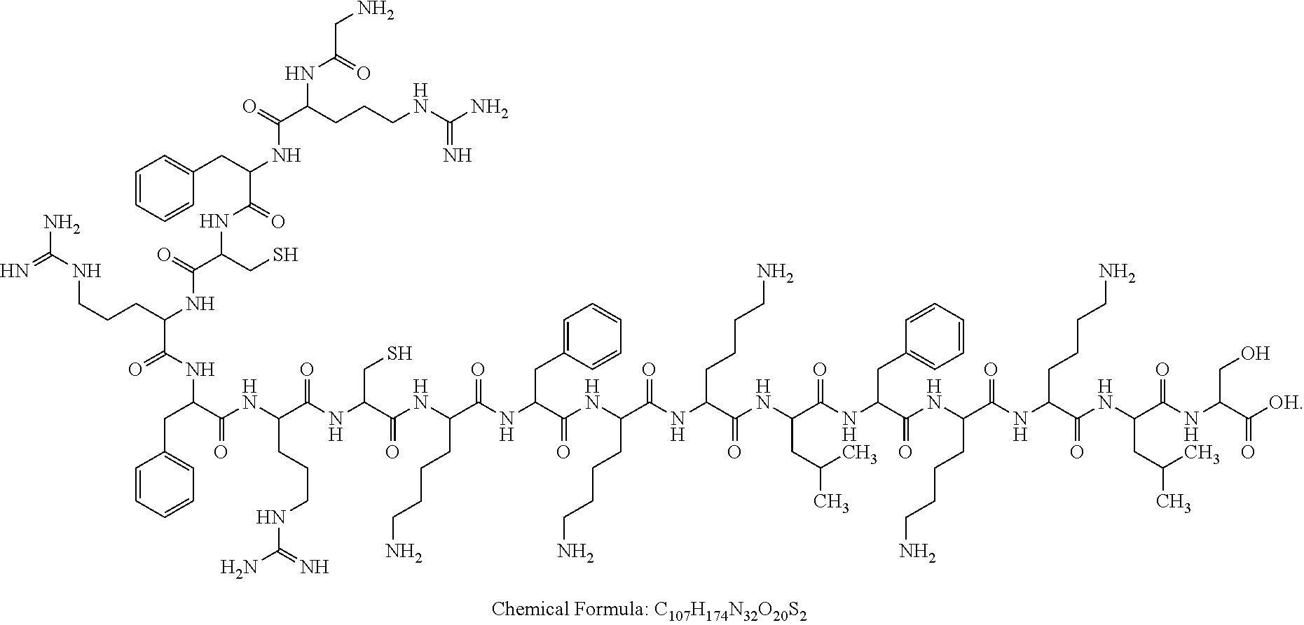
The present disclosure provides a polypeptide with an anti-Mycobacterium tuberculosis activity and having the sequence shown in SEQ ID NO: 3, where a structural formula of the polypeptide is as follows:
Figure US12303545-20250520-C00001
The present disclosure also provides a preparation method of the polypeptide, including the following steps:
    • (1) preparing a straight-chain polypeptide with a sequence shown in SEQ ID NO: 1; and
    • (2) allowing sulfhydryl groups of two cysteine residues in the straight-chain polypeptide to undergo a dehydration-condensation reaction with dimethylolurea to obtain the polypeptide.
In the present disclosure, a molar ratio of the straight-chain polypeptide to the dimethylolurea is 1:(8-12).
In the present disclosure, in the step (2), the straight-chain polypeptide and the dimethylolurea are dissolved in a solvent separately, and then mixed to allow the dehydration-condensation reaction.
In the present disclosure, a solvent for the straight-chain polypeptide is one of a mixed solution of trifluoroacetic acid (TFA) and guanidine hydrochloride, an aqueous urea solution, acetonitrile, and phosphate buffered saline (PBS); and a solvent for the dimethylolurea is one of water, acetonitrile, methanol, and N,N-dimethylformamide (DMF).
In the present disclosure, a concentration of the aqueous urea solution is 4 mol/L to 8 mol/L.
In the present disclosure, a concentration of the straight-chain polypeptide in the solvent is 1 mmol/L to 8 mmol/L, and a concentration of the dimethylolurea in the solvent is 18 mmol/L to 80 mmol/L.
In the present disclosure, during the reaction in the step (2), an acid reaction regulator is added; the acid reaction regulator is TFA; and a volume ratio of a solution of the straight-chain polypeptide to the acid reaction regulator is 1:1 to 1:3.
In the present disclosure, the reaction in the step (2) is conducted at 0° C. to 40° C. for 0 min to 10 min.
The present disclosure also provides a use of the polypeptide in preparation of an anti-Mycobacterium tuberculosis drug.
The polypeptide SAH1 of the present disclosure has an anti-Mycobacterium tuberculosis activity, and has the following advantages over the polypeptide BMAP-18 without modification: (1) Safety and few side effects: SAH1 exhibits a significantly-lower cytotoxicity than BMAP-18 to mouse macrophages. A cytotoxicity IC50 value of SAH1 is 300 μM and a cytotoxicity IC50 value of BMAP-18 is 150 μM. Therefore, the safety of SAH1 is significantly improved. (2) Improved anti-Mycobacterium tuberculosis effect: SAH1 can exhibit a strong anti-BCG bacterial infection effect at a concentration of 12.5 μM in the minimum inhibitory concentration (MIC) test. However, under the same conditions, a MIC of BMAP-18 is 300 μM, and an antibacterial ability of BMAP-18 is merely one twenty-fourth of an antibacterial ability of SAH1. Under a scanning electron microscope, it is also intuitively observed that the antibacterial ability of SAH1 is significantly improved compared with BMAP-18. (3) Strong anti-protease hydrolysis ability: After BMAP-18 is added to trypsin at a concentration of 5 μg/mL, BMAP-18 is completely hydrolyzed at 30 min. However, when SAH1 is placed in the same environment, 45.89% of SAH1 is still not hydrolyzed at 30 min, and 30.21% of SAH1 can still be left at 1 h. The polypeptide SAH1 is an antimicrobial peptide, which can play a disease-treating role while avoiding drug resistance and side effects through a unique action mode in the post-antibiotic era. Therefore, the polypeptide SAH1 has a very promising application prospect and a great research value.
BRIEF DESCRIPTION OF THE DRAWINGS
FIG. 1 shows a reaction principle for the preparation of the polypeptide SAH1;
FIG. 2 shows a chromatography spectrum of the crude straight-chain polypeptide GRFC4RFRC8KFKKLFKKLS-OH (SEQ ID NO: 1);
FIG. 3 is a mass spectrometry spectrum of the polypeptide GRFC4RFRC8KFKKLFKKLS-OH (SEQ ID NO: 1);
FIG. 4 is a preparative chromatography spectrum of a dimethylolurea-modified polypeptide;
FIG. 5 is a mass spectrometry spectrum of a dimethylolurea-modified polypeptide;
FIG. 6 shows cell viabilities of RAW264.7 cells treated with SAH1 and BMAP-18 at each concentration, where an x-coordinate represents a concentration in μM, and a y-coordinate represents a cell viability in %;
FIG. 7 shows fitting results of IC50 values of SAH1 and BMAP-18 for RAW264.7 cells;
FIGS. 8A-8C show impacts of SAH1 and BMAP-18 on the integrity of cell membranes of BCG observed under a scanning electron microscope, where FIG. 8A is for a negative control, FIG. 8B is for BCG treated with BMAP-18, and FIG. 8C is for BCG treated with SAH1;
FIGS. 9A-9B show an impact of SAH1 on a cell membrane and an intracellular structure of BCG observed under a transmission electron microscope;
FIGS. 10A-10C show reverse phase-high performance liquid chromatography (RP-HPLC) spectra illustrating polypeptide residues of BMAP-18 in a trypsin (5 μg/mL) environment at different time points, where a reaction time of FIG. 10A is 0 min, a reaction time of FIG. 10B is 15 min, and a reaction time of FIG. 10C is 30 min; and
FIGS. 11A-11D show RP-HPLC spectra illustrating polypeptide residues of SAH1 under an action of trypsin (5 μg/mL) at different time points, where a reaction time of FIG. 11A is 0 min, a reaction time of FIG. 11B is 15 min, a reaction time of FIG. 11C is 30 min, and a reaction time of FIG. 11D is 60 min.
DETAILED DESCRIPTION OF THE EMBODIMENT
The BCG bacteria in the present application are attenuated Mycobacterium bovis, which has an ATCC number of bio-77983 and is purchased from the China Institute of Veterinary Drug Control.
The technical solutions of the present disclosure are described in detail below in conjunction with the accompanying drawings, but the protection scope of the present disclosure is not limited to the following examples.
Example 1 Screening and Preparation of the Polypeptide SAH1
1. Screening of the Polypeptide SAH1
A sequence of BMAP-18 (from N-terminus to C-terminus) was as follows: G-R-F-K-R-F-R-K-K-F-K-K-L-F-K-K-L-S-OH (Gly-Arg-Phe-Lys-Arg-Phe-Arg-Lys-Lys-Phe-Lys-Lys-Leu-Phe-Lys-Lys-Leu-Ser-OH) (SEQ ID NO: 2). BMAP-18 was modified through a point mutation of an amino acid at any position or a point mutation of amino acids at two random positions. It was finally found that the compound SAH1 exhibited both a reduced cytotoxicity and a highly-effective anti-Mycobacterium tuberculosis effect. As shown in FIG. 1 , a specific modification method was as follows: Amino acids at positions 4 and 8 of BMAP-18 were mutated into cysteine to obtain G-R-F-C-R-F-R-C-K-F-K-K-L-F-K-K-L-S-OH (Gly-Arg-Phe-Cys-Arg-Phe-Arg-Cys-Lys-Phe-Lys-Lys-Leu-Phe-Lys-Lys-Leu-Ser-OH) (SEQ ID NO: 1), and then the mutant was subjected to a dehydration-condensation reaction with dimethylolurea to produce a cyclic peptide with the following structure:
Figure US12303545-20250520-C00002

2. Preparation of the Polypeptide SAH1
(1) Preparation of a Straight-Chain Polypeptide GRFC4RFRC8KFKKLFKKLS-OH (SEQ ID NO: 1)
The Fmoc solid-phase synthesis was adopted to synthesize the polypeptide: GRFC4RFRC8KFKKLFKKLS-OH (from N-terminus to C-terminus) (SEQ ID NO: 1).
Coupling of a first amino acid: 0.2 mmol of a Wang resin was weighed and added to a solid-phase reaction column with xacin, washed with DMF 2 times, and then swelled with DMF for 30 min. 383.4 mg (1 mmol) of Fmoc-Ser (tBu)-OH and 142.11 mg (1 mmol) of Oxyma (ethyl cyanoglyoxylate-2-oxime) were weighed and dissolved in DMF, and then 0.25 g (2 mmol) of N,N′-diisopropylcarbodiimide (DIC) was added to allow activation for 3 min to obtain a coupling solution. The coupling solution was then added to the resin swelled with DMF to allow a coupling reaction at room temperature for 1 h. An end point of the reaction was detected with ninhydrin. If the resin was colorless and transparent, the reaction was terminated. If the resin was colored, the reaction was extended by 1 h. At the end of the reaction, a reaction liquid was removed, and a resulting resin was washed with DMF three times and then subjected to Fmoc protective group removal as follows: A piperidine aqueous solution with a concentration of 20% was added to make the resin immersed, a resulting mixture was stirred with a glass rod to allow a reaction for 3 min, and then a resulting piperidine solution was removed. A resulting resin was washed with DMF 3 times, then a piperidine aqueous solution with a concentration of 20% was added once again to make the resin immersed, a reaction was conducted for 4 min, a resulting reaction liquid was removed, and a resulting resin was washed with DMF 3 times to 5 times.
A second protective amino acid was coupled according to the method of coupling of Fmoc-Ser (tBu)-OH.
With the above method, the remaining amino acids were coupled sequentially from C-terminus to N-terminus to finally obtain a polypeptide GRFC4RFRC8KFKKLFKKLS-OH (SEQ ID NO: 1) resin.
The polypeptide GRFC4RFRC8KFKKLFKKLS-OH (SEQ ID NO: 1) resin was washed with DMF 3 times, washed with dichloromethane (DCM) 2 times, vacuum-dried, and subjected to a cleavage reaction specifically as follows: A lysis buffer was added to allow a lysis reaction at room temperature for 1 h, a resulting resin was filtered out, and a resulting filtrate was collected. Then the cleavage was repeated once with a small amount of the lysis buffer. Filtrates produced after the two times of cleavage were combined and slowly added to glacial diethyl ether for precipitation, and a resulting system was centrifuged to obtain a precipitate and a supernatant. The supernatant was discarded, and the precipitate was dried under reduced pressure to obtain a crude straight-chain polypeptide GRFC4RFRC8KFKKLFKKLS-OH (SEQ ID NO: 1). The lysis buffer was a mixed solution of TFA, triisopropylsilane (TIS), and H2O in a volume ratio of 95:2.5:2.5.
The crude straight-chain polypeptide GRFC4RFRC8KFKKLFKKLS-OH (SEQ ID NO: 1) was lyophilized as follows: The crude straight-chain polypeptide was frozen with liquid nitrogen into a solid, and then the solid was suspended in a lyophilizer for lyophilization to obtain a solid powder. The straight-chain polypeptide GRFC4RFRC8KFKKLFKKLS-OH (SEQ ID NO: 1) was identified by liquid chromatography-mass spectrometry (LC-MS). A corresponding chromatography spectrum was shown in FIG. 2 , and a corresponding mass spectrometry spectrum was shown in FIG. 3 . Mass spectrometry data were as follows: M=2292.3, found: 1147 [M+2H+]2+, 765.25 [M+3H+]3+, 574.33 [M+4H+]4+, 459.75 [M+5H+]5+.
A structural formula of the straight-chain polypeptide GRFC4RFRC8KFKKLFKKLS-OH (SEQ ID NO: 1) was as follows:
Figure US12303545-20250520-C00003
A sequence of the straight-chain polypeptide (SEQ ID NO: 1) was as follows: GRFCRFRCKFKKLFKKLS.
(2) Two cysteine residues in the straight-chain polypeptide were modified and stapled with dimethylolurea.
The crude straight-chain polypeptide GRFC4RFRC8KFKKLFKKLS-OH (SEQ ID NO: 1) was allowed to directly react with dimethylolurea under acidic conditions to obtain a stapled cyclic peptide, which reduced the operation processes, improved the production efficiency, and reduced the consumption of organic solvents. Specific steps were as follows:
Step 1: The crude straight-chain polypeptide GRFC4RFRC8KFKKLFKKLS-OH (SEQ ID NO: 1) was taken and dissolved in a mixed solution of TFA and guanidine hydrochloride in a volume ratio of 1:1 to obtain a 2 mmol/L polypeptide solution. Dimethylolurea was dissolved in water to obtain a 20 mmol/L dimethylolurea aqueous solution.
Step 2: At room temperature, anhydrous TFA was added to the 2 mmol/L polypeptide solution, then the 20 mmol/L dimethylolurea aqueous solution was added, and a reaction was conducted at room temperature for 1 min to obtain a crude dimethylolurea-modified polypeptide. The polypeptide solution, the anhydrous TFA, and the dimethylolurea aqueous solution were in a volume ratio of 1:1:1.
Step 3: The crude dimethylolurea-modified polypeptide was purified by semi-preparative high-performance liquid chromatography under the following conditions: mobile phases: A: 0.1% (volume percentage concentration) TFA-containing H2O, and B: 0.1% (volume percentage concentration) TFA-conatining acetonitrile; an elution procedure: 0 min to 30 min: the mobile phase B: from 0% to 60%, with an increment of 2% of B per minute; a chromatographic column: Waters C18; a flow rate: 15 mL/min; and a detection wavelength: 214 nm. As shown in FIG. 4 , the prepared dimethylolurea-modified polypeptide had a high purity.
The purified dimethylolurea-modified polypeptide was lyophilized as follows: The purified dimethylolurea-modified polypeptide was frozen with liquid nitrogen into a solid, and then the solid was suspended in a lyophilizer for lyophilization obtain a solid powder. The purified dimethylolurea-modified polypeptide was identified by mass spectrometry, as shown in FIG. 5 . Mass spectrometry data were as follows: M=2376.9, found: 1188.92 [M+2H+]2+, 792.92 [M+3H+]3+, 595.00 [M+4H+]4+, 476.25 [M+5H+]5+.
A structural formula of the dimethylolurea-modified polypeptide was as follows:
Figure US12303545-20250520-C00004
The dimethylolurea-modified polypeptide was denoted as a polypeptide SAH1.
Example 2 Detection of Cell Viabilities of RAW264.7 Cells Treated with SAH1 and BMAP-18 by a CCK-8 Method
The toxicities of SAH1 and BMAP-18 to RAW264.7 cells were determined by the CCK-8 method, namely, a WST (chemical name: 2-(2-methoxy-4-nitrophenyl)-3-(4-nitrophenyl)-5-(2,4-dithophenyl)-2H-tetrazolium monosodium salt) colorimetric test. A specific method was as follows: 1×10⁵ RAW 264.7 cells were inoculated into each well of a 96-well plate, 100 μL of a polypeptide SAH1 aqueous solution serially diluted to a concentration of 3.125 μM to 200 μM was added to each test well, and the plate was incubated at 37° C. and 5% CO2 for 24 h. A positive control well was set. The positive control well was the same as test wells except that the polypeptide SAH1 aqueous solution was replaced with a DMEM complete medium. A blank well was set. In the blank well, no cells were added, and only the DMEM complete medium was added. After the incubation, 10 μL of a CCK-8 reaction solution (Beijing Solarbio) was added to each well, and the plate was further incubated at 37° C. for 2 h. Finally, the absorbance at 450 nm was detected with a microplate reader (Synersymx, Biotek) to determine a cell viability. According to the formula: cell viability=(absorbance of a test well−absorbance of a blank well)/(absorbance of a positive control well−absorbance of a blank well) * 100%, a cell viability relative to the positive control well was calculated.
In addition, the same method was used to detect viabilities of cells treated with BMAP-18 aqueous solutions serially diluted to concentrations of 3.125 μM to 200 μM.
It can be seen from FIG. 6 that SAH1 exhibits a significantly lower cytotoxicity than BMAP-18 at each concentration gradient, SAH1 does not exhibit a significant cytotoxicity to RAW264.7 cells (cell viability: higher than 90%) at a concentration of 50 μM or lower, and BMAP-18 does not exhibit a significant cytotoxicity to RAW264.7 cells (cell viability: higher than 90%) at a concentration of 25 μM or lower.
Example 3 Fitting Results of IC50 Values of SAH1 and BMAP-18 for RAW264.7 Cells
1×10⁵ RAW 264.7 cells were inoculated into each well of a 96-well plate, 100 μL of a polypeptide SAH1 aqueous solution serially diluted to a concentration of 3.125 μM to 200 μM was added to each test well, and the plate was incubated at 37° C. and 5% CO2 for 24 h. A positive control well was set. The positive control well was the same as test wells except that the polypeptide SAH1 aqueous solution was replaced with a DMEM complete medium. After the incubation, 10 μL of a CCK-8 reaction solution (Beijing Solarbio) was added to each well, and the plate was further incubated at 37° C. for 2 h. Finally, the absorbance at 450 nm was detected with a microplate reader (Synersymx, Biotek). An OD value of the positive control well was BO, an OD value of a test well in which an antimicrobial peptide was added was B, and B/BO was called an inhibition rate. A concentration of an antimicrobial peptide corresponding to an inhibition rate of 50% was called IC50. The lower the IC50 value, the stronger the cytotoxicity of the antimicrobial peptide. With a concentration of an antimicrobial peptide as an x-coordinate and an inhibition rate as a y-coordinate, a line chart was plotted, and IC50 was obtained through fitting.
IC50 of BMAP-18 for RAW 264.7 cells was obtained by the same method.
It can be seen from FIG. 7 that IC50 values of SAH1 and BMAP-18 for RAW 264.7 cells are 300 μM and 150 μM, respectively, indicating that SAH1 has a lower cytotoxicity than BMAP-18.
Example 4 Determination of an Ability of SAH1 to Inhibit the Growth of BCG Bacteria by MIC Assay
An antibacterial effect of SAH1 at each concentration on BCG bacteria was investigated. SAH1 was serially diluted in a range of 6.25 μM to 3,200 μM with a 7H9 medium as a solvent to obtain SAH1 solutions with different concentrations. BMAP-18 was serially diluted in a range of 6.25 μM to 3,200 μM with a 7H9 medium as a solvent to obtain BMAP-18 solutions with different concentrations. Rifampicin, isoniazid, and ethambutol each were serially diluted in a range of 25 μM to 12,800 μM with a 7H9 medium as a solvent to obtain rifampicin solutions, isoniazid solutions, and ethambutol solutions each with different concentrations. A solution of each drug was filtered through a 0.22 μm filter to remove bacteria.
An SAH1 test group was set as follows: 0.1 mL of an SAH1 solution at each concentration was added to a centrifuge tube, and then 0.1 mL of BCG bacteria at 1.5×10⁷ cfu/mL was added. A positive control was set as follows: the positive control was the same as the SAH1 test group except that a 7H9 medium was used instead of an SAH1 solution. A negative control was set as follows: the negative control was the same as the SAH1 test group except that the same volume of ultrapure water was used instead of BCG bacteria. In addition, a BMAP-18 test group, a rifampicin test group, an isoniazid test group, and an ethambutol test group were set. These test groups were the same as the SAH1 test group except that BMAP-18, rifampicin, isoniazid, or ethambutol was used instead of the SAH1 solution. After each centrifuge tube was incubated at 37° C. for 7 d, a resazurin indicator was added to allow an action for 2 d, and a color change was observed. After the two days of the action: If a medium was blue, it meant that there were no bacteria in a corresponding centrifuge tube, indicating that a drug at a corresponding concentration had an antibacterial effect. If a medium was purple and red, it meant that there were viable bacteria in a corresponding centrifuge tube, indicating that a drug at a corresponding concentration had no antibacterial effect.
Results were shown in Table 1. MIC of SAH1 was 12.5 μM, which was significantly lower than MICs of BMAP-18, rifampicin, isoniazid, and ethambutol, indicating that an antibacterial ability of SAH1 was significantly improved compared with BMAP-18.
TABLE 1
MIC of each drug
Drug MIC
SAH1 12.5 μM
BMAP-18  300 μM
Rifampicin
 400 μM
Isoniazid
 800 μM
Ethambutol
 800 μM
Example 5 Observation of Impacts of SAH1 and BMAP-18 on the Integrity of a Cell Membrane of BCG by a Scanning Electron Microscope
BCG bacteria growing to a logarithmic phase were collected, suspended in a fresh 7H9 medium, and adjusted to OD600 nm of 0.5. Then SAH1 at a final concentration of 12.5 μM and BMAP-18 at a final concentration of 300 μM were added separately. In a negative control, sterile ddH2O was added instead of an antimicrobial peptide. The cells were cultivated at 37° C. for 1 h. At the end of cultivation, bacterial cells were collected through centrifugation, washed 3 times with PBS (0.1 M, pH 7.0), resuspended with 2.5% glutaraldehyde, thoroughly dispersed, and placed at 4° C. overnight to fix bacteria. A resulting bacterial pellet was washed 3 times with PBS (0.1 M, pH 7.0), a 1% OSO4 aqueous solution was added to allow an action for 1 h, and then the bacterial pellet was subjected to centrifugal washing 3 times with PBS (0.1 M, pH 7.0). The dehydration was conducted with ethanol at different concentrations (30%, 50%, 70%, 80%, 90%, and 100%) for 15 min to 20 min each time. A resulting product was then treated with a mixture of ethanol and isoamyl acetate (v:v=1:1) for 30 min, then treated with pure isoamyl acetate for about 1 h, dehydrated with liquid carbon dioxide in a critical point dryer, and then coated with gold at 45 mA for 2 min. Finally, a treated sample was observed under a scanning electron microscope.
It can be seen from FIGS. 8A-8C that SAH1 exhibits a significantly-higher damage degree than BMAP-18 to the cell membrane and cell of BCG at MIC, and when a cell membrane of Mycobacteria is damaged, contents leak to cause the atrophy and even death of bacteria.
Example 6 Observation of an Impact of SAH1 on a Cell Membrane and an Intracellular Structure of BCG by a Transmission Electron Microscope
BCG at a logarithmic phase was treated according to the method in Example 5. BCG was pre-dehydrated with ethanol at different concentration gradients, then treated with anhydrous acetone for 20 min, then treated with a mixed solution of anhydrous acetone and an EPON812 resin (V/V=1:1) at room temperature for 1 h, and then treated with a mixed solution of anhydrous acetone and an EPON812 resin (V/V=1:3) at room temperature for 3 h. Then, a bacterial sample was soaked overnight in an EPON812 resin twice. A treated sample was placed in an EP tube with the EPON812 resin and treated at 70° C. for 9 h or more. Sectioning was conducted with an ultramicrotome, and staining was conducted with uranyl acetate and alkaline lead citrate successively for 5 min to 10 min. Finally, a bacterial sample was observed under a transmission electron microscope. In addition, a negative control was set. A specific treatment method for the negative control was the same as the treatment method for each sample except that BCG at a logarithmic phase was not treated with any antimicrobial substance.
It can be seen from FIGS. 9A-9B that contents of BCG cells treated with SAH1 are largely missed, resulting in cavities and bacterial death.
Example 7 Determination of Abilities of SAH1 and BMAP-18 to Resist Protease Hydrolysis by Trypsin Assay
A 4 mg/mL SAH1 solution and a 10 μg/mL trypsin solution each were prepared with PBS (pH 7.4) as a solvent. 250 μL of the trypsin solution was added to 250 μL of the SAH1 solution, and a reaction was conducted in an ice bath. A sample was collected at different time points after the reaction to determine the decomposition of SAH1 by RP-HPLC. RP-HPLC conditions were as follows: A C18 reversed-phase Bio-Rad analytical column was adopted: 250× 4 mm, a pore size: 30 nm, and a particle size: 7 μm. At 0 min to 40 min, linear gradient elution was conducted with 10% to 90% acetonitrile aqueous solutions, with 0.1% TFA (w/v) in both acetonitrile and water. A flow rate was 1.2 mL/min.
In addition, with the same method as above, the SAH1 aqueous solution was replaced with a 4 mg/mL BMAP-18 aqueous solution to detect the hydrolysis of BMAP-18 by trypsin.
After SAH1 reacted with trypsin for different time periods, a residual polypeptide amount was determined by RP-HPLC. According to Table 2, FIGS. 10A-10C, and FIGS. 11A-11D: Residual polypeptide proportions of SAH1 treated with trypsin for 15 min, 30 min, and 60 min were 67.59%, 45.89%, and 30.21%, respectively. After BMAP-18 was treated with trypsin for 15 min, a residual polypeptide proportion was merely 17.76%. After BMAP-18 was treated with trypsin for 30 min, the polypeptide was almost completely hydrolyzed. It can be known that an anti-protease hydrolysis ability of SAH1 is significantly enhanced compared with BMAP-18.
TABLE 2
Polypeptide residues of SAH1 and
BMAP-18 under the action of trypsin
Antimicrobial Reaction Polypeptide
peptide name time Peak area residue
BMAP-18  0 min 254.43   100%
15 min 45.20 17.76%
30 min 0.02  0.01%
SAH1
 0 min 274.44   100%
15 min 185.49 67.59%
30 min 125.94 45.89%
60 min 82.92 30.21%
Notes: A calculation method of a polypeptide residue in Table 2 is as follows: With an HPLC peak area at the initial reaction time of O min as 100%, a polypeptide residue is calculated according to the following formula: RP-HPLC peak area of a polypeptide residue/RP-HPLC peak area of an initial polypeptide * 100%.

Claims (9)

What is claimed is:
1. A polypeptide with an anti-Mycobacterium tuberculosis activity and having the sequence according to SEQ ID NO: 3, wherein the SEQ ID NO: 3 has a structural formula of the polypeptide as follows:
Figure US12303545-20250520-C00005
2. A method of preparing the polypeptide according to claim 1, comprising the following steps:
(1) synthesizing a straight-chain polypeptide according to SEQ ID NO: 1 by Fmoc solid-phase coupling; and
(2) dissolving the straight-chain polypeptide and dimethylolurea in a solvent separately, and then mixing to allow sulfhydryl groups of two cysteine residues in the straight-chain polypeptide to undergo a dehydration-condensation reaction with dimethylolurea to obtain the polypeptide.
3. The method according to claim 2, wherein a molar ratio of the straight-chain polypeptide to the dimethylolurea is 1:8 to 1:12.
4. The method according to claim 3, wherein the solvent for the straight-chain polypeptide is one of a mixed solution of trifluoroacetic acid (TFA) and guanidine hydrochloride, an aqueous urea solution, acetonitrile, and phosphate buffered saline (PBS); and the solvent for the dimethylolurea is one of water, the acetonitrile, methanol, and N,N-dimethylformamide (DMF).
5. The method according to claim 4, wherein a concentration of the aqueous urea solution is 4 mol/L to 8 mol/L.
6. The method according to claim 5, wherein a concentration of the straight-chain polypeptide in the solvent for the straight-chain polypeptide is 1 mmol/L to 8 mmol/L, and a concentration of the dimethylolurea in the solvent for the dimethylolurea is 18 mmol/L to 80 mmol/L.
7. The method according to claim 6, the method comprising adding an acid reaction regulator to the dehydration-condensation reaction in the step (2); the acid reaction regulator is a TFA; and a volume ratio of a solution of the straight-chain polypeptide to the acid reaction regulator is 1:1 to 1:3.
8. The method according to claim 7, the method comprising conducting the dehydration-condensation reaction in the step (2) at 0° C. to 40° C. for 0 min to 10 min.
9. An anti-Mycobacterium tuberculosis drug in aqueous form comprising the polypeptide according to claim 1.
US18/824,970 2023-06-15 2024-09-05 Polypeptide with anti-Mycobacterium tuberculosis activity, and preparation method and use thereof Active US12303545B2 (en)

Applications Claiming Priority (3)

Application Number Priority Date Filing Date Title
CN202310710031.5A CN116987149B (en) 2023-06-15 2023-06-15 Polypeptide with anti-mycobacterium tuberculosis activity, preparation method and application thereof
CN202310710031.5 2023-06-15
PCT/CN2023/140163 WO2024255185A1 (en) 2023-06-15 2023-12-20 Polypeptide having anti-mycobacterium tuberculosis activity, preparation method therefor and use thereof

Related Parent Applications (1)

Application Number Title Priority Date Filing Date
PCT/CN2023/140163 Continuation WO2024255185A1 (en) 2023-06-15 2023-12-20 Polypeptide having anti-mycobacterium tuberculosis activity, preparation method therefor and use thereof

Publications (2)

Publication Number Publication Date
US20240415925A1 US20240415925A1 (en) 2024-12-19
US12303545B2 true US12303545B2 (en) 2025-05-20

Family

ID=93845360

Family Applications (1)

Application Number Title Priority Date Filing Date
US18/824,970 Active US12303545B2 (en) 2023-06-15 2024-09-05 Polypeptide with anti-Mycobacterium tuberculosis activity, and preparation method and use thereof

Country Status (1)

Country Link
US (1) US12303545B2 (en)

Citations (7)

* Cited by examiner, † Cited by third party
Publication number Priority date Publication date Assignee Title
US20140296137A1 (en) * 2013-04-01 2014-10-02 Los Alamos National Security, Llc Methods and compositions for controlling rotifers
US20150259382A1 (en) 2011-11-21 2015-09-17 Board Of Regents Of The University Of Nebraska Anti-Microbial Peptides and Methods of Use Thereof
US20180355005A1 (en) 2015-11-25 2018-12-13 Indiana University Research And Technology Corporation Bacteriocidal peptides and uses thereof
CN109762051A (en) 2019-03-29 2019-05-17 常州大学 A kind of ring-type antibacterial peptide and its preparation method and application
CN109897091A (en) 2019-03-29 2019-06-18 常州大学 A kind of cyclic annular antibacterial peptide and its preparation method and application containing unnatural amino acid
WO2023049162A1 (en) 2021-09-21 2023-03-30 Biomedit, Llc Antimicrobial peptides
CN116987149A (en) 2023-06-15 2023-11-03 南京农业大学 A polypeptide with anti-Mycobacterium tuberculosis activity, preparation method and application thereof

Patent Citations (7)

* Cited by examiner, † Cited by third party
Publication number Priority date Publication date Assignee Title
US20150259382A1 (en) 2011-11-21 2015-09-17 Board Of Regents Of The University Of Nebraska Anti-Microbial Peptides and Methods of Use Thereof
US20140296137A1 (en) * 2013-04-01 2014-10-02 Los Alamos National Security, Llc Methods and compositions for controlling rotifers
US20180355005A1 (en) 2015-11-25 2018-12-13 Indiana University Research And Technology Corporation Bacteriocidal peptides and uses thereof
CN109762051A (en) 2019-03-29 2019-05-17 常州大学 A kind of ring-type antibacterial peptide and its preparation method and application
CN109897091A (en) 2019-03-29 2019-06-18 常州大学 A kind of cyclic annular antibacterial peptide and its preparation method and application containing unnatural amino acid
WO2023049162A1 (en) 2021-09-21 2023-03-30 Biomedit, Llc Antimicrobial peptides
CN116987149A (en) 2023-06-15 2023-11-03 南京农业大学 A polypeptide with anti-Mycobacterium tuberculosis activity, preparation method and application thereof

Non-Patent Citations (1)

* Cited by examiner, † Cited by third party
Title
Kanjana Madhingsa, et al., Antimicrobial Action of the Cyclic Peptide Bactenecin on Burkholderia pseudomallei Correlates with Efficient Membrane Permeabilization, PLOS Neglected Tropical Diseases, 2013, pp. 1-13, vol. 7 No. 6.

Also Published As

Publication number Publication date
US20240415925A1 (en) 2024-12-19

Similar Documents

Publication Publication Date Title
CN111533786B (en) Beta-hairpin antibacterial peptide with tryptophan and arginine cross-chain interaction and preparation method thereof
CN101570569A (en) Synthetic antibacterial peptide and preparation method and application thereof
CN111363010B (en) Symmetrical short-sequence antibacterial peptide analogue and application thereof
CN116987149B (en) Polypeptide with anti-mycobacterium tuberculosis activity, preparation method and application thereof
CN111057134A (en) A class of antibacterial peptides with broad-spectrum antibacterial activity, low toxicity and no induced drug resistance and their applications
CN112661832A (en) High-stability antibacterial peptide and application thereof
CN109810178B (en) A kind of anti-enzymolysis antibacterial peptide I9H12 and its preparation method and application
CN114790226A (en) Antibacterial peptide and preparation method thereof
CN114031671A (en) Antibacterial peptide targeting fungi and preparation method and application thereof
CN116693621B (en) Narrow-spectrum antibacterial peptide PC for inhibiting gram-negative bacteria, and preparation method and application thereof
US12303545B2 (en) Polypeptide with anti-Mycobacterium tuberculosis activity, and preparation method and use thereof
CN109535227B (en) Antibacterial peptide, preparation method of antibacterial peptide, antibacterial composition, antibacterial method and application
CN113214355B (en) Special antifungal antibacterial peptide GL4W as well as preparation method and application thereof
CN112625092B (en) Antibacterial polypeptide compound based on polybia-MPI and synthesis and application thereof
CN110615830B (en) Application of antibacterial peptide in anti-mycobacterial infection medicine
CN118388589B (en) Cell penetrating antibacterial peptide for resisting intracellular bacteria, and preparation method and application thereof
CN111423493B (en) A kind of palmitated anti-enzymolysis antibacterial peptide and its preparation method and application
Güell et al. Peptidotriazoles with antimicrobial activity against bacterial and fungal plant pathogens
CN105837675B (en) Cationic antibacterial peptides and preparation method thereof
CN110283245B (en) Pig marrow derived PMAP-23 derived antibacterial peptide, preparation method and application
CN113549137A (en) A proline-rich antimicrobial peptide Pyr-2 targeting gram-negative bacteria and its preparation method and application
CN109021068B (en) A kind of linear pseudopolypeptide and its preparation method and application in antibacterial drugs
CN116535470B (en) Polypeptides with anti-tuberculosis activity, preparation methods and applications
CN107337713A (en) Antibacterial peptides and preparation method thereof
CN116514925B (en) A polypeptide against Mycobacterium tuberculosis, its preparation method and its application

Legal Events

Date Code Title Description
AS Assignment

Owner name: AGRICULTURAL UNIVERSITY OF NANJING, CHINA

Free format text: ASSIGNMENT OF ASSIGNORS INTEREST;ASSIGNORS:XUE, FENG;SUN, HAO;HUANG, QUNTAO;AND OTHERS;REEL/FRAME:068490/0939

Effective date: 20240719

FEPP Fee payment procedure

Free format text: ENTITY STATUS SET TO UNDISCOUNTED (ORIGINAL EVENT CODE: BIG.); ENTITY STATUS OF PATENT OWNER: SMALL ENTITY

FEPP Fee payment procedure

Free format text: ENTITY STATUS SET TO SMALL (ORIGINAL EVENT CODE: SMAL); ENTITY STATUS OF PATENT OWNER: SMALL ENTITY

STPP Information on status: patent application and granting procedure in general

Free format text: NON FINAL ACTION MAILED

STPP Information on status: patent application and granting procedure in general

Free format text: RESPONSE TO NON-FINAL OFFICE ACTION ENTERED AND FORWARDED TO EXAMINER

STPP Information on status: patent application and granting procedure in general

Free format text: NOTICE OF ALLOWANCE MAILED -- APPLICATION RECEIVED IN OFFICE OF PUBLICATIONS

STCF Information on status: patent grant

Free format text: PATENTED CASE