US12280148B2 - Composition for promoting extracellular vesicle production containing peptide derived from noxa protein and method for producing extracellular vesicles by using same - Google Patents

Composition for promoting extracellular vesicle production containing peptide derived from noxa protein and method for producing extracellular vesicles by using same Download PDF

Info

Publication number
US12280148B2
US12280148B2 US17/424,232 US202117424232A US12280148B2 US 12280148 B2 US12280148 B2 US 12280148B2 US 202117424232 A US202117424232 A US 202117424232A US 12280148 B2 US12280148 B2 US 12280148B2
Authority
US
United States
Prior art keywords
extracellular vesicles
peptide
extracellular
present
solution
Prior art date
Legal status (The legal status is an assumption and is not a legal conclusion. Google has not performed a legal analysis and makes no representation as to the accuracy of the status listed.)
Active, expires
Application number
US17/424,232
Other versions
US20220304931A1 (en
Inventor
Tae-Hyoung Kim
Jung Hee Park
Ji Hye Han
Seung-Hyun MYUNG
Current Assignee (The listed assignees may be inaccurate. Google has not performed a legal analysis and makes no representation or warranty as to the accuracy of the list.)
Exocalibre Therapeutics Co Ltd
Original Assignee
Exocalibre Therapeutics Co Ltd
Priority date (The priority date is an assumption and is not a legal conclusion. Google has not performed a legal analysis and makes no representation as to the accuracy of the date listed.)
Filing date
Publication date
Application filed by Exocalibre Therapeutics Co Ltd filed Critical Exocalibre Therapeutics Co Ltd
Assigned to INDUSTRY-ACADEMIC COOPERATION FOUNDATION, CHOSUN UNIVERSITY reassignment INDUSTRY-ACADEMIC COOPERATION FOUNDATION, CHOSUN UNIVERSITY ASSIGNMENT OF ASSIGNORS INTEREST (SEE DOCUMENT FOR DETAILS). Assignors: HAN, JI HYE, KIM, TAE-HYOUNG, MYUNG, SEUNG-HYUN, PARK, JUNG HEE
Publication of US20220304931A1 publication Critical patent/US20220304931A1/en
Assigned to EXOCALIBRE THERAPEUTICS CO., LTD. reassignment EXOCALIBRE THERAPEUTICS CO., LTD. ASSIGNMENT OF ASSIGNORS INTEREST (SEE DOCUMENT FOR DETAILS). Assignors: INDUSTRY-ACADEMIC COOPERATION FOUNDATION, CHOSUN UNIVERSITY
Application granted granted Critical
Publication of US12280148B2 publication Critical patent/US12280148B2/en
Active legal-status Critical Current
Adjusted expiration legal-status Critical

Links

Images

Classifications

    • AHUMAN NECESSITIES
    • A61MEDICAL OR VETERINARY SCIENCE; HYGIENE
    • A61KPREPARATIONS FOR MEDICAL, DENTAL OR TOILETRY PURPOSES
    • A61K9/00Medicinal preparations characterised by special physical form
    • A61K9/10Dispersions; Emulsions
    • A61K9/127Synthetic bilayered vehicles, e.g. liposomes or liposomes with cholesterol as the only non-phosphatidyl surfactant
    • A61K9/1277Preparation processes; Proliposomes
    • CCHEMISTRY; METALLURGY
    • C12BIOCHEMISTRY; BEER; SPIRITS; WINE; VINEGAR; MICROBIOLOGY; ENZYMOLOGY; MUTATION OR GENETIC ENGINEERING
    • C12NMICROORGANISMS OR ENZYMES; COMPOSITIONS THEREOF; PROPAGATING, PRESERVING, OR MAINTAINING MICROORGANISMS; MUTATION OR GENETIC ENGINEERING; CULTURE MEDIA
    • C12N5/00Undifferentiated human, animal or plant cells, e.g. cell lines; Tissues; Cultivation or maintenance thereof; Culture media therefor
    • C12N5/0068General culture methods using substrates
    • AHUMAN NECESSITIES
    • A61MEDICAL OR VETERINARY SCIENCE; HYGIENE
    • A61KPREPARATIONS FOR MEDICAL, DENTAL OR TOILETRY PURPOSES
    • A61K31/00Medicinal preparations containing organic active ingredients
    • A61K31/33Heterocyclic compounds
    • A61K31/395Heterocyclic compounds having nitrogen as a ring hetero atom, e.g. guanethidine or rifamycins
    • A61K31/435Heterocyclic compounds having nitrogen as a ring hetero atom, e.g. guanethidine or rifamycins having six-membered rings with one nitrogen as the only ring hetero atom
    • A61K31/47Quinolines; Isoquinolines
    • A61K31/473Quinolines; Isoquinolines ortho- or peri-condensed with carbocyclic ring systems, e.g. acridines, phenanthridines
    • AHUMAN NECESSITIES
    • A61MEDICAL OR VETERINARY SCIENCE; HYGIENE
    • A61KPREPARATIONS FOR MEDICAL, DENTAL OR TOILETRY PURPOSES
    • A61K31/00Medicinal preparations containing organic active ingredients
    • A61K31/70Carbohydrates; Sugars; Derivatives thereof
    • A61K31/7028Compounds having saccharide radicals attached to non-saccharide compounds by glycosidic linkages
    • A61K31/7034Compounds having saccharide radicals attached to non-saccharide compounds by glycosidic linkages attached to a carbocyclic compound, e.g. phloridzin
    • A61K31/704Compounds having saccharide radicals attached to non-saccharide compounds by glycosidic linkages attached to a carbocyclic compound, e.g. phloridzin attached to a condensed carbocyclic ring system, e.g. sennosides, thiocolchicosides, escin, daunorubicin
    • AHUMAN NECESSITIES
    • A61MEDICAL OR VETERINARY SCIENCE; HYGIENE
    • A61KPREPARATIONS FOR MEDICAL, DENTAL OR TOILETRY PURPOSES
    • A61K31/00Medicinal preparations containing organic active ingredients
    • A61K31/70Carbohydrates; Sugars; Derivatives thereof
    • A61K31/7088Compounds having three or more nucleosides or nucleotides
    • A61K31/711Natural deoxyribonucleic acids, i.e. containing only 2'-deoxyriboses attached to adenine, guanine, cytosine or thymine and having 3'-5' phosphodiester links
    • AHUMAN NECESSITIES
    • A61MEDICAL OR VETERINARY SCIENCE; HYGIENE
    • A61KPREPARATIONS FOR MEDICAL, DENTAL OR TOILETRY PURPOSES
    • A61K35/00Medicinal preparations containing materials or reaction products thereof with undetermined constitution
    • A61K35/12Materials from mammals; Compositions comprising non-specified tissues or cells; Compositions comprising non-embryonic stem cells; Genetically modified cells
    • A61K35/13Tumour cells, irrespective of tissue of origin
    • AHUMAN NECESSITIES
    • A61MEDICAL OR VETERINARY SCIENCE; HYGIENE
    • A61KPREPARATIONS FOR MEDICAL, DENTAL OR TOILETRY PURPOSES
    • A61K38/00Medicinal preparations containing peptides
    • A61K38/16Peptides having more than 20 amino acids; Gastrins; Somatostatins; Melanotropins; Derivatives thereof
    • A61K38/17Peptides having more than 20 amino acids; Gastrins; Somatostatins; Melanotropins; Derivatives thereof from animals; from humans
    • A61K38/177Receptors; Cell surface antigens; Cell surface determinants
    • AHUMAN NECESSITIES
    • A61MEDICAL OR VETERINARY SCIENCE; HYGIENE
    • A61KPREPARATIONS FOR MEDICAL, DENTAL OR TOILETRY PURPOSES
    • A61K38/00Medicinal preparations containing peptides
    • A61K38/16Peptides having more than 20 amino acids; Gastrins; Somatostatins; Melanotropins; Derivatives thereof
    • A61K38/17Peptides having more than 20 amino acids; Gastrins; Somatostatins; Melanotropins; Derivatives thereof from animals; from humans
    • A61K38/19Cytokines; Lymphokines; Interferons
    • AHUMAN NECESSITIES
    • A61MEDICAL OR VETERINARY SCIENCE; HYGIENE
    • A61KPREPARATIONS FOR MEDICAL, DENTAL OR TOILETRY PURPOSES
    • A61K45/00Medicinal preparations containing active ingredients not provided for in groups A61K31/00 - A61K41/00
    • A61K45/06Mixtures of active ingredients without chemical characterisation, e.g. antiphlogistics and cardiaca
    • AHUMAN NECESSITIES
    • A61MEDICAL OR VETERINARY SCIENCE; HYGIENE
    • A61KPREPARATIONS FOR MEDICAL, DENTAL OR TOILETRY PURPOSES
    • A61K47/00Medicinal preparations characterised by the non-active ingredients used, e.g. carriers or inert additives; Targeting or modifying agents chemically bound to the active ingredient
    • A61K47/46Ingredients of undetermined constitution or reaction products thereof, e.g. skin, bone, milk, cotton fibre, eggshell, oxgall or plant extracts
    • AHUMAN NECESSITIES
    • A61MEDICAL OR VETERINARY SCIENCE; HYGIENE
    • A61KPREPARATIONS FOR MEDICAL, DENTAL OR TOILETRY PURPOSES
    • A61K48/00Medicinal preparations containing genetic material which is inserted into cells of the living body to treat genetic diseases; Gene therapy
    • A61K48/0008Medicinal preparations containing genetic material which is inserted into cells of the living body to treat genetic diseases; Gene therapy characterised by an aspect of the 'non-active' part of the composition delivered, e.g. wherein such 'non-active' part is not delivered simultaneously with the 'active' part of the composition
    • A61K48/0025Medicinal preparations containing genetic material which is inserted into cells of the living body to treat genetic diseases; Gene therapy characterised by an aspect of the 'non-active' part of the composition delivered, e.g. wherein such 'non-active' part is not delivered simultaneously with the 'active' part of the composition wherein the non-active part clearly interacts with the delivered nucleic acid
    • A61K48/0033Medicinal preparations containing genetic material which is inserted into cells of the living body to treat genetic diseases; Gene therapy characterised by an aspect of the 'non-active' part of the composition delivered, e.g. wherein such 'non-active' part is not delivered simultaneously with the 'active' part of the composition wherein the non-active part clearly interacts with the delivered nucleic acid the non-active part being non-polymeric
    • AHUMAN NECESSITIES
    • A61MEDICAL OR VETERINARY SCIENCE; HYGIENE
    • A61PSPECIFIC THERAPEUTIC ACTIVITY OF CHEMICAL COMPOUNDS OR MEDICINAL PREPARATIONS
    • A61P35/00Antineoplastic agents
    • CCHEMISTRY; METALLURGY
    • C07ORGANIC CHEMISTRY
    • C07KPEPTIDES
    • C07K14/00Peptides having more than 20 amino acids; Gastrins; Somatostatins; Melanotropins; Derivatives thereof
    • C07K14/435Peptides having more than 20 amino acids; Gastrins; Somatostatins; Melanotropins; Derivatives thereof from animals; from humans
    • C07K14/46Peptides having more than 20 amino acids; Gastrins; Somatostatins; Melanotropins; Derivatives thereof from animals; from humans from vertebrates
    • C07K14/47Peptides having more than 20 amino acids; Gastrins; Somatostatins; Melanotropins; Derivatives thereof from animals; from humans from vertebrates from mammals
    • C07K14/4701Peptides having more than 20 amino acids; Gastrins; Somatostatins; Melanotropins; Derivatives thereof from animals; from humans from vertebrates from mammals not used
    • C07K14/4747Apoptosis related proteins
    • CCHEMISTRY; METALLURGY
    • C12BIOCHEMISTRY; BEER; SPIRITS; WINE; VINEGAR; MICROBIOLOGY; ENZYMOLOGY; MUTATION OR GENETIC ENGINEERING
    • C12NMICROORGANISMS OR ENZYMES; COMPOSITIONS THEREOF; PROPAGATING, PRESERVING, OR MAINTAINING MICROORGANISMS; MUTATION OR GENETIC ENGINEERING; CULTURE MEDIA
    • C12N15/00Mutation or genetic engineering; DNA or RNA concerning genetic engineering, vectors, e.g. plasmids, or their isolation, preparation or purification; Use of hosts therefor
    • C12N15/09Recombinant DNA-technology
    • C12N15/87Introduction of foreign genetic material using processes not otherwise provided for, e.g. co-transformation
    • C12N15/88Introduction of foreign genetic material using processes not otherwise provided for, e.g. co-transformation using microencapsulation, e.g. using amphiphile liposome vesicle
    • CCHEMISTRY; METALLURGY
    • C12BIOCHEMISTRY; BEER; SPIRITS; WINE; VINEGAR; MICROBIOLOGY; ENZYMOLOGY; MUTATION OR GENETIC ENGINEERING
    • C12NMICROORGANISMS OR ENZYMES; COMPOSITIONS THEREOF; PROPAGATING, PRESERVING, OR MAINTAINING MICROORGANISMS; MUTATION OR GENETIC ENGINEERING; CULTURE MEDIA
    • C12N5/00Undifferentiated human, animal or plant cells, e.g. cell lines; Tissues; Cultivation or maintenance thereof; Culture media therefor
    • C12N5/06Animal cells or tissues; Human cells or tissues
    • C12N5/0602Vertebrate cells
    • C12N5/0693Tumour cells; Cancer cells
    • AHUMAN NECESSITIES
    • A61MEDICAL OR VETERINARY SCIENCE; HYGIENE
    • A61KPREPARATIONS FOR MEDICAL, DENTAL OR TOILETRY PURPOSES
    • A61K9/00Medicinal preparations characterised by special physical form
    • A61K9/48Preparations in capsules, e.g. of gelatin, of chocolate
    • A61K9/50Microcapsules having a gas, liquid or semi-solid filling; Solid microparticles or pellets surrounded by a distinct coating layer, e.g. coated microspheres, coated drug crystals
    • A61K9/5005Wall or coating material
    • A61K9/5063Compounds of unknown constitution, e.g. material from plants or animals
    • A61K9/5068Cell membranes or bacterial membranes enclosing drugs
    • CCHEMISTRY; METALLURGY
    • C12BIOCHEMISTRY; BEER; SPIRITS; WINE; VINEGAR; MICROBIOLOGY; ENZYMOLOGY; MUTATION OR GENETIC ENGINEERING
    • C12NMICROORGANISMS OR ENZYMES; COMPOSITIONS THEREOF; PROPAGATING, PRESERVING, OR MAINTAINING MICROORGANISMS; MUTATION OR GENETIC ENGINEERING; CULTURE MEDIA
    • C12N2501/00Active agents used in cell culture processes, e.g. differentation
    • C12N2501/998Proteins not provided for elsewhere
    • CCHEMISTRY; METALLURGY
    • C12BIOCHEMISTRY; BEER; SPIRITS; WINE; VINEGAR; MICROBIOLOGY; ENZYMOLOGY; MUTATION OR GENETIC ENGINEERING
    • C12NMICROORGANISMS OR ENZYMES; COMPOSITIONS THEREOF; PROPAGATING, PRESERVING, OR MAINTAINING MICROORGANISMS; MUTATION OR GENETIC ENGINEERING; CULTURE MEDIA
    • C12N2509/00Methods for the dissociation of cells, e.g. specific use of enzymes
    • C12N2509/10Mechanical dissociation
    • CCHEMISTRY; METALLURGY
    • C12BIOCHEMISTRY; BEER; SPIRITS; WINE; VINEGAR; MICROBIOLOGY; ENZYMOLOGY; MUTATION OR GENETIC ENGINEERING
    • C12NMICROORGANISMS OR ENZYMES; COMPOSITIONS THEREOF; PROPAGATING, PRESERVING, OR MAINTAINING MICROORGANISMS; MUTATION OR GENETIC ENGINEERING; CULTURE MEDIA
    • C12N2510/00Genetically modified cells
    • C12N2510/02Cells for production
    • YGENERAL TAGGING OF NEW TECHNOLOGICAL DEVELOPMENTS; GENERAL TAGGING OF CROSS-SECTIONAL TECHNOLOGIES SPANNING OVER SEVERAL SECTIONS OF THE IPC; TECHNICAL SUBJECTS COVERED BY FORMER USPC CROSS-REFERENCE ART COLLECTIONS [XRACs] AND DIGESTS
    • Y02TECHNOLOGIES OR APPLICATIONS FOR MITIGATION OR ADAPTATION AGAINST CLIMATE CHANGE
    • Y02ATECHNOLOGIES FOR ADAPTATION TO CLIMATE CHANGE
    • Y02A50/00TECHNOLOGIES FOR ADAPTATION TO CLIMATE CHANGE in human health protection, e.g. against extreme weather
    • Y02A50/30Against vector-borne diseases, e.g. mosquito-borne, fly-borne, tick-borne or waterborne diseases whose impact is exacerbated by climate change

Definitions

  • the present invention relates to a composition for promoting extracellular vesicle production containing a peptide derived from Noxa protein and to a method for producing extracellular vesicles (EV) by using the same and, specifically, to a composition containing a peptide derived from Noxa protein that plays a key role in apoptosis and to a method for efficiently mass-producing extracellular vesicles by using a solution containing a sugar.
  • Extracellular vesicles are phospholipid bilayer-enclosed vesicles in a unit smaller than the cell and are present in all types of body fluids. Extracellular vesicles serve to exchange contents or information between cells, and have recently been receiving attention due to the potential for being used in a new drug delivery method. Extracellular vesicles have advantages greatly compared with other drug delivery methods in that they receive little resistance from the individual immune systems, are not significantly affected by natural barriers, such as cell membranes, and can have selectivity to specific cells.
  • Extracellular vesicles are classified into three types: exosomes, apoptotic bodies, and microvesicles (ectosomes) according to the size and generation procedure.
  • Exosomes are 30 to 150 nm in size and are derived from endosomes in cells.
  • Apoptotic bodies are 100 to 5000 nm in size and are derived from the cytoskeleton that is destroyed in the initial stage of apoptosis.
  • Microvesicles are 50 to 1000 nm in size and are formed as the cell membrane protrudes by a specific drug or physical stress. Exosomes and microvesicles among extracellular vesicles are receiving attention as new drug delivery measures, and particularly, many studies are being conducted using exosomes.
  • Exosomes are made from cell membranes invaginating into cells. When the cell membrane invaginates into the cell through endocytosis, early sorting endosomes (ESE) are formed, and the early sorting endosomes may be combined with the endoplasmic reticulum (ER) or the trans-Golgi network (TGN). Early sorting endosomes form late sorting endosomes (LSE), and then form multivesicular bodies (MVB) containing intraluminal vesicles by bringing other intracytoplasmic substances into endosomes or exporting the substance in the endosomes into the cytoplasm.
  • ESE early sorting endosomes
  • ER endoplasmic reticulum
  • TGN trans-Golgi network
  • Early sorting endosomes form late sorting endosomes (LSE), and then form multivesicular bodies (MVB) containing intraluminal vesicles by bringing other intracytoplasmic substances into endosomes or exporting the
  • Multivesicular bodies bind to autophagosomes, and then decompose by combination with lysosomes or expel intraluminal vesicles out of cells by binding to the inside of the cell membrane.
  • the expelled intraluminal vesicles are called exosomes.
  • An indicator for isolating and purifying exosomes includes CD9, CD63, CD81, flotillin, HSP70, HSP90, or the like.
  • Exosomes perform various functions by moving between cells. Exosomes interact with various organelles in cells, and as a result, exosomes contain not only cellular components, such as proteins, DNA, or RNA, but also amino acids and other metabolites. Therefore, exosomes play a key role in reproduction, development, immune responses, or outbreaks of various diseases including cancer, through interactions between cells.
  • exosomes can reflect all the substances existing inside and outside cells, and an attempt has been made to diagnose cerebrovascular diseases, central nervous system diseases, and the like by using exosomes and, particularly, exosomes are used to conduct many studies on the diagnosis of cancer.
  • exosomes can reflect all the substances existing inside and outside cells, and an attempt has been made to diagnose cerebrovascular diseases, central nervous system diseases, and the like by using exosomes and, particularly, exosomes are used to conduct many studies on the diagnosis of cancer.
  • exosomes and, particularly, exosomes are used to conduct many studies on the diagnosis of cancer.
  • the possibility of cancer diagnosis according to the analysis of DNA, RNA, proteins, or the like contained in exosomes has been recently proposed very frequently.
  • exosomes are receiving attention as a new drug delivery means. Exosomes not only enter cells more easily than liposomes, but also receive little resistance from the immune system. Furthermore, abundant ligands present in the membrane surface of exosomes show the possibility of cell-specific delivery through receptors.
  • Microvesicles are formed as the cell membrane protrudes outward from the cell. Typically, there are microvesicles formed from platelets and red blood cells, and these microvesicles are formed when cells are activated, shear stress is applied, or apoptosis occurs. Microvesicles also have a similar composition to the cell membrane of their origin cell, and thus microvesicles receive little resistance from the immune system, can easily enter the cell, and show the possibility of cell-specific delivery.
  • the present inventors completed a method for efficiently and uniformly mass-producing extracellular vesicles (EV) by treating cell lines with a peptide, derived from the Noxa protein that plays a key role in apoptosis, and derivatives thereof under particular conditions.
  • the present inventors verified that a substance poorly passing through cell membranes can be loaded in extracellular vesicles.
  • An aspect of the present invention is to provide a composition for promoting extracellular vesicle production containing a peptide derived from the Noxa protein.
  • Another aspect of the present invention is to provide a medium for extracellular vesicle production containing: a peptide derived from the Noxa protein; and a solution containing a sugar.
  • Still another aspect of the present invention is to provide a method for producing extracellular vesicles by using a medium for extracellular vesicle production.
  • Still another aspect of the present invention is to provide a method for loading an extracellular vesicle payload by using a medium for extracellular vesicle production.
  • the present invention is directed to a composition for promoting extracellular vesicle production containing a peptide derived from Noxa protein and a method for producing extracellular vesicles (EV) by using the same and, specifically, to a composition containing a peptide derived from Noxa protein that plays a key role in apoptosis and to a method for efficiently mass-producing extracellular vesicles by using a solution containing a sugar.
  • An embodiment of the present invention is directed to a composition for promoting extracellular vesicle production containing a peptide derived from the Noxa protein, a derivative thereof, or a combination thereof.
  • the Noxa protein binds to Mcl1 and Bcl2A1 via the Bcl-2 homology 3 (BH3) domain to inhibit Mcl1 and Bcl2A1, thereby activating BAX and BAK proteins, and thus Cytochrome-C is released into the cytoplasm to activate the Caspase system, causing apoptosis.
  • BH3 Bcl-2 homology 3
  • peptide may refer to a linear molecule formed of amino acid residues binding to each other via peptide linkage.
  • derivative may refer to a peptide having a modification by chemical modification or amino acid addition at the N-terminus, C-terminus, or the like of the peptide of the present invention, but is not limited thereto, and may refer to a peptide, which performs identical or similar functions to the peptide of the present invention.
  • the peptide derived from the Noxa protein may be a peptide having homology of at least about 60%, at least about 70%, at least about 80%, at least about 90%, at least about 95%, at least about 98%, or at least about 99% to a peptide containing the amino acid sequence of SEQ ID NO: 1.
  • the peptide derived from the Noxa protein may be a peptide having homology of at least about 60%, at least about 70%, at least about 80%, at least about 90%, at least about 95%, at least about 98%, or at least about 99% to a peptide containing any one amino acid sequence of SEQ ID NOS: 2 to 21.
  • the peptide may be prepared by a chemical direct synthesis method using solid-phase peptide synthesis, a synthesis method using an automatic synthesizer, or a method of inserting a nucleotide sequence encoding the peptide into a vector and expressing the nucleotide sequence, but is not limited thereto.
  • vector may refer to a means for expressing a target gene in a host cell.
  • examples of the vector may include plasmid vectors, cosmid vectors, and viral vectors, such as bacteriophage vector, adenoviral vector, retroviral vector, and adeno-associated viral (AAV) vector, but are not limited thereto.
  • viral vectors such as bacteriophage vector, adenoviral vector, retroviral vector, and adeno-associated viral (AAV) vector, but are not limited thereto.
  • Another embodiment of the present invention is directed to a medium for extracellular vesicle production, the medium containing at least one peptide selected from the group consisting of peptides containing amino acid sequence of SEQ ID NO: 1 to SEQ ID NO: 21; and a solution containing a sugar.
  • the concentration of the peptide contained in the solution may be 10 to 80 ⁇ M, 10 to 70 ⁇ M, 10 to 60 ⁇ M, 10 to 50 ⁇ M, 10 to 40 ⁇ M, 15 to 80 ⁇ M, 15 to 70 ⁇ M, 15 to 60 ⁇ M, 15 to 50 ⁇ M, 15 to 40 ⁇ M, 20 to 80 ⁇ M, 20 to 70 ⁇ M, 20 to 60 ⁇ M, 20 to 50 ⁇ M, or 20 to 40 ⁇ M, and for example, 20 to 40 ⁇ M, but is not limited thereto.
  • the sugar may be at least one selected from the group consisting of glucose, sorbitol, and sucrose, and may be for example sucrose, but is not limited thereto.
  • the concentration of the glucose contained in the solution may be 3 to 70 mM, 3 to 60 mM, 3 to 50 mM, 4 to 70 mM, 4 to 60 mM, 4 to 50 mM, 5 to 70 mM, 5 to 60 mM, or 5 to 50 mM, and may be for example 5 to 50 mM, but is not limited thereto.
  • the concentration of glucose contained in the solution may be 80 to 300 mM, 80 to 290 mM, 80 to 280 mM, 80 to 270 mM, 80 to 260 mM, 80 to 250 mM, 90 to 300 mM, 90 to 290 mM, 90 to 280 mM, 90 to 270 mM, 90 to 260 mM, 90 to 250 mM, 100 to 300 mM, 100 to 290 mM, 100 to 280 mM, 100 to 270 mM, 100 to 260 mM, or 100 to 250 mM, and may be for example 100 to 250 mM, but is not limited thereto.
  • the concentration of sucrose contained in the solution may be 80 to 300 mM, 80 to 290 mM, 80 to 280 mM, 80 to 270 mM, 80 to 260 mM, 80 to 250 mM, 90 to 300 mM, 90 to 290 mM, 90 to 280 mM, 90 to 270 mM, 90 to 260 mM, 90 to 250 mM, 100 to 300 mM, 100 to 290 mM, 100 to 280 mM, 100 to 270 mM, 100 to 260 mM, or 100 to 250 mM, and may be for example 100 to 250 mM, but is not limited thereto.
  • the concentration of sorbitol contained in the solution may be 100 to 350 mM, 100 to 340 mM, 100 to 330 mM, 100 to 320 mM, 100 to 310 mM, 100 to 300 mM, 125 to 350 mM, 125 to 340 mM, 125 to 330 mM, 125 to 320 mM, 125 to 310 mM, 125 to 300 mM, 150 to 350 mM, 150 to 340 mM, 150 to 330 mM, 150 to 320 mM, 150 to 310 mM, or 150 to 300 mM, and may be for example 150 to 300 mM, but is not limited thereto.
  • the medium may be at least one selected from the group consisting of a solid-phase medium and a liquid-phase medium, and may be for example a liquid-phase base, but is not limited thereto.
  • the concentration of 3-(N-morpholino)propanesulfonic acid (MOPS) contained in the solution may be 7 to 13 mM, 7 to 12 mM, 7 to 11 mM, 8 to 13 mM, 8 to 12 mM, 8 to 11 mM, 9 to 13 mM, 9 to 12 mM, 9 to 11 mM, 10 to 13 mM, 10 to 12 mM, 10 to 11 mM, and may be for example 10 mM, but is not limited thereto.
  • the pH of the solution may be 5 to 10, 5 to 9, 5 to 8, 6 to 10, 6 to 9, 6 to 8, 7 to 10, 7 to 9, or 7 to 8, and may be for example 7 to 8.
  • the pH of the solution is important in order to produce extracellular vesicles, and extracellular vesicles cannot be produced in a pH of higher than 10 or lower than 5.
  • the solution may further contain at least one selected from the group consisting of sodium chloride (NaCl), potassium chloride (KCl), sodium gluconate (Na-gluconate), potassium gluconate (K-gluconate), sodium phosphate (NaPO 4 ), and potassium phosphate (KPO 4 ).
  • NaCl sodium chloride
  • KCl potassium chloride
  • Na-gluconate sodium gluconate
  • K-gluconate potassium gluconate
  • NaPO 4 sodium phosphate
  • KPO 4 potassium phosphate
  • the concentration of sodium chloride (NaCl) contained in the solution may be 1 mM or less, and may be 0 to 0.8 mM, 0 to 0.6 mM, 0 to 0.4 mM, 0.1 to 1 mM, 0.1 to 0.8 mM, 0.1 to 0.6 mM, or 0.1 to 0.4 mM, and may be for example 0.1 mM, but is not limited thereto.
  • the concentration of potassium chloride (KCl) contained in the solution may be 1 mM or less, and may be 0 to 0.8 mM, 0 to 0.6 mM, 0 to 0.4 mM, 0.1 to 1 mM, 0.1 to 0.8 mM, 0.1 to 0.6 mM, or 0.1 to 0.4 mM, and may be for example 0.1 mM, but is not limited thereto.
  • the concentration of sodium gluconate (Na-gluconate) contained in the solution may be 1 mM or less, and may be 0 to 0.8 mM, 0 to 0.6 mM, 0 to 0.4 mM, 0.1 to 1 mM, 0.1 to 0.8 mM, 0.1 to 0.6 mM, or 0.1 to 0.4 mM, and may be for example 0.1 mM, but is not limited thereto.
  • the concentration of potassium gluconate (K-gluconate) contained in the solution may be 1 mM or less, and may be 0 to 0.8 mM, 0 to 0.6 mM, 0 to 0.4 mM, 0.1 to 1 mM, 0.1 to 0.8 mM, 0.1 to 0.6 mM, or 0.1 to 0.4 mM, and may be for example 0.1 mM, but is not limited thereto.
  • the concentration of sodium phosphate (NaPO 4 ) contained in the solution may be 1 mM or less, and may be 0 to 0.8 mM, 0 to 0.6 mM, 0 to 0.4 mM, 0.1 to 1 mM, 0.1 to 0.8 mM, 0.1 to 0.6 mM, or 0.1 to 0.4 mM, and may be for example 0.1 mM, but is not limited thereto.
  • the concentration of potassium phosphate (KPO 4 ) contained in the solution may be 1 mM or less, and may be 0 to 0.8 mM, 0 to 0.6 mM, 0 to 0.4 mM, 0.1 to 1 mM, 0.1 to 0.8 mM, 0.1 to 0.6 mM, or 0.1 to 0.4 mM, and may be for example 0.1 mM, but is not limited thereto.
  • the solution may contain 5 mM glucose, 10 mM MOPS, and 250 mM sucrose.
  • the solution may contain 255 mM glucose and 10 mM MOPS.
  • the solution may contain 5 mM glucose, 10 mM MOPS, and 250 mM sorbitol.
  • Still another embodiment of the present invention is directed to a method for producing extracellular vesicles, the method including the following steps:
  • the cell line may be one selected from the group consisting of mouse colon carcinoma (CT26), human cervical cancer (HeLa), human renal epithelium (HEK293), mouse adipocytes (3T3-L1), and human mesenchymal stem cells (HMSC), and may be for example HeLa or HEK293, but is not limited thereto.
  • CT26 mouse colon carcinoma
  • HeLa human cervical cancer
  • HEK293 human renal epithelium
  • HMSC human mesenchymal stem cells
  • HMSC human mesenchymal stem cells
  • Still another embodiment of the present invention is directed to a method for loading an extracellular vesicle payload, the method including:
  • the payload may be at least one selected from the group consisting of proteins, DNA, RNA, plasmid DNA, and an anticancer substance, and may be for example plasmid DNA, but is not limited thereto.
  • the loading may refer to loading the payload in the extracellular vesicles, but is not limited thereto.
  • the anticancer substance may be at least one selected from the group consisting of an anticancer protein, a tumor suppressing gene, and an anticancer compound, but is not limited thereto.
  • the anticancer protein may be at least one selected from the group consisting of asparaginase, Botulinum toxin, Tetanus toxin, Shiga toxin, Diphtheria toxin (DT), ricin, Pseudomonas exotoxin (PE), cytolysin A (ClyA), y-Gelonin, Vascular endothelial growth factor (VEGF), angiopoietin 1 (Ang1), angiopoietin 2 (Ang2), transforming growth factor- ⁇ , TGF- ⁇ integrin, vascular endothelial (VE)-cadherin, plasminogen activator (PA), ephrin, platelet-derived growth factor (PDGF), monocyte chemotactic protein-1 (MCP-1), fibroblast growth factor (FGF), placenta growth factor (PIGF), von HippelLindau (VHL), adenomatous polyposis coli (APC), cluster
  • the tumor suppressing gene may be at least one selected from the group consisting of von HippelLindau (VHS), adenomatous polyposis coli (APC), cluster of differentiation 95 (CD95), suppression of tumorigenicity 5 (ST5), yippee like 3 (YPEL3), suppression of tumorigenicity 7 (ST7), and suppression of tumorigenicity 14 (ST14), but is not limited thereto.
  • VHS von HippelLindau
  • APC adenomatous polyposis coli
  • CD95 cluster of differentiation 95
  • ST5 suppression of tumorigenicity 5
  • YPEL3 yippee like 3
  • ST7 suppression of tumorigenicity 7
  • ST14 suppression of tumorigenicity 14
  • the anticancer compound may be at least one selected from the group consisting of methotrexate, 5-fluorouracil, gemcitabine, arabinosylcytosine, hydroxy urea, mercaptopurine, thioguanine, nitrogen Mustard, cyclosporamide, anthracycline, daunorubicin, doxorubicin, epirubicin, idarubicin, pixantrone, sabarubicin, valrubicin, actinomycin D, vincristine, Taxol, combretastatin A4, Fumagillin, herbimycin A, 2-methoxyestradiol, OGT 2115, TNP 470, tranilast, XRP44X, thalidomide, endostatin, salmosin, angiostatin, plasminogen, a kringle domain of apolipoprotein, oxalopatin, carboplatin, cisplatin,
  • the use of the peptide for promoting extracellular vesicle production and the method for producing extracellular vesicles by using the same of the present invention can efficiently and uniformly mass-produce extracellular vesicles, and can efficiently produce extracellular vesicles which load drugs poorly passing through cellular membranes as well as recombinant proteins or plasmid DNA
  • FIG. 1 shows microscopic observation images of extracellular vesicles generated and settled on the bottom of the plate when the HeLa cell line was treated with the eMTD ⁇ 4 peptide in combination with a solution according to an example of the present invention.
  • FIG. 2 shows microscopic observation images of extracellular vesicles generated when the HeLa cell line was treated with the eMTD ⁇ 4 peptide in combination with solutions according to examples of the present invention.
  • FIG. 3 shows microscopic observation images of extracellular vesicles generated when the HeLa cell line was treated with the eMTD ⁇ 4 peptide in combination with a solution containing glucose, sorbitol, or sucrose.
  • FIG. 4 shows confocal microscopic observation images of extracellular vesicles formed after the transfection of the HeLa cell line with HSP90, an exosome indicator, with EGFP, a fluorescent protein, attached thereto.
  • FIG. 5 shows images, taken by using a confocal microscope every two seconds, of the generation of extracellular vesicles after the membrane proteins CD9 and CD81 to which mEmerald and mCherry were attached, respectively, were transfected into HeLa cell lines.
  • FIG. 6 shows images depicting the sizes of extracellular vesicles generated in the HeLa cell line, as measured by an atomic microscope.
  • FIG. 7 shows heat-maps depicting the quantities of extracellular vesicles produced by MTD, and eMTD ⁇ 4 and derivatives thereof in the HEK-293 cell line, as quantified by Bradford solution.
  • FIG. 8 shows graphs depicting the sizes and quantities of extracellular vesicles produced by MTD, and eMTD ⁇ 4 and derivatives thereof in the HEK-293 cell line, as analyzed by the Nanoparticle Tracking Analysis (NTA) system.
  • NTA Nanoparticle Tracking Analysis
  • FIG. 9 shows the results of performing acrylamide gel electrophoresis after the TRAIL protein, which is a recombinant protein, was loaded in extracellular vesicles.
  • FIG. 10 shows the results of performing agarose gel electrophoresis after pUC19 and pEGFP-C1, which are plasmid DNA, were loaded in extracellular vesicles.
  • FIG. 11 is a schematic diagram of a procedure where plasmid DNA was loaded in extracellular vesicles and isolated therefrom.
  • FIG. 12 shows images confirming the results by a confocal microscope after doxorubicin, a drug favorably passing through cell membranes, was loaded in extracellular vesicles produced by the treatment of the HeLa cell line with eMTD ⁇ 4.
  • FIG. 13 shows images confirming the results by a confocal microscope after propidium iodide (PI), a drug poorly passing through cell membranes, was loaded in extracellular vesicles produced by the treatment of the HeLa cell line with eMTD ⁇ 4.
  • PI propidium iodide
  • the mitochondrial targeting domain (MTD) of Noxa protein is shown as SEQ ID NO: 1 on Table 1.
  • MTD mitochondrial targeting domain
  • a manual Fmoc synthesis method in units of 0.25 mmol was basically used. Specifically, a resin was cleanly washed with dimethylformamide (DMF), and then 10 mL of a 20% piperidine/DMF solution was added to the resin. After stirring for 1 minute, 10 mL of a 20% piperidine/dimethylformamide solution was again added thereto and shaken for 30 minutes. After the resin was again washed with dimethylformamide, no piperidine remained was confirmed through the ninhydrin test (resin turned blue).
  • DMF dimethylformamide
  • the prepared solution was added to the resin, and shaken for 30 minutes. After the solution was drained from the resin, the resin was washed with dimethyl formamide. Then, for coupling of amino acids, the solution was again added, and then a coupling step was repeated to synthesize Noxa protein-derived eMTD ⁇ 4 and derivative peptides thereof.
  • the peptides thus synthesized were analyzed by high-performance liquid chromatography, Instrument: Waters 2690 separations module, Flow rate: 1.0 ml/min, gradient: 0-20% B, 5 minutes; 20-50%, B 20 minutes; 50-80%, B 5 minutes, A; 0.1% TFA water, B; 0.1% TFA acetonitrile, column: waters C18, 5 microns, Detection: 220 nm, purity: 95%) and a mass analyzer (HP 1100 series LC/MSD).
  • the synthesized peptides are shown as SEQ ID NO: 2 to SEQ ID NO: 21 on Table 1.
  • Example 1 For extracellular vesicle production using eMTD ⁇ 4, derived from the mitochondrial targeting domain of Noxa protein, and the derivative peptides thereof, the solutions of Example 1 and Comparative Examples 1 to 18 containing the compositions and contents shown in Table 2 were prepared.
  • Dulbecco's modified eagle medium (DMEM), RPMI 1640, fatal bovine serum (FBS), trypsin-ethylenediaminetetraacetic acid (trypsin-EDTA), and Hank's balanced salt solution (HBSS) required for culture were purchased from Gibco, and Effectene, which is a transfection reagent, was purchased from Qiagen.
  • HeLa and HEK293 cell lines were purchased from Korean Cell Line Bank (KCLB).
  • HeLa and HEK293 cell lines were subcultured at least three times before use in experiments.
  • the HeLa cell line was cultured using DMEM containing 10% fetal bovine serum (FBS) and the HEK-293 cell line was cultured using RPMI 1640 containing 10% fetal bovine serum, under a gas condition of 5% (v/v) CO 2 and a temperature condition of 37° C.
  • FBS fetal bovine serum
  • RPMI 1640 containing 10% fetal bovine serum
  • the HeLa cell line was cultured in a 6-well plate, the culture was removed, and then the HeLa cell line was treated with the solution in Example 1.
  • the control group was not treated with any peptide, and the experiment group was treated with eMTD ⁇ 4 peptide of SEQ ID NO: 2 (final concentration: 20 ⁇ M). After 10 minutes, the generation of extracellular vesicles was observed by an optical microscope (bright field), and the observation results are shown in FIG. 1 .
  • Extracellular vesicle production was carried out by the experimental method of Experimental Example 2-1 using the R(8):MTD peptide of SEQ ID NO: 4 and the solutions of Example 1 and Comparative Examples 1 to 18, and the results are shown in FIGS. 2 A to 2 S .
  • Example 2-1 Extracellular vesicle production was carried out by the experimental method of Example 2-1 using the R(8):MTD peptide of SEQ ID NO: 4 and the solutions of Examples 1 to 3 on Table 3, and the results are shown in FIGS. 3 A to 3 C .
  • the HeLa cell line was cultured overnight in a 6-well plate, and then treated with a plasmid into which Effectene and pEGFP-HSP90 were cloned, and after 4 hours, the culture was replaced.
  • Example 1 The next day, the culture was removed from the HeLa cell line, and the solution of Example 1 was added. A control group was not treated with any peptide, and an experiment group was treated with the eMTD ⁇ 4 peptide of SEQ ID NO: 2 (final concentration: 20 ⁇ M). The HSP90 protein was excited using an argon laser of 488 nm. After 10 minutes, the control group and the experiment group were observed using a confocal microscope (Leica TCS SP5 Microsystems), and the results are shown in FIG. 4 .
  • the cell morphology of the control group was not damaged and the HSP90 protein was located in the cytoplasm.
  • the experiment group eMTD ⁇ 4
  • the cell morphology was damaged and extracellular vesicles containing the HSP90 protein were settled on the bottom of the plate.
  • the reasoning was determined to be that the HSP90 protein of the cytoplasm was released out of the cell through the cell membrane defects that occurred during the production of extracellular vesicles.
  • the HeLa cell line was cultured in a 12-well plate overnight, and in order to identify the CD9 protein, an extracellular vesicle indicator, the HeLa cell line was treated with a plasmid into which Effectene and mEmerald-CD9 were cloned, and after 4 hours, the culture was replaced.
  • the culture was removed from the HeLa cell line, and the HeLa cell line was treated with the solution of Example 1.
  • a laser having a wavelength of 488 nm was used to excite mEmerald-CD9.
  • the cells were observed for 10 minutes while images were taken every 2 seconds by using a confocal microscope, and the results are shown in FIG. 5 A .
  • both CD9 and CD81 proteins were sufficient to observe the formation of extracellular vesicles, and CD9 and CD81 proteins were released into the extracellular matrix. It could be verified that extracellular vesicles were released to the extracellular matrix within a few seconds after generation, although the release procedure could not be specifically identified.
  • the HeLa cell line was cultured in a culture plate, treated with the eMTD ⁇ 4 peptide (final concentration: 20 ⁇ M) in combination with the solution of Example 1 and, after 10 minutes, fixed with 4% (v/v) paraformaldehyde.
  • the sizes of extracellular vesicles were measured to be about 200 nm. These results prove that the extracellular vesicles of the present invention have a size similar to the previously known extracellular vesicle sizes.
  • the 293-HEK cell line was cultured in a 6-well plate at a rate of 2 ⁇ 10 5 cells/mL, and the next day, the cell line was treated with the solution of Example 1 and a peptide of any one of SEQ ID NOs: 1 to 21. After minutes, the solution was removed, followed by centrifugation at an RCF of 12000, and the extracellular vesicles were quantified using the supernatant by Bradford solution. The results are shown in FIG. 7 and Table 4.
  • the culture was removed, and the cells were treated with the eMTD ⁇ 4 peptide (final concentration: 20 ⁇ m), which has been dissolved in 5 ml of the solution of Example 1. After 20 minutes, the solution was removed, followed by centrifugation at an RCF of 12000, and then the size and quantity of extracellular vesicles were measured using a Nanoparticle Tracking Analysis (NTA) system (Nanosight LM10, Malvern Instruments). The results are shown in FIGS. 8 A to 8 D and Table 5.
  • NTA Nanoparticle Tracking Analysis
  • the mean was 184.5 nm
  • the mode was 126.3 nm
  • the standard deviation (SD) was 87.8 nm when the eMTD ⁇ peptide was used.
  • the mean was 181.1 nm, the mode was 115.6 nm, and the standard deviation was 75.2 nm when the MTD peptide was used.
  • the mean was 229.6 nm, the mode was 150.9 nm, and the standard deviation was 104.3 nm when the TU17 peptide was used.
  • the mean was 231.1 nm, the mode was 182.3 nm, and the standard deviation was 110.2 nm when the TU114 peptide was used.
  • the culture was removed, and the cell line was treated with the eMTD ⁇ 4 peptide (final concentration: 25 ⁇ M) and the recombinant TRAIL, which have been dissolved to concentrations of 25 ⁇ M and 2 ⁇ g/ml in the solution of Example 1, respectively.
  • the supernatant was collected, and PEG (final concentration: 8%) was added, followed by centrifugation at an RCF of 12000 for 10 minutes, thereby settling extracellular vesicles, and the extracellular vesicles were loaded on the SDS-acrylamide gel to check the TRAIL protein.
  • the results are shown in FIG. 9 and Table 6.
  • the culture was removed, and the cell line was treated with the eMTD ⁇ 4 peptide (final concentration: 25 ⁇ M) and plasmid DNA (PUC19 3 ⁇ g/ml, EGFP-C1 6 ⁇ g/ml), which have been dissolved in the solution of Example 1.
  • the plasmid DNA externally added during the production of extracellular vesicles can be loaded in the extracellular vesicles.
  • the plasmid DNA could be again isolated from the extracellular vesicles by the process shown in FIG. 11 .
  • plasmid DNA can be efficiently loaded in extracellular vesicles.
  • Doxorubicin which is a drug passing through cell membranes well, is used to treat cancers, such as breast cancer, bladder cancer, Kaposi's sarcoma, lymphoma, or acute lymphoblastic leukemia.
  • cancers such as breast cancer, bladder cancer, Kaposi's sarcoma, lymphoma, or acute lymphoblastic leukemia.
  • the HeLa cell line was cultured in a 12-well plate overnight, then transfected with a plasmid expressing EGFP-HSP90 using effectene, and then the culture was replaced after 4 hours. The next day, the culture was removed from the HeLa cell line to prepare a cell line.
  • the control group was treated with only doxorubicin without any peptide, and the experiment group was treated with the eMTD ⁇ 4 peptide (final concentration: 25 ⁇ M) and doxorubicin (final concentration: 100 ⁇ M), which have been dissolved in the solution of Example 1, and then after 10 minutes, the groups were observed by a confocal microscope. The results are shown in FIG. 12 .
  • the generated extracellular vesicles contained doxorubicin as well as the HSP90 protein, an exosome indicator.
  • Propidium iodide which is a drug poorly passing through cell membranes, is a fluorescent substance capable of being used to stain cells.
  • the HSP90 protein was transfected into the HeLa cell line to prepare a cell line.
  • the control group was treated with only PI without any peptide, and the experiment group was treated with the eMTD ⁇ 4 peptide (final concentration: 25 ⁇ M) and 5 ⁇ g/ml PI, which have been in combination dissolved in the solution of Example 1, and then after 10 minutes, the groups were observed by a confocal microscope. The results are shown in FIG. 13 .
  • the generated extracellular vesicles contained PI as well as the HSP90 protein, an exosome indicator.
  • proteins, DNA, various drugs that cannot pass through cell membranes, and the like can be loaded in the extracellular vesicles, which would be advantageously used as a new drug delivery system.

Landscapes

  • Health & Medical Sciences (AREA)
  • Life Sciences & Earth Sciences (AREA)
  • Chemical & Material Sciences (AREA)
  • Engineering & Computer Science (AREA)
  • General Health & Medical Sciences (AREA)
  • Medicinal Chemistry (AREA)
  • Genetics & Genomics (AREA)
  • Veterinary Medicine (AREA)
  • Public Health (AREA)
  • Animal Behavior & Ethology (AREA)
  • Pharmacology & Pharmacy (AREA)
  • Zoology (AREA)
  • Organic Chemistry (AREA)
  • Epidemiology (AREA)
  • Biomedical Technology (AREA)
  • Biotechnology (AREA)
  • Bioinformatics & Cheminformatics (AREA)
  • Wood Science & Technology (AREA)
  • Biochemistry (AREA)
  • Molecular Biology (AREA)
  • Cell Biology (AREA)
  • General Engineering & Computer Science (AREA)
  • Microbiology (AREA)
  • Gastroenterology & Hepatology (AREA)
  • Proteomics, Peptides & Aminoacids (AREA)
  • Biophysics (AREA)
  • Immunology (AREA)
  • Oncology (AREA)
  • Toxicology (AREA)
  • Virology (AREA)
  • Plant Pathology (AREA)
  • Botany (AREA)
  • Physics & Mathematics (AREA)
  • Dispersion Chemistry (AREA)
  • Chemical Kinetics & Catalysis (AREA)
  • General Chemical & Material Sciences (AREA)
  • Nuclear Medicine, Radiotherapy & Molecular Imaging (AREA)
  • Developmental Biology & Embryology (AREA)
  • Peptides Or Proteins (AREA)
  • Micro-Organisms Or Cultivation Processes Thereof (AREA)

Abstract

A method for producing extracellular vesicles (EV) in large quantities by using a peptide derived from Noxa protein, which plays a key role in apoptosis, and derivatives thereof, are described. Also described are the use of the peptide for promoting extracellular vesicle production and methods for producing extracellular vesicles by using the same. The methods of the present invention can efficiently and uniformly be used to mass-produce extracellular vesicles and can be used to produce extracellular vesicles which load drugs that pass poorly through cellular membranes, as well as recombinant proteins or plasmid DNA.

Description

TECHNICAL FIELD
This application claims priority to and the benefit of Korean Patent Application No. 10-2020-0115145 filed in the Korean Intellectual Property Office on 9 Sep. 2020, the disclosure of which is incorporated herein by reference.
The present invention relates to a composition for promoting extracellular vesicle production containing a peptide derived from Noxa protein and to a method for producing extracellular vesicles (EV) by using the same and, specifically, to a composition containing a peptide derived from Noxa protein that plays a key role in apoptosis and to a method for efficiently mass-producing extracellular vesicles by using a solution containing a sugar.
BACKGROUND ART
Extracellular vesicles (EV) are phospholipid bilayer-enclosed vesicles in a unit smaller than the cell and are present in all types of body fluids. Extracellular vesicles serve to exchange contents or information between cells, and have recently been receiving attention due to the potential for being used in a new drug delivery method. Extracellular vesicles have advantages greatly compared with other drug delivery methods in that they receive little resistance from the individual immune systems, are not significantly affected by natural barriers, such as cell membranes, and can have selectivity to specific cells.
Extracellular vesicles are classified into three types: exosomes, apoptotic bodies, and microvesicles (ectosomes) according to the size and generation procedure. Exosomes are 30 to 150 nm in size and are derived from endosomes in cells. Apoptotic bodies are 100 to 5000 nm in size and are derived from the cytoskeleton that is destroyed in the initial stage of apoptosis. Microvesicles are 50 to 1000 nm in size and are formed as the cell membrane protrudes by a specific drug or physical stress. Exosomes and microvesicles among extracellular vesicles are receiving attention as new drug delivery measures, and particularly, many studies are being conducted using exosomes.
Exosomes are made from cell membranes invaginating into cells. When the cell membrane invaginates into the cell through endocytosis, early sorting endosomes (ESE) are formed, and the early sorting endosomes may be combined with the endoplasmic reticulum (ER) or the trans-Golgi network (TGN). Early sorting endosomes form late sorting endosomes (LSE), and then form multivesicular bodies (MVB) containing intraluminal vesicles by bringing other intracytoplasmic substances into endosomes or exporting the substance in the endosomes into the cytoplasm. Multivesicular bodies bind to autophagosomes, and then decompose by combination with lysosomes or expel intraluminal vesicles out of cells by binding to the inside of the cell membrane. The expelled intraluminal vesicles are called exosomes. An indicator for isolating and purifying exosomes includes CD9, CD63, CD81, flotillin, HSP70, HSP90, or the like.
Exosomes perform various functions by moving between cells. Exosomes interact with various organelles in cells, and as a result, exosomes contain not only cellular components, such as proteins, DNA, or RNA, but also amino acids and other metabolites. Therefore, exosomes play a key role in reproduction, development, immune responses, or outbreaks of various diseases including cancer, through interactions between cells.
The use of exosomes for diagnostic and therapeutic purposes is being actively studied. Exosomes can reflect all the substances existing inside and outside cells, and an attempt has been made to diagnose cerebrovascular diseases, central nervous system diseases, and the like by using exosomes and, particularly, exosomes are used to conduct many studies on the diagnosis of cancer. The possibility of cancer diagnosis according to the analysis of DNA, RNA, proteins, or the like contained in exosomes has been recently proposed very frequently.
In addition, exosomes are receiving attention as a new drug delivery means. Exosomes not only enter cells more easily than liposomes, but also receive little resistance from the immune system. Furthermore, abundant ligands present in the membrane surface of exosomes show the possibility of cell-specific delivery through receptors.
Microvesicles are formed as the cell membrane protrudes outward from the cell. Typically, there are microvesicles formed from platelets and red blood cells, and these microvesicles are formed when cells are activated, shear stress is applied, or apoptosis occurs. Microvesicles also have a similar composition to the cell membrane of their origin cell, and thus microvesicles receive little resistance from the immune system, can easily enter the cell, and show the possibility of cell-specific delivery.
However, despite these advantages, it is difficult to efficiently and uniformly mass-produce extracellular vesicles.
DETAILED DESCRIPTION OF THE INVENTION Technical Problem
In order to solve the above problems, the present inventors completed a method for efficiently and uniformly mass-producing extracellular vesicles (EV) by treating cell lines with a peptide, derived from the Noxa protein that plays a key role in apoptosis, and derivatives thereof under particular conditions.
In addition, the present inventors verified that a substance poorly passing through cell membranes can be loaded in extracellular vesicles.
An aspect of the present invention is to provide a composition for promoting extracellular vesicle production containing a peptide derived from the Noxa protein.
Another aspect of the present invention is to provide a medium for extracellular vesicle production containing: a peptide derived from the Noxa protein; and a solution containing a sugar.
Still another aspect of the present invention is to provide a method for producing extracellular vesicles by using a medium for extracellular vesicle production.
Still another aspect of the present invention is to provide a method for loading an extracellular vesicle payload by using a medium for extracellular vesicle production.
Technical Solution
The present invention is directed to a composition for promoting extracellular vesicle production containing a peptide derived from Noxa protein and a method for producing extracellular vesicles (EV) by using the same and, specifically, to a composition containing a peptide derived from Noxa protein that plays a key role in apoptosis and to a method for efficiently mass-producing extracellular vesicles by using a solution containing a sugar.
Hereinafter, the present disclosure will be described in more detail.
An embodiment of the present invention is directed to a composition for promoting extracellular vesicle production containing a peptide derived from the Noxa protein, a derivative thereof, or a combination thereof.
In the present invention, the Noxa protein binds to Mcl1 and Bcl2A1 via the Bcl-2 homology 3 (BH3) domain to inhibit Mcl1 and Bcl2A1, thereby activating BAX and BAK proteins, and thus Cytochrome-C is released into the cytoplasm to activate the Caspase system, causing apoptosis.
As used herein, the term “peptide” may refer to a linear molecule formed of amino acid residues binding to each other via peptide linkage.
As used herein, the term “derivative” may refer to a peptide having a modification by chemical modification or amino acid addition at the N-terminus, C-terminus, or the like of the peptide of the present invention, but is not limited thereto, and may refer to a peptide, which performs identical or similar functions to the peptide of the present invention.
In an embodiment of the present invention, the peptide derived from the Noxa protein may be a peptide having homology of at least about 60%, at least about 70%, at least about 80%, at least about 90%, at least about 95%, at least about 98%, or at least about 99% to a peptide containing the amino acid sequence of SEQ ID NO: 1.
In another embodiment of the present invention, the peptide derived from the Noxa protein may be a peptide having homology of at least about 60%, at least about 70%, at least about 80%, at least about 90%, at least about 95%, at least about 98%, or at least about 99% to a peptide containing any one amino acid sequence of SEQ ID NOS: 2 to 21.
In the present invention, the peptide may be prepared by a chemical direct synthesis method using solid-phase peptide synthesis, a synthesis method using an automatic synthesizer, or a method of inserting a nucleotide sequence encoding the peptide into a vector and expressing the nucleotide sequence, but is not limited thereto.
As used herein, the term “vector” may refer to a means for expressing a target gene in a host cell.
In the present invention, examples of the vector may include plasmid vectors, cosmid vectors, and viral vectors, such as bacteriophage vector, adenoviral vector, retroviral vector, and adeno-associated viral (AAV) vector, but are not limited thereto.
Another embodiment of the present invention is directed to a medium for extracellular vesicle production, the medium containing at least one peptide selected from the group consisting of peptides containing amino acid sequence of SEQ ID NO: 1 to SEQ ID NO: 21; and a solution containing a sugar.
In the present invention, the concentration of the peptide contained in the solution may be 10 to 80 μM, 10 to 70 μM, 10 to 60 μM, 10 to 50 μM, 10 to 40 μM, 15 to 80 μM, 15 to 70 μM, 15 to 60 μM, 15 to 50 μM, 15 to 40 μM, 20 to 80 μM, 20 to 70 μM, 20 to 60 μM, 20 to 50 μM, or 20 to 40 μM, and for example, 20 to 40 μM, but is not limited thereto.
In the present invention, the sugar may be at least one selected from the group consisting of glucose, sorbitol, and sucrose, and may be for example sucrose, but is not limited thereto.
In the present invention, when the solution contains any sugar other than the glucose of the present invention, the concentration of the glucose contained in the solution may be 3 to 70 mM, 3 to 60 mM, 3 to 50 mM, 4 to 70 mM, 4 to 60 mM, 4 to 50 mM, 5 to 70 mM, 5 to 60 mM, or 5 to 50 mM, and may be for example 5 to 50 mM, but is not limited thereto.
In the present invention, when the solution does not contain any sugar other than the glucose of the present invention, the concentration of glucose contained in the solution may be 80 to 300 mM, 80 to 290 mM, 80 to 280 mM, 80 to 270 mM, 80 to 260 mM, 80 to 250 mM, 90 to 300 mM, 90 to 290 mM, 90 to 280 mM, 90 to 270 mM, 90 to 260 mM, 90 to 250 mM, 100 to 300 mM, 100 to 290 mM, 100 to 280 mM, 100 to 270 mM, 100 to 260 mM, or 100 to 250 mM, and may be for example 100 to 250 mM, but is not limited thereto.
In the present invention, the concentration of sucrose contained in the solution may be 80 to 300 mM, 80 to 290 mM, 80 to 280 mM, 80 to 270 mM, 80 to 260 mM, 80 to 250 mM, 90 to 300 mM, 90 to 290 mM, 90 to 280 mM, 90 to 270 mM, 90 to 260 mM, 90 to 250 mM, 100 to 300 mM, 100 to 290 mM, 100 to 280 mM, 100 to 270 mM, 100 to 260 mM, or 100 to 250 mM, and may be for example 100 to 250 mM, but is not limited thereto.
In the present invention, the concentration of sorbitol contained in the solution may be 100 to 350 mM, 100 to 340 mM, 100 to 330 mM, 100 to 320 mM, 100 to 310 mM, 100 to 300 mM, 125 to 350 mM, 125 to 340 mM, 125 to 330 mM, 125 to 320 mM, 125 to 310 mM, 125 to 300 mM, 150 to 350 mM, 150 to 340 mM, 150 to 330 mM, 150 to 320 mM, 150 to 310 mM, or 150 to 300 mM, and may be for example 150 to 300 mM, but is not limited thereto.
In the present invention, the medium may be at least one selected from the group consisting of a solid-phase medium and a liquid-phase medium, and may be for example a liquid-phase base, but is not limited thereto.
In the present invention, the concentration of 3-(N-morpholino)propanesulfonic acid (MOPS) contained in the solution may be 7 to 13 mM, 7 to 12 mM, 7 to 11 mM, 8 to 13 mM, 8 to 12 mM, 8 to 11 mM, 9 to 13 mM, 9 to 12 mM, 9 to 11 mM, 10 to 13 mM, 10 to 12 mM, 10 to 11 mM, and may be for example 10 mM, but is not limited thereto.
In the present invention, the pH of the solution may be 5 to 10, 5 to 9, 5 to 8, 6 to 10, 6 to 9, 6 to 8, 7 to 10, 7 to 9, or 7 to 8, and may be for example 7 to 8.
In the present invention, the pH of the solution is important in order to produce extracellular vesicles, and extracellular vesicles cannot be produced in a pH of higher than 10 or lower than 5.
In the present invention, the solution may further contain at least one selected from the group consisting of sodium chloride (NaCl), potassium chloride (KCl), sodium gluconate (Na-gluconate), potassium gluconate (K-gluconate), sodium phosphate (NaPO4), and potassium phosphate (KPO4).
In the present invention, the concentration of sodium chloride (NaCl) contained in the solution may be 1 mM or less, and may be 0 to 0.8 mM, 0 to 0.6 mM, 0 to 0.4 mM, 0.1 to 1 mM, 0.1 to 0.8 mM, 0.1 to 0.6 mM, or 0.1 to 0.4 mM, and may be for example 0.1 mM, but is not limited thereto.
In the present invention, the concentration of potassium chloride (KCl) contained in the solution may be 1 mM or less, and may be 0 to 0.8 mM, 0 to 0.6 mM, 0 to 0.4 mM, 0.1 to 1 mM, 0.1 to 0.8 mM, 0.1 to 0.6 mM, or 0.1 to 0.4 mM, and may be for example 0.1 mM, but is not limited thereto.
In the present invention, the concentration of sodium gluconate (Na-gluconate) contained in the solution may be 1 mM or less, and may be 0 to 0.8 mM, 0 to 0.6 mM, 0 to 0.4 mM, 0.1 to 1 mM, 0.1 to 0.8 mM, 0.1 to 0.6 mM, or 0.1 to 0.4 mM, and may be for example 0.1 mM, but is not limited thereto.
In the present invention, the concentration of potassium gluconate (K-gluconate) contained in the solution may be 1 mM or less, and may be 0 to 0.8 mM, 0 to 0.6 mM, 0 to 0.4 mM, 0.1 to 1 mM, 0.1 to 0.8 mM, 0.1 to 0.6 mM, or 0.1 to 0.4 mM, and may be for example 0.1 mM, but is not limited thereto.
In the present invention, the concentration of sodium phosphate (NaPO4) contained in the solution may be 1 mM or less, and may be 0 to 0.8 mM, 0 to 0.6 mM, 0 to 0.4 mM, 0.1 to 1 mM, 0.1 to 0.8 mM, 0.1 to 0.6 mM, or 0.1 to 0.4 mM, and may be for example 0.1 mM, but is not limited thereto.
In the present invention, the concentration of potassium phosphate (KPO4) contained in the solution may be 1 mM or less, and may be 0 to 0.8 mM, 0 to 0.6 mM, 0 to 0.4 mM, 0.1 to 1 mM, 0.1 to 0.8 mM, 0.1 to 0.6 mM, or 0.1 to 0.4 mM, and may be for example 0.1 mM, but is not limited thereto.
In an embodiment of the present invention, the solution may contain 5 mM glucose, 10 mM MOPS, and 250 mM sucrose.
In an embodiment of the present invention, the solution may contain 255 mM glucose and 10 mM MOPS.
In an embodiment of the present invention, the solution may contain 5 mM glucose, 10 mM MOPS, and 250 mM sorbitol.
Still another embodiment of the present invention is directed to a method for producing extracellular vesicles, the method including the following steps:
    • a medium preparing step of preparing a medium containing: at least one peptide selected from the group consisting of peptides containing amino acid sequence of SEQ ID NO: 1 to SEQ ID NO: 21; and a solution containing a sugar; and
    • a mixing step of mixing the medium and a cell line.
In the present invention, the cell line may be one selected from the group consisting of mouse colon carcinoma (CT26), human cervical cancer (HeLa), human renal epithelium (HEK293), mouse adipocytes (3T3-L1), and human mesenchymal stem cells (HMSC), and may be for example HeLa or HEK293, but is not limited thereto.
Still another embodiment of the present invention is directed to a method for loading an extracellular vesicle payload, the method including:
    • a medium preparing step of preparing a medium containing: at least one peptide selected from the group consisting of peptides containing amino acid sequence of SEQ ID NO: 1 to SEQ ID NO: 21; and a solution containing a sugar; and
    • a mixing step of mixing the medium, a payload, and a cell line.
In the present invention, the payload may be at least one selected from the group consisting of proteins, DNA, RNA, plasmid DNA, and an anticancer substance, and may be for example plasmid DNA, but is not limited thereto.
In the present invention, the loading may refer to loading the payload in the extracellular vesicles, but is not limited thereto.
In the present invention, the anticancer substance may be at least one selected from the group consisting of an anticancer protein, a tumor suppressing gene, and an anticancer compound, but is not limited thereto.
In the present invention, the anticancer protein may be at least one selected from the group consisting of asparaginase, Botulinum toxin, Tetanus toxin, Shiga toxin, Diphtheria toxin (DT), ricin, Pseudomonas exotoxin (PE), cytolysin A (ClyA), y-Gelonin, Vascular endothelial growth factor (VEGF), angiopoietin 1 (Ang1), angiopoietin 2 (Ang2), transforming growth factor-β, TGF-βintegrin, vascular endothelial (VE)-cadherin, plasminogen activator (PA), ephrin, platelet-derived growth factor (PDGF), monocyte chemotactic protein-1 (MCP-1), fibroblast growth factor (FGF), placenta growth factor (PIGF), von HippelLindau (VHL), adenomatous polyposis coli (APC), cluster of differentiation 95 (CD95), suppression of tumorigenicity 5 (ST5), Yippee like 3 (YPEL3), suppression of tumorigenicity 7 (ST7), and suppression of tumorigenicity 14 (ST14), but is not limited thereto.
In the present invention, the tumor suppressing gene may be at least one selected from the group consisting of von HippelLindau (VHS), adenomatous polyposis coli (APC), cluster of differentiation 95 (CD95), suppression of tumorigenicity 5 (ST5), yippee like 3 (YPEL3), suppression of tumorigenicity 7 (ST7), and suppression of tumorigenicity 14 (ST14), but is not limited thereto.
In the present invention, the anticancer compound may be at least one selected from the group consisting of methotrexate, 5-fluorouracil, gemcitabine, arabinosylcytosine, hydroxy urea, mercaptopurine, thioguanine, nitrogen Mustard, cyclosporamide, anthracycline, daunorubicin, doxorubicin, epirubicin, idarubicin, pixantrone, sabarubicin, valrubicin, actinomycin D, vincristine, Taxol, combretastatin A4, Fumagillin, herbimycin A, 2-methoxyestradiol, OGT 2115, TNP 470, tranilast, XRP44X, thalidomide, endostatin, salmosin, angiostatin, plasminogen, a kringle domain of apolipoprotein, oxalopatin, carboplatin, cisplatin, bortezomib, and radionuclides, but is not limited thereto.
Advantageous Effects
The use of the peptide for promoting extracellular vesicle production and the method for producing extracellular vesicles by using the same of the present invention can efficiently and uniformly mass-produce extracellular vesicles, and can efficiently produce extracellular vesicles which load drugs poorly passing through cellular membranes as well as recombinant proteins or plasmid DNA
BRIEF DESCRIPTION OF THE DRAWINGS
FIG. 1 shows microscopic observation images of extracellular vesicles generated and settled on the bottom of the plate when the HeLa cell line was treated with the eMTDΔ4 peptide in combination with a solution according to an example of the present invention.
FIG. 2 shows microscopic observation images of extracellular vesicles generated when the HeLa cell line was treated with the eMTDΔ4 peptide in combination with solutions according to examples of the present invention.
FIG. 3 shows microscopic observation images of extracellular vesicles generated when the HeLa cell line was treated with the eMTDΔ4 peptide in combination with a solution containing glucose, sorbitol, or sucrose.
FIG. 4 shows confocal microscopic observation images of extracellular vesicles formed after the transfection of the HeLa cell line with HSP90, an exosome indicator, with EGFP, a fluorescent protein, attached thereto.
FIG. 5 shows images, taken by using a confocal microscope every two seconds, of the generation of extracellular vesicles after the membrane proteins CD9 and CD81 to which mEmerald and mCherry were attached, respectively, were transfected into HeLa cell lines.
FIG. 6 shows images depicting the sizes of extracellular vesicles generated in the HeLa cell line, as measured by an atomic microscope.
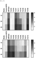
FIG. 7 shows heat-maps depicting the quantities of extracellular vesicles produced by MTD, and eMTDΔ4 and derivatives thereof in the HEK-293 cell line, as quantified by Bradford solution.
FIG. 8 shows graphs depicting the sizes and quantities of extracellular vesicles produced by MTD, and eMTDΔ4 and derivatives thereof in the HEK-293 cell line, as analyzed by the Nanoparticle Tracking Analysis (NTA) system.
FIG. 9 shows the results of performing acrylamide gel electrophoresis after the TRAIL protein, which is a recombinant protein, was loaded in extracellular vesicles.
FIG. 10 shows the results of performing agarose gel electrophoresis after pUC19 and pEGFP-C1, which are plasmid DNA, were loaded in extracellular vesicles.
FIG. 11 is a schematic diagram of a procedure where plasmid DNA was loaded in extracellular vesicles and isolated therefrom.
FIG. 12 shows images confirming the results by a confocal microscope after doxorubicin, a drug favorably passing through cell membranes, was loaded in extracellular vesicles produced by the treatment of the HeLa cell line with eMTDΔ4.
FIG. 13 shows images confirming the results by a confocal microscope after propidium iodide (PI), a drug poorly passing through cell membranes, was loaded in extracellular vesicles produced by the treatment of the HeLa cell line with eMTDΔ4.
MODE FOR CARRYING OUT THE INVENTION
Hereinafter, the present invention will be described in more detail with reference to exemplary embodiments. These exemplary embodiments are provided only for the purpose of illustrating the present disclosure in more detail, and therefore, according to the purpose of the present disclosure, it would be apparent to a person skilled in the art that these examples are not construed to limit the scope of the present disclosure.
Preparative Example 1: Preparation of Noxa Protein-Derived Peptide and Derivatives Thereof
The mitochondrial targeting domain (MTD) of Noxa protein is shown as SEQ ID NO: 1 on Table 1. For the synthesis of a peptide derived from MTD, a manual Fmoc synthesis method in units of 0.25 mmol was basically used. Specifically, a resin was cleanly washed with dimethylformamide (DMF), and then 10 mL of a 20% piperidine/DMF solution was added to the resin. After stirring for 1 minute, 10 mL of a 20% piperidine/dimethylformamide solution was again added thereto and shaken for 30 minutes. After the resin was again washed with dimethylformamide, no piperidine remained was confirmed through the ninhydrin test (resin turned blue).
The following solutions were prepared for coupling: 1 mmol Fmoc-amino acid, 2.1 ml of 0.45 M 2-(1H-benzotriazol-1-yl)-1,1,3,3-tetramethyluronium hexafluorophosphate, hexafluorophosphate Benzotriazole Tetramethyl Uronium/Hydroxybenzotriazole (HBTU/HOBT, 1 mmol), 348 μL of N,N-diisopropylethylamine (DIEA, 2 mmol).
The prepared solution was added to the resin, and shaken for 30 minutes. After the solution was drained from the resin, the resin was washed with dimethyl formamide. Then, for coupling of amino acids, the solution was again added, and then a coupling step was repeated to synthesize Noxa protein-derived eMTDΔ4 and derivative peptides thereof.
The peptides thus synthesized were analyzed by high-performance liquid chromatography, Instrument: Waters 2690 separations module, Flow rate: 1.0 ml/min, gradient: 0-20% B, 5 minutes; 20-50%, B 20 minutes; 50-80%, B 5 minutes, A; 0.1% TFA water, B; 0.1% TFA acetonitrile, column: waters C18, 5 microns, Detection: 220 nm, purity: 95%) and a mass analyzer (HP 1100 series LC/MSD). The synthesized peptides are shown as SEQ ID NO: 2 to SEQ ID NO: 21 on Table 1.
TABLE 1
SEQ ID NO Name Sequence Note
1 MTD KLLNLISKLF 10aa
2 eMTDΔ4 KLNFRQKLLNLISKLF 16aa
3 TU17 RPARPARGGKLLNLISKLF 19aa
4 R(8):MTD RRRRRRRRKLLNLISKLF 18aa
5 TU103 KLLNLWSLLFGYTIK 15aa
6 TU104 MEWWYLLKLLNLISKLF 17aa
7 TU107 LRSLRDKLLNLISKLF 16aa
8 TU111 GLKSLRKLLNLISKLF 16aa
9 TU114 KWYAKLLNLISKLF 14aa
10 TU124 KLLNLWSLLKGYTIK 15aa
11 TU128 AEYRKLLNLISKLF 14aa
12 TU135 AEYSRKLLNLISKLF 15aa
13 TU146 YWLPLRKLLNLISKLF 16aa
14 TU149 AHFLRKLLNLISKLF 15aa
15 TU151 AFFLRKLLNLISKLF 15aa
16 TU155 QFAQYLRNLISKLF 14aa
17 TU156 KVSIFLKNLISKLF 14aa
18 TU166 KLNFAEFLRNLISKLF 16aa
19 TU168 KLNFRLGLRSLREKLF 16aa
20 TU171 KLNFRQKLARLLTKLF 16aa
21 TU172 LNFKWYSLLNLISKLF 16aa
Example 2: Preparation of Solutions for Extracellular Vesicle Production
For extracellular vesicle production using eMTDΔ4, derived from the mitochondrial targeting domain of Noxa protein, and the derivative peptides thereof, the solutions of Example 1 and Comparative Examples 1 to 18 containing the compositions and contents shown in Table 2 were prepared.
TABLE 2
Na- K- NaPO4 KPO4
(mM) Glucose MOPS NaCl KCl gluconate gluconate Sucrose (pH7.4) (pH7.4)
Example 1 5 10 250.00
Comparative 5 10 145
Example 1
Comparative 5 10 145
Example 2
Comparative 5 10 145
Example 3
Comparative 5 10 145
Example 4
Comparative 5 10 50 95
Example 5
Comparative 5 10 50 95
Example 6
Comparative 5 10 241.37 10
Example 7
Comparative 5 10 232.37 20
Example 8
Comparative 5 10 200.00 58
Example 9
Comparative 5 10 75 91.37 10
Example 10
Comparative 5 10 75 82.75 20
Example 11
Comparative 5 10 75 50.00 58
Example 12
Comparative 5 10 241.37 10
Example 13
Comparative 5 10 232.37 20
Example 14
Comparative 5 10 200.00 58
Example 15
Comparative 5 10 75 91.37 10
Example 16
Comparative 5 10 75 82.75 20
Example 17
Comparative 5 10 75 50.00 58
Example 18
Experimental Example 1: Culture of Cancer Cell Lines
1-1. Preparation of Cell Lines and Reagents
Cancer cell lines were cultured prior to testing the activity to induce extracellular vesicle production by the Noxa protein-derived peptide and the derivatives thereof in Preparative Example 1.
Dulbecco's modified eagle medium (DMEM), RPMI 1640, fatal bovine serum (FBS), trypsin-ethylenediaminetetraacetic acid (trypsin-EDTA), and Hank's balanced salt solution (HBSS) required for culture were purchased from Gibco, and Effectene, which is a transfection reagent, was purchased from Qiagen. HeLa and HEK293 cell lines were purchased from Korean Cell Line Bank (KCLB).
1-2. Cell Culture
HeLa and HEK293 cell lines were subcultured at least three times before use in experiments. The HeLa cell line was cultured using DMEM containing 10% fetal bovine serum (FBS) and the HEK-293 cell line was cultured using RPMI 1640 containing 10% fetal bovine serum, under a gas condition of 5% (v/v) CO2 and a temperature condition of 37° C.
Experimental Example 2: Production of Extracellular Vesicles
2-1. Production of Extracellular Vesicles
After the HeLa cell line was cultured in a 6-well plate, the culture was removed, and then the HeLa cell line was treated with the solution in Example 1.
The control group was not treated with any peptide, and the experiment group was treated with eMTDΔ4 peptide of SEQ ID NO: 2 (final concentration: 20 μM). After 10 minutes, the generation of extracellular vesicles was observed by an optical microscope (bright field), and the observation results are shown in FIG. 1 .
As can be confirmed in FIG. 1 , the bottom of the plate was clean in the control group, but small extracellular vesicle particles were formed and settled on the bottom of the plate in the experiment group.
2-2. Production of Extracellular Vesicles Using Various Solutions
Extracellular vesicle production was carried out by the experimental method of Experimental Example 2-1 using the R(8):MTD peptide of SEQ ID NO: 4 and the solutions of Example 1 and Comparative Examples 1 to 18, and the results are shown in FIGS. 2A to 2S.
As can be confirmed in FIG. 2A, many extracellular vesicle particles were generated and most of the particles were settled on the bottom of the plate.
However, as can be confirmed in FIGS. 2B to 2S, extracellular vesicle particles were not generated, and thus no extracellular vesicles settled on the bottom of the plate could be observed, and only the formation of particles that occurs at the time of apoptosis inside cells was observed.
2-3. Production of Extracellular Vesicles Using Various Sugars
Extracellular vesicle production was carried out by the experimental method of Example 2-1 using the R(8):MTD peptide of SEQ ID NO: 4 and the solutions of Examples 1 to 3 on Table 3, and the results are shown in FIGS. 3A to 3C.
TABLE 3
(mM) Glucose Sucrose Sorbitol MOPS
Example 1 5 250 10
Example 2 255 10
Example 3 5 250 10
As can be confirmed in FIGS. 3A to 3C, even when an excess of glucose or sorbitol, instead of sucrose, was added to the solution, extracellular vesicles were well formed.
Experimental Example 3: Identification of Extracellular Vesicle Morphology
In order to transfect the HSP90 protein, an exosome indicator, into the HeLa cell line, the HeLa cell line was cultured overnight in a 6-well plate, and then treated with a plasmid into which Effectene and pEGFP-HSP90 were cloned, and after 4 hours, the culture was replaced.
The next day, the culture was removed from the HeLa cell line, and the solution of Example 1 was added. A control group was not treated with any peptide, and an experiment group was treated with the eMTDΔ4 peptide of SEQ ID NO: 2 (final concentration: 20 μM). The HSP90 protein was excited using an argon laser of 488 nm. After 10 minutes, the control group and the experiment group were observed using a confocal microscope (Leica TCS SP5 Microsystems), and the results are shown in FIG. 4 .
As can be confirmed in FIG. 4 , the cell morphology of the control group was not damaged and the HSP90 protein was located in the cytoplasm. However, in the experiment group (eMTDΔ4), the cell morphology was damaged and extracellular vesicles containing the HSP90 protein were settled on the bottom of the plate.
The reasoning was determined to be that the HSP90 protein of the cytoplasm was released out of the cell through the cell membrane defects that occurred during the production of extracellular vesicles.
Experimental Example 4: Investigation of Production of Extracellular Vesicles
The HeLa cell line was cultured in a 12-well plate overnight, and in order to identify the CD9 protein, an extracellular vesicle indicator, the HeLa cell line was treated with a plasmid into which Effectene and mEmerald-CD9 were cloned, and after 4 hours, the culture was replaced.
The next day, the culture was removed from the HeLa cell line, and the HeLa cell line was treated with the solution of Example 1. After each HeLa cell line was treated with the eMTDΔ4 peptide (final concentration: 20 μM), a laser having a wavelength of 488 nm was used to excite mEmerald-CD9. Thereafter, the cells were observed for 10 minutes while images were taken every 2 seconds by using a confocal microscope, and the results are shown in FIG. 5A.
In order to identify the CD81 protein, an extracellular vesicle indicator, the same procedure as in the method for observing the CD9 protein was performed except that the HeLa cells were treated with a plasmid into which mCherry-CD81 instead of mEmerald-CD9 was cloned and mCherry-CD81 was excited using a laser having a wavelength of 561 nm, and the results are shown in FIG. 5B.
As can be confirmed in FIGS. 5A and 5B, both CD9 and CD81 proteins were sufficient to observe the formation of extracellular vesicles, and CD9 and CD81 proteins were released into the extracellular matrix. It could be verified that extracellular vesicles were released to the extracellular matrix within a few seconds after generation, although the release procedure could not be specifically identified.
Experimental Example 5: Identification of Extracellular Vesicle Size
The HeLa cell line was cultured in a culture plate, treated with the eMTDΔ4 peptide (final concentration: 20 μM) in combination with the solution of Example 1 and, after 10 minutes, fixed with 4% (v/v) paraformaldehyde.
Thereafter, the cells were observed using an atomic microscope (Surface Imaging Systems, NANO Station II, with a cantilever with a pyramidal-shaped tip, a frequency of 146-236 kHz, a spring constant of 21-98 N/m, a length of 225 nm, and a resistance of 0.01-0.02 Ωcm), and the observation results are shown in FIGS. 6A to 6C.
As can be confirmed in FIG. 6A, extracellular vesicles with various sizes were generated, and the sizes thereof could be measured. The results of measuring the sizes of extracellular vesicles are shown as graphs in FIGS. 6B and 6C.
As can be confirmed in FIGS. 6B and 6C, the sizes of extracellular vesicles were measured to be about 200 nm. These results prove that the extracellular vesicles of the present invention have a size similar to the previously known extracellular vesicle sizes.
Experimental Example 6: Verification of Extracellular Vesicle Output
The 293-HEK cell line was cultured in a 6-well plate at a rate of 2×10⁵ cells/mL, and the next day, the cell line was treated with the solution of Example 1 and a peptide of any one of SEQ ID NOs: 1 to 21. After minutes, the solution was removed, followed by centrifugation at an RCF of 12000, and the extracellular vesicles were quantified using the supernatant by Bradford solution. The results are shown in FIG. 7 and Table 4.
TABLE 4
Amount of proteins contained in
extracellular vesicles (unit: μg/μl)
Item Name 10 μM 20 μM 40 μM
1 Media 1.03 0.75 0.76
(Control)
2 MTD 0.80 1.17 2.87
3 eMTDΔ4 4.93 6.40 7.56
4 TU103 0.75 0.40 0.55
5 TU104 0.62 0.75 1.02
6 TU107 5.58 6.69 7.45
7 TU111 4.17 4.98 6.44
8 TU124 3.30 5.02 6.24
9 TU128 3.86 5.29 7.25
10 TU135 1.81 4.41 6.54
11 TU146 2.01 2.89 3.50
12 TU149 1.71 2.00 2.57
13 TU151 1.51 1.30 1.40
14 TU155 1.56 1.77 2.85
15 TU156 1.17 1.55 3.93
16 TU166 5.77 6.68 8.10
17 TU168 0.77 2.17 3.74
18 TU171 2.64 3.62 5.30
19 TU172 0.83 0.44 0.59
As can be confirmed in FIG. 7 and Table 4, most of the peptides shown in Table 4 well produced extracellular vesicles although there is a difference in output.
In addition, as the final peptide treatment concentration increased from 10 μM to 20 μM and 40 μM, the amount of proteins contained in the extracellular vesicles tended to increase.
Experimental Example 6: Identification of Extracellular Vesicle Size and Quantity
After the 293-HEK cell line was cultured to 90% confluency (about 2×10⁷ cells) in a 10-cm culture dish, the culture was removed, and the cells were treated with the eMTDΔ4 peptide (final concentration: 20 μm), which has been dissolved in 5 ml of the solution of Example 1. After 20 minutes, the solution was removed, followed by centrifugation at an RCF of 12000, and then the size and quantity of extracellular vesicles were measured using a Nanoparticle Tracking Analysis (NTA) system (Nanosight LM10, Malvern Instruments). The results are shown in FIGS. 8A to 8D and Table 5.
TABLE 5
Quantity
Stats: Merged data (nm) (/ml, 50-fold diluted)
eMTDΔ4 Mean 184.5 7.44 × 10⁸ ± 6.51 × 10⁷
Mode 126.3
SD 87.8
D10 111.0
D50 152.6
D90 298.4
MTD Mean 181.1 8.01 × 10⁸ ± 1.75 × 10⁷
Mode 115.6
SD 75.2
D10 113.4
D50 154.9
D90 288.5
TU17 Mean 229.6 7.95 × 10⁸ ± 7.15 × 10⁷
Mode 150.9
SD 104.3
D10 131.6
D50 193.2
D90 407.9
TU114 Mean 231.1 8.75 × 10⁸ ± 9.27 × 10⁷
Mode 182.3
SD 110.2
D10 137.9
D50 191.3
D90 390.5
As can be confirmed in FIGS. 8A to 8D and Table 5, with respect to the size of extracellular vesicles, the mean was 184.5 nm, the mode was 126.3 nm, and the standard deviation (SD) was 87.8 nm when the eMTDΔ peptide was used.
The mean was 181.1 nm, the mode was 115.6 nm, and the standard deviation was 75.2 nm when the MTD peptide was used.
The mean was 229.6 nm, the mode was 150.9 nm, and the standard deviation was 104.3 nm when the TU17 peptide was used.
The mean was 231.1 nm, the mode was 182.3 nm, and the standard deviation was 110.2 nm when the TU114 peptide was used.
It could be therefore verified that when the peptide for promoting extracellular vesicle production of the present invention is used under a specific solution condition, extracellular vesicles with various sizes can be efficiently mass-produced.
Experimental Example 7: Production of Extracellular Vesicles Loading Recombinant Protein
After the 293-HEK cell line was cultured to 90% confluency in a 6-well plate, the culture was removed, and the cell line was treated with the eMTDΔ4 peptide (final concentration: 25 μM) and the recombinant TRAIL, which have been dissolved to concentrations of 25 μM and 2 μg/ml in the solution of Example 1, respectively. The supernatant was collected, and PEG (final concentration: 8%) was added, followed by centrifugation at an RCF of 12000 for 10 minutes, thereby settling extracellular vesicles, and the extracellular vesicles were loaded on the SDS-acrylamide gel to check the TRAIL protein. The results are shown in FIG. 9 and Table 6.
TABLE 6
Treated or not
Cell + + + +
Recombinant TRAIL + + + +
Peptide + + + +
Protein detected or not X X X X X X X
As can be confirmed from FIG. 9 and Table 6, when the peptide for promoting extracellular vesicle production of the present invention was used under a specific solution condition, a recombinant protein externally added during the production of extracellular vesicles can be efficiently loaded in the extracellular vesicles.
Experimental Example 8: Production of Extracellular Vesicles Loading Plasmid DNA
After the 293-HEK cell line was cultured to 90% confluency in a 6-well plate, the culture was removed, and the cell line was treated with the eMTDΔ4 peptide (final concentration: 25 μM) and plasmid DNA (PUC19 3 μg/ml, EGFP-C1 6 μg/ml), which have been dissolved in the solution of Example 1.
1.15 ml of the supernatant was collected and 0.35 ml of Qiagen buffer P3 was added, thereby making a total volume of 1.5 ml, of which 0.75 ml was then passed through the DNA miniprep column (Qiagen), and the passed solution was purified with phenol and precipitated with ethanol.
After 30 μl of DNA bound to the column and 30 μl of DNA precipitated with ethanol were dissolved in water, 10 μl of DNA bound to the column and 10 μl of DNA precipitated with ethanol were subjected to agarose gel electrophoresis, and then stained with EtBr. The results are shown in FIG. 10 .
As can be confirmed in FIG. 10 , the plasmid DNA externally added during the production of extracellular vesicles can be loaded in the extracellular vesicles.
In addition, the plasmid DNA could be again isolated from the extracellular vesicles by the process shown in FIG. 11 .
Therefore, when the peptide for promoting extracellular vesicle production of the present invention is used under a specific solution condition, plasmid DNA can be efficiently loaded in extracellular vesicles.
Experimental Example 9: Production of Extracellular Vesicles Loading Doxorubicin
Doxorubicin, which is a drug passing through cell membranes well, is used to treat cancers, such as breast cancer, bladder cancer, Kaposi's sarcoma, lymphoma, or acute lymphoblastic leukemia. First, the HeLa cell line was cultured in a 12-well plate overnight, then transfected with a plasmid expressing EGFP-HSP90 using effectene, and then the culture was replaced after 4 hours. The next day, the culture was removed from the HeLa cell line to prepare a cell line.
The control group was treated with only doxorubicin without any peptide, and the experiment group was treated with the eMTDΔ4 peptide (final concentration: 25 μM) and doxorubicin (final concentration: 100 μM), which have been dissolved in the solution of Example 1, and then after 10 minutes, the groups were observed by a confocal microscope. The results are shown in FIG. 12 .
As can be confirmed in FIG. 12 , the generated extracellular vesicles contained doxorubicin as well as the HSP90 protein, an exosome indicator.
Experimental Example 10: Production of Extracellular Vesicles Loading Propidium Iodide (PI)
Propidium iodide (PI), which is a drug poorly passing through cell membranes, is a fluorescent substance capable of being used to stain cells. First, the HSP90 protein was transfected into the HeLa cell line to prepare a cell line.
The control group was treated with only PI without any peptide, and the experiment group was treated with the eMTDΔ4 peptide (final concentration: 25 μM) and 5 μg/ml PI, which have been in combination dissolved in the solution of Example 1, and then after 10 minutes, the groups were observed by a confocal microscope. The results are shown in FIG. 13 .
As can be confirmed in FIG. 13 , the generated extracellular vesicles contained PI as well as the HSP90 protein, an exosome indicator.
Therefore, proteins, DNA, various drugs that cannot pass through cell membranes, and the like can be loaded in the extracellular vesicles, which would be advantageously used as a new drug delivery system.

Claims (5)

The invention claimed is:
1. A medium for extracellular vesicle production, the medium comprising: at least one peptide selected from the group consisting of SEQ ID NOs: 2 and 5-15; and a solution containing a sugar.
2. The medium of claim 1, wherein the sugar is at least one selected from the group consisting of glucose, sucrose, and sorbitol.
3. The medium of claim 1, wherein the solution further contains 3-(N-morpholino) propanesulfonic acid (MOPS).
4. A medium for extracellular vesicle production, the medium comprising:
at least one peptide, where the peptide is selected from the group consisting of: peptides comprising an amino acid sequence of SEQ ID NO: 16 to SEQ ID NO: 21; and
a solution containing a sugar.
5. The medium of claim 4, wherein the sugar is at least one selected from the group consisting of glucose, sucrose, and sorbitol.
US17/424,232 2020-09-09 2021-06-03 Composition for promoting extracellular vesicle production containing peptide derived from noxa protein and method for producing extracellular vesicles by using same Active 2043-11-07 US12280148B2 (en)

Applications Claiming Priority (3)

Application Number Priority Date Filing Date Title
KR10-2020-0115145 2020-09-09
KR1020200115145A KR102452657B1 (en) 2020-09-09 2020-09-09 Composition for promoting the production of extracellular vesicles containing a peptide derived from noxa protein and a method for producing extracellular vesicles using the same
PCT/KR2021/006905 WO2022055077A1 (en) 2020-09-09 2021-06-03 Composition for promoting production of extra-cellular vesicles containing peptide derived from noxa protein, and method for producing extra-cellular vesicles using same

Publications (2)

Publication Number Publication Date
US20220304931A1 US20220304931A1 (en) 2022-09-29
US12280148B2 true US12280148B2 (en) 2025-04-22

Family

ID=80630322

Family Applications (1)

Application Number Title Priority Date Filing Date
US17/424,232 Active 2043-11-07 US12280148B2 (en) 2020-09-09 2021-06-03 Composition for promoting extracellular vesicle production containing peptide derived from noxa protein and method for producing extracellular vesicles by using same

Country Status (5)

Country Link
US (1) US12280148B2 (en)
JP (1) JP7615509B2 (en)
KR (1) KR102452657B1 (en)
CN (1) CN116113690A (en)
WO (1) WO2022055077A1 (en)

Families Citing this family (1)

* Cited by examiner, † Cited by third party
Publication number Priority date Publication date Assignee Title
WO2025135893A1 (en) * 2023-12-22 2025-06-26 서울대학교산학협력단 Method for preparing extracellular vesicles derived from glia-like cells differentiated from stem cells, and composition for alleviating or treating nerve damage, comprising same as active ingredient

Citations (7)

* Cited by examiner, † Cited by third party
Publication number Priority date Publication date Assignee Title
WO2010050448A1 (en) 2008-10-28 2010-05-06 中外製薬株式会社 Peptide-containing culture medium for culturing animal cell
JP2015523069A (en) 2012-06-15 2015-08-13 メルク パテント ゲゼルシャフト ミット ベシュレンクテル ハフツングMerck Patent Gesellschaft mit beschraenkter Haftung Method for preparing cell culture medium
US20180052150A1 (en) * 2015-03-23 2018-02-22 Astellas Institute For Regenerative Medicine Improved assays for potency of human retinal pigment epithelium (rpe) cells and photoreceptor progenitors
WO2019124522A1 (en) 2017-12-21 2019-06-27 合同会社みらか中央研究所 Method for stabilizing extracellular vesicles
KR20200088408A (en) 2017-11-16 2020-07-22 보드 오브 리전츠, 더 유니버시티 오브 텍사스 시스템 Method for producing exosome derived from MSC
KR20200093328A (en) 2019-01-28 2020-08-05 조선대학교산학협력단 Cell-killing peptide eMTD derived from Noxa
JP2020115769A (en) 2019-01-22 2020-08-06 東亞合成株式会社 Exosome production method

Patent Citations (9)

* Cited by examiner, † Cited by third party
Publication number Priority date Publication date Assignee Title
WO2010050448A1 (en) 2008-10-28 2010-05-06 中外製薬株式会社 Peptide-containing culture medium for culturing animal cell
JP2015523069A (en) 2012-06-15 2015-08-13 メルク パテント ゲゼルシャフト ミット ベシュレンクテル ハフツングMerck Patent Gesellschaft mit beschraenkter Haftung Method for preparing cell culture medium
US20180052150A1 (en) * 2015-03-23 2018-02-22 Astellas Institute For Regenerative Medicine Improved assays for potency of human retinal pigment epithelium (rpe) cells and photoreceptor progenitors
KR20200088408A (en) 2017-11-16 2020-07-22 보드 오브 리전츠, 더 유니버시티 오브 텍사스 시스템 Method for producing exosome derived from MSC
WO2019124522A1 (en) 2017-12-21 2019-06-27 合同会社みらか中央研究所 Method for stabilizing extracellular vesicles
JP2020115769A (en) 2019-01-22 2020-08-06 東亞合成株式会社 Exosome production method
US11230586B2 (en) 2019-01-22 2022-01-25 Toagosei Co., Ltd Exosome production method
KR20200093328A (en) 2019-01-28 2020-08-05 조선대학교산학협력단 Cell-killing peptide eMTD derived from Noxa
US11248024B2 (en) * 2019-01-28 2022-02-15 Industry-Academic Cooperation Foundation, Chosun University Noxa-derived, cell death-inducing peptide eMTD

Non-Patent Citations (13)

* Cited by examiner, † Cited by third party
Title
Cruickshanks et al., "Lapatinib and obatoclax kill breast cancer cells through reactive oxygen species-dependent endoplasmic reticulum stress," Mol Pharmacol. 82(6):1217-29 (Dec. 2012).
International Search Report and Written Opinion mailed Dec. 14, 2021 for International Application No. PCT/KR2021/006905, Kim et al., "Composition for Promoting Extracellular Vesicle Production Containing Peptide Derived from NOXA Protein and Method for Producing Extracellular Vesicles by Using Same," filed Jun. 3, 2021 (English translation) (19 pages).
Kakarla et al., "Apoptotic cell-derived exosomes: messages from dying cells," Experimental & Molecular Medicine. 52(1):1-6 (Jan. 2020).
Kim et al., "Minimal killing unit of the mitochondrial targeting domain of Noxa," J Pept Sci. 19(8):485-490 (Epub Jun. 2013).
Kim, Jeong-Mi, Thesis: "Characteristics of Exosomes released from caspasedependent cell deaths," Master, Department of Biomedical Sciences, Graduate School of Ajou University (2017) (60 pages).
Kozaki et al., "Effective modification of cell death-inducing intracelluar peptides by means of a photo-cleavable peptide array-based screening system," Journal of Bioscience and Bioengineering, 2017, 124(2): 209-214. (Year: 2017). *
Lim et al., "Rapid production method with increased yield of high-purity extracellular vesicles obtained using extended mitochondrial targeting domain peptide," J Extracell Vesicles. 11(10):e12274 (Oct. 2022) (17 pages).
Nacalai Tesque, Cell Culture Reagents, Medium, pp. 1-6. Accessed Jul. 31, 2024. (Year: 2024). *
Office Action dated Feb. 14, 2024 for Japanese Patent Application No. JP 2023-507310, Kim et al., "Composition for Promoting Extracellular Vesicle Production Containing Peptide Derived from NOXA Protein and Method for Producing Extracellular Vesicles by Using Same," filed Jun. 3, 2021 (English translation) (11 pages).
Office Action dated Jun. 26, 2024 for Japanese Patent Application No. JP 2023-507310, Kim et al., "Composition for Promoting Extracellular Vesicle Production Containing Peptide Derived from NOXA Protein and Method for Producing Extracellular Vesicles by Using Same," filed Jun. 3, 2021 (English translation) (10 pages).
Office Action dated Mar. 30, 2022 for Korean Patent Application No. KR 10-2020-0115145, Kim et al., "Noxa Composition for Promoting the Production of Extracellular Vesicles Containing A Peptide Derived from NOXA Protein and A Method for Producing Extracellular Vesicles Using the Same," filed Sep. 9, 2020 (English translation) (11 pages).
Seo et al., "The cell death-inducing activity of the peptide containing Noxa mitochondrial-targeting domain is associated with calcium release," Cancer Res. 69(21):8356-65 (Nov. 2009).
Sucorse from Millipore Sigma, pp. 1-7. Accessed Aug. 1, 2024. (Year: 2024). *

Also Published As

Publication number Publication date
CN116113690A (en) 2023-05-12
US20220304931A1 (en) 2022-09-29
JP7615509B2 (en) 2025-01-17
JP2023536184A (en) 2023-08-23
KR20220033138A (en) 2022-03-16
KR102452657B1 (en) 2022-10-11
WO2022055077A1 (en) 2022-03-17

Similar Documents

Publication Publication Date Title
Nakase et al. Cell-surface accumulation of flock house virus-derived peptide leads to efficient internalization via macropinocytosis
LaRochelle et al. Fluorescence correlation spectroscopy reveals highly efficient cytosolic delivery of certain penta-arg proteins and stapled peptides
Takeuchi et al. Direct and rapid cytosolic delivery using cell-penetrating peptides mediated by pyrenebutyrate
Magzoub et al. Cell-penetrating peptides: small from inception to application
Tang et al. Direct delivery of functional proteins and enzymes to the cytosol using nanoparticle-stabilized nanocapsules
Young Kim et al. Discovery of a non-cationic cell penetrating peptide derived from membrane-interacting human proteins and its potential as a protein delivery carrier
Arafiles et al. Cell-surface-retained peptide additives for the cytosolic delivery of functional proteins
US9694087B2 (en) Development of novel macromolecule transduction domain with improved cell permeability and method for using same
Jha et al. CyLoP-1: a novel cysteine-rich cell-penetrating peptide for cytosolic delivery of cargoes
IL255415B (en) A production method for an exosome containing a target protein and a method for transferring a target protein to the cytoplasm using the exosome produced by the method
KR101258279B1 (en) Development of the macromolecule transduction domain with improved cell permeability and its applications
JP6368304B2 (en) Functional liposomes useful for delivery of bioactive compounds
Wang et al. Selective pericellular hydrogelation by the overexpression of an enzyme and a membrane receptor
Li et al. Transferrin Receptor Targeted Cellular Delivery of Doxorubicin Via a Reduction-Responsive Peptide-Drug Conjugate: Li et al.
US12280148B2 (en) Composition for promoting extracellular vesicle production containing peptide derived from noxa protein and method for producing extracellular vesicles by using same
Matsubara et al. Selective intracellular delivery of ganglioside GM3-binding peptide through caveolae/raft-mediated endocytosis
Yin et al. Cellular uptake and cytosolic delivery of a cyclic cystine knot scaffold
US8207293B2 (en) Peptides derived from maurocalcine used as vectors for intracellular addressing of molecules of interest
CN107417772B (en) Polypeptide HIP-20 capable of antagonizing RNA binding activity of hnRNPU protein and application thereof
Heckl et al. CNN-Gd3+ enables cell nucleus molecular imaging of prostate cancer cells: The last 600 nm
Im et al. Intracellular and transdermal protein delivery mediated by non-covalent interactions with a synthetic guanidine-rich molecular carrier
Laniel et al. Characterization of PGua4, a guanidinium-rich peptoid that delivers IgGs to the cytosol via macropinocytosis
EP2108698A1 (en) Vascular endothelial cell-binding peptide
US20180153819A1 (en) Nanocapsule utilizing mutant chaperonin complex for system of local drug delivery into cell
Bhat et al. Intracellular Delivery of Peptides and Proteins with an Engineered Membrane Translocation Domain

Legal Events

Date Code Title Description
AS Assignment

Owner name: INDUSTRY-ACADEMIC COOPERATION FOUNDATION, CHOSUN UNIVERSITY, KOREA, REPUBLIC OF

Free format text: ASSIGNMENT OF ASSIGNORS INTEREST;ASSIGNORS:KIM, TAE-HYOUNG;PARK, JUNG HEE;HAN, JI HYE;AND OTHERS;REEL/FRAME:056917/0335

Effective date: 20210712

FEPP Fee payment procedure

Free format text: ENTITY STATUS SET TO UNDISCOUNTED (ORIGINAL EVENT CODE: BIG.); ENTITY STATUS OF PATENT OWNER: SMALL ENTITY

FEPP Fee payment procedure

Free format text: ENTITY STATUS SET TO SMALL (ORIGINAL EVENT CODE: SMAL); ENTITY STATUS OF PATENT OWNER: SMALL ENTITY

STPP Information on status: patent application and granting procedure in general

Free format text: DOCKETED NEW CASE - READY FOR EXAMINATION

AS Assignment

Owner name: EXOCALIBRE THERAPEUTICS CO., LTD., KOREA, REPUBLIC OF

Free format text: ASSIGNMENT OF ASSIGNORS INTEREST;ASSIGNOR:INDUSTRY-ACADEMIC COOPERATION FOUNDATION, CHOSUN UNIVERSITY;REEL/FRAME:062795/0948

Effective date: 20221220

STPP Information on status: patent application and granting procedure in general

Free format text: NON FINAL ACTION MAILED

STPP Information on status: patent application and granting procedure in general

Free format text: RESPONSE TO NON-FINAL OFFICE ACTION ENTERED AND FORWARDED TO EXAMINER

STPP Information on status: patent application and granting procedure in general

Free format text: NON FINAL ACTION MAILED

STPP Information on status: patent application and granting procedure in general

Free format text: RESPONSE TO NON-FINAL OFFICE ACTION ENTERED AND FORWARDED TO EXAMINER

STPP Information on status: patent application and granting procedure in general

Free format text: NOTICE OF ALLOWANCE MAILED -- APPLICATION RECEIVED IN OFFICE OF PUBLICATIONS

STCF Information on status: patent grant

Free format text: PATENTED CASE