US12275929B2 - Cell expansion system - Google Patents
Cell expansion system Download PDFInfo
- Publication number
- US12275929B2 US12275929B2 US16/981,050 US201916981050A US12275929B2 US 12275929 B2 US12275929 B2 US 12275929B2 US 201916981050 A US201916981050 A US 201916981050A US 12275929 B2 US12275929 B2 US 12275929B2
- Authority
- US
- United States
- Prior art keywords
- cells
- cell
- mammalian
- solution
- hydrogel
- Prior art date
- Legal status (The legal status is an assumption and is not a legal conclusion. Google has not performed a legal analysis and makes no representation as to the accuracy of the status listed.)
- Active, expires
Links
Images


Classifications
-
- C—CHEMISTRY; METALLURGY
- C12—BIOCHEMISTRY; BEER; SPIRITS; WINE; VINEGAR; MICROBIOLOGY; ENZYMOLOGY; MUTATION OR GENETIC ENGINEERING
- C12M—APPARATUS FOR ENZYMOLOGY OR MICROBIOLOGY; APPARATUS FOR CULTURING MICROORGANISMS FOR PRODUCING BIOMASS, FOR GROWING CELLS OR FOR OBTAINING FERMENTATION OR METABOLIC PRODUCTS, i.e. BIOREACTORS OR FERMENTERS
- C12M41/00—Means for regulation, monitoring, measurement or control, e.g. flow regulation
- C12M41/46—Means for regulation, monitoring, measurement or control, e.g. flow regulation of cellular or enzymatic activity or functionality, e.g. cell viability
-
- C—CHEMISTRY; METALLURGY
- C12—BIOCHEMISTRY; BEER; SPIRITS; WINE; VINEGAR; MICROBIOLOGY; ENZYMOLOGY; MUTATION OR GENETIC ENGINEERING
- C12M—APPARATUS FOR ENZYMOLOGY OR MICROBIOLOGY; APPARATUS FOR CULTURING MICROORGANISMS FOR PRODUCING BIOMASS, FOR GROWING CELLS OR FOR OBTAINING FERMENTATION OR METABOLIC PRODUCTS, i.e. BIOREACTORS OR FERMENTERS
- C12M23/00—Constructional details, e.g. recesses, hinges
- C12M23/20—Material Coatings
-
- C—CHEMISTRY; METALLURGY
- C12—BIOCHEMISTRY; BEER; SPIRITS; WINE; VINEGAR; MICROBIOLOGY; ENZYMOLOGY; MUTATION OR GENETIC ENGINEERING
- C12M—APPARATUS FOR ENZYMOLOGY OR MICROBIOLOGY; APPARATUS FOR CULTURING MICROORGANISMS FOR PRODUCING BIOMASS, FOR GROWING CELLS OR FOR OBTAINING FERMENTATION OR METABOLIC PRODUCTS, i.e. BIOREACTORS OR FERMENTERS
- C12M25/00—Means for supporting, enclosing or fixing the microorganisms, e.g. immunocoatings
- C12M25/14—Scaffolds; Matrices
-
- C—CHEMISTRY; METALLURGY
- C12—BIOCHEMISTRY; BEER; SPIRITS; WINE; VINEGAR; MICROBIOLOGY; ENZYMOLOGY; MUTATION OR GENETIC ENGINEERING
- C12M—APPARATUS FOR ENZYMOLOGY OR MICROBIOLOGY; APPARATUS FOR CULTURING MICROORGANISMS FOR PRODUCING BIOMASS, FOR GROWING CELLS OR FOR OBTAINING FERMENTATION OR METABOLIC PRODUCTS, i.e. BIOREACTORS OR FERMENTERS
- C12M25/00—Means for supporting, enclosing or fixing the microorganisms, e.g. immunocoatings
- C12M25/16—Particles; Beads; Granular material; Encapsulation
-
- C—CHEMISTRY; METALLURGY
- C12—BIOCHEMISTRY; BEER; SPIRITS; WINE; VINEGAR; MICROBIOLOGY; ENZYMOLOGY; MUTATION OR GENETIC ENGINEERING
- C12M—APPARATUS FOR ENZYMOLOGY OR MICROBIOLOGY; APPARATUS FOR CULTURING MICROORGANISMS FOR PRODUCING BIOMASS, FOR GROWING CELLS OR FOR OBTAINING FERMENTATION OR METABOLIC PRODUCTS, i.e. BIOREACTORS OR FERMENTERS
- C12M33/00—Means for introduction, transport, positioning, extraction, harvesting, peeling or sampling of biological material in or from the apparatus
-
- C—CHEMISTRY; METALLURGY
- C12—BIOCHEMISTRY; BEER; SPIRITS; WINE; VINEGAR; MICROBIOLOGY; ENZYMOLOGY; MUTATION OR GENETIC ENGINEERING
- C12N—MICROORGANISMS OR ENZYMES; COMPOSITIONS THEREOF; PROPAGATING, PRESERVING, OR MAINTAINING MICROORGANISMS; MUTATION OR GENETIC ENGINEERING; CULTURE MEDIA
- C12N11/00—Carrier-bound or immobilised enzymes; Carrier-bound or immobilised microbial cells; Preparation thereof
- C12N11/02—Enzymes or microbial cells immobilised on or in an organic carrier
- C12N11/04—Enzymes or microbial cells immobilised on or in an organic carrier entrapped within the carrier, e.g. gel or hollow fibres
-
- C—CHEMISTRY; METALLURGY
- C12—BIOCHEMISTRY; BEER; SPIRITS; WINE; VINEGAR; MICROBIOLOGY; ENZYMOLOGY; MUTATION OR GENETIC ENGINEERING
- C12N—MICROORGANISMS OR ENZYMES; COMPOSITIONS THEREOF; PROPAGATING, PRESERVING, OR MAINTAINING MICROORGANISMS; MUTATION OR GENETIC ENGINEERING; CULTURE MEDIA
- C12N11/00—Carrier-bound or immobilised enzymes; Carrier-bound or immobilised microbial cells; Preparation thereof
- C12N11/02—Enzymes or microbial cells immobilised on or in an organic carrier
- C12N11/08—Enzymes or microbial cells immobilised on or in an organic carrier the carrier being a synthetic polymer
- C12N11/082—Enzymes or microbial cells immobilised on or in an organic carrier the carrier being a synthetic polymer obtained by reactions only involving carbon-to-carbon unsaturated bonds
- C12N11/084—Polymers containing vinyl alcohol units
-
- C—CHEMISTRY; METALLURGY
- C12—BIOCHEMISTRY; BEER; SPIRITS; WINE; VINEGAR; MICROBIOLOGY; ENZYMOLOGY; MUTATION OR GENETIC ENGINEERING
- C12N—MICROORGANISMS OR ENZYMES; COMPOSITIONS THEREOF; PROPAGATING, PRESERVING, OR MAINTAINING MICROORGANISMS; MUTATION OR GENETIC ENGINEERING; CULTURE MEDIA
- C12N11/00—Carrier-bound or immobilised enzymes; Carrier-bound or immobilised microbial cells; Preparation thereof
- C12N11/02—Enzymes or microbial cells immobilised on or in an organic carrier
- C12N11/10—Enzymes or microbial cells immobilised on or in an organic carrier the carrier being a carbohydrate
-
- C—CHEMISTRY; METALLURGY
- C12—BIOCHEMISTRY; BEER; SPIRITS; WINE; VINEGAR; MICROBIOLOGY; ENZYMOLOGY; MUTATION OR GENETIC ENGINEERING
- C12N—MICROORGANISMS OR ENZYMES; COMPOSITIONS THEREOF; PROPAGATING, PRESERVING, OR MAINTAINING MICROORGANISMS; MUTATION OR GENETIC ENGINEERING; CULTURE MEDIA
- C12N5/00—Undifferentiated human, animal or plant cells, e.g. cell lines; Tissues; Cultivation or maintenance thereof; Culture media therefor
- C12N5/0068—General culture methods using substrates
-
- C—CHEMISTRY; METALLURGY
- C12—BIOCHEMISTRY; BEER; SPIRITS; WINE; VINEGAR; MICROBIOLOGY; ENZYMOLOGY; MUTATION OR GENETIC ENGINEERING
- C12N—MICROORGANISMS OR ENZYMES; COMPOSITIONS THEREOF; PROPAGATING, PRESERVING, OR MAINTAINING MICROORGANISMS; MUTATION OR GENETIC ENGINEERING; CULTURE MEDIA
- C12N2500/00—Specific components of cell culture medium
- C12N2500/05—Inorganic components
- C12N2500/10—Metals; Metal chelators
- C12N2500/12—Light metals, i.e. alkali, alkaline earth, Be, Al, Mg
- C12N2500/14—Calcium; Ca chelators; Calcitonin
-
- C—CHEMISTRY; METALLURGY
- C12—BIOCHEMISTRY; BEER; SPIRITS; WINE; VINEGAR; MICROBIOLOGY; ENZYMOLOGY; MUTATION OR GENETIC ENGINEERING
- C12N—MICROORGANISMS OR ENZYMES; COMPOSITIONS THEREOF; PROPAGATING, PRESERVING, OR MAINTAINING MICROORGANISMS; MUTATION OR GENETIC ENGINEERING; CULTURE MEDIA
- C12N2533/00—Supports or coatings for cell culture, characterised by material
- C12N2533/30—Synthetic polymers
- C12N2533/40—Polyhydroxyacids, e.g. polymers of glycolic or lactic acid (PGA, PLA, PLGA); Bioresorbable polymers
-
- C—CHEMISTRY; METALLURGY
- C12—BIOCHEMISTRY; BEER; SPIRITS; WINE; VINEGAR; MICROBIOLOGY; ENZYMOLOGY; MUTATION OR GENETIC ENGINEERING
- C12N—MICROORGANISMS OR ENZYMES; COMPOSITIONS THEREOF; PROPAGATING, PRESERVING, OR MAINTAINING MICROORGANISMS; MUTATION OR GENETIC ENGINEERING; CULTURE MEDIA
- C12N2537/00—Supports and/or coatings for cell culture characterised by physical or chemical treatment
- C12N2537/10—Cross-linking
Definitions
- the present disclosure relates generally to using a cell expansion system for culturing and expanding cells in hydrogel tubes.
- the system allows for expansion of cells in a cost effective and efficient manner.
- the cells for expansion are primary human T cells for adoptive immunotherapy.
- Adoptive immunotherapy refers to the transfer of immune cells (e.g. T lymphocytes) with antitumor activity into a patient to mediate tumor regression.
- immune cells e.g. T lymphocytes
- Basic, translational and clinical studies have shown adoptive immunotherapy to be highly effective for treating many cancers, such as melanoma, cervical cancer, lymphoma, leukemia.
- the cost for manufacturing T cells with current cell culturing technologies is extremely high. For instance, one dose of a recently approved engineered T cells for treating children and young adults with B-cell acute lymphoblastic leukemia costs $475,000.
- TILs tumor infiltrating lymphocytes
- CAR T cells chimeric antigen receptor
- TCR T cells conventional T cell receptor
- T cells are first isolated from the patient through leukapheresis, then activated and engineered to express CARs capable of specifically recognizing tumor cells' surface antigens, and expanded to clinically relevant numbers and infused back to the patient.
- CAR T cells recognizing CD19 antigens have achieved huge success in treating B cell leukemia and lymphomas in clinical studies.
- scientists are currently studying using T cells expressing CARs recognizing other tumor antigens, such as CD138, CD171, CEA, EGFRvIII, and ErbB to treat various solid tumors.
- TCR T cell-based therapy is very similar to CAR T cell therapy except that TCRs recognizing tumor antigens are expressed on the T cell surface.
- TCR T cells specific for NY-ESO1, MART-1 and gp100 antigen have shown excellent anti-tumor responses in patients with melanoma and sarcoma in clinical trials.
- T cells Conventionally, to engineer the T cells, gene expression vectors for TCRs or CARs are delivered to cells with retrovirus, lentivirus and mRNAs through transfection.
- WAVE bioreactor GE Healthcare Life Science
- T cells can grow up to a moderate density ( ⁇ 1 ⁇ 10 7 cells/mL) and up to 25-liter culture volume can be achieved with this technology.
- GE Healthcare Life Science GE Healthcare Life Science
- the present disclosure is generally directed to a cell expansion system and to methods of using the system.
- the system allows for culturing and expanding cells in hydrogel tubes.
- this methodology allows for expansion of cells in a cost effective and efficient manner.
- the present disclosure is directed to a cell expansion system for expanding cells.
- the system comprises: a cap comprising: an extruder comprising at least a first inlet and at least a second inlet, the first inlet operable for introducing a cell solution into the extruder, the second inlet operable for introducing a hydrogel-forming solution into the extruder; and a tubular housing in fluid connection with the extruder of the cap, wherein the tubular housing comprises a cell compatible buffer.
- the present disclosure is directed to a method of expanding cells.
- the method comprises culturing cells in the cell expansion system described above.
- the method includes: extruding the cell solution and the hydrogel-forming solution into a cell compatible solution, the cell compatible solution crosslinking polymers within the hydrogel-forming solution to form hydrogel fibers; suspending the fibers including cells from the cell solution in cell culture medium or cell compatible buffer in the tubular housing; and culturing the cells.
- FIGS. 1 A- 1 E depict T cells cultured in alginate hydrogel tubes (AlgTubes).
- FIGS. 1 A & 1 B show that the setup for processing AlgTubes has two syringe pumps, a custom-made micro-extruder and a CaCl 2 buffer.
- a cell solution and an alginate solution is pumped into the central channel and side channel of the micro-extruder, respectively, to form a coaxial core-shell flow that is extruded through the nozzle into the CaCl 2 buffer.
- the shell alginate flow is crosslinked by Ca 2+ ions to form an alginate hydrogel tube within seconds.
- FIG. 1 C depicts design principles of AlgTubes.
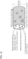
- FIGS. 1 D & 1 E show that in AlgTubes, T cells first associate to form small clusters that subsequently grow and fill the tubes. Scale bar: 200 pm.
- FIG. 1 F depicts live dead cell staining show very few dead T cells in AlgTubes. Scale bar: 200 pm.
- FIG. 1 G is a photograph of the white cell masses in one AlgTube in a 6-well plate. Scale bar: 1 cm.
- FIG. 2 A depicts a schematic of an exemplary process for making hydrogel tubes.
- FIG. 2 B depicts that laboratory set-up of an exemplary device for making hydrogel tubes.
- FIG. 2 C is a close-up view of the extruder component of the device in FIG. 2 B .
- FIG. 3 A depicts a cell expansion system of one embodiment of the present disclosure for use in a large-scale cell production and its components.
- FIG. 3 B depicts a cell expansion system of one embodiment of the present disclosure for use in a large-scale cell production and its components.
- FIG. 4 depicts cells seeded in alginate hydrogel tubes. The first 24 hours, cells form small aggregates, followed by exponential cell growth over the next 9 days. Opacity increases with cell density and at day 10 the cells file the tube, approaching a cell density of (5-10) ⁇ 10 8 cells/mL.
- FIGS. 5 A- 5 C depict a micro-extruder as used in the process of the present disclosure.
- FIG. 5 A is an image of an assembled micro-extruder.
- FIGS. 5 B & 5 C are an illustration ( FIG. 5 B ) and photograph ( FIG. 5 C ) of a micro-extruder with 8 nozzles for simultaneously extruding 8 AlgTubes.
- FIGS. 6 A- 6 D depict screening culture medium and activators for T cell expansion.
- FIGS. 6 A- 6 C are microscopy images of day 2 T cells grown with ImmunoCultTM-XF T cells expansion medium (ImmunoCult) and anti-CD3/CD28 or anti-CD3/CD28/CD2 activators, or with CTSTM OpTmizerTM T cells expansion medium (CTS) and anti-CD3/CD28-Dynabeads activators in static 3D ( FIG. 6 A ), dynamic 3D ( FIG. 6 B ) suspension culturing or AlgTubes ( FIG. 6 C ). Cells were seeded at 1 ⁇ 10 6 cells/ml. Scale bars: 200 ⁇ m.
- FIG. 6 D depict the cumulative cell expansion folds. For static or dynamic 3D suspension culturing, cells were mechanically dissociated and seeded into multiple wells at 1 ⁇ 10 6 cells/ml on day 3, 6, 9 and 12, respectively.
- FIGS. 7 A- 7 F depict processing AlgTubes with different hydrogel shell thickness or diameter.
- FIG. 7 A provides that the equations used to predict the shell thickness are based on the volumetric flow rates of the cell solution and alginate solution and the tube outer diameter.
- FIG. 7 B shows that the experimental shell thickness fits well with the predicted data.
- FIG. 7 C are phase images of T cells in AlgTubes with varied shell thickness (20, 40, and 60 ⁇ m) on day 0. Scale bar: 200 ⁇ m.
- FIG. 7 D are phase images of T cells in AlgTubes with varied diameters (400, 250, and 120 ⁇ m) on day 0 and 14. Scale bar: 200 ⁇ m.
- FIGS. 7 E & 7 F depict expansion folds and volumetric yields on day 14 in AlgTubes with varied shell thickness or diameter.
- FIGS. 8 A- 8 H depict culturing T cells from different donors (#1, #2 and #3) in AlgTubes, static 3D and dynamic 3D suspension culturing.
- FIGS. 8 A- 8 C are microscopy images of T cells from donor #1 grown in AlgTubes ( FIG. 8 A ), static 3D ( FIG. 8 B ) and dynamic 3D ( FIG. 8 C ). Scale bar: 200 ⁇ m.
- FIGS. 8 D- 8 F are photographs of the white cell masses in a 6-well plate. Scale bar: 1 cm.
- FIGS. 8 G & 8 H depict the cell density and cumulative expansion fold on different days of a 14-day culture with AlgTubes, static 3D and dynamic 3D suspension culturing. ***: p ⁇ 0.001.
- FIGS. 9 A- 9 F depict culturing T cells from donor #2 in AlgTubes, static 3D and dynamic 3D suspension culturing.
- FIGS. 9 A- 9 C are microscopy images of T cells from donor #2 grown in AlgTubes ( FIG. 9 A ), static 3D ( FIG. 9 B ) and dynamic 3D ( FIG. 9 C ). Scale bar: 200 ⁇ m.
- FIGS. 9 D- 9 F are photographs of the white cell masses in a 6-well plate. Scale bar: 1 cm.
- FIGS. 10 A- 10 F depict culturing T cells from donor #3 in AlgTubes, static 3D and dynamic 3D suspension culturing.
- FIGS. 10 A- 10 C are microscopy images of T cells from donor #2 grown in AlgTubes ( FIG. 10 A ), static 3D ( FIG. 10 B ) and dynamic 3D ( FIG. 10 C ). Scale bar: 200 ⁇ m.
- FIGS. 10 D- 10 F are photographs of the white cell masses in a 6-well plate. Scale bar: 1 cm.
- FIGS. 11 A- 11 E depict cell death, cell cycle, cytokine release and DNA damage analysis.
- FIG. 11 A depicts the percentage of dead cells (normalized to the initial cells) on day 3, 6, 9, 12 and 14 in AlgTubes, static 3D and dynamic 3D suspension culturing.
- FIG. 11 B depicts the percentage of cells in G1, S and G2/M on day 3 of the 14-day culture as analyzed with propidium iodide staining and flow cytometry.
- FIG. 11 C depicts the percentage of CD3+, CD4+ and CD8+ T cells in the day 14 cells.
- FIG. 11 D shows cytokines in day 14 medium.
- FIG. 11 E depicts the percentage of tail DNA in 138 randomly selected nuclei for each culture condition as quantified using Comet assay. ***: p ⁇ 0.001; *: p ⁇ 0.05.
- FIGS. 12 A- 12 C depict flow cytometry analysis of T cells subtypes in the day 14 products at passage 1 cultured in AlgTubes, static 3D and dynamic 3D. Particularly, FIGS. 12 A- 12 C depict the percentage of CD3+, CD4+ and CD8+ T cells in AlgTubes ( FIG. 12 A ), static 3D ( FIG. 12 B ) and dynamic 3D ( FIG. 12 C ) from donor #1, #2 and #3.
- FIGS. 13 A- 13 C depict DNA damage analysis.
- FIG. 13 A shows that the head and tail of the comet correspond to the intact and broken DNA of a nucleus.
- FIGS. 13 B & 13 C show 30 randomly selected nuclei for each sample.
- FIGS. 14 A- 14 G depict long-term culturing of T cells from donor #1 and #2 in AlgTubes.
- FIG. 14 A are microscopy images of T cells from donor #1 grown in AlgTubes at passage 3. Scale bar: 200 ⁇ m.
- FIG. 14 B is a photograph of the white cell masses in one AlgTube in a 6-well plate. Scale bar: 1 cm.
- FIGS. 14 C & 14 D depict the cell density and expansion fold on day 14 of passage 1, 2 and 3.
- FIG. 14 E depicts the percentage of CD3+, CD4+ and CD8+ T cells at passage 1 and 3.
- FIG. 14 F depict cytokines in day 14 medium of passage 1 and 3.
- FIG. 14 G depicts the cumulative expansion folds.
- FIGS. 15 A- 15 D depict long-term culturing of T cells in AlgTubes.
- FIG. 15 A are microscopy images of T cells from donor #2 grown in AlgTubes at passage 3. Scale bar: 200 ⁇ m.
- FIG. 15 B is a photograph of the white cell masses in one AlgTube in a 6-well plate. Scale bar: 1 cm.
- FIGS. 15 C & 15 D depict flow cytometry analysis of CD3+, CD4+ and CD8+ T cells in AlgTube at passage 3 from donor #1 ( FIG. 15 C ) and #2 ( FIG. 15 D ).
- FIGS. 16 A- 16 F depict automated T cell expansion.
- FIGS. 16 A- 16 C depict the prototype cell expansion system consists of a mechanic stage, a controller, a bellow bottle and a conical tube. Medium was stored in the plastic bellow bottle that could be pressed to flow the medium into, or released to withdraw, the medium from the conical tube. The controller could be programmed for the pressing and releasing speed, as well as the duration of the interval between the pressing and releasing.
- FIGS. 16 D- 16 F depict that on day 1, AlgTubes with T cells were processed into the 50 mL conical tube ( FIG. 16 D ), where cells were expanded for 14 days ( FIG. 16 E ) before harvest through adding EDTA solution ( FIG. 16 F ). The whole process was completed in the closed 50 mL conical tube. Three conical tubes were used to produce T cells from three donors.
- FIGS. 17 A- 17 L depict making human pluripotent stem cells (hPSCs) derived endothelial cells (ECs).
- hPSCs human pluripotent stem cells
- ECs endothelial cells
- FIG. 17 C- 17 G depict that on day 0, single hPSCs mixed with 1.5% HA solution and 1.5% alginate solution were pumped into the central and side channel of the home-made micro-extruder respectively, and extruded into a CaCl 2 buffer (100 mM) ( FIG. 17 C ).
- Cells were cultured in E8 medium for 5 days ( FIG. 17 D ), followed by additional 5 days of EC differentiation medium ( FIG. 17 E ). Medium was continuously perfused.
- alginate hydrogel were dissolved by 0.5 mM EDTA for 5 minutes. Cell masses were pelleted by centrifugation. Cell masses were dissociated into single cells through incubating in Accutase at 37° C. for 10 minutes ( FIG. 17 F ).
- FIGS. 17 H- 17 K depict phase image ( FIG. 17 H ), live/dead staining ( FIG. 17 I ), flow cytometer analysis ( FIG. 17 J ) and immunostaining ( FIG. 17 K ) of day 10 cells.
- Scale bars 200 ⁇ m and 100 ⁇ m, respectively.
- FIG. 17 L shows that when transplanted subcutaneously with a Matrigel matrix, ECs formed nice vascular structures. H9s were used in this Figure. Scale bar, 50 ⁇ m.
- FIGS. 18 A- 18 H depict making human pluripotent stem cells (hPSCs) derived neural stem cells (NSCs).
- FIGS. 18 A & 18 B depict a cell expansion system of the present disclosure consisting of a mechanic stage, a controller, a bellow bottle and a 50 mL conical tube. Medium was stored in the plastic bellow bottle that could be pressed to flow the medium into, or released to withdraw, the medium from the conical tube. The controller could be programmed for the pressing and releasing speed, as well as the duration of the interval between the pressing and releasing. As shown in FIG.
- FIG. 18 D depicts immunostaining of day 12 cells for NSCs markers, PAX6 and NESTIN. Scale bar, 50 ⁇ m.
- FIG. 18 E shows that the cells pulled down by the magnetic anti-SSEA4 beads were positive for OCT3/4 and NANOG. Scale bar, 50 ⁇ m.
- FIGS. 19 A- 19 H depict a cell expansion system for the present disclosure for personalized cell production.
- FIG. 19 A shows that the system consists of a mechanic stage, a controller, a bellow bottle and a 50-mL conical tube.
- FIG. 19 B are pictures showing cell production with the system.
- FIGS. 19 C & 19 D are phase images and live/dead staining of day 11 DA progenitor cells. Scale bars, 200 ⁇ m.
- FIGS. 19 E & 19 F depict immunostaining ( FIG. 19 E ) and flow cytometry analysis ( FIG. 19 F ) of the day 11 cells for DA progenitor markers LMX1A and FOXA2. Scale bars, 100 ⁇ m.
- FIGS. 19 G & 19 H show that 6 weeks post-transplantation, cells survived well and matured into DA neurons. Scale bar, 200 ⁇ m and 100 ⁇ m, respectively.
- the present disclosure is directed to an automatable cell expansion system for expanding cells that can significantly reduce the production time and cost, while increase the production capacity. It is designed to provide cells in a culture microenvironment that has no hydrodynamic stresses in order to produce cells at high yield, high quantity and high quality. It was previously found that culturing human cells under hydrodynamic stresses are highly detrimental to the cells and eliminating these hydrodynamic stresses leads to significant improvement in cell viability, growth rate, yield and quality. High yield can reduce the culture volume, and thus production cost, for each patient. Furthermore, reduction in culture volume allows for the development of a miniature device for automated cell production for large numbers of patients.
- the methods of the present disclosure include processing into and culturing cells (with their activators) in a cell expansion system including microscale hydrogel tubes that are suspended in the cell culture medium in a culture vessel ( FIGS. 1 A & 1 B ).
- the hydrogel tubes create cell-friendly microspaces that allow cells to interact with each other and expand. Meanwhile, the hydrogel tubes protect cells from hydrodynamic stresses in the culture vessel. Further, the hydrogel tubes confine the cell masses (typically, to less than 400 ⁇ m in radial diameter) to ensure efficient mass transport during the entire culture ( FIG. 1 C ).
- the cell expansion systems of the present disclosure are designed to be simple, scalable, defined, reproducible, cost-effective and compatible with the current Good Manufacturing Practices to make it commercially viable.
- Non-limiting examples of cells that can be processed, cultured and expanded using the methods and systems described herein include mammalian cells, insert cells (e.g., drosophila S2 cells), plant cells, yeast cells, and bacterial cells. While described more fully using mammalian cells, especially human T cells, it should be recognized that the methods and systems described herein can be used with any of the above-listed types of cells without departing from the scope of the present disclosure.
- mammalian cells refer to cells derived from both humans and animals. Particularly suitable mammalian cells for use in the methods and systems of the present disclosure include, mammalian embryonic stem cells, mammalian induced pluripotent stem cells, mammalian naive pluripotent stem cells, cells differentiated from mammalian embryonic stem cells, mammalian induced pluripotent stem cells and mammalian naive pluripotent stem cells, mammalian cells reprogrammed from other cell types (e.g.
- human neurons reprogrammed from human fibroblasts include mammalian primary cells (e.g., human umbilical vein endothelial cells, cancer cells, T cells), mammalian tissue stem cells (e.g., mesenchymal stem cells, fetal neural stem cells), and mammalian cell lines (e.g., human embryonic kidney (HEK293) cells, Chinese hamster ovary (CHO) cells).
- mammalian primary cells e.g., human umbilical vein endothelial cells, cancer cells, T cells
- mammalian tissue stem cells e.g., mesenchymal stem cells, fetal neural stem cells
- mammalian cell lines e.g., human embryonic kidney (HEK293) cells, Chinese hamster ovary (CHO) cells.
- Microscale hydrogel tubes are prepared as known in the art.
- the tubes are prepared as hollow fibers prepared from alginate polymer material.
- Suitable alginate polymer material for use in preparing the tubes include any commercially available or home-purified alginate polymer, such as alginate acid or sodium alginate from Sigma (+W201502), and modified alginate polymers, such as methacrylate modified alginate, and combinations thereof.
- “combinations thereof” refer to mixtures of the polymers as well as polymer blends.
- other polymers such as hyaluronic acids can be blended or incorporated into the alginate polymers to dope the alginate hydrogel.
- alginate polymers are first dissolved in water or cell compatible buffer to form alginate solutions including from about 0.01% (w/v) to about 20% (w/v) alginate.
- the tubes are then prepared and filled with cells using an extruder. Extrusion conditions will be those known in the art suitable for the particular cell survival and growth.
- hydrogel tubes While described herein using alginate hydrogel tubes, it should be understood that other hydrogel materials may be suitable for use in making the tubes.
- the hydrogel tubes could be made from materials such as polyethylene glycol, poly(vinyl alcohol), and the like, and combinations thereof.
- a cell solution including cells is supplied via a first inlet 200 and the hydrogel-forming (e.g., alginate) solutions are supplied via at least a second inlet (shown in FIG. 2 A as inlets 202 ).
- Both the first stream including the cell solution and the second stream including the alginate solution are extruded into a cell compatible solution containing calcium ions or other ions or chemicals, such as barium ions, that can crosslink the alginate polymers in the alginate solution.
- the cell compatible solution allows the alginate polymers to instantly crosslink, thereby gelling the alginate solution and forming the tubes.
- the tubes are sufficiently crosslinked in a time period of typically ranging from about one minute to about 30 minutes.
- the tubes will be sized for the particular cells and amount of cell expansion desired.
- the tubes confine the cell masses less than the human tissue diffusion limit (e.g., typically 500 ⁇ m in radial diameter) to ensure efficient mass transport during the entire culture ( FIG. 1 C ).
- the tubes can have a length typically ranging from millimeters to meters.
- the outer and inner diameters of the hydrogel tubes can vary from micrometers to millimeters.
- the cell compatible solution is removed and cell culture medium is added to culture the cells now within the crosslinked alginate hydrogel tubes.
- the fibers, including cells are suspended in cell culture medium in cell culture vessels or bioreactors (an exemplary cell expansion system including a bioreactor having a tubular housing is shown in FIG. 3 A , discussed below).
- the cell culture medium can be any medium known in the cell culture art that is suitable for supporting cell survival, growth, expansion and differentiation.
- the cell culture medium will include, but is not limited to, a carbon source, a nitrogen source, and growth factors.
- the specific cell culture medium for use in culturing the cells within the alginate hydrogel tubes will depend on the cell type to be cultured.
- Cell culture conditions will vary depending on the type of cell, the amount of cell expansion, and the number of cells desired. Once sufficient cell expansion and desired numbers of cells are reached, the cells can be passaged and seeded into new alginate hydrogel tubes for a subsequent round of growth and expansion. Alternatively, the expanded cells can be differentiated into the final desired cell type within the hollow tube.
- the cell expansion system 300 has several unique features.
- the system 300 includes a cap 302 in fluid connection with a tubular housing 308 .
- the cap 302 of the system 300 (detailed in top right of FIG. 3 ) has an extruder 305 including a first inlet 303 operable for introducing a cell solution into the cap and a second inlet 304 operable for introducing a hydrogel-forming solution (e.g., an alginate solution) into the cap.
- the extruder 305 can be made with multiple nozzles (e.g., from 2 to thousands) to simultaneously process multiple hydrogel tubes to scale up the hydrogel tube processing.
- the two solutions are pumped into the extruder and the pumping rates have been calculated by computational fluid dynamics modeling and experimentation. Future users will be provided with exact pumping schedules to control tube dimensions and stability of operation.
- the advantage of the built-in cap is sterility.
- the tubular housing 308 is initially filled with a CaCl 2 solution.
- the housing 308 includes a mesh 310 serving as a support for the alginate tubes 312 .
- the mesh 310 is shown in the axial configuration. This configuration is designed for operating the system in a horizontal position. In the horizontal configuration, the alginate tubes are aligned more or less in the axial direction although it is not critical to have in perfect alignment.
- the mesh is a round disc positioned about one third from the reactor base 320 . The purpose of the mesh is to separate the section of the system that contains the pump inlet/outlet from the alginate tubes.
- Growth medium can be added through: (1) semi-batch, where medium is pumped in and replaced when critical levels for certain growth constituents and/or metabolic waste constituents are triggered, (2) perfusion, where growth medium is continuously pumped through the bioreactor; or (3) pumped in and out of the bioreactor in a continuous cycle referred to as flood/ebb cycles by means of the reciprocating pump.
- This method of growth medium cycling has been specially designed to assure that conditions around the tubes remain as homogeneous as possible and is closely connected to the use of the mesh platform.
- the alginate tubes have neutral buoyancy (which changes slightly as cell density increases) and tubes become suspended in the growth medium during the flood stage and collapse onto the mesh during the ebb stage.
- the advantage of this flood/ebb approach is that the alginate tubes are exposed to more homogeneous conditions, i.e., dead pockets of fluid between tubes are eliminated and variations in bulk conditions are reduced—a prerequisite for homogeneous cell growth, but without damaging the fragile tubes.
- the growth medium is pumped from the bioreactor into the pump vessel, which is equipped with a pH, dO and glucose sensor. If anyone of the sensors detects a value below the set value, then the growth medium is pumped to the spent tank 320 and replaced with fresh medium 322 before the flood/ebb cycle is resumed.
- an imaging port 330 (which is merely a flat section of the wall and made of a high quality material that permits optical transparency in the visible and near infra-red range (quartz is such a material).
- a high resolution endoscope 332 can take images of sections of the tubes, as shown in FIG. 4 (right panel). These images are fed to a neural network (NN), which have been trained to identify cells. The NN provides a cell density. Since this assessment is based on a 2-d projection of the cell density in the tubes, a calibration curve is used to interpret the NN result in terms of cell density (cells/mL). The calibration curve is cell specific.
- the curve is calculated by using experimental results to define key parameters of a cell aggregation-and-growth model.
- the model provides a 3-D mathematical image and a projection of that image onto a 2-D plane is matched with the endoscope image.
- the advantage of the AI imaging is unobtrusive, continuous real-time monitoring of cell density.
- the bioreactor 300 also makes provision for a port 340 through which an optical fiber 342 can be inserted into the bioreactor to monitor the growth medium.
- Raman spectroscopy provides qualitative and quantitative (once calibrated) information of complex molecules like proteins and cytokines. Again, depending on the specifics of the application, Raman spectroscopy can be used to monitor cell viability, apoptosis and the secretion of specific molecules which correlate with a cell fate.
- Cells are finally released from the hollow space of the tubes by dissolving the tubes chemically or physically.
- the tube is dissolved using a chemical dissolvent such as ethylenediaminetetraacetic acid (EDTA), ethylene glycol tetraacetic acid (EGTA), or an alginate lyase solution (available from Sigma-Aldrich).
- EDTA ethylenediaminetetraacetic acid
- EGTA ethylene glycol tetraacetic acid
- alginate lyase solution available from Sigma-Aldrich
- the tube is dissolved using a mechanical force.
- the duration of the cells within the tube can typically vary from days to months.
- the cells are useful in both research laboratories and industry. Small scale and large scale of cells can be manufactured with the system for laboratorial and industrial applications, respectively.
- the cell expansion system of the present disclosure can be used for either scale-up (large production in a single tube) or scale-out (large number of small tubes, each one operated independently from the other).
- the bioreactor will have a typical volume of 50 mL for personalized expansion applications (scale out) and 1 L and more for scale-up applications.
- FIG. 3 B provides an exemplary cell expansion system for scaling up cell production.
- the tube (about 1 liter) can contain about 200 mL of alginate hydrogel tubes to produce ⁇ 1.0 ⁇ 10 11 cells.
- Medium is pumped into and out of the tubular housing of the system. Being modular, this housing can be further scaled up to produce more cells through increasing its length.
- the tubular housing can be vertical or horizontal. The tubular housing diameter can be adjusted based on needs.
- Cells can be efficiently and effectively prepared in size and number for use in degenerative disease and injury treatment, drug screening, for expressing proteins and the like. Additionally, the cells can be used to manufacture proteins and vaccines. In yet other aspects, the cells can be used for tissue engineering.
- primary T cells with their activators e.g. anti-CD3/CD28/CD2 antibodies
- their activators e.g. anti-CD3/CD28/CD2 antibodies
- AlgTubes microscale alginate hydrogel tubes
- the AlgTubes enabled expanding T cells with high cell viability, low DNA damage, high growth rate ( ⁇ 320-fold expansion over 14 days), high purity ( ⁇ 98% CD3+) and high yield ( ⁇ 3.2 ⁇ 108 cells/mL), all offering considerable advantages over current approaches.
- the expanded T cells secreted high levels of T cell cytokines, indicating their normal functions. This system can significantly reduce the manufacturing cost and increase the production capability of T cells to advance the adoptive immunotherapy.
- CD3+ T cells were obtained from Astarte Biologics (donor #1, cat #1017-3503MA17; donor #2, cat #1017-3535AP17; donor #3, cat #1057-3325SE16). CD3+ T cells were grown in ImmunoCultTM-XF T cell expansion medium (cat #10981, StemCell Technologies) with anti-CD3/CD28/CD2 activators (cat #10970, StemCell Technologies) in the presence of 100 IU/ml IL-2. For dynamic 3D culture, the culturing was rocked at 15 rocks/min (rpm).
- the cell aggregates were dissociated by gently pipetting and split into multiple wells at a density of 1.0 ⁇ 10 6 cells/mL on day 3, 6, 9, and 12. Cells were cultured in an incubator with 5% CO 2 , 21% O 2 at 37° C.
- T cells in AlgTubes For a typical cell culture, 40 ⁇ L of cell solution in alginate hydrogel tubes were suspended in 3 mL ImmunoCultTM-XF T cell expansion medium in a 6-well plate and cultured in an incubator with 5% CO 2 , 21% O 2 at 37° C. To passage cells, medium was removed and alginate hydrogel tubes were dissolved with 0.5 mM EDTA for 5 minutes. T cells were collected by centrifuging at 300 g for 5 minutes, and dissociated into single cells by gently pipetting for the next passaging or analysis.
- AKs Adenylate kinases
- T cells were collected and dissociated into single cells and fixed. Cells were stained with the following antibodies (all from Biolegend), PE anti-human CD3 (cat #317308), FITC anti-human CD4 (cat #317408), APC anti-human CD8 (cat #300912), and analyzed with a flow cytometer (Cytek, BD).
- Comet assay were performed with the COMETASSAY® 2 well ES Unit w/Starter kit (cat #4250-050-ESK-PS1, Trevigen) according to the product instructions.
- single cells 1.0 ⁇ 10 5 /ml
- molten LMAgarose at 37° C.
- molten LMAgarose at 37° C.
- CometSlide molten LMAgarose
- the slide was immersed in the Lysis Solution (cat #4250-050-01) overnight at 4° C. to lyse the cells.
- the slide was then immersed in freshly prepared Alkaline Unwinding Solution containing 200 mM NaOH and 1 mM EDTA (pH>13) for 1 hour at 4° C. in the dark. Electrophoresis was then carried out at 21 volts for 30 minutes in Alkaline Electrophoresis Solution containing 200 mM NaOH and 1 mM EDTA (pH>13).
- the slide was gently immersed twice in dH 2 O with 5 minutes each, then in 70% ethanol for 5 minutes.
- the slide was then stained with SYBR® Gold for 30 minutes at room temperature.
- the slides were imaged with a fluorescence microscopy (SYBR® Gold maximum excitation/emission is 496 nm/522 nm).
- the Comet Analysis Software (cat #4260-000-CS) was used to evaluate 138 comets per sample.
- Cytokines analysis Quantibody Human Cytokine Array 1 (QAH-CYT-1-1, RayBiotech) was used to quantify the cytokine secretion in the culture medium according to manufacturer's instruction. The results were analyzed using the RayBiotech Q Analyzer program. In brief, the array chips were blocked with blocking buffer for 30 minutes at room temperature. 100 pl cell culture medium was placed into each well and incubated overnight at 4° C. After extensive washing, the biotin labeled detection antibody was added for 2 hours at room temperature. Cy3 equivalent dye-conjugated streptavidin was then added for 1 hour at room temperature. The array was scanned and analyzed by RayBiotech.
- a micro-extruder was designed and made for processing AlgTubes ( FIGS. 1 A- 1 B and FIGS. 5 A- 5 C ).
- a solution containing single T cells, T cell activators (e.g. anti-CD3/CD28/CD2 antibodies) and 2% hyaluronic acid polymer, and a solution containing 1.5% alginate polymer was pumped into the central channel and side channel of the micro-extruder, respectively, to form a coaxial core-shell flow that was extruded into a CaCl 2 buffer.
- the shell alginate flow was instantly crosslinked by Ca 2+ ions to form an alginate hydrogel tube ( FIGS. 1 A- 1 C ).
- the CaCl 2 buffer was replaced by the T cell culture medium and cells were grown in the tubes.
- the cell solution and alginate solution should have close viscosity to process defect-free AlgTubes. Both hyaluronic acid (HA) and methylcellulose (MC) solutions could be used to suspend cells for this purpose.
- HA hyaluronic acid
- MC methylcellulose
- single T cells first associated to form small cell clusters that subsequently grew and filled the tubes ( FIGS. 1 D, 1 E & 1 G ). T cells in AlgTubes had very high viability during the entire culture as shown by the undetectable dead cells through live/dead staining ( FIG. 1 F ).
- the AlgTubes could be dissolved with the cell-compatible ethylenediaminetetraacetic acid (EDTA) solution (0.5 mM, 5 minutes at room temperature) to release the micro cell masses that could be dissociated into single cells by gently mechanical pipetting for the following analysis or passage.
- EDTA cell-compatible ethylenediaminetetraacetic acid
- a few culturing medium and activators have been successfully used to expand T cells in the literature. These include the combination of using magnetic nanoparticles coated with anti-CD3/CD28 antibodies (Dynabeads CD3/CD28, Invitrogen) as activators and CTSTM OpTmizerTM T Cell Expansion SFM medium (Invitrogen) as the culture medium, or the tetrameric anti-CD3/CD28 antibodies or tetrameric anti-CD3/CD28/CD2 antibodies (Stem Cell Technology) as activators and ImmunoCultTM-XF T Cell Expansion medium (Stem Cell Technology) as the culture medium. Initially, these medium and activators were directed compared in order to find out the best combination for culturing T cells ( FIGS.
- T cells were also cultured in static three dimensional (3D) suspension culturing (i.e. static 3D, in which cells were suspended in culture medium without agitation) and dynamic 3D suspension (i.e. dynamic 3D, in which cells were suspended in culture medium with gentle rocking) for comparison.
- 3D three dimensional
- dynamic 3D suspension i.e. dynamic 3D, in which cells were suspended in culture medium with gentle rocking
- T cells were seeded at 1.0 ⁇ 10 6 cells/mL for all three methods and cultured for 14 days.
- T cells grew as single cells or small clusters (e.g.
- T cells grew as both single cells and large non-spherical aggregates ( FIG. 6 A ).
- T cells grew as aggregates under all the three culturing conditions. Additionally, these aggregates were much larger than these in static 3D culturing ( FIG. 6 B ).
- T cells first formed small clusters (e.g.
- FIG. 6 C T cells expanded ⁇ 55, ⁇ 28, ⁇ 250 folds in static 3D, dynamic 3D and AlgTubes. There was no significant difference between different culture medium and activators ( FIG. 6 D ) ImmunoCult and anti-CD3/CD28/CD2 activators were used for the rest of this Example.
- the AlgTubes' diameter and hydrogel shell thickness could be precisely controlled through adjusting the nozzle diameter of the micro-extruder, the flow rates of the cell solution and alginate solution ( FIGS. 7 A & 7 B ).
- the relationship between the tube' inner and outer diameter, shell thickness, volumetric flow rate of the cell solution and alginate solution, and the length of tube processed per unit time can be described with the equations shown in FIG. 7 A .
- the tube's outer diameter and the extruder nozzle's inner diameter was roughly equal.
- T cells were shown to have similar morphologies, viability, growth rate and yield in tubes with shell thickness of 60, 40 and 20 ⁇ m or tubes with diameter of 400 ⁇ m, 250 ⁇ m and 120 ⁇ m ( FIGS. 7 C- 7 F ). It was concluded that AlgTubes with shell thickness ⁇ 60 ⁇ m and outer diameter ⁇ 400 ⁇ m were appropriate for T cell expansion.
- T cells were cultured from three donors in parallel (Table 1). Cells were also cultured in static 3D and dynamic 3D for comparison. T cells were seeded at 1.0 ⁇ 10 6 cells/mL. In AlgTubes, T cells were continuously cultured for 14 days without passaging or splitting. T cells first formed small clusters (e.g. within the first 24 hours) that subsequently grew and filled the tubes, producing mono-dispersed (in radial direction) fibrous cell masses on day 14 ( FIGS. 8 A & 8 D , FIGS. 9 A & 9 D , and FIGS.
- T cells expanded about 51, 123, 320-fold to yield around 0.51 ⁇ , 1.2 ⁇ , 3.2 ⁇ 10 8 cells/mL on day 6, 9 and 14, respectively ( FIGS. 8 G & 8 H ). There was minimal difference between the three donors ( FIGS. 8 A- 8 H, 9 A- 9 F, and 10 A- 10 F ).
- T cells quickly aggregated and grew as spherical cell aggregates with diameter between 100 to 800 pm ( FIGS. 8 B & 8 E , FIGS. 9 B & 9 E , and FIGS. 10 B & 10 E ).
- the aggregates were mechanically dissociated into small clusters on day 3, 6, 9 and 12 and split into multiple samples (e.g. seeded at 1.0 ⁇ 10 6 cells/mL after splitting) in order to increase the growth rate.
- T cells cumulatively expanded about 10, 35, 55-fold on day 6, 9 and 14, respectively ( FIG. 8 H ).
- the maximal cell density could be achieved was about 3.5 ⁇ 10 6 cells/mL ( FIG. 8 G ).
- T cells severely aggregated or agglomerated in dynamic 3D culturing ( FIGS. 8 C & 8 F , FIGS. 9 C & 9 F , and FIGS. 10 C & 10 F ).
- the aggregates were mechanically dissociated into small clusters on day 3, 6, 9 and 12 and split into multiple samples (e.g. seeded at 1.0 ⁇ 10 6 cells/mL after splitting).
- T cells expanded cumulatively about 7, 17, 28-fold on day 6, 9 and 14, respectively ( FIG. 8 H ).
- the maximal cell density could be achieved was about 2.5 ⁇ 10 6 cells/mL ( FIG. 8 G ).
- There was minimal difference between the three donors FIGS. 8 A- 8 H, 9 A- 9 F, and 10 A- 10 F ).
- the typical T cell subtypes after the 14-day culture were analyzed using immunostaining and flow cytometry ( FIG. 11 C and FIGS. 12 A- 12 C ).
- the percentage of CD3+ T cells in AlgTubes and static 3D culturing were very similar to the original uncultured cells (e.g. 98%, 99% and 99% for donor #1, #2, and #3, respectively).
- the dynamic 3D culturing reduced the CD3+ T cells to 83%, 85% and 61% for donor #1, #2 and #3, respectively.
- the percentage of CD4+ T cells in AlgTubes were close to the original uncultured cells (e.g.
- the dynamic 3D culturing reduced the CD4+ T cells to 59%, 67% and 68% for donor #1, #2 and #3, respectively, and the static 3D culturing resulted in only 32%, 22%, and 18% CD4+ T cells for the three donors.
- the percentage of CD8+ T cells in AlgTubes were close to the original uncultured cells (e.g. 16%, 29%, 22% for donor #1, #2, and #3, respectively).
- T cells from all culture methods showed a typical T cell cytokine profile characterized by the high production of interleukin-2 (IL-2), interleukin-4 (IL-4), interferon-y (IFN-y) and tumor necrosis factor- ⁇ (TNF- ⁇ ).
- IL-2 interleukin-2
- IL-4 interleukin-4
- IFN-y interferon-y
- TNF- ⁇ tumor necrosis factor- ⁇
- the comet assay (single-cell gel electrophoresis) is a simple method for measuring DNA strand breaks in eukaryotic cells. Single T cells were embedded in agarose hydrogel on a microscope slide, and were lysed with detergent and high salt to form nucleoids containing supercoiled loops of DNA. Electrophoresis resulted in structures resembling comets, which were recorded by fluorescence microscopy. The intensity of the comet tail relative to the head is proportional to the number of DNA breaks.
- T cells cultured in AlgTubes had significantly less DNA breaks than cells from dynamic 3D culturing ( FIG. 11 E and FIGS. 13 A- 13 C ), indicating the cell-friendly AlgTubes can improve the genetic stability of cultured T cells.
- T cells could be cultured in AlgTubes for long terms was also evaluated.
- T cells were cultured for 3 passages, total of 42 days in AlgTubes ( FIGS. 14 A- 14 G and FIGS. 15 A- 15 DF ).
- T cells at passage 3 had very similar morphology, viability, cell growth rate, yield, subtype distribution and cytokine releasing to these at passage 1. The results show prolonged culture can be performed with AlgTubes if large numbers of T cells are needed.
- FIG. 16 A A prototype device for automated T cell production ( FIG. 16 A ) was built. On day 1, three milliliters of AlgTubes containing T cells from each donor were processed and contained in one closed 50 mL conical tube ( FIG. 16 D ), where T cells were expanded for 14 days ( FIG. 16 E ). On day 14, EDTA solution was pumped into the conical tube to dissolve the AlgTubes and the cell mass was collected through mild centrifugation (e.g. 100 g for 2 minutes) for downstream application ( FIG. 16 F ). During the 14-day culture, the cell culture medium was stored in a plastic bellow bottle that could be pressed to flow the medium into the conical tube or released to withdraw the medium from the conical tube, respectively ( FIGS.
- hPSCs human pluripotent stem cells
- MSCs human mesenchymal stem cells
- Agitation (or shaking or rocking, typically in the range of 75 to 120 rpm) is usually used to enhance the mass transport and reduce cell agglomeration in 3D suspension culturing.
- agitation cannot eliminate cell agglomeration.
- agitation generates complicated hydrodynamic conditions including the medium flow direction, velocity, shear force, and chemical environment. These conditions vary spatially and temporally, resulting in locations (e.g. close to the vessel wall) with critical stresses that induce cell death and phenotype changes, low cell viability, growth, and yield.
- the hydrodynamic conditions in a bioreactor are sensitive to many factors including the impeller geometry, size and position, the bioreactor geometry and size, the positions of probes for pH, temperature and oxygen, the medium viscosity, and the agitation rate. They are currently not well understood and are hard to control. Additionally, how different types of cells respond to the hydrodynamic conditions is not well known and is hard to study.
- the AlgTubes are designed to simultaneously eliminate the cell agglomeration and hydrodynamic stresses.
- the AlgTubes produce mono-dispersed (in radial diameter) cell masses that can be precisely controlled in any range between 100 ⁇ m to 400 ⁇ m. This can ensure efficient mass transport to all cells ( FIGS. 7 A- 7 F ).
- cells in AlgTubes are protected from hydrodynamic stresses by the hydrogel shells. This reduces the hydrodynamic-conditions-induced negative effects. The protection from the AlgTubes, scaling up the culture volume, did not change the cell grow rate and automating the production could be readily achieved.
- the tubes provide free space for cells to interact with each other and expand, leading to extremely high volumetric yield, which is about 30-fold of the current state-of-art ( FIGS. 8 G & 8 H ).
- alginates for processing the tubes makes this technology scalable, cost-effective, Good Manufacture Practice (GMP)-compatible and commercially viable.
- GMP compliance is required for producing therapeutic cells by regulatory agencies (e.g., FDA).
- High quality and quantity alginates are available and affordable.
- Alginates are non-toxic to cells and have been used in clinics. They can be instantly crosslinked to process large-scale AlgTubes.
- the resulting hydrogel tubes are mechanically and chemically stable and suitable for culturing cells in large-scale and for long-term. Additionally, the tubes can be dissolved easily with cell-compatible EDTA solution to release the product, and are transparent so that the cell growth can be monitored with microcopies.
- T cells could be cultured with much higher expansion and yield than other culture methods ( FIGS. 8 G & 8 H ).
- T cells expanded cumulatively 320, 55, 28 fold in a 14-day culture in AlgTubes, static 3D and dynamic 3D culturing, respectively ( FIG. 8 H ).
- the maximal volumetric yield was 3.2 ⁇ 10 8 cells/mL, 3.5 ⁇ 10 6 cells/mL, 2.5 ⁇ 10 6 cells/mL in AlgTubes, static 3D and dynamic 3D culturing, respectively ( FIG. 8 G ).
- the high expansion rate and yield have large impacts on T cell production. For instance, T cells required for one patient (e.g.
- T cells cultured in AlgTubes had much less phenotype changing or subtype enrichment ( FIG. 11 C ). Phenotype changing can lead to large variations in the product efficacy and potency. T cells in AlgTubes had minimal DNA damages ( FIG. 11 E ), indicating better product safety and quality.
- T cells could be cultured for long-term to generate more cells ( FIGS. 14 A- 14 G ).
- the AlgTubes will significantly advance the adoptive immunotherapy and will be of broad interest to individual laboratories, institutions, and biotechnology companies working on adoptive immunotherapy.
- a cell expansion system was designed for scalable endothelial cells (ECs) production.
- hPSCs solution in AlgTube were suspended in a 50 mL conical culture tube with septum cap.
- hPSCs were cultured in E8 medium with 5% CO 2 , 21% O 2 at 37° C. for 5 days.
- E8 medium was removed and replaced with EC differentiation medium for 3 days, followed by ECs induction medium for 2 days.
- medium was stored in a bellow bottle that was periodically pressed to flow the medium into, or released to withdraw, the medium from, the 50 mL culture tube.
- hydrogel tubes were dissolved by adding 0.5 mM EDTA buffer. Cell masses were pelleted by centrifugation.
- Cell masses were dissociated into single cells through incubating in Accutase at 37° C. for 10 minutes. Magnetic beads coated with anti-SSEA4 antibodies were added to pull down the undifferentiated SSEA4+ hPSCs with a magnetic cell separator. The supernatant was transferred into a new tube. Cells were pelleted by spinning at 300 g for 5 minutes and transported to the surgery room for injection.
- FIGS. 17 A & 17 B an exemplary cell expansion system for scalable ECs production was designed ( FIGS. 17 A & 17 B ).
- single hPSCs mixed with 1.5% HA solution and 1.5% alginate solution were pumped into the central and side channel of the home-made micro-extruder respectively, and extruded into a CaCl 2 buffer (100 mM).
- Cells were cultured in E8 medium for 5 days, followed by additional 5 days of ECs differentiation medium.
- alginate hydrogel were dissolved by adding 0.5 mM EDTA solution for 5 minutes. Cell masses were pelleted by spinning the tube at 100 g for 3 minutes.
- FIGS. 17 C- 17 G Phase image and live/dead cell staining showed few dead cells ( FIGS. 17 H & 17 I ).
- Flow cytometer analysis and immunostaining showed 82.6% of the day 10 cells were ECs ( FIGS. 17 J & 17 K ).
- ECs formed nice vascular structures ( FIG. 17 L ).
- NSCs neural stem cells
- hPSCs solution in AlgTube were suspended in a 50 mL conical culture tube with septum cap.
- hPSCs were cultured in E8 medium with 5% CO 2 , 21% O 2 at 37° C. for 5 days. Medium was stored in a bellow bottle that was periodically pressed to flow the medium into, or released to withdraw, the medium from, the 50 mL culture tube. E8 medium was removed and replaced with neural induction medium for 7 days.
- hydrogel tubes were dissolved by adding 0.5 mM EDTA buffer. Cell masses were pelleted by centrifugation. Cell masses were dissociated into single cells through incubating in Accutase at 37° C. for 10 minutes. Magnetic beads coated with anti-SSEA4 antibodies were added to pull down the undifferentiated SSEA4+ hPSCs with a magnetic cell separator. The supernatant was transferred into a new tube. Cells were pelleted
- FIGS. 18 A & 18 B an exemplary cell expansion system for scalable NSCs production was designed ( FIGS. 18 A & 18 B ).
- single hPSCs mixed with 1.5% HA solution and 1.5% alginate solution were pumped into the central and side channel of the home-made micro-extruder respectively, and extruded into a CaCl 2 buffer (100 mM).
- Cells were cultured in E8 medium for 5 days, followed by additional 7 days of NSCs induction medium.
- alginate hydrogel were dissolved by adding 0.5 mM EDTA solution for 5 minutes. Cell masses were pelleted by spinning the tube at 100 g for 3 minutes.
- Purified NSCs were injected into the striatum of Sprague dawley rats with a stereotactic injector. 7 days post-transplantation, substantial numbers of human unclear antigen positive cells were found in the rat brain, and 30 days after transplantation, large numbers of HuNu+ and TUJ-1+ cells were found in the rat brain ( FIG. 18 H ).
- an exemplary cell expansion system was designed for personalized cell production.
- reprogramming factors hOSKUL+EGFP
- ⁇ 2 ⁇ 10 7 cells/mL hydrogel were processed into AlgTubes into a closed 50-mL conical tube.
- Cells were reprogrammed for 20 days, expanded for 10 days and differentiated into DA progenitors for 11 days.
- 0.5 mM EDTA was infused to dissolve the tubes.
- Accutase was then infused to dissociate the fibrous cell mass into single cells.
- Magnetic beads coated with anti-SSEA4 antibodies were then added into the tube to pull down the undifferentiated SSEA4+ iPSCs with a magnetic cell separator.
- 3 ⁇ 10 5 cells suspended in 4 ⁇ l PBS were injected into the striatum (AP+0.5 mm; ML ⁇ 3.0 mm; DV ⁇ 6 mm) at 0.5 ⁇ l/minute using a 10 ⁇ l Hamilton syringe (Hamilton Company, USA) with a stereotaxic frame (RWD Life Science Inc.).
- rats were anesthetized with ketamine/xylazine and perfused with PBS followed by 4% paraformaldehyde.
- the brain was serially sectioned (40 ⁇ m in thickness) with a Leica cryosection machine, and free-floating ections were stained with antibodies.
- the system consists of a mechanic stage, a controller, a bellow bottle and a 50-mL conical tube ( FIG. 19 A ).
- Medium was stored in the plastic bellow bottle that could be pressed to flow the medium into, or released to withdraw, the medium from the conical tube.
- the controller could be programmed for the pressing and releasing speed, as well as the duration of the interval between the pressing and releasing ( FIG. 19 A ). Since AlgTubes have similar density with the cell culture medium, they were uniformly suspended and dispersed in the medium when the medium was pumped into the conical tube. They became collected and contacted with each other when the medium was withdrawn from the conical tube.
- This periodic dispersion and collection of AlgTubes was designed to enhance the medium mixing.
- reprogramming factors were delivered to fibroblasts through electroporation and cells were processed into AlgTubes into the closed 50-mL conical tube. Cells were reprogrammed for 20 days, expanded for 10 days and differentiated into DA progenitors for 11 days.
- 0.5 mM EDTA was infused into the conical tube to dissolve AlgTubes.
- Accutase was then infused to digest the cell mass into single cells.
- Magnetic beads coated with anti-SSEA4 antibodies were then added into the conical tube to pull down the undifferentiated SSEA4+ iPSCs with a magnetic cell separator.
- FIG. 19 B Purified cells were transplanted into brains of Sprague dawley rats with a stereotactic injector ( FIG. 19 B ). Very few cell deaths occurred during the production ( FIGS. 19 C & 19 D ). ⁇ 90% produced cells were LMX1A+/FOXA2+ ( FIGS. 19 E & 19 F ). 6 weeks post-transplantation, these cells survived well by HuNu staining ( FIG. 19 G ). A large percentage of the cells matured into TH+ DA neurons ( FIG. 19 H ).
Landscapes
- Health & Medical Sciences (AREA)
- Life Sciences & Earth Sciences (AREA)
- Chemical & Material Sciences (AREA)
- Engineering & Computer Science (AREA)
- Organic Chemistry (AREA)
- Bioinformatics & Cheminformatics (AREA)
- Wood Science & Technology (AREA)
- Zoology (AREA)
- Genetics & Genomics (AREA)
- Biomedical Technology (AREA)
- Biotechnology (AREA)
- Biochemistry (AREA)
- Microbiology (AREA)
- General Engineering & Computer Science (AREA)
- General Health & Medical Sciences (AREA)
- Sustainable Development (AREA)
- Immunology (AREA)
- Cell Biology (AREA)
- Molecular Biology (AREA)
- Analytical Chemistry (AREA)
- Clinical Laboratory Science (AREA)
- Dispersion Chemistry (AREA)
- Micro-Organisms Or Cultivation Processes Thereof (AREA)
- Apparatus Associated With Microorganisms And Enzymes (AREA)
Abstract
Description
| TABLE 1 |
| Donor Information |
| Weight | ABO | ||||||||||
| Donor # | Supplier | Age | Gender | Race | Height | (lbs) | type | | CD4+ | CD8+ | |
| 1 | Astarte | 41 | Female | Caucasian | 5′6″ | 208 | A | 98.2% | 76.6% | 19.3 |
| Biologics | ||||||||||
| 2 | Astarte | 27 | Female | Caucasian | 5′5″ | 200 | A | 99.0% | 62.5% | 33.6 |
| Biologics | ||||||||||
| 3 | |
20 | Male | Caucasian | 5′6″ | 145 | O | 99.2% | 70.4% | 26.5% |
| Biologics | ||||||||||
Claims (19)
Priority Applications (1)
| Application Number | Priority Date | Filing Date | Title |
|---|---|---|---|
| US16/981,050 US12275929B2 (en) | 2018-03-16 | 2019-03-15 | Cell expansion system |
Applications Claiming Priority (3)
| Application Number | Priority Date | Filing Date | Title |
|---|---|---|---|
| US201862643894P | 2018-03-16 | 2018-03-16 | |
| PCT/US2019/022594 WO2019178549A1 (en) | 2018-03-16 | 2019-03-15 | Cell expansion system |
| US16/981,050 US12275929B2 (en) | 2018-03-16 | 2019-03-15 | Cell expansion system |
Publications (2)
| Publication Number | Publication Date |
|---|---|
| US20210017485A1 US20210017485A1 (en) | 2021-01-21 |
| US12275929B2 true US12275929B2 (en) | 2025-04-15 |
Family
ID=67906933
Family Applications (1)
| Application Number | Title | Priority Date | Filing Date |
|---|---|---|---|
| US16/981,050 Active 2042-02-10 US12275929B2 (en) | 2018-03-16 | 2019-03-15 | Cell expansion system |
Country Status (7)
| Country | Link |
|---|---|
| US (1) | US12275929B2 (en) |
| EP (1) | EP3765590A4 (en) |
| JP (1) | JP2021515581A (en) |
| CN (1) | CN112119150A (en) |
| AU (1) | AU2019236287A1 (en) |
| CA (1) | CA3096742A1 (en) |
| WO (1) | WO2019178549A1 (en) |
Families Citing this family (5)
| Publication number | Priority date | Publication date | Assignee | Title |
|---|---|---|---|---|
| CN113029736B (en) * | 2021-03-08 | 2023-01-03 | 中国科学院苏州生物医学工程技术研究所 | Single cell extraction method and device based on near-infrared response hydrogel |
| US12577524B1 (en) * | 2022-02-04 | 2026-03-17 | CellGro Technologies, LLC | Multi-axial multi-port extruder for cell culture |
| EP4328296A1 (en) | 2022-08-24 | 2024-02-28 | Mirai Foods AG | Method for operating a bioreactor for cultivated meat and corresponding bioreactor |
| JPWO2024116831A1 (en) * | 2022-11-29 | 2024-06-06 | ||
| TW202515993A (en) | 2023-06-26 | 2025-04-16 | 日商細胞纖維股份有限公司 | Manufacturing method of cell capsules, cell groups or manufacturing method, soution and cell groups of cell products |
Citations (9)
| Publication number | Priority date | Publication date | Assignee | Title |
|---|---|---|---|---|
| US5175093A (en) | 1989-11-07 | 1992-12-29 | Lehigh University | Bioactive cells immobilized in alginate beads containing voids formed with polyethylene glycol |
| US5264359A (en) | 1988-04-18 | 1993-11-23 | Nitta Gelatin Inc. | Methods for large-scale cultivation of animal cells and for making supporting substrata for the cultivation |
| US5387522A (en) | 1984-06-01 | 1995-02-07 | Schering Corporation | Apparatus having a biphasic spray head for entrapping biological material in a hydrophilic gel |
| CN101130751A (en) * | 2007-07-26 | 2008-02-27 | 上海交通大学 | Plant cell stirred bioreactor continuous perfusion culture device |
| US20130189723A1 (en) | 2003-07-17 | 2013-07-25 | Global Cell Solutions, Llc | Automated cell culture system and process |
| US20130206673A1 (en) * | 2010-10-25 | 2013-08-15 | Jackie Y. Ying | Tubular fiber membrane with nanoporous skin |
| WO2017091662A1 (en) | 2015-11-25 | 2017-06-01 | Nutech Ventures | Large scale cell manufacture system |
| US20170239191A1 (en) | 2008-06-06 | 2017-08-24 | Boehringer Ingelheim International Gmbh | Pharmaceutical dosage form for immediate release of an indolinone derivative |
| JP2017217323A (en) * | 2016-06-09 | 2017-12-14 | 国立大学法人 鹿児島大学 | Method for producing microchannel structure having capillary network-like microchannel |
Family Cites Families (1)
| Publication number | Priority date | Publication date | Assignee | Title |
|---|---|---|---|---|
| WO2012167223A1 (en) * | 2011-06-02 | 2012-12-06 | Massachusetts Institute Of Technology | Modified alginates for cell encapsulation and cell therapy |
-
2019
- 2019-03-15 JP JP2020549552A patent/JP2021515581A/en active Pending
- 2019-03-15 CA CA3096742A patent/CA3096742A1/en not_active Abandoned
- 2019-03-15 AU AU2019236287A patent/AU2019236287A1/en not_active Abandoned
- 2019-03-15 CN CN201980032722.6A patent/CN112119150A/en active Pending
- 2019-03-15 WO PCT/US2019/022594 patent/WO2019178549A1/en not_active Ceased
- 2019-03-15 EP EP19767990.5A patent/EP3765590A4/en not_active Withdrawn
- 2019-03-15 US US16/981,050 patent/US12275929B2/en active Active
Patent Citations (9)
| Publication number | Priority date | Publication date | Assignee | Title |
|---|---|---|---|---|
| US5387522A (en) | 1984-06-01 | 1995-02-07 | Schering Corporation | Apparatus having a biphasic spray head for entrapping biological material in a hydrophilic gel |
| US5264359A (en) | 1988-04-18 | 1993-11-23 | Nitta Gelatin Inc. | Methods for large-scale cultivation of animal cells and for making supporting substrata for the cultivation |
| US5175093A (en) | 1989-11-07 | 1992-12-29 | Lehigh University | Bioactive cells immobilized in alginate beads containing voids formed with polyethylene glycol |
| US20130189723A1 (en) | 2003-07-17 | 2013-07-25 | Global Cell Solutions, Llc | Automated cell culture system and process |
| CN101130751A (en) * | 2007-07-26 | 2008-02-27 | 上海交通大学 | Plant cell stirred bioreactor continuous perfusion culture device |
| US20170239191A1 (en) | 2008-06-06 | 2017-08-24 | Boehringer Ingelheim International Gmbh | Pharmaceutical dosage form for immediate release of an indolinone derivative |
| US20130206673A1 (en) * | 2010-10-25 | 2013-08-15 | Jackie Y. Ying | Tubular fiber membrane with nanoporous skin |
| WO2017091662A1 (en) | 2015-11-25 | 2017-06-01 | Nutech Ventures | Large scale cell manufacture system |
| JP2017217323A (en) * | 2016-06-09 | 2017-12-14 | 国立大学法人 鹿児島大学 | Method for producing microchannel structure having capillary network-like microchannel |
Non-Patent Citations (2)
| Title |
|---|
| Horiguchi et al., Alginate Encapsulation of Pluripotent Stem Cells Using a Co-axial Nozzel; Journal of Visualized Experiments, 2015, vol. 101, 7-pages. |
| Ikeda et al., "3D culture of mouse iPSCs in hydrogel core-shell microfibers." 2015 28th IEEE International Conference on Micro Electro Mechanical Systems (MEMS), pp. 463-464. (Year: 2015). * |
Also Published As
| Publication number | Publication date |
|---|---|
| CN112119150A (en) | 2020-12-22 |
| EP3765590A1 (en) | 2021-01-20 |
| EP3765590A4 (en) | 2021-12-22 |
| AU2019236287A1 (en) | 2020-10-08 |
| JP2021515581A (en) | 2021-06-24 |
| US20210017485A1 (en) | 2021-01-21 |
| WO2019178549A1 (en) | 2019-09-19 |
| CA3096742A1 (en) | 2019-09-19 |
Similar Documents
| Publication | Publication Date | Title |
|---|---|---|
| US12275929B2 (en) | Cell expansion system | |
| Zhou et al. | Harnessing 3D in vitro systems to model immune responses to solid tumours: a step towards improving and creating personalized immunotherapies | |
| Wu et al. | Stem cells in microfluidics | |
| Shanti et al. | Multi-compartment 3D-cultured organ-on-a-chip: towards a biomimetic lymph node for drug development | |
| CN101918532B (en) | Culture systems and methods for in vitro testing of immunogenicity and immune function | |
| ES2523366T3 (en) | Use of a tangential flow filtration device and methods for leukocyte enrichment | |
| Dos Santos et al. | Bioreactor design for clinical‐grade expansion of stem cells | |
| US20220145227A1 (en) | Large scale cell manufacture system | |
| US20180291336A1 (en) | Method of manufacturing and purifying exosomes from non-terminally differentiated cells | |
| Zhang et al. | One-step generation and purification of cell-encapsulated hydrogel microsphere with an easily assembled microfluidic device | |
| JP2019535284A (en) | Individualized cell biomanufacturing using closed small cell culture system | |
| Horiguchi et al. | Protection of human induced pluripotent stem cells against shear stress in suspension culture by Bingham plastic fluid | |
| CN112601813A (en) | Method for culturing or inducing cells | |
| Aranda Hernandez et al. | Microfluidic devices as process development tools for cellular therapy manufacturing | |
| Suraiya et al. | Gelatin-based 3D microgels for in vitro T lineage cell generation | |
| Wong et al. | Flicking technique for microencapsulation of cells in calcium alginate leading to the microtissue formation | |
| Mansouri et al. | Generation of oxygenating fluorinated methacrylamide chitosan microparticles to increase cell survival and function in large liver spheroids | |
| US7179643B2 (en) | Device and a process for expansion of haemopoeitic stem cells for therapeutic use | |
| Aijaz et al. | Polymeric materials for cell microencapsulation | |
| Zhang et al. | Application of bioreactor in stem cell culture | |
| CN111787929A (en) | cell reprogramming therapy | |
| Carvell et al. | Human leucocytes processed by fast-rate inertial microfluidics retain conventional functional characteristics | |
| WO2025155925A1 (en) | Systems and methods for processing cells | |
| Kim et al. | Cryopreserving 3D cell culture models of Alzheimer’s disease in hydrogel microbeads | |
| CN111534489A (en) | T lymphocyte amplification method based on 3D printing |
Legal Events
| Date | Code | Title | Description |
|---|---|---|---|
| AS | Assignment |
Owner name: NUTECH VENTURES, NEBRASKA Free format text: ASSIGNMENT OF ASSIGNORS INTEREST;ASSIGNORS:LEI, YUGUO;VILJOEN, HENDRIK;SIGNING DATES FROM 20180320 TO 20180910;REEL/FRAME:053775/0435 |
|
| FEPP | Fee payment procedure |
Free format text: ENTITY STATUS SET TO UNDISCOUNTED (ORIGINAL EVENT CODE: BIG.); ENTITY STATUS OF PATENT OWNER: SMALL ENTITY |
|
| FEPP | Fee payment procedure |
Free format text: ENTITY STATUS SET TO SMALL (ORIGINAL EVENT CODE: SMAL); ENTITY STATUS OF PATENT OWNER: SMALL ENTITY |
|
| STPP | Information on status: patent application and granting procedure in general |
Free format text: APPLICATION DISPATCHED FROM PREEXAM, NOT YET DOCKETED |
|
| STPP | Information on status: patent application and granting procedure in general |
Free format text: DOCKETED NEW CASE - READY FOR EXAMINATION |
|
| STPP | Information on status: patent application and granting procedure in general |
Free format text: NON FINAL ACTION MAILED |
|
| STPP | Information on status: patent application and granting procedure in general |
Free format text: RESPONSE TO NON-FINAL OFFICE ACTION ENTERED AND FORWARDED TO EXAMINER |
|
| STPP | Information on status: patent application and granting procedure in general |
Free format text: FINAL REJECTION MAILED |
|
| STPP | Information on status: patent application and granting procedure in general |
Free format text: ADVISORY ACTION MAILED |
|
| STPP | Information on status: patent application and granting procedure in general |
Free format text: DOCKETED NEW CASE - READY FOR EXAMINATION |
|
| STPP | Information on status: patent application and granting procedure in general |
Free format text: NOTICE OF ALLOWANCE MAILED -- APPLICATION RECEIVED IN OFFICE OF PUBLICATIONS |
|
| STPP | Information on status: patent application and granting procedure in general |
Free format text: PUBLICATIONS -- ISSUE FEE PAYMENT VERIFIED |
|
| STCF | Information on status: patent grant |
Free format text: PATENTED CASE |