US12158463B2 - Ex vivo subcutaneous injection model - Google Patents
Ex vivo subcutaneous injection model Download PDFInfo
- Publication number
- US12158463B2 US12158463B2 US16/971,599 US201916971599A US12158463B2 US 12158463 B2 US12158463 B2 US 12158463B2 US 201916971599 A US201916971599 A US 201916971599A US 12158463 B2 US12158463 B2 US 12158463B2
- Authority
- US
- United States
- Prior art keywords
- skin explant
- skin
- matrix
- insert
- hypodermis
- Prior art date
- Legal status (The legal status is an assumption and is not a legal conclusion. Google has not performed a legal analysis and makes no representation as to the accuracy of the status listed.)
- Active, expires
Links
Images



Classifications
-
- A—HUMAN NECESSITIES
- A01—AGRICULTURE; FORESTRY; ANIMAL HUSBANDRY; HUNTING; TRAPPING; FISHING
- A01N—PRESERVATION OF BODIES OF HUMANS OR ANIMALS OR PLANTS OR PARTS THEREOF; BIOCIDES, e.g. AS DISINFECTANTS, AS PESTICIDES OR AS HERBICIDES; PEST REPELLANTS OR ATTRACTANTS; PLANT GROWTH REGULATORS
- A01N1/00—Preservation of bodies of humans or animals, or parts thereof
- A01N1/10—Preservation of living parts
- A01N1/12—Chemical aspects of preservation
- A01N1/128—Chemically defined matrices for immobilising, holding or storing living parts, e.g. alginate gels; Chemically altering living parts, e.g. by cross-linking
-
- A—HUMAN NECESSITIES
- A61—MEDICAL OR VETERINARY SCIENCE; HYGIENE
- A61L—METHODS OR APPARATUS FOR STERILISING MATERIALS OR OBJECTS IN GENERAL; DISINFECTION, STERILISATION OR DEODORISATION OF AIR; CHEMICAL ASPECTS OF BANDAGES, DRESSINGS, ABSORBENT PADS OR SURGICAL ARTICLES; MATERIALS FOR BANDAGES, DRESSINGS, ABSORBENT PADS OR SURGICAL ARTICLES
- A61L27/00—Materials for grafts or prostheses or for coating grafts or prostheses
- A61L27/50—Materials characterised by their function or physical properties, e.g. injectable or lubricating compositions, shape-memory materials, surface modified materials
- A61L27/60—Materials for use in artificial skin
-
- C—CHEMISTRY; METALLURGY
- C12—BIOCHEMISTRY; BEER; SPIRITS; WINE; VINEGAR; MICROBIOLOGY; ENZYMOLOGY; MUTATION OR GENETIC ENGINEERING
- C12M—APPARATUS FOR ENZYMOLOGY OR MICROBIOLOGY; APPARATUS FOR CULTURING MICROORGANISMS FOR PRODUCING BIOMASS, FOR GROWING CELLS OR FOR OBTAINING FERMENTATION OR METABOLIC PRODUCTS, i.e. BIOREACTORS OR FERMENTERS
- C12M21/00—Bioreactors or fermenters specially adapted for specific uses
- C12M21/08—Bioreactors or fermenters specially adapted for specific uses for producing artificial tissue or for ex-vivo cultivation of tissue
-
- C—CHEMISTRY; METALLURGY
- C12—BIOCHEMISTRY; BEER; SPIRITS; WINE; VINEGAR; MICROBIOLOGY; ENZYMOLOGY; MUTATION OR GENETIC ENGINEERING
- C12M—APPARATUS FOR ENZYMOLOGY OR MICROBIOLOGY; APPARATUS FOR CULTURING MICROORGANISMS FOR PRODUCING BIOMASS, FOR GROWING CELLS OR FOR OBTAINING FERMENTATION OR METABOLIC PRODUCTS, i.e. BIOREACTORS OR FERMENTERS
- C12M25/00—Means for supporting, enclosing or fixing the microorganisms, e.g. immunocoatings
- C12M25/02—Membranes; Filters
-
- C—CHEMISTRY; METALLURGY
- C12—BIOCHEMISTRY; BEER; SPIRITS; WINE; VINEGAR; MICROBIOLOGY; ENZYMOLOGY; MUTATION OR GENETIC ENGINEERING
- C12M—APPARATUS FOR ENZYMOLOGY OR MICROBIOLOGY; APPARATUS FOR CULTURING MICROORGANISMS FOR PRODUCING BIOMASS, FOR GROWING CELLS OR FOR OBTAINING FERMENTATION OR METABOLIC PRODUCTS, i.e. BIOREACTORS OR FERMENTERS
- C12M25/00—Means for supporting, enclosing or fixing the microorganisms, e.g. immunocoatings
- C12M25/02—Membranes; Filters
- C12M25/04—Membranes; Filters in combination with well or multiwell plates, i.e. culture inserts
-
- C—CHEMISTRY; METALLURGY
- C12—BIOCHEMISTRY; BEER; SPIRITS; WINE; VINEGAR; MICROBIOLOGY; ENZYMOLOGY; MUTATION OR GENETIC ENGINEERING
- C12M—APPARATUS FOR ENZYMOLOGY OR MICROBIOLOGY; APPARATUS FOR CULTURING MICROORGANISMS FOR PRODUCING BIOMASS, FOR GROWING CELLS OR FOR OBTAINING FERMENTATION OR METABOLIC PRODUCTS, i.e. BIOREACTORS OR FERMENTERS
- C12M25/00—Means for supporting, enclosing or fixing the microorganisms, e.g. immunocoatings
- C12M25/14—Scaffolds; Matrices
-
- C—CHEMISTRY; METALLURGY
- C12—BIOCHEMISTRY; BEER; SPIRITS; WINE; VINEGAR; MICROBIOLOGY; ENZYMOLOGY; MUTATION OR GENETIC ENGINEERING
- C12N—MICROORGANISMS OR ENZYMES; COMPOSITIONS THEREOF; PROPAGATING, PRESERVING, OR MAINTAINING MICROORGANISMS; MUTATION OR GENETIC ENGINEERING; CULTURE MEDIA
- C12N5/00—Undifferentiated human, animal or plant cells, e.g. cell lines; Tissues; Cultivation or maintenance thereof; Culture media therefor
- C12N5/06—Animal cells or tissues; Human cells or tissues
- C12N5/0697—Artificial constructs associating cells of different lineages, e.g. tissue equivalents
- C12N5/0698—Skin equivalents
-
- G—PHYSICS
- G01—MEASURING; TESTING
- G01N—INVESTIGATING OR ANALYSING MATERIALS BY DETERMINING THEIR CHEMICAL OR PHYSICAL PROPERTIES
- G01N33/00—Investigating or analysing materials by specific methods not covered by groups G01N1/00 - G01N31/00
- G01N33/48—Biological material, e.g. blood, urine; Haemocytometers
- G01N33/50—Chemical analysis of biological material, e.g. blood, urine; Testing involving biospecific ligand binding methods; Immunological testing
- G01N33/5005—Chemical analysis of biological material, e.g. blood, urine; Testing involving biospecific ligand binding methods; Immunological testing involving human or animal cells
- G01N33/5008—Chemical analysis of biological material, e.g. blood, urine; Testing involving biospecific ligand binding methods; Immunological testing involving human or animal cells for testing or evaluating the effect of chemical or biological compounds, e.g. drugs, cosmetics
- G01N33/5082—Supracellular entities, e.g. tissue, organisms
-
- C—CHEMISTRY; METALLURGY
- C12—BIOCHEMISTRY; BEER; SPIRITS; WINE; VINEGAR; MICROBIOLOGY; ENZYMOLOGY; MUTATION OR GENETIC ENGINEERING
- C12N—MICROORGANISMS OR ENZYMES; COMPOSITIONS THEREOF; PROPAGATING, PRESERVING, OR MAINTAINING MICROORGANISMS; MUTATION OR GENETIC ENGINEERING; CULTURE MEDIA
- C12N2500/00—Specific components of cell culture medium
- C12N2500/05—Inorganic components
- C12N2500/10—Metals; Metal chelators
- C12N2500/12—Light metals, i.e. alkali, alkaline earth, Be, Al, Mg
- C12N2500/14—Calcium; Ca chelators; Calcitonin
-
- C—CHEMISTRY; METALLURGY
- C12—BIOCHEMISTRY; BEER; SPIRITS; WINE; VINEGAR; MICROBIOLOGY; ENZYMOLOGY; MUTATION OR GENETIC ENGINEERING
- C12N—MICROORGANISMS OR ENZYMES; COMPOSITIONS THEREOF; PROPAGATING, PRESERVING, OR MAINTAINING MICROORGANISMS; MUTATION OR GENETIC ENGINEERING; CULTURE MEDIA
- C12N2500/00—Specific components of cell culture medium
- C12N2500/70—Undefined extracts
- C12N2500/80—Undefined extracts from animals
- C12N2500/84—Undefined extracts from animals from mammals
-
- C—CHEMISTRY; METALLURGY
- C12—BIOCHEMISTRY; BEER; SPIRITS; WINE; VINEGAR; MICROBIOLOGY; ENZYMOLOGY; MUTATION OR GENETIC ENGINEERING
- C12N—MICROORGANISMS OR ENZYMES; COMPOSITIONS THEREOF; PROPAGATING, PRESERVING, OR MAINTAINING MICROORGANISMS; MUTATION OR GENETIC ENGINEERING; CULTURE MEDIA
- C12N2533/00—Supports or coatings for cell culture, characterised by material
- C12N2533/70—Polysaccharides
- C12N2533/76—Agarose, agar-agar
Definitions
- the present invention relates to the field of subcutaneous injection and provides, more specifically, the first ex vivo model for this type of injection.
- Syringeability and injectability are two critical characteristics of any formulation intended for parenteral injection. The first refers to the ability of a formulation to pass easily through a hypodermic needle, and the second refers to the performance of that formulation during injection (GROVES, Parenteral Technology Manual. Interpharm Press. Vol. 9, p:99-100, 1988).
- Syringeability is based on various factors such as ease of clogging, foaming, or measuring the dose to be injected. As for injectability, it is based on the required injection pressure or force, uniformity of flow, and absence from clogging.
- parenteral formulations packaged in pre-filled syringes or in auto-injector cartridges should be subject to test procedures and acceptance criteria related to the functionality of the delivery system.
- evaluation of syringe performance is required. These prerequisites should be addressed by determining the force required to initiate and maintain the movement of the plunger in the chamber, and the ability of the syringe to deliver the quantity of drug product.
- the inventors have demonstrated that despite the “soft” structure of the hypodermis, the use of a skin explant comprising at least 5 mm of hypodermis in a specific culture method does not, as might have been expected, cause the hypodermis to become sluggish over time. It is also common that the loss of the three-dimensional structure leads to functional changes and modification of the cell structure with, in particular, dedifferentiation or inappropriate differentiation phenomena. On the contrary, the inventors were able to demonstrate that such a skin explant used in this culture method retains its native three-dimensional structure where the connective tissue as well as the adipocyte tissue forming the hypodermis do not show any change over time, especially at the cellular level. It is thus possible, after 5 to 7 days, to make a skin fold on the latter in order to carry out a subcutaneous injection.
- a first object of the invention relates to an in vitro method comprising the steps of:
- the skin explant comprises a thickness of at least 5 mm of hypodermis and in that the method aims at obtaining an ex vivo model for subcutaneous injection.
- the method according to the invention further comprises the steps of:
- a second object of the invention relates to a cell culture insert obtainable by the method according to the invention, which can be placed in a container or culture well, which has a bottom consisting of a porous membrane, and which contains the skin explant trapped in a solidified matrix which is in contact with the inner edge of the insert and the porous membrane, and wherein the epidermis of the skin explant is in contact with the atmosphere, and the dermis, epidermal appendages, and hypodermis of this skin explant are trapped in the solidified matrix.
- this insert constitutes the ex vivo model for subcutaneous injection obtained by the method according to the invention.
- a third object of the invention relates to the use of such an insert as an ex vivo model for subcutaneous injection.
- the bolus of injection of the composition with the local toxicity resulting from the injection of the composition and/or the efficacy of the composition
- a fourth object of the invention relates to a kit comprising such an insert and an automatic injection device, which is preferably coupled to a dynamometer, so as to be able to determine the injectability characteristics of a composition.
- FIG. 1 is a representation of the cell culture insert ( 1 ) with lugs ( 5 ), the bottom of which consists of a porous membrane ( 2 ) and which contains the skin explant ( 3 ) trapped in a solidified matrix ( 4 ).
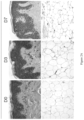
- FIGS. 2 A and 2 B show sections of skin explants from two separate donors used in the method according to the invention at 0, 3, and 7 days of culture.
- FIG. 3 illustrates the stability over time of adipocyte size within the skin explant in the ex vivo model for subcutaneous injection.
- FIG. 4 shows sections of skin explants 6 h, 12 h, and 24 h after injection of a pro-inflammatory cocktail into the adipose tissue.
- subcutaneous injection an injection that is carried out in the hypodermis, which is also referred to as a “hypodermic” injection.
- This type of injection which is well known to the one skilled in the art, generally requires a skin fold to be made with the fingers and the subcutaneous injection is then performed in the skin fold.
- skin explant is meant a skin fragment that comprises, in addition to the epidermis, dermis, and epidermal appendages, a thickness of at least 5 mm of hypodermis.
- the epidermal appendages correspond to the hair follicles, sebaceous glands, and sweat glands.
- the hypodermis is the layer of tissue immediately below the dermis of the skin.
- the hypodermis is a loose connective tissue that is richly vascularized and also contains adipose tissue.
- the skin explant comprises between 5 and 15 mm of hypodermis and preferentially between 5 and 10 mm of hypodermis.
- the immersion of the skin explant is such that the hypodermis is completely immersed in the solidifiable liquid matrix and as for the upper layer of dermis, it is immersed to at least 90% (in the direction of the thickness), preferably to at least 95%, and ideally completely.
- the epidermal layer, on the surface of the skin explant emerges by at least 90% (in the direction of the thickness), preferably by at least 95% and, ideally again, completely.
- the solidification phase it is carried out so that the exposure of the hypodermis, dermis, and epidermis is the same as following the phase of immersion of the skin explant in the solidifiable liquid matrix.
- the skin explant is advantageously cylindrical in shape, other geometrical shapes may be perfectly suitable for implementing the method according to the invention.
- the dimensions of the skin explant should be selected so that a skin fold can be made, which is typically achieved with a fine clamp. Now, there is no absolute need to make a skin fold to carry out the subcutaneous injection. In the case of an automated subcutaneous injection device, for example, there is no need for such a skin fold, and this injection can be performed by setting the subcutaneous injection device so that it injects at a needle penetration depth into the explant that is previously set.
- this skin explant is taken from a mammal, the choice is between human or pig. Now, in view of the preferred destination of the method according to the invention, it will be best to use an explant of human skin.
- the skin explant In relation to the origin of the skin explant, it can come from a plasty from any part of the body, including plasties of the abdomen, chest, buttocks, back and even, why not, the scalp or any other part of the body with skin.
- the skin explant be removed less than 72 h before its use in the method according to the invention.
- an explant that was taken less than 48 hours ago is preferably used.
- the skin explant takes at least one hour before it can be used in the method according to the invention.
- the method according to the invention of which constitutes, in a way, an improvement also makes it possible to dispense with the association of the skin explant with a ring (or perforated disc) consisting of a hydrophobic material and which will confer sufficient buoyancy to the explant within the liquid matrix.
- the method according to the invention will not comprise any step, prior to step i), of fixing to the skin explant, in particular on the epidermal surface, a hydrophobic material; which hydrophobic material is normally used to improve the buoyancy of the skin explant.
- the method according to the invention will then comprise, prior to the immersion of the skin explant in the solidifiable liquid matrix, a step i) of fixing to the surface of the epidermis of the skin explant a ring, or perforated disc, consisting of a hydrophobic material, the external diameter of which is greater than that of the epidermal surface of the skin explant, and the internal diameter of which is that of this same epidermal surface of the skin explant.
- hydrophobic material that can be used to make this ring, materials that are not toxic to the skin are preferred, such as, for example, a paraffin polymer, such as PARAFILM® (SIGMA), or a silicone polymer.
- this ring As regards the fixation of this ring to the surface of the epidermis of the skin explant, it is preferably carried out by means of an adhesive, which adhesive is preferably added to the lower surface of the ring.
- This adhesive can be selected among any type of skin-harmless material and causing the ring to adhere to the epidermal surface of the skin explant.
- a hydrophobic adhesive such as, for example, a silicone-based adhesive, can be selected.
- solidifiable liquid matrix any liquid solution, the specific composition of which (both in terms of compound(s) and concentration) is such that, when implementing suitable conditions, especially particular temperature conditions, the liquid solution takes on a solid- or gel-like consistency.
- the nature of this solidifiable matrix must allow the skin biopsy cells to remain alive, that is to say not have any cytotoxic effect and have a solidification/polymerization temperature at room temperature (namely from 15° C. to 25° C.).
- Its specific composition can be of animal, vegetable, synthetic, or even mixed origin.
- the nature and concentration(s) of these components shall be selected according to the desired physico-chemical characteristics of the matrix when solidified, in particular in terms of flexibility and strength.
- volume of the liquid matrix it will be 1 ⁇ 3 to 2 ⁇ 3 of the total volume of the insert, preferably 2 ⁇ 5 to 3 ⁇ 5 of the total volume; with half of the total volume of the insert being the preferred volume.
- this solidifiable liquid matrix it will be selected from liquid solutions, preferably nutritive, capable of taking the form of a solid or a gel under particular conditions compatible with the survival and culture of the cells which make up the skin explant.
- An example of such a solidifiable liquid matrix is blood plasma, a blood plasma-derived solution (e.g.
- a dilution of blood plasma in a physiological buffer in particular a dilution of blood plasma to at least 10%, 20%, 30%, or even at least 40% (weight/total weight of the matrix)
- a fibrinogen solution in particular a dilution of blood plasma to at least 10%, 20%, 30%, or even at least 40% (weight/total weight of the matrix)
- a fibrinogen solution in particular a dilution of blood plasma to at least 10%, 20%, 30%, or even at least 40% (weight/total weight of the matrix)
- a fibrinogen solution e.g. a collagen solution, a gelatin solution, synthetic polymer solutions, natural polymer solutions (e.g. agarose (low melting point agarose or agar-agar), starch, polysaccharides), and mixtures thereof.
- this solidifiable liquid matrix contains no growth factors, and even better, no serum.
- the solidifiable liquid matrix is composed of two solutions which are mixed together in the insert prior to the step i) of immersing the skin explant.
- This matrix is then composed of a first solution selected from a blood plasma solution, a blood plasma-derived solution, a fibrinogen solution, a collagen solution, and mixtures thereof, and a second solution of low melting point agarose or agar-agar.
- the first solution selected from a blood plasma solution, a blood plasma-derived solution, a fibrinogen solution, a collagen solution, or mixtures thereof, is advantageously a nutrient solution.
- this first solution is mainly able to solidify under the action of an increase or decrease in temperature and/or by the addition of a specific compound or composition.
- the compound allowing this first solution to solidify is the Ca 2+ ion.
- the liquid matrix has a Ca 2+ concentration between 1 mM and 5 mM, preferably between 1.5 mM and 4.5 mM; which concentration will cause it to solidify.
- the liquid matrix has a Ca 2+ concentration between 1 mM and 2 mM, preferably between 1.2 mM and 1.4 mM.
- the liquid matrix has a Ca 2+ concentration between 2 mM and 3 mM, preferably between 2.5 mM and 2.9 mM, more preferentially 2.8 mM of Ca 2 .
- this solution comprises at least one anti-fibrinolytic agent, such as sodium citrate, tranexamic acid, or aprotinin, and in sufficient concentration to obtain the desired anti-fibrinolytic activity.
- the liquid matrix has a final concentration (weight/total weight of the matrix) between 2 and 5% of this at least one anti-fibrinolytic agent.
- the first solution will preferably be a fibrinogen solution.
- the second solution of low melting point agarose or agar-agar is previously heated for a time and at a temperature sufficient to be liquid and to remain liquid at about 37° C. for the time sufficient to be mixed with the first solution in said insert and until the skin explant is immersed.
- this second solution is preheated to its melting temperature or to a slightly higher temperature, preferably to a temperature between 65° C. and 70° C.
- the choice of low melting point agarose or agar-agar is made so as to benefit, for a 1.5% (weight/composition total weight) solution, from a gelling temperature between 24° C. and 28° C., and a melting temperature above 65.5° C.
- the agarose called Low Melting Point LMP Agarose (GIBCOBRL, LIFE TECHNOLOGIES) can be mentioned.
- its concentration in low melting point agarose or agar-agar is between 1% and 5% (preferably in a physiological solution), more preferably between 2% and 5%, between 3% and 4.5%, between 3.5% and 4.5%, or between 3.8% and 4.2%, or between 3.9% and 4.1%, with 4% being the most preferred concentration (by weight relative to the total weight of the composition).
- this second solution of low melting point agarose or agar-agar can be stored in liquid form for at least 1 hour at 37°, and ideally for at least 4 hours, 10 hours, or 16 hours.
- the solidifiable liquid matrix comprising said first and said second low melting point agarose or agar-agar solution has a final concentration in low melting point agarose or agar-agar between 0.1% and 2%, preferably between 0.2% and 1.8% (weight/total weight of the matrix).
- Such a concentration makes it possible to obtain not only a matrix which once solidified makes it possible to preserve the three-dimensional structure and keep said skin fragment or biopsy alive, but also to obtain a matrix which is solid but sufficiently flexible to be non-brittle and resistant to punctual shocks.
- Said liquid matrix is solidified after laying the skin fragment or biopsy by leaving the device thus obtained at a temperature between 37° C. and the room temperature, preferably 20° C.
- the final concentration of low melting point agarose or agar-agar in the liquid matrix (comprising the first and said second solution) is between 0.5% and 2%, preferably between 0.5% and 1.25%, more preferably between 0.5% and 1.0%, with a concentration of 0.7% (weight/total weight of the matrix) being the most preferred concentration.
- the final concentration of low melting point agarose or agar-agar in the liquid matrix is between 0.1% and 2%, preferably between 0.2% and 1.75%, with 0.25% being the most preferred concentration.
- a concentration makes it possible to obtain a matrix which, once solidified, makes it possible both to preserve the three-dimensional structure and keep the skin explant alive, and simultaneously obtain a matrix which is sufficiently flexible to be non-brittle and sufficiently resistant to mechanical effects applied to the skin explant.
- This liquid matrix is solidified after immersing the skin explant by allowing the assembly to cool down.
- the solidifiable liquid matrix further comprises cells other than the cells that make up the skin explants, which cells are selected from the group consisting of fibroblasts, endothelial cells, and nerve cells.
- these cells are fibroblasts, and ideally primary fibroblasts (as opposed to fibroblast cell lines), such as dermal fibroblasts obtained from human foreskin.
- primary fibroblasts as opposed to fibroblast cell lines
- dermal fibroblasts obtained from human foreskin.
- these primary, especially dermal, fibroblasts can be prepared and obtained from standard methods well known to the one skilled in the art (see for example the document HOWARD B V et al., A new method for the establishment of diploid fibroblast cell cultures from human foreskins, Proc. Soc. Exp. Biol. Med ., vol.
- these cells, and especially the fibroblasts are contained in the matrix at a concentration between 5 ⁇ 10 3 and 5 ⁇ 10 5 cells/ml, preferably still between 10 4 and 10 5 cells/ml, with the range 3 ⁇ 10 4 to 5 ⁇ 10 4 cells/ml being the most preferred concentration range.
- the liquid matrix may comprise various compounds such as preservatives, pH agents, etc.
- the liquid matrix will contain between 5 and 500 mg/mL of ascorbic acid, preferably between 25 and 75 mg/mL, with a preferred ascorbic acid concentration of 50 mg/mL.
- the solidifiable liquid matrix is a blood plasma-derived solution and comprises:
- the insert it can take multiple forms and in particular it can correspond to a suspended insert or an insert on stilts.
- a suspended insert will be preferably used.
- the bottom of this insert consists of a porous membrane, the diameter of which is between 5 and 40 mm, and more preferably between 9.5 and 30 mm.
- the porosity of this membrane it must be such as to prevent the liquid matrix from passing through it before it solidifies.
- this porous membrane will have a porosity between 0.4 and 8 ⁇ m, preferably between 0.4 ⁇ m and 1.5 ⁇ m, with the range 0.8 ⁇ m to 1.2 ⁇ m being the preferred porosity range.
- a porous membrane selected from polyethylene terephthalate (PET), nitrocellulose, and polycarbonate membranes can be used.
- PET polyethylene terephthalate
- nitrocellulose nitrocellulose
- MILLIPORE MILLICELL
- the inserts used have a PET membrane with a porosity between 0.8 ⁇ m and 1 ⁇ m, and are suitable for the wells of 8-well culture plates.
- the step of solidifying the liquid matrix is carried out in the presence of calcium ions, preferably also in the presence of thrombin.
- the liquid matrix is a liquid matrix containing a blood plasma solution, a blood plasma-derived solution, a fibrinogen solution, or a collagen solution
- the solidification of this matrix in step ii) is obtained for this solution following the addition of thrombin and/or following an increase in temperature and/or in the presence of factors secreted by the cells such as primary fibroblasts integrated into the matrix.
- this liquid matrix solidifies after a maximum of 8 hours, preferably less than one hour, with a preferred duration of the solidification step of less than 10 min after immersion of the skin explant in the solidifiable liquid matrix in step i).
- the method according to the invention further comprises the steps of:
- the container or well in which this insert is deposited is a well of a cell culture plate with 6, 8, 12, 24, or 48 wells.
- the culture plates that can be used in the method according to the invention are those supplied in particular by the companies NUNC, CORNING, BECTON DICKINSON (BD FALCON), MILLIPORE (MILLICELL).
- the bottom of the insert is located at a distance between 1 and 2.5 mm from the bottom of the container/well containing it.
- the method according to the invention may, furthermore, comprise a step iii′), following step iii), which consists in affixing a cover or film to the container or well in which the insert was placed in step iii), with a view to making this container or well watertight.
- a step iii′ following step iii), which consists in affixing a cover or film to the container or well in which the insert was placed in step iii), with a view to making this container or well watertight.
- culture or “culturing” is meant here, in particular, keeping the physiological state and, where appropriate, the morphological state of the skin explant, and therefore of said cells forming it.
- the aim of this step is to limit cell death phenomena and to maintain the state of differentiation of the cells (in fact to limit inappropriate dedifferentiation or differentiation phenomena).
- medium or “culture medium” is meant a liquid composition comprising all the elements necessary for the culture of skin explant cells (e.g. William's E medium, KBM, DMEM, etc.).
- This culture medium by its nature as much as by its volume in the container, helps to promote the survival of the skin explant and the cells that make it up over time.
- the culture medium in question makes it possible, in particular by limiting the stress thereon, to maintain the cells of the skin explant in their initial state, whether in structural or functional terms.
- step iii′ In the case where the method according to the invention does not comprise step iii′) described above, it could comprise a step iv′), following step iii), which consists in affixing a cover or film to the container or well in which the insert was placed in step iii), with a view to making this container or well watertight. In this way, the container or well obtained can be transported without difficulty, whether by land, sea, or air.
- the method according to the invention further comprises the step of:
- the composition in question is a test composition which is in liquid form.
- the volume of this composition is between 10 ⁇ l and 1 ml, preferably between 10 ⁇ l and 500 ⁇ l, and particularly preferably between 10 ⁇ l and 200 ⁇ l.
- the needle for injection of the composition typically has sufficient length to reach the hypodermis.
- needles with a length of 10 mm or more are preferably used.
- needles with a length of 12, 16, 20, 25, 30, 35, 40, or even 45 mm may be used.
- the needle thus has a length between 16 and 45 mm, preferably a length between 20 and 40 mm.
- the diameter of the needle to be used it can be identified simply by the one skilled in the art with regard to his/her general knowledge.
- hypodermic needles are 18 G-, 19 G-, 20 G-, 21 G-, 22 G-, 23 G-, 25 G-, 26 G-, 27 G-, 28 G-, 29 G-, 30 G-, or even 31 G-type needles.
- step v) is carried out by an experimenter.
- the injection step v) may be directly preceded by a step v°) of pinching the skin explant by the experimenter so as to allow the formation of a skin fold and to facilitate, for the experimenter, the subcutaneous injection step v).
- this step v) is carried out by an automatic injection device.
- the device allows an injection at a certain depth, relative to the surface of the epidermis, so that a subcutaneous injection is obtained.
- the method according to the invention further comprises the step of:
- the injection step v) When the injection step v) is carried out by an experimenter, he/she will determine the injectability of this composition. To do so, it is possible to associate this injectability with arbitrary values associated with specific characteristics of this injection. By way of an example, the experimenter could use the scale below:
- the experimenter by injecting the given composition several times, can assign it an average injectability value.
- the injectability is determined thereby.
- the automatic injection device is coupled to a dynamometer.
- the automatic injection device is able to determine the force required (mPa) for subcutaneous injection of the composition.
- the dynamometer makes it possible to determine:
- the method according to the invention may comprise a step of:
- the method according to the invention makes it possible to truly determine the injection bolus by selecting a volume of a composition which has a given concentration of an active ingredient.
- the method according to the invention makes it possible to access the local toxicity resulting from the injection. This can be done by carrying out morphological and/or molecular analysis at the injection site. Such an analysis can be carried out by embedding the ex vivo skin model following injection, preparing sections from the block obtained, and finally visually and/or immunohistochemically analyzing the sections located at the injection site.
- the second object of the invention is a cell culture insert obtainable at the end of step ii) of the method according to the invention.
- This insert may in particular consist of a porous membrane, the diameter of which is between 5 and 40 mm, in particular between 9.5 and 30 mm, the porosity of which is between 0.4 and 8 ⁇ m, or even between 0.4 ⁇ m and 1.5 ⁇ m, and which is selected from polyethylene terephthalate (PET), nitrocellulose, and polycarbonate membranes.
- PET polyethylene terephthalate
- kits comprising an insert as defined above and an automatic injection device, which is preferably coupled to a dynamometer, so as to be able to determine the injectability characteristics of a composition.
- the automatic injection device is able to determine the initial glide force (PBF), the maximum force (Fmax), and the dynamic glide force (“dynamic glide force” or DGF) of the composition.
- Skin explants are prepared from two complete skin specimens from two separate donors, which specimens comprise the epidermis, dermis, and hypodermis (1.5 to 2 cm).
- the explants (epidermis, dermis, and hypodermis) are then cut out using a metal punch to obtain cylinders 11 to 20 mm in diameter in which the thickness of the hypodermis is adjusted to the desired value (0.5 to 1 cm). They are then kept floating in a buffered saline solution until the stage of “embedding” into the solidified matrix.
- Each skin explant is then embedded with a method similar to the one used for the NATIVESKINTM model. Briefly, the skin explant is laid delicately on an insert (MILLICELLTM 8-well cavity) with a porous membrane (made of PET, 1 ⁇ m porosity) at the bottom, containing a blood plasma-derived solution treated with an anticoagulant agent with reversible properties in the presence of calcium ions (sodium citrate).
- MILLICELLTM 8-well cavity with a porous membrane (made of PET, 1 ⁇ m porosity) at the bottom, containing a blood plasma-derived solution treated with an anticoagulant agent with reversible properties in the presence of calcium ions (sodium citrate).
- This solution contains 42% blood plasma, 50% of a 0.9% NaCl solution, 8% of a 1% CaCl 2 ) salt solution, an anti-fibrinolytic agent (tranaxemic acid or aprotinin), and 0.7% low melting point melted agarose (Agarose LMP GIBCOBRL, LIFE TECHNOLOGIES) (melted in a stove at 65.5° C.).
- the plasma acts as a dermal support on which the skin explant adheres.
- the coagulation mainly consists of the transformation, in the presence of calcium ions and thrombin, of the fibrinogen present in the plasma into a scaffolding of fibrin molecules linked together by covalent bonds.
- the function of the anti-fibrinolytic agent is to inhibit the enzymes capable of degrading the plasma matrix, with said enzymes being secreted by the skin explant, and thus to maintain the integrity of the explant.
- the 0.7% agarose solution contained in the plasma solution gradually gels at 37° C., thereby holding the skin explant firmly in the insert.
- FIG. 1 shows such a cell culture insert ( 1 ) with lugs ( 5 ), the bottom of which consists of a porous membrane ( 2 ) and which contains the skin explant ( 3 ) after it has been trapped in the solidified matrix ( 4 ).
- the assembly of the skin explant is kept in culture for 7 days in a CO 2 incubator at 37° C. with the epidermis in contact with air.
- the DMEM culture medium supplemented with calcium and vitamin C contained in the insert placed (suspended) on the well of the cell culture plate is changed daily.
- the skin explants are dehydrated by a first alcohol bath, and then by a second xylene bath. Finally, a first bath in paraffin allows to replace the water previously contained in the skin explant by paraffin.
- the paraffin-impregnated samples are removed from their bath and transferred to a container, the bottom of which is lined with absorbent paper, to be taken to the vicinity of the embedding station.
- the samples, enclosed in histology cassettes, are immersed in liquid paraffin at 56° C. to again melt the paraffin impregnating them.
- the histology cassette is opened, the sample is possibly cut in half.
- An embedding mold is filled with liquid paraffin, and the sample (or the 2 sample pieces) is placed in the mold and oriented in the desired direction for cutting.
- the mold is transferred onto a refrigerated rack in order to solidify the paraffin at the bottom of the mold and hold the sample therein.
- the lid of the histology cassette with the sample reference placed on top is placed thereover, so that the paraffin passes therethrough (paraffin can be added if necessary), and then the assembly is placed in cold storage (refrigerator, freezer, cold room . . . ) for several minutes (5 to 6), in order to solidify the paraffin into a block, thus trapping the sample in the right orientation with the lid of the histology cassette which will become the support of the block.
- the block Once the block has solidified, it is released from the mold. Excess paraffin is possibly scraped off with a spatula on the sides of the lid of the embedding cassette.
- Serial sections of a thickness varying from 4 to 5 ⁇ m are then made along the entire length of the paraffin block containing the sample.
- FIGS. 2 A and 2 B illustrate the results of staining performed (Hematoxylin and Eosin) on the skin explants from two separate donors used in the method according to the invention after 0, 3, or 7 days of culture.
- the results show a remarkable stability of the explants over time both in terms of structure (the different layers of the skin, including the hypodermis, show no noticeable change) and viability (no significant cell death is observed).
- FIG. 3 shows the distribution of adipocytes depending on their surface area in the different explants at 0 and 7 days of culture.
- a pro-inflammatory solution consisting of TNF-alpha (500 ng/mL) and LPS (0.2 mg/mL) was injected into the adipose tissue of the models using a syringe and a 12 mm long 27 G needle.
- the models were then cultured under cell culture conditions (incubator at 37° C., 5% CO 2 , and water-saturated atmosphere for 6 h, 12 h, or 24 h.
- FIG. 4 shows H&E staining performed on 5 ⁇ m thick paraffin sections to evaluate the effect of the injected solution on tissue viability and structure.
Landscapes
- Health & Medical Sciences (AREA)
- Engineering & Computer Science (AREA)
- Life Sciences & Earth Sciences (AREA)
- Biomedical Technology (AREA)
- Chemical & Material Sciences (AREA)
- Bioinformatics & Cheminformatics (AREA)
- Wood Science & Technology (AREA)
- Zoology (AREA)
- Organic Chemistry (AREA)
- Biotechnology (AREA)
- General Health & Medical Sciences (AREA)
- Genetics & Genomics (AREA)
- Immunology (AREA)
- Microbiology (AREA)
- Biochemistry (AREA)
- Cell Biology (AREA)
- General Engineering & Computer Science (AREA)
- Molecular Biology (AREA)
- Hematology (AREA)
- Urology & Nephrology (AREA)
- Sustainable Development (AREA)
- Medicinal Chemistry (AREA)
- Pathology (AREA)
- Tropical Medicine & Parasitology (AREA)
- Toxicology (AREA)
- Food Science & Technology (AREA)
- Physics & Mathematics (AREA)
- Analytical Chemistry (AREA)
- General Physics & Mathematics (AREA)
- Dentistry (AREA)
- Environmental Sciences (AREA)
- Transplantation (AREA)
- Oral & Maxillofacial Surgery (AREA)
- Dermatology (AREA)
- Epidemiology (AREA)
- Animal Behavior & Ethology (AREA)
- Public Health (AREA)
- Veterinary Medicine (AREA)
- Micro-Organisms Or Cultivation Processes Thereof (AREA)
- Agricultural Chemicals And Associated Chemicals (AREA)
Abstract
Description
-
- i) immersing a skin explant in a solidifiable liquid matrix such that the upper face of the epidermis is not covered, which matrix is itself contained in a cell culture insert, the bottom of which consists of a porous membrane, and
- ii) solidifying this matrix so as to trap the immersed portion of this skin explant, wherein the upper face of the epidermis is not covered, and adhering this same matrix to the side walls and the porous membrane of the insert;
-
- iii) placing the insert in a container or culture well, and
- iv) culturing the skin explant in an appropriate medium.
-
- a) 25 to 75% (volume/total volumeA*) of fibrinogen, preferably 35% to 45% (v/v),
- b) 5% to 12% (volume/total volume) of a 1% CaCl2) saline solution, preferably 8%,
- c) 5% to 2%, preferably of the anti-fibrinolytic agent, preferably said anti-fibrinolytic agent being selected from tranexamic acid or aprotinin
- d) 0.5% to 4% of low melting point agarose, preferably 1% to 2%, and
- e) a physiological solution such as a 0.9% NaCl solution, q.s. 100%.
-
- iii) placing the insert in a container or culture well, and
- iv) culturing the skin explant in an appropriate medium.
-
- v) injecting, subcutaneously and in the skin explant, a composition.
-
- vi) determining the injectability of the composition.
-
- 1) the initial glide force (“initial glide force” or PBF), which corresponds to the force required to move the syringe plunger;
- 2) the maximum force (Fmax) which corresponds to the largest measured force value for moving the syringe plunger before the syringe plunger completes its stroke at the end of the syringe; and/or
- 3) the dynamic glide force (“dynamic glide force” or DGF), which corresponds to the force required to keep the syringe plunger moving so that it expels the contents thereof.
-
- vi) determining the injection bolus of the composition, with the local toxicity resulting from the injection of the composition and/or the efficacy of the composition.
Claims (5)
Applications Claiming Priority (3)
| Application Number | Priority Date | Filing Date | Title |
|---|---|---|---|
| FR1870232A FR3078541B1 (en) | 2018-03-05 | 2018-03-05 | EX VIVO SUBCUTANEOUS INJECTION MODEL |
| FR1870232 | 2018-03-05 | ||
| PCT/EP2019/000061 WO2019170281A1 (en) | 2018-03-05 | 2019-03-03 | Ex vivo subcutaneous injection model |
Related Parent Applications (1)
| Application Number | Title | Priority Date | Filing Date |
|---|---|---|---|
| PCT/EP2019/000061 A-371-Of-International WO2019170281A1 (en) | 2018-03-05 | 2019-03-03 | Ex vivo subcutaneous injection model |
Related Child Applications (1)
| Application Number | Title | Priority Date | Filing Date |
|---|---|---|---|
| US18/923,157 Continuation-In-Part US20250044277A1 (en) | 2018-03-05 | 2024-10-22 | Ex vivo subcutaneous injection model |
Publications (2)
| Publication Number | Publication Date |
|---|---|
| US20200400652A1 US20200400652A1 (en) | 2020-12-24 |
| US12158463B2 true US12158463B2 (en) | 2024-12-03 |
Family
ID=63638116
Family Applications (1)
| Application Number | Title | Priority Date | Filing Date |
|---|---|---|---|
| US16/971,599 Active 2041-09-28 US12158463B2 (en) | 2018-03-05 | 2019-03-03 | Ex vivo subcutaneous injection model |
Country Status (8)
| Country | Link |
|---|---|
| US (1) | US12158463B2 (en) |
| EP (1) | EP3762481A1 (en) |
| JP (1) | JP7361039B2 (en) |
| KR (1) | KR102717715B1 (en) |
| CA (1) | CA3091505A1 (en) |
| FR (1) | FR3078541B1 (en) |
| SG (1) | SG11202010680QA (en) |
| WO (1) | WO2019170281A1 (en) |
Families Citing this family (4)
| Publication number | Priority date | Publication date | Assignee | Title |
|---|---|---|---|---|
| WO2022236144A1 (en) | 2021-05-06 | 2022-11-10 | Massachusetts Institute Of Technology | Ex vivo tissue explant and graft platform and uses thereof |
| FR3123125B1 (en) | 2021-05-20 | 2025-08-15 | Genoskin | Ex vivo human model for the evaluation of allergic or pseudo-allergic inflammatory potential |
| MX2024002137A (en) * | 2021-08-20 | 2024-03-06 | Amgen Inc | METHODS INVOLVING THERAPEUTIC COMPOUNDS AND MAMMALIAN SKIN IN VITRO. |
| FR3132146B1 (en) | 2022-01-27 | 2025-10-31 | Genoskin | Ex vivo human model intended for evaluating the vaccine potential of a composition |
Citations (4)
| Publication number | Priority date | Publication date | Assignee | Title |
|---|---|---|---|---|
| WO2012059703A1 (en) | 2010-11-05 | 2012-05-10 | Centre National De La Recherche Scientifique (Cnrs) | In vitro or ex vivo method for storing and/or keeping an epidermis alive |
| WO2013164436A1 (en) | 2012-05-03 | 2013-11-07 | Genoskin | System for keeping alive and transporting skin biopsies and applications of said system |
| US20140271508A1 (en) * | 2013-03-15 | 2014-09-18 | Mary Kay Inc. | Cosmetic compositions and uses thereof |
| EP3256568A1 (en) | 2015-02-10 | 2017-12-20 | Genoskin | Method for implanting cell clumps in a skin explant |
Family Cites Families (3)
| Publication number | Priority date | Publication date | Assignee | Title |
|---|---|---|---|---|
| US6372494B1 (en) | 1999-05-14 | 2002-04-16 | Advanced Tissue Sciences, Inc. | Methods of making conditioned cell culture medium compositions |
| AU2010284266A1 (en) * | 2009-08-21 | 2012-03-08 | Johnson & Johnson Consumer Companies, Inc. | Human skin explant culture system and use therefor |
| FR3061206B1 (en) * | 2016-12-23 | 2020-06-26 | Laboratoires Clarins | SKIN MODEL INCLUDING EPIDERMIS, DERMIS AND HYPODERMIS, ITS PREPARATION METHOD AND ITS USE |
-
2018
- 2018-03-05 FR FR1870232A patent/FR3078541B1/en active Active
-
2019
- 2019-03-03 SG SG11202010680QA patent/SG11202010680QA/en unknown
- 2019-03-03 JP JP2020544812A patent/JP7361039B2/en active Active
- 2019-03-03 KR KR1020207028321A patent/KR102717715B1/en active Active
- 2019-03-03 WO PCT/EP2019/000061 patent/WO2019170281A1/en not_active Ceased
- 2019-03-03 CA CA3091505A patent/CA3091505A1/en active Pending
- 2019-03-03 US US16/971,599 patent/US12158463B2/en active Active
- 2019-03-03 EP EP19711245.1A patent/EP3762481A1/en active Pending
Patent Citations (6)
| Publication number | Priority date | Publication date | Assignee | Title |
|---|---|---|---|---|
| WO2012059703A1 (en) | 2010-11-05 | 2012-05-10 | Centre National De La Recherche Scientifique (Cnrs) | In vitro or ex vivo method for storing and/or keeping an epidermis alive |
| WO2013164436A1 (en) | 2012-05-03 | 2013-11-07 | Genoskin | System for keeping alive and transporting skin biopsies and applications of said system |
| US20150132737A1 (en) * | 2012-05-03 | 2015-05-14 | Genoskin | System for keeping alive and transporting skin biopsies and applications of said system |
| EP2882290A1 (en) | 2012-05-03 | 2015-06-17 | Genoskin | System for keeping alive and transporting skin biopsies and applications of said system |
| US20140271508A1 (en) * | 2013-03-15 | 2014-09-18 | Mary Kay Inc. | Cosmetic compositions and uses thereof |
| EP3256568A1 (en) | 2015-02-10 | 2017-12-20 | Genoskin | Method for implanting cell clumps in a skin explant |
Non-Patent Citations (8)
| Title |
|---|
| De Wever et al. "Human Skin Models for Research Applications in Pharmacology and Toxicology: Introducing NativeSkin®, the "Missing Link" Bridging Cell Culture and/or Reconstructed Skin Models and Human Clinical Testing," Applied In Vitro Toxicology, vol. 1, No. 1, 2015, pp. 26-32. |
| Howard et al., "A New Method for the Establishment of Diploid Fibroblast Cell Cultures from Human Foreskins (39528)," Proceedings of the Society for Experimental Biology and Medicine, vol. 153, 1976, pp. 280-283. |
| ICH Expert Working Group, ICH Harmonised Tripartite Guideline, "Specifications: Test Procedures and Acceptance Criteria for New Drug Substances and New Drug Products: Chemical Substances Q6A," International Conference on Harmonisation of Technical Requirements for Registration of Pharmaceuticals for Human Use, Oct. 6, 1999, 35 pages. |
| International Search Report for PCT/EP2019/000061 dated Apr. 23, 2019, 7 pages, with English Translation. |
| Lebonvallet et al., "The evolution and use of skin explants: potential and limitations for dermatological research," European Journal of Dermatology, vol. 20, No. 6, Nov.-Dec. 2010, 14 pages. |
| Park et al., "Usefulness of Skin Explants for Histologic Analysis after Fractional Photothermolysis," Annals of Dermatology, vol. 27, No. 3, 2015, pp. 283-290. |
| Written Opinion of the International Searching Authority issued in International Application No. PCT/EP2019/000061 dated Apr. 23, 2019 with English translation provided. |
| Written Opinion of the ISA for PCT/EP2019/000061 dated Apr. 23, 2019, 6 pages. |
Also Published As
| Publication number | Publication date |
|---|---|
| FR3078541B1 (en) | 2022-07-08 |
| FR3078541A1 (en) | 2019-09-06 |
| KR20200128124A (en) | 2020-11-11 |
| EP3762481A1 (en) | 2021-01-13 |
| JP2021516542A (en) | 2021-07-08 |
| US20200400652A1 (en) | 2020-12-24 |
| SG11202010680QA (en) | 2020-11-27 |
| WO2019170281A1 (en) | 2019-09-12 |
| CA3091505A1 (en) | 2019-09-12 |
| JP7361039B2 (en) | 2023-10-13 |
| KR102717715B1 (en) | 2024-10-16 |
Similar Documents
| Publication | Publication Date | Title |
|---|---|---|
| US20200400652A1 (en) | Ex vivo subcutaneous injection model | |
| Bellas et al. | In vitro 3D full‐thickness skin‐equivalent tissue model using silk and collagen biomaterials | |
| US20050129730A1 (en) | Tissue composites and uses thereof | |
| US9585381B2 (en) | System for keeping alive and transporting skin biopsies and applications of said system | |
| KR102102153B1 (en) | A bead for cell encapsulation and culture based on decellularized tissue, and use thereof | |
| JP7054523B2 (en) | Cell encapsulation device and its use | |
| JP2016013094A (en) | Substrate for artificial cartilage formation | |
| EP4182440B1 (en) | Microfluidic system to control perfusion, diffusion and collection of molecules over long periods in an ex-vivo skin model | |
| US20250044277A1 (en) | Ex vivo subcutaneous injection model | |
| KR20150040848A (en) | Tissue regeneration construct, and method for producing tissue regeneration construct | |
| JP7260563B2 (en) | Cell transplantation kit, method for producing bag-like structure, and antidiabetic agent | |
| Plant et al. | Schwann cell transplantation methods using biomaterials | |
| JP2005305177A (en) | Artificial tissue including tissue ancillary organ-like structure and its manufacturing method | |
| BABA et al. | Combined automated culture system for tubular structure assembly and maturation for vascular tissue engineering | |
| KR101776236B1 (en) | Animal as models for wound, method of manufacturing thereof and method of analyzing efficacy of wound therapeutic agent using thereof | |
| KR102380496B1 (en) | Methods for cryopreservation of organ-on-a-chip | |
| Akkaya et al. | In vitro 3D Spheroid Wound Modeling: An Alternative to Experimental Animal Studies | |
| Zuniga | Development of a three-dimensional in vitro vascularized human skin equivalent employing collagen/keratin hydrogels | |
| Musial | The Cytoseeder™–A Novel Seeding Device for Tissue Engineered Scaffolds | |
| Isaac et al. | Construction of a skin substitute composed of porcine collagen matrix populated with human dermal fibroblasts and keratinocytes: histological evaluation | |
| JP2026078116A (en) | Culture equipment | |
| JP2025002254A (en) | Artificial cultured skin and method for producing the same | |
| WO2004113514A1 (en) | Reconstructed dermal papillae | |
| DaValle | Mammary Epithelial Cell Growth on a Three-Dimensional Scaffold in an Operating Bioreactor | |
| Mallette | Growth in Culture of Trypsin Dissociated Thyroid Cells from Control and Altitude Acclimated Rats |
Legal Events
| Date | Code | Title | Description |
|---|---|---|---|
| FEPP | Fee payment procedure |
Free format text: ENTITY STATUS SET TO UNDISCOUNTED (ORIGINAL EVENT CODE: BIG.); ENTITY STATUS OF PATENT OWNER: SMALL ENTITY |
|
| STPP | Information on status: patent application and granting procedure in general |
Free format text: APPLICATION DISPATCHED FROM PREEXAM, NOT YET DOCKETED |
|
| AS | Assignment |
Owner name: GENOSKIN, FRANCE Free format text: ASSIGNMENT OF ASSIGNORS INTEREST;ASSIGNORS:DESCARGUES, PASCAL;PAGES, EMELINE;JARDET, CLAIRE;REEL/FRAME:054903/0942 Effective date: 20210112 |
|
| STPP | Information on status: patent application and granting procedure in general |
Free format text: DOCKETED NEW CASE - READY FOR EXAMINATION |
|
| STPP | Information on status: patent application and granting procedure in general |
Free format text: NON FINAL ACTION MAILED |
|
| STPP | Information on status: patent application and granting procedure in general |
Free format text: RESPONSE TO NON-FINAL OFFICE ACTION ENTERED AND FORWARDED TO EXAMINER |
|
| STPP | Information on status: patent application and granting procedure in general |
Free format text: NON FINAL ACTION MAILED |
|
| STPP | Information on status: patent application and granting procedure in general |
Free format text: RESPONSE TO NON-FINAL OFFICE ACTION ENTERED AND FORWARDED TO EXAMINER |
|
| STPP | Information on status: patent application and granting procedure in general |
Free format text: NOTICE OF ALLOWANCE MAILED -- APPLICATION RECEIVED IN OFFICE OF PUBLICATIONS |
|
| STPP | Information on status: patent application and granting procedure in general |
Free format text: AWAITING TC RESP., ISSUE FEE NOT PAID |
|
| STPP | Information on status: patent application and granting procedure in general |
Free format text: NOTICE OF ALLOWANCE MAILED -- APPLICATION RECEIVED IN OFFICE OF PUBLICATIONS |
|
| FEPP | Fee payment procedure |
Free format text: ENTITY STATUS SET TO SMALL (ORIGINAL EVENT CODE: SMAL); ENTITY STATUS OF PATENT OWNER: SMALL ENTITY |
|
| STCF | Information on status: patent grant |
Free format text: PATENTED CASE |