US12136194B2 - Method and apparatus for bone suppression in X-ray image - Google Patents
Method and apparatus for bone suppression in X-ray image Download PDFInfo
- Publication number
- US12136194B2 US12136194B2 US17/691,863 US202217691863A US12136194B2 US 12136194 B2 US12136194 B2 US 12136194B2 US 202217691863 A US202217691863 A US 202217691863A US 12136194 B2 US12136194 B2 US 12136194B2
- Authority
- US
- United States
- Prior art keywords
- image
- compensated
- row
- pixel
- ray image
- Prior art date
- Legal status (The legal status is an assumption and is not a legal conclusion. Google has not performed a legal analysis and makes no representation as to the accuracy of the status listed.)
- Active, expires
Links
Images


Classifications
-
- G—PHYSICS
- G06—COMPUTING OR CALCULATING; COUNTING
- G06T—IMAGE DATA PROCESSING OR GENERATION, IN GENERAL
- G06T5/00—Image enhancement or restoration
- G06T5/50—Image enhancement or restoration using two or more images, e.g. averaging or subtraction
-
- A—HUMAN NECESSITIES
- A61—MEDICAL OR VETERINARY SCIENCE; HYGIENE
- A61B—DIAGNOSIS; SURGERY; IDENTIFICATION
- A61B6/00—Apparatus or devices for radiation diagnosis; Apparatus or devices for radiation diagnosis combined with radiation therapy equipment
- A61B6/52—Devices using data or image processing specially adapted for radiation diagnosis
- A61B6/5258—Devices using data or image processing specially adapted for radiation diagnosis involving detection or reduction of artifacts or noise
-
- G—PHYSICS
- G06—COMPUTING OR CALCULATING; COUNTING
- G06T—IMAGE DATA PROCESSING OR GENERATION, IN GENERAL
- G06T5/00—Image enhancement or restoration
- G06T5/77—Retouching; Inpainting; Scratch removal
-
- A—HUMAN NECESSITIES
- A61—MEDICAL OR VETERINARY SCIENCE; HYGIENE
- A61B—DIAGNOSIS; SURGERY; IDENTIFICATION
- A61B6/00—Apparatus or devices for radiation diagnosis; Apparatus or devices for radiation diagnosis combined with radiation therapy equipment
- A61B6/52—Devices using data or image processing specially adapted for radiation diagnosis
- A61B6/5205—Devices using data or image processing specially adapted for radiation diagnosis involving processing of raw data to produce diagnostic data
-
- G—PHYSICS
- G06—COMPUTING OR CALCULATING; COUNTING
- G06T—IMAGE DATA PROCESSING OR GENERATION, IN GENERAL
- G06T12/00—Tomographic reconstruction from projections
- G06T12/30—Image post-processing, e.g. metal artefact correction
-
- G—PHYSICS
- G06—COMPUTING OR CALCULATING; COUNTING
- G06T—IMAGE DATA PROCESSING OR GENERATION, IN GENERAL
- G06T3/00—Geometric image transformations in the plane of the image
- G06T3/18—Image warping, e.g. rearranging pixels individually
-
- G—PHYSICS
- G06—COMPUTING OR CALCULATING; COUNTING
- G06T—IMAGE DATA PROCESSING OR GENERATION, IN GENERAL
- G06T5/00—Image enhancement or restoration
- G06T5/20—Image enhancement or restoration using local operators
-
- G—PHYSICS
- G06—COMPUTING OR CALCULATING; COUNTING
- G06T—IMAGE DATA PROCESSING OR GENERATION, IN GENERAL
- G06T7/00—Image analysis
- G06T7/10—Segmentation; Edge detection
- G06T7/13—Edge detection
-
- G—PHYSICS
- G06—COMPUTING OR CALCULATING; COUNTING
- G06T—IMAGE DATA PROCESSING OR GENERATION, IN GENERAL
- G06T7/00—Image analysis
- G06T7/10—Segmentation; Edge detection
- G06T7/136—Segmentation; Edge detection involving thresholding
-
- G—PHYSICS
- G06—COMPUTING OR CALCULATING; COUNTING
- G06T—IMAGE DATA PROCESSING OR GENERATION, IN GENERAL
- G06T2207/00—Indexing scheme for image analysis or image enhancement
- G06T2207/10—Image acquisition modality
- G06T2207/10116—X-ray image
-
- G—PHYSICS
- G06—COMPUTING OR CALCULATING; COUNTING
- G06T—IMAGE DATA PROCESSING OR GENERATION, IN GENERAL
- G06T2207/00—Indexing scheme for image analysis or image enhancement
- G06T2207/20—Special algorithmic details
- G06T2207/20036—Morphological image processing
- G06T2207/20041—Distance transform
-
- G—PHYSICS
- G06—COMPUTING OR CALCULATING; COUNTING
- G06T—IMAGE DATA PROCESSING OR GENERATION, IN GENERAL
- G06T2207/00—Indexing scheme for image analysis or image enhancement
- G06T2207/20—Special algorithmic details
- G06T2207/20212—Image combination
- G06T2207/20221—Image fusion; Image merging
-
- G—PHYSICS
- G06—COMPUTING OR CALCULATING; COUNTING
- G06T—IMAGE DATA PROCESSING OR GENERATION, IN GENERAL
- G06T2207/00—Indexing scheme for image analysis or image enhancement
- G06T2207/30—Subject of image; Context of image processing
- G06T2207/30004—Biomedical image processing
- G06T2207/30008—Bone
Definitions
- the present invention relates to a method and an apparatus for bone suppression in an x-ray image, and more particularly, to a method and an apparatus for bone suppression, which are capable of acquiring an X-ray image in which a bone is suppressed by compensating a single photographed X-ray image.
- DXR dual energy X-ray
- the present invention has been made in an effort to provide a method and an apparatus for bone suppression, which are capable of acquiring an X-ray image in which a bone is suppressed by compensating a single photographed X-ray image.
- An exemplary embodiment of the present invention provides a method for bone suppression in an X-ray image, which includes: (a) extracting an upper contour line and a lower contour line corresponding to a bone to be suppressed from the original X-ray image; (b) generating a first binarization image and a second binarization image based on the upper contour line and the lower contour line, respectively; (c) generating a first distance transform image and a second distance transform image from the first binarization image and the second binarization image, respectively through distance transform; (d) generating a compensated first X-ray image and a compensated second X-ray image by compensating a pixel value of a region which belongs to the bone by using the first distance transform image and the second distance transform image, respectively from the original X-ray image; and (e) synthesizing the compensated first X-ray image and the compensated second X-ray image to obtain a bone-suppressed X-ray image.
- Step (d) above may include (d1) generating a first coordinate transformed image and a second coordinate transformed image through coordinate system transform by using the first distance transform image and the second distance transform image, respectively from the original X-ray image, (d2) generating a compensated first coordinate transformed image and a compensated second coordinate transformed image by compensating the pixel value of the region which belongs to the bone in the first coordinate transformed image and the second coordinate transformed image, respectively, and (d3) generating the compensated first X-ray image and the compensated second X-ray image from the compensated first coordinate transformed image and the compensated second coordinate transformed image, respectively through coordinate system inverse transform.
- a pixel value may be compensated per at least one row in a corresponding coordinate transformed image.
- a pixel value of a corresponding row may be compensated by using pixel values of a compensated row and a row to be compensated, which is adjacent thereto.
- step (d2) above with respect to a pixel to be compensated, multiple blocks having a predetermined size may be set, which belong to the compensated row and the row to be compensated, and have the corresponding pixel and with respect to each block, a difference between an average of the pixel value of the compensated row and an average of the pixel value of the row to be compensated may be calculated, and the pixel value of the corresponding pixel may be compensated by using an average of the differences of the multiple blocks.
- a weight may be granted for each block.
- the weight may be determined according to a distribution a difference in pixel value between the compensated pixel and the pixel to be compensated for each column in the corresponding block.
- the weight may be granted for each pixel.
- the weight may be determined according to a difference between the average of the pixel value of the row to be compensated in the block and the pixel value of the corresponding pixel.
- Another exemplary embodiment of the present invention provides an apparatus for bone suppression in an X-ray image, which includes: a contour line extraction unit extracting an upper contour line and a lower contour line corresponding to a bone to be suppressed from the original X-ray image; a distance transform unit generating a first binarization image and a second binarization image based on the upper contour line and the lower contour line, respectively, and generating a first distance transform image and a second distance transform image from the first binarization image and the second binarization image, respectively through distance transform; an X-ray image compensation unit generating a compensated first X-ray image and a compensated second X-ray image by compensating a pixel value of a region which belongs to the bone by using the first distance transform image and the second distance transform image, respectively from the original X-ray image; and an image synthesis unit synthesizing the compensated first X-ray image and the compensated second X-ray image to obtain a bone-suppressed X-ray image.
- the X-ray image compensation unit may include a coordinate system transform unit generating a first coordinate transformed image and a second coordinate transformed image through coordinate system transform by using the first distance transform image and the second distance transform image, respectively from the original X-ray image, an image compensation unit generating a compensated first coordinate transformed image and a compensated second coordinate transformed image by compensating the pixel value of the region which belongs to the bone in the first coordinate transformed image and the second coordinate transformed image, respectively, and a coordinate system inverse transform unit generating the compensated first X-ray image and the compensated second X-ray image from the compensated first coordinate transformed image and the compensated second coordinate transformed image, respectively through coordinate system inverse transform.
- the image compensation unit may compensate a pixel value per at least one row in a corresponding coordinate transformed image.
- the image compensation unit may compensate a pixel value of a corresponding row by using pixel values of a compensated row and a row to be compensated, which is adjacent thereto.
- the image compensation unit may set, with respect to a pixel to be compensated, multiple blocks having a predetermined size, which belong to the compensated row and the row to be compensated, and have the corresponding pixel and calculate, with respect to each block, a difference between an average of the pixel value of the compensated row and an average of the pixel value of the row to be compensated, and compensate the pixel value of the corresponding pixel by using an average of the differences of the multiple blocks.
- a weight may be granted for each block.
- the weight may be determined according to a distribution a difference in pixel value between the compensated pixel and the pixel to be compensated for each column in the corresponding block.
- the weight may be granted for each pixel.
- the weight may be determined according to a difference between the average of the pixel value of the row to be compensated in the block and the pixel value of the corresponding pixel.
- an X-ray image in which a bone is suppressed by compensating a single photographed X-ray image can be acquired. Accordingly, additional radiation exposure to an examinee is not required and there is no worry about occurrence of artifact due to movement of an involuntary muscle in the body.
- a peripheral region of the bone is more substantially reflected by compensating an image based on each of an upper contour line and a lower contour line of the bone to enhance compensation performance.
- FIG. 1 is a block diagram of an apparatus for bone suppression in an X-ray image according to an exemplary embodiment of the present invention.
- FIG. 2 is a flowchart of a method for bone suppression in an X-ray image according to an exemplary embodiment of the present invention.
- FIG. 3 illustrates an example of gradient based feature point extraction.
- FIG. 4 illustrates an example of a state in which feature points, and an upper contour line and a lower contour line are extracted from an original X-ray image.
- FIG. 5 is a diagram conceptually illustrating a contour line, a binarization image, a distance transform image, and a coordinate transformed image.
- FIG. 6 is a diagram for describing a concept of coordinate system transform.
- FIGS. 7 A to 7 D illustrate an example of a process of compensating a pixel value per row in a coordinate transformed image.
- FIG. 8 illustrates an example of a block satisfying an assumption that a degree of attenuation due to a bone is equal in a block and a block not satisfying the assumption.
- FIG. 9 illustrates an example of a case where a weight is differently granted to a pixel to be compensated for each block.
- FIG. 10 illustrates an example of a case where there are large differences between any pixel and other pixels in the block.
- FIG. 11 illustrates an example of a process of acquiring an X-ray image in which a bone is suppressed from an original X-ray image according to an exemplary embodiment of the present invention.
- FIG. 1 is a block diagram of an apparatus for bone suppression in an X-ray image according to an exemplary embodiment of the present invention.
- the apparatus for bone suppression according to the exemplary embodiment may include a contour line extraction unit 110 , a distance transform unit 120 , an X-ray image compensation unit 130 and an image synthesis unit 140 .
- a single photographed original X-ray image is input into the contour line extraction unit 110 and the X-ray image compensation unit 130 .
- the original X-ray image as an X-ray image including a bone component and a chest photographed image is described as an example, but may be photographing images of various other portions in addition to a chest.
- the contour line extraction unit 110 extracts an upper contour line and a lower contour line corresponding to a bone to be suppressed from the original X-ray image.
- the distance transform unit 120 generates a first binarization image and a second binarization image based on the upper contour line and the lower contour line, respectively, and generates a first distance transform image and a second distance transform image through distance transform from the first binarization image and the second binarization image, respectively.
- the X-ray image compensation unit 130 generates a compensated first X-ray image and a compensated second X-ray image by compensating a pixel value of a region which belongs to the bone by using the first distance transform image and the second distance transform image, respectively from the original X-ray image.
- the X-ray image compensation unit 130 may include a coordinate system transform unit 131 , an image compensation unit 132 , and a coordinate system inversion transform unit 133 .
- the coordinate system transform unit 131 generates a first coordinate transformed image and a second coordinate transformed image through coordinate system transform by using the first distance transform image and the second distance transform image, respectively from the original X-ray image.
- the image compensation unit 132 generates a compensated first coordinate transformed image and a compensated second coordinate transformed image by compensating the pixel value of the region which belongs to the bone in the first coordinate transformed image and the second coordinate transformed image, respectively.
- the coordinate system inversion transform unit 133 generates a compensated first X-ray image and a compensated second X-ray image through coordinate system inverse transform corresponding to the coordinate system transform of the coordinate system transform unit 131 from the compensated first coordinate transformed image and the compensated second coordinate transformed image, respectively.
- the image synthesis unit 140 synthesizes the compensated first X-ray image and the compensated second X-ray image to acquire a bone-suppressed X-ray image.
- FIG. 2 is a flowchart of a method for bone suppression in an X-ray image according to an exemplary embodiment of the present invention.
- FIG. 2 an operation of the apparatus for bone suppression according to FIG. 1 will be described in more detail.
- step 210 the contour line extraction unit 110 extracts feature points constituting a contour line of the bone to be suppressed from the original X-ray image.
- the feature points as points which are positioned between the bone to be suppressed and a background as illustrated in FIG. 4 may be extracted by using features in the image.
- various methods including a gradient based method, a method using an artificial intelligence algorithm, a Scale Invariant Feature Transform (SIFT) algorithm, and the like may be used.
- FIG. 3 illustrates an example of gradient based feature point extraction. As illustrated in FIG. 3 , a pixel in which a gradient of a pixel value is rapidly changed among pixels which are on a straight line crossing the bone to be suppressed may be extracted as the feature points.
- the contour line extraction unit 110 extracts an upper contour line and a lower contour line corresponding to a bone to be suppressed from the feature points.
- various methods including a least squares method, a method for fitting with a single polynomial function by an algorithm Random Sample Consensus (RANSAC), a method for fitting for each section by using a plurality of polynomial functions, and the like may be used.
- RANSAC Random Sample Consensus
- a final contour line c(x) may be determined from multiple polynomial functions P(x) through the following equation.
- C ( x ) ⁇ P 1 ( x )+ ⁇ P t+1 ( x )( S i,x ⁇ x ⁇ S t+1,x ) [Equation 2]
- ⁇ and ⁇ are weights determined from as in the following equation.
- FIG. 4 illustrates an example of features points and a state in which an upper contour line and a lower contour line are extracted from the feature points in an original X-ray image. As illustrated, a smoothly seamless contour line may be formed by using the extracted feature points.
- the upper contour line and the lower contour lines will be referred to as C upper (x) and C below (x) respectively.
- the distance transform unit 120 generates the first binarization image and the second binarization image based on the upper contour line and the lower contour line, respectively.
- the binarization image is an image in which a foreground is expressed as white and a background is expressed as black with the contour line as a boundary. In the exemplary embodiment, it is assumed that a portion to which the bone to be suppressed is the foreground and a portion other than the bone is the background.
- an upper side of the upper contour line is expressed as black and a lower side of the upper contour line is expressed as white.
- the upper side of the lower contour line is expressed as white and the lower side of the lower contour line is expressed as black.
- the first binarization image and the second binarization image will be referred to as B upper and B below respectively.
- the distance transform unit 120 generates the first distance transform image and the second distance transform image from the first binarization image and the second binarization image, respectively through a distance transform.
- the distance transform as one of image transform techniques performed for the binarization image is a transform which allows pixels which belong to the foreground to represent a distance from the pixel which belongs to the background when there is the binarization image in which the foreground is expressed as white and the background is expressed as black.
- a peripheral portion of the bone has a comparatively low distance value and a central portion of the bone has a comparatively high distance value.
- FIGS. 5 , 5 A, 5 B, and 5 C conceptually illustrate the extracted contour line, the binarization image generated based on the contour line, and the distance transform image generated from the binarization image, respectively.
- the first distance transform image and the second distance transform image will be referred to as D upper and D below , respectively.
- step 250 the coordinate system transform unit 131 generates a first coordinate transformed image and a second coordinate transformed image through coordinate system transform by using the first distance transform image and the second distance transform image, respectively from the original X-ray image.
- the coordinate system transform is for transforming into a coordinate system suitable for image compensation to be described below, and coordinate system transform using the distance transform image may be expressed as in the following equation. T :( x,y ) ( s,d ) [Equation 4]
- the coordinate system transform T transforms a coordinate system (x,y) of an original image (original X-ray image) into a coordinate system (s,d).
- s means one point on the contour line C positioned on a distance closest to one point (x,y) in the original image
- d means a distance up to from one point (x,y) in the original image.
- a value of d may be obtained from the distance transform image.
- FIG. 6 is a diagram for describing a concept of the coordinate system transform.
- a coordinate transformed image K may be obtained from an original image I. Pixels of the coordinate transformed image K may have pixel values of the original image I, and this may be expressed as the following equation.
- K ( T ( x,y )) I ( x,y ) [Equation 5]
- FIG. 5 ( d ) conceptually illustrates the coordinate transformed image K obtained by using the distance transform image.
- the pixels of the coordinate transformed image K have pixel values of the corresponding pixels of the original image I, but in FIG. 5 ( d ) , a distance value of a corresponding pixel of the distance transform image is represented in each pixel to help understand thereof.
- the first coordinate transformed image K upper is generated from the original X-ray image through the coordinate system transform using the first distance transform image D upper and the second coordinate transformed image K below is generated from the original X-ray image through the coordinate system transform using the second distance transform image D below .
- the first coordinate transformed image K upper corresponds to an upper contour line C upper (x) and the second coordinate transformed image K below corresponds to a lower contour line C below (x).
- step 260 the image compensation unit 132 generates a compensated first coordinate transformed image and a compensated second coordinate transformed image by compensating the pixel value of the region which belongs to the bone in the first coordinate transformed image and the second coordinate transformed image, respectively.
- the first coordinate transformed image K upper and the second coordinate transformed image K below will be collectively referred to as the coordinate transformed image K, and described.
- Attenuation due to the bone from the X-ray image becomes stronger toward the center from the periphery of the bone, the attenuation is changed along each row in the coordinate transformed image.
- compensating a pixel value of a row where attenuation occurs adjacent to a row (or a compensated row) where the attenuation due to the bone does not occur according to an attenuation degree is performed per row in the coordinate transformed image.
- the image compensation unit 132 compensates the pixel value per row in the coordinate transformed image, but the pixel value of the corresponding row is compensated by using the pixel values of the row (or the compensated row) where the attenuation does not occur and a row to be compensated, which is adjacent thereto.
- the pixel value is compensated per single row, but in some exemplary embodiments, the pixel value may also be compensated per multiple rows.
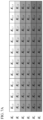
- FIGS. 7 A to 7 D illustrate an example of a process of compensating a pixel value per row in a coordinate transformed image.
- a row K 0 corresponds to a region (background region) where the attenuation due to the bone does not occur and the remaining rows K 1 , K 2 , K 3 , K 4 , . . . correspond to a region (foreground region) where the attenuation due to the bone occurs.
- a process of sequentially compensating the remaining rows K 1 , K 2 , K 3 , K 4 , . . . from the row K 0 may be expressed as in the following equation.
- a 0 K 0
- a n ⁇ ( A n-1 ,K n ) [Equation 6]
- A means the compensated row
- a subscript 0, 1, . . . , n ⁇ 1, n means an index of the row
- ⁇ means a function of performing the compensation. That is, A n-1 means a row which does not have the attenuation or is compensated, K n means a row to be compensated, which is adjacent to A n-1 , and A n means a row compensated for K n . Equation 6 expresses that it is repeated that the row K 1 is compensated based on the row K 0 where the attenuation due to the bone does not occur, and then a next row to the compensated row is compensated.
- the image compensation unit 132 sets multiple blocks having a predetermined size, which belong to the compensated row (or the row where the attenuation does not occur) and the row to be compensated, and include the corresponding pixel, with respect to a pixel to be compensated, in order to remove an attenuation component due to the bone from the pixel value of the row where the attenuation occurs.
- a difference between an average of the pixel values of the compensated rows and an average of the pixel values of the row to be compensated in the respective blocks is calculated, and an average of the difference of the multiple blocks is added to the pixel value of the corresponding pixel to compensate the pixel value.
- W means a width of the block
- n means an index of the row
- x means an index of a column.
- the compensated row A 1 is the row to be compensated is K 2
- the pixel to be compensated is K 2,2 .
- B1, B2, and B3 which are blocks having a width of 3 are set, which belong to the row A 1 and the row K 2 and include the pixel K 2,2 .
- a difference between an average of A 1,0 , A 1,1 , and A 1,2 and an average of K 2,0 , K 2,1 , and K 2,2 is calculated, with respect to block B2, a difference between an average of A 1,1 , A 1,2 , and A 1,3 and an average of K 2,1 , K 2,2 , and K 2,3 is calculated, and with respect to block B3, a difference between an average of A 1,2 , A 1,3 , and A 1,4 and an average of K 2,2 , K 2,3 , and K 2,4 is calculated.
- a value acquired by adding an average of the difference calculated with respect to the blocks B1, B2, and B3 to K 2,2 becomes A 2,2 of FIG. 7 C .
- a compensation first coordinate transformed image A upper and a compensated second coordinate transformed image A below may be obtained from the first coordinate transformed image K upper and the second coordinate transformed image K below , respectively.
- step 270 the coordinate system inversion transform unit 133 generates a compensated first X-ray image and a compensated second X-ray image through coordinate system inverse transform corresponding to the coordinate transformed image in step 250 above from the compensated first coordinate transformed image and the compensated second coordinate transformed image, respectively.
- the image synthesis unit 140 synthesizes the compensated first X-ray image and the compensated second X-ray image to obtain the bone-suppressed X-ray image.
- the compensated first X-ray image is an image in which the compensation is performed based on the upper contour line of the bone and the compensated second X-ray image corresponds to an image in which the compensation is performed based on the lower contour line of the bone. Accordingly, the compensated first X-ray image and the compensated second X-ray image, a boundary between a region where the compensation is performed and a region where the compensation is not performed may be remarkably shown.
- a natural image may be obtained by using an image blending technique such as an alpha blending or Poisson image blending technique.
- the bone-suppressed X-ray image may be obtained without a problem as a whole through the exemplary embodiment of the present invention, but compensation performance may also deteriorate partially around a spine or a region where the pixel value shows a large difference from a surrounding like a case where other materials are jointly photographed.
- modified exemplary embodiments of a process of compensating the pixel value will be described.
- the process of compensating the pixel value in step 260 above is based on an assumption that the degree of the attenuation due to the bone is equal in the same block.
- a block which does not satisfy the assumption may be present in the image.
- FIG. 8 illustrates an example of a block satisfying an assumption that a degree of attenuation due to a bone is similar in a block and a block not satisfying the assumption. Referring to FIG. 8 , when it is assumed that there is a difference in pixel value between a white shade region and a gray shade region, blocks B1 and B8 satisfy that the assumption that the attenuation due to the bone is equal in the block, but block B5 does not satisfy the assumption (that is to say, a boundary portion of the spine).
- the weight for each block may be determined according to a distribution (for example, a standard deviation) of a difference in pixel value between a compensated pixel (or pixel where the attenuation does not occur) and a pixel to be compensated for each column in the corresponding block.
- a distribution for example, a standard deviation
- the weight W inter for each block may be calculated according to the following equation, for example.
- n means the row to be compensated
- S n represents the difference in pixel value between the compensated pixel (or pixel where the attenuation does not occur) and the pixel to be compensated for each column in the corresponding block
- stddev(S n,k ) means a standard deviation of S n for a k-th block
- W inter n,k means a weight of the k-th block. That is, W inter n,k represents a reciprocal number of the standard deviation of S n . Accordingly, as the standard deviation of S n is smaller, a higher weight is granted and as the standard deviation of S n is larger, a lower weight is granted. Since the corresponding block satisfies the assumption that the attenuation due to the bone is similar in the block as the standard deviation of S n is smaller, the high weight is granted to the block to obtain a compensated image having small artifact.
- FIG. 9 illustrates an example of a case where a weight is differently granted to a pixel to be compensated for each block.
- blocks B3, B4, and B5 are set.
- S n has values of A 0,2 -K 1,2 , A 0,3 -K 1,3 , and A 0,4 -K 1,4
- S n has values of A 0,3 -K 1,3 , A 0,4 -K 1,4 , and A 0,5-1 -K 1,5
- S n has values of A 0,4 -K 1,4 , A 0,5 -K 1,5 , and A 0,6 -K 1,6 .
- FIG. 10 illustrates an example of a case where there are large differences between any pixel and other pixels in the block.
- the pixel value of the pixel K 1,5 shows a large difference from those of other pixels in the block B.
- the average of the pixel value may be calculated by granting a different weight for each pixel.
- the weight for each pixel in the block may be determined according to a difference between the average of the pixel value of the row to be compensated in the block and the pixel value of the corresponding pixel. That is to say, a high weight is granted to a pixel having a pixel value close to the average of the pixel value of the row to be compensated and a low weight is granted to a pixel having a pixel value distant from the average of the pixel value of the row to be compensated.
- a weight of an W intra n,k,j pixel in a k-th block in an n-th row may be calculated according to the equation.
- a(a>0) represents a parameter for determining a reflection level of the weight
- K n,k,j means a pixel value of the j-th pixel in the k-th block of the n-th row
- K n,k means the average of the pixel value in k-th the block of the n-th row.
- W intra n,k,j has a value close to 1 and as K n,k,j is distant from K n,k , W intra n,k,j has a value close to 0.
- the low weight is granted to the pixel which shows a large difference from other pixels in the block as such to increase reliability in calculating the difference between the average of the pixel value of the compensated row and the average of the pixel value of the row to be compensated in the block.
- Equation 7 which is a pixel value compensation function
- both the weight for each block and the weight for each pixel in the block are reflected to Equation 10, but in some exemplary embodiments, only the weight for each block may also be reflected and only the weight for each pixel in the block may also be reflected.
- FIG. 11 illustrates an example of a process of acquiring an X-ray image in which a bone is suppressed from an original X-ray image according to an exemplary embodiment of the present invention.
- a contour line (b) is extracted from an original X-ray image (a), and a bone-suppressed X-ray image (c) is obtained through pixel compensation of a region which belongs to the bone.
- the apparatus may include a processor, a memory storing and executing program data, a permanent storage such as a disk drive, a communication port communicating with an external apparatus, a user interface device such as a touch panel, a key, a button, etc., and the like.
- Methods implemented by a software module or algorithm as computer-readable codes or program commands executable on the processor may be stored in computer-readable recording media.
- the computer-readable recording media include magnetic storage media (e.g., a read-only memory (ROM), a random-access memory (RAM), a floppy disk, a hard disk, etc.) and optical reading media (e.g., a CD-ROM, a digital versatile disc (DVD), etc.).
- the computer-readable recording media may be stored and executed as codes which may be distributed in the computer system connected through a network and read by a computer in a distribution method.
- the media are readable by a computer, stored in the memory executable by the processor.
- the exemplary embodiments of the present invention may be represented by functional block components and various processing steps.
- the functional blocks may be implemented by various numbers of hardware or/and software components executing specific functions.
- the exemplary embodiments may adopt integrated circuit components including a memory, processing, logic, a look-up table, etc., which may execute various functions by control by one or more microprocessors or by other control devices.
- the component of the present invention may be executed by software programming or software elements
- the exemplary embodiment includes a data structure, processes, routines, or various algorithms implemented by a combination of other programming components to be implemented by a programming or scripting language such as C, C++, Java, assembler, etc.
- Functional aspects may be implemented by an algorithm executed by one or more processors.
- the exemplary embodiment may adopt related art for electronic environmental setting, signal processing, and/or data processing.
- Terminologies such as “mechanism”, “element”, “means”, and “component” may be widely used, and are not limited to mechanical and physical components.
- the terminologies may mean a meaning of a series of routines of software in link with a processor, etc.
- connection or connection members of lines among components exemplarily represent functions connections and/or physical or circuitry connections and may be represented as various functional connections, physical connections, or circuitry connections which are replaceable or added in an actual device. Further, unless otherwise specified, such as “essential”, “important”, etc., the connections may not be components particularly required for application of the present invention.
Landscapes
- Engineering & Computer Science (AREA)
- Physics & Mathematics (AREA)
- General Physics & Mathematics (AREA)
- Theoretical Computer Science (AREA)
- Health & Medical Sciences (AREA)
- Life Sciences & Earth Sciences (AREA)
- Medical Informatics (AREA)
- Computer Vision & Pattern Recognition (AREA)
- Pathology (AREA)
- Heart & Thoracic Surgery (AREA)
- Nuclear Medicine, Radiotherapy & Molecular Imaging (AREA)
- Optics & Photonics (AREA)
- Biophysics (AREA)
- Radiology & Medical Imaging (AREA)
- Biomedical Technology (AREA)
- High Energy & Nuclear Physics (AREA)
- Molecular Biology (AREA)
- Surgery (AREA)
- Animal Behavior & Ethology (AREA)
- General Health & Medical Sciences (AREA)
- Public Health (AREA)
- Veterinary Medicine (AREA)
- Image Processing (AREA)
- Apparatus For Radiation Diagnosis (AREA)
Abstract
Description
C(x)=αP 1(x)+βP t+1(x)(S i,x ≤x≤S t+1,x) [Equation 2]
T:(x,y)
K(T(x,y))=I(x,y) [Equation 5]
A 0 =K 0
A n=ƒ(A n-1 ,K n) [Equation 6]
Claims (18)
Applications Claiming Priority (2)
| Application Number | Priority Date | Filing Date | Title |
|---|---|---|---|
| KR10-2021-0031914 | 2021-03-11 | ||
| KR1020210031914A KR102480389B1 (en) | 2021-03-11 | 2021-03-11 | Method and apparatus for bone suppression in X-ray Image |
Publications (2)
| Publication Number | Publication Date |
|---|---|
| US20220292656A1 US20220292656A1 (en) | 2022-09-15 |
| US12136194B2 true US12136194B2 (en) | 2024-11-05 |
Family
ID=83193902
Family Applications (1)
| Application Number | Title | Priority Date | Filing Date |
|---|---|---|---|
| US17/691,863 Active 2043-03-31 US12136194B2 (en) | 2021-03-11 | 2022-03-10 | Method and apparatus for bone suppression in X-ray image |
Country Status (2)
| Country | Link |
|---|---|
| US (1) | US12136194B2 (en) |
| KR (1) | KR102480389B1 (en) |
Families Citing this family (1)
| Publication number | Priority date | Publication date | Assignee | Title |
|---|---|---|---|---|
| WO2023059173A1 (en) | 2021-10-06 | 2023-04-13 | 주식회사 엘지에너지솔루션 | Separator for secondary battery |
Citations (7)
| Publication number | Priority date | Publication date | Assignee | Title |
|---|---|---|---|---|
| US20090214099A1 (en) * | 2008-02-27 | 2009-08-27 | Siemens Computer Aided Diagnosis Ltd. | Method of suppressing obscuring features in an image |
| US20120257810A1 (en) | 2009-12-22 | 2012-10-11 | Koninklijke Philips Electronics N.V. | Bone suppression in x-ray radiograms |
| WO2015157067A1 (en) * | 2014-04-08 | 2015-10-15 | VuComp, Inc | Lung segmentation and bone suppression techniques for radiographic images |
| US9269139B2 (en) | 2011-10-28 | 2016-02-23 | Carestream Health, Inc. | Rib suppression in radiographic images |
| US20170270640A1 (en) * | 2016-03-16 | 2017-09-21 | General Electric Company | Method and system for reducing grid line artifacts in x-ray image |
| KR20190143708A (en) | 2018-06-21 | 2019-12-31 | 한국외국어대학교 연구산학협력단 | Apparatus and Method for Bone Suppression from X-ray Image based on Deep Learning |
| US20200167930A1 (en) * | 2017-06-16 | 2020-05-28 | Ucl Business Ltd | A System and Computer-Implemented Method for Segmenting an Image |
Family Cites Families (3)
| Publication number | Priority date | Publication date | Assignee | Title |
|---|---|---|---|---|
| EP2051205B1 (en) | 2007-10-17 | 2012-03-14 | Deutsches Krebsforschungszentrum | Method, computer program and workstation for removing undesirable objects for a digital medical image |
| CN106023200B (en) | 2016-05-19 | 2019-06-28 | 四川大学 | A kind of x-ray chest radiograph image rib cage suppressing method based on Poisson model |
| KR102216818B1 (en) * | 2018-12-28 | 2021-02-17 | 부산대학교 산학협력단 | Manufacturing method for estimating position of liver with two-dimensional of dual-energy x-ray absorptiometry |
-
2021
- 2021-03-11 KR KR1020210031914A patent/KR102480389B1/en active Active
-
2022
- 2022-03-10 US US17/691,863 patent/US12136194B2/en active Active
Patent Citations (7)
| Publication number | Priority date | Publication date | Assignee | Title |
|---|---|---|---|---|
| US20090214099A1 (en) * | 2008-02-27 | 2009-08-27 | Siemens Computer Aided Diagnosis Ltd. | Method of suppressing obscuring features in an image |
| US20120257810A1 (en) | 2009-12-22 | 2012-10-11 | Koninklijke Philips Electronics N.V. | Bone suppression in x-ray radiograms |
| US9269139B2 (en) | 2011-10-28 | 2016-02-23 | Carestream Health, Inc. | Rib suppression in radiographic images |
| WO2015157067A1 (en) * | 2014-04-08 | 2015-10-15 | VuComp, Inc | Lung segmentation and bone suppression techniques for radiographic images |
| US20170270640A1 (en) * | 2016-03-16 | 2017-09-21 | General Electric Company | Method and system for reducing grid line artifacts in x-ray image |
| US20200167930A1 (en) * | 2017-06-16 | 2020-05-28 | Ucl Business Ltd | A System and Computer-Implemented Method for Segmenting an Image |
| KR20190143708A (en) | 2018-06-21 | 2019-12-31 | 한국외국어대학교 연구산학협력단 | Apparatus and Method for Bone Suppression from X-ray Image based on Deep Learning |
Also Published As
| Publication number | Publication date |
|---|---|
| KR20220127529A (en) | 2022-09-20 |
| KR102480389B1 (en) | 2022-12-23 |
| US20220292656A1 (en) | 2022-09-15 |
Similar Documents
| Publication | Publication Date | Title |
|---|---|---|
| Lyu et al. | Encoding metal mask projection for metal artifact reduction in computed tomography | |
| Song et al. | Bottleneck feature supervised U-Net for pixel-wise liver and tumor segmentation | |
| Ko et al. | Rigid and non-rigid motion artifact reduction in X-ray CT using attention module | |
| US11941807B2 (en) | Artificial intelligence-based medical image processing method and medical device, and storage medium | |
| CN110827216B (en) | Multi-Generator Generative Adversarial Network Learning Method for Image Denoising | |
| CN113256529B (en) | Image processing method, image processing device, computer equipment and storage medium | |
| CN115760651B (en) | Improved RegGAN low-dose CT image denoising method and related device | |
| Li et al. | Low-dose computed tomography image reconstruction via a multistage convolutional neural network with autoencoder perceptual loss network | |
| CN118229741A (en) | Image processing method and image processing device | |
| US12136194B2 (en) | Method and apparatus for bone suppression in X-ray image | |
| Li et al. | FDDM: unsupervised medical image translation with a frequency-decoupled diffusion model | |
| Huang et al. | Sparse Bayesian deep learning for cross domain medical image reconstruction | |
| Navaneethan et al. | MSACNet: an advanced multi-scale artifact correction network with motion-adaptive attention for the high-fidelity low-dose x-ray imaging | |
| US10192308B2 (en) | Image processing apparatus, image processing method, and storage medium | |
| Schiller et al. | xU-NetFullSharp: The Novel Deep Learning Architecture for Chest X-ray Bone Shadow Suppression | |
| Tzikas et al. | Variational bayesian blind image deconvolution with student-t priors | |
| CN113052774A (en) | Image optimization method, training method of related model, related device and equipment | |
| CN119722498A (en) | Medical image enhancement method, device, equipment and medium based on deep features | |
| Sandhu et al. | A Comprehensive review of GAN-based denoising models for low-dose computed tomography images | |
| Carmo et al. | Evaluating multiple combinations of models and encoders to enhance LDCT images: NC do Carmo et al. | |
| CN113744149B (en) | A deep learning post-processing method to solve the over-smoothing problem of low-dose CT images | |
| Yoon et al. | CLIMAR: classified linear interpolation based metal artifact reduction for severe metal artifact reduction in x-ray CT imaging | |
| Aljaddouh et al. | Enhancing boundary accuracy in semantic segmentation of chest x-ray images using gaussian process regression | |
| EP4437488A1 (en) | A contrast enhancing agent for diagnostic imaging methods and systems | |
| Rue et al. | Misalignment-aware mri-to-ct synthesis for lung segmentation on mri |
Legal Events
| Date | Code | Title | Description |
|---|---|---|---|
| AS | Assignment |
Owner name: VIEWORKS CO., LTD., KOREA, REPUBLIC OF Free format text: ASSIGNMENT OF ASSIGNORS INTEREST;ASSIGNOR:HAN, YOUNG JOO;REEL/FRAME:059228/0186 Effective date: 20220310 |
|
| FEPP | Fee payment procedure |
Free format text: ENTITY STATUS SET TO UNDISCOUNTED (ORIGINAL EVENT CODE: BIG.); ENTITY STATUS OF PATENT OWNER: SMALL ENTITY |
|
| FEPP | Fee payment procedure |
Free format text: ENTITY STATUS SET TO SMALL (ORIGINAL EVENT CODE: SMAL); ENTITY STATUS OF PATENT OWNER: SMALL ENTITY |
|
| STPP | Information on status: patent application and granting procedure in general |
Free format text: DOCKETED NEW CASE - READY FOR EXAMINATION |
|
| STPP | Information on status: patent application and granting procedure in general |
Free format text: NON FINAL ACTION MAILED |
|
| STPP | Information on status: patent application and granting procedure in general |
Free format text: RESPONSE TO NON-FINAL OFFICE ACTION ENTERED AND FORWARDED TO EXAMINER |
|
| STPP | Information on status: patent application and granting procedure in general |
Free format text: NOTICE OF ALLOWANCE MAILED -- APPLICATION RECEIVED IN OFFICE OF PUBLICATIONS |
|
| STPP | Information on status: patent application and granting procedure in general |
Free format text: PUBLICATIONS -- ISSUE FEE PAYMENT RECEIVED |
|
| STPP | Information on status: patent application and granting procedure in general |
Free format text: PUBLICATIONS -- ISSUE FEE PAYMENT VERIFIED |
|
| STCF | Information on status: patent grant |
Free format text: PATENTED CASE |