US12098385B2 - Gene therapy strategy to restore cardiac electrical and structural function in arrhythmogenic right ventricular cardiomyopathy - Google Patents
Gene therapy strategy to restore cardiac electrical and structural function in arrhythmogenic right ventricular cardiomyopathy Download PDFInfo
- Publication number
- US12098385B2 US12098385B2 US16/862,326 US202016862326A US12098385B2 US 12098385 B2 US12098385 B2 US 12098385B2 US 202016862326 A US202016862326 A US 202016862326A US 12098385 B2 US12098385 B2 US 12098385B2
- Authority
- US
- United States
- Prior art keywords
- arvc
- cardiac
- connexin
- cell
- hipsc
- Prior art date
- Legal status (The legal status is an assumption and is not a legal conclusion. Google has not performed a legal analysis and makes no representation as to the accuracy of the status listed.)
- Active, expires
Links
Images


Classifications
-
- C—CHEMISTRY; METALLURGY
- C12—BIOCHEMISTRY; BEER; SPIRITS; WINE; VINEGAR; MICROBIOLOGY; ENZYMOLOGY; MUTATION OR GENETIC ENGINEERING
- C12N—MICROORGANISMS OR ENZYMES; COMPOSITIONS THEREOF; PROPAGATING, PRESERVING, OR MAINTAINING MICROORGANISMS; MUTATION OR GENETIC ENGINEERING; CULTURE MEDIA
- C12N15/00—Mutation or genetic engineering; DNA or RNA concerning genetic engineering, vectors, e.g. plasmids, or their isolation, preparation or purification; Use of hosts therefor
- C12N15/09—Recombinant DNA-technology
- C12N15/63—Introduction of foreign genetic material using vectors; Vectors; Use of hosts therefor; Regulation of expression
- C12N15/79—Vectors or expression systems specially adapted for eukaryotic hosts
- C12N15/85—Vectors or expression systems specially adapted for eukaryotic hosts for animal cells
- C12N15/86—Viral vectors
- C12N15/861—Adenoviral vectors
-
- A—HUMAN NECESSITIES
- A01—AGRICULTURE; FORESTRY; ANIMAL HUSBANDRY; HUNTING; TRAPPING; FISHING
- A01K—ANIMAL HUSBANDRY; AVICULTURE; APICULTURE; PISCICULTURE; FISHING; REARING OR BREEDING ANIMALS, NOT OTHERWISE PROVIDED FOR; NEW BREEDS OF ANIMALS
- A01K67/00—Rearing or breeding animals, not otherwise provided for; New or modified breeds of animals
- A01K67/027—New or modified breeds of vertebrates
- A01K67/0275—Genetically modified vertebrates, e.g. transgenic
-
- A—HUMAN NECESSITIES
- A61—MEDICAL OR VETERINARY SCIENCE; HYGIENE
- A61K—PREPARATIONS FOR MEDICAL, DENTAL OR TOILETRY PURPOSES
- A61K35/00—Medicinal preparations containing materials or reaction products thereof with undetermined constitution
- A61K35/12—Materials from mammals; Compositions comprising non-specified tissues or cells; Compositions comprising non-embryonic stem cells; Genetically modified cells
- A61K35/34—Muscles; Smooth muscle cells; Heart; Cardiac stem cells; Myoblasts; Myocytes; Cardiomyocytes
-
- A—HUMAN NECESSITIES
- A61—MEDICAL OR VETERINARY SCIENCE; HYGIENE
- A61K—PREPARATIONS FOR MEDICAL, DENTAL OR TOILETRY PURPOSES
- A61K38/00—Medicinal preparations containing peptides
- A61K38/16—Peptides having more than 20 amino acids; Gastrins; Somatostatins; Melanotropins; Derivatives thereof
- A61K38/17—Peptides having more than 20 amino acids; Gastrins; Somatostatins; Melanotropins; Derivatives thereof from animals; from humans
- A61K38/177—Receptors; Cell surface antigens; Cell surface determinants
-
- A—HUMAN NECESSITIES
- A61—MEDICAL OR VETERINARY SCIENCE; HYGIENE
- A61K—PREPARATIONS FOR MEDICAL, DENTAL OR TOILETRY PURPOSES
- A61K48/00—Medicinal preparations containing genetic material which is inserted into cells of the living body to treat genetic diseases; Gene therapy
- A61K48/005—Medicinal preparations containing genetic material which is inserted into cells of the living body to treat genetic diseases; Gene therapy characterised by an aspect of the 'active' part of the composition delivered, i.e. the nucleic acid delivered
- A61K48/0058—Nucleic acids adapted for tissue specific expression, e.g. having tissue specific promoters as part of a contruct
-
- A—HUMAN NECESSITIES
- A61—MEDICAL OR VETERINARY SCIENCE; HYGIENE
- A61K—PREPARATIONS FOR MEDICAL, DENTAL OR TOILETRY PURPOSES
- A61K48/00—Medicinal preparations containing genetic material which is inserted into cells of the living body to treat genetic diseases; Gene therapy
- A61K48/0075—Medicinal preparations containing genetic material which is inserted into cells of the living body to treat genetic diseases; Gene therapy characterised by an aspect of the delivery route, e.g. oral, subcutaneous
-
- A—HUMAN NECESSITIES
- A61—MEDICAL OR VETERINARY SCIENCE; HYGIENE
- A61P—SPECIFIC THERAPEUTIC ACTIVITY OF CHEMICAL COMPOUNDS OR MEDICINAL PREPARATIONS
- A61P9/00—Drugs for disorders of the cardiovascular system
-
- C—CHEMISTRY; METALLURGY
- C12—BIOCHEMISTRY; BEER; SPIRITS; WINE; VINEGAR; MICROBIOLOGY; ENZYMOLOGY; MUTATION OR GENETIC ENGINEERING
- C12N—MICROORGANISMS OR ENZYMES; COMPOSITIONS THEREOF; PROPAGATING, PRESERVING, OR MAINTAINING MICROORGANISMS; MUTATION OR GENETIC ENGINEERING; CULTURE MEDIA
- C12N15/00—Mutation or genetic engineering; DNA or RNA concerning genetic engineering, vectors, e.g. plasmids, or their isolation, preparation or purification; Use of hosts therefor
- C12N15/09—Recombinant DNA-technology
- C12N15/63—Introduction of foreign genetic material using vectors; Vectors; Use of hosts therefor; Regulation of expression
- C12N15/79—Vectors or expression systems specially adapted for eukaryotic hosts
- C12N15/85—Vectors or expression systems specially adapted for eukaryotic hosts for animal cells
- C12N15/86—Viral vectors
-
- A—HUMAN NECESSITIES
- A01—AGRICULTURE; FORESTRY; ANIMAL HUSBANDRY; HUNTING; TRAPPING; FISHING
- A01K—ANIMAL HUSBANDRY; AVICULTURE; APICULTURE; PISCICULTURE; FISHING; REARING OR BREEDING ANIMALS, NOT OTHERWISE PROVIDED FOR; NEW BREEDS OF ANIMALS
- A01K2217/00—Genetically modified animals
- A01K2217/07—Animals genetically altered by homologous recombination
- A01K2217/072—Animals genetically altered by homologous recombination maintaining or altering function, i.e. knock in
-
- A—HUMAN NECESSITIES
- A01—AGRICULTURE; FORESTRY; ANIMAL HUSBANDRY; HUNTING; TRAPPING; FISHING
- A01K—ANIMAL HUSBANDRY; AVICULTURE; APICULTURE; PISCICULTURE; FISHING; REARING OR BREEDING ANIMALS, NOT OTHERWISE PROVIDED FOR; NEW BREEDS OF ANIMALS
- A01K2217/00—Genetically modified animals
- A01K2217/07—Animals genetically altered by homologous recombination
- A01K2217/075—Animals genetically altered by homologous recombination inducing loss of function, i.e. knock out
-
- A—HUMAN NECESSITIES
- A01—AGRICULTURE; FORESTRY; ANIMAL HUSBANDRY; HUNTING; TRAPPING; FISHING
- A01K—ANIMAL HUSBANDRY; AVICULTURE; APICULTURE; PISCICULTURE; FISHING; REARING OR BREEDING ANIMALS, NOT OTHERWISE PROVIDED FOR; NEW BREEDS OF ANIMALS
- A01K2227/00—Animals characterised by species
- A01K2227/10—Mammal
- A01K2227/105—Murine
-
- A—HUMAN NECESSITIES
- A01—AGRICULTURE; FORESTRY; ANIMAL HUSBANDRY; HUNTING; TRAPPING; FISHING
- A01K—ANIMAL HUSBANDRY; AVICULTURE; APICULTURE; PISCICULTURE; FISHING; REARING OR BREEDING ANIMALS, NOT OTHERWISE PROVIDED FOR; NEW BREEDS OF ANIMALS
- A01K2267/00—Animals characterised by purpose
- A01K2267/03—Animal model, e.g. for test or diseases
- A01K2267/0306—Animal model for genetic diseases
-
- A—HUMAN NECESSITIES
- A01—AGRICULTURE; FORESTRY; ANIMAL HUSBANDRY; HUNTING; TRAPPING; FISHING
- A01K—ANIMAL HUSBANDRY; AVICULTURE; APICULTURE; PISCICULTURE; FISHING; REARING OR BREEDING ANIMALS, NOT OTHERWISE PROVIDED FOR; NEW BREEDS OF ANIMALS
- A01K2267/00—Animals characterised by purpose
- A01K2267/03—Animal model, e.g. for test or diseases
- A01K2267/035—Animal model for multifactorial diseases
- A01K2267/0375—Animal model for cardiovascular diseases
-
- C—CHEMISTRY; METALLURGY
- C12—BIOCHEMISTRY; BEER; SPIRITS; WINE; VINEGAR; MICROBIOLOGY; ENZYMOLOGY; MUTATION OR GENETIC ENGINEERING
- C12N—MICROORGANISMS OR ENZYMES; COMPOSITIONS THEREOF; PROPAGATING, PRESERVING, OR MAINTAINING MICROORGANISMS; MUTATION OR GENETIC ENGINEERING; CULTURE MEDIA
- C12N2710/00—MICROORGANISMS OR ENZYMES; COMPOSITIONS THEREOF; PROPAGATING, PRESERVING, OR MAINTAINING MICROORGANISMS; MUTATION OR GENETIC ENGINEERING; CULTURE MEDIA dsDNA viruses
- C12N2710/00011—Details
- C12N2710/10011—Adenoviridae
- C12N2710/10311—Mastadenovirus, e.g. human or simian adenoviruses
- C12N2710/10341—Use of virus, viral particle or viral elements as a vector
- C12N2710/10343—Use of virus, viral particle or viral elements as a vector viral genome or elements thereof as genetic vector
-
- C—CHEMISTRY; METALLURGY
- C12—BIOCHEMISTRY; BEER; SPIRITS; WINE; VINEGAR; MICROBIOLOGY; ENZYMOLOGY; MUTATION OR GENETIC ENGINEERING
- C12N—MICROORGANISMS OR ENZYMES; COMPOSITIONS THEREOF; PROPAGATING, PRESERVING, OR MAINTAINING MICROORGANISMS; MUTATION OR GENETIC ENGINEERING; CULTURE MEDIA
- C12N2750/00—MICROORGANISMS OR ENZYMES; COMPOSITIONS THEREOF; PROPAGATING, PRESERVING, OR MAINTAINING MICROORGANISMS; MUTATION OR GENETIC ENGINEERING; CULTURE MEDIA ssDNA viruses
- C12N2750/00011—Details
- C12N2750/14011—Parvoviridae
- C12N2750/14111—Dependovirus, e.g. adenoassociated viruses
- C12N2750/14141—Use of virus, viral particle or viral elements as a vector
- C12N2750/14143—Use of virus, viral particle or viral elements as a vector viral genome or elements thereof as genetic vector
-
- C—CHEMISTRY; METALLURGY
- C12—BIOCHEMISTRY; BEER; SPIRITS; WINE; VINEGAR; MICROBIOLOGY; ENZYMOLOGY; MUTATION OR GENETIC ENGINEERING
- C12N—MICROORGANISMS OR ENZYMES; COMPOSITIONS THEREOF; PROPAGATING, PRESERVING, OR MAINTAINING MICROORGANISMS; MUTATION OR GENETIC ENGINEERING; CULTURE MEDIA
- C12N2840/00—Vectors comprising a special translation-regulating system
- C12N2840/007—Vectors comprising a special translation-regulating system cell or tissue specific
Definitions
- constructs include a construct or vector capable of conferring expression of connexin 43 on cardiac tissue.
- the construct or vector is a plasmid or a viral vector, such as an adenovirus construct.
- the construct or vector is or an adeno-associated virus construct.
- the connexin 43 polypeptide sequence is the 382 amino acid sequence of P17302 (CXA_1HUMAN; UniProtKB) (SEQ ID NO: 1).
- the encoding polynucleotide may have a polynucleotide sequence associated with P17302 or any other nucleic acid sequence encoding the same polypeptide sequence.
- the encoding nucleic acid sequence is GenBank CR541660.1 (SEQ ID NO: 2).
- cardiac electrical (non-structural) and structural dysfunction is reduced and structural integrity is improved.
- cardiac physiologic dysfunction is reduced.
- survival is prolonged.
- the mechanical junction gene or protein is N-cadherin, desmoplakin (DSP), plakoglobin (JUP) plakophillin 2 (PKP2) and/or desmoglein 2 (DSG2).
- Some embodiments are methods of increasing or upregulating the expression of one or more genes in a cell wherein the genes are selected from the group consisting of N-cadherin, desmoplakin (DSP), plakoglobin (JUP) plakophillin 2 (PKP2) and desmoglein 2 (DSG2) by contacting the cell with an agent that upregulates connexin 43 nucleic acid or polypeptide.
- the agent upregulating connexin 43 nucleic acid or polypeptide is a vector encoding a connexin 43 polypeptide sequence or functional fragments thereof.
- a nucleotide sequence encoding the connexin polypeptide is operably linked to a promoter that is active in cardiac muscle tissue.
- the cell in which expression of the one or more genes is increased or upregulated is a cardiac muscle cell, a cardiac fibroblast, a cardiomyocyte or a cardiac macrophage.
- the cell is in or from a subject having cardiovascular disease.
- the cardiovascular disease is arrhythmogenic right ventricular cardiomyopathy (ARVC).
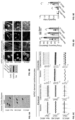
- FIGS. 1 A- 1 G depict that restoration of connexin 43 protein levels closer to wild type levels can rescue physiological abnormalities in ARVC-plakophilin-2 mutant (PKP2 mut ) hiPSC-derived cardiomyocytes
- FIGS. 1 C- 1 D depict representative impedance (contractility) trace (note frequency and shape of spikes in the trace) for Non-ARVC and ARVC-PKP2mut (ARVC-S) hiPSC-derived cardiomyocytes at basal level ( FIG. 1 C ) and under ISO stimulation ( FIG. 1 D ), with Ad-GFP or Ad43 infection for 48 h.
- FIG. 1 F depicts quantification of firing irregularity index under ISO stimulus of Non-ARVC and ARVC-PKP2 mut (ARVC-S) with Ad-GFP or Ad43 infection.
- FIG. 1 G depicts expression levels of connexin 43 (Cx43), desmoplakin (DSP), desmoglein-2 (DSG2), plakoglobin (JUP) and plakophilin-2 (PKP2) in non-ARVC, ARVC-PKP2 mut (ARVC-S) and ARVC-demosglein-2 mutant (DSG2 mut (ARVC-E, also known as ARVC-Electrical (E)) hiPSC-derived cardiomyocytes with Ad-GFP or Ad43 infection for 48 h., ⁇ -myosin heavy chain ( ⁇ -MHC), cardiomyocytes loading control, glyceraldehyde 3-phosphate dehydrogenase (GAPDH), loading control.
- Cx43 connexin 43
- DSP desmoplakin
- DSG2 desmoglein-2
- JUP plakoglobin
- PGP2 mut plakophilin-2
- ARVC-S ARVC-PKP2 mut
- ARVC-E
- FIGS. 2 A- 2 B depict that physiological abnormalities in ARVC-S cardiomyocytes can be rescued by Cx43 overexpression in a dose-dependent manner.
- Representative field potential traces from Non-ARVC ( FIG. 2 A ) and ARVC-PKP2 mut ( FIG. 2 B ) hiPSC-derived cardiomyocytes after dose-dependent Ad43 infection are shown. Note that the irregular frequency and pattern of spikes in ARVC-PKP2 mut hiPSC-derived cardiomyocytes becomes progressively more regular in frequency of spikes with increasing doses (MOI 5, MOI 10 and MOI 20) of Cx43. Scale bar, vertical: amplitude of field potential ( ⁇ V); horizontal: time (1 s). MOI: multiplicity of infection.
- FIGS. 3 A- 3 F depict that restoration of connexin 43 protein levels can rescue physiological abnormalities in ARVC-DSG2 mut (ARVC-E) hiPSC-derived cardiomyocytes under catecholamine (ISO) stimulation, to mimic exercise/stress conditions.
- FIG. 3 C- 3 D depict representative impedance (contractility) trace for Non-ARVC and ARVC-DSG2 mut hiPSC-derived cardiomyocytes at basal level ( FIG. 3 C ) and under ISO stimulation ( FIG. 3 D ) with Ad-GFP or Ad43 infection for 48 h. Scale bar, vertical: cell index; horizontal: time (1 s).
- FIG. 3 E depict representative impedance (contractility) trace for Non-ARVC and ARVC-DSG2 mut hiPSC-derived cardiomyocytes at basal level ( FIG. 3 C ) and under ISO stimulation ( FIG. 3 D ) with Ad-GFP or
- 3 F depicts quantification of firing irregularity index under ISO stimulus of Non-ARVC and ARVC-DSG2 mut (ARVC-E) with Ad-GFP or Ad43 infection.
- FIGS. 4 A- 4 G depict that restoration of connexin 43 protein levels can improve heart rhythm and function in an ARVC mouse model (cardiac-specific desmoplakin knockout model ((DSP-cKO).
- FIG. 4 A depicts the experimental strategy for adeno-associated virus tagged with green fluorescent protein (AAV-GFP)/adeno-associated virus harboring connexin-43 and tagged with GFP (AAV-Cx43) injection and electrophysiological and cardiac function analysis.
- AAV-GFP green fluorescent protein
- AAV-Cx43 GFP
- FIG. 4 B depicts echocardiography of control and DSP-cKO mice (DSP floxed mice; Cre positive) at 6 weeks of age and prior to adeno-associated virus (AAV) injection, showed a significant decrease in fractional shortening (% FS) in DSP-cKO mice compared to control (DSP floxed mice; Cre negative).
- FIG. 4 C depicts echocardiography of control and DSP-cKO mouse at four (10 weeks old) and five (11 weeks old)-weeks post-AAV injection, revealed an improvement in % FS (6% to 15%) in DSP-cKO mouse injected with AAV-Cx43. No significant changes in % FS could be observed in control mice injected with AAV-Cx43.
- FIG. 4 D depicts representative surface ECG traces from control and DSP-cKO mouse at four (10 weeks old) and five (11 weeks old)-weeks post-AAV-Cx43 injection.
- FIG. 4 E depicts quantification of PVCs in DSP-cKO mouse at four (10 weeks old) and five (11 weeks old) weeks post-AAV-Cx43 injection revealed a 1.5 fold reduction in the number of PVCs at five weeks-post AAV-Cx43.
- FIGS. 4 F- 4 H depicts survival ( FIG. 4 H ), western blot analysis ( FIG. 4 F ), and immunostaining analysis of GFP.
- FIG. 4 G revealed restoration of Cx43 protein in a DSP-cKO mouse at five weeks post-AAV-Cx43 injection that survived (DSP-cKO2-AAV-Cx43, green). In contrast, no restoration of Cx43 protein was observed at three weeks post-AAV-Cx43 injection in a DSP-cKO mouse that died (DSP-cKO1-AAV-Cx43, red)
- FIG. 5 depicts a vector map of Ad43 (SEQ ID NO: 7).
- FIG. 6 depicts a vector map of AAV-Cx43 (SEQ ID NO: 8).
- FIG. 7 A-E demonstrate human induced pluripotent stem cell derived cardiomyocytes can recapitulate electrical and structural disease hallmarks found in hearts of distinct ARVC donors.
- 7 A Clinical Features of ARVC donors
- 7 B H&E staining of ARVC cardiac biopsies, white arrowheads point out loss of cardiomyocytes in cardiac biopsy
- 7 C Human Cardiac Biopsies stained for plakoglobin (black arrow indicate JUP stain)
- 7 D TEM analyses of Non-ARVC, ARVC-NS and ARVC-S hiPSC-derived cardiomyocytes, white arrowheads point out the desmosome ultrastructure, scale bar 500 nm.
- 7 E Oil Red 0 staining of Non-ARVC, ARVC-NS and ARVC-S hiPSC-derived cardiac myocytes, Scale bar, 50 ⁇ m.
- FIG. 8 A-E demonstrate ARVC hiPSC-derived cardiomyocytes exhibit a dose-dependent loss of connexin 43 protein levels in a donor-reflective manner, which then predict severity of functional alterations in cells.
- 8 A Connexin 43 immunostaining pattern in cardiac biopsies from non-ARVC, ARVC-NS and ARVC-S patients. Black arrows indicate Cx43 localization
- 8 B Connexin 43 protein expression in hiPSC-derived cardiomyocytes lysates. Cardiac troponin I (TNI) and GAPDH are used as cardiomyocyte and total protein loading controls, respectively.
- TKI Cardiac troponin I
- GAPDH are used as cardiomyocyte and total protein loading controls, respectively.
- FIG. 9 A-E demonstrate restoration of connexin 43 protein levels can rescue physiological abnormalities in both structural and electrical ARVC hiPSC-derived cardiomyocytes in vitro.
- 9 A Protein blot analysis of connexin 43 (Cx43), DSP, DSG2, plakoglobin (JUP) and PKP2 in hiPSC-derived cardiomyocytes infected for 48 h with a control virus (AdYFP) or an adenovirus to connexin 43 (Ad43).
- AdYFP control virus
- AdYFP a control virus
- Ad43 adenovirus to connexin 43
- Alpha-myosin heavy chain ( ⁇ -MHC) and GAPDH were used as cardiomyocyte and total protein loading controls, respectively.
- 9 B Protein blot analysis of connexin 43 (Cx43), DSP, DSG2, plakoglobin (JUP) and PKP2 in hiPSC-derived cardiomyocytes infected for 48 h with a control virus
- FIG. 10 A-C demonstrate cardiac-specific connexin43 restoration in a mouse model with end-stage ARVC is sufficient to prolong life and restore cardiac cell-cell junction gene expression.
- 10 B Desmosomal gene mRNA levels were examined in DSP-control, DSP-cKO+GFP and DSP-cKO+Cx43 groups.
- FIG. 11 depicts abnormal cardiac electrocardiographic recordings from ARVC donors display classic electrical hallmarks (inverted T waves) of ARVC. Grey arrows indicate inverted T wave.
- FIG. 12 depicts karyotype analysis of different clones from non-ARVC and ARVC patient derived human induced pluripotent stem cell lines.
- FIG. 13 shows human induced pluripotent stem cells derived from skin of non-ARVC and ARVC patients expressed prototypical pluri-potency markers.
- OCT4, SOX2, LIN28 and NANOG are pluripotency markers.
- FIG. 14 shows human induced pluripotent stem cells differentiated to embryoid bodies give rise to cell derivatives from the three embryonic germ layers.
- B-III tubulin neuron specific microtubule marker
- Phalloidin stain for actin filaments
- SMA smooth muscle actin
- HNF3b liver specific marker.
- FIG. 15 A-D depicts Pluritest analysis of expression arrays indicates that generated iPS display gene expression patterns that resemble H9 hESCs.
- 15 A graphically depicts pluripotency for the tested cells, and 15 B-D display heat maps of expression levels across the genome.
- FIG. 16 depicts sequence analysis of skin fibroblasts confirms the presence of expected mutations in ARVD/C patients.
- FIG. 17 shows non-ARVC and ARVC human induced pluripotent stem cell lines can be differentiated to cardiomyocytes, which express prototypical cardiac markers. SaAct, sarcomere alpha actinin, NKX2.5, cardiac-specific transcription factor.
- FIG. 18 A-B shows human induced pluripotent stem cell-derived cardiomyocytes display adult-like desmosomal plaques and express key components of the desmosomal, fascia adherens and gap junction.
- 18 A Representative images from Non-ARVC hiPSC-derived cardiomyocytes show DSP (desmoplakin), JUP (plakoglobin), N-CAD (N-cadherin) and Cx43 (connexin 43) stain with DAPI counter-stain (nucleus) and Obsc (obscurin) or SaAct (sarcomere alpha actinin) respectively. Scale bar 10 ⁇ m.
- 18 B Cell lysates from three clones of Non-ARVC hiPSC-derived cardiomyocytes show expression of key components of cardiac intercalated disc. GJ, gap junction. ⁇ -MHC, cardiomyocytes loading control. GAPDH, loading control.
- FIG. 19 shows connexin43 overexpression in Non-ARVC, ARVC-NS and ARVC-S hiPSC derived cardiomyocytes redistribute connexin43 to intercalated disc.
- SaACT sarcomeric alpha actinin
- AdCx43 with YFP tag
- DAPI nuclear stain
- FIG. 20 A-B depict gene delivery efficiency and vector copy number assays in DSP-cKO mice with AAV injection.
- 20 A Representative GFP images from DSP-control and DSP-cKO with AAV9-cTNT-GFP-Cx43 injection heart sections.
- FIG. 21 A-B demonstrate cardiac connexin 43 gene therapy in neonates restores the cardiac mechanical junction complex in 4 week old DSP-cKO mice.
- 21 A Schemata depicting intra-peritoneal injection of AAV in neonate mice to assess end-point analysis in adult heart. Virus dose: 5 ⁇ 10 ⁇ circumflex over ( ) ⁇ 11 GC/mouse.
- 21 B Protein analysis of cell junction proteins in DSP control and DSP-cKO mice treated with no virus ( ⁇ ), or adeno-associated viruses (+) harboring GFP (control) and connexin43. Beta-actin is the loading control.
- FIG. 22 A-D demonstrate cardiac CSN6 loss accelerated desmosomal protein dissolution in vivo.
- Protein blot analyses ( 22 A) and quantification of protein expression levels ( 22 C) in total protein extracts from CSN6-iKO and control hearts at 2 weeks post tamoxifen injection (n 3 per group).
- Protein blot analyses ( 22 B) and quantification of protein expression levels ( 22 D) in total protein extracts from CSN6-iKO and control hearts at 6 weeks post tamoxifen injection (n 3 per group).
- FA fascia adherens.
- GJ gap junction.
- GAPDH was used as a loading control. Data are mean ⁇ s.e.m; two-way ANOVA with Sidak multiple comparison test, *P ⁇ 0.05, **P ⁇ 0.01, ***P ⁇ 0.001, ns, not significant. Experiments were repeated twice with identical results.
- FIG. 23 A-D demonstrate human desmosomal mutations that destabilize CSN6 are sufficient to disrupt CSN6 expression, neddylation and trigger ARVD/C features in mice.
- connexin 43 is known to impact electrical function in the heart, we show that connexin 43 restoration can also prolong life as well as improve contractile (structural) function in a genetic mouse model harboring a severe structural form of ARVC in late stages of disease (life).
- hiPSC Human induced pluripotent stem cells
- ARVC arrhythmogenic right ventricular cardiomyopathy
- ARVC hiPSC-derived cardiac cell phenotypes can basally capture varying (electrical versus structural) cardiac disease phenotypes observed in heart tissue of donor ARVC patients.
- candidate gene approaches exploiting the xCelligence® RTCA CardioECR system in the inventors' ARVC hiPSC lines the inventors have also uncovered new therapeutics that can commonly reverse cardiac physiological defects underlying both electrical and structural forms of ARVC.
- the present invention is based on the seminal discovery that restoring connexin 43 alone (to levels closer to healthy controls) in the face of a structurally compromised heart (via a one shot cardiac-targeted gene therapy) is sufficient for a positive therapeutic outcome.
- the studies described herein suggest that connexin 43 is a primary driver of structural based cardiac disease progression where cardiac muscle cell structural integrity is compromised.
- connexin 43 is found between cardiac muscle cells and cardiac fibroblasts as well as cardiac muscle cells and cardiac macrophages, structural connections to these other associated cell types may also be compromised in a structurally diseased heart, and thus, connexin 43 restoration may also restore structural integrity of these cell-cell interactions. This is an unexpected finding as connexin 43 has never been considered as a therapeutic in the context of a structurally compromised heart.
- the therapeutic methods described herein are targeted primarily towards the rare arrhythmogenic cardiac disease, arrhythmogenic right ventricular cardiomyopathy, which is a cardiac disease where mechanical cell-cell junctions of the desmosome are falling apart/lost and connexin 43 is an early hallmark of the disease. It is believed that connexin 43 restoration may only alleviate the early electrical defects in ARVC hiPSC derived cardiomyocytes that harbored electrical defects. Results shown herein were surprising in late stage structural models of ARVC.
- ARVC hiPSC derived cardiomyocytes with structural hallmarks of disease (human in vitro model) as well as a genetic mouse model mimicking a severe structural form of ARVC (DSP cKO mouse in vivo model), the data revealed that even though a major mechanical (desmosomal) cell-cell junction gene is ablated/mutated and other associated desmosomal cell-cell junction proteins are falling apart/lost, that connexin 43 gene therapy was effective in improving survival, heart rhythm and function in (late) structural stages of ARVC disease.
- ARVC is a predominantly genetic-based heart disease characterized by right, but also recently left, ventricular dysfunction, and fibro-fatty replacement of the myocardium resulting in fatal/severe ventricular arrhythmias leading to sudden cardiac death in young people and athletes.
- ARVC is responsible for 10% of sudden cardiac deaths in people 65 years of age and 24% in people 30 years of age.
- ARVC is thought to be a rare disease as it occurs in 1 in 1000-5000 people, although the prevalence may be higher as some patients are undiagnosed or misdiagnosed due to poor diagnostic markers.
- Growing evidence also reveals earlier onset since pediatric populations ranging from infants to children in their teens are also particularly vulnerable to ARVC, highlighting the critical need to identify and treat patients at an earlier stage of the disease.
- a gene therapy strategy to rescue/treat cardiac electrical and physiological dysfunction in the arrhythmogenic disorder, ARVC, via targeted overexpression of connexin 43 polypeptide in cardiac muscle cells using vector delivery strategies.
- a one-time viral-mediated delivery of connexin 43 polynucleotide encoding connexin 43 polypeptide to heart muscle cells of two ARVC hiPSC patient lines in vitro and a novel mouse model of ARVC in vivo was sufficient to substantially reverse the cardiac electrical and physiological dysfunction associated with ARVC.
- connexin 43 gene therapy treatment strategy can be exploited during late-stage disease states to prolong life by circumventing the sudden death associated with ARVC.
- This strategy can also have treatment applications to other genetic disorders that give rise to arrhythmic sudden death (e.g., hypertrophic cardiomyopathy) as well as late stages of heart failure, where there is increased risk of arrhythmias and death as well as where loss of connexin 43 polypeptide has been reported.
- arrhythmic sudden death e.g., hypertrophic cardiomyopathy
- connexin 43 polypeptides may have a role beyond electrical function and also act as a scaffold to bridge structural connections between cells (independent of classic structural proteins, such as desmosomal proteins) as cardiac function could also be improved in the inventors' models with severe structural disease (e.g., structural ARVC hiPSC line (PKP2 mut ) and the mouse model of ARVC), which is distinct from previous thoughts of the role of this protein in the field as classically associated with electrical function only.
- PGP2 mut structural ARVC hiPSC line
- ARVC mouse model of ARVC
- overexpression refers to an increased level of expression as compared to the disease state, and in particular embodiments in comparison with the absent or minimal level of connexin 43 polypeptide expression found in a disease state, and not as compared to a normal, healthy or wild-type state.
- Restoration of connexin 43 polypeptide expression levels to at least about 5% of normal are beneficial, if not necessarily fully restorative of cardiac function.
- Restoration of connexin 43 expression levels to ⁇ 5%, 10%, 15%, 20%, 25% or as much as 50% of normal levels are effective for restoring normal cardiac function.
- vector has its ordinary meaning in the field and includes the understanding that it is capable of transferring nucleic acid sequences to target cells.
- a vector may comprise a coding sequence capable of being expressed in a target cell.
- vector construct generally refer to any nucleic acid construct capable of directing the expression of a gene of interest and which is useful in transferring the gene of interest into target cells.
- the term includes cloning and expression vectors, as well as integrating vectors.
- AAV adeno-associated virus
- AAV has its ordinary meaning in the field and includes without limitation AAV type 1, AAV type 2, AAV type 3 (including types 3A and 3B), AAV type 4, AAV type 5, AAV type 6, AAV type 7, AAV type 8, AAV type 9, AAV type 10, AAV type 11, AAV2i8, (Asokan et al., Nat Biotechnol. (1):79-82, 2010, which is incorporated herein by reference in its entirety) avian AAV, bovine AAV, canine AAV, equine AAV, and ovine AAV and any other AAV now known or later discovered.
- a number of additional AAV serotypes and clades have been identified, which are also encompassed by the term “AAV.”
- genomic sequences of various AAV and autonomous parvoviruses as well as the sequences of the inverted terminal repeats (ITRs), Rep proteins, and capsid subunits are known in the art. Such sequences may be found in the literature or in public databases such as the GenBank® database.
- cardiomyocyte or “cardiac myocyte” have their ordinary meaning in the field and include a specialized muscle cell that primarily forms the myocardium of the heart. Cardiomyocytes can have five major components: (1) cell membrane (sarcolemma) and T-tubules, for impulse conduction, (2) sarcoplasmic reticulum, a calcium reservoir needed for contraction, (3) contractile elements, (4) mitochondria, and (5) a nucleus. Cardiomyocytes can be subdivided into subtypes including, but not limited to, atrial cardiomyocyte, ventricular cardiomyocyte, sinoatrial (SA) nodal cardiomyocyte, peripheral SA nodal cardiomyocyte, or central SA nodal cardiomyocyte.
- SA sinoatrial
- Stem cells can be propagated to mimic the physiological functions of cardiomyocytes or alternatively, differentiate into cardiomyocytes. This differentiation can be detected by the use of markers selected from, but not limited to, myosin heavy chain, myosin light chain, actinin, troponin, tropomyosin, GATA4, myocyte enhancer factor (Mef)2c, and Nkx-2.5.
- markers selected from, but not limited to, myosin heavy chain, myosin light chain, actinin, troponin, tropomyosin, GATA4, myocyte enhancer factor (Mef)2c, and Nkx-2.5.
- control has its ordinary meaning in the field and includes an alternative subject or sample used in an experiment for comparison purpose.
- a control can be “positive” or “negative.”
- the purpose of the experiment is to determine a correlation of an altered expression level of a gene with a particular phenotype, it is generally preferable to use a positive control (a sample from a subject, carrying such alteration and exhibiting the desired phenotype), and a negative control (a subject or a sample from a subject lacking the altered expression or phenotype).
- stem cell has its ordinary meaning in the field and includes a cell that is in an undifferentiated or partially differentiated state and has the capacity for self-renewal and/or to generate differentiated progeny.
- Self-renewal is defined as the capability of a stem cell to proliferate and give rise to more such stem cells, while maintaining its developmental potential (i.e., totipotent, pluripotent, multipotent, etc.).
- stem cell has its ordinary meaning in the field and includes any stem cell derived from non-embryonic tissue, including fetal, juvenile, and adult tissue.
- Natural somatic stem cells have been isolated from a wide variety of adult tissues including blood, bone marrow, brain, olfactory epithelium, skin, pancreas, skeletal muscle, and cardiac muscle.
- exemplary naturally-occurring somatic stem cells include, but are not limited to, mesenchymal stem cells (MSCs) and neural stem cells (NSCs).
- the stem or progenitor cells can be embryonic stem cells.
- embryonic stem cells has its ordinary meaning in the field and includes stem cells derived from tissue formed after fertilization but before the end of gestation, including pre-embryonic tissue (such as, for example, a blastocyst), embryonic tissue, or fetal tissue taken any time during gestation, typically but not necessarily before approximately 10-12 weeks gestation. Most frequently, embryonic stem cells are pluripotent cells derived from the early embryo or blastocyst. Embryonic stem cells can be obtained directly from suitable tissue, including, but not limited to human tissue, or from established embryonic cell lines. As used herein, the term “embryonic-like stem cells” has its ordinary meaning in the field and includes cells that share one or more, but not all characteristics, of an embryonic stem cell.
- a “pluripotent cell” has its ordinary meaning in the field and includes a less differentiated cell that can give rise to at least two distinct (genotypically and/or phenotypically) further differentiated progeny cells.
- a “pluripotent cell” includes an Induced Pluripotent Stem Cell (iPSC) which is an artificially derived stem cell from a non-pluripotent cell, typically an adult somatic cell, that has been produced by inducing expression of one or more stem cell-specific genes.
- iPSC Induced Pluripotent Stem Cell
- stem cell specific genes include, but are not limited to, the family of octamer transcription factors, i.e.
- Sox Sox1, Sox2, Sox3, Sox 15, and Sox 18
- Kruppel-like factor (KIf) genes i.e. Klf1, Klf2, Klf4, and Klf5
- Myc genes i.e. c-myc and L-myc
- Nanog genes i.e., OCT4, NANOG, and REX1; or LIN28.
- induced pluripotent cell has its ordinary meaning in the field and includes embryonic-like cells reprogrammed to the immature phenotype from adult cells.
- Human iPSCs also express stem cell markers and are capable of generating cells characteristic of all three germ layers.
- the term “effective amount” has its ordinary meaning in the field and includes a concentration or amount of a reagent or composition, such as a composition as described herein, a viral or other vector, that is effective for producing an intended result, including cell growth and/or differentiation in vitro or in vivo, or for the treatment of a condition as described herein. It will be appreciated that the amount of viral or other vector to be administered will vary depending on the specifics of the disorder to be treated, including but not limited to size or total volume/surface area to be treated, as well as proximity of the site of administration to the location of the region to be treated, among other factors familiar to the medicinal biologist.
- ARVC arrhythmogenic right ventricular cardiomyopathy
- hypotrophic cardiomyopathy has its ordinary meaning in the field and includes a situation where the heart muscle cells enlarge and cause the ventricles to thicken.
- the term “connexin 43” has its ordinary meaning in the field and includes a gap junction protein. Gap junctions can be essential for many physiological processes, such as the coordinated depolarization of cardiac cells, proper embryonic development, and the conducted response in microvasculature. For this reason, mutations in connexin-encoding genes can lead to functional and developmental abnormalities.
- the amino acid sequence of the human protein is found under Genbank Accession number AAA52131.
- the amino acid sequence of the murine protein is found under Accession number AAA37444.
- the amino acid sequence of the canine protein is found under Accession number AAR25626.
- the amino acid sequence of the equine protein is found under Accession number NP_001296155.
- the presently disclosed methods comprise a viable treatment strategy that has implications in prolonging life by rescuing the cardiac electrical and physiological dysfunction associated with arrhythmogenic disorders, such as ARVC.
- arrhythmogenic disorders such as ARVC.
- connexin 43-based treatments or gene therapy treatments for ARVC patients.
- connexin 43-based strategies are focused on using peptide mimetics and small molecule drugs, which primarily act to re-localize the existing connexin 43 from the cell to the “correct location;” these strategies rely on having existing connexin 43 protein in the heart.
- ARVC patients and end-stage heart failure patients have extremely little or no connexin 43 protein in heart muscle cells, thus these approaches do not work in these settings.
- connexin 43 polypeptide is targeted for clinical therapies for ARVC patients by generating a cardiac troponin T promoter-driven adeno-associated viral vector (serotype 9) containing the human connexin 43 cDNA and amplifying and delivering clinical grade virus to ARVC patients as a means to restore connexin 43 protein levels as well as rescue electrical and contractile dysfunction.
- a cardiac troponin T promoter-driven adeno-associated viral vector serotype 9
- the vector uses a different promoter. While the promoter must be active in cardiac tissue, the promoter can be, but is not necessarily, only or primarily active in cardiac tissue.
- the vector is an adeno-associated viral vector.
- the vector is based on a different virus. In some embodiments the vector is non-viral.
- the vector is a viral vector.
- the viral vector is based on, or derived from, a replication-deficient virus.
- Non-limiting examples of viral vectors suitable for delivering a nucleic acid molecule of the disclosure to a subject include those derived from adenovirus, retrovirus (e.g., lentivirus), adeno-associated virus (AAV), and herpes simplex-1 (HSV-1).
- the viral vector is derived from AAV.
- the vector has a tropism for cardiac tissue, such as adeno-associated virus serotype 9 (AAV9), but in other embodiments the vector is not specific for cardiac tissue.
- connexin 43 expression is restricted to cardiac tissue as a result of a tissue-specific promoter, or a cardiotropic vector, or both.
- the vector confers long-lasting expression of connexin 43 polypeptide.
- the vector is non-integrative.
- effective treatment is accomplished with a single administration of a gene therapy vector.
- weak or non-immunogenicity is of reduced importance as compared to vectors that might be, or are expected to be, administered more than once.
- the vector is one to which the recipient does not have a pre-existing immune response, for example, an anti-vector antibody response.
- This strategy can also have treatment applications to other genetic disorders that give rise to arrhythmic sudden death (e.g., hypertrophic cardiomyopathy) as well as late-stage heart failure, where there is increased risk of arrhythmias and death as well as where loss of connexin 43 has been reported.
- arrhythmic sudden death e.g., hypertrophic cardiomyopathy
- late-stage heart failure where there is increased risk of arrhythmias and death as well as where loss of connexin 43 has been reported.
- the method comprises, or alternatively consists essentially of, or yet further consists of, administering to the subject a vector encoding a connexin 43 polypeptide sequence, wherein as a result of the administration of an effective amount of the vector, connexin 43 levels in at least a portion of the heart of the subject are increased.
- Increased connexin 43 polypeptide is typically inferred by improvements in symptomology. In some embodiments improvements in symptomology can be seen within days of expression of connexin 43 being increased or within days of a connexin 43 gene therapy vector being administered, for example, within 1, 2, 3, or 4 days.
- the connexin 43 polypeptide comprises at least a 382 amino acid sequence of P17302 (CXA_1HUMAN; UniProtKB) (SEQ ID NO. 1), or a biological equivalent thereof.
- CXA_1HUMAN; UniProtKB SEQ ID NO. 1
- there fragments are encoded by SEQ ID NOS: 9 to 12, respectively.
- the polypeptide can be delivered in a gene delivery vehicle or construct, comprising the polynucleotide encoding connexin 43 operatively linked to sequences for expression of the polynucleotide in vivo.
- Non-limiting examples of such include, for example a cardiac troponin T promoter, cardiac myosin light chain promoter, cardiac myosin heavy chain promoter, and an ⁇ -cardiac actin enhancer attached to an elongation factor 1 ⁇ promoter.
- Cardiomyocyte-specific spatially restricted promoters include, but are not limited to control sequences derived from the following genes: myosin light chain-2, alpha-myosin heavy chain, AE3, cardiac troponin C, cardiac actin, and the like.
- the constructs can be further contained within a viral vector; non-limiting examples of such include an adenoviral vector, an adeno-associated vector (AAV), or a lentiviral vector.
- the viral vector can be selected for tissue tropism to the heart, e.g., an AAV vector from the group of an AAV1, AAV2, AAV2i8, or an AAV9 serotype, (see An Emerging Adeno-Associated Viral Vector Pipeline for Cardiac Gene Therapy, Asokan and Samulski, Hum Gene Ther. 24(11): 906-913, 2013, and Systemic Gene Transfer to Skeletal Muscle Using Reengineered AAV Vectors, Phillips et al., Methods Mol Biol.
- the AAV vector can be chimeric, further adding to the tissue tropism of the vector.
- Non-limiting examples of such include, for example, AAV1/2 vectors described in AAV Vectors for Cardiac Gene Transfer: Experimental Tools and Clinical Opportunities (Mol. Ther. 19(9):1582-1590, 2011), which is incorporated herein by reference in its entirety for all that is teaches about in vivo cardiac gene transfer.
- the vectors containing the connexin 43 polynucleotide are administered locally or systemically.
- the methods are useful for the treatment of mammals, such as a human patient.
- mammals such as a human patient.
- the protein and/or polynucleotide will be from the same species of the subject being treated.
- An effective amount of the polynucleotide and/or vector should be delivered, e.g., from about 2 ⁇ 10 11 to about 2 ⁇ 10 14 viral genomes per kg of body weight of the subject.
- the vectors can be delivered in pharmaceutically acceptable carriers.
- the methods comprise, or alternatively consist essentially of, or yet further consist of, administering to the subject an effective amount of a vector conferring expression of connexin 43 polypeptide as herein disclosed.
- connexin 43 levels are increased in model systems by immunohistochemistry, western blot, affinity chromatography, or indirectly based on detection of mRNA through northern blot, reverse transcriptase polymerase chain reaction (RT-PCR), and the like. In actual patients, detection of increased connexin 43 polypeptide is typically inferred by improvements in symptomology.
- the connexin 43 polypeptide comprises at least a 382 amino acid sequence of P17302 (CXA_1HUMAN; UniProtKB) (SEQ ID NO. 1), or a biological equivalent thereof.
- compositions comprising a vector capable of conferring connexin 43 polypeptide expression and a pharmaceutically acceptable carrier.
- Pharmaceutically acceptable carriers for such product are typically sterile aqueous solutions.
- the pharmaceutically acceptable carrier can comprise culture media, phosphate-buffered saline, or HEPES-buffered saline.
- the vector is supplied in a liquid formulation, which may be stored frozen, for direct use. In other embodiments the vector is supplied in freeze-dried form and reconstituted shortly prior to administration with water-for-injection or a sterile aqueous solution.
- treating broadly includes, both collectively and as individual embodiments, any kind of treatment activity, including the diagnosis, mitigation, or prevention of disease in man or other animals, or any activity that otherwise affects the structure or any function of the body of man or other animals. Treating can include obtaining a desired pharmacologic and/or physiologic effect.
- the effect can be prophylactic in terms of completely or partially preventing a disorder or sign or symptom thereof, and/or can be therapeutic in terms of a partial or complete cure for a disorder and/or adverse effect attributable to the disorder.
- treatment examples include but are not limited to: preventing a disorder from occurring in a subject that may be predisposed to a disorder, but has not yet been diagnosed as having it; inhibiting a disorder, i.e., arresting its development; and/or relieving or ameliorating the symptoms of disorder, e.g., cardiac arrhythmia.
- treatment can include systemic amelioration of the symptoms associated with the pathology and/or a delay in onset of symptoms such as chest pain. Clinical and sub-clinical evidence of “treatment” will vary with the pathology, the individual and the treatment.
- Treatment activity includes the administration of the medicaments, dosage forms, and pharmaceutical compositions described herein to a patient, especially according to the various methods of treatment disclosed herein, whether by a healthcare professional, the patient his/herself, or any other person.
- Treatment activities include the orders, instructions, and advice of healthcare professionals such as physicians, physician's assistants, nurse practitioners, and the like that are then acted upon by any other person including other healthcare professionals or the patient his/herself.
- treatment activity can also include encouraging, inducing, or mandating that a particular medicament, or combination thereof, be chosen for treatment of a condition—and the medicament is actually used—by approving insurance coverage for the medicament, denying coverage for an alternative medicament, including the medicament on, or excluding an alternative medicament, from a drug formulary, or offering a financial incentive to use the medicament, as might be done by an insurance company or a pharmacy benefits management company, and the like.
- treatment activity can also include encouraging, inducing, or mandating that a particular medicament be chosen for treatment of a condition—and the medicament is actually used—by a policy or practice standard as might be established by a hospital, clinic, health maintenance organization, medical practice or physicians group, and the like.
- connexin 43 polypeptide expressing vectors are prepared for administration to a mammal or administered to a mammal.
- the mammal is a human.
- the mammal is a domestic pet, for example a cat or a dog.
- the mammal is an agricultural animal, for example, a horse, a cow, a sheep, or a hog.
- the mammal is a laboratory animal, for example a mouse, a rat, a hamster, or a rabbit.
- compositions of the present invention are administered to a subject as a prophylactic or ameliorative modality.
- “ameliorative,” means to improve or relieve a subject of symptoms associated with a disorder, and includes curing such a disorder.
- compositions as disclosed herein may be administered in combination with other agents as well, such as, e.g., other proteins or polypeptides or various pharmaceutically-active agents.
- agents such as, e.g., other proteins or polypeptides or various pharmaceutically-active agents.
- additional agents do not cause a significant adverse effect upon contact with the target cells or host tissues.
- the compositions may thus be delivered along with various other agents as required in the particular instance.
- Such compositions may be purified from host cells or other biological sources, or alternatively may be chemically synthesized as described herein.
- Administration of gene therapy vectors is typically by injection or infusion. In some embodiments intravenous administration is used. In other embodiments the vectors are administered into a tissue, organ, or body cavity that is, or is in communication with, the site where treatment is to take effect; such as the heart itself, or the pericardial space.
- Suitable doses of the vector can be administered to a subject in need thereof.
- methods of administration include subcutaneous administration, intravenous administration, intramuscular administration, intradermal administration, intraperitoneal administration, oral administration, infusion, intracranial administration, intrathecal administration, intranasal administration and intra-arterial.
- administration can involve injection of a formulation of the vector composition.
- Continuous and discontinuous administration schedules by any method also include dosing schedules in which the dose of vector is modulated throughout the effective period, such that, for example, at the beginning of the connexin 43 administration period; the dose is low and increased until the end of the connexin administration period; the dose is initially high and decreased during the administration period; the dose is initially low, increased to a peak level, then reduced towards the end of the administration period; and any combination thereof.
- the dosing schedules may be performed using any method of standard in the art, such as a catheter system.
- Recombinant AAV (rAAV) virions or cells transduced in vitro may be delivered directly to muscle by injection with a needle, catheter or related device, using techniques known in the art.
- the rAAV virions will be formulated into pharmaceutical compositions and one or more dosages may be administered directly in the indicated manner.
- a therapeutically effective dose will include on the order of from about 10 8 /kg to 10 16 /kg of the rAAV virions, more preferably 10 10 /kg to 10 14 /kg, and even more preferably about 10 11 /kg to 10 13 /kg of the rAAV virions (or viral genomes, also termed “vg” or “v.g.”), or any value within these ranges.
- CED convection-enhanced delivery
- recombinant virions can be delivered to many cells over large areas of muscle.
- the delivered vectors efficiently express transgenes in muscle cells.
- Any convection-enhanced delivery device may be appropriate for delivery of viral vectors.
- the device is an osmotic pump or an infusion pump. Both osmotic and infusion pumps are commercially available from a variety of suppliers, for example Alzet Corporation, Hamilton Corporation, Alza, Inc. (Palo Alto, CA).
- a viral vector is delivered via CED devices as follows.
- a catheter, cannula or other injection device is inserted into appropriate muscle tissue in the chosen subject, such as skeletal muscle.
- appropriate muscle tissue such as skeletal muscle.
- the method encompasses perfusion methods such as closed-circuit perfusion methods carried out at body temperature, and under defined conditions at, for example, 37 degrees C., for about 2, 5, 10, 12, 15, 30, 60 or more minutes, or in larger animals or humans for about 2, 4, 6, 8, 10, 12 or more hours, allowing viral entry into the target cells and to create optimal conditions for gene expression and protein synthesis.
- various excipients e.g., natural and un-natural amino acids, growth factors and the like may be added to provide enough material for protein synthesis.
- Each method of treatment may be expressed as a composition(s) for use in such a medical method.
- embodiments comprising a connexin 43 polypeptide expressing vector for use in treating an arrhythmogenic disease.
- each method of treatment may be expressed as a composition(s) for use in the manufacture of a medicament.
- Example 1 Increased Connexin 43 Expression Promoted Prolonged Life and Restoration of Cardiac Rhythm and Function in a Genetic Model of Cardiomyopathy
- Connexin 43 was lost in arrhythmogenic right ventricular cardiomyopathy hiPSC-derived cardiomyocytes that displayed abnormal electrical, structural, and contractile activity as well as cardiac connexin 43 was lost early on in a mouse model of ARVC that exhibits cardiac dysfunction and sudden death. Attempts at re-localizing the existing connexin 43 that was left in the cell using a connexin 43 carboxy terminus mimetic alpha-carboxy terminus 1 (CT1) peptide and rotigaptide failed, which suggested that there is not enough connexin 43 and thus genetic restoration of connexin 43 would be required to restore its levels and function.
- CT1 mimetic alpha-carboxy terminus 1
- connexin 43 overexpression is sufficient to restore normal cardiomyocyte rhythm (as measured via field potential) and contractile function (as measured via impedance) in a severely structurally abnormal human ARVC patient iPSC cell line carrying the PKP2 c. 1171-2A>G (PKP2 mut ) ( FIG. 1 A-F ).
- Western blot analysis revealed that connexin43 overexpression was sufficient on its own to rescue functional alterations in the ARVC (PKP2 mut ) without impacting desmosomal (mechanical cell-cell junction) proteins that were shown to be lost in this ARVC hiPSC line ( FIG. 1 G ).
- connexin 43 gene therapy can be used in conjunction with adjunctive therapies directed at desmosomal protein restoration to further reinforce the cell-cell junction in ARVC.
- the present inventors also showed that the rescued effects of connexin43 overexpression in the structural ARVC (PKP2 mut ) hiPSC line were dose-dependent ( FIG. 2 ).
- connexin 43 overexpression could also rescue the catecholamine induced arrhythmias and dysfunction in an electrical ARVC hiPSC line carrying the DSG2 c. 1498 C>A (DSG mut ) following isoproterenol stimulation ( FIG. 3 A-F ).
- Cx43 GFP adeno-associated virus (AAV) construct Using the Cx43 GFP adeno-associated virus (AAV) construct, one time gene delivery of Cx43 GFP AAV can prolong life (by 2-fold) and improve cardiac function (by 2.5-fold) and rhythm (by 1.5-fold) in an adult mouse model of ARVC (DSP-cKO), that undergoes premature sudden death ( FIG. 4 A-H ).
- DSP-cKO adeno-associated virus
- the present inventors have tested the proof of concept prototype in vitro using a human connexin 43-yellow fluorescent protein tagged adenovirus driven by the human cytomegalovirus (CMV) immediate early enhancer/promoter in human iPSC-derived cardiomyocytes from two ARVC patient lines that exhibit primarily electrical (catecholamine-induced defects) as well as combined structural/electrical characteristics.
- CMV human cytomegalovirus
- Restoration of connexin 43 to control levels was sufficient to rescue both catecholamine-induced electrical and contractile deficits in the inventors' electrical ARVC hiPSC line as well as basal and catecholamine-induced electrical and contractile deficits in the inventors' combined structural and electrical ARVC hiPSC line ( FIGS. 1 - 3 ).
- the present inventors have also generated a cardiac troponin T-driven adeno-associated virus (cardiotropic serotype 9) harboring human connexin 43 and tagged with green fluorescent protein to show that the virus can be successfully delivered and express in a late-stage diseased mouse heart harboring ARVC (DSP-cKO mice) using a one-time intravenous delivery method (retro-orbital delivery).
- cardiac troponin T-driven adeno-associated virus (cardiotropic serotype 9) harboring human connexin 43 and tagged with green fluorescent protein to show that the virus can be successfully delivered and express in a late-stage diseased mouse heart harboring ARVC (DSP-cKO mice) using a one-time intravenous delivery method (retro-orbital delivery).
- the present inventors also showed that the Cx43 AAV likely takes between 3-6 weeks at a dose of 2.4 ⁇ 10 11 viral genomes/mouse for it to be optimally expressed based on the DSP cKO mouse that received the virus and died (3 weeks post AAV injection, showing no connexin 43 protein expression) versus the DSP cKO mouse that received the virus and survived (6 weeks post AAV injection, showing robust connexin 43 protein expression) ( FIG. 4 ).
- the present inventors further show that DSP cKO mouse that received the virus and showed robust connexin 43 protein expression lived longer (by 2 fold) and demonstrated improved cardiac function (by 2.5 fold) and rhythm (by 1.5 fold) ( FIG. 4 ).
- the present inventors also show that this AAV virus does not impact cardiac electrical and contractile function in control mice, throughout the duration of the study (sacrifice occurred at 5 weeks post-AAV injection) ( FIG. 4 ).
- Example 2 Increased Cx43 Expression Promotes Prolonged Life and Restoration of Cardiac Rhythm and Function in an Injury Model of Cardiac Hypertrophy
- mice undergo transverse aortic constriction for 4 weeks to induce pressure overload induced cardiac hypertrophy and heart failure.
- the mice are administered Cx43 gene therapy after this 4 week period via a one-time retro-orbital vein injection at similar doses to the ARVC model used in Example 1 (above).
- Mice are monitored continuously for 1, 2 and 4 weeks via echocardiography and telemetry to monitor left ventricular function and heart rhythm, respectively.
- AAV Cx43 treated mice have improved cardiac function, such as fractional shortening, reduction in rhythm abnormalities, such as less PVCs, as well as improved survival. Histological analysis is performed to assess heart size and fibrotic infiltration into the heart muscle post infection.
- the AAV Cx43 treated mice display reduced heart size (as well as cardiac dimensions) and less fibrosis following pressure overload when compared to controls.
- ARVC is a complex disease that harbors significant variance in cardiac structural alterations from patient to patient.
- the gap junction protein, connexin43 is classically associated with electrical function and cell-cell communication, but is also consistently downregulated and considered an early disease hallmark of ARVC, as it is thought to result from the subsequent loss of desmosomal mechanical junction integrity.
- limited efforts have focused on dissecting the role of connexin43 during end-stage cardiac disease, such as ARVC, where structural alterations are integral in facilitating ventricular failure, arrhythmias and premature death.
- hiPSC lines were generated from healthy and ARVC donors, harboring different myocardial biopsy structural characteristics.
- FIG. 7 A , B Two unrelated patients with clinical diagnosis of ARVC who harbored mutations in classic desmosomal genes but with distinct cardiac biopsy histological characteristics ( FIG. 7 A , B) were identified. Invasive electrophysiological studies demonstrated that these patients harbored classic electrophysiological ARVC characteristics including T wave inversions and inducible ventricular tachycardia consistent with an origin from the right ventricle ( FIG. 11 ). ARVC patient 1 carried a novel desmoglein-2 (DSG2) Pro497Thr (c. 1498 C>A) missense mutation ( FIG. 7 A ).
- DSG2 novel desmoglein-2
- Pro497Thr c. 1498 C>A
- ARVC-Structural AChinsky fibrosis-Structural (ARVC-S)
- FIG. 7 B Hematoxylin and eosin staining revealed significant cardiomyocyte loss (yellow arrows) in conjunction with replacement fibrosis in the biopsy from ARVC patient 2 and thus, was termed “ARVC-Structural (ARVC-S)” ( FIG. 7 B ).
- ARVC-Structural ARVC-Structural
- FIG. 7 B histological analyses of the cardiac biopsy from ARVC patient 1 did not exhibit replacement fibrosis and was similar in pattern to non-ARVC control and thus, was termed “ARVC-Non-Structural (ARVC-NS)” ( FIG. 7 B ).
- Dermal fibroblasts were isolated from all subjects (from Example 3) and reprogrammed to induced pluripotent stem cells (iPSC) using previously described methods that exploit Sendai viral delivery of the reprogramming factors Oct4, Sox2, Klf4 and c-Myc (Ban, H. et al. Efficient generation of transgene-free human induced pluripotent stem cells (iPSCs) by temperature-sensitive Sendai virus vectors. (Ban, H. et al. Efficient generation of transgene-free human induced pluripotent stem cells (iPSCs) by temperature-sensitive Sendai virus vectors. Proceedings of the National Academy of Sciences of the United States of America 108:14234-14239 (2011)).
- Resulting hiPSC colonies were propagated as three individual clones.
- Karyotype analysis chromosomal G-banding indicated cytogenetic normality of established clones ( FIG. 12 ).
- Pluripotency of hiPSC lines was established through (i) immunoflurescence staining of established pluripotency markers, Oct4, Sox2, Lin28, and Nanog ( FIG. 13 ), (ii) their ability to generate cellular elements of organs derived from the three embryonic germ layers ( FIG. 14 ) and (iii) gene expression analyses ( FIG. 15 A-D ). Sequencing analysis validated the presence of DSG2 Pro497Thr (c. 1498 C>A) and PKP2 IVS4-2 (c.
- FIG. 16 A>G mutations in ARVD/C hiPSC lines ( FIG. 16 ).
- Human iPSCs were differentiated to cardiomyocytes using a previously described monolayer method that employed defined cytokines and serum-free conditions (Burridge, P. W. et al. Chemically defined and small molecule-based generation of human cardiomyocytes. Nat Methods 11:855-860 (2014); Zanella, F. & Sheikh, F. Patient-Specific Induced Pluripotent Stem Cell Models: Generation and Characterization of Cardiac Cells. Methods Mol Biol, 1353:147-162 (2014)). Beating sheets of cardiomyocytes were evident from days 8-12 and at day 60 post-differentiation.
- hiPSC-derived cardiomyocytes displayed evident sarcomeric patterning, as seen through sarcomeric ⁇ -actinin staining, alongside clear nuclear localization of the cardiac-specific transcription factor Nkx2.5, validating their cardiomyocyte identity ( FIG. 17 ). No significant differences were observed in the cardiogenic potential of independent hiPSC lines.
- Human iPSC-derived cardiomyocytes established specialized mechano-electrical cell junctions as desmoplakin (desmosome), plakoglobin (desmosome/fascia adherens), N-cadherin (fascia adherens) and connexin43 (gap junction), all key components of cell-cell junctions, were localized to the periphery of the sarcolemmal membrane in hiPSC-derived cardiomyocytes ( FIG. 18 A ), a pattern previously observed in human fetal cardiomyocytes. Protein blot analyses validated that hiPSC-derived cardiomyocytes expressed key components of the cell-cell junction, including the desmosome ( FIG. 18 B ).
- hiPSC-derived cardiomyocytes displayed adult-like desmosomal plaques, similar to adult mouse heart tissue, highlighting hiPSC-derived cardiomyocytes as a platform to study postnatal cardiac desmosomal structures and diseases, such as ARVC.
- ARVC hiPSC-derived cardiac cells phenocopy desmosomal structural defects observed in ARVC heart biopsies in a donor-specific manner.
- the cardiac biopsy from the ARVC-S patient exhibiting cardiac pathology (loss of cardiomyocytes & replacement fibrosis) was associated with molecular loss of plakoglobin ( FIG. 7 A-C ), previously thought to be a diagnostic marker of ARVC.
- Cardiac cells from the ARVC-S hiPSC line phenocopied these features and exhibited not only the loss of plakoglobin but also plakophilin-2, desmoplakin and desmoglein-2 indicative of a major structural breach ( FIG.
- FIG. 7 D which was also evidenced by the fragmented desmosomal structures at the ultrastructural level in these cells ( FIG. 7 D ).
- These ultrastructural defects were strikingly similar to ultrastructural defects observed in end-stage hearts of ARVC patients at autopsy, further signifying the structural nature of disease in this patient's heart and line.
- the cardiac biopsy from the ARVC-NS patient exhibited limited structural abnormalities with no molecular loss of plakoglobin ( FIGS. 7 B-C ).
- Cardiac cells from this ARVC hiPSC line also phenocopied the molecular defects observed in this ARVC donor heart as no major defects in plakoglobin protein location nor in levels of other desmosomal proteins were observed.
- FIG. 7 D desmosomal ultrastructure also appeared to be preserved ( FIG. 7 D ), further signifying the non-structural nature of disease in this patient's heart and line.
- Oil Red O analyses further revealed that ARVC-NS and ARVC-S hiPSC-derived cardiomyocytes did not generate adipocyte/lipid deposition even in the presence of adipogenic media similar to non-ARVC hiPSC-derived cardiomyocytes, further demonstrating the absence of fat deposition ( FIG. 9 F ) and thus, the ability of hiPSC-cardiomyocytes to reflect the absence of fat in myocardial biopsies from ARVC donors ( FIG. 7 B ).
- ARVC-NS hiPSC-derived cardiomyocytes displayed partial connexin43 loss only exhibited electrical and contractile deficits in the presence of catecholamine (isoproterenol) stress ( FIG. 8 C-E ), highlighting connexin43 loss as a potential molecular predictor of the physiological severity of ARVC and thus, an important target for intervention via its restoration.
- Connexin43 restoration is sufficient to rescue cardiac physiological deficits in ARVC hiPSC-derived cardiomyocytes in the face of desmosomal structural alterations.
- Acute adenoviral administration of connexin43 (Ad43) was sufficient to restore the dose-dependent loss of connexin43 protein levels in ARVC-NS and ARVC-S hiPSC-derived cardiomyocytes, respectively ( FIG. 9 A ).
- connexin43 restoration had no acute impact on desmosomal protein levels in ARVC-NS and ARVC-S hiPSC-derived cardiomyocytes ( FIG. 9 A ).
- connexin43 restoration was able to rescue baseline and/or isoproterenol-induced electrical and contractile abnormalities in both ARVC-S and ARVC-NS hiPSC-derived cardiac myocytes, respectively when compared to controls ( FIGS. 9 B-E ).
- Immunofluorescence microscopy analyses further validated that connexin43 protein localization was restored at cell-cell junctions in ARVC-S and ARVC-NS hiPSC-derived cardiomyocytes ( FIG. 19 ).
- Cardiac-specific connexin43 and control (GFP) gene delivery was administered once to 4-6 week old cardiac-specific desmoplakin deficient (DSP-cKO) mice ( FIG. 10 A ), a previously characterized genetic mouse model of ARVC harboring severe structural disease and loss of connexin 43 at this age (Lyon, R. C. et al. Connexin defects underlie arrhythmogenic right ventricular cardiomyopathy in a novel mouse model. Hum Mol Genet 23:1134-1150 (2014)) ( FIG. 20 A ).
- This one time connexin43 gene delivery strategy could significantly extend lifespan in DSP-cKO mice by 2 fold when compared to control injected and uninjected DSP-cKO mice ( FIG. 10 A ).
- RNA analyses of DSP-cKO mice further revealed that cardiac-specific connexin43 gene delivery expectedly increased connexin43 RNA levels; however, there was an unexpected increase in RNA levels of junctional genes, that were found to be the most transcriptionally downregulated (N-cadherin, PKP2) in DSP-cKO hearts ( FIG. 10 B ). This expression pattern was in stark contrast to control infected DSP-cKO mice which showed the complete absence of expression of these mechanical junction proteins in their heart.
- AAV9-TnT-connexin43 targeted delivery in the DSP-cKO neonates could similarly achieve robust connexin43 expression and “resurrection” of the cardiac mechanical junction complex in adult DSP-cKO mice when compared to controls in vivo. ( FIG. 21 ), further reinforcing the perception that connexin43 is an important target for intervention in ARVC.
- PKP2 homozygous mutant (PKP2 Hom) mice were viable at birth yet displayed adult hallmarks of ARVD/C including ventricular arrhythmias, right and left ventricular dysfunction, and fibro-fatty replacement of myocardium leading to sudden death.
- RNA and sequencing analyses of exons spanning the PKP2 mutation revealed a larger PKP2 transcript that retains a 54 bp intronic sequence.
- PKP2 RNA transcripts at similar levels to wild type PKP2, suggesting that total PKP2 RNA levels were not impacted.
- PKP2 Hom hearts expressed a higher molecular weight mutant PKP2 protein in the absence of endogenous PKP2, implying that either loss of wild type PKP2 or gain of mutant PKP2 protein mechanistically drove ARVC.
- This novel model will allow for understanding the functional impact of wild type and mutant PKP2 protein in PKP2 Horn cardiomyocytes and hearts by providing mechanistic insights on the functional impact of PKP2 splice site mutations on PKP2 protein quality and ARVC.
- connexin43 was a disease predictor of ARVC.
- connexin43 as a therapeutic target for ARVC, as connexin43 restoration was sufficient to (i) rescue cardiomyocyte physiological deficits in ARVC hiPSC-derived cardiomyocytes harboring severe structural and electrical deficits and (ii) restore the cardiac mechanical junction complex (including PKP2) and prolong life in an independent mouse model of ARVC (cardiac-specific specific desmoplakin knockout mice); highlighting its potential to impact disease relevant targets and the disease course in PKP2 Horn mice.
- This novel mouse model of ARVC harboring a PKP2 IVS10-1 G>C mutation, provides an ideal system to test the effects of connexin 43 restoration to alter the disease course of ARVC.
- Adeno-associated viral based delivery of connexin43 to neonatal mice harboring the PKP2 (IVS10-1 G>C) splice site mutation mediates restoration of connexin43 protein levels and thereby treats ARVC disease.
- ARVC patients were referred to the Genetics of Cardiac Arrhythmias Program at University of California-San Francisco based on presenting and clinical criteria that fulfilled revised clinical and diagnostic task force criteria for definite ARVC (Marcus, F. I. et al. Diagnosis of arrhythmogenic right ventricular cardiomyopathy/dysplasia: proposed modification of the Task Force Criteria. Eur Heart J 31:806-814 (2010)). Patients underwent routine comprehensive evaluation by undergoing cardiac magnetic resonance imaging, echocardiography, electrophysiology study, and endomyocardial biopsy collection.
- Skin punch (3.5 mm size) biopsies were obtained from consenting ARVC patients (52 year old male, 17 year old female) and a control healthy donor (64 year old female) with no clinical history of ARVC, following procedures approved by UCSF and UCSD Embryonic Stem Cell Research Oversight and Institution Review Boards. Skin tissue was fragmented and incubated on fetal bovine serum-coated culture dishes for 24 hours. Skin tissues were subsequently maintained in DMEM (Life technologies) supplemented with 20% FBS (Gibco) and antibiotics (Gibco) until fibroblast cells grew to 50% of the culture dish area. Cells were subsequently dissociated with 0.25% Trypsin-EDTA (Gibco) for 5 min at 37° C.
- PCR analysis was performed on control and ARVC patient fibroblast cell DNA by using Dsg2 primers (forward, TGGCAAGGGAATTCAAACTA [SEQ ID NO:13]; reverse, TAGGGTGGGCTAGCAGAATG [SEQ ID NO:14]) and Pkp2 primers (forward, AGAGCCTCAGTTGTGCTACA [SEQ ID NO:15]; reverse, TGTGGCTCAAATCTGGAGTCT [SEQ ID NO:16]) using standard procedures. Sequence analysis (Bio Applied Technologies Joint Inc., CA, USA) was performed on PCR products to verify the absence and presence of reported mutations using standard procedures.
- hiPSC human induced pluripotent stem cell
- Human iPSC were passaged by washing confluent cells once with phosphate buffered saline without calcium and magnesium, followed by dissociation with 0.5 mM EDTA (Invitrogen) for 5 min and re-plated in E8 medium containing 5 mM Y27632 (Selleck Chemicals) at sub-confluent densities that would allow cells to regain confluence within three to five days.
- Non-ARVC and ARVC patients' heart biopsies were fixed with 10% formalin and embedded in paraffin. 5 ⁇ m thick sections were cut by Leica RM 2125 Microtome. Biopsy sections were stained with hematoxylin and eosin (Sigma-Aldrich). Immunohistochemistry staining of Plakoglobin (JUP) and connexin 43 were counterstained in hematoxylin to visualize nuclei. Images were obtained with the hamamatsu Nanozoomer 2.0 HT Slide Scanning System.
- Human iPSCs were differentiated to cardiomyocytes using a defined small molecule-based protocol [25], with minor modifications [18]. Briefly, hiPSCs were washed with RPMI (Life Technologies) medium and subsequently treated with 6 ⁇ M CHIR90021-HCl (Selleck Chemicals) in RPMI medium supplemented with Recombinant human serum albumin (HSA) (Sigma-Aldrich) and L-Ascrobic acid 2-phosphate (AA) (Sigma-Aldrich) containing 10 ⁇ g/ml insulin (Life Technologies) for 48 h.
- HSA human serum albumin
- AA L-Ascrobic acid 2-phosphate
- Cells were subsequently switched to 1 ⁇ M C59 (Selleck Chemicals) in RPMI maintenance media supplemented with HSA and L-AA (without insulin) for another 48 h.
- Medium for hiPSC-derived cardiomyocyte was serially changed every two days as previously described (Zanella, F. & Sheikh, F. Patient-Specific Induced Pluripotent Stem Cell Models: Generation and Characterization of Cardiac Cells. Methods Mol Biol, 1353, 147-162 (2014)).
- cells were switched to a medium containing 75% DMEM with glutamine (Life technologies) and 25% M199 (Corning) supplemented with HSA, AA and 10 ⁇ g/ml insulin and maintained until day 60.
- beating clusters of cardiomyocytes on day 25 of differentiation were microdissected and treated with 200 units/ml Collagenase type II (Worthington) in HBSS without calcium and magnesium (Corning) and with 5 ⁇ M Y27632 at 37° C. incubator for 30 min.
- Cells were subsequently dissociated in the same collagenase solution including 10 mM Taurin (Sigma-Aldrich), 0.1 mM EGTA (Sigma-Aldrich) and 1 mg/ml bovine serum albumin (Gibco), by passing the cell suspension through a 20G syringe needle 6 times.
- hiPSC-derived cardiomyocytes were re-plated and allowed to recover up to seven days before treatment and viral infections.
- adenoviral vectors containing human full-length connexin43 cDNAs fused to yellow fluorescent protein (or yellow fluorescent protein only) under CMV promotor were prepared and used at multiplicity of infection of 5 to 20 pfu/cell in hiPSC-derived cardiomyocytes for 48 h.
- hiPSCs were subcultivated as and allowed to grow for 48 hs to 60% confluence. Subsequently cells were washed with PBS and fixed with 4% PFA for 30 min at room temperature. Following fixation cells were stained with antibodies against OCT4 (Santa Cruz, 1:100), SOX2 (Abcam, 1:100), LIN28 (Abcam, 1:100) and NANOG (Abcam, 1:100) followed by the appropriate secondary antibodies and imaging through confocal microscopy.
- OCT4 Santa Cruz, 1:100
- SOX2 Abcam, 1:100
- LIN28 Abcam, 1:100
- NANOG Abcam, 1:100
- Cells were subsequently incubated with fluorescent-labeled secondary antibodies (1:400, Dako) and Hoechst 33342 or 4′,6-diamidino-2-phenylindole (DAPI) nuclear stain as indicated, followed by imaging using confocal microscopy (Olympus FV1000).
- fluorescent-labeled secondary antibodies (1:400, Dako) and Hoechst 33342 or 4′,6-diamidino-2-phenylindole (DAPI) nuclear stain as indicated, followed by imaging using confocal microscopy (Olympus FV1000).
- 10 ⁇ m heart tissue cryosections were fixed in 100% acetone at ⁇ 20 C for 10 min then followed by permeabilization with 0.2% Triton X-100/PBS for 10 min. Relative primary and secondary antibodies were used for staining. Images were taken by Olympus FV1000 confocal microscopy or Keyence (UCSD microscopy Core, NS047101) and
- Beating clusters of hiPSC-derived cardiomyocytes were microdissected at day 60 of cardiac differentiation and processed for electron microscopy and imaged using a Zeiss 10 electron microscope as previously described (Lyon et al. Hum Mol Genet 23:1134-1150 (2014)).
- hiPSC-derived cardiomyocytes were treated for 2 weeks with adipogenic differentiation media containing DMEM supplemented with 10% FBS, 0.5 mM isobutyl-methylxanthine (Sigma-Aldrich), 1 ⁇ M dexamethasone (Sigma-Aldrich), 10 ⁇ g/ml insulin, and 200 ⁇ M indomethacin (Sigma-Aldrich).
- Cells were subsequently fixed with 4% paraformaldehyde (Electron Microscopy Sciences) for 10 min at room temperature and rinsed with 60% isopropanol (Fisher Scientific, Cat. No. A4164) followed by Oil Red O (Sigma-Aldrich) staining according to manufacturer's instructions.
- Isoproterenol (ISO, 1 ⁇ M) (Sigma-Aldrich) was used for accelerating cell beating rate, and contractile and field potential signals were recorded immediately after ISO treatment for 5 mins.
- ISO Isoproterenol
- Cardiac-specific desmoplakin knockout mice (Desmoplakin-floxed mice crossed with ventricular myosin light chain-2 Cre (MLC2v); DSP cKO) were generated as previous described [7]. All animal procedures were in full compliance with the guidelines approved by the University of California—San Diego Animal Care and Use Committee.
- Full-length human connexin43 cDNA was cloned into AAV-vector plasmid backbone under cardiac troponin T promotor, together with Green fluorescence protein (GFP) as a reporter gene inserted at the N-terminal of connexin43 (pAAV-cTNT-GFP-Cx43). GFP and connexin43 was linked by P2A fragment which is predicted to be cleaved after translation.
- AAV (AAV9-cTNT-GFP or AAV9-cTNT-GFP-Cx43) packaging were finished according to standard manufacturing procedures.
- Full-length human connexin43 cDNA fused to yellow fluorescent protein (or yellow fluorescent protein only) under CMV promotor was packaged into adenovirus.
- mice Four week old desmoplakin control and cKO mice were injected with AAV9-cTNT-GFP or AAV9-cTNT-GFP-Cx43 at a dose of 5 ⁇ 10 ⁇ circumflex over ( ) ⁇ 11GC/mouse via retro-orbital injection.
- Liver tissues from AAV9-cTNT-GFP or AAV-cTNT-GFP-Cx43 injected mice were dissected for viral genome assay as previous described [27]. Briefly, total genomic DNA were prepared by standard phenol—chloroform extraction methods. Plasmid pAAV-cTnT-GFP with known copy numbers (103-108) were used to calculate the standard curve. Samples (100 ng) of genomic DNA were used to perform the real-time quantitative PCR assays. Results were presented as mean AAV vector genome copy numbers per ⁇ g of genomic DNA.
- RNA from mice heart tissues were isolated using TRIzol reagent (Thermo Fisher Scientific) following manufacture's manual. RNA quality and quantity were assessed using a NanoDrop 1000 spectrometer (Thermo Fisher Scientific). 1 ⁇ g total RNA was converted to cDNA using PrimeScript RT Reagent Kit with gDNA Eraser (Takara) following manufacture's manual. Real-time PCR was performed using SYBR Green PCR Master Mix (Applied Biosystems) on BioRad CFX96 Real Time System.
- CSN6 The COP9 signalosome (CSN), composed of eight subunits (CSN1-CSN8) is evolutionarily conserved in eukaryotes and thought to have multi-faceted roles in controlling ubiquitin-mediated protein degradation as well as acting as a signaling docking hub.
- CSN6 The COP9 signalosome (CSN), composed of eight subunits (CSN1-CSN8) is evolutionarily conserved in eukaryotes and thought to have multi-faceted roles in controlling ubiquitin-mediated protein degradation as well as acting as a signaling docking hub.
- MHC cardiac-specific ⁇ -myosin-heavy chain
- CSN6 loss on the cardiac desmosome was further underscored as protein components of non-desmosomal structures (e.g., fascia adherens junction and costameric proteins) within cardiomyocytes were not impacted in CSN6 knockout hearts ( FIGS. 22 B and D).
- RNA expression of desmosomal genes was also not impacted in CSN6 knockout hearts, highlighting a post-transcriptional role for CSN6 within the desmosomal protein complex.
- a human patient with a clinical diagnosis of ARVC who carried the previously identified pathogenic DSP R315C missense mutation and pathogenic PKP2 IVS10-1 G>C splice site mutation was identified.
- This patient provided a genetic platform to assess the importance of the CSN6-desmosomal interaction in human ARVD/C, as CSN6 can complex with both DSP and PKP2.
- CSN6 localization was lost at the cardiac cell junction in human ARVC heart harboring these desmosomal mutations, highlighting destabilization of the desmosomal-CSN6 complex in the human ARVC heart.
- mice harboring the human desmosomal mutations Double Het, DSPR315C/+; PKP2IVS10-1 G>C/+
- destabilizing CSN6 as in the human ARVC patient.
- the data reveal non-canonical functions for connexin43 in mechanical modulation of junctions and as a regulator of the mechanical junction gene program, which provides biological relevance to recent studies highlighting connexin43 as a potential transcriptional regulator of the mechanical junction gene, N-cadherin and paramount to the integrity of the cardiac cell-cell junction complex.
- This application contains a sequence listing having the filename 1959169-00059_Sequence_Listing_ST25.txt, which is 39 KB in size, and was created on Sep. 29, 2023. The entire content of this sequence listing is incorporated herein by reference.
Landscapes
- Health & Medical Sciences (AREA)
- Life Sciences & Earth Sciences (AREA)
- Engineering & Computer Science (AREA)
- Chemical & Material Sciences (AREA)
- General Health & Medical Sciences (AREA)
- Genetics & Genomics (AREA)
- Animal Behavior & Ethology (AREA)
- Biotechnology (AREA)
- Veterinary Medicine (AREA)
- Medicinal Chemistry (AREA)
- Public Health (AREA)
- Pharmacology & Pharmacy (AREA)
- Zoology (AREA)
- Bioinformatics & Cheminformatics (AREA)
- Biomedical Technology (AREA)
- Epidemiology (AREA)
- Organic Chemistry (AREA)
- Cell Biology (AREA)
- Molecular Biology (AREA)
- General Engineering & Computer Science (AREA)
- Cardiology (AREA)
- Virology (AREA)
- Wood Science & Technology (AREA)
- Developmental Biology & Embryology (AREA)
- Immunology (AREA)
- Biochemistry (AREA)
- Proteomics, Peptides & Aminoacids (AREA)
- Chemical Kinetics & Catalysis (AREA)
- Physics & Mathematics (AREA)
- Biophysics (AREA)
- Nuclear Medicine, Radiotherapy & Molecular Imaging (AREA)
- Plant Pathology (AREA)
- Vascular Medicine (AREA)
- Heart & Thoracic Surgery (AREA)
- Microbiology (AREA)
- Gastroenterology & Hepatology (AREA)
- General Chemical & Material Sciences (AREA)
- Environmental Sciences (AREA)
- Biodiversity & Conservation Biology (AREA)
- Animal Husbandry (AREA)
Abstract
Description
Claims (4)
Priority Applications (1)
| Application Number | Priority Date | Filing Date | Title |
|---|---|---|---|
| US16/862,326 US12098385B2 (en) | 2017-09-20 | 2020-04-29 | Gene therapy strategy to restore cardiac electrical and structural function in arrhythmogenic right ventricular cardiomyopathy |
Applications Claiming Priority (4)
| Application Number | Priority Date | Filing Date | Title |
|---|---|---|---|
| US201762560989P | 2017-09-20 | 2017-09-20 | |
| PCT/US2018/052057 WO2019060619A1 (en) | 2017-09-20 | 2018-09-20 | A gene therapy strategy to restore electrical and cardiac function, and cardiac structure, in arrhythmogenic right ventricular cardiomyopathy |
| US202016648922A | 2020-03-19 | 2020-03-19 | |
| US16/862,326 US12098385B2 (en) | 2017-09-20 | 2020-04-29 | Gene therapy strategy to restore cardiac electrical and structural function in arrhythmogenic right ventricular cardiomyopathy |
Related Parent Applications (2)
| Application Number | Title | Priority Date | Filing Date |
|---|---|---|---|
| PCT/US2018/052057 Continuation-In-Part WO2019060619A1 (en) | 2017-09-20 | 2018-09-20 | A gene therapy strategy to restore electrical and cardiac function, and cardiac structure, in arrhythmogenic right ventricular cardiomyopathy |
| US16/648,922 Continuation-In-Part US12186369B2 (en) | 2017-09-20 | 2018-09-20 | Gene therapy strategy to restore electrical and cardiac function, and cardiac structure, in arrhythmogenic right ventricular cardiomyopathy |
Publications (2)
| Publication Number | Publication Date |
|---|---|
| US20210024956A1 US20210024956A1 (en) | 2021-01-28 |
| US12098385B2 true US12098385B2 (en) | 2024-09-24 |
Family
ID=74189267
Family Applications (1)
| Application Number | Title | Priority Date | Filing Date |
|---|---|---|---|
| US16/862,326 Active 2040-01-14 US12098385B2 (en) | 2017-09-20 | 2020-04-29 | Gene therapy strategy to restore cardiac electrical and structural function in arrhythmogenic right ventricular cardiomyopathy |
Country Status (1)
| Country | Link |
|---|---|
| US (1) | US12098385B2 (en) |
Families Citing this family (9)
| Publication number | Priority date | Publication date | Assignee | Title |
|---|---|---|---|---|
| EP3947700A4 (en) | 2019-04-01 | 2023-01-04 | Tenaya Therapeutics, Inc. | ADENO-ASSOCIATED VIRUS INCLUDING A MODIFIED CAPSID |
| BR112023006305A2 (en) | 2020-10-09 | 2023-05-09 | Tenaya Therapeutics Inc | METHODS AND COMPOSITIONS OF GENE THERAPY WITH PLACOFILLIN-2 |
| US11781156B2 (en) * | 2020-10-09 | 2023-10-10 | Tenaya Therapeutics, Inc. | Plakophillin-2 gene therapy methods and compositions |
| AU2022239846A1 (en) * | 2021-03-19 | 2023-10-26 | Ucl Business Ltd | Gene therapy composition and treatment of right ventricular arrhythmogenic cardiomyopathy |
| KR20220163299A (en) * | 2021-06-02 | 2022-12-09 | 한국생명공학연구원 | Novel pluripotent cell |
| CN113652422A (en) * | 2021-08-08 | 2021-11-16 | 华中科技大学同济医学院附属协和医院 | JUP gene mutants and their applications |
| CN116479042A (en) * | 2022-10-12 | 2023-07-25 | 复旦大学附属中山医院 | Kit and method for constructing animal model of right ventricular cardiomyopathy |
| CN116023465A (en) * | 2022-11-24 | 2023-04-28 | 百世诺(北京)医疗科技有限公司 | Arrhythmia right ventricular dysplasia cardiomyopathy variant gene DSP and application thereof |
| GB202315695D0 (en) | 2023-10-13 | 2023-11-29 | Nuevocor Pte Ltd | TReatment of cariomyopathy |
Citations (19)
| Publication number | Priority date | Publication date | Assignee | Title |
|---|---|---|---|---|
| WO1998002150A1 (en) | 1996-07-17 | 1998-01-22 | Medtronic, Inc. | System for genetically treating cardiac conduction disturbances |
| US6309634B1 (en) | 1998-05-27 | 2001-10-30 | Avigen, Inc. | Methods of treating Parkinson's disease using recombinant adeno-associated vector (rAAV) |
| WO2002019966A2 (en) | 2000-09-06 | 2002-03-14 | Johns Hopkins University | Cardiac arrhythmia treatment methods |
| CN1612691A (en) | 2001-11-08 | 2005-05-04 | 加利福尼亚大学董事会 | Methods and compositions for correction of cardiac conduction disturbances |
| US20050260623A1 (en) | 2004-02-27 | 2005-11-24 | Michigan State University | Oct-4 and GJIC expression as markers for adult human stem cells and cancer cell precursors |
| US20060088503A1 (en) | 2004-10-13 | 2006-04-27 | Vinod Sharma | Cellular and genetic intervention to treat ventricular tachycardia |
| WO2006134494A2 (en) | 2005-02-03 | 2006-12-21 | Coda Therapeutics Limited | Anti-connexin compounds and therapeutic uses thereof |
| US7317950B2 (en) | 2002-11-16 | 2008-01-08 | The Regents Of The University Of California | Cardiac stimulation system with delivery of conductive agent |
| JP2008506467A (en) | 2004-07-14 | 2008-03-06 | カーディアック・ペースメーカーズ・インコーポレーテッド | Method and apparatus for controlled gene or protein delivery |
| WO2009025812A2 (en) | 2007-08-22 | 2009-02-26 | Cardiac Pacemakers, Inc. | Apparatus to treat and prevent atrial tachyarrhythmias |
| US20090054828A1 (en) | 2007-08-22 | 2009-02-26 | Cardiac Pacemakers, Inc. | Systems for transient conduction control |
| US7840263B2 (en) | 2004-02-27 | 2010-11-23 | Cardiac Pacemakers, Inc. | Method and apparatus for device controlled gene expression |
| US20110077702A1 (en) | 2008-03-27 | 2011-03-31 | Academisch Medisch Centrum Bij De Universiteit Van Amsterdam | Means and Methods for Influencing Electrical Activity of Cells |
| US20110129449A1 (en) | 2008-03-07 | 2011-06-02 | Columbia University In The City Of New York | Compensating for atrioventricular block using a nucleic acid encoding a sodium channel or gap junction protein |
| CN106596972A (en) | 2016-12-16 | 2017-04-26 | 青岛科技大学 | Method for detecting content of myocardium connexin 43 in exhaustive exercise mouse |
| WO2017152149A1 (en) | 2016-03-03 | 2017-09-08 | University Of Massachusetts | Closed-ended linear duplex dna for non-viral gene transfer |
| WO2017180896A1 (en) | 2016-04-14 | 2017-10-19 | Cedars-Sinai Medical Center | Gja1 isoforms protect against metabolic stress |
| US10501756B2 (en) | 2013-04-17 | 2019-12-10 | Universitätsklinikum Hamburg-Eppendorf | Gene-therapy vectors for treating cardiomyopathy |
| WO2021025725A1 (en) | 2019-08-07 | 2021-02-11 | Northwestern University | Materials and methods for gene delivery in the heart |
-
2020
- 2020-04-29 US US16/862,326 patent/US12098385B2/en active Active
Patent Citations (25)
| Publication number | Priority date | Publication date | Assignee | Title |
|---|---|---|---|---|
| WO1998002150A1 (en) | 1996-07-17 | 1998-01-22 | Medtronic, Inc. | System for genetically treating cardiac conduction disturbances |
| US6309634B1 (en) | 1998-05-27 | 2001-10-30 | Avigen, Inc. | Methods of treating Parkinson's disease using recombinant adeno-associated vector (rAAV) |
| WO2002019966A2 (en) | 2000-09-06 | 2002-03-14 | Johns Hopkins University | Cardiac arrhythmia treatment methods |
| US7494644B2 (en) | 2001-11-08 | 2009-02-24 | The Regents Of The University Of California | Methods and compositions for correction of cardiac conduction disturbances |
| CN1612691A (en) | 2001-11-08 | 2005-05-04 | 加利福尼亚大学董事会 | Methods and compositions for correction of cardiac conduction disturbances |
| JP2005527482A (en) | 2001-11-08 | 2005-09-15 | ザ レジェンツ オブ ザ ユニバーシティー オブ カリフォルニア サン ディエゴ | Methods and compositions for correction of cardiac conduction disorders |
| US20080019953A1 (en) | 2001-11-08 | 2008-01-24 | Lee Randall J | Methods and compositions for correction of cardiac conduction disturbances |
| US7317950B2 (en) | 2002-11-16 | 2008-01-08 | The Regents Of The University Of California | Cardiac stimulation system with delivery of conductive agent |
| US20050260623A1 (en) | 2004-02-27 | 2005-11-24 | Michigan State University | Oct-4 and GJIC expression as markers for adult human stem cells and cancer cell precursors |
| US7840263B2 (en) | 2004-02-27 | 2010-11-23 | Cardiac Pacemakers, Inc. | Method and apparatus for device controlled gene expression |
| JP2008506467A (en) | 2004-07-14 | 2008-03-06 | カーディアック・ペースメーカーズ・インコーポレーテッド | Method and apparatus for controlled gene or protein delivery |
| US20100179609A1 (en) | 2004-07-14 | 2010-07-15 | Girouard Steven D | Method for preparing an implantable controlled gene or protein delivery device |
| US20060088503A1 (en) | 2004-10-13 | 2006-04-27 | Vinod Sharma | Cellular and genetic intervention to treat ventricular tachycardia |
| JP2008515997A (en) | 2004-10-13 | 2008-05-15 | メドトロニック・インコーポレーテッド | Cellular and genetic interventions to treat ventricular tachycardia |
| WO2006134494A2 (en) | 2005-02-03 | 2006-12-21 | Coda Therapeutics Limited | Anti-connexin compounds and therapeutic uses thereof |
| WO2009025812A2 (en) | 2007-08-22 | 2009-02-26 | Cardiac Pacemakers, Inc. | Apparatus to treat and prevent atrial tachyarrhythmias |
| US20090054828A1 (en) | 2007-08-22 | 2009-02-26 | Cardiac Pacemakers, Inc. | Systems for transient conduction control |
| US20110129449A1 (en) | 2008-03-07 | 2011-06-02 | Columbia University In The City Of New York | Compensating for atrioventricular block using a nucleic acid encoding a sodium channel or gap junction protein |
| US20110256112A2 (en) * | 2008-03-07 | 2011-10-20 | The Trustees Of Columbia University In The City Of New York | Compensating for Atrioventricular Block Using a Nucleic Acid Encoding a Sodium Channel or Gap Junction Protein |
| US20110077702A1 (en) | 2008-03-27 | 2011-03-31 | Academisch Medisch Centrum Bij De Universiteit Van Amsterdam | Means and Methods for Influencing Electrical Activity of Cells |
| US10501756B2 (en) | 2013-04-17 | 2019-12-10 | Universitätsklinikum Hamburg-Eppendorf | Gene-therapy vectors for treating cardiomyopathy |
| WO2017152149A1 (en) | 2016-03-03 | 2017-09-08 | University Of Massachusetts | Closed-ended linear duplex dna for non-viral gene transfer |
| WO2017180896A1 (en) | 2016-04-14 | 2017-10-19 | Cedars-Sinai Medical Center | Gja1 isoforms protect against metabolic stress |
| CN106596972A (en) | 2016-12-16 | 2017-04-26 | 青岛科技大学 | Method for detecting content of myocardium connexin 43 in exhaustive exercise mouse |
| WO2021025725A1 (en) | 2019-08-07 | 2021-02-11 | Northwestern University | Materials and methods for gene delivery in the heart |
Non-Patent Citations (35)
| Title |
|---|
| "Arrhythmogenic right ventricular cardiomyopathy," Jpn. J. Electrocardiology, 2014, vol. 43, No. 3, pp. 245-263. |
| Asokan and Samulski "An Emerging Adeno-Associated Viral Vector Pipeline for Cardiac Gene Therapy," Hum Gene Ther. 24(11): 906-913, 2013. |
| Asokan et al., "Reengineering a receptor footprint of adeno-associated virus enables selective and systemic gene transfer to muscle," Nat Biotechnol. (1):79-82, 2010. |
| Bikou et al. "Connexin 43 gene therapy prevents persistent atrial fibrillation in a porcine model," Cardiovasc Res 92:218-225, 2011. |
| Chelko et al. "Central role for GSK3b in the pathogenesis of arrhythmogenic cardiomyopathy," JCI Insight. 2016;1(5):e85923. doi:10.1172/jci.insight.85923. |
| Chen, et al., "MG132 proteasome inhibitor upregulates the expression of connexin 43 in rats with adriamycin-induced heart failure," Molecular Medicine Reports, 2015, vol. 12, 7595-7602. |
| CNIPA, Rejection Decision for Chinese Application No. 201880061433.4, mailed on Aug. 22, 2023, 9 pages with unofficial English translation. |
| EPO, Extended European Search Report for Application No. 188858620.0, Mail Date: May 25, 2021, 7 pages. |
| EPO, Extended European Search Report for Application No. 23210043.8, mailed May 13, 2024, 8 pages. |
| Fernandes et al. "Cardiac cell therapy: overexpression of connexin43 in skeletal myoblasts and prevention of ventricular arrhythmias," J Cell Mol Med 13:3703-3712, 2009. |
| Fidler et al. "Abnormal connexin43 in arrhythmogenic right ventricular cardiomyopathy caused by plakophillin-2 mutations," J Cell Mol Med 13:4219-4228, 2009. |
| Fishman et al., "The Human Connexin Gene Family of Gap Junction Proteins: Distinct Chromosomal Locations but Similar Structures,"Genomics vol. 10 pp. 250-256, 1991. |
| Greener et al. "Connexin43 gene transfer reduces ventricular tachycardia susceptibility after myocardial infarction," 60:1103-1110, 2012. |
| Igarashi et al. "Connexin gene transfer preserves conduction velocity and prevents atrial fibrillation," Circulation 125:216-225, 2012. |
| ILPO, Office Action for Israeli Application No. 273448, mailed on Nov. 12, 2023, 4 pages. |
| IP Office Australia, Exam Report No. 1 for Australian Application No. 2018335401, Mail Date: Jul. 31, 2024, 5 pages. |
| IP Office China, First Office Action with Search Report for Application No. 201880061433.4, Mail Date: Jul. 13, 2022. |
| IP Office China, Second Office Action with Search Report for Application No. 201880061433.4, Mail Date: Mar. 10, 2023. |
| IP Office Japan, Notice of Reasons for Refusal for Application No. 2020-516572, Mail Date: Jul. 26, 2022. |
| IP Office Japan, Notice of Reasons for Refusal for Application No. 2020-516572, Mail Date: Mar. 7, 2023. |
| ISA/US, International Search Report and Written Opinion for Application No. PCT/US18/52057, Mail Date: Feb. 14, 2019, 14 pages. |
| JPO, Decision to Grant for Japanese Application No. 2020-516572, mailed on Nov. 28, 2023, 6 pages with unofficial English translation. |
| Kurotobi et al. Multiple arrhythmogenic foci associated with the development of perpetuation of atrial fibrillation. Circ Arrhythm Electrophysiol 3:39-45, 2010. |
| Lyon et al. "Connexin defects underlie arrhythmogenic right ventricular cardiomyopathy in a novel mouse model," Hum Mol Genet 23:1134-1150, 2014. |
| Lyon et al., "Connexin defects underlie arrhythmogenic right ventricular cardiomyopathy in a novel mouse model," Human Molecular Genetics, vol. 23, No. 5: 1134-1150 (Year: 2014). * |
| Ozawa "Gene Therapy Using AAV Vectors," Drug Delivery System, 2007, vol. 22-6, pp. 643-650. |
| Pacak and Byrne "AAV Vectors for Cardiac Gene Transfer: Experimental Tools and Clinical Opportunities," Mol. Ther. 19(9): 1582-1590, 2011. |
| Phillips et al. "Systemic Gene Transfer to Skeletal Muscle Using Reengineered AAV Vectors," Methods Mol Biol. 709:141-51, 2011. |
| Rickelt, "Plakophilin-2: a cell-cell adhesion plaque molecule of selective and fundamental importance in cardiac functions and tumor cell growth," Cell Tissue Res 348:281-294 (Year: 2012). * |
| Smyth et al., "Autoregulation of connexin43 gap junction formation by internally translated isoforms," Cell Rep. 5(3):611-8, 2013. |
| USPTO, Notice of Allowance for U.S. Appl. No. 16/648,922, mailed on Jan. 4, 2024, 10 pages. |
| USPTO, Notice of Allowance for U.S. Appl. No. 16/648,922, mailed on Mar. 27, 2024, 13 pages. |
| Wolfram and Donohue "Gene therapy to treat cardiovascular disease," J Am Heart Assoc 2013;2:e000119 doi:10.1161/JAHA.113.000119. |
| Wu and Lu "Loss of anti-arrhythmic effect of vagal nerve stimulation on ischemia-induced ventricular tachyarrhythmia in aged rats," Tohoku Journal of Experimental Medicine, 2011, vol. 223, No. 1, pp. 27-33. |
| Zhang et al., "Silencing of desmoplakin decreases connexin43/Nav1.5 expression and sodium current in HL-1 cardiomyocytes," Molecular Medicine Reports 8: 780-786 (Year: 2013). * |
Also Published As
| Publication number | Publication date |
|---|---|
| US20210024956A1 (en) | 2021-01-28 |
Similar Documents
| Publication | Publication Date | Title |
|---|---|---|
| US12098385B2 (en) | Gene therapy strategy to restore cardiac electrical and structural function in arrhythmogenic right ventricular cardiomyopathy | |
| US20250127854A1 (en) | A gene therapy strategy to restore electrical and cardiac function, and cardiac structure, in arrhythmogenic right ventricular cardiomyopathy | |
| Ozhan et al. | Wnt/β-catenin signaling in heart regeneration | |
| CN103263439B (en) | CD34 stem cell-related methods and compositions | |
| Edokobi | The Role of Voltage-Gated Sodium Channel Gene Scn1b in the Developing Pediatric Heart | |
| Massoz et al. | Zebra-Fishing for Regenerative Awakening in Mammals. Biomedicines 2021, 9, 65 | |
| Springer | Histological Assessment of Myocardial Infarct Size in the Mouse Chronic Infarction Model: Infarct Length Measurement Is More Accurate than Infarct Area Measurement: P105 | |
| Singh | Deficiency of β1 Integrins Exaggerates β-Adrenergic Receptor-Stimulated Apoptosis and Heart Failure: P78 | |
| Xiao | Nuclear Ca2+/CaM/CaMK Signaling Regulated β-Myosin Heavy Chain Gene Transcription in Pressure Overload-Induced Hypertrophic Heart of Rat: P62 | |
| Kubota | Diacylglycerol Kinase ζ Prevents Cardiac Remodeling by Mechanical Overload: P76 | |
| Engelhardt | Direct Determination of β1-Adrenergic Receptor Activation Reveals Differential Antagonist Sensitivity of Naturally Occurring Receptor Variants: P117 | |
| Yan | Erk5 Activation Inhibits Inflammatory Responses via Peroxisome Proliferator-Activated Receptor δ Stimulation in Skeletal Muscle: Possible Involvement in Aging: P61 | |
| Valencik | Overexpression of Calreticulin in Adult Cardiac Myocytes Is Not the Cause of Lethality in Mice with Expression of “Activated” α5 Integrin: P86 | |
| Husain | Inhibiting Calmodulin-Cyclin E Interactions Arrests Cell Cycle Progression of Vascular Smooth Muscle Cells: P113 | |
| Hill | Cardiac Autophagy Is a Maladaptive Response to Hemodynamic Stress: P69 | |
| Clarke | Increased Mitochondrial Uncoupling Protein 3 Is Associated with the Cardiomyopathy in the Dystrophic Mouse Heart: P122 | |
| Okita | Cell Fusion Was Not Essential for Cardiomyogenesis from Bone Marrow Stem Cells: P109 | |
| Molkentin | Cardiac-Specific Deletion of the Small Rho GTPase Cdc42 Reveals Its Pivotal Function in Cardiac Hypertrophy: P75 | |
| Hare | Reduced Neuronal Nitric Oxide Synthase Expression Contributes to Increased Oxidative Stress and Nitroso-Redox Imbalance in Murine Model of Obesity: P80 | |
| Sen | Intrinsic Mechanism of Myotrophin-Driven Cardiac Hypertrophy in Neonatal Rat Cardiomyocytes: P81 | |
| Khullar | Genetic Polymorphisms of the Renin-Angiotensin System in End-Stage Renal Disease and Evolution of Coronary Artery Disease: P119 | |
| Deten | Mononuclear Cord Blood Cells or Unrestricted Somatic Stem Cells Do Not Improve Survival or Heart Function After Myocardial Infarction in Rats: P103 | |
| Low | Tauroursodeoxycholic Acid Reduces Apoptosis Following Myocardial Infarction in the Rat: P129 | |
| Hool | Evidence of Persisting Mitochondrial Dysfunction in a Model of Reversible Cardiac Hypertrophy: P124 | |
| Nagai | The Cardiovascular Transcription Factor Kruppel-like Factor 5 Inhibits Apoptosis Through a Mechanism Involving Interaction with Poly (ADP-ribose) Polymerase-1 and Further Regulation by Acetylation: P71 |
Legal Events
| Date | Code | Title | Description |
|---|---|---|---|
| FEPP | Fee payment procedure |
Free format text: ENTITY STATUS SET TO UNDISCOUNTED (ORIGINAL EVENT CODE: BIG.); ENTITY STATUS OF PATENT OWNER: SMALL ENTITY |
|
| FEPP | Fee payment procedure |
Free format text: ENTITY STATUS SET TO SMALL (ORIGINAL EVENT CODE: SMAL); ENTITY STATUS OF PATENT OWNER: SMALL ENTITY |
|
| STPP | Information on status: patent application and granting procedure in general |
Free format text: APPLICATION DISPATCHED FROM PREEXAM, NOT YET DOCKETED |
|
| AS | Assignment |
Owner name: THE REGENTS OF THE UNIVERSITY OF CALIFORNIA, CALIFORNIA Free format text: ASSIGNMENT OF ASSIGNORS INTEREST;ASSIGNORS:SHEIKH, FARAH;ZHANG, JING;REEL/FRAME:056376/0010 Effective date: 20200430 |
|
| STPP | Information on status: patent application and granting procedure in general |
Free format text: DOCKETED NEW CASE - READY FOR EXAMINATION |
|
| STPP | Information on status: patent application and granting procedure in general |
Free format text: NON FINAL ACTION MAILED |
|
| STPP | Information on status: patent application and granting procedure in general |
Free format text: NON FINAL ACTION MAILED |
|
| STPP | Information on status: patent application and granting procedure in general |
Free format text: RESPONSE TO NON-FINAL OFFICE ACTION ENTERED AND FORWARDED TO EXAMINER |
|
| STPP | Information on status: patent application and granting procedure in general |
Free format text: NOTICE OF ALLOWANCE MAILED -- APPLICATION RECEIVED IN OFFICE OF PUBLICATIONS |
|
| ZAAA | Notice of allowance and fees due |
Free format text: ORIGINAL CODE: NOA |
|
| ZAAB | Notice of allowance mailed |
Free format text: ORIGINAL CODE: MN/=. |
|
| STPP | Information on status: patent application and granting procedure in general |
Free format text: DOCKETED NEW CASE - READY FOR EXAMINATION |
|
| STPP | Information on status: patent application and granting procedure in general |
Free format text: NOTICE OF ALLOWANCE MAILED -- APPLICATION RECEIVED IN OFFICE OF PUBLICATIONS |
|
| STPP | Information on status: patent application and granting procedure in general |
Free format text: PUBLICATIONS -- ISSUE FEE PAYMENT VERIFIED |
|
| STPP | Information on status: patent application and granting procedure in general |
Free format text: AWAITING TC RESP, ISSUE FEE PAYMENT VERIFIED |
|
| STCF | Information on status: patent grant |
Free format text: PATENTED CASE |