US12082575B2 - Perfusion solution - Google Patents
Perfusion solution Download PDFInfo
- Publication number
- US12082575B2 US12082575B2 US14/708,159 US201514708159A US12082575B2 US 12082575 B2 US12082575 B2 US 12082575B2 US 201514708159 A US201514708159 A US 201514708159A US 12082575 B2 US12082575 B2 US 12082575B2
- Authority
- US
- United States
- Prior art keywords
- perfusion
- heart
- donor
- preservation
- solution
- Prior art date
- Legal status (The legal status is an assumption and is not a legal conclusion. Google has not performed a legal analysis and makes no representation as to the accuracy of the status listed.)
- Active, expires
Links
Images


Classifications
-
- A01N1/0226—
-
- A—HUMAN NECESSITIES
- A01—AGRICULTURE; FORESTRY; ANIMAL HUSBANDRY; HUNTING; TRAPPING; FISHING
- A01N—PRESERVATION OF BODIES OF HUMANS OR ANIMALS OR PLANTS OR PARTS THEREOF; BIOCIDES, e.g. AS DISINFECTANTS, AS PESTICIDES OR AS HERBICIDES; PEST REPELLANTS OR ATTRACTANTS; PLANT GROWTH REGULATORS
- A01N1/00—Preservation of bodies of humans or animals, or parts thereof
- A01N1/10—Preservation of living parts
- A01N1/12—Chemical aspects of preservation
- A01N1/122—Preservation or perfusion media
- A01N1/126—Physiologically active agents, e.g. antioxidants or nutrients
-
- A—HUMAN NECESSITIES
- A61—MEDICAL OR VETERINARY SCIENCE; HYGIENE
- A61P—SPECIFIC THERAPEUTIC ACTIVITY OF CHEMICAL COMPOUNDS OR MEDICINAL PREPARATIONS
- A61P9/00—Drugs for disorders of the cardiovascular system
Definitions
- the present disclosure relates to a perfusion composition and a method for preserving a donor organ for transplantation.
- the present disclosure also relates to a perfusion stock solution for preparing the perfusion composition, a kit comprising the perfusion stock composition or perfusion composition, and a perfusion apparatus for perfusing the donor organ with the perfusion composition.
- Organ viability or survival is a critical link in the chain of donation, transportation and transplantation and has a significant effect on post-transplant organ function and organ survival.
- DCD cardiac death
- One method to prolong organ viability involves warm perfusion of the organ, maintaining physiological pressure and flow parameters. Such methods essentially rely on a heart lung machine to perfuse blood.
- a vast quantity of blood of the correct blood type is required to avoid any blood incompatibility reactions with either the donor heart or with the recipient.
- the blood must be anticoagulated.
- the blood type antigens are located on the red cell membranes, so that using purified hemoglobin instead of whole blood eliminates any blood incompatibility reactions, but exposes the recipient to the complications of hemoglobin transfusions.
- plasma and chemical solutions have been used for warm perfusion.
- the devices required for warm perfusion are bulky, awkward, heavy, difficult to transport, and expensive.
- hypothermic preservation is based on the reduction of cellular metabolism by hypothermia.
- a cardioplegic solution at 4° C. is injected into the donor's circulation to stop the heart beating and minimize energy consumption.
- the donor heart is promptly harvested under sterile conditions, then quickly washed with ice cold iso-osmotic saline solution.
- the heart is then put into a plastic bag containing a preservation solution (a buffered salt solution containing nutrients) and kept on ice until transplantation.
- the solution is not oxygenated and is not perfused through the organ blood vessels.
- Advantages of hypothermic preservation include universal availability and ease of transport. However, 4 hours is the generally accepted limit of cold ischemia. Furthermore, hypothermic preservation has not been successful in transplantation from DCD hearts, thus restricting the pool of potential organs for transplantation.
- hypothermic perfusion relies on perfusion through the vascular bed of the organ with a buffered salt solution containing nutrients. Ex vivo survival of an isolated organ can be extended further if the perfusion solution is oxygenated.
- the perfusion fluid continuously replenishes the oxygen and nutrients available to the organ, removes lactic acid and other toxic metabolites, and maintains ion-pump activity and metabolism, including synthesis of adenosine triphosphate (ATP) and other molecules.
- the buffer maintains the physiological pH and tonic strength of the organ.
- Cold perfusion methods have increased the viability of transplanted organs for a longer period of time but are generally limited to 6 to 8 hour period of ischemia.
- the Collins preservation solution contains high concentrations of potassium, magnesium, phosphate, sulphate, and glucose.
- the high level of glucose acts as an effective osmotic agent, which suppresses cell swelling.
- Magnesium acts as a membrane stabilizer, but in the presence of phosphate, magnesium phosphate formed a precipitate.
- Euro-Collins solution is a modification of the original Collins solution and contains high concentrations of potassium, phosphate, and glucose, but lacks magnesium.
- the Ross-Marshall preservation solutions were developed as alternatives to the Collins solutions. Their electrolytic compositions are similar except that citrate replaces phosphate, and mannitol replaces glucose.
- the citrate acts as a buffer and chelates with magnesium to form an impermeable molecule that helps stabilize the extracellular environment.
- the University of Wisconsin (UW) preservation solution was developed for liver, kidney, and pancreas preservation. It has been considered the standard for renal and hepatic preservation, effectively extending the ischemic time for kidneys and livers and allowing them to be transported considerable distances to waiting recipients.
- the Bretschneider preservation solution includes histidine, mannitol, tryptophan and alpha-ketoglutaric acid. It also contains low concentrations of sodium, potassium, and magnesium. Histidine serves as a buffer, and tryptophan, histidine, and mannitol act as oxygen free-radical scavengers.
- CELSIOR® is a recently developed extracellular-type, low-viscosity preservation solution that couples the impermeant, inert osmotic carrier from UW solution and the strong buffer from Bretschneider solution.
- the reduced glutathione in CELSIOR® solution is used as an antioxidant removing dangerous free-radicals.
- the solution was specifically designed for heart transplantation.
- Some preservation solutions have introduced compounds, which are believed to increase the viability of the organ during and after transport, for example, neuregulin or taxol.
- preservation solutions are not designed for perfusion. Nevertheless, many preservation solutions have been used to perfuse hearts. With the exception of CELSIOR®, they will not work as perfusion solutions, and CELSIOR® does not work as well as specifically tailored perfusion solutions.
- preservation solutions are viscous and require machine perfusion. Perfusion using preservation solutions is often incomplete, not reaching the distal vessels in the apex of the heart. For example, the Wisconsin solution is so viscous that it will not flow through the capillary bed. Perfusion with preservation solutions has resulted in a little prolongation of heart viability. Solutions with an intracellular electrolyte profile are toxic as perfusion solutions.
- a sterile aqueous solution was used to perfuse donor hearts, particular donor hearts from deceased cardiac donors as well as other donor organs comprising:
- TRIS tris[hydroxymethyl]aminomethane hydrochloride
- a similar buffer preferably 20 mM TRIS
- the pH of the solution adjusted to 7.4 at 22 degrees C. and the solution was oxygenated using 50-100% O 2 .
- the osmolarity of the solution was 330 mOsm/L.
- the present perfusion solution does not contain any phosphate, so that precipitates of magnesium phosphate are avoided. Phosphate is also toxic to the heart cells, particularly during long perfusions.
- the present perfusion solution as well as the present cardioplegic solutions contain aspartate that stimulates the malate-aspartate shuttle that improves recovery of energy production particularly minimizing ischemic damage upon restoration of the circulation after transplantation.
- Bicarbonate is incorporated in the present perfusion solution not as a buffer but for CO2 bicarbonate exchange to enhance removal of intracellular CO2 produced in the heart cells during perfusion as carbon dioxide diffusion through the cell membrane is enhanced by the presence of bicarbonate in the perfusion solution.
- This perfusion solution has a combination of insulin and glucose. The insulin stimulates glucose uptake by the heart muscle cells, which is used as substrate for metabolism sparing the glycogen stores of the heart cells.
- the sterile aqueous solution as described above can also be provided in kit form where one part of the kit contains a sterile aqueous solution comprising:
- This part of the solution has a pH of the solution adjusted between 7.2 and 7.4 at 22 degrees C. and the solution was oxygenated using 50-100% O 2 .
- the osmolarity of the solution was between 280 and 380 mOsm/L, preferably 330 mOsm/L.
- kits contains a sterile solution comprising:
- the kit is stored at a temperature below OQC and upon slight warming, the two parts of the kit are combined and used within up to 48 hours.
- a perfusion apparatus comprising a temperature controlled enclosure containing a perfusion solution reservoir adapted to be coupled to the aortic root of a heart via an adjustable valve, and means to suspend the heart from the aortic root whereby the heart is microperfused by the perfusion solution, which is gravity fed through the heart, without the use of any mechanical or pneumatic devices to increase supply pressures in a single pass and discarded, without the use of any mechanical or pneumatic devices to increase supply pressures or to re-circulate the solution.
- a perfusion composition and a method for preserving donor organs for transplantation have been developed that not only prolongs the preservation period and hence the viability of the organ, but also promotes recovery and resuscitation of the donor organ in the organ recipient.
- the perfusion composition and method is suitable for heart, kidney, liver, lung and heart transplantation, it has been demonstrated that the perfusion composition and method is particularly suited to DCD donor organ transplantation and particularly heart transplantation.
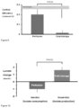
- FIG. 1 is a diagrammatic comparison of the brain dead and controlled donation after cardiac death (DCD) donor.
- FIG. 2 is a graph depicting myocardial oxygen demand at temperatures between 5° C. and 35° C.
- FIG. 3 is a perspective view illustrating a cabinet to house a perfusion composition delivery line assembly attached to a donor heart.
- FIG. 4 is a perspective view of the cabinet of FIG. 3 containing the assembly coupled to the donor heart with the door of the cabinet removed.
- FIG. 5 is a detailed view of the delivery line assembly coupled to the donor heart.
- FIG. 6 is a diagrammatic representation of the working heart apparatus used for functional assessment of the heart perfused with the apparatus of FIGS. 3 to 5 .
- FIG. 7 graphs the effect of left atrial pressure on cardiac output in normal hearts perfused for 12 hours according to Example 2.
- FIG. 8 graphs the effect of left atrial pressure on cardiac power in normal hearts perfused for 12 hours according to Example 2.
- FIG. 9 graphs the effect of cold storage or perfusion on cardiac efficiency in normal hearts perfused for 12 hours according to Example 2.
- FIG. 10 graphs the effect of cold storage (anaerobic metabolism) or perfusion (aerobic metabolism) on lactate change in normal hearts perfused for 12 hours according to Example 2.
- FIG. 11 is a schematic representation of the experimental protocols of Example 3 for comparing the perfusion composition of the present disclosure versus current clinical practice. Left, DCD hearts subjected to hypothermic perfusion preservation or standard preservation for 4 hours. Right, normal hearts.
- FIG. 12 is an echocardiographic view used for calculation of fractional area change.
- FIG. 13 A graphs perfusion pressure of individual experiments during perfusion preservation of DCD hearts perfused for 4 hours according to FIG. 11 and Example 3.
- FIG. 13 B graphs mean perfusion pressure for all experiments of FIG. 13 A .
- FIG. 14 graphs myocardial oxygen consumption during perfusion against coronary perfusion flow in DCD hearts perfused for 4 hours according to FIG. 11 and Example 3.
- FIG. 15 A graphs the effect of perfusion time on lactate production in DCD hearts perfused for 4 hours according to FIG. 11 and Example 3.
- FIG. 15 B graphs the lactate level on the RIG apparatus of DCD hearts perfused for 4 hours according to FIG. 11 and Example 3.
- FIG. 16 A graphs the effect of left atrial pressure on cardiac power in DCD hearts perfused for 4 hours according to FIG. 11 and Example 3.
- FIG. 16 B graphs the effect of 4 hours of standard preservation (cold storage) or perfusion on cardiac power of DCD hearts at 15 mmHg left atrial pressure treated according to FIG. 11 and Example 3.
- FIG. 17 A graphs the effect of left atrial pressure on cardiac output in DCD hearts perfused for 4 hours according to FIG. 11 and Example 3.
- FIG. 17 B graphs the effect of 4 hours of standard preservation (cold storage) or perfusion on cardiac output of DCD hearts at 15 mmHg left atrial pressure treated according to FIG. 11 and Example 3.
- FIG. 18 graphs the effect of 4 hours of standard preservation (cold storage) or perfusion on maximum rate of change of left ventricular pressure of DCD hearts at 15 mmHg left atrial pressure treated according to FIG. 11 and Example 3.
- FIG. 19 graphs the effect of 4 hours of standard preservation (cold storage) or perfusion on myocardial oxygen efficiency of DCD hearts at 10 mmHg left atrial pressure treated according to FIG. 11 and Example 3.
- the present disclosure relates to a perfusion composition, which, when perfused through an ex vivo organ, will sustain the critical chemical balances necessary to minimize cellular and reperfusion damage.
- the present disclosure also relates to a method of preserving a donor organ for transplantation.
- the present disclosure accounts for the potassium/sodium balance in cells and the elimination of harmful free radicals during ischemia.
- this perfusion composition is exemplified as a cardiac perfusion composition, the composition may be used to perfuse other organs, such as kidney, liver, lung and pancreas.
- preservation refers to maintenance of viability of a donor organ from harvest to reanimation in the organ recipient so that the donor organ performs comparably in the recipient as it did in the donor prior to donation.
- a “preservation” composition is a composition designed to passively preserve a donor organ in the absence of perfusion.
- perfusion is a composition designed to actively preserve a donor organ by perfusion.
- the organ immediately begins to degrade due to ischemia and these organs are then subject to reperfusion injury when the transplanted organ is introduced to its new host.
- This damage to tissue can continue when blood supply returns to the tissue after a period of ischemia, in particular, the oxygen that is carried in the blood.
- Reintroduction of oxygen causes a greater production of damaging free radicals as well as allowing, via removal of the extracellular acidotic conditions, influx of calcium and thus calcium overloading.
- Such radicals can attack cell membrane lipids, proteins, and glycosaminoglycans, causing further damage.
- the absence of oxygen and nutrients from blood also creates a condition in which the reperfusion results in inflammation and oxidative damage through the induction of oxidative stress rather than restoration of normal function.
- the perfusion composition of the present disclosure is used to perfuse organs between 2 and 10° C. In another embodiment, the perfusion composition may be used to perfuse organs at 2, 3, 4, 5, 6, 7, 8, 9, 10, 11, 12, 13, 14, 15, 16, 17, 18, 19, 20° C., or any range there between.
- metabolism and utilization of cellular energy stores are slowed, ATP and adenosine diphosphate (ADP), the major sources of cellular metabolic energy, are also gradually depleted during hypothermia. Hypothermia can also cause a phase transition of lipids and result in reduced membrane stability. In addition, it drastically alters the function of membrane bound enzymes. Hypothermia-induced structural changes in the membrane increase permeability, which contributes to cell swelling.
- Perfusion at 4 to 8° C. balances the need to reduce overall metabolism, but to maintain basal aerobic metabolism in preference to basal anaerobic metabolism.
- a principal goal during cold perfusion of an ischemic heart is to maintain the integrity of the cardiac cell membrane, and the integrity of the cardiac cell membrane potential.
- the cardiac cell normally has a high concentration of potassium and a low concentration of sodium, while the extracellular fluid has a low potassium concentration and high sodium concentration.
- the intracellular cardiac ion concentrations are maintained by pumping sodium ions out of the cell by an energetically driven process. When the heart is cooled, energy production by oxidative phosphorylation stops, and sodium ions are no longer pumped out. The intracellular sodium concentration then increases.
- the sodium overload produced is accompanied by an abnormally high calcium influx that causes muscle cell injury and death by several different mechanisms. Under these conditions, a switch from aerobic to anaerobic glycolysis is accomplished but the production of lactic acid also increases.
- Brain death describes the irreversible cessation of function of the entire brain, including the brainstem.
- CT computed tomography
- EEG electroencephalography
- transplanted organs have come from standard criteria donors. These are brain dead donors, also called heart-beating donors (HBDs), who meet strict medical criteria for donation. Standard criteria donors are regarded as the “ideal” donor due to their young age, favorable medical condition and location within the hospital intensive care unit (ICU), which allows timely organ procurement, the immediate initiation of preservation techniques and reduction of ischemic time. In an attempt to increase transplantation, some transplant units have begun utilizing organs not considered ideal by standard criteria (e.g., organs from older patients), referred to as marginal donors or expanded criteria donors.
- standard criteria e.g., organs from older patients
- DCD Donation after cardiac death
- two criteria must be fulfilled to diagnose the donor as dead. The first is cessation of cardiopulmonary function, i.e., asystole, apnea and absence of response to stimuli. The second is that the cessation of function is irreversible.
- the DCD donor is declared dead 5 minutes after the onset of cardiac arrest (asystole), which is defined as the lack of a palpable pulse and/or the absence of electrical activity on electrocardiogram (ECG) monitoring.
- Terms synonymous with DCD donor include non-heart-beating donor (NHBD) and donation after cardiocirculatory death donor.
- warm in situ ischemia is inherent in DCD leading to significant myocardial injury ( FIG. 1 ).
- warm ischemic time is the duration between the systolic blood pressure falling below 50 mmHg and the start of cold preservation.
- Another common definition is the interval of time between cessation of mechanical ventilation until the initiation of cold preservation. This includes the stand-off period that is applied between cardiac arrest and declaration of patient death (typically 2 to 5 minutes). The brain dead donor encounters no such warm ischemia. Death is pronounced well in advance of donation based on neurologic criteria and the organs remain perfused by the heart until the moment cold preservation is administered.
- Cold ischemic time extends from the beginning of cold preservation until the restoration of blood perfusion after transplantation and includes the period of graft implantation. This period is the same for brain dead donors and DCD donors.
- the category III donor is the most common source of DCD organ donation. This donor has severe, irreversible brain damage with no hope of recovery but does not meet the criteria for brain death. Once informed consent is obtained for both withdrawal of life support and organ donation, mechanical ventilation and life support is withdrawn either in the ICU or operating theatre. Hypoxic cardiac arrest results and after a mandatory stand-off period, death is pronounced. Only then can organ procurement proceed.
- the category Ill donor is called a controlled DCD donor as the moment of circulatory arrest can be planned and the precise period of warm ischemia is known.
- DCD donors include category I, II and IV donors.
- category IV the brain dead patient develops cardiac arrest during organ procurement, minutes before the initiation of cold perfusion. Hence warm ischemia is limited to only a few minutes.
- the category IV donor is considered a controlled DCD donor. Categories I and II are the uncontrolled DCD donors. Death is unexpected and the exact duration of warm ischemia is often unknown. This raises the question of organ viability complicating the possible application to transplant practice.
- the DCD donor scenario mandates the consideration of additional factors over and above the established ethical principles of organ transplantation.
- the duration of observation following onset of asystole required to declare an irreversible cessation of cardiopulmonary function is controversial.
- Transplant centers have also utilized DCD donors in liver transplantation and lung transplantation, with DCD donor lungs derived from category III donors. There are also encouraging accounts of uncontrolled DCD donor lung transplantation.
- the first human heart transplant in fact used a DCD donor.
- clinical transplantation of DCD hearts has been rare for a number of reasons.
- First and foremost is the great concern over the vulnerability of the heart to warm ischemia.
- the heart is unlike the kidney, liver and lung, which are better able to tolerate this insult.
- reperfusion injury is particularly severe in the DCD heart and adds further insult to the already-damaged myocardium.
- preservation techniques have failed to provide consistent and adequate myocardial recovery of the DCD heart.
- Ischemia means insufficient blood supply in relation to demand. It is most often due to a reduction or interruption of blood flow caused by a mechanical obstruction in the arterial vasculature leading to a decrease in the supply of oxygen and nutrients. At the onset of ischemia, oxidative phosphorylation ceases causing a reduction in ATP generation. Although the ischemic myocardium is able to continue producing ATP via anaerobic glycolysis, this process is very inefficient and is only able to yield 2 moles of ATP per mole of glucose (compared to 38 moles of ATP in aerobic conditions). Declining ATP leads to a failure of the sodium-potassium pump resulting in intracellular sodium overload and edema.
- the myocardium is extremely vulnerable to ischemic injury.
- the exact duration of ischemia that causes reversible injury to become irreversible is unknown.
- canine hearts showed a 70% reduction in ATP levels
- after 20 minutes warm ischemia there is evidence of irreversible damage to myocardial tissue and after a 60 minute warm ischemic interval the heart becomes hyper-contracted with no systolic function, a state referred to as “the stone heart.” It is commonly believed that irreversible injury to the myocardium begins approximately 20 to 30 minutes after the onset of severe ischemia.
- the heart is very sensitive to ischemia and the damage it sustains rapidly progresses from reversible to irreversible.
- reperfusion injury or “ischemia-reperfusion injury” and is an important consideration in myocardial infarction, stroke and organ transplantation.
- ROS reactive oxygen species
- Leucocyte activation causes release of ROS, proteases and elastases resulting in increased microvascular permeability, edema, thrombosis and parenchymal cell death.
- the donor heart is subjected to many potential sources of injury. These include brain death-induced myocardial damage, warm in situ ischemia in DCD, surgical injury, cold ex vivo ischemia during storage and reperfusion injury. Minimizing the severity of each of these insults maximizes the donor heart's chance of recovery (Table 2). Donor heart management is even more crucial in the DCD donor, due to the heart's poor tolerance of warm ischemia.
- Stage of transplant Minimization of injury Prior to explantation Brain dead donor following Minimization of myocardial damage from brain death brain death through timely procurement DCD donor following Minimization of warm in situ ischemia cardiopulmonary death through timely procurement 2. Donor procurement Prevention of cardiac distension Prompt and efficient delivery of cardioplegia 3. Storage for transport Optimal cardioplegia/storage solution Hypothermic perfusion preservation 4. Implantation Continued myocardial protection (frequent doses of cardioplegia) Prevention of cardiac distension 5. Reperfusion Prevention of cardiac distension 6. Early postoperative period Avoidance of excessive use of inotropes Avoidance of hypoxia
- Cardioplegic solutions are used in most forms of cardiac surgery including coronary artery bypass grafting, valve repair and replacement. These solutions are also used in donor heart procurement to rapidly induce cardiac arrest and reduce the temperature of the heart in order to decrease myocardial energy demand and preserve energy stores ( FIG. 2 ).
- cardioplegia refers to intentional and temporary cessation of cardiac activity, generally induced using a “cardioplegic solution” known to the person skilled in the art.
- standard cardioplegia refers to cardioplegia induced using a single cardioplegic solution known to the person skilled in the art.
- cardioplegic solutions have been developed in an effort to optimize organ protection. These can be broadly divided into the intracellular-type and extracellular-type solutions. The former are exemplified by the University of Wisconsin (UW) solution and the latter by the St. Thomas' Hospital No. 2 and CELSIOR® solutions (Table 3). Intracellular-type solutions have similar ionic concentrations to the physiological intracellular space and extracellular-type solutions have similar ionic concentrations to the physiological extracellular space. There are also numerous additives that have been used in cardioplegic solutions, e.g., lactobionate, raffinose and glutathione. A detailed discussion about the relative effectiveness of the various cardioplegic solutions and additives is beyond the scope of this review but following is a brief comparison of intracellular-type and extracellular-type solutions.
- Intracellular solutions were originally developed for the preservation of solid organs including kidney and liver. In routine cardiac surgery however, surgeons predominantly use extracellular solutions to arrest the heart. At the Alfred Hospital, St. Thomas' Hospital No. 2 solution is the standard cardioplegia used in routine cardiac surgery as well as donor heart preservation.
- hypothermia reduces the energy requirements of the myocardium ( FIG. 2 ). By reducing the metabolic rate of the ischemic heart, hypothermia slows down tissue deterioration. It has been shown that the ideal temperature for prolonged preservation (3 to 6 hours) is 4° C., although the accepted limit is 4 hours. This technique also has disadvantages including inhibition of enzyme function, interference with ATP generation and utilization and cellular edema.
- Cold storage is the standard technique of cardiac preservation used in heart transplantation today. Following cardioplegia arrest, the heart is placed in a bag filled with cold preservation fluid for the storage period. The bag is surrounded with ice to maintain hypothermia.
- hypomic preservation As used herein, “hypothermic preservation,” “cold storage preservation,” “cold storage” and similar terms refer to maintenance of a donor organ at approximately 2 to 4° C., commonly using ice or ice substitutes, and without perfusion.
- standard preservation refers to cardioplegia using a single cardioplegic solution and hypothermic preservation, also referred to as cold storage.
- Perfusion preservation is the technique of perfusing an organ, ex vivo, either with a blood or crystalloid (non-blood) composition, e.g., solution, known as the perfusate. It has many benefits and has been identified as a potential method of improving the preservation of various organs including kidney, liver, pancreas, lung and heart.
- perfusion refers to the process of delivery to a capillary bed in the donor organ of nutrients provided in a “perfusion composition” or “perfusate.”
- perfusates are based on cardioplegic solutions with various additives designed to preserve the integrity of the myocardium. Both intracellular and extracellular solutions have been used.
- hypomic perfusion preservation As used herein, “hypothermic perfusion preservation,” “hypothermic perfusion,” “cold perfusion” and similar terms refer to preservation of a donor organ by maintenance of the organ at approximately 0 to 10° C. coupled with perfusion.
- the perfusate or perfusion composition may comprise solely blood, a solution comprising blood, or a non-blood solution.
- Cardioplegia may be standard cardioplegia using a single cardioplegic solution known to the person skilled in the art or may be two-part cardioplegia using two cardioplegic solutions as disclosed herein.
- hypothermic perfusion preservation may improve donor heart preservation compared to cold storage. It prolongs the safe ischemic time, which allows the accessing of organs from greater distances and the opportunity for better donor/recipient tissue matching before transplant.
- a continuous supply of substrate and oxygen over ischemic times varying from 4 to 24 hours allows aerobic metabolism to proceed, which better protects myocardial ATP stores and tissue pH compared to cold storage. Oxidative stress, damage to DNA and apoptosis are also reduced.
- Hypothermic perfusion preservation reduces lactate production suggesting that these hearts can utilize the provided substrates and oxygen for aerobic metabolism.
- hypothermic perfusion preservation improves graft function after both short and long storage intervals.
- Tissue edema is a primary concern of perfusion preservation.
- Early studies showed a five-fold higher degree of weight gain in perfused hearts compared to hearts preserved with cold storage.
- the development of edema can cause an increase in coronary resistance due to vessel compression, resulting in impaired circulation and suboptimal myocardial protection.
- edema can be reversible, and a small amount of edema may not necessarily impair heart function.
- Better understanding of the mechanisms involved in edema has allowed the present perfusion method to be altered to minimize this problem, e.g., lower perfusion rates and addition of oncotic agents to perfusates.
- hypothermic perfusion preservation provides better myocardial recovery than cold storage
- transplant units have continued using cold storage as the standard technique for donor heart preservation. This is because cold storage is simple, safe, predictable, inexpensive and provides adequate protection of the standard donor heart if ischemia is restricted to 4 hours.
- the heterogeneous nature of existing techniques and restricted clinical application of hypothermic perfusion preservation means the optimal protocol for perfusion preservation of the heart is still undetermined.
- the concerted effort to expand the donor pool by utilizing marginal and DCD donors has renewed interest in perfusion preservation as a technique for preserving these damaged organs.
- Cold storage in ice may be adequate for preservation of standard brain dead donor hearts up to 4 hours, however, the DCD heart sustains a severe warm ischemic insult during the agonal period and thus any further damage is much more likely to cause irreversible injury.
- An early study subjected pig hearts to warm ischemic periods varying between 0 and 60 minutes followed by 2 hours of cold storage. The authors concluded that hearts procured 10 minutes or greater after death and then cold stored were unable to be resuscitated and were unsuitable for transplantation.
- a subsequent study from the unit demonstrated in a canine model that hearts left untouched for 30 minutes following cessation of ventilation and subjected to 4 hours of cold storage recovered very poorly. Functional recovery can be improved experimentally with administration of donor pretreatments.
- donor pretreatments such as methylprednisolone, dextrose, nifedipine, and prostaglandin E 1 may be desirable for optimal organ protection, they are ethically unacceptable because they are of no benefit to the donor and therefore not suitable clinically.
- hypothermic perfusion preservation uses crystalloid (non-blood) composition, e.g., solutions, that differ from modified cardioplegic solutions. So-called “hypothermic crystalloid perfusion” of the DCD donor heart has been tested only in animal models in which death was induced by exsanguination, which is not relevant to the DCD donor.
- hypomic crystalloid preservation refers to “hypothermic perfusion preservation” in which the perfusion composition or perfusate is a non-blood composition, e.g., a solution.
- Perfusion can also be delivered at hypothermic temperatures using a crystalloid (non-blood) composition, e.g., solution.
- a crystalloid non-blood composition
- This technique is potentially more cost-effective and simpler than blood perfusion, and has been demonstrated to be effective in normal hearts.
- hypothermic crystalloid perfusion of DCD donor hearts have provided encouraging results, these were conducted under conditions not applicable to clinical practice.
- This method of preservation has not been carried out in a model that mirrors the majority of clinical DCD donation, that is, Maastricht category III DCD donation.
- Table 4 compares cold storage, normothermic blood perfusion and hypothermic crystalloid perfusion for preservation of the donor heart.
- cardioplegia In the DCD donor, although cardiac arrest is induced by hypoxia, “acidic mitochondrial pore inhibiting” cardioplegia has been utilized, which has a role in cooling the heart, supplying metabolic substrates and reducing reperfusion injury. Hypothermic crystalloid perfusion as disclosed herein utilized two-part cardioplegia, which is distinct from standard cardioplegia. Both parts are based on St. Thomas' Hospital No. 2 (Table 3) cardioplegia with additives and modifications including:
- the preferred pH of the cardioplegic solution is 7.2. This acidic pH has been shown to protect mitochondria.
- the pH of the cardioplegic solution may be 7.1, 7.11, 7.12, 7.13, 7.14, 7.15, 7.16, 7.17, 7.18, 7.19, 7.21, 7.22, 7.23, 7.24, 7.25, 7.26, 7.27, 7.28, 7.29, or 7.3.
- the perfusion composition of the present disclosure was recently developed as a perfusate for donor heart perfusion, but may be used for perfusing a kidney, a lung, a liver or a pancreas. It is similar to extracellular cardioplegic solutions and has various additives.
- the perfusion composition will keep an ischemic heart in a condition to successfully survive to transplant for 12 hours or more. In other embodiments, the perfusion composition will preserve the donor organ for 1, 2, 3, 4, 5, 6, 7, 8, 9, 10, 11, 13, 14, 15, 16, 17, 18, 19, 20, 21, 22, 23, 24, 25, 26, 27, 28, 29, 30, 31, 32, 33, 34, 35, 36 or more hours after harvest.
- a summary of the formulation of the perfusion composition follows.
- the sodium-potassium adenosine triphosphatase (Na—K ATPase) pump functions to maintain the ionic composition of the cell.
- the pump is disrupted by ischemia because of the lack of ATP production and by excessive production of hydrogen ions because of anaerobic metabolism during ischemia. Under ischemic conditions, there is a switch from aerobic to anaerobic glycolysis, and the production of lactic acid increases.
- sodium-potassium ATPase pump is disrupted, potassium moves out of the cell, whereas sodium, which is normally kept at a low concentration in the cell, pours in. This ionic shift causes cell swelling and disruption of the cell if unchecked.
- the perfusion composition comprises 0.5 mM calcium.
- the perfusion composition may comprise 0.1, 0.2, 0.25, 0.3, 0.4, 0.6, 0.7, 0.75, 0.8, 0.9 or 1.0 mM calcium.
- the source of Ca 2+ is calcium chloride.
- the perfusion composition comprises 15 mM potassium.
- the perfusion composition may comprise 1, 2, 3, 4, 5, 6, 7, 8, 9, 10, 11, 12, 13, 14, 16, 17, 18, 19, 20, 21, 22, 23, 24, 25, 26, 27, 28, 29, or 30 mM potassium.
- the source of K + is potassium chloride.
- a sodium/hydrogen (Na + /H + ) exchanger (NHE) inhibitor will also aid in potassium retention by the heart cells, along with reducing the damage from anoxia and reperfusion injury after transplant.
- the perfusion composition comprises a high concentration of magnesium (Mg′) relative to the concentration of calcium to keep the heart in hyperpolarized arrest and help preserve the heart muscle cell membrane so that membrane excitability is better restored after transplantation.
- Mg′ magnesium
- Magnesium acts as a calcium antagonist, thus preventing calcium overload.
- Magnesium is also present to stabilize the myocardial membrane by inhibiting a myosin phosphorylase, which protects ATP reserves for post-ischemic activity.
- Magnesium regulates and balances the sodium-potassium-calcium pump of the heart cells.
- Magnesium is also present to counteract lactic acidosis associated with ischemia. Low magnesium compromises the integrity of the cell wall causing lesions.
- the perfusion composition comprises 7.5 mM of magnesium.
- the perfusion composition may comprise 1, 2, 3, 4, 5, 6, 7, 8, 9, 10, 11, 12, 13, 14, or 15 mM magnesium.
- the source of Mg 2+ is magnesium chloride.
- the concentration of sodium (Na + ) in the perfusion composition maintains integrity of the cell membrane to lower the likelihood of calcium paradox during reperfusion.
- the perfusion composition comprises 80 mM sodium.
- the perfusion composition may comprise 10, 20, 30, 40, 50, 60, 70, 90, 100, 110, 120, 130, 140, 150, or 160 mM sodium.
- the source of Na + is sodium chloride.
- Chloride (Cl ⁇ ) may be present as a counter ion to maintain the electroneutrality of the composition.
- the source of Cl may be derived from the source of any one or more of sources of Ca 2+ , Mg 2+ , K + , or Na + .
- salts other than chloride salts may be the source of Ca 2+ , Mg 2+ , K + , or Na + .
- insoluble salts of Ca 2+ and Mg 2+ are to be avoided.
- the counter ion for the source of Ca 2+ , Mg 2+ , K + , or Na + is gluconate.
- the salt sources of Ca 2+ and Mg 2+ may be hydrates.
- Prolonged survival of cardiac muscle at 4° C. depends on glycolysis that utilizes the muscle glycogen stores and produces lactic acid and other metabolites that produce CO2.
- the cell is able to take up and metabolize glucose, thereby preserving the cellular glycogen stores that then will not require replenishing after transplanting the organ.
- the glucose and insulin taken up by the perfused heart gives a boost to metabolism and ATP production when the perfused heart is transplanted, and the circulation is restored.
- the perfusion composition comprises 14 mM glucose. In another embodiment, the perfusion composition may comprise 1, 2, 3, 4, 5, 6, 7, 8, 9, 10, 11, 12, 13, 15, 16, 17, 18, 19, 20, 21, 22, 23, 24, 25, 26, 27, 28, 29, or 30 mM glucose.
- Insulin is a component of this perfusion composition, at a lower concentration than in the cardioplegic solution, and enhances the uptake of glucose into heart muscle cells. Insulin has a direct positive inotropic effect on the reperfused heart. Insulin also promotes glucose utilization and oxidation increases during reperfusion. Insulin also inhibits programmed cell death (apoptosis). Insulin, when combined with glucose and potassium, as in this present disclosure, also attenuates myocardial reperfusion injury and thus may exert significant cardioprotection upon transplantation.
- the perfusion composition comprises 6 Units of short-acting or regular insulin. In another embodiment, the perfusion composition may comprise 1, 2, 3, 4, 5, 7, 8, 9, 10, 11, 12, 13, 15, 16, 17, 18, 19, 20 Units of short-acting or regular insulin.
- FDP Fructose-1,6-diphosphate
- the perfusion composition comprises 2 mM FDP.
- the perfusion composition may comprise 0.2, 0.4, 0.6, 0.8, 1, 1.2, 1.4, 1.6, 1.8, 2.2, 2.4, 2.6, 2.8, 3, 3.2, 3.4, 3.6, 3.8, 4, 4.2, 4.4, 4.6, 4.8, 5, 5.2, 5.4, 5.6, 5.8, 6, 6.2, 6.4, 6.6, 6.8, 7, 7.2, 7.4, 7.6, 7.8, 8, 8.2, 8.4, 8.6, 8.8, 9, 9.2, 9.4, 9.6, 9.8, or 10 mM FDP.
- FDP may be provided as a sodium or potassium salt.
- Aspartate is incorporated into this perfusion solution at the same or similar concentration as incorporated into the cardioplegic solution above.
- the aspartate stimulates the malate-aspartate shuttle and thus improves recovery of energy production particularly upon restoration of the circulation upon transplantation after cold perfusion.
- Adenosine, or a substitute may be incorporated into the perfusion composition at the same or similar concentration as incorporated into the cardioplegic solution above.
- the perfusion composition comprises 5 mM adenosine, cAMP or cGMP.
- the perfusion composition may comprise 1, 2, 3, 4, 6, 7, 8, 9, 10, 11, 12, 13, 14, 15, 16, 17, 18, 19, 20, 21, 22, 23, 24, or 25 mM adenosine, cAMP or cGMP.
- the perfusion composition may comprise 1, 2, 3, 4, 5, 6, or 7 g/L adenosine, cAMP or cGMP.
- the perfusion composition may comprise reduced glutathione (GSH), which functions as a reducing agent and a free radical scavenger.
- GSH reduced glutathione
- Agents known to scavenge or inhibit the formation of free radicals can prevent reperfusion-induced injury.
- GSH is a cofactor for the enzymatic destruction of hydrogen peroxide and other organic hydroperoxides. While all cells in the human body are capable of synthesizing glutathione, liver glutathione synthesis has been shown to be essential to the normal functioning of the human body.
- the perfusion composition comprises 3 mM GSH. In another embodiment, the perfusion composition may comprise 0.5, 1, 1.5, 2, 2.5, 3.5, 4, 4.5, 5, 5.5, 6, 6.5, 7, 7.5, 8, 8.5, 9, 9.5, or 10 mM GSH.
- Tris(hydroxymethyl)aminomethane hydrochloride (Tris or THAM) is used a buffer, that has an effective pH range between 7.0 and 9.2, which counteracts the occurrence of metabolic acidosis. Tissues in the ischemic heart resort to anaerobic metabolism in the absence of oxygen and significant amounts of lactic acid are released into the muscle tissue and into the surrounding intercellular fluid. Tris counteracts the presence of the acid to maintain the proper pH of both the heart cell and perfusate.
- the perfusion composition comprises 20 mM Tris or HEPES, MOPS, IVIES, BES or TES.
- the perfusion composition may comprise 4-(2-hydroxyethyl)-1-piperazineethanesulfonic acid (HEPES), 3-(N-morpholino)propanesulfonic acid (MOPS), 2-(N-morpholino)ethanesulfonic acid (IVIES), N,N-bis-(2-hydroxyethyl)-2-aminoethansulfonic acid (BES), or N-tris(hydroxymethyl)methyl-2-aminoethanesulfonic acid (TES).
- HPES 4-(2-hydroxyethyl)-1-piperazineethanesulfonic acid
- MOPS 3-(N-morpholino)propanesulfonic acid
- IVIES 2-(N-morpholino)ethanesulfonic acid
- BES N,N-bis-(2-hydroxyethyl)-2-aminoethansulfonic acid
- TES N-tris(hydroxymethyl)methyl-2-aminoethanesulfonic acid
- the perfusion composition may comprise 1, 2, 3, 4, 5, 6, 7, 8, 9, 10, 11, 12, 13, 15, 16, 17, 18, 19, 21, 22, 23, 24, 25, 26, 27, 28, 29, 30, 31, 32, 33, 34, 35, 36, 37, 38, 39, or 40 mM Tris, HEPES, MOPS, MES, BES or TES.
- Bicarbonate hydrogen carbonate, HCO3-
- HCO3- hydrogen carbonate
- Bicarbonate, in this perfusion solution is used to promote CO2 ⁇ ->HCO-3 exchange and is not used as a pH buffer and is used in addition to any buffer, such as TRIS, in the perfusion solution.
- Bicarbonate may also be used to combat metabolic acidosis, which produces lactic acid and a build-up of CO2, by controlling extracellular acidosis and is used to regulate hyperkalemia, as potassium levels are brought into balance during the beginnings of ischemia.
- the perfusion composition comprises 20 mM bicarbonate.
- the perfusion composition comprises 1, 2, 3, 4, 5, 6, 7, 8, 9, 10, 11, 12, 13, 15, 16, 17, 18, 19, 21, 22, 23, 24, 25, 26, 27, 28, 29, 30, 31, 32, 33, 34, 35, 36, 37, 38, 39, or 40 mM bicarbonate.
- the source of HCO3- is K HCO3 or Na HCO3.
- the optimum pH of the perfusion solution is 7.4 at 22 degrees C.
- the pH increases slightly upon cooling of the solution to 4 degrees C.
- the pH of the perfusion composition is 7.4.
- the pH of the perfusion composition may be 7.2, 7.21, 7.22, 7.23, 7.24, 7.25, 7.26, 7.27, 7.28, 7.29, 7.31, 7.32, 7.33, 7.34, 7.35, 7.36, 7.37, 7.38, 7.39, or 7.4.
- Interstitial edema is one of the potential drawbacks of organ perfusion preservation.
- Oncotic pressure of a perfusate should match that of the interstitial tissue in order to minimize fluid shift into the interstitium.
- Blood contains albumin and globulins that provide oncotic pressure in vivo and in blood-based perfusion compositions.
- crystalloid (non-blood) perfusates do not have these natural oncotic agents.
- the use of non-blood perfusates without added colloid greatly increases the risk of tissue edema. This risk can be lessened by providing lower perfusion pressures however, this subjects the organ to the possibility of inadequate and uneven tissue perfusion.
- Another factor contributing to the development of tissue edema is a lack of lymphatic flow, a means by which a small amount of fluid usually returns from the interstitium back to the circulation.
- the perfusion composition of the present disclosure comprises lactobionate, a semi permeable compound, to reduce interstitial edema.
- the perfusion composition comprises 70 mM lactobionate and/or mannitol.
- the perfusion composition may comprise 10, 20, 30, 40, 50, 60, 80, 90, 100, 110, 120, 130, 140, or 150 mM lactobionate and/or mannitol.
- the fluid distribution between intracellular and extracellular spaces is determined mainly by the osmotic effect of solutes in both compartments.
- Normal osmolarity of the body fluids is 280 to 300 mOsm/L.
- the perfusion composition is iso-osmotic and the osmotic pressure is 280, 290 or 300 mOsm/L.
- the perfusion composition is hyperosmotic and the osmotic pressure is 310, 320, 330, 340, 350, 360, 370 or 380 mOsm/L.
- the perfusion composition of the present disclosure may be supplemented with oxygen by direct bubbling with 80%, 90% or 100% oxygen. Oxygenation may be achieved by supplying oxygen to the perfusion composition, shaking the perfusion composition, venting the perfusion composition and repeating once, twice or more. During each round of shaking, the supplied oxygen will equilibrate with the perfusion composition, with each round of supply, shaking and venting increasing the oxygen concentration of the perfusion composition.
- the perfusion composition is 100% saturated with oxygen.
- the oxygen saturation may be 50, 60, 70, 80 or 90% saturated with oxygen.
- the perfusion composition may comprise a pO 2 of 200, 300, 400, 500 or 600 mmHg.
- Table 5 provides the final composition of one embodiment of the perfusion composition.
- perfusate flow should be delivered in a pulsatile or non-pulsatile manner is an important consideration in ex vivo organ perfusion.
- Pulsatile pumps are complex, expensive and heavy and it would be a considerable economical advantage to employ a simpler pump if possible.
- the perfusion composition of the present disclosure may be perfused using a pulsatile pump, a non-pulsatile pump, or gravity.
- the perfusion composition of the present disclosure may be perfused with a low flow rate or by microperfusion.
- microperfusion refers to a flow rate of 2 to 8 ml/100 g/min.
- the perfusion composition is microperfused at a rate of 4 or 5 ml/100 g/min (20 ml/min).
- the perfusion composition may be microperfused at a rate of 2, 3, 4, 6, 7, or 8 ml/100 g/min.
- the perfusion composition may be perfused through the donor organ at a pressure at the aortic root of 2 to 10 mmHg.
- the pressure at the aortic root may be 4 to 8 mmHg.
- the pressure at the aortic root may be 5 to 7 mmHg.
- the mean pressure at the aortic root during perfusion may be 1, 2, 3, 4, 5, 6, 7, 8, 9, 10, 11, or 12 mmHg.
- controlled reperfusion refers to the technique of modifying conditions (e.g., temperature, pressure etc.) when initially reperfusing ischemic tissue. Controlled reperfusion has been shown to reduce reperfusion injury in the heart, lung, brain and extremities. Techniques that have demonstrated benefits include:
- components that are less stable than other components of the perfusion composition are kept separate from the other components and are added to the other components prior to use of the perfusion composition.
- the less stable components that may be added prior to use of the perfusion composition comprise insulin, FDP and GSH. These components may be added separately to the other components. Alternatively, one or more of the less stable components may be added simultaneously to the other components.
- the less stable components may be formulated in a second composition to be combined with a first composition comprising the other components, thereby producing the perfusion composition.
- FDP and GSH may be formulated in one composition, with insulin added to either the FDP plus GSH composition or to the other components just before use of the perfusion composition.
- each composition may be buffered appropriately to produce the correctly buffered perfusion composition.
- the perfusion composition may be provided in two or more parts, for example, in a kit, separated until use.
- the perfusion composition should be used within 48 hours.
- the cardioplegic solutions may be provided in a concentrated form that is dilutable to prepare the cardioplegic solutions for use.
- the perfusion composition may be provided in a concentrated form that is dilutable to prepare the perfusion composition for use.
- the cardioplegic solutions and the perfusion composition may be provided in unit dose form.
- the cardioplegic solutions and the perfusion composition may be provided in a syringe, bottle, vial, ampoule or bag. Multiple unit doses may be used in the method of the present disclosure depending upon the duration of perfusion and flow rate of perfusion.
- the less stable components may be provided in a syringe, or may be provided in a bottle, vial, ampoule or bag and transferred to a syringe, and then injected into a bag, for example, containing the other components of the perfusion composition to prepare the perfusion composition for use.
- the bag is a gravity-fed, drip-style bag.
- a concentrate of the cardioplegic solutions or the perfusion composition may be provided in a syringe, bottle, vial, ampoule or bag.
- the components are added to de-ionized water to a volume of 800 ml.
- the pH is measured and adjusted using sodium hydroxide or hydrochloric acid to 7.3+/ ⁇ 0.15 at 22.5QC.
- water is added to a volume of 1000 ml.
- the pH may be again checked and adjusted if needed.
- the heart produces lactic acid via glycolysis.
- the pH of the perfusion composition is adjusted to a slightly more alkaline pH than usual to neutralize the lactic acid.
- the stock composition may be diluted with a diluent.
- the diluent is water.
- the diluent may be sodium chloride solution (saline) or potassium chloride solution, provided that the diluent is accounted for as a source of Na + or K + and Cl ⁇ in the perfusion composition.
- the cardioplegic solutions and the perfusion composition are sterile.
- sterilization may be achieved without difficulty by moist heat sterilization, dry heat sterilization, chemical cold sterilization, radiation sterilization or filter sterilization.
- the cardioplegic solutions and the perfusion composition are free of pyrogen and endotoxin, which may be achieved by dry heat sterilization, for example.
- the cardioplegic solutions and the perfusion composition may comprise an antibacterial drug.
- the cardioplegic solution and the perfusion composition may comprise: a bacterial wall synthesis inhibitor (e.g., a penicillin, a cephalosporin, a carbapenem, or vancomycin); an agent that damages the cytoplasmic membrane (e.g., a polymixin); an agent that modifies synthesis or metabolism of a nucleic acid (e.g., a quinolone, rifampin, or nitrofurantoin); a protein synthesis inhibitor (e.g., an aminoglycoside, a tetracycline, chloramphenicol, erythromycin, or clindamycin); or a folate inhibitor or agent that modifies energy metabolism (e.g., a sulphonamide, or trimethoprim).
- a bacterial wall synthesis inhibitor e.g., a penicillin, a cephalosporin, a carbapenem,
- the novel perfusion solution has been described. It has a very low viscosity and fills the entire coronary circulation including the distal blood vessels at the apex of the heart by gravity alone without the necessity of a pump or a pressure head.
- the perfusion solution flows through the coronary bed once and is discarded. Since the perfusion fluid is not recirculated the concentration of the constituents of the perfusion solution are not changed with time, and there is not an accumulation of the end products of heart metabolism and other materials that are excreted into the perfusion solution some of which may be toxic.
- the constituents of the perfusion solution and their actions are summarized in Table 5, and discussed in more detail in the specifications.
- hearts perfused with the solution maintain their viability for up to 18 hours. This permits many improvements, including time to do more laboratory studies on the donor hearts, more time to find and prepare a suitable recipient, time to transport the donor heart to a distant location for transplantation, and so on.
- the perfusion kit is simple, compact and light. No blood is required and neither is a large, bulky, heavy and complex perfusion device.
- kit refers to a physical arrangement of items.
- the items may comprise the cardioplegic solutions and/or the perfusion composition(s), which may be presented in the form of a kit.
- the cardioplegic solutions and/or the perfusion composition(s) of the kit may be “ready to use” in unit dose form.
- the cardioplegic solutions and/or the perfusion composition may be presented in concentrated form for dilution prior to use.
- the perfusion composition may be divided into less stable components and other components, again either “ready to use,” other than combining, in unit dose form, or in concentrated form for diluting and combining. Where necessary for preparation of the perfusion composition, diluting and combining may be performed in any order.
- a “method” of preserving a donor organ for transplantation may be defined in alternative forms.
- the method may be defined in the form of “use” of selected components for preserving a donor organ for transplantation.
- the perfusion composition, perfusion stock composition, kit or apparatus of the present disclosure may be defined in alternative forms.
- One form designates either suitability for or restriction to a specific use and is indicated by the word “for.”
- Another form is restricted to a specific use only and is indicated by the words “in use” or “when used for” or similar.
- the method may be defined in the “Swiss” style, e.g., use of selected components in the manufacture of a perfusion composition for preserving a donor organ for transplantation.
- the method may be defined in the “agent for use” form, e.g., a perfusion composition comprising selected components for use in preserving a donor organ for transplantation.
- the perfusion solution is made by combining a first and second solution in intravenous drip style bags.
- the perfusion solution should be used within approximately 24 hours of combining.
- 100% oxygen is then bubbled into the bag(s) containing the first and second solutions in a three stage procedure in order to assure that the solutions are oxygenated.
- a multiple of bags 10 are hung in an insulated enclosure 3 forming part of a micro-perfusion drip apparatus 1 .
- the enclosure 3 is a self-standing rectangular cabinet with a hinged glass door 4 at the front.
- Carry handles 7 , 8 are located at the sides and on the top of the cabinet.
- the cabinet is manufactured in insulated plastics often used in portable fridges and coolers.
- the insulated enclosure 3 is maintained at a temperature of between 4° C. and 10° C. by use of ice bags (not shown).
- the solution bags 10 are attached to the top of the enclosure and a stand 5 is used to support the heart 2 so that it is suspended within a plastics bag 19 .
- the bags 10 are connected by drip lines 9 to a common manifold 14 , which exits into a drip chamber 12 .
- the outlet of the drip chamber 12 is coupled to a length of soft plastics tubing, which is in turn coupled to a three way connector 15 with a tap.
- a flow regulator in the form of an adjustable gate clamp 11 is positioned on the soft plastics connector 13 .
- the exit of the three way connector 15 is coupled to an aortic cannula 23 , which is securely fitted inside the aorta of the heart 2 .
- the cannula is held within the aorta by appropriate clamps (not shown).
- the aortic valve of the heart is closed during the perfusion.
- the solution flows through the coronary arteries, coronary sinus, right atrium, to drop into the bag 19 and then escape into a waste collection or effluent bag 16 , located in the base of the enclosure 3 .
- the fluid is collected and not reused.
- heart 2 is hung by gravity within the plastics bag 19 without support.
- the solution 18 within the base of the plastics bag 19 ensures that the heart is in a moist environment to prevent drying out. It is important that the heart does not float in the fluid 18 .
- the angled exit of the three way connector is coupled to a pressure line 22 near the cannula 23 to provide measure of the pressure caused by the flow rate of the solution.
- Pressure line 22 provides important pressure feedback.
- the pressure line 22 is coupled to a water manometer 26 attained to the outside of the enclosure. Alternatively, the manometer can be located within the enclosure or on the glass door of the enclosure. Reading of pressure is useful for two reasons: 1) too much pressure can lead to edema and 2) an easy check to determine whether the aortic valve is closed is to briefly increase the flow rate and the pressure should increase. Regular pressure tests are conducted where the perfusion flow rate is temporarily increased. If the aortic valve is competent, the aortic lost pressure will rise accordingly. If the pressure does not rise, this indicates aortic incompetence. This is rectified by pressurization of the valve achieved by increasing the flow and ensuring that the heart is positioned correctly to ensure the valve is closed.
- the heart 2 is suspended by the aortic cannula 23 at a temperature of between 4 and 10° C.
- a temperature gauge is positioned to be visible within the enclosure.
- the heart is perfused with the solution for four hours at a flow of 20 ml/min, during which time the myocardial temperature remained between 5 and 10° C. and the aortic root pressure was between 4-8 mm Hg.
- the perfusion apparatus described with reference to FIGS. 3 to 5 provides a very simple and effective portable device. It is designed to be light and easily transportable and is considered to be particularly reliable. There are no moving parts, no need for batteries or power sources, pumps, gas cylinders and refrigeration devices.
- the apparatus by controlling the flow rate and through the use of a gravity feed at controlled temperatures ensures that the heart is continually perfused during transportation before transplantation.
- the apparatus is designed to be robust to resist damage during transport.
- the apparatus has been designed so that it can be managed by a non-expert without the need for a highly skilled technician to ensure efficient operation.
- Greyhounds were anesthetized, and the heart removed after arrest with St. Thomas's potassium cardioplegia.
- Greyhound hearts have a structure, weight and composition very similar to human hearts.
- Perfusion hearts received cold crystalloid gravity-feed microperfusion (20 ml/min, 6 mmHg, 4-10° C.) with the perfusion composition of the present disclosure.
- Cold storage hearts were preserved for 12 hours in ice as in conventional clinical practice, which is known to add to the ischemic damage suffered by the heart after removal, especially those hearts that are donated after cardiac arrest.
- the sets of hearts were then transferred to a blood perfused working heart apparatus for 2 hours of reperfusion followed by final assessment.
- FIGS. 7 through 10 detail the test results of the perfused normal hearts versus those kept according to standard cardioplegia practices in ice and those normal hearts used as a control lot. It was determined that the perfusion composition herein disclosed as compared to conventional ice storage allowed for the donor hearts to utilize oxygen during their preservation, which is associated with superior post-preservation pump function, efficiency and lactate metabolism. During perfusion, the perfused hearts consumed oxygen. After preservation compared to cold storage hearts, perfused hearts had higher cardiac output, LV dP/dt max and efficiency, with lower lactate; hemodynamic values were 50% to 80% of non-preserved hearts. In terms of lactate metabolism, it was shown that after perfusion, the hearts were aerobic, consuming lactate while the cold storage hearts were anaerobic, producing harmful lactate.
- the present disclosure as disclosed will extend the time of organ viability during transport, between harvest and transplantation, over currently available methods and solutions. It also presents an organ that is more adapted to reperfusion and thus more likely to successfully transplant and function in the new body.
- Heparin 10,000 U was administered intravenously to allow the exsanguination of blood (600 to 900 ml) from the femoral artery. This blood was required to prime the isolated heart (RIG) apparatus. Arterial pressure and heart rate were monitored carefully as blood was removed. Ringer's solution was used to replace blood volume and intravenous phenylephrine (5-10 mg) was given as required to maintain blood pressure at physiological levels.
- a two-part cardioplegia was administered over 6 minutes in combination with topical cooling with ice.
- 1000 ml of crystalloid cardioplegia was infused at a temperature of 4° C.
- Cardioplegia was vented through the left atrial appendage and the inferior vena cava.
- the first part was AMPI Cardioplegia. Five hundred ml of AMPI cardioplegia was administered over 3 minutes. The base for this solution was St. Thomas' Hospital No. 2 cardioplegia with the following additives:
- the solution was made acidic (pH of 7.2) by saturation with 20% carbon dioxide.
- the second part was called the “Recovery Cardioplegia.” Once 500 ml of AMPI cardioplegia was delivered, 500 ml of Recovery Cardioplegia was administered over 3 minutes.
- the base solution was St. Thomas' Hospital No. 2 cardioplegia saturated with 100% oxygen with the following additives:
- Myocardial temperature was between 5-15° C. following cardioplegia.
- the heart was then excised and weighed.
- the pulmonary veins were ligated, and an aortic cannula inserted (three eighth-inch diameter PVC tubing with a collar of half-inch diameter PVC tubing (Lovell Surgical, Melbourne)).
- the left atrial appendage and pulmonary artery were then cannulated with quarter-inch diameter PVC tubing.
- the heart was transferred to the perfusion apparatus of the present disclosure ( FIGS. 3 to 5 ). It was designed to be as simple as possible in order to make it portable and convenient. It comprises a polystyrene box with dimensions 92 cm ⁇ 35 cm ⁇ 35 cm. The front door has a window to allow monitoring and is closed with Velcro straps. Initially, four 1 liter bags of perfusion composition were suspended from the top of the box, attached to a manifold, which leads to a drip chamber. These bags were replaced with new ones as required. The heart was attached distal to this drip chamber and received an infusion of perfusate through the aortic root. The flow rate was controlled by a gate clamp.
- the heart was suspended by the aortic cannula, which was attached to the drip chamber. Ice packs were hung on the walls of the box to maintain a low ambient temperature. There was constant monitoring of pulmonary arterial effluent flow, myocardial temperature (Shiley Inc., California), temperature inside the perfusion apparatus box and aortic pressure (Datex Ohmeda, Melbourne).
- the heart was perfused with perfusion composition for 4 hours at a flow of 20 ml/min, during which time the myocardial temperature remained between 5-10° C. and the mean aortic root pressure between 4-8 mmHg ( FIG. 13 ).
- the perfusion composition used in these experiments is defined in Table 5.
- the heart was then connected to the RIG apparatus ( FIG. 6 ) and reperfused with blood for 50 minutes in the non-working mode.
- the RIG apparatus is a modified extracorporeal membrane oxygenation (ECMO) circuit containing a roller pump (COBE cardiovascular, Arvada), membrane oxygenator (Capiox SX18, Terumo, Melbourne), leucocyte filter (LeukoGuard, Pall, Sydney) and a heater cooler unit (Jostra, N.J.).
- ECMO extracorporeal membrane oxygenation
- the coronary arteries are perfused through the aortic root and the heart is not required to eject against resistance.
- the heart is perfused through the left atrium and must eject against an afterload.
- the perfusate consisted of whole blood collected from the greyhound. In some experiments, Ringer's solution was added to achieve the 1200 ml required priming volume of the circuit.
- the blood was leucocyte depleted, temperature controlled, and its partial pressure of oxygen and carbon dioxide carefully regulated. The following were added to the blood during circuit priming:
- the aortic root pressure was increased to 30 mmHg, heater cooler unit temperature increased to 30° C. and the oxygen: air ratio increased to 50%. Then at 15 minutes of reperfusion, the aortic root pressure was increased to 35 mmHg, heater cooler unit temperature increased to 39° C. and the carbon dioxide flow adjusted in order to achieve a partial pressure of 40 mmHg. Finally, at 20 minutes of reperfusion, the aortic root pressure was increased to 60 mmHg.
- Aortic Heater Oxygen root cooler unit air Reperfusion pressure temperature mixture CO 2 time (min) (mmHg) (° C.) (%) (mL/min) 0 20-25 20 20 150 5 30 30 50 150 15 35 39 50 Adjusted to achieve 20 60 39 50 pCO 2 of 40 mmHg
- the hearts were gradually warmed and electrically defibrillated when the myocardium reached 36-37° C. All hearts received lignocaine (1 mg/kg) to prevent arrhythmias. Amiodarone (2.5 mg/kg) was administered if hearts did not achieve a stable rhythm. Electrical pacing was introduced if the heart rate dropped below 90 beats per minute. The blood perfusate composition was carefully controlled with particular attention given to pH, partial pressure of oxygen, partial pressure of carbon dioxide, base excess and concentration of potassium. Inotropes were not administered.
- the RIG apparatus was switched to working mode in order to conduct a final assessment of function and metabolism. To conclude the experiment the whole heart was perfused with 10% neutral buffered formalin through the aortic root.
- Standard preservation data are derived in part from the historical data.
- the aorta was cross clamped and 1000 ml of cardioplegia infused into the aortic root via a 12-gauge intravenous cannula.
- the cardioplegic solution used in this group was the standard one used in human heart transplantation at the Alfred Hospital.
- the cardioplegia was administered over 6 minutes at 4° C. and the effluent discarded.
- the heart also received topical cooling with ice.
- Myocardial temperature was between 5-15° C. after cardioplegia.
- the heart was then excised and weighed.
- the pulmonary veins were surgically ligated, a cannula inserted into the aorta and a myocardial temperature probe positioned in the myocardium.
- the heart was secured within a watertight bag filled with cold saline (4° C.), which was subsequently placed in an ice box and surrounded by ice.
- the myocardial temperature gradually decreased in the early stages of preservation, and remained between 1-4° C. for the majority of storage. In total the heart was cold stored for 4 hours.
- a normal heart group was included to provide a reference point for the previously described experimental groups. There was no DCD process or storage period in this group ( FIG. 11 ).
- MV O 2 (CPF ⁇ ([P O 2 ] ⁇ [E O 2 ]))/(Heart weight/100) 2.
- MV O 2v myocardial oxygen consumption (ml O 2 /100 g/min)
- CPF coronary perfusate flow (ml/min)
- P O 2 ] oxygen content of perfusate (ml O 2 /ml)
- E O 2 ] oxygen content of effluent (ml O 2 /ml)
- Lactate production Lactate level ⁇ CPF
- Lactate production (mmol/min)
- Lactate level (mmol/L)
- CPF coronary perfusion flow (L/min) Perfusion pressure was recorded at regular intervals throughout preservation.
- LVP Left ventricular pressure
- Myocardial oxygen efficiency Cardiac Power/O 2 consumption Myocardial oxygen efficiency (J/ml O 2 ), Cardiac Power (J/min), O 2 consumption (ml O 2 /min) 3.
- a p-value ⁇ 0.05 was used for statistical significance. Statistical comparisons were made only between the perfusion technique and standard preservation groups. The normal heart group simply provided an indication of the normal range. Data that followed the normal distribution is presented as mean plus/minus standard error of mean (mean ⁇ SEM). An independent t-test, paired t-test, one-way analysis of variance (ANOVA) and repeated measures ANOVA were used for statistical comparison. Non-parametric data is expressed as median and inter-quartile range (median(IQR)) and the Mann-Whitney Signed Rank Test was used to determine statistical significance.
- Perfusion pressure remained low (generally between 4-8 mmHg) and stable throughout perfusion ( FIG. 13 ).
- the mean perfusion pressure over all time periods for all experiments was 5.4 ⁇ 0.8 mmHg.
- Myocardial oxygen consumption increased with coronary perfusion flow. It also showed a trend toward decreasing throughout the perfusion period ( FIG. 14 ).
- myocardial oxygen consumption at a flow of 10 ml/min was 0.046 ml O 2 per 100 g heart weight per minute (ml O 2 /100 g/min), 0.092 ml O 2 /100 g/min at 20 ml/min and 0.138 ml O 2 /100 g/min at 30 ml/min.
- myocardial oxygen consumption decreased and at a flow of 10 ml/min was 0.038 ml O 2 /100 g/min, 0.073 ml O 2 /100 g/min at 20 ml/min and 0.108 ml O 2 /100 g/min at 30 ml/min.
- Cardiac function was assessed by measuring cardiac power and cardiac output at various left atrial pressures (LAP).
- LAP left atrial pressures
- Cardiac power was significantly greater in the perfusion technique group 9.6 (9.56-9.96) J/min, compared to the standard preservation group 0.09 (0.04 ⁇ 0.43) J/min (p 0.007).
- Cardiac output was 2.34 (2.23 ⁇ 2.35) Umin in the normal heart group ( FIG. 17 B ).
- the normal heart group had a LV+dp/dt of 2319 (2015 ⁇ 2344) mmHg/sec ( FIG. 18 ).
- Myocardial oxygen efficiency was calculated at a LAP of 10 mmHg for statistical analysis.
- the efficiency of the normal heart group was 0.334 (0.282 ⁇ 0.393) J/mL O 2 ( FIG. 19 ).
- the donation after cardiac death (DCD) donor heart demonstrated substantial oxygen consumption.
- Oxygen consumption and lactate production decreased throughout perfusion.
- Perfusion pressure generally remained low and there was no increase in pressure between the beginning and end of perfusion.
- the perfusion technique group showed significantly superior recovery in terms of cardiac power, cardiac output, maximum rate of change of left ventricular pressure, myocardial oxygen efficiency and lactate metabolism.
- the perfusion technique group did not achieve the function of the normal heart group in terms of cardiac power and cardiac output, but was comparable in maximum rate of change of left ventricular pressure and myocardial oxygen efficiency.
- the cardiac power, cardiac output and maximum rate of change of left ventricular pressure together give a sound indication of the systolic function (pump function) of the left ventricle.
- Cardiac power and cardiac output are influenced by both preload and afterload, whereas LV+dp/dt is affected by preload but relatively independent of afterload.
- LAP left atrial pressure
- Afterload was adjusted in order to maintain aortic pressure at 120/80 mmHg.
- the perfusion group showed significantly superior cardiac power, cardiac output and LV+dp/dt when compared to the standard preservation group. Moreover, it was observed that hearts preserved in the standard way consistently displayed almost no function and would quickly fail when subjected to even small increases in preload.
- the efficiency of the heart can be estimated by dividing the external work by the amount of oxygen consumed.
- a healthy heart is able to use the energy it forms from aerobic metabolism in an efficient manner to perform work and pump blood through the systemic and pulmonary vasculature.
- a damaged heart on the other hand contains necrotic myocardium, which is unable to produce external work and damaged myocardium, which must expend energy on internal work (e.g., repairing cellular damage) rather than external work (e.g., contraction).
- Perfusion technique hearts showed significantly superior myocardial oxygen efficiency when compared to standard preservation hearts.
- lactate levels give an indication of the underlying state of metabolism of the heart. During periods of hypoxia or anoxia, anaerobic glycolysis leads to the formation of lactate. Conversely, in aerobic conditions lactate is consumed. Both groups showed overall lactate production (and not consumption) following simulated transplant. However, after final assessment, the perfusion technique group had a decreased lactate level compared to the previous measurement, whereas the standard preservation group had an increased lactate level. This is evidence that perfused hearts demonstrated aerobic metabolism unlike the hearts preserved in the standard way, which were metabolizing anaerobically.
- the perfused DCD donor hearts consumed appreciable amounts of oxygen and had decreasing levels of oxygen consumption and lactate production throughout the preservation period.
- the oxygen consumption of the perfused DCD donor heart was flow-dependent as reflected by a steady increase in oxygen consumption with increasing flows. Oxygen consumption rose in a linear fashion from coronary flows of 10 ml/min up to 40 ml/min.
- the perfused DCD donor heart consumed more oxygen in the early stages of perfusion (between 0 and 2 hours of perfusion) compared to at a later stage (between 2 and 4 hours). Lactate production also decreased during perfusion.
- Perfusate flow was maintained at 20 ml/min during perfusion except during the measurement of myocardial oxygen consumption. This low flow resulted in a perfusion pressure that generally remained between 4-8 mmHg. Although pressure was slightly greater at the end of perfusion compared to the beginning, this increase was not significant.
- a perfused DCD donor heart on the other hand is provided during the preservation period with nutrient substrates (glucose, aspartate, and adenosine) and oxygen allowing it to metabolize aerobically.
- the donor heart receives these via a coronary perfusate, which is delivered at a low rate to minimize reperfusion injury.
- This continuous flow of perfusate also washes out metabolic waste products such as lactate thus better preserving myocardial acid-base balance.
- the perfusate is fortified with buffers, mainly TRIS, which provide pH control. Reduced glutathione minimizes the oxidative stress during early reperfusion. In this way, perfusion improves functional and metabolic recovery of the DCD donor heart by preventing the progression of ischemic damage throughout the preservation period and minimizing reperfusion injury.
- Perfusion preservation resuscitated the DCD donor heart to a degree.
- the results showed that during perfusion DCD donor hearts had decreasing oxygen consumption and lactate production.
- a likely explanation for this is that in the early stages of perfusion, with the ability to metabolize aerobically, the DCD donor heart recovers from the damage sustained during the agonal period. As cell integrity is restored, the heart requires diminishing amounts of oxygen to maintain physiological cell status and also produces less lactate.
- cardioplegia used at the commencement of preservation.
- the standard preservation group received the cardioplegia that is routinely used at the Alfred Hospital in clinical transplantation of the brain dead donor heart.
- This cardioplegia however was developed to induce cardiac arrest in the routine cardiac surgery and is not appropriate for the DCD donor heart, which is already arrested at the time of cardioplegia administration and has sustained a severe warm ischemic injury.
- a two-part cardioplegia was developed for the perfusion group in order to reduce reperfusion injury.
- the provision of aspartate, adenosine, cyclosporine and cariporide at the commencement of preservation could add significant benefit to the DCD donor heart.
- Perfusion pressure is indicative of tissue edema as pressure rises in the presence of edema. Perfusion pressure generally remained low in the study and did not increase over time. This suggests that there was little or no edema formation. It is believed that the perfusion technique limited the development of edema (by using a low flow rate and the oncotic agent sodium lactobionate) and that the presence or absence of edema did not adversely affect perfused hearts, which showed good function.
- the perfusion method two-part cardioplegia and hypothermic perfusion preservation:
Landscapes
- Health & Medical Sciences (AREA)
- Life Sciences & Earth Sciences (AREA)
- Engineering & Computer Science (AREA)
- General Health & Medical Sciences (AREA)
- Dentistry (AREA)
- Wood Science & Technology (AREA)
- Zoology (AREA)
- Environmental Sciences (AREA)
- Chemical Kinetics & Catalysis (AREA)
- Nuclear Medicine, Radiotherapy & Molecular Imaging (AREA)
- Heart & Thoracic Surgery (AREA)
- Chemical & Material Sciences (AREA)
- Bioinformatics & Cheminformatics (AREA)
- General Chemical & Material Sciences (AREA)
- Medicinal Chemistry (AREA)
- Cardiology (AREA)
- Organic Chemistry (AREA)
- Pharmacology & Pharmacy (AREA)
- Animal Behavior & Ethology (AREA)
- Public Health (AREA)
- Veterinary Medicine (AREA)
- Biophysics (AREA)
- Physiology (AREA)
- Agricultural Chemicals And Associated Chemicals (AREA)
Abstract
Description
-
- (a) between 60 and 130 mM sodium, preferably 110 mM sodium,
- (b) between 10 and 20 mM potassium, preferably 15 mM potassium,
- (c) between 5 and 10 mM magnesium, preferably 7.5 mM magnesium,
- (d) between 0.2 and 1.0 mM calcium, preferably 0.5 mM calcium,
- (e) between 10 and 40 mM TRIS (tris[hydroxymethyl]aminomethanehydrochloride) or a similar buffer, preferably 20 mM TRIS,
- (f) between 10 and 30 mM sodium bicarbonate, preferably 20 mM sodium bicarbonate,
- (g) between 1 and 40 mM aspartate, preferably 20 mM aspartate,
- (h) between 1 and 30 mM glucose, preferably 14 mM glucose,
- (i) between 1 and 20 mM adenosine, cAMP or cGMP, preferably 5 mM adenosine,
- (j) between 30 and 100 mM sodium lactobionate or mannitol, preferably 70 mM sodium lactobionate,
-
- (k) between 1 and 20 units/L insulin, preferably 10 units/L regular insulin,
- (l) between 1 and 10 mM reduced glutathione, preferably 3 mM reduced glutathione,
- (m) between 1 and 10 mM fructose diphosphate or a salt thereof, preferably 3 mM fructose diphosphate.
| TABLE 1 |
| The “Maastricht categories” of donation after |
| cardiac death (DCD) donors. |
| Category I. | Dead on arrival at hospital | ||
| Category II. | Death with Unsuccessful resuscitation | ||
| Category III. | Awaiting cardiac death | ||
| Category IV. | Cardiac arrest while brain dead | ||
| TABLE 2 |
| The stages of injury to the donor heart and ways |
| in which injury may be minimized. |
| Stage of transplant | Minimization of |
| 1. Prior to explantation | |
| Brain dead donor following | Minimization of myocardial damage from |
| brain death | brain death through timely procurement |
| DCD donor following | Minimization of warm in situ ischemia |
| cardiopulmonary death | through |
| 2. Donor procurement | Prevention of cardiac distension |
| Prompt and efficient delivery of | |
| |
|
| 3. Storage for transport | Optimal cardioplegia/storage solution |
| |
|
| 4. Implantation | Continued myocardial protection |
| (frequent doses of cardioplegia) | |
| Prevention of |
|
| 5. Reperfusion | Prevention of |
| 6. Early postoperative period | Avoidance of excessive use of inotropes |
| Avoidance of hypoxia | |
| TABLE 3 |
| Composition of representative examples of intracellular-type |
| (University of Wisconsin) and extracellular-type (St. Thomas' |
| Hospital No. 2 and CELSIOR ®) cardioplegic solutions. |
| University of | St. Thomas' | ||
| Wisconsin | Hospital | ||
| Component | (UW) | No. 2 | CELSIOR ® |
| Sodium | 25 | mM | 110 | |
100 | mM |
| Potassium | 125 | |
16 | |
15 | |
| Magnesium | ||||||
| 5 | |
16 | |
13 | mM |
| Calcium | — | 1.2 | mM | 0.25 | mM |
| Chloride | — | 139 | mM | 41.5 | mM |
| Bicarbonate | — | 10 | mM | — |
| Phosphate | 25 | mM | — | — |
| |
100 | mM | — | 80 | mM |
| Mannitol | — | — | 60 | mM |
| Raffinose | 30 | mM | — | — |
| |
3 | mM | — | 3 | mM |
| Hydroxyethyl | 50 | g/L | — | — |
| | ||||
| Adenosine | ||||
| 5 | mM | — | — |
| Glutamate | — | — | 20 | mM |
| Histidine | — | — | 30 | mM |
| Insulin | 40 | U/L | — | — |
| |
8 | mg/L | — | — |
| Penicillin | 200,000 | U/L | — | — |
| |
1 | mM | — | — |
| pH | 7.4 | (at 4° C.) | 7.8 | (at 4° C.) | 7.3 | (at 20° C.) |
| TABLE 4 |
| Comparison of cold storage, normothermic blood perfusion and |
| hypothermic crystalloid perfusion for preservation of the heart. |
| Hypothermic | |||
| Normothermic | crystalloid | ||
| Cold storage | blood perfusion | perfusion | |
| Technique | Cardioplegia arrest | Perfuse coronary | Perfuse coronary |
| and immersion in | arteries with | arteries | |
| cold preservation | whole blood at | with non-blood | |
| solution | warm | composition | |
| temperature | at cold | ||
| temperature | |||
| Metabolism | Anaerobic | Aerobic | Aerobic |
| Waste products | Accumulate | Washed out | Washed out |
| Application in | Standard practice | Rarely used | Rarely used |
| brain-dead | but safe ischemic | ||
| donor hearts | period limited to | ||
| 4 to 6 hours | |||
| Recovery | Inadequate | Adequate | Adequate |
| of DCD | clinically-applicable | ||
| donor hearts | model | ||
| Cost and | Simple and | Highly complex | Simple and |
| complexity | inexpensive | and very | relatively |
| expensive | inexpensive | ||
-
- Aspartate, an amino acid that stimulates ATP production. Aspartate may improve functional recovery of the heart as measured by both aortic flow and cardiac output. Aspartate may maintain ionic integrity of myocardial tissue and is an important intermediary metabolite in the heart. Aspartate may aid the transport of minerals and nutrients to the cells. Aspartate may also counteract the excitotoxicity when high levels of calcium ions enter the cells during the period when the ATPase pump is disrupted. Glutamate may be used as an alternative to aspartate. In one embodiment, the cardioplegic solution comprises 14 mM aspartate or glutamate. In another embodiment, the cardioplegic solution comprises 20 mM aspartate or glutamate. In other embodiments, the cardioplegic solution may comprise 1, 2, 3, 4, 5, 6, 7, 8, 9, 10, 11, 12, 13, 15, 16, 17, 18, 19, 21, 22, 23, 24, 25, 26, 27, 28, 29, 30, 31, 32, 33, 34, 35, 36, 37, 38, 39, or 40 mM aspartate or glutamate. Aspartate and glutamate may be provided as a K+ or Na+ salt.
- Adenosine causes vasodilation of the coronary arteries via the A2B adrenergic receptors thereby decreasing coronary vascular resistance. Adenosine also may reduce lactate accumulation and improve function. A vasodilator may increase the permeability of the cellular membrane. Adenosine is a hyperpolarized mediated calcium channel blocker, affecting the level of intracellular calcium thereby decreasing intracellular calcium. Adenosine also increases the ATP-sensitive potassium channel, which stabilizes membrane potential during ischemic events. Adenosine prevents peripheral vasoconstriction in the coronary circulation during long term perfusion as it also increases the store of high energy phosphates in heart muscle and thus facilitates the restoration of metabolism on reperfusion. Other vasodilators can be used in the cardioplegic solution, for example, cyclic adenosine monophosphate (cAMP) and cyclic guanosine monophosphate (cGMP), which also have positive effects on glycolytic activity. In one embodiment, the cardioplegic solution comprises 5 M adenosine, cAMP or cGMP. In other embodiments, the cardioplegic solution may comprise 1, 2, 3, 4, 6, 7, 8, 9, 10, 11, 12, 13, 14, 15, 16, 17, 18, 19, 20, 21, 22, 23, 24, or 25 M adenosine, cAMP or cGMP. Alternatively, the cardioplegic solution may comprise 1, 2, 3, 4, 5, 6, or 7 mg/L adenosine, cAMP or cGMP.
- Insulin may be included in the cardioplegic solution. In one embodiment, the cardioplegic solution comprises 100 U/L insulin. In other embodiments, the cardioplegic solution may comprise 10, 20, 30, 40, 50, 60, 70, 80, 90, 110, 120, 130, 140, 150, 160, 170, 180, 190, or 200 U/L insulin.
- Cyclosporine is a mitochondrial permeability transition pore (MPTP) inhibitor that improves post-ischemic function by protecting mitochondria from ischemia/reperfusion injury and reduces the ischemic release of lactate dehydrogenase and troponin I. Acidosis during early reperfusion prevents MPTP formation, thus reducing oxidative stress and reperfusion injury. In one embodiment, the cardioplegic solution comprises 5 mg/L cyclosporine. In other embodiments, the cardioplegic solution may comprise 1, 2, 3, 4, 6, 7, 8, 9, or 10 mg/L cyclosporine.
- Cariporide is a sodium/hydrogen (Na+/H+) exchanger (NHE) inhibitor, which may improve the recovery of perfused donor organs, for example, DCD donor hearts. Alternatively, amiloride or another sodium-hydrogen exchange inhibitor may be incorporated into the cardioplegic solution. In one embodiment, the cardioplegic solution comprises 3.79 mg/L cariporide. In other embodiments, the AMPI cardioplegic solution may comprise 1, 2, 3, 4, 5, 6, 7, or 8 mg/L cariporide.
- Oxygen facilitates aerobic metabolism and may be bubbled into the cardioplegic solution using 80% O2.
| TABLE 5 |
| Final composition of one embodiment of the |
| perfusion composition. |
| Component | Concentration | Purpose |
| Sodium | 110 | mM | Maintenance of cardiac |
| arrest and prevention of | |||
| | |||
| Potassium | |||
| 15 | mM | ||
| Calcium | 0.5 | mM | |
| Magnesium | 7.5 | | |
| TRIS | |||
| 20 | mM | As a pH Buffering | |
| Sodium bicarbonate | |||
| 20 | mM | As a CO2 <-> | |
| exchanger | |||
| Glucose | |||
| 14 | mM | Provision of | |
| substrate | |||
| Insulin | |||
| 10 | Units | Enhance uptake of glucose | |
| into | |||
| Aspartate | |||
| 20 | mM | Increases | |
| Adenosine | |||
| 5 | mM | Vasodilation/essential | |
| co-factor | |||
| Fructose-1,6- |
3 | mM | To stabilize heart |
| cell membranes | |||
| Sodium lactobionate | 70 | mM | Reduction of interstitial |
| oedema | |||
| Glutathione (reduced) | 3 | mM | Antioxidant action |
| Oxygen 50-100% | Facilitation of aerobic | ||
| Saturation | metabolism |
| pH | 7.4 | Regulation of acid-base |
| status of myocardium | |||
| Regulation of acid-base | |||
| status of myocardium | |||
| Osmolarity | 330 | mOsm/L | Reduction of intracellular |
| oedema mOsm/L | |||
-
- Leucocyte depletion;
- Tepid temperature between 20 to 26° C. and low perfusion pressure between 15 to 40 mm Hg; and
- Initial low oxygen tension (approximately 60-70 mm Hg).
| TABLE 6 | |
| Summary of Results of Example 2 | |
| Perfusion (n = 5) | Cold Storage (n = 7) | ||
| Measurement | Mean ± SEM or | Mean ± SEM or | P value |
| Perfusion | 5.4 | ± 0.8 mmHg | — | — |
| pressure | ||||
| O2 consumption | 0.09 | ± 0.01 | — | — |
| Cardiac output | 1.24 | (1.08-1.33) | 0.28 | (0.24-0.46) | 0.007 |
| Umin | Umin |
| Cardiac power | 9.64 | (9.56-9.96) | 0.09 | (0.04-0.43) | 0.007 |
| J/min | J/min |
| Efficiency | 0.262 | (0.177-0.361) | 0.011 | (0.000-0.74) | 0.018 |
| J/ml O2 | ||||
| Lactate |
| metabolism | |||||
| Following | 1.7 | ± 0.2 mM | 4.4 | ± 1.5 mM | 0.015 |
| simulated | |||||
| transplant | |||||
| Following final | 0.9 | ± 0.5 mM | 5.6 | ± 1.1 mM | |
| assessment | |||||
-
- Aspartate (14 mM)
- Sodium bicarbonate (10 mM)
- Cariporide (3.79 mg/L)
- The pH of the second or recovery cardioplegic solution was 7.8 at 4° C.
-
- Heparin (1000 IU/1200 ml)
- Glucose (2 g/1200 ml)
- Insulin (50 U/1200 ml)
- Aspartate (14 mmol/1200 ml)
- Sodium bicarbonate (20 mmol/1200 ml)
-
- Aortic pressure of 20-25 mmHg Blood temperature of 20° C.
- Blood oxygenated with a 20% oxygen: air mixture
- Carbon dioxide flow of approximately 150 ml/min in order to render the blood acidic (pH 7.30-7.35) and partial pressure of carbon dioxide high (45-60 mmHg)
- The first 100-200 ml of perfusate was discarded to ensure that cardioplegia remaining in the heart from the preservation period was not added to the circuit.
| TABLE 7 |
| Controlled reperfusion protocol. |
| Aortic | Heater | Oxygen: | ||
| root | cooler unit | air | ||
| Reperfusion | pressure | temperature | mixture | CO2 |
| time (min) | (mmHg) | (° C.) | (%) | (mL/min) |
| 0 | 20-25 | 20 | 20 | 150 |
| 5 | 30 | 30 | 50 | 150 |
| 15 | 35 | 39 | 50 | Adjusted to achieve |
| 20 | 60 | 39 | 50 | pCO2 of 40 mmHg |
-
- Aspartate (14 mM)
- Sodium bicarbonate (10 mM)
Fractional area change(%)=(Diastolic area−Systolic area)/Diastolic area
[O2]=(pO2 of diluted perfusate or effluent×0.0289×2 ml)−(pO2 of desaturated perfusate×0.0289×1 ml) 1.
MV O2=(CPF×([P O2]−[E O2]))/(Heart weight/100) 2.
Lactate production=Lactate level×CPF
Cardiac Power=0.0133×cox(MAP−LAP)
[O2]=[Hb]×1.34×(SaO2/100)+(PO2×0.003) 1.
O2 consumption=CBF×([AO2]−[VO2]) 2.
Myocardial oxygen efficiency=Cardiac Power/O2 consumption Myocardial oxygen efficiency (J/ml O2), Cardiac Power (J/min), O2 consumption (ml O2/min) 3.
| TABLE 8 |
| Baseline characteristics of Example 3. |
| Perfusion | Standard | Normal | ||
| technique | preservation | heart | p value | |
| Dog weight (kg) | 32.0 | ± 1.3 | 30.4 | ± 0.7 | 30.1 | ± 2.1 | 0.510 |
| Baseline fractional | 47.2 | ± 8.0 | 58.7 | ± 7.9 | 45.7 | ± 3.0 | 0.447 |
| area change(%) | ||||||
| Time to cardiac | 10.8 | ± 1.0 | 8.1 | ± 0.9 | — | 0.079 |
| arrest (min) | ||||||
| Total ischemic | 340 | ± 5 | 331 | ± 5 | 0.222 | |
| time (min) | ||||||
| TABLE 9 |
| Summary of results for Example 3. |
| Standard | |||
| Parameter | preservation | ||
| (n = 5)(median | Perfusion | (n = 7)(median | |
| IQR)) | technique | IQR)) | P value |
| Oxygen consumption | Early: 0.092 | ||
| during perfusion | Late: 0.073 | ||
| (mU 100 g/min) | |||
| (mean) | |||
| Perfusion pressure | 5.4 ± 0.8 | ||
| (mmHg) (mean ± SEM) | |||
| Lactate production | Beginning: | ||
| during perfusion | 0.030 ± 0.005 | ||
| (mmol/min) | End: | ||
| (mean ± SEM) | 0.007 ± 0.002 | ||
| Cardiac power (J/min) | 9.64(9.56-9.96) | 0.09(0.04-0.43) | 0.007 |
| Mocardial oxygen | 0.262(0.177- | 0.011(0.000- | 0.018 |
| efficiency (J/mL O2) | 0.361) | 0.074) | |
| Lactate Level Change | 1.7 ± 0.2 | 4.4 ± 1.5 | 0.015 |
| (mmol/L) | 0.9 ± 0.5 | 5.6 ± 1.1 | |
| (mean ± SEM) | |||
| Following Simulated | |||
| Transplant | |||
| Post final assessment | |||
| Cardiac output (L/min) | 1.24(1.08-1.33) | 0.28(0.24-0.46) | 0.007 |
| Maximum rate of | 2127(2057- | 190(139-395) | 0.004 |
| change of left | 2162) | ||
| Ventricular pressure | |||
| (mmHg/sec) | |||
Claims (6)
Priority Applications (3)
| Application Number | Priority Date | Filing Date | Title |
|---|---|---|---|
| US14/708,159 US12082575B2 (en) | 2010-09-01 | 2015-05-08 | Perfusion solution |
| US18/520,988 US12550887B2 (en) | 2010-09-01 | 2023-11-28 | Perfusion solution |
| US19/378,734 US20260053134A1 (en) | 2010-09-01 | 2025-11-04 | Perfusion solution |
Applications Claiming Priority (5)
| Application Number | Priority Date | Filing Date | Title |
|---|---|---|---|
| US37920710P | 2010-09-01 | 2010-09-01 | |
| US201161474722P | 2011-04-12 | 2011-04-12 | |
| PCT/AU2011/001121 WO2012027787A1 (en) | 2010-09-01 | 2011-09-01 | Perfusion composition |
| US201313818670A | 2013-02-23 | 2013-02-23 | |
| US14/708,159 US12082575B2 (en) | 2010-09-01 | 2015-05-08 | Perfusion solution |
Related Parent Applications (3)
| Application Number | Title | Priority Date | Filing Date |
|---|---|---|---|
| PCT/AU2011/001121 Division WO2012027787A1 (en) | 2010-09-01 | 2011-09-01 | Perfusion composition |
| US13/818,670 Division US9060507B2 (en) | 2010-09-01 | 2011-09-01 | Perfusion solution |
| US201313818670A Division | 2010-09-01 | 2013-02-23 |
Related Child Applications (1)
| Application Number | Title | Priority Date | Filing Date |
|---|---|---|---|
| US18/520,988 Division US12550887B2 (en) | 2010-09-01 | 2023-11-28 | Perfusion solution |
Publications (3)
| Publication Number | Publication Date |
|---|---|
| US20210307316A1 US20210307316A1 (en) | 2021-10-07 |
| US12082575B2 true US12082575B2 (en) | 2024-09-10 |
| US20240334925A9 US20240334925A9 (en) | 2024-10-10 |
Family
ID=92933933
Family Applications (1)
| Application Number | Title | Priority Date | Filing Date |
|---|---|---|---|
| US14/708,159 Active 2037-02-27 US12082575B2 (en) | 2010-09-01 | 2015-05-08 | Perfusion solution |
Country Status (1)
| Country | Link |
|---|---|
| US (1) | US12082575B2 (en) |
Families Citing this family (2)
| Publication number | Priority date | Publication date | Assignee | Title |
|---|---|---|---|---|
| WO2024050139A1 (en) * | 2022-09-02 | 2024-03-07 | AnaBios Corporation | Therapeutic uses of cardiac myosin inhibitors |
| CN118121624B (en) * | 2024-05-10 | 2024-09-13 | 成都青山利康药业股份有限公司 | Basic liquid solution product of cardioplegia, preparation method, combined product and combined infusion bag |
Citations (27)
| Publication number | Priority date | Publication date | Assignee | Title |
|---|---|---|---|---|
| US3738914A (en) | 1969-10-06 | 1973-06-12 | Baxter Laboratories Inc | Apparatus for preservation of organs |
| WO1988005261A1 (en) | 1987-01-16 | 1988-07-28 | Tops Systems, Inc. | Total organ perfusion system |
| EP0376763A2 (en) | 1988-10-26 | 1990-07-04 | McKelvey, Karen | A device for transportation of human organs used for transplantation |
| WO1995031897A1 (en) | 1994-05-20 | 1995-11-30 | Vec Tec, Inc. | Method and apparatus monitoring viability of transplantable organs |
| US5472876A (en) | 1991-07-08 | 1995-12-05 | The American National Red Cross | Computer controlled cryoprotectant perfusion apparatus |
| US5494822A (en) | 1992-09-21 | 1996-02-27 | Bio-Preserve Medical Corporation | Organ perfusion device |
| US5498427A (en) | 1990-11-20 | 1996-03-12 | Pasteur Merieux Serums Et Vaccines | Solutions for the perfusion, preservation and reperfusion of organs |
| WO1996018293A1 (en) | 1994-12-12 | 1996-06-20 | Charlotte-Mecklenburg Hospital Authority | Organ transplant solutions and method for transplanting an organ |
| WO1996029865A1 (en) | 1995-03-27 | 1996-10-03 | Organ, Inc. | Organ evaluation and resuscitation device and method |
| US5565317A (en) | 1992-06-26 | 1996-10-15 | Torii Pharmaceutical Co., Ltd. | Perfusion and storage solution containing sodium lactobionate, sodium dihydrogenphosphate, raffinose, glutathione, allopurinol and nafamostat mesylate |
| US5586438A (en) | 1995-03-27 | 1996-12-24 | Organ, Inc. | Portable device for preserving organs by static storage or perfusion |
| US5599173A (en) | 1994-02-10 | 1997-02-04 | Baxter International, Inc. | Blood pump system |
| WO1997046091A1 (en) | 1996-06-06 | 1997-12-11 | Medtronic, Inc. | Heart preservation and transportation apparatus and method |
| US5786136A (en) | 1993-06-07 | 1998-07-28 | Mayer; Berndt | Method and device for conserving organs, body extremities and pieces of body tissue |
| US5807737A (en) | 1996-07-26 | 1998-09-15 | Schill Enterprises, Inc. | Heart and lung support assembly |
| US5965433A (en) | 1996-05-29 | 1999-10-12 | Trans D.A.T.A. Service, Inc. | Portable perfusion/oxygenation module having mechanically linked dual pumps and mechanically actuated flow control for pulsatile cycling of oxygenated perfusate |
| US6046046A (en) | 1997-09-23 | 2000-04-04 | Hassanein; Waleed H. | Compositions, methods and devices for maintaining an organ |
| WO2000018226A2 (en) | 1998-09-29 | 2000-04-06 | Organ Recovery Systems, Inc. | Apparatus and method for maintaining and/or restoring viability of organs |
| US6677150B2 (en) | 2001-09-14 | 2004-01-13 | Organ Transport Systems, Inc. | Organ preservation apparatus and methods |
| US6977140B1 (en) | 1998-09-29 | 2005-12-20 | Organ Recovery Systems, Inc. | Method for maintaining and/or restoring viability of organs |
| US20060264359A1 (en) | 2005-05-12 | 2006-11-23 | Wisconsin Alumni Research Foundation | Blockade of Pin1 Prevents Cytokine Production by Activated Immune Cells |
| US20060292544A1 (en) | 2005-06-28 | 2006-12-28 | Transmedics, Inc. | Systems, methods, compositions and solutions for perfusing an organ |
| US20070190636A1 (en) | 2004-10-07 | 2007-08-16 | Transmedics, Inc. | Systems and methods for ex-vivo organ care |
| US20070275364A1 (en) | 2004-10-07 | 2007-11-29 | Waleed Hassanein | Systems and methods for ex-vivo organ care and for using lactate as an indication of donor organ status |
| US7897327B2 (en) | 2003-06-02 | 2011-03-01 | Organ Recovery Systems, Inc. | Method and apparatus for pressure control for maintaining viability of organs |
| FR2953684A1 (en) | 2009-12-11 | 2011-06-17 | Univ Claude Bernard Lyon | METHOD OF HYPOTHERMIC PERFUSION OF ORGANS AND HYPOTHERMAL INFUSION DEVICE FOR ITS IMPLEMENTATION |
| WO2012027787A1 (en) * | 2010-09-01 | 2012-03-08 | Organ Perfusion Pty Limited | Perfusion composition |
-
2015
- 2015-05-08 US US14/708,159 patent/US12082575B2/en active Active
Patent Citations (28)
| Publication number | Priority date | Publication date | Assignee | Title |
|---|---|---|---|---|
| US3738914A (en) | 1969-10-06 | 1973-06-12 | Baxter Laboratories Inc | Apparatus for preservation of organs |
| WO1988005261A1 (en) | 1987-01-16 | 1988-07-28 | Tops Systems, Inc. | Total organ perfusion system |
| EP0376763A2 (en) | 1988-10-26 | 1990-07-04 | McKelvey, Karen | A device for transportation of human organs used for transplantation |
| US5498427A (en) | 1990-11-20 | 1996-03-12 | Pasteur Merieux Serums Et Vaccines | Solutions for the perfusion, preservation and reperfusion of organs |
| US5472876A (en) | 1991-07-08 | 1995-12-05 | The American National Red Cross | Computer controlled cryoprotectant perfusion apparatus |
| US5565317A (en) | 1992-06-26 | 1996-10-15 | Torii Pharmaceutical Co., Ltd. | Perfusion and storage solution containing sodium lactobionate, sodium dihydrogenphosphate, raffinose, glutathione, allopurinol and nafamostat mesylate |
| US5494822A (en) | 1992-09-21 | 1996-02-27 | Bio-Preserve Medical Corporation | Organ perfusion device |
| US5786136A (en) | 1993-06-07 | 1998-07-28 | Mayer; Berndt | Method and device for conserving organs, body extremities and pieces of body tissue |
| US5599173A (en) | 1994-02-10 | 1997-02-04 | Baxter International, Inc. | Blood pump system |
| WO1995031897A1 (en) | 1994-05-20 | 1995-11-30 | Vec Tec, Inc. | Method and apparatus monitoring viability of transplantable organs |
| WO1996018293A1 (en) | 1994-12-12 | 1996-06-20 | Charlotte-Mecklenburg Hospital Authority | Organ transplant solutions and method for transplanting an organ |
| WO1996029865A1 (en) | 1995-03-27 | 1996-10-03 | Organ, Inc. | Organ evaluation and resuscitation device and method |
| US5586438A (en) | 1995-03-27 | 1996-12-24 | Organ, Inc. | Portable device for preserving organs by static storage or perfusion |
| US5965433A (en) | 1996-05-29 | 1999-10-12 | Trans D.A.T.A. Service, Inc. | Portable perfusion/oxygenation module having mechanically linked dual pumps and mechanically actuated flow control for pulsatile cycling of oxygenated perfusate |
| WO1997046091A1 (en) | 1996-06-06 | 1997-12-11 | Medtronic, Inc. | Heart preservation and transportation apparatus and method |
| US5716378A (en) | 1996-06-06 | 1998-02-10 | Medtronic, Inc. | Heart preservation and transportation apparatus and method employing low rate cardiac pacing for improved removal of catabolites from the myocardium |
| US5807737A (en) | 1996-07-26 | 1998-09-15 | Schill Enterprises, Inc. | Heart and lung support assembly |
| US6046046A (en) | 1997-09-23 | 2000-04-04 | Hassanein; Waleed H. | Compositions, methods and devices for maintaining an organ |
| US6977140B1 (en) | 1998-09-29 | 2005-12-20 | Organ Recovery Systems, Inc. | Method for maintaining and/or restoring viability of organs |
| WO2000018226A2 (en) | 1998-09-29 | 2000-04-06 | Organ Recovery Systems, Inc. | Apparatus and method for maintaining and/or restoring viability of organs |
| US6677150B2 (en) | 2001-09-14 | 2004-01-13 | Organ Transport Systems, Inc. | Organ preservation apparatus and methods |
| US7897327B2 (en) | 2003-06-02 | 2011-03-01 | Organ Recovery Systems, Inc. | Method and apparatus for pressure control for maintaining viability of organs |
| US20070190636A1 (en) | 2004-10-07 | 2007-08-16 | Transmedics, Inc. | Systems and methods for ex-vivo organ care |
| US20070275364A1 (en) | 2004-10-07 | 2007-11-29 | Waleed Hassanein | Systems and methods for ex-vivo organ care and for using lactate as an indication of donor organ status |
| US20060264359A1 (en) | 2005-05-12 | 2006-11-23 | Wisconsin Alumni Research Foundation | Blockade of Pin1 Prevents Cytokine Production by Activated Immune Cells |
| US20060292544A1 (en) | 2005-06-28 | 2006-12-28 | Transmedics, Inc. | Systems, methods, compositions and solutions for perfusing an organ |
| FR2953684A1 (en) | 2009-12-11 | 2011-06-17 | Univ Claude Bernard Lyon | METHOD OF HYPOTHERMIC PERFUSION OF ORGANS AND HYPOTHERMAL INFUSION DEVICE FOR ITS IMPLEMENTATION |
| WO2012027787A1 (en) * | 2010-09-01 | 2012-03-08 | Organ Perfusion Pty Limited | Perfusion composition |
Also Published As
| Publication number | Publication date |
|---|---|
| US20240334925A9 (en) | 2024-10-10 |
| US20210307316A1 (en) | 2021-10-07 |
Similar Documents
| Publication | Publication Date | Title |
|---|---|---|
| US12550887B2 (en) | Perfusion solution | |
| EP3128836B1 (en) | Modulation of calcium ion homeostasis in harvested transplantable hearts | |
| US11033021B2 (en) | Composition and solution with controlled calcium ion level, and related method and use for reperfusion | |
| US10300029B2 (en) | Organ protection solutions and method of use | |
| AU2015336862B2 (en) | Novel composition and solution with controlled calcium ion level, and related method and use for reperfusion | |
| US12082575B2 (en) | Perfusion solution | |
| US20110159474A1 (en) | Solutions for perfusing and preserving organs and tissues | |
| Zhang et al. | Continuous perfusion of donor hearts with oxygenated blood cardioplegia improves graft function | |
| Scheule et al. | A non-heart-beating donor model to evaluate functional and morphologic outcomes in resuscitated pig hearts | |
| KR100304594B1 (en) | Composition for the Preservation of Organs and Blood Cells | |
| Takeuchi et al. | Prolonged preservation of the blood-perfused canine heart with glycolysis-promoting solution | |
| Li et al. | Effect of pressure on myocardial function after 6-hour preservation with blood cardioplegia | |
| Toledo-Pereyra et al. | Kidney preservation | |
| Seville | The design and build of a portable heart preservation device to improve the retrieval of organs for transplantation |
Legal Events
| Date | Code | Title | Description |
|---|---|---|---|
| AS | Assignment |
Owner name: ORGAN TRANSPORT PTY LTD, AUSTRALIA Free format text: AGREEMENT;ASSIGNOR:SABCO LLC, MARLIN ALFORD SUCCESSORS, ROBERT DOWBEN SUCCESSORS;REEL/FRAME:058964/0145 Effective date: 20211018 |
|
| STPP | Information on status: patent application and granting procedure in general |
Free format text: RESPONSE TO NON-FINAL OFFICE ACTION ENTERED AND FORWARDED TO EXAMINER |
|
| STPP | Information on status: patent application and granting procedure in general |
Free format text: NON FINAL ACTION MAILED |
|
| STPP | Information on status: patent application and granting procedure in general |
Free format text: RESPONSE TO NON-FINAL OFFICE ACTION ENTERED AND FORWARDED TO EXAMINER |
|
| STPP | Information on status: patent application and granting procedure in general |
Free format text: FINAL REJECTION MAILED |
|
| STPP | Information on status: patent application and granting procedure in general |
Free format text: RESPONSE AFTER FINAL ACTION FORWARDED TO EXAMINER |
|
| ZAAA | Notice of allowance and fees due |
Free format text: ORIGINAL CODE: NOA |
|
| STPP | Information on status: patent application and granting procedure in general |
Free format text: NOTICE OF ALLOWANCE MAILED -- APPLICATION RECEIVED IN OFFICE OF PUBLICATIONS |
|
| ZAAB | Notice of allowance mailed |
Free format text: ORIGINAL CODE: MN/=. |
|
| STPP | Information on status: patent application and granting procedure in general |
Free format text: PUBLICATIONS -- ISSUE FEE PAYMENT VERIFIED |
|
| ZAAA | Notice of allowance and fees due |
Free format text: ORIGINAL CODE: NOA |
|
| STCB | Information on status: application discontinuation |
Free format text: ABANDONMENT FOR FAILURE TO CORRECT DRAWINGS/OATH/NONPUB REQUEST |
|
| AS | Assignment |
Owner name: ORGAN TRANSPORT PTY LTD, AUSTRALIA Free format text: QUITCLAIM ASSIGNMENT;ASSIGNOR:SABCO LLC;REEL/FRAME:062749/0448 Effective date: 20230210 Owner name: ORGAN TRANSPORT PTY LTD, AUSTRALIA Free format text: QUITCLAIM ASSIGNMENT;ASSIGNOR:ORGAN PERFUSION PTY LIMITED;REEL/FRAME:062749/0445 Effective date: 20230210 Owner name: GREAT WESTERN TECHNOLOGIES, INC., CANADA Free format text: ASSIGNMENT OF ASSIGNORS INTEREST;ASSIGNORS:ALFORD, MARLIN;DOWBEN, ROBERT;ROSENFELDT, FRANKLIN;SIGNING DATES FROM 20110411 TO 20110415;REEL/FRAME:062695/0907 Owner name: PERFUSION FLUID TECHNOLOGIES, INC., CALIFORNIA Free format text: ASSIGNMENT OF ASSIGNORS INTEREST;ASSIGNOR:ORGAN PERFUSION PTY LIMITED;REEL/FRAME:062696/0020 Effective date: 20130701 |
|
| AS | Assignment |
Owner name: ORGAN TRANSPORT PTY LTD, AUSTRALIA Free format text: QUITCLAIM ASSIGNMENT;ASSIGNOR:GREAT WESTERN TECHNOLOGIES, INC.;REEL/FRAME:062888/0077 Effective date: 20230210 |
|
| AS | Assignment |
Owner name: ORGAN PERFUSION PTY LIMITED, AUSTRALIA Free format text: ASSIGNMENT OF ASSIGNORS INTEREST;ASSIGNOR:GREAT WESTERN TECHNOLOGIES, INC.;REEL/FRAME:063881/0594 Effective date: 20110826 |
|
| AS | Assignment |
Owner name: ORGAN TRANSPORT PTY LTD, AUSTRALIA Free format text: QUITCLAIM ASSIGNMENT;ASSIGNOR:PERFUSION FLUID TECHNOLOGIES, INC.;REEL/FRAME:063888/0707 Effective date: 20230606 |
|
| STCB | Information on status: application discontinuation |
Free format text: ABANDONMENT FOR FAILURE TO CORRECT DRAWINGS/OATH/NONPUB REQUEST |
|
| STPP | Information on status: patent application and granting procedure in general |
Free format text: WITHDRAW FROM ISSUE AWAITING ACTION |
|
| STPP | Information on status: patent application and granting procedure in general |
Free format text: DOCKETED NEW CASE - READY FOR EXAMINATION |
|
| STPP | Information on status: patent application and granting procedure in general |
Free format text: NOTICE OF ALLOWANCE MAILED -- APPLICATION RECEIVED IN OFFICE OF PUBLICATIONS |
|
| ZAAA | Notice of allowance and fees due |
Free format text: ORIGINAL CODE: NOA |
|
| ZAAB | Notice of allowance mailed |
Free format text: ORIGINAL CODE: MN/=. |
|
| STPP | Information on status: patent application and granting procedure in general |
Free format text: PUBLICATIONS -- ISSUE FEE PAYMENT VERIFIED |
|
| STPP | Information on status: patent application and granting procedure in general |
Free format text: PUBLICATIONS -- ISSUE FEE PAYMENT VERIFIED |
|
| ZAAA | Notice of allowance and fees due |
Free format text: ORIGINAL CODE: NOA |
|
| STPP | Information on status: patent application and granting procedure in general |
Free format text: NOTICE OF ALLOWANCE MAILED -- APPLICATION RECEIVED IN OFFICE OF PUBLICATIONS |
|
| ZAAA | Notice of allowance and fees due |
Free format text: ORIGINAL CODE: NOA |
|
| ZAAB | Notice of allowance mailed |
Free format text: ORIGINAL CODE: MN/=. |
|
| FEPP | Fee payment procedure |
Free format text: PETITION RELATED TO MAINTENANCE FEES GRANTED (ORIGINAL EVENT CODE: PTGR); ENTITY STATUS OF PATENT OWNER: SMALL ENTITY |
|
| STPP | Information on status: patent application and granting procedure in general |
Free format text: PUBLICATIONS -- ISSUE FEE PAYMENT RECEIVED |
|
| STPP | Information on status: patent application and granting procedure in general |
Free format text: PUBLICATIONS -- ISSUE FEE PAYMENT VERIFIED |
|
| STCF | Information on status: patent grant |
Free format text: PATENTED CASE |
|
| CC | Certificate of correction |