US12062193B2 - Apparatus for preprocessing image data - Google Patents
Apparatus for preprocessing image data Download PDFInfo
- Publication number
- US12062193B2 US12062193B2 US17/602,617 US202017602617A US12062193B2 US 12062193 B2 US12062193 B2 US 12062193B2 US 202017602617 A US202017602617 A US 202017602617A US 12062193 B2 US12062193 B2 US 12062193B2
- Authority
- US
- United States
- Prior art keywords
- image
- area
- curvature
- controller
- oral scan
- Prior art date
- Legal status (The legal status is an assumption and is not a legal conclusion. Google has not performed a legal analysis and makes no representation as to the accuracy of the status listed.)
- Active, expires
Links
Images


Classifications
-
- G—PHYSICS
- G06—COMPUTING OR CALCULATING; COUNTING
- G06T—IMAGE DATA PROCESSING OR GENERATION, IN GENERAL
- G06T7/00—Image analysis
- G06T7/30—Determination of transform parameters for the alignment of images, i.e. image registration
-
- G—PHYSICS
- G06—COMPUTING OR CALCULATING; COUNTING
- G06T—IMAGE DATA PROCESSING OR GENERATION, IN GENERAL
- G06T7/00—Image analysis
- G06T7/0002—Inspection of images, e.g. flaw detection
- G06T7/0012—Biomedical image inspection
-
- A—HUMAN NECESSITIES
- A61—MEDICAL OR VETERINARY SCIENCE; HYGIENE
- A61B—DIAGNOSIS; SURGERY; IDENTIFICATION
- A61B6/00—Apparatus or devices for radiation diagnosis; Apparatus or devices for radiation diagnosis combined with radiation therapy equipment
- A61B6/02—Arrangements for diagnosis sequentially in different planes; Stereoscopic radiation diagnosis
- A61B6/03—Computed tomography [CT]
- A61B6/032—Transmission computed tomography [CT]
-
- A—HUMAN NECESSITIES
- A61—MEDICAL OR VETERINARY SCIENCE; HYGIENE
- A61B—DIAGNOSIS; SURGERY; IDENTIFICATION
- A61B6/00—Apparatus or devices for radiation diagnosis; Apparatus or devices for radiation diagnosis combined with radiation therapy equipment
- A61B6/50—Apparatus or devices for radiation diagnosis; Apparatus or devices for radiation diagnosis combined with radiation therapy equipment specially adapted for specific body parts; specially adapted for specific clinical applications
- A61B6/51—Apparatus or devices for radiation diagnosis; Apparatus or devices for radiation diagnosis combined with radiation therapy equipment specially adapted for specific body parts; specially adapted for specific clinical applications for dentistry
-
- A—HUMAN NECESSITIES
- A61—MEDICAL OR VETERINARY SCIENCE; HYGIENE
- A61B—DIAGNOSIS; SURGERY; IDENTIFICATION
- A61B6/00—Apparatus or devices for radiation diagnosis; Apparatus or devices for radiation diagnosis combined with radiation therapy equipment
- A61B6/52—Devices using data or image processing specially adapted for radiation diagnosis
- A61B6/5211—Devices using data or image processing specially adapted for radiation diagnosis involving processing of medical diagnostic data
- A61B6/5223—Devices using data or image processing specially adapted for radiation diagnosis involving processing of medical diagnostic data generating planar views from image data, e.g. extracting a coronal view from a 3D image
-
- G—PHYSICS
- G06—COMPUTING OR CALCULATING; COUNTING
- G06T—IMAGE DATA PROCESSING OR GENERATION, IN GENERAL
- G06T17/00—Three-dimensional [3D] modelling for computer graphics
- G06T17/20—Finite element generation, e.g. wire-frame surface description, tesselation
- G06T17/205—Re-meshing
-
- G—PHYSICS
- G06—COMPUTING OR CALCULATING; COUNTING
- G06T—IMAGE DATA PROCESSING OR GENERATION, IN GENERAL
- G06T7/00—Image analysis
- G06T7/10—Segmentation; Edge detection
- G06T7/12—Edge-based segmentation
-
- G—PHYSICS
- G06—COMPUTING OR CALCULATING; COUNTING
- G06T—IMAGE DATA PROCESSING OR GENERATION, IN GENERAL
- G06T7/00—Image analysis
- G06T7/30—Determination of transform parameters for the alignment of images, i.e. image registration
- G06T7/33—Determination of transform parameters for the alignment of images, i.e. image registration using feature-based methods
- G06T7/344—Determination of transform parameters for the alignment of images, i.e. image registration using feature-based methods involving models
-
- A—HUMAN NECESSITIES
- A61—MEDICAL OR VETERINARY SCIENCE; HYGIENE
- A61B—DIAGNOSIS; SURGERY; IDENTIFICATION
- A61B6/00—Apparatus or devices for radiation diagnosis; Apparatus or devices for radiation diagnosis combined with radiation therapy equipment
- A61B6/46—Arrangements for interfacing with the operator or the patient
- A61B6/461—Displaying means of special interest
- A61B6/466—Displaying means of special interest adapted to display 3D data
-
- G—PHYSICS
- G06—COMPUTING OR CALCULATING; COUNTING
- G06T—IMAGE DATA PROCESSING OR GENERATION, IN GENERAL
- G06T2207/00—Indexing scheme for image analysis or image enhancement
- G06T2207/10—Image acquisition modality
- G06T2207/10028—Range image; Depth image; 3D point clouds
-
- G—PHYSICS
- G06—COMPUTING OR CALCULATING; COUNTING
- G06T—IMAGE DATA PROCESSING OR GENERATION, IN GENERAL
- G06T2207/00—Indexing scheme for image analysis or image enhancement
- G06T2207/10—Image acquisition modality
- G06T2207/10072—Tomographic images
- G06T2207/10081—Computed x-ray tomography [CT]
-
- G—PHYSICS
- G06—COMPUTING OR CALCULATING; COUNTING
- G06T—IMAGE DATA PROCESSING OR GENERATION, IN GENERAL
- G06T2207/00—Indexing scheme for image analysis or image enhancement
- G06T2207/10—Image acquisition modality
- G06T2207/10072—Tomographic images
- G06T2207/10088—Magnetic resonance imaging [MRI]
-
- G—PHYSICS
- G06—COMPUTING OR CALCULATING; COUNTING
- G06T—IMAGE DATA PROCESSING OR GENERATION, IN GENERAL
- G06T2207/00—Indexing scheme for image analysis or image enhancement
- G06T2207/30—Subject of image; Context of image processing
- G06T2207/30004—Biomedical image processing
- G06T2207/30036—Dental; Teeth
Definitions
- the present invention relates to an apparatus for preprocessing image data, and more particularly, to an apparatus for separating and removing elements that interfere with image registration of an oral scan image and a dental CT image from the oral scan image and the dental CT image before image registration.
- image registration refers to processing for displaying these different images in one coordinate system.
- image registration is performed between a dental CT image and an oral scan image before a procedure such as an implant.
- the registered image can be used as important data for determining the optimal implant operation position by allowing the location of the bone tissue and neural tube to be identified.
- the oral scan image includes an unnecessary area during image registration with the CT image.
- unnecessary areas such as the gum area and the dental root area, may become an interference element in image registration, thereby reducing the time, accuracy, and efficiency of image registration.
- the present invention is directed to providing an apparatus for separating and removing a gum area and a dental root area, which are elements that interfere with image registration of an oral scan image and a dental CT image from the oral scan image and the CT image before image registration.
- the present invention provides an apparatus for preprocessing image data, comprising: a preprocessor for reducing a mesh resolution of an oral scan image to a reference resolution; a start line detector for detecting, from the preprocessed oral scan image, a start line corresponding to a bottom of a gum area; a boundary line detector for detecting, from the preprocessed oral scan image, a boundary line between a dental area and a gum area; and an area separator for separating a gum area from the oral scan image by using the start line and the boundary line.
- the start line detector calculates the number of adjacent vertexes for each mesh of the oral scan image, selects vertexes having the calculated number of vertexes equal to or less than a reference number, and detects the start line by connecting the selected vertexes.
- the start line detector calculates a curvature of each mesh of the oral scan image, selects meshes having the calculated curvature equal to or less than a reference curvature, and detects the start line by connecting the vertexes included in the selected meshes.
- the boundary line detector calculates a curvature between adjacent meshes of the oral scan image, selects meshes having the calculated curvature equal to or greater than a reference curvature, and determines a vertex shared by the selected meshes as a boundary point.
- the boundary line detector calculates a maximum curvature and a minimum curvature between adjacent meshes of the oral scan image, and calculates at least one of a Gaussian curvature and an average curvature by using the calculated maximum and minimum curvatures.
- the boundary line detector selects meshes having at least any one of the calculated Gaussian curvature and the average curvature equal to or greater than a reference curvature, and determines a vertex shared by the selected meshes as a boundary point.
- the boundary line detector connects the adjacent boundary points by sequentially dilating and eroding the boundary points.
- boundary line detector labels each of the connected boundary points with the same label.
- the boundary line detector dilates at least once for each labeled label, and integrates adjacent labels.
- the apparatus for preprocessing image data of the present invention may further include an area setting unit for integrating vertexes connected from the start line to the boundary line and setting it as a separation target area.
- the apparatus for preprocessing image data of the present invention may further include a reference line determination unit for determining the outermost line of the separation target area as a separation reference line for separating a gum area from the oral scan image.
- the area separator separates a gum area from the oral scan image using the separation reference line.
- the apparatus for preprocessing image data of the present invention may further include a reference plane extraction unit for extracting a reference plane from a CT image; an area setting unit for setting an area spaced apart by a predetermined height from the reference plane as a dental root area; and an image processing unit for processing the CT image data so that the dental root area is removed from the CT image.
- a reference plane extraction unit for extracting a reference plane from a CT image
- an area setting unit for setting an area spaced apart by a predetermined height from the reference plane as a dental root area
- an image processing unit for processing the CT image data so that the dental root area is removed from the CT image.
- the reference plane extraction unit may set a plane at heights of the maxillary outermost teeth or at a height spaced apart from it by a predetermined height as a reference plane for the maxilla, and set a plane at heights of the mandibular outermost teeth or at a height spaced apart from it by a predetermined height as a reference plane for the mandible, respectively.
- the reference plane extraction unit may extract a three-dimensional boundary surface that is located in a space between the maxillary teeth and the mandibular teeth and reflects heights of the maxillary teeth and the mandibular teeth, as a reference plane.
- the reference plane extraction unit may set a three-dimensional boundary surface corresponding to a shape of ends of maxillary teeth as a reference plane for the maxilla, and a three-dimensional boundary surface corresponding to a shape of ends of mandibular teeth as a reference plane for the mandible, respectively.
- the present invention configured as described above has useful benefits that it makes it possible to easily separate and remove the gum area and dental root area, which are elements that interfere with image registration of oral scan images and dental CT images, before image registration, thereby reducing the time, accuracy, and efficiency of image registration.
- FIG. 1 is a block diagram showing a main configuration of an apparatus for preprocessing image data according to an exemplary embodiment of the present invention
- FIG. 2 is a detailed block diagram of a configuration for preprocessing an oral scan image of a controller of FIG. 1 ;
- FIG. 3 is a view illustrating before and after preprocessing of an oral scan image according to an exemplary embodiment of the present invention
- FIG. 4 is a view illustrating a start line detected in an oral scan image according to an exemplary embodiment of the present invention
- FIG. 5 is a view for explaining a method of calculating a curvature between adjacent meshes of an oral scan image according to an exemplary embodiment of the present invention
- FIG. 6 is a view illustrating boundary points in an oral scan image
- FIG. 7 is a view for explaining a method of connecting boundary points in an oral scan image according to an exemplary embodiment of the present invention.
- FIG. 8 is a view illustrating an oral scan image labeled with the same label for each of the connected boundary points
- FIG. 9 is a view illustrating an area-dilated oral scan image for each label
- FIG. 10 is a view illustrating an oral scan image in which an area-dilated label is integrated
- FIG. 11 is a view for explaining a method of setting a separation target area in an oral scan image according to an exemplary embodiment of the present invention.
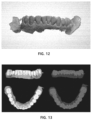
- FIG. 12 is a view illustrating an oral scan image in which a separation reference line is indicated
- FIG. 13 is a view illustrating an oral scan image in which tooth and gum areas are separated based on a separation reference line
- FIG. 14 is a detailed block diagram of a configuration for preprocessing a CT image of a controller of FIG. 1 ;
- FIGS. 15 and 16 are exemplary views for explaining a preprocessing method when a flat reference plane RP 1 is applied.
- FIGS. 17 and 18 are exemplary views for explaining a preprocessing method when a reference plane RP 2 having a three-dimensional boundary surface is applied.
- terms such as “or” and “at least one” may represent one of the words listed together or a combination of two or more.
- “A or B” and “at least one of A and B” may include only one of A or B, or may also include both A and B.
- first and second may be used to describe various elements, but, the above elements should not be limited by the terms above.
- the above terms should not be construed as limiting the order of each component, and may be used for the purpose of distinguishing one element from another.
- first element may be named as “second element” and similarly, “second element” may also be named as “first element.”
- the apparatus for preprocessing image data is an electronic apparatus for removing elements that interfere with image registration of an oral scan image and a CT image before image registration, and may be divided into a configuration for preprocessing an oral scan image and a configuration for preprocessing a CT image.
- the configuration for preprocessing the oral scan image will be described first, and then the configuration for preprocessing the CT image will be described.
- FIG. 1 is a block diagram showing a main configuration of an apparatus for preprocessing image data according to an exemplary embodiment of the present invention
- FIG. 2 is a detailed block diagram of a configuration for preprocessing an oral scan image of a controller of FIG. 1 .
- the apparatus 100 for preprocessing image data may include a communicator 110 , an input unit 120 , a display unit 130 , a storage 140 , and a controller 150 in order to separate and remove a gum area and a dental root area that interfere with image registration of an oral scan image and a dental CT image from the oral scan image and the CT image, respectively.
- the communicator 110 communicates with an external device such as an image photographing device (not shown) and a server (not shown). It may receive image data for the inside of the oral cavity.
- the communicator 110 may perform wireless communication such as 5th generation communication (5G), long term evolution-advanced (LTE-A), long term evolution (LTE), Bluetooth, Bluetooth low energy (BLE), and near field communication (NFC), and may perform wired communication such as cable communication.
- 5G 5th generation communication
- LTE-A long term evolution-advanced
- LTE long term evolution
- BLE Bluetooth low energy
- NFC near field communication
- the image data may include, in addition to the oral scan image and CT image data, other image data measured by a different measurement method for the inside of the oral cavity of the recipient.
- the other image data may include magnetic resonance image data and the like.
- the CT image and the magnetic resonance image are three-dimensional images that display the internal state of the teeth, while the oral scan image is a three-dimensional image that displays the surface state of the teeth.
- the CT image is an image taken through a computed tomography (CT) device using radiation. That is, the CT image may represent information on the distribution of internal tissues such as the dental crown, dental root, and alveolar bone in the oral cavity and bone density information based on the transmittance of the radiation.
- CT computed tomography
- the oral scan image is an image that provides information on the shape of the dental crown portion of the tooth exposed to the outside and the shape of the gum around the tooth.
- the oral scan image may be obtained by directly scanning the inside of the recipient's oral cavity through an oral scanner or may be obtained by scanning an impression model that mimics the inside of the recipient's oral cavity with an intaglio or a plaster model created through embossing of the impression model, and the scan image of the impression model can be inverted and used as an oral scan image.
- the input unit 120 generates input data in response to a user input of the electronic apparatus 100 .
- the input unit 120 includes at least one input means.
- the input unit 120 may include a keyboard, a keypad, a dome switch, a touch panel, a touch key, a mouse, a menu button, and the like.
- the display unit 130 displays display data according to the operation of the electronic apparatus 100 .
- the display unit 130 may include a liquid crystal display (LCD), a light emitting diode (LED) display, an organic light emitting diode (OLED) display, a micro electro mechanical systems (MEMS) display, and an electronic paper display.
- the display unit 130 may be combined with the input unit 120 to be implemented as a touch screen.
- the storage 140 stores operation programs of the electronic apparatus 100 .
- the storage 140 may store an algorithm necessary for separating a gum area from an oral scan image, such as a decimation algorithm, a floor detection algorithm, a morphological operation algorithm, and a grouping algorithm, and an algorithm necessary for separating the dental root area from a CT image.
- the storage 140 may store a plurality of image data received from an image photographing device or the like.
- the controller 150 receives an oral scan image and a CT image from an external device such as an image photographing device and a server, separates and removes the gum area from the oral scan image, and separates and removes the dental root area from the CT image.
- an external device such as an image photographing device and a server
- the apparatus 100 for preprocessing image data can improve the registration speed and accuracy by accurately separating and removing the gum area from the oral scan image and registering it and the dental CT image.
- the controller 150 may include a preprocessor 151 , a start line detector 152 , a boundary line detector 153 , an area setting unit 154 , a reference line determination unit 155 , and an area separator 156 .
- FIG. 3 is a view illustrating before and after preprocessing of an oral scan image according to an exemplary embodiment of the present invention.
- the preprocessor 151 serves to lower the mesh resolution of the oral scan image to a reference resolution (e.g., 40,000 meshes). For example, the preprocessor 151 may reduce the number of meshes by using a decimation algorithm that reduces the number of data dimensions.
- an oral scan image photographed by an image photographing device has a non-uniform distribution of mesh information and a relatively large number of meshes of 40,000 to 800,000, which takes a long time to process the mesh information.
- the preprocessor 151 when the preprocessor 151 according to an exemplary embodiment of the present invention reduces the number of meshes to 40,000 by using the decimation algorithm, the distribution of mesh information becomes uniform, and the processing speed of mesh information can be improved. However, since most features disappear when the number of meshes is excessively reduced, it is preferable to reduce the number to an appropriate number.
- FIG. 4 is a view illustrating a start line detected in an oral scan image according to an exemplary embodiment of the present invention.
- the start line detector 152 detects a start line that is a set of vertexes corresponding to a bottom of the gum area in the oral scan image preprocessed by the preprocessor 151 .
- the start line detector 152 may detect the start line using a Floor Detection algorithm.
- start line detector 152 to detect the start line will be described, but it is natural that the start plane can be detected by extending this.
- the oral scan image is divided into a hand type and a desk type, depending on an image photographing device that has photographed the image.
- the hand type has an open bottom of the oral scan image
- the desk type has a flat bottom surface with the bottom of the oral scan image blocked as shown in FIG. 4 ( b ) . Therefore, a method of detecting the start line is different depending on the type of the oral scan image.
- the start line detector 152 calculates the number of adjacent vertexes for each mesh of the oral scan image, and selects vertexes having the calculated number of vertexes equal to or less than a reference number (e.g., 3). Next, the start line is detected by connecting the selected vertexes.
- a reference number e.g. 3
- the start line detector 152 calculates a curvature of each mesh of the oral scan image, and selects meshes having the calculated curvature equal to or less than a reference curvature.
- the start line is detected by connecting the vertexes included in the selected meshes.
- the boundary line detector 153 detects boundary lines between the dental area and the gum area from the oral scan image preprocessed by the preprocessor 151 .
- FIG. 5 is a view for explaining a method of calculating a curvature between adjacent meshes of an oral scan image according to an exemplary embodiment of the present invention
- FIG. 6 is a view illustrating boundary points in an oral scan image.
- the boundary line detector 153 calculates a curvature between adjacent meshes of the oral scan image, selects meshes having the calculated curvature equal to or greater than a reference curvature, and determines a vertex shared by the selected meshes as a boundary point.
- the boundary line detector 153 calculates a maximum curvature and a minimum curvature between adjacent meshes of the oral scan image, and calculates at least one of a Gaussian curvature and an average curvature by using the calculated maximum and minimum curvatures. And, it selects meshes having at least any one of the calculated Gaussian curvature and the average curvature equal to or greater than a reference curvature, and determines a vertex shared by the selected meshes as a boundary point.
- the boundary points extracted using the Gaussian curvature and the average curvature are more distinct than the boundary points extracted using the Gaussian curvature (see FIG. 6 ( a ) ), so in terms of complementarity, it is preferable to determine the boundary point using both the Gaussian curvature and the average curvature.
- the boundary line detector 153 draws, at a vertex shared by adjacent meshes, a plane that includes a normal vector and intersects the mesh curved surface, and then continues to intersect the plane with the mesh curved surface while rotating the plane around the normal vector.
- a number of curves centered on the vertex may be obtained on the mesh curved surface, and each curvature may be calculated using the angle between these curves and the tangent plane of the vertex.
- the largest value among these curvatures may be defined as the maximum curvature, and the smallest value may be defined as the minimum curvature.
- a plane including two curves each having the largest maximum curvature and the smallest minimum curvature at the vertex among the calculated curvatures may be defined as a plane of principal curvatures.
- the Gaussian curvature is defined as the product of the maximum curvature and the minimum curvature
- the average curvature is defined as the average of the maximum curvature and the minimum curvature.
- a vertex shared by the selected meshes is determined as a boundary point.
- FIG. 7 is a view for explaining a method of connecting boundary points in an oral scan image according to an exemplary embodiment of the present invention.
- the boundary line detector 153 connects adjacent boundary points by sequentially dilating and eroding boundary points using a morphological operation algorithm.
- the morphological operation algorithm is an algorithm for dilating and eroding an area, and is generally used for connecting or disconnecting adjacent points.
- the boundary property between the tooth and the gum areas can be further improved because only the connection between adjacent boundary points is performed while maintaining the thickness of the existing boundary point component.
- FIG. 8 is a view illustrating an oral scan image labeled with the same label for each of the connected boundary points
- FIG. 9 is a view illustrating an area-dilated oral scan image for each label
- FIG. 10 is a view illustrating an oral scan image in which an area-dilated label is integrated.
- the boundary line detector 153 labels each of the connected boundary points with the same label. In FIG. 8 , different colors were displayed for each label.
- the boundary line detector 153 dilates at least once for each labeled label, and integrates adjacent labels. Here, the dilation may be repeated until all labels are integrated. Accordingly, the boundary line can be labeled with one integrated label.
- FIG. 11 is a view for explaining a method of setting a separation target area in an oral scan image according to an exemplary embodiment of the present invention
- FIG. 12 is a view illustrating an oral scan image in which a separation reference line is indicated
- FIG. 13 is a view illustrating an oral scan image in which tooth and gum areas are separated based on a separation reference line.
- the area setting unit 154 sets a gum area, which is a separation target area, based on the previously detected start line and boundary line. That is, the area setting unit 154 sequentially integrates vertexes connected from the start line to the boundary line and set it as the separation target area.
- the reference line determination unit 155 determines the outermost line of the separation target area as a separation reference line for separating a gum area from the oral scan image.
- the area separator 156 separates a gum area from the oral scan image using the separation reference line.
- the apparatus 100 for preprocessing image data can improve the registration speed and accuracy by relatively quickly and accurately separating and removing the gum area from the oral scan image and registering it and the dental CT image.
- FIG. 14 is a detailed block diagram of a configuration for preprocessing a CT image of a controller of FIG. 1 .
- the controller 150 may include a reference plane extraction unit 157 , an area setting unit 158 , and an image processing unit 159 to preprocess the CT image data.
- the reference plane extraction unit 157 extracts a reference plane RP from the CT image.
- the reference plane RP is a reference plane for setting the dental root area RA, which is the root area of the tooth, and may be a plane RP 1 or a three-dimensional boundary surface RP 2 having a curved shape.
- one reference plane RP may be extracted to be equally applied to the maxilla and the mandible, or two or more may be extracted to be applied to each of the maxilla and the mandible.
- the area setting unit 158 sets an area spaced apart by a predetermined height from the reference plane RP as the dental root area RA of the CT image.
- the dental root area RA is an unnecessary area of a CT image that is not included in other images, such as an oral scan image, and is an area that can reduce the time and efficiency of image registration by acting as an interference element in image registration between the CT image and the oral scan image. Accordingly, which part of the dental root area RA is in the CT image may be set at S 202 . However, when the dental root area RA is precisely set, the load for the setting operation may increase, so it may be desirable to set the dental root area RA roughly according to the method described above or later.
- the image processing unit 159 processes the CT image data so that the dental root area RA set by the area setting unit 158 is removed from the CT image. That is, data corresponding to the dental root area RA may be removed from the CT image data.
- the CT image from which the dental root area RA has been removed may be used for image registration with other images according to various algorithms.
- image registration according to these various algorithms is completed, by adding the removed dental root area RA to the corresponding CT image, a CT image and other images on which complete image registration has been performed may be derived.
- reference plane extraction unit 157 and the area setting unit 158 according to the type of the reference plane RP will be described.
- the reference plane (RP) and the preprocessing example are shown only for the mandible, but the present invention is not limited thereto, and the same method as for the mandible can be applied to the maxilla.
- FIGS. 15 and 16 are exemplary views for explaining a preprocessing method when a flat reference plane RP 1 is applied.
- the reference plane RP may be a plane RP 1 .
- two different reference planes RP 1 may be set for the maxilla and mandible.
- the reference plane extraction unit 157 may set a plane at heights EH 1 and EH 2 of the mandibular outermost teeth ET 1 and ET 2 (e.g., molars or wisdom teeth, etc.) or at a height spaced apart from the heights EH 1 and EH 2 by a predetermined height as the reference plane RP 1 for the mandible. That is, the reference plane RP 1 for the mandible may be a plane passing through the heights EH 1 and EH 2 of one end of the two mandibular outermost teeth ET 1 and ET 2 , or a plane passing through a height spaced apart from the heights EH 1 and EH 2 by a predetermined height.
- the reference plane RP 1 for the mandible may be a plane passing through the heights EH 1 and EH 2 of one end of the two mandibular outermost teeth ET 1 and ET 2 , or a plane passing through a height spaced apart from the heights EH 1 and EH 2 by a predetermined height
- the reference plane extraction unit 157 may set a plane at heights of the maxillary outermost teeth or at a height spaced apart from it by a predetermined height as the reference plane RP 1 for the maxilla.
- the area setting unit 158 sets an area (checkered area) spaced downward by a predetermined height RH 1 from the reference plane RP 1 for the mandible as the dental root area RA for the mandible of the CT image.
- the area setting unit 158 may set an area spaced upward by a predetermined height from the reference plane RP 1 for the maxilla as the dental root area RA for the maxilla of the CT image.
- the separation distance RH 1 from the reference plane RP 1 for the mandible and the separation distance from the reference plane RP 1 for the maxilla set by the area setting unit 158 may be different from each other. This is because the average height of the maxillary teeth and the mandibular teeth may be different from each other.
- FIGS. 17 and 18 are exemplary views for explaining a preprocessing method when a reference plane RP 2 having a three-dimensional boundary surface is applied.
- the reference plane RP may be a three-dimensional boundary surface RP 2 .
- the same one reference plane RP 2 may be set or two different reference planes RP 2 may be set for the maxilla and the mandible.
- the reference plane extraction unit 157 may set a three-dimensional boundary surface that is located in the space between the maxillary teeth and the mandibular teeth (hereinafter referred to as an “interspace”) in the CT image and reflects each height of the maxillary teeth and each height of the mandibular teeth, as a reference plane RP 2 of the CT image.
- the interspace is formed between a three-dimensional boundary surface of a shape corresponding to the ends of the maxillary teeth (hereinafter, referred to as an “upper three-dimensional boundary surface”) and a three-dimensional boundary surface of a shape corresponding to the ends of the mandibular teeth (hereinafter, referred to as an “lower three-dimensional boundary surface”).
- the reference plane extraction unit 157 may set a three-dimensional boundary surface composed of the average height values of the upper three-dimensional boundary surface, the lower three-dimensional boundary surface, or the upper three-dimensional boundary surface and the lower three-dimensional boundary surface as a reference plane RP 2 .
- the area setting unit 158 sets an area (checkered area) spaced downward by a predetermined height RH 2 from the reference plane RP 2 as the dental root area RA for the mandible of the CT image. Similarly, the area setting unit 158 may set an area spaced upward by a predetermined height from the reference plane RP 2 as the dental root area RA for the maxilla of the CT image.
- the separation distance RH 2 from the reference plane RP 2 for setting the dental root area RA for the mandible and the separation distance from the reference plane RP 2 for setting the dental root area RA for the maxilla set by the area setting unit 158 may be different from each other. This is because the average height of the maxillary teeth and the mandibular teeth may be different from each other.
- the reference plane extraction unit 157 may set the lower three-dimensional boundary surface as the reference plane RA 2 for the mandible and the upper three-dimensional boundary surface as the reference plane for the maxilla, respectively.
- the area setting unit 158 sets an area (checkered area) spaced downward by a predetermined height RH 2 from the reference plane RP 2 for the mandible as the dental root area RA for the mandible of the CT image.
- the area setting unit 158 may set an area spaced upward by a predetermined height from the reference plane RP 2 for the maxilla as the dental root area RA for the maxilla of the CT image.
- the separation distance RH 2 from the reference plane RP 2 for setting the dental root area RA for the mandible and the separation distance from the reference plane RP 2 for setting the dental root area RA for the maxilla set by the area setting unit 158 may be different from each other. This is because the average height of the maxillary teeth and the mandibular teeth may be different from each other.
- the apparatus 100 for preprocessing image data may be manufactured as a separate apparatus or may be included in an image registration apparatus.
- the image registration apparatus may register the oral scan image from which the gum area is removed and the CT image from which the dental root area RA is removed. Accordingly, the registration speed and the registration accuracy can be improved.
- the apparatus for preprocessing image data according to the present invention may be used in various dental treatment fields such as implant operation.
Landscapes
- Engineering & Computer Science (AREA)
- Health & Medical Sciences (AREA)
- Life Sciences & Earth Sciences (AREA)
- Physics & Mathematics (AREA)
- Medical Informatics (AREA)
- Theoretical Computer Science (AREA)
- Computer Vision & Pattern Recognition (AREA)
- General Physics & Mathematics (AREA)
- Radiology & Medical Imaging (AREA)
- Nuclear Medicine, Radiotherapy & Molecular Imaging (AREA)
- General Health & Medical Sciences (AREA)
- Animal Behavior & Ethology (AREA)
- Biophysics (AREA)
- High Energy & Nuclear Physics (AREA)
- Optics & Photonics (AREA)
- Pathology (AREA)
- Veterinary Medicine (AREA)
- Biomedical Technology (AREA)
- Heart & Thoracic Surgery (AREA)
- Molecular Biology (AREA)
- Public Health (AREA)
- Surgery (AREA)
- Software Systems (AREA)
- Geometry (AREA)
- Computer Graphics (AREA)
- Pulmonology (AREA)
- Dentistry (AREA)
- Oral & Maxillofacial Surgery (AREA)
- Quality & Reliability (AREA)
- Apparatus For Radiation Diagnosis (AREA)
- Dental Tools And Instruments Or Auxiliary Dental Instruments (AREA)
- Stereoscopic And Panoramic Photography (AREA)
- Image Analysis (AREA)
Abstract
Description
Claims (15)
Applications Claiming Priority (5)
| Application Number | Priority Date | Filing Date | Title |
|---|---|---|---|
| KR10-2019-0042437 | 2019-04-11 | ||
| KR1020190042381A KR102177886B1 (en) | 2019-04-11 | 2019-04-11 | Method and apparatus for preprocessing computerized tomography image data |
| KR10-2019-0042381 | 2019-04-11 | ||
| KR1020190042437A KR102224064B1 (en) | 2019-04-11 | 2019-04-11 | Apparatus For Separating Gum Area From Oral Scan Image |
| PCT/KR2020/002755 WO2020209495A1 (en) | 2019-04-11 | 2020-02-26 | Apparatus for preprocessing image data |
Publications (2)
| Publication Number | Publication Date |
|---|---|
| US20220198686A1 US20220198686A1 (en) | 2022-06-23 |
| US12062193B2 true US12062193B2 (en) | 2024-08-13 |
Family
ID=72752034
Family Applications (1)
| Application Number | Title | Priority Date | Filing Date |
|---|---|---|---|
| US17/602,617 Active 2040-09-11 US12062193B2 (en) | 2019-04-11 | 2020-02-26 | Apparatus for preprocessing image data |
Country Status (5)
| Country | Link |
|---|---|
| US (1) | US12062193B2 (en) |
| EP (1) | EP3954293A4 (en) |
| CN (1) | CN113692250B (en) |
| TW (1) | TWI782269B (en) |
| WO (1) | WO2020209495A1 (en) |
Families Citing this family (3)
| Publication number | Priority date | Publication date | Assignee | Title |
|---|---|---|---|---|
| US9008769B2 (en) | 2012-12-21 | 2015-04-14 | Backbeat Medical, Inc. | Methods and systems for lowering blood pressure through reduction of ventricle filling |
| KR102460621B1 (en) * | 2020-09-23 | 2022-10-28 | 주식회사 메디트 | An intraoral image processing apparatus, and an intraoral image processing method |
| US20240161431A1 (en) * | 2022-11-10 | 2024-05-16 | Sdc U.S. Smilepay Spv | Tooth point correspondence detection |
Citations (18)
| Publication number | Priority date | Publication date | Assignee | Title |
|---|---|---|---|---|
| US6338000B1 (en) * | 1997-03-24 | 2002-01-08 | Honda Giken Kogyo Kabushiki Kaisha | Method of generating shape data method of verifying shape data |
| US20060083422A1 (en) | 2004-01-27 | 2006-04-20 | Ernst Maurice M | Three-dimensional modeling of the oral cavity |
| US20090213119A1 (en) * | 2007-12-17 | 2009-08-27 | Electronics And Telecommunications Research Institute | Remeshing method and apparatus for restoring sharp features of mesh made smooth enough |
| KR20110138125A (en) | 2010-06-18 | 2011-12-26 | 정제교 | Marker Tray for Matching Heterogeneous Image Images and Head and Neck Image Registration |
| KR20140015239A (en) | 2010-06-29 | 2014-02-06 | 쓰리세이프 에이/에스 | 2d image arrangement |
| CN105046750A (en) | 2015-08-24 | 2015-11-11 | 杭州美齐科技有限公司 | Method for automatically segmenting whole dental triangular mesh model |
| US20160070821A1 (en) | 2014-09-08 | 2016-03-10 | 3M Innovative Properties Company | Method of aligning intra-oral digital 3d models |
| CN105551081A (en) | 2016-02-05 | 2016-05-04 | 杭州美齐科技有限公司 | Virtual gum triangular mesh algorithm construction and follow-up algorithm |
| WO2016108453A1 (en) | 2014-12-31 | 2016-07-07 | 오스템임플란트 주식회사 | Teeth image automatic matching method, apparatus therefor, and recording medium therefor |
| KR101655910B1 (en) | 2015-05-06 | 2016-09-09 | 재단법인 아산사회복지재단 | Method and program for angiograph image registration |
| US20170132791A1 (en) * | 2015-11-05 | 2017-05-11 | Shenyang Neusoft Medical Systems Co., Ltd. | Cutting three-dimensional image |
| US20170169562A1 (en) * | 2015-12-10 | 2017-06-15 | 3M Innovative Properties Company | Method for automatic tooth type recognition from 3d scans |
| KR20170127950A (en) | 2016-05-13 | 2017-11-22 | 연세대학교 산학협력단 | Fully automatic dental segmentation system and method for computerized tomography |
| KR20180108797A (en) | 2016-04-06 | 2018-10-04 | 엑스-네브 테크놀로지스, 엘엘씨 | A system that provides probe trace IG point tracking |
| KR101913586B1 (en) | 2017-05-24 | 2018-11-01 | 오스템임플란트 주식회사 | Dental image registration method, apparatus, and recording medium thereof |
| KR101953341B1 (en) | 2017-09-20 | 2019-03-04 | 오스템임플란트 주식회사 | Crown digital design method for processing overlapped region, apparatus, and recording medium thereof |
| KR20190024360A (en) | 2017-08-31 | 2019-03-08 | 주식회사 디오 | Method for adjusting intraorla images of different types |
| US20210322136A1 (en) * | 2018-09-04 | 2021-10-21 | Promaton Holding B.V. | Automated orthodontic treatment planning using deep learning |
Family Cites Families (10)
| Publication number | Priority date | Publication date | Assignee | Title |
|---|---|---|---|---|
| KR101146862B1 (en) * | 2010-12-02 | 2012-05-16 | 이준호 | Method to change from two-dimensional panorama picture to three-dimensional picture and the recorded media thereof |
| DE102011010975A1 (en) * | 2011-02-10 | 2012-08-16 | Martin Tank | Method and analysis system for geometrical analysis of scan data of oral structures |
| WO2012129160A2 (en) * | 2011-03-21 | 2012-09-27 | Carestream Health, Inc. | A method for tooth surface classification |
| CN103700103A (en) * | 2013-12-05 | 2014-04-02 | 嘉兴学院 | Method for automatically extracting gingiva curves of three-dimensional digital dentition model |
| CN105447908B (en) * | 2015-12-04 | 2018-04-17 | 山东山大华天软件有限公司 | Dental arch model generation method based on oral cavity scan data and CBCT data |
| CN105662610B (en) * | 2015-12-30 | 2018-03-13 | 青岛达芬奇科技有限公司 | The generation method of the virtual gum for stealthy correction based on model |
| CN106214175B (en) * | 2016-08-26 | 2020-06-16 | 中国科学院深圳先进技术研究院 | A method and device for estimating three-dimensional tooth axis |
| CN106846307B (en) * | 2017-01-19 | 2020-09-22 | 深圳市深图医学影像设备有限公司 | Image processing method and device based on cone beam computed tomography |
| CN108242056B (en) * | 2018-02-06 | 2020-04-28 | 北京朗视仪器有限公司 | Three-dimensional tooth grid data segmentation method based on harmonic field algorithm |
| CN108648283B (en) * | 2018-04-02 | 2022-07-05 | 北京正齐口腔医疗技术有限公司 | Tooth segmentation method and device |
-
2020
- 2020-02-26 US US17/602,617 patent/US12062193B2/en active Active
- 2020-02-26 CN CN202080027597.2A patent/CN113692250B/en active Active
- 2020-02-26 EP EP20786737.5A patent/EP3954293A4/en not_active Withdrawn
- 2020-02-26 WO PCT/KR2020/002755 patent/WO2020209495A1/en not_active Ceased
- 2020-04-10 TW TW109112222A patent/TWI782269B/en active
Patent Citations (19)
| Publication number | Priority date | Publication date | Assignee | Title |
|---|---|---|---|---|
| US6338000B1 (en) * | 1997-03-24 | 2002-01-08 | Honda Giken Kogyo Kabushiki Kaisha | Method of generating shape data method of verifying shape data |
| US20060083422A1 (en) | 2004-01-27 | 2006-04-20 | Ernst Maurice M | Three-dimensional modeling of the oral cavity |
| US20090213119A1 (en) * | 2007-12-17 | 2009-08-27 | Electronics And Telecommunications Research Institute | Remeshing method and apparatus for restoring sharp features of mesh made smooth enough |
| KR20110138125A (en) | 2010-06-18 | 2011-12-26 | 정제교 | Marker Tray for Matching Heterogeneous Image Images and Head and Neck Image Registration |
| KR20140015239A (en) | 2010-06-29 | 2014-02-06 | 쓰리세이프 에이/에스 | 2d image arrangement |
| CN106604692A (en) | 2014-09-08 | 2017-04-26 | 3M创新有限公司 | Method for aligning intraoral digital 3D models |
| US20160070821A1 (en) | 2014-09-08 | 2016-03-10 | 3M Innovative Properties Company | Method of aligning intra-oral digital 3d models |
| WO2016108453A1 (en) | 2014-12-31 | 2016-07-07 | 오스템임플란트 주식회사 | Teeth image automatic matching method, apparatus therefor, and recording medium therefor |
| KR101655910B1 (en) | 2015-05-06 | 2016-09-09 | 재단법인 아산사회복지재단 | Method and program for angiograph image registration |
| CN105046750A (en) | 2015-08-24 | 2015-11-11 | 杭州美齐科技有限公司 | Method for automatically segmenting whole dental triangular mesh model |
| US20170132791A1 (en) * | 2015-11-05 | 2017-05-11 | Shenyang Neusoft Medical Systems Co., Ltd. | Cutting three-dimensional image |
| US20170169562A1 (en) * | 2015-12-10 | 2017-06-15 | 3M Innovative Properties Company | Method for automatic tooth type recognition from 3d scans |
| CN105551081A (en) | 2016-02-05 | 2016-05-04 | 杭州美齐科技有限公司 | Virtual gum triangular mesh algorithm construction and follow-up algorithm |
| KR20180108797A (en) | 2016-04-06 | 2018-10-04 | 엑스-네브 테크놀로지스, 엘엘씨 | A system that provides probe trace IG point tracking |
| KR20170127950A (en) | 2016-05-13 | 2017-11-22 | 연세대학교 산학협력단 | Fully automatic dental segmentation system and method for computerized tomography |
| KR101913586B1 (en) | 2017-05-24 | 2018-11-01 | 오스템임플란트 주식회사 | Dental image registration method, apparatus, and recording medium thereof |
| KR20190024360A (en) | 2017-08-31 | 2019-03-08 | 주식회사 디오 | Method for adjusting intraorla images of different types |
| KR101953341B1 (en) | 2017-09-20 | 2019-03-04 | 오스템임플란트 주식회사 | Crown digital design method for processing overlapped region, apparatus, and recording medium thereof |
| US20210322136A1 (en) * | 2018-09-04 | 2021-10-21 | Promaton Holding B.V. | Automated orthodontic treatment planning using deep learning |
Non-Patent Citations (4)
| Title |
|---|
| Machine translated, KR-101655910. Date Published: Sep. 9, 2016. (Year: 2016). * |
| Office Action issued to related CN Application No. 202080027597.2., dated Jan. 19, 2024, 11 pages (with English translation). |
| Office Action issued to related European Application No. 20786737.5, dated Jan. 3, 2023, 8 pages. |
| Wu et al., "Tooth segmentation on dental meshes using morphologic skeleton", Computers & Graphics 38 (2014) p. 199-211. (Year: 2014). * |
Also Published As
| Publication number | Publication date |
|---|---|
| EP3954293A1 (en) | 2022-02-16 |
| CN113692250A (en) | 2021-11-23 |
| TW202038189A (en) | 2020-10-16 |
| CN113692250B (en) | 2024-07-23 |
| TWI782269B (en) | 2022-11-01 |
| EP3954293A4 (en) | 2023-01-11 |
| US20220198686A1 (en) | 2022-06-23 |
| WO2020209495A1 (en) | 2020-10-15 |
Similar Documents
| Publication | Publication Date | Title |
|---|---|---|
| US11978203B2 (en) | Dental object detection method, and image matching method and device using dental object | |
| US12070375B2 (en) | Area of interest overlay on dental site using augmented reality | |
| US20220241056A1 (en) | Monitoring of dentition | |
| US11534275B2 (en) | Method for constructing a restoration | |
| EP3140809B1 (en) | Identification of areas of interest during intraoral scans | |
| US12062193B2 (en) | Apparatus for preprocessing image data | |
| JP2022516488A (en) | Teeth segmentation using tooth alignment | |
| EP3806034B1 (en) | Segmentation device | |
| KR20200120035A (en) | Method and apparatus for detecting tooth object in oral scan image | |
| KR20210019816A (en) | Method and apparatus for matching oral scan image and oral scan image | |
| KR102224064B1 (en) | Apparatus For Separating Gum Area From Oral Scan Image | |
| US20230215027A1 (en) | Oral image marker detection method, and oral image matching device and method using same | |
| KR102322634B1 (en) | Method and apparatus for matching of images using tooth object | |
| KR102267488B1 (en) | Apparatus And Method For Dental Image Registration | |
| CA3255418A1 (en) | Tooth position determination and generation of 2d reslice images with an artificial neural network | |
| EP3973878B1 (en) | Nerve detection method and device | |
| KR102177886B1 (en) | Method and apparatus for preprocessing computerized tomography image data | |
| US11869203B2 (en) | Dental image registration device and method | |
| KR20210131685A (en) | Apparatus and method for oral image registration | |
| CN121752222A (en) | Method and apparatus for processing scanned image of oral scanner, and recording medium having instructions recorded thereon |
Legal Events
| Date | Code | Title | Description |
|---|---|---|---|
| FEPP | Fee payment procedure |
Free format text: ENTITY STATUS SET TO UNDISCOUNTED (ORIGINAL EVENT CODE: BIG.); ENTITY STATUS OF PATENT OWNER: SMALL ENTITY |
|
| FEPP | Fee payment procedure |
Free format text: ENTITY STATUS SET TO SMALL (ORIGINAL EVENT CODE: SMAL); ENTITY STATUS OF PATENT OWNER: SMALL ENTITY |
|
| AS | Assignment |
Owner name: DIO CORPORATION, KOREA, DEMOCRATIC PEOPLE'S REPUBLIC OF Free format text: ASSIGNMENT OF ASSIGNORS INTEREST;ASSIGNORS:KIM, JIN CHEOL;KIM, JIN BAEK;REEL/FRAME:058306/0989 Effective date: 20211001 |
|
| STPP | Information on status: patent application and granting procedure in general |
Free format text: DOCKETED NEW CASE - READY FOR EXAMINATION |
|
| STPP | Information on status: patent application and granting procedure in general |
Free format text: NON FINAL ACTION MAILED |
|
| STPP | Information on status: patent application and granting procedure in general |
Free format text: RESPONSE TO NON-FINAL OFFICE ACTION ENTERED AND FORWARDED TO EXAMINER |
|
| STPP | Information on status: patent application and granting procedure in general |
Free format text: NOTICE OF ALLOWANCE MAILED -- APPLICATION RECEIVED IN OFFICE OF PUBLICATIONS |
|
| ZAAB | Notice of allowance mailed |
Free format text: ORIGINAL CODE: MN/=. |
|
| STPP | Information on status: patent application and granting procedure in general |
Free format text: PUBLICATIONS -- ISSUE FEE PAYMENT VERIFIED |
|
| STCF | Information on status: patent grant |
Free format text: PATENTED CASE |