US12046367B2 - Medical image reading assistant apparatus and method providing hanging protocols based on medical use artificial neural network - Google Patents
Medical image reading assistant apparatus and method providing hanging protocols based on medical use artificial neural network Download PDFInfo
- Publication number
- US12046367B2 US12046367B2 US16/896,801 US202016896801A US12046367B2 US 12046367 B2 US12046367 B2 US 12046367B2 US 202016896801 A US202016896801 A US 202016896801A US 12046367 B2 US12046367 B2 US 12046367B2
- Authority
- US
- United States
- Prior art keywords
- neural network
- artificial neural
- medical image
- display setting
- analysis result
- Prior art date
- Legal status (The legal status is an assumption and is not a legal conclusion. Google has not performed a legal analysis and makes no representation as to the accuracy of the status listed.)
- Active, expires
Links
Images


Classifications
-
- A—HUMAN NECESSITIES
- A61—MEDICAL OR VETERINARY SCIENCE; HYGIENE
- A61B—DIAGNOSIS; SURGERY; IDENTIFICATION
- A61B5/00—Measuring for diagnostic purposes; Identification of persons
- A61B5/72—Signal processing specially adapted for physiological signals or for diagnostic purposes
- A61B5/7235—Details of waveform analysis
- A61B5/7264—Classification of physiological signals or data, e.g. using neural networks, statistical classifiers, expert systems or fuzzy systems
-
- A—HUMAN NECESSITIES
- A61—MEDICAL OR VETERINARY SCIENCE; HYGIENE
- A61B—DIAGNOSIS; SURGERY; IDENTIFICATION
- A61B5/00—Measuring for diagnostic purposes; Identification of persons
- A61B5/74—Details of notification to user or communication with user or patient; User input means
- A61B5/742—Details of notification to user or communication with user or patient; User input means using visual displays
- A61B5/743—Displaying an image simultaneously with additional graphical information, e.g. symbols, charts, function plots
-
- A—HUMAN NECESSITIES
- A61—MEDICAL OR VETERINARY SCIENCE; HYGIENE
- A61B—DIAGNOSIS; SURGERY; IDENTIFICATION
- A61B5/00—Measuring for diagnostic purposes; Identification of persons
- A61B5/74—Details of notification to user or communication with user or patient; User input means
- A61B5/742—Details of notification to user or communication with user or patient; User input means using visual displays
- A61B5/7435—Displaying user selection data, e.g. icons in a graphical user interface
-
- A—HUMAN NECESSITIES
- A61—MEDICAL OR VETERINARY SCIENCE; HYGIENE
- A61B—DIAGNOSIS; SURGERY; IDENTIFICATION
- A61B6/00—Apparatus or devices for radiation diagnosis; Apparatus or devices for radiation diagnosis combined with radiation therapy equipment
- A61B6/52—Devices using data or image processing specially adapted for radiation diagnosis
- A61B6/5211—Devices using data or image processing specially adapted for radiation diagnosis involving processing of medical diagnostic data
- A61B6/5217—Devices using data or image processing specially adapted for radiation diagnosis involving processing of medical diagnostic data extracting a diagnostic or physiological parameter from medical diagnostic data
-
- G—PHYSICS
- G06—COMPUTING OR CALCULATING; COUNTING
- G06F—ELECTRIC DIGITAL DATA PROCESSING
- G06F3/00—Input arrangements for transferring data to be processed into a form capable of being handled by the computer; Output arrangements for transferring data from processing unit to output unit, e.g. interface arrangements
- G06F3/01—Input arrangements or combined input and output arrangements for interaction between user and computer
- G06F3/048—Interaction techniques based on graphical user interfaces [GUI]
- G06F3/0481—Interaction techniques based on graphical user interfaces [GUI] based on specific properties of the displayed interaction object or a metaphor-based environment, e.g. interaction with desktop elements like windows or icons, or assisted by a cursor's changing behaviour or appearance
-
- G—PHYSICS
- G06—COMPUTING OR CALCULATING; COUNTING
- G06F—ELECTRIC DIGITAL DATA PROCESSING
- G06F3/00—Input arrangements for transferring data to be processed into a form capable of being handled by the computer; Output arrangements for transferring data from processing unit to output unit, e.g. interface arrangements
- G06F3/14—Digital output to display device ; Cooperation and interconnection of the display device with other functional units
-
- G—PHYSICS
- G06—COMPUTING OR CALCULATING; COUNTING
- G06N—COMPUTING ARRANGEMENTS BASED ON SPECIFIC COMPUTATIONAL MODELS
- G06N3/00—Computing arrangements based on biological models
- G06N3/02—Neural networks
- G06N3/10—Interfaces, programming languages or software development kits, e.g. for simulating neural networks
-
- G—PHYSICS
- G06—COMPUTING OR CALCULATING; COUNTING
- G06T—IMAGE DATA PROCESSING OR GENERATION, IN GENERAL
- G06T7/00—Image analysis
- G06T7/0002—Inspection of images, e.g. flaw detection
- G06T7/0012—Biomedical image inspection
-
- G—PHYSICS
- G06—COMPUTING OR CALCULATING; COUNTING
- G06V—IMAGE OR VIDEO RECOGNITION OR UNDERSTANDING
- G06V10/00—Arrangements for image or video recognition or understanding
- G06V10/70—Arrangements for image or video recognition or understanding using pattern recognition or machine learning
- G06V10/82—Arrangements for image or video recognition or understanding using pattern recognition or machine learning using neural networks
-
- G—PHYSICS
- G09—EDUCATION; CRYPTOGRAPHY; DISPLAY; ADVERTISING; SEALS
- G09G—ARRANGEMENTS OR CIRCUITS FOR CONTROL OF INDICATING DEVICES USING STATIC MEANS TO PRESENT VARIABLE INFORMATION
- G09G5/00—Control arrangements or circuits for visual indicators common to cathode-ray tube indicators and other visual indicators
- G09G5/14—Display of multiple viewports
-
- G—PHYSICS
- G16—INFORMATION AND COMMUNICATION TECHNOLOGY [ICT] SPECIALLY ADAPTED FOR SPECIFIC APPLICATION FIELDS
- G16H—HEALTHCARE INFORMATICS, i.e. INFORMATION AND COMMUNICATION TECHNOLOGY [ICT] SPECIALLY ADAPTED FOR THE HANDLING OR PROCESSING OF MEDICAL OR HEALTHCARE DATA
- G16H15/00—ICT specially adapted for medical reports, e.g. generation or transmission thereof
-
- G—PHYSICS
- G16—INFORMATION AND COMMUNICATION TECHNOLOGY [ICT] SPECIALLY ADAPTED FOR SPECIFIC APPLICATION FIELDS
- G16H—HEALTHCARE INFORMATICS, i.e. INFORMATION AND COMMUNICATION TECHNOLOGY [ICT] SPECIALLY ADAPTED FOR THE HANDLING OR PROCESSING OF MEDICAL OR HEALTHCARE DATA
- G16H30/00—ICT specially adapted for the handling or processing of medical images
- G16H30/40—ICT specially adapted for the handling or processing of medical images for processing medical images, e.g. editing
-
- G—PHYSICS
- G16—INFORMATION AND COMMUNICATION TECHNOLOGY [ICT] SPECIALLY ADAPTED FOR SPECIFIC APPLICATION FIELDS
- G16H—HEALTHCARE INFORMATICS, i.e. INFORMATION AND COMMUNICATION TECHNOLOGY [ICT] SPECIALLY ADAPTED FOR THE HANDLING OR PROCESSING OF MEDICAL OR HEALTHCARE DATA
- G16H50/00—ICT specially adapted for medical diagnosis, medical simulation or medical data mining; ICT specially adapted for detecting, monitoring or modelling epidemics or pandemics
- G16H50/20—ICT specially adapted for medical diagnosis, medical simulation or medical data mining; ICT specially adapted for detecting, monitoring or modelling epidemics or pandemics for computer-aided diagnosis, e.g. based on medical expert systems
-
- G—PHYSICS
- G16—INFORMATION AND COMMUNICATION TECHNOLOGY [ICT] SPECIALLY ADAPTED FOR SPECIFIC APPLICATION FIELDS
- G16H—HEALTHCARE INFORMATICS, i.e. INFORMATION AND COMMUNICATION TECHNOLOGY [ICT] SPECIALLY ADAPTED FOR THE HANDLING OR PROCESSING OF MEDICAL OR HEALTHCARE DATA
- G16H50/00—ICT specially adapted for medical diagnosis, medical simulation or medical data mining; ICT specially adapted for detecting, monitoring or modelling epidemics or pandemics
- G16H50/70—ICT specially adapted for medical diagnosis, medical simulation or medical data mining; ICT specially adapted for detecting, monitoring or modelling epidemics or pandemics for mining of medical data, e.g. analysing previous cases of other patients
-
- A—HUMAN NECESSITIES
- A61—MEDICAL OR VETERINARY SCIENCE; HYGIENE
- A61B—DIAGNOSIS; SURGERY; IDENTIFICATION
- A61B5/00—Measuring for diagnostic purposes; Identification of persons
- A61B5/0033—Features or image-related aspects of imaging apparatus, e.g. for MRI, optical tomography or impedance tomography apparatus; Arrangements of imaging apparatus in a room
- A61B5/0037—Performing a preliminary scan, e.g. a prescan for identifying a region of interest
-
- A—HUMAN NECESSITIES
- A61—MEDICAL OR VETERINARY SCIENCE; HYGIENE
- A61B—DIAGNOSIS; SURGERY; IDENTIFICATION
- A61B5/00—Measuring for diagnostic purposes; Identification of persons
- A61B5/05—Detecting, measuring or recording for diagnosis by means of electric currents or magnetic fields; Measuring using microwaves or radio waves
- A61B5/055—Detecting, measuring or recording for diagnosis by means of electric currents or magnetic fields; Measuring using microwaves or radio waves involving electronic [EMR] or nuclear [NMR] magnetic resonance, e.g. magnetic resonance imaging
-
- A—HUMAN NECESSITIES
- A61—MEDICAL OR VETERINARY SCIENCE; HYGIENE
- A61B—DIAGNOSIS; SURGERY; IDENTIFICATION
- A61B6/00—Apparatus or devices for radiation diagnosis; Apparatus or devices for radiation diagnosis combined with radiation therapy equipment
- A61B6/02—Arrangements for diagnosis sequentially in different planes; Stereoscopic radiation diagnosis
- A61B6/03—Computed tomography [CT]
- A61B6/032—Transmission computed tomography [CT]
-
- G—PHYSICS
- G06—COMPUTING OR CALCULATING; COUNTING
- G06T—IMAGE DATA PROCESSING OR GENERATION, IN GENERAL
- G06T2200/00—Indexing scheme for image data processing or generation, in general
- G06T2200/24—Indexing scheme for image data processing or generation, in general involving graphical user interfaces [GUIs]
-
- G—PHYSICS
- G06—COMPUTING OR CALCULATING; COUNTING
- G06T—IMAGE DATA PROCESSING OR GENERATION, IN GENERAL
- G06T2207/00—Indexing scheme for image analysis or image enhancement
- G06T2207/10—Image acquisition modality
- G06T2207/10072—Tomographic images
- G06T2207/10081—Computed x-ray tomography [CT]
-
- G—PHYSICS
- G06—COMPUTING OR CALCULATING; COUNTING
- G06T—IMAGE DATA PROCESSING OR GENERATION, IN GENERAL
- G06T2207/00—Indexing scheme for image analysis or image enhancement
- G06T2207/10—Image acquisition modality
- G06T2207/10072—Tomographic images
- G06T2207/10088—Magnetic resonance imaging [MRI]
-
- G—PHYSICS
- G06—COMPUTING OR CALCULATING; COUNTING
- G06T—IMAGE DATA PROCESSING OR GENERATION, IN GENERAL
- G06T2207/00—Indexing scheme for image analysis or image enhancement
- G06T2207/20—Special algorithmic details
- G06T2207/20081—Training; Learning
-
- G—PHYSICS
- G06—COMPUTING OR CALCULATING; COUNTING
- G06T—IMAGE DATA PROCESSING OR GENERATION, IN GENERAL
- G06T2207/00—Indexing scheme for image analysis or image enhancement
- G06T2207/20—Special algorithmic details
- G06T2207/20084—Artificial neural networks [ANN]
-
- G—PHYSICS
- G06—COMPUTING OR CALCULATING; COUNTING
- G06T—IMAGE DATA PROCESSING OR GENERATION, IN GENERAL
- G06T2207/00—Indexing scheme for image analysis or image enhancement
- G06T2207/30—Subject of image; Context of image processing
- G06T2207/30004—Biomedical image processing
-
- G—PHYSICS
- G06—COMPUTING OR CALCULATING; COUNTING
- G06T—IMAGE DATA PROCESSING OR GENERATION, IN GENERAL
- G06T2207/00—Indexing scheme for image analysis or image enhancement
- G06T2207/30—Subject of image; Context of image processing
- G06T2207/30004—Biomedical image processing
- G06T2207/30096—Tumor; Lesion
-
- G—PHYSICS
- G09—EDUCATION; CRYPTOGRAPHY; DISPLAY; ADVERTISING; SEALS
- G09G—ARRANGEMENTS OR CIRCUITS FOR CONTROL OF INDICATING DEVICES USING STATIC MEANS TO PRESENT VARIABLE INFORMATION
- G09G2320/00—Control of display operating conditions
- G09G2320/06—Adjustment of display parameters
- G09G2320/0613—The adjustment depending on the type of the information to be displayed
-
- G—PHYSICS
- G09—EDUCATION; CRYPTOGRAPHY; DISPLAY; ADVERTISING; SEALS
- G09G—ARRANGEMENTS OR CIRCUITS FOR CONTROL OF INDICATING DEVICES USING STATIC MEANS TO PRESENT VARIABLE INFORMATION
- G09G2320/00—Control of display operating conditions
- G09G2320/08—Arrangements within a display terminal for setting, manually or automatically, display parameters of the display terminal
-
- G—PHYSICS
- G09—EDUCATION; CRYPTOGRAPHY; DISPLAY; ADVERTISING; SEALS
- G09G—ARRANGEMENTS OR CIRCUITS FOR CONTROL OF INDICATING DEVICES USING STATIC MEANS TO PRESENT VARIABLE INFORMATION
- G09G2380/00—Specific applications
- G09G2380/08—Biomedical applications
Definitions
- the present invention relates to an apparatus and method for supporting the reading of a medical image of a subject. More specifically, the present invention relates to a computing system for providing a hanging protocol, which is a setting in which a medical image is displayed, using an analysis result of a medical artificial neural network, and software that is executed in the computing system.
- the present invention was derived from the research conducted as part of the Electronic System Industry Core Technology R&D Project sponsored by the Korean Ministry of Trade, Industry and Energy and the Korea Evaluation Institute of Industrial Technology [Task Serial Number: 1415160865; Detailed Task Number: 10072064; and Project Name: Development of Artificial Intelligence Source Technology for Supporting Reading of Lung, Liver and Heart Disease Images and Commercialization Associated with PACS].
- CT computed tomography
- chest CT images are frequently used for reading because they enable readers to observe abnormalities in the body, such as the lungs, the bronchi, and the heart.
- the reading of a lesion using CAD may include a process of identifying a suspected lesion first and then evaluating a score (e.g., confidence, malignity, or the like) for that region. For example, if a plurality of nodules is found in the lungs, it will be necessary to specify a nodule that is expected to have the highest malignity and to determine future treatment plans.
- a score e.g., confidence, malignity, or the like
- Korean Patent No. 10-1943011 entitled “Method for Supporting Reading of Medical Image of Subject and Apparatus using the Same” proposes a method that introduces a score evaluation method into a conventional lesion detection system and allow a lesion with a higher score (e.g., confidence, malignancy, or the like) among detected lesions can be read first, thereby increasing the efficiency of reading, and also proposes an apparatus using the same.
- a score evaluation method into a conventional lesion detection system and allow a lesion with a higher score (e.g., confidence, malignancy, or the like) among detected lesions can be read first, thereby increasing the efficiency of reading, and also proposes an apparatus using the same.
- Korean Patent No. 10-1943011 discloses technology in which when a number of lesions are detected for a single type of disease, a list in which entries are arranged from a lesion having the highest score, such as reliability, malignancy or the like, within a single display setting is displayed, and an image related to a selected lesion is displayed when a user selects the lesion from the list.
- Korean Patent No. 10-1943011 assumes a case where a plurality of types of lesions is detected for a single type of disease, and thus it does not propose a method corresponding to a case where lesions for a plurality of types of diseases are detected.
- a medical image series is acquired through a single acquisition process, and the medical image series is not limited to a single type of lesion but may also be used to detect various types of lesions.
- the hanging protocols refer to display settings for medical images.
- a medical image series is acquired through a single acquisition process, and the medical image series is not limited to a single type of lesion but may also be used to detect various types of lesions.
- a lung nodule as well as chronic obstructive pulmonary disease (COPD) may be diagnosed, emphysema may be diagnosed, and/or chronic bronchitis and/or an airway-related disease may also be diagnosed.
- COPD chronic obstructive pulmonary disease
- An object of the present invention is to provide hanging protocols that propose display layout settings appropriate for each type of lesion and disease based on image information and diagnostic information included in a medical image by considering a plurality of types of lesions that can be detected in the same body part and execute the display layout settings based on information related to the type of lesion and disease detected in the medical image.
- An object of the present invention is to propose hanging protocols based on disease codes detected for medical images.
- An object of the present invention is to execute hanging protocols based on disease codes detected in medical images in an environment having a CAD capable of detecting a plurality of types of lesions and to shorten reading time and increase the efficiency of a workflow so that a clinician or radiologist can devote time only to tasks that are directly related to reading.
- An object of the present invention is to provide a user interface and display environment that increase the efficiency of reading, assist a clinician or radiologist in deriving a more accurate diagnosis result within a short period of time, and increase the accuracy of analysis.
- a medical image reading assistant apparatus providing hanging protocols based on a medical artificial neural network
- the medical image reading assistant apparatus including a computing system, the computing system including at least one processor.
- the at least one processor is configured to acquire or receive a first analysis result obtained through the inference of a first artificial neural network from a first medical image, to generate a first display setting based on the first analysis result, and to execute the first display setting by controlling a display device so that the first medical image and the first analysis result are displayed on a screen of the display device based on the first display setting.
- the at least one processor may be further configured to execute the first display setting to include a user menu adapted to receive information about whether a user approves the first analysis result displayed on the screen based on the first display setting.
- the user menu may be provided in the form of a user interface that allows the user to select either “Confirm” or “Reject.”
- the at least one processor may be further configured to generate the first display setting based on at least one of the type of disease, the type of lesion, and a quantitative measurement result of the lesion detected in the first medical image and indicated by the first analysis result.
- the type of disease detected in the first medical image may be classified as a “disease code,” and may be a basis for the generation of the first display setting.
- the first display setting may include settings for at least one view of the first medical image related to the first analysis result, a menu adapted to display the first analysis result in the first medical image, the layout of the at least one view of the first medical image, and a user menu adapted to allow a user to respond to the first analysis result displayed in the first medical image.
- the first display setting may be referred to as a hanging protocol when specialized in the medical field.
- the at least one processor may be further configured to, when the types of diseases detected in the first medical image and included in the first analysis result are plural in number, generate a plurality of sub-display settings for the plurality of types of diseases, respectively.
- the at least one processor may be further configured to arrange the plurality of sub-display settings in separate areas on the screen or to preferentially display a first sub-display setting among a plurality of sub-display settings on the screen.
- the at least one processor may be further configured to, when the first sub-display setting is preferentially displayed, display another sub-display setting instead of the first sub-display setting on the screen in response to user input.
- the first artificial neural network may be an artificial neural network including the function of diagnosing a plurality of types of diseases for a single body part in a medical image.
- the first artificial neural network may be implemented by combining a plurality of sub-artificial neural network modules including the function of diagnosing a single type of disease for a single body part in a medical image.
- the first artificial neural network may be an artificial neural network that receives information obtained by at least an expert by diagnosing a plurality of types of diseases for a single body part included in a plurality of second medical images and learns the function of diagnosing the plurality of types of diseases included in the plurality of second medical images.
- the first artificial neural network may correspond to a case where an artificial neural network has learned the function of diagnosing a plurality of types of diseases via a single neural network model.
- the computing system may further include a second artificial neural network that is an artificial neural network that receives a plurality of second display settings selected by at least an expert for a plurality of types of diseases diagnosed for a plurality of third medical images and learns the function of generating display settings based on the types of diseases diagnosed.
- the at least one processor may be further configured to input the first analysis result to the second artificial neural network and to control the second artificial neural network so that the first display setting is acquired through the inference of the second artificial neural network.
- the computing system may further include a third artificial neural network that is an artificial neural network that receives a plurality of third display settings selected by at least an expert for a plurality of third analysis results obtained through the inference of the first artificial neural network for a plurality of third medical images and learns correlations between at least one of the type of disease, the type of lesion, and a quantitative measurement result of the lesion, detected in the plurality of third medical images and included in the plurality of third analysis results, and the plurality of third display settings.
- the at least one processor may be further configured to input the first analysis result to the third artificial neural network and to control the third artificial neural network so that the first display setting is acquired through the inference of the third artificial neural network.
- an artificial neural network-based medical image reading assistant method the artificial neural network-based medical image reading assistant method being performed by program instructions executed by a computing system, the computing system including at least one processor, the artificial neural network-based medical image reading assistant method including: acquiring or receiving, by the at least one processor, a first analysis result obtained through the inference of a first artificial neural network from a first medical image; generating, by the at least one processor, a first display setting based on the first analysis result; and executing, by the at least one processor, the first display setting by controlling a display device so that the first medical image and the first analysis result are displayed on a screen of the display device based on the first display setting.
- FIG. 1 is a diagram showing a medical image reading assistant apparatus providing hanging protocols based on a medical artificial neural network according to an embodiment of the present invention
- FIG. 2 is a diagram showing a medical image reading assistant apparatus providing hanging protocols based on a medical artificial neural network according to an embodiment of the present invention
- FIG. 3 is a diagram showing a medical image reading assistant apparatus providing hanging protocols based on a medical artificial neural network according to an embodiment of the present invention
- FIG. 4 is a diagram showing an example of a first artificial neural network of a medical image reading assistant apparatus providing hanging protocols based on a medical artificial neural network according to an embodiment of the present invention
- FIG. 5 is a diagram showing an example in which a first display setting based on a first analysis result of a first artificial neural network according to an embodiment of the present invention is executed;
- FIG. 6 is a diagram showing an example in which a first display setting based on a first analysis result of a first artificial neural network according to an embodiment of the present invention is executed;
- FIG. 7 is a diagram showing an example in which a first display setting based on a first analysis result of a first artificial neural network according to an embodiment of the present invention is executed.
- FIG. 8 is an operation flowchart showing an artificial neural network-based medical image reading assistant method according to an embodiment of the present invention.
- Deep learning/CNN-based artificial neural network technology which has recently developed rapidly, is considered for the purpose of identifying a visual element that is difficult to identify with the human eye when it is applied to the imaging field.
- the field of application of the above technology is expected to expand to various fields such as security, medical imaging, and non-destructive testing.
- this artificial neural network technology is applied and performs the analysis process of detecting a disease or lesion difficult to identify with the human eye in a medical image, segmenting a region of interest such as a specific tissue, and measuring the segmented region.
- the present invention relates to a medical image reading support system that provides a configuration that visualizes various analysis techniques, to which such artificial neural network technology is applied, in the most appropriate form that can be read by human experts.
- FIG. 1 is a diagram showing a medical image reading assistant apparatus providing hanging protocols based on a medical artificial neural network according to an embodiment of the present invention.
- the medical image reading assistant apparatus providing hanging protocols based on a medical artificial neural network includes a computing system 100 , and the computing system 100 includes at least one processor 130 .
- the computing system 100 may further include a database 120 .
- the at least one processor 130 acquires or receives a first analysis result 112 obtained through the inference of a first artificial neural network 110 from a first medical image 140 , generates a first display setting 150 based on the first analysis result 112 , and executes the first display setting 150 so that the first medical image 140 and the first analysis result 112 are displayed on a screen based on the first display setting 150 .
- the at least one processor 130 may execute the first display setting 150 to include a user menu adapted to receive information about whether a user approves the first analysis result 112 displayed on the screen based on the first display setting 150 .
- the user menu may be provided in the form of a user interface that allows the user to select either “Confirm” or “Reject.”
- the at least one processor 130 may generate the first display setting 150 based on at least one of the type of disease, the type of lesion, and a quantitative measurement result of the lesion detected in the first medical image 140 and indicated by the first analysis result 112 .
- the type of disease detected in the first medical image 140 may be classified as a “disease code,” and may be a basis for the generation of the first display setting 150 .
- the first display setting 150 may include settings for at least one view of the first medical image 140 related to the first analysis result 112 , a menu adapted to display the first analysis result 112 in the first medical image 140 , the layout of the at least one view of the first medical image 140 , and a user menu adapted to allow a user to respond to the first analysis result 112 displayed in the first medical image 140 .
- the first display setting 150 may include settings for a plurality of views of the first medical image 140 related to the first analysis result 112 , a visualized menu adapted to display the first analysis result 112 in at least one of the plurality of views of the first medical image 140 , a visualized menu adapted to display a related portion in the first medical image 140 clinically related to the first analysis result 112 , the layout of the plurality of views of the first medical image 140 , a visual/audible expression adapted such that the first analysis result 112 and the related portion in the first medical image 140 clinically related to the first analysis result 112 are synchronized among the plurality of views of the first medical image 140 , and a user menu configured to allow a user to respond to the first analysis result 112 displayed in at least one of the plurality of views of the first medical image 140 and/or the related portion clinically related to the first analysis result 112 displayed in the first medical image 140 .
- the first display setting 150 is referred to as a hanging protocol when specialized in the medical field.
- the processor 130 may generate the first display setting 150 based on rules adapted to generate hanging protocols, which are stored in the database 120 .
- the processor 130 may extract the first analysis result 112 or the principal features of the first analysis result 112 , and may sends a query 114 to the database 120 .
- the database 120 may provide the processor 130 with a hanging protocol response 122 related to the query 114 based on the rules stored in the database 120 in response to the query 114 .
- the processor 130 may generate a query 114 for each of the disease codes, may transmit it to the database 120 , and may generate the first response settings 150 by combining hanging protocol responses 122 received from the database 120 or by assigning priorities to the hanging protocol responses 122 .
- the processor 130 may transmit the first display setting 150 to a display device, and may control the display device so that the first display setting 150 is executed on the screen of the display device.
- the processor 130 may generate the first display setting 150 according to a rule-based hanging protocol corresponding to the lesion or disease detected as having the critical likelihood or higher.
- Rules indicative of correlations between provisional or preliminary reading results and hanging protocol layouts may be stored in the database 120 . Furthermore, when a provisional or preliminary reading result includes a plurality of disease codes, rules adapted to designate hanging protocol layouts related to the respective disease codes may be stored in the database 120 .
- the processor 130 may transmit a query 114 to the database 120 based on the type of each result, may receive hanging protocol responses 122 to respective queries 114 , and may generate the first display setting 150 by combining or coupling the hanging protocol responses 122 for the respective queries 114 or by assigning priorities to and arranging the hanging protocol responses 122 .
- rules adapted to define corresponding hanging protocol settings based on the types of results that the first analysis result 112 may include may be stored in the database 120 .
- the first artificial neural network 110 may be an artificial neural network including a function capable of diagnosing a plurality of types of diseases for a single body part in a medical image.
- the first artificial neural network 110 may be implemented by combining a plurality of sub-artificial neural network modules including the function of diagnosing a single type of disease for a single body part in a medical image.
- the first artificial neural network 110 may be an artificial neural network that receives information obtained by at least an expert by diagnosing a plurality of types of diseases for a single body part included in a plurality of second medical images and diagnosing the plurality of types of diseases included in the plurality of second medical images.
- the first artificial neural network 110 may correspond to a case where an artificial neural network has learned the function of diagnosing a plurality of types of diseases via a single neural network model.
- the at least one processor 130 may generate a plurality of sub-display settings for the plurality of types of diseases, respectively. In this case, the at least one processor 130 may arrange the plurality of sub-display settings in separate areas on the screen.
- the at least one processor 130 may preferentially display a first sub-display setting among a plurality of sub-display settings on a screen.
- the at least one processor 130 may display another sub-display setting instead of the first sub-display setting on the screen in response to user input (which may be designated as a specific function key promised in advance, e.g., a “Tab” key).
- a factor determining the first display setting 150 provided by the processor 130 is information included in the first medical image 140 , and is information included in the first analysis result 112 obtained by the analysis and inference of the first artificial neural network 110 .
- the first display setting 150 based on the first analysis result 112 may be derived through the association between clinical features (disease codes) or functional features (segmentation, detection, identification, diagnosis, or measurement) included in the first analysis result 112 and the rules stored in database 120 .
- FIG. 2 is a diagram showing a medical image reading assistant apparatus providing hanging protocols based on a medical artificial neural network according to an embodiment of the present invention.
- first artificial neural network 210 Since the first artificial neural network 210 , first medical image 240 , and first analysis result 212 of FIG. 2 are the same as the first artificial neural network 110 , first medical image 140 , and first analysis result 112 of FIG. 1 , redundant descriptions will be omitted.
- the computing system 200 may further include a second artificial neural network 220 that is an artificial neural network that receives a plurality of second display settings selected by an expert for a plurality of types of diseases diagnosed for a plurality of third medical images and learns the function of generating display settings based on the types of diseases diagnosed.
- disease codes may be provided as input during the learning of the second artificial neural network 220 , and the second artificial neural network 220 may be trained to predict a plurality of second display settings selected by an expert for the disease codes.
- the at least one processor 230 may input the first analysis result 112 to the second artificial neural network 220 (see an input 214 ), and may control the second artificial neural network 220 so that the first display setting 250 is acquired through the inference 222 of the second artificial neural network 220 .
- the computing system 200 may further include a third artificial neural network (not shown) that is an artificial neural network that receives a plurality of third display settings selected by an expert for a plurality of third analysis results obtained through the inference of the first artificial neural network 210 for a plurality of third medical images and learns correlations between at least one of the type of disease, the type of lesion, and a quantitative measurement result of the lesion, detected in the plurality of third medical images and included in the plurality of third analysis results, and the plurality of third display settings.
- the at least one processor 230 may input the first analysis result 212 to the third artificial neural network, and may control the third artificial neural network so that the first display setting 250 is acquired through the inference of the third artificial neural network.
- FIG. 3 is a diagram showing a medical image reading assistant apparatus providing hanging protocols based on a medical artificial neural network according to an embodiment of the present invention.
- the medical image reading assistant apparatus providing hanging protocols based on a medical artificial neural network includes a computing system 300 , and the computing system 300 includes at least one processor 330 .
- the computing system 300 may further include a first artificial neural network 310 and a database 320 .
- the at least one processor 330 acquires or receives a first analysis result 312 obtained through the inference of the first artificial neural network 310 for a first medical image 340 , generates a first display setting 350 based on the first analysis result 312 , and executes the first display setting 350 so that the first medical image 340 and the first analysis result 312 are displayed on a screen based on the first display setting 350 .
- the at least one processor 330 may execute the first display setting 350 to include a user menu adapted to receive information about whether a user approves the first analysis result 312 displayed on the screen based on the first display setting 350 .
- the user menu may be provided in the form of a user interface that allows the user to select either “Confirm” or “Reject.”
- the at least one processor 330 may generate the first display setting 350 based on at least one of the type of disease, the type of lesion, and a quantitative measurement result of the lesion detected in the first medical image 340 and indicated by the first analysis result 312 .
- the type of disease detected in the first medical image 340 may be classified as a “disease code,” and may be a basis for the generation of the first display setting 350 .
- the first display setting 350 may include settings for at least one view of the first medical image 340 related to the first analysis result 312 , a menu adapted to display the first analysis result 312 in the first medical image 340 , the layout of the at least one view of the first medical image 340 , and a user menu adapted to allow a user to respond to the first analysis result 312 displayed in the first medical image 340 .
- the first display setting 350 may be a hanging protocol that is specialized in the medical field.
- FIG. 4 is a diagram showing an example of a first artificial neural network 410 of a medical image reading assistant apparatus providing hanging protocols based on a medical artificial neural network according to an embodiment of the present invention.
- the first artificial neural network 410 may be an artificial neural network including the function of diagnosing a plurality of types of diseases for a single body part in a medical image.
- the first artificial neural network 410 may be implemented by combining a plurality of sub-artificial neural network modules 411 and 413 including the function of diagnosing a single type of disease for a single body part in a medical image.
- the first sub-artificial neural network module 411 may be an artificial neural network module trained to detect a lung nodule
- the second sub-artificial neural network module 413 may be an artificial neural network module trained to diagnose emphysema.
- a first artificial neural network may be an artificial neural network that receives information obtained by an expert by diagnosing a plurality of types of diseases for a single body part included in a plurality of second medical images and learns the function of diagnosing the plurality of types of diseases included in the plurality of second medical images.
- the first artificial neural network may correspond to a case where an artificial neural network has learned the function of diagnosing a plurality of types of diseases via a single neural network model.
- a plurality of second medical images may be provided as input, and a plurality of types of diseases may be provided as metadata. Furthermore, whether each medical image corresponds to a disease may be provided as a variable to be predicted by the first artificial neural network.
- the first artificial neural network may be trained to diagnose a plurality of types of diseases for a single body part from the beginning by adding a plurality of types of diseases and whether images correspond to the diseases as metadata.
- FIG. 5 is a diagram showing an example in which a first display setting based on a first analysis result of a first artificial neural network according to an embodiment of the present invention is executed.
- the processor may generate and execute a preset display setting 510 in order to read emphysema.
- Emphysema and/or chronic bronchitis are known to be the cause of Chronic Obstructive Pulmonary Disease (COPD).
- COPD is also highly related to emphysema and chronic bronchitis, and COPD patients often have both emphysema and chronic bronchitis.
- Emphysema is a disease in which the lungs lose elasticity due to the destruction of the walls of the alveoli attributable to harmful substances such as cigarettes and the access of air is not free. It may be advantageous for a clinician or radiologist making a clinical decision about emphysema to display a CT image together with measurement results or statistical analysis rather than simply displaying a CT image.
- At least one processor selects a specific disease code as a first disease code, and may preferentially display a first sub-display setting corresponding to the first disease code on a screen.
- the plurality of disease codes are related to a plurality of sub-display settings, respectively, and the processor may preferentially display the first sub-display setting corresponding to the first disease code among the plurality of sub-display settings.
- the at least one processor may display another sub-display setting instead of the first sub-display setting on the screen in response to user input (e.g., the pressing of a specific function key).
- FIG. 6 is a diagram showing an example in which a first display setting based on a first analysis result of a first artificial neural network according to an embodiment of the present invention is executed.
- the at least one processor may generate a plurality of sub-display settings for the plurality of types of diseases, respectively. In this case, the at least one processor may arrange the plurality of sub-display settings in separate areas on the screen.
- a first sub-display setting 610 may include efficient screen layout and an efficient user interface in order to detect and diagnose a pulmonary nodule
- a second sub-display setting 620 may include effective screen layout, the efficient configurations of screen modules and an efficient user interface in order to diagnose emphysema.
- the configuration and layout of a screen may be changed accordingly.
- the layout of a screen, the type of image disposed on the screen, the view of the image disposed on the screen, a user menu disposed on the screen, and the function of the user menu may be changed.
- rules may be defined to determine a lesion/disease having a higher priority among various types of lesions/diseases by referring to the gender, age and past medical history of a subject and to start from a hanging protocol for the determined lesion/disease.
- hanging protocols/display settings may be defined to display a display setting for a lesion/disease having a first priority so that the display setting is most prominently visualized on the screen (e.g., the display setting is placed in the largest area or is most visually emphasized) and to remarkably display a display setting having a subsequent priority after a clinician or radiologist has read the lesion/disease having a first priority based on the display setting having a first priority.
- the priorities of disease codes may be determined in advance.
- the priorities may be determined in advance according to the severity of their disease.
- the priorities may be adaptively determined according to the severity of their disease appearing in the first analysis result.
- a user display setting on a screen corresponding to the specific disease code may be optimized.
- the disease code may be classified by the first artificial neural network.
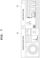
- FIG. 7 is a diagram showing an example in which a first display setting based on a first analysis result of a first artificial neural network according to an embodiment of the present invention is executed.
- the plurality of analysis results are allocated to and displayed in separate area on the screen.
- FIG. 7 shows a case in which emphysema and airway thickness are selected as candidate diseases having a high likelihood of disease, respectively.
- the first sub-display setting 710 is an example of a display setting appropriate for emphysema.
- the second sub-display setting 720 is an example of a display setting appropriate for checking the thickness of the airway.
- the second sub-display setting 720 includes the result of the segmentation of the airway and a quantitative measurement based on the segmentation of the airway, and the first sub-display setting 710 and the second sub-display setting 720 may be displayed in separate areas on the screen.
- An artificial neural network-based medical image reading assistant apparatus includes a computing system, and the computing system includes at least one processor.
- the computing system acquires a first medical image such as a CT image or receives an already acquired first medical image
- a first artificial neural network generates a first analysis result including an automatic analysis result through inference from the first medical image.
- the provisional or preliminary reading of the first medical image is performed by the analysis of the first artificial neural network, and the processor provides a displayed screen to a medical staff (who may include all of a clinician, a radiologist, and a technical staff) according to a hanging protocol based on the result of the provisional or preliminary reading.
- the medical staff may check on the screen a lesion, a disease, or a quantitative measurement result to be checked according to the result of the provisional or preliminary reading.
- Visualization elements are arranged on the screen so that the medical staff can easily view the lesion, the disease, or the quantitative measurement result on the screen and make a clinical decision, and the first medical image may be provided as various views through an image processing process as needed.
- the medical staff may view the displayed screen, may make a clinical decision, and may then enter “Confirm/Reject” for the lesion, disease, or quantitative measurement result displayed on the screen. “Confirm/Reject” may be received by the user menu provided to the medical staff through the user interface, and the clinical decision is incorporated into “Confirm/Reject.”
- a factor determining the hanging protocol is information included in the first medical image, and is information included in the first analysis result generated by the analysis of the first artificial neural network.
- FIG. 8 is an operation flowchart showing an artificial neural network-based medical image reading assistant method according to an embodiment of the present invention.
- the artificial neural network-based medical image reading assistant method is performed by the computing system 100 , 200 or 300 , the computing system 100 , 200 or 300 includes the at least one processor 130 , 230 or 330 .
- the method according to the present invention includes: step S 810 of acquiring or receiving, by the at least one processor 130 , 230 , 330 , the first analysis result 112 , 212 or 312 obtained through the inference of the first artificial neural network 110 , 210 or 310 for the first medical image 140 , 240 or 340 ; step S 820 of generating, by the at least one processor 130 , 230 or 330 , the first display setting 150 , 250 or 350 based on the first analysis result 112 , 212 or 312 ; and step S 830 of executing, by the at least one processor 130 , 230 or 330 , the first display setting 150 , 250 or 350 so that the first medical image 140 , 240 or 340 and the first analysis result 112 , 212 or 312 are displayed on the screen based on the first display setting 150 , 250 or 350 .
- step S 830 of executing the first display setting 150 , 250 or 350 the at least one processor 130 , 230 or 330 may execute the first display setting 150 , 250 or 350 to include the user menu configured to receive whether a user approves the first analysis result 112 , 212 or 312 displayed on the screen based on the first display setting 150 , 250 or 350 .
- step S 820 of generating the first display setting 150 , 250 or 350 the at least one processor 130 , 230 or 330 may generate the first display setting 150 , 250 or 350 based on at least one of the type of disease, the type of lesion, and a quantitative measurement result of the lesion detected in the first medical image 140 , 240 or 340 and indicated by the first analysis result 112 , 212 or 312 .
- the first display setting 150 , 250 or 350 may include settings for at least one view of the first medical image 140 , 240 or 340 related to the first analysis result 112 , 212 or 312 , a menu adapted to display the first analysis result 112 , 212 or 312 in the first medical image 140 , 240 or 340 , the layout of the at least one view of the first medical image 140 , 240 or 340 , and a user menu adapted to allow a user to respond to the first analysis result 112 , 212 or 312 displayed in the first medical image 140 , 240 or 340 .
- step S 820 of generating the first display setting 150 , 250 or 350 when the types of diseases detected in the first medical image 140 , 240 or 340 and included in the first analysis result 112 , 212 or 312 are plural in number, the at least one processor 130 , 230 or 330 may generate a plurality of sub-display settings for a plurality of types of diseases, respectively.
- step S 830 of executing the first display setting 150 , 250 or 350 the at least one processor 130 , 230 or 330 may arrange the plurality of sub-display settings in separate areas on the screen, and may display a first sub-display setting among the plurality of sub-display settings on the screen.
- the at least one processor 130 , 230 or 330 may display another sub-display setting instead of the first sub-display setting on the screen in response to user input.
- the first artificial neural network 110 , 210 or 310 may be an artificial neural network including the function of diagnosing a plurality of types of diseases for a single body part in a medical image.
- the computing system 200 may include a second artificial neural network that is an artificial neural network that inputs a plurality of second display settings selected by an expert for a plurality of types of diseases diagnosed for a plurality of third medical images and learns the function of generating display settings based on the types of disease diagnosed.
- the at least one processor 230 may input the first analysis result 212 to the second artificial neural network, and may control the second artificial neural network so that the first display setting 250 can be acquired through the inference of the second artificial neural network.
- the computing system may further include a third artificial neural network that is an artificial neural network that receives a plurality of third display settings selected by an expert for a plurality of third analysis results obtained through the inference of the first artificial neural network for a plurality of third medical images and learns correlations between at least one of the type of disease, the type of lesion, and a quantitative measurement result of the lesion, detected in the plurality of third medical images and included in the plurality of third analysis results, and the plurality of third display settings.
- a third artificial neural network is an artificial neural network that receives a plurality of third display settings selected by an expert for a plurality of third analysis results obtained through the inference of the first artificial neural network for a plurality of third medical images and learns correlations between at least one of the type of disease, the type of lesion, and a quantitative measurement result of the lesion, detected in the plurality of third medical images and included in the plurality of third analysis results, and the plurality of third display settings.
- step S 820 of generating the first display setting based on the first analysis result the at least one processor may input the first analysis result to the third artificial neural network, and may control the third artificial neural network so that the first display setting is acquired through the inference of the third artificial neural network.
- the method according to an embodiment of the present invention may be implemented in the form of program instructions, and may be then recorded in a computer-readable storage medium.
- the computer-readable storage medium may include program instructions, data files, and data structures solely or in combination.
- Program instructions recorded on the storage medium may have been specially designed and configured for the present invention, or may be known to or available to those who have ordinary knowledge in the field of computer software.
- Examples of the computer-readable storage medium include all types of hardware devices specially configured to record and execute program instructions, such as magnetic media, such as a hard disk, a floppy disk, and magnetic tape, optical media, such as compact disk (CD)-read only memory (ROM) and a digital versatile disk (DVD), magneto-optical media, such as a floptical disk, ROM, random access memory (RAM), and flash memory.
- Examples of the program instructions include machine code, such as code created by a compiler, and high-level language code executable by a computer using an interpreter. These hardware devices may be configured to operate as one or more software modules in order to perform the operation of the present invention, and the vice versa.
- a display layout designed to fit the diagnosis of each of the lesions may be implemented.
- display layout settings appropriate for each type of lesion and disease based on image information and diagnostic information included in a medical image may be proposed by considering a plurality of types of lesions that can be detected in the same body part.
- hanging protocols that execute the display layout settings may be provided based on information related to the type of lesion and disease detected in the medical image.
- hanging protocols based on disease codes detected for medical images there may be provided hanging protocols based on disease codes detected for medical images.
- a user interface and display environment that increase the efficiency of reading, assists a clinician or radiologist in deriving a more accurate diagnosis result within a short period of time, and increase the accuracy of analysis.
Landscapes
- Engineering & Computer Science (AREA)
- Health & Medical Sciences (AREA)
- Medical Informatics (AREA)
- Public Health (AREA)
- Life Sciences & Earth Sciences (AREA)
- General Health & Medical Sciences (AREA)
- Physics & Mathematics (AREA)
- Biomedical Technology (AREA)
- Theoretical Computer Science (AREA)
- Pathology (AREA)
- Epidemiology (AREA)
- Primary Health Care (AREA)
- Data Mining & Analysis (AREA)
- Radiology & Medical Imaging (AREA)
- Nuclear Medicine, Radiotherapy & Molecular Imaging (AREA)
- General Physics & Mathematics (AREA)
- Molecular Biology (AREA)
- Biophysics (AREA)
- Computer Vision & Pattern Recognition (AREA)
- Surgery (AREA)
- Animal Behavior & Ethology (AREA)
- Databases & Information Systems (AREA)
- Veterinary Medicine (AREA)
- Heart & Thoracic Surgery (AREA)
- General Engineering & Computer Science (AREA)
- Artificial Intelligence (AREA)
- Evolutionary Computation (AREA)
- Optics & Photonics (AREA)
- High Energy & Nuclear Physics (AREA)
- Computing Systems (AREA)
- Software Systems (AREA)
- Human Computer Interaction (AREA)
- Physiology (AREA)
- Mathematical Physics (AREA)
- Quality & Reliability (AREA)
- Computational Linguistics (AREA)
- Pulmonology (AREA)
- Signal Processing (AREA)
- Psychiatry (AREA)
- Fuzzy Systems (AREA)
Abstract
Description
Claims (18)
Applications Claiming Priority (2)
| Application Number | Priority Date | Filing Date | Title |
|---|---|---|---|
| KR10-2019-0092909 | 2019-07-31 | ||
| KR1020190092909A KR102222015B1 (en) | 2019-07-31 | 2019-07-31 | Apparatus and method for medical image reading assistant providing hanging protocols based on medical use artificial neural network |
Publications (2)
| Publication Number | Publication Date |
|---|---|
| US20210035687A1 US20210035687A1 (en) | 2021-02-04 |
| US12046367B2 true US12046367B2 (en) | 2024-07-23 |
Family
ID=74258432
Family Applications (1)
| Application Number | Title | Priority Date | Filing Date |
|---|---|---|---|
| US16/896,801 Active 2043-04-24 US12046367B2 (en) | 2019-07-31 | 2020-06-09 | Medical image reading assistant apparatus and method providing hanging protocols based on medical use artificial neural network |
Country Status (2)
| Country | Link |
|---|---|
| US (1) | US12046367B2 (en) |
| KR (1) | KR102222015B1 (en) |
Cited By (8)
| Publication number | Priority date | Publication date | Assignee | Title |
|---|---|---|---|---|
| US12144669B2 (en) | 2022-03-10 | 2024-11-19 | Cleerly, Inc. | Systems, devices, and methods for non-invasive image-based plaque analysis and risk determination |
| US12156759B2 (en) | 2020-01-07 | 2024-12-03 | Cleerly, Inc. | Systems, methods, and devices for medical image analysis, diagnosis, risk stratification, decision making and/or disease tracking |
| US12245882B2 (en) | 2020-01-07 | 2025-03-11 | Cleerly, Inc. | Systems, methods, and devices for medical image analysis, diagnosis, risk stratification, decision making and/or disease tracking |
| US12283046B2 (en) | 2020-01-07 | 2025-04-22 | Cleerly, Inc. | Systems, methods, and devices for medical image analysis, diagnosis, risk stratification, decision making and/or disease tracking |
| US12299885B2 (en) | 2022-03-10 | 2025-05-13 | Cleerly, Inc. | Systems, devices, and methods for non-invasive image-based plaque analysis and risk determination |
| US12380560B2 (en) | 2022-03-10 | 2025-08-05 | Cleerly, Inc. | Systems, methods, and devices for image-based plaque analysis and risk determination |
| US12440180B2 (en) | 2022-03-10 | 2025-10-14 | Cleerly, Inc. | Systems, devices, and methods for non-invasive image-based plaque analysis and risk determination |
| US12620092B2 (en) | 2023-08-25 | 2026-05-05 | Cleerly, Inc. | Systems, devices, and methods for non-invasive image-based plaque analysis and risk determination |
Families Citing this family (4)
| Publication number | Priority date | Publication date | Assignee | Title |
|---|---|---|---|---|
| JP7511276B2 (en) * | 2021-04-23 | 2024-07-05 | 株式会社fcuro | Information processing device, information processing method, information processing program, and information processing system |
| WO2022241190A2 (en) * | 2021-05-14 | 2022-11-17 | H. Lee Moffitt Cancer Center And Research Institute, Inc. | Machine learning-based systems and methods for extracting information from pathology reports |
| KR102676569B1 (en) | 2021-05-17 | 2024-06-20 | 주식회사 코어라인소프트 | Medical image analysis apparatus and method, medical image visualization apparatus and method |
| CN113888490B (en) * | 2021-09-27 | 2025-07-01 | 上海商汤善萃医疗科技有限公司 | Image analysis method and related device, equipment, and storage medium |
Citations (19)
| Publication number | Priority date | Publication date | Assignee | Title |
|---|---|---|---|---|
| US6697506B1 (en) * | 1999-03-17 | 2004-02-24 | Siemens Corporate Research, Inc. | Mark-free computer-assisted diagnosis method and system for assisting diagnosis of abnormalities in digital medical images using diagnosis based image enhancement |
| US20050260770A1 (en) * | 2004-04-01 | 2005-11-24 | Cohen Irun R | Antigen array and diagnostic uses thereof |
| JP2008188214A (en) | 2007-02-05 | 2008-08-21 | Konica Minolta Medical & Graphic Inc | Medical image display system |
| US8165368B2 (en) | 2008-09-29 | 2012-04-24 | General Electric Company | Systems and methods for machine learning based hanging protocols |
| KR101263705B1 (en) | 2011-12-14 | 2013-06-13 | 주식회사 인피니트헬스케어 | System for supplying medical image and method thereof |
| KR20140091176A (en) | 2013-01-10 | 2014-07-21 | 삼성전자주식회사 | Apparatus and method for lesion diagnosis |
| US8923580B2 (en) | 2011-11-23 | 2014-12-30 | General Electric Company | Smart PACS workflow systems and methods driven by explicit learning from users |
| KR20150006807A (en) | 2013-07-09 | 2015-01-19 | 주식회사바텍 | method and system for diagnosis of oral lesion using medical image |
| US9152760B2 (en) | 2011-11-23 | 2015-10-06 | General Electric Company | Smart 3D PACS workflow by learning |
| KR101818074B1 (en) | 2017-07-20 | 2018-01-12 | (주)제이엘케이인스펙션 | Artificial intelligence based medical auto diagnosis auxiliary method and system therefor |
| KR20180040287A (en) | 2016-10-12 | 2018-04-20 | (주)헬스허브 | System for interpreting medical images through machine learnings |
| KR101923962B1 (en) | 2018-02-12 | 2018-11-30 | 주식회사 뷰노 | Method for facilitating medical image view and apparatus using the same |
| KR101943011B1 (en) | 2018-01-22 | 2019-01-28 | 주식회사 뷰노 | Method for facilitating medical image reading and apparatus using the same |
| US20190130565A1 (en) * | 2017-10-26 | 2019-05-02 | Samsung Electronics Co., Ltd. | Method of processing medical image, and medical image processing apparatus performing the method |
| US20190371464A1 (en) | 2018-05-30 | 2019-12-05 | Jlk Inspection Inc. | Method and system for clinical effectiveness evaluation of artificial intelligence based medical device |
| US20220000351A1 (en) * | 2019-03-18 | 2022-01-06 | Olympus Corporation | Control apparatus, diagnosis support method, and recording medium |
| US20230181160A1 (en) * | 2019-07-24 | 2023-06-15 | Teratech Corporation | Devices and methods for ultrasound monitoring |
| US20230210442A1 (en) * | 2013-01-25 | 2023-07-06 | Wesley W.O. Krueger | Systems and methods to measure ocular parameters and determine neurologic health status |
| US11918176B2 (en) * | 2019-03-08 | 2024-03-05 | Fujifilm Corporation | Medical image processing apparatus, processor device, endoscope system, medical image processing method, and program |
Family Cites Families (1)
| Publication number | Priority date | Publication date | Assignee | Title |
|---|---|---|---|---|
| MY164758A (en) | 2010-09-27 | 2018-01-30 | Gcp Applied Tech Inc | Dilution-stable cement grinding additive composition |
-
2019
- 2019-07-31 KR KR1020190092909A patent/KR102222015B1/en active Active
-
2020
- 2020-06-09 US US16/896,801 patent/US12046367B2/en active Active
Patent Citations (20)
| Publication number | Priority date | Publication date | Assignee | Title |
|---|---|---|---|---|
| US6697506B1 (en) * | 1999-03-17 | 2004-02-24 | Siemens Corporate Research, Inc. | Mark-free computer-assisted diagnosis method and system for assisting diagnosis of abnormalities in digital medical images using diagnosis based image enhancement |
| US20050260770A1 (en) * | 2004-04-01 | 2005-11-24 | Cohen Irun R | Antigen array and diagnostic uses thereof |
| JP2008188214A (en) | 2007-02-05 | 2008-08-21 | Konica Minolta Medical & Graphic Inc | Medical image display system |
| US8165368B2 (en) | 2008-09-29 | 2012-04-24 | General Electric Company | Systems and methods for machine learning based hanging protocols |
| US8923580B2 (en) | 2011-11-23 | 2014-12-30 | General Electric Company | Smart PACS workflow systems and methods driven by explicit learning from users |
| US9152760B2 (en) | 2011-11-23 | 2015-10-06 | General Electric Company | Smart 3D PACS workflow by learning |
| KR101263705B1 (en) | 2011-12-14 | 2013-06-13 | 주식회사 인피니트헬스케어 | System for supplying medical image and method thereof |
| KR20140091176A (en) | 2013-01-10 | 2014-07-21 | 삼성전자주식회사 | Apparatus and method for lesion diagnosis |
| US9773305B2 (en) | 2013-01-10 | 2017-09-26 | Samsung Electronics Co., Ltd. | Lesion diagnosis apparatus and method |
| US20230210442A1 (en) * | 2013-01-25 | 2023-07-06 | Wesley W.O. Krueger | Systems and methods to measure ocular parameters and determine neurologic health status |
| KR20150006807A (en) | 2013-07-09 | 2015-01-19 | 주식회사바텍 | method and system for diagnosis of oral lesion using medical image |
| KR20180040287A (en) | 2016-10-12 | 2018-04-20 | (주)헬스허브 | System for interpreting medical images through machine learnings |
| KR101818074B1 (en) | 2017-07-20 | 2018-01-12 | (주)제이엘케이인스펙션 | Artificial intelligence based medical auto diagnosis auxiliary method and system therefor |
| US20190130565A1 (en) * | 2017-10-26 | 2019-05-02 | Samsung Electronics Co., Ltd. | Method of processing medical image, and medical image processing apparatus performing the method |
| KR101943011B1 (en) | 2018-01-22 | 2019-01-28 | 주식회사 뷰노 | Method for facilitating medical image reading and apparatus using the same |
| KR101923962B1 (en) | 2018-02-12 | 2018-11-30 | 주식회사 뷰노 | Method for facilitating medical image view and apparatus using the same |
| US20190371464A1 (en) | 2018-05-30 | 2019-12-05 | Jlk Inspection Inc. | Method and system for clinical effectiveness evaluation of artificial intelligence based medical device |
| US11918176B2 (en) * | 2019-03-08 | 2024-03-05 | Fujifilm Corporation | Medical image processing apparatus, processor device, endoscope system, medical image processing method, and program |
| US20220000351A1 (en) * | 2019-03-18 | 2022-01-06 | Olympus Corporation | Control apparatus, diagnosis support method, and recording medium |
| US20230181160A1 (en) * | 2019-07-24 | 2023-06-15 | Teratech Corporation | Devices and methods for ultrasound monitoring |
Cited By (18)
| Publication number | Priority date | Publication date | Assignee | Title |
|---|---|---|---|---|
| US12324695B2 (en) | 2020-01-07 | 2025-06-10 | Cleerly, Inc. | Systems, methods, and devices for medical image analysis, diagnosis, risk stratification, decision making and/or disease tracking |
| US12396695B2 (en) | 2020-01-07 | 2025-08-26 | Cleerly, Inc. | Systems, methods, and devices for medical image analysis, diagnosis, risk stratification, decision making and/or disease tracking |
| US12171606B2 (en) | 2020-01-07 | 2024-12-24 | Cleerly, Inc. | Systems, methods, and devices for medical image analysis, diagnosis, risk stratification, decision making and/or disease tracking |
| US12178627B2 (en) | 2020-01-07 | 2024-12-31 | Cleerly, Inc. | Systems, methods, and devices for medical image analysis, diagnosis, risk stratification, decision making and/or disease tracking |
| US12245882B2 (en) | 2020-01-07 | 2025-03-11 | Cleerly, Inc. | Systems, methods, and devices for medical image analysis, diagnosis, risk stratification, decision making and/or disease tracking |
| US12283046B2 (en) | 2020-01-07 | 2025-04-22 | Cleerly, Inc. | Systems, methods, and devices for medical image analysis, diagnosis, risk stratification, decision making and/or disease tracking |
| US12558048B2 (en) | 2020-01-07 | 2026-02-24 | Cleerly, Inc. | Systems, methods, and devices for medical image analysis, diagnosis, risk stratification, decision making and/or disease tracking |
| US12555228B2 (en) | 2020-01-07 | 2026-02-17 | Cleerly, Inc. | Systems, methods, and devices for medical image analysis, diagnosis, risk stratification, decision making and/or disease tracking |
| US12156759B2 (en) | 2020-01-07 | 2024-12-03 | Cleerly, Inc. | Systems, methods, and devices for medical image analysis, diagnosis, risk stratification, decision making and/or disease tracking |
| US12499539B2 (en) | 2020-01-07 | 2025-12-16 | Cleerly, Inc. | Systems, methods, and devices for medical image analysis, diagnosis, risk stratification, decision making and/or disease tracking |
| US12380560B2 (en) | 2022-03-10 | 2025-08-05 | Cleerly, Inc. | Systems, methods, and devices for image-based plaque analysis and risk determination |
| US12406365B2 (en) | 2022-03-10 | 2025-09-02 | Cleerly, Inc. | Systems, devices, and methods for non-invasive image-based plaque analysis and risk determination |
| US12440180B2 (en) | 2022-03-10 | 2025-10-14 | Cleerly, Inc. | Systems, devices, and methods for non-invasive image-based plaque analysis and risk determination |
| US12144669B2 (en) | 2022-03-10 | 2024-11-19 | Cleerly, Inc. | Systems, devices, and methods for non-invasive image-based plaque analysis and risk determination |
| US12324696B2 (en) | 2022-03-10 | 2025-06-10 | Cleerly, Inc. | Systems, devices, and methods for non-invasive image-based plaque analysis and risk determination |
| US12299885B2 (en) | 2022-03-10 | 2025-05-13 | Cleerly, Inc. | Systems, devices, and methods for non-invasive image-based plaque analysis and risk determination |
| US12599352B2 (en) | 2022-03-10 | 2026-04-14 | Cleerly, Inc. | Systems, devices, and methods for non-invasive image-based plaque analysis and risk determination |
| US12620092B2 (en) | 2023-08-25 | 2026-05-05 | Cleerly, Inc. | Systems, devices, and methods for non-invasive image-based plaque analysis and risk determination |
Also Published As
| Publication number | Publication date |
|---|---|
| KR20210014893A (en) | 2021-02-10 |
| KR102222015B1 (en) | 2021-03-04 |
| US20210035687A1 (en) | 2021-02-04 |
Similar Documents
| Publication | Publication Date | Title |
|---|---|---|
| US12046367B2 (en) | Medical image reading assistant apparatus and method providing hanging protocols based on medical use artificial neural network | |
| US11915822B2 (en) | Medical image reading assistant apparatus and method for adjusting threshold of diagnostic assistant information based on follow-up examination | |
| US12361551B2 (en) | Artificial neural network-based image analysis apparatus and method for evaluating analysis results of artificial neural network | |
| US12475993B2 (en) | Apparatus and method for medical image reading assistant providing representative image based on medical use artificial neural network | |
| US12308107B2 (en) | Medical image diagnosis assistance apparatus and method for providing user-preferred style based on medical artificial neural network | |
| JP6585772B2 (en) | Methods and systems for analyzing, prioritizing, visualizing, and reporting medical images | |
| JP5100285B2 (en) | MEDICAL DIAGNOSIS SUPPORT DEVICE, ITS CONTROL METHOD, PROGRAM, AND STORAGE MEDIUM | |
| KR102289277B1 (en) | Medical image diagnosis assistance apparatus and method generating evaluation score about a plurality of medical image diagnosis algorithm | |
| US20230343455A1 (en) | Medical image diagnosis assistant apparatus and method for generating and visualizing assistant information based on distributions of signal intensities in medical images | |
| EP4183344A1 (en) | Device and method for supporting chest medical image reading | |
| KR102360615B1 (en) | Medical image diagnosis assistance apparatus and method using a plurality of medical image diagnosis algorithm for endoscope images | |
| CN111226287B (en) | Methods, systems, program products, and media for analyzing medical imaging data sets | |
| US20080215525A1 (en) | Medical image retrieval system | |
| JP2007528746A (en) | CAD support for medical diagnostic imaging that uses machine learning to adapt the CAD (computer aided decision) process to the knowledge gained from routine CAD system usage | |
| JP6215227B2 (en) | Imaging inspection protocol update recommendation section | |
| JP2007502469A (en) | System and method for supporting CAD (computer aided diagnosis) | |
| JP5456132B2 (en) | Diagnosis support device, diagnosis support device control method, and program thereof | |
| KR20220076296A (en) | Medical image reconstruction apparatus and method for screening a plurality of types lung diseases | |
| JP5348998B2 (en) | Image search apparatus and method | |
| Krishnakumar et al. | Improving Medical Imaging Diagnostics with Deep Convolutional Networks for Early Detection and Treatment | |
| Gossmann et al. | Considerations in the assessment of machine learning algorithm performance for medical imaging | |
| KR20230151865A (en) | Medical image diagnosis assistant apparatus and method generating and visualizing assistant information based on distribution of intensity in medical images |
Legal Events
| Date | Code | Title | Description |
|---|---|---|---|
| FEPP | Fee payment procedure |
Free format text: ENTITY STATUS SET TO UNDISCOUNTED (ORIGINAL EVENT CODE: BIG.); ENTITY STATUS OF PATENT OWNER: SMALL ENTITY |
|
| FEPP | Fee payment procedure |
Free format text: ENTITY STATUS SET TO SMALL (ORIGINAL EVENT CODE: SMAL); ENTITY STATUS OF PATENT OWNER: SMALL ENTITY |
|
| AS | Assignment |
Owner name: CORELINE SOFT CO., LTD., KOREA, REPUBLIC OF Free format text: ASSIGNMENT OF ASSIGNORS INTEREST;ASSIGNORS:YI, JAEYOUN;YU, DONGHOON;JI, HYEON;REEL/FRAME:053172/0964 Effective date: 20200609 |
|
| STPP | Information on status: patent application and granting procedure in general |
Free format text: DOCKETED NEW CASE - READY FOR EXAMINATION |
|
| STPP | Information on status: patent application and granting procedure in general |
Free format text: NON FINAL ACTION MAILED |
|
| STPP | Information on status: patent application and granting procedure in general |
Free format text: RESPONSE TO NON-FINAL OFFICE ACTION ENTERED AND FORWARDED TO EXAMINER |
|
| STPP | Information on status: patent application and granting procedure in general |
Free format text: NOTICE OF ALLOWANCE MAILED -- APPLICATION RECEIVED IN OFFICE OF PUBLICATIONS |
|
| ZAAA | Notice of allowance and fees due |
Free format text: ORIGINAL CODE: NOA |
|
| ZAAB | Notice of allowance mailed |
Free format text: ORIGINAL CODE: MN/=. |
|
| STPP | Information on status: patent application and granting procedure in general |
Free format text: PUBLICATIONS -- ISSUE FEE PAYMENT VERIFIED |
|
| STCF | Information on status: patent grant |
Free format text: PATENTED CASE |