US12013388B2 - Method and kit for single cell protein expression profiling of floorplate mesencephalic dopaminergic progenitor cells - Google Patents
Method and kit for single cell protein expression profiling of floorplate mesencephalic dopaminergic progenitor cells Download PDFInfo
- Publication number
- US12013388B2 US12013388B2 US16/632,229 US201816632229A US12013388B2 US 12013388 B2 US12013388 B2 US 12013388B2 US 201816632229 A US201816632229 A US 201816632229A US 12013388 B2 US12013388 B2 US 12013388B2
- Authority
- US
- United States
- Prior art keywords
- cells
- antigen binding
- cell
- human
- progenitor cells
- Prior art date
- Legal status (The legal status is an assumption and is not a legal conclusion. Google has not performed a legal analysis and makes no representation as to the accuracy of the status listed.)
- Active, expires
Links
Images


Classifications
-
- G—PHYSICS
- G01—MEASURING; TESTING
- G01N—INVESTIGATING OR ANALYSING MATERIALS BY DETERMINING THEIR CHEMICAL OR PHYSICAL PROPERTIES
- G01N33/00—Investigating or analysing materials by specific methods not covered by groups G01N1/00 - G01N31/00
- G01N33/48—Biological material, e.g. blood, urine; Haemocytometers
- G01N33/50—Chemical analysis of biological material, e.g. blood, urine; Testing involving biospecific ligand binding methods; Immunological testing
- G01N33/5005—Chemical analysis of biological material, e.g. blood, urine; Testing involving biospecific ligand binding methods; Immunological testing involving human or animal cells
- G01N33/5008—Chemical analysis of biological material, e.g. blood, urine; Testing involving biospecific ligand binding methods; Immunological testing involving human or animal cells for testing or evaluating the effect of chemical or biological compounds, e.g. drugs, cosmetics
- G01N33/5044—Chemical analysis of biological material, e.g. blood, urine; Testing involving biospecific ligand binding methods; Immunological testing involving human or animal cells for testing or evaluating the effect of chemical or biological compounds, e.g. drugs, cosmetics involving specific cell types
- G01N33/5058—Neurological cells
-
- G—PHYSICS
- G01—MEASURING; TESTING
- G01N—INVESTIGATING OR ANALYSING MATERIALS BY DETERMINING THEIR CHEMICAL OR PHYSICAL PROPERTIES
- G01N33/00—Investigating or analysing materials by specific methods not covered by groups G01N1/00 - G01N31/00
- G01N33/48—Biological material, e.g. blood, urine; Haemocytometers
- G01N33/50—Chemical analysis of biological material, e.g. blood, urine; Testing involving biospecific ligand binding methods; Immunological testing
- G01N33/5005—Chemical analysis of biological material, e.g. blood, urine; Testing involving biospecific ligand binding methods; Immunological testing involving human or animal cells
-
- C—CHEMISTRY; METALLURGY
- C12—BIOCHEMISTRY; BEER; SPIRITS; WINE; VINEGAR; MICROBIOLOGY; ENZYMOLOGY; MUTATION OR GENETIC ENGINEERING
- C12N—MICROORGANISMS OR ENZYMES; COMPOSITIONS THEREOF; PROPAGATING, PRESERVING, OR MAINTAINING MICROORGANISMS; MUTATION OR GENETIC ENGINEERING; CULTURE MEDIA
- C12N5/00—Undifferentiated human, animal or plant cells, e.g. cell lines; Tissues; Cultivation or maintenance thereof; Culture media therefor
- C12N5/06—Animal cells or tissues; Human cells or tissues
- C12N5/0602—Vertebrate cells
- C12N5/0618—Cells of the nervous system
- C12N5/0623—Stem cells
-
- G—PHYSICS
- G01—MEASURING; TESTING
- G01N—INVESTIGATING OR ANALYSING MATERIALS BY DETERMINING THEIR CHEMICAL OR PHYSICAL PROPERTIES
- G01N33/00—Investigating or analysing materials by specific methods not covered by groups G01N1/00 - G01N31/00
- G01N33/48—Biological material, e.g. blood, urine; Haemocytometers
- G01N33/50—Chemical analysis of biological material, e.g. blood, urine; Testing involving biospecific ligand binding methods; Immunological testing
- G01N33/58—Chemical analysis of biological material, e.g. blood, urine; Testing involving biospecific ligand binding methods; Immunological testing involving labelled substances
-
- C—CHEMISTRY; METALLURGY
- C12—BIOCHEMISTRY; BEER; SPIRITS; WINE; VINEGAR; MICROBIOLOGY; ENZYMOLOGY; MUTATION OR GENETIC ENGINEERING
- C12N—MICROORGANISMS OR ENZYMES; COMPOSITIONS THEREOF; PROPAGATING, PRESERVING, OR MAINTAINING MICROORGANISMS; MUTATION OR GENETIC ENGINEERING; CULTURE MEDIA
- C12N2501/00—Active agents used in cell culture processes, e.g. differentation
- C12N2501/50—Cell markers; Cell surface determinants
- C12N2501/599—Cell markers; Cell surface determinants with CD designations not provided for elsewhere
-
- C—CHEMISTRY; METALLURGY
- C12—BIOCHEMISTRY; BEER; SPIRITS; WINE; VINEGAR; MICROBIOLOGY; ENZYMOLOGY; MUTATION OR GENETIC ENGINEERING
- C12N—MICROORGANISMS OR ENZYMES; COMPOSITIONS THEREOF; PROPAGATING, PRESERVING, OR MAINTAINING MICROORGANISMS; MUTATION OR GENETIC ENGINEERING; CULTURE MEDIA
- C12N2501/00—Active agents used in cell culture processes, e.g. differentation
- C12N2501/60—Transcription factors
Definitions
- the present invention relates to a method for analyzing the protein expression profile on a single cell level of human floorplate mesencephalic dopaminergic progenitor cells.
- DA neurons secrete the neurotransmitter dopamine and play a central role in different brain functions, like cognition, memory processing, motor control, and motion. They are found in three distinct nuclei, the substantia nigra pars compacta (A9 group), the ventral tegmental area (A10 group) and the retrorubral field (A8 group).
- the cell bodies of the A9 group in the substantia nigra pars compacta (SNpc) of the midbrain innervate the caudate putamen, the dorsalateral striatum and form the nigrostriatal pathway, which regulates motor function by the controlled release of dopamine from projecting dopaminergic neurons.
- Degeneration of the A9 dopaminergic neurons in the substantia nigra is mainly responsible for dysfunction of the dopamine system and for several motor features described as the disorder Parkinson's disease (PD).
- PD involves also degeneration of other neuronal subtypes, loss of substantia nigra pars compacta dopaminergic neurons is mainly responsible for PD symptoms and has become a primary target for cell replacement therapy.
- fetal development progenitors of these DA neurons are formed in the ventral neural tube of the developing mesencephalon.
- Progenitor cells from the so-called floor plate region are characterized by expression of the transcription factors FOXA2, LMX1A and OTX2. These cells give rise to DA SNpc neurons (A9 group) and to DA ventral tegmental area neurons (A10 group).
- FOXA2 the transcription factors
- LMX1A LMX1A
- OTX2 OTX2
- DA ventral tegmental area neurons A10 group. It was shown that fetal cells dissected form the fetal mesencephalon and transplanted to the striatum of Parkinson patients could functionally re-innervate the striatum and restore the functions of dopaminergic neurons (Barker, 2013; Kefalopoulou, 2014). But, the limited availability of fetal DA progenitors and ethical as well as logistical concerns using this tissue emphasized the
- mesDA progenitor cells differentiated from embryonic stem cells have the same features as cells derived from the human fetal ventral mesencephalon in terms of i) subtype-specific maturation, ii) integration and projection capacity in a rat Parkinson model, iii) regulated DA release and iv) functional reversion of Parkinson symptoms (Grealish, 2014; Kirkeby, 2012).
- Protocols are based on dual SMAD inhibition for neural conversion and regionalization by chemical inhibition of glycogen synthase kinase 3 (GSK3) to induce WNT signaling for caudalised patterning into midbrain fates and activation of sonic hedgehog (SHH) signaling for patterning into ventral fates (Kirkeby, 2013).
- GSK3 glycogen synthase kinase 3
- one need within the generation of GMP-grade PSC/iPSC derived mesDA neurons is the set-up of a reliable in vitro assay for comprehensive cell product characterization, that allows to determine the identity and purity of the cell lot. Furthermore, the assay must correlate with the cells' potential to engraft and to mature to midbrain dopaminergic neurons, as well as reveal a potential tumorigenicity.
- mesencephalic floor plate markers like LMX1A, FOXA2 and OTX2 are used to confirm the identity of the cells prior to grafting.
- mesencephalic floor plate markers like LMX1A, FOXA2 and OTX2 are used to confirm the identity of the cells prior to grafting.
- Kirkeby et al. identified a set of in vitro markers that allows prediction of successful cell grafting by qRT-PCR (Kirkeby et al., 2017).
- predictive markers for a successful grafting results were EN1, ETV5, CNPY1, PAX8, and SPRY1.
- diencephalic domain markers, as EPHA3, FEZF1, and WNT7B were associated with a negatively correlated graft outcome.
- expression was analyzed on the bulk of cells on the mRNA level.
- the mRNA expression level of a gene reflects only its potential to be expressed or present as a protein. Based on a multitude of posttranscriptional regulatory mechanisms, posttranslational modifications and differing stability and turnover of proteins and mRNAs, the conversion of an mRNA to an actual protein can or cannot happen and if it happens it can result in a multitude of different, slightly varying forms of a given protein. Moreover, while the mRNA of a gene might not be detected at a given moment, the corresponding protein can still be present for weeks or even moths. Therefore, it is not possible to irrevocably predict the presence of a certain form of a protein by detecting the corresponding mRNA neither positively nor negatively.
- RNAs and ribozymes only a protein and not its corresponding mRNA exerts a function influencing the status of a cell and their environment.
- measurements on a bulk of cells do not reflect the true status of a single cell but rather generate levelled results which might even do not exist in a single cell. But as only a proportion of cells in a given mixture might be the cells of interest or danger the bulk analysis does not have the sensitivity and discriminatory power to allow for a true and quantitative description of different phenotypes of cells in a cell mixture.
- methods using PCR based techniques are time-consuming and prone to contamination.
- the invention describes a new user friendly, standardized method for protein profiling of floorplate mesDA progenitor cells on the single cell level, that can also be used under GMP conditions.
- Antigen binding molecules such as antibody-fluorochrome conjugates specific for positive and negative floorplate mesDA progenitor cell markers are used e.g. in a flow cytometry assay that allows for the first time a single cell, quantitative, standardized protein profiling of differentiated floorplate mesDA progenitor cells within approximately 2.5 hours for identification of the cell product's identity.
- the set consists of antigen binding molecules, e.g. monoclonal antibody clones that are specific for some or all positive and negative markers of mesDA neurons as disclosed herein (positive markers: FOXA2, OTX2, CD47, NKX2.1; negative markers: OCT3/4, PAX6, SOX1, NKX6.1, KI-67). Percentages of cells being positive or negative for some or all of these cell markers depict a clear picture of the cellular phenotype and purity.
- antigen binding molecules e.g. monoclonal antibody clones that are specific for some or all positive and negative markers of mesDA neurons as disclosed herein (positive markers: FOXA2, OTX2, CD47, NKX2.1; negative markers: OCT3/4, PAX6, SOX1, NKX6.1, KI-67). Percentages of cells being positive or negative for some or all of these cell markers depict a clear picture of the cellular phenotype and purity.
- NKX6.1 which is expressed in the basal plate of the midbrain as well as in the hindbrain of the developing embryo is a surprisingly efficient marker for detecting the presence of hindbrain progenitor fates in differentiated stem cell populations. Therefore, NKX6.1 serves as a negative marker to discriminate lateral midbrain and hindbrain cells from midbrain floor plate-patterned cells. Furthermore, surprisingly, we found that the Marker NKX2.1 is expressed differently when assessed on protein level by flow cytometry compared to PCR. While NKX2.1 mRNA is absent in the floor plate midbrain dopaminergic neurons we have seen that there is still a substantial percentage of NKX2.1 positive cells after conversion to human mesDA progenitor cells. The quantification of NKX2.1 on a protein level can thus monitor potential batch-to-batch variations in cultures of floorplate midbrain progenitors.
- FIG. 1 A first figure.
- the neural tube can be subdivided into different regions along the rostro-caudal axis and the dorso-ventral axis.
- the most rostral part of the neural tube is called the forebrain, followed by the midbrain and the most caudal part forms the hindbrain.
- the most ventral part of the neural tube is called the floor plate.
- Cells of the different brain regions are characterized by specific marker expression (forebrain: PAX6, OTX2, NKX2.1), ventral midbrain: FOXA2, OTX2, NKX2.1, hindbrain: NKX6.1).
- Protocols are based on dual SMAD inhibition for neural conversion and regionalization by inhibition of glycogen synthase kinase 3 (GSK3) by CHIR99021 to induce WNT signaling for rostro-caudal patterning and activation of sonic hedgehog (SHH-C24II) for ventralised patterning (Kirkeby, 2012, 2013).
- GSK3 glycogen synthase kinase 3
- SHH-C24II sonic hedgehog
- Different media are optimized for neural induction (medium 1), neural proliferation (medium 2) and neural differentiation (medium 3).
- the depicted protocol follows an adherent differentiation process.
- the protocol can be modified, for example addition of Y-27632 instead of thiazovivin and addition of Y-27632 at day 1 as well as day 11.
- the present invention discloses cell markers that can be used for identification of human floorplate mesDA progenitor cells in a cell composition or a sample thereof comprising said cells and possibly other cells by determining the expression profile of the cells with regard to said cell markers as disclosed herein.
- the present invention provides an in-vitro method for analyzing a cell composition comprising human floorplate mesDA progenitor cells, the method comprising
- Said antigen binding molecules may be conjugated to fluorophores.
- the cells of said cell composition or of said sample may be treated with a fixative and a detergent before contacting with said antigen binding molecules as the antigens are transcription factors, therefore the cells of said cell composition or of said sample (step a) may be fixed and permeabilized cells.
- the contacting of the cells of said cell composition or of said sample with fluorophore conjugated antigen binding molecules specific for the antigens FOXA2, OTX2, PAX6, and NKX6.1 may be performed consecutively, e.g. firstly a fluorophore conjugated antigen binding molecule specific for FOXA2 may be contacted with the cells of said cell composition or of a sample thereof and subsequently the percentage of said cells may be determined that are labelled with said antigen binding molecule specific for FOXA2, followed by the next fluorophore conjugated antigen binding molecule specific for the next marker as disclosed herein and so on.
- the antigen binding molecule specific for the first antigen may be removed from the cells of said cell composition or a sample thereof before the second antigen binding molecule specific for the second antigen in contacted with the cells of said cell composition or a sample thereof and so on.
- the cell composition or a sample thereof provided in step a) of the method is sub-divided into several sub-samples and each antigen binding molecule specific for one of the markers as disclosed herein is contacted with the cells of a single sub-sample.
- the same cell composition or sample thereof provided in step a) or the sub-samples of the method is used simultaneously for some or all antigen binding molecules specific for the markers as disclosed herein when the antigen binding molecules used are conjugated to distinct detection moieties such as fluorophores that allow for parallel detection of the labeled cells.
- step a) the cells of said cell composition or of said sample thereof are additionally contacted with an antigen binding molecule specific for the antigen OCT3/4 if said cells of said cell composition or of said sample thereof are derived from OCT3/4 positive cells, and wherein in step) said protein expression profile of the cells of said cell composition or of said sample thereof comprises additionally:
- Said antigen binding molecule specific for the antigen OCT3/4 may be conjugated to a fluorophore.
- step a) the cells of said cell composition or of said sample thereof are additionally contacted with an antigen binding molecule specific for the antigen NKX2.1, and wherein in step b) said protein expression profile of the cells of said cell composition or of said sample thereof comprises additionally:
- Said antigen binding molecule specific for the antigen NKX2.1 may be conjugated to a fluorophore.
- step a) the cells of said cell composition or of said sample thereof are additionally contacted with an antigen binding molecule specific for the antigen KI-67, and wherein in step b) said protein expression profile of the cells of said cell composition or of said sample thereof comprises additionally:
- Said antigen binding molecule specific for the antigen KI-67 may be conjugated to a fluorophore.
- step a) the cells of said cell composition or of said sample thereof are additionally contacted with an antigen binding molecule specific for the antigen IAP, and wherein in step b) said protein expression profile of the cells of said cell composition or of said sample thereof comprises additionally:
- Said antigen binding molecule specific for the antigen IAP may be conjugated to a fluorophore.
- step a) the cells of said cell composition or of said sample thereof are additionally contacted with an antigen binding molecule specific for the antigen SOX1, and wherein in step b) said protein expression profile of the cells of said cell composition or of said sample thereof comprises additionally:
- Said antigen binding molecule specific for the antigen SOX1 may be conjugated to a fluorophore.
- step a) the cells of said cell composition or of said sample thereof are additionally contacted with at least one antigen binding molecule specific for an antigen selected from the group consisting of the antigens KI-67, IAP and SOX1, and wherein in step b) said protein expression profile of the cells of said cell composition or of said sample thereof comprises additionally:
- step a) an antigen binding molecule specific for IAP is used
- step a) an antigen binding molecule specific for SOX1 is used.
- Said antigen binding molecules may be conjugated to fluorophores.
- said cell composition comprising human floorplate mesDA progenitor cells is generated or is in the process of being generated by an in-vitro differentiation process from human cells, which can be converted to human floorplate mesDA progenitor cells
- Said human cells which can be converted into floorplate mesDA progenitor cells may be selected from the group consisting of human iPS cells, human embryonic stem cells or directly reprogrammed human somatic cells.
- Said antigen binding molecules may be antibodies or antigen binding fragments thereof.
- the present invention provides a kit for analyzing the protein profile of human floorplate mesDA progenitor cells comprising antigen binding molecules specific for the antigens FOXA2, OTX2, PAX6, and NKX6.1.
- kit wherein said kit additionally comprises an antigen binding molecule specific for the antigen Oct3/4.
- kit wherein said kit additionally comprises an antigen binding molecule specific for the antigen NKX2.1.
- kits wherein said kit additionally comprises an antigen binding molecule specific for the antigen KI-67.
- kit wherein said kit additionally comprises an antigen binding molecule specific for the antigen IAP.
- kit wherein said kit additionally comprises an antigen binding molecule specific for the antigen SOX1.
- kit wherein said kit additionally comprises at least one antigen binding molecule specific for an antigen selected from the group consisting of the antigens for the antigens KI-67, IAP and SOX1.
- Said antigen binding molecules may be conjugated to fluorophores.
- human floorplate mesDA progenitor cells from a starting cell composition comprising cells that can be converted into human floorplate mesDA progenitor cells such as pluripotent stem cells are well-known in the art and is disclosed e.g. in Kriks, 2011; Kirkeby, 2012; Kirkeby, 2013; Grealish, 2015; Kirkeby 2017.
- cell composition comprising human floorplate mesDA progenitor cells refers to the (complete) cell composition or to a sample thereof comprising human floorplate mesDA progenitor cells and other cells, eg. with other neural identities in any ratio.
- Said cell composition comprising human floorplate mesDA progenitor cells may be generated or may be in the process of being generated by an in-vitro differentiation process from human cells, which can be converted to human floorplate mesDA progenitor cells.
- the human floorplate mesDA progenitor cells of said cell composition may be derived from human cells, which can be converted to human floorplate mesDA progenitor cells, e.g. human iPS cells, human embryonic stem cells or directly reprogrammed human somatic cells using protocols of differentiation or direct reprogramming well-known in the art such as e.g. in Kriks, 2011; Kirkeby, 2012; Ladewig 2012; Kirkeby, 2013; Grealish, 2015; Kirkeby 2017.
- the present method as disclosed herein may be used to monitor and/or identify the status of the cells during this process of differentiation of the stem cells into cells that qualify as human floorplate mesDA progenitor cells.
- analyzing in the context of analyzing a cell composition comprising human floorplate mesDA progenitor cells means e.g. identifying human floorplate mesDA progenitor cells in a cell composition or sample by determining the expression profile as disclosed herein of cells of said cell composition or said sample.
- a sample may be taken from the (complete) cell composition comprising human floorplate mesDA progenitor cells, e.g. one or more aliquots. Then the method as disclosed herein may comprise the step “providing a sample from said cell composition” before the step a).
- Determining the percentage of said cells, i.e. the cells of said cell composition or of said sample thereof, labelled with said antigen binding molecules for each of said antigens refers to the determination of the amount of cells in the cell composition or in the sample taken from the cell composition that are e.g. fluorescently labelled for a respective antigen, e.g. if in said cell composition or said sample 9 from 10 cells are labelled for the antigen FOXA2, then 90% of the cells of the cell composition or the sample are positive for FOXA2, if in the same cell composition or sample 8 from 10 cells are labeled for the antigen OTX2, then 80% of the cells of the same cell composition or sample are also positive for OTX2. It is irrelevant for the determination if some or all cells are double positive for the markers.
- human floorplate mesDA progenitor cells refers to ex-vivo cells that are able to functionally re-innervate the striatum and restore the functions of dopaminergic neurons to at least partially revert the Parkinson symptoms after transplantation into a subject suffering from PD.
- cell composition comprising human floorplate mesDA progenitor cells is generated or is in the process of being generated by an in-vitro differentiation process from human cells, which can be converted to human floorplate mesDA progenitor cells” refers to cells of a cell population to which a differentiation protocol was or is applied that includes factors critical for successful differentiation into floorplate mesDA progenitor cells. Therefore, the process allows to differentiate pluripotent cells to human floorplate mesDA progenitor cells, but may contain also cells with unwanted cell fate or differentiation stages.
- Human cells which can be converted to human floorplate mesDA progenitor cells may be OCT3/4 positive human iPS cells, OCT3/4 positive human embryonic stem cells or OCT3/4 negative directly reprogrammed human somatic cells.
- Human embryonic stem cells can be isolated from embryos without destruction as disclosed e.g. in WO03/046141.
- pluripotent stem cell refers to cells being capable to self-renew and have the potential to differentiate into any of the embryonic germ layers endoderm, mesoderm and ectoderm and cells derived from this. These criteria hold true for embryonic stem cells (ESC) and induced pluripotent stem cells (iPSC). Preferentially these cells are human. Different degrees of pluripotency are known in the art, referred to as “primed state” pluripotent stem cells, “naive state” pluripotent stem cells or “reset stage” pluripotent stem cells.
- embryonic stem cells refer to pluripotent stem cells derived from the inner cell mass of a blastocyst at an early-stage before implantation. ESCs are capable to self-renew and have the potential to differentiate into any of the embryonic germ layers endoderm, mesoderm and ectoderm and cells derived from this. ESCs show expression of the pluripotency marker OCT3/4.
- iPSC induced pluripotent stem cells
- iPSCs pluripotent stem cells generated by conversion of cells of lower potency, i.e. more differentiated cells, typically a somatic cell, to a state of pluripotency, the resulting cells being capable to self-renew and having the potential to differentiate into any of the embryonic germ layers endoderm, mesoderm and ectoderm and cells derived from this.
- iPSCs show expression of the pluripotency marker OCT3/4.
- Reprogramming may be achieved by methods known in the art such as nuclear transfer, cell fusion, or factor induced reprogramming, i.e.
- reprogramming factors such as but not limited to OCT3/4, SOX2, KLF4, C-MYC, NANOG, LIN28, etc.
- Reprogramming factors may be introduced as nucleic acids, or proteins by viral transduction or by transfection. Different culture conditions and reprogramming factor combinations may result in different degrees of pluripotency, referred to as “primed state” pluripotent stem cells, “naive state” pluripotent stem cells or “reset stage” pluripotent stem cells.
- human floorplate mesencephalic dopaminergic progenitor cells refers to in vitro differentiated cells characterized by expression of FOXA2 (80-100%), OTX2 (80-100%), PAX6 (below 10%), NKX6.1 (below 10%).
- This composition reflects a high purity of mesDA progenitor cells with a profile similar to the mesDA progenitors present in the ventral mesencephalon of post coitum week 6-7.5 human embryos, and it is the most promising cell population for clinical use in the context of PD treatment.
- Cells may be derived in vitro from different starting cell compositions such as but not limited to embryonic stem cells, induced pluripotent stem cells, reprogrammable somatic cells.
- differentiation refers to cellular differentiation, a term used in developmental biology, and describes the process by which a less specialized cell becomes a more specialized cell type.
- differentiation inducing substances such as specialized cultivation media, cytokines, receptor antagonists or small molecules to the cultivation media.
- the cultivation matrix i.e. the coating of the cell culture ware, may be used to bias the differentiation process by providing different proteins or chemical compounds that are in contact with the cells.
- markers refers to a cell antigen that is specifically expressed by a certain cell type.
- the markers may be positive selection markers such as FOXA2, IAP, NKX2.1 and OTX2 as disclosed herein or may be negative selection markers such as OCT3/4, PAX6, SOX1, NKX6.1, KI-67 as used herein.
- expression is defined as the transcription and/or translation of a particular nucleotide sequence driven by its promoter in a cell.
- a “protein expression profile” or “protein signature” of the cells of a cell population or of a cell composition or sample thereof indicates which genes are expressed by these cells.
- antigen binding molecule conjugated to a fluorophore means the coupling or conjugation of the antigen-binding molecule, e.g. an antibody or antigen binding fragment thereof, to a fluorophore. In some cases the conjugation is a direct coupling or binding of the antigen binding molecule to the fluorophore. In other cases the conjugation is an indirect coupling or binding of the antigen binding molecule to a fluorophore, e.g. via biotin and streptavidin.
- the antigen may be bound the antigen binding molecule (e.g. primary antibody) and a second molecule (e.g. secondary antibody) that has a conjugated fluorophore then may bind to the antigen binding molecule.
- Methods for analysis comprise, for example, flow cytometry based methods or fluorescence microscopy.
- Flow cytometry is a laser- or impedance-based, biophysical technology employed e.g. in cell sorting and biomarker detection by suspending e.g. cells in a stream of fluid and passing them by an electronic detection apparatus. It allows simultaneous multiparametric analysis of the physical and chemical characteristics of up to thousands of particles per second.
- Fluorescence-activated cell sorting FACSTM is a specialized type of flow cytometry. It provides a method for sorting a heterogeneous mixture of biological cells into two or more containers, one cell at a time, based upon the specific light scattering and fluorescent characteristics of each cell.
- Immunocytochemistry is a common laboratory technique that is used to anatomically visualize the localization of a specific protein or antigen in cells by use of e.g. a specific primary antibody that binds to it.
- the primary antibody allows visualization of the protein under a fluorescence microscope when it is directly conjugated to a fluorophore or bound by e.g. a secondary antibody that has a conjugated fluorophore.
- ICC allows to evaluate whether or not cells in a particular sample express the antigen in question.
- antigen-binding molecule refers to any molecule that binds preferably to or is specific for the desired target molecule of the cell, i.e. the antigen.
- antigen-binding molecule comprises e.g. an antibody or antigen binding fragment thereof.
- antibody refers to polyclonal or monoclonal antibodies, which can be generated by methods well known to the person skilled in the art. The antibody may be of any species, e.g. murine, rat, sheep, human.
- antibody comprises both intact molecules and antibody fragments (antigen binding fragments), such as Fab, Fab′, F(ab′)2, Fv and single-chain antibodies.
- antigen-binding molecule includes any molecule other than antibodies or antigen binding fragments thereof that binds preferentially to the desired target molecule of the cell. Suitable molecules include, without limitation, oligonucleotides known as aptamers that bind to desired target molecules, carbohydrates, lectins or any other antigen binding protein (e.g. receptor-ligand interaction).
- the linkage (coupling) between antibody and fluorophore can be covalent or non-covalent. A non-covalent linkage is e.g. via biotin-avidin. Methods for coupling antibodies to fluorophores are well known to the skilled person in the art.
- an antigen-binding molecule e.g. an antibody or antigen binding fragment thereof
- an antigen-binding molecule in case of an antibody or fragment thereof to an antigen-binding domain
- An antigen-binding domain of an antibody or fragment thereof that binds specifically to an antigen from one species may bind also to that antigen from another species. This cross-species reactivity is not contrary to the definition of “specific for” as used herein.
- An antigen-binding domain of an antibody or fragment thereof that specifically binds to an antigen may also bind substantially to different variants of said antigen (allelic variants, splice variants, isoforms etc.). This cross reactivity is not contrary to the definition of that antigen-binding domain as specific for the antigen.
- FOXA2 Forkhead box protein A2
- Homeobox protein OTX2 is a protein that in humans is encoded by the OTX2 gene.
- Paired box protein PAX6 is a protein that in humans is encoded by the PAX6 gene.
- NKX6.1 (also NKX6-1) is a protein that in humans is encoded by the NKX6-1 gene.
- Oct-3/4 octamer-binding transcription factor 3/4, also known as Oct-3 or Oct-4
- POU5F1 octamer-binding transcription factor 3/4
- NK2 homeobox 1 (NKX2-1, NKX2.1 or TTF-1) is a protein which in humans is encoded by the NKX2-1 gene.
- Antigen KI-67 also known as Ki-67 or MKI67 is a protein that in humans is encoded by the MKI67 gene (antigen identified by monoclonal antibody Anti-Ki-67).
- IAP integrated protein
- CD47 Cluster of Differentiation 47
- SOX1 is a gene, which encodes a transcription factor in the HMG (high mobility group) DNA binding domain, and functions primarily in neurogenesis.
- a sample or several samples are taken from a cell composition that are expected to comprise human floorplate mesDA progenitor cells.
- the cells of the cell composition were in-vitro differentiated from human iPS cells to human floorplate mesDA progenitor cells using e.g. the embryoid body (EB) based protocol described in example 1.
- EB embryoid body
- said samples are taken from said cell composition at several time-points, e.g. 4, 6, 8, 11, 14, 16, and/or 18 days after starting the differentiation procedure.
- the cells of the samples are contacted with antigen binding molecules conjugated to fluorophores specific for the antigens FOXA2, OTX2, PAX6, and NKX6.1, thereby labeling the cells of said sample.
- said sample or samples are also contacted with one, more or all of the following antigen binding molecules conjugated to fluorophores specific for the antigens: OCT3/4, NKX2.1, KI-67, IAP, SOX1.
- the protein expression profile of the cells of the sample(s) is/are determined e.g. by flow cytometry.
- the cells of the sample or samples that show the following protein expression profile qualify the cells of the sample(s) as human floorplate mesDA progenitor cells:
- the cells of said sample reflect the composition of the cells in said cell composition from which the sample has been taken. Therefore, if e.g. the sample taken on day 16 after the differentiation process shows a protein expression profile as indicated above, the cells of the cell composition qualify as human floorplate mesDA progenitor cells and might be transplanted e.g. in a patient in need to be treated for Parkinson Disease. These cells are qualified as floorplate mesDA progenitor cells, that have the potential to integrate into the striatum after transplantation and restore the functions of dopaminergic neurons.
- a sample or several samples are taken from a cell composition that is expected to comprise human floorplate mesDA progenitor cells.
- the cells of the cell composition were in-vitro differentiated from human iPS cells to human floorplate mesDA progenitor cells using e.g. the EB based protocol described in example 1.
- samples are taken from said cell composition at several time-points, e.g. 4, 6, 8, 11, 14, 16, and/or 18 days after starting the differentiation procedure.
- the cells of the samples are subdivided and contacted with combinations of the markers described for protein expression profiling of floorplate mesDA differentiated progenitor cells.
- the cells of the cell composition are in-vitro differentiated from human pluripotent stem cells such as iPS cells or human embryonic stem cells to human floorplate mesDA progenitor cells using e.g. an adherent differentiation protocol described in example 2.
- the protein expression profile of the cells is determined as described above after several time points.
- the cells of said sample reflect the composition of the cells in said cell composition from which the sample has been taken. Therefore, if e.g. the sample taken on day 16 after the differentiation process shows a protein expression profile as described in the present invention, the cells of the cell composition qualify as floorplate mesDA progenitor cells, that have the potential to integrate into the striatum after transplantation and restore the functions of dopaminergic neurons.
- cells of a cell composition were in-vitro differentiated from human ES cells to human floorplate mesDA progenitor cells using e.g. an adherent differentiation protocol described in example 2.
- the complete cell composition is contacted with combinations of the markers disclosed herein for protein expression profiling of floorplate mesDA differentiated progenitor cells.
- the cells of the cell composition are directly reprogrammed from OCT3/4 negative human somatic cells to human floorplate mesDA progenitor cells by methods (protocols) known in the art.
- samples are taken from said cell composition at several time-points, e.g. 4, 6, 8, 11, 14, 16, and/or 18 days after starting the differentiation procedure.
- the cells of the samples are subdivided and contacted with combinations of the markers described for protein expression profiling of floorplate mesDA differentiated progenitor cells.
- floorplate mesDA progenitor cells are generated using e.g. an adherent differentiation protocol described in example 2 and analyzed by immunocytochemistry after 16 days of differentiation using antigen binding molecules conjugated to fluorophores specific for the antigens FOXA2, OTX2, PAX6, and NKX6.1.
- Example 1 Differentiation of Human Embryonic Stem Cells to Floorplate mesDA Progenitor Cells based on Embryoid Body (EB) Formation and Single Cell Protein Profiling using Flow Cytometry after 16 Days In Vitro
- Human embryonic stem cells were differentiated towards floorplate mesDA progenitors cells. The protocol was adapted from Kirkeby et al. 2012, 2013. For differentiation the human embryonic stem cells were harvested with TrypLE (Life Technologies, 12563-029). Single cells were seeded in low attachment plates (2 ⁇ 10 6 /2 ml/6 well) to form EBs in DMEM-F12:MACS Neuro (1:1), N2 supplement (1:100; Gibco 17502-48), NeuroBrew-21 w/o vitamin A (1:50; Miltenyi Biotec 130-097-263), 2 mM L-Glutamine. ROCK-Inhibitor (Thiazovivin 2 ⁇ M, Miltenyi Biotec 130-104-461) was added for the first two days.
- the cells were plated on polyornithine (PO: 15 ⁇ g/ml; Sigma P3655), fibronectin (FN: 5 ⁇ g/ml; BIOPUR AG 11-50-1104) and laminin (LN: 5 ⁇ g/ml; Sigma L20-20) coated plastic ware.
- PO polyornithine
- FN fibronectin
- LN laminin
- the cells were dissociated into single cells with TrypLE and replated on dry PO/LN/FN coated plates in droplets of 10,000 cells/ ⁇ l in MACS Neuro medium, NeuroBrew-21 w/o vitamin A (1:50), BDNF (20 ng/ml; Miltenyi Biotec 130-096-285), GDNF (10 ng/ml; Miltenyi Biotec 130-096-290) and ascorbic acid (200 ⁇ M; Sigma A5960).
- Single cell protein expression profiling was carried out on day 11 of the differentiation process. Therefore, cells were dissociated into single cells with TrypLE and contacted with antibodies conjugated to fluorophores specific for the antigens FOXA2, OTX2, PAX6, and NKX6.1, thereby labeling the cells of said sample.
- cells were fixed and permeabilized using the FoxP3 buffer set (Miltenyi Biotec 130-093-142). Then, 0.5 ⁇ 10E6 cells were used for staining with FOXA2, OTX2, PAX6, and NKX6.1 specific antibodies coupled to different fluorophors and percentages of positive cells were determined by flow cytometry to analyze if cells showed a protein expression profile that qualifies them as human floorplate mesDA progenitor cells according to the present invention.
- Example 2 Adherent Differentiation of Human Embryonic Stem Cells to Floorplate Mesencephalic Dopaminergic Progenitor Cells and Single Cell Protein Profiling Using Flow Cytometry after 16 Days In Vitro
- hES cells Human embryonic stem cells
- the protocol was adapted from Kirkeby et al. 2012, 2013, whereas the EB based protocol was adapted to a fully adherent cultivation ( FIG. 2 ).
- the hES cells were harvested with TrypLE (Life Technologies, 12563-029).
- Single cells were seeded at a density of 90,000 cells per 12 well (23684 cells/cm 2 ) on Laminin-111 (Biolamina, LN111) in a neural induction medium, containing DMEM-F12:MACS Neuro (1:1), N2 supplement (1:100; Gibco 17502-48), NeuroBrew-21 w/o vitamin A (1:50; Miltenyi Biotec 130-097-263), 2 mM L-Glutamine (Medium 1). ROCK-Inhibitor (Thiazovivin 2 ⁇ M, Miltenyi Biotec 130-104-461) was added for the first two days.
- the medium was changed to a neural proliferation medium (Medium 2), that contains DMEM-F12:MACS Neuro (1:1), N2 supplement (1:200), NeuroBrew-21 w/o vitamin A (1:100), and 2 mM L-Glutamine.
- a neural proliferation medium (Medium 2), that contains DMEM-F12:MACS Neuro (1:1), N2 supplement (1:200), NeuroBrew-21 w/o vitamin A (1:100), and 2 mM L-Glutamine.
- SB431542 (10 ⁇ M, Miltenyi Biotec 130-105-336), Noggin (100 ng/ml Miltenyi Biotec 130-103-456), CHIR99021 (0.7 ⁇ M, Miltenyi Biotec 130-103-926), hSHH-C24-II (600 ng/ml, Miltenyi Biotec 130-095-727).
- FGF8b 100 ng/ml, Miltenyi Biotec 130-095-740
- FGF8b 100 ng/ml, Miltenyi Biotec 130-095-740
- the cells were dissociated into single cells with TrypLE and replated on Laminin-111 coated plates with a density of 800,000 cells/cm 2 in MACS Neuro medium, NeuroBrew-21 w/o vitamin A (1:50), 2 mM L-glutamine (Medium 3).
- BDNF 20 ng/ml; Miltenyi Biotec 130-096-286
- ascorbic acid 200 ⁇ M; Sigma A5960
- Single cell protein expression profiling was carried out on day 16 of the differentiation process. Therefore, cells were dissociated into single cells with TrypLE and contacted with antibodies conjugated to fluorophores specific for the antigens FOXA2, OTX2, PAX6, and NKX6.1 thereby labeling the cells of said sample.
- the cells showed a protein expression profile that qualifies them as human floorplate mesDA progenitor cells according to the present invention ( FIG. 3 ).
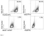
- Example 3 Adherent Differentiation of Induced Pluripotent Stem Cells to Floorplate Mesencephalic Dopaminergic Progenitor Cells and Single Cell Protein Profiling Using Flow Cytometry after 16 Days In Vitro
- iPS cells Induced pluripotent human stem cells (iPS cells) were differentiated towards floorplate mesDA progenitors cells according to the differentiation protocol described in example 2. From d0 to d9 the following neuralization and patterning factors were present SB431542 (10 ⁇ M, Miltenyi Biotec 130-105-336), Noggin (100 ng/ml Miltenyi Biotec 130-103-456), CHIR99021 (1 ⁇ M, Miltenyi Biotec 130-103-926), hSHH-C24-II (400 ng/ml, Miltenyi Biotec 130-095-727).
- the cells were dissociated into single cells with TrypLE and replated on Laminin-111 coated plates with a density of 800,000 cells/cm 2 in MACS Neuro medium, NeuroBrew-21 w/o vitamin A (1:50), 2 mM L-glutamine (Medium 3).
- FGF8b 100 ng/ml, Miltenyi Biotec 130-095-740
- BDNF 20 ng/ml; Miltenyi Biotec 130-096-286
- ascorbic acid 200 ⁇ M; Sigma A5960
- Single cell protein expression profiling was carried out on day 16 of the differentiation process. Therefore, cells were dissociated into single cells with TrypLE and contacted with antibodies conjugated to fluorophores specific for the antigens FOXA2, OTX2, PAX6, and NKX6.1 thereby labeling the cells of said sample.
- the cells showed a protein expression profile that qualifies them as human floorplate mesDA progenitor cells according to the present invention ( FIG. 4 ).
- Example 4 Adherent Differentiation of Human Embryonic Stem Cells with Different Concentrations of CHIR and Single Cell Protein Profiling Using Flow Cytometry after 16 Days In Vitro
- Human embryonic stem cells were differentiated according to the adherent differentiation protocol described in example 2.
- the patterning factors CHIR99021 (Miltenyi Biotec 130-103-926) and hSHH-C24-II (Miltenyi Biotec 130-095-727) were applied at different concentrations to pattern cells towards a ventral forebrain, a dorsal forebrain and a ventral hindbrain identity.
- Single cell protein expression profiling was carried out on day 16 of the differentiation process. Therefore, cells were dissociated into single cells with TrypLE, fixed and permeabilized using the FoxP3 buffer set (Miltenyi Biotec 130-093-142). Then cells were subdivided into three sub-sample and contacted with antibodies conjugated to fluorophores specific for the antigens FOXA2, OTX2, PAX6, NKX6.1, OCT3/4, NKX2.1, IAP, KI-67, SOX1, whereas 0.5 ⁇ 10E6 cells were used for each staining.
- Example 5 Adherent Differentiation of Human Embryonic Stem Cells to Floorplate Mesencephalic Dopaminergic Progenitor Cells and Single Cell Protein Profiling Using Flow Cytometry after 4, 8, 11, and 16 Days In Vitro
- Human embryonic stem cells were differentiated according to the adherent differentiation protocol described in example 2.
- the patterning factors CHIR99021 (Miltenyi Biotec 130-103-926) and hSHH-C24-II (Miltenyi Biotec 130-095-727) were applied at a concentration of 0.7 ⁇ M and 600 ng/ml, respectively.
- Single cell protein expression profiling was carried out on day 4, 8, 11, and 16 of the differentiation process. Therefore, cells were dissociated into single cells with TrypLE, fixed and permeabilized using the FoxP3 buffer set (Miltenyi Biotec 130-093-142). Then cells were subdivided into three sub-sample and contacted with antibodies conjugated to fluorophores specific for the antigens FOXA2, OTX2, PAX6, NKX6.1, OCT3/4, NKX2.1, whereas 0.5 ⁇ 10E6 cells were used for each staining.
- Flow cytometric analysis shows that the cells did not resemble an expression profile that qualifies them as floorplate dopaminergic neurons at d4, d8, and d16 of differentiation according to the present invention. Only cells of d11 of the differentiation process showed a protein expression profile that qualifies them as human floorplate mesDA progenitor cells according to the present invention ( FIG. 6 ).
- Human embryonic stem cells were differentiated according to the adherent differentiation protocol described in example 2.
- the patterning factors CHIR99021 (Miltenyi Biotec 130-103-926) and hSHH-C24-II (Miltenyi Biotec 130-095-727) were applied at a concentration of 0.7 ⁇ M and 600 ng/ml, respectively.
- a sample of differentiated living cells that showed an expression profile which qualifies them as floorplate dopaminergic neurons and a sample of differentiated living cells that did not resemble an expression profile that qualifies them as floorplate dopaminergic neurons were used for transplantation studies into rats.
- the first sample was correlated with a good engraftment and a high yield of dopaminergic neurons after transplantation in vivo whereas the second sample resulted in a significantly lower yield of dopaminergic neurons in vivo.
- Human embryonic stem cells were differentiated according to the adherent differentiation protocol described in example 2.
- the patterning factors CHIR99021 (Miltenyi Biotec 130-103-926) and hSHH-C24-II (Miltenyi Biotec 130-095-727) were applied at a concentration of 0.7 ⁇ M and 600 ng/ml, respectively.
Landscapes
- Health & Medical Sciences (AREA)
- Life Sciences & Earth Sciences (AREA)
- Engineering & Computer Science (AREA)
- Biomedical Technology (AREA)
- Immunology (AREA)
- Chemical & Material Sciences (AREA)
- Molecular Biology (AREA)
- Hematology (AREA)
- Urology & Nephrology (AREA)
- Biotechnology (AREA)
- Cell Biology (AREA)
- General Health & Medical Sciences (AREA)
- Microbiology (AREA)
- Biochemistry (AREA)
- General Physics & Mathematics (AREA)
- Food Science & Technology (AREA)
- Medicinal Chemistry (AREA)
- Physics & Mathematics (AREA)
- Analytical Chemistry (AREA)
- Pathology (AREA)
- Bioinformatics & Cheminformatics (AREA)
- Tropical Medicine & Parasitology (AREA)
- Neurosurgery (AREA)
- Neurology (AREA)
- Organic Chemistry (AREA)
- Zoology (AREA)
- Wood Science & Technology (AREA)
- Genetics & Genomics (AREA)
- General Engineering & Computer Science (AREA)
- Toxicology (AREA)
- Developmental Biology & Embryology (AREA)
- Measuring Or Testing Involving Enzymes Or Micro-Organisms (AREA)
- Micro-Organisms Or Cultivation Processes Thereof (AREA)
- Medicines Containing Material From Animals Or Micro-Organisms (AREA)
- Peptides Or Proteins (AREA)
- Investigating Or Analysing Biological Materials (AREA)
Abstract
Description
-
- FOXA2 (80-100% (preferentially 90-100%),
- OTX2 (80-100%, preferentially 90-100%),
- PAX6 (below 10%, preferentially below 5%),
- NKX6.1 (below 10%, preferentially below 5%),
- Optionally NKX2.1 (5-90%, preferentially 5-70%)
- Optionally OCT3/4 (below 0.1%), if the cells are generated from OCT3/4 positive cells
-
- KI-67 (10-90%),
- IAP (80-100%, preferentially 90-100%),
- SOX1 (below 10%, preferentially below 5%).
-
- a) contacting the cells of said cell composition or the cells of a sample thereof with antigen binding molecules specific for the antigens FOXA2, OTX2, PAX6, and NKX6.1, thereby labeling the cells of said cell composition or of said sample
- b) Determining the percentage of said cells that are labelled with said antigen binding molecules for each of said antigens, and wherein the cells of said cell composition qualify as human floorplate mesDA progenitor cells if the protein expression profile of said cells is:
- 80-100%, preferentially 90-100% of said cells are positive for FOXA2,
- 80-100%, preferentially 90-100% of said cells are positive for OTX2,
- Less than 10%, preferentially less than 5% of said cells are positive for PAX6, and
- Less than 10%, preferentially less than 5% of said cells are positive for NKX6.1.
-
- Anti-FoxA2-APC,
- Anti-Otx2-FITC
- Anti-Pax6-PE,
- Anti-Nkx6.1-AlexaFluor647
-
- Anti-FoxA2-APC, Anti-Otx2-FITC, Anti-IAP-PE
- Anti-Pax6-PE, Anti-Oct3/4-APC, Anti-Ki-67-FITC
- Anti-Nkx6.1-AlexaFluor 647, Anti-Nkx2.1-AlexaFluor 488, Anti-Sox1-PE
-
- Anti-FoxA2-APC, Anti-Otx2-FITC
- Anti-Pax6-PE, Anti-Oct3/4-APC
- Anti-Nkx6.1-AlexaFluor 647, Anti-Nkx2.1-AlexaFluor 488
-
- Anti-FoxA2-APC, Anti-Otx2-FITC, Anti-IAP-PE
- Anti-Pax6-PE, Anti-Oct3/4-APC, Anti-Ki-67-FITC
- Anti-Nkx6.1-AlexaFluor 647, Anti-Nkx2.1-AlexaFluor 488, Anti-Sox1-PE
- Barker R A1, Barrett J, Mason S L, Björklund A, 2013. Fetal dopaminergic transplantation trials and the future of neural grafting in Parkinson's disease. Lancet Neurol. 2013 Jan. 12; (1):84-91.
- Grealish S, Diguet E, Kirkeby A, Mattsson B, Heuer A, Bramoulle Y, Van Camp N, Perrier A L, Hantraye P, Björklund A, Parmar M, 2014. Human ESC-derived dopamine neurons show similar preclinical efficacy and potency to fetal neurons when grafted in a rat model of Parkinson's disease. Cell Stem Cell. 2014 Nov. 6; 15(5):653-65.
- Kefalopoulou Z, Politis M, Piccini P, Mencacci N, Bhatia K, Jahanshahi M, Widner H, Rehncrona S, Brundin P, Björklund A, Lindvall O, Limousin P, Quinn N, Foltynie T, 2014. Long-term clinical outcome of fetal cell transplantation for Parkinson disease: two case reports. JAMA Neurol. 2014 January; 71(1):83-7.
- Kriks S, Shim J W, Piao J, Ganat Y M, Wakeman D R, Xie Z, Carrillo-Reid L, Auyeung G, Antonacci C, Buch A, Yang L, Beal M F, Surmeier D J, Kordower J H, Tabar V, Studer L, 2012. Dopamine neurons derived from human ES cells efficiently engraft in animal models of Parkinson's disease. Nature. 2011 Nov. 6; 480(7378):547-51.
- Kirkeby A, Grealish S, Wolf D A, Nelander J, Wood J, Lundblad M, Lindvall O, Parmar M., 2012. Generation of regionally specified neural progenitors and functional neurons from human embryonic stem cells under defined conditions. Cell Rep. 2012 Jun. 28; 1(6):703-14. doi: 10.1016/j.celrep.2012.04.009.
- Kirkeby A, Nelander J, Parmar M., 2013. Generating regionalized neuronal cells from pluripotency, a step-by-step protocol. Front Cell Neurosci. 2013 Jan. 3; 6:64. doi: 10.3389/fnce1.2012.00064.
- Kirkeby A, Nolbrant S, Tiklova K, Heuer A, Kee N, Cardoso T, Ottosson D R, Lelos M J, Rifes P, Dunnett S B, Grealish S, Perlmann T, Parmar M, 2017. Predictive Markers Guide Differentiation to Improve Graft Outcome in Clinical Translation of hESC-Based Therapy for Parkinson's Disease. Cell Stem Cell. 2017 Jan. 5; 20(1):135-148. doi: 10.1016/j.stem.2016.09.004.
- Ladewig J, Mertens J, Kesavan J, Doerr J, Poppe D, Glaua F, Herms S, Wernet P, Kögler G, Müller F J, Koch P, Brüstle O, 2012. Small molecules enable highly efficient neuronal conversion of human fibroblasts. Nature Methods, 9: 575-578.
Claims (12)
Applications Claiming Priority (4)
| Application Number | Priority Date | Filing Date | Title |
|---|---|---|---|
| EP17181588 | 2017-07-17 | ||
| EP17181588 | 2017-07-17 | ||
| EP17181588.9 | 2017-07-17 | ||
| PCT/EP2018/069197 WO2019016113A1 (en) | 2017-07-17 | 2018-07-16 | A method for single cell protein expression profiling of floorplate mesencephalic dopaminergic progenitor cells |
Related Parent Applications (1)
| Application Number | Title | Priority Date | Filing Date |
|---|---|---|---|
| PCT/EP2018/069197 A-371-Of-International WO2019016113A1 (en) | 2017-07-17 | 2018-07-16 | A method for single cell protein expression profiling of floorplate mesencephalic dopaminergic progenitor cells |
Related Child Applications (1)
| Application Number | Title | Priority Date | Filing Date |
|---|---|---|---|
| US18/443,732 Continuation US12613235B2 (en) | 2017-07-17 | 2024-02-16 | Method and kit for characterizing dopaminergic progenitor cells obtained by differentiating pluripotent stem cells |
Publications (2)
| Publication Number | Publication Date |
|---|---|
| US20200173983A1 US20200173983A1 (en) | 2020-06-04 |
| US12013388B2 true US12013388B2 (en) | 2024-06-18 |
Family
ID=59387891
Family Applications (2)
| Application Number | Title | Priority Date | Filing Date |
|---|---|---|---|
| US16/632,229 Active 2040-11-12 US12013388B2 (en) | 2017-07-17 | 2018-07-16 | Method and kit for single cell protein expression profiling of floorplate mesencephalic dopaminergic progenitor cells |
| US18/443,732 Active 2038-09-16 US12613235B2 (en) | 2017-07-17 | 2024-02-16 | Method and kit for characterizing dopaminergic progenitor cells obtained by differentiating pluripotent stem cells |
Family Applications After (1)
| Application Number | Title | Priority Date | Filing Date |
|---|---|---|---|
| US18/443,732 Active 2038-09-16 US12613235B2 (en) | 2017-07-17 | 2024-02-16 | Method and kit for characterizing dopaminergic progenitor cells obtained by differentiating pluripotent stem cells |
Country Status (6)
| Country | Link |
|---|---|
| US (2) | US12013388B2 (en) |
| EP (1) | EP3655774B1 (en) |
| JP (1) | JP7125977B2 (en) |
| CN (1) | CN111213057A (en) |
| ES (1) | ES2988722T3 (en) |
| WO (1) | WO2019016113A1 (en) |
Families Citing this family (3)
| Publication number | Priority date | Publication date | Assignee | Title |
|---|---|---|---|---|
| KR20220158046A (en) | 2020-03-25 | 2022-11-29 | 사나 바이오테크놀로지, 인크. | Immunocompromised Neuronal Cells for Treatment of Nervous System Disorders and Conditions |
| CN116593700B (en) * | 2023-05-24 | 2024-02-06 | 中日友好医院(中日友好临床医学研究所) | A molecular marker for identifying patients with anti-MDA5-positive dermatomyositis |
| WO2025193168A1 (en) * | 2024-03-14 | 2025-09-18 | National University Of Singapore | Method of generating midbrain dopamine progenitors and neurons from stem cells |
Citations (3)
| Publication number | Priority date | Publication date | Assignee | Title |
|---|---|---|---|---|
| WO2010096496A2 (en) * | 2009-02-17 | 2010-08-26 | Memorial Sloan-Kettering Cancer Center | Methods of neural conversion of human embryonic stem cells |
| WO2016196661A1 (en) * | 2015-06-01 | 2016-12-08 | Memorial Sloan-Kettering Cancer Center | Methods of in vitro differentiation of midbrain dopamine (mda) neurons |
| US10072245B2 (en) * | 2015-02-27 | 2018-09-11 | Miltenyi Biotec Gmbh | Method for generation of a cell composition of mesencephalic dopaminergic progenitor cells |
Family Cites Families (4)
| Publication number | Priority date | Publication date | Assignee | Title |
|---|---|---|---|---|
| MXPA04005010A (en) | 2001-11-26 | 2005-04-08 | Advanced Cell Tech Inc | Methods for making and using reprogrammed human somatic cell nuclei and autologous and isogenic human stem cells. |
| US7939322B2 (en) * | 2008-04-24 | 2011-05-10 | Centocor Ortho Biotech Inc. | Cells expressing pluripotency markers and expressing markers characteristic of the definitive endoderm |
| DE102014110993A1 (en) * | 2014-08-01 | 2016-02-04 | Seidel GmbH & Co. KG | lighting device |
| JP6998772B2 (en) * | 2015-04-09 | 2022-02-04 | ビオラミナ アーベー | Methods and Compositions for Producing Stem Cell-Derived Dopaminergic Cells for Use in the Treatment of Neurodegenerative Diseases |
-
2018
- 2018-07-16 WO PCT/EP2018/069197 patent/WO2019016113A1/en not_active Ceased
- 2018-07-16 ES ES18743736T patent/ES2988722T3/en active Active
- 2018-07-16 CN CN201880047594.8A patent/CN111213057A/en active Pending
- 2018-07-16 EP EP18743736.3A patent/EP3655774B1/en active Active
- 2018-07-16 JP JP2020503000A patent/JP7125977B2/en active Active
- 2018-07-16 US US16/632,229 patent/US12013388B2/en active Active
-
2024
- 2024-02-16 US US18/443,732 patent/US12613235B2/en active Active
Patent Citations (3)
| Publication number | Priority date | Publication date | Assignee | Title |
|---|---|---|---|---|
| WO2010096496A2 (en) * | 2009-02-17 | 2010-08-26 | Memorial Sloan-Kettering Cancer Center | Methods of neural conversion of human embryonic stem cells |
| US10072245B2 (en) * | 2015-02-27 | 2018-09-11 | Miltenyi Biotec Gmbh | Method for generation of a cell composition of mesencephalic dopaminergic progenitor cells |
| WO2016196661A1 (en) * | 2015-06-01 | 2016-12-08 | Memorial Sloan-Kettering Cancer Center | Methods of in vitro differentiation of midbrain dopamine (mda) neurons |
Non-Patent Citations (13)
| Title |
|---|
| Barker et al., Fetal dopaminergic transplantation trials and the future of neural grafting in Parkinson's disease, Lancet Neurol. Jan. 2013., 12(1), 84-91. |
| Grealish et al., Human ESC-derived dopamine neurons show similar preclinical efficacy and potency to fetal neurons when grafted in a rat model of Parkinson's disease, Cell Stem Cell, Nov. 6, 2014;15(5), 653-665. |
| International Application No. PCT/EP2018/069197, Written Opinion of the International Searching Authority and International Search Report, 7 pages. |
| Kefalopoulou et al., Long-term clinical outcome of fetal cell transplantation for Parkinson disease two case reports, JAMA Neurol. Jan. 2014., 71(1), 83-87. |
| Kirkeby et al., Building authentic midbrain dopaminergic neurons from stem cells-lessons from development, Translational Neuroscience, vol. 3, No. 4, Dec. 2012 (Dec. 2012), 314-319, XP002774029. |
| Kirkeby et al., Generating regionalized neuronal cells from pluripotency, a step-by-step protocol, Front. Cell Neurosci., Jan. 3, 2013, 6, 64, 1-4, doi: 10.3389/fncel.2012.00064. |
| Kirkeby et al., Generation of regionally specified neural progenitors and functional neurons from human embryonic stem cells under defined conditions, Cell Rep. Jun. 28, 2012, 1(6), 703-714, doi: 10.1016/j.celrep.2012.04.009. |
| Kirkeby et al., Predictive Markers Guide Differentiation to Improve Graft Outcome in Clinical Translation of hESC-Based Therapy for Parkinson's Disease, Cell Stem Cell, Jan. 5, 2017; 20(1), 135-148, doi: 10.1016/j.stem.2016.09.004. |
| Kriks et al., Dopamine neurons derived from human ES cells efficiently engraft in animal models of Parkinson's disease. Nature, Nov. 6, 2011, 480(7378), 547-551. |
| Ladewig et al., Small molecules enable highly efficient neuronal conversion of human fibroblasts, Nature Methods, 2012, vol. 9, 575-578. |
| Nakatani et al., Lmxla and Lmxlb cooperate with Foxa2 to coordinate the specification of dopaminergic neurons and control of floor plate cell differentiation in the developing mesencephalon, Developmental Biology Mar. 1, 2010, vol. 339, No. 1, Mar. 1, 2010 (Mar. 1, 2010), 101-113, XP002774030, SSN: 1095-564X. |
| Puelles et al., Otx2 regulates the extent, identity and fate of neuronal progenitor domains in the ventral midbrain, Development, vol. 131, No. 9, Mar. 31, 2004 (Mar. 31, 2003), 2037-2048, XP055101628, GB ISSN: 0950-1991, DOI: 10.1242/dev. 01107. |
| Sonja Kriks et al., Dopamine neurons derived from human ES cells efficiently engraft in animal models of Parkinson's disease, 2011, Nature, vol. 480, Issue 7378, pp. 547-551 (Year: 2011). * |
Also Published As
| Publication number | Publication date |
|---|---|
| ES2988722T3 (en) | 2024-11-21 |
| US12613235B2 (en) | 2026-04-28 |
| EP3655774A1 (en) | 2020-05-27 |
| US20240248076A1 (en) | 2024-07-25 |
| CN111213057A (en) | 2020-05-29 |
| US20200173983A1 (en) | 2020-06-04 |
| JP7125977B2 (en) | 2022-08-25 |
| JP2020529586A (en) | 2020-10-08 |
| EP3655774C0 (en) | 2024-09-25 |
| WO2019016113A1 (en) | 2019-01-24 |
| EP3655774B1 (en) | 2024-09-25 |
Similar Documents
| Publication | Publication Date | Title |
|---|---|---|
| US12613235B2 (en) | Method and kit for characterizing dopaminergic progenitor cells obtained by differentiating pluripotent stem cells | |
| US11560546B2 (en) | Methods for neural conversion of human embryonic stem cells | |
| CN109844102B (en) | Methods to differentiate pluripotent cells | |
| US12173313B2 (en) | Assembly of functionally integrated human forebrain spheroids and methods of use thereof | |
| US20120276063A1 (en) | Priming of pluripotent stem cells for neural differentiation | |
| JP2013099345A (en) | Stem cell culture medium and culture method | |
| KR20170072941A (en) | Production method for retinal tissue | |
| Ahlfors et al. | Examining the fundamental biology of a novel population of directly reprogrammed human neural precursor cells | |
| US10072245B2 (en) | Method for generation of a cell composition of mesencephalic dopaminergic progenitor cells | |
| JP2012530502A (en) | How to predict the toxicity of chemicals | |
| Stover et al. | Process‐based expansion and neural differentiation of human pluripotent stem cells for transplantation and disease modeling | |
| WO2005017131A2 (en) | Methods for the differentiation of human stem cells | |
| Hermanto et al. | Transplantation of feeder‐free human induced pluripotent stem cell–derived cortical neuron progenitors in adult male Wistar rats with focal brain ischemia | |
| CN111386338B (en) | Preparation method of cell mass containing nervous system cells or nervous tissue and non-neural epithelial tissue and the cell mass | |
| JP2019509744A (en) | Oligodendrocyte progenitor cell composition | |
| US20220251504A1 (en) | Functional astrocytes derived from pluripotent stem cells and methods of making and using the same | |
| WO2023153464A1 (en) | Method for assessing differentiation potential of cells in culture broth in differentiation of pluripotent stem cells into neural cells of midbrain floor plate region | |
| US10174285B2 (en) | Methods of obtaining cell populations enriched with desired cells | |
| US20240384229A1 (en) | Method for providing a cell population enriched in neurons and precursors thereof | |
| Chen et al. | Chemically defined neural conversion of human pluripotent stem cells | |
| Shaltouki | Derivation of Dopaminergic Neurons from Human Pluripotent Stem Cells Using a Defined System and/or Small Molecules | |
| IL225348A (en) | Methods of obtaining cell populations enriched with desired cells |
Legal Events
| Date | Code | Title | Description |
|---|---|---|---|
| FEPP | Fee payment procedure |
Free format text: ENTITY STATUS SET TO UNDISCOUNTED (ORIGINAL EVENT CODE: BIG.); ENTITY STATUS OF PATENT OWNER: LARGE ENTITY |
|
| STPP | Information on status: patent application and granting procedure in general |
Free format text: APPLICATION DISPATCHED FROM PREEXAM, NOT YET DOCKETED |
|
| STPP | Information on status: patent application and granting procedure in general |
Free format text: DOCKETED NEW CASE - READY FOR EXAMINATION |
|
| STPP | Information on status: patent application and granting procedure in general |
Free format text: NON FINAL ACTION MAILED |
|
| STPP | Information on status: patent application and granting procedure in general |
Free format text: FINAL REJECTION MAILED |
|
| STPP | Information on status: patent application and granting procedure in general |
Free format text: RESPONSE AFTER FINAL ACTION FORWARDED TO EXAMINER |
|
| STPP | Information on status: patent application and granting procedure in general |
Free format text: ADVISORY ACTION MAILED |
|
| STCV | Information on status: appeal procedure |
Free format text: NOTICE OF APPEAL FILED |
|
| STPP | Information on status: patent application and granting procedure in general |
Free format text: NOTICE OF ALLOWANCE MAILED -- APPLICATION RECEIVED IN OFFICE OF PUBLICATIONS |
|
| ZAAB | Notice of allowance mailed |
Free format text: ORIGINAL CODE: MN/=. |
|
| AS | Assignment |
Owner name: MILTENYI BIOTEC B.V. & CO. KG, GERMANY Free format text: ASSIGNMENT OF ASSIGNORS INTEREST;ASSIGNORS:JUNGBLUT, MELANIE;BOSIO, ANDREAS;KNOEBEL, SEBASTIAN;AND OTHERS;SIGNING DATES FROM 20230904 TO 20240501;REEL/FRAME:067287/0182 |
|
| STPP | Information on status: patent application and granting procedure in general |
Free format text: AWAITING TC RESP, ISSUE FEE PAYMENT VERIFIED |
|
| STPP | Information on status: patent application and granting procedure in general |
Free format text: PUBLICATIONS -- ISSUE FEE PAYMENT VERIFIED |
|
| STCF | Information on status: patent grant |
Free format text: PATENTED CASE |