US12012454B2 - Anti-tumor antigen nanobody and nucleic acid encoding sequence thereof, and uses of the same - Google Patents
Anti-tumor antigen nanobody and nucleic acid encoding sequence thereof, and uses of the same Download PDFInfo
- Publication number
- US12012454B2 US12012454B2 US17/692,599 US202217692599A US12012454B2 US 12012454 B2 US12012454 B2 US 12012454B2 US 202217692599 A US202217692599 A US 202217692599A US 12012454 B2 US12012454 B2 US 12012454B2
- Authority
- US
- United States
- Prior art keywords
- hla
- nanobody
- receptor
- antibody
- seq
- Prior art date
- Legal status (The legal status is an assumption and is not a legal conclusion. Google has not performed a legal analysis and makes no representation as to the accuracy of the status listed.)
- Active, expires
Links
Images


Classifications
-
- C—CHEMISTRY; METALLURGY
- C07—ORGANIC CHEMISTRY
- C07K—PEPTIDES
- C07K16/00—Immunoglobulins [IG], e.g. monoclonal or polyclonal antibodies
- C07K16/18—Immunoglobulins [IG], e.g. monoclonal or polyclonal antibodies against material from animals or humans
- C07K16/28—Immunoglobulins [IG], e.g. monoclonal or polyclonal antibodies against material from animals or humans against receptors, cell surface antigens or cell surface determinants
- C07K16/2803—Immunoglobulins [IG], e.g. monoclonal or polyclonal antibodies against material from animals or humans against receptors, cell surface antigens or cell surface determinants against the immunoglobulin superfamily
- C07K16/2833—Immunoglobulins [IG], e.g. monoclonal or polyclonal antibodies against material from animals or humans against receptors, cell surface antigens or cell surface determinants against the immunoglobulin superfamily against MHC-molecules, e.g. HLA-molecules
-
- A—HUMAN NECESSITIES
- A61—MEDICAL OR VETERINARY SCIENCE; HYGIENE
- A61P—SPECIFIC THERAPEUTIC ACTIVITY OF CHEMICAL COMPOUNDS OR MEDICINAL PREPARATIONS
- A61P35/00—Antineoplastic agents
-
- G—PHYSICS
- G01—MEASURING; TESTING
- G01N—INVESTIGATING OR ANALYSING MATERIALS BY DETERMINING THEIR CHEMICAL OR PHYSICAL PROPERTIES
- G01N33/00—Investigating or analysing materials by specific methods not covered by groups G01N1/00 - G01N31/00
- G01N33/48—Biological material, e.g. blood, urine; Haemocytometers
- G01N33/50—Chemical analysis of biological material, e.g. blood, urine; Testing involving biospecific ligand binding methods; Immunological testing
- G01N33/53—Immunoassay; Biospecific binding assay; Materials therefor
- G01N33/575—Immunoassay; Biospecific binding assay; Materials therefor for cancer
- G01N33/5758—Immunoassay; Biospecific binding assay; Materials therefor for cancer involving compounds serving as markers for tumours, cancers or neoplasias, e.g. cellular determinants, receptors, heat shock/stress proteins, A-protein, oligosaccharides or metabolites
- G01N33/5759—Immunoassay; Biospecific binding assay; Materials therefor for cancer involving compounds serving as markers for tumours, cancers or neoplasias, e.g. cellular determinants, receptors, heat shock/stress proteins, A-protein, oligosaccharides or metabolites involving compounds localised on the membrane of tumour or cancer cells
-
- A—HUMAN NECESSITIES
- A61—MEDICAL OR VETERINARY SCIENCE; HYGIENE
- A61K—PREPARATIONS FOR MEDICAL, DENTAL OR TOILETRY PURPOSES
- A61K39/00—Medicinal preparations containing antigens or antibodies
- A61K2039/505—Medicinal preparations containing antigens or antibodies comprising antibodies
-
- C—CHEMISTRY; METALLURGY
- C07—ORGANIC CHEMISTRY
- C07K—PEPTIDES
- C07K2317/00—Immunoglobulins specific features
- C07K2317/30—Immunoglobulins specific features characterized by aspects of specificity or valency
- C07K2317/31—Immunoglobulins specific features characterized by aspects of specificity or valency multispecific
-
- C—CHEMISTRY; METALLURGY
- C07—ORGANIC CHEMISTRY
- C07K—PEPTIDES
- C07K2317/00—Immunoglobulins specific features
- C07K2317/50—Immunoglobulins specific features characterized by immunoglobulin fragments
- C07K2317/52—Constant or Fc region; Isotype
-
- C—CHEMISTRY; METALLURGY
- C07—ORGANIC CHEMISTRY
- C07K—PEPTIDES
- C07K2317/00—Immunoglobulins specific features
- C07K2317/50—Immunoglobulins specific features characterized by immunoglobulin fragments
- C07K2317/56—Immunoglobulins specific features characterized by immunoglobulin fragments variable (Fv) region, i.e. VH and/or VL
- C07K2317/565—Complementarity determining region [CDR]
-
- C—CHEMISTRY; METALLURGY
- C07—ORGANIC CHEMISTRY
- C07K—PEPTIDES
- C07K2317/00—Immunoglobulins specific features
- C07K2317/50—Immunoglobulins specific features characterized by immunoglobulin fragments
- C07K2317/56—Immunoglobulins specific features characterized by immunoglobulin fragments variable (Fv) region, i.e. VH and/or VL
- C07K2317/569—Single domain, e.g. dAb, sdAb, VHH, VNAR or nanobody®
-
- C—CHEMISTRY; METALLURGY
- C07—ORGANIC CHEMISTRY
- C07K—PEPTIDES
- C07K2317/00—Immunoglobulins specific features
- C07K2317/70—Immunoglobulins specific features characterized by effect upon binding to a cell or to an antigen
- C07K2317/73—Inducing cell death, e.g. apoptosis, necrosis or inhibition of cell proliferation
-
- C—CHEMISTRY; METALLURGY
- C07—ORGANIC CHEMISTRY
- C07K—PEPTIDES
- C07K2317/00—Immunoglobulins specific features
- C07K2317/70—Immunoglobulins specific features characterized by effect upon binding to a cell or to an antigen
- C07K2317/76—Antagonist effect on antigen, e.g. neutralization or inhibition of binding
-
- C—CHEMISTRY; METALLURGY
- C07—ORGANIC CHEMISTRY
- C07K—PEPTIDES
- C07K2317/00—Immunoglobulins specific features
- C07K2317/90—Immunoglobulins specific features characterized by (pharmaco)kinetic aspects or by stability of the immunoglobulin
- C07K2317/92—Affinity (KD), association rate (Ka), dissociation rate (Kd) or EC50 value
-
- G—PHYSICS
- G01—MEASURING; TESTING
- G01N—INVESTIGATING OR ANALYSING MATERIALS BY DETERMINING THEIR CHEMICAL OR PHYSICAL PROPERTIES
- G01N2333/00—Assays involving biological materials from specific organisms or of a specific nature
- G01N2333/435—Assays involving biological materials from specific organisms or of a specific nature from animals; from humans
- G01N2333/705—Assays involving receptors, cell surface antigens or cell surface determinants
- G01N2333/70503—Immunoglobulin superfamily, e.g. VCAMs, PECAM, LFA-3
- G01N2333/70539—MHC-molecules, e.g. HLA-molecules
Definitions
- the present invention relates to an anti-tumor antigen nanobody and nucleic acid encoding sequences thereof, and uses of the same.
- Cancer also known as malignancy, is a state of abnormal proliferation of cells, and these proliferating cells may invade other parts of the body as a disease caused by a malfunction in the control of cell division and proliferation.
- the number of people suffering from cancer worldwide has a growing trend. Cancer is one of the top ten causes of death for the Chinese people and has been the top ten causes of death for consecutive years.
- Cancer immunotherapy is another method for treating cancer except the above methods.
- the immune system of the patient is activated in the cancer immunotherapy by using tumor cells or tumor antigens to induce specific cellular and humoral immune responses for enhancing the anti-cancer ability of the patient, preventing the growth, spread, and recurrence of tumors, and achieving the purpose of removing or controlling tumors.
- the current tumor treatments still have the problems of ineffectiveness and strong side effects, and even lead to other immune-related disorders.
- HLA-G Human leukocyte antigen-G
- a primary objective of the present invention is to provide an anti-tumor antigen nanobody that specifically binds to a human leukocyte antigen-G (HLA-G), comprising an amino acid sequence selected from the group consisting of: SEQ ID NO:1, SEQ ID NO:2, SEQ ID NO:3, and any combination thereof.
- HLA-G human leukocyte antigen-G
- the amino acid sequence is an amino acid sequence of a heavy chain variable domain (VHH) of the anti-tumor antigen nanobody.
- VHH heavy chain variable domain
- the anti-tumor antigen nanobody is conjugated with a fragment crystallizable region (Fc region).
- Fc region fragment crystallizable region
- the anti-tumor antigen nanobody is conjugated with a second antibody to form a bispecific T-cell engager (BiTE), triple specific T-cell engager (TriTE), bispecific killer cell enager (BiKE), triple specific killer cell engager (TriKE), or any bispecific antibody.
- BiTE bispecific T-cell engager
- TriTE triple specific T-cell engager
- BiKE bispecific killer cell enager
- TripleKE triple specific killer cell engager
- the anti-tumor antigen nanobody blocks interaction and/or binding of the HLA-G with a receptor of the HLA-G.
- the receptor is killer cell immunoglobulin like receptor, two Ig domains and long cytoplasmic tail 4 (KIR2DL4) or leukocyte immunoglobulin-like receptor subfamily B member 1 (LILRB1).
- Another objective of the present invention is to provide an isolated nucleic acid encoding the above mentioned anti-tumor antigen nanobody, wherein the isolated nucleic acid comprises a nucleotide sequence selected from the group consisting of: SEQ ID NO:4, SEQ ID NO:5, SEQ ID NO:6, and any combination thereof.
- Another objective of the present invention is to provide a pharmaceutical composition, comprising the above mentioned anti-tumor antigen nanobody and a pharmaceutically acceptable carrier.
- Another objective of the present invention is to provide a method for treating cancer and immune-related disorders, comprising administering to a subject in need thereof the above mentioned pharmaceutical composition.
- Another objective of the present invention is to provide a method for detecting expression levels of HLA-G, comprising administering to a biological sample the above mentioned anti-tumor antigen nanobody.
- the biological sample is blood, urine, sputum, saliva, body fluid or human placenta.
- the anti-tumor antigen nanobody of the present invention has the following effect.
- the anti-HLA-G nanobody blocks the interaction between HLA-G and one of its receptor killer cell immunoglobulin like receptor, two Ig domains and long cytoplasmic tail 4 (KIR2DL4) and leukocyte immunoglobulin-like receptor subfamily B member 1 (LILRB1) within the 50% blocking activity as IC50 by competitive enzyme linked immunosorbent assay (ELISA), enhances cytolysis and cytotoxicity of human breast cancer cell line MDA-MB-231 with natural killer cells (NK cells), could recognize HLA-G protein from cellular lysate of human cancer cell line MDA-MB-231 and A549 cells by Western blotting analysis, and is used for flow cytometric analysis.
- ELISA competitive enzyme linked immunosorbent assay
- the anti-HLA-G nanobody also could recognize HLA-G protein on cell membrane, the expression of HLA-G is co-localized with the plasma membrane marker pan-cadherin on MDA-MB-231 and A549 cells by immunocytochemistry, and is used to detect the expression of HLA-G by immunohistochemistry (IHC) staining, thereby achieving the effect of treating cancer and immune-related disorders.
- IHC immunohistochemistry
- the gene must be transfected into cells by a vector to express the antibody function
- the anti-tumor antigen nanobody of the present invention can be prepared in vitro on a large scale, and directly administered to the individual in need for treatment.
- the present invention can also achieve the effect of detecting the expression level of HLA-G.
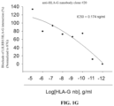
- FIGS. 1 A- 1 H show that the HLA-G/killer cell immunoglobulin like receptor, two Ig domains and long cytoplasmic tail 4 (KIR2DL4) or leukocyte immunoglobulin-like receptor subfamily B member 1 (LILRB1) axis blockade of the anti-HLA-G nanobodies are determined by competitive enzyme linked immunosorbent assay (ELISA), wherein LILRB1 represents leukocyte immunoglobulin-like receptor subfamily B member 1; KIR2DL4 represents killer cell immunoglobulin like receptor, two Ig domains and long cytoplasmic tail 4; nb represents nanobody; 87G represents commercial anti-HLA-G monoclonal antibody.
- ELISA competitive enzyme linked immunosorbent assay
- FIG. 2 is a data diagram, showing that the anti-HLA-G nanobody enhances cytolysis of human breast cancer cell line MDA-MB-231 with natural killer cells (NK cells), wherein HLA-G mAB (87G) represents commercial anti-HLA-G monoclonal antibody (87G).
- FIG. 3 A shows the result of Western blotting analysis of the anti-HLA-G nanobody
- the used cell line is human breast cancer cell line MDA-MB-231
- the commercial antibody is HLA-G (E8N9C) XP® Rabbit mAb #79769
- the number in the upper row represents the amount of cell lysate of MDA-MB-231 cell line ( ⁇ g)
- the concentration of the primary antibody is 1 ng/ml
- the secondary antibody of the commercial antibody group is anti-rabbit-horseradish peroxidase (anti-Rab-HRP)(1:1000)
- anti-HLA-G nanobody is heavy chain variable domain (VHH) nanobody (1 ng/ml)
- the secondary antibody of the experimental groups (#9 and #20) is anti-VHH-HRP (1:1000).
- FIG. 3 B shows the result of Western blotting analysis of the anti-HLA-G nanobody
- the used cell line is non-small cell lung cancer cell line A549
- the commercial antibody is HLA-G (E8N9C) XP® Rabbit mAb #79769
- the number in the upper row represents the amount of cell lysate of A549 cell line ( ⁇ g)
- the concentration of the primary antibody is 1 ng/ml
- the secondary antibody of the commercial antibody group is anti-rabbit-horseradish peroxidase (anti-Rab-HRP)(1:1000)
- anti-HLA-G nanobody is heavy chain variable domain (VHH) nanobody (1 ng/ml)
- the secondary antibody of the experimental groups (#9 and #20) is anti-VHH-HRP (1:1000).
- FIG. 4 shows the result of flow cytometric analysis of the anti-HLA-G nanobody, wherein the amount of human breast cancer cell line MDA-MB-231 and human non-small cell lung cancer cell line A549 is 1 ⁇ 10 6 , the commercial antibodies (Ab) are PE (#12-9957-42) (an anti-HLA-G monoclonal antibody, 0.25 ⁇ g in 100 ⁇ l of PBS solution) and 87G, the anti-HLA-G nanobody is heavy chain variable domain (VHH) nanobody (0.25 ⁇ g in 100 ⁇ l of PBS solution), the secondary antibody is rabbit anti-camelid VHH, iFluor555 (0.5 ⁇ g in 100 ⁇ l of PBS solution), MFI represents mean fluorescence intensity.
- PE #12-9957-42
- VHH heavy chain variable domain
- iFluor555 0.5 ⁇ g in 100 ⁇ l of PBS solution
- MFI represents mean fluorescence intensity.
- FIGS. 5 A and 5 B show the immunocytochemistry result of the anti-HLA-G nanobody, wherein the cell line used in FIG. 5 A is human breast cancer cell line MDA-MB-231, the cell line used in FIG. 5 B is human non-small cell lung cancer cell line A549, the commercial antibody 4H84 is an anti-HLA-G monoclonal antibody, the concentration of the anti-HLA-G nanobody is 1 ng/ml, and the secondary antibody is anti-VHH-fluorescein (FITC)(1:5000).
- FITC anti-VHH-fluorescein
- FIG. 6 shows the immunohistochemistry (IHC) staining result of the anti-HLA-G nanobody, wherein the used sample is human placenta, the commercial antibody is 4H84 (an anti-HLA-G monoclonal antibody)(#sc-21799), concentration is 200 ⁇ g/ml, working concentration is 4 ⁇ g/ml, the secondary antibody of the commercial antibody group is goat anti-rabbit HRP, DAB represents diaminobenzidine (the most sensitive and commonly used chromogenic reactant for horseradish peroxidase), the anti-HLA-G nanobody is heavy chain variable domain (VHH) nanobody (working concentration of 4 ⁇ g/ml), the antibodies of the experimental group (#9) include rabbit anti-camelid VHH antibody, biotin (0.5 ⁇ g in 100 ⁇ l of PBS solution) and goat anti-rabbit HRP.
- the commercial antibody is 4H84 (an anti-HLA-G monoclonal antibody)(#sc-21799)
- concentration 200 ⁇
- the data provided represent experimental values that can vary within a range of ⁇ 20%, preferably within ⁇ 10%, and most preferably within ⁇ 5%.
- anti-human leukocyte antigen-G (HLA-G) nanobody (NB) and “anti-tumor antigen nanobody” can be used interchangeably.
- second antibody refers to the antibody conjugated with the nanobody to form a bispecific T-cell engager (BiTE), triple specific T-cell engager ( TriTE ), bispecific killer cell enager (BiKE), triple specific killer cell engager (TriKE), or any bispecific antibody.
- BiTE bispecific T-cell engager
- TriTE triple specific T-cell engager
- BiKE bispecific killer cell enager
- TripleKE triple specific killer cell engager
- the second antibody includes, but is not limited to, anti-CD3 ⁇ antibody, anti-programmed cell death ligand 1 (PD-L1) antibody, anti-programmed cell death ligand 2 (PD-L2) antibody, anti-T-cell immunoglobulin domain and mucin domain 3 (Tim3) antibody, anti-epidermal growth factor receptor (EGFR) antibody, anti-EGFRvIII antibody, anti-human epidermal growth factor receptor 2 (Her2) antibody, anti-B-cell maturation antigen (BCMA) antibody, anti-CD19 antibody, anti-CD20 antibody, anti-CD34 antibody, anti-CD16 antibody, Fc, anti-epithelial cell adhesion molecule (EpCAM) antibody, anti-mesothelin antibody, anti-New York esophageal squamous cell carcinoma-1 (NY-ESO-1) antibody, anti-glycoprotein 100 (gp100) antibody, and anti-Muc 1 antibody.
- PD-L1 anti-programmed cell death ligand 1
- treating refers to alleviating, reducing, ameliorating, relieving or controlling one or more clinical signs of a disease or disorder, and lowering, stopping, or reversing the progression of severity regarding the condition or symptom being treated.
- the pharmaceutical composition can be manufactured to a dosage form suitable for parenteral or oral administration, using techniques well known to those skilled in the art, including, but not limited to, injection (e.g., sterile aqueous solution or dispersion), sterile powder, tablet, troche, lozenge, pill, capsule, dispersible powder or granule, solution, suspension, emulsion, syrup, elixir, slurry, and the like.
- injection e.g., sterile aqueous solution or dispersion
- sterile powder sterile powder
- tablet troche, lozenge
- pill capsule
- dispersible powder or granule solution, suspension, emulsion, syrup, elixir, slurry, and the like.
- composition according to the present invention may be administered by a parenteral route selected from the group consisting of: intraperitoneal injection, subcutaneous injection, intraepidermal injection, intradermal injection, intramuscular injection, intravenous injection, and intralesional injection.
- the pharmaceutical composition may further comprise a pharmaceutically acceptable carrier which is widely used in pharmaceutically manufacturing techniques.
- the pharmaceutically acceptable carrier can comprise one or more reagents selected from the group consisting of solvent, emulsifier, suspending agent, decomposer, binding agent, excipient, stabilizing agent, chelating agent, diluent, gelling agent, preservative, lubricant, absorption delaying agent, liposome, and the like. The selection and quantity of these reagents fall within the scope of the professional literacy and routine techniques of those skilled in the art.
- the pharmaceutically acceptable carrier comprises a solvent selected from the group consisting of water, normal saline, phosphate buffered saline (PBS), sugar-containing solution, aqueous solution containing alcohol, and combinations thereof.
- a solvent selected from the group consisting of water, normal saline, phosphate buffered saline (PBS), sugar-containing solution, aqueous solution containing alcohol, and combinations thereof.
- nucleic acid refers to a sequence of deoxyribonucleotides or ribonucleotides in single- or double-stranded forms, and comprises known naturally occurring nucleotides or artificially chemical mimics.
- nucleic acid is used interchangeably with the terms “gene”, “cDNA”, “mRNA”, “oligonucleotide” and “polynucleotide”.
- the preparation process of the anti-human leukocyte antigen-G (HLA-G) nanobody (NB) is as follows.
- the heavy chain variable domain (VHH) production protocol is as follows.
- the VHH gene was constructed in expression vector pET22b (Amp resistance) or pSB-init (CmR resistance);
- the plasmid was identified by restriction enzyme digestion and sequenced verification. 1 ⁇ L identified plasmid (about 50 ng) was added to BL21(DE3), and incubated overnight at 37° C.
- LB culture medium containing resistance was inoculated with a single colony and the cultures were incubated overnight at 37° C., 220 r/min Overnight culture was inoculated in a fresh LB medium (10 L-20 L) containing resistance at a ratio of 1:100, and cultured at 37° C. and 220 r/min. It was cooled to room temperature when the OD 600 reaches 0.8. Isopropyl- ⁇ -D-thiogalactopyranoside (IPTG) was added with a final concentration of 0.1 mM and induced overnight at 20° C., 220 r/min. The cells and supernatant were harvested after cell disruption by centrifugation (20 mM Tris pH 8.0, 150 mM NaCl).
- IPTG Isopropyl- ⁇ -D-thiogalactopyranoside
- Supernatant was combined with Ni-NTA beads (1 mL) by flow-through.
- the Ni-NTA beads were washed and eluted with buffers containing suitable gradient imidazole (10 mM, 20 mM, 50 mM, 100 mM, 250 mM and 500 mM).
- Elution fraction was analyzed by SDS-PAGE, and the subsequent purification scheme was determined according to the purity and yield of the protein (ion exchange chromatography or gel filtration chromatography).
- the protein that meets the requirements was separated and purified by gel filtration chromatography, and buffer was replaced with PBS buffer.
- the protein component was analyzed by SDS-PAGE, the components were merged and concentrated that meet the requirements, filtered with 0.22 ⁇ m filter and aliquot.
- the protein was stored at ⁇ 20° C. or lower.
- E. coli The production and purification of nanobodies are from E. coli .
- E. coli For producing nanobody form E. coli is modified in view of Microb Cell Fact. 2019 Mar. 11; 18(1):47.
- the E. coli strain HB2151 was used.
- the plasmid pET (Creative Biolab) coding an ampicillin resistance was used for cytoplasmic protein production.
- Freshly transformed E. coli HB2151 with pET-HLA-G or HLA-G multyspecific nanobody plasmids would be inoculated in 5 mL of media containing 50 ⁇ g/mL of ampicillin and cultivated at 37° C. for overnight.
- the gravity-flow-based chromatography would be carried out under native conditions according to the manufacturer protocol (Clontech Laboratories). Efficient cell lysis would be achieved by addition of 1 mL xTractor cell lysis buffer (Clontech Laboratories) supplemented with EDTA-free protease inhibitor cocktail (Roche Diagnostics) and 25 U endonuclease (Thermo Scientific Pierce) to each 200 mg bacterial cell pellet. After incubation on ice for 15 min and centrifugation at 10,000 ⁇ g and 4° C. for 20 min for removal of cellular debris, the clarified supernatant would be loaded onto a gravity-flow column containing 1 mL of prepacked resin and incubated at room temperature for 30 min.
- the column Before elution of the nanobodies by addition of elution buffer containing 300 mM imidazole, the column would be washed twice with increasing imidazole concentrations of 20 and 40 mM. Removal of imidazole and buffer exchange would be achieved by dialysis against PBS using a cellulose ester membrane with a molecular weight cut-off of 3.5-5 kDa (Spectrum® Laboratories).
- the alignment and amino acid sequences of the complementarity determining regions (CDRs) for each clone of anti-HLA-G nanobodies are shown in Table 1.
- the amino acid sequence of anti-HLA-G nanobody clone #9 is SEQ ID NO:1;
- the amino acid sequence of anti-HLA-G nanobody clone #20 is SEQ ID NO:2;
- the amino acid sequence of anti-HLA-G nanobody clone #33 is SEQ ID NO:3;
- the nucleotide sequence encoding the amino acid sequence of anti-HLA-G nanobody clone #9 is SEQ ID NO:4;
- the nucleotide sequence encoding the amino acid sequence of anti-HLA-G nanobody clone #20 is SEQ ID NO:5;
- the nucleotide sequence encoding the amino acid sequence of anti-HLA-G nanobody clone #33 is SEQ ID NO:6.
- HLA-G/Killer Cell Immunoglobulin Like Receptor Two Ig Domains and Long Cytoplasmic Tail 4 (KIR2DL4) or Leukocyte Immunoglobulin-Like Receptor Subfamily B Member 1 (LILRB1) Axis Blockade of Anti-HLA-G Nanobodies are Determined by Competitive Enzyme Linked Immunosorbent Assay (ELISA)
- HLA-G/KIR2DL4 axis blockade of the anti-HLA-G nanobodies were determined by competitive ELISA.
- the procedure is as follows. HLA-G recombinant protein (CAT #: TP305216, Origene) (0.2 mg/ml, 100 ⁇ l each well) was coated on 96-well plate overnight at 4° C. Next day, the coating buffer was discarded, and blocked with 3% skin milk for 2 hr at room temperature, followed by washing 5 times with PBST (0.05% Tween in PBS). Different concentrations of anti-HLA-G nanobody (clone #9, #20 or #33) or commercial anti-HLA-G monoclonal antibody (87G) were added overnight at 4° C.
- PBST 0.05% Tween in PBS
- biotinylated KIR2DL4 (Sino Biological, Cat: 13052-H02S) (0.2 mg/ml, 100 ⁇ l each well) was added for 2 hr at room temperature. After 9 times of washing, each well was incubated with 100 ⁇ l PBST containing streptavidin-HRP conjugates (ThermoFisher, Cat No: N100, dilution titer: 5000:1) for 2 hr at room temperature. After 9 times of washing with PBST, 50 ⁇ l of TMB substrate for detecting HRP activity (ThermoFisher, Cat No:N301) was added.
- the reactions were stopped by adding 50 ⁇ l stop solution (ThermoFisher, Cat No:N600), and it was measured by ELISA reader using 450 nm channel.
- the highest concentration of commercial HLA-G monoclonal antibody (87G) was set to 100% blockade of KIR2DL4/HLA-G interaction for calculating other reactions.
- HLA-G/LILRB1 axis blockade of the anti-HLA-G nanobodies were also determined by competitive ELISA.
- the procedure is basically the same as above, the differences are: biotinylated KIR2DL4 was replaced with biotinylated LILRB1 (Sino Biological, Cat: 16014-H08H), and the highest concentration of commercial HLA-G monoclonal antibody (87G) was set to 100% blockade of LILRB1/HLA-G interaction for calculating other reactions.
- FIGS. 1 A- 1 H The results of this example are shown in FIGS. 1 A- 1 H , wherein LILRB1 represents leukocyte immunoglobulin-like receptor subfamily B member 1; KIR2DL4 represents killer cell immunoglobulin like receptor, two Ig domains and long cytoplasmic tail 4; nb represents nanobody; 87G represents commercial anti-HLA-G monoclonal antibody.
- LILRB1 represents leukocyte immunoglobulin-like receptor subfamily B member 1
- KIR2DL4 represents killer cell immunoglobulin like receptor, two Ig domains and long cytoplasmic tail 4
- nb represents nanobody
- 87G commercial anti-HLA-G monoclonal antibody.
- the result of this example demonstrates that commercial anti-HLA-G monoclonal antibody (87G) blocks the interaction between HLA-G and one of its receptor KIR2DL4 within the 50% blocking activity as 41.4 ng/ml (IC50).
- the anti-HLA-G nanobody clone #9 blocks the interaction between HLA-G and one of its receptor KIR2DL4 within the 50% blocking activity as 6.14 ng/ml (IC50), the blocking activity was normalized to 87G.
- the anti-HLA-G nanobody clone #20 blocks the interaction between HLA-G and one of its receptor KIR2DL4 within the 50% blocking activity as 814 ng/ml (IC50), the blocking activity was normalized to 87G.
- the anti-HLA-G nanobody clone #33 blocks the interaction between HLA-G and one of its receptor KIR2DL4 within the 50% blocking activity as 53.3 ng/ml (IC50), the blocking activity was normalized to 87G.
- anti-HLA-G monoclonal antibody blocks the interaction between HLA-G and its another one receptor LILRB1 within the 50% blocking activity as 100.9 ng/ml (IC50).
- the anti-HLA-G nanobody clone #9 blocks the interaction between HLA-G and its another one receptor LILRB1 within the 50% blocking activity as 0.825 ng/ml (IC50), the blocking activity was normalized to 87G.
- the anti-HLA-G nanobody clone #20 blocks the interaction between HLA-G and its another one receptor LILRB1 within the 50% blocking activity as 0.174 ng/ml (IC50), the blocking activity was normalized to 87G.
- the anti-HLA-G nanobody clone #33 blocks the interaction between HLA-G and its another one receptor LILRB1 within the 50% blocking activity as 0.074 ng/ml (IC50), the blocking activity was normalized to 87G.
- FIG. 2 The result of anti-HLA-G nanobody on enhancing cytolysis of human breast cancer cell line MDA-MB-231 with NK cells is shown in FIG. 2 .
- the result of this example demonstrates that anti-HLA-G nanobody clones #9, #20 and #33 enhance NK-induced cytotoxicity to tumor cells (MDA-MB-231).
- the membranes After 5% BSA in TBST blocking, the membranes would be incubated with primary antibodies in TBST at 4° C. overnight. They would be then washed 4 times and incubated with horseradish peroxidase (HRP)-conjugated goat anti-mouse or rabbit IgG (Upstate, Billerica, MA, USA) for 2 hours. After washing with TBST 4 times, the blots would be incubated for 1 min with the SuperSignal West Pico ECL reagent (Pierce Biotechnology, Rockford, IL, USA), and chemiluminescence would be detected using by exposure to Kodak-X-Omat film.
- HRP horseradish peroxidase
- FIGS. 3 A and 3 B The Western blotting result of anti-HLA-G nanobody is shown in FIGS. 3 A and 3 B , wherein the cell line used in FIG. 3 A is human breast cancer cell line MDA-MB-231, the commercial antibody is HLA-G (E8N9C) XP® Rabbit mAb #79769, the number in the upper row represents the amount of cell lysate of MDA-MB-231 cell line ( ⁇ g), the concentration of the primary antibody is 1 ng/ml, the secondary antibody of the commercial antibody group is anti-rabbit-horseradish peroxidase (anti-Rab-HRP)(1:1000), anti-HLA-G nanobody is heavy chain variable domain (VHH) nanobody (1 ng/ml), the secondary antibody of the experimental groups (#9 and #20) is anti-VHH-HRP (1:1000).
- the cell line used in FIG. 3 A is human breast cancer cell line MDA-MB-231
- the commercial antibody is HLA
- the cell line used in FIG. 3 B is non-small cell lung cancer cell line A549 (purchased from American Type Culture Collection (ATCC)), the commercial antibody is HLA-G (E8N9C) XP Rabbit mAb #79769, the number in the upper row represents the amount of cell lysate of A549 cell line ( ⁇ g), the concentration of the primary antibody is 1 ng/ml, the secondary antibody of the commercial antibody group is anti-rabbit-horseradish peroxidase (anti-Rab-HRP)(1:1000), anti-HLA-G nanobody is heavy chain variable domain (VHH) nanobody (1 ng/ml), the secondary antibody of the experimental groups (#9 and #20) is anti-VHH-HRP (1:1000).
- the result of this example shows that anti-HLA-G nanobodies clone #9 and #20 could recognize HLA-G protein from cellular lysate of human cancer cell line MDA-MB-231 and A549 cells by western blo
- HLA-G recombinant protein (CAT #: TP305216, Origene) (0.2 mg/ml, 100 ⁇ l each well) was coated on 96-well plate overnight at 4° C. Next day, the coating buffer was discarded, and blocked with 3% skin milk for 2 hr at room temperature, followed by washing 5 times with PBST (0.05% Tween in PBS). Different concentrations of anti-HLA-G nanobody (clone #9, #20 or #33) or commercial anti-HLA-G monoclonal antibody (87G) were added overnight at 4° C.
- PBST 0.05% Tween in PBS
- biotinylated KIR2DL4 (Sino Biological, Cat: 13052-H02S) (0.2 mg/ml, 100 ⁇ l each well) was added for 2 hr at room temperature. After 9 times of washing, each well was incubated with 100 ⁇ l PBST containing streptavidin-HRP conjugates (ThermoFisher, Cat No: N100, dilution titer: 5000:1) for 2 hr at room temperature. After 9 times of washing with PBST, 50 ⁇ l of TMB substrate for detecting HRP activity (ThermoFisher, Cat No:N301) was added.
- the reactions were stopped by adding 50 ⁇ l stop solution (ThermoFisher, Cat No:N600), and it was measured by ELISA reader using 450 nm channel.
- the highest concentration of commercial HLA-G monoclonal antibody (87G) was set to 100% blockade of KIR2DL4/HLA-G interaction for calculating other reactions.
- the flow cytometric analysis result of anti-HLA-G nanobody is shown in FIG. 4 , wherein the amount of human breast cancer cell line MDA-MB-231 and human non-small cell lung cancer cell line A549 is 1 ⁇ 10 6 , the commercial antibodies (Ab) are PE (#12-9957-42) (an anti-HLA-G monoclonal antibody, 0.25 ⁇ g in 100 of PBS solution) and 87G, the anti-HLA-G nanobody is heavy chain variable domain (VHH) nanobody (0.25 ⁇ g in 100 ⁇ l of PBS solution), the secondary antibody is rabbit anti-camelid VHH, iFluor555 (0.5 ⁇ g in 100 ⁇ l of PBS solution), MFI represents mean fluorescence intensity.
- the commercial antibodies (Ab) are PE (#12-9957-42) (an anti-HLA-G monoclonal antibody, 0.25 ⁇ g in 100 of PBS solution) and 87G
- the anti-HLA-G nanobody is heavy chain variable domain (VHH
- the procedures of immunocytochemistry of anti-HLA-G nanobody are as follows. Tumor cells (1 ⁇ 10 5 ) were seeded on coverslips in a 6-well plate, incubated overnight. After the indicated treatments, cells were fixed in 1% paraformaldehyde, washed with PBS, permeabilized using 0.1% Triton X-100 in PBS containing 0.5% BSA for 30 min, blocked with 2% BSA, and incubated with specific antibodies in 2% BSA/PBS containing 0.05% Tween-20 (PBST).
- PBST Tween-20
- the cells were incubated with fluorescein-conjugated secondary antibodies, washed with PBST, and mounted using a water-based mounting medium containing an anti-fade agent and 4′,6-diamidino-2-phenylindole (DAPI). Images were analyzed under a Leica TCS SP8 ⁇ confocal microscope (Leica).
- FIGS. 5 A and 5 B The immunocytochemistry result of anti-HLA-G nanobody is shown in FIGS. 5 A and 5 B , wherein the cell line used in FIG. 5 A is human breast cancer cell line MDA-MB-231, the cell line used in FIG. 5 B is human non-small cell lung cancer cell line A549, the commercial antibody 4H84 is an anti-HLA-G monoclonal antibody, the concentration of the anti-HLA-G nanobody is 1 ng/ml, and the secondary antibody is anti-VHH-fluorescein (FITC)(1:5000). As shown in FIGS.
- anti-HLA-G nanobodies clone #9 and #20 could recognize HLA-G protein on cell membrane, the expression of HLA-G was co-localized with the plasma membrane marker Pan-cadherin on MDA-MB-231 and A549 cells by immunocytochemistry using anti-HLA-G nanobodies clone #9 and clone #20, HLA-G commercial antibody 4H84 and commercial pan-cadherin antibody.
- the procedures of IHC staining of anti-HLA-G nanobody are as follows.
- the primary antibodies would be incubated overnight at 4° C. After washing, the sections would be then incubated with diluted biotin-conjugated secondary antibodies for 2 h at room temperature or overnight at 4° C.
- the sections would be then incubated with polymer for 10 min at room temperature and following diaminobenzidine (DAB, the most sensitive and commonly used chromogenic reactant for horseradish peroxidase) staining, after then the sections would be stained lightly with hematoxylin and eosin and fixed using neutral balata. Quantification of the staining would be performed independently by optical microscope (Nikon) at 40 ⁇ and 400 ⁇ magnification.
- DAB diaminobenzidine
- the result of IHC staining of anti-HLA-G nanobody is shown in FIG. 6 , the sample is human placenta, the commercial antibody is 4H84 (an anti-HLA-G monoclonal antibody)(#sc-21799), concentration is 200 ⁇ g/ml, working concentration is 4 ⁇ g/ml, the secondary antibody of the commercial antibody group is goat anti-rabbit HRP, DAB represents diaminobenzidine (the most sensitive and commonly used chromogenic reactant for horseradish peroxidase), the anti-HLA-G nanobody is heavy chain variable domain (VHH) nanobody (working concentration of 4 ⁇ g/ml), the antibodies of the experimental group (#9) include rabbit anti-camelid VHH antibody, biotin (0.5 ⁇ g in 100 ⁇ l of PBS solution) and goat anti-rabbit HRP.
- anti-tumor antigen nanobody i.e., anti-HLA-G nanobody
- concentration is 200 ⁇ g/ml
- working concentration is 4
- the anti-tumor antigen nanobody i.e., anti-HLA-G nanobody
- the anti-tumor antigen nanobody of the present invention blocks the interaction between HLA-G and one of its receptor killer cell immunoglobulin like receptor, two Ig domains and long cytoplasmic tail 4 (KIR2DL4) and leukocyte immunoglobulin-like receptor subfamily B member 1 (LILRB1) within the 50% blocking activity as IC50 by competitive enzyme linked immunosorbent assay (ELISA), enhances cytolysis and cytotoxicity of human breast cancer cell line MDA-MB-231 with natural killer cells (NK cells), could recognize HLA-G protein from cellular lysate of human cancer cell line MDA-MB-231 and A549 cells by Western blotting analysis, and is used for flow cytometric analysis.
- ELISA competitive enzyme linked immunosorbent assay
- the anti-HLA-G nanobody also could recognize HLA-G protein on cell membrane, the expression of HLA-G is co-localized with the plasma membrane marker pan-cadherin on MDA-MB-231 and A549 cells by immunocytochemistry, and is used to detect the expression of HLA-G by immunohistochemistry (IHC) staining, thereby achieving the effect of treating cancer and immune-related disorders.
- IHC immunohistochemistry
- the gene must be transfected into cells by a vector to express the antibody function
- the anti-tumor antigen nanobody of the present invention can be prepared in vitro on a large scale, and directly administered to the individual in need for treatment.
- the present invention can also achieve the effect of detecting the expression level of HLA-G.
Landscapes
- Health & Medical Sciences (AREA)
- Immunology (AREA)
- Chemical & Material Sciences (AREA)
- Life Sciences & Earth Sciences (AREA)
- Organic Chemistry (AREA)
- General Health & Medical Sciences (AREA)
- Medicinal Chemistry (AREA)
- Molecular Biology (AREA)
- Engineering & Computer Science (AREA)
- Biochemistry (AREA)
- Hematology (AREA)
- Biomedical Technology (AREA)
- Urology & Nephrology (AREA)
- Veterinary Medicine (AREA)
- Genetics & Genomics (AREA)
- Proteomics, Peptides & Aminoacids (AREA)
- Biophysics (AREA)
- Public Health (AREA)
- Animal Behavior & Ethology (AREA)
- Pharmacology & Pharmacy (AREA)
- Chemical Kinetics & Catalysis (AREA)
- General Chemical & Material Sciences (AREA)
- Nuclear Medicine, Radiotherapy & Molecular Imaging (AREA)
- Physics & Mathematics (AREA)
- Pathology (AREA)
- General Physics & Mathematics (AREA)
- Microbiology (AREA)
- Biotechnology (AREA)
- Food Science & Technology (AREA)
- Cell Biology (AREA)
- Analytical Chemistry (AREA)
- Peptides Or Proteins (AREA)
- Medicines Containing Antibodies Or Antigens For Use As Internal Diagnostic Agents (AREA)
- Measuring Or Testing Involving Enzymes Or Micro-Organisms (AREA)
- Preparation Of Compounds By Using Micro-Organisms (AREA)
- Medicines That Contain Protein Lipid Enzymes And Other Medicines (AREA)
Abstract
Description
| TABLE 1 | |||
| Clone | CDR1 | | CDR3 |
| # | |||
| 9 | GRTYSSNC | IYTGGDGI | AADPNRRRMGVGGSC |
| (SEQ ID NO: 7) | (SEQ ID NO: 8) | (SEQ ID NO: 9) | |
| #20 | GFTVDDSD | ITSGGGK | VAPAWTGYGCT |
| (SEQ ID NO: 10) | (SEQ ID NO: 11) | (SEQ ID NO: 12) | |
| #33 | AYTFSASG | AATYTRSAKT | AVARCAGRPDRSTL |
| (SEQ ID NO: 13) | (SEQ ID NO: 14) | TSFAW | |
| (SEQ ID NO: 15) | |||
Claims (17)
Priority Applications (5)
| Application Number | Priority Date | Filing Date | Title |
|---|---|---|---|
| US17/692,599 US12012454B2 (en) | 2021-03-24 | 2022-03-11 | Anti-tumor antigen nanobody and nucleic acid encoding sequence thereof, and uses of the same |
| EP22163473.6A EP4063392B1 (en) | 2021-03-24 | 2022-03-22 | Anti-tumor antigen nanobody and nucleic acid encoding sequence thereof, and uses of the same |
| TW111110937A TWI799205B (en) | 2021-03-24 | 2022-03-23 | Anti-tumor antigen nanobody and nucleic acid encoding sequence thereof, and uses of the same |
| JP2022046582A JP7368528B2 (en) | 2021-03-24 | 2022-03-23 | Antitumor antigen nanobodies and their nucleic acid coding sequences and uses thereof |
| CN202210292073.7A CN115124622B (en) | 2021-03-24 | 2022-03-23 | Anti-tumor antigen nano antibody and nucleic acid coding sequence and application thereof |
Applications Claiming Priority (2)
| Application Number | Priority Date | Filing Date | Title |
|---|---|---|---|
| US202163165266P | 2021-03-24 | 2021-03-24 | |
| US17/692,599 US12012454B2 (en) | 2021-03-24 | 2022-03-11 | Anti-tumor antigen nanobody and nucleic acid encoding sequence thereof, and uses of the same |
Publications (2)
| Publication Number | Publication Date |
|---|---|
| US20220306747A1 US20220306747A1 (en) | 2022-09-29 |
| US12012454B2 true US12012454B2 (en) | 2024-06-18 |
Family
ID=80930336
Family Applications (1)
| Application Number | Title | Priority Date | Filing Date |
|---|---|---|---|
| US17/692,599 Active 2042-03-11 US12012454B2 (en) | 2021-03-24 | 2022-03-11 | Anti-tumor antigen nanobody and nucleic acid encoding sequence thereof, and uses of the same |
Country Status (5)
| Country | Link |
|---|---|
| US (1) | US12012454B2 (en) |
| EP (1) | EP4063392B1 (en) |
| JP (1) | JP7368528B2 (en) |
| CN (1) | CN115124622B (en) |
| TW (1) | TWI799205B (en) |
Families Citing this family (4)
| Publication number | Priority date | Publication date | Assignee | Title |
|---|---|---|---|---|
| USD1007520S1 (en) * | 2021-04-06 | 2023-12-12 | China Medical University | Display screen or portion thereof with graphical user interface |
| DK4190349T3 (en) * | 2021-12-03 | 2025-04-14 | Univ China Medical | PRODUCTION OF EXOSOMES AND THEIR APPLICATION |
| WO2024108568A1 (en) * | 2022-11-25 | 2024-05-30 | 深圳先进技术研究院 | Anti-human leukocyte antigen-g nanobody, and preparation method therefor and use thereof |
| AU2023201146B2 (en) * | 2023-02-26 | 2025-02-13 | China Medical University | Production of exosomes and uses thereof |
Citations (2)
| Publication number | Priority date | Publication date | Assignee | Title |
|---|---|---|---|---|
| WO2011051327A2 (en) * | 2009-10-30 | 2011-05-05 | Novartis Ag | Small antibody-like single chain proteins |
| TW202035456A (en) | 2018-09-27 | 2020-10-01 | 美商提聖納醫療公司 | Anti-hla-g antibodies, compositions comprising anti-hla-g antibodies and methods of using anti-hla-g antibodies |
Family Cites Families (5)
| Publication number | Priority date | Publication date | Assignee | Title |
|---|---|---|---|---|
| EP2730588A1 (en) * | 2012-11-12 | 2014-05-14 | Intelectys | Antibodies and fragments thereof raised against the alpha-3 domain of HLA-G protein, methods and means for their preparation, and uses thereof |
| AR115052A1 (en) * | 2018-04-18 | 2020-11-25 | Hoffmann La Roche | MULTI-SPECIFIC ANTIBODIES AND THE USE OF THEM |
| AR114789A1 (en) * | 2018-04-18 | 2020-10-14 | Hoffmann La Roche | ANTI-HLA-G ANTIBODIES AND THE USE OF THEM |
| CN110903399B (en) * | 2018-09-17 | 2022-02-01 | 台湾中国医药大学附设医院 | Chimeric antigen receptor, nucleic acid thereof, expression plasmid, cell, use and composition |
| CN109734814A (en) * | 2019-02-12 | 2019-05-10 | 南京卡提医学科技有限公司 | The purposes of engineering T cell treating cancer with immunity receptor |
-
2022
- 2022-03-11 US US17/692,599 patent/US12012454B2/en active Active
- 2022-03-22 EP EP22163473.6A patent/EP4063392B1/en active Active
- 2022-03-23 CN CN202210292073.7A patent/CN115124622B/en active Active
- 2022-03-23 JP JP2022046582A patent/JP7368528B2/en active Active
- 2022-03-23 TW TW111110937A patent/TWI799205B/en active
Patent Citations (2)
| Publication number | Priority date | Publication date | Assignee | Title |
|---|---|---|---|---|
| WO2011051327A2 (en) * | 2009-10-30 | 2011-05-05 | Novartis Ag | Small antibody-like single chain proteins |
| TW202035456A (en) | 2018-09-27 | 2020-10-01 | 美商提聖納醫療公司 | Anti-hla-g antibodies, compositions comprising anti-hla-g antibodies and methods of using anti-hla-g antibodies |
Non-Patent Citations (4)
| Title |
|---|
| Chen et al., Enhancement and destruction of antibody function by somatic mutation: unequal occurrence is controlled by V gene combinatorial associations. EMBO J. Jun. 15, 1995;14(12):2784-94. (Year: 1995). * |
| Edwards et al., The remarkable flexibility of the human antibody repertoire; isolation of over one thousand different antibodies to a single protein, BLyS. J Mol Biol. Nov. 14, 2003;334(1):103-18. (Year: 2003). * |
| Koenig et al., Mutational landscape of antibody variable domains reveals a switch modulating the interdomain conformational dynamics and antigen binding. PNAS Jan. 24, 2017 114 (4) E486-E495; first published Jan. 5, 2017; (Year: 2017). * |
| Kussie, Paul H., "A Single Engineered Amino Acid Substitution Changes Antibody Fine Specificity", 1994, Journal of Immunology 152(1): pp. 146-152. (Year: 1994). * |
Also Published As
| Publication number | Publication date |
|---|---|
| JP7368528B2 (en) | 2023-10-24 |
| EP4063392B1 (en) | 2025-07-30 |
| TW202237666A (en) | 2022-10-01 |
| TWI799205B (en) | 2023-04-11 |
| EP4063392A1 (en) | 2022-09-28 |
| JP2022151798A (en) | 2022-10-07 |
| CN115124622B (en) | 2025-07-18 |
| US20220306747A1 (en) | 2022-09-29 |
| CN115124622A (en) | 2022-09-30 |
Similar Documents
| Publication | Publication Date | Title |
|---|---|---|
| US12012454B2 (en) | Anti-tumor antigen nanobody and nucleic acid encoding sequence thereof, and uses of the same | |
| EP3623388A1 (en) | Bispecific recombinant protein and use thereof | |
| US11795227B2 (en) | Immunomodulation and anti-tumor-related nanobody and nucleic acid encoding sequence thereof, and uses of the same | |
| JP7358426B2 (en) | EGFL6-specific monoclonal antibodies and methods of using them | |
| WO2023201311A1 (en) | Multi-chain chimeric polypeptide for use in the treatment of treating neuroinflammatory disorder | |
| CN116731169B (en) | Nano antibody with sortilin 1 specificity and application thereof | |
| CA3211762A1 (en) | Bispecific antibodies and uses of the same thereof | |
| US11795224B2 (en) | Anti-T-cell nanobody and nucleic acid encoding sequence thereof, and uses of the same | |
| CA2844126C (en) | Microvesicle membrane protein and application thereof | |
| US11685784B2 (en) | Anti-immune-checkpoint nanobody and nucleic acid encoding sequence thereof, and uses of the same | |
| EP1780220A1 (en) | Use of CEACAM8-specific substances for treating autoimmune diseases and a method for screening substances which induce apoptosis | |
| WO2025031098A1 (en) | Sortilin 1-specific nanoantibody, recombinant aav containing same, and use |
Legal Events
| Date | Code | Title | Description |
|---|---|---|---|
| AS | Assignment |
Owner name: CHINA MEDICAL UNIVERSITY HOSPITAL, TAIWAN Free format text: ASSIGNMENT OF ASSIGNORS INTEREST;ASSIGNORS:CHO, DER-YANG;CHIU, SHAO-CHIH;HUANG, SHI-WEI;AND OTHERS;REEL/FRAME:059239/0922 Effective date: 20220214 |
|
| FEPP | Fee payment procedure |
Free format text: ENTITY STATUS SET TO UNDISCOUNTED (ORIGINAL EVENT CODE: BIG.); ENTITY STATUS OF PATENT OWNER: LARGE ENTITY |
|
| STPP | Information on status: patent application and granting procedure in general |
Free format text: DOCKETED NEW CASE - READY FOR EXAMINATION |
|
| FEPP | Fee payment procedure |
Free format text: PETITION RELATED TO MAINTENANCE FEES GRANTED (ORIGINAL EVENT CODE: PTGR); ENTITY STATUS OF PATENT OWNER: LARGE ENTITY |
|
| STPP | Information on status: patent application and granting procedure in general |
Free format text: NON FINAL ACTION MAILED |
|
| AS | Assignment |
Owner name: CHINA MEDICAL UNIVERSITY HOSPITAL, TAIWAN Free format text: CORRECTIVE ASSIGNMENT TO CORRECT THE ADDRESS OF THE ASSIGNEE TO NO. 2, YUDE RD., NORTH DIST., TAICHUNG CITY 404327,TAIWAN PREVIOUSLY RECORDED ON REEL 059239 FRAME 0922. ASSIGNOR(S) HEREBY CONFIRMS THE ASSIGNMENT;ASSIGNORS:CHO, DER-YANG;CHIU, SHAO-CHIH;HUANG, SHI-WEI;AND OTHERS;REEL/FRAME:063442/0397 Effective date: 20230424 |
|
| STPP | Information on status: patent application and granting procedure in general |
Free format text: RESPONSE TO NON-FINAL OFFICE ACTION ENTERED AND FORWARDED TO EXAMINER |
|
| STPP | Information on status: patent application and granting procedure in general |
Free format text: NON FINAL ACTION MAILED |
|
| STPP | Information on status: patent application and granting procedure in general |
Free format text: RESPONSE TO NON-FINAL OFFICE ACTION ENTERED AND FORWARDED TO EXAMINER |
|
| STPP | Information on status: patent application and granting procedure in general |
Free format text: NOTICE OF ALLOWANCE MAILED -- APPLICATION RECEIVED IN OFFICE OF PUBLICATIONS |
|
| STPP | Information on status: patent application and granting procedure in general |
Free format text: PUBLICATIONS -- ISSUE FEE PAYMENT VERIFIED |
|
| STCF | Information on status: patent grant |
Free format text: PATENTED CASE |