US11976382B2 - Nucleic acid based biosensor and methods thereof - Google Patents
Nucleic acid based biosensor and methods thereof Download PDFInfo
- Publication number
- US11976382B2 US11976382B2 US16/949,217 US202016949217A US11976382B2 US 11976382 B2 US11976382 B2 US 11976382B2 US 202016949217 A US202016949217 A US 202016949217A US 11976382 B2 US11976382 B2 US 11976382B2
- Authority
- US
- United States
- Prior art keywords
- biosensor
- domain
- fluorophore
- rna
- target
- Prior art date
- Legal status (The legal status is an assumption and is not a legal conclusion. Google has not performed a legal analysis and makes no representation as to the accuracy of the status listed.)
- Active, expires
Links
Images


Classifications
-
- C—CHEMISTRY; METALLURGY
- C40—COMBINATORIAL TECHNOLOGY
- C40B—COMBINATORIAL CHEMISTRY; LIBRARIES, e.g. CHEMICAL LIBRARIES
- C40B40/00—Libraries per se, e.g. arrays, mixtures
- C40B40/04—Libraries containing only organic compounds
- C40B40/06—Libraries containing nucleotides or polynucleotides, or derivatives thereof
- C40B40/08—Libraries containing RNA or DNA which encodes proteins, e.g. gene libraries
-
- C—CHEMISTRY; METALLURGY
- C12—BIOCHEMISTRY; BEER; SPIRITS; WINE; VINEGAR; MICROBIOLOGY; ENZYMOLOGY; MUTATION OR GENETIC ENGINEERING
- C12N—MICROORGANISMS OR ENZYMES; COMPOSITIONS THEREOF; PROPAGATING, PRESERVING, OR MAINTAINING MICROORGANISMS; MUTATION OR GENETIC ENGINEERING; CULTURE MEDIA
- C12N15/00—Mutation or genetic engineering; DNA or RNA concerning genetic engineering, vectors, e.g. plasmids, or their isolation, preparation or purification; Use of hosts therefor
- C12N15/09—Recombinant DNA-technology
- C12N15/10—Processes for the isolation, preparation or purification of DNA or RNA
- C12N15/1034—Isolating an individual clone by screening libraries
- C12N15/1048—SELEX
-
- C—CHEMISTRY; METALLURGY
- C12—BIOCHEMISTRY; BEER; SPIRITS; WINE; VINEGAR; MICROBIOLOGY; ENZYMOLOGY; MUTATION OR GENETIC ENGINEERING
- C12N—MICROORGANISMS OR ENZYMES; COMPOSITIONS THEREOF; PROPAGATING, PRESERVING, OR MAINTAINING MICROORGANISMS; MUTATION OR GENETIC ENGINEERING; CULTURE MEDIA
- C12N15/00—Mutation or genetic engineering; DNA or RNA concerning genetic engineering, vectors, e.g. plasmids, or their isolation, preparation or purification; Use of hosts therefor
- C12N15/09—Recombinant DNA-technology
- C12N15/11—DNA or RNA fragments; Modified forms thereof; Non-coding nucleic acids having a biological activity
- C12N15/115—Aptamers, i.e. nucleic acids binding a target molecule specifically and with high affinity without hybridising therewith ; Nucleic acids binding to non-nucleic acids, e.g. aptamers
-
- G—PHYSICS
- G01—MEASURING; TESTING
- G01N—INVESTIGATING OR ANALYSING MATERIALS BY DETERMINING THEIR CHEMICAL OR PHYSICAL PROPERTIES
- G01N21/00—Investigating or analysing materials by the use of optical means, i.e. using sub-millimetre waves, infrared, visible or ultraviolet light
- G01N21/62—Systems in which the material investigated is excited whereby it emits light or causes a change in wavelength of the incident light
- G01N21/63—Systems in which the material investigated is excited whereby it emits light or causes a change in wavelength of the incident light optically excited
- G01N21/64—Fluorescence; Phosphorescence
- G01N21/6428—Measuring fluorescence of fluorescent products of reactions or of fluorochrome labelled reactive substances, e.g. measuring quenching effects, using measuring "optrodes"
-
- C—CHEMISTRY; METALLURGY
- C12—BIOCHEMISTRY; BEER; SPIRITS; WINE; VINEGAR; MICROBIOLOGY; ENZYMOLOGY; MUTATION OR GENETIC ENGINEERING
- C12N—MICROORGANISMS OR ENZYMES; COMPOSITIONS THEREOF; PROPAGATING, PRESERVING, OR MAINTAINING MICROORGANISMS; MUTATION OR GENETIC ENGINEERING; CULTURE MEDIA
- C12N2310/00—Structure or type of the nucleic acid
- C12N2310/10—Type of nucleic acid
- C12N2310/16—Aptamers
-
- C—CHEMISTRY; METALLURGY
- C12—BIOCHEMISTRY; BEER; SPIRITS; WINE; VINEGAR; MICROBIOLOGY; ENZYMOLOGY; MUTATION OR GENETIC ENGINEERING
- C12N—MICROORGANISMS OR ENZYMES; COMPOSITIONS THEREOF; PROPAGATING, PRESERVING, OR MAINTAINING MICROORGANISMS; MUTATION OR GENETIC ENGINEERING; CULTURE MEDIA
- C12N2320/00—Applications; Uses
- C12N2320/10—Applications; Uses in screening processes
- C12N2320/11—Applications; Uses in screening processes for the determination of target sites, i.e. of active nucleic acids
-
- C—CHEMISTRY; METALLURGY
- C12—BIOCHEMISTRY; BEER; SPIRITS; WINE; VINEGAR; MICROBIOLOGY; ENZYMOLOGY; MUTATION OR GENETIC ENGINEERING
- C12N—MICROORGANISMS OR ENZYMES; COMPOSITIONS THEREOF; PROPAGATING, PRESERVING, OR MAINTAINING MICROORGANISMS; MUTATION OR GENETIC ENGINEERING; CULTURE MEDIA
- C12N2320/00—Applications; Uses
- C12N2320/10—Applications; Uses in screening processes
- C12N2320/13—Applications; Uses in screening processes in a process of directed evolution, e.g. SELEX, acquiring a new function
-
- G—PHYSICS
- G01—MEASURING; TESTING
- G01N—INVESTIGATING OR ANALYSING MATERIALS BY DETERMINING THEIR CHEMICAL OR PHYSICAL PROPERTIES
- G01N21/00—Investigating or analysing materials by the use of optical means, i.e. using sub-millimetre waves, infrared, visible or ultraviolet light
- G01N21/62—Systems in which the material investigated is excited whereby it emits light or causes a change in wavelength of the incident light
- G01N21/63—Systems in which the material investigated is excited whereby it emits light or causes a change in wavelength of the incident light optically excited
- G01N21/64—Fluorescence; Phosphorescence
- G01N21/6428—Measuring fluorescence of fluorescent products of reactions or of fluorochrome labelled reactive substances, e.g. measuring quenching effects, using measuring "optrodes"
- G01N2021/6439—Measuring fluorescence of fluorescent products of reactions or of fluorochrome labelled reactive substances, e.g. measuring quenching effects, using measuring "optrodes" with indicators, stains, dyes, tags, labels, marks
Definitions
- the instant disclosure relates to nucleic acid molecules which comprise of at least one stem and/or loop structure that may bind to a reporter molecule, such as a fluorophores, and/or a target molecule, methods of making the nucleic acid molecules, methods to use the nucleic acid molecules as a biosensor, and kits comprising the nucleic acid molecules for practicing their method.
- a reporter molecule such as a fluorophores
- a target molecule such as a fluorophores
- Aptamers comprised of nucleic acids that may bind fluorogenic molecules are emerging as useful tools for basic and applied biology for use as biosensors. These aptamers may be genetically encoded like fluorescent proteins but enable observation at different levels. Also, unlike fluorescent proteins these aptamers are not natural and instead are engineered in the lab using various methods such as fluorescence or complex affinity based in vitro selections (Ellington et al., 1990, In vitro selection of RNA molecules that bind specific ligands. Nature 346: 818-822; Bock et al. 1992, Selection of single-stranded DNA molecules that bind and inhibit human thrombin. Nature 355: 564-566, both herein incorporated in their entirety). A high fluorescence signal above background is possible due to fluorescence enhancement of a second molecule upon binding to the aptamer.
- RNA Mango series of aptamers have the promising combination of tight binding to its ligand and significant fluorescence enhancement.
- the RNA Mango aptamers bind biotinylated derivatives of Thiazole Orange with low nanomolar KD and fluorescence enhancement of approximately 1,100-fold.
- There are currently several variants of the RNA Mango with subtle nucleotide sequence variations that lead to different ligand affinities and fluorescence enhancements Dolgosheina et al., 2014, RNA Mango aptamer-fluorophore: a bright, high affinity, complex for RNA labeling and tracking.
- Mango-I The structures of the original aptamer, termed Mango-I, is quite simple with a single base-paired stem capped by a three tiered G-quadruplex where the ligand binds.
- RNA Spinach and related aptamers also have G-quadruplex ligand binding sites, but with more than one base-paired stem joined to the quadruple.
- additional helical elements have been used in several applications that require more complex structures, such as the detection of RNA-RNA interactions and dual-aptamer allosteric RNA biosensors but lack the signal to background ratio of the Mango aptamers making them less sensitive and inferior biosensors, so much so that they may not be able to identify some targets when the target is in a low quantity.
- quadruplex flanking helices have been shown to modulate aptamer properties such as metal ion specificity.
- RNA Mango-I is limiting to efforts to design more complex functionalities because it is unclear how to add additional nucleotide elements to the G-quadruplex core without disrupting ligand binding or if a switching effect could be achieved by the addition of additional nucleotides.
- Computational tools to predict quadruplex structures are advancing, but do not currently provide the ability to predict ligand affinity and fluorescence enhancement.
- the present disclosure relates to nucleic acid molecules related to the Mango aptamer.
- Mango-I aptamer which traditionally has a single opened stem attached to a square shaped G-quadruplex core, wherein the G-quadruplex may bind a heterocyclic fluorophore, such as Thiazole Orange (TO1)-biotin (TO1B) bi-functional fluorescent molecule.
- TO1B Thiazole Orange
- the Mango aptamer due to this square link shape, may be thought to have four edges, i, ii, iii, and iv, and four corners, i-ii, ii-ii, iii-iv, and iv-i.
- the open stem may comprise one of the corners of the G-quadruplex core.
- the present disclosure relates to the addition of one or more open or closed nucleotide stems to the corners of a Mango aptamer, wherein the additional stem includes a linker and a sensor domain.
- the sensor domain may bind to a target molecule, if the target molecule is present within the sample.
- the linker domain may effect a conformational change in the reporter domain in response to a target ligand molecule binding to the sensor domain which will allow the reporter to bind to the reporter domain.
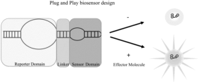
- An embodiment of the disclosure is a biosensor comprising of three components, a Mango core domain with an open stem, a linker, and a sensor domain.
- the linker and sensor domain may form an additional closed stem or open stem (split biosensor).
- a split biosensor may be comprised of either two different polynucleotides or a nicked closed stem. More complex designs may also be made through, by nonlimiting example, nucleotide origami where there may be two or more polynucleotides comprising the biosensor.
- the three components are modular so that once a specific domain is designed, it may be easily switched to develop a biosensor with different properties.
- the open stem of the Mango core domain is positioned on an adjacent corner to at least one closed stem of a linker and sensor domain.
- the open stem and another closed stem are positioned on opposite corners of each other on the Mango core domain.
- the biosensor may have two or more closed stems or three closed stems. The closed stems may bind to the same or different target ligands and may have the same or different linkers. The additional open stems may be on adjacent or opposite corners of the Mango core.
- the biosensor may contain additional functional units, such as, but not limited to, promoters or handles, attached to the open end.
- a plasmid comprises the biosensor and additional functional units, such as promoters, which will express the biosensor.
- a different embodiment of the disclosure is a method of selecting a biosensor of the disclosure which binds to a defined target. For a biosensor to function properly, it must be able to detect a target ligand in a sample, and then provide a discriminating signal.
- the method comprises obtaining a library of randomly generated sequences, which comprise the Mango core, different linkers, and at least one sensor domain; subjecting the library to one or more rounds of negative and positive selection; and measuring the change in brightness from an unbound to a bound state.
- the negative selection depletes sequence which only bind to the reporter, for example TO1B, in the absence of the target ligand.
- the positive selection enriches for the binding of the reporter in the presence of the target ligand. However, the presence of the target ligand does not make it essential that the reporter will bind to the reporter domain.
- the number of rounds of selection may be determined by one skilled in the art, to save costs the number of positive and negative selection rounds is less than about 10.
- a following round of selection is performed on the results from the prior round.
- the negative selection is performed before the positive selection.
- the same type of selection is performed on the results of the prior round. In other embodiments, the type of selection is alternated between rounds. In another embodiment, one or negative rounds is followed by one or more positive rounds or one or more positive rounds is followed by one or more negative rounds.
- either positive or negative selection is performed.
- the pool may be split into two after partial selection, with the first split being subjected to a final round of negative selection and the second split being subjected to a final round of positive selection, the two splits sequenced, and the number of specific sequences counted.
- the ratio between the negative and positive split may then be used to determine biosensors according to this disclosure.
- an embodiment comprises a method of binding the biosensors of the invention to a target in a sample.
- the method comprises of introducing into a sample a biosensor of the disclosure and the reporter; and incubating the sample to allow for binding to a target ligand.
- the sample is then excited by the appropriate wavelengths to allow for the amount of the target ligand to be quantified by measuring the difference in brightness of the biosensor in the sample to a control biosensor.
- a fluorescent label is further introduced into the sample.
- the quantity of target ligand may be obtained by comparing the signal from the biosensor to the fluorescent label.
- the method further includes fixing a sample prior to introducing the biosensor and reporter into the sample. This may be done to locate the position of a target within a sample, such as, but not limited to, subcellular structures, RNA, or cells, such as bacteria cells within an environmental sample.
- an embodiment comprises a kit which may be used to binding a desired target in a sample.
- the kit may comprise of the biosensor and/or a nucleic acid comprising the biosensor and additional functional regions, such as, but not limited to, one or more promoter regions.
- the kit may further comprise of the reporter molecule, by way of nonlimiting example TOB1; buffers; solvents, such as but not limited to polar or organic solvents; and/or instructions.
- FIG. 1 A is a schematic representation of the crystal structure of a single strand Mango I aptamer bound to TO1-biotin with the edges of the G quadruplex labeled i, ii, iii, and iv.
- FIG. 1 B is a schematic representation of an exemplary Mango I aptamer bound to TO1-biotin with individual nucleotides numbered 1-39 and edge nucleotides labeled with their edge designation as set out in FIG. 1 A . Rectangles along the edges are guanine residues (SEQ ID NO: 2).
- FIG. 2 A is a schematic representation of an exemplary Mango I aptamer bound to TO1-biotin with a i-iv open stem and an ii-i closed stem with individual nucleotides numbered and edge nucleotides labeled with their edge designation as set out in FIG. 1 A . Rectangles along the edges are guanine residues (SEQ ID NO: 3).
- FIG. 2 B is a schematic representation of an exemplary Mango I aptamer bound to TO1-biotin with a i-iv open stem and an iii-ii closed stem with individual nucleotides numbered and edge nucleotides labeled with their edge designation as set out in FIG. 1 A .
- FIG. 2 C is a schematic representation of an exemplary Mango I aptamer bound to TO1-biotin with a i-iv open stem and an iii-iv closed stem with individual nucleotides numbered and edge nucleotides labeled with their edge designation as set out in FIG. 1 A .
- Rectangles along the edges are guanine residues (SEQ ID NO: 5).
- FIG. 2 D is a schematic representation of an exemplary Mango I aptamer bound to TO1-biotin with a ii-i open stem and an iii-ii closed stem with individual nucleotides numbered and edge nucleotides labeled with their edge designation as set out in FIG. 1 A . Rectangles along the edges are guanine residues (SEQ ID NO: 6).
- FIG. 2 E is a schematic representation of an exemplary Mango I aptamer bound to TO1-biotin with a ii-i open stem and a iv-iii closed stem with individual nucleotides numbered and edge nucleotides labeled with their edge designation as set out in FIG. 1 A .
- FIG. 2 F is a schematic representation of an exemplary Mango I aptamer bound to TO1-biotin with a iii-ii open stem and a iv-iii closed stem with individual nucleotides numbered and edge nucleotides labeled with their edge designation as set out in FIG. 1 A .
- Rectangles along the edges are guanine residues (SEQ ID NO: 8).
- FIG. 2 G is a schematic representation of an exemplary split Mango I aptamer bound to TO1-biotin with iv-i and ii-i open stems with individual nucleotides numbered and edge nucleotides labeled with their edge designation as set out in FIG. 1 A . Rectangles along the edges are guanine residues (SEQ ID NOs: 9-10).
- FIG. 2 H is a schematic representation of an exemplary Mango I aptamer bound to TO1-biotin with ii-i and iii-i open stems with individual nucleotides numbered and edge nucleotides labeled with their edge designation as set out in FIG. 1 A . Rectangles along the edges are guanine residues (SEQ ID NOs: 11-12).
- FIG. 3 A is a schematic representation of the Mango core, linker, and a closed sensor domain showing the presence of the target ligand determining the final state of the fluorophore through allosteric fluorescence.
- FIG. 3 B is a schematic representation of the induction of fluorescence by a target ligand showing the unbound biosensor is stabilized by the target ligand, which in turn stabilizing the fluorophore binding pocket.
- FIG. 4 A is a simplified schematic representation of a Mango-Theophylline-N8 starting library with a closed stem in the i-iv position and a closed stem with the N8 linker and Theophylline sensor in the ii-i position, or further abbreviated A position (MTS14A).
- FIG. 4 B is a simplified schematic representation of a Mango-Theophylline-N8 starting library with a closed stem in the i-iv position and a closed stem with the N8 linker and Theophylline sensor in the iii-i position, or further abbreviated B position (MTS14B).
- FIG. 4 A is a simplified schematic representation of a Mango-Theophylline-N8 starting library with a closed stem in the i-iv position and a closed stem with the N8 linker and Theophylline sensor in the iii-i position, or further abbreviated B position (MTS14B).
- FIG. 4 A is
- FIG. 4 C is a simplified schematic representation of a Mango-Theophylline-N8 starting library with a closed stem in the i-iv position and a closed stem with the N8 linker and Theophylline sensor in the iv-iii position, or further abbreviated C position (MTS14C).
- FIG. 5 A is a graphical representation of the fluorescence saturation curve of the two-stemmed Mango variants with 25 nM TO1-B and variable RNA concentrations. Shaded regions show 95% C.I. of the curve fit with the second closed stem at the i-iv corner (see FIGS. 2 A- 2 C for schematic representations).
- FIG. 5 B is a graphical representation of the fluorescence saturation curve of the two-stemmed Mango variants with 25 nM TO1-B and variable RNA concentrations. Shaded regions show 95% C.I. of the curve fit with the second closed stem at the ii-i corner (see FIGS. 2 D- 2 E for schematic representations).
- FIG. 5 A is a graphical representation of the fluorescence saturation curve of the two-stemmed Mango variants with 25 nM TO1-B and variable RNA concentrations. Shaded regions show 95% C.I. of the curve fit with the second closed stem at the ii-i corner (see FIGS. 2 D
- FIG. 5 C is a graphical representation of the fluorescence saturation curve of the two-stemmed Mango variants with 25 nM TO1-B and variable RNA concentrations. Shaded regions show 95% C.I. of the curve fit with the second closed stem at the iii-ii corner (see FIG. 2 F for schematic representations).
- FIG. 5 D is a graphical representation of the relative maximum fluorescence (F max ) of the two-stemmed variants.
- FIG. 5 E is a graphical representation of the fraction of Mango i-iv ii-i in free solution over a range of TO1-B concentrations.
- FIG. 5 F is a graphical representation of the Best fit KD curve with 95% C.I. (shaded).
- FIG. 6 A is a photograph representation of the fluorescence of split Mango constructs showing the difference in fluorescence between the 5′ Split Mango i-iv ii-i strand only, 3′ Split Mango i-iv ii-i strand only, both strands of Split Mango i-iv ii-i, no RNA, Mango i-iv ii-i, and Mango I (see FIGS. 2 G and 2 H for graphical representations of split Mango).
- FIG. 6 B is a graphical representation of the normalized F max of both split Mango i-iv-ii-i and split Mango ii-i ii-ii when compared to Mango-I in 40 nM TO1-B.
- FIG. 7 A is a graphical representation of the plots of sequences and their normalized reads for the two starting libraries (R5 and R5a) compared against each other.
- FIG. 7 B is a graphical representation of the normalized reads for pools R6 and R5 compared against each other. Selected sequences that exhibited high enrichment ratios (lower circle) or high-count enrichment (top circle) and are predicted to exhibit allosteric fluorescence.
- FIG. 7 C is a graphical representation of the fold change of R6a+ and R6a ⁇ after being normalized to R5a pool. Selected sequences that are predicted to dim (anti) in the presence of theophylline (lower circle). Sequences that are predicted to exhibit allosteric fluorescence with theophylline (top circle).
- FIG. 8 A is a graphical representation of the difference of fluorescence between RNA samples with theophylline (solid line) and without theophylline (dashed line) after every round of selection.
- FIG. 8 B is a graphical representation of the average change in fluorescence of samples R5 and R6 after the addition of theophylline.
- FIG. 8 C is a graphical representation of the total brightness of fluorescence after the addition of theophylline for R % and R6 to confirmation verification.
- FIG. 8 D is a graphical representation of the individual sequences with their change in fluorescence after the addition of theophylline grouped by sorting algorithm.
- FIG. 9 A is a graphical representation of the effects of theophylline saturation and time on the allosteric fluorescence of biosensor exemplar MTS546 showing the measured relative fluorescence of MTS546 at 200 nM RNA and 300 nM after incubation. Fluorescence measurements at time points of 0 minutes and 60 minutes for the same pool of RNA.
- FIG. 9 B is a graphical representation of the fluorescent saturation curves at different time points with theophylline (solid lines) and without theophylline (dashed lines). Theophylline saturation (vertical dashed line) where the concentration of theophylline equals the RNA concentration.
- description of a range such as from 1 to 6 should be considered to have specifically disclosed sub-ranges such as from 1 to 3, from 1 to 4, from 1 to 5, from 2 to 4, from 2 to 6, from 3 to 6 etc., as well as individual numbers within that range, for example, 1, 2, 3, 4, 5, and 6, and decimals and fractions, for example, 1.2, 3.8, 11 ⁇ 2, and 4% This applies regardless of the breadth of the range.
- the term “about” modifying the quantity of an ingredient in the compositions of the invention or employed in the methods of the invention refers to variation in the numerical quantity that can occur, for example, through typical measuring and liquid handling procedures used for making concentrates or use solutions in the real world; through inadvertent error in these procedures; through differences in the manufacture, source, or purity of the ingredients employed to make the compositions or carry out the methods; and the like.
- the term about also encompasses amounts that differ due to different equilibrium conditions for a composition resulting from a particular initial mixture. Whether or not modified by the term “about,” the claims include equivalents to the quantities.
- isolated is used to indicate that a cell, peptide or nucleic acid is separated from its native environment. Isolated peptides and nucleic acids may be substantially pure, i.e. essentially free of other substances with which they may bound in nature.
- nucleic acid refers to a polymer of deoxyribonucleotide, ribonucleotide, or analogs thereof, in linear or circular conformation, and in either single- or double-stranded form.
- polynucleotide refers to a polymer of deoxyribonucleotide, ribonucleotide, or analogs thereof, in linear or circular conformation, and in either single- or double-stranded form.
- these terms are not to be construed as limiting with respect to the length of a polymer.
- the terms can encompass known analogues of natural nucleotides, as well as nucleotides that are modified in the base, sugar and/or phosphate moieties (e.g., phosphorothioate backbones).
- an analogue of a particular nucleotide has the same base-pairing specificity; i.e., an analogue of A will base-pair with T.
- a nucleic acid can be single-stranded or double-stranded.
- a single-stranded nucleic acid can have double-stranded regions and a double-stranded nucleic acid can have single-stranded regions.
- nucleotide is any nucleoside linked to a phosphate group.
- the nucleoside may be natural, including but not limited to, any of cytidine, uridine, adenosine, guanosine, thymidine, inosine (hypoxanthine), or uric acid; or synthetic, including but not limited to methyl-substituted phenol analogs, hydrophobic base analogs, purine/pyrimidine mimics, icoC, isoG, thymidine analogs, fluorescent base analogs, or X or Y synthetic bases.
- nucleotide may be abasic, such as but not limited to 3-hydroxy-2-hydroxymethyl-tetrahydrofuran, which act as a linker group lacking a base or be a nucleotide analog.
- nucleotide is used interchangeably with “nucleic acid.”
- nucleotide duplex is when two strands of complement nucleotide oligomers complementary bind to each other. The two strands may be part of the same nucleotide molecule or separate nucleotide molecules.
- the nucleotides making up the biosensors may be natural, including but not limited to, any of cytosine, uracil, adenine, guanine, thymine, hypoxanthine, or uric acid; or synthetic, including but not limited to methyl-substituted phenol analogs, hydrophobic base analogs, purine/pyrimidine mimics, icoC, isoG, thymidine analogs, fluorescent base analogs, or X or Y synthetic bases.
- a nucleotide may be abasic, such as but not limited to 3-hydroxy-2-hydroxymethyl-tetrahydrofuran, or alternatively a nucleotide analog may be used.
- Non-limiting examples of synthetic nucleobases and analogs include, but are not limited to methyl-substituted phenyl analogs, such as but not limited to mono-, di-, tri-, or tatramethylated benzene analogs; hydrophobic base analogs, such as but not limited to 7-propynyl isocarbostyril nucleoside, isocarbostyril nucleoside, 3-methylnapthalene, azaindole, bromo phenyl derivates at positions 2, 3, and 4, cyano derivatives at positions 2, 3, and 4, and fluoro derivates at position 2 and 3; purine/pyrimidine mimics, such as but not limited to azole hetercyclic carboxamides, such as but not limited to (1H)-1,2,3-triazole-4-carboxamide, 1,2,4-triazole-3-carboxamide, 1,2,3-triazole-4-carboxamide, or 1,2-pyrazole-3-carboxamide
- Non-limiting examples of nucleotide analogs include, but are not limited to, phosporothioate nucleotides, 2′-O-methyl ribonucleotides, 2′-O-methoxy-ethyl ribonucleotides, peptide nucleotides, N3′-P5′ phosphoroamidate, 2′-fluoro-arabino nucleotides, locked nucleotides (LNA), unlocked nucleotides (UNA), morpholino phosphoroamidate, cyclohexene nucleotides, tricyclo-deoxynucleotides, or triazole-linked nucleotides.
- LNA locked nucleotides
- UNA unlocked nucleotides
- morpholino phosphoroamidate cyclohexene nucleotides, tricyclo-deoxynucleotides, or triazole-linked nucleotides.
- polypeptide “peptide” and “protein” are used interchangeably to refer to a polymer of amino acid residues.
- the term also applies to amino acid polymers in which one or more amino acids are chemical analogues or modified derivatives of a corresponding naturally-occurring amino acids.
- amino acids include natural or unnatural amino acids.
- amino acid includes compounds which depart from the structure of the naturally occurring amino acids, but which have substantially the structure of an amino acid, such that they can be substituted within a peptide which retains is activity, e.g., biological activity.
- amino acids can also include amino acids having side chain modifications or substitutions, and also include related organic acids, amides or the like.
- an amino acid can be a proteogenic or non-proteogenic amino acid.
- proteogenic indicates that the amino acid can be incorporated into a protein in a cell through well-known metabolic pathways.
- Exemplary amino acids amenable to the present invention include, but are not limited to, alanine; argnine; asparagine; aspartic acid; cysteine; glutamic acid; glutamine; glycine; histadine; isoleucine; leucine; lysine; methionine; phenylalanine; proline; serine; threonine; tryptophan; tyrosine; valine; homocysteine; phosphoserine; phosphothreonine; phosphotyrosine; hydroxyproline; y-carboxyglutamate; hippuric acid; octahydroindole-2-carboxylic acid; statine; 1,2,3,4,-tetrahydroisoquinoline-3-carboxylic acid; penicillamine (3-mercapto-D-valine); ornithine (Orn); citruline; alpha-methyl-alanine; para-benzoylphenylalanine;
- a “ligand” is a type of molecule that is recognized by a receptor or aptamer and either causes the receptor to signal, an “agonist,” or prevents the receptor to signal, an “antagonist.”
- a “target ligand” is the ligand which is to be assayed within a sample by the biosensor and may be, by nonlimiting example, a small molecule, a ribonuclear protein, a protein, a bacterial cell wall or membrane, a viral coat, an organic or inorganic molecule, a macromolecular complex, or an oligonucleotide.
- target ligand and “target molecule” are used interchangeably herein.
- a “small molecule” is any low molecular weight ( ⁇ 900 daltons) organic compound with a size on the order of 1 nm.
- a “macromolecular complex” refers to a collection of molecules that may be random, ordered or partially ordered in their arrangement.
- the term encompasses biological organisms such as bacteriophage, viruses, bacteria, unicellular pathogenic organisms, multicellular pathogenic organisms and prokaryotic or eukaryotic cells.
- the term also encompasses non-living assemblages of molecules, such as liposomes, microcapsules, microparticles, magnetic beads and microdevices. The only requirement is that the complex contains more than one molecule.
- the molecules may be identical or may differ from each other.
- a “receptor” may be peptides, proteins, glycoproteins, lipoproteins, epitopes, antibodies, lipids, carbohydrates, multi-molecular structures, a specific conformation of one or more molecules and a morphoanatomic entity that has a binding affinity for a specific group of chemicals or molecules, such as other proteins or viruses.
- the receptor Upon recognition and binding of the chemical or molecule, the receptor can cause some form of signaling or other process within a cell to respond to the chemical or molecule.
- the chemical or molecule can cause a receptor to stop functioning property and shut off processes.
- RNP Ribonuclear protein
- operative linkage and “operatively linked” (or “operably linked”) are used interchangeably with reference to a juxtaposition of two or more components (such as sequence elements), in which the components are arranged such that both components function normally and allow the possibility that at least one of the components can mediate a function that is exerted upon at least one of the other components.
- a transcriptional regulatory sequence such as a promoter
- a transcriptional regulatory sequence is generally operatively linked in cis with a coding sequence but need not be directly adjacent to it.
- an enhancer is a transcriptional regulatory sequence that is operatively linked to a coding sequence, even though they are not contiguous.
- coding region and “coding sequence” are used interchangeably and refer to a nucleotide sequence that encodes a polypeptide and, when placed under the control of appropriate regulatory sequences expresses the encoded polypeptide.
- the boundaries of a coding region are generally determined by a translation start codon at its 5′ end and a translation stop codon at its 3′ end.
- a “regulatory sequence” is a nucleotide sequence that regulates expression of a coding sequence to which it is operably linked.
- Non-limiting examples of regulatory sequences include promoters, enhancers, transcription initiation sites, translation start sites, translation stop sites, and transcription terminators.
- amplified is meant the construction of multiple copies of a nucleic acid sequence or multiple copies complementary to the nucleic acid sequence using at least one of the nucleic acid sequences as a template.
- Amplification systems include the polymerase chain reaction (PCR) system, ligase chain reaction (LCR) system, nucleic acid sequence based amplification (NASBA, Canteen, Mississauga, Ontario), Q-Beta Replicase systems, transcription-based amplification system (TAS), and strand displacement amplification (SDA). See, e.g., Diagnostic Molecular Microbiology: Principles and Applications, D. H. Persing et al., Ed., American Society for Microbiology, Washington, D.C. (1993). The product of amplification is termed an amplicon.
- expression vector covers a DNA molecule, linear or circular, that comprises a segment encoding a polypeptide of the invention, and which is operably linked to additional segments that provide for its transcription.
- the term vector refers broadly to any plasmid or virus encoding an exogenous nucleic acid.
- the term should also be construed to include non-plasmid and non-viral compounds which facilitate transfer of nucleic acid into virions or cells, such as, for example, polylysine compounds and the like.
- the vector may be a viral vector that is suitable as a delivery vehicle for delivery of the nucleic acid, or mutant thereof, to a cell, or the vector may be a non-viral vector which is suitable for the same purpose. Examples of viral and non-viral vectors for delivery of DNA to cells and tissues are well known in the art and are described, for example, in Ma et al. (1997, Proc. Natl. Acad. Sci.
- U.S.A. 94:12744-12746 examples include, but are not limited to, a recombinant vaccinia virus, a recombinant adenovirus, a recombinant retrovirus, a recombinant adeno-associated virus, a recombinant avian pox virus, and the like (Cranage et al., 1986, EMBO J. 5:3057-3063; U.S. Pat. No. 5,591,439).
- non-viral vectors include, but are not limited to, liposomes, polyamine derivatives of DNA, and the like.
- host cell includes any cell type which is susceptible to transformation with a nucleic acid construct.
- host cell is meant a cell which contains a vector and supports the replication and/or expression of the vector.
- Host cells may be prokaryotic cells such as E. coli , or eukaryotic cells such as yeast, insect, amphibian, or mammalian cells.
- promoter includes reference to a region of DNA upstream from the start of transcription and involved in recognition and binding of RNA polymerase and other proteins to initiate transcription.
- recombinant includes reference to a cell or vector, that has been modified by the introduction of a heterologous nucleic acid or that the cell is derived from a cell so modified.
- recombinant cells express genes that are not found in identical form within the native (non-recombinant) form of the cell or express native genes that are otherwise abnormally expressed, under-expressed or not expressed at all as a result of deliberate human intervention.
- the term “recombinant” as used herein does not encompass the alteration of the cell or vector by naturally occurring events (e.g., spontaneous mutation, natural transformation/transduction/transposition) such as those occurring without deliberate human intervention.
- a “recombinant expression cassette” is a nucleic acid construct, generated recombinantly or synthetically, with a series of specified nucleic acid elements which permit transcription of a particular nucleic acid in a host cell.
- the recombinant expression cassette can be incorporated into a plasmid, chromosome, mitochondrial DNA, plastid DNA, virus, or nucleic acid fragment.
- the recombinant expression cassette portion of an expression vector includes, among other sequences, a nucleic acid to be transcribed, and a promoter.
- sample is a small part or quantity intended to represent the whole.
- an environmental sample could be a small quantity of soil from a field or water from a lake. It could also be a blood or tissue sample from a human or animal. Sometimes the sample is purified to select an even smaller or more specific sample, such as isolating RNA, DNA, or protein from a blood or tissue sample.
- the biosensor comprises of three or more domains in a modular design.
- the domains comprise a reporter domain, one or more linker domains, and one or more sensor domains.
- a biosensor comprises of one or more strands of polynucleotides and may be RNA, DNA, PNA, LNA, or UNA. While typically made of RNA, these other nucleic acids may be used to alter the rigidity of the biosensor. For example, UNA may be used to make a more relaxed backbone while LNA may make a more stable biosensor compared to a biosensor comprised of RNA.
- the reporter domain comprises a Mango core as described in WO2018/198013 (herein incorporated in its entirety).
- the Mango core comprises of two section, an open stem and a G-quadruplex which binds to fluorophore.
- the biosensor may be any nucleotide, such as DNA, RNA, UNA, PNA, or LNA
- the core sequence may be represented by the RNA sequence: 5′-GG@(T 1 /WGW)GG(# 1 H/WG)WGGN@(# 2 / ⁇ )G(T 2 /H)GNH(AN@T 3 /G)-3′ (a, c, g)
- the G-quadruplex has been further labeled by the edges in this disclosure, being numbered: i, ii, iii, and iv (see FIG. 1 A ).
- edges There are nucleotides not involved in G-quartets, which bind the fluorophore, at the corners of the G-quadruplex, termed loops, where two edges meet. Corners are designated by the two edges which comprise the corner, for example corners i-ii and ii-i are both synonymous for the corner between edges i and ii (see FIG. 1 B ). As shown in FIGS. 1 B and 2 A- 2 H , corner i-ii lacks an A/U flap present at the other corners. The order of the corner edges is not important but may be used for additional clarification where noted.
- the G-quadruplex may then bind to a heterocyclic fluorophore represented in Formula I or II:
- alkyl and heteroalkyl each includes any reasonable combination of the following: (1) saturated alkyls as well as unsaturated alkyls (e.g. alkenyls and alkynyls); (2) linear or branched; (3) acyclic, cyclic (aromatic or nonaromatic) or multi-cyclic (fused rings, multiple non-fused rings or a combination thereof); and (4) unsubstituted or substituted.
- an alkyl or heteroalkyl i.e.
- alkyl/heteroalkyl may be saturated, branched and cyclic, or unsaturated, branched and cyclic, or linear and unsaturated, or any other reasonable combination according to the skill of the person of skill in the art.
- the size of the alkyl/heteroalkyl is specified as X 1 -Xz, where z is any integer larger than 1 (e.g. 15, 18, 30, 100 or the like), it will be understood that the alkyl/heteroalkyl comprises at least 3 carbons and heteroatoms so as to form a ring. If unspecified, the size of the alkyl/heteroalkyl is what would be considered reasonable to the person of skill in the art.
- the size of an alkyl may be 1, 2, 3, 4, 5, 6, 7, 8, 9, 10, 11, 12, 13, 14, 15, 16, 17, 18, 19, 20, 21, 22, 23, 24, 25, 26, 27, 28, 29, 30, 31, 32, 33, 34, 35, 36, 37, 38, 39, 40, 41, 42, 43, 44, 45, 46, 47, 48, 49, 50, 51, 52, 53, 54, 55, 56, 57, 58, 59, 60, 61, 62, 63, 64, 65, 66, 67, 68, 69, 70, 71, 72, 73, 74, 75, 76, 77, 78, 79, 80, 81, 82, 83, 84, 85, 86, 87, 89, 90, 91, 92, 93, 94, 95, 96, 97, 98, 99, 100 or more than 100 carbons in length, subject to the common general knowledge of the person of skill in the art.
- the size of a heteroalkyl may be 1, 2, 3, 4, 5, 6, 7, 8, 9, 10, 11, 12, 13, 14, 15, 16, 17, 18, 19, 20, 21, 22, 23, 24, 25, 26, 27, 28, 29, 30, 31, 32, 33, 34, 35, 36, 37, 38, 39, 40, 41, 42, 43, 44, 45, 46, 47, 48, 49, 50, 51, 52, 53, 54, 55, 56, 57, 58, 59, 60, 61, 62, 63, 64, 65, 66, 67, 68, 69, 70, 71, 72, 73, 74, 75, 76, 77, 78, 79, 80, 81, 82, 83, 84, 85, 86, 87, 88, 89, 90, 91, 92, 93, 94, 95, 96, 97, 98, 99, 100 or more than 100 carbons and heteroatoms in length, subject to the common general knowledge of the person of skill in the art.
- alkyl shall without limitation include “alkylenyl” unless the context of its use clearly excludes alkylenyls, and vice versa.
- R 1 , R 2 , and R 3 in R 1 -R 2 -R 3 are identified as alkyl groups, it will be understood that R 2 is an alkylenyl group and, similarly, R 1 and R 3 do not include alkylenyl groups.
- linear may be used as it is normally understood to a person of skill in the art and generally refers to a chemical entity that comprises a skeleton or main chain that does not split off into more than one contiguous chain.
- linear alkyls include methyl, ethyl, n-propyl, and n-butyl.
- branched may be used as it is normally understood to a person of skill in the art and generally refers to a chemical entity that comprises a skeleton or main chain that splits off into more than one contiguous chain.
- the portions of the skeleton or main chain that split off in more than one direction may be linear, cyclic or any combination thereof.
- Non-limiting examples of a branched alkyl group include tert-butyl and isopropyl.
- saturated when referring to a chemical entity may be used as it is normally understood to a person of skill in the art and generally refers to a chemical entity that comprises only single bonds.
- Non-limiting examples of a saturated C 1 -C 15 alkyl group may include methyl, ethyl, n-propyl, i-propyl, sec-propyl, n-butyl, i-butyl, sec-butyl, t-butyl, n-pentyl, i-pentyl, sec-pentyl, t-pentyl, n-hexyl, i-hexyl, 1,2-dimethylpropyl, 2-ethylpropyl, 1-methyl-2-ethylpropyl, l-ethyl-2-methylpropyl, 1,1,2-trimethylpropyl, 1,1,2-triethylpropyl, 1, 1-dimethylbutyl, 2,2-dimethylbut
- Non-limiting examples of C 2 -C 15 alkenyl group may include vinyl, allyl, isopropenyl, 1-propene-2-yl, 1-butene-1-yl, 1-butene-2-yl, 1-butene-3-yl, 2-butene-1-yl, 2-butene-2-yl, octenyl and decenyl.
- Non-limiting examples of C 2 -C 15 alkynyl group may include ethynyl, propynyl, butynyl, pentynyl, hexynyl, heptynyl, octynyl, nonynyl and decynyl.
- X1-X15 heteroalkyl would encompass each of the above-defined saturated C 1 -C 5 alkyls, C 2 -C 15 alkenyls and C 2 -C 15 alkynyls, where one or more of the carbon atoms is independently replaced with a heteroatom.
- the person of skill in the art would understand that various combinations of different heteroatoms may be used.
- aryl and heteroaryl each includes any reasonable combination of the following: (1) cyclic or multi-cyclic (fused rings, multiple non-fused rings or a combination thereof); and (2) aromatic (i.e. unsaturated rings) or nonaromatic (i.e. saturated rings); and (3) unsubstituted or substituted.
- aryls or heteroaryls include: phenyl, naphthyl, thienyl, indolyl, pyridyl and the like. If unspecified, the size of the aryl/heteroaryl is what would be considered reasonable to the person of skill in the art.
- the size of an aryl may be 3, 4, 5, 6, 7, 8, 9, 10, 11, 12, 13, 14, 15, 16, 17, 18, 19, 20, 21, 22, 23, 24, 25, 26, 27, 28, 29, 30, 31, 32, 33, 34, 35, 36, 37, 38, 39, 40, 41, 42, 43, 44, 45, 46, 47, 48, 49, 50, 51, 52, 53, 54, 55, 56, 57, 58, 59, 60, 61, 62, 63, 64, 65, 66, 67, 68, 69, 70, 71, 72, 73, 74, 75, 76, 77, 78, 79, 80, 81, 82, 83, 84, 85, 86, 87, 88, 89, 90, 91, 92, 93, 94, 95, 96, 97, 98, 99, 100 or more than 100 carbons in length, subject to the common general knowledge of the person of skill in the art.
- the size of a heteroaryl may be 3, 4, 5, 6, 7, 8, 9, 10, 11, 12, 13, 14, 15, 16, 17, 18, 19, 20, 21, 22, 23, 24, 25, 26, 27, 28, 29, 30, 31, 32, 33, 34, 35, 36, 37, 38, 39, 40, 41, 42, 43, 44, 45, 46, 47, 48, 49, 50, 51, 52, 53, 54, 55, 56, 57, 58, 59, 60, 61, 62, 63, 64, 65, 66, 67, 68, 69, 70, 71, 72, 73, 74, 75, 76, 77, 78, 79, 80, 81, 82, 83, 84, 85, 86, 87, 88, 89, 90, 91, 92, 93, 94, 95, 96, 97, 98, 99, 100 or more than 100 carbons and heteroatoms in length, subject to the common general knowledge of the person of skill in the art.
- an aryl or heteroaryl may have all or only a portion of its skeleton or main chain bonded in such a way so as to form a ‘loop’, circle or ring of atoms bonded together. That is, the aryl/heteroaryl may comprise linear or branched chains of carbons/heteroatoms that are not part of a ring or loop.
- substituted is used as it would normally be understood to a person of skill in the art and generally refers to a compound or chemical entity that has one chemical group replaced with a different chemical group.
- a substituted alkyl may be an alkyl in which one or more hydrogen atom(s) may be/are replaced with one or more atom(s) that may be/are not hydrogen(s).
- chloromethyl is a non-limiting example of a substituted alkyl, more particularly an example of a substituted methyl.
- Aminoethyl is another non-limiting example of a substituted alkyl, more particularly an example of a substituted ethyl.
- a substituted compound or group may be substituted with any chemical group reasonable to the person of skill in the art.
- a hydrogen bonded to a carbon or heteroatom e.g. N
- halide e.g.
- unsubstituted is used as it would normally be understood to a person of skill in the art.
- Non-limiting examples of unsubstituted alkyls include methyl, ethyl, tert-butyl, and pentyl.
- the expression “optionally substituted” is used interchangeably with the expression “unsubstituted or substituted”.
- the fluorophore is preferably Thiazole Orange (TO1)-biotin (TO1B) bi-functional fluorescent molecule.
- the Mango core and the fluorophore have a low dissociation constant, K d , with the fluorophores.
- the K d is at least about 0.5 ⁇ M, at least about 0.7 ⁇ M, at least about 1.0 ⁇ M, at least about 1.5 ⁇ M, or at least about 2.0 ⁇ M. While a low K d may generally be an indicator of a biosensor, it has been surprisingly found that the biosensors of the instant application do not show a perfect correlation with K d in predicting biosensors with larger shifts in their brightness between a bound and unbound state. Without being bound by theory, it may be easier to stabilize a dye than it is to enhance fluorescence through an allosteric mechanism.
- the Mango core advantageously, has a fluorophore binding affinity of at least about 400 nM, about 300 nM, about 200 nM, about 100 nM, about 50 nM, about 40 nM, about 30 nM, about 20 nM, about 10 nM, about 5 nM, about 1 nM, or about 0.5 nM when the core is in a fluorophore binding conformation. More advantageously, the invention relates to the biosensor according to the disclosure, wherein the fluorophore-Mango core complex has a brightness of at least 7,000 M/cm, 8,000 M/cm, 9,000 M/cm, 10,000 M/cm, or 43,000 M/cm.
- the fluorophore-Mango core has a fluorescent lifetime of at least 1 ns, or at least 2 ns, or at least 3 ns, or at least 4 ns or at least 5 ns, or at least 6 ns, or in the range of 1-6 ns, i.e. 1, or 2, or 3, or 4, or 5 or 6 ns.
- linker refers to the linker domain.
- Signal transduction happens between the target domain and the reporter domain via the linker domain.
- setting the size of the open stem on the Mango core to a length of 5 paired nucleotides will reduce florescence and at removing the stem altogether will suppress the fluorescent ability of the Mango core.
- another stem could be placed onto the Mango core and have the Mango core still retain its fluorophore binding properties, and if so, if length would be a factor.
- the linker may be about 16 nucleotides or less (for example 8 nucleotides per side), such as about 20 nucleotides or less, about 30 nucleotides or less, or about 40 nucleotides or less.
- the linker domain is between about 2 and about 14 nucleotides, between about 4 and about 12 nucleotides, or between about 6 and about 10 nucleotides. The exact length may be determined by the needs of the targeting domain.
- the linker has preferably a symmetrical design so that half of the linker is one either side of the target domain, for example a about 16 nucleotide long linker domain would have two oligonucleotides of eight bases, one on either side flanking the target domain.
- the linker domain may be asymmetrical, i.e. having different numbers of nucleotides on each side of the stem.
- the sequence can be generated randomly or may be selected. If randomly generated, libraries may be created comprising of 4 n , where n is the number of nucleotides, unique sequences because the two sides of the linker domain may contain mismatches. These libraries are abbreviated as Nn libraries, where n is the number of nucleotides used in the construction of the library. If using Nn libraries of sufficient size, care should be taken to ensure necessary motifs are not duplicated between the linker domain and target domain. By way of non-limiting example, the necessary GAAA tetraloop-like motif may be omitted from a theophylline aptamer in the target domain if using an N8 or larger library as at least some of the linker domains would contain this motif.
- the linker domain may or may not be positioned in such a way as to alter the binding of either reporter or target domain for their respective targets.
- the linker domain may be attached to either the reporter domain or the target domain in such a way to partially destabilize either binding pockets but without destroying the binding pocket ability.
- the linker may be coupled to a truncated target domain with a single base pair on the end of the conserved binding pocket in order to partially reduce the binding of the target domain to its target.
- the linker domain are attached to the reporter domain at the corners of the G-quadruplex. These positions are abbreviated herein as A, B, or C in a clockwise direction from the open stem of the reporter domain (for example, see FIGS. 4 A- 4 C ). If a single linker domain is attached, it is preferably in the B configuration.
- Identifying suitable target domain comprising of a polynucleotide basically involves selecting polynucleotides that bind a particular target molecule with sufficiently high affinity (e.g., K d ⁇ 500 nM when not reduced by the linker domain) and specificity from a pool or library of nucleic acids containing a random region of varying or predetermined length.
- identifying suitable nucleic acid aptamers of the present invention can be carried out using an established in vitro selection and amplification scheme known as SELEX. The SELEX scheme is described in detail in U.S. Pat. No.
- One or more target domains may be attached to each linker domain. If more than one target domain is used, they may either bind to the same or different targets. Multiple target domains may work independently or together to effect an allosteric change in the biosensor.
- the target molecule of interest can be any biomaterial or small molecule including, without limitation, proteins, nucleic acids (RNA or DNA), lipids, oligosaccharides, carbohydrates, small molecules, hormones, cytokines, chemokines, cell signaling molecules, metabolites, organic molecules, and metal ions.
- the target molecule of interest can be one that is associated with a disease state or pathogen infection.
- the target domain binds specifically to a target nucleic acid via hybridization (e.g., Watson-Crick base-pairing).
- the second domain has a nucleotide sequence that is sufficiently complementary to its target nucleic acid so as to hybridize under appropriate conditions with a target nucleic acid molecule that is physiologically found within a cell or within a biological sample.
- the target nucleic acid molecule is effectively labeled by the fluorophore. Presence of the target nucleic acid therefore can be detected based on the presence of fluorescence by the particular fluorophore employed.
- Protein or polypeptide targets can be any length, and can include, without limitation, phosphoproteins, lipid-modified proteins, nitrosylated proteins, sulfenated proteins, acylated proteins, methylated proteins, demethylated proteins, C-terminal amidated proteins, biotinylated proteins, formylated proteins, gamma-carboxylated proteins, glutamylated proteins, glycylated proteins, iodinated proteins, hydroxylated proteins, isoprenylated proteins, lipoylated proteins (including prenylation, myristoylation, famesylation, palmitoylation, or geranylation), proteins covalently linked to nucleotides such as ADP ribose (ADP-ribosylated) or flavin, oxidated proteins, proteins modified with phosphatidylinositol groups, proteins modified with pyroglutamate, sulfated proteins, selenoylated proteins, proteins covalent
- proteins or peptides that possess a mutation can be distinguished from wildtype forms.
- Complexes of two or more molecules include, without limitation, complexes have the following interactions: protein-protein, protein-cofactor, protein-inhibiting small molecules, protein-activating small molecules, protein-small molecules, protein-ion, protein-RNA, protein-DNA, DNA-DNA, RNA-DNA, RNA-RNA, modified nucleic acids-DNA or RNA, aptamer-aptamer.
- nucleic acids that possess a mutation can be distinguished from wildtype forms.
- Nucleic acid targets can be any type of nucleic acid including, without limitation, DNA, RNA, LNA, PNA, UNA, genomic DNA, viral DNA, synthetic DNA, DNA with modified bases or backbone, mRNA, noncoding RNA, PIWI RNA, termini-associated RNA, promoter-associated RNA, tRNA, rRNA, microRNA, siRNA, post-transcriptionally modified RNA, synthetic RNA, RNA with modified bases or backbone, viral RNA, bacteria RNA, RNA aptamers, DNA aptamers, ribozymes, and DNAzymes.
- Lipid targets include, without limitation, phospholipids, glycolipids, mono-, di-, tri-glycerides, sterols, fatty acyl lipids, glycerolipids, glycerophospholipids, sphingolipids, sterol lipids, prenol lipids, saccharolipids, polyketides, eicosanoids, prostaglandins, leukotrienes, thromboxanes, N-acyl ethanolamine lipids, cannabinoids, anandamides, terpenes, and lipopolysaccharides.
- Small molecule targets include, without limitation, carbohydrates, monosaccharides, polysaccharides, galactose, fructose, glucose, amino acids, peptides, nucleic acids, nucleotides, nucleosides, cyclic nucleotides, polynucleotides, vitamins, drugs, inhibitors, single atom ions (such as magnesium, potassium, sodium, zinc, cobalt, lead, cadmium, etc.), multiple atom ions (such as phosphate), radicals (such as oxygen or hydrogen peroxide), and carbon-based gases (carbon dioxide, carbon monoxide, etc.).
- single atom ions such as magnesium, potassium, sodium, zinc, cobalt, lead, cadmium, etc.
- multiple atom ions such as phosphate
- radicals such as oxygen or hydrogen peroxide
- carbon-based gases carbon dioxide, carbon monoxide, etc.
- Targets can also be whole cells or molecules expressed on the surface of whole cells.
- Exemplary cells include, without limitation, cancer cells, bacterial cells, or normal cells.
- Targets can also be viral particles.
- Additional oligonucleotides may also be attached the biosensor.
- these additional oligonucleotides may be handles, barcodes, or promoters.
- These additional groups generally aid in the sequencing of the biosensor; in the production of the biosensor; or in the identification of the biosensor.
- a T7 promoter may be attached to the biosensor prior to for the production of an RNA biosensor encoded by a double stranded DNA oligonucleotide.
- the biosensor may be produced using any method of synthetic oligonucleotide syntheses, preferably solid-state synthesis; PCR amplification; or production in a cell. If produced in a cell, a host cell may be transformed with an expression vector comprising the biosensor operantly linked to a promoter capable of expressing the biosensor within the host cell.
- biosensor of the present invention can be synthesized from chemical precursor, they also can be prepared either in vitro or in vivo using recombinant templates or constructs, including transgenes, that encode the biosensors of the present invention. Whether using in vitro transcription or transgenes suitable for expression in vivo, these genetic constructs can be prepared using well known recombinant techniques.
- a further aspect of the present invention relates to a constructed DNA molecule that includes a first region encoding an RNA aptamer molecule of the invention.
- the constructed DNA molecule encodes an RNA fusion product.
- a product is formed by joining together one piece of DNA encoding an RNA molecule of interest and a second piece of DNA encoding biosensor that binds specifically to a fluorophore of the invention, and which may bind the RNA molecule of interest, the protein produced from the RNA of interest, or a downstream effect caused by the introduction of the RNA of interest into a host cell.
- the constructed DNA molecule encodes a biosensor of the disclosure, which is formed by joining together one piece of DNA encoding a target domain that is specific for a target ligand and a second piece of DNA encoding a receptor domain that binds specifically to a fluorophore, and a third piece of DNA encoding the linker domain.
- the biosensor may be made in a modular format though preparing an empty construct for preparation of specific domains of the biosensor.
- an empty construct includes a DNA sequence encoding one or more of the reporter, linker, and/or target domain(s), along with appropriate regulatory sequences (discussed below), and a restriction enzyme insertion site that can be used for subsequent insertion of a desired DNA molecule, which may encode the remaining domains.
- the restriction enzyme insertion site can include one or more enzymatic cleavage sites to facilitate insertion of virtually any DNA coding sequence as desired.
- the restriction enzyme insertion site is preferably located between the promoter sequence and the aptamer-encoding DNA sequence.
- the constructed DNA molecule encodes a biosensor or the disclosure, however, within the region encoding the biosensor, an intron is positioned therein. This spatially segregates the biosensor-encoding regions, whereby transcription in the absence of a proper spliceosome will not afford a functional aptamer molecule. In the presence of a proper spliceosome, excision of the intron from a transcript of the constructed DNA molecule affords the biosensor of the disclosure. This will allow the biosensor to bind to the fluorophore to induce fluorescence.
- sequences within the intron contribute to the reporter domain, whereby prior to splicing the RNA molecule is capable of exhibiting fluorescence when bound to the fluorophore. However, in the presence of a proper spliceosome, splicing of the RNA molecule destroys the reporter domain, thereby inhibiting fluorescence.
- RNA biosensors may be made from DNA molecules, such as expression vectors, in vitro or in vivo.
- Preparation of the DNA molecule can be carried out by well-known methods of DNA ligation.
- DNA ligation utilizes DNA ligase enzymes to covalently link or ligate fragments of DNA together by catalyzing formation of a phosphodiester bond between the 5′ phosphate of one strand of DNA and the 3′ hydroxyl of another.
- ligation reactions require a strong reducing environment and ATP.
- T4 DNA ligase is an exemplary DNA ligase in preparing the constructs of this disclosure.
- the expression vector or the biosensor of the present disclosure can be incorporated into host cells as described infra.
- Transcription of the DNA molecule of the present invention is often dependent upon the presence of a promoter, which is a DNA sequence that directs the binding of RNA polymerase and thereby promotes RNA synthesis.
- the DNA molecule of the present invention may include a promoter operably coupled to the first region to control expression of the RNA aptamer. Because not all polymerases require promoters, the promoter sequence is optional.
- eukaryotic promoters differ from those of prokaryotic promoters. Furthermore, eukaryotic promoters and accompanying genetic signals may not be recognized in or may not function in a prokaryotic system and, further, prokaryotic promoters are not recognized and do not function in eukaryotic cells.
- Promoters vary in their “strength” (i.e., their ability to promote transcription). Depending on the application, it may be desirable to use strong promoters in order to obtain a high level of transcription. For instance, when used simply as a label high expression levels may be preferred, whereas to assess transcript behavior it may be desirable to obtain lower levels of expression that allow the cell to process the transcript.
- any one of a number of suitable promoters may be used.
- promoters such as the T7 phage promoter, lac promoter, trp promoter, recA promoter, ribosomal RNA promoter, the PR and PL promoters of coliphage lambda and others, including but not limited, to lacUV5, ompF, bla, lpp, and the like, may be used to direct high levels of transcription of adjacent DNA segments.
- a hybrid trp-lacUV5 (tac) promoter or other E. coli promoters produced by recombinant DNA or other synthetic DNA techniques may be used to provide for transcription of the inserted gene.
- Bacterial host cell strains and expression vectors may be chosen which inhibit the action of the promoter unless specifically induced.
- the addition of specific inducers is necessary for efficient transcription of the inserted DNA.
- the lac operon is induced by the addition of lactose or IPTG (isopropylthio-beta-D-galactoside).
- IPTG isopropylthio-beta-D-galactoside.
- one type of regulatory sequence is a promoter located upstream or 5′ to the coding sequence of the DNA molecule.
- the promoter for not only in vitro production of the RNA aptamer, but also in vivo production in cultured cells or whole organisms, as described below.
- another suitable class of promoters is an inducible promoter which induces transcription of the DNA molecule in response to specific conditions, thereby enabling expression of the RNA aptamer as desired (i.e., expression within specific tissues, or at specific temporal and/or developmental stages).
- the various promoter types can be driven by RNA polymerases I, II, or III.
- Suitable promoters for use with the constructed DNA molecule of the present invention include, without limitation, a T7 promoter, a SUP4 tRNA promoter, an RPRI promoter, a GPD promoter, a GALI promoter, an hsp70 promoter, an Mtn promoter, a UAShs promoter, and functional fragments thereof.
- the T7 promoter is a well-defined, short DNA sequence that can be recognized and utilized by T7 RNA polymerase of the bacteriophage T7.
- the T7 RNA polymerase can be purified in large scale and is commercially available.
- the transcription reaction with T7 promoter can be conducted in vitro to produce a large amount of the molecular complex of the present invention (Milligan et al., “Oligoribonucleotide Synthesis Using T7 RNA Polymerase and Synthetic DNA Templates,” Nucleic Acids Res. 15(21):8783-8798 (1987), which is hereby incorporated by reference in its entirety).
- the T7 RNA polymerase can also be used in mammalian and bacterial cells to produce very high levels of RNA.
- the SUP4 tRNA promoter and RPRI promoter are driven by RNA polymerase III of the yeast Saccharomyces cerevisiae , and suitable for high level expression of RNA less than 400 nucleotides in length (Kurjan et al., Mutation at the Yeast SUP4 tRNAtyr Locus: DNA Sequence Changes in Mutants Lacking Suppressor Activity,” Cell 20:701-709 (1980); Lee et al., “Expression of RNase P RNA in Saccharomyces cerevisiae is Controlled by an Unusual RNA Polymerase III Promoter,” Proc. Natl. Acad. Sci. USA 88:6986-6990 (1991), each of which is hereby incorporated by reference in its entirety).
- the glyceraldehyde-3-phosphate dehydrogenase (GPD) promoter in yeast is a strong constitutive promoter driven by RNA polymerase II (Bitter et al., “Expression of Heterologous Genes in Saccharomyces cerevisiae from Vectors Utilizing the Glyceraldehyde-3-phosphate Dehydrogenase Gene Promoter,” Gene 32:263-274 (1984), which is hereby incorporated by reference in its entirety).
- the galactokinase (GALI) promoter in yeast is a highly inducible promoter driven by RNA polymerase II (Johnston and Davis, “Sequences that Regulate the Divergent GALI-GALI0 Promoter in Saccharomyces cerevisiae ,” Mal. Cell. Biol. 4:1440-1448 (1984), which is hereby incorporated by reference in its entirety).
- the heat shock promoters are heat inducible promoters driven by the RNA polymerase II in eukaryotes. The frequency with which RNA polymerase II transcribes the major heat shock genes can be increased rapidly in minutes over 100-fold upon heat shock.
- RNA polymerase II Another inducible promoter driven by RNA polymerase II that can be used in the present invention is a metallothionine (Mtn) promoter, which is inducible to the similar degree as the heat shock promoter in a time course of hours (Stuart et al., “A 12-Base-Pair Motif that is Repeated Several Times in Metallothionine Gene Promoters Confers Metal Regulation to a Heterologous Gene,” Proc. Natl. Acad. Sci. USA 81:7318-7322 (1984), which is hereby incorporated by reference in its entirety).
- Mtn metallothionine
- Initiation of transcription in mammalian cells requires a suitable promoter, which may include, without limitation, -globin, GAPDH, -actin, actin, Cstf2t, SV40, MMTV, metallothionine-1, adenovirus Ela, CMV immediate early, immunoglobulin heavy chain promoter and enhancer, and RSV-LTR.
- a suitable promoter which may include, without limitation, -globin, GAPDH, -actin, actin, Cstf2t, SV40, MMTV, metallothionine-1, adenovirus Ela, CMV immediate early, immunoglobulin heavy chain promoter and enhancer, and RSV-LTR.
- Termination of transcription in eukaryotic genes involves cleavage at a specific site in the RNA which may precede termination of transcription. Also, eukaryotic termination varies depending on the RNA polymerase that transcribes the gene. However, selection of suitable 3′ transcription termination regions
- tissue-specific promoters which have to be driven by the RNA polymerase II.
- the many types of cells in animals and plants are created largely through mechanisms that cause different genes to be transcribed in different cells, and many specialized animal cells can maintain their unique character when grown in culture.
- the tissue-specific promoters involved in such special gene switching mechanisms which are driven by RNA polymerase II, can be used to drive the transcription templates that code for the molecular complex of the present invention, providing a means to restrict the expression of the molecular complex in particular tissues. Any of a variety of tissue-specific promoters can be selected as desired.
- suitable promoters may include, without limitation, nos promoter, the small subunit ribulose bisphosphate carboxylase genes, the small subunit chlorophyll AB binding polypeptide, the 35S promoter of cauliflower mosaic virus, and promoters isolated from plant genes, including the Pto promoter itself (See Vallejos, et al., “Localization in the Tomato Genome of DNA Restriction Fragments Containing Sequences Homologous to the rRNA (45S), the major chlorophyll AJB Binding Polypeptide and the Ribulose Bisphosphate Carboxylase Genes,” Genetics 112: 93-105 (1986) (disclosing the small subunit materials), which is hereby incorporated by reference in its entirety).
- nos promoter and the 35S promoter of cauliflower mosaic virus are well known in the art.
- the constructed DNA molecule may also include an operable 3′ regulatory region, selected from among those which are capable of providing correct transcription termination and polyadenylation of mRNA for expression in plant cells.
- an operable 3′ regulatory region selected from among those which are capable of providing correct transcription termination and polyadenylation of mRNA for expression in plant cells.
- 3′ regulatory regions are known to be operable in plants. Exemplary 3′ regulatory regions include, without limitation, the nopaline synthase 3′ regulatory region (Fraley, et al., “Expression of Bacterial Genes in Plant Cells,” Proc. Nat'l. Acad. Sci.
- Enhancer elements do not need to be located immediately upstream of the promoter or the sequence which encodes the transcript that will be made. Enhancers can, in fact, be located very far away. Nevertheless, they can also serve as regulatory elements, and could potentially be regulated by signaling molecules and thereby influence the expression of a target RNA inside a cell.
- exemplary enhancer elements include, without limitation, the well-known SV40 enhancer region and the 35S enhancer element.
- the DNA molecule of the present invention can be incorporated into cells using conventional recombinant DNA technology. Generally, this involves inserting the DNA molecule into an expression system to which the DNA molecule is heterologous (i.e., not normally present). The heterologous DNA molecule is inserted into the expression system or vector in proper sense orientation. The vector contains the necessary elements for their persistent existence inside cells and for the transcription of an RNA molecule that can be translated into the molecular complex of the present invention.
- Recombinant viruses can be generated by transfection of plasmids into cells infected with virus.
- Suitable vectors include, but are not limited to, the following viral vectors such as lambda vector system gtl 1, gt WES.tB, Charon 4, and plasmid vectors such as pBR322, pBR325, pACYCl 77, pACYC184, pUC8, pUC9, pUC18, pUC19, pLG339, pR290, pKC37, pKCIOl, SV 40, pBluescript II SK+/ ⁇ or KS+/ ⁇ (see “Stratagene Cloning Systems” Catalog (1993) from Stratagene, La Jolla, Calif., which is hereby incorporated by reference in its entirety), pQE, pIH821, pGEX, pET series (see Studier et al., “Use of T7 RNA Polymerase to Direct Expression of Cloned
- pIIIEx426 RPR see Good and Engelke, “Yeast Expression Vectors Using RNA Polymerase III Promoters,” Gene 151:209-214 (1994), which is hereby incorporated by reference in its entirety
- p426GPD see Mumberg et al., “Yeast Vectors for the Controlled Expression of Heterologous Proteins in Different Genetic Background,” Gene 156:119-122 (1995), which is hereby incorporated by reference in its entirety
- p426GAL1 see Mumberg et al., “Regulatable Promoters of Saccharomyces cerevisiae : Comparison of Transcriptional Activity and Their Use for Heterologous Expression,” Nucl.
- host-vector systems may be utilized to express the DNA molecule.
- the vector system must be compatible with the host cell used.
- Host-vector systems include but are not limited to the following: bacteria transformed with bacteriophage DNA, plasmid DNA, or cosmid DNA; microorganisms such as yeast containing yeast vectors; mammalian cell systems infected with virus (e.g., vaccinia virus, adenovirus, adeno-associated virus, retrovial vectors, etc.); insect cell systems infected with virus (e.g., baculovirus); and plant cells infected by bacteria or transformed via particle bombardment (i.e., biolistics).
- the expression elements of these vectors vary in their strength and specificities. Depending upon the host-vector system utilized, any one of a number of suitable transcription elements can be used.
- DNA sequences are cloned into the vector using standard cloning procedures in the art, as described by Maniatis et al., Molecular Cloning: A Laboratory Manual, Cold Springs Laboratory, Cold Springs Harbor, New York (1982), which is hereby incorporated by reference in its entirety.
- Suitable host cells include, but are not limited to, bacteria, yeast, mammalian cells, insect cells, plant cells, and the like. The host cell is preferably present either in a cell culture (ex vivo) or in a whole living organism (in vivo).
- Mammalian cells suitable for carrying out the present invention include, without limitation, COS (e.g., ATCC No. CRL 1650 or 1651), BHK (e.g., ATCC No. CRL 6281), CHO (ATCC No. CCL 61), HeLa (e.g., ATCC No. CCL 2), 293 (ATCC No. 1573), CHOP, NS-I cells, embryonic stem cells, induced pluripotent stem cells, and primary cells recovered directly from a mammalian organism. With regard to primary cells recovered from a mammalian organism, these cells can optionally be reintroduced into the mammal from which they were harvested or into other animals.
- COS e.g., ATCC No. CRL 1650 or 1651
- BHK e.g., ATCC No. CRL 6281
- CHO ATCC No. CCL 61
- HeLa e.g., ATCC No. CCL 293
- CHOP e.g., ATCC
- RNA aptamers as effective Protein Antagonists In a Multicellular Organism
- nuclear targets Klug et al., “In Vitro and In Vivo Characterization of Novel mRNA Motifs that Bind Special Elongation Factor SelB,” Proc. Natl. Acad. Sci. USA 94:6676-6681 (1997); Shi et al., “RNA Aptamers as Effective Protein Antagonists In a Multicellular Organism,” Proc. Natl. Acad. Sci.
- RNA aptamers have been used (Chaloin et al., “Endogenous Expression of a High-Affinity Pseudoknot RNA Aptamer Suppresses Replication of HIV-I,” Nucl. Acids Res. 30:4001-4008 (2002), which is hereby incorporated by reference in its entirety).
- Functional RNA aptamers have also been directly delivered to the cytoplasm by lipofection (Theis et al., “Discriminatory Aptamer Reveals Serum Response Element Transcription Regulated by Cytohesin-2,” Proc. Natl. Acad. Sci.
- Plant tissues suitable for transformation include leaf tissue, root tissue, meristems, zygotic and somatic embryos, and anthers. It is particularly preferred to utilize embryos obtained from anther cultures.
- the expression system of the present invention can be used to transform virtually any plant tissue under suitable conditions, and the transformed cells can be regenerated into whole plants.
- One approach to transforming plant cells and/or plant cell cultures, tissues, suspensions, etc. with a DNA molecule of the present invention is particle bombardment (also known as biolistic transformation) of the host cell.
- particle bombardment also known as biolistic transformation
- This technique is disclosed in U.S. Pat. Nos. 4,945,050, 5,036,006, and 5,100,792, all to Sanford, et al., which are hereby incorporated by reference in their entirety.
- Another method of introducing DNA molecules into a host cell is fusion of protoplasts with other entities, either minicells, cells, lysosomes, or other fusible lipid-surfaced bodies that contain the DNA molecule (Fraley et al., “Expression of Bacterial Genes in Plant Cells,” Proc. Natl.
- the DNA molecule of the present invention may also be introduced into the plant cells and/or plant cell cultures, tissues, suspensions, etc. by electroporation (Fromm et al., “Expression of Genes Transferred into Monocot and Dicot Plant Cells by Electroporation,” Proc. Natl. Acad. Sci. USA 82:5824 (1985), which is hereby incorporated by reference in its entirety).
- the DNA construct in a vector described above can be microinjected directly into plant cells by use of micropipettes to transfer mechanically the recombinant DNA (Crossway, “Integration of Foreign DNA Following Microinjection of Tobacco Mesophyll Protoplasts,” Mal. Gen. Genetics 202:179-85 (1985), which is hereby incorporated by reference in its entirety).
- the genetic material may also be transferred into the plant cell using polyethylene glycol (Krens et al., “In Vitro Transformation of Plant Protoplasts with Ti-Plasmid DNA,” Nature 296:72-74 (1982), which is hereby incorporated by reference in its entirety).
- genetic sequences can be introduced into appropriate plant cells by means of the Ti plasmid of A. tumefaciens or the Ri plasmid of A. rhizogenes , which is transmitted to plant cells on infection by Agrobacterium and is stably integrated into the plant genome (Schell, “Transgenic Plants as Tools to Study the Molecular Organization of Plant Genes,” Science 237:1176-83 (1987), which is hereby incorporated by reference in its entirety). After transformation, the transformed plant cells must be regenerated, and this can be accomplished using well known techniques as described in Evans et al., Handbook of Plant Cell Cultures, Vol.
- biosensors are generally initially made using randomly generated sequences for both the linker domain and the target domain. Hence, they are initially made in pools of a mixture of different biosensors. Not all the biosensors in this initial pool will have the properties of a biosensor, namely a high affinity for both the fluorophore and the target ligand and undergo an allosteric shift which effects the fluorescence of the biosensor in the presence of the target ligand compared to the absence of the target ligand. Therefore, it is preferable for this initial pool must undergo enrichment selection to reduce the number of possible biosensors.
- Any method of selection known in the art may be used to enrich for biosensors which undergoes a shift in florescence due to an allosteric shift after a target ligand binds to a target domain.
- Any method of selection known in the art may be used to enrich for biosensors which undergoes a shift in florescence due to an allosteric shift after a target ligand binds to a target domain.
- U.S. application Ser. No. 14/235,227 herein incorporated by reference in its entirety
- selection in order to enrich for biosensors which bind to both the target ligand and the fluorophore, and do not have cross binding to other fluorophores.
- the term “positive selection” means an enrichment step where the biosensor will bind to the fluorophore in the presence of the target ligand.
- the term “negative selection” means an enrichment step that removes biosensors that bind the fluorophore in the absence of the target ligand.
- the fluorophore is bound to a solid substrate.
- This solid substrate may be any substrate known in the art, including, but not limited to, agarose beads, glass slides, or magnetic beads.
- the biosensors are then introduced to the bound fluorophores with either the target ligand present or absent.
- the target ligand is omitted from the process, but for positive selection the target ligand is present.
- the mixture is incubated to allow time for allosteric binding, followed by washing.
- the elute resulting from negative selection contains biosensors which may bind the fluorophore only in the presence of the target ligand or may not have an affinity for the fluorophore, therefore some of the biosensors in the elute may be suitable biosensors.
- the bound biosensors bind to the fluorophore without the target ligand present and are therefore unsuitable as biosensors.
- the elute from the positive selection are the biosensors which will not bind to the fluorophore in the presence of the target ligand and are therefore unsuitable for a biosensor.
- the biosensors which bind the fluorophore in the presence of the target ligand and may be suitable as a biosensor, which can then be washed off the solid support. Therefore, the eluate of the negative selection and the bound biosensors in the positive selection may be suitable for use as a biosensor according to the disclosure.
- negative and positive selection may be performed.
- positive selection follows a round of negative selection to take advantage of the elute of a round of negative selection comprising of biosensors which may be suitable according to this disclosure.
- about 10 or fewer, about 8 or fewer, about 6 or fewer, or about 4 or fewer total rounds of selection are performed.
- a pool of potential biosensors would be put through a round of negative selection, followed by two alternating rounds of positive followed by negative selection. Following the rounds of enrichment selection, the resulting pool of biosensors may then be optionally sequenced and individual biosensors chosen.
- the number of rounds of selection may be minimized by comparing the changes in counts or fold changes between rounds of selection.
- a pool of potential biosensors A 0
- a 1 a pool of potential biosensors
- Representative samples of A 0 and A 1 may then be sequenced and the counts of unique sequences (potential biosensors) normalized and compared.
- Biosensors may be selected that exhibit an increase in count from A 0 to A 1 or that have a high A 1 :A 0 ratio as these may show allosteric fluorescence (see Example 2 and FIG. 7 B ).
- Another optional selection method would be to split a pool of potential biosensors, B 0 , into two equal molar pools, B 0a and B 0b .
- B 0a may then undergo negative selection, B 1a , and B 0b may undergo positive selection, B 1b .
- Representative samples of B 0 , and the eluate of B 1a , and B 1b may then be sequenced and the fold change of B 1a and B 1b calculated based on B 0 .
- Potential biosensors may then be selected based on the change in fold change, with some biosensors expected to dim in the presence of the target ligand while others would show allosteric fluorescence (see Example 2 and FIG. 7 C ).
- compositions of the various embodiments may be used to bind to a target ligand in a sample.
- the sample may be environmental, such as a water or soil sample, or be isolated from a subject, such as a human or animal blood or tissue sample.
- a subject such as a human or animal blood or tissue sample.
- One skilled in the art would know how to obtain their respective sample for use with a biosensor.
- a biosensor of the disclosure is introduced into the sample along with the fluorophore.
- the sample, biosensor, fluorophore mix is incubated to allow for binding to a target ligand to form a molecular complex of the biosensor with both the target ligand or ligands and the fluorophore.
- the fluorophore may then be excited by the appropriate wavelengths to allow for the amount of the target ligand to be qualified or quantified by measuring the difference in brightness of the biosensor in the sample to a control biosensor.
- the change in brightness may either be an increase in brightness due to allosteric fluorescence or a dimming in brightness when compared to a control sample.
- An alternative to comparing signal to a control sample is to compare to a control reporter within the sample.
- a known quantity of the control reporter may be added across samples, allowing the comparison of signal from one sample to another.
- the method further includes fixing a sample prior to introducing the biosensor and reporter into the sample. This may be done in to locate the position of a target within a sample, such as, but not limited to, subcellular structures, RNA, or cells, such as bacteria cells within an environmental sample.
- the biosensors may form molecular complexes with the target ligand and the fluorophore if both are present within a sample.
- These molecular complexes can exist in vitro, in isolated form, or in vivo following introduction of the biosensor (or a genetic construction or expression system encoding the same) into a sample, such as, but not limited to, a host cell or isolated environment or subject sample.
- the molecular complex therefore, can include the biosensor, one or more target ligands (bound specifically by the target domain(s)), and the fluorophore which is bound to the reporter domain
- These molecular complexes can exist in vitro, in isolated form or tethered to a substrate such as on an arrayed surface, or in vivo following introduction of the nucleic acid molecule (or a genetic construction or expression system encoding the same) into a host cell.
- the nucleic acid molecule includes a plurality of target domain concatamers, each monomer including a target domain.
- the molecular complex therefore, can include the biosensor, a plurality of target molecules (bound specifically by the plurality of target domains), and a fluorophore that is bound to the reporter domain.
- These molecular complexes can exist in vitro, in isolated form or tethered to a substrate such as on an arrayed surface, or in vivo following introduction of the nucleic acid molecule (or a genetic construction or expression system encoding the same) into a host cell.
- the nucleic acid molecule includes target domain linked to a hybridization probe sequence that is complementary to a target domain.
- the molecular complex therefore, can include the biosensor hybridized to the target nucleic acid molecule, and a fluorophore bound specifically to the reporter domain.
- These molecular complexes can exist in vitro, in isolated form or tethered to a substrate such as on an arrayed surface, or in vivo following introduction of the nucleic acid molecule (or a genetic construction or expression system encoding the same) into a host cell. In certain embodiments, these complexes can exist in fixed cells or on histologic tissue sections in the manner of an in situ hybridization protocol.
- the host cells can be present in a whole organism, preferably a non-human organism.
- the fluorophore is introduced into the cell where it can interact with (and be bound by) the receptor domain.
- the cell or the sample is contacted with the fluorophore by incubating the cell or the sample with the fluorophore.
- the fluorophore will be taken up by the cell, where it may freely diffuse throughout the cell.
- the fluorophore is injected into the cell or administered to a plant, embryo, mammal, or transgenic animal including the cell.
- molecular complexes of the invention e.g., fluorophore:biosensor:target complexes
- Detection of molecular complex formation through the fluorescent output of the fluorophore or a FRET partner (e.g., donor or acceptor), can be used to detect complex formation in a cell-free sample (e.g., cell extracts, fractions of cell extracts, or cell lysates), histological or fixed samples, tissues or tissue extracts, bodily fluids, serum, blood and blood products, environmental samples, or in whole cells.
- a cell-free sample e.g., cell extracts, fractions of cell extracts, or cell lysates
- histological or fixed samples e.g., cell extracts, fractions of cell extracts, or cell lysates
- tissues or tissue extracts e.g., bodily fluids, serum, blood and blood products, environmental samples, or in whole cells.
- detection and quantification can be carried out in vivo by fluorescence micros
- the genetic constructs can be introduced into living cells using infective or non-infective transformation procedures that are well known in the art.
- a suitable radiation source is used to illuminate the fluorophore after exposing the fluorophore and aptamer to one another.
- the radiation source can be used alone or with optical fibers and any optical waveguide to illuminate the sample.
- Suitable radiation sources include, without limitation, filtered, wide-spectrum light sources (e.g., tungsten, or xenon arc), laser light sources, such as gas lasers, solid state crystal lasers, semiconductor diode lasers (including multiple quantum well, distributed feedback, and vertical cavity surface emitting lasers), dye lasers, metallic vapor lasers, free electron lasers, and lasers using any other substance as a gain medium.
- Common gas lasers include Argon-ion, Krypton-ion, and mixed gas (e.g., Ar Kr) ion lasers, emitting at 455, 458, 466, 476, 488, 496, 502, 514, and 528 nm (Ar ion); and 406, 413, 415, 468, 476, 482, 520, 531, 568, 647, and 676 nm (Kr ion). Also included in gas lasers are Helium Neon lasers emitting at 543, 594, 612, and 633 mn.
- Ar Kr mixed gas
- Typical output lines from solid state crystal lasers include 532 nm (doubled Nd:YAG) and 408/816 nm (doubled/primary from Ti:Sapphire).
- Typical output lines from semiconductor diode lasers are 635, 650, 670, and 780 mm. Infrared radiation sources can also be employed.
- Excitation wavelengths and emission detection wavelengths will vary depending on both the fluorophore and the nucleic acid aptamer molecule that are being employed.
- thiazole orange has an excitation of 512 nm and an emission of 533 nm.
- Detection of the emission spectra can be achieved using any suitable detection system.
- Exemplary detection systems include, without limitation, a cooled CCD camera, a cooled intensified CCD camera, a single-photon-counting detector (e.g., PMT or APD), dual-photon counting detector, spectrometer, fluorescence activated cell sorting (FACS) systems, fluorescence plate readers, fluorescence resonance energy transfer, and other methods that detect photons released upon fluorescence or other resonance energy transfer excitation of molecules.
- the detector is optically coupled to receive the output emissions of the fluorophore:biosensor:target complex through a lens system, such as in an optical microscope.
- a fiber optic coupler is used, where the input to the optical fiber is placed in close proximity to the substrate surface of a biosensor, either above or below the substrate.
- the optical fiber provides the substrate for the attachment of nucleic acid sensor molecules and the biosensor is an integral part of the optical fiber.
- the interior surface of a glass or plastic capillary tube provides the substrate for the attachment of the fluorophore or the biosensor or a fluorophore:biosensor.
- the capillary can be either circular or rectangular in cross-section, and of any dimension.
- the capillary section containing the biosensors can be integrated into a microfluidic liquid-handling system which can inject different wash, buffer, and analyte-containing solutions through the sensor tube. Spatial encoding of the fluorophore or nucleic acid sensor molecules can be accomplished by patterning them longitudinally along the axis of the tube, as well as radially, around the circumference of the tube interior.
- Excitation can be accomplished by coupling a laser source (e.g., using a shaped output beam, such as from a VCSEL) into the glass or plastic layer forming the capillary tube.
- the coupled excitation light will undergo TIR at the interior surface/solution interface of the tube, thus selectively exciting fluorescently labeled biosensors attached to the tube walls, but not the bulk solution.
- detection can be accomplished using a lens-coupled, or proximity-coupled large area segmented (pixelated) detector, such as a CCD.
- a scanning (i.e., longitudinal/axial and azimuthal) microscope objective lens/emission filter combination is used to image the biosensor substrate onto a CCD detector.
- a high resolution CCD detector with an emission filter in front of it is placed in extremely close proximity to the capillary to allow direct imaging of the fluorophore:nucleic acid aptamer complexes.
- highly efficient detection is accomplished using a mirrored tubular cavity that is elliptical in cross-section. The sensor tube is placed along one focal axis of the cavity, while a side-window PMT is placed along the other focal axis with an emission filter in front of it. Any light emitted from the biosensor tube in any direction will be collected by the cavity and focused onto the window of the PMT.
- the optical properties of a biosensor are analyzed using a spectrometer (e.g., such as a luminescence spectrometer).
- the spectrometer can perform wavelength discrimination for excitation and detection using either monochromators (i.e., diffraction gratings), or wavelength bandpass filters.
- monochromators i.e., diffraction gratings
- wavelength bandpass filters i.e., wavelength bandpass filters.
- the fluorophores of the molecular complexes are excited at absorption maxima appropriate to the fluorophore being used and fluorescence intensity is measured at emission wavelengths appropriate for the complexes being detected.
- the biosensor molecules are in solution and are pipetted (either manually or robotically) into a cuvette or a well in a microtiter plate within the spectrometer.
- the spectrometer is a multifunction plate reader capable of detecting optical changes in fluorescence or luminescence intensity (atone or more wavelengths), time-resolved fluorescence, fluorescence polarization (FP), absorbance (epi and transmitted), etc., such as the Fusion multifunction plate reader system (Packard Biosciences, Meriden, Conn.).
- Fusion multifunction plate reader system Packard Biosciences, Meriden, Conn.
- Such a system can be used to detect optical changes in biosensors either in solution, bound to the surface of microwells in plates, or immobilized on the surface of solid substrate (e.g., a microarray on a glass substrate).
- This type of multiplate/multisubstrate detection system coupled with robotic liquid handling and sample manipulation, is particularly amenable to high-throughput, low-volume assay formats.
- the sensor molecules or fluorophores are attached to substrates, such as a glass slide or in microarray format, it is desirable to reject any stray or background light in order to permit the detection of low intensity fluorescence signals.
- substrates such as a glass slide or in microarray format
- a small sample volume (about 10 nl) is probed to obtain spatial discrimination by using an appropriate optical configuration, such as evanescent excitation or confocal imaging.
- background light can be minimized by the use of narrow-bandpass wavelength filters between the sample and the detector and by using opaque shielding to remove any ambient light from the measurement system.
- spatial discrimination of a molecular complex of the invention is obtained by evanescent wave excitation.
- evanescent wave excitation utilizes electromagnetic energy that propagates into the lower-index of refraction medium when an electromagnetic wave is totally internally reflected at the interface between higher and lower-refractive index materials.
- a collimated laser beam is incident on the substrate/solution interface (at which the fluorophore:biosensor complexes or fluorophore:biosensor:target molecule complexes are immobilized) at an angle greater than the critical angle for total internal reflection (TIR).
- TIR total internal reflection
- excitation can be localized to within a few hundred nanometers of the substrate/solution interface, thus eliminating autofluorescence background from the bulk analyte solution, optics, or substrate.
- Target recognition is detected by a change in the fluorescent emission of the molecular complex, whether a change in intensity or polarization. Spatial discrimination in the plane of the interface (i.e., laterally) is achieved by the optical system.
- a TIRF evanescent wave excitation optical configuration is implemented using a detection system that includes a universal fluorescence microscope. Any fluorescent microscope compatible with TIRF can be employed.
- the TIRF excitation light or laser can be set at either an angle above the sample shining down on the sample, or at an angle through the objective shining up at the sample.
- Effective results can been obtained with immobilization of either the aptamer or the fluorophore using NETS-activated glass slides.
- the fluorophore containing a free amine (at the R1 position) can be used to react with the NETS-slide.
- RNA can be modified with a 5′ amine for NETS reactions by carrying out T7 synthesis in the presence of an amine modified GTP analog (commercially available).
- the output of the detection system is preferably coupled to a processor for processing optical signals detected by the detector.
- the processor can be in the form of personal computer, which contains an input/output (I/O) card coupled through a data bus into the processor.
- CPU/processor receives and processes the digital output signal and can be coupled to a memory for storage of detected output signals.
- the memory can be a random access memory (RAM) and/or read only memory (ROM), along with other conventional integrated circuits used on a single board computer as are well known to those of ordinary skill in the art.
- the memory may include a floppy disk, a hard disk, CD ROM, or other computer readable medium which is read from and/or written to by a magnetic, optical, or other reading and/or writing system that is coupled to one or more processors.
- the memory can include instructions written in a software package (for image processing) for carrying out one or more aspects of the present invention as described herein.
- an embodiment comprises a kit which may be used to binding a desired target in a sample.
- the kit may comprise of the biosensor and/or a nucleic acid comprising the biosensor and additional functional regions, such as, but not limited to, one or more promoter regions.
- the kit may further comprise of the reporter molecule, by way of nonlimiting example TOB1; reference dye; buffers; solvents, such as but not limited to polar or organic solvents; and/or instructions.
- RNA aptamers that bind fluorogenic ligands are emerging as useful tools for basic and applied biology.
- the binding of TO1-biotin to the RNA Mango-I aptamer includes interactions between the G bases at the face of the G-quadruplex and interactions with nucleobases from the loops of the quadruplex.
- the heterocyclic ring of TO1-Biotin is stacked above by two adenosine residues and the biotin moiety interacts with the base surface of a uracil.
- These “A/U flaps” were highly conserved in molecules selected to bind TO1-biotin and are necessary for fluorescence. This is important because any two-stemmed designs must not only enable proper quadruplex formation but must also allow these nucleotide flaps to interact favorably with the ligand.
- RNA Mango-I variants that have two base-paired stems attached to the G-quadruplex
- the Mango-I aptamer was redesigned based off the crystal structure of Mango-I. Looking at this structure with the ligand-binding face of the G-quadruplex toward you, the structure appears square-like. Each edge of the square comprised of three G residues that each contribute to one tier of the quadruplex ( FIG. 1 A ). To facilitate the design process, we have created an abstract representation of the structure ( FIG. 1 B ).
- the Mango aptamer also requires that the stem is connected to the corner of the quadruplex with a four-nucleotide bulge region that is referred to as a tetraloop-like motif
- Mango variants were made by in vitro transcription from synthetic oligonucleotide templates (IDT).
- IDTT synthetic oligonucleotide templates
- the T7 promoter regions of each template were made double stranded by annealing a second oligonucleotide with the sequence of the top strand of the promoter.
- DNA was heated to 95° C. for 5 min and cooled to room temperature in 1 ⁇ T7 buffer.
- This primer-annealed template was added to a transcription reaction containing 15 mM ribonucleotide triphosphate mix (NEB: N0466S), 5 mM DTT, 2 mM Spermidine, 3 mM Tris pH 7.5, and ⁇ 800 units of T7 RNA polymerase (Thermo Fisher Scientific: EP0113).
- RNA dilutions were made using a 1 ⁇ Mango Buffer which contains 1 mM MgCl2, 10 mM NaH 2 PO 4 (pH 7.4), and 140 mM KCl. Relative fluorescence was measured with a DeNovix QFX fluorometer. Best fit curves were generated using a non-linear regression function from the Scipy_Optimize package in python. The averages of the triplicate data were fit using a Trust Region Reflective least squares algorithm to Eq. 1 where [apt] and [TO1-B] are the concentrations of Mango and TO1-Biotin in nM, respectively. F′ is the molar maximum fluorescence which is the F max /[TO1-B]. The 95% confidence intervals were calculated using the python uncertainties package.
- F ⁇ ( [ apt ] ) F ′ ⁇ [ ( K D + [ apt ] + [ T ⁇ ⁇ 01 - B ] ) - ( ( [ apt ] - [ T ⁇ 0 ⁇ 1 - B ] 2 + K D ⁇ ( K D + 2 ⁇ [ apt ] + 2 ⁇ [ T ⁇ 0 ⁇ 1 - B ] ) ) ] 2 Eq . ⁇ 1 Split RNA Preparation
- RNA aptamers were synthesized and purified as described above as individual strands.
- the purified split RNA aptamer strands were mixed in 1 ⁇ Mango buffer at the appropriate concentrations.
- the samples were then incubated at 65 C for 2 minutes and cooled to room temperature for 5 minutes. Upon completion of the room temperature incubation, the samples were serially diluted tested for fluorescence.
- the dissociation constant was measured by affinity exclusion assay with a KinExA model 3200 (Sapidyne, Boise, ID).
- a KinExA model 3200 (Sapidyne, Boise, ID).
- the stationary phase of the system consisted of polymethylmethacrylate beads coated with BSA-biotin (Sigma), followed by avadin (MP Biomedicals) and TO1-3PEG-biotin in accordance with Sapidyne protocols. Following introduction of the sample, buffer was flowed across the flow cell for seven minutes. Free aptamer from each equilibrium mixture bound to the TO1B affixed to the beads and the resulting fluorescence was measured. K D was calculated by the KinExA software using the equilibrium protocol.
- Mango constructs were designed by joining the two stems to different corners of the G-quadruplex ( FIGS. 2 A- 2 D ). These designs are designated by the corners where each stem was joined to the quadruplex.
- Mango i-iv ii-i has the closing stem connected at the i-iv corner with the second stem in the ii-i corner.
- the closing stem and the second stem each replace at least one nucleotide in the loops when inserted into the G-quadruplex.
- a single adenosine residue in was inserted into corner i-iv for designs that did not have a stem at this corner.
- Our designs used the GAAA tetraloop-like linker in the same orientation for both stems.
- the binding affinity for this brightest Mango i-iv design was confirmed using a KinExA binding affinity assay, which measured a K d of 4.35 nM (95% C.I. of 3.28-5.57 nM) ( FIGS. 5 E- 5 F ).
- This two-stemmed construct and the original Mango-I share the same closing stem connection point at corner i-iv.
- the Mango i-iv iii-ii construct produces higher maximum fluorescence than the Mango I.
- a second stem may add structural stability, resulting in a greater fraction of correctly folded RNA molecules. While this is possible, the Mango ii-i iii-ii construct should also have the added stability of the second stem, but this construct had a maximum fluorescence that was the same as Mango I. This suggests that second stem stability alone is not enough to improve fluorescence.
- the second stem creates better fluorescence enhancement of the ligand once bound. This could be accomplished if the added sequence creates specific tertiary interactions between the second stem and the quadruplex core.
- Mango-III This is observed, for example, in the structure reported for the variant referred to as Mango-III, which also shows brighter maximum fluorescence than Mango-I. Mango-III achieves this through a pseudoknot-like tertiary interaction. Another possibility is that the second stem of the Mango i-iv iii-ii construct interacts directly and favorably with the TO-1 biotin.
- the high fluorescence of the two brightest constructs may enable fluorescent-enhancing ligand binding even when the aptamer is split into two or more separate RNA strands and reconstituted in a bi-molecular structure.
- the tetraloop was removed in the second stem to effectively break the structure into two separate RNA sequences.
- Each required RNA sequence was transcribed in separate in vitro transcriptions and then mixed them back together in the presence of TO1-biotin and Mango buffer to determine fluorescence ( FIGS. 6 A- 6 B ).
- a simple visual appraisal in tubes confirmed fluorescence ( FIGS. 2 G- 2 H ).
- the strand that contains edge i is required to be in excess in by 2-fold to the other strand in order to reach a maximum fluorescence similar to the unimolecular two-stemmed construct.
- the split designs showed nearly identical F max as compared to the unimolecular two-stemmed constructs ( FIG. 6 B ), indicating efficient folding of the bimolecular structures.
- RNA Mango constructs reported expand the designability of the fluorogenic Mango aptamers. It remains difficult to predict unwanted sequence interactions that might occur during different applications but having two different two-stemmed constructs that do not compromise ligand affinity or maximum fluorescence might be useful for challenging designs.
- the Mango i-iv ii-i two-stemmed construct that showed brighter maximum fluorescence than the original single-stemmed Mango-I may prove to be useful for in vivo applications. It was found that the A/U flaps that interact with TO1-biotin ligand appeared to limit the placement of multiple stems in this RNA aptamer. This highlights the challenges that remain for RNA structural prediction, especially when non-canonical structures such as G-quadruplexes are important for function.
- RNA-RNA interactions are critical for numerous processes including spliceosomal assembly and function, RNA modification, RNA interference pathways, bacterial small RNA and CRISPR/Cas systems, among others.
- our split designs could be used to detect RNA-RNA interactions, for example, if one of the stems is replaced by parts of two separate RNA molecules that interact with appropriate affinity. Fine tuning of both stems will likely be required.
- the simple split designs reported here could function as a rapid fluorescence-based assay for RNA helicase activity. The split designs might also be useful in designing genetically encodable fret systems and other RNA origami applications.
- Biosensors must have the ability to detect target, transduce signal, and express signal. Some sensors require a dose-response analysis, and some require confirmation of the presence or absence of an analyte. In addition to these requirements, a biosensor that has the ability to be genetically encodable, utilize a fluorescent reporter, and have a simple workflow is desirable. While this disclosure can accommodate both the qualitative and quantitative reporting requirements of in vitro diagnostics (IVD), this example of a biosensor was limited to the presence or absence of the effector molecule Theophylline. There are numerous reports detailing which paradigm should be reported by diagnostics making it more important that the biosensors of the disclosure can do both. Biosensor were designed as a plug and play (PnP) platform that requires minimal selections to develop new and novel biosensors.
- PnP plug and play
- the PnP biosensor workflow is different from many in vitro diagnostic testing workflows, such as Enzyme-linked Immunosorbent Assay (ELISA).
- ELISA testing requires several wash and incubations steps during which much care must be taken to control the temperature, incubation times, and exposure to light. Any deviation during one of the critical steps, such as incomplete removal of a chromophore linked antibody, will yield false results.
- the biosensor design laid out by this process was designed to have no washing steps with only a single incubation step.
- the incubation step was designed to be compatible with prokaryotic and eukaryotic cell culture temperatures and may be altered in future protocols.
- Our simple workflow biosensor is a PnP design which is centered around modular domains each with a separate and distinct function.
- the reporter domain is based off the Mango platform and Example 1 above.
- the positions of the second stems are important, as shown above the location where additional aptamers may be inserted and still have the desired interaction between the G-quadruplex and the dye.
- the reporter domain is responsible for the emission of the signal, in this case fluorescence, which is one of the requirements for a biosensor.
- the sensor domain is responsible for target recognition which is comprised of a known aptamer that has been previously characterized. Signal transduction happens between the target domain and the reporter domain via the communication domain (linker or linker domain).
- This allosteric mechanism is similar to a class of molecules called Ribozymes. This class of molecules utilize allosteric functions in nature and were an early target for ligand induced allosteric function ( FIG. 3 B ).
- Allosteric fluorescence is the induction of a fluorescent signal due to the binding of an effector molecule.
- the theophylline aptamer was chosen as an exemplar effector molecule because it is well characterized and has been used in other allosteric aptamers. However, any target domain may be substituted for the theophylline aptamer.
- the theophylline aptamer was previously discovered by the SELEX procedure and was highly discriminatory for theophylline over similar xanthines.
- RNA Spinach was the first fluorogenic aptamer to undergo the addition of allosteric fluorescence. Initially, aptamers that bound ADP, SAM, GTP, and adenosine were fused to the RNA Spinach aptamer. RNA Spinach has an advantage over RNA Mango when considering design and engineering. As stated earlier, RNA Mango I is comprised of a single closing stem which limits the ability to add additional structure. Unlike RNA Mango, RNA Spinach has two stems which is more conducive to allosteric engineering.
- Example 1 above of engineering insertion points of additional structure into and around Mango's central core provides multiple design and engineering pathways to adding complex structures into the Mango G-quadruplex.
- early structural engineering of Mango I only included simple hairpins and did not include allosteric elements.
- a known aptamer that has well established functionality was added in addition to a linker domain. Since Mango I has been shown to tolerate additional stems being coupled to the central core in Example 1, a biosensor with allosteric fluorescence was designed.
- Example 1 shows that additional stems are capable of being inserted, several unknowns still must be addressed when we designed initial libraries for the Mango-Theophylline Switch (MTS).
- MTS Mango-Theophylline Switch
- the i-iv closing stem with all the second stem permutation anchor points for a total of three libraries was initially selected ( FIG. 4 A- 4 C ).
- This closing stem series demonstrated a wide range of from fluorescence from the highly fluorescent 2S i-iv ii-i (14A) to the dim 2S i-iv (14B). Another unknown was how long to make the communication domain. It has been demonstrated that an 8-base paired stem will induce fluorescence of the Mango I central core. It is also known that the reducing the closing stem below 5 base pairs will significantly reduce fluorescence and that removing the stem will suppress the fluorescent ability of the Mango I central core.
- Library size was 196,608 unique sequences when all three-equal molar Man-Theo N8 (4 8 ) libraries were combined. Another benefit of the N8 library is that it provides good coverage throughout the whole selection process. Each of or sequences will have approximately 3,000,000 ⁇ coverage when initial rounds of selection are started.
- a strategic pattern of round type was employed to manipulate our RNA selection pools.
- a negative selection round to deplete the non-functional sequences was used to start, which was estimated to abundant in the starting library.
- An alternating pattern of negative round followed by a positive round of selections was then followed.
- the sequences that bind TO1B in the absence of theophylline were depleted.
- the following positive round enriched for sequences that bind TO1B in the presence of theophylline.
- the presence of theophylline does not make it crucial to the binding of TO1B. The coupling of these two types of rounds minimizes the total number of selection rounds need to find the allosteric switches.
- RNA pools R1-R5
- each pool contained the total 65,563 unique sequences at varying abundances.
- RNA pools were part of two separate experiments.
- a pool of RNA (R5) that had most recently been through a negative selection was selected.
- a positive enrichment round was then performed and prepared the eluate pool for sequencing as stated above.
- R5 RNA was remade (R5a) and split the RNA pool into two separate equal molar pools (R6a).
- R6a ⁇ a negative selection on one pool
- R6a+ a positive round on the other
- the first analysis of the pool was to compare pools R5 and R5a to see if there were any significant differences between each pool. There was a correlation between the counts of each unique sequence between the two starting pools with an R 2 value of 0.989 ( FIG. 7 A ). Normalized reads of R5 and R6 were then compared ( FIG. 7 B ) and sequences were selected that exhibited an increase in count number from R5 to R6 as wells as sequences that had high R6:R5 ratios. It should be noted that there were no common sequences between the top twenty candidates in any of the analysis.
- the second experimental analyzed the difference in enrichment between two experimental groups. Instead of calculating the fold difference between the two pools, the change in fold difference between the two experimental rounds was calculated (R6a+ and R6a ⁇ ).
- the fold difference for R5a:R6a ⁇ and R5a:R6a+ was determined and then calculated the change in those ratios.
- the abundance of sequences in the flow through in R6a ⁇ was measured, as the sequences that are bound to TO1B in the absence of theophylline are removed from the flow through leaving sequences that require theophylline to bind TO1B.
- RNA sequences were screened individually for allosteric effects. Four groups of sequences were selected based on different criteria and the fluorescence enhancement of individual sequences was tested ( FIGS. 7 B and 7 C , circles). Surprisingly, it was found that the fluorescence for MTS variant 545 decreased ( FIG. 8 D , circle). Variant 545 was predicted to be a good variant due to it having the largest enrichment ratio (data not shown). Previously, it has been shown that TO1B fluorescence brightness is generally correlated with K d . However, surprisingly it was found that brightness of the TO1B does not correlate 100% with K d .
- MTS546 exhibited allosteric fluorescence when saturated with theophylline. The best candidate over a range of theophylline at different RNA concentrations was tested. It was observed that there was significant allosteric fluorescence at multiple RNA concentrations. These results demonstrate that the biosensor has a greater fluorescent enhancement after the designed incubation time period with a p-value of ⁇ 0.001 ( FIG. 9 A ). The time-dependent fluorescence increase is since of all of the selection incubation times were greater than 30 minutes. It is not surprising that the biosensor requires an incubation time greater than 30 minutes. The concentrations of TO1B were held in excess of at least 5 ⁇ for the fluorescence tests. This was done to ensure that each RNA molecule had an opportunity to bind a dye molecule. Unsurprisingly, the total brightness of the reactions increased with the increase in RNA concentration.
- the plug and play can produce a biosensor for a small molecule and needs little user intervention beyond a single incubation.
- This work flow requires no washing and is congruent with many rapid screen in vitro diagnostics.
- the MTS546 construct demonstrates significant fluorescence enhancement before and after incubation. While it demonstrates allosteric fluorescence in saturation, it is not yet suitable for qualitative measurements below saturation, however, it is anticipated that this is possible.
- This design utilized 6 alternating negative-positive rounds of selection, which is nearly half of what was used during Mango I's initial selection rounds. This decrease in selection rounds is likely due to the relatively small N8 library size. Therefore, this disclosure teaches that using the design principals and procedural protocols described herein can be used to create new and novel biosensors that can detect a wide variety of effector molecules.
- RNA Components RNA theophylline with randomized 4 bp region (34 nts) (SEQ ID NO: 13) 5′ NNNNGAUACCAGCCGAAAGGCCCUUGGAGCNNNN Five Prime Handle (Fh) (SEQ ID NO: 14) 5′ GGUCUAACCCUCAA Three Prime Handle (Th) (SEQ ID NO: 15) 5′ AAAUAGACAGCCGAAGCC DMS 14 A (SEQ ID NO: 3) 5′ GGAUCGCGGAAGGGAGUACGUGCGAAAGCACGUACGAAGGUGCGGAG AGGAGACGCGAUCC DMS 14B (SEQ ID NO: 4) 5′ GGAUCGCGGAAGGGACGGUGAGUACGUGCGAAAGCACGUACGAAGGA GAGGAGACGCGAUCC DMS 14 C (SEQ ID NO: 5) 5′ GGAUCGCGGAAGGGACGGUGCGGAGAGUACGUGCGAAAGCACGUACG AAGGAGACGCGAUCC RNA Domain Substitutions: 14A-
Landscapes
- Health & Medical Sciences (AREA)
- Life Sciences & Earth Sciences (AREA)
- Genetics & Genomics (AREA)
- Chemical & Material Sciences (AREA)
- Engineering & Computer Science (AREA)
- Biomedical Technology (AREA)
- Organic Chemistry (AREA)
- Biotechnology (AREA)
- General Engineering & Computer Science (AREA)
- Bioinformatics & Cheminformatics (AREA)
- Wood Science & Technology (AREA)
- Zoology (AREA)
- Molecular Biology (AREA)
- Physics & Mathematics (AREA)
- Biochemistry (AREA)
- General Health & Medical Sciences (AREA)
- Biophysics (AREA)
- Microbiology (AREA)
- Plant Pathology (AREA)
- Immunology (AREA)
- Chemical Kinetics & Catalysis (AREA)
- Bioinformatics & Computational Biology (AREA)
- Crystallography & Structural Chemistry (AREA)
- Optics & Photonics (AREA)
- Nuclear Medicine, Radiotherapy & Molecular Imaging (AREA)
- Analytical Chemistry (AREA)
- General Physics & Mathematics (AREA)
- Pathology (AREA)
- General Chemical & Material Sciences (AREA)
- Medicinal Chemistry (AREA)
- Measuring Or Testing Involving Enzymes Or Micro-Organisms (AREA)
Abstract
Description
-
- represents no nucleotide (gap);
- K represents U or G;
- S represents C or G,
- R represents A or G;
- W represents A or U;
- H represents A, C, or U;
- N represents A, C, G, or U; and
- @ represents N or no nucleotide;
- wherein/between the brackets ( ) represents an alternative;
- and wherein T1 represents any nucleotide, T2 and T3 being defined as follows:
- when T1 is A, T2 can be either A, G or U; and
- when T1 is A and T2 is A, then T3 is U;
- when T1 is A and T2 is G, then T3 is U; and
- when T1 is A and T2 is U, then T3 is A or U; or
- when T1 is A, T2 can be either A, G or U; and
- when T1 is C, T2 can be either G or U; and
- when T1 is C and T2 is G, then T3 is C or G; and
- when T1 is C and T2 is U, then T3 is G; or
- when T1 is G, T2 can be either G or C, and T3 is C; or
- when T1 is U, T2 can be either A or C; and
- when T1 is U and T2 is A, then T3 is A or U; and
- when T1 is U and T2 is C, then T3 is A;
- wherein #1 and #2 represents any nucleotide pair such that:
- when #1 is A, then #2 represents A, C, G, or U; or
- when #1 is C, then #2 is C; or
- when #1 is G, then #2 is G; or
- when #1 is U, then #2 represents A, G, or U.
- wherein #1 and #2 represents any nucleotide pair such that:
- when #1 is A, then #2 represents A, C, G, or U; or
- when #1 is C, then #2 is C; or
- when #1 is G, then #2 is G; or
- when #1 is U, then #2 represents A, G, or U,
wherein the aptamer adopts a determined tridimensional conformation which is a fluorophore binding conformation, said aptamer when it adopts the fluorophore binding conformation being liable to interact with a fluorophore; wherein the aptamer further comprises, contiguous with the active core sequence, a 5′ leader sequence attached, or operably linked to (by covalent bound, i.e. phosphodiester bound), to the 5′ terminus of the active core and a 3′ tail sequence attached, or operably linked to (by covalent bridge mentioned below), to the 3′ terminus of the active core, wherein the 5′ leader sequence and the 3′ tail sequence together mediate the juxtaposition of the 5′ terminus of the active core and the 3′ terminus of the active core when the aptamer is the fluorophore binding conformation (seeFIGS. 1A and 1B ).

wherein:
-
- X represents O, S, Se, or C(CH3)n;
- R1 and R2 represent an alkyl having form 1-6 carbons;
- R3 is either a fused benzene, an alkyl having 1-6 carbons, a methoxy or H;
- R4 is an alkyl having 1-6 carbons, a methoxy or H; and
- n=zero or an integer from 1-6;

wherein:
-
- X represents O, S, or Se;
- R1 represents any substituent
- R2-R12 represents independently an alkyl having 1-6 carbons, H, F, Cl, Br, linear
- polymer, or extended heterocycles; and
- n=zero or an integer from 1-5, preferably 1, 3, or 5.
Split RNA Preparation
| RNA Components: |
| RNA theophylline with randomized 4 bp region (34 |
| nts) |
| (SEQ ID NO: 13) |
| 5′ NNNNGAUACCAGCCGAAAGGCCCUUGGAGCNNNN |
| Five Prime Handle (Fh) |
| (SEQ ID NO: 14) |
| 5′ GGUCUAACCCUCAA |
| Three Prime Handle (Th) |
| (SEQ ID NO: 15) |
| 5′ AAAUAGACAGCCGAAGCC |
| DMS 14 A |
| (SEQ ID NO: 3) |
| 5′ GGAUCGCGGAAGGGAGUACGUGCGAAAGCACGUACGAAGGUGCGGAG |
| AGGAGACGCGAUCC |
| DMS 14B |
| (SEQ ID NO: 4) |
| 5′ GGAUCGCGGAAGGGACGGUGAGUACGUGCGAAAGCACGUACGAAGGA |
| GAGGAGACGCGAUCC |
| DMS 14 C |
| (SEQ ID NO: 5) |
| 5′ GGAUCGCGGAAGGGACGGUGCGGAGAGUACGUGCGAAAGCACGUACG |
| AAGGAGACGCGAUCC |
| RNA Domain Substitutions: |
| 14A-Fh-Man8-N8Theo-Th (106 nts) ε = 1.0426 uM−1 |
| cm−1 |
| (SEQ ID NO: 16) |
| 5′ GGUCUAACCCUCAAGGAUCGCGGAAGGGANNNNGAUACCAGCCGAAA |
| GGCCCUUGGAGCNNNNGAAGGUGCGGAGAGGAGACGCGAUCCAAAUAGAC |
| AGCCGAAGC |
| 14B-Fh-Man8-N8Theo-Th (107 nts) ε = 1.0560 uM−1 |
| cm−1 |
| (SEQ ID NO: 17) |
| 5′ GGUCUAACCCUCAAGGAUCGCGGAAGGGACGGUGANNNNGAUACCAG |
| CCGAAAGGCCCUUGGAGCNNNNGAAGGAGAGGAGACGCGAUCCAAAUAGA |
| CAGCCGAAGC |
| 14C-Fh-Man8-N8Theo-Th (107 nts) ε = 1.0494 uM−1 |
| cm−1 |
| (SEQ ID NO: 18) |
| 5′ GGUCUAACCCUCAAGGAUCGCGGAAGGGACGGUGCGGAGANNNNGAU |
| ACCAGCCGAAAGGCCCUUGGAGCNNNNGAAGGAGACGCGAUCCAAAUAGA |
| CAGCCGAAGC |
| ssDNA (RC RNA) with T7 Promoter: |
| 14A-Fh-Man8-N8Theo-Th (123 nts) ε = 1.114 uM−1 |
| cm−1 |
| (SEQ ID NO: 19) |
| 5′ GCTTCGGCTGTCTATTTGGATCGCGTCTCCTCTCCGCACCTTCNNNN |
| GCTCCAAGGGCCTTTCGGCTGGTATCNNNNTCCCTTCCGCGATCCTTGAG |
| GGTTAGACCTATAGTGAGTCGTATTA |
| 14B-Fh-Man8-N8Theo-Th (124 nts) ε = 1.124 uM−1 |
| cm−1 |
| (SEQ ID NO: 20) |
| 5′ GCTTCGGCTGTCTATTTGGATCGCGTCTCCTCTCCTTCNNNNGCTCC |
| AAGGGCCTTTCGGCTGGTATCNNNNTCACCGTCCCTTCCGCGATCCTTGA |
| GGGTTAGACCTATAGTGAGTCGTATTA |
| 14C-Fh-Man8-N8Theo-Th (124 nts) ε = 1.125 uM−1 |
| cm−1 |
| (SEQ ID NO: 21) |
| 5′ GCTTCGGCTGTCTATTTGGATCGCGTCTCCTTCNNNNGCTCCAAGGG |
| CCTTTCGGCTGGTATCNNNNTCTCCGCACCGTCCCTTCCGCGATCCTTGA |
| GGGTTAGACCTATAGTGAGTCGTATTA |
| Primers: Ta = 53° C. |
| R-Primer (H358) |
| (SEQ ID NO: 22) |
| 5′ GGCTTCGGCTGTCTATTTGGATC |
| F-Primer (H359) |
| (SEQ ID NO: 23) |
| 5′ TAATACGACTCACTATAGGTCTAACCCTCAAGGATCG |
Claims (32)
Priority Applications (1)
| Application Number | Priority Date | Filing Date | Title |
|---|---|---|---|
| US16/949,217 US11976382B2 (en) | 2019-10-23 | 2020-10-20 | Nucleic acid based biosensor and methods thereof |
Applications Claiming Priority (2)
| Application Number | Priority Date | Filing Date | Title |
|---|---|---|---|
| US201962924907P | 2019-10-23 | 2019-10-23 | |
| US16/949,217 US11976382B2 (en) | 2019-10-23 | 2020-10-20 | Nucleic acid based biosensor and methods thereof |
Publications (2)
| Publication Number | Publication Date |
|---|---|
| US20210123055A1 US20210123055A1 (en) | 2021-04-29 |
| US11976382B2 true US11976382B2 (en) | 2024-05-07 |
Family
ID=75586882
Family Applications (1)
| Application Number | Title | Priority Date | Filing Date |
|---|---|---|---|
| US16/949,217 Active 2042-01-25 US11976382B2 (en) | 2019-10-23 | 2020-10-20 | Nucleic acid based biosensor and methods thereof |
Country Status (1)
| Country | Link |
|---|---|
| US (1) | US11976382B2 (en) |
Families Citing this family (1)
| Publication number | Priority date | Publication date | Assignee | Title |
|---|---|---|---|---|
| US20220381777A1 (en) * | 2021-05-27 | 2022-12-01 | Palamedrix, Inc. | Solution phase single molecule capture and associated techniques |
Citations (4)
| Publication number | Priority date | Publication date | Assignee | Title |
|---|---|---|---|---|
| US20120252699A1 (en) | 2009-02-18 | 2012-10-04 | Cornell University | Coupled recognition/detection system for in vivo and in vitro use |
| US20140220560A1 (en) | 2011-07-27 | 2014-08-07 | Cornell University | Methods for rna detection and quantification |
| US20160370355A1 (en) | 2013-09-04 | 2016-12-22 | National Centre For Biological Sciences (Ncbs-Tifr) | Nucleotide sequences, nucleic acid sensors and methods thereof |
| WO2018198013A1 (en) | 2017-04-24 | 2018-11-01 | Centre National De La Recherche Scientifique | Fluorogen-binding rna aptamers |
-
2020
- 2020-10-20 US US16/949,217 patent/US11976382B2/en active Active
Patent Citations (4)
| Publication number | Priority date | Publication date | Assignee | Title |
|---|---|---|---|---|
| US20120252699A1 (en) | 2009-02-18 | 2012-10-04 | Cornell University | Coupled recognition/detection system for in vivo and in vitro use |
| US20140220560A1 (en) | 2011-07-27 | 2014-08-07 | Cornell University | Methods for rna detection and quantification |
| US20160370355A1 (en) | 2013-09-04 | 2016-12-22 | National Centre For Biological Sciences (Ncbs-Tifr) | Nucleotide sequences, nucleic acid sensors and methods thereof |
| WO2018198013A1 (en) | 2017-04-24 | 2018-11-01 | Centre National De La Recherche Scientifique | Fluorogen-binding rna aptamers |
Also Published As
| Publication number | Publication date |
|---|---|
| US20210123055A1 (en) | 2021-04-29 |
Similar Documents
| Publication | Publication Date | Title |
|---|---|---|
| US11629131B2 (en) | Coupled recognition/detection system for in vivo and in vitro use | |
| US9664676B2 (en) | RNA sequences that induce fluorescence of small molecule fluorophores | |
| US10444224B2 (en) | Method for RNA detection and quantification | |
| Autour et al. | Fluorogenic RNA Mango aptamers for imaging small non-coding RNAs in mammalian cells | |
| Ullman et al. | The nucleoporin nup153 plays a critical role in multiple types of nuclear export | |
| US20250043290A1 (en) | Rna sequences that induce fluorescence of small molecule fluorophores, molecular complexes, sensors, and methods of use thereof | |
| JP2010246570A (en) | Methods and materials using signaling probes | |
| EP3211083A1 (en) | Fluorescent aptamers and their applications | |
| WO2022016010A2 (en) | Deoxyribonucleic acid-based biosensor and associated methods | |
| Cordes et al. | Sensing DNA opening in transcription using quenchable Forster resonance energy transfer | |
| US11976382B2 (en) | Nucleic acid based biosensor and methods thereof | |
| Gentry et al. | The mechanism of mRNA activation | |
| CN114058622B (en) | A novel method for RNA detection and quantification | |
| Kahlscheuer et al. | Single-molecule pull-down FRET to dissect the mechanisms of biomolecular machines | |
| US20070003534A1 (en) | Protein forming complex with c-jun protein, nucleic acid encoding the same and method of using the same | |
| US7670787B2 (en) | Protein forming complex with c-Fos protein, nucleic acid encoding the same and method of using the same | |
| Fürbacher et al. | Co-evolution of a near-infrared aptamer: dye system for live-cell super-resolution RNA imaging | |
| Cheon et al. | Biophysical and Biochemical Approaches for R-Loop Sensing | |
| WO2011156376A1 (en) | Transcriptome in vivo analysis | |
| RU2741115C2 (en) | Method of analyzing matrix nucleic acid, method for analyzing target substance, kit for analyzing matrix nucleic acid or target substance and analyzer for matrix nucleic acid or target substance | |
| Anzelon | The Structural Basis for RNA Silencing by piRNAs | |
| JP2005006578A (en) | Method for producing functional rna molecule by utilizing in-vitro transcription reaction | |
| Alfi | Investigation of the Mechanisms of Action of Exonic Splicing Enhancers Using Single Molecule Methods | |
| US20040038221A1 (en) | Test system for detecting a splicing reaction and use therof | |
| Kuthiala | In-vitro studies on aptamer-induced FRET between λN22 [lambdaN22] tagged fluorescent protein variants |
Legal Events
| Date | Code | Title | Description |
|---|---|---|---|
| AS | Assignment |
Owner name: BOISE STATE UNIVERSITY, IDAHO Free format text: ASSIGNMENT OF ASSIGNORS INTEREST;ASSIGNORS:BURDEN, STEVEN;HAYDEN, ERIC;SHULTS, NICHOLAS;REEL/FRAME:054113/0426 Effective date: 20200117 |
|
| FEPP | Fee payment procedure |
Free format text: ENTITY STATUS SET TO UNDISCOUNTED (ORIGINAL EVENT CODE: BIG.); ENTITY STATUS OF PATENT OWNER: SMALL ENTITY |
|
| FEPP | Fee payment procedure |
Free format text: ENTITY STATUS SET TO SMALL (ORIGINAL EVENT CODE: SMAL); ENTITY STATUS OF PATENT OWNER: SMALL ENTITY |
|
| STPP | Information on status: patent application and granting procedure in general |
Free format text: APPLICATION DISPATCHED FROM PREEXAM, NOT YET DOCKETED |
|
| STPP | Information on status: patent application and granting procedure in general |
Free format text: DOCKETED NEW CASE - READY FOR EXAMINATION |
|
| STPP | Information on status: patent application and granting procedure in general |
Free format text: NON FINAL ACTION MAILED |
|
| STPP | Information on status: patent application and granting procedure in general |
Free format text: RESPONSE TO NON-FINAL OFFICE ACTION ENTERED AND FORWARDED TO EXAMINER |
|
| STPP | Information on status: patent application and granting procedure in general |
Free format text: NOTICE OF ALLOWANCE MAILED -- APPLICATION RECEIVED IN OFFICE OF PUBLICATIONS |
|
| STPP | Information on status: patent application and granting procedure in general |
Free format text: PUBLICATIONS -- ISSUE FEE PAYMENT RECEIVED |
|
| STPP | Information on status: patent application and granting procedure in general |
Free format text: PUBLICATIONS -- ISSUE FEE PAYMENT VERIFIED |
|
| STCF | Information on status: patent grant |
Free format text: PATENTED CASE |
|
| CC | Certificate of correction |