US11793982B2 - Drug delivery devices and methods of fabrication and use therefor - Google Patents
Drug delivery devices and methods of fabrication and use therefor Download PDFInfo
- Publication number
- US11793982B2 US11793982B2 US17/344,362 US202117344362A US11793982B2 US 11793982 B2 US11793982 B2 US 11793982B2 US 202117344362 A US202117344362 A US 202117344362A US 11793982 B2 US11793982 B2 US 11793982B2
- Authority
- US
- United States
- Prior art keywords
- needles
- flexible film
- individual
- drug delivery
- delivery device
- Prior art date
- Legal status (The legal status is an assumption and is not a legal conclusion. Google has not performed a legal analysis and makes no representation as to the accuracy of the status listed.)
- Active, expires
Links
Images


Classifications
-
- A—HUMAN NECESSITIES
- A61—MEDICAL OR VETERINARY SCIENCE; HYGIENE
- A61M—DEVICES FOR INTRODUCING MEDIA INTO, OR ONTO, THE BODY; DEVICES FOR TRANSDUCING BODY MEDIA OR FOR TAKING MEDIA FROM THE BODY; DEVICES FOR PRODUCING OR ENDING SLEEP OR STUPOR
- A61M37/00—Other apparatus for introducing media into the body; Percutany, i.e. introducing medicines into the body by diffusion through the skin
- A61M37/0015—Other apparatus for introducing media into the body; Percutany, i.e. introducing medicines into the body by diffusion through the skin by using microneedles
- A61M2037/0023—Drug applicators using microneedles
-
- A—HUMAN NECESSITIES
- A61—MEDICAL OR VETERINARY SCIENCE; HYGIENE
- A61M—DEVICES FOR INTRODUCING MEDIA INTO, OR ONTO, THE BODY; DEVICES FOR TRANSDUCING BODY MEDIA OR FOR TAKING MEDIA FROM THE BODY; DEVICES FOR PRODUCING OR ENDING SLEEP OR STUPOR
- A61M37/00—Other apparatus for introducing media into the body; Percutany, i.e. introducing medicines into the body by diffusion through the skin
- A61M37/0015—Other apparatus for introducing media into the body; Percutany, i.e. introducing medicines into the body by diffusion through the skin by using microneedles
- A61M2037/0053—Methods for producing microneedles
-
- A—HUMAN NECESSITIES
- A61—MEDICAL OR VETERINARY SCIENCE; HYGIENE
- A61M—DEVICES FOR INTRODUCING MEDIA INTO, OR ONTO, THE BODY; DEVICES FOR TRANSDUCING BODY MEDIA OR FOR TAKING MEDIA FROM THE BODY; DEVICES FOR PRODUCING OR ENDING SLEEP OR STUPOR
- A61M37/00—Other apparatus for introducing media into the body; Percutany, i.e. introducing medicines into the body by diffusion through the skin
- A61M37/0015—Other apparatus for introducing media into the body; Percutany, i.e. introducing medicines into the body by diffusion through the skin by using microneedles
- A61M2037/0061—Methods for using microneedles
-
- A—HUMAN NECESSITIES
- A61—MEDICAL OR VETERINARY SCIENCE; HYGIENE
- A61M—DEVICES FOR INTRODUCING MEDIA INTO, OR ONTO, THE BODY; DEVICES FOR TRANSDUCING BODY MEDIA OR FOR TAKING MEDIA FROM THE BODY; DEVICES FOR PRODUCING OR ENDING SLEEP OR STUPOR
- A61M2205/00—General characteristics of the apparatus
- A61M2205/02—General characteristics of the apparatus characterised by a particular materials
- A61M2205/0244—Micromachined materials, e.g. made from silicon wafers, microelectromechanical systems [MEMS] or comprising nanotechnology
Definitions
- the present invention generally relates to topical drug delivery devices.
- the invention particularly relates to drug delivery devices having nanoscopic needles on a substrate with therapeutic drugs loaded onto the needles, as well as to methods of fabricating and using such delivery devices.
- Melanoma the most serious form of skin cancer, is typically caused by ultraviolet radiation from natural sunshine or tanning beds and developed at the stratum corneum of epidermis (about 15-25 ⁇ m from the skin surface). Given the aggressive and recurrent nature of melanoma cells, repeated treatments are often necessary, thereby increasing the risk of toxicity and side effects.
- An effective treatment involves using the topical administration of chemotherapeutics into tumor tissues using polymeric microneedles fabricated on a substrate, which offers a relatively less-invasive and painless route than traditional treatments.
- the microneedles are typically characterized by a level of surface porosity that determines at least in part the drug loading capacity of the microneedles, in other words, the amount of a drug that can be loaded onto a microneedle, for example, through conjugation (e.g., covalent bonding (linking) or non-covalent bonding) of the drug to the surfaces of p-Si needles.
- a drug loaded (conjugated) onto the microneedles is sometimes referred to as the “drug cargo” of the needles.
- nanoscale p-Si needles made of porous-silicon (p-Si) has emerged as an attractive candidate for intratissue injection that can offer a favorable safety profile and controlled biodegradability.
- the nanoscale p-Si needles benefit from their amenability to existing nanofabrication processing, and therefore provide several advantages including: precise control of the size, geometry, tapering, and tip morphology of the p-Si needles at the nanoscale level; rational tuning of porosity on the surfaces of the p-Si needles to influence the drug loading capacity of the p-Si needles; uniform delivery of the drug cargo of the p-Si needles owing to the high density of the needles per projected surface area; pre-programmable dissolution rate of the p-Si needles through surface oxidations (encapsulation); and long-lasting release of the drug cargo thereto by gradual degradation of the p-Si needles in tissue fluids over time.
- These attributes promote controlled, sustained, and minimally-invasive topical
- a PDMS film may cause irritation or discomfort to the wearer especially under vigorous deformations of tissues by body movements.
- the present invention provides drug delivery devices that include nanoscale p-Si needles and methods of use thereof suitable for topical delivery of therapeutic drug cargos to living tissue.
- a drug delivery device includes a flexible film, an array of nanoscopic, porous needles attached to a surface of the flexible film, and therapeutic drug cargos loaded on individual needles of the array of nanoscopic, porous needles.
- a method includes providing a drug delivery device that includes an array of nanoscopic, porous needles attached to a surface of a flexible film, applying the drug delivery device to living tissue such that the surface of the flexible film contacts the living tissue and some or all of the needles are inserted into the tissue, and dissolving the flexible film while leaving the inserted needles in the tissue.
- the needles degrade in the living tissue over time causing release of therapeutic drug cargos that were loaded onto the needles.
- FIGS. 1 A through 1 F represent a nonlimiting architecture and details relating to a fabrication process for a drug delivery device in accordance with certain aspects of the present invention.
- FIG. 1 A schematically represents construction of the device including nanoscale p-Si needles on a water-soluble film. The inset image highlights a bottom undercut and nanopores at the bottom and on the surface of the p-Si needles, respectively.
- FIG. 1 B includes optical images of p-Si needles integrated with a polyvinyl alcohol (PVA) film. The inset image highlights the sharpened angular tip of the p-Si needles.
- FIG. 1 C includes an SEM image of nanopores formed on the surface of p-Si needles.
- FIG. 1 A schematically represents construction of the device including nanoscale p-Si needles on a water-soluble film. The inset image highlights a bottom undercut and nanopores at the bottom and on the surface of the p-Si needles, respectively.
- FIG. 1 D contains representative finite element analysis (FEA) results showing the distribution of principal strains along a p-Si needle during constant peeling.
- FIG. 1 E contains experimental and FEA results for the effect of D/d ratio on strain energy release rate (G).
- FIG. 1 F contains experimental and theoretical results for the effect of peeling rate (v) on the strain energy release rate (G).
- FIGS. 2 A through 2 F represent degradation of p-Si needles in biofluids.
- FIG. 2 A includes SEM images of p-Si needles at one day (left image) and 90 days (right image) after immersing in 50 ml of phosphate-buffered saline (PBS) (pH 7.4) at 37.5° C.
- FIG. 2 B contains measurement results of D/D 0 ratio (%) obtained from p-Si needles with varied surface porosities of 0%, 30%, 45%, and 60%.
- FIG. 2 C includes SEM images of p-Si needles at one day (left image) and twenty days (right image) after immersing in 50 ml of PBS (pH 10.0) at 37.5° C.
- FIG. 2 D contains measurement results of D/D 0 ratio (%) obtained from p-Si needles with varied surface porosities.
- FIG. 2 E includes snapshot images of molecular dynamics (MD) simulation at different time frames.
- FIG. 2 F contains results of the number of dissociated Si atoms in solutions at acidic (pH 2.2) and basic (pH 10.0) conditions formed by the addition of H + and OH ⁇ groups, respectively, compared to a neutral condition (pH 7.0).
- FIGS. 3 A through 3 H represent biocompatibility and controlled drug release of the drug delivery device of FIG. 1 .
- FIG. 3 A includes real-time bioluminescence images on the epidermis (on top of the skin) and subcutaneous muscle (under the skin) of mice at five hours following the implementation of p-Si needles built on a medical-grade PVA (left column), industrial-grade PVA (middle column), and control PMA treatments (right column).
- FIG. 3 A includes real-time bioluminescence images on the epidermis (on top of the skin) and subcutaneous muscle (under the skin) of mice at five hours following the implementation of p-Si needles built on a medical-grade PVA (left column), industrial-grade PVA (middle column), and control PMA treatments (right column).
- FIG. 3 A includes real-time bioluminescence images on the epidermis (on top of the skin) and subcutaneous muscle (under the skin) of mice at five hours following the implementation of p-Si needles built
- FIG. 3 B contains results of MTT (3-[4,5-dimethylthiazol-2-yl]-2,5 diphenyl tetrazolium bromide) assay in the cytotoxicity tests of human dermal fibroblast (HDF) cells incubated with a medical-grade PVA with and without p-Si needles, as well as an industrial-grade PVA. Error bars represent the standard deviation (SD) of three replicates. ****p ⁇ 0.0001 compared to the medical-grade PVA with p-Si needles using one-way analysis of variance (ANOVA).
- FIG. 3 C contains a confocal microscopy image of the p-Si needles loaded with a chemotherapy drug doxorubicin (DOX).
- DOX chemotherapy drug doxorubicin
- FIG. 3 D contains a confocal microscopy image of p-Si needles embedded inside a 2.8% (w/v) agarose gel (color index by penetration length).
- FIG. 3 E represents cumulative release of DOX obtained from p-Si needles with varied surface porosities of 0%, 30%, 45% and 60% after twenty days of immersion in PBS (pH 7.4) at 37.5° C. *p ⁇ 0.05 compared to 0% surface porosity using ANOVA.
- FIG. 3 F contains release profiles obtained from p-Si needles with covalently-linked DOX, compared to median lethal dose of DOX in mice (LD 50 , horizontal dotted line).
- FIG. 3 G shows release profiles obtained from p-Si needles with a surface porosity of about 45% using amide, urea, and control physical bonds of DOX. **p ⁇ 0.01 and ****p ⁇ 0.0001 compared to the control physical bond using ANOVA.
- FIG. 3 H represents cumulative release (%) of the covalently-linked (amide and urea bonds) DOX, compared to the control physically trapped DOX.
- FIGS. 4 A and 4 B show unobtrusive topical delivery of p-Si needles.
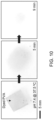
- FIG. 4 A includes optical images of the p-Si needles with fluorescent dyes (DyLightTM 800) on a PVA film, applied to the epidermis (left image), subcutaneous muscle (middle image), and cornea (right image) of mice.
- FIG. 4 B includes images obtained with an IVIS® Spectrum in vivo imaging system (commercially available from PerkinElmer) of the mice interfaced with the p-Si needles for two days following the implementations.
- IVIS® Spectrum in vivo imaging system
- FIGS. 5 A through 5 F represent evaluations in a murine melanoma model in vivo.
- FIG. 5 A includes schematic illustrations of experimental protocols for in vivo studies in a murine melanoma model.
- FIG. 5 B contains a representative optical image of the shaved skin of a mouse post-nanoinjection of DOX.
- FIG. 5 F includes representative optical images highlighting the treated sites after ten days post-injection. The dotted circle denotes skin lesions.
- FIG. 6 includes scanning electron microscope (SEM) images of p-Si needles fabricated on a donor Si wafer with varied geometries.
- the inset schematic image denotes a base diameter (D) and undercut diameter (d) of the p-Si needles.
- FIG. 7 A includes SEM images of a PVA film coated on p-Si needles at varied spin-casting speeds of 150, 300, 500, and 1000 rpm.
- FIG. 7 B includes a plot of measured thicknesses of PVA film and air gap size as a function of spin casting speed ranging from 150 rpm to 1000 rpm.
- FIG. 8 includes SEM images of a donor Si wafer (left image) and a receiver PVA film (right image).
- FIG. 9 includes SEM images of p-Si needles with varied surface porosities of 0%, 30%, 45%, and 60%.
- FIG. 10 includes time-dependent optical images of a PVA film immersed in a solution of PBS (pH 7.4) at 37.5° C.
- FIG. 11 A is an optical image of an experimental setup for the automated peeling of p-Si needles from a donor Si wafer.
- FIG. 11 B represents measurement results of the peeling load obtained from a unit specimen (1 ⁇ 1 cm 2 ).
- FIG. 12 includes FEA results displaying the distribution of principal strains along a p-Si nanoneedle with varied D/d ratios under constant mechanical peeling.
- FIG. 13 is a representative SEM image displaying compressed marks remaining on a surface of a PVA film peeled out of optimal conditions.
- FIG. 14 includes SEM images of p-Si needles immersed in 50 ml of PBS (pH 7.4) at 37.5° C. for 90 days.
- FIG. 15 A includes optical (top panel) and SEM (bottom panel) images of p-Si needles built on a water-soluble poly(lactic-co-glycolic acid) (PLGA) film.
- PLGA water-soluble poly(lactic-co-glycolic acid)
- FIG. 15 B represents corresponding results of MTT assay for the cytotoxicity test of HDF cells. Error bars represent the standard deviation of the three replicates. ****p ⁇ 0.0001 compared to the industrial-grade PVA with p-Si needles using ANOVA.
- FIG. 16 includes top-view (left image) and tilted-view (right image) microscope images of p-Si needles penetrated into a 2.8% (w/v) agarose gel.
- FIG. 17 includes schematic diagrams of amide, urea, and physical bonds of DOX to surfaces of p-Si needles.
- FIG. 18 includes enlarged optical images of the epidermis, subcutaneous muscle, and cornea of mice receiving p-Si needles.
- FIG. 19 includes optical images of a nude mouse wearing on its backside a drug delivery device comprising control p-Si needles on a PDMS film.
- FIG. 21 includes enlarged optical images of the treated sites of mice at ten days post-injection.
- the delivery devices have an array of bioresorbable, miniaturized porous-silicon (p-Si) needles that are attached to and project from a surface of a flexible substrate, generally referred to herein as a flexible film (or simply film) that is also preferably water soluble.
- p-Si porous-silicon
- the devices are capable of being loaded with therapeutic drug cargos that are fluidically coupled to the needles.
- the devices may be used for therapeutic treatments such as but not limited to topical administration of chemotherapeutics into tumor tissues.
- the devices may be applied to living tissue such that a surface of the flexible film comprising the needles contacts and preferable conforms to the living tissue and at least some of, preferably most or all of, the needles penetrate and are inserted into the tissue. Subsequently, the flexible film may be dissolved such that the inserted needles remain embedded in the tissue.
- the flexible film may be dissolved by application of a water-based solution to the film such as but not limited to a saline solution or interstitial fluid.
- a water-based solution such as but not limited to a saline solution or interstitial fluid.
- the film may be dissolved in one minute or less after application of the device to the living tissue to avoid any discomfort or physical restrictions.
- the remaining p-Si needles embedded in the tissue preferably become unobtrusive (nearly unnoticeable) to a wearer without affecting their natural motions due to their miniaturized size. Over time, the needles degrade in the living tissue causing release of the drug cargos that were loaded onto the needles, for example, via gradual hydrolysis due to the action of fluids in the living tissue.
- the film may be used as a temporary substrate during the insertion of p-Si needles into living tissues, and can be subsequently dissolved by the application of an appropriate solution, for example, a water-based solution.
- an appropriate solution for example, a water-based solution.
- p-Si needle refers to a nanoscale or nanoscopic needle that is generally conical or tubular in shape and has geometric features that include minimum tip diameters of less than 1 ⁇ m, base diameters of less than 10 ⁇ m, and lengths of at least 1 ⁇ m.
- p-Si needles disclosed herein preferably have minimum tip diameters of about 50 to 900 nm, base diameters of about 0.9 to 5 ⁇ m, and lengths of about 1 to 100 ⁇ m, and more preferably minimum tip diameters of about 150 nm, base diameters of about 2 to 4 ⁇ m, and lengths of about 10 to 70 ⁇ m.
- the size and thickness of the film may be adjusted depending on the size and shape of the living tissue to which it is intended to be applied in order to provide predetermined flexibility and dissolution times.
- the films are sufficiently flexible to conform to contours of the human anatomy to be treated, in which case these films are preferably relatively thin, for example, less than one millimeter in thickness, and in some cases less than 500 micrometers.
- the films must also be sufficiently thick to provide mechanical strength for handling, which in some cases may require a thickness of at least 200 micrometers.
- the porosity of the p-Si needles relates to the loading capacity and release rate of their drug cargos.
- the porosity of the p-Si needles may range from about 0 to about 80 percent, for example, from about 0 to 60 percent.
- the p-Si needles preferably have an average porosity of about 15 to 60 percent, more preferably between about 25 to 50 percent, for example, about 45 percent.
- the drug delivery devices may have a drug loading capacity of 10 ⁇ g or more per 1 ⁇ 1 cm 2 area of the surface of the film to which the p-Si needles are attached, as a nonlimiting example, 15 ⁇ g to about 50 ⁇ g, per 1 ⁇ 1 cm 2 .
- the drug delivery devices may have a drug release profile that includes a rapid release of their drug cargos within 24 hours post-inoculation until they gradually reach a sustained, predetermined dose that is released and maintained for a period of time.
- the drug delivery devices preferably provide a sustained release dose sufficient to provide a therapeutic effect for the application (e.g., at or above a minimum inhibitory concentration for the drug of interest), and more preferably provide a sustained release dose above a half maximal inhibitory concentration (IC 50 ) value for the application.
- Such sustained releases at the predetermined doses preferably last for a period of time of at least 24 hours, for example, up to about 3 days.
- Such sustained release doses and time periods may be controlled based on the specific application.
- PVA polyvinyl alcohol
- E mechanical modulus
- an array of vertically-ordered p-Si needles may be initially fabricated on a polished monocrystalline Si wafer through sequential steps of photolithographic patterning, dry and wet etchings, and metal-assisted chemical etching (MACE) ( FIG. 1 A , left). This step also forms uniform undercuts and nanopores at the bottom and on the surface of p-Si needles, respectively (inset image).
- MACE metal-assisted chemical etching
- the fabrication process may include immersing a bulk Si wafer (p-type; e.g, about 525 ⁇ m-thick and about 0 to about 100 ⁇ cm) in a solution of buffered oxide etch for one minute to eliminate any native oxide layer thereon.
- a deep reactive-ion etching may be carried out under a radiofrequency (RF) plasma power of, for example, 450 W and a platen power of, for example, 11 W using sulfur hexafluoride (SF 6 ) gas with the flow rate of, for example, 85 sccm to create vertically-ordered Si micropillars at a prescribed aspect ratio.
- RF radiofrequency
- SF 6 sulfur hexafluoride
- the deposition of a (C x F y ) n polymer may be followed to form a partial passivation layer using octafluorocyclobutane (C 4 F 8 ) gas with the flow rate of, for example, 130 sccm under the RF plasma power of, for example, 800 W.
- Additional isotropic dry etching under the plasma power of, for example, 450 W and platen power of, for example, 30 W by SF 6 gas with the flow rate of, for example, 85 sccm may be conducted to create undercuts at the bottom of the Si micropillars.
- the entire specimen may then be treated with an oxygen (O 2 ) plasma (e.g., 20 sccm, 150 W, 50 mtorr, 15 min), followed by cleaning with standard piranha solution (75% of sulfuric acid (H 2 SO 4 ) and 25% of hydrogen peroxides (H 2 O 2 )) to eliminate the remaining passivation layer on the surface of the Si micropillars.
- O 2 oxygen
- the entire specimen may be immersed in a solution of potassium hydroxide (KOH; 15 wt %) at 25° C. to reduce the overall size of the Si micropillars, preferably down to the nanoscale.
- KOH potassium hydroxide
- the overall size of the miniaturized Si pillars may be determined by controlling the molarity of etching solution, temperature, and etching time.
- the next step involves the MACE by immersing the specimen in a mixed solution of 20 mM silver nitrate (AgNO 3 ) and 49% hydrofluoric acid (HF) to form nanopores on the surface.
- the overall surface porosity was controlled by adjusting the etching time.
- FIG. 6 shows representative scanning electron microscopy (SEM) images of p-Si needles fabricated according to this process, exhibiting a minimum tip diameter (d) of 150 nm, a base diameter (D) of 2-4 ⁇ m, and a length (L) of 10-70 ⁇ m.
- the entire structure may be spin-cast with a pre-cured solution of 10 wt % PVA (200-300 ⁇ m-thick) or 5.5 wt % PLGA (lactide:glycolide (50:50)), allowing an air gap to form at the interface due to surface tension ( FIG. 1 A , middle).
- the length of the p-Si needles and the thickness of the film are determined by this air gap that may be adjusted by controlling the spin-casting speed within the range from, for example, 300 rpm to 500 rpm, leading to the consequent air gap size of about 20 ⁇ m and about 50 ⁇ m, respectively ( FIGS. 7 A and 7 B ).
- the spin-casting of the film may be repeated until its total thickness reaches at least about 200-300 ⁇ m to provide sufficient mechanical strength for handling. Subsequently, a thermal annealing may be performed to complete the solidification of the film using a convection oven maintained at, for example, 70° C. for 30 minutes. Finally, the fully cured film may be peeled from the Si wafer at constant peeling rate of, for example, 50 mm/min using an automated peeling apparatus ( FIG. 1 A , right). During this peeling process, mechanical stress can be concentrated predominantly at the bottom undercuts of the p-Si needles to generate cracks allowing for their physical liberation from the Si wafer.
- Various therapeutic drug cargos may be loaded onto the p-Si needles. Covalent and non-covalent conjugation of drug cargos are believed to be capable of providing reliable drug loading onto the surfaces of the p-Si needles and sustained releasing behavior from the surfaces of the p-Si needles.
- the chemotherapy drug doxorubicin (DOX) may be loaded by covalent bonding onto the needles via 3-triethoxysilylpropyl succinic anhydride (TESPSA) as a cross-linker.
- TESPSA 3-triethoxysilylpropyl succinic anhydride
- This exemplary process may begin by washing the as-fabricated p-Si needles on a Si wafer with distilled (DI) water, followed by thorough drying with nitrogen (N 2 ) gas.
- the cleaned p-Si needles may be immersed in a solution of 3-triethoxysilylpropyl succinic anhydride (TESPSA) for two hours to functionalize the surface with amide.
- TESPSA 3-triethoxysilylpropyl succinic anhydride
- ICPTS 3-triethoxysilylpropyl isocyanate
- the p-Si needles may then be rinsed with ethanol and then baked at 120° C. for one hour, followed by immersing in a solution of DOX (e.g., 0.5 mg/ml) for 24 hours at room temperature.
- DOX e.g., 0.5 mg/ml
- Nonlimiting embodiments of the invention will now be described in reference to experimental investigations leading up to the invention. Specifically, comprehensive experimental and computational studies were performed to provide an insight into the structural design and construction of p-Si needles on a water-soluble film over centimeter-scale areas and analyze their fundamental attributes. Demonstrations of p-Si needles for the topical delivery of chemotherapy in a murine melanoma model illustrated the practical utility of this concept.
- FIG. 1 B shows representative optical (left) and enlarged microscope images (right) of p-Si needles that were physically transferred to a thin layer (200-300 ⁇ m-thick) of a PVA film according to the above-describe process.
- the intrinsically thin and flexible property of the film can facilitate intimate contact to the soft, irregular surface of tissues, while the sharpened angular tip of the p-Si needles promotes penetration.
- the overall height, vertical arrangement, and tip morphology of the transferred p-Si needles were consistent across the entire specimen area (3 ⁇ 3 cm 2 ).
- FIG. 8 shows the fractured planes on both the donor Si wafer and the receiver PVA film, suggesting that the cracking occurred uniformly at the bottom undercut of the p-Si needles.
- FIG. 8 shows the fractured planes on both the donor Si wafer and the receiver PVA film, suggesting that the cracking occurred uniformly at the bottom undercut of the p-Si needles.
- FIG. 10 presents a series of optical images at various stages during the dissolution of the PVA film (colored with food dye for visualization) when immersed in 50 ml of phosphate-buffered saline (PBS; pH 7.4) at 37.5° C.
- PBS phosphate-buffered saline
- the results indicate that the peeling load increased rapidly to a maximum that initiated cracking, and then reached a plateau for steady-state crack propagation.
- FIG. 1 D shows finite element analysis (FEA) results, revealing that the principal strain ( ⁇ ) remained localized near the bottom undercut of p-Si needles during the constant peeling.
- FEA finite element analysis
- FIG. 1 E The experimental and FEA results of strain energy release rate (G) for the cracking are summarized in FIG. 1 E .
- the shaded area denotes where the cracking typically occurred with high-fidelity.
- FIG. IF experimentally and theoretically reveals the dependence of G on peeling rate ( ⁇ ) of the PVA film. The results showed a clear power-law relationship of them (i.e., the G increased rapidly at low ⁇ and then gradually reached steady-state) due to the viscoelastic property of the PVA film.
- a rapid peeling ( ⁇ >20 mm/min) of the PVA film provided sufficiently large adhesive strength to peel the p-Si needles away from the Si wafer.
- a slow peeling ( ⁇ 20 mm/min) of the PVA film was unable to hold the p-Si needles, resulting in compressed marks left on the surface ( FIG. 13 ).
- the energy release rate was obtained by assuming that the PVA film was monolithically bonded to the p-Si needles without embedding inside, causing the discrepancy with the experimental results especially at high peeling rates ( ⁇ >100 mm/min).
- FIG. 2 A shows gradual dissolution of a unit array (1 ⁇ 1 cm 2 ) of p-Si needles with a fixed initial base diameter (D 0 ) of 3 ⁇ m when immersed in 50 ml of PBS (pH 7.4) at 37.5° C. for ninety days, while refreshing the solution every ten days to maintain the pH value.
- the magnified SEM images of the p-Si needles at predetermined time intervals are shown in FIG. 14 .
- the dissolution of the p-Si needles occurred via hydrolysis of Si to silicic acid and hydrogen (i.e., Si+4H 2 O ⁇ Si(OH) 4 +2H 2 ), which involves nucleophilic attack at the surface to weaken the interior bonds of Si atoms.
- FIG. 2 B shows measurement results of the gradual diameter reduction (D/D 0 ) of the p-Si needles with varied surface porosities, indicating that the dissolution rate was increased from about 10 nm/day to about 20 nm/day as the surface porosity was increased from 0% to 60%.
- FIGS. 2 C and 2 D compare the dissolution of the p-Si needles in higher pH environment (PBS; pH 10.0) at 37.5° C.
- FIG. 2 E shows a series of snapshot images obtained from molecular dynamics (MD) simulation at different time frames, revealing the dissociation process of a Si atom in water (H 2 O) due to the effect of nucleophile attack by OH ⁇ groups (enlarged).
- FIG. 2 F shows the variation of number of dissociated Si atoms in solutions at acidic (pH 2.2) and basic (pH 10.0) conditions formed by addition of H + and OH ⁇ groups, respectively, compared to a neutral condition (pH 7.0).
- the results show that the number was increased over the simulation time from 0 ns to 12 ns, while the dissolution was accelerated at higher pH due to the increased concentration of OH ⁇ groups. It is also anticipated that the enhanced surface porosity would also affect the reaction (dissolution) rate due to the increased contact area between the Si and liquid solution.
- HDF human dermal fibroblast
- the length of the p-Si needles (20 ⁇ m-long) was tailored for the HDF cells of which the average diameter is about 20-30 ⁇ m.
- the cell viability remained over 99.3% during the entire period (three days) of the assay without substantial difference compared to that of the control bare medical-grade PVA film without the p-Si needles.
- acute toxicity appeared in the industrial-grade PVA film due to the residual ethanol and butanol.
- FIG. 3 C shows representative fluorescence microscopy images (top view) of p-Si needles where the surface was covalently linked with a chemotherapy drug doxorubicin (DOX) using 3-Triethoxysilylpropyl succinic anhydride (TESPSA) as a cross-linker.
- DOX chemotherapy drug
- TESPSA 3-Triethoxysilylpropyl succinic anhydride
- a confocal lens (40 ⁇ ) was focused at the bottom of the p-Si needles, resulting in ring-shaped fluorescence of the DOX.
- FIG. 3 E shows the total cumulative amount of DOX released from p-Si needles with varied surface porosities of 0% to 60% in PBS (pH 7.4) at 37.5° C.
- the corresponding release profiles as a function of time are shown in FIG. 3 F , exhibiting that rapid release of DOX occurred within 24 hours and then gradually reached a plateau at the predefined doses.
- the range of the released doses (18 to 35 ⁇ g) were comparable to those used in studies reported in the literature that used polymeric microneedles.
- FIG. 3 G shows the cumulative release of covalently-linked DOX using different cross-linkers of amide and urea in PBS (pH 7.4), as compared to that of physically-trapped DOX.
- FIG. 3 H presents the corresponding release profiles (%) as a function of time (up to 24 hours), highlighting the longer-lasting release of the covalently-linked DOX than counterparts. For instance, more than 80% of the covalently-linked DOX was released over about 24 hours, which was substantially longer than control specimens with the physically-bonded DOX (about 8 hours) and conventional polymeric microneedles (typically, 15 minutes to 2 hours; shaded area), respectively.
- FIG. 4 A shows representative optical images, pointing out the injection sites where p-Si needles were embedded after the PVA film was completely dissolved with saline solution.
- the size of the p-Si needles was much smaller than that of conventionally-used polymeric microneedles (typically, d>5 ⁇ m, D>300 ⁇ m, and L>600 ⁇ m) and remained nearly unnoticeable on the tissue surface by visual observations ( FIG. 18 ).
- This aspect may help reduce the risk of irritation or discomfort during/after the injection of the p-Si needles.
- the mice exhibited normal behaviors without showing any evidence of discomfort against natural movements for the entire period of observation (about three months).
- FIG. 1 shows representative optical images, pointing out the injection sites where p-Si needles were embedded after the PVA film was completely dissolved with saline solution.
- the size of the p-Si needles was much smaller than that of conventionally-used polymeric microneedles (typically, d>5
- FIG. 19 shows optical images of a control unit array (1 ⁇ 1 cm 2 ) of p-Si needles integrated with a flexible, yet non-water-soluble PDMS film (200 ⁇ m-thick), which was attached on the back of a nude mouse.
- a control unit array (1 ⁇ 1 cm 2 ) of p-Si needles integrated with a flexible, yet non-water-soluble PDMS film (200 ⁇ m-thick), which was attached on the back of a nude mouse.
- no wrinkles were observed on the skin over the PDMS film while other areas of the skin were easily wrinkled according to body movements.
- Tumor relapse after surgical resection that occurs by an outgrowth of residual microtumors remains a significant challenge in current treatments.
- Systemic chemotherapy and radiotherapy are often employed to prevent the recurrence of residual tumors, but these methods lead to toxic side effects and do not provide a long-lasting protection unless frequently repeated.
- Sustained topical delivery of therapeutic drug cargos with precisely controlled doses for a prolonged time, after surgical resection, may reduce the risk of tumor relapse with minimal side effects and improved convenience of patients and healthcare providers.
- mice were subcutaneously inoculated with 1 ⁇ 10 6 B16F10 melanoma cells to mimic a situation where melanoma resection is incomplete and residual cells are present.
- FIG. 5 A This basic procedure for injection of the p-Si needles will be hereinafter referred to as “nanoinjection”.
- a representative photograph in FIG. 5 B highlights the nanoinjection site on the shaved skin of a mouse.
- the mouse receiving the p-Si needles moved freely without any sign of discomfort.
- Two other control groups of mice were intratumorally administered using a medical 28 G insulin syringe with a single dose of PBS (50 ⁇ l) and DOX (50 ⁇ l), representing no-treatment control and a conventional bolus injection, respectively.
- FIG. 5 C shows that, in the mice treated with the nanoinjection of DOX, tumor growth was suppressed over ten days post-inoculation.
- FIG. 5 D shows comparisons of sizes of the tumors at ten days post-inoculation. All of these treatments were well tolerated by the mice with negligible weight loss during the surviving period ( FIG. 5 E ); however, local skin lesions were observed on the mice treated with the syringe injection of DOX ( FIG. 5 F and FIG. 21 ), which is a typical side effect of the drug.
Landscapes
- Health & Medical Sciences (AREA)
- Engineering & Computer Science (AREA)
- Dermatology (AREA)
- Medical Informatics (AREA)
- Anesthesiology (AREA)
- Biomedical Technology (AREA)
- Heart & Thoracic Surgery (AREA)
- Hematology (AREA)
- Life Sciences & Earth Sciences (AREA)
- Animal Behavior & Ethology (AREA)
- General Health & Medical Sciences (AREA)
- Public Health (AREA)
- Veterinary Medicine (AREA)
- Medicinal Preparation (AREA)
Abstract
Description
Claims (16)
Priority Applications (1)
| Application Number | Priority Date | Filing Date | Title |
|---|---|---|---|
| US17/344,362 US11793982B2 (en) | 2020-06-10 | 2021-06-10 | Drug delivery devices and methods of fabrication and use therefor |
Applications Claiming Priority (2)
| Application Number | Priority Date | Filing Date | Title |
|---|---|---|---|
| US202063037127P | 2020-06-10 | 2020-06-10 | |
| US17/344,362 US11793982B2 (en) | 2020-06-10 | 2021-06-10 | Drug delivery devices and methods of fabrication and use therefor |
Publications (2)
| Publication Number | Publication Date |
|---|---|
| US20210386986A1 US20210386986A1 (en) | 2021-12-16 |
| US11793982B2 true US11793982B2 (en) | 2023-10-24 |
Family
ID=78824323
Family Applications (1)
| Application Number | Title | Priority Date | Filing Date |
|---|---|---|---|
| US17/344,362 Active 2042-01-05 US11793982B2 (en) | 2020-06-10 | 2021-06-10 | Drug delivery devices and methods of fabrication and use therefor |
Country Status (1)
| Country | Link |
|---|---|
| US (1) | US11793982B2 (en) |
Citations (1)
| Publication number | Priority date | Publication date | Assignee | Title |
|---|---|---|---|---|
| US20140194379A1 (en) * | 2011-06-03 | 2014-07-10 | University Of Washington Through Its Center For Commercialization | Methods for the production of chitin nanofibers and uses thereof |
-
2021
- 2021-06-10 US US17/344,362 patent/US11793982B2/en active Active
Patent Citations (1)
| Publication number | Priority date | Publication date | Assignee | Title |
|---|---|---|---|---|
| US20140194379A1 (en) * | 2011-06-03 | 2014-07-10 | University Of Washington Through Its Center For Commercialization | Methods for the production of chitin nanofibers and uses thereof |
Non-Patent Citations (6)
| Title |
|---|
| Aykul et al. "Determination of Half-Maximal Inhibitory Concentration Using Biosensor-Based Protein Interaction Analysis", 2016, Anal. Biochem.; 508: 97-103. (Year: 2016). * |
| Chiappini, C. et al., "Biodegradable Silicon Nanoneedles Delivering Nucleic Acids Intracellularly Induce Localized In Vivo Neovascularization," Nat. Mater. 2015, 14, 532-539. |
| Fang, Y. et al., "Texturing Silicon Nanowires for Highly Localized Optical Modulation of Cellular Dynamics," Nano Lett. 2018, 18, 4487-4492. |
| Gopal, S. et al., "Porous Silicon Nanoneedles Modulate Endocytosis to Deliver Biological Payloads," Adv. Mater. 2019, 31, 1-8. |
| Kim, H. et al., "Flexible Elastomer Patch with Vertical Silicon Nanoneedles for Intracellular and Intratissue Nanoinjection of Biomolecules," Sci. Adv. 2018, 4, 1-8. |
| Li, W. et al., "Rapidly Separable Microneedle Patch for the Sustained Release of a Contraceptive," Nat. Biomed. Eng. 2019, 3, 220-230. |
Also Published As
| Publication number | Publication date |
|---|---|
| US20210386986A1 (en) | 2021-12-16 |
Similar Documents
| Publication | Publication Date | Title |
|---|---|---|
| Waghule et al. | Microneedles: A smart approach and increasing potential for transdermal drug delivery system | |
| JP5863824B2 (en) | Manufacturing method of microstructure | |
| Dharadhar et al. | Microneedles for transdermal drug delivery: a systematic review | |
| Gittard et al. | Fabrication of polymer microneedles using a two-photon polymerization and micromolding process | |
| Ita | Dissolving microneedles for transdermal drug delivery: Advances and challenges | |
| KR102064503B1 (en) | Microstructure for Transdermal Absorption and Process for Preparing the Same | |
| CN101808588B (en) | Delivery device and method | |
| TWI675666B (en) | Hyaluronic acid microstructure having excellent dissolving properties | |
| Zhang et al. | An update on biomaterials as microneedle matrixes for biomedical applications | |
| Xie et al. | Enhanced in vitro efficacy for inhibiting hypertrophic scar by bleomycin-loaded dissolving hyaluronic acid microneedles | |
| CN113332588B (en) | Tip drug-loaded dissolvable microneedle patch for oral mucosal administration and preparation method thereof | |
| Liu et al. | Fabrication of rapidly separable microneedles for transdermal delivery of metformin on diabetic rats | |
| Zhang et al. | Dissolving microneedle rollers for rapid transdermal drug delivery | |
| Yang et al. | 3D-printed morphology-customized microneedles: Understanding the correlation between their morphologies and the received qualities | |
| JP2017104490A (en) | Micro structure production method | |
| US11793982B2 (en) | Drug delivery devices and methods of fabrication and use therefor | |
| KR102883442B1 (en) | Composition for microneedle, microneedle array, and transdermal patch comprising the same | |
| JP5222512B2 (en) | Micro needle tip | |
| CN117243886A (en) | A microneedle patch for treating diabetic lower limb ischemic lesions and its application | |
| Shaikh et al. | Microneedle platform for biomedical applications | |
| Borey et al. | A review recent advanced of fabrication techniques and application of micro-needle | |
| Li et al. | A dissolvable microneedle patch based on medical adhesive tape for transdermal drug delivery | |
| Chen et al. | Fabrication of hollow microneedle patch with controllable microstructure for cell therapy | |
| KR20220152660A (en) | Microneedle with drug storage space and manufacturing method thereof | |
| Chaudhri et al. | Out-of-plane, high strength, polymer microneedles for transdermal drug delivery |
Legal Events
| Date | Code | Title | Description |
|---|---|---|---|
| FEPP | Fee payment procedure |
Free format text: ENTITY STATUS SET TO UNDISCOUNTED (ORIGINAL EVENT CODE: BIG.); ENTITY STATUS OF PATENT OWNER: SMALL ENTITY |
|
| AS | Assignment |
Owner name: INDUSTRY-UNIVERSITY COOPERATION FOUNDATION HANYANG UNIVERSITY, KOREA, REPUBLIC OF Free format text: ASSIGNMENT OF ASSIGNORS INTEREST;ASSIGNOR:KIM, DONG RIP;REEL/FRAME:056544/0594 Effective date: 20210319 Owner name: PURDUE RESEARCH FOUNDATION, INDIANA Free format text: ASSIGNMENT OF ASSIGNORS INTEREST;ASSIGNORS:LEE, CHI HWAN;YEO, YOON;REEL/FRAME:056544/0366 Effective date: 20210318 |
|
| FEPP | Fee payment procedure |
Free format text: ENTITY STATUS SET TO SMALL (ORIGINAL EVENT CODE: SMAL); ENTITY STATUS OF PATENT OWNER: SMALL ENTITY |
|
| STPP | Information on status: patent application and granting procedure in general |
Free format text: DOCKETED NEW CASE - READY FOR EXAMINATION |
|
| STPP | Information on status: patent application and granting procedure in general |
Free format text: RESPONSE TO NON-FINAL OFFICE ACTION ENTERED AND FORWARDED TO EXAMINER |
|
| STPP | Information on status: patent application and granting procedure in general |
Free format text: NOTICE OF ALLOWANCE MAILED -- APPLICATION RECEIVED IN OFFICE OF PUBLICATIONS |
|
| STPP | Information on status: patent application and granting procedure in general |
Free format text: PUBLICATIONS -- ISSUE FEE PAYMENT RECEIVED |
|
| STPP | Information on status: patent application and granting procedure in general |
Free format text: PUBLICATIONS -- ISSUE FEE PAYMENT VERIFIED |
|
| STCF | Information on status: patent grant |
Free format text: PATENTED CASE |