US11653636B2 - Method of making a rat model of retinal degeneration and rat model made thereby - Google Patents
Method of making a rat model of retinal degeneration and rat model made thereby Download PDFInfo
- Publication number
- US11653636B2 US11653636B2 US16/686,024 US201916686024A US11653636B2 US 11653636 B2 US11653636 B2 US 11653636B2 US 201916686024 A US201916686024 A US 201916686024A US 11653636 B2 US11653636 B2 US 11653636B2
- Authority
- US
- United States
- Prior art keywords
- rat
- pde6b
- retinal
- retinal degeneration
- genetically modified
- Prior art date
- Legal status (The legal status is an assumption and is not a legal conclusion. Google has not performed a legal analysis and makes no representation as to the accuracy of the status listed.)
- Active, expires
Links
Images


Classifications
-
- A—HUMAN NECESSITIES
- A01—AGRICULTURE; FORESTRY; ANIMAL HUSBANDRY; HUNTING; TRAPPING; FISHING
- A01K—ANIMAL HUSBANDRY; AVICULTURE; APICULTURE; PISCICULTURE; FISHING; REARING OR BREEDING ANIMALS, NOT OTHERWISE PROVIDED FOR; NEW BREEDS OF ANIMALS
- A01K67/00—Rearing or breeding animals, not otherwise provided for; New or modified breeds of animals
- A01K67/027—New or modified breeds of vertebrates
- A01K67/0275—Genetically modified vertebrates, e.g. transgenic
- A01K67/0276—Knock-out vertebrates
-
- A—HUMAN NECESSITIES
- A61—MEDICAL OR VETERINARY SCIENCE; HYGIENE
- A61K—PREPARATIONS FOR MEDICAL, DENTAL OR TOILETRY PURPOSES
- A61K49/00—Preparations for testing in vivo
- A61K49/0004—Screening or testing of compounds for diagnosis of disorders, assessment of conditions, e.g. renal clearance, gastric emptying, testing for diabetes, allergy, rheuma, pancreas functions
- A61K49/0008—Screening agents using (non-human) animal models or transgenic animal models or chimeric hosts, e.g. Alzheimer disease animal model, transgenic model for heart failure
-
- C—CHEMISTRY; METALLURGY
- C12—BIOCHEMISTRY; BEER; SPIRITS; WINE; VINEGAR; MICROBIOLOGY; ENZYMOLOGY; MUTATION OR GENETIC ENGINEERING
- C12N—MICROORGANISMS OR ENZYMES; COMPOSITIONS THEREOF; PROPAGATING, PRESERVING, OR MAINTAINING MICROORGANISMS; MUTATION OR GENETIC ENGINEERING; CULTURE MEDIA
- C12N15/00—Mutation or genetic engineering; DNA or RNA concerning genetic engineering, vectors, e.g. plasmids, or their isolation, preparation or purification; Use of hosts therefor
- C12N15/09—Recombinant DNA-technology
- C12N15/63—Introduction of foreign genetic material using vectors; Vectors; Use of hosts therefor; Regulation of expression
- C12N15/79—Vectors or expression systems specially adapted for eukaryotic hosts
- C12N15/85—Vectors or expression systems specially adapted for eukaryotic hosts for animal cells
- C12N15/8509—Vectors or expression systems specially adapted for eukaryotic hosts for animal cells for producing genetically modified animals, e.g. transgenic
-
- A—HUMAN NECESSITIES
- A01—AGRICULTURE; FORESTRY; ANIMAL HUSBANDRY; HUNTING; TRAPPING; FISHING
- A01K—ANIMAL HUSBANDRY; AVICULTURE; APICULTURE; PISCICULTURE; FISHING; REARING OR BREEDING ANIMALS, NOT OTHERWISE PROVIDED FOR; NEW BREEDS OF ANIMALS
- A01K2217/00—Genetically modified animals
- A01K2217/07—Animals genetically altered by homologous recombination
- A01K2217/075—Animals genetically altered by homologous recombination inducing loss of function, i.e. knock out
-
- A—HUMAN NECESSITIES
- A01—AGRICULTURE; FORESTRY; ANIMAL HUSBANDRY; HUNTING; TRAPPING; FISHING
- A01K—ANIMAL HUSBANDRY; AVICULTURE; APICULTURE; PISCICULTURE; FISHING; REARING OR BREEDING ANIMALS, NOT OTHERWISE PROVIDED FOR; NEW BREEDS OF ANIMALS
- A01K2227/00—Animals characterised by species
- A01K2227/10—Mammal
- A01K2227/105—Murine
-
- A—HUMAN NECESSITIES
- A01—AGRICULTURE; FORESTRY; ANIMAL HUSBANDRY; HUNTING; TRAPPING; FISHING
- A01K—ANIMAL HUSBANDRY; AVICULTURE; APICULTURE; PISCICULTURE; FISHING; REARING OR BREEDING ANIMALS, NOT OTHERWISE PROVIDED FOR; NEW BREEDS OF ANIMALS
- A01K2267/00—Animals characterised by purpose
- A01K2267/03—Animal model, e.g. for test or diseases
- A01K2267/0306—Animal model for genetic diseases
-
- C—CHEMISTRY; METALLURGY
- C12—BIOCHEMISTRY; BEER; SPIRITS; WINE; VINEGAR; MICROBIOLOGY; ENZYMOLOGY; MUTATION OR GENETIC ENGINEERING
- C12N—MICROORGANISMS OR ENZYMES; COMPOSITIONS THEREOF; PROPAGATING, PRESERVING, OR MAINTAINING MICROORGANISMS; MUTATION OR GENETIC ENGINEERING; CULTURE MEDIA
- C12N15/00—Mutation or genetic engineering; DNA or RNA concerning genetic engineering, vectors, e.g. plasmids, or their isolation, preparation or purification; Use of hosts therefor
- C12N15/09—Recombinant DNA-technology
- C12N15/63—Introduction of foreign genetic material using vectors; Vectors; Use of hosts therefor; Regulation of expression
- C12N15/79—Vectors or expression systems specially adapted for eukaryotic hosts
- C12N15/85—Vectors or expression systems specially adapted for eukaryotic hosts for animal cells
- C12N15/8509—Vectors or expression systems specially adapted for eukaryotic hosts for animal cells for producing genetically modified animals, e.g. transgenic
- C12N2015/8527—Vectors or expression systems specially adapted for eukaryotic hosts for animal cells for producing genetically modified animals, e.g. transgenic for producing animal models, e.g. for tests or diseases
- C12N2015/8536—Animal models for genetic diseases
Definitions
- the present invention relates to a Pde6b-deficient animal model of retinal degeneration produced by engineered endonucleases, and a method for producing the same.
- the retina refers to the innermost layer of nerve tissue covering the eye.
- the visual cells convert light information into electrical information, which, in turn, passes through inner retinal layer cells and is delivered, through the optic nerve, to the brain where visual information is recognizable.
- the retina may be divided into thin transparent membranes having different thicknesses depending on locations; and the central part of the retina is subdivided into fovea centralis, parafovea, and perifovea.
- the fovea centralis is clinically referred to as the macula.
- the retina may be histologically composed of ten layers in a superficial to deep direction of the eye.
- the ten constituent layers are, respectively, retinal pigment epithelium, photoreceptor layer, outer limiting membrane, outer nuclear layer, outer plexiform layer, inner nuclear layer, inner plexiform layer, ganglion cell layer, nerve fiber layer, and internal limiting membrane.
- the photoreceptor layer is a light-sensing part and consists of two types of visual cells, that is, cone cells and rod cells.
- the human retina is known to have about 100 million rod cells and about 6 million cone cells.
- the retina has special regions such as optic disk and macula, and visual information is received through the visual stimulus recognition function of the retina.
- retinal damage may lead to severe vision problems. Therefore, various therapeutic methods have been studied to improve symptoms caused by such retinal degeneration and damage, and production of an animal model is indispensable, for example, for identifying effects of improving clinical symptoms in such studies.
- a technique called genome editing may be used to induce deletion or overexpression of specific genes, thereby freely editing genetic information of living organisms.
- the genome editing technique has the advantage that as such a technique is used to alter genetic information of animals including humans, plants, or microorganisms, its application range is dramatically expanding.
- engineered endonucleases are a molecular tool designed to specifically cut only the desired genetic information, and play a key role in the genome editing technique.
- Korean Patent No. 10-1842014 discloses that Cpf1 endonucleases are used to induce deletion of Prkdc gene, thereby producing a transgenic immunodeficient mouse.
- study results, obtained by using the CRISPR-Cas system on genes that may affect eye development have been known (DiCarlo, James E., et al. Translational vision science & technology 6.3 (2017): 13-13).
- the present inventors have made attempts to produce an animal model in which gene knockout is specifically induced through this Engineered endonucleases, and as a result, have produced Pde6b gene knockout rats using the CRISPR-Cpf1.
- the present inventors have found that the rats significantly show findings of retinal degeneration, and thus can be used as an animal model of ocular diseases caused by retinal degeneration, thereby completing the present invention.
- An object of the present invention is to provide an animal model of retinal degeneration and a method for producing the same.
- Another object of the present invention is to provide a method for screening a therapeutic agent for retinal degeneration, using the animal model of retinal degeneration.
- the present invention provides a method for producing an animal model of retinal degeneration, comprising a step of inducing Pde6b gene knockout.
- the step of inducing Pde6b gene knockout may be carried out through the following steps i) to iii):
- the animal may be a mammal other than a human.
- the mammal may be a rodent, and may preferably be a rat.
- the retinal degeneration may be any one or more selected from the group consisting of retinal pigment degeneration, angioid streak, drusen, and macular degeneration.
- the present invention provides a Pde6b-deficient animal model of retinal degeneration.
- the Pde6b gene knockout may be induced by engineered endonucleases.
- the present invention provides a method for screening a prophylactic or therapeutic drug for retinal degeneration, comprising the following steps i) and ii):
- the improvement in symptoms of retinal degeneration in step ii) may be any one or more selected from the group consisting of improved retinal vascular morphology, increased retinal single-layer thickness, increased electroretinogram amplitude, and increased cell number in retinal tissue.
- the retinal degeneration may be any one or more selected from the group consisting of retinal pigment degeneration, angioid streak, drusen, and macular degeneration.
- the present invention provides a Pde6b-deficient animal model of retinal degeneration and a method for producing the same.
- the animal model of retinal degeneration according to the present invention only a specific target gene may be removed using engineered endonucleases and this may be expected to be inherited through germline transmission, so that mutagenesis can be stably achieved.
- FIGS. 1 A to 1 D illustrate a preparation process of CRISPR-Cpf1 for removing Pde6b gene.
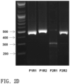
- FIGS. 2 A to 2 D illustrate design of genotyping primers for identifying Pde6b gene knockout, and genotyping results obtained by using the same.
- FIGS. 3 A to 3 C illustrate results obtained by sequencing the Pde6b gene in Pde6b gene knockout rats.
- FIG. 4 illustrates results obtained by identifying, with Western blotting, expression of Pde6b protein in the retina of wild type rats and Pde6b knockout rats.
- FIG. 5 illustrates fundus photographic images for Pde6b-deficient rats.
- FIG. 6 illustrates optical coherence tomographic images for Pde6b-deficient rats.
- FIG. 7 illustrate electroretinogram results for Pde6b-deficient rats.
- FIGS. 8 A to 8 D illustrate results obtained by observing, with histochemical analysis, the eye tissue of Pde6b-deficient rats.
- FIG. 9 illustrate TUNEL staining results for wild-type rats and Pde6b knockout rats at each time point (scale bars: 50 ⁇ m).
- the present invention provides a method for producing an animal model of retinal degeneration, comprising a step of inducing Pde6b gene knockout.
- the present invention provides an animal model of retinal degeneration, produced according to the above method.
- the term “animal model” refers to an animal having a disease that is morphologically very similar to a human disease.
- a diseased animal model is significant due to physiological or genetic similarities between humans and animals.
- diseased animal models for biomedicine provide research materials for various causes, pathogenesis, and diagnosis of diseases; allow for identification of genes related to diseases through studies with the diseased animal models; allow for understanding of interactions between genes; and make it possible to obtain basic data, through actual efficacy and toxicity tests of newly developed drug candidates, for determining whether such drug candidates can be put to practical use.
- the Pde6b gene is mutated using the engineered endonucleases prepared using the methods known in Korean Laid-open Patent Publication No. 10-2017-0137354 and Kim et al. Nature Biotechnology 34.8 (2016): 808. More specifically, it is more preferred that the step of inducing Pde6b gene knockout is carried out through, but not limited to, the following steps i) to iii):
- the technique using engineered endonucleases may be understood, by those skilled in the art, as a technique mainly used for knock-out research tools, through which the function of a gene is eliminated by recognizing and cutting a desired DNA sequence to cause damage, and allowing mutagenesis, which causes changes in the number and type of nucleotide sequence, to occur in the course of repairing the damage.
- the engineered endonucleases is a technique of introducing a mutation into a DNA nucleotide sequence to alter the genetic code itself, and is different from gene knockdown in which RNA interference (RNAi) is used to target RNA.
- RNA interference RNA interference
- miRNA, siRNA, or shRNA used in RNAi technique binds to transcribed mRNA to degrade it or inhibit translation thereof, thereby decreasing a gene expression level. Therefore, the engineered endonucleases is preferred because it can induce a target gene knockout in a more sensitive and effective manner than knocking down the target gene.
- the terms “deletion”, “knock-out”, and “deficiency” all mean abolishing the function of a particular gene and are used interchangeably in the present specification.
- the method for producing an animal model of retinal degeneration of the present invention comprises knocking out a particular gene, such as Pde6b gene, using various Cpf1 orthologs which are class 2 single RNA-guide endonucleases.
- the crRNA and Cpf1 mRNA in step i) may be prepared by methods known in the art.
- the crRNA targets the coding region of exon 1 of the Pde6b gene as illustrated in FIG. 1 A .
- the crRNA may be prepared with reference to the method disclosed in Kim et al. Nature Biotechnology 34.8 (2016): 808.
- the present inventors selected a CRISPR RNA sequence in the rat Pde6b gene that exhibits three or more mismatches and at least one mismatch residing in the 5′ PAM-proximal region.
- Cpf1 ortholog gene derived from Acidaminococcus sp. or Lachnospiraceae bacterium may be used. More specifically, Cpf1 ortholog gene derived from Acidaminococcus sp. BV3L6 (AsCpf1) or Lachnospiraceae bacterium N D2006 (LbCpf1) may be used.
- the Cpf1 is prepared as mRNA for injection into rats, and the Cpf1 mRNA may be prepared by linearizing a vector containing a nucleotide sequence encoding Cpf1, and then subjecting the resultant to an in vitro transcription process.
- step ii) of injecting the crRNA and the Cpf1 mRNA into an embryo of an animal model may be carried out, for example, by a simple method in which the crRNA and the Cpf1 mRNA are mixed and the mixture is injected into the cytoplasm of a fertilized egg.
- the crRNA and the Cpf1 mRNA may be injected into a rat embryo by methods commonly known in the art. For example, microinjection, electroporation, liposome-mediated transfer method, and retrovirus-mediated transfer method may be applied therefor.
- the embryo for injection may be obtained by the following process.
- superovulation is induced in female SD rats (5 to 6 weeks old) by injection with 30 to 40 IU pregnant mare serum gonadotropin (PMSG; Sigma-Aldrich Corp., St. Louis, Mo., USA) and 40 to 100 IU human chorionic gonadotropin (hCG; Daesung Microbiological Labs Co., Ltd., Gyeounggi, Republic of Korea) at 48- to 50-hour intervals.
- PMSG pregnant mare serum gonadotropin
- hCG human chorionic gonadotropin
- the superovulated female rats are crossed with SD stud male rats, and 1-cell stage embryos are collected from the oviducts 6 to 14 hours after fertilization.
- a microinjector for example, 100 ng/ml of crRNA and 50 ng/ml of Cpf1 mRNA may be co-injected into the cytoplasm of pronuclear stage embryos.
- step iii) is a step of transplanting the embryo, into which the crRNA and the Cpf1 mRNA have been injected, into the oviduct of a pseudo-pregnant foster mother to produce a Pde6b-deficient animal, in which, for example, the embryo after injection with the crRNA and the Cpf1 mRNA may be incubated for 2 to 24 hours in a 37° C. incubator and transplanted, at the 1-cell or 2-cell stage, into the foster mother.
- the incubation is performed according to known methods.
- a suitable medium may be developed for incubation of animal cells, in particular, mammalian cells, or any available medium may be used which may be prepared in the laboratory with appropriate ingredients required for animal cell growth, such as anabolic carbon, nitrogen, and/or micronutrients.
- the medium may be any basal medium suitable for animal embryo growth.
- basal medium generally used for incubation include M2, M16, m-RECM, Modification of medium SOM (KSOM), Human Tubal Fluid (HTF), Minimum Essential Medium (MMEM), Dulbecco modified Eagle Medium (DMEM), Roswell Park Memorial Institute Medium (RPMI), and Keratinocyte Serum Free Medium (K-SFM).
- KSOM Modification of medium SOM
- HEF Human Tubal Fluid
- MMEM Minimum Essential Medium
- DMEM Dulbecco modified Eagle Medium
- RPMI Roswell Park Memorial Institute Medium
- K-SFM Keratinocyte Serum Free Medium
- any medium used in the art may be used without limitation.
- the medium may be selected from the group consisting of M2 (SIGMA), M16 (SIGMA), m-RECM (COSMOBIO), KSOM (COSMOBIO), HTF (Irvine Scientific), ⁇ -MEM medium (GIBCO), K-SFM medium, DMEM medium (Welgene), MCDB 131 medium (Welgene), IMEM medium (GIBCO), DMEM/F12 medium, PCM medium, M199/F12 (mixture) (GIBCO), and MSC expansion medium (Chemicon).
- anabolic sources of carbon, nitrogen, and micronutrients of which non-limiting examples include serum sources, growth factors, amino acids, antibiotics, vitamins, reducing agents, and/or sugar sources.
- the method of the present invention may further comprise step iv) of screening for Pde6b gene mutations in the newborn animals produced in step iii), to select founder animals (F 0 ) with mutations that are expected to lack the function of the Pde6b gene.
- Identification of the mutations may be accomplished by genotyping, sequencing, or the like, and may also be accomplished by other methods known in the art.
- Mutations caused by engineered endonucleases may be detected by a variety of methods including, for example, mismatch-sensitive T7 endonuclease I (T7E1) or Surveyor nuclease assay, RFLP, capillary electrophoresis of fluorescence-labeled PCR products, dideoxy sequencing, and deep sequencing.
- T7E1 mismatch-sensitive T7 endonuclease I
- RFLP capillary electrophoresis of fluorescence-labeled PCR products
- dideoxy sequencing dideoxy sequencing
- deep sequencing deep sequencing.
- founder (F0) rats with a targeted mutation were screened by using PCR primers (rPde6b_F1: SEQ ID NO: 8 and rPde6b_R1: SEQ ID NO: 10). Mutant alleles were identified by Sanger sequencing of cloned PCR products (generated by using a T-Blunt PCR Cloning Kit [SolGent Co., Ltd., Daejeon, Republic of Korea]).
- the method of the present invention may further comprise step v) of crossing mutant animal founders (F 0 ) with Pde6b deficient alleles with wild-type animals, to obtain heterozygous Pde6b knockout animals (F 1 ); and step vi) of crossing the heterozygous Pde6b knockout male animals with the heterozygous Pde6b knockout female animals, to obtain homozygous Pde6b knockout animals (F 2 ).
- a primer pair (rPde6b_F2: SEQ ID NO: 9; rPde6b_R5: SEQ ID NO: 34) that produced a short PCR product (173 bp in wild-type rats) was used to detect deletion with a frameshift mutation in the knockout allele.
- FIGS. 3 B and 3 C Western blot analysis identified deficiency of Pde6b protein in the homozygous mutant retina, indicating successful generation of Pde6b-deficient rats with Cpf1-mediated gene targeting ( FIG. 4 ).
- the animal model of retinal degeneration is preferably a mammal other than a human. More specifically, the animal model is more preferably, but is not limited to, a rodent.
- the rodent is most preferably a rat or a mouse.
- the rat model has greater translational relevance than previously established mouse models due to its similarities to humans in many biological aspects. Therefore, in the present invention, rats are used to generate an animal model of retinal degeneration.
- the retinal degeneration is preferably any one or more selected from, but not limited to, the group consisting of retinal pigment degeneration, angioid streak, drusen, and macular degeneration; and any disease that may be caused by damage to the retina or developmental inhibition thereof may be included without limitation so long as the disease is within the scope recognized by those skilled in the art.
- the Pde6b knockout rats produced by the method of the present invention may be used to screen potential drugs for the prevention or treatment of various diseases associated with retinal degeneration. Screening for useful drugs includes, for example, a step of administering a candidate drug to rats over a range of doses, and a step of analyzing, at various time points, effects of the drug in the disease to be evaluated.
- the present invention provides a method for screening a prophylactic or therapeutic drug for retinal degeneration, comprising the following steps i) and ii):
- the prophylactic or therapeutic drug refers to a substance exerting all actions that alleviate the patient's health condition, such as preventing or delaying onset of disease and improving symptoms.
- the candidate drug includes candidate drugs in all routes of administration.
- drugs for parenteral administration such as intraocular administration and for oral administration may be screened.
- the optimal dosage may be appropriately determined by making a comprehensive decision based on conditions such as nature of drug, kind of subject to be administered, and the subject's age or body weight. Conditions such as time of administration and number of administrations may be appropriately set depending on nature of drug, purposes of test and evaluation, and the like. In addition, these methods may be used to quantify effects of drugs, so that the effects of drugs are quantitatively determined.
- an untreated control which is a Pde6b-deficient animal model not treated with a candidate drug
- an untreated control which is a Pde6b-deficient animal model not treated with a candidate drug
- Normal control rats in which Pde6b deficiency is not induced may also be used as a control.
- the progress of symptoms of retinal degeneration in step ii) may be identified by comparison with an untreated control and/or a normal control in terms of, but not limited to, retinal vascular morphology, retinal single-layer thickness, electroretinogram amplitude, and cell number in retinal tissue.
- the improvement in symptoms of retinal degeneration in step ii) is preferably, but is not limited to, any one or more selected from the group consisting of improved retinal vascular morphology, increased retinal single-layer thickness, increased electroretinogram amplitude, and increased cell number in retinal tissue.
- the retinal degeneration is preferably any one or more selected from, but not limited to, the group consisting of retinal pigment degeneration, angioid streak, drusen, and macular degeneration; and any disease that may be caused by damage to the retina or developmental inhibition thereof may be included without limitation so long as the disease is within the scope recognized by those skilled in the art.
- the Pde6b knockout animal model of the present invention may be useful for studying effects of Pde6b gene mutation.
- Embodiments of the Pde6b knockout animal model and progeny thereof of the present invention will also have various uses depending on additional transgenes that can be expressed and/or knockout constructs they may contain.
- the engineered endonucleases may be those that can perform in rats using AsCpf1.
- CRISPR-Cpf1 the engineered endonucleases
- two CRISPR RNAs Pde6b-CR1: SEQ ID NO: 1; and Pde6b-CR2: SEQ ID NO: 2 were first selected using Benching software (https://benchling.com/; in the public domain) ( FIG. 1 B ).
- T7-AsPde6b_CR1 SEQ ID NO: 3; and T7-AsPde6b_CR2: SEQ ID NO: 4
- DNA oligomers FIG. 1 D : SEQ ID NOS: 5 to 7
- Cpf1 mRNA was generated using pcDNA3.1-hAsCpf1 (Addgene #69982), and CRISPR RNA was generated in the same manner as previously reported (Kim, Yongsub, et al. Nature Biotechnology 34.8 (2016): 808).
- Genotyping primers were selected for the production of Pde6b knockout rats. According to a known experimental method, primer sequences were designed to produce a PCR product of heteroduplex DNA (Zhu, Xiaoxiao, et al. Scientific Reports 4 (2014): 6420). As illustrated in FIG. 2 A , for primer sequences, two forward primers (rPde6b_F1: SEQ ID NO: 8; rPde6b_F2: SEQ ID NO: 9) and three reverse primers (rPde6b_R1: SEQ ID NO: 10; rPde6b_R2: SEQ ID NO: 11, rPde6b_R5: SEQ ID NO: 34) were produced ( FIG.
- FIG. 2 A data not shown for rPde6b_R5
- FIG. 2 B data not shown for rPde6b_R5
- touchdown PCR was performed according to the PCR conditions as summarized in FIG. 2 C .
- the PCR product of heteroduplex DNA was separated by electrophoresis, and a variant identification method was performed thereon (data not shown for rPde6b_R5).
- rPde6b_F1 and rPde6b_R1 primer pair was used for the screening of F 0 generation.
- rPde6b_F2 SEQ ID NO: 9
- rPde6b_R5 SEQ ID NO: 34 (GACGCTCTCTTGCATGTCCT) were used.
- Pde6b-deficient rat embryos were produced by microinjection and used to produce mutant rats via foster mothers.
- Sprague Dawley (SD) rats were purchased from OrientBio as embryo donors and foster mothers. The purchased rats were acclimatized to a breeding environment, and then the 5- to 6-week-old female rats were intraperitoneally injected with 40 IU pregnant mare serum gonadotropin (PMSG, Sigma). Then, superovulation was induced by injecting 40 IU human chorionic gonadotropin (hCG, Sigma) at 48-hour intervals. The superovulation-induced female rats were crossed with SD stud male rats, and fertilized eggs were obtained from the oviducts.
- PMSG pregnant mare serum gonadotropin
- hCG human chorionic gonadotropin
- Example ⁇ 1-1> When the fertilized egg (embryo) obtained was at the pronuclear stage, 100 ng/ml of crRNA and 50 ng/ml of Cpf1 mRNA prepared in Example ⁇ 1-1> were microinjected into the cytoplasm of the embryo. The microinjected embryo was caused to migrate into the oviduct of a pseudo-pregnant foster mother, so that the embryo was induced to implant in the uterus.
- FIG. 3 A illustrates results of PAGE-based genotyping assay which identifies founder rats originating from embryos at the pronuclear stage into which Cpf1 mRNA and its cognate crRNA targeting the rat Pde6b locus have been intracytoplasmically injected.
- FIG. 3 A it was identified that Pde6b mutant founders of #26 and #33 are obtained, and it was identified, through sequencing thereof, that each Pde6b mutant founder has an out-of-frame mutation ( FIG. 3 B ).
- the target sequence is underlined and PAM is boxed. “-” represents a deleted nucleotide.
- FIG. 3 C illustrates representative PCR genotyping results that determine wild type, heterozygous mutant and homozygous mutant progeny of F0 #33.
- the respective mutant rats were crossed with wild-type rats to produce F1 rats. Among these, the mutant rats obtained were sequenced again to identify that the allele of the mutant identified in the founder was germline transmitted.
- mutant rats of founders with desired knockout alleles were crossed with wild-type SD rats, and the F1 heterozygotes were screened and sequenced. After breeding heterozygous knockout rats by crossing with wild-type SD rats, homozygous Pde6b knockout rats were generated by crossing male and female heterozygotes. For routine PCR genotyping, a primer pair that produced a short PCR product (173 bp in the wild-type mouse) was used to detect deletion with a frameshift mutation in the knockout allele.
- PVDF polyvinylidene fluoride membrane
- Example ⁇ 2-2> fundus photography was performed at 1 day and 8 weeks of postnatal age.
- the eyes of Pde6b KO rats were anesthetized with a topical anesthetic, and pupillary dilatation was induced with eye drops containing 5 mg/ml of tropicamide and 5 mg/ml of phenylephrine HCl.
- An ophthalmic artificial tear ointment was used to prevent drying of the cornea during fundus photography.
- Retinal photographs were taken with the Micron IV fundus camera (Phoenix Research Laboratories, Pleasanton, Calif., USA).
- optical coherence tomography (Phoenix Research Labs) was performed at 1 day, 3 weeks, and 8 weeks of postnatal age.
- the retinal single-layer was scanned six times in a repeated manner, and the resulting values were averaged to acquire an image.
- the acquired images were output in tagged image file (.tif) format, and retinal thickness and retinal pigment epithelium, outer nuclear layer, outer plexiform layer, inner nuclear layer, inner plexiform layer, and the like were compared.
- As a normal control for one male and one female normal rats, optical coherence tomography was performed in the same manner at 1 day, 3 weeks, and 8 weeks of postnatal age.
- the three-week-old Pde6b KO rats clearly show a decrease in overall retinal thickness and loss of the photoreceptor layer and the outer nuclear layer.
- the eight-week-old Pde6b KO rats showed the findings that along with the outer nuclear layer and the outer plexiform layer, the retinal pigment epithelium adjacent thereto is also lost.
- Example ⁇ 2-2> using two male and two female Pde6b KO rats produced in Example ⁇ 2-2> as an experimental group, electroretinograms were performed on 1-day-old, 3-week-old, and 8-week-old rats.
- Each rat in the experimental group was dark-adapted for 12 hours or longer before the electroretinograms, and prepared for electroretinogram measurement under a dark environment with illuminance ( ⁇ ) of less than 600 nm.
- ⁇ illuminance
- each rat in the experimental group was anesthetized by intraperitoneal injection of Zoletil® (125 mg/ml of Tiletamine and 125 mg/ml of Zolazepam, Virbac, France) at a dose of 0.01 ml.
- the pupils were dilated by the application of Midrin®-P eye drop (5 mg/ml of phenylephrine hydrochloride and 5 mg/ml of tropicamide, Santen, Japan).
- Alcaine® (0.5% proparacaine hydrochloride, Alcon Laboratories Inc.) was applied for ocular anesthesia.
- the anesthetized rat was stably fixed on a support for experimental animals so that its position was maintained for highly-reproducible electrophysiological tests.
- Electroretinograms were performed using the Ganzfeld ERG system (Phoenix Research Labs) with light stimulus and electrical signal measurement. Electroretinogram signals were measured using the right eye of the experimental rat, and the electrical signals were sequentially measured while increasing the intensity of white light. Light exposure time was set to 10 msec, and the electric signal was recorded using an average value of 10 measurements at each light intensity. Corneal electrodes made of pure gold were placed around the cornea. Reference electrodes were placed in the center of the scalp; and ground leads were placed in the skin at the base of the tail. In addition, in order to identify the function of each of the retina's rod cells and cone cells, dark- and light-adapted a-wave and b-wave responses were checked. As a normal control, for one male and one female normal rats, electroretinograms were performed in the same manner at 1 day, 3 weeks, and 8 weeks of postnatal age.
- Pde6b-deficient rats produced in the present invention can be used as an animal model of retinal degeneration
- eye tissues were fixed to prepare samples and histochemical observations were performed thereon.
- Example ⁇ 2-2> histochemical pathological findings were identified on 1-day-old, 3-week-old, and 8-week-old rats.
- the experimental rats were anesthetized by intraperitoneal injection with 0.01 ml of Zoletil® (125 mg/ml of Tiletamine and 125 mg/ml of Zolazepam, Virbac, France). Then, the eyes were extracted and fixed in a 4% paraformaldehyde solution (pH 7.4).
- the fixed eye was dissected around the optic nerve, embedded in paraffin, cut into 4 ⁇ m thicknesses, and subjected to H & E staining (hematoxylin-eosin staining) to prepare histochemical samples. Histopathological findings were identified by comparing the prepared tissue samples with those of a normal control. As the normal control, for one male and one female normal rats, histopathological findings were identified in the same manner at 1 day, 3 weeks, and 8 weeks of postnatal age.
- FIGS. 8 A to 8 D it was identified that histochemical changes in the retina are observed over time in the PED6B KO rats.
- Photoreceptor apoptosis was determined by using the terminal deoxynucleotide transferase nick-end labeling (TUNEL) assay with the DeadEnd Fluorometric TUNEL System (Promega, Madison, Wis., USA).
- TUNEL terminal deoxynucleotide transferase nick-end labeling
- sections were deparaffinized, rehydrated, treated with Proteinase K, reacted with TdT/nucleotide mix (containing fluorescein-12-dUTP), and counterstained with DAPI-blue. All samples were examined on the Zeiss LSM 780 confocal microscopy system (Carl Zeiss Meditec AG, Jena, Germany).
- the TUNEL-positive nuclei within a section of the superior and inferior retina 500 to 750 ⁇ m from the optic disc were compared between Pde6b knockout rats (two males, two females) and age-matched wild-type rats (one male, one female) at each time point.
- the retinal tissue sections of the Pde6b knockout rats showed TUNEL-positive findings which are not seen in the wild-type rats (TUNEL-positive cells show yellow-green).
- Apoptosis began 2 weeks of postnatal age, reached its peak at 3 weeks of postnatal age, and then decreased. Signals were specific in the outer nuclear layer and little or no signal was observed in the inner nuclear layer.
- the wild-type rats showed very little signal in the inner nuclear layer at 1 to 10 weeks of postnatal age.
Landscapes
- Life Sciences & Earth Sciences (AREA)
- Health & Medical Sciences (AREA)
- Engineering & Computer Science (AREA)
- Zoology (AREA)
- Genetics & Genomics (AREA)
- Biotechnology (AREA)
- General Health & Medical Sciences (AREA)
- Veterinary Medicine (AREA)
- Biomedical Technology (AREA)
- Environmental Sciences (AREA)
- Animal Behavior & Ethology (AREA)
- Organic Chemistry (AREA)
- Bioinformatics & Cheminformatics (AREA)
- Chemical & Material Sciences (AREA)
- General Engineering & Computer Science (AREA)
- Wood Science & Technology (AREA)
- Biodiversity & Conservation Biology (AREA)
- Animal Husbandry (AREA)
- Molecular Biology (AREA)
- Microbiology (AREA)
- Plant Pathology (AREA)
- Biophysics (AREA)
- Biochemistry (AREA)
- Physics & Mathematics (AREA)
- Diabetes (AREA)
- Endocrinology (AREA)
- Gastroenterology & Hepatology (AREA)
- Pathology (AREA)
- Rheumatology (AREA)
- Toxicology (AREA)
- Urology & Nephrology (AREA)
- Epidemiology (AREA)
- Public Health (AREA)
- Investigating Or Analysing Biological Materials (AREA)
Abstract
Description
Claims (10)
Priority Applications (1)
| Application Number | Priority Date | Filing Date | Title |
|---|---|---|---|
| US16/686,024 US11653636B2 (en) | 2019-11-15 | 2019-11-15 | Method of making a rat model of retinal degeneration and rat model made thereby |
Applications Claiming Priority (1)
| Application Number | Priority Date | Filing Date | Title |
|---|---|---|---|
| US16/686,024 US11653636B2 (en) | 2019-11-15 | 2019-11-15 | Method of making a rat model of retinal degeneration and rat model made thereby |
Publications (2)
| Publication Number | Publication Date |
|---|---|
| US20210144976A1 US20210144976A1 (en) | 2021-05-20 |
| US11653636B2 true US11653636B2 (en) | 2023-05-23 |
Family
ID=75908403
Family Applications (1)
| Application Number | Title | Priority Date | Filing Date |
|---|---|---|---|
| US16/686,024 Active 2041-10-06 US11653636B2 (en) | 2019-11-15 | 2019-11-15 | Method of making a rat model of retinal degeneration and rat model made thereby |
Country Status (1)
| Country | Link |
|---|---|
| US (1) | US11653636B2 (en) |
Families Citing this family (1)
| Publication number | Priority date | Publication date | Assignee | Title |
|---|---|---|---|---|
| CN114561408B (en) * | 2022-01-24 | 2023-04-21 | 四川省医学科学院·四川省人民医院 | Construction method and application of retinal pigment degeneration disease model |
Citations (7)
| Publication number | Priority date | Publication date | Assignee | Title |
|---|---|---|---|---|
| US6225291B1 (en) * | 1997-04-21 | 2001-05-01 | University Of Florida | Rod opsin mRNA-specific ribozyme compositions and methods for the treatment of retinal diseases |
| US7595430B2 (en) * | 2002-10-30 | 2009-09-29 | University Of Kentucky Research Foundation | Methods and animal model for analyzing age-related macular degeneration |
| KR20100121710A (en) | 2009-04-30 | 2010-11-18 | 한국과학기술원 | Method for prevention and treatment of retinal degeneration comprising activity regulation and administration of pten |
| US20150079047A1 (en) * | 2012-04-16 | 2015-03-19 | The Regents Of The University Of California | Ocular therapeutics using embryonic stem cell microvesicles |
| KR20170137354A (en) | 2016-06-03 | 2017-12-13 | 울산대학교 산학협력단 | A knockout Animal Model by Cpf1-mediated gene targeting and the Preparation Method thereof |
| US20190203207A1 (en) * | 2016-05-20 | 2019-07-04 | The Trustees Of Columbia University In The City Of New York | Anabolic Enhancers for Ameliorating Neurodegeneration |
| US20210115505A1 (en) | 2018-05-18 | 2021-04-22 | The Asan Foundation | Method for detecting mycoplasma using mitochondrial dna as internal control sample |
-
2019
- 2019-11-15 US US16/686,024 patent/US11653636B2/en active Active
Patent Citations (8)
| Publication number | Priority date | Publication date | Assignee | Title |
|---|---|---|---|---|
| US6225291B1 (en) * | 1997-04-21 | 2001-05-01 | University Of Florida | Rod opsin mRNA-specific ribozyme compositions and methods for the treatment of retinal diseases |
| US7595430B2 (en) * | 2002-10-30 | 2009-09-29 | University Of Kentucky Research Foundation | Methods and animal model for analyzing age-related macular degeneration |
| KR20100121710A (en) | 2009-04-30 | 2010-11-18 | 한국과학기술원 | Method for prevention and treatment of retinal degeneration comprising activity regulation and administration of pten |
| US20150079047A1 (en) * | 2012-04-16 | 2015-03-19 | The Regents Of The University Of California | Ocular therapeutics using embryonic stem cell microvesicles |
| US20190203207A1 (en) * | 2016-05-20 | 2019-07-04 | The Trustees Of Columbia University In The City Of New York | Anabolic Enhancers for Ameliorating Neurodegeneration |
| KR20170137354A (en) | 2016-06-03 | 2017-12-13 | 울산대학교 산학협력단 | A knockout Animal Model by Cpf1-mediated gene targeting and the Preparation Method thereof |
| US20210115505A1 (en) | 2018-05-18 | 2021-04-22 | The Asan Foundation | Method for detecting mycoplasma using mitochondrial dna as internal control sample |
| US11434527B2 (en) | 2018-05-18 | 2022-09-06 | The Asan Foundation | Method for detecting mycoplasma using mitochondrial DNA as internal control sample |
Non-Patent Citations (8)
| Title |
|---|
| Carter-Dawson (Invest. Ophthalmol. Visual Sci., 1978, p. 489-498). * |
| DiCarlo et al., "CRISPR-Cas Genome Surgery in Ophthalmology," Translational Vision Science & Technology (2017) 6(3): Article 13, 13 pgs. |
| Kim et al., "Generation of knockout mice by Cpf1-mediated gene targeting," Nat Biotechnol. Aug. 2016;34(8):808-10. |
| Muradov et al., "Atypical retinal degeneration 3 in mice is caused by defective PDE6B pre-mRNA splicing," Vision Res. (2012) 57: 1-8. |
| Tsang (Human Mutation, 2007, vol. 28, vol. 28, No. 3, p. 243-254). * |
| Yeo (ARVO Annual Meeting, Jul. 2019). * |
| Yeo (Ophthalmol. Vis. Sci. Apr. 4, 2019, vol. 60, No. 5, p. 1519-1526). * |
| Zhu et al., "An efficient genotyping method for genome-modified animals and human cells generated with CRISPR/Cas9 system," Sci Rep. (2014) 4: 6420. |
Also Published As
| Publication number | Publication date |
|---|---|
| US20210144976A1 (en) | 2021-05-20 |
Similar Documents
| Publication | Publication Date | Title |
|---|---|---|
| Mazzoni et al. | Retinal ganglion cells survive and maintain normal dendritic morphology in a mouse model of inherited photoreceptor degeneration | |
| Pook et al. | Rescue of the Friedreich's ataxia knockout mouse by human YAC transgenesis | |
| JP2002504821A (en) | Transgenic fish with tissue-specific expression | |
| Horie et al. | Investigation of Oxtr-expressing neurons projecting to nucleus accumbens using Oxtr-ires-Cre knock-in prairie voles (Microtus ochrogaster) | |
| CN109476716A (en) | Methods of treating mitochondrial disorders | |
| Pristerà et al. | Impact of N-tau on adult hippocampal neurogenesis, anxiety, and memory | |
| Qian et al. | Stage specific requirement of platelet-derived growth factor receptor-α in embryonic development | |
| JPH08504104A (en) | Genetically engineered mice containing alterations in the gene encoding the retinoic acid receptor protein | |
| Zaffran et al. | Ectopic expression of Hoxb1 induces cardiac and craniofacial malformations | |
| Fogerty et al. | 174delG mutation in mouse MFRP causes photoreceptor degeneration and RPE atrophy | |
| KR102177174B1 (en) | A retinal degenerated animal model by PDE6B gene deletion and the preparation method thereof | |
| US11653636B2 (en) | Method of making a rat model of retinal degeneration and rat model made thereby | |
| KR102634464B1 (en) | Method for producing animal model with retinal degeneration | |
| KR102922515B1 (en) | An animal model for the retinal degeneration having IMPDH1 gene mutation and method for preparing the same | |
| CN108396036B (en) | A transgenic mouse model overexpressing COX5A and its construction method and application | |
| US7642399B2 (en) | Mouse with deficiency of glutamate transporter GLAST function | |
| CN118421703B (en) | Method for constructing non-human mammal model with spontaneous emotion abnormal fluctuation and application thereof | |
| CN121249799B (en) | Methods for constructing Dhx16 mutant mouse models and their applications in disease research | |
| US20140033333A1 (en) | Transgenic pig for mutant gucy2d as cone dystrophy model | |
| Ran et al. | A mouse model of a novel missense mutation (Gly83Arg) in a Chinese kindred manifesting vitreous amyloidosis only | |
| KR20250116237A (en) | An animal model for the retinal degeneration having PDE6B gene mutation and method for preparing the same | |
| 김보라 | Development of a novel humanized Knockin mouse of retinitis pigmentosa with IMPDH1 mutation | |
| CN108315350A (en) | It is overexpressed COX5A/ low expression BDNF transgenic mouse models and its construction method and application | |
| Mays | An Experimental Toolkit for Analysis of Single Monoaminergic Axons in the Mouse Brain | |
| CN118592398A (en) | A method for constructing an animal model of Menkes disease and its application |
Legal Events
| Date | Code | Title | Description |
|---|---|---|---|
| FEPP | Fee payment procedure |
Free format text: ENTITY STATUS SET TO UNDISCOUNTED (ORIGINAL EVENT CODE: BIG.); ENTITY STATUS OF PATENT OWNER: SMALL ENTITY |
|
| FEPP | Fee payment procedure |
Free format text: ENTITY STATUS SET TO SMALL (ORIGINAL EVENT CODE: SMAL); ENTITY STATUS OF PATENT OWNER: SMALL ENTITY |
|
| AS | Assignment |
Owner name: UNIVERSITY OF ULSAN FOUNDATION FOR INDUSTRY COOPERATION, KOREA, REPUBLIC OF Free format text: ASSIGNMENT OF ASSIGNORS INTEREST;ASSIGNORS:BAEK, IN JEOUNG;SUNG, YOUNG HOON;REEL/FRAME:051262/0653 Effective date: 20191104 Owner name: THE ASAN FOUNDATION, KOREA, REPUBLIC OF Free format text: ASSIGNMENT OF ASSIGNORS INTEREST;ASSIGNOR:LEE, JOO YONG;REEL/FRAME:051262/0671 Effective date: 20191102 |
|
| STPP | Information on status: patent application and granting procedure in general |
Free format text: DOCKETED NEW CASE - READY FOR EXAMINATION |
|
| STPP | Information on status: patent application and granting procedure in general |
Free format text: NON FINAL ACTION MAILED |
|
| STPP | Information on status: patent application and granting procedure in general |
Free format text: RESPONSE TO NON-FINAL OFFICE ACTION ENTERED AND FORWARDED TO EXAMINER |
|
| STCF | Information on status: patent grant |
Free format text: PATENTED CASE |