US11648559B2 - Devices and methods for separating circulating tumor cells from biological samples - Google Patents
Devices and methods for separating circulating tumor cells from biological samples Download PDFInfo
- Publication number
- US11648559B2 US11648559B2 US16/636,517 US201816636517A US11648559B2 US 11648559 B2 US11648559 B2 US 11648559B2 US 201816636517 A US201816636517 A US 201816636517A US 11648559 B2 US11648559 B2 US 11648559B2
- Authority
- US
- United States
- Prior art keywords
- cells
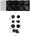
- stage
- cell
- sample
- biological sample
- Prior art date
- Legal status (The legal status is an assumption and is not a legal conclusion. Google has not performed a legal analysis and makes no representation as to the accuracy of the status listed.)
- Active, expires
Links
Images


Classifications
-
- G—PHYSICS
- G01—MEASURING; TESTING
- G01N—INVESTIGATING OR ANALYSING MATERIALS BY DETERMINING THEIR CHEMICAL OR PHYSICAL PROPERTIES
- G01N1/00—Sampling; Preparing specimens for investigation
- G01N1/28—Preparing specimens for investigation including physical details of (bio-)chemical methods covered elsewhere, e.g. G01N33/50, C12Q
- G01N1/40—Concentrating samples
- G01N1/4077—Concentrating samples by other techniques involving separation of suspended solids
-
- B—PERFORMING OPERATIONS; TRANSPORTING
- B01—PHYSICAL OR CHEMICAL PROCESSES OR APPARATUS IN GENERAL
- B01L—CHEMICAL OR PHYSICAL LABORATORY APPARATUS FOR GENERAL USE
- B01L3/00—Containers or dishes for laboratory use, e.g. laboratory glassware; Droppers
- B01L3/50—Containers for the purpose of retaining a material to be analysed, e.g. test tubes
- B01L3/502—Containers for the purpose of retaining a material to be analysed, e.g. test tubes with fluid transport, e.g. in multi-compartment structures
- B01L3/5027—Containers for the purpose of retaining a material to be analysed, e.g. test tubes with fluid transport, e.g. in multi-compartment structures by integrated microfluidic structures, i.e. dimensions of channels and chambers are such that surface tension forces are important, e.g. lab-on-a-chip
- B01L3/502761—Containers for the purpose of retaining a material to be analysed, e.g. test tubes with fluid transport, e.g. in multi-compartment structures by integrated microfluidic structures, i.e. dimensions of channels and chambers are such that surface tension forces are important, e.g. lab-on-a-chip specially adapted for handling suspended solids or molecules independently from the bulk fluid flow, e.g. for trapping or sorting beads or physically stretching molecules
-
- B—PERFORMING OPERATIONS; TRANSPORTING
- B01—PHYSICAL OR CHEMICAL PROCESSES OR APPARATUS IN GENERAL
- B01L—CHEMICAL OR PHYSICAL LABORATORY APPARATUS FOR GENERAL USE
- B01L3/00—Containers or dishes for laboratory use, e.g. laboratory glassware; Droppers
- B01L3/50—Containers for the purpose of retaining a material to be analysed, e.g. test tubes
- B01L3/502—Containers for the purpose of retaining a material to be analysed, e.g. test tubes with fluid transport, e.g. in multi-compartment structures
- B01L3/5027—Containers for the purpose of retaining a material to be analysed, e.g. test tubes with fluid transport, e.g. in multi-compartment structures by integrated microfluidic structures, i.e. dimensions of channels and chambers are such that surface tension forces are important, e.g. lab-on-a-chip
- B01L3/502769—Containers for the purpose of retaining a material to be analysed, e.g. test tubes with fluid transport, e.g. in multi-compartment structures by integrated microfluidic structures, i.e. dimensions of channels and chambers are such that surface tension forces are important, e.g. lab-on-a-chip characterised by multiphase flow arrangements
- B01L3/502776—Containers for the purpose of retaining a material to be analysed, e.g. test tubes with fluid transport, e.g. in multi-compartment structures by integrated microfluidic structures, i.e. dimensions of channels and chambers are such that surface tension forces are important, e.g. lab-on-a-chip characterised by multiphase flow arrangements specially adapted for focusing or laminating flows
-
- B—PERFORMING OPERATIONS; TRANSPORTING
- B01—PHYSICAL OR CHEMICAL PROCESSES OR APPARATUS IN GENERAL
- B01L—CHEMICAL OR PHYSICAL LABORATORY APPARATUS FOR GENERAL USE
- B01L2200/00—Solutions for specific problems relating to chemical or physical laboratory apparatus
- B01L2200/06—Fluid handling related problems
- B01L2200/0647—Handling flowable solids, e.g. microscopic beads, cells, particles
- B01L2200/0652—Sorting or classification of particles or molecules
-
- B—PERFORMING OPERATIONS; TRANSPORTING
- B01—PHYSICAL OR CHEMICAL PROCESSES OR APPARATUS IN GENERAL
- B01L—CHEMICAL OR PHYSICAL LABORATORY APPARATUS FOR GENERAL USE
- B01L2200/00—Solutions for specific problems relating to chemical or physical laboratory apparatus
- B01L2200/06—Fluid handling related problems
- B01L2200/0647—Handling flowable solids, e.g. microscopic beads, cells, particles
- B01L2200/0668—Trapping microscopic beads
-
- B—PERFORMING OPERATIONS; TRANSPORTING
- B01—PHYSICAL OR CHEMICAL PROCESSES OR APPARATUS IN GENERAL
- B01L—CHEMICAL OR PHYSICAL LABORATORY APPARATUS FOR GENERAL USE
- B01L2300/00—Additional constructional details
- B01L2300/06—Auxiliary integrated devices, integrated components
- B01L2300/0681—Filter
-
- B—PERFORMING OPERATIONS; TRANSPORTING
- B01—PHYSICAL OR CHEMICAL PROCESSES OR APPARATUS IN GENERAL
- B01L—CHEMICAL OR PHYSICAL LABORATORY APPARATUS FOR GENERAL USE
- B01L2300/00—Additional constructional details
- B01L2300/08—Geometry, shape and general structure
- B01L2300/0861—Configuration of multiple channels and/or chambers in a single devices
- B01L2300/0864—Configuration of multiple channels and/or chambers in a single devices comprising only one inlet and multiple receiving wells, e.g. for separation, splitting
-
- B—PERFORMING OPERATIONS; TRANSPORTING
- B01—PHYSICAL OR CHEMICAL PROCESSES OR APPARATUS IN GENERAL
- B01L—CHEMICAL OR PHYSICAL LABORATORY APPARATUS FOR GENERAL USE
- B01L2300/00—Additional constructional details
- B01L2300/08—Geometry, shape and general structure
- B01L2300/0861—Configuration of multiple channels and/or chambers in a single devices
- B01L2300/0883—Serpentine channels
-
- B—PERFORMING OPERATIONS; TRANSPORTING
- B01—PHYSICAL OR CHEMICAL PROCESSES OR APPARATUS IN GENERAL
- B01L—CHEMICAL OR PHYSICAL LABORATORY APPARATUS FOR GENERAL USE
- B01L2400/00—Moving or stopping fluids
- B01L2400/04—Moving fluids with specific forces or mechanical means
- B01L2400/0403—Moving fluids with specific forces or mechanical means specific forces
- B01L2400/043—Moving fluids with specific forces or mechanical means specific forces magnetic forces
Definitions
- the present disclosure generally relates to microfluidics and uses thereof.
- Circulating tumor cells are cancer cells that are detached from primary solid tumors and carried through the vasculature to potentially seed distant site metastases in vital organs, representing the main cause of death in cancer patients.
- Molecular assessments of CTCs not only could benefit basic cancer research, but also might eventually lead to a more effective cancer treatment.
- one major limitation has been the limited availability of viable CTCs for investigations, due in part to the small patient blood volumes that are allowable for research, which usually yielded less than 100 CTCs from 1 mL of whole blood.
- Label-based CTC separation technologies were developed to selectively enrich a subset of CTCs from blood, primarily through the use of specific biological markers including epithelial cell adhesion molecule (EpCAM). These antigen-based labels were a rate-limiting factor in effective CTC separation, as the inherent heterogeneity of CTCs render these technologies ineffective for general use.
- EpCAM epithelial cell adhesion molecule
- EMT Epithelial-to-Mesenchymal Transitions
- most label-based technologies do not conveniently enable comprehensive molecular analysis of separated CTCs because they are either dead or immobilized to a surface.
- microfluidic devices and methods of using microfluidic devices are provided that overcome one or more of the aforementioned deficiencies.
- the devices and methods can combine filters, sheathing separation, and flow focusing to provide for high throughput cell separation without the need for labeling the circulating tumor cells.
- Methods are provided for enriching, separating, or isolating unlabeled rare cells such as circulating tumor cells from a sample, e.g. a biological sample such as whole blood.
- a biological sample such as whole blood.
- the biological sample is or includes whole blood.
- the biological sample includes about 50 to about 250 circulating tumor cells per milliliter of the biological sample.
- the circulating tumor cells can include those selected from the group consisting of a primary cancer cell, a lung cancer cell, a prostate cancer cell, a breast cancer cell, a pancreatic cancer cell, and a combination thereof.
- FIG. 1 A is a schematic illustration of traditional and frequently used label-based magnetophoresis for CTC separation, in which rare cells were targeted via specific biomarkers such as epithelial cell adhesion molecule (EpCAM) through functionalized magnetic particles in order to pull these cells through magnetic force towards magnetic field maxima in a continuous-flow manner.
- FIG. 1 B is a schematic illustration of a label-free ferrohydrodynamic cell separation (FCS) for CTCs.
- FCS label-free ferrohydrodynamic cell separation
- RBC-lysed blood and biocompatible ferrofluids colloidal suspensions of magnetic nanoparticles
- FIG. 1 C is a top-view of the FCS device with labels of inlets, debris filters and outlets. A total of 6 outlets were fabricated in order to account for the broad size distributions of cells. The arrow indicates the direction of magnetic field during device operation. Dimensions of the FCS device and magnet are described below.
- FIGS. 2 A- 2 D show the results of optimization of FCS devices with their device geometry shown in FIG. 1 C for high-throughput, high-recovery and biocompatible CTC separation.
- a 3D analytical model considering magnetic buoyancy force, hydrodynamic drag force, laminar flow profiles and cancer/blood cell physical properties was developed to guide the optimization. The validity of the model was confirmed by comparing its simulated trajectories with experimental ones.
- Numerical optimization of deflection distance Y c and separation distance ⁇ Y (corresponding to recovery rate and purity) at the end of the FCS device was conducted with parameters including: ( FIG. 2 A ) & ( FIG. 2 B ) magnetic field gradient, and ( FIG. 2 C ) & ( FIG.
- FIG. 2 D ferrofluid concentration at flow rates between 1.2 and 7.2 mL h ⁇ 1 .
- Ferrofluid concentration was fixed at 0.26% (v/v) for ( FIG. 2 A ) & ( FIG. 2 B ).
- Magnetic field was fixed at 443 mT and its gradient was fixed at 56.2 T m ⁇ 1 for ( FIG. 2 C ) & ( FIG. 2 D ).
- FIGS. 3 A- 3 D are micrographs of spiked cancer cells of cell culture lines and undiluted WBCs separation process in a FCS device.
- 1 ⁇ 10 5 cells H1299 lung cancer cells were spiked into 1 mL of undiluted WBCs to increase the cancer cell concentration so that their fluorescent signals were visible.
- the cell mixture was processed at the flow rate of 6 mL h ⁇ 1 .
- a ferrofluid with its concentration of 0.26% (v/v) was used; magnetic field was fixed at 443 mT and its gradient was fixed at 56.2 T m ⁇ 1 .
- FIG. 3 A In absence of magnetic fields, cell mixtures exited the channel through outlets 1 and 2.
- Scale bar 200 ⁇ m.
- FIG. 3 B When magnetic fields were present, larger H1299 lung cancer cells and some WBCs were deflected and exited through outlets 5 and 6 (collection outlets), while smaller WBCs exited through lower outlets (outlets 1-4, waste outlets). Scale bar: 200 ⁇ m.
- FIG. 3 C Fluorescence image of spiked H1299 lung cancer cell streams during the separation process when magnetic fields were present. H1299 cells were stained by CellTracker Green. Scale bar: 200 ⁇ m.
- FIG. 3 D Zoomed-in bright-field images of outlets 1-6 when the magnetic fields were present. Scale bars: 100 ⁇ m.
- FIGS. 4 A- 4 D show verification of FCS devices for high-throughput and high-recovery spiked cancer cells separation.
- FIG. 4 A Recovery rates of spiked H1299 lung cancer cells from undiluted WBCs at flow rates from 1.2 mL h ⁇ 1 to 6.0 mL h ⁇ 1 . Approximately 100 HI299 cancer cells were spiked into 1 mL of undiluted WBCs. Recovery rates decreased from 98.6 ⁇ 5.0% to 92.3 ⁇ 3.6% when flow rate increased from 1.2 mL h ⁇ 1 to 6.0 mL h ⁇ 1 .
- FIG. 4 A Recovery rates of spiked H1299 lung cancer cells from undiluted WBCs at flow rates from 1.2 mL h ⁇ 1 to 6.0 mL h ⁇ 1 .
- FIG. 4 B A series of spike-in separation experiments in which a certain number (50, 100, 200, 500, 1000, and 2000) of H1299 cells were spiked into 1 mL of undiluted WBCs to simulate clinically relevant CTC concentration at the flow rate of 6.0 mL h ⁇ 1 .
- FIG. 4 C The removal rate of WBCs increased with the flow rate. 99.92 ⁇ 2.2% of WBCs were removed at a flow rate of 6 mL h ⁇ 1 .
- FIG. 4 D Recovery rates and purity of separated cancer cells (about 100 cell/mL) for different cancer cell lines at the flow rate of 6 mL h ⁇ 1 . Recovery rates of 92.3 ⁇ 3.6%, 88.3 ⁇ 5.5%, 93.7 ⁇ 5.5%, 95.3 ⁇ 6.0%, 94.7 ⁇ 4.0%, and 93.0 ⁇ 5.3% were achieved for H1299 (lung cancer), A549 (lung cancer), H3122 (lung cancer), PC-3 (prostate cancer), MCF-7 (breast cancer), and HCC1806 (breast cancer) cell lines, respectively.
- the corresponding purities of cancer cells of each cell line are 11.1 ⁇ 1.2% (H1299), 10.1 ⁇ 1.7% (A549), 12.1 ⁇ 2.1% (H3122), 12.8 ⁇ 1.6% (PC-3), 11.9 ⁇ 1.8 (MCF-7), and 12.2 ⁇ 1.6% (HCC1806), respectively.
- H1299 10.1 ⁇ 1.7%
- A549 12.1 ⁇ 2.1%
- PC-3 12.8 ⁇ 1.6%
- MCF-7 11.9 ⁇ 1.8
- HCC1806 12.2 ⁇ 1.6%
- FIGS. 5 A- 5 D show the effect of FCS on cancer cell viability, proliferation and biomarker expressions.
- FIG. 5 B Representative images of Live/Dead cell staining for before (top) and after (bottom) separation groups. Calcein AM (green, live cells) and EhD-1 (red, dead cells) channels were merged. Scale bars: 100 ⁇ m.
- FIG. 5 A Short-term cell viability comparison before and after FCS process using a Live/Dead assay. Cell viabilities of H1299 lung cancer cells before and after separation process were determined to be 98.9 ⁇ 0.9% and 96.3 ⁇ 0.9%, respectively. Error bars indicate standard deviation (s.d
- FIG. 5 C Bright field images of cultured H1299 cells collected after separation from day 1 to day 5. A Live/Dead staining of the cultured cells on day 5 showed excellent cell viability. Scale bars; 50 ⁇ m.
- FIG. 5 D Comparison of expressions of two key biomarkers (epithelial cell adhesion molecule-EpCAM and cytokeratin-CK) on HCC1806 breast cancer cells before (top) and after (bottom) separation. They showed qualitatively similar EpCAM and CK fluorescence. Scale bars; 20 ⁇ m
- CTCs Cells were identified as CTCs if the staining pattern is CK+/CD45 ⁇ or EpCAM+/CD45 ⁇ or CK+/EpCAM+/CD45 ⁇ , WBC were identified as CK ⁇ /EpCAM ⁇ /CD45+. Scale bars: 10 ⁇ m.
- FIG. 7 A is a schematic illustration of traditional label-based magnetophoresis for CTC separation, in which rare cells were targeted via specific biomarkers such as epithelial cell adhesion molecule (EpCAM) through functionalized magnetic particles in order to pull these cells through magnetic force towards magnetic field maxima in a continuous-flow manner.
- FIG. 7 B is a schematic illustration of label-free ferrohydrodynamic cell separation (FCS) for CTCs. Large rare cells in ferrofluid experience large magnetic buoyance force and will be pushed towards magnetic field minimum
- FIG. 7 C is a schematic of integrated ferrohydrodynamic cell separation (iFCS) for CTC separation. In iFCS, RBC-lysed blood and biocompatible ferrofluid were processed in a single straight channel.
- FIG. 7 D is a photograph of a prototype iFCS device filled with dye and a permanent magnet.
- FIG. 7 E is a top-view of the iFCS device with labels of inlets, outlets.
- FIG. 7 F is a simulation of magnetic flux density and particle trajectories of magnetic particles and non-magnetic particles in channel (L ⁇ W ⁇ H, 57.80 mm ⁇ 0.90 mm ⁇ 0.15 mm).
- FIGS. 8 A- 8 I show optimization of iFCS device via simulation and beads calibration.
- a 3D model was developed to simulate particle trajectories and validated by comparing simulation and experimental results.
- Numerical optimization of final position Y, separation distance ⁇ Y, and the expected range (standard deviation) at the end of the channel was conducted with different parameters: ( FIG. 8 A ) & ( FIG. 8 D ) & ( FIG. 8 G ) throughput, ( FIG. 8 B ) & ( FIG. 8 E ) & ( FIG. 8 H ) ferrofluid concentration, and ( FIG. 8 C ) & ( FIG. 8 F ) & ( FIG. 8 I ) magnetic field gradient. Ferrofluid concentration was fixed at 0.292% for ( FIG.
- FIG. 8 A & ( FIG. 8 D ) & ( FIG. 8 G ) & ( FIG. 8 C ) & ( FIG. 8 F ) & ( FIG. 8 I )
- magnetic field gradient was fixed at 132 T m ⁇ 1 for ( FIG. 8 A ) & ( FIG. 8 D ) & ( FIG. 8 G )
- throughput was fixed at 100 ⁇ L min ⁇ 1 for ( FIG. 8 B ) & ( FIG. 8 E ) & ( FIG. 8 H ) & ( FIG. 8 C ) & ( FIG. 8 F ) & ( FIG. 8 I ).
- FIG. 8 D ) & ( FIG. 8 E ) & ( FIG. 8 F ) were simulated particles positions and corresponding standard deviations.
- FIG. 8 G & ( FIG. 8 H ) & ( FIG. 8 I ) were normalized particle distributions in experiment.
- FIGS. 9 A- 9 F demonstrate iFCS applied to multiple beads separation.
- FIG. 9 A average separation distance between non-magnetic beads and magnetic beads. Non-magnetic beads (20.3 ⁇ m, 8.0 ⁇ m and 5.7 ⁇ m) and magnetic beads (11.8 ⁇ m) were spiked into 0.292% ferrofluid at throughput between 10 and 350 ⁇ L min ⁇ 1 . the magnetic field gradient was fixed at 132 T m ⁇ 1 .
- FIG. 9 B simulated and experimental separation distance between 8.0 ⁇ m non-magnetic beads and 11.8 ⁇ m magnetic beads at low ferrofluid concentration between 0.01% and 0.1%, and flow rate between 10 and 210 ⁇ L min ⁇ 1 .
- FIG. 9 E When magnetic fields were present, non-magnetic beads (20.3 and 8.0 ⁇ m) will be collected from outlet 1. Most of the magnetic beads (11.8 ⁇ m) will be collected at outlet 3, and the rest can be identified in outlet 2. Scale bar: 200 ⁇ m.
- FIG. 9 F Image of collected beads from 3 outlets. The red fluorescent signal comes from 8.0 ⁇ m non-magnetic beads and the yellow fluorescent signal comes from 11.8 ⁇ m magnetic beads. Scale bar: 200 ⁇ m.
- FIGS. 10 A- 10 C demonstrate size of cancer cell lines and labelling results of WBCs.
- FIG. 10 A the size distribution of different cancer cell lines—Prostate (PC-3), Breast (MCF-7, MDA-MB-231, HCC1806) and Lung (H1299, H3122, H69, DMS79), and white blood cells from healthy donor.
- FIG. 10 C Percentage of magnetic content in labelled WBCs. The volume fraction of magnetic content in dynabeads is 11.5%, the magnetic content for labelled WBC was calculated based on volume fraction.
- FIGS. 11 A- 11 F show iFCS applied to cancer cells separation. Certain number of PC-3 prostate cancer cells and 1 ⁇ 10 6 WBCs were spiked into 1 mL ferrofluid with concentration of 0.049% (v/v). The cell mixture was processed at the flow rate of 100 ⁇ L mind; the magnetic field gradient was fixed at 132 T ( FIG. 11 A ) Bright field and fluorescent images of 1 ⁇ 10 5 PC-3 cancer cells and WBCs separation process. Green signal comes from PC-3 cancer cells and red signal comes from WBCs Scale bar: 500 ⁇ m. ( FIG. 11 B ) Collected PC-3 from outlet 1. ( FIG. 11 C ) Normalized size distributions of spiked and collected cancer cells from outlet 1 for PC-3, MCF-7 and MDA-MB-231, ( FIG.
- FIG. 11 D Magnetic beads enumeration on collected WBCs from outlet 1 with different concentration of ferrofluid.
- FIG. 11 E Recovery rate and purity for different cancer cell lines ( ⁇ 100 cells per mL). Recovery rate of 97.9 ⁇ 1.0%, 97.6 ⁇ 1.0%, 98.8 ⁇ 1.4%, 99.4 ⁇ 0.6%, 98.7 ⁇ 0.6%, 95.0 ⁇ 1.2%, 95.9 ⁇ 1.3% and 99.7 ⁇ 0.6% were achieved for MCF-7, MDA-MB-231, HCC1806, H1299, H3122, DMS79, H69 and PC-3 cell lines, respectively.
- FIGS. 12 A- 12 C FCSv2 device design and operating principle.
- FIG. 12 A Top-view of the FCSv2 device with labels of inlets, debris filters and outlets. The arrow indicates the direction of magnetic field during device operation.
- the device integrates 3 stages into one single device for biomarker- and size-independent OTC enrichment. Stage one filters out large cell debris.
- Stage 2 depletes the unbound magnetic beads and WBCs bound with 3 beads into waste outlet 1 ( FIG. 12 B ii).
- Stage 3 continuously deflects unlabeled CTCs into collection outlet, while at the same time focuses WBCs bound with ⁇ 1 bead into waste outlet 2 ( FIG. 12 B iii).
- FIG. 12 A Top-view of the FCSv2 device with labels of inlets, debris filters and outlets. The arrow indicates the direction of magnetic field during device operation.
- the device integrates 3 stages into one single device for biomarker- and size-independent OTC enrichment. Stage one filters out large cell debris.
- microfluidic device is sandwiched between the device holders, which consist of top and bottom magnet arrays repelling each other and are secured with screws and nuts.
- the thickness of channel is 250 ⁇ m.
- the width of stage 2 is 1600 ⁇ m and width of stage 3 is 1200 ⁇ m.
- FIG. 13 The microfluidic device is sandwiched between the two device holders, which consist of top and bottom magnet arrays repelling each other and is secured with screws and nuts.
- the center of the stage 3's microchannel in FIG. 1 is aligned exactly with the center of the magnet arrays.
- FIG. 14 The simulation result shows the magnetic field is stronger in the center of the stage 3 region in FIG. 1 .
- the labeled white blood cells and free magnetic beads are focused in the center of the channel and are collected through the waste outlet.
- any unlabeled cells including circulating tumor cells are deflected into two sides of the channel and are collected through collection outlets.
- Whole blood is labeled and lysed by RBC lysing buffer before it is processed with the FCSv2.
- Leukocyte-specific biotinylated antibodies anti-CD45, anti-CD66b and anti-CD16
- magnetic Dynabeads are added to the blood sample for incubation.
- FIG. 15 A Recovery rate of separated cancer cells for different cancer cell lines at the flow rate of 12 mL/h. recovery rates of 98.46 ⁇ 0.50%, 99.68 ⁇ 0.46%, 99.05 ⁇ 0.75%, 99.35 ⁇ 0.46%, 99.40 ⁇ 0.85%, 99.13 ⁇ 0.49%, 99.11 ⁇ 1.25%, and 99.11 ⁇ 0.74% are achieved for HCC1806 (breast cancer), HCC70 (breast cancer), MCF7 (breast cancer), MDA-MB-231 (breast cancer), H1299 (non-small cell lung cancer), H3122 (non-small cell lung cancer), DMS79 (small cell lung cancer), and H69 (small cell lung cancer) cell lines, respectively.
- FIG. 15 B A series of spike-in separation experiments in which a certain number (20, 50, 100, and 200) of HCC70 breast cancer cells are spiked into 1 mL of labeled white blood cells. An average recovery rate is 99.08%.
- FIGS. 16 A- 16 B Bright field and fluorescence image of spiked MDA-MB-231 breast cancer cells (labeled with CellTracker Green) during the separation process confirmed the cancer cell trajectories at the end of stages 2 and 3 in FIGS. 12 A- 12 C . Scale bars: 500 ⁇ m.
- FIG. 17 Measured magnetic field and its gradient of the center of magnet's surface vs. distance between the magnet's surface and the microfluidic channel wall.
- FIGS. 18 A- 18 B Schematic and relevant dimensions of a FCS device,
- FIG. 18 A Top-view of the FCS device and relevant dimensions.
- FIG. 18 B Cross-section view of the FCS device.
- the red arrow indicates the direction of permanent magnet's magnetization.
- FIG. 19 A Cell trajectory simulation of H1299 lung cancer cell (16.9 ⁇ m) and WBCs (11.1 ⁇ m) in a FCS device.
- FIG. 19 B Zoomed-in view of cell trajectories at the end of FCS device. Blue and red trajectories indicate H1299 and WBCs, respectively.
- Flow rate of cell inlet (Inlet A) was fixed at 6 mL h ⁇ 1
- ferrofluid concentration was fixed at 0.26% (v/v)
- magnetic field was fixed at 443 mT and its gradient was fixed at 56.2 T m ⁇ 1 for this simulation.
- FIG. 20 FCS device calibration with H1299 cells (replaced with beads of similar size, 15.6 ⁇ m) and WBCs (11.1 ⁇ m).
- the left-bottom number in each figure indicates the associated flow rate of cell inlet A (mL h ⁇ 1 ).
- Flow rate of cell inlet (Inlet A) was fixed at 6 mL h ⁇ 1
- ferrofluid concentration was fixed at 0.26% (v/v)
- magnetic field was fixed at 443 mT and its gradient was fixed at 56.2 T m ⁇ 1 for this calibration.
- 1 ⁇ 10 4 polystyrene microparticles were mixed with 1 mL of undiluted WBCs. Scale bars: 500 ⁇ m.
- FIG. 21 Comparison of cell trajectories from calibration experiments and simulations of H1299 cells and WBCs at the end of FCS device. Blue lines are the boundary of the simulated H1299 cell trajectory, and red lines are the boundary of the simulated WBC trajectory.
- the simulated trajectories considered the initial width of microparticle and cell streams at the entry of the channel, therefore had an up and low bound of trajectories. Overall the simulated trajectories matched well with the experimental calibration trajectories, therefore could be used for subsequent FCS device optimization.
- Flow rate of cell inlet (Inlet A) was fixed at 6 mL h ⁇ 1 , ferrofluid concentration was fixed at 0.26% (v/v), and magnetic field was fixed at 443 mT and its gradient was fixed at 56.2 T m ⁇ 1 for simulation and calibration.
- Scale bar 500 ⁇ m.
- FIGS. 22 A- 22 F Characterization of custom-made ferrofluids.
- FIG. 22 A Magnetization of the as-synthesized ferrofluid. Solid red lines are the fitting of the experimental date to the Langevin function. Saturation magnetization of this ferrofluid was 0.96 kA m ⁇ 1 , corresponding to a 0.26 volume fraction or concentration.
- FIG. 22 B Rheological plots of the ferrofluid and blood. The viscosity of ferrofluid was measured to be 2.92 mPa ⁇ s.
- FIG. 22 D Size distribution of maghemite nanoparticles was measured by dynamic light scattering (DLS). Hydrodynamic diameter was 40.77 ⁇ 12.71 nm.
- FIG. 22 E Zeta potential of ferrofluid was measured to be ⁇ 27.2 ⁇ 11.4 mV, indicating a negative surface charge on the particles.
- FIG. 22 F A transmission electron microscopy (TEM) image of the maghemite nanoparticles. Scale bar: 20 nm.
- FIGS. 23 A- 23 D FIGS. 23 A- 23 D
- FIG. 23 A Cell viability of H1299 lung cancer cells in different concentrations of ferrofluids was evaluated by a MTT assay. Cell viability was 80.8 ⁇ 2.4% after 12-h incubation with a 0.26% (v/v) concentration ferrofluid.
- FIG. 23 B Colloidal stability of biocompatible ferrofluids. The maghemite nanoparticles remained colloidally stable for at least 10 months in solution and there was no visible precipitation over time.
- FIG. 23 C Blood cells, mixed with a commercial water-based ferrofluid, showed an irreversible flocculation.
- FIG. 23 D No flocculation or aggregation of blood cells was found within the biocompatible ferrofluid. Scale bars: 50 ⁇ m.
- FIG. 24 An image of a FCS device and an attached collection chamber.
- the FCS device was connected to a serpentine collection chamber that was used to accurately enumerate cancer cells for the FCS calibration using cultured cancer cell lines.
- the depth of collection chamber is 50 ⁇ m.
- the size of the glass slide is 75 ⁇ 50 mm. Blue dye was used to visualize the microchannel.
- FIG. 25 Representative micrographs of lung cancer H1299 cells and WBCs after a separation of spiked cancer cells in a FCS device at a throughput of 6 mL h ⁇ 1 . ⁇ 100 CellTracker Green stained H1299 cells were spiked into 1 mL of undiluted WBCs.
- A H1299 lung cancer cells and WBCs were identified in the outlet (outlet 6) reservoir. Scale bars: 100 ⁇ m.
- B and (C) H1299 lung cancer cells and WBCs were identified in the serpentine collection chamber. Scale bars: 50 ⁇ m.
- FIG. 26 Cell type distribution of cells collected from outlets 1-6 after a separation of ⁇ 100 H1299 cells spiked into 1 mL of undiluted WBCs using a FCS device at a throughput of 6 mL h ⁇ 1 .
- FIG. 27 A The average cell size of six cancer cell lines and WBCs measured by a cell counter.
- FIG. 27 B Size distribution of cancer cells and WBCs.
- FIG. 28 Representative images of CTC identification from patient A and B, with their blood processed by FCS devices. Black arrows indicate the identified CTCs. Scale bars: 50 ⁇ m.
- microfluidic devices and methods of using microfluidic devices are provided for separating and/or enriching circulating tumor cells in a biological sample such as whole blood.
- the methods are capable of high throughputs with high levels of retention and separation of the circulating tumor cells.
- ratios, concentrations, amounts, and other numerical data can be expressed herein in a range format. It is to be understood that such a range format is used for convenience and brevity, and thus, should be interpreted in a flexible manner to include not only the numerical values explicitly recited as the limits of the range, but also to include all the individual numerical values or sub-ranges encompassed within that range as if each numerical value and sub-range is explicitly recited.
- a numerical range of “about 0.1% to about 5%” should be interpreted to include not only the explicitly recited values of about 0.1% to about 5%, but also include individual values (e.g., 1%, 2%, 3%, and 4%) and the sub-ranges (e.g., 0.5%, 1.1%, 2.2%, 3.3%, and 4.4%) within the indicated range.
- the stated range includes one or both of the limits, ranges excluding either or both of those included limits are also included in the disclosure, e.g. the phrase “x to y” includes the range from ‘x’ to ‘y’ as well as the range greater than ‘x’ and less than ‘y’.
- the range can also be expressed as an upper limit, e.g.
- ‘about x, y, z, or less’ and should be interpreted to include the specific ranges of ‘about x’, ‘about y’, and ‘about z’ as well as the ranges of ‘less than x’, less than y’, and ‘less than z’.
- the phrase ‘about x, y, z, or greater’ should be interpreted to include the specific ranges of ‘about x’, ‘about y’, and ‘about z’ as well as the ranges of ‘greater than x’, greater than y’, and ‘greater than z’.
- the term “about” can include traditional rounding according to significant figures of the numerical value.
- the phrase “about ‘x’ to ‘y’”, where ‘x’ and ‘y’ are numerical values includes “about ‘x’ to about ‘y’”.
- a biocompatible substance or fluid, as described herein, indicates that the substance or fluid does not adversely affect the short-term viability or long-term proliferation of a target cell within a particular time range.
- microfluidic devices and methods of using microfluidic devices are provided for high throughput sorting, separation, and/or enrichment of circulating tumor cells and other unlabeled rare cells in a biological sample such as blood.
- the devices are single-stage devices, while in some aspects the devices have multiple stages.
- a multistage a multi-stage microfluidic device for enriching circulating tumor cells in a biological sample.
- the device can include at least three stages, although there may be more in some applications. Therefore, the terms first, second, third and so-on, when used to describe the stages, should not be considered limiting on the total number of stages but is used for simplicity to describe the relative ordering of the stages. Additional stages, not explicitly described, may in some aspects appear before the first stage.
- the first stage in the multi-stage microfluidic device includes a first end, a second end, a first microfluidic channel fluidly connecting the first end and the second end, a first fluid inlet fluidly connected to the first microfluidic channel at the first end, and one or more filters along a length of the first microfluidic channel.
- the first fluid inlet can be used for introducing, pumping, and/or injecting a biological sample into the device. Where there is a stage prior to the first stage, the inlet may be fluidly connected to the previous stage in such a way as to receive a sample (a biological sample) from the previous stage.
- the one or more filters can filter large cell debris from the biological sample.
- a second stage in the multi-stage microfluidic device includes a third end, a fourth end, a second microfluidic channel fluidly connecting the third end and the fourth end.
- the second stage can be used to separate at least a portion of the white blood cells that are associated with the magnetic beads by flowing the filtered sample through a sheath flow in a nonuniform magnetic field to produce a first enriched sample.
- the second stage can include, for example, a second fluid inlet fluidly connected to the second microfluidic channel at the third end, and a first fluid outlet fluidly connected to the second microfluidic channel at the fourth end; wherein the second fluid inlet is configured to receive a sheathing fluid; wherein the first fluid outlet is configured to receive a second plurality of waste particles from the biological sample.
- the third end of the second microfluidic channel can be fluidly connected to the second end of the first microfluidic channel, i.e. the first stage and the second stage can be fluidly connected such that there is a fluid connection from the first microfluidic channel to the second microfluidic channel.
- a third stage in the multi-stage microfluidic device includes a fifth end, a sixth end, a third microfluidic channel fluidly connecting the fifth end and the sixth end; a second fluid outlet fluidly connected to the third microfluidic channel at the sixth end, and one or more circulating tumor cell outlets fluidly connected to the third microfluidic channel at the sixth end; wherein the second fluid outlet is configured to receive a third plurality of waste particles from the biological sample; and wherein the one or more circulating tumor cell outlets are configured to receive a majority of the circulating tumor cells from the biological sample.
- the fifth end of the third microfluidic channel can be fluidly connected to the fourth end of the second microfluidic channel, i.e.
- the second stage and the third stage can be fluidly connected such that there is a fluid connection from the second microfluidic channel to the third microfluidic channel.
- the third stage can be used for isolating a majority of the unlabeled rare cells by magnetic flow focusing to separate magnetically labeled white blood cells from the unlabeled rare cells.
- the microfluidic device includes one or more magnetic sources, wherein the one or more magnetic sources cause one or both of: (a) a non-uniform magnetic field along a length of the second microfluidic channel having a component sufficiently perpendicular to the second microfluidic channel to cause magnetic particles in the second microfluidic channel to be deflected into the first fluid outlet; and (b) a focusing magnetic field having a field maximum along a length of the third microfluidic channel sufficient to cause magnetic particles in the third microfluidic channel to be focused toward a center of the third microfluidic channel.
- the device can include a first magnet array and a second magnet array; wherein the third stage is sandwiched between the first magnet array and the second magnet array; wherein the first magnet array and the second magnet array are oriented to repel each other; and wherein the third stage is oriented such that the length of the third microfluidic channel is centrally aligned between the first magnet array and the second magnet array.
- one or more of the first microfluidic channel, the second microfluidic channel, and the third microfluidic channel have a thickness of about 10 ⁇ m to about 10000 ⁇ m, about 10 ⁇ m to about 1000 ⁇ m, about 10 ⁇ m to about 500 ⁇ m, about 150 ⁇ m to about 350 ⁇ m, about 220 ⁇ m to about 280 ⁇ m, or about 250 ⁇ m.
- the second stage has a width of about 50 ⁇ m to about 10000 ⁇ m, about 500 ⁇ m to about 5000 ⁇ m, about 1200 ⁇ m to about 2000 ⁇ m, about 1400 ⁇ m to about 1800 ⁇ m, or about 1600 ⁇ m.
- the third stage has a width of about 50 ⁇ m to about 10000 ⁇ m, about 500 ⁇ m to about 5000 ⁇ m, about 800 ⁇ m to about 1600 ⁇ m, about 1000 ⁇ m to about 1400 ⁇ m, or about 1200 ⁇ m.
- the methods are capable of isolating a majority of the unlabeled rare cells.
- the unlabeled rare cells are circulating tumor cells in a whole blood sample, and the majority of the circulating tumor cells comprises about 90%, about 92%, about 95%, about 97%, or more of the circulating tumor cells as compared to a total number of circulating tumor cells present in the biological sample inserted into the first fluid inlet when in operation.
- the biological sample includes whole blood, wherein the whole blood includes a plurality of components.
- the plurality of components comprises magnetically labelled white blood cells, and wherein at least 95%, at least 98%, at least 99%, at least 99.9%, or more of the white blood cells are not collected in the one or more circulating tumor cell outlets as compared to a total number of white blood cells present in the whole blood inserted into the first fluid inlet when in operation.
- a single-stage microfluidic device for enriching circulating tumor cells in a biological sample, the device comprising a first stage comprising: a first end, a second end, a microfluidic channel fluidly connecting the first end and the second end, a fluid inlet fluidly connected to the microfluidic channel at the first end, three fluid outlets each fluidly connected to the microfluidic channel at the second end, and a magnet along a length of the microfluidic channel to create a non-uniform magnetic field along the microfluidic channel.
- the microfluidic channel has a length of about 1 cm to about 100 cm, about 2 cm to about 50 cm, about 2 cm to about 10 cm, or about 3 cm to about 5 cm.
- Methods are provided for enriching, separating, or isolating unlabeled rare cells such as circulating tumor cells from a sample, e.g. a biological sample such as whole blood.
- a biological sample such as whole blood.
- the biological sample is or includes whole blood.
- the biological sample includes about 50 to about 250 circulating tumor cells per milliliter of the biological sample.
- the circulating tumor cells can include those selected from the group consisting of a primary cancer cell, a lung cancer cell, a prostate cancer cell, a breast cancer cell, a pancreatic cancer cell, and a combination thereof.
- the methods can include enriching circulating tumor cells in a sample of whole blood, wherein the whole blood includes unlabeled rare cells and white blood cells, the method including: (i) adding a plurality of magnetic beads to the sample to produce a magnetically labeled sample, wherein at least some of the white blood cells are associated with the magnetic beads; (ii) filtering the magnetically labeled sample in a microfluidic device to produce a filtered sample by removing large cell debris from the magnetically labeled sample; (iii) separating at least a portion of the white blood cells that are associated with the magnetic beads by flowing the filtered sample through a sheath flow in a nonuniform magnetic field to produce a first enriched sample; and (iv) isolating a majority of the unlabeled rare cells by magnetic flow focusing the first enriched sample in a microfluidic channel.
- the methods can include introducing the biological sample, which may be mixed with a ferrofluid prior to introduction, into the first fluid inlet of a microfluidic device described herein at a flow rate sufficient to cause the biological sample to flow along the microfluidic channel(s) of the device such that a majority of the circulating tumor cells from the biological sample are collected in the one or more circulating tumor cell outlets.
- the methods can include introducing a biocompatible ferrofluid, which may include mixing the biological sample with the ferrofluid as well as the use of the ferrofluid as a sheathing fluid flow in the operation of the device.
- the methods are devices are capable of high throughput.
- the throughput is about 6 milliliters to about 25 milliliters of the biological sample per hour.
- the flow rate is about the flow rate is about 10 ⁇ L to about 600 ⁇ L per minute.
- FCS laminar-flow microfluidic ferrohydrodynamic cell separation
- FCS devices The capability of the FCS devices was verified by successfully separating low-concentration ( ⁇ 100 cells mL ⁇ 1 ) cancer cells using six cultured cell lines from undiluted white blood cells (WBCs), with an average 92.9% cancer cell recovery rate and an average 11.7% purity of separated cancer cells, at a throughput of 6 mL per hour.
- WBCs undiluted white blood cells
- the recovery rates of cancer cells were 92.3 ⁇ 3.6% (H1299 lung cancer), 88.3 ⁇ 5.5% (A549 lung cancer), 93.7 ⁇ 5.5% (H3122 lung cancer), 95.3 ⁇ 6.0% (PC-3 prostate cancer), 94.7 ⁇ 4.0% (MCF-7 breast cancer), and 93.0 ⁇ 5.3% (HCC1806 breast cancer), and the corresponding purities of separated cancer cells were 11 1% ⁇ 1.2% (H1299 lung cancer), 10.1 ⁇ 1.7% (A549 lung cancer), 12.1 ⁇ 2.1% (H3122 lung cancer), 12.8 ⁇ 1.6% (PC-3 prostate cancer), 11.9 ⁇ 1.8% (MCF-7 breast cancer), and 12.2 ⁇ 1.6% (HCC1806 breast cancer).
- FCS Three-Dimensional Model of Ferrohydrodynamic Cell Separation
- the simulated magnetic field distribution (flux density, strength, and gradient) was then confirmed to be valid and used in subsequent FCS device optimizations.
- ⁇ 0 4 ⁇ 10 ⁇ 7 H/m is the permeability of free space
- V c is the volume of a single cell
- is magnetization of the magnetic fluid surrounding the body and is the magnetic field strength at the center of the body. 4
- the magnetization of the ferrofluid under an external field is a Langevin function
- M ⁇ f ( coth ⁇ ( ⁇ f ) - 1 ⁇ f ) ⁇ ⁇ ⁇ M ⁇ f , b [ S2 ]
- ⁇ f ⁇ 0 ⁇ M f,b Hd f 3 /6k B T.
- M f,b saturation moments of the bulk magnetic materials
- d f diameters of magnetic nanoparticles in ferrofluid
- ⁇ B the Boltzmann constant
- T is the temperature.
- ⁇ is the concentration (volume fraction) of the magnetic nanoparticles in the ferrofluid. 4
- ⁇ right arrow over (F) ⁇ d ⁇ 3 ⁇ D c ( ⁇ right arrow over (U) ⁇ c ⁇ right arrow over (U) ⁇ f ) f D [S3]
- ⁇ viscosity of magnetic fluids
- D c diameter of a spherical cell
- ⁇ right arrow over (U) ⁇ c and ⁇ right arrow over (U) ⁇ f are velocity vectors of the cell and the fluids respectively
- f D is hydrodynamic drag force coefficient of a moving cell considering the influence with a solid surface in its vicinity, which is referred to as the “wall effect”.
- the velocity vectors of the fluids ⁇ right arrow over (U) ⁇ f were extracted from a 3D velocity profile simulation generated in COMSOL Multiphysics (Version 3.5, COMSOL Inc., Burlington, Mass.) through an interpolation method.
- the COMSOL simulation was conducted with exact conditions of experiments.
- Average diameters of WBC and H1299 cells were 11.1 ⁇ m and 16.9 ⁇ m.
- Dimensions of the permanent magnet were 50800 ⁇ m (length) ⁇ 12700 ⁇ m (width) ⁇ 12700 ⁇ m (height) and the B field at the polar surface was measured to be 0.5 T.
- Trajectories of the cells in the device were obtained by (1) calculating the 3D magnetic buoyancy force via an experimentally verified and analytical distribution of magnetic fields as well as their gradients, together with a nonlinear Langevin magnetization model of the ferrofluid, (2) deriving the hydrodynamic viscous drag force with an velocity profile of the channel obtained from COMSOL Multiphysics (Version 3.5, COMSOL Inc., Burlington, Mass.), (3) solving governing equations of motion using analytical expressions of magnetic buoyancy force and hydrodynamic viscous drag force in MATLAB (Math Works Inc., Natick, Mass.). The parameters of simulation (device dimension and geometry, fluid and cell properties, and magnetic fields) reflected exact experimental conditions.
- Polystyrene microparticles (Polysciences, Inc., Warminster, Pa.) with diameters of 15.7 ⁇ m were mixed together with WBCs at the concentration of 1 ⁇ 10 4 particles mL ⁇ 1 for model calibration.
- Microparticle and cell mixtures were injected into inlet A of a FCS device with a flow rate of 1.2-6 mL h ⁇ 1 .
- the flow rate of inlet B was fixed at 6 mL h ⁇ 1 for all experiments.
- the magnet was placed 1 mm away from the channel, which corresponded to magnetic field strengths 443 mT and magnetic field gradients 56.2 T m ⁇ 1, (ESI, t FIG. S 1 ).
- a ferrofluid with a concentration of 0.26% (v/v) were used in calibration experiments.
- a water-based ferrofluid with maghemite nanoparticle was synthesized by a chemical co-precipitation method and made biocompatible following a protocol previously described.
- Ammonium hydroxide solution (28%), iron (II) chloride tetrahydrate (99%), iron (III) chloride hexahydrate (97%), nitric acid (70%), iron (III) nitrate nonahydrate (98%), and sodium hydroxide (98%) were purchased from a commercial vendor (Sigma-Aldrich, St. Louis, Mo.). All reagents were used as received.
- Maghemite nanoparticles were synthesized by a chemical co-precipitation method.
- the maghemite nanoparticle suspension was centrifuged at 3000 ⁇ g for 3 minutes and finally dispersed in 120 mL of deionized (DI) water, yielding a stable dispersion with a pH of 1.5-2.
- the pH of the dispersion was adjusted to 2.9 by 1 M sodium hydroxide solution.
- ferrofluid was dialyzed with a dialysis membrane (Spectrum Labs, Collinso Dominguez, Calif.) against DI water for one week. DI water was refreshed every 24 hours. After dialysis, excess water was vaporized at 72° C. Finally, 10% (v/v) 10 ⁇ Hank's balanced salt solution (HBSS; Life Technologies, Carlsbad, Calif.) was added into the ferrofluid to render it isotonic for cells followed by adjusting pH to 7.0. Sterile filtration of ferrofluid was performed with a 0.2 ⁇ m filter (VWR, Radnor, Pa.) and ferrofluids were exposed to UV light for 12 hours before experimental use.
- HBSS Hank's balanced salt solution
- TEM transmission electron microscopy
- VSM vibrating sample magnetometer
- the magnetic moment of ferrofluid was measured over a range of applied fields from ⁇ 21.5 to +21.5 kOe.
- the measurements were conducted in step field mode at a stepsize of 250 Oe s ⁇ 1 .
- Zeta potential of the ferrofluid was measured with a Zetasizer Nano ZS (Malvern Instruments, Westborough, Mass.).
- the hydrodynamic diameter of nanoparticles was measured by dynamic light scattering (DLS).
- the viscosity of ferrofluids was characterized with a compact rheometer (Anton Paar, Ashland, Va.) at room temperature.
- Size and morphology of the maghemite nanoparticles were characterized via transmission electron microscopy (TEM; FEI Corp., Eindhoven, the Netherlands). Magnetic properties of the resulting biocompatible ferrofluid were measured at room temperature using a vibrating sample magnetometer (VSM; MicroSense, LLC, Lowell, Mass.). Briefly, particle size distribution of the custom-made ferrofluid was 10.25 ⁇ 2.96 nm. Saturation magnetization of the as-synthesized ferrofluid was 0.96 kA m ⁇ 1 , corresponding to an estimated 0.26% volume fraction of magnetic content. This ferrofluid was colloidally stable for up to 10 months' storage, did not show particle agglomeration during microfluidic operations, and was made to be isotonic and have a 7.0 pH and neutral surfactant for biocompatible cell separation.
- TEM transmission electron microscopy
- VSM vibrating sample magnetometer
- H1299, A549 and H3122 were cultured in RPMI-1640 medium (Mediatech, Inc., Manassas, Va.) supplemented with 10% (v/v) fetal bovine serum (FBS; Life Technologies, Carlsbad, Calif.) and 1% (v/v) penicillin/streptomycin solution (Mediatech, Inc., Manassas, Va.).
- MCF-7 cells were cultured in Dulbecco's modified eagle medium (DMEM; Life Technologies, Carlsbad, Calif.) supplemented with 10% (v/v) FBS, 1% (v/v) penicillin/streptomycin solution and 0.1 mM MEM non-essential amino acid (NEAA; Life Technologies, Carlsbad, Calif.). All cell cultures were maintained at 37° C. under a humidified atmosphere of 5% CO2. Cell lines were released through incubation with 0.05% Trypsin-EDT A solution (Life Technologies, Carlsbad, Calif.) at 37° C. for 5-10 minutes before each use.
- DMEM Dulbecco's modified eagle medium
- FBS 1%
- NEAA penicillin/streptomycin solution
- NEAA non-essential amino acid
- the number of cancer cells spiked was determined by the average of two counts, with an average of 5.2% difference between the counts.
- H1299 cells from a FCS device were collected into a centrifuge tube and washed three times with culture medium to remove the nanoparticles, and then the cells were suspended in culture medium and seeded into a 24-well plate (Coming Inc., Coming, N.Y.). Cells were then cultured at 37° C. under a humidified atmosphere of 5% CO2, the medium was refreshed every 24 h during the first 3 days. Cellular morphology was inspected every 24 hours.
- cells were immunostained with primary antibodies, anti-cytokeratin 8/18/19 (Abeam, Cambridge, Mass.), human EpCAM/TROP-1 (R&D System, Minneapolis, Minn.). Appropriately matched secondary Alexa Fluor-conjugated antibodies (Life Technologies, Carlsbad, Calif.) were used to identify cells. Nuclei were stained with 4′,6-Diamidino-2-Phenylindole (DAPI; Life Technologies, Carlsbad, Calif.). After immunofluorescence staining, cells were washed with PBS and stored at 4° C. or imaged with a fluorescence microscope.
- primary antibodies anti-cytokeratin 8/18/19 (Abeam, Cambridge, Mass.), human EpCAM/TROP-1 (R&D System, Minneapolis, Minn.).
- Appropriately matched secondary Alexa Fluor-conjugated antibodies (Life Technologies, Carlsbad, Calif.) were used to identify cells. Nuclei were stained with 4′,6-Diamidino-2-Phenylind
- Microfluidic devices were made of polydimethylsiloxane (PDMS) using standard soft lithography techniques.
- the thickness of the microfluidic channel was measured to be 52 ⁇ m by a profilometer (Veeco Instmments, Chadds Ford, Pa.).
- One NdFeB permanent magnet (K&J Magnetics, Pipersville, Pa.) was embedded into the PDMS channel with their magnetization direction vertical to the channel during the curing stage.
- the magnet is 5.08 cm in length, 1.27 cm in both width and thickness.
- Flux density at the center of magnet's surface was measured to be 0.5 T by a Gauss meter (Sypris, Orlando, Fla.) and an axial probe with 0.381 mm diameter of circular active area.
- Fabricated devices were first flushed by 70% ethanol for 10 minutes at the flow rate of 6 mL h ⁇ 1 and then primed with 1 ⁇ PBS supplemented with 0.5% (w/v) BSA and 2 mM EDTA (Thermo Fisher Scientific, Waltham, Mass.) for 10 minutes at the flow rate of 6 mL h ⁇ 1 before each use.
- the sample was then loaded into a 10-mL syringe (BD, Franklin Lakes, N.J.) followed by processing with the FCS device at a flow rate of 6 mL h ⁇ 1 .
- a stainless-steel sphere (BC Precision, Chattanooga, Tenn.) with a diameter of 1.6 mm was also loaded into a syringe.
- a magnet was used to gently agitate the sphere to prevent blood cells from settling down every 5-10 minutes.
- the FCS device was flushed by PBS or ThinPrep PreservCyt solution (Hologic, Marlborough, Mass.) at 30 mL h ⁇ 1 for 20 minutes to remove any cells in outlet reservoir. During the separation, the cells from outlet 6 of a FCS device were directly preserved in ThinPrep PreservCyt solution for further analysis.
- ThinPrep 2000 processor Hologic, Marlborough, Mass.
- ThinPrep 2000 processor Hologic, Marlborough, Mass.
- the instrument transferred diagnostic cells in the sample to a slide that was then immersed in cell fixative bath ready for staining.
- Papanicolaou (Pap) staining of the slides was performed using Shandon Gemini stainer (Thermo Fisher Scientific, Waltham, Mass.) followed by cover-slipping using permount.
- ThinPrep slides were afterwards evaluated by a cytopathologist using light microscopy to identify and count the number of CTCs. Collected cells were also fixed with 4% (w/v) PFA for 30 minutes and subsequently permeabilized with 0.2% (v/v) Triton X-100 in PBS for 10 minutes. Cells were then blocked by 0.5% (w/v) BSA in PBS for 20 minutes. Afterblocking nonspecific binding sites, cells were immunostained with primary antibodies, anticytokeratin 8/18/19, human EpCAM/TROP-1, and anti-CD45 (Abeam, Cambridge, Mass.). Following, the appropriately matched secondary Alexa Fluor-conjugated antibodies (Life Technologies, Carlsbad, Calif.) were used to identify cells. After immunofluorescence staining, cells were washed with PBS and stored at 4° C. or imaged with a fluorescence microscope.
- FCS ferrohydrodynamic cell separation
- the parameters that will affect the above-mentioned criteria include device geometry, magnetic field and its gradient, flow rate of cells, and ferrofluid properties (i.e., magnetic volume fraction or concentration, pH, tonicity, materials and surfactants of nanoparticles, colloidal stability). These parameters are highly coupled with each other and for this reason an effective model was needed for systematic device optimization.
- ferrofluid properties i.e., magnetic volume fraction or concentration, pH, tonicity, materials and surfactants of nanoparticles, colloidal stability.
- FCS ferrohydrodynamic cell sorting
- ⁇ is the viscosity of ferrofluids
- D c is the diameter of the cell
- ⁇ right arrow over (U) ⁇ c and ⁇ right arrow over (U) ⁇ f are the velocity vectors of the cell and ferrofluids respectively, is the hydrodynamic drag force coefficient for a cell moving near a solid surface, often referred to as the “wall effect”.
- FIG. 2 A shows when the magnetic field gradient increased, the deflection distance of cancer cells Y c increased monotonically for all flow rates. This was because the driving force, magnetic buoyancy force on cells, was proportional to the magnitude of magnetic field gradient. As the cell inlet flow rate increased, Y c decreases due to reduced time in the channel.
- FIG. 2 B shows similar trend of separation distance ⁇ Y increasing as the field gradient increased when flow rates are 4.8, 6.0 and 7.2 mL h ⁇ 1 Interestingly, when cell input flow rates are smaller (e.g., 1.2, 2.4 and 3.6 mL h ⁇ 1 ), the separation distance ⁇ Y between two cell types had different trends.
- ferrofluid After optimizing flow rate and magnetic field gradient, another critical parameter that still needs to be optimized is the ferrofluid itself.
- the ferrofluid needs to possess properties that are not only biocompatible to CTCs but also enable its colloidal stability under high flow rates and strong magnetic fields. Therefore, its pH value, tonicity, materials and surfactants of nanoparticles need to be optimized as a biocompatible medium for cells, while at the same time the overall colloidal stability of the ferrofluid will have to be well maintained.
- the diameter of the nanoparticles was chosen to preserve the colloidal stability of ferrofluids against agglomeration due to gravitational settling and magnetic dipole-dipole attraction. As a result, our ferrofluids remained colloidally stable after at least 10 months' storage.
- the nanoparticles were functionalized with a graft copolymer as surfactants to prevent them from coming too close to one another when there was a magnetic field.
- the volume fraction of the magnetic content of the ferrofluid is 0.26%. This low volume fraction of the ferrofluid not only leaded to excellent biocompatibility for cell sorting, but also enabled us to observe cell motion in microchannel directly with bright-field microscopy, which was difficult with opaque ferrofluids of high solid volume fractions.
- the ferrofluid was made to be isotonic and its pH was adjusted to 7.0 for biocompatible cell separation.
- FIG. 2 D shows there was an optimal ferrofluid concentration close to 0.6% (v/v) at 6.0 mL h ⁇ 1 flow rate for I:i Y.
- FIG. 3 shows a typical cancer cell (Lung cancer H1299) separation process in the FCS device.
- all cell types including cancer cells and WBCs were flowing near the bottom sidewall of the channel and exiting through outlets 1 and 2 ( FIG. 3 A ).
- the magnetic field was present, a separation between cancer cells and WBCs was visible.
- FIG. 4 A shows the relationship between cancer cell recovery rates and flow rates for H1299 cancer cells.
- flow rates increased from 1.2 mL h ⁇ 1 to 6 mL h ⁇ 1
- recovery rates decreased from 98.6 ⁇ 5.0% to 92.3 ⁇ 3.6%.
- An average recovery rate of 92.3% was achieved for current FCS devices with a throughput of 6 mL h ⁇ 1 when ⁇ 100 H1299 cancer cells were spiked into 1 mL of WBCs.
- a series of spike-in experiments in which a certain number of H1299 cells (50, 100, 200, 500, 1000, and 2000) were spiked into 1 mL of WBCs. As shown in FIG.
- FIG. 4 C shows the relationship between removal rates of WBCs and cell input flow rates. As the flow rate increased, more WBCs were removed during the separation process. For example, 99.92 ⁇ 2.2% of WBCs were removed at the flow rate of 6 mL h ⁇ 1 when ⁇ 100HI299 cancer cells were spiked into 1 mL of WBCs. The corresponding purity of separated cancer cells was 11.1% ⁇ 1.2%.
- the purities of separated cancer cells in other spike-in experiments were 4.8%-67.4% (4.8 ⁇ 1.6%, 20.3 ⁇ 2.8%, 31.2 ⁇ 4.7%, 41.7 ⁇ 4.9%, and 67.4 ⁇ 3.3% when 50, 200, 500, 1000, and 2000 H1299 cancer cells were spiked into 1 mL of WBCs).
- the purity was defined as the number of identified cancer cells over the total number of cells from FCS device's collection outlets. As the number of spiked cells increased, the number of separated cancer cells also increased, which leaded to a higher purity value.
- FCS device After successfully demonstrating low-concentration cancer cell separation using HI299 lung cancer cell line, we also characterized the FCS device with 5 other types of cancer cells lines. Size distribution of CTCs from clinical samples is unknown, it is therefore important to characterize the performance of FCS devices with cancer cell culture lines with different sizes.
- lung cancer, prostate cancer, and breast cancer cell culture lines were used to characterize the cancer cell recovery rates at 6 mL h ⁇ 1 throughput with ⁇ 100 cell mL ⁇ 1 spike ratio. As shown in FIG.
- FCS The recovery rate increased as the mean cell size of cancer cells increased (Table 1), which was expected as FCS was based on size difference of cell types.
- FCS device was capable of separating cancer cells from WBCs with a flow rate of 6 mL with a cancer cell recovery rate of 92.9% and a separated cancer cell purity of 11.7% averaged from all 6 cancer cell lines at ⁇ 100 cell mL ⁇ 1 spike ratio, which allowed us to use the devices to process the clinical samples.
- the operating parameters of the FCS device need to preserve cell integrity during its cell separation process.
- short-term cell viability long-term cell proliferation, as well as biomarker expression of cancer cells following the separation process.
- the short-term viability of cancer cells in ferro fluids was first evaluated by 3-(4,5-dimethylthiazol-2-yl)-2,5-diphenyl-tetrazolium bromide (MTT) assay for 12-h incubation with different concentrations of ferrofluids.
- MTT 3-(4,5-dimethylthiazol-2-yl)-2,5-diphenyl-tetrazolium bromide
- cancer cells collected from outlet 6 were stained with 211M calcein-AM and 4 MM EthD-1 for 30 minutes at room temperature to determine their viability.
- Cells with a calcein ⁇ AM+/EthD ⁇ 1 ⁇ staining pattern were counted as live cells, whereas cells with calcein ⁇ AM ⁇ /EhD ⁇ 1+staining patterns were counted as dead cells.
- Cell viability of H1299 cells before and after separation groups were determined to be 98.9 ⁇ 0.9% and 96.3 ⁇ 0.9%, respectively, indicating a very slight decrease in cell viability before and after the ferrohydrodynamic separation process. Representative fluorescence images of cells are shown in FIG. 5 B .
- FIG. 5 C shows the images of the cultured H1299 cells over a 5-day period. These cells were able to proliferate to confluence and maintain their morphologies after the ferrohydrodynamic separation process. Fluorescence image in FIG. 5 C also confirms that cells were viable after the 5-day culture.
- FIG. 6 A shows a few Pap-stained CTCs and WBCs separated from two NSCLC patients.
- CTCs were identified from 6.5 and 5.6 mL of blood samples, respectively. Purity of CTCs (defined as the number of identified CTCs over the total number of cells from FCS device's collection outlets) from these two patients was 17.0 ⁇ 7.8%. Additionally, Immunofluorescent staining of CKS/18/19, EpCAM, and leukocyte marker CD45 was also used to confirm the presence CTCs separated from patient B's blood. Cells were identified as CTCs if the staining pattern is CK+/CD45 ⁇ or EpCAM+/CD45 ⁇ or CK+!EpCAM+/CD45 ⁇ , otherwise, cells were identified as WBCs. Typical fluorescent images are shown in FIG. 6 B based on this immunostaining detection criteria.
- FCS ferrohydrodynamic cell separation
- FCS devices were capable of separating various types of low-concentration cancer cells of cultured cell lines ( ⁇ 100 cell mL ⁇ 1) from WBCs under a flow rate of 6 mL h ⁇ 1.
- FCS devices demonstrated a high-recovery and biocompatible separation of rare cancer cells at a clinically relevant throughput, and was validated with NSCLC patient blood, it was still at its early stage of development and could benefit from further system optimization or integration with other methods in order to achieve high-throughput, high-recovery, high-purity separation of intact CTCs.
- rate of recovery of cancer cells was higher than the current average reported value of 82%, including methods based on standing surface acoustic wave (>83%), dean flow (>85%), vortex technology (up to 83%), and deterministic lateral displacement (>85%).
- FCS currently distinguished cells primarily based on their size difference. For cancer cells that have similar size as WBC's, this method will result in lower separated cancer cell purity than label-based method. Additional cell characteristics or methods could be integrated with FCS to further improve the purity of separated cancer cells.
- One possible strategy is for future FCS devices to exploit both size and magnetic labels of cells for ere separation. 22
- WBC's in blood can be labeled with sufficient number of anti-CD45 magnetic beads so that the overall magnetization of the WBC-bead complex is larger than its surrounding ferrofluids. The direction of magnetic force on the complex is then pointing towards magnetic field maxima.
- Circulating tumor cells contains abundant information regarding the location, type and stage of cancer and have significant implications in both diagnostic target and guiding personalized treatment.
- the traditional isolation technologies rely on the properties of CTCs such as antigens (e.g., epithelial cell adhesion molecule or EpCAM) or size to separate them from blood.
- iFCS Integrated—ferrohydrodynamic cell separation
- iFCS a size-independent and marker-independent method, can isolate cancer cells with large size distribution in biocompatible ferrofluids with a throughput 6 mL h ⁇ 1 , an average recovery rate of 97.9%, and an 99.95% WBC depletion.
- Integrated ferrohydrodynamic cell separation (iFCS) scheme integrates both label-based magnetophoresis and label-free negative magnetophoresis in a single device ( FIGS. 7 D and 7 E ) under non-uniform magnetic field. Particles and/or cells of magnetization larger than surrounding medium and smaller than surrounding medium will move in different direction in the device, as shown in FIGS. 7 C and 7 F , resulting in separation of particles and/or cells based on their relative magnetization to the fluid medium.
- FIG. 10 A In order to show the size-independent characteristic of iFCS device, we measured the size of cancer cell lines before separation. The size distributions of various cancer cell lines and white blood cells are presented in FIG. 10 A . There is large size overlap between cancer cell lines and white blood cells (WBCs), which was the main challenge in existing label-free cancer cell separation methods. For example, the minimum size of PC-3 cell line was measured to be around 8 ⁇ m. This makes it challenging to enrich small PC-3 cancer cells from WBCs, because they have overlapping sizes ( FIG. 10 A ).
- Dynabeads were washed twice with 0.01% TWEED 20 in PBS, then washed with 0.1% BSA in PBS and resuspended in PBS.
- the whole blood was firstly labeled with antibodies for 30 min and lysed by RBC lysis buffer (eBioscience, San Diego, Calif.) for 7 min at room temperature. Cell mixtures were centrifuged for 5 min at 800 ⁇ g and the pellet were suspended in PBS with Dynabeads. Incubate the tube for 25 min on the rocker. Added ferrofluid and 0.1% (v/v) Pluronic F-68 non-ionic surfactant (Thermo Fisher Scientific, Waltham, Mass.) to achieve the same volume with whole blood and keep mixing for 5 more minutes.
- FIG. 11 A indicated that all the cells were randomly distributed at the inlet of the device, then started to deflect when they entered the magnetic field region and were completely separated at the outlets.
- PC-3 cells collected from collection outlet had polydisperse sizes, as shown in FIG. 11 B .
- Minimum Minimum cell average average diameter diameter diameter diameter Cancer (spiked, (collected, (spiked, (collected, cell line ⁇ m) ⁇ m) ⁇ m) ⁇ m) MCF-7 7.0 6.6 17.5 ⁇ 4.3 18.8 ⁇ 5.3 MDA-MB-231 7.0 7.0 19.1 ⁇ 3.6 19.9 ⁇ 4.6 PC-3 8.1 6.6 19.9 ⁇ 4.6 20.4 ⁇ 4.9
- the corresponding purities of separated cancer cells for each cell line were 22.0 ⁇ 1.3% (MCF-7), 23.5 ⁇ 0.7% (MDA-MB-231), 25.2 ⁇ 1.5% (HCC1806), 23.1 ⁇ 0.9% (H1299), 22.2 ⁇ 0.9 (H3122), and 23.3 ⁇ 0.4 (PC-3), confirming the robustness of the iFCS device for cancer cell separation.
- MCF-7 MCA-7
- MDA-MB-231 MDA-MB-231
- HCC1806 23.1 ⁇ 0.9%
- H1299 22.2 ⁇ 0.9
- PC-3 23.3 ⁇ 0.4
- iFCS integrated ferrohydrodynamic cell separation
- the magnet holder consists of top and bottom parts and is secured with screws and nuts. Two magnet arrays repelling each other are placed in the top and bottom holders, respectively.
- the microfluidic device is sandwiched between the magnet holders and the center of the stage 3's channel is exactly aligned with the center of the magnet arrays ( FIGS. 12 A- 12 C and FIG. 13 ).
- CTCs enrichment is achieved by inputting red blood cell (RBC)-lysed blood that is pre-labeled with magnetic beads (Dynabeads MyOne Streptavidin T1) targeting WBCs via CD45, CD16 and CD66B surface antigens ( FIG. 14 ).
- RBC red blood cell
- magnetic beads Dynabeads MyOne Streptavidin T1
- the recovery rates of cancer cells were 98.46 ⁇ 0.50% (HCC1806 breast cancer), 99.68 ⁇ 0.46% (HCC70 breast cancer), 99.05 ⁇ 0.75% (MCF7 breast cancer), 99.35 ⁇ 0.46% (MDA-MB-231 breast cancer), 99.40 ⁇ 0.85% (H1299 non-small cell lung cancer), 99.13 ⁇ 0.49% (H3122 non-small cell lung cancer), 99.11 ⁇ 1.25% (DMS79 small cell lung cancer), and 99.11 ⁇ 0.74% (H69 small cell lung cancer).
- FIGS. 15 A- 15 B show the separation of spiked cancer cells (stained with CellTrack Green) at the end of stages 2 and 3.
- a multi-stage microfluidic device for enriching circulating tumor cells in a biological sample, the device comprising: (i) a first stage comprising a first end, a second end, a first microfluidic channel fluidly connecting the first end and the second end, a first fluid inlet fluidly connected to the first microfluidic channel at the first end, and one or more filters along a length of the first microfluidic channel; wherein the first fluid inlet is configured to receive the biological sample; and wherein the one or more filters are configured to remove a first plurality of waste particles from the biological sample; (ii) a second stage comprising a third end, a fourth end, a second microfluidic channel fluidly connecting the third end and the fourth end, a second fluid inlet fluidly connected to the second microfluidic channel at the third end, and a first fluid outlet fluidly connected to the second microfluidic channel at the fourth end; wherein the second fluid inlet is configured to receive a sheathing fluid; wherein the first fluid outlet
- Feature 2 The microfluidic device according to Feature 1, further comprising one or more magnetic sources, wherein the one or more magnetic sources cause one or both of: (a) a non-uniform magnetic field along a length of the second microfluidic channel having a component sufficiently perpendicular to the second microfluidic channel to cause magnetic particles in the second microfluidic channel to be deflected into the first fluid outlet; and (b) a focusing magnetic field having a field maximum along a length of the third microfluidic channel sufficient to cause magnetic particles in the third microfluidic channel to be focused toward a center of the third microfluidic channel.
- Feature 3 The microfluidic device according to any one of Features 1-2, wherein the device comprises a first magnet array and a second magnet array; wherein the third stage is sandwiched between the first magnet array and the second magnet array; wherein the first magnet array and the second magnet array are oriented to repel each other; and wherein the third stage is oriented such that the length of the third microfluidic channel is centrally aligned between the first magnet array and the second magnet array.
- Feature 4 The microfluidic device according to any one of Features 1-3, wherein one or more of the first microfluidic channel, the second microfluidic channel, and the third microfluidic channel have a thickness of about 10 ⁇ m to about 10000 ⁇ m, about 10 ⁇ m to about 1000 ⁇ m, about 10 ⁇ m to about 500 ⁇ m, about 150 ⁇ m to about 350 ⁇ m, about 220 ⁇ m to about 280 ⁇ m, or about 250 ⁇ m.
- Feature 5 The microfluidic device according to any one of Features 1-4, wherein the second stage has a width of about 50 ⁇ m to about 10000 ⁇ m, about 500 ⁇ m to about 5000 ⁇ m, about 1200 ⁇ m to about 2000 ⁇ m, about 1400 ⁇ m to about 1800 ⁇ m, or about 1600 ⁇ m.
- Feature 6 The microfluidic device according to any one of Features 1-5, wherein the third stage has a width of about 50 ⁇ m to about 10000 ⁇ m, about 500 ⁇ m to about 5000 ⁇ m, about 800 ⁇ m to about 1600 ⁇ m, about 1000 ⁇ m to about 1400 ⁇ m, or about 1200 ⁇ m.
- Feature 8 The microfluidic device according to any one of Features 1-7, wherein the biological sample comprises whole blood, wherein the whole blood comprises a plurality of components.
- Feature 9 The microfluidic device according to any one of Features 1-8, wherein the plurality of components comprises magnetically labelled white blood cells, and wherein at least 95%, at least 98%, at least 99%, at least 99.9%, or more of the white blood cells are not collected in the one or more circulating tumor cell outlets as compared to a total number of white blood cells present in the whole blood inserted into the first fluid inlet when in operation.
- Feature 11 The microfluidic device according to any one of Features 1-10, wherein the plurality of components comprise unlabeled rare cells and at least 90%, 92%, 95%, or more of the unlabeled rare cells are collected in the one or more circulating tumor cell outlets as compared to a total number of unlabeled rare cells present in the whole blood inserted into the first fluid inlet when in operation.
- a method of enriching circulating tumor cells in a biological sample comprising a plurality of components, the method comprising introducing the biological sample into the first fluid inlet of a microfluidic device according to any one of claims 1 - 11 at a flow rate sufficient to cause the biological sample to flow along the first microfluidic channel, the second microfluidic channel, and the third microfluidic channel such that a majority of the circulating tumor cells from the biological sample are collected in the one or more circulating tumor cell outlets.
- Feature 13 The method according to Feature 12, wherein the biological sample is whole blood.
- Feature 14 The method according to Feature 12 or Feature 13, wherein the biological sample comprises about 50 to about 250 circulating tumor cells per milliliter of the biological sample.
- Feature 16 The method according to any one of Features 12-15, wherein the circulating tumor cells are selected from the group consisting of a primary cancer cell, a lung cancer cell, a prostate cancer cell, a breast cancer cell, a pancreatic cancer cell, and a combination thereof.
- a single-stage microfluidic device for enriching circulating tumor cells in a biological sample comprising a first stage comprising: a first end, a second end, a microfluidic channel fluidly connecting the first end and the second end, a fluid inlet fluidly connected to the microfluidic channel at the first end, three fluid outlets each fluidly connected to the microfluidic channel at the second end, and a magnet along a length of the microfluidic channel to create a non-uniform magnetic field along the microfluidic channel; wherein the microfluidic channel has a length of about 1 cm to about 100 cm; wherein the microfluidic channel has a width of about 50 ⁇ m to about 10000 ⁇ m; wherein the microfluidic channel has a thickness of about 10 ⁇ m to about 10000 ⁇ m; and wherein a gradient of magnetic field flux density of the magnet is about 0.001 T/m to 1000 T/m.
- Feature 23 The method according to any one of Features 18-22, wherein the circulating tumor cells are selected from the group consisting of a primary cancer cell, a lung cancer cell, a prostate cancer cell, a breast cancer cell, a pancreatic cancer cell, and a combination thereof.
- a method of enriching circulating tumor cells in a sample of whole blood, wherein the whole blood comprises unlabeled rare cells and white blood cells comprising: (i) adding a plurality of magnetic beads to the sample to produce a magnetically labeled sample, wherein at least some of the white blood cells are associated with the magnetic beads; (ii) filtering the magnetically labeled sample in a microfluidic device to produce a filtered sample by removing large cell debris from the magnetically labeled sample; (iii) separating at least a portion of the white blood cells that are associated with the magnetic beads by flowing the filtered sample through a sheath flow in a nonuniform magnetic field to produce a first enriched sample; and (iv) isolating a majority of the unlabeled rare cells by magnetic flow focusing the first enriched sample in a microfluidic channel.
Landscapes
- Chemical & Material Sciences (AREA)
- Health & Medical Sciences (AREA)
- General Health & Medical Sciences (AREA)
- Analytical Chemistry (AREA)
- Physics & Mathematics (AREA)
- Hematology (AREA)
- Clinical Laboratory Science (AREA)
- Chemical Kinetics & Catalysis (AREA)
- Dispersion Chemistry (AREA)
- Fluid Mechanics (AREA)
- Life Sciences & Earth Sciences (AREA)
- Biochemistry (AREA)
- General Physics & Mathematics (AREA)
- Immunology (AREA)
- Pathology (AREA)
- Investigating Or Analysing Biological Materials (AREA)
- Apparatus Associated With Microorganisms And Enzymes (AREA)
Abstract
Description
{right arrow over (F)} m=μ0 V c[(M{right arrow over (M)} c −{right arrow over (M)} f)·∇]{right arrow over (H)} [S1]
where μ0=4π×10−7 H/m is the permeability of free space, Vc is the volume of a single cell,
where αf=μ0πMf,bHdf 3/6kBT. Mf,b is saturation moments of the bulk magnetic materials, df is diameters of magnetic nanoparticles in ferrofluid, κB is the Boltzmann constant and T is the temperature. ϕ is the concentration (volume fraction) of the magnetic nanoparticles in the ferrofluid.4
{right arrow over (F)} d=−3πηD c({right arrow over (U)} c −{right arrow over (U)}f)f D [S3]
where η is viscosity of magnetic fluids, Dc is diameter of a spherical cell, {right arrow over (U)}c and {right arrow over (U)}f are velocity vectors of the cell and the fluids respectively, fD is hydrodynamic drag force coefficient of a moving cell considering the influence with a solid surface in its vicinity, which is referred to as the “wall effect”. The velocity vectors of the fluids {right arrow over (U)}f were extracted from a 3D velocity profile simulation generated in COMSOL Multiphysics (Version 3.5, COMSOL Inc., Burlington, Mass.) through an interpolation method. The COMSOL simulation was conducted with exact conditions of experiments.
{right arrow over (F)} m +{right arrow over (F)} d=0. [S4]
where μ0=4π*10−7 H m−1 is the permeability of free space, Vc is the volume of the magnetized body, in this case a cell,
where αf=μ0πMf,bHdf 3/6kB T, ϕf is the volume fraction of the magnetic materials in ferrofluids, Mf,b is saturation moment of the bulk magnetic materials, and df is the diameter of nanoparticles in a ferrofluid. kB is the Boltzmann constant, T is temperature. In ferrohydrodynamic cell sorting, the magnetization of the cell
{right arrow over (F)} d=−3πηD c({right arrow over (U)} c −{right arrow over (U)} f)f D (3)
where η is the viscosity of ferrofluids, Dc is the diameter of the cell, and {right arrow over (U)}c and {right arrow over (U)}f are the velocity vectors of the cell and ferrofluids respectively, is the hydrodynamic drag force coefficient for a cell moving near a solid surface, often referred to as the “wall effect”. Because of the low Reynolds number in FCS devices, inertial effects on the cell were neglected and motion of cells in ferrofluids could be determined by the balance of hydrodynamic viscous drag force and magnetic buoyancy force. From Equations 1-3, it can be seen that cells with different volumes experience different magnitudes of magnetic buoyancy force, which can result in the separation of these cells in ferrofluids in a continuous-flow manner.
| TABLE 1 |
| Rare cell separation with spiked cancer cells from cultured cell lines. ~100 cancer |
| cells were spiked into 1 mL of undiluted WBCs (3-7 × 10⁶ cells mL4). The recovery |
| rate was defined as the ratio of the number of identified cancer cells collected from |
| collection outlets ( |
| from all outlets. The purity was defined as the number of identified cancer cells |
| over the total number of cells from FCS device’s collection outlets. Waste outlets |
| were outlet 1-4. Size of cells were measured and summarized in supplementary information |
| (ESI, † FIG. S11). Data are expressed as mean ± standard deviation (s.d.), n = 3. |
| Measured | No. of | No. of | No. of | ||||
| average cell | spiked | cells | cells | ||||
| Cancer | Cancer | diameter | cancer | (collection | (waste | Recovery | |
| cell line | cell type | (μm) | cells | outlets) | outlets) | rate | Purity |
| A549 | Lung | 15.5 | 99 ± 2 | 89 ± 4 | 10 ± 6 | 88.3 ± 5.5% | 10.1 ± 1.7% |
| H1299 | Lung | 16.9 | 99 ± 3 | 91 ± 1 | 8 ± 4 | 92.3 ± 3.6% | 11.1 ± 1.2% |
| HCC1806 | Breast | 17.6 | 100 ± 4 | 93 ± 4 | 7 ± 4 | 93.0 ± 5.3% | 12.2 ± 1.6% |
| H3122 | Lung | 17.8 | 101 ± 4 | 92 ± 6 | 9 ± 4 | 93.7 ± 5.5% | 12.1 ± 2.1% |
| MCF-7 | Breast | 18.7 | 100 ± 3 | 94 ± 3 | 6 ± 3 | 94.7 ± 4.0% | 11.9 ± 1.8% |
| PC-3 | Prostate | 18.9 | 100 ± 7 | 95 ± 7 | 5 ± 7 | 95.3 ± 6.0% | 12.8 ± 1.6% |
| TABLE 2 |
| Statistics about spiked and collected cancer cells |
| under optimized experimental conditions. |
| Minimum | Minimum cell | average | average | |
| diameter | diameter | diameter | diameter | |
| Cancer | (spiked, | (collected, | (spiked, | (collected, |
| cell line | μm) | μm) | μm) | μm) |
| MCF-7 | 7.0 | 6.6 | 17.5 ± 4.3 | 18.8 ± 5.3 |
| MDA-MB-231 | 7.0 | 7.0 | 19.1 ± 3.6 | 19.9 ± 4.6 |
| PC-3 | 8.1 | 6.6 | 19.9 ± 4.6 | 20.4 ± 4.9 |
| TABLE 3 |
| Cancer cell separation under optimized conditions. ~100 cancer cells and 1 × 10⁶ WBCs were spiked |
| into 1 mL of ferrofluid with concentration of 0.049% (v/v). The recovery rate was defined as the ratio |
| of the number of cancer cells collected from |
| all outlets. The purity was defined as the number of identified cancer cells over the total number of |
| cells (WBCs + cancer cells) from |
| Measured | Measured | No. of | No. of | No. of | |||||
| minimum | Average | spiked | cancer | cells | |||||
| Cancer | Cancer | diameter | diameter | cancer | cells | (outlet | Recovery | No. of | |
| cell line | cell type | (μm) | (μm) | cells | (outlet 1) | 2 & 3) | rate | WBCs | Purity |
| MCF-7 | Breast | 7.0 | 17.5 | 110 ± 9 | 108 ± 8 | 2 ± 1 | 97.9 ± 1.0 | 384 ± 2 | 22.0 ± 1.3 |
| MDA- | Breast | 7.0 | 19.1 | 110 ± 7 | 108 ± 7 | 3 ± 1 | 97.6 ± 1.0 | 354 ± 6 | 23.5 ± 0.7 |
| MB-231 | |||||||||
| HCC1806 | Breast | 6.7 | 15.4 | 109 ± 4 | 108 ± 5 | 1 ± 2 | 98.8 ± 1.4 | 324 ± 6 | 25.2 ± 1.5 |
| H1299 | Lung | 7.2 | 20.4 | 101 ± 4 | 100 ± 3 | 1 ± 1 | 99.4 ± 0.6 | 336 ± 4 | 23.1 ± 0.9 |
| H3122 | Lung | 6.7 | 17.7 | 104 ± 3 | 103 ± 3 | 1 ± 1 | 98.7 ± 0.6 | 360 ± 2 | 22.2 ± 0.9 |
| DMS79 | Lung | 3.2 | 12.8 | 118 ± 12 | 112 ± 10 | 6 ± 2 | 95.0 ± 1.2 | 350 ± 4 | 24.2 ± 1.7 |
| H69 | Lung | 4.0 | 11.2 | 106 ± 4 | 102 ± 4 | 4 ± 1 | 95.9 ± 1.3 | 366 ± 7 | 21.7 ± 0.8 |
| PC-3 | Prostate | 8.1 | 19.4 | 99 ± 3 | 99 ± 3 | 0 ± 1 | 99.7 ± 0.6 | 324 ± 1 | 23.3 ± 0.4 |
Claims (15)
Priority Applications (1)
| Application Number | Priority Date | Filing Date | Title |
|---|---|---|---|
| US16/636,517 US11648559B2 (en) | 2017-08-04 | 2018-08-04 | Devices and methods for separating circulating tumor cells from biological samples |
Applications Claiming Priority (4)
| Application Number | Priority Date | Filing Date | Title |
|---|---|---|---|
| US201762541552P | 2017-08-04 | 2017-08-04 | |
| US201862668355P | 2018-05-08 | 2018-05-08 | |
| US16/636,517 US11648559B2 (en) | 2017-08-04 | 2018-08-04 | Devices and methods for separating circulating tumor cells from biological samples |
| PCT/US2018/045294 WO2019028445A1 (en) | 2017-08-04 | 2018-08-04 | Devices and methods for separating circulating tumor cells from biological samples |
Publications (2)
| Publication Number | Publication Date |
|---|---|
| US20200238287A1 US20200238287A1 (en) | 2020-07-30 |
| US11648559B2 true US11648559B2 (en) | 2023-05-16 |
Family
ID=71733686
Family Applications (1)
| Application Number | Title | Priority Date | Filing Date |
|---|---|---|---|
| US16/636,517 Active 2039-12-11 US11648559B2 (en) | 2017-08-04 | 2018-08-04 | Devices and methods for separating circulating tumor cells from biological samples |
Country Status (1)
| Country | Link |
|---|---|
| US (1) | US11648559B2 (en) |
Families Citing this family (1)
| Publication number | Priority date | Publication date | Assignee | Title |
|---|---|---|---|---|
| CN111896512B (en) * | 2020-08-10 | 2022-09-13 | 中南大学湘雅医院 | Lung cancer circulating tumor cell detection equipment |
Citations (17)
| Publication number | Priority date | Publication date | Assignee | Title |
|---|---|---|---|---|
| US6432630B1 (en) | 1996-09-04 | 2002-08-13 | Scandinanian Micro Biodevices A/S | Micro-flow system for particle separation and analysis |
| US20060223178A1 (en) * | 2005-04-05 | 2006-10-05 | Tom Barber | Devices and methods for magnetic enrichment of cells and other particles |
| US20100044232A1 (en) * | 2007-04-05 | 2010-02-25 | Adam Yuh Lin | Particle-Based Microfluidic Device for Providing High Magnetic Field Gradients |
| DE102009005925A1 (en) | 2009-01-23 | 2010-07-29 | Hahn-Schickard-Gesellschaft für angewandte Forschung e.V. | Device for handling bio molecules to accomplish immunoassay, has rotary encoder producing relative motion between magnetic force element and channel structure for moving magnetic particles over phase boundary of multiphase flow |
| US20100297733A1 (en) | 2009-04-21 | 2010-11-25 | Qiao Lin | Systems And Methods For The Capture And Separation Of Microparticles |
| US20110003303A1 (en) * | 2009-06-10 | 2011-01-06 | Cynvenio Biosystems, Inc. | Sheath flow devices and methods |
| US8021614B2 (en) * | 2005-04-05 | 2011-09-20 | The General Hospital Corporation | Devices and methods for enrichment and alteration of cells and other particles |
| US20120080360A1 (en) | 2009-04-10 | 2012-04-05 | President And Fellows Of Harvard College | Manipulation of particles in channels |
| WO2012057878A1 (en) * | 2010-10-28 | 2012-05-03 | Yale University | Microfluidic processing of target species in ferrofluids |
| US20130306566A1 (en) | 2012-05-18 | 2013-11-21 | University Of Georgia Research Foundation, Inc. | Devices and methods for separating particles |
| US20150219544A1 (en) * | 2014-02-03 | 2015-08-06 | Microsensor Labs, LLC. | Cell or particle analyzer and sorter |
| US9278353B2 (en) | 2012-06-25 | 2016-03-08 | The General Hospital Corporation | Sorting particles using high gradient magnetic fields |
| US20160123858A1 (en) | 2014-11-03 | 2016-05-05 | The General Hospital Corporation | Concentrating particles in a microfluidic device |
| US20160244714A1 (en) * | 2013-10-18 | 2016-08-25 | The General Hosptial Corporation | Microfluidic sorting using high gradient magnetic fields |
| US20170029782A1 (en) | 2015-07-31 | 2017-02-02 | University Of Georgia Research Foundation, Inc. | Devices and methods for separating particles |
| US20170248508A1 (en) * | 2015-08-24 | 2017-08-31 | Gpb Scientific, Llc | Methods and devices for multi-step cell purification and concentration |
| US20210001339A1 (en) * | 2017-01-05 | 2021-01-07 | Microsensor Labs, LLC | System and method for detection and sorting of cells |
-
2018
- 2018-08-04 US US16/636,517 patent/US11648559B2/en active Active
Patent Citations (18)
| Publication number | Priority date | Publication date | Assignee | Title |
|---|---|---|---|---|
| US6432630B1 (en) | 1996-09-04 | 2002-08-13 | Scandinanian Micro Biodevices A/S | Micro-flow system for particle separation and analysis |
| US20060223178A1 (en) * | 2005-04-05 | 2006-10-05 | Tom Barber | Devices and methods for magnetic enrichment of cells and other particles |
| US8021614B2 (en) * | 2005-04-05 | 2011-09-20 | The General Hospital Corporation | Devices and methods for enrichment and alteration of cells and other particles |
| US20100044232A1 (en) * | 2007-04-05 | 2010-02-25 | Adam Yuh Lin | Particle-Based Microfluidic Device for Providing High Magnetic Field Gradients |
| DE102009005925A1 (en) | 2009-01-23 | 2010-07-29 | Hahn-Schickard-Gesellschaft für angewandte Forschung e.V. | Device for handling bio molecules to accomplish immunoassay, has rotary encoder producing relative motion between magnetic force element and channel structure for moving magnetic particles over phase boundary of multiphase flow |
| US20120080360A1 (en) | 2009-04-10 | 2012-04-05 | President And Fellows Of Harvard College | Manipulation of particles in channels |
| US20100297733A1 (en) | 2009-04-21 | 2010-11-25 | Qiao Lin | Systems And Methods For The Capture And Separation Of Microparticles |
| US20110003303A1 (en) * | 2009-06-10 | 2011-01-06 | Cynvenio Biosystems, Inc. | Sheath flow devices and methods |
| WO2012057878A1 (en) * | 2010-10-28 | 2012-05-03 | Yale University | Microfluidic processing of target species in ferrofluids |
| US20130306566A1 (en) | 2012-05-18 | 2013-11-21 | University Of Georgia Research Foundation, Inc. | Devices and methods for separating particles |
| US20170072405A1 (en) | 2012-05-18 | 2017-03-16 | University Of Georgia Research Foundation, Inc. | Devices and methods for separating particles |
| US9278353B2 (en) | 2012-06-25 | 2016-03-08 | The General Hospital Corporation | Sorting particles using high gradient magnetic fields |
| US20160244714A1 (en) * | 2013-10-18 | 2016-08-25 | The General Hosptial Corporation | Microfluidic sorting using high gradient magnetic fields |
| US20150219544A1 (en) * | 2014-02-03 | 2015-08-06 | Microsensor Labs, LLC. | Cell or particle analyzer and sorter |
| US20160123858A1 (en) | 2014-11-03 | 2016-05-05 | The General Hospital Corporation | Concentrating particles in a microfluidic device |
| US20170029782A1 (en) | 2015-07-31 | 2017-02-02 | University Of Georgia Research Foundation, Inc. | Devices and methods for separating particles |
| US20170248508A1 (en) * | 2015-08-24 | 2017-08-31 | Gpb Scientific, Llc | Methods and devices for multi-step cell purification and concentration |
| US20210001339A1 (en) * | 2017-01-05 | 2021-01-07 | Microsensor Labs, LLC | System and method for detection and sorting of cells |
Non-Patent Citations (40)
| Title |
|---|
| Adams, et al., "Circulating giant macrophages as a potential biomarker of solid tumors," Proceedings of the National Academy of Sciences of the United Statesof America, 2014, p. 3514-3519, vol. 111, No. 9, National Academy of Sciences. |
| Antfolk, et al., "A single inlet two-stage acoustophoresis chip enabling tumor cell enrichment from white blood cells," Lab Chip, 2015, p. 2102-2109, 15, The Royal Society of Chemistry. |
| Autebert, et al., "High purity microfluidic sorting and analysis of circulating tumor cells: towards routine mutation detection," Lab Chip, 2015, p. 2090-2101,15, The Royal Society of Chemistry. |
| Campton, et al., "High-recovery visual identification and single-cell retrieval of circulating tumor cells for genomic analysis using a dual-technology platform integrated with automated immunofluorescence staining," BMC Cancer, 2015, p. 1-13, BioMed Central. |
| Clawson, et al., "Circulating Tumor Cells in Melanoma Patients," PLoS One, 2012, p. 1-12, vol. 7, Issue: 7, e41052. |
| Desitter, et al., "A New Device for Rapid Isolation by Size and Characterization of Race Circulating Tumor Cells," Anticancer Research, 2011, p. 427-442, 31. |
| Earhart, et al., "Isolation and mutational analysis of circulating tumor cells from lung cancer patients with magnetic sifters and biochips," Lab Chip, 2014, p. 78-88, 14, The Royal Society of Chemistry. |
| European Search Report dated Jul. 8, 2021 in co-pending European Patent Application No. 18 84 1411 filed Aug. 4, 2018. |
| Fachin, et al., "Monolithic Chip for High-throughput Blood Cell Depletion to Sort Rare Circulating Tumor Cells," Scientific Reports, 2017, p. 1-11, 7: 10936. |
| Gleghorn, et al., "Capture of circulating tumor cells from whole blood of prostate cancer patients using geometrically enhanced differential immunocapture (GEDI) and a prostate-specific antibody," Lab Chip, 2010, p. 27-29, 10, The Royal Society of Chemistry. |
| Gupta, et al., "ApoStreamTM, a new dielectrophoretic device for antibody independent isolation and recovery of viable cancer cells from blood," Biomicrofluidics, 2012, p. 024133-1-024133-14, 6, American Institute of Physics. |
| Harb, et al., "Mutational Analysis of Circulating Tumor Cells Using a Novel Microfluidic Collection Device and qPCR Assay," Translational Oncology, 2013, p. 528-538, vol. 6, No. 5. |
| Harouaka, et al., "Flexible Micro Spring Array Device for High-Throughput Enrichment of Viable Circulating Tumor Cells," Molecular Diagnostics and Genetics, 2014, p. 323-333, 60:2, American Association for Clinical Chemistry. |
| He, et al., "Quantitation of circulating tumor cells in blood samples from ovarian and prostate cancer patients using tumor-specific fluorescent ligands," Publication of the International Union Against Cancer, 2008, p. 1968-1973, Global Cancer Control. |
| Hoshino, et al., "Microchip-based immunomagnetic detection of circulating tumor cells," Lab Chip, 2011, p. 3449-3457, 11, The Royal Society of Chemistry. |
| Hou, et al., "Isolation and retrieval of circulating tumor cells using centrifugal forces," Scientific Reports, 2013, p. 1-8, 3: 1259. |
| International Search Report in PCT/US2018/45294 dated Oct. 18, 2018. |
| Kang, et al., "A combined micromagnetic-microfluidic device for rapid capture and culture of rare circulating tumor cells," Lab Chip, 2012, p. 2175-2181, 12, The Royal Society of Chemistry. |
| Karabacak, et al., "Micro-fluidic, marker-free isolation of circulating tumor cells from blood samples," Nature Protocols, 2014, p. 694-710, vol. 9, No. 3, Nature American, Inc. |
| Lara, et al., "Enrichment of rare cancer cells through depletion of normal cells using density and flow-through, immunomagnetic cell separation," International Society for Experimental Hematology, 2004, p. 891-904, 32, Elsevier Inc. |
| Li, et al., "Accoustic separation of circulating tumor cells," Proceedings of the National Academy of Sciences of the United States of America, 2015, p. 4970-4975, vol. 112, No. 16. |
| Liu, et al., "Negative enrichment by immunomagnetic nanobeads for unbiased characterization of circulating tumor cells from peripheral blood of cancer patients," Journal of Translational Medicine, 2011, p. 1-8, 9:70 BioMed Central. |
| Lu, et al., "Nano-Velcro Chip for CTC enumeration in prostate cancer patients," National Institutes of Health, 2013, p. 1-20, 64(2). |
| Mikolajczyk, et al., "Detection of EpCAM—Negative and Cytokeratin—Negative Circulating Tumor Cells in Peripheral Blood," Journal of Oncology, 2011, p. 1-10, vol. 2011, Hindawi Publishing Corporation. |
| Nagrath, et al., "Isolation of rare circulating tumor cells in cancer patients by microchip technology," Nature, 2007, p. 1235-1241, vol. 450:20/27, Nature Publishing Group. |
| Ozkumur, et al., Inertial Focusing for Tumor Antigen-Dependent and Independent Sorting of Rare Circulating Tumor Cells, National Institutes of Health, 2013, p. 1-20, 5(179). American Association for the Advancement of Science. |
| Qin, et al., "Size and deformability based separation of circulating tumor cells from castrate resistant prostate cancer patients using resettable cell traps," Lab Chip, 2015, p. 2278-2286, 15, The Royal Society of Chemistry. |
| Riethdorf, et al., "Detection of Circulating Tumor Cells in Peripheral Blood of Patients with Metastatic Breast Cancer: A Validation Study of the CellSearch System," Clinical Cancer Research, 2007, p. 920-928, 13, American Association for Cancer Research Journals. |
| Sarioglu, et al., "A microfluidic device for label-free, physical capture of circulating tumor cell clusters," Nature Methods, 2015, p. 685-694, vol. 12, No. 7, Nature American, Inc. |
| Sheng, et al., "Capture, release and culture of circulating tumor cells from pancreatic cancer patients using an enhanced mixing chip," Lap Chip, 2014, p. 89-98, 14, The Royal Society of Chemistry. |
| Sollier, et al., "Size-selection collection of circulating tumor cells using Vortex technology," Lab Chip, 2014, p. 63-77, 14, The Royal Society of Chemistry. |
| Stott, et al., "Isolation of circulating tumor cells using a microvortex-generating herringbone chip," Proceedings of the National Academy of Sciences of the United States of America, 2010, p. 18392-18397, vol. 107, No. 43. |
| Talasaz, et al., "Isolating highly enriched populations of circulating epithelial cells and other rare cells from blood using a magnetic sweeper device," Proceedings of the National Academy of Sciences of the United States of America, 2008, p. 3970-3975, vol. 106, No. 10. |
| Tang, et al., "A chip assisted immunomagnetic separation system for the efficient capture and in situ identification of circulating tumor cells," Lab Chip, 2016, p. 1214-1223, 16, The Royal Society of Chemistry. |
| Vona, et al., "Isolation by Size of Epithelial Tumor Cells: A New Method for the Immunomorphological and Molecular Characterization of Circulating Tumor Cells," American Journal of Pathology, 2000, p. 57-63, vol. 156, No. 1, American Society for Investigative Pathology. |
| Weitz, et al., "Dissemination of Tumor Cells in Patients Undergoing Surgery for Colorectal Cancer," Clinical Cancer Research, 1998, p. 343-348, vol. 4, American Association for Cancer Research Journals. |
| Yoon, et al., "Sensitive capture of circulating tumor cells by functionalized graphene oxide nanosheets," Nature Nanotechnology, 2013, p. 735-741, vol. 8, Macmillian Publishers Limited. |
| Zhao, et al., "Biocompatible and label-free separation of cancer cells from cell culture lines from white blood cells in ferrofluids," Lab Chip, 2017, p. 2243-2255, 17, The Royal Society of Chemistry. |
| Zhao, et al., "Label-free ferrohydrodynamic cell separation of circulating tumor cells," Lab Chip, 2017, p. 3097-3111, 17, The Royal Society of Chemistry. |
| Zhou, et al., "Separable Bilayer Microfiltration Device for Viable Label-free Enrichment of Circulating Tumour Cells," Scientific Reports, 2014, p. 1-10, 4: 7392. |
Also Published As
| Publication number | Publication date |
|---|---|
| US20200238287A1 (en) | 2020-07-30 |
Similar Documents
| Publication | Publication Date | Title |
|---|---|---|
| Zhao et al. | Label-free ferrohydrodynamic cell separation of circulating tumor cells | |
| US11725180B2 (en) | Microfluidic sorting using high gradient magnetic fields | |
| Zhao et al. | Biocompatible and label-free separation of cancer cells from cell culture lines from white blood cells in ferrofluids | |
| US20200129981A1 (en) | Devices and methods for separating particles | |
| Zhao et al. | Tumor antigen-independent and cell size variation-inclusive enrichment of viable circulating tumor cells | |
| US12485419B2 (en) | Sorting biological and non-biological moieties using magnetic levitation | |
| US11292000B2 (en) | Devices and methods for separating circulating tumor cells from biological samples | |
| Chang et al. | Circulating tumor cell detection using a parallel flow micro-aperture chip system | |
| CN103266050B (en) | For micro-fluidic chip of sorting and application thereof | |
| Liu et al. | Label-free ferrohydrodynamic separation of exosome-like nanoparticles | |
| Ni et al. | Inertia-magnetic microfluidics for rapid and high-purity separation of malignant tumor cells | |
| US20180348213A1 (en) | Centrifuge-free isolation and detection of rare cells | |
| Liu et al. | Simultaneous biochemical and functional phenotyping of single circulating tumor cells using ultrahigh throughput and recovery microfluidic devices | |
| US20240230634A9 (en) | Devices, kits, and methods for label-free focusing and/or separation of sub-micron particles | |
| WO2021236044A1 (en) | A microfluidic cell sorting platform based on magnetic levitation principle | |
| WO2019028445A1 (en) | Devices and methods for separating circulating tumor cells from biological samples | |
| US11648559B2 (en) | Devices and methods for separating circulating tumor cells from biological samples | |
| CA3071593C (en) | Devices and methods for separating circulating tumor cells from biological samples | |
| US20210170423A1 (en) | Magnetic separation system and devices | |
| Liu | Isolation and Characterization of Circulating Tumor Cells and Tumor-derived Exosomes Using Ferrohydrodynamic Techniques in Microfluidic Systems | |
| US20240026267A1 (en) | Methods, systems, and devices for separating and characterizing circulating rare cells from biological samples | |
| CA3000438A1 (en) | Device for magnetic profiling of particles in a flow | |
| WO2019217539A1 (en) | Devices and methods for separating circulating tumor cells from biological samples | |
| Zhu et al. | CONTINUOUS-FLOW AND LABEL-FREE FERROHYDRODYNAMIC SORTING OF MAMMALIAN CELLS IN BIOCOMPATIBLE FERROFLUIDS | |
| Zhou et al. | Complete cell separation by modulating inertial force |
Legal Events
| Date | Code | Title | Description |
|---|---|---|---|
| FEPP | Fee payment procedure |
Free format text: ENTITY STATUS SET TO UNDISCOUNTED (ORIGINAL EVENT CODE: BIG.); ENTITY STATUS OF PATENT OWNER: SMALL ENTITY |
|
| FEPP | Fee payment procedure |
Free format text: ENTITY STATUS SET TO SMALL (ORIGINAL EVENT CODE: SMAL); ENTITY STATUS OF PATENT OWNER: SMALL ENTITY |
|
| STPP | Information on status: patent application and granting procedure in general |
Free format text: DOCKETED NEW CASE - READY FOR EXAMINATION |
|
| AS | Assignment |
Owner name: NATIONAL SCIENCE FOUNDATION, VIRGINIA Free format text: CONFIRMATORY LICENSE;ASSIGNOR:UNIVERSITY OF GEORGIA;REEL/FRAME:054453/0749 Effective date: 20200221 |
|
| STPP | Information on status: patent application and granting procedure in general |
Free format text: NON FINAL ACTION MAILED |
|
| STPP | Information on status: patent application and granting procedure in general |
Free format text: RESPONSE TO NON-FINAL OFFICE ACTION ENTERED AND FORWARDED TO EXAMINER |
|
| STPP | Information on status: patent application and granting procedure in general |
Free format text: RESPONSE TO NON-FINAL OFFICE ACTION ENTERED AND FORWARDED TO EXAMINER |
|
| STPP | Information on status: patent application and granting procedure in general |
Free format text: NOTICE OF ALLOWANCE MAILED -- APPLICATION RECEIVED IN OFFICE OF PUBLICATIONS |
|
| AS | Assignment |
Owner name: UNIVERSITY OF GEORGIA RESEARCH FOUNDATION, INC., GEORGIA Free format text: ASSIGNMENT OF ASSIGNORS INTEREST;ASSIGNOR:MAO, LEIDONG;REEL/FRAME:062653/0690 Effective date: 20200204 |
|
| STCF | Information on status: patent grant |
Free format text: PATENTED CASE |
|
| AS | Assignment |
Owner name: NATIONAL SCIENCE FOUNDATION, VIRGINIA Free format text: CONFIRMATORY LICENSE;ASSIGNOR:UNIVERSITY OF GEORGIA;REEL/FRAME:063593/0910 Effective date: 20200221 |
|
| AS | Assignment |
Owner name: UNIVERSITY OF GEORGIA RESEARCH FOUNDATION, INC, ALABAMA Free format text: ASSIGNMENT OF ASSIGNORS INTEREST;ASSIGNORS:ZHAO, WUJUN;LIU, YANG;REEL/FRAME:064379/0385 Effective date: 20230709 |
|
| CC | Certificate of correction |