US11399757B1 - Medical imaging with features to determine emotional state - Google Patents
Medical imaging with features to determine emotional state Download PDFInfo
- Publication number
- US11399757B1 US11399757B1 US17/378,648 US202117378648A US11399757B1 US 11399757 B1 US11399757 B1 US 11399757B1 US 202117378648 A US202117378648 A US 202117378648A US 11399757 B1 US11399757 B1 US 11399757B1
- Authority
- US
- United States
- Prior art keywords
- data
- subject
- brain
- fmri
- emotion
- Prior art date
- Legal status (The legal status is an assumption and is not a legal conclusion. Google has not performed a legal analysis and makes no representation as to the accuracy of the status listed.)
- Active
Links
Images
Classifications
-
- G—PHYSICS
- G06—COMPUTING OR CALCULATING; COUNTING
- G06T—IMAGE DATA PROCESSING OR GENERATION, IN GENERAL
- G06T7/00—Image analysis
- G06T7/0002—Inspection of images, e.g. flaw detection
- G06T7/0012—Biomedical image inspection
-
- A—HUMAN NECESSITIES
- A61—MEDICAL OR VETERINARY SCIENCE; HYGIENE
- A61B—DIAGNOSIS; SURGERY; IDENTIFICATION
- A61B5/00—Measuring for diagnostic purposes; Identification of persons
- A61B5/0033—Features or image-related aspects of imaging apparatus, e.g. for MRI, optical tomography or impedance tomography apparatus; Arrangements of imaging apparatus in a room
- A61B5/004—Features or image-related aspects of imaging apparatus, e.g. for MRI, optical tomography or impedance tomography apparatus; Arrangements of imaging apparatus in a room adapted for image acquisition of a particular organ or body part
- A61B5/0042—Features or image-related aspects of imaging apparatus, e.g. for MRI, optical tomography or impedance tomography apparatus; Arrangements of imaging apparatus in a room adapted for image acquisition of a particular organ or body part for the brain
-
- A—HUMAN NECESSITIES
- A61—MEDICAL OR VETERINARY SCIENCE; HYGIENE
- A61B—DIAGNOSIS; SURGERY; IDENTIFICATION
- A61B5/00—Measuring for diagnostic purposes; Identification of persons
- A61B5/02—Detecting, measuring or recording for evaluating the cardiovascular system, e.g. pulse, heart rate, blood pressure or blood flow
- A61B5/026—Measuring blood flow
- A61B5/0263—Measuring blood flow using NMR
-
- A—HUMAN NECESSITIES
- A61—MEDICAL OR VETERINARY SCIENCE; HYGIENE
- A61B—DIAGNOSIS; SURGERY; IDENTIFICATION
- A61B5/00—Measuring for diagnostic purposes; Identification of persons
- A61B5/05—Detecting, measuring or recording for diagnosis by means of electric currents or magnetic fields; Measuring using microwaves or radio waves
- A61B5/055—Detecting, measuring or recording for diagnosis by means of electric currents or magnetic fields; Measuring using microwaves or radio waves involving electronic [EMR] or nuclear [NMR] magnetic resonance, e.g. magnetic resonance imaging
-
- A—HUMAN NECESSITIES
- A61—MEDICAL OR VETERINARY SCIENCE; HYGIENE
- A61B—DIAGNOSIS; SURGERY; IDENTIFICATION
- A61B5/00—Measuring for diagnostic purposes; Identification of persons
- A61B5/16—Devices for psychotechnics; Testing reaction times ; Devices for evaluating the psychological state
- A61B5/165—Evaluating the state of mind, e.g. depression, anxiety
-
- A—HUMAN NECESSITIES
- A61—MEDICAL OR VETERINARY SCIENCE; HYGIENE
- A61B—DIAGNOSIS; SURGERY; IDENTIFICATION
- A61B5/00—Measuring for diagnostic purposes; Identification of persons
- A61B5/40—Detecting, measuring or recording for evaluating the nervous system
- A61B5/4058—Detecting, measuring or recording for evaluating the nervous system for evaluating the central nervous system
- A61B5/4064—Evaluating the brain
-
- G—PHYSICS
- G16—INFORMATION AND COMMUNICATION TECHNOLOGY [ICT] SPECIALLY ADAPTED FOR SPECIFIC APPLICATION FIELDS
- G16H—HEALTHCARE INFORMATICS, i.e. INFORMATION AND COMMUNICATION TECHNOLOGY [ICT] SPECIALLY ADAPTED FOR THE HANDLING OR PROCESSING OF MEDICAL OR HEALTHCARE DATA
- G16H30/00—ICT specially adapted for the handling or processing of medical images
- G16H30/20—ICT specially adapted for the handling or processing of medical images for handling medical images, e.g. DICOM, HL7 or PACS
-
- G—PHYSICS
- G16—INFORMATION AND COMMUNICATION TECHNOLOGY [ICT] SPECIALLY ADAPTED FOR SPECIFIC APPLICATION FIELDS
- G16H—HEALTHCARE INFORMATICS, i.e. INFORMATION AND COMMUNICATION TECHNOLOGY [ICT] SPECIALLY ADAPTED FOR THE HANDLING OR PROCESSING OF MEDICAL OR HEALTHCARE DATA
- G16H30/00—ICT specially adapted for the handling or processing of medical images
- G16H30/40—ICT specially adapted for the handling or processing of medical images for processing medical images, e.g. editing
-
- G—PHYSICS
- G16—INFORMATION AND COMMUNICATION TECHNOLOGY [ICT] SPECIALLY ADAPTED FOR SPECIFIC APPLICATION FIELDS
- G16H—HEALTHCARE INFORMATICS, i.e. INFORMATION AND COMMUNICATION TECHNOLOGY [ICT] SPECIALLY ADAPTED FOR THE HANDLING OR PROCESSING OF MEDICAL OR HEALTHCARE DATA
- G16H50/00—ICT specially adapted for medical diagnosis, medical simulation or medical data mining; ICT specially adapted for detecting, monitoring or modelling epidemics or pandemics
- G16H50/20—ICT specially adapted for medical diagnosis, medical simulation or medical data mining; ICT specially adapted for detecting, monitoring or modelling epidemics or pandemics for computer-aided diagnosis, e.g. based on medical expert systems
-
- G—PHYSICS
- G01—MEASURING; TESTING
- G01R—MEASURING ELECTRIC VARIABLES; MEASURING MAGNETIC VARIABLES
- G01R33/00—Arrangements or instruments for measuring magnetic variables
- G01R33/20—Arrangements or instruments for measuring magnetic variables involving magnetic resonance
- G01R33/44—Arrangements or instruments for measuring magnetic variables involving magnetic resonance using nuclear magnetic resonance [NMR]
- G01R33/48—NMR imaging systems
- G01R33/4806—Functional imaging of brain activation
-
- G—PHYSICS
- G01—MEASURING; TESTING
- G01R—MEASURING ELECTRIC VARIABLES; MEASURING MAGNETIC VARIABLES
- G01R33/00—Arrangements or instruments for measuring magnetic variables
- G01R33/20—Arrangements or instruments for measuring magnetic variables involving magnetic resonance
- G01R33/44—Arrangements or instruments for measuring magnetic variables involving magnetic resonance using nuclear magnetic resonance [NMR]
- G01R33/48—NMR imaging systems
- G01R33/54—Signal processing systems, e.g. using pulse sequences ; Generation or control of pulse sequences; Operator console
- G01R33/56—Image enhancement or correction, e.g. subtraction or averaging techniques, e.g. improvement of signal-to-noise ratio and resolution
- G01R33/5608—Data processing and visualization specially adapted for MR, e.g. for feature analysis and pattern recognition on the basis of measured MR data, segmentation of measured MR data, edge contour detection on the basis of measured MR data, for enhancing measured MR data in terms of signal-to-noise ratio by means of noise filtering or apodization, for enhancing measured MR data in terms of resolution by means for deblurring, windowing, zero filling, or generation of gray-scaled images, colour-coded images or images displaying vectors instead of pixels
-
- G—PHYSICS
- G06—COMPUTING OR CALCULATING; COUNTING
- G06T—IMAGE DATA PROCESSING OR GENERATION, IN GENERAL
- G06T2207/00—Indexing scheme for image analysis or image enhancement
- G06T2207/10—Image acquisition modality
- G06T2207/10016—Video; Image sequence
-
- G—PHYSICS
- G06—COMPUTING OR CALCULATING; COUNTING
- G06T—IMAGE DATA PROCESSING OR GENERATION, IN GENERAL
- G06T2207/00—Indexing scheme for image analysis or image enhancement
- G06T2207/10—Image acquisition modality
- G06T2207/10072—Tomographic images
- G06T2207/10088—Magnetic resonance imaging [MRI]
-
- G—PHYSICS
- G06—COMPUTING OR CALCULATING; COUNTING
- G06T—IMAGE DATA PROCESSING OR GENERATION, IN GENERAL
- G06T2207/00—Indexing scheme for image analysis or image enhancement
- G06T2207/30—Subject of image; Context of image processing
- G06T2207/30004—Biomedical image processing
- G06T2207/30016—Brain
-
- G—PHYSICS
- G06—COMPUTING OR CALCULATING; COUNTING
- G06V—IMAGE OR VIDEO RECOGNITION OR UNDERSTANDING
- G06V2201/00—Indexing scheme relating to image or video recognition or understanding
- G06V2201/03—Recognition of patterns in medical or anatomical images
- G06V2201/031—Recognition of patterns in medical or anatomical images of internal organs
-
- G—PHYSICS
- G16—INFORMATION AND COMMUNICATION TECHNOLOGY [ICT] SPECIALLY ADAPTED FOR SPECIFIC APPLICATION FIELDS
- G16H—HEALTHCARE INFORMATICS, i.e. INFORMATION AND COMMUNICATION TECHNOLOGY [ICT] SPECIALLY ADAPTED FOR THE HANDLING OR PROCESSING OF MEDICAL OR HEALTHCARE DATA
- G16H20/00—ICT specially adapted for therapies or health-improving plans, e.g. for handling prescriptions, for steering therapy or for monitoring patient compliance
- G16H20/10—ICT specially adapted for therapies or health-improving plans, e.g. for handling prescriptions, for steering therapy or for monitoring patient compliance relating to drugs or medications, e.g. for ensuring correct administration to patients
-
- G—PHYSICS
- G16—INFORMATION AND COMMUNICATION TECHNOLOGY [ICT] SPECIALLY ADAPTED FOR SPECIFIC APPLICATION FIELDS
- G16H—HEALTHCARE INFORMATICS, i.e. INFORMATION AND COMMUNICATION TECHNOLOGY [ICT] SPECIALLY ADAPTED FOR THE HANDLING OR PROCESSING OF MEDICAL OR HEALTHCARE DATA
- G16H20/00—ICT specially adapted for therapies or health-improving plans, e.g. for handling prescriptions, for steering therapy or for monitoring patient compliance
- G16H20/70—ICT specially adapted for therapies or health-improving plans, e.g. for handling prescriptions, for steering therapy or for monitoring patient compliance relating to mental therapies, e.g. psychological therapy or autogenous training
Definitions
- This specification relates to brain imaging and analysis of brain images.
- Medical imaging includes the technique and process of creating visual representations of the interior of a body for clinical analysis and medical intervention, as well as visual representation of the function of some organs or tissues (physiology). Medical imaging seeks to reveal internal structures hidden by the skin and bones, as well as to diagnose and treat disease. Medical imaging also establishes a database of normal anatomy and physiology to make it possible to identify abnormalities.
- This specification describes technologies for generating information about a subject's emotional states based on brain imaging. These technologies generally involve scanning a subject's brain to generate a time series such as functional magnetic resonance imaging (fMRI) data. Then, that fMRI data can be analyzed and compared to data that describes brain activity associated with known emotions. From this analysis, a current emotional state for the subject can be found, as well as data defining transitions from one state to another. In some cases, these states may be defined by a weighted mix of various emotional states, and the movement through emotional states can define movement through a manifold space with dimensions matching the emotional states.
- fMRI functional magnetic resonance imaging
- a system of one or more computers can be configured to perform particular operations or actions by virtue of having software, firmware, hardware, or a combination of them installed on the system that in operation causes or cause the system to perform the actions.
- One or more computer programs can be configured to perform particular operations or actions by virtue of including instructions that, when executed by data processing apparatus, cause the apparatus to perform the actions.
- One general aspect includes a system for generating diagnostic data from medical imaging, one or more computers and one or more storage devices on which are stored instructions that are operable, when executed by the one or more computers, to cause the one or more computers to perform operations that can include accessing fMRI data of a subject brain including a plurality of time-sequenced volumetric activity data for a subject brain.
- the operations also include accessing a plurality of emotion vectors, each emotion vector tagged with a specified emotional state.
- An emotion vector can be thought of as representing a mental state of a specific kind corresponding to a physiological state and/or a psychological state. In many cases, such a mental state can be surfaced in a subject by exposing the subject to known stimuli.
- the operations also include determining, from the fMRI data and using the emotion vectors, a plurality of fMRI state vectors at various points in time, where each fMRI state vector is a combination of the emotion vectors and represents the state of the subject brain at a particular point in time.
- the operations also include determining flow data to identify a trajectory, over time, of the subject brain as reflected by the fMRI data, through a state space defined by the emotion vectors, where the flow data is based at least in part on the fMRI vectors at various points in time.
- the operations also include generating, from the flow data, data that shows changes, through time, in at least one emotional state of the brain.
- Other embodiments of this aspect include corresponding computer systems, apparatus, and computer programs recorded on one or more computer storage devices, each configured to perform the actions of the methods.
- a system of one or more computers to be configured to perform particular operations or actions means that the system has installed on it software, firmware, hardware, or a combination of them that in operation cause the system to perform the operations or actions.
- one or more computer programs to be configured to perform particular operations or actions means that the one or more programs include instructions that, when executed by data processing apparatus, cause the apparatus to perform the operations or actions.
- the operations may include accessing records of stimuli provided to the subject while the fMRI data was collected; and recording, in the diagnostic data, correspondences between provision of stimuli and the changes in emotional state in the brain.
- the operations may include selecting one of the stimuli for future exposure to the subject based on the recorded correspondences.
- the operations may include recording in the diagnostic data a resting emotional state for the subject based on the changes in emotional state of the brain.
- the operations may include providing the diagnostic data to a recommendation engine configured to receive the diagnostic data and provide one or more recommendations for interventions for the subject.
- the recommendation is one of the group including a drug intervention, and a behavioral intervention (e.g., verbal counseling of an individual to help build habits to improve the individual's health).
- the plurality of emotion vectors can include thirty-four emotion vectors.
- the emotion vectors can be labelled according to the stimuli that produce the vectors.
- the labels can include sexual desire, aesthetic appreciation, entrancement, disgust, amusement, fear, interest, anxiety, surprise, joy, horror, adoration, calmness, romance, awe, nostalgia, empathic pain, relief, awkwardness, craving, excitement, sadness, boredom, romance, sympathy, admiration, anger, satisfaction, confusion, disappointment, pride, envy, contempt, and guilt.
- Brain imaging is advanced.
- the technology describe here can be used to generate objective measure of emotions of a subject, and changes to emotional state in time.
- This technology can aid clinical interventions by allowing a clinician to see measures of emotions in response to treatments or other stimuli.
- This technology can create quantitative visualization of emotional states during a time course statistically separated from the rest of the non-emotional state features in a data set. Unlike other approaches which do not process the data in a way which both is free from confounding from other aspects of influence, this technology can provide information this is more objective, less bias, and thus usable in a wider range of applications.
- FIG. 1 is a diagram of a system that generates information about a subject's emotional state.
- FIGS. 2 and 3 show example data that can be used when generating information about a subject's emotional state.
- FIG. 4 shows an example process for generating information about a subject's emotional state.
- FIG. 5 shows an example process for providing stimuli to a subject.
- Emotional state and emotional state changes of a subject can be sensed using imaging of the subject's brain. For example, volumetric images of the brain over time can be examined to identify a mix of emotional contributors that combine to define the emotional state at each time. These contributors can be tracked over time to identify how the subject's emotional state changes over time.
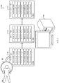
- FIG. 1 is a diagram of a system 100 that generates information about a subject's emotional state.
- an imager 102 images a subject 104 to generate brain image data 106 .
- a computer system 108 analyses the brain image data 106 with stored data 110 in order to generate data 112 about emotional states over time of the subject 104 .
- the imager 102 may perform, for example, fMRI imaging to create a sequence of volumetric “images” of the subject's 104 brain.
- volumetric “images” can record data about both the relatively static shape of the brain as well as the relatively dynamic electrical activity of the brain.
- the stored data 110 can include emotion vectors that each record data about brain activity when a subject is experiencing a particular emotional state.
- the emotion vectors 110 can be generated by imaging many subject's brains while the subjects are experiencing the associated emotion. This training data may then be aggregated to find common functional data across the training data and recorded in the emotion vectors of data 110 .
- a corresponding emotional state can be generated by the computer system 108 and stored in the data 112 .
- the computer system 108 can compare the first volumetric image, assigned a time value of 0, with each of the emotion vectors and record a resulting emotion state at time value 0. Then, the next volumetric image at time value 1 can be similarly examined and a corresponding emotional state can be stored in the data 112 .
- each emotion state can be mapped to a single point in the emotion space.
- This emotion-space may be high dimension (e.g., greater than three dimensions) and manifold (e.g., locally similar to Euclidean space near each point).
- manifold e.g., locally similar to Euclidean space near each point.
- the movement of the points through the emotion space may describe changes in the subject's 102 emotions. As these points move through a manifold in the space, predictions can be made about future locations of the emotional state.
- a range of possible emotions may be identified by modeling a speed, acceleration, and orientation in the manifold, and projecting where a point will move to after a second, minute, etc. In this way, predictions of future emotional states may be generated.
- a stimuli can be provided to the subject, and the movement of their emotional state can be compared to the projected emotional state, and a difference assigned to the stimuli.
- the subject 102 may be suffering from a cognitive bias where they feel a high degree of fear of public speaking relative to an average person.
- a clinician may prescribe exposure therapy in which the subject becomes exposed to small amounts of their fear stimulus and learns they do not need to fear that stimulus. Once the small amount of stimulus is acceptable to the subject, greater levels of the stimulus may be exposed to the subject.
- the subject 102 is imaged while speaking with the clinician.
- the clinician uses the system 100 to determine a ‘base’ emotional state for the subject, which can include a mix of fear and other emotions.
- the clinician can then speak with the subject, receive consent to begin the exposure therapy, and tell the subject to think about speaking in front of two members of the subject's family about a song the subject enjoys.
- the subject continues to be imaged and emotional state data continues to be generated. If the emotional state data shows an increase in fear, the clinician can ask the subject to modify the thought experiment to speaking only to one family member. However, if the emotional state data does not show an increase in fear, the clinician can ask the subject to continue to visualize the thought experiment and describe the visualization.
- the clinician can ask the subject to change the setting in a way that increases the stimulus, and can continue to modify the exercise based on the data 112 .
- other uses of the system 100 are possible, including both clinical and non-clinical uses.
- FIG. 2 shows example data that can be used when generating information about a subject's emotional state.
- the data shown here may be used in the system 100 .
- Primary/Base emotion vectors 200 each store data about a brain's functional state when the subject of the brain is experiencing a particular primary/base emotion. It is believed that emotional states are a combination of physiological phenomena as well as psychological phenomena. As such, the functional state of the brain, as captured by an fMRI or other imaging process, can record sufficient data to uniquely identify a functional state that corresponds to specified emotion(s).
- emotional states can be treated as a weighted mix of a specific set of primary/base emotions.
- most or all images of a brain will show functional state data that reflects a mix of the primary/base emotion vectors.
- These primary/base emotion vectors may be disentangled from each other, for example, by performing principal component analysis on recordings of the mixed emotional state data.
- one emotion vector 200 may be generated and recorded for each of a set of primary emotional states, e.g., recorded when a subject is exposed to specific stimuli known to elicit a specific emotional response.
- Each of the vectors 200 can record data in binary format on a computer disk that can be interpreted as, for example, an array of numeric values, ascii characters, rendered as an image, or in another format or formats depending on the needs of the system 100 .
- the array of numeric values can correspond to relative blood-oxygen-level-dependent (BOLD) signal binned relative to the maximum and minima of those areas.
- Each of the vectors 200 can be tagged with a specific emotional state.
- the first emotion vector 200 can be tagged with fear
- the second emotion vector 200 can be tagged with aesthetic appreciation, etc.
- the emotion vector 200 can then record values reflecting average brain activity for brains (e.g., brains other than the subject's 102 brain) that have experienced the specified emotional state.
- This data may include an image itself, and/or may include a set of data that is generated from the image.
- the vectors 200 may include a cell for each parcelation in a brain parcelation map and may record a numeric value describing the activity of that parcelation as recorded, for example, as a BOLD signal detected via magnetic signal variation.
- the emotion vector 200 may include less data than a full brain image.
- there may be thirty-four emotion vectors 200 those being sexual desire, aesthetic appreciation, entrancement, disgust, amusement, fear, interest, anxiety, surprise, joy, horror, adoration, calmness, romance, awe, nostalgia, empathic pain, relief, awkwardness, craving, excitement, sadness, boredom, romance, sympathy, admiration, anger, satisfaction, confusion, disappointment, pride, envy, contempt, and guilt.
- more, fewer, or different vectors may be used.
- Volumetric images 202 include volumetric images recorded of the subject's 102 brain at sequential time periods.
- an fMRI imaging system can image a subject's 102 brain.
- the image can record a three-dimensional (3D) image of the brain, and can combine the 3D images to create a four-dimensional (4D) image of the brain that shows the state of the subject's brain over time.
- Each of the volumetric images 202 can record data in binary format on a computer disk that can be interpreted as, for example, an array of numeric values, ascii characters, rendered as an image, or in another format or formats depending on the needs of the system 100 .
- a state table 204 can record emotional states of the subject 102 over time based on, for example, the emotion vectors 200 and the volumetric images 202 . As shown, at various time points, values for V 1 , V 2 , V 3 , V 4 . . . may be stored. The values for V N can record a weighted contribution that corresponding emotion vectors 200 contribute to the emotional state of the subject at the given time. In this example, the weight is recorded as a number from 0 to 1 recorded to three decimal places, however other data formats can be used. As shown here, V N values closer to 1 record emotional states that more heavily incorporate the emotion of the corresponding emotion vector, and V N values closer to 0 record emotional states that have little contribution from the corresponding emotion vector.
- a change table 206 can record changes in emotional state of the subject 102 over time based on, for example, the state table 204 .
- each row shows a change value that records the level of change of a particular V N compared to the time period directly previous.
- V 1 changed from 0.165 to 0.165, resulting in a value of 0 recorded in the change table 206 .
- V 2 changed from 0.491 to 0.713, resulting in a change value of 0.221 recorded in the change table 206 .
- positive values indicate an increase in the corresponding emotion and negative values indicate a decrease in the corresponding value.
- a change table 206 can record a change compared to an initial or base emotion for the subject 102 .
- Such a table can be useful, for example, to show net change over a long period of time, where the moment-to-moment changes which may be more noisy, are less useful.
- FIG. 3 shows the data 200 - 204 in a schematic arrangement to show the relationships between volumetric images 202 , state table 204 , and emotion vectors 200 .
- a single volumetric image 202 has associated a plurality of weights 204 , which are associated with (e.g., multiplied by elements of) a corresponding emotion vector 200 .
- Another volumetric image (not shown) would also have another sequence of weights associated for the same emotion vectors 200 .
- FIG. 4 shows an example process 400 for generating information about a subject's emotional state.
- the process 400 may be used, for example, by the computer system 108 in generating the data 204 and 206 . However, other systems may be used to perform the process 400 and other similar processes.
- fMRI data of a subject brain is accessed 402 .
- the data includes a plurality of time-sequenced volumetric images of activity in a subject brain.
- the imager 102 can capture an fMRI of a human subject 104 and provide volumetric images 202 based on the fMRI data to the computer 108 .
- a plurality of emotion vectors are accessed 404 .
- the computer system 108 can access the emotion vectors 200 from long-term memory and load the emotion vectors 200 into short-term memory for usage in the process 400 .
- a plurality of fMRI state vectors at various points in time are determined 406 .
- the computer system 108 can analyze each of the volumetric images 202 . This analysis may be performed in parallel, in sequence, or in a combination of the two.
- Each volumetric image 202 can be mapped into a space defined by the emotion vectors to place a point in a manifold (e.g., a collection of points forming a certain kind of set, such as those of a topologically closed surface or an analog of this in three or more dimensions) based on the contribution of each emotion vector 200 to the volumetric image 202 .
- This analysis can create the state table 204 previously described, or another data structure.
- Flow data is determined to identify a trajectory, over time, of the subject brain as reflected by the fMRI data, through a state space defined by the emotion vectors, wherein the flow data is based at least in part on the fMRI vectors at various points in time 408 .
- the computer system 108 can generate the change table 206 to record how the volumetric images 202 , and thus the subject's emotional state, change over time.
- This change table 206 can record a flow of the subject's emotional state over the manifold depending on the emotional change.
- Data that shows changes, through time, in at least one emotional state of the brain is generated from the flow data 410 .
- a graphic user interface may be rendered that graphically shows the changes in emotion.
- a brain rendering may be shown with various colors gaining or losing intensity as the weight of an emotion vector increases or decreases.
- a report showing the top M emotions at each time may be generated.
- a resting emotional state for the subject is recorded in diagnostic data based on the changes in emotional state of the brain.
- a first emotional state, or an emotional state recorded when minimal stimulus is provided may be recorded as a resting emotional state. This can allow, for example, for a comparison with a given emotional state at any time point to determine how a subject's emotions are being influenced by stimulus.
- the diagnostic data is provided to a recommendation engine configured to receive the diagnostic data and provide one or more recommendations for potential interventions for the subject 412 .
- the recommendation engine may provide to a clinician one or more possible courses of treatment for the subject based on the subject's measured emotional state. The clinician may then accept, reject, or modify one of those suggestions based on their clinical expertise.
- the recommendation may be a drug intervention, a behavioral intervention, transcranial magnetic stimulation, neurosurgery and/or other appropriate intervention.
- FIG. 5 shows an example process for providing stimuli to a subject.
- the process 500 may be used, for example, by the computer system 108 . However, other systems may be used to perform the process 500 and other similar processes.
- the process 500 can be used to monitor a subject while providing a stimuli to the subject.
- a clinician can provide treatment to a patient that is modulated based on the emotional response of the patient.
- a researcher can use the process 500 while investigating emotional responses in humans or other subjects.
- Records of stimuli are accessed 502 .
- the records record stimuli provided to the subject while the fMRI data was collected.
- diagnostic data correspondences between provision of stimuli and the changes in emotional state in the brain are recorded 504 .
- the computer system 108 can access historical records for the subject that include a table of stimuli, intensity, and duration that are provided to the subject. This table can then be augmented with data from the tables 204 and/or 206 to record how those stimuli influenced the subject.
- One of the stimuli is selected for future exposure to the subject based on the recorded correspondences 506 . For example, a researcher or clinician may determine that it is appropriate to elicit a particular emotion, and may look up the stimuli that have historically elicited that emotion from the subject. Then, the researcher or clinician may apply that stimuli.
- Embodiments of the subject matter and the functional operations described in this specification can be implemented in digital electronic circuitry, in tangibly-embodied computer software or firmware, in computer hardware, including the structures disclosed in this specification and their structural equivalents, or in combinations of one or more of them.
- Embodiments of the subject matter described in this specification can be implemented as one or more computer programs, i.e., one or more modules of computer program instructions encoded on a tangible non-transitory storage medium for execution by, or to control the operation of, data processing apparatus.
- the computer storage medium can be a machine-readable storage device, a machine-readable storage substrate, a random or serial access memory device, or a combination of one or more of them.
- the program instructions can be encoded on an artificially-generated propagated signal, e.g., a machine-generated electrical, optical, or electromagnetic signal, that is generated to encode information for transmission to suitable receiver apparatus for execution by a data processing apparatus.
- data processing apparatus refers to data processing hardware and encompasses all kinds of apparatus, devices, and machines for processing data, including by way of example a programmable processor, a computer, or multiple processors or computers.
- the apparatus can also be, or further include, special purpose logic circuitry, e.g., an FPGA (field programmable gate array) or an ASIC (application-specific integrated circuit).
- the apparatus can optionally include, in addition to hardware, code that creates an execution environment for computer programs, e.g., code that constitutes processor firmware, a protocol stack, a database management system, an operating system, or a combination of one or more of them.
- a computer program which may also be referred to or described as a program, software, a software application, an app, a module, a software module, a script, or code, can be written in any form of programming language, including compiled or interpreted languages, or declarative or procedural languages; and it can be deployed in any form, including as a stand-alone program or as a module, component, subroutine, or other unit suitable for use in a computing environment.
- a program may, but need not, correspond to a file in a file system.
- a program can be stored in a portion of a file that holds other programs or data, e.g., one or more scripts stored in a markup language document, in a single file dedicated to the program in question, or in multiple coordinated files, e.g., files that store one or more modules, sub-programs, or portions of code.
- a computer program can be deployed to be executed on one computer or on multiple computers that are located at one site or distributed across multiple sites and interconnected by a data communication network.
- the processes and logic flows described in this specification can be performed by one or more programmable computers executing one or more computer programs to perform functions by operating on input data and generating output.
- the processes and logic flows can also be performed by special purpose logic circuitry, e.g., an FPGA or an ASIC, or by a combination of special purpose logic circuitry and one or more programmed computers.
- Computers suitable for the execution of a computer program can be based on general or special purpose microprocessors or both, or any other kind of central processing unit.
- a central processing unit will receive instructions and data from a read-only memory or a random access memory or both.
- the essential elements of a computer are a central processing unit for performing or executing instructions and one or more memory devices for storing instructions and data.
- the central processing unit and the memory can be supplemented by, or incorporated in, special purpose logic circuitry.
- a computer will also include, or be operatively coupled to receive data from or transfer data to, or both, one or more mass storage devices for storing data, e.g., magnetic, magneto-optical disks, or optical disks.
- a computer need not have such devices.
- a computer can be embedded in another device, e.g., a mobile telephone, a personal digital assistant (PDA), a mobile audio or video player, a game console, a Global Positioning System (GPS) receiver, or a portable storage device, e.g., a universal serial bus (USB) flash drive, to name just a few.
- PDA personal digital assistant
- GPS Global Positioning System
- USB universal serial bus
- Computer-readable media suitable for storing computer program instructions and data include all forms of non-volatile memory, media and memory devices, including by way of example semiconductor memory devices, e.g., EPROM, EEPROM, and flash memory devices; magnetic disks, e.g., internal hard disks or removable disks; magneto-optical disks; and CD-ROM and DVD-ROM disks.
- semiconductor memory devices e.g., EPROM, EEPROM, and flash memory devices
- magnetic disks e.g., internal hard disks or removable disks
- magneto-optical disks e.g., CD-ROM and DVD-ROM disks.
- embodiments of the subject matter described in this specification can be implemented on a computer having a display device, e.g., a CRT (cathode ray tube) or LCD (liquid crystal display) monitor, for displaying information to the user and a keyboard and a pointing device, e.g., a mouse or a trackball, by which the user can provide input to the computer.
- a display device e.g., a CRT (cathode ray tube) or LCD (liquid crystal display) monitor
- keyboard and a pointing device e.g., a mouse or a trackball
- Other kinds of devices can be used to provide for interaction with a user as well; for example, feedback provided to the user can be any form of sensory feedback, e.g., visual feedback, auditory feedback, or tactile feedback; and input from the user can be received in any form, including acoustic, speech, or tactile input.
- a computer can interact with a user by sending documents to and receiving documents from a device that is used by the user; for example, by sending web pages to a web browser on a user's device in response to requests received from the web browser.
- a computer can interact with a user by sending text messages or other forms of message to a personal device, e.g., a smartphone, running a messaging application, and receiving responsive messages from the user in return.
- Embodiments of the subject matter described in this specification can be implemented in a computing system that includes a back-end component, e.g., as a data server, or that includes a middleware component, e.g., an application server, or that includes a front-end component, e.g., a client computer having a graphical user interface, a web browser, or an app through which a user can interact with an implementation of the subject matter described in this specification, or any combination of one or more such back-end, middleware, or front-end components.
- the components of the system can be interconnected by any form or medium of digital data communication, e.g., a communication network. Examples of communication networks include a local area network (LAN) and a wide area network (WAN), e.g., the Internet.
- LAN local area network
- WAN wide area network
- the computing system can include clients and servers.
- a client and server are generally remote from each other and typically interact through a communication network. The relationship of client and server arises by virtue of computer programs running on the respective computers and having a client-server relationship to each other.
- a server transmits data, e.g., an HTML page, to a user device, e.g., for purposes of displaying data to and receiving user input from a user interacting with the device, which acts as a client.
- Data generated at the user device e.g., a result of the user interaction, can be received at the server from the device.
Landscapes
- Health & Medical Sciences (AREA)
- Engineering & Computer Science (AREA)
- Life Sciences & Earth Sciences (AREA)
- Physics & Mathematics (AREA)
- General Health & Medical Sciences (AREA)
- Medical Informatics (AREA)
- Public Health (AREA)
- Nuclear Medicine, Radiotherapy & Molecular Imaging (AREA)
- Biomedical Technology (AREA)
- Radiology & Medical Imaging (AREA)
- Pathology (AREA)
- Animal Behavior & Ethology (AREA)
- Surgery (AREA)
- Molecular Biology (AREA)
- Heart & Thoracic Surgery (AREA)
- Biophysics (AREA)
- Veterinary Medicine (AREA)
- Primary Health Care (AREA)
- Epidemiology (AREA)
- Neurology (AREA)
- General Physics & Mathematics (AREA)
- Psychiatry (AREA)
- Computer Vision & Pattern Recognition (AREA)
- Psychology (AREA)
- High Energy & Nuclear Physics (AREA)
- Condensed Matter Physics & Semiconductors (AREA)
- Physiology (AREA)
- Neurosurgery (AREA)
- Quality & Reliability (AREA)
- Hospice & Palliative Care (AREA)
- Databases & Information Systems (AREA)
- Child & Adolescent Psychology (AREA)
- Developmental Disabilities (AREA)
- Theoretical Computer Science (AREA)
- Educational Technology (AREA)
- Data Mining & Analysis (AREA)
- Social Psychology (AREA)
- Cardiology (AREA)
- Hematology (AREA)
- Artificial Intelligence (AREA)
Abstract
fMRI data of a subject brain is accessed and may include a plurality of time-sequenced volumetric images of activity in a subject brain. A plurality of emotion vectors are accessed, each emotion vector tagged with a specified emotional state. From the fMRI data and using the emotion vectors, a plurality of fMRI state vectors are determined at various points in time, where each fMRI state vector is a combination of the emotion vectors and represents the state of the subject brain at a particular point in time. Flow data is determined to identify a trajectory, over time, of the subject brain as reflected by the fMRI data, through a state space defined by the emotion vectors, where the flow data is based at least in part on the fMRI vectors at various points in time. From the flow data, data is generated that shows changes, through time, in at least one emotional state of the brain.
Description
This specification relates to brain imaging and analysis of brain images.
Medical imaging includes the technique and process of creating visual representations of the interior of a body for clinical analysis and medical intervention, as well as visual representation of the function of some organs or tissues (physiology). Medical imaging seeks to reveal internal structures hidden by the skin and bones, as well as to diagnose and treat disease. Medical imaging also establishes a database of normal anatomy and physiology to make it possible to identify abnormalities.
This specification describes technologies for generating information about a subject's emotional states based on brain imaging. These technologies generally involve scanning a subject's brain to generate a time series such as functional magnetic resonance imaging (fMRI) data. Then, that fMRI data can be analyzed and compared to data that describes brain activity associated with known emotions. From this analysis, a current emotional state for the subject can be found, as well as data defining transitions from one state to another. In some cases, these states may be defined by a weighted mix of various emotional states, and the movement through emotional states can define movement through a manifold space with dimensions matching the emotional states.
A system of one or more computers can be configured to perform particular operations or actions by virtue of having software, firmware, hardware, or a combination of them installed on the system that in operation causes or cause the system to perform the actions. One or more computer programs can be configured to perform particular operations or actions by virtue of including instructions that, when executed by data processing apparatus, cause the apparatus to perform the actions. One general aspect includes a system for generating diagnostic data from medical imaging, one or more computers and one or more storage devices on which are stored instructions that are operable, when executed by the one or more computers, to cause the one or more computers to perform operations that can include accessing fMRI data of a subject brain including a plurality of time-sequenced volumetric activity data for a subject brain. The operations also include accessing a plurality of emotion vectors, each emotion vector tagged with a specified emotional state. An emotion vector can be thought of as representing a mental state of a specific kind corresponding to a physiological state and/or a psychological state. In many cases, such a mental state can be surfaced in a subject by exposing the subject to known stimuli.
The operations also include determining, from the fMRI data and using the emotion vectors, a plurality of fMRI state vectors at various points in time, where each fMRI state vector is a combination of the emotion vectors and represents the state of the subject brain at a particular point in time. The operations also include determining flow data to identify a trajectory, over time, of the subject brain as reflected by the fMRI data, through a state space defined by the emotion vectors, where the flow data is based at least in part on the fMRI vectors at various points in time. The operations also include generating, from the flow data, data that shows changes, through time, in at least one emotional state of the brain. Other embodiments of this aspect include corresponding computer systems, apparatus, and computer programs recorded on one or more computer storage devices, each configured to perform the actions of the methods.
For a system of one or more computers to be configured to perform particular operations or actions means that the system has installed on it software, firmware, hardware, or a combination of them that in operation cause the system to perform the operations or actions. Similarly, for one or more computer programs to be configured to perform particular operations or actions means that the one or more programs include instructions that, when executed by data processing apparatus, cause the apparatus to perform the operations or actions.
The foregoing and other embodiments can each optionally include one or more of the following features, alone or in combination. In particular, one embodiment includes all the following features in combination. The operations may include accessing records of stimuli provided to the subject while the fMRI data was collected; and recording, in the diagnostic data, correspondences between provision of stimuli and the changes in emotional state in the brain. The operations may include selecting one of the stimuli for future exposure to the subject based on the recorded correspondences. The operations may include recording in the diagnostic data a resting emotional state for the subject based on the changes in emotional state of the brain. The operations may include providing the diagnostic data to a recommendation engine configured to receive the diagnostic data and provide one or more recommendations for interventions for the subject. The recommendation is one of the group including a drug intervention, and a behavioral intervention (e.g., verbal counseling of an individual to help build habits to improve the individual's health). The plurality of emotion vectors can include thirty-four emotion vectors. The emotion vectors can be labelled according to the stimuli that produce the vectors. The labels can include sexual desire, aesthetic appreciation, entrancement, disgust, amusement, fear, interest, anxiety, surprise, joy, horror, adoration, calmness, romance, awe, nostalgia, empathic pain, relief, awkwardness, craving, excitement, sadness, boredom, triumph, sympathy, admiration, anger, satisfaction, confusion, disappointment, pride, envy, contempt, and guilt. In certain embodiments, each emotion vector can store values reflecting average brain activity for other brains that have experienced the specified emotional state. Implementations of the described techniques may include hardware, a method or process, or computer software on a computer-accessible medium.
The subject matter described in this specification can be implemented in particular embodiments so as to realize one or more of the following advantages. Brain imaging is advanced. For example, the technology describe here can be used to generate objective measure of emotions of a subject, and changes to emotional state in time. This technology can aid clinical interventions by allowing a clinician to see measures of emotions in response to treatments or other stimuli. This technology can create quantitative visualization of emotional states during a time course statistically separated from the rest of the non-emotional state features in a data set. Unlike other approaches which do not process the data in a way which both is free from confounding from other aspects of influence, this technology can provide information this is more objective, less bias, and thus usable in a wider range of applications.
The details of one or more embodiments of the subject matter of this specification are set forth in the accompanying drawings and the description below. Other features, aspects, and advantages of the subject matter will become apparent from the description, the drawings, and the claims.
Like reference numbers and designations in the various drawings indicate like elements.
Emotional state and emotional state changes of a subject (e.g., a human or other animal) can be sensed using imaging of the subject's brain. For example, volumetric images of the brain over time can be examined to identify a mix of emotional contributors that combine to define the emotional state at each time. These contributors can be tracked over time to identify how the subject's emotional state changes over time.
When the imager 102 generates the brain image data 106, the imager may perform, for example, fMRI imaging to create a sequence of volumetric “images” of the subject's 104 brain. These volumetric “images” can record data about both the relatively static shape of the brain as well as the relatively dynamic electrical activity of the brain.
The stored data 110 can include emotion vectors that each record data about brain activity when a subject is experiencing a particular emotional state. As will be appreciated the emotion vectors 110 can be generated by imaging many subject's brains while the subjects are experiencing the associated emotion. This training data may then be aggregated to find common functional data across the training data and recorded in the emotion vectors of data 110.
For each of the volumetric images of the brain data 106, a corresponding emotional state can be generated by the computer system 108 and stored in the data 112. For example, the computer system 108 can compare the first volumetric image, assigned a time value of 0, with each of the emotion vectors and record a resulting emotion state at time value 0. Then, the next volumetric image at time value 1 can be similarly examined and a corresponding emotional state can be stored in the data 112.
In this way, not only can emotional state be determined and recorded, but so to can changes in emotional state over time be determined and recorded. By using the emotion vectors to define an emotion-space, each emotion state can be mapped to a single point in the emotion space. This emotion-space may be high dimension (e.g., greater than three dimensions) and manifold (e.g., locally similar to Euclidean space near each point). In using such a space, the movement of the points through the emotion space may describe changes in the subject's 102 emotions. As these points move through a manifold in the space, predictions can be made about future locations of the emotional state. For example, a range of possible emotions may be identified by modeling a speed, acceleration, and orientation in the manifold, and projecting where a point will move to after a second, minute, etc. In this way, predictions of future emotional states may be generated. Similarly, a stimuli can be provided to the subject, and the movement of their emotional state can be compared to the projected emotional state, and a difference assigned to the stimuli.
One example use of this technology for a subject is described. The subject 102 may be suffering from a cognitive bias where they feel a high degree of fear of public speaking relative to an average person. In order to help this subject, a clinician may prescribe exposure therapy in which the subject becomes exposed to small amounts of their fear stimulus and learns they do not need to fear that stimulus. Once the small amount of stimulus is acceptable to the subject, greater levels of the stimulus may be exposed to the subject.
To begin, the subject 102 is imaged while speaking with the clinician. The clinician uses the system 100 to determine a ‘base’ emotional state for the subject, which can include a mix of fear and other emotions. The clinician can then speak with the subject, receive consent to begin the exposure therapy, and tell the subject to think about speaking in front of two members of the subject's family about a song the subject enjoys. The subject continues to be imaged and emotional state data continues to be generated. If the emotional state data shows an increase in fear, the clinician can ask the subject to modify the thought experiment to speaking only to one family member. However, if the emotional state data does not show an increase in fear, the clinician can ask the subject to continue to visualize the thought experiment and describe the visualization. If the subject's emotional state still does not show an increase in fear, the clinician can ask the subject to change the setting in a way that increases the stimulus, and can continue to modify the exercise based on the data 112. As will be appreciated, other uses of the system 100 are possible, including both clinical and non-clinical uses.
Primary/Base emotion vectors 200 each store data about a brain's functional state when the subject of the brain is experiencing a particular primary/base emotion. It is believed that emotional states are a combination of physiological phenomena as well as psychological phenomena. As such, the functional state of the brain, as captured by an fMRI or other imaging process, can record sufficient data to uniquely identify a functional state that corresponds to specified emotion(s).
In some cases, emotional states can be treated as a weighted mix of a specific set of primary/base emotions. In such cases, most or all images of a brain will show functional state data that reflects a mix of the primary/base emotion vectors. These primary/base emotion vectors may be disentangled from each other, for example, by performing principal component analysis on recordings of the mixed emotional state data. In such cases, one emotion vector 200 may be generated and recorded for each of a set of primary emotional states, e.g., recorded when a subject is exposed to specific stimuli known to elicit a specific emotional response. Each of the vectors 200 can record data in binary format on a computer disk that can be interpreted as, for example, an array of numeric values, ascii characters, rendered as an image, or in another format or formats depending on the needs of the system 100. In some cases, the array of numeric values can correspond to relative blood-oxygen-level-dependent (BOLD) signal binned relative to the maximum and minima of those areas.
Each of the vectors 200 can be tagged with a specific emotional state. For example, the first emotion vector 200 can be tagged with fear, the second emotion vector 200 can be tagged with aesthetic appreciation, etc. The emotion vector 200 can then record values reflecting average brain activity for brains (e.g., brains other than the subject's 102 brain) that have experienced the specified emotional state. This data may include an image itself, and/or may include a set of data that is generated from the image. For example, the vectors 200 may include a cell for each parcelation in a brain parcelation map and may record a numeric value describing the activity of that parcelation as recorded, for example, as a BOLD signal detected via magnetic signal variation. In some cases, the emotion vector 200 may include less data than a full brain image.
In one scheme, there may be thirty-four emotion vectors 200, those being sexual desire, aesthetic appreciation, entrancement, disgust, amusement, fear, interest, anxiety, surprise, joy, horror, adoration, calmness, romance, awe, nostalgia, empathic pain, relief, awkwardness, craving, excitement, sadness, boredom, triumph, sympathy, admiration, anger, satisfaction, confusion, disappointment, pride, envy, contempt, and guilt. However, more, fewer, or different vectors may be used.
A state table 204 can record emotional states of the subject 102 over time based on, for example, the emotion vectors 200 and the volumetric images 202. As shown, at various time points, values for V1, V2, V3, V4 . . . may be stored. The values for VN can record a weighted contribution that corresponding emotion vectors 200 contribute to the emotional state of the subject at the given time. In this example, the weight is recorded as a number from 0 to 1 recorded to three decimal places, however other data formats can be used. As shown here, VN values closer to 1 record emotional states that more heavily incorporate the emotion of the corresponding emotion vector, and VN values closer to 0 record emotional states that have little contribution from the corresponding emotion vector.
A change table 206 can record changes in emotional state of the subject 102 over time based on, for example, the state table 204. In this example change table 206, each row shows a change value that records the level of change of a particular VN compared to the time period directly previous. In this example, V1 changed from 0.165 to 0.165, resulting in a value of 0 recorded in the change table 206. Similarly, V2 changed from 0.491 to 0.713, resulting in a change value of 0.221 recorded in the change table 206. This show that the subject had no change in the emotion of V1 and an increase in the emotion of V2. As will be understood, positive values indicate an increase in the corresponding emotion and negative values indicate a decrease in the corresponding value.
Other formats of the tables are possible. For example, instead of recording a change compared to the previous time point, a change table 206 can record a change compared to an initial or base emotion for the subject 102. Such a table can be useful, for example, to show net change over a long period of time, where the moment-to-moment changes which may be more noisy, are less useful.
fMRI data of a subject brain is accessed 402. The data includes a plurality of time-sequenced volumetric images of activity in a subject brain. For example, the imager 102 can capture an fMRI of a human subject 104 and provide volumetric images 202 based on the fMRI data to the computer 108. A plurality of emotion vectors are accessed 404. For example, the computer system 108 can access the emotion vectors 200 from long-term memory and load the emotion vectors 200 into short-term memory for usage in the process 400.
From the fMRI data and using the emotion vectors, a plurality of fMRI state vectors at various points in time are determined 406. For example, the computer system 108 can analyze each of the volumetric images 202. This analysis may be performed in parallel, in sequence, or in a combination of the two. Each volumetric image 202 can be mapped into a space defined by the emotion vectors to place a point in a manifold (e.g., a collection of points forming a certain kind of set, such as those of a topologically closed surface or an analog of this in three or more dimensions) based on the contribution of each emotion vector 200 to the volumetric image 202. This analysis can create the state table 204 previously described, or another data structure.
Flow data is determined to identify a trajectory, over time, of the subject brain as reflected by the fMRI data, through a state space defined by the emotion vectors, wherein the flow data is based at least in part on the fMRI vectors at various points in time 408. For example, the computer system 108 can generate the change table 206 to record how the volumetric images 202, and thus the subject's emotional state, change over time. This change table 206 can record a flow of the subject's emotional state over the manifold depending on the emotional change.
Data that shows changes, through time, in at least one emotional state of the brain is generated from the flow data 410. For example, a graphic user interface may be rendered that graphically shows the changes in emotion. For example, a brain rendering may be shown with various colors gaining or losing intensity as the weight of an emotion vector increases or decreases. In another example, a report showing the top M emotions at each time may be generated.
A resting emotional state for the subject is recorded in diagnostic data based on the changes in emotional state of the brain. In some cases, a first emotional state, or an emotional state recorded when minimal stimulus is provided may be recorded as a resting emotional state. This can allow, for example, for a comparison with a given emotional state at any time point to determine how a subject's emotions are being influenced by stimulus.
The diagnostic data is provided to a recommendation engine configured to receive the diagnostic data and provide one or more recommendations for potential interventions for the subject 412. For example, the recommendation engine may provide to a clinician one or more possible courses of treatment for the subject based on the subject's measured emotional state. The clinician may then accept, reject, or modify one of those suggestions based on their clinical expertise. The recommendation may be a drug intervention, a behavioral intervention, transcranial magnetic stimulation, neurosurgery and/or other appropriate intervention.
The process 500 can be used to monitor a subject while providing a stimuli to the subject. With the process 500, for example, a clinician can provide treatment to a patient that is modulated based on the emotional response of the patient. In another example, a researcher can use the process 500 while investigating emotional responses in humans or other subjects.
Records of stimuli are accessed 502. The records record stimuli provided to the subject while the fMRI data was collected. In diagnostic data, correspondences between provision of stimuli and the changes in emotional state in the brain are recorded 504. For example, the computer system 108 can access historical records for the subject that include a table of stimuli, intensity, and duration that are provided to the subject. This table can then be augmented with data from the tables 204 and/or 206 to record how those stimuli influenced the subject.
One of the stimuli is selected for future exposure to the subject based on the recorded correspondences 506. For example, a researcher or clinician may determine that it is appropriate to elicit a particular emotion, and may look up the stimuli that have historically elicited that emotion from the subject. Then, the researcher or clinician may apply that stimuli.
Embodiments of the subject matter and the functional operations described in this specification can be implemented in digital electronic circuitry, in tangibly-embodied computer software or firmware, in computer hardware, including the structures disclosed in this specification and their structural equivalents, or in combinations of one or more of them. Embodiments of the subject matter described in this specification can be implemented as one or more computer programs, i.e., one or more modules of computer program instructions encoded on a tangible non-transitory storage medium for execution by, or to control the operation of, data processing apparatus. The computer storage medium can be a machine-readable storage device, a machine-readable storage substrate, a random or serial access memory device, or a combination of one or more of them. Alternatively or in addition, the program instructions can be encoded on an artificially-generated propagated signal, e.g., a machine-generated electrical, optical, or electromagnetic signal, that is generated to encode information for transmission to suitable receiver apparatus for execution by a data processing apparatus.
The term “data processing apparatus” refers to data processing hardware and encompasses all kinds of apparatus, devices, and machines for processing data, including by way of example a programmable processor, a computer, or multiple processors or computers. The apparatus can also be, or further include, special purpose logic circuitry, e.g., an FPGA (field programmable gate array) or an ASIC (application-specific integrated circuit). The apparatus can optionally include, in addition to hardware, code that creates an execution environment for computer programs, e.g., code that constitutes processor firmware, a protocol stack, a database management system, an operating system, or a combination of one or more of them.
A computer program, which may also be referred to or described as a program, software, a software application, an app, a module, a software module, a script, or code, can be written in any form of programming language, including compiled or interpreted languages, or declarative or procedural languages; and it can be deployed in any form, including as a stand-alone program or as a module, component, subroutine, or other unit suitable for use in a computing environment. A program may, but need not, correspond to a file in a file system. A program can be stored in a portion of a file that holds other programs or data, e.g., one or more scripts stored in a markup language document, in a single file dedicated to the program in question, or in multiple coordinated files, e.g., files that store one or more modules, sub-programs, or portions of code. A computer program can be deployed to be executed on one computer or on multiple computers that are located at one site or distributed across multiple sites and interconnected by a data communication network.
The processes and logic flows described in this specification can be performed by one or more programmable computers executing one or more computer programs to perform functions by operating on input data and generating output. The processes and logic flows can also be performed by special purpose logic circuitry, e.g., an FPGA or an ASIC, or by a combination of special purpose logic circuitry and one or more programmed computers.
Computers suitable for the execution of a computer program can be based on general or special purpose microprocessors or both, or any other kind of central processing unit. Generally, a central processing unit will receive instructions and data from a read-only memory or a random access memory or both. The essential elements of a computer are a central processing unit for performing or executing instructions and one or more memory devices for storing instructions and data. The central processing unit and the memory can be supplemented by, or incorporated in, special purpose logic circuitry. Generally, a computer will also include, or be operatively coupled to receive data from or transfer data to, or both, one or more mass storage devices for storing data, e.g., magnetic, magneto-optical disks, or optical disks. However, a computer need not have such devices. Moreover, a computer can be embedded in another device, e.g., a mobile telephone, a personal digital assistant (PDA), a mobile audio or video player, a game console, a Global Positioning System (GPS) receiver, or a portable storage device, e.g., a universal serial bus (USB) flash drive, to name just a few.
Computer-readable media suitable for storing computer program instructions and data include all forms of non-volatile memory, media and memory devices, including by way of example semiconductor memory devices, e.g., EPROM, EEPROM, and flash memory devices; magnetic disks, e.g., internal hard disks or removable disks; magneto-optical disks; and CD-ROM and DVD-ROM disks.
To provide for interaction with a user, embodiments of the subject matter described in this specification can be implemented on a computer having a display device, e.g., a CRT (cathode ray tube) or LCD (liquid crystal display) monitor, for displaying information to the user and a keyboard and a pointing device, e.g., a mouse or a trackball, by which the user can provide input to the computer. Other kinds of devices can be used to provide for interaction with a user as well; for example, feedback provided to the user can be any form of sensory feedback, e.g., visual feedback, auditory feedback, or tactile feedback; and input from the user can be received in any form, including acoustic, speech, or tactile input. In addition, a computer can interact with a user by sending documents to and receiving documents from a device that is used by the user; for example, by sending web pages to a web browser on a user's device in response to requests received from the web browser. Also, a computer can interact with a user by sending text messages or other forms of message to a personal device, e.g., a smartphone, running a messaging application, and receiving responsive messages from the user in return.
Embodiments of the subject matter described in this specification can be implemented in a computing system that includes a back-end component, e.g., as a data server, or that includes a middleware component, e.g., an application server, or that includes a front-end component, e.g., a client computer having a graphical user interface, a web browser, or an app through which a user can interact with an implementation of the subject matter described in this specification, or any combination of one or more such back-end, middleware, or front-end components. The components of the system can be interconnected by any form or medium of digital data communication, e.g., a communication network. Examples of communication networks include a local area network (LAN) and a wide area network (WAN), e.g., the Internet.
The computing system can include clients and servers. A client and server are generally remote from each other and typically interact through a communication network. The relationship of client and server arises by virtue of computer programs running on the respective computers and having a client-server relationship to each other. In some embodiments, a server transmits data, e.g., an HTML page, to a user device, e.g., for purposes of displaying data to and receiving user input from a user interacting with the device, which acts as a client. Data generated at the user device, e.g., a result of the user interaction, can be received at the server from the device.
While this specification contains many specific implementation details, these should not be construed as limitations on the scope of any invention or on the scope of what may be claimed, but rather as descriptions of features that may be specific to particular embodiments of particular inventions. Certain features that are described in this specification in the context of separate embodiments can also be implemented in combination in a single embodiment. Conversely, various features that are described in the context of a single embodiment can also be implemented in multiple embodiments separately or in any suitable subcombination. Moreover, although features may be described above as acting in certain combinations and even initially be claimed as such, one or more features from a claimed combination can in some cases be excised from the combination, and the claimed combination may be directed to a subcombination or variation of a subcombination.
Similarly, while operations are depicted in the drawings in a particular order, this should not be understood as requiring that such operations be performed in the particular order shown or in sequential order, or that all illustrated operations be performed, to achieve desirable results. In certain circumstances, multitasking and parallel processing may be advantageous. Moreover, the separation of various system modules and components in the embodiments described above should not be understood as requiring such separation in all embodiments, and it should be understood that the described program components and systems can generally be integrated together in a single software product or packaged into multiple software products.
Particular embodiments of the subject matter have been described. Other embodiments are within the scope of the following claims. For example, the actions recited in the claims can be performed in a different order and still achieve desirable results. As one example, the processes depicted in the accompanying figures do not necessarily require the particular order shown, or sequential order, to achieve desirable results. In some cases, multitasking and parallel processing may be advantageous.
Claims (20)
1. A system for generating diagnostic data from medical imaging, the system comprising:
one or more computers and one or more storage devices on which are stored instructions that are operable, when executed by the one or more computers, to cause the one or more computers to perform operations comprising:
accessing fMRI data of a subject brain comprising a plurality of time-sequenced volumetric images of activity in a subject brain;
accessing a plurality of emotion vectors, each emotion vector tagged with a specified emotional state;
determining, from the fMRI data and using the emotion vectors, a plurality of fMRI state vectors at various points in time, where each fMRI state vector is a combination of the emotion vectors and represents the state of the subject brain at a particular point in time;
determining flow data to identify a trajectory, over time, of the subject brain as reflected by the fMRI data, through a state space defined by the emotion vectors, wherein the flow data is based at least in part on the fMRI vectors at various points in time; and
generating, from the flow data, data that shows changes, through time, in at least one emotional state of the brain.
2. The system of claim 1 , the operations further comprising accessing records of stimuli provided to the subject while the fMRI data was collected; and
recording, in the diagnostic data, correspondences between provision of stimuli and the changes in emotional state in the brain.
3. The system of claim 2 , the operations further comprising selecting one of the stimuli for future exposure to the subject based on the recorded correspondences.
4. The system of claim 1 , the operations further comprising recording in the diagnostic data a resting emotional state for the subject based on the changes in emotional state of the brain.
5. The system of claim 1 , the operations further comprising providing the diagnostic data to a recommendation engine configured to receive the diagnostic data and provide one or more recommendations for interventions for the subject.
6. The system of claim 5 , wherein the recommendation is one of the group comprising a drug intervention, and a behavioral intervention.
7. The system of claim 1 , wherein the plurality of emotion vector comprises thirty-four emotion vectors.
8. The system of claim 7 , wherein the emotions comprise sexual desire, aesthetic appreciation, entrancement, disgust, amusement, fear, interest, anxiety, surprise, joy, horror, adoration, calmness, romance, awe, nostalgia, empathic pain, relief, awkwardness, craving, excitement, sadness, boredom, triumph, sympathy, admiration, anger, satisfaction, confusion, disappointment, pride, envy, contempt, and guilt.
9. The system of claim 1 , each emotion vector storing values reflecting average brain activity for other brains that have experienced the specified emotional state.
10. The system of claim 1 , wherein the operations further comprise:
accessing training-fMRI data for a plurality of other brains, the plurality of other brains not including the subject brain;
aggregating the training-fMRI data to find common functional data across the training-fMRI data; and
storing the common functional data as the plurality of emotion vectors.
11. The system of claim 10 , wherein accessing fMRI data of a subject brain is performed after storing the common functional data as the plurality of emotion vectors.
12. A method for generating diagnostic data from medical imaging, the method comprising:
or more computers to perform operations comprising:
accessing fMRI data of a subject brain comprising a plurality of time-sequenced volumetric images of activity in a subject brain;
accessing a plurality of emotion vectors, each emotion vector tagged with a specified emotional state;
determining, from the fMRI data and using the emotion vectors, a plurality of fMRI state vectors at various points in time, where each fMRI state vector is a combination of the emotion vectors and represents the state of the subject brain at a particular point in time;
determining flow data to identify a trajectory, over time, of the subject brain as reflected by the fMRI data, through a state space defined by the emotion vectors, wherein the flow data is based at least in part on the fMRI vectors at various points in time; and
generating, from the flow data, data that shows changes, through time, in at least one emotional state of the brain.
13. The method of claim 12 , the method further comprising accessing records of stimuli provided to the subject while the fMRI data was collected; and
recording, in the diagnostic data, correspondences between provision of stimuli and the changes in emotional state in the brain.
14. The method of claim 13 , the method further comprising selecting one of the stimuli for future exposure to the subject based on the recorded correspondences.
15. The method of claim 12 , the method further comprising recording in the diagnostic data a resting emotional state for the subject based on the changes in emotional state of the brain.
16. The method of claim 12 , the method further comprising providing the diagnostic data to a recommendation engine configured to receive the diagnostic data and provide one or more recommendations for interventions for the subject.
17. The method of claim 16 , wherein the recommendation is one of the group comprising a drug intervention, and a behavioral intervention.
18. The method of claim 12 , wherein the plurality of emotion vector comprises thirty-four emotion vectors.
19. The method of claim 18 , wherein the emotions comprise sexual desire, aesthetic appreciation, entrancement, disgust, amusement, fear, interest, anxiety, surprise, joy, horror, adoration, calmness, romance, awe, nostalgia, empathic pain, relief, awkwardness, craving, excitement, sadness, boredom, triumph, sympathy, admiration, anger, satisfaction, confusion, disappointment, pride, envy, contempt, and guilt.
20. The method of claim 12 , each emotion vector storing values reflecting average brain activity for other brains that have experienced the specified emotional state.
Priority Applications (1)
| Application Number | Priority Date | Filing Date | Title |
|---|---|---|---|
| US17/378,648 US11399757B1 (en) | 2021-07-16 | 2021-07-16 | Medical imaging with features to determine emotional state |
Applications Claiming Priority (1)
| Application Number | Priority Date | Filing Date | Title |
|---|---|---|---|
| US17/378,648 US11399757B1 (en) | 2021-07-16 | 2021-07-16 | Medical imaging with features to determine emotional state |
Publications (1)
| Publication Number | Publication Date |
|---|---|
| US11399757B1 true US11399757B1 (en) | 2022-08-02 |
Family
ID=82652375
Family Applications (1)
| Application Number | Title | Priority Date | Filing Date |
|---|---|---|---|
| US17/378,648 Active US11399757B1 (en) | 2021-07-16 | 2021-07-16 | Medical imaging with features to determine emotional state |
Country Status (1)
| Country | Link |
|---|---|
| US (1) | US11399757B1 (en) |
Cited By (1)
| Publication number | Priority date | Publication date | Assignee | Title |
|---|---|---|---|---|
| US20230342914A1 (en) * | 2022-04-21 | 2023-10-26 | Canon Medical Systems Corporation | Method and system for monitoring a patient emotional state and segmenting obtained emission data based on the patient emotional state data |
Citations (6)
| Publication number | Priority date | Publication date | Assignee | Title |
|---|---|---|---|---|
| US20120179003A1 (en) * | 2011-01-12 | 2012-07-12 | Faber Group Synergy, Llc | Therapeutic Treatment Systems and Processes for Stress-Related Disorders |
| US20150339363A1 (en) * | 2012-06-01 | 2015-11-26 | Next Integrative Mind Life Sciences Holding Inc. | Method, system and interface to facilitate change of an emotional state of a user and concurrent users |
| US20190102511A1 (en) | 2017-10-02 | 2019-04-04 | Blackthorn Therapeutics, Inc. | Methods and tools for detecting, diagnosing, predicting, prognosticating, or treating a neurobehavioral phenotype in a subject |
| US20190213761A1 (en) | 2016-09-01 | 2019-07-11 | The General Hospital Corporation | System and method for automated transform by manifold approximation |
| US10586326B2 (en) | 2014-07-15 | 2020-03-10 | The Brigham And Women's Hospital, Inc. | Systems and methods for generating biomarkers based on multivariate classification of functional imaging and associated data |
| US10874340B2 (en) | 2014-07-24 | 2020-12-29 | Sackett Solutions & Innovations, LLC | Real time biometric recording, information analytics and monitoring systems for behavioral health management |
-
2021
- 2021-07-16 US US17/378,648 patent/US11399757B1/en active Active
Patent Citations (6)
| Publication number | Priority date | Publication date | Assignee | Title |
|---|---|---|---|---|
| US20120179003A1 (en) * | 2011-01-12 | 2012-07-12 | Faber Group Synergy, Llc | Therapeutic Treatment Systems and Processes for Stress-Related Disorders |
| US20150339363A1 (en) * | 2012-06-01 | 2015-11-26 | Next Integrative Mind Life Sciences Holding Inc. | Method, system and interface to facilitate change of an emotional state of a user and concurrent users |
| US10586326B2 (en) | 2014-07-15 | 2020-03-10 | The Brigham And Women's Hospital, Inc. | Systems and methods for generating biomarkers based on multivariate classification of functional imaging and associated data |
| US10874340B2 (en) | 2014-07-24 | 2020-12-29 | Sackett Solutions & Innovations, LLC | Real time biometric recording, information analytics and monitoring systems for behavioral health management |
| US20190213761A1 (en) | 2016-09-01 | 2019-07-11 | The General Hospital Corporation | System and method for automated transform by manifold approximation |
| US20190102511A1 (en) | 2017-10-02 | 2019-04-04 | Blackthorn Therapeutics, Inc. | Methods and tools for detecting, diagnosing, predicting, prognosticating, or treating a neurobehavioral phenotype in a subject |
Non-Patent Citations (3)
| Title |
|---|
| Horikawa et al., "The neural representation of emotion is high-dimensional, categorical, anddistributed across transmodal brain regions" Department of Neuroinformatics, ATR Computational Neuroscience Laboratories. Dec. 11, 2019, 60 pages. |
| Jirsa, "Structured Flows on Manifolds as guiding concepts in brain science," Hal Archives-ouvertes, dated Apr. 8, 2020, 15 pages. |
| Shine et al., "The low dimensional dynamic and integrative core of cognition in the human brain," The University of Sydney, Feb. 18, 2018, 33 pages. |
Cited By (1)
| Publication number | Priority date | Publication date | Assignee | Title |
|---|---|---|---|---|
| US20230342914A1 (en) * | 2022-04-21 | 2023-10-26 | Canon Medical Systems Corporation | Method and system for monitoring a patient emotional state and segmenting obtained emission data based on the patient emotional state data |
Similar Documents
| Publication | Publication Date | Title |
|---|---|---|
| Arora et al. | Generative adversarial networks and synthetic patient data: current challenges and future perspectives | |
| Kaplan et al. | Processing narratives concerning protected values: A cross-cultural investigation of neural correlates | |
| AlSlaity et al. | Mobile applications for health and wellness: a systematic review | |
| Wilson-Mendenhall et al. | Neural evidence that human emotions share core affective properties | |
| Speer et al. | Reading stories activates neural representations of visual and motor experiences | |
| Leshikar et al. | Medial prefrontal cortex supports source memory accuracy for self-referenced items | |
| Aguirre | Functional neuroimaging: technical, logical, and social perspectives | |
| AU2019391425B2 (en) | Apparatus and method for utilizing a brain feature activity map database to characterize content | |
| Parrish et al. | Self-compassion and responses to negative social feedback: The role of fronto-amygdala circuit connectivity | |
| Chen et al. | Development of audio and visual attention assessment system in combination with brain wave instrument: Apply to children with Attention Deficit Hyperactivity Disorder | |
| Wager et al. | Principles of fMRI | |
| US20220130513A1 (en) | Method and system for dynamically generating profile-specific therapeutic protocols using machine learning models | |
| Woźniacka et al. | Artificial intelligence in medicine and dermatology | |
| Li et al. | Dispositional mindfulness and self-referential neural activity during the resting state | |
| Jonas et al. | Designing a wearable IoT-based bladder level monitoring system for neurogenic bladder patients | |
| Safikhani et al. | The influence of realism on the sense of presence in virtual reality: Neurophysiological insights using EEG | |
| Cacioppo et al. | Predicting intentions of a familiar significant other beyond the mirror neuron system | |
| US11399757B1 (en) | Medical imaging with features to determine emotional state | |
| Kaboodvand et al. | Preparatory activity of anterior insula predicts conflict errors: integrating convolutional neural networks and neural mass models | |
| Hu et al. | The language network supports both lexical access and sentence generation during language production | |
| Xiong et al. | Gravity matters for the neural representations of action semantics | |
| Suran et al. | The contribution of the left inferior frontal gyrus in affective processing of social groups | |
| Jung et al. | A multimodal fMRI dataset unifying naturalistic processes with a rich array of experimental tasks | |
| Mitra et al. | Diagnosing Alzheimer’s disease using deep learning techniques | |
| Urban | Visibilities and the analysis of interdiscourse: The case of digital health practices |
Legal Events
| Date | Code | Title | Description |
|---|---|---|---|
| FEPP | Fee payment procedure |
Free format text: ENTITY STATUS SET TO UNDISCOUNTED (ORIGINAL EVENT CODE: BIG.); ENTITY STATUS OF PATENT OWNER: SMALL ENTITY |
|
| FEPP | Fee payment procedure |
Free format text: ENTITY STATUS SET TO SMALL (ORIGINAL EVENT CODE: SMAL); ENTITY STATUS OF PATENT OWNER: SMALL ENTITY |
|
| STCF | Information on status: patent grant |
Free format text: PATENTED CASE |