US11382947B2 - Targeting G3BP proteins to accelerate nerve regeneration - Google Patents
Targeting G3BP proteins to accelerate nerve regeneration Download PDFInfo
- Publication number
- US11382947B2 US11382947B2 US16/857,636 US202016857636A US11382947B2 US 11382947 B2 US11382947 B2 US 11382947B2 US 202016857636 A US202016857636 A US 202016857636A US 11382947 B2 US11382947 B2 US 11382947B2
- Authority
- US
- United States
- Prior art keywords
- g3bp1
- axonal
- neurons
- regeneration
- gfp
- Prior art date
- Legal status (The legal status is an assumption and is not a legal conclusion. Google has not performed a legal analysis and makes no representation as to the accuracy of the status listed.)
- Active
Links
Images


Classifications
-
- A—HUMAN NECESSITIES
- A61—MEDICAL OR VETERINARY SCIENCE; HYGIENE
- A61K—PREPARATIONS FOR MEDICAL, DENTAL OR TOILETRY PURPOSES
- A61K38/00—Medicinal preparations containing peptides
- A61K38/04—Peptides having up to 20 amino acids in a fully defined sequence; Derivatives thereof
- A61K38/10—Peptides having 12 to 20 amino acids
Definitions
- the present invention relates to vehicles and methods for improving nerve regeneration after nerve injury.
- peripheral nerve is a term used synonymously to describe the peripheral nervous system.
- the peripheral nervous system is a network of motor and sensory nerves that connect the brain and spinal cord (the central nervous system or CNS) to the entire human body. These nerves control the functions of sensation, movement and motor coordination.
- the peripheral nerves are a complicated, extensive network of nerves that are the tool for the brain and spinal cord to communicate with the rest of the body. They are fragile and can be damaged easily. Trauma, including battlefield injuries, is a major cause of peripheral nerve injury along with birth trauma (brachial plexus injury) and injuries related to surgeries. Limited data is available to determine the incidence of peripheral nerve injury.
- Neurons nerve cells that are the basic building block of the nervous system, generate their own proteins within cytoplasmic processes that extend for centimeters in rodents and more than a meter in humans. These locally generated proteins are needed for regeneration after nerve injury. Protein synthesis in axons contributes to axon growth during development and regeneration.
- stress granules cytoplasmic RNA-protein complexes referred to as stress granules.
- Stress granules dense aggregations in the cytosol (the aqueous component of the cytoplasm of a cell) comprised of proteins and RNAs, are used to store mRNAs during periods of cellular stress, but nerve injury with compromise of axon integrity paradoxically causes stress granules to disaggregate presumably releasing mRNAs for translation.
- Stress granules contain non-translating mRNAs, translation initiation components, and many additional proteins affecting mRNA function.
- Stress granules have been proposed to affect mRNA translation and stability, as well as being linked to apoptosis and nuclear processes. Stress granules also interact with P-bodies, another cytoplasmic RNP granule containing non-translating mRNA, translation repressors and some mRNA degradation machinery. Together, stress granules and P-bodies reveal a dynamic cycle of distinct biochemical and mRNA-protein complexes (mRNPs) in the cytosol, with implications for the control of mRNA function.
- mRNPs biochemical and mRNA-protein complexes
- Nerve regeneration is abysmally slow in the peripheral nervous system and does not occur spontaneously in the central nervous system. Despite that regeneration occurs in the periphery, the slow growth in humans means that by the time regeneration occurs the distal nerve is no longer a growth-supportive environment and the target tissues are no longer receptive for reinnervation that restores the nerve supply to a part of the body. There is a pressing clinical need for treatments that will accelerate axon regeneration in peripheral nerves.
- a method for treating nerve injury in a mammal may include introducing a polypeptide comprising between 15 and 20 amino acids to a nerve injury site in the mammal.
- the polypeptide may interfere with function of stress granules and increases intra-axonal rates of translation of proteins needed for nerve regeneration.
- the polypeptide may be an amino acid sequence set as forth in SEQ ID NO. 2.
- the polypeptide specifically may target mRNA storage sites in neurons and increases rates of neuron regeneration.
- the polypeptide may disrupt G3BP function. Further yet, disruption of G3BP functions may include activating intra-axonal mRNA translation, increasing axon growth in neurons, and accelerating nerve regeneration in vivo.
- disruption of G3BP functions may be accomplished via siRNA-mediated knockdown of G3BP1.
- disrupting G3BP1's function in an assembly of axonal stress granule structures may increase intra-axonal protein synthesis and accelerate peripheral nervous system axon regeneration. Even still further, accelerated axon growth regeneration may be facilitated by sequestering Imp ⁇ 1 mRNA from translation.
- a method of disrupting G3BP functions may include overexpressing a dominant-negative protein.
- the dominant-negative protein may: disassemble axonal stress granule-like structures, activate intra-axonal mRNA translation, increase axon growth in neurons; and accelerate nerve regeneration in vivo.
- the protein may comprise between 15 and 20 amino acids.
- the protein may have an amino acid sequence as set forth in SEQ ID NO. 2.
- the protein may be cell permeable and target mRNA storage sites in neurons.
- disruption of G3BP functions may be accomplished via siRNA-mediated knockdown of G3BP1.
- disrupting G3BP1's function in an assembly of axonal stress granule structures may increase intra-axonal protein synthesis and accelerate peripheral nervous system axon regeneration. Still furthermore, preventing stress granule-like aggregation of axonal proteins during regeneration may increase the rate of axon regrowth.
- FIG. 1 shows a schematic of how translation is regulated in axons.
- FIG. 2 shows an immunofluorescence comparison of the presence of G3BP1 protein in axons pre-injury and post-injury.
- FIG. 3 shows an immunofluorescence (IF) comparison and a graph illustrating the differences in phosphorylated G3BP in axons.
- FIG. 4 shows an immunofluorescence comparison and a graph of differential colocalization of G3BP with axonal mRNAs in axons of naive vs. injury-conditioned neurons.
- FIG. 5 shows an immunofluorescence comparison and a graph showing expression of known G3BP domains vis-à-vis axon growth.
- FIG. 6 shows an immunofluorescence comparison and a graph illustrating that G3BP B-domain increases axon regeneration in vivo.
- FIG. 7 shows a graph illustrating that the peptide for G3BP1 residues supports axon growth in both PNS and CNS neurons as well as a predicted 3D structure of the peptide.
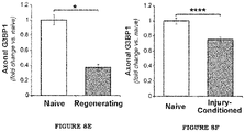
- FIG. 8A shows Immunofluorescence for G3BP1 shows signals in cell body (asterisk) and along distal neurites (arrows) in a cultured DRG neuron.
- FIG. 8B shows single optical planes for axons of na ⁇ ve DRG cultures.
- FIG. 8C also shows single optical planes for axons of na ⁇ ve DRG cultures.
- FIG. 8D shows exposure-matched images of G3BP1 in distal axons of DRGs cultured from na ⁇ ve vs. 7 day injury conditioned animals.
- FIG. 8E shows quantification of axonal G3BP1 immunoreactivity from sciatic nerve.
- FIG. 8F shows quantification of axonal G3BP1 immunoreactivity from axons of cultured DRG neurons.
- FIG. 9A shows axons of DRG neurons transfected with indicated G3BP1 constructs vs. eGFP.
- FIG. 9B shows quantification of axonal aggregates for G3BP1-GFP, G3BP1 S149A -GFP, and G3BP1 S149E -GFP.
- FIG. 9C shows Flourescence Recovery After Photobleaching (FRAP) analyses for neurons transfected with constructs from FIG. 9B .
- FIG. 9D shows exposure-matched confocal images for G3BP1 PS149 and neurofilament (NF) for sciatic nerve as in FIG. 8D .
- FIG. 9E shows quantification of the signals of FIG. 9D .
- FIG. 9F shows distal axons of cultured DRGs immunostained with pan-G3BP1 vs. G3BP1 PS149 antibodies.
- FIG. 9G shows quantification of the signals of FIG. 9F .
- FIG. 10A shows images of Fluorescence in situ Hybridization (FISH) plus IF for indicated mRNAs and G3BP1 protein for axons of na ⁇ ve and 7 d injury-conditioned DRG neurons.
- FISH Fluorescence in situ Hybridization
- FIG. 10B shows quantification of colocalizations for Nrn1, Imp ⁇ 1, and Gap43 mRNAs with G3BP1 in axons of neurons cultured from na ⁇ ve or 7 day injury-conditioned animals.
- FIG. 10C shows schematics of translation reporter constructs used in panels d-f.
- FIG. 10D shows representative FRAP image sequences for DRG neurons co-transfected with GFP MYR 5′/3′nrn1 plus BFP or G3BP1-BFP.
- FIG. 10E shows quantifications of FRAP assays from DRGs expressing GFP MYR 5′/3′nrn1.
- FIG. 10F shows quantifications of FRAP assays from DRGs expressing GFP MYR 5′/3′imp ⁇ 1.
- FIG. 10G shows HEK293T cells transfected with GFP MYR 5′/3′nrn1, GFP MYR 5′/3′imp ⁇ 1 and mCh MYR 5′/3′gap43.
- FIG. 11A shows a schematic of G3BP1 domains as defined by Tourriere et al. (2003).
- FIG. 11B shows representative images for NF-labeled DRG neurons transfected with indicated constructs.
- FIG. 11C shows quantitation of axon growth from DRGs (left) and cortical neurons (right) treated with cell-permeable 168-189 or 190-208 G3BP1 peptides is shown.
- FIG. 11D shows extent of axon regeneration at 7 d post sciatic nerve crush in adult rats transduced with AAV5 encoding G3BP1-BFP, G3BP1 B domain-BFP, G3BP1 D domain-BFP, or GFP control.
- FIG. 12B shows quantitation that shows no change in cell body Puro signals.
- FIG. 12C shows that the axons show a significant increase in Puro signals in the G3BP1 B domain expressing neurons (c).
- FIG. 12D show puro incorporation in DRG axons was also significantly increased by the 190-208 G3BP1 peptide treatment compared to control and 168-189 peptide exposure.
- FIG. 12E shows RTddPCR for axonal mRNAs associated with G3BP1-GFP in DRG neurons as average % mRNA associated with G3BP1-GFP ⁇ SEM.
- FIG. 12F show representative images of G3BP1-mCh in DRG axons ⁇ treatment with 190-208 G3BP1 peptide for 15 min.
- FIG. 12G show density of G3BP1-mCh aggregates along 100 ⁇ m length axons from DRG cultures treated as in FIG. 12F .
- FIG. 12H shows the size of G3BP1-mCh aggregate as indicated bins for from DRG cultures treated as in FIG. 12F .
- FIG. 13A shows representative FRAP image sequences.
- FIG. 14A shows FRAP analyses for DRGs expressing mCh MYR 5′/3′gap43 plus the indicated G3BP1-BFP constructs or BFP control.
- FIG. 14B shows FRAP analyses for DRGs expressing GFP MYR 5′/3′nrn1 or mGFP MYR 5′/3′imp ⁇ 1 plus the G3BP1 B domain-BFP or G3BP1 D domain-BFP.
- FIG. 14C shows FRAP analyses for DRGs expressing GFP MYR 5′/3′nrn1, GFP MYR 5′/3′imp ⁇ 1, or mCh MYR 5′/3′gap43 plus G3BP1-BFP.
- FIG. 15A shows axon growth parameters for DRG neurons transfected as in FIG. 11B .
- FIG. 15B shows RT-ddPCR analyses of G3BP1 and G3BP2 mRNA levels in DRGs transfected with control (siCntl) and G3BP1 (siG3BP1) siRNAs.
- FIG. 15C shows axon growth data for DRG neurons transfected with control (siCntl) or G3BP1 (siG3BP1) siRNAs plus GFP, G3BP1-GFP, or G3BP1 B domain-GFP.
- FIG. 16A shows Representative images of dansyl chloride fluorescence of internalized peptides in DRG cell bodies.
- FIG. 16B shows neurite outgrowth analyses for dissociated DRG neurons treated with peptides immediately after plating.
- FIG. 16C shows representative images of axonal compartment of microfluidic culture device with cortical neurons stained for tau at DIV 6.
- FIG. 17A shows representative images of distal axons of na ⁇ ve DRG neurons were transfected with G3BP1 B domain-GFP, D domain-GFP, CD domain-GFP, or BCD domain-GFP.
- FIG. 17B shows representative, exposure-matched confocal images of crushed sciatic nerves of adult rats transduced with AAV5 encoding the G3BP1-BFP, G3BP1 B domain-BFP, G3BP1 D domain-BFP or GFP.
- FIG. 18A shows representative images for puromycin (Puro) incorporation in DRG neurons treated with indicated peptides.
- FIG. 18B shows representative images of HuR immunoreactivity in NIH-3T3 cells that were transfected GFP vs.
- G3BP1 B domain-GFP are shown after 30 min treatment with sodium arsenite (0.5 mM).
- FIG. 18C shows quantification SGs in the transfected NIH-3T3 cells from FIG. 18A are shown based on the indicated immunostaining.
- FIG. 19 shows one embodiment of a method of the current disclosure.
- FIG. 20 shows one embodiment of an alternative method of the current disclosure.
- the current disclosure presents data on the general mechanism for targeting the G3BP proteins and efficacy of viral targeting for accelerating axon regeneration.
- a cell permeable G3BP1 peptide is disclosed that may have potential for pharmaceutical development.
- a method is provided for a mechanism for targeting G3BP1 function is provided.
- the current disclosure provides that knockdown of G3BP1, a stress granule aggregating protein, increases axon growth in vitro.
- Introduction of the B domain of G3BP1 (which may include but is not limited to amino acids 140-208) and may increase axon regeneration in vitro and in vivo. Importantly, this may accelerate the rate of regeneration in vivo after peripheral nerve injury.
- the G3BP1 B domain is highly conserved across species.
- a polypeptide comprising amino acids 190-208 of rat G3BP1 increases axonal outgrowth in cultured mammalian sensory and cortical neurons and even accelerates axon growth beyond the effect of injury-conditioned sensory neurons.
- Structure predictions of the 190-208 G3BP1 peptide show that acidic glutamate side chains of seven glutamate-proline repeats may be arrayed along the periphery of the peptide and only conservative changes in amino acid sequence between rat, mouse and human (e.g., substitution of aspartate for glutamate).
- mRNA colocalization with endogenous G3BP1 protein and translation assays strongly suggest that the peptide interferes with function of stress granules and increases intra-axonal rates of translation of proteins needed for regeneration.
- the current disclosures provides a cell-permeable polypeptide that specifically targets mRNA storage sites in neurons and increases rates of regeneration after traumatic injury. This may be used as the basis for therapeutic development or could increase efficacy of existing therapies. Additionally, there could be neuroprotective applications for neuropathic conditions (diabetic neuropathy, chemotherapy induced neuropathy, etc.) that have much higher incidence.
- FIG. 1 shows a schematic of how translation is regulated in axons. Changes in intra-axonal translation occur after injury of peripheral nerves. mRNAs are stored before injury, and an influx of calcium recruits mRNAs into translation. Based on localization to axons and dynamic changes after injury, it is believed that ‘stress granules’ are the site of mRNA storage before injury and likely provide a level of mRNA sequestration after injury/during regeneration.
- FIG. 2 illustrates that immunofluorescence shows G3BP1 protein in axons in a granular profile before injury. Seven (7) days after injury, when axons have fully initiated regeneration, granular profiles of G3BP1 in axons are decreased compared to the naive nerve. There is a commensurate increase in G3BP1 phosphorylated on serine 149. This phosphorylation is known to ‘dissaggregate’ G3BP1.
- FIG. 3 shows that consistent with phosphorylation decreasing G3BP1 aggregation, phosphomimetic G3BP1 (G3BP1-S149E) shows much lower aggregation in axons than does non-phosphorylatable G3BP1 (G3BP1-S149A) and wild-type G3BP1 in axons of in cultured rat sensory neurons.
- FIG. 4 illustrates differential colocalization of G3BP1 with axonal mRNAs in axons of na ⁇ ve vs. injury-conditioned neurons.
- G3BP1 colocalizes with some axonal mRNAs.
- Representative fluorescence in situ hybridization to detect axonal mRNA combined with antibody staining for G3BP1 protein is shown on left. Quantifications of the colocalization coefficients are shown in the graph. This comparison is for naive vs. injury-conditioned sensory neurons, where the conditioning injury triggers a rapid outgrowth of axons.
- Importin ⁇ 1 mRNA (Imp ⁇ 1), encoded by an ‘injury response gene’, shows increased colocalization with G3BP1 in axons of the injury-conditioned neurons.
- Neuritin 1 mRNA (Nrn1), encoded by a ‘regeneration-associated gene’, shows decreased colocalization with G3BP1 protein in axons of the injury-conditioned neurons.
- GAP43 shows low colocalization coefficients and no change with injury-conditioning.
- FIG. 5 shows that expression of G3BP1 domains alters axon growth.
- Introduction of full length (ABCD) or separate domains of G3BP1 into DRG neurons alters axon growth in culture (schematic in upper right).
- Representative images of B and D domain GFP fusion proteins show localization into distal axons (longitudinal images in mid-left); all other fusion proteins similarly localized into axons.
- Representative images of axonal outgrowth are shown on bottom left.
- D domain decreases axon length and B domain substantially increases axon lengths. Quantification of axonal lengths over biological replicate experiments ( ⁇ 3) is shown on right (*** p ⁇ 0.001, **** p ⁇ 0.0001 by a Anova with Tukey HSD post-hoc test compared to GFP control).
- FIG. 6 illustrates that the G3BP1 B domain increases axon regeneration in vivo.
- Sciatic nerves of adult rats were transduced with adeno-associated virus 5 (AAV5) encoding GFP, G3BP1-BFP (full length G3BP1) or G3BP1-B domain-BFP.
- AAV5 adeno-associated virus 5
- the animals After seven (7) days for expression of the virus, the animals underwent sciatic nerve crush at mid-thigh. Seven (7) days later the animals were euthanized and analyzed for axon regeneration. Representative images of immunofluorescence for axonal marker neurofilament are shown on the left for each condition. The graph on the right shows the extent of regeneration. As FIG. 6 illustrates, regeneration is significantly enhanced by introducing the G3BP1 B domain.
- FIG. 7 shows that the peptide for G3BP1 residues 190-208 supports axon growth in both peripheral nervous system and central nervous system neurons.
- FIG. 7 shows, axon growth analyses for embryonic cortical neurons and naive or injury-conditioned adult dorsal root ganglion (DRG) neurons treated with cell permeable peptides corresponding to G3BP1 residues 160-189 or 190-208 are shown.
- the peptides were designed in house and synthesized by Bachem.
- the DRG neurons were treated with peptides by whole bath application after twelve (12) hours in culture and analyzed twenty-four (24) hours later.
- Cortical neurons were cultured in a microfluidic device for three (3) days and then G3BP1 peptides were bath applied to only the axonal compartment.
- FIG. 7 illustrates, the 190-208 peptide significantly increased axon growth in CNS neurons.
- Protein sequence alignments for corresponding regions of 190-208 peptide are shown in the upper right.
- the lower right shows predicted tertiary structure of rat 190-208 peptide with approximately 180 degree vertical rotation between views.
- the acidic side chains of glutamates can be seen arrayed along the periphery of the structure.
- axotomy induces translation of stored axonal mRNAs via regulation of the stress granule protein G3BP1, to support regeneration of peripheral nerves.
- G3BP1 aggregates in axons of peripheral nerves in stress granule-like structures that decrease during regeneration, with a commensurate increase in phosphorylated G3BP1.
- Colocalization of G3BP1 with axonal mRNAs is also correlated with the growth state of the neuron.
- Disrupting G3BP functions by overexpressing a dominant-negative protein disassembles axonal stress granule-like structures, activates intra-axonal mRNA translation, increases axon growth in cultured neurons and accelerates nerve regeneration in vivo.
- Injured axons in the peripheral nervous system use locally translated proteins for retrograde injury-signaling and regenerative growth.
- Translation of axonal mRNAs can be activated by different stimuli including axotomy in mature neurons and in response to guidance cues in developing neurons, indicating that a significant fraction of axonal mRNAs are stored until a particular stimulus activates their translation.
- Stress granules (SG) serve as storage depots for mRNAs in non-neuronal systems, providing a mechanism to respond to cellular stress by sequestering unneeded mRNAs from translation.
- G3BP1 RasGAP SH3 domain binding protein 1
- the current disclosure shows that translation of specific axonal mRNAs is negatively regulated in intact axons by G3BP1, but in regenerating peripheral nerves post injury, this negative regulation is removed by dispersion of aggregated G3BP1, to then support accelerated axon growth.
- G3BP1 PS149 When phosphorylated on serine 149 (G3BP1 PS149 ), G3BP1's oligomerization is blocked and SGs disassemble, presumably releasing bound mRNAs for translation.
- Loss of G3BP1 aggregation in SG-like structures in regenerating axons is accompanied by an increase in phosphorylated G3BP1.
- Disrupting G3BP1 function with a dominant-negative approach activates intra-axonal mRNA translation, increases axon growth in cultured neurons and accelerates nerve regeneration in vivo, and therefore represents a new pro-regenerative therapeutic approach.
- Axonal G3BP1 Aggregates Decrease During Nerve Regeneration—
- Axonal G3BP1 signals showed higher colocalization with known SG components than components of processing bodies (PB) that are linked to RNA degradation ( FIGS. 8B and 8C ). These axonal G3BP1 aggregates are smaller than those described for SGs in non-neuronal cells (diameter ⁇ 0.2-0.8 ⁇ m vs. ⁇ 1 ⁇ m), so the axonal SG-like structures approximate the ⁇ 250 nm diameter described for SG core structure.
- FIG. 8B and 8C show single optical planes for axons of na ⁇ ve DRG cultures co-labeled for G3BP1, HuR, FMRP and FXR1 or G3BP1, DCP1A and XRN1 are shown as indicated; boxed region represents the area higher magnification images are taken from ( FIG. 8B ).
- PLA shows higher colocalization for G3BP1 and HuR than G3BP1 and DCP1A in axons.
- DRG neurons that are conditioned by an in vivo crush injury 7 days prior to culture show more rapid axonal outgrowth over 18-48 h in vitro compared to uninjured (na ⁇ ve) DRGs, and the rapidly growing axons of injury-conditioned neurons showed decreased levels of G3BP1 aggregates compared to those of na ⁇ ve DRG cultures ( FIGS. 8D and 8F ).
- G3BP1 is Phosphorylated in Regenerating Axons—
- G3BP1 S149A -GFP non-phosphorylatable and phosphomimetic G3BP1 mutants in cultured DRGs.
- Axonal G3BP1 S149A -GFP showed aggregated signals that overlapped with HuR, while axonal G3BP1 S149E -GFP appeared diffuse ( FIGS. 9A and 9B ).
- G3BP1 S149E GFP also showed significantly higher mobility in axons than G3BP1 S149A -GFP, GFP, and G3BP1-GFP showed mobility intermediate between G3BP1 S149E -GFP and G3BP1 S149A -GFP ( FIG. 9C , FIG. 13A ). This is consistent with G3BP1 S149A -GFP aggregating into SG-like structures in axons. Axonal G3BP1 PS149 immunoreactivity was increased in regenerating compared to uninjured nerves ( FIGS. 9D and 9E ).
- axonal injury decreases the prevalence of axonal SG-like structures, and this correlates with an increase in axonal G3BP1 PS149 levels.
- the ratio of axonal G3BP1 PS149 to axonal G3BP1 aggregates increases in distal axons and growth cones ( FIGS. 9F and 9G ), indicating that the axonal SG-like structures are dynamically regulated along the growing axon.
- Axonal G3BP1 Modulates Axonal mRNA Translation
- the current disclosure moved to fluorescent reporters to determine if axonal SG-like structures contribute to translation.
- the current disclosure generated axonally targeted GFP MYR and mCherry MYR reporters containing the 5′ and 3′ untranslated regions (UTR) of Imp ⁇ 1, Nrn1, and Gap43 mRNAs (GFP MYR 5′/3′imp ⁇ 1, GFP MYR 5′/3′nrn1, and mCh MYR 5′/3′gap43, respectively; FIG. 10C ).
- the membrane localizing myristoylation (MYR) of the fluorescent reporter proteins dramatically limits their diffusion from sites of translation, so these have provided a surrogate for localized protein synthesis in dendrites and axons using fluorescence recovery after photobleaching (FRAP).
- the 3′ (Imp ⁇ 1 and Gap43) and 5′ (Nrn1) UTRs provide axonal targeting for reporter mRNAs, and with both 5′ and 3′UTRs, the reporter approximates the translational regulation of the endogenous mRNAs.
- RNA immunoprecipitation (RIP) analyses showed enrichment of GFP MYR 5′/3′imp ⁇ 1 and GFP MYR 5′/3′nrn1, but not mCh MYR 5′/3′gap43, in G3BP1 immunoprecipitates ( FIG. 10G ).
- Acidic domain of G3BP1 increases axonal growth
- G3BP1 is composed of four separate domains: N-terminal NTF2-like ‘A domain’, a highly acidic ‘B domain’, PxxP motif containing ‘C domain’, and Cterminal RNA hinging motif containing ‘D domain’ ( FIG. 11A ).
- Expression of G3BP1 deletion constructs in naive DRG cultures showed that G3BP1 B, CD, BCD and D domain proteins all localized to axons ( FIG. 17A ).
- Neurons expressing the G3BP1 B domain showed significantly longer axons, while those expressing the D or CD domains showed shorter axons ( FIG. 11B , FIG. 15A ).
- G3BP1 D domain expression decreases protein synthesis in non-neuronal cells through eIF2 ⁇ phosphorylation.
- expressing G3BP1 BCD domains together reversed the growth deficit seen for CD, pointing to a dominant effect of the B domain in absence of G3BP1's aggregating NTF2 like region ( FIG. 15A ).
- full-length G3BP1 overexpression had no significant effect on axon growth ( FIG. 15A ), perhaps indicating that G3BP1 is at saturating levels in DRG neurons.
- siRNA-mediated knockdown (KD) of G3BP1 significantly increased axon growth and this was completely reversed by co-transfection with a siRNA-resistant G3BP1-GFP ( FIG. 15C ).
- co-transfecting with the G3BP1 B domain did not further increase axon length, suggesting that B domain inhibits G3BP1 function.
- DRG cultures expressing full-length G3BP1-, B domain-, and CD domain-GFP showed modest decline in neurite branching ( FIG. 15A ).
- the current disclosure generated fluorescently-labeled, cell-permeable Tat fusion peptides corresponding to residues 147-166, 168-189, and 190-208 of rat G3BP1.
- Peptides penetrated the neurons in DRG cultures by 30 min. after application ( FIG. 16A ).
- both the 147-166 and 190-208 peptides increased axon length and the 190-208 peptide increased number of neurites per neuron ( FIG. 16B ).
- G3BP1-BFP, G3BP1 B domain-BFP, and G3BP1 D domain-BFP were visible in the regenerating sciatic nerve axons ( FIG. 17B ).
- the G3BP1 B domain-BFP transduced animals showed significantly increased axon regeneration compared to G3BP1-BFP, and G3BP1 D domain-BFP, and GFP transduced animals ( FIG. 11D , FIG. 17B ).
- G3BP1's acidic domain activates intra-axonal translation through disassembly of stress granule protein aggregates—
- the cell-permeable G3BP1 190-208 peptide also significantly rescued axonal translation of GFP MYR 573′nrn1 mRNA in G3BP1-overexpressing DRGs, but did not affect translation of GFP MYR 5′/3′imp ⁇ 31 or mCh MYR 5′/3′gap43 mRNAs ( FIG. 14C ).
- G3BP1 B domain expression led to significantly higher protein synthesis in axons but not cell bodies of cultured DRGs ( FIGS. 12A, 12B, and 12C ).
- G3BP1 B domain expression attenuated SG aggregation in NIH 3T3 cells exposed to sodium arsenite ( FIGS. 18B and 18C ), a potent inducer of SG aggregation.
- the current disclosure next used time-lapse imaging with DRG cultures to determine if the B domain affects the axonal SG-like structures, focusing on axons before and after addition of the cell-permeable 190-208 peptide.
- the 190-208 peptide caused a striking decrease in axonal G3BP1-mCh aggregates within 15 min ( FIG. 5 f - g ). Moreover, the remaining SG-like structures in axons were significantly smaller after 190-208 peptide treatment ( FIG. 5 h ), and the remaining aggregates showed greater motility than in control conditions).
- axonal G3BP1 is a negative modulator of intra-axonal protein synthesis and axon growth.
- G3BP1 G3BP1
- Imp ⁇ 1 and Nrn1 mRNAs The colocalization of different mRNAs with these G3BP1 aggregates correlates with the growth status of neurons, and blocking G3BP1 aggregation provides a novel strategy to accelerate regeneration.
- Nrn1 protein promotes neurite growth, and increasing axonal targeting of Nrn1 mRNA increases axon growth in DRG neurons.
- Nrn1 mRNA translation is induced by axotomy, with its protein product providing a retrograde signal to activate regeneration-associated gene expression in the soma.
- Imp ⁇ 1 mRNA Continued translation of Imp ⁇ 1 mRNA likely decreases axon elongation due to its role in axon length sensing. Consequently, rapid axon growth after injury conditioning would be facilitated by sequestering Imp ⁇ 1 mRNA from translation.
- the current disclosure points to axonal G3BP1 as a modulator of intra-axonal protein synthesis and axon growth. Since G3BP1 is aggregated in uninjured PNS axons, the current disclosure's data points to unrealized functions for SG-like aggregates in axons under non-stress conditions. Preventing this SG-like aggregation of axonal proteins during regeneration increases the rate of axon regrowth.
- the growth-promoting effects of the cell-permeable 190-208 G3BP1 peptide may represent a novel therapeutic lead for accelerating nerve regeneration. Since peripheral nerves typically regenerate at only 1-2 mm per day, accelerating axon growth rates by interfering with axonal G3BP1 function could significantly shorten recovery times and allow axons to reach a more receptive environment to reinnervate target tissues.
- FIG. 8 G3BP1 localizes to axons in stress granule-like aggregates.
- FIGS. 8B and 8C show single optical planes for axons of na ⁇ ve DRG cultures co-labeled for G3BP1, HuR, FMRP and FXR1 or G3BP1, DCP1A and XRN1 are shown as indicated; boxed region represents the area higher magnification images are taken from (b).
- FIG. 9 G3BP1 is phosphorylated in regenerating axons.
- FIG. 9A shows axons of DRG neurons transfected with indicated G3BP1 constructs vs. eGFP are shown.
- G3BP1-GFP and G3BP1 S149A -GFP show prominent aggregates in axons that colocalize with HuR (arrows).
- FIG. 9B shows quantification of axonal aggregates for G3BP1-GFP, G3BP1 S149A -GFP, and G3BP1 S149E -GFP is shown as average ⁇ SEM (N ⁇ 10 neurons over 3 repetitions; p ⁇ 0.005 by one-way ANOVA with Tukey HSD post-hoc).
- FIG. 9C shows FRAP analyses for neurons transfected with constructs as in A are shown as average normalized % recovery ⁇ SEM (see FIG. 13A for representative FRAP image sequences).
- G3BP1 S149A -GFP shows much lower recovery than G3BP1 S149E -GFP;
- G3BP1-GFP is intermediate between G3BP1 S149A -GFP and G3BP1 S149E -GFP (N ⁇ 13 axons over 3 repetitions; * p ⁇ 0.05 between G3BP1 S149A -GFP vs. G3BP1 S149E GFP by one-way ANOVA with Tukey HSD post-hoc).
- Only the 0-320 sec recovery signals for GFP are shown (at 840 sec GFP showed 85.5 ⁇ 4.7% recovery; p ⁇ 0.0001 vs. G3BP1 S149E -GFP by one-way ANOVA with Tukey HSD post-hoc).
- FIGS. 9D and 9E shows exposure-matched confocal images for G3BP1 PS149 and NF are shown for sciatic nerve (d) as in FIG. 8D .
- G3BP1 PS149 signal in the regenerating axons.
- FIG. 10 G3BP1 regulates translation of axonal mRNAs.
- FIG. 10A shows images of FISH/IF for indicated mRNAs and G3BP1 protein are shown for axons of na ⁇ ve and 7 d injury-conditioned DRG neurons.
- FIG. 10B shows quantification of colocalizations for Nrn1, Imp ⁇ 1, and Gap43 mRNAs with G3BP1 in axons of neurons cultured from na ⁇ ve or 7 d injury-conditioned animals shown as average Pearson's coefficient ⁇ SEM (N ⁇ 21 neurons over 3 repetitions; ** p ⁇ 0.01 and *** p ⁇ 0.005 by one-way ANOVA with Tukey HSD posthoc).
- FIG. 10C shows schematics of translation reporter constructs used in panels d-f.
- FIG. 10D shows representative FRAP image sequences for DRG neurons co-transfected with GFP MYR 5′/3′nrn1 plus BFP or G3BP1-BFP. Boxed regions represent the photobleached ROIs.
- FIGS. 10E and 10F show quantifications of FRAP assays from DRGs expressing GFP MYR 5′/3′nrn1 (e) or GFP MYR 5′/3′imp ⁇ 1 (f) translation reporters along with G3BP1-BFP or control BFP are shown as normalized, average % recovery ⁇ SEM (see FIG. 14A for mCh MYR 5′/3′gap43; N ⁇ 11 neurons over 3 repetitions; * p ⁇ 0.05, and ** p ⁇ 0.01 by one-way ANOVA with Tukey HSD post-hoc). Cultures treated with translational inhibitors showed reduced fluorescence recovery that paralleled G3BP1-overexpressing neurons.
- FIG. 10G shows HEK293T cells transfected with GFP MYR 5′/3′nrn1, GFP MYR 5′/3′ imp ⁇ 1, and mCh MYR 5′/3′gap43 show significant enrichment of GFP MYR 5′/3′nrn1 and GFP MYR 5′/3′ imp ⁇ 1 mRNAs coimmunoprecipitating with G3BP1 immunoprecipitate vs. control.
- FIG. 11 G3BP1 acidic domain expression accelerates nerve regeneration.
- FIG. 11A shows a schematic of G3BP1 domains as defined by Tourriere et al. (2003).
- FIG. 17A shows axonal localization of these G3BP1-GFP domain proteins and
- FIG. 15A shows quantitation of axon growth from G3BP1 domain-expressing DRG neurons.
- FIG. 11C shows quantitation of axon growth from DRGs (left) and cortical neurons (right) treated with cell-permeable 168-189 or 190-208 G3BP1 peptides.
- DRGs peptides were added to dissociated na.ve or 7 d injury-conditioned DRGs at 12 h and axon growth was assessed at 36 h in vitro.
- cortical neurons peptides were added to the axonal compartment of microfluidic devices at 3 d in vitro (DIV), and axon growth was assessed at 6 DIV. See FIG. 16C for images of cortical cultures (N ⁇ 95 over 3 DRG cultures and 9 microfluidic devices over 3 cultures; *** p ⁇ 0.005 by one-way ANOVA with Tukey HSD post-hoc).
- FIG. 11D shows the extent of axon regeneration at 7 day post sciatic nerve crush in adult rats transduced with AAV5 encoding G3BP1-BFP, G3BP1 B domain-BFP, G3BP1 D domain-BFP, or GFP control as mean axonal profiles relative to crush site (0 mm) ⁇ SEM.
- G3BP1-BFP G3BP1 B domain-BFP
- G3BP1 D domain-BFP GFP control as mean axonal profiles relative to crush site (0 mm) ⁇ SEM.
- FIG. 12 G3BP1 acidic domain increases axonal mRNA translation and disassembles stress granules.
- FIG. 12D show puro incorporation in DRG axons was also significantly increased by the 190-208 G3BP1 peptide treatment compared to control and 168-189 peptide exposure (N ⁇ 83 axons over 3 DRG cultures; ****p ⁇ 0.0001 by one-way ANOVA with Tukey HSD post-hoc). Representative images are shown in FIG. 18A .
- FIG. 12G show density of G3BP1-mCh aggregates along 100 ⁇ m length axons from DRG cultures treated as in FIG. 12F are shown (N ⁇ 51 axons over 3 repetitions; **** p ⁇ 0.0001 by Student's t-test).
- FIG. 12H size of G3BP1-mCh aggregate is shown as indicated bins for from DRG cultures treated as in FIG. 5 f (N ⁇ 50 aggregates over 3 repetitions; **** p ⁇ 0.0001 for entire population distributions by Kolmogorov-Smirnov test).
- Axoplasm was obtained from sciatic nerve at 3, 7, 14, 21 and 28 d after crush injury at mid-thigh level. 2 cm segments of nerve proximal to the injury site (or equivalent level on contralateral [na ⁇ ve] side) were dissected and axoplasm extruded into 20 mM HEPES [pH 7.3], 110 mM potassium acetate, and 5 mM magnesium acetate (nuclear transport buffer) supplemented with protease/phosphatase inhibitor cocktail (Roche) and RNasin Plus (Promega). After clearing by 20,000 ⁇ g centrifugation at 4° C. for 30 min, supernatants were mixed with 3 volumes of Trizol LS (Invitrogen) for protein precipitation. 3 animals were used for each time point.
- L4-5 DRG were harvested in Hybernate-A medium (BrainBits) and then dissociated as known to those of skill in the art. After centrifugation and washing in DMEM/F12 (Life Technologies), cells were resuspended in DMEM/F12, 1 ⁇ N1 supplement (Sigma), 10% fetal bovine serum (Hyclone), and 10 ⁇ M cytosine arabinoside (Sigma). Dissociated DRGs were plated immediately on laminin/poly-L-lysine-coated coverslips or transfected (see below) and then plated on coated coverslips.
- E18 cortices were dissected in Hibernate E (BrainBits) and dissociated using the Neural Tissue Dissociation kit (Miltenyi Biotec). For this, minced cortices were incubated in a pre-warmed enzyme mix at 37° C. for 15 min; tissues were then triturated and applied to a 40 ⁇ m cell strainer. After washing and centrifugation, neurons were seeded at a density of 1 ⁇ 105 cells per poly-D-lysine-coated microfluidic device (Xona Microfluidics).
- NbActive-1 medium (BrainBits) supplemented with 100 U/ml of Penicillin-Streptomycin (Life Technologies), 2 mM L-Glutamine (Life Technologies), and 1 ⁇ N21 supplement (R&D Systems) was used as culture medium.
- NIH-3T3 and HEK293T cells were maintained in DMEM (Life Technologies) supplemented with 10% FBS (Gibco) and 100 U/ml of Penicillin-Streptomycin (Life Technologies).
- ganglia were pelleted by centrifugation at 100 ⁇ g for 5 min and resuspended in ‘nucleofector solution’ (Rat Neuron Nucleofector kit; Lonza). 5-7 ⁇ g plasmid was electroporated using an AMAXA Nucleofector apparatus (program SCN-8; Lonza).
- siRNA transfection 100 nM siRNAs (Dharmacon) were used with DharmaFECT 3 reagent and incubated for 36 h.
- G3BP1 siRNA sequence 5′ ccacauaggagcugggaauuu 3′.
- Non-targeting siRNAs were as control.
- RTddPCR was used to test the efficiency of depletion (see below).
- HEK293T cells were transfected using Lipofectamine® 2000 per manufacturer's instructions (Invitrogen). AAV5 preparations were titrated in DRG cultures by incubating with 1.8-2.8 ⁇ 1010 particles of AAV5 overnight.
- transfected NIH3T3 cells were grown to 60-80% confluence and were treated with 0.5 mM sodium arsenite (Sigma) for 30 min.
- peptide treatments 10 ⁇ M Tat-fused peptides were added to dissociated DRG cultures at 2 or 12 h after plating. Neurite outgrowth was assessed 24 h after addition of peptides.
- 10 ⁇ M peptide was applied to the axonal compartment at 3 d in vitro (DIV) and axonal growth was assessed at 6 DIV.
- RNA translation was based on eGFP with myristoylation element (GFP MYR ; originally provided by Dr. Erin Schuman, Max-Plank Inst., Frankfurt) or mCherry plasmid with myristoylation element (mCh MYR ). Reporter constructs containing 5′ and 3′UTRs of rat Nrn1 and Gap43 mRNAs have been published. For Imp ⁇ 1, the 5′UTR was cloned by PCR and inserted directly upstream of the initiation codon in GFP MYR 3′imp ⁇ 1 (includes 3′UTR of rat Imp ⁇ 1 mRNA).
- G3BP1 wild type, S149A, S149E and deletion constructs as GFP-tagged proteins were generously provided by Dr. Jamal Tazi, Institut de Génétique Molé Diagram de Struktur.
- the G3BP1-mCherry construct was generated by PCR, amplifying G3BP1 CDS with 5′ NheI and 3′ HindIII restriction sites. After NheI+HindIII digestion, G3BP1 CDS was subcloned into NheI+HindIII-digested pmCherry-N1 vector (Clontech).
- AAV5 preparations were generated in UNC Chapel Hill Viral Vector Core. All plasmid inserts were fully sequenced prior to generating AAV.
- BglII+XhoI digested human G3BP1 cDNA was subcloned into BamHI+XhoI digested pAAV-cDNA6-V5His vector (Vector Biolabs).
- G3BP1 deletion constructs were amplified by PCR with terminal HindIII and XhoI restriction sites (primer sequences available on request). After digestion with HindIII and XhoI, products were cloned into HindIII+XhoI-digested pAAV-cDNA6-V5His vector.
- BFP was excised from the pTagBFP-N vector (Evrogen) using EcoRI+NotI and ligated in-frame directly 3′ of the G3BP1 sequences in pAAV-cDNA6-V5His.
- peptides were generated from the rat G3BP1 B domain sequence (amino acids 140-220; UniProt ID # D3ZYS7 RAT) by Bachem Americas, Inc. Peptides were synthesized with N-terminal dansyl chloride or FITC and N- or C-terminal HIV Tat peptide for cell permeability; the Tat sequence was placed at the least conserved end of the sequence based on P-BLAST of sequences available in UniProt database.
- Peptide sequences with Tat sequence were (Tat shown in italics): 147-166, SEQ ID NO: 13 SEQ ID NO: 13 Glu Glu Ser Glu Glu Glu Val Glu Glu Pro Glu Glu Arg Gln Gln Ser Pro Glu Val Val Tvr Glv Asn Lvs Lvs Asn Asn Gln Asn Asn; 168-189, SEQ ID NO: 14 Am Asp Ser Gly Thr Phe Tyr Asp Gln Thr Val Ser Asn Asn Leu Glu Glu His Leu Glu Glu Pro Tvr Glv Asn Lvs Lvs Asn Asn Gln Asn Asn; and 190-208, SEQ ID NO: 15 Tvr Glv Asn Lvs Lvs Asn Asn Asn Gln Asn Asn Val Val Glu Pro Glu Pro Glu Pro Glu Pro Glu Pro Glu Pro Glu Pro Glu Pro Glu Pro Glu Pro Glu Pro Glu Pro Glu Pro
- RT room temperature
- Primary antibodies consisted of: rabbit anti-G3BP1 (1:200, Sigma), RT97 mouse anti-neurofilament (NF; 1:500, Devel. Studies Hybridoma Bank), and rabbit anti-G3BP1 PS149 (1:300, Sigma).
- FITC-conjugated donkey anti-rabbit and Cy3-conjugated donkey anti-mouse were used as secondary antibodies.
- Zenon antibody labeling kit (Life Technologies) was used to directly label antibodies with fluorophores. Combinations of rabbit anti-G3BP1 (Sigma)+Alexa-488, rabbit anti-HuR (Millipore)+Alexa-405, rabbit anti-FMRP (Cell Signaling Tech)+Alexa-555, and rabbit anti-FXR1 (kind gift from Dr.
- Paraffin sections were used for analyses of nerve regeneration. For this, 10 ⁇ m thick paraffin sections of sciatic nerve were deparaffinized in 100% xylene (2 ⁇ 10 min) followed by 100% ethanol (2 ⁇ 10 min). Sections were rehydrated by sequential incubations in 95, 75 and 50% ethanol for 5 min each, and then rinsed in deionized water. Sections were permeabilized in 0.3% Triton X-100 in PBS, and then rinsed in PBS for 20 min and equilibrated in 50 mM Tris [pH 7.4], 150 mM NaCl, 1% heat-shock bovine serum albumin (BSA), and 1% protease-free BSA (Roche) (‘IF buffer’).
- Sections were then blocked in IF buffer plus 2% heat-shock BSA, and 2% fetal bovine serum for 1.5-2 h. After blocking, samples were incubated overnight at 4° C. in humidified chamber with the primary antibodies in IF buffer. Samples were washed in IF buffer three times and then incubated in secondary antibodies diluted in IF buffer for 45 min. Samples were washed in IF buffer three times followed by rinse in PBS and deionized water. Primary antibodies consisted of: RT97 mouse anti-NF (1:300) and rabbit anti-RFP (1:100, Rockland Immun. Chem.). The RFP antibody was confirmed to detect BFP by immunoblotting (see below) and immunolabeling of transfected DRG neurons (data not shown). Secondary antibodies were used as above.
- FISH Fluorescence In-Situ Hybridization
- RNA-protein colocalization was performed using custom 5′ Cy3-labeled ‘Stellaris’ probes (probe sequences available upon request; Biosearch Tech.). Scrambled probes were used as control for specificity; samples processed without addition of primary antibody were used as control for antibody specificity.
- Primary antibodies consisted of rabbit anti-G3BP1 (1:100) and RT97 mouse anti-NF (1:200).
- FITC-conjugated donkey anti-rabbit and Cy5-conjugated donkey anti-mouse both at 1:200 were used as secondaries. Samples were mounted as above and analyzed using a Leica SP8X confocal microscope. Samples were post-processed with Huygens deconvolution integrated into the Leica LSM software and analyzed as outlined below for RNA-protein colocalization.
- FRAP was used to test for axonal mRNA protein synthesis using diffusion-limited GFP MYR and mCherry MYR reporters as described with minor modifications.
- DRG neurons were co-transfected with GFP MYR 5′/3′nrn1+mCherry MYR 5′/3′gap43 or GFP MYR 5′/3′imp ⁇ 1+mCherry MYR 5′/3′gap43 so that recovery of both reporters could be analyzed simultaneously.
- Cells were maintained at 37° C., 5% CO2 during imaging sequences.
- DRG neurons were transfected with G3BP1 S149A -GFP or G3BP1 S149E -GFP and imaged as above but only the 488 nm laser was used for photobleaching (Argon laser at 70% power, pulsed every 0.82 sec for 80 frames).
- DRG neurons were transfected with G3BP1-mCherry, and 36 h later distal axons were imaged using Leica SP8X confocal microscope with environmental chamber maintained at 37° C., 5% CO2 (with 63 ⁇ /1.4 NA oil immersion objective). Confocal pinhole was set to 0.43 airy units.
- G3BP1-mCherry signals were imaged in axon shaft every 2 sec for 100 frames (at 540 nm excitation and 23% white light laser power; 565-597 nm emission).
- 10 ⁇ M FITC-conjugated peptide was added to the media and 15 min later imaging was continued.
- G3BP1-mCherry aggregates a 100 ⁇ m of the axon shaft was considered ( ⁇ 200 ⁇ m from cell body). Thresholding was applied to acquired image sequences using ImageJ to generate binary masks. ImageJ particle analyzer was used for analysis. G3BP1 aggregates with area of ⁇ 1 ⁇ m2 were considered as SG-like structures. For analyzing the G3BP1 aggregate velocity, ImageJ Trackmate plug-in was used.
- the current disclosure used the Click-iT® Plus OPP Protein Synthesis Assay Kit per manufacturer's instructions (Invitrogen/Life Technologies). Briefly, 3 DIV cultures were incubated with 20 ⁇ M O-propargyl-puromycin (OPP) for 30 min at 37° C. OPP-labeled proteins were detected by crosslinking with Alexa Fluor-594 picolyl azide molecule. Coverslips were then mounted with Prolong Gold Antifade (Invitrogen) and imaged with Leica DMI6000 epifluorescent microscope as above. ImageJ was used to quantify the Puromycinylation signals in distal axons and cell bodies.
- OPP O-propargyl-puromycin
- protein lysates or immunoprecipitates were denatured by boiling in Laemmle sample buffer, fractionated by SDS-PAGE, and transferred to nitrocellulose membranes. Blots were blocked for 1 h at room temperature with 5% non-fat dry milk in Tris-buffered saline with 0.1% Tween 20 (TBST) for anti-GFP, -tagBFP, and -G3BP1 antibodies; 5% BSA in TBST was used for blocking anti-G3BP1 PS149 antibody. Primary antibodies diluted in appropriate blocking buffer were added to the membranes and incubated overnight incubation at 4° C. with rocking.
- Primary antibodies consisted of: rabbit anti-G3BP1 (1:2,000; Sigma), rabbit anti-G3BP1PS149 (1:1,000; Sigma), rabbit anti-GFP (1:5,000; Abcam), rabbit anti-TagBFP (1:4,000; Evrogen). After washing in TBST, blots were incubated HRP-conjugated anti-rabbit IgG antibodies (1:5,000; Jackson lab) diluted in blocking buffer for 1 h at room temperature. After washing signals were detected using ECL PrimeTM (GE Healthcare).
- HEK293T cells or DRG neurons were lysed in 100 mM KCl, 5 mM MgCl 2 , 10 mM HEPES [pH 7.4], 1 mM DTT, and 0.5% NP-40 (RIP buffer) supplemented with 1 ⁇ protease inhibitor cocktail (Roche) and RNasin Plus (Invitrogen). Cells were passed through 25 Ga needle 5-7 times and cleared by centrifugation at 12,000 ⁇ g for 20 min. Cleared lysates were pre-absorbed with Protein A-Dynabeads (Invitrogen) for 30 min.
- RNA samples were then incubated with primary antibodies for 3 h and then immunocomplexes precipitated with Protein G-Dynabeads (Invitrogen) for additional 2 h at 4° C. with rotation.
- Mouse anti-G3BP1 (5 ⁇ g, BD Biosciences) and rabbit anti-GFP (5 ⁇ g, Abcam) antibodies were used for immunoprecipitation. Beads were washed 6 times with cold RIP buffer. Bound RNAs were purified and analyzed by RTddPCR (see below).
- xyz scan sequence captured 100 ⁇ m segments of axon shaft (separated from cell body and growth cone by ⁇ 200 ⁇ m) were deconvolved using Huygens Hyvolution software. Colocalization was analyzed using ImageJ JACoP plug-in (https://imagej.nih.gov/ij/plugins/track/jacop.html) to calculate Pearson's coefficient. These coefficient calculations were independently validated with Volocity software (Perkin Elmer).
- z planes of the xyz tile scans from 3-5 locations along each nerve section were analyzed using ImageJ.
- Colocalization plug-in was used to extract protein signals that overlap with axonal marker (NF) in each plane, with the extracted ‘axon-only’ signal projected as a separate channel.
- NF axonal marker
- For calculating axonal G3BP1 aggregate and G3BP1 PS149 signal intensities absolute signal intensity was quantified in each xy plane of the ‘Colocalization’ extracted images for axonal only G3BP1 and G3BP1 PS149 using ImageJ. Protein signal intensities across the individual xy planes were then normalized to NF immunoreactivity area. The relative protein signal intensity was averaged for all image locations in each biological replicate.
- NF-stained nerve sections were post-processed by Straighten plug-in for ImageJ (http://imagej.nih.gov/ij/). NF positive axon profiles were then counted in 30 ⁇ m bins at 0.3 mm intervals distal from crush site. Number of axon profiles present in the proximal crush site was treated as the baseline, and values from the distal bins were normalized to this to calculate the percentage of regenerating axons.
- Kaleidagraph (Synergy) or Prism (GraphPad) software packages were used for statistical analyses.
- One-way ANOVA was used to compare means of independent groups and Student's t-test was used to compare smaller sample sizes of the in vivo analyses. p values of ⁇ 0.05 were considered as statistically significant.
- a method 100 for treating nerve injury in a mammal may be provided.
- a polypeptide comprising between 15 and 20 amino acids may be introduced to a nerve injury site in the mammal.
- the polypeptide may interfere with function of stress granules and increase intra-axonal rates of translation of proteins needed for nerve regeneration.
- the polypeptide may have an amino acid sequence set as forth in SEQ ID NO. 2.
- the polypeptide may specifically target mRNA storage sites in neurons and increases rates of neuron regeneration.
- the polypeptide may disrupt G3BP functions.
- disruption of G3BP functions may: activate, intra-axonal mRNA translation; increase axon growth in neurons; and accelerate nerve regeneration in vivo.
- Disruption of G3BP functions may be accomplished via siRNA-mediated knockdown of G3BP1.
- disrupting G3BP1's function in an assembly of axonal stress granule structures may increases intra-axonal protein synthesis and accelerates peripheral nervous system axon regeneration. Accelerated axon growth regeneration may be facilitated by sequestering Imp ⁇ 1 mRNA from translation.
- FIG. 20 shows an alternative method 200 wherein.
- G3BP function may be disrupted.
- a dominant-negative protein may be overexpressed.
- the dominant-negative protein may disassemble axonal stress granule-like structures, activate intra-axonal mRNA translation, increase axon growth in neurons; and accelerate nerve regeneration in vivo.
- the protein may comprise between 15 and 20 amino acids and may have an amino acid sequence as set forth in SEQ ID NO: 2.
- the protein is cell permeable and targets mRNA storage sites in neurons.
- disruption of G3BP functions may be accomplished via siRNA-mediated knockdown of G3BP1.
- disrupting G3BP1's function in an assembly of axonal stress granule structures may increase intra-axonal protein synthesis and accelerate peripheral nervous system axon regeneration.
- preventing stress granule-like aggregation of axonal proteins during regeneration may increase the rate of axon regrowth.
Landscapes
- Health & Medical Sciences (AREA)
- Life Sciences & Earth Sciences (AREA)
- Pharmacology & Pharmacy (AREA)
- Proteomics, Peptides & Aminoacids (AREA)
- Engineering & Computer Science (AREA)
- Bioinformatics & Cheminformatics (AREA)
- Immunology (AREA)
- Medicinal Chemistry (AREA)
- Gastroenterology & Hepatology (AREA)
- Chemical & Material Sciences (AREA)
- Epidemiology (AREA)
- Animal Behavior & Ethology (AREA)
- General Health & Medical Sciences (AREA)
- Public Health (AREA)
- Veterinary Medicine (AREA)
- Peptides Or Proteins (AREA)
- Medicines That Contain Protein Lipid Enzymes And Other Medicines (AREA)
Abstract
Description
Claims (10)
Priority Applications (2)
| Application Number | Priority Date | Filing Date | Title |
|---|---|---|---|
| US16/857,636 US11382947B2 (en) | 2017-02-15 | 2020-04-24 | Targeting G3BP proteins to accelerate nerve regeneration |
| US17/839,820 US20220313775A1 (en) | 2017-02-15 | 2022-06-14 | Targeting g3bp proteins to accelerate nerve regeneration |
Applications Claiming Priority (3)
| Application Number | Priority Date | Filing Date | Title |
|---|---|---|---|
| US201762459095P | 2017-02-15 | 2017-02-15 | |
| US15/897,444 US10668128B2 (en) | 2017-02-15 | 2018-02-15 | Targeting G3BP proteins to accelerate nerve regeneration |
| US16/857,636 US11382947B2 (en) | 2017-02-15 | 2020-04-24 | Targeting G3BP proteins to accelerate nerve regeneration |
Related Parent Applications (1)
| Application Number | Title | Priority Date | Filing Date |
|---|---|---|---|
| US15/897,444 Continuation US10668128B2 (en) | 2017-02-15 | 2018-02-15 | Targeting G3BP proteins to accelerate nerve regeneration |
Related Child Applications (1)
| Application Number | Title | Priority Date | Filing Date |
|---|---|---|---|
| US17/839,820 Continuation US20220313775A1 (en) | 2017-02-15 | 2022-06-14 | Targeting g3bp proteins to accelerate nerve regeneration |
Publications (2)
| Publication Number | Publication Date |
|---|---|
| US20200360467A1 US20200360467A1 (en) | 2020-11-19 |
| US11382947B2 true US11382947B2 (en) | 2022-07-12 |
Family
ID=63357145
Family Applications (3)
| Application Number | Title | Priority Date | Filing Date |
|---|---|---|---|
| US15/897,444 Active US10668128B2 (en) | 2017-02-15 | 2018-02-15 | Targeting G3BP proteins to accelerate nerve regeneration |
| US16/857,636 Active US11382947B2 (en) | 2017-02-15 | 2020-04-24 | Targeting G3BP proteins to accelerate nerve regeneration |
| US17/839,820 Pending US20220313775A1 (en) | 2017-02-15 | 2022-06-14 | Targeting g3bp proteins to accelerate nerve regeneration |
Family Applications Before (1)
| Application Number | Title | Priority Date | Filing Date |
|---|---|---|---|
| US15/897,444 Active US10668128B2 (en) | 2017-02-15 | 2018-02-15 | Targeting G3BP proteins to accelerate nerve regeneration |
Family Applications After (1)
| Application Number | Title | Priority Date | Filing Date |
|---|---|---|---|
| US17/839,820 Pending US20220313775A1 (en) | 2017-02-15 | 2022-06-14 | Targeting g3bp proteins to accelerate nerve regeneration |
Country Status (1)
| Country | Link |
|---|---|
| US (3) | US10668128B2 (en) |
Cited By (1)
| Publication number | Priority date | Publication date | Assignee | Title |
|---|---|---|---|---|
| US20220313775A1 (en) * | 2017-02-15 | 2022-10-06 | University Of South Carolina | Targeting g3bp proteins to accelerate nerve regeneration |
Families Citing this family (4)
| Publication number | Priority date | Publication date | Assignee | Title |
|---|---|---|---|---|
| JP2021531038A (en) * | 2018-07-13 | 2021-11-18 | ザ、トラスティーズ オブ プリンストン ユニバーシティ | Systems and methods for regulating stress granule assembly |
| US20230190887A1 (en) * | 2019-07-22 | 2023-06-22 | University Of South Carolina | Targeting g3bp aggregation to prevent neurodegeneration |
| US11851462B2 (en) * | 2019-07-22 | 2023-12-26 | University Of South Carolina | Targeting G3BP aggregation to prevent neurodegeneration |
| CN114057858B (en) | 2020-08-10 | 2023-03-21 | 上海瑞吉康生物医药有限公司 | Polypeptides having a disaggregating effect on protein aggregation causing neurodegenerative and neurodegenerative diseases |
Citations (1)
| Publication number | Priority date | Publication date | Assignee | Title |
|---|---|---|---|---|
| US20180250356A1 (en) * | 2017-02-15 | 2018-09-06 | University Of South Carolina | Targeting g3bp proteins to accelerate nerve regeneration |
-
2018
- 2018-02-15 US US15/897,444 patent/US10668128B2/en active Active
-
2020
- 2020-04-24 US US16/857,636 patent/US11382947B2/en active Active
-
2022
- 2022-06-14 US US17/839,820 patent/US20220313775A1/en active Pending
Patent Citations (2)
| Publication number | Priority date | Publication date | Assignee | Title |
|---|---|---|---|---|
| US20180250356A1 (en) * | 2017-02-15 | 2018-09-06 | University Of South Carolina | Targeting g3bp proteins to accelerate nerve regeneration |
| US10668128B2 (en) * | 2017-02-15 | 2020-06-02 | University Of South Carolina | Targeting G3BP proteins to accelerate nerve regeneration |
Non-Patent Citations (3)
| Title |
|---|
| Sahoo, Pabitra K., et al., Axonal G3BP1 Stress Granule Protein Limits Axonal mRNA Translation and Nerve Regeneration, www.nature.com/naturecommunication, 2018. |
| Vanderweyde et al., Contrasting Pathology of the Stress Granule Proteins TIA-1 and G3BP in Tauopathies, Jun. 13, 2012, ⋅ The Journal of Neuroscience 32(24):8270-8283 (Year: 2012). * |
| White, James P., et al., Inhibition of Cytoplasmic mRNA Stress Granule Formation by a Viral Proteinase, Cell Host & Microbe 2:295-305, 2007, Elsevier, Inc. |
Cited By (1)
| Publication number | Priority date | Publication date | Assignee | Title |
|---|---|---|---|---|
| US20220313775A1 (en) * | 2017-02-15 | 2022-10-06 | University Of South Carolina | Targeting g3bp proteins to accelerate nerve regeneration |
Also Published As
| Publication number | Publication date |
|---|---|
| US20220313775A1 (en) | 2022-10-06 |
| US10668128B2 (en) | 2020-06-02 |
| US20180250356A1 (en) | 2018-09-06 |
| US20200360467A1 (en) | 2020-11-19 |
Similar Documents
| Publication | Publication Date | Title |
|---|---|---|
| US20220313775A1 (en) | Targeting g3bp proteins to accelerate nerve regeneration | |
| Sahoo et al. | Axonal G3BP1 stress granule protein limits axonal mRNA translation and nerve regeneration | |
| Thayanidhi et al. | α-Synuclein delays endoplasmic reticulum (ER)-to-Golgi transport in mammalian cells by antagonizing ER/Golgi SNAREs | |
| Leal-Ortiz et al. | Piccolo modulation of Synapsin1a dynamics regulates synaptic vesicle exocytosis | |
| Ryan et al. | Microtubule stability, Golgi organization, and transport flux require dystonin-a2–MAP1B interaction | |
| Yoo et al. | A HuD‐ZBP1 ribonucleoprotein complex localizes GAP‐43 mRNA into axons through its 3′ untranslated region AU‐rich regulatory element | |
| Yang et al. | L1 stimulation of human glioma cell motility correlates with FAK activation | |
| Tymanskyj et al. | MAP1B enhances microtubule assembly rates and axon extension rates in developing neurons | |
| Patel et al. | Intra-axonal translation of Khsrp mRNA slows axon regeneration by destabilizing localized mRNAs | |
| Caceres et al. | Vesicle-associated membrane protein 2 (VAMP2) but Not VAMP3 mediates cAMP-stimulated trafficking of the renal Na+-K+-2Cl− co-transporter NKCC2 in thick ascending limbs | |
| CA3079865C (en) | Methods and pharmaceutical compositions for treating tubulin carboxypeptidases associated diseases | |
| van de Willige et al. | Cytolinker Gas2L1 regulates axon morphology through microtubule‐modulated actin stabilization | |
| Mellor et al. | Intracellular domain fragment of CD44 alters CD44 function in chondrocytes | |
| Moretto et al. | TSPAN5 enriched microdomains provide a platform for dendritic spine maturation through neuroligin-1 clustering | |
| DiBattista et al. | Very low density lipoprotein receptor regulates dendritic spine formation in a RasGRF1/CaMKII dependent manner | |
| McMillan et al. | Sorting nexin-27 regulates AMPA receptor trafficking through the synaptic adhesion protein LRFN2 | |
| Kim et al. | Repulsive Sema3E-Plexin-D1 signaling coordinates both axonal extension and steering via activating an autoregulatory factor, Mtss1 | |
| US11851462B2 (en) | Targeting G3BP aggregation to prevent neurodegeneration | |
| Bekku et al. | Glia trigger endocytic clearance of axonal proteins to promote rodent myelination | |
| Zenko et al. | Endocytic sorting and downregulation of the M2 acetylcholine receptor is regulated by ubiquitin and the ESCRT complex | |
| Hayashi et al. | Hyaluronan synthesis supports glutamate transporter activity | |
| Vieweg et al. | Towards deciphering the Nt17 code: How the sequence and conformation of the first 17 amino acids in Huntingtin regulate the aggregation, cellular properties and neurotoxicity of mutant Httex1 | |
| Fujiwara et al. | Cooperative effect of p150Glued and microtubule stabilization to suppress excitotoxicity-induced axon degeneration | |
| Fatimy et al. | Fragile Mental Retardation Protein Interacts with the RNA-Binding Protein Caprin1 in Neuronal RiboNucleoProtein Complexes. | |
| Zdradzinski et al. | KHSRP-mediated decay of axonally localized prenyl-Cdc42 mRNA slows nerve regeneration |
Legal Events
| Date | Code | Title | Description |
|---|---|---|---|
| AS | Assignment |
Owner name: UNIVERSITY OF SOUTH CAROLINA, SOUTH CAROLINA Free format text: ASSIGNMENT OF ASSIGNORS INTEREST;ASSIGNORS:TWISS, JEFFERY L.;SAHOO, PABITRA;REEL/FRAME:052488/0367 Effective date: 20171101 |
|
| FEPP | Fee payment procedure |
Free format text: ENTITY STATUS SET TO UNDISCOUNTED (ORIGINAL EVENT CODE: BIG.); ENTITY STATUS OF PATENT OWNER: SMALL ENTITY |
|
| FEPP | Fee payment procedure |
Free format text: ENTITY STATUS SET TO SMALL (ORIGINAL EVENT CODE: SMAL); ENTITY STATUS OF PATENT OWNER: SMALL ENTITY |
|
| STPP | Information on status: patent application and granting procedure in general |
Free format text: RESPONSE TO NON-FINAL OFFICE ACTION ENTERED AND FORWARDED TO EXAMINER |
|
| STPP | Information on status: patent application and granting procedure in general |
Free format text: FINAL REJECTION MAILED |
|
| STCB | Information on status: application discontinuation |
Free format text: ABANDONED -- FAILURE TO RESPOND TO AN OFFICE ACTION |
|
| STPP | Information on status: patent application and granting procedure in general |
Free format text: RESPONSE AFTER FINAL ACTION FORWARDED TO EXAMINER |
|
| STPP | Information on status: patent application and granting procedure in general |
Free format text: NOTICE OF ALLOWANCE MAILED -- APPLICATION RECEIVED IN OFFICE OF PUBLICATIONS |
|
| STPP | Information on status: patent application and granting procedure in general |
Free format text: PUBLICATIONS -- ISSUE FEE PAYMENT RECEIVED |
|
| STCF | Information on status: patent grant |
Free format text: PATENTED CASE |
|
| FEPP | Fee payment procedure |
Free format text: PETITION RELATED TO MAINTENANCE FEES GRANTED (ORIGINAL EVENT CODE: PTGR); ENTITY STATUS OF PATENT OWNER: SMALL ENTITY |
|
| AS | Assignment |
Owner name: UNITED STATES GOVERNMENT, MARYLAND Free format text: CONFIRMATORY LICENSE;ASSIGNOR:UNIVERSITY OF SOUTH CAROLINA;REEL/FRAME:063147/0348 Effective date: 20200430 |
|
| MAFP | Maintenance fee payment |
Free format text: PAYMENT OF MAINTENANCE FEE, 4TH YR, SMALL ENTITY (ORIGINAL EVENT CODE: M2551); ENTITY STATUS OF PATENT OWNER: SMALL ENTITY Year of fee payment: 4 |