US11337965B2 - Kinase mutation-associated neurodegenerative disorders - Google Patents
Kinase mutation-associated neurodegenerative disorders Download PDFInfo
- Publication number
- US11337965B2 US11337965B2 US16/640,146 US201816640146A US11337965B2 US 11337965 B2 US11337965 B2 US 11337965B2 US 201816640146 A US201816640146 A US 201816640146A US 11337965 B2 US11337965 B2 US 11337965B2
- Authority
- US
- United States
- Prior art keywords
- braf
- mice
- cells
- inhibitor
- microglia
- Prior art date
- Legal status (The legal status is an assumption and is not a legal conclusion. Google has not performed a legal analysis and makes no representation as to the accuracy of the status listed.)
- Active, expires
Links
Images


Classifications
-
- A—HUMAN NECESSITIES
- A61—MEDICAL OR VETERINARY SCIENCE; HYGIENE
- A61K—PREPARATIONS FOR MEDICAL, DENTAL OR TOILETRY PURPOSES
- A61K31/00—Medicinal preparations containing organic active ingredients
- A61K31/33—Heterocyclic compounds
- A61K31/395—Heterocyclic compounds having nitrogen as a ring hetero atom, e.g. guanethidine or rifamycins
- A61K31/435—Heterocyclic compounds having nitrogen as a ring hetero atom, e.g. guanethidine or rifamycins having six-membered rings with one nitrogen as the only ring hetero atom
- A61K31/4353—Heterocyclic compounds having nitrogen as a ring hetero atom, e.g. guanethidine or rifamycins having six-membered rings with one nitrogen as the only ring hetero atom ortho- or peri-condensed with heterocyclic ring systems
- A61K31/437—Heterocyclic compounds having nitrogen as a ring hetero atom, e.g. guanethidine or rifamycins having six-membered rings with one nitrogen as the only ring hetero atom ortho- or peri-condensed with heterocyclic ring systems the heterocyclic ring system containing a five-membered ring having nitrogen as a ring hetero atom, e.g. indolizine, beta-carboline
-
- A—HUMAN NECESSITIES
- A61—MEDICAL OR VETERINARY SCIENCE; HYGIENE
- A61K—PREPARATIONS FOR MEDICAL, DENTAL OR TOILETRY PURPOSES
- A61K31/00—Medicinal preparations containing organic active ingredients
- A61K31/33—Heterocyclic compounds
- A61K31/395—Heterocyclic compounds having nitrogen as a ring hetero atom, e.g. guanethidine or rifamycins
- A61K31/495—Heterocyclic compounds having nitrogen as a ring hetero atom, e.g. guanethidine or rifamycins having six-membered rings with two or more nitrogen atoms as the only ring heteroatoms, e.g. piperazine or tetrazines
- A61K31/505—Pyrimidines; Hydrogenated pyrimidines, e.g. trimethoprim
- A61K31/506—Pyrimidines; Hydrogenated pyrimidines, e.g. trimethoprim not condensed and containing further heterocyclic rings
-
- A—HUMAN NECESSITIES
- A61—MEDICAL OR VETERINARY SCIENCE; HYGIENE
- A61K—PREPARATIONS FOR MEDICAL, DENTAL OR TOILETRY PURPOSES
- A61K31/00—Medicinal preparations containing organic active ingredients
- A61K31/33—Heterocyclic compounds
- A61K31/395—Heterocyclic compounds having nitrogen as a ring hetero atom, e.g. guanethidine or rifamycins
- A61K31/495—Heterocyclic compounds having nitrogen as a ring hetero atom, e.g. guanethidine or rifamycins having six-membered rings with two or more nitrogen atoms as the only ring heteroatoms, e.g. piperazine or tetrazines
- A61K31/505—Pyrimidines; Hydrogenated pyrimidines, e.g. trimethoprim
- A61K31/513—Pyrimidines; Hydrogenated pyrimidines, e.g. trimethoprim having oxo groups directly attached to the heterocyclic ring, e.g. cytosine
-
- A—HUMAN NECESSITIES
- A61—MEDICAL OR VETERINARY SCIENCE; HYGIENE
- A61K—PREPARATIONS FOR MEDICAL, DENTAL OR TOILETRY PURPOSES
- A61K31/00—Medicinal preparations containing organic active ingredients
- A61K31/33—Heterocyclic compounds
- A61K31/395—Heterocyclic compounds having nitrogen as a ring hetero atom, e.g. guanethidine or rifamycins
- A61K31/495—Heterocyclic compounds having nitrogen as a ring hetero atom, e.g. guanethidine or rifamycins having six-membered rings with two or more nitrogen atoms as the only ring heteroatoms, e.g. piperazine or tetrazines
- A61K31/505—Pyrimidines; Hydrogenated pyrimidines, e.g. trimethoprim
- A61K31/519—Pyrimidines; Hydrogenated pyrimidines, e.g. trimethoprim ortho- or peri-condensed with heterocyclic rings
-
- A—HUMAN NECESSITIES
- A61—MEDICAL OR VETERINARY SCIENCE; HYGIENE
- A61K—PREPARATIONS FOR MEDICAL, DENTAL OR TOILETRY PURPOSES
- A61K31/00—Medicinal preparations containing organic active ingredients
- A61K31/33—Heterocyclic compounds
- A61K31/395—Heterocyclic compounds having nitrogen as a ring hetero atom, e.g. guanethidine or rifamycins
- A61K31/535—Heterocyclic compounds having nitrogen as a ring hetero atom, e.g. guanethidine or rifamycins having six-membered rings with at least one nitrogen and one oxygen as the ring hetero atoms, e.g. 1,2-oxazines
- A61K31/5375—1,4-Oxazines, e.g. morpholine
- A61K31/5377—1,4-Oxazines, e.g. morpholine not condensed and containing further heterocyclic rings, e.g. timolol
-
- A—HUMAN NECESSITIES
- A61—MEDICAL OR VETERINARY SCIENCE; HYGIENE
- A61K—PREPARATIONS FOR MEDICAL, DENTAL OR TOILETRY PURPOSES
- A61K45/00—Medicinal preparations containing active ingredients not provided for in groups A61K31/00 - A61K41/00
-
- A—HUMAN NECESSITIES
- A61—MEDICAL OR VETERINARY SCIENCE; HYGIENE
- A61K—PREPARATIONS FOR MEDICAL, DENTAL OR TOILETRY PURPOSES
- A61K45/00—Medicinal preparations containing active ingredients not provided for in groups A61K31/00 - A61K41/00
- A61K45/06—Mixtures of active ingredients without chemical characterisation, e.g. antiphlogistics and cardiaca
-
- A—HUMAN NECESSITIES
- A61—MEDICAL OR VETERINARY SCIENCE; HYGIENE
- A61P—SPECIFIC THERAPEUTIC ACTIVITY OF CHEMICAL COMPOUNDS OR MEDICINAL PREPARATIONS
- A61P25/00—Drugs for disorders of the nervous system
- A61P25/28—Drugs for disorders of the nervous system for treating neurodegenerative disorders of the central nervous system, e.g. nootropic agents, cognition enhancers, drugs for treating Alzheimer's disease or other forms of dementia
Definitions
- the present disclosure relates to a method for treatment of neurodegenerative disorders.
- Neurodegenerative diseases are hallmarked by progressive neuronal dysfunction and loss, and chronic glial activation. Whether microglial activation, which is viewed in general as a secondary process, is harmful or protective in neurodegeneration remains unclear.
- BRAF is a serine/threonine-protein kinase that is part of the RAS/MAPK/ERK signaling pathway, which affects cell senescence or proliferation, differentiation, and secretion, depending on the cell type.
- the BRAF V600E point mutation results in constitutive ERK activation, and is present in numerous tumors including melanomas, thyroid, colon and liver carcinoma, and hairy cell leukemia (HCL), as well as in clonal macrophage disorders known as histiocytoses. Histiocytoses display considerable heterogeneity in terms of prognostic and clinical presentation, and are characterized by the occurrence of neurodegenerative syndromes.
- Microglia belong to the lineage of tissue macrophages that develop during organogenesis from yolk-sac erythro-myeloid progenitors (EMPs) distinct from haematopoietic stem cells.
- EMPs yolk-sac erythro-myeloid progenitors
- the technology of the present disclosure is based on the observation that the conditional expression of a BRAF V600E allele in a small number of erythro-myeloid progenitors (EMPs) does not grossly affect embryonic development, but results in the accumulation of BRAF V600E macrophage clones in various tissues and causes neurodegeneration.
- EMPs erythro-myeloid progenitors
- the animal model described herein overcomes a limitation of previous murine models where constitutive expression of Cre resulted in a very high frequency of cells expressing BRAF V600E within hematopoietic cells of different lineages, which may not accurately model the behavior of a limited number of BRAF V600E progenitors of a particular hematopoietic lineage in competition with wild type progenitors in patients.
- the present disclosure provides a method for treating or preventing BRAF V600E -associated neurodegenerative disease in a subject in need thereof, comprising administering to the subject a therapeutically effective amount of a BRAF, MEK, and/or CSF-1R inhibitor or a pharmaceutically acceptable salt thereof.
- a BRAF, MEK, and/or CSF-1R inhibitor or a pharmaceutically acceptable salt thereof comprising administering to the subject a therapeutically effective amount of a BRAF, MEK, and/or CSF-1R inhibitor or a pharmaceutically acceptable salt thereof.
- at least a portion of the resident macrophages in the central nervous system of the subject are BRAF V600E+ .
- the present disclosure provides a method for treating or preventing BRAF V600E -associated neurodegenerative disease comprising: (a) isolating resident macrophages from a neuronal environment of the subject; (b) determining whether the resident macrophages express BRAF V600E+ ; and (c) administering to the subject a therapeutically effective amount of a BRAF, MEK, and/or CSF-1R inhibitor, or a pharmaceutically acceptable salt thereof, when the isolated resident macrophages express BRAF V600E+ .
- the neurodegenerative disease is characterized by one or more of impaired cognitive functions, dementia, ataxia, dysarthria, reduced motor coordination and synchrony as compared to a normal control subject, paralysis, microglia accumulation, astrogliosis, microglia phagocytosis, demyelination, neuronal loss in the central nervous system, synaptic loss in the central nervous system, and amyloid precursor protein (APP) deposits in the brain.
- impaired cognitive functions dementia, ataxia, dysarthria, reduced motor coordination and synchrony as compared to a normal control subject
- paralysis microglia accumulation, astrogliosis, microglia phagocytosis, demyelination
- neuronal loss in the central nervous system synaptic loss in the central nervous system
- APP amyloid precursor protein
- the BRAF inhibitor is selected from the group consisting of vemurafenib, dabrafenib, encorafenib, PLX7904, PLX8394, GDC-0879, LGX818, and PLX4720
- the MEK inhibitor is selected from the group consisting of AZD8330, refametinib, E6201, MEK162 (binimetinib), PD0325901, pimasertib, R04987655, selumetinib, TAK-733, GDC-0623, WX-544, cobimetinib, and trametinib
- the CSF-1R inhibitor is selected from the group consisting of GW2580, BLZ945, pexidartinib (PLX3397), ARRY-382, PLX7486, and JNJ-40346527.
- the BRAF inhibitor is vemurafenib.
- the route of administration of the BRAF, MEK, or CSF-1R inhibitor is parenteral, intradermal, intramuscular, intraperitoneal, intravenous, subcutaneous, intranasal, epidural, oral, sublingual, intranasal, intracerebral, intrathecal, intravaginal, transdermal, rectal, by inhalation, or topical.
- treatment of the neurodegenerative disease comprises one or more of improving cognitive functions, reducing dementia, reducing ataxia, reducing dysarthria, increasing motor coordination and synchrony, relieving paralysis, reducing microglia accumulation, reducing astrogliosis, reducing microglia phagocytosis, reducing demyelination, reducing neuronal loss, reducing synaptic loss, or reducing amyloid precursor protein (APP) expression in the brain as compared to an untreated control.
- APP amyloid precursor protein
- the present disclosure provides a method for treating or preventing neurodegenerative disease in a subject in need thereof, comprising administering to the subject a therapeutically effective amount of a PI 3-kinase inhibitor or a pharmaceutically acceptable salt thereof, wherein at least a portion of the resident macrophages in the central nervous system of the subject comprise one or more PI 3-kinase mutations.
- at least a portion of the resident macrophages in the central nervous system of the subject are PIK3CA H1047R+ .
- the PI 3-kinase inhibitor is selected from the group consisting of idelalisib, BKM120, GDC-0980, PF-04691502, XL147, IPI-145, BYL719, SF1126, BAY80-6946, GSK2126458, NVP-BEZ235, GDC-0941, PX-866, XL765, and ZSTK474.
- the route of administration of the PI 3-kinase inhibitor is parenteral, intradermal, intramuscular, intraperitoneal, intravenous, subcutaneous, intranasal, epidural, oral, sublingual, intranasal, intracerebral, intrathecal, intravaginal, transdermal, rectal, by inhalation, or topical.
- the present disclosure relates to non-human animals, for example rodents, that conditionally express a mutant BRAF allele that confers a pathological phenotype on the non-human animal expressing the allele.
- the pathological phenotype is histiocytosis.
- the non-human animal of the disclosure is characterized by expression of BRAF V600E in erythromyeloid progenitors.
- the non-human animals comprise a mutant BRAF allele flanked upstream and downstream with site-specific recombinase recognition sites (SRRSs), and the non-human animal comprises a recombinase that recognizes the SRRSs, wherein the recombinase is inducible.
- SRRSs site-specific recombinase recognition sites
- the present disclosure relates to a genetically modified mouse that comprises a nucleic acid construct comprising a mutant exon encoding a BRAF V600E mutation, wherein the mutant is flanked upstream and downstream by SRRSs and the mouse comprises an inducible recombinase gene encoding a recombinase.
- the SRRSs are recognized by the inducible Cre recombinase.
- the present disclosure relates to a genetically modified mouse comprising the genotype Csf1r iCre ; BRAF V600E ; Rosa26 LSL-YFP .
- the present disclosure relates to a method for recapitulating development of neurodegeneration in clonal histiocytic disorders comprising: (a) providing a transgenic mouse whose genome comprises a BRAF V600E transgene and a Rosa26 LSL-YFP transgene, the transgenes operably linked to a tamoxifen-inducible regulatory sequence for expression of BRAF V600E and YFP in erythro-myeloid progenitors (EMPs) of said mouse, and Csf1r MeriCreMer ; (b) contacting said mouse in utero with 4-hydroxy-tamoxifen (OH-TAM) wherein expression of BRAF V600E and Rosa26 LSI-YFP is induced in EMPs of said mouse. The mouse (embryo) is exposed to OH-TAM at E8.5.
- O-TAM 4-hydroxy-tamoxifen
- FIG. 1 is a schematic showing that the erythro-myeloid progenitors (EMP) from the yolk sac colonize the fetal liver and give rise to macrophage (M ⁇ ) precursors that colonize the embryo from E9.5 in a Cx3cr1-dependent manner, to give rise to adult F4/80+ resident macrophages.
- EMP erythro-myeloid progenitors
- M ⁇ macrophage precursors that colonize the embryo from E9.5 in a Cx3cr1-dependent manner, to give rise to adult F4/80+ resident macrophages.
- M ⁇ specification starting from E10.25, is initiated by the expression of tissue-specific transcriptional regulators.
- FIG. 2 is a schematic showing the developmental diversity of the myeloid system.
- EMPs emerge in the yolk sac at E8.5, migrate to the fetal liver, and give rise to fetal macrophages in vivo. Resident macrophages develop from EMPs in the absence of Myb and persist as residents in post-natal tissues. Within the embryo proper, the hemogenic endothelium of large arteries gives rise to Hematopoietic Stem Cells (HSCs) at ⁇ E10.5. HSCs migrate to the fetal liver and to the bone marrow, where they persist, self-renew throughout life, and give rise to adult-type red blood cells, lymphoid cells and short-lived myeloid cells.
- HSCs Hematopoietic Stem Cells
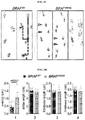
- FIGS. 3A-3D show constitutive expression of BRAF V600E in Csf1r+ cells.
- FIG. 3A is a graphic showing the breeding scheme.
- FIG. 3C Brightlight (upper panel) and epifluorescence microscopy (lower panel) of Csf1r iCre+ ; BRAF WT ; Rosa26 LSL-YFP and Csf1r iCre+ ; BRAF V600E ; Rosa26 LSL-YFP embryos showing hemorrhagic foci in the liver (arrow) and accumulation of YFP+ cells in fetal liver. Cross ( ⁇ ) indicates dead embryo.
- FIG. 3D Csf1r iCre+ ; BRAF V600E ; Rosa26 LSL-YFP mice are associated with 100% lethality beyond E14.5.
- FIG. 4 shows Tamoxifen-inducible mouse models used to target EMP versus HSC.
- Csfl MeriCreMer ; Rosa26 LSL-YFP do not target HSCs in adult mice, while pulse-labeling of Cxcr4 CreERT2+ ; Rosa26 LSL-YFP results in labeling of 10% HSCs.
- LSK Lin-Sca1+Kit+.
- FIGS. 5A-5E show the results of experiments with a Csf1r MeriCreMer ; BRAF V600E ; Rosa26 LSL-YFP inducible model.
- FIG. 5A Breeding scheme.
- FIGS. 5C and 5D Flow cytometric analysis and IF analysis of livers of 4-week old mice. YFP+ cells are F4/80hi CD11blo (gated on CD45+ cells).
- FIG. 5E Histologic analysis of the brain. Clusters of YFP+ cells in the cerebellum and brainstem. YFP+ cells also stain for CD68 (and Iba1, not shown).
- FIGS. 6A-6D show neurological disease in the Csf1r MeriCreMer ; BRAF V600E ; Rosa26 LSL-YFP model.
- FIG. 6A Test of limb-clasping reflexes of 6-8-month old mice. When lifted by the tail BRAF WT mice behave normally, extending their hind limbs. In contrast, BRAF V600E mice bend their legs towards their trunk.
- FIG. 6B Cumulative incidence rate of behavioral abnormalities.
- FIG. 6C Footprint assay assessing locomotion defects. Front paws were painted with red ink, hind paws with blue ink. BRAF V600E mice lose coordination; front/hind legs do not overlap, and later mice are paralyzed (far right panel).
- FIG. 6D Measurement of footprint assay. Numbers below graphs correspond to the measurements performed as displayed in ( FIG. 6C ).
- FIGS. 7A and 7B show histological analysis of the spinal cord in Cre+ BRAF V600E and littermate controls.
- FIG. 7A Iba1+ microglia accumulate in the white matter.
- FIG. 7B Flow cytometry analysis of F4/80+ macrophages from the spinal cord. Littermates are color-coded.
- FIGS. 8A and 8B show demyelination of the spinal cord of Cre+ BRAF V600E mice compared to wild type mice.
- FIG. 8A Luxol fast blue stain shows demyelination and leukocyte infiltration in the white matter of the spinal cord.
- FIG. 8B Flow cytometry analysis shows that CD3+ cells accumulate in the spinal cord in Cre+ BRAF V600E mice.
- FIGS. 9A and 9B show histological analysis of the spinal cord from 6-month old BRAF V600E and control mice. Immunostaining was performed with CD68, GFP, and pERK antibodies.
- FIG. 10 shows transcription factors that are differentially expressed between macrophages from the brain, liver, skin, kidney, and lung during development and in postnatal mice.
- FIGS. 11A-11C show the analysis of the liver in 6-8 month old Csf1r MeriCreMer ; BRAF V600E ; Rosa26 LSL-YFP and littermates.
- FIG. 11A Flow cytometry analysis of total and YFP+ Kupffer cells. Littermates are color-coded.
- FIG. 11B Histological analysis shows accumulation of YFP+ Kupffer cells (stained with a cross-reactive GFP antibody).
- FIG. 11C Serum analysis of liver enzymes.
- FIG. 12 shows cumulative incidence rate of behavioral abnormalities in Csf1r MeriCreMer ; BRAF V600E ; Rosa26 LSL-YFP mice placed on long-term PLX4720 diet (PLX diet) at 3 months of age, and mice on normal control diet (Ctrl diet).
- FIGS. 13A-13I Targeting BRAF V600E in tissue-resident macrophages.
- FIG. 13E Proportion of YFP + F4/80 + cells in tissues from 1-month-old mice. Circles represent individual mice. Unpaired two-tailed t-test.
- FIG. 13F A>T transversion encoding BRAF V600E in YFP + Kupffer cells at the Braf locus. Red and blue bars indicate forward and reverse strands.
- FIG. 13I Heatmap representation of selected genes from ( FIG. 13H ), values are displayed as z-score. See also FIG. 17 .
- FIGS. 14A-14H Neurodegenerative disease in BRAF VE mice.
- FIG. 14C Cumulative incidence of behavioral abnormalities in BRAF VE mice and controls. Log-rank (Mantel-Cox) test.
- FIG. 14D Overlap distance, and stride length of mice on control or PLX4270 diet from 1 month or 3 months of age. Average values ⁇ s.d.
- FIG. 14E Disease progression in mice from (d), average score excluding mice euthanized for paralysis ( ⁇ ).
- FIG. 14F Cumulative incidence of behavioral abnormalities in mice from (d). Log-rank (Mantel-Cox) test
- One-way ANOVA See also FIGS. 20 and 22 .
- FIGS. 15A-15G ERK activation in BRAF V600E microglia.
- FIG. 15B pERK + microglia in brainstem. Circles represent individual mice. One-way ANOVA.
- FIG. 15A ERK activation in BRAF V600E microglia.
- FIG. 15B pERK + microglia in brainstem. Circles represent individual mice.
- FIG. 15C ERK phosphorylation in spinal cords and brains from 6-9 month-old mice. Top: representative western blot, bottom: p
- FIG. 15E Numbers of microglia from 5-9 month-old mice Circles represent individual mice. One-way ANOVA.
- FIGS. 16A-16I Molecular features of ERK-activated microglia, and their presence in histiocytoses patients.
- FIG. 16A GSEA of differentially expressed genes in YFP + microglia from 6-7 month-old littermates. q-value ⁇ 0.05. EMT: epithelial-mesenchymal transition.
- FIG. 16B Heatmap representation of selected DEG, values are displayed as z-score.
- FMO fluorescence minus one.
- FIG. 16E Bioplex analysis of Il1b and Il17a in spinal cords from 6-9 month-old mice. Circles represent individual mice. Unpaired two-tailed t-test.
- FIG. 16G CD163, pERK and BRAF V600E expression in Erdheim-Chester disease (ECD) brain tissue. Scale bar upper panels: 40 ⁇ m, lower panels: 5 ⁇ m.
- FIGS. 17A-17H Analysis of one-month old Csf1r MeriCreMer ; BRAF LSL-V600E Rosa26 LSL-YFP mice.
- FIG. 17C Flow cytometry analysis of YFP + cells in the liver.
- FIG. 17E Total tissue-resident macrophages cell numbers per gram ( FIG.
- FIGS. 18A-18F Effect of constitutive BRAF V600E expression in Csf1r-expressing cells.
- FIG. 18A Breeding scheme.
- FIG. 18A Breeding scheme.
- FIG. 18A Breeding scheme.
- FIG. 18B Embryonic lethality of Csf1r iCre+ ; BRAF LSL-V600E ; Rosa26L
- FIG. 18D Mouse embryos found alive during different developmental stages. Csf1r iCre+ ; BRAF LSL-V600E ; Rosa26 LSL-YFP mice are associated with 100% lethality beyond E14.5.
- FIG. 18E Liver weight in E12.5 embryos. Circles represent individual mice.
- FIGS. 19A-19G Analysis of CD11c Cre ; BRAF V600E mouse model.
- FIG. 19B Representative photographs of lung and spleen from BRAF VE mice at time of death with representative BRAF WT control organs.
- FIGS. 19C and 19D Hematoxylin and eosin (HE) stain of lung tissue from BRAF VE and littermate controls.
- FIG. 19E CD68 immunohistochemistry (IHC) of BRAF VE lung tissue.
- FIG. 19B Representative photographs of lung and spleen from BRAF VE mice at time of death with representative BRAF WT control organs.
- FIGS. 19F HE stain of liver tissue from BRAF VE and littermate controls with magnified view of granuloma in the BRAF VE liver.
- FIGS. 20A-20G Longitudinal study and PLX treatment of the Csf1r MeriCreMer ; BRAF V600E ; Rosa26 LSL-YFP mice.
- FIG. 20A Rotarod assay at 1 to 4 months of age. Values are mean ⁇ s.d.
- FIG. 20B Rotarod and footprint assay at 4 months of age displaying single values. Mice that are score 1 are labeled in red.
- FIG. 20D Representative weight curve of BRAF WT and BRAF VE mice on control or PLX4720 diet.
- FIG. 20D Representative weight curve of BRAF WT and BRAF VE mice on control or PLX4720 diet.
- FIG. 20E PLX4720 concentration in serum (ng
- FIG. 20G Disease progression for BRAF VE mice on control or PLX4720 diet. ⁇ indicates animal death due to paralysis.
- FIGS. 21A and 21B Microglia activation in the brain starts at early, preclinical stages.
- FIGS. 22A-22D Neurodegenerative process in BRAF VE mice.
- FIG. 22B Immunohistochemistry and immunofluorescence as used for quantification in FIG.
- FIGS. 23A-23G Microglia and T-cell phenotype in BRAF VE mice.
- FIG. 23B Representative tSNE analysis of flow cytometry staining of CD
- FIGS. 23F and 23G Analysis of CD8 + , CD4 + and Foxp3 + T-cell numbers ( FIG. 23F ) and proliferation ( FIG.
- FIGS. 24A-24F BRAF VE mice analysis outside the central nervous system.
- FIG. 24B Analysis of liver Kupffer cells as in ( FIG.
- FIGS. 25A-25C Erdheim-Chester disease (ECD) patients.
- FIG. 25A Table summarizing observed pathological and molecular findings in brain tissue of three ECD patients with neurologic presentations. BRAF status was determined by immunohistochemical analysis and by sequencing. Neuronal loss and demyelination was determined by immunohistochemistry of neurofilament and myelin basic protein (MBP). RF: Rosenthal fiber. n/a: not applicable/no tissue available for further analysis.
- FIGS. 26A-26C Isolation of microglia, neuron, and glia nuclei by cell sorting in a patient with neurodegeneration.
- FIG. 26A Nuclei extracted from ⁇ 350 mg of fresh frozen control brain tissue using a non-ionic detergent buffer containing DAPI and flow-sorted using anti-NeuN and anti-Pu.1. DN: double negative.
- FIG. 26B Top panel: Nuclei extraction and flow-sorting using anti-Pu.1 and anti-NeuN antibodies was performed in four different brain regions, two affected and two non-affected, obtained from a deceased patient with histiocytosis and neurodegenerative disease. Double negative (DN), NeuN+, and Pu.1+ populations were sorted for sequencing.
- FIG. 26C FACS graph of nuclei isolated from 3 patients with Alzheimer's disease (AD), Parkinson's disease (PD), and Adult-Onset-Leukoencephalopathy with spheroids (ALSP).
- AD Alzheimer's disease
- PD Parkinson's disease
- ALSP Adult-Onset-Leukoencephalopathy with spheroids
- FIG. 26C Example of quality control of DNA obtained from AD patient. DNA is immediately isolated after nuclei sorting with QIAamp DNA Mini Kit (Qiagen). DNA quality is determined based on DNA Integrity Number (DIN) measured by Agilent 4200 TapeStation.
- DIN DNA Integrity Number
- FIGS. 27A-27B Analysis of BRAF V600E mutation in microglia, neuron, and glia nuclei from a 25-year old patient with neurodegeneration.
- FIG. 27A BRAF V600E allele frequency determined ddPCR in sorted nuclei from 8 brain areas from 1 patient with neurodegeneration, whole brain tissue and blood were processed for gDNA extractions using QIAamp DNA Mini Kit (Qiagen) and quality and quantity was determined with Agilent 4200 TapeStation.
- BRAF V600E allele frequency is determined by ddPCR in the same DNA samples before library preparation. Multiple numbers indicate independent determinations. Counts ⁇ 5 are not reproducible and below sensitivity level.
- BRAF V600E allele frequency determined by targeted deep sequencing (HEME-PACT, mean sequencing depth 409 ⁇ ) in sorted nuclei from four of the above brain areas, whole brain tissue, and blood. Approximately 200 ng of DNA were used for library preparation with KAPA HyperPrep Kit (Roche). Libraries are then captured with custom-designed biotinylated probes (NimbleGen) and sequenced with Illumina HiSeq 2500. Counts ⁇ 4 (1%) are considered below sensitivity level.
- FIG. 28 Hippocampus of patient with neurodegenerative disease due to BRAF V600E microglia. Immunohistochemistry analysis for CD163, Neurofilament-1 (NF), and GFAP of post-mortem brain tissue from the hippocampal area.
- NF Neurofilament-1
- FIG. 29 Preliminary analysis of somatic mosaicism from Heme-PACT targeted sequencing. Venn diagrams represent the number and repartition among cell types and tissues (brain regions and blood) of somatic variants (SNV and indel) detected by Heme-PACT sequencing and analysis. Variants with an allele frequency >35% are considered germline and eliminated from analysis. SNVs are validated bioinformatically and by ddPCR and Ampli-seq. This analysis suggests that mutations are either common to all tissues ($, with high 15%-35% allelic frequency) and considered early somatic events, or local to a cell type and brain region (with lower allelic frequencies) for neurons, microglia, and DN (double negative) cells suggesting their local development independent of blood cells. The 2 mutations common to hippocampus and cerebellum (*) are at higher allelic frequencies and correspond to BRAF V600E (11% and 21%) and a non-coding mutation in MSH6 (16% and 26%).
- FIGS. 31A-31B Neurodegenerative disease in PIK3CA HR mice.
- FIG. 31A Cumulative incidence of behavioral abnormalities in PIK3CA HR mice and control.
- FIG. 31B Footprint assay example, which measures the overlap distance between hind and front paws.
- the embryonic stages used herein relate to embryonic day (for example D1 is E1.0) of mouse development. This staging by “days” relate to in the female presence of a vaginal plug indicating that the mating occurred.
- prevention refers to one or more compounds that, in a statistical sample, reduces the occurrence of the disorder or condition in the treated sample relative to an untreated control sample, or delays the onset of one or more symptoms of the disorder or condition relative to the untreated control sample.
- the terms “subject,” “individual,” or “patient” can be an individual organism, a vertebrate, a mammal, or a human.
- a “therapeutically effective amount” of a compound refers to compound or agent levels in which the physiological effects of a disease or disorder are, at a minimum, ameliorated.
- a therapeutically effective amount can be given in one or more administrations.
- the amount of a compound which constitutes a therapeutically effective amount will vary depending on the compound, the disorder and its severity, and the general health, age, sex, body weight and tolerance to drugs of the subject to be treated, but can be determined routinely by one of ordinary skill in the art.
- Treating,” “treat,” or “treatment” as used herein covers the treatment of a disease or disorder described herein, in a subject, such as a human, and includes: (i) inhibiting a disease or disorder, i.e., arresting its development; (ii) relieving a disease or disorder, i.e., causing regression of the disorder; (iii) slowing progression of the disorder; and/or (iv) inhibiting, relieving, or slowing progression of one or more symptoms of the disease or disorder.
- the various modes of treatment or prevention of medical diseases and conditions as described are intended to mean “substantial,” which includes total but also less than total treatment or prevention, and wherein some biologically or medically relevant result is achieved.
- the treatment may be a continuous prolonged treatment for a chronic disease or a single, or few time administrations for the treatment of an acute condition.
- administer refers to either directly administering a therapeutic agent, such as, but not limited to, a BRAF, MEK, or CSF-1R inhibitor, or a combination of thereof, to a subject.
- a therapeutic agent such as, but not limited to, a BRAF, MEK, or CSF-1R inhibitor, or a combination of thereof, to a subject.
- routes of administration include, for example: intradermal, intramuscular, intraperitoneal, intravenous, subcutaneous, intranasal, epidural, oral, sublingual, intranasal, intracerebral, intravaginal, transdermal, rectally, by inhalation, or topically, particularly to the ears, nose, eyes, or skin.
- the administering is effected orally or by parenteral injection.
- the mode of administration can be left to the discretion of the practitioner, and will depend in-part upon the site of the medical condition. In most instances, administration results in the release of any agent described herein into the bloodstream. In specific embodiments, it may be desirable to administer locally to the area in need of treatment or blocking.
- tissue or cellular niche where the resident macrophages are found.
- the technology of the present disclosure relates to the observation that conditional expression of a BRAF V600E allele in EMP clones in vivo does not grossly affect embryonic development, but results in the accumulation of BRAF V600E macrophage clones in various tissues, and is responsible for a neurodegenerative syndrome in adult mice that recapitulates for the first time one of the most poorly understood and adverse characteristics of human histiocytic neoplasms.
- Characterizing a yolk sac origin for Erdheim-Chester disease (ECD) and Langerhans Cell Histiocytosis (LCH) carries significant implications for the pathophysiology of these diseases, and in particular may illuminate the cause of neurodegenerative syndromes as well as liver/lung fibrosis in these disorders, which are only partially sensitive or even refractory to current therapy.
- ECD Erdheim-Chester disease
- LCH Langerhans Cell Histiocytosis
- somatic mutations that confer a proliferative, survival, or activation advantage called “oncogenic” or “driver” mutations
- oncogenic or driver mutations a proliferative, survival, or activation advantage
- progress in sequencing technologies has revealed a high burden of somatic mutations, including driver mutations, in normal (non-tumoral) tissues and their association with neurodevelopmental disorders, neurodegeneration, and non-cancerous proliferative diseases.
- driver mutations include driver mutations, in normal (non-tumoral) tissues and their association with neurodevelopmental disorders, neurodegeneration, and non-cancerous proliferative diseases.
- inherited mutations that activate the RAS/MAPK/ERK pathway are incompatible with life, or cause severe and early-onset tumoral, developmental, and neurodevelopmental disorders (“RASopathies”) and are therefore rarely observed.
- RASopathies severe and early-onset tumoral, developmental, and neurodevelopmental disorders
- somatic mosaicism for such mutations is frequently observed in “normal” tissues and results in distinct phenotypes that vary according to several factors related to the time at which mutations occur, including the cellular lineage(s) harboring the mutation, the size of the clone, defects in DNA repair, and the occurrence of secondary hits.
- mosaicism for mutations activating the RAS pathway in the early embryo can cause RASopathies
- mosaicism restricted to melanocytes is responsible for limited melanocyte proliferation and activation and give rise to the common and giant nevi.
- Mosaicism for mutations activating the RAS pathway in macrophages causes histiocytic lesions, but causes leukemia in hematopoietic cells.
- Tissue resident macrophages such as microglia in mice originate from erythro-myeloid precursors (EMP) after gastrulation. EMP-derived cells colonize the brain during embryogenesis to give rise to microglia that self-maintain in the adult brain due to their long life span and slow local proliferation. Therefore, resident macrophages, including microglia, develop and maintain independently from the hematopoietic stem cell (HSC) lineage that give rise to blood cells.
- HSC hematopoietic stem cell
- BRAF V600E mutation the most frequent mutation that drives constitutive activation of the RAS-MEK-ERK pathway was expressed in a mosaic manner in EMPs in mouse embryos.
- somatic mosaicism for the BRAF V600E in a limited number of embryonic EMP precursors does not have detectable effects on development but results in mosaicism of mutant macrophages well tolerated in all organs, except for the brain, where positive selection of microglia mutant clones is associated with the progressive development of the cardinal features of neurodegeneration: microgliosis, astrogliosis, synaptic loss, and neuronal death.
- PLX BRAF V600E inhibitor
- tissue macrophages originate and renew from hematopoietic stem cells (HSCs) via circulating progenitors such as blood monocytes.
- HSCs hematopoietic stem cells
- recent studies have led to a major paradigm shift identifying that tissue-resident macrophages, including brain microglia, liver Kupffer cells, kidney macrophages, and Langerhans cells of the epidermis, persist in adult tissues with little contribution from bone marrow progenitors and monocytes.
- EMP yolk sac erythro-myeloid progenitors
- Tissue-Resident Macrophages Originate at Least in Part from EMPs from the Yolk Sac.
- EMPs emerge from yolk sac hemogenic endothelium at embryonic day (E)8.25. They are distinguished from HSCs by the lack of lymphoid potential, both in vitro and in vivo, the lack of long term repopulating potential, and cell surface phenotype (Sca1 ⁇ CD45 low AA4.1 + , CD41 + , Fc ⁇ RII/III + ). EMPs express Myb but, in contrast to HSCs, their commitment and differentiation into myeloid fate is unaltered in Myb-deficient embryos, although their erythroid potential is blocked. In vivo, EMPs are generated from Tie2 + yolk sac ancestors from E7.5 to E10.5. They seed the fetal liver as early as E9.5.
- EMPs and EMP-derived Kit + cells can be detected until E14.5.
- EMPs are thus distinct from HSCs, which develop from the intra-embryonic hemogenic endothelium around embryonic day E10.5, require the transcription factor Myb to colonize the fetal liver and later the bone marrow, and give rise to specialized hematopoietic cells, including myeloid cells in postnatal mice.
- the present disclosure is based on several key observations (summarized below) that demonstrate that tissue-resident macrophages originate in large part from EMPs, and provide novel experimental tools and strategies for the study of histiocytoses.
- the majority of resident tissue macrophages that are present in mouse tissues originate from Kit + , CD45 low , AA4.1 + , Csf1r + yolk sac early hematopoietic progenitors (defined as EMP) and can self-maintain in adults.
- Resident Macrophages are a Diverse Family of Professional Phagocytes and “Accessory Cells” Involved in Tissue Remodeling.
- Resident macrophages are sessile but continuously explore their immediate environment/niche using motile filopodia, recognize, and scavenge pathogens as well as unfit cells and cell debris, glycoproteins and lipids, and produce a large range of bioactive molecules and growth factors.
- macrophages are found all over the body within each specialized tissue and their serous membranes.
- Long elongated dendritic filopodial processes frequently extend from their cell bodies and build up 3D network-like structures that provide for extensive contacts with specialized cells such as brain, liver, kidney, or epidermal cells, thereby allowing constant surveillance or scavenging of their tissue.
- Macrophages are involved in the clearance of apoptotic and senescent cells during organogenesis, in the brain, limbs and lung, in branching morphogenesis, and regulate blood and lymphatic vessel morphogenesis and maturation during fetal and postnatal development. They pursue this task in adults, contribute to the pruning of neuronal synapses, and scavenge and digest nuclei released daily by billions (2 ⁇ 10 11 in humans) of maturing erythroblasts. Altogether, a considerable literature indicates that macrophages play a role as regulators of morphogenesis, homeostasis and tissue remodeling, inflammatory processes and tumor growth.
- monocytes and macrophages are relevant to disease pathogenesis and may be relevant to the pathophysiology of histiocytoses.
- Rossi and colleagues demonstrated that the two cell types have distinct expansion mechanisms and distinct functions in the brain during Experimental Auto-immune Encephalitis (EAE); infiltrating monocytes are recruited via extravasation from blood vessels and produce inflammatory mediators important for disease progression but do not persist after the resolution of inflammation, while in contrast activated resident microglia proliferate locally, persist, and return to quiescence following remission.
- EAE Experimental Auto-immune Encephalitis
- pleural resident macrophages adopt “anti-inflammatory” phenotypes in host carrying helminths while passenger monocytes exhibit pro-inflammatory responses.
- tissue macrophages can exert distinct functions within the same environment and in response to the same challenge, suggesting that tissue cues or polarizing signals may not suffice to account for their functions.
- tissue macrophages it might not be always accurate, however, to describe tissue macrophages as being anti-inflammatory as opposed to passenger monocytes being pro-inflammatory.
- proliferating resident microglial cells were proposed to contribute to neuronal damage during the development of the disease.
- somatic BRAF V600E mutations in EMPs may be causative of histiocytoses
- BRAF V600E somatic mutations in HSCs and EMPs may have different consequences at the clinical, cellular, and molecular levels, and may thus contribute to the clinical heterogeneity of the disease and influence its prognosis and clinical course.
- EMPs are not self-renewing and disappear in the late embryo the mutation would be detectable only in their progeny, the resident macrophages, and not in HSCs.
- modeling BRAF V600E mutations in EMPs in mice is a strategy of choice to understanding its role in resident macrophage biology, should provide insight into the role of macrophages in promoting neurological dysfunction as well as lung and liver fibrosis, and may thus illuminate the pathophysiology of histiocytoses.
- the technology of the present disclosure relates to a method for preventing or treating BRAF V600E -associated neurodegeneration by administering to a subject in whom BRAF V600E+ resident macrophages have been identified in the neuronal microenvironment of the subject a therapeutically effective amount of a BRAF, MEK, PI 3-kinase, Ras, and/or CSF-1R inhibitor.
- Suitable BRAF inhibitors include, but are not limited to, vemurafenib, dabrafenib, encorafenib, PLX7904, PLX8394, GDC-0879, LGX818, and PLX4720.
- Suitable MEK inhibitors include, but are not limited to, AZD8330, refametinib, E6201, MEK162 (binimetinib), PD0325901, pimasertib, R04987655, selumetinib, TAK-733, GDC-0623, WX-544, cobimetinib, and trametinib.
- Suitable PI 3-kinase inhibitors include, but are not limited to, idelalisib, BKM120, GDC-0980, PF-04691502, XL147, IPI-145, BYL719, SF1126, BAY80-6946, GSK2126458, NVP-BEZ235, GDC-0941, PX-866, XL765, and ZSTK474.
- Suitable Ras inhibitors include, but are not limited to, salirasib and TLN-4601.
- Suitable CSF-1R inhibitors include, but are not limited to, GW2580, BLZ945, pexidartinib (PLX3397), ARRY-382, PLX7486, and JNJ-40346527.
- One aspect of the present technology includes methods of treating BRAF V600E -associated neurodegenerative disease in a subject diagnosed as having, suspected as having, or at risk of having one or more BRAF V600E mutations comprising the administration of a BRAF, MEK, PI 3-kinase, Ras, and/or CSF-1R inhibitor.
- the present technology includes methods of treating neurodegenerative disease in a subject in need thereof, wherein the neurodegenerative disease is characterized by cells expressing one or more BRAF V600E mutations.
- the cells are resident macrophages in the central nervous system, or microglia.
- the neurodegenerative disease is Alzheimer's disease, Parkinson's disease, Huntington's disease, amyotrophic lateral sclerosis (ALS), or multiple sclerosis.
- treatment of a BRAF V600E -associated neurodegenerative disease comprises relieving one or more symptoms selected from the group consisting of: impaired cognitive functions and dementia, ataxia, dysarthria, decreased motor coordination and synchrony, paralysis, microglia accumulation, astrogliosis, microglia phagocytosis, demyelination, neuronal loss, synaptic loss, and amyloid precursor protein (APP) expression.
- impaired cognitive functions and dementia ataxia, dysarthria, decreased motor coordination and synchrony
- paralysis microglia accumulation, astrogliosis, microglia phagocytosis, demyelination, neuronal loss, synaptic loss, and amyloid precursor protein (APP) expression.
- APP amyloid precursor protein
- One aspect of the present technology includes methods of treating phosphoinositide 3-kinases (PI 3-kinase)-associated neurodegenerative disease in a subject diagnosed as having, suspected as having, or at risk of having one or more mutations in one or more PI 3-kinases comprising the administration of a BRAF, MEK, PI 3-kinase, Ras, and/or CSF-1R inhibitor.
- the present technology includes methods of treating neurodegenerative disease in a subject in need thereof, wherein the neurodegenerative disease is characterized by cells expressing one or more mutations in one or more PI 3-kinases.
- the cells are resident macrophages in the central nervous system, or microglia.
- the neurodegenerative disease is characterized by at least a portion of the resident macrophages in the central nervous system of the subject being PIK3CA H1047R+ .
- the neurodegenerative disease is Alzheimer's disease, Parkinson's disease, Huntington's disease, or amyotrophic lateral sclerosis (ALS).
- treatment of a PI 3-kinase-associated neurodegenerative disease comprises relieving one or more symptoms selected from the group consisting of: ataxia, decreased motor coordination and synchrony, paralysis, microglia accumulation, astrogliosis, microglia phagocytosis, demyelination, neuronal loss, synaptic loss, and amyloid precursor protein (APP) expression in the brain.
- symptoms selected from the group consisting of: ataxia, decreased motor coordination and synchrony, paralysis, microglia accumulation, astrogliosis, microglia phagocytosis, demyelination, neuronal loss, synaptic loss, and amyloid precursor protein (APP) expression in the brain.
- APP amyloid precursor protein
- compositions or medicaments comprising a BRAF inhibitor, MEK inhibitor, Ras inhibitor, PI 3-kinase inhibitor, CSF-1R inhibitor, or a combination thereof, as disclosed herein, are administered to a subject suspected of, or already suffering from a neurodegenerative disorder, in an amount sufficient to cure, or at least partially arrest, the symptoms of the disease, including its complications and intermediate pathological phenotypes in development of the disease.
- the BRAF inhibitor is selected from the group consisting of vemurafenib, dabrafenib, encorafenib, PLX7904, PLX8394, GDC-0879, LGX818, and PLX4720.
- the BRAF inhibitor is PLX4720.
- the MEK inhibitor is selected from the group consisting of AZD8330, refametinib, E6201, MEK162 (binimetinib), PD0325901, pimasertib, R04987655, selumetinib, TAK-733, GDC-0623, WX-544, cobimetinib, and trametinib.
- the Ras inhibitor is selected from the group consisting of salirasib and TLN-4601.
- the PI 3-kinase inhibitor is selected from the group consisting of idelalisib, BKM120, GDC-0980, PF-04691502, XL147, IPI-145, BYL719, SF1126, BAY80-6946, GSK2126458, NVP-BEZ235, GDC-0941, PX-866, XL765, and ZSTK474.
- the CSF-1R inhibitor is selected from the group consisting of GW2580, BLZ945, pexidartinib (PLX3397), ARRY-382, PLX7486, and JNJ-40346527.
- the BRAF inhibitor is selected from the group consisting of vemurafenib, dabrafenib, encorafenib, PLX7904, PLX8394, GDC-0879, LGX818, and PLX4720. In some embodiments, the BRAF inhibitor is PLX4720.
- the MEK inhibitor is selected from the group consisting of AZD8330, refametinib, E6201, MEK162 (binimetinib), PD0325901, pimasertib, R04987655, selumetinib, TAK-733, GDC-0623, WX-544, cobimetinib, and trametinib.
- the Ras inhibitor is selected from the group consisting of salirasib and TLN-4601.
- the PI 3-kinase inhibitor is selected from the group consisting of idelalisib, BKM120, GDC-0980, PF-04691502, XL147, IPI-145, BYL719, SF1126, BAY80-6946, GSK2126458, NVP-BEZ235, GDC-0941, PX-866, XL765, and ZSTK474.
- the CSF-1R inhibitor is selected from the group consisting of GW2580, BLZ945, pexidartinib (PLX3397), ARRY-382, PLX7486, and JNJ-40346527. Suitable methods include in vitro, ex vivo, or in vivo methods.
- In vivo methods typically include the administration of BRAF, MEK, PI 3-kinase, Ras, and/or CSF-1R inhibitor, such as those described above, to a mammal, suitably a human.
- BRAF, MEK, PI 3-kinase, Ras, and/or CSF-1R inhibitor inhibitors are administered to the subject in effective amounts (i.e., amounts that have desired therapeutic effect).
- effective amounts i.e., amounts that have desired therapeutic effect.
- the dose and dosage regimen will depend upon the degree of the neurodegenerative disease in the subject, the characteristics of the particular BRAF, MEK, PI 3-kinase, Ras, and/or CSF-1R inhibitor used, e.g., its therapeutic index, the subject, and the subject's history.
- the effective amount may be determined during pre-clinical trials and clinical trials by methods familiar to physicians and clinicians.
- An effective amount of a particular BRAF, MEK, PI 3-kinase, Ras, and/or CSF-1R inhibitor useful in the methods of the present technology may be administered to a mammal in need thereof by any of a number of well-known methods for administering pharmaceutical compounds.
- the BRAF, MEK, PI 3-kinase, Ras, and/or CSF-1R inhibitor may be administered systemically or locally.
- compositions are typically formulated to be compatible with its intended route of administration.
- routes of administration include, but are not limited to orally, intranasally, parenterally (e.g., intravenously, intramuscularly, intraperitoneally, intradermally, or subcutaneously), systemically, transdermally, iontophoretically, intradermal, intraocularly, or topically.
- Dosage, toxicity and therapeutic efficacy of any therapeutic agent can be determined by standard pharmaceutical procedures in cell cultures or experimental animals, e.g., for determining the LD50 (the dose lethal to 50% of the population) and the ED50 (the dose therapeutically effective in 50% of the population).
- the dose ratio between toxic and therapeutic effects is the therapeutic index and it can be expressed as the ratio LD50/ED50.
- Compounds that exhibit high therapeutic indices are advantageous. While compounds that exhibit toxic side effects may be used, care should be taken to design a delivery system that targets such compounds to the site of affected tissue in order to minimize potential damage to uninfected cells and, thereby, reduce side effects.
- the data obtained from the cell culture assays and animal studies can be used in formulating a range of dosage for use in humans.
- the dosage of such compounds may be within a range of circulating concentrations that include the ED50 with little or no toxicity.
- the dosage may vary within this range depending upon the dosage form employed and the route of administration utilized.
- the therapeutically effective dose can be estimated initially from cell culture assays.
- a dose can be formulated in animal models to achieve a circulating plasma concentration range that includes the IC 50 (i.e., the concentration of the test compound which achieves a half-maximal inhibition of symptoms) as determined in cell culture.
- IC 50 i.e., the concentration of the test compound which achieves a half-maximal inhibition of symptoms
- levels in plasma may be measured, for example, by high performance liquid chromatography.
- an effective amount of the BRAF, MEK, PI 3-kinase, Ras, and/or CSF-1R inhibitor ranges from about 0.000001 mg per kilogram body weight per day to about 10,000 mg per kilogram body weight per day. In some embodiments, the dosage ranges are from about 0.0001 mg per kilogram body weight per day to about 100 mg per kilogram body weight per day. In some embodiments, the dosage ranges are from about 0.0001 mg per kilogram body weight per day to about 50 mg per kilogram body weight per day.
- dosages can be 1 mg/kg body weight, 10 mg/kg body weight, or 50 mg/kg body weight every day, every two days or every three days or within the range of 1-50 mg/kg every week, every two weeks, or every three weeks.
- a single dosage of a BRAF, MEK, PI 3-kinase, Ras, and/or CSF-1R inhibitor ranges from 0.001-10,000 micrograms per kg body weight.
- BRAF, MEK, PI 3-kinase, Ras, and/or CSF-1R inhibitor concentrations in a carrier range from 0.2 to 2000 micrograms per delivered milliliter.
- An exemplary treatment regime entails administration once per day or once a week. In therapeutic applications, a relatively high dosage at relatively short intervals is sometimes required until progression of the disease is reduced or terminated, or until the subject shows partial or complete amelioration of symptoms of disease. Thereafter, the patient can be administered a prophylactic regimen.
- treatment of a subject with a therapeutically effective amount of the therapeutic compositions described herein can include a single treatment or a series of treatments.
- the mammal treated in accordance with the present methods can be any mammal, including, for example, farm animals, such as sheep, pigs, cows, and horses; pet animals, such as dogs and cats; laboratory animals, such as rats, mice and rabbits.
- the mammal is a human.
- EMPs appear in the yolk sac at embryonic day (E) 8.5 and express the Csf1 receptor (Csf1r). They colonize the fetal liver from E9.5 and give rise to macrophage precursors (pMacs) that distribute in embryonic tissues and differentiate into tissue-specific macrophage subsets such as microglia in the central nervous system. Therefore, to investigate the consequences of BRAF V600E expression in the EMP lineage, EMPs were genetically targeted by pulse labeling Csf1r MeriCreMer ; BRAF LSL-V600E ; Rosa26 LSL-YEP E8.5 embryos with 4-hydroxytamoxifen (4-OHT, Sigma).
- Embryonic development was estimated considering the day of vaginal plug formation as 0.5 days post-coitum (dpc). Cre recombination in Csf1r MeriCreMer ; Rosa26 LSL-YFP ; BRAF LSL-V600E embryos was induced by single injection of 37.5 mg per kg (body weight) of OH-TAM into pregnant females.
- Short-lived 4-OHT leads to transient nuclear translocation of the estrogen receptor-Cre recombinase fusion protein (MeriCreMer) in cells expressing the Csfr1 MeriCreMer transgene and deletion of a floxed stop cassette (LSL) in the BRAF LSL-V600E and Rosa26L SL-YFP alleles.
- OH-TAM was supplemented with 18.75 mg per kg (body weight) progesterone (Sigma) to counteract the mixed oestrogen agonist effects of tamoxifen, which can result in fetal abortions.
- mice Csf1r MeriCreMer , Csf1r iCre , CD11 Cre , Rosa26 YFP-LSL mice and BRAF LSL-V600E mice kindly provided by C. Pritchard (Leicester, UK) were maintained under SPF conditions. Animal procedures were performed in adherence to our project licenses issued by the Institutional Review Board (IACUC 15-04-006 and 13-04-003) from MSKCC. Genotyping was performed according to protocols described previously for Csf1r iCre Csf1r MeriCreMer , CD11 Cre and BRAF LSL-V600E mice.
- Cre recombination in Csf1r MeriCreMer ; Rosa26 LSL-YFP ; BRAF LSL-V600E embryos was induced by single injection at E8.5 of 37.5 mg per kg (body weight) of 4-hydroxytamoxifen (OH-TAM, Sigma) into pregnant females.
- OH-TAM 4-hydroxytamoxifen
- Sigma 4-hydroxytamoxifen
- OH-TAM was supplemented with 18.75 mg per kg (body weight) progesterone (Sigma) to counteract the mixed oestrogen agonist effects of tamoxifen, which can result in fetal abortions.
- Embryonic development was estimated considering the day of vaginal plug formation as 0.5 dpc.
- mice were placed on ad libitum PLX4720 diet at 1 or 3 months of age (PLX4720-containing chow 417 ppm, provided by Plexxikon Inc.).
- BRAF WT and BRAF VE male and female littermates were assigned randomly into the control or treated group. Scoring of mice was performed blinded and at least weekly by assessing hindlimb reflexes and other behavioral phenotypes such as axial rolling. The investigators were not blinded to allocation during experiments and outcome assessment.
- Footprint analysis Mice were given two trials to run down a runway before the experiment. Mouse forepaws and hindpaws were painted with red and blue ink, respectively. Mice were then allowed to run down a runway lined with white paper. At least four steps from the middle portion of each run were measured for (1) overlap between forepaw and hindpaw placement, (2) stride length, (3) front-base width (the distance between the right and left forelimb strides), and (4) hind-base width (the distance between the right and left hindlimb strides). Mean values were used for graphs and statistical analyses.
- Rotarod assay was conducted using a ROTO-ROD series 8 (IITC Life Sciences) with accelerating speed (accelerated from 4 to 40 rpm over 120 s). The mice were trained on the accelerating rotarod with 3 training session per day for 3 days. On the day of the experiment, the mean latency to fall off the rotarod recorded in the 3 trials was used in the analysis.
- mice Preparation of cell suspensions, flow cytometry and cell sorting. Pregnant females were killed by exposure to CO2. Embryos were removed from the uterus, washed in 4° C. phosphate-buffered saline (PBS, Invitrogen) and dissected under a Leica M80 microscope. For blood phenotyping of adult mice, mice were anaesthetized and blood was collected by cardiac puncture. Bone marrow was collected by flushing one leg with 5 ml RPMI (Invitrogen).
- PBS phosphate-buffered saline
- organs were incubated in PBS containing 1 mg/ml collagenase D (Roche), 100 U/ml DNase I (Sigma), 2.4 mg/ml of dispase (Invitrogen) and 3% FCS (Invitrogen) at 37° C. for 30 min prior to mechanical disruption.
- PBS containing 1 mg/ml collagenase D (Roche), 100 U/ml DNase I (Sigma), 2.4 mg/ml of dispase (Invitrogen) and 3% FCS (Invitrogen) at 37° C. for 30 min prior to mechanical disruption.
- FCS Invitrogen
- tissues were digested for 30 min at RT in PBS containing 2 mg/ml of collagenase D (Roche), 200 U/ml DNase I (Sigma), 4.8 mg/ml of dispase (Invitrogen), 3% FCS (Invitrogen) and 1 uM of flavopiridol (Sigma) followed by mechanical disruption under a 100 um filter.
- Cell suspensions were centrifuged at 320 g for 7 min, resuspended in FACS buffer (PBS, 0.5% BSA and 2 mm EDTA) containing purified anti-CD16/32 (1:100 dilution) and 5% normal mouse, 5% normal rat and 5% normal rabbit serum and incubated for 15 min at 4° C.
- cell suspensions were purified by a Percoll (Sigma) gradient (70/37/30%), and cells were collected from the 70/37 interface. After washing twice with PBS, cells were stained with a viability dye (Ghost Dye Red 780, Tonbo Biosciences) according to the manufacturer's protocol. Cells were first stained with antibodies for surface markers (see Supplementary Table 5), then processed for intracellular stainings using the Foxp3/Transcription Factor Staining Buffer Set (Affymetrix eBioscience) according to the manufacturer's protocol. Cell numbers per organ or per gram of tissue were calculated as follows.
- RNA-seq libraries were mapped to the Mouse genome mm10 using the Star aligner that maps reads gnomically and resolves reads across splice junctions.
- Several QC metrics were used for the RNA-seq library, including intronexon ratio, intragenic reads fraction, and GC bias. Exon and gene expression were quantified using Sailfish against the Mus musculus transcriptome GRCh38. After merging technical replicates, differential expression tests were performed using the DESEQ2 algorithm. Genes that had a FDR ⁇ 0.01 were considered to be significantly different between genotypes and ranked significant genes by fold-change with a cutoff of 1.
- Gene set enrichment analysis on KEGG pathway, GO term, Reactome, and MSigDB gene set collection was done using the over-representation test with hypergeometric model to assess whether the number of selected genes associated with disease is larger than expected.
- RNA-seq generation and analysis of human brain RNA-seq. Snap frozen normal brain tissues were obtained from the MSKCC Medical Donation Program. RNA was extracted using the Qiagen all prep DNA/RNA mini kit (Cat #80204) according to the manufacturer's instructions. RNAs were submitted to ribogreen quantification and quality control on Agilent BioAnalyzer. Average amount was 5.3 ug, Average RIN was 8.9. 500 ng of total RNA underwent polyA selection and Truseq library preparation according to instruction provided by Illumina (TruSeqTM RNA Sample Prep Kit v2), with 8 cycles of PCR.
- Illumina TruSeqTM RNA Sample Prep Kit v2
- RNA-seq libraries Several QC metrics were used for the RNA-seq library, including intronexon ratio, intragenic reads fraction, and GC bias. Exon and gene expression were quantified using Salmon against Homo sapiens transcriptome GRCh37. Differential expression tests, statistical tests and pathway analysis was performed as described above for murine RNA-seq.
- Embryos were imaged using a Leica M80 or Zeiss Axio Zoom.V16. Tissues were fixed for 1-3 days in 4% formaldehyde (Sigma). After fixation, tissues for cryosections were incubated overnight in 30% sucrose and embedded in OCT compound (Sakura Finetek). Cryoblocks were cut at a thickness of 16 ⁇ m for liver and 50 ⁇ m for spinal cord and brain and then blocked with PBS containing 10% normal goat serum (Invitrogen); 1% BSA (w/v); 0.3% Triton X-100 for 1 hour at room temperature. Livers were incubated overnight, brains and spinal cord for 48 hours at 4° C.
- rat anti-mouse F4/80 (1:300, cat no: MCA497GA, Biorad), rabbit anti-mouse Iba1 (1:300; cat no: 019-19741, Wako), chicken anti-GFP for YFP detection (1:500, cat no: A10262, Invitrogen), rabbit anti-pHis3 (1:100, cat no: sc-8656-R, Santa Cruz), goat anti-IL-1b (1:40, cat no: AF-401-SP, R&D), rabbit anti-pERK1/2 (1:100, cat no: #4370, Cell signaling), rabbit anti-Collagen IV (1:100, cat no: 2150-1470, Biorad), rabbit anti-Collagen VI (1:200, cat no: ab6588, Abcam), rabbit anti-cleaved Caspase3 (1:600, cat no: #9661, Cell Signaling), rat anti-Ki-67 (1:200, cat no: 14-5698-80, eBioscience).
- LLB-PAS luxol fast blue
- Primary antibodies were rabbit anti-CD68 (5 ⁇ g/ml, Boster Biological Technology, cat. no: PA1518), rabbit anti-pERKl/2 (1 ⁇ g/ml, cat no: #4370, Cell signaling), chicken anti-GFP for YFP detection (1:5000, cat no: AB13970, Abcam), mouse anti-APP (1:3000, cat no: MAB348, Millipore), rabbit anti-Iba-1 (1:500, cat no: 019-19741, Wako), rabbit anti-GFAP (1:200, cat. no.
- nuclei were counterstained with 4,6-diamidino-2-phenylindole (DAPI, cat. no. 236276, Boehringer). All slides were mounted in Vectashield (Vector Laboratories). Images were taken using a Zeiss Lab.A1 or BZ-9000 Biorevo microscope (Keyence) and analyzed using the BZ-II Analyzer (Keyence). For quantification of cell numbers, at least three parasagittal sections per mouse were analyzed. In cases, where the cell density was too high and single cells could not be quantified, the immunolabeled area size per brain section was calculated.
- DAPI 4,6-diamidino-2-phenylindole
- Imaging was performed using an Olympus Fluoview 1000 confocal laser scanning microscope using a 20 ⁇ 0.95 NA objective.
- Z-stacks with 0.05- ⁇ m steps in the z direction, 1,024 ⁇ 1,024 pixel resolution, were recorded and analyzed using Imaris software (Bitplane). Colocalization of the presynaptic marker synaptophysin with the postsynaptic marker Homer1 was quantified.
- Immunohistochemical analysis was performed on paraffin sections with rabbit anti-Iba-1 (1:500, cat no: 019-19741, Wako) and mouse anti-pERK1/2 (1:200, sc-136521, Santa Cruz), or mouse anti-CD163 (0.06 ug/mL, cat #760-4437, Cell Marque), rabbit anti-pERK1/2 (1 ⁇ g/ml, cat no: #4370, Cell Signaling) and mouse anti-BRAF V600E (VE1) (1:800, cat no: #E19294, clone VE1, Spring). Secondary Alexa- or HRP-conjugated antibodies (Invitrogen) were added at 1:200.
- Cytokine analysis in spinal cords Proteins were extracted from 25 mg of tissue using Bio-Plex cell lysis kit. Cytokine concentrations were measured using Bio-Plex Pro Mouse Cytokine 23-plex Assay according to manufacturer's protocol.
- Serum analysis 70-75 uL of serum were analyzed for liver enzymes using Beckman Coulter AU680 Chemistry analyzer.
- PLX4720 concentration measurement Analysis of PLX4720 concentrations in brain, liver and serum was performed in collaboration with Plexxikon Inc. 25 ⁇ l serum and 20 mg of homogenized tissue were measured using a standard curve that was generated by adding known amounts of PLX4720 to an untreated serum or homogenized sample.
- RNA-seq statistical analysis
- R software was used (see Generation and analysis of Kupffer cells and microglia of RNA-seq in BRAFVE 548 and BRAFWT littermate controls and Generation and analysis of human brain RNA-seq). Significance was considered at p ⁇ 0.05. Animals that were labeled moribund by veterinarian services had to be euthanized and were therefore excluded from further longitudinal analyses as indicated in the figure legends. Kaplan-Meier survival analysis was used to estimate overall survival and cumulative incidence rate. Experiments were repeated to ensure reproducibility of the observations. Equal variance was assumed for cell counting experiments. No statistical methods were used to predetermine sample size.
- Example 1 Constitutive Expression of BRAF V600E in Csf1r-Expressing Cells Results in a Leukemic Phenotype
- BRAF V600E expression in hematopoietic progenitors in Vav Cre BRAF V600E mice results in a lethal and transplantable hematopoietic disorder characterized by splenomegaly, anemia, and thrombocytopenia.
- a triple transgenic mouse model: Csf1r iCre ; BRAF V600E ; Rosa26 LSL-YFP was established to target expression of BRAF V600E and a fluorescent YFP reporter to cells expressing Csf1r ( FIG. 3 ).
- the Rosa26 LSL-YFP transgene allows for facile identification of targeted cells in vivo and ex vivo.
- the resulting embryos develop normally until E11.5, but show increased fetal liver size, weight, and accumulation of YFP + lineage-negative cells in the fetal liver and haemorrhagic foci from E12.5 onwards ( FIG. 3C , and not shown) and all BRAF V600E fetuses die between E13.5-E14.5 with a phenotype reminiscent of the Vav Cre ; BRAF V600E mice (30) ( FIGS. 3B-3D ).
- BRAF V600E expression of BRAF V600E in a large number of early hematopoietic precursors leads to a leukemic-like phenotype and an increase of early macrophages (not shown). This did not phenocopy histiocytoses, where a leukemic phenotype is rare.
- a limitation of this and previous murine models is that constitutive expression of Cre results in a very high frequency of cells expressing BRAF V600E within a target population, and this may not accurately model the behaviour of a limited number of BRAF V600E clonogenic progenitors.
- an aim of the present technology is to target the BRAF V600E mutation to only a fraction of myeloid progenitors either EMPs or HSCs to investigate the contribution of the affected cell lineage to disease pathophysiology.
- mouse models are utilized that will allow targeting of a small proportion of EMPs or HSCs, thereby obtaining mosaicism and more closely modelling a somatic mutation.
- BRAF V600E To target BRAF V600E expression specifically to a small number of EMPs or HSCs, BRAF V600E ; Rosa26 LSL-YFP and Rosa26 LSL-YFP mice were crossed either to a tamoxifen-inducible Csf1r MeriCreMer model, thereby allowing selective targeting of EMPs when pulsed with OH-TAM at E8.5, at time when EMP start to express Csf1r, or to a Cxcr4 CreERT2 model allowing selective targeting of HSCs when pulsed with OH-TAM at E9.5 ( FIG. 4 ). Pregnant females receive low doses of OH-TAM via i.p.
- Example 3 The Tamoxifen-Inducible Csf1r MeriCreMer ; BRAF V600E ; Rosa26′′ LSL-YFP Mouse Model Targets Resident Macrophages
- Example 4 Csf1r MeriCreMer ; Rosa26 LSL-YFP ; BRAF V600E Mice Develop Clinical Features of Neurodegenerative Disease
- Cre + BRAF V600E mice began to develop impaired limb-clasping reflexes and ataxia, which was not detected in control littermates ( FIGS. 6A and 6C ).
- the cumulative incidence of these phenotypes reached ⁇ 95% by 7 months of age, while BRAF WT littermates remained normal ( FIG. 6B ).
- Motor coordination and synchrony was assessed by footprint assay, measuring stride length, base width, and overlap between fore and hind paws ( FIG. 6C ).
- ND-LCH neurodegenerative disease
- mice with a neurological phenotype already presented with extended histological lesions such as a massive demyelination in the white matter of the spinal cord ( FIG. 8A ) associated with an inflammatory infiltrate rich in CD3 + T lymphocytes ( FIG. 8B ).
- extended histological lesions such as a massive demyelination in the white matter of the spinal cord ( FIG. 8A ) associated with an inflammatory infiltrate rich in CD3 + T lymphocytes ( FIG. 8B ).
- This model may faithfully recapitulate the disease in a manner, which has not been accomplished by BRAF V600E expression in other hematopoietic cellular compartments.
- Example 5 Determining the Molecular and Cellular Mechanisms that Underlie the Development of Brain Neurodegenerative Disease
- Example 6 Determining the Onset of Neurological Disease in the Csf1r MeriCreMer ; BRAF V600E ; Rosa26 LSL-YFP Mice
- DI diabetes insipidus
- the rotarod assay is a sensitive assay that can detect subtle impairments in motor activity, and can be repeated over time in a cohort of animals, to monitor the onset and progression of neurological impairment or a motor coordination phenotype.
- the rotarod is a horizontally oriented cylinder that can rotate at a fixed or an accelerating speed.
- mice Normally, animals try to stay on the rotarod, and avoid falling to the ground.
- each mouse is placed on the rotarod at a constant speed (24 rpm) for 60 seconds.
- Csf1r MeriCreMer ; BRAF V600E ; Rosa26 LSL-YFP mice and BRAF WT littermate controls at the age of 4 weeks receive four trials per day for three consecutive days, by which time they reach a steady performance baseline level.
- mice receive two trials for 60 seconds at eight increasing speed levels: 4, 8, 15, 20, 24, 31, 33 and 44 rpm.
- the mean latency to fall off the rotarod is used in subsequent analysis. If a mouse stays on the rod until the end of the trial, a time of 60 sec is recorded.
- the same mice are analyzed over a time period of 6 months (or until paralysis) every 2 weeks.
- the cohort of mice tested with rotarod above is also longitudinally studied with the footprint assay.
- the footprint assay measures motor coordination and synchrony, and has been used to assess microglial inflammation in murine models, and it was found that 6-9 months old Csf1r MeriCreMer ; BRAF V600E ; Rosa26 LSL-YFP mice presented with ataxia in this assay (see FIG. 6 ).
- To obtain footprints the front and hind paws of the mice are coated with red and blue ink, respectively. The animals are then allowed to walk along a 50-cm-long, 20-cm-wide runway (with 15-cm-high walls).
- mice will have 2-3 training runs before ink is applied. A fresh sheet of white paper is placed on the floor of the runway before each run.
- the footprint patterns are analyzed for four parameters (see also FIG. 6 ): (1) Stride length, (2) hind-base width, and (3) front-base width are measured as the average distance between left and right hind footprints and left and right front footprints, respectively. These values are determined by measuring the perpendicular distance of a given step to a line connecting its opposite preceding and proceeding steps. (4) Distance from left or right front footprint/hind footprint overlap is used to measure uniformity of step alternation. If the center of the front footprint falls on the center of the preceding hind footprint, a value is recorded as 0 cm.
- the distance between the centers of the footprints is measured.
- at least three values are measured and averaged, excluding footprints made at the beginning of the run where the animal was initiating movement.
- Locomotion and exploratory behavior are also assessed using an open field analysis in a new environment (clear plexiglas 40 ⁇ 40 ⁇ 30 cm open-field arena). This test can only be performed once in a cohort to avoid the confounding effects of habituation to the open field, and is initially performed on the cohort at the time of the first symptoms in the rotarod or footprint assays.
- Activity in the open-field is quantified by a computer-operated Photobeam activity system (AccuScan Instruments, Columbus, Ohio).
- mice are recorded for the total distance moved (cm), number of vertical episodes (rearing), and distance moved in the center of the arena (cm). The distance moved in the center (cm) is divided by the total distance moved (cm) to obtain center/total distance ratio values. Data is collected at 5-10 min intervals over 20-60 min test sessions. All results are analyzed comparing Cre + BRAF V600E and BRAF WT mice, for statistical analyses Student's t-test is performed and p-values of ⁇ 0.05 are considered significant.
- results from this longitudinal approach characterize the onset of the motor coordination phenotype observed in the Csf1r MeriCreMer ; BRAF V600E ; Rosa26 LSL-YFP mice. These results guide the animal imaging histological and molecular analysis described below. To assure an unbiased analysis of behavioral assays the researcher is blind to the genotype of the mice during testing and in case of the open field study, also later during analysis of the tapes.
- Example 7 Analyzing Brain Histology and Neural Cell Populations in Csf1r MeriCreMer ; BRAF V600E ; Rosa26 LSL-YFP Mice During Development and Through Progress of Neurodegeneration
- BRAF V600E expression To uncover the consequences of BRAF V600E expression in microglia differentiation, proliferation, and activation during development and in post-natal animals, a detailed analysis of microglia proliferation and activation and its consequences on other neural cells populations is performed at different time points during development of Csf1r MeriCreMer , BRAF V600E ; Rosa26 LSL-YFP mice and BRAF littermate controls.
- mice Experiments are performed at E12.5, E14.5, E18.5, and after birth at postnatal week 1, week 2, at week 4 (before the onset of clinical symptoms), at the onset of symptoms, and in 5-6 month old mice.
- Embryos and brains are harvested for flow cytometry experiments or immediately fixed in a solution of 4% formaldehyde before processing for cryosections (for immunofluorescence) or paraffin sections (immunohistochemistry, IHC).
- Proliferation rate of BRAF V600E and BRAFWT microglia (F4/80 + CD11b + CD45 lo is quantitated in situ and by flow cytometry using phosphorylated histone H3 (pHis3) antibody.
- the number of astrocytes is typically elevated when destruction of neurons occurs, and this is assessed by IHC of GFAP, a classic marker for reactive gliosis.
- Expression of maturation and activation markers Csf1r, CD31, CD44, CD62L and MHC class II by microglia (F4/80 + )CD11b + CD45 1 °, and the presence and composition of an inflammatory infiltrate are determined by flow cytometry using a LSR Fortessa flow cytometer to identify T cells (CD3, CD4, CD8), B cells (CD19), eosinophils, monocytes and neutrophils (Ly6C, Ly6G, Siglec-F, CD11b).
- immunostainings and flow cytometry analyses are performed on at least 3 embryos/brains per genotype and 3 imaged sections per embryo/brain. p-values of ⁇ 0.05 obtained using student's t-test are considered significant.
- Raf-Mek-Erk signaling is involved in differentiation and cell identity, cell proliferation and activation via the control of gene transcription and chromatin accessibility.
- the transcriptional consequences of BRAF V600E expression in macrophages have not been investigated.
- the transcriptional profiles of EMP-derived WT macrophages were identified in murine tissues, during development and in adults, using RNAseq analysis ( FIG. 10 ).
- a transcriptional analysis of YFP + and YFP ⁇ macrophages obtained from the brain (microglia) is performed and for comparison from the liver (Kupffer cells) of Csf1r MeriCreMer ; BRAF V600E ; Rosa26 LSL-YFP and BRAF WT littermates, at 4 weeks, before the onset of disease, at the onset of disease and at 5 months, when the mice are sick.
- Comparison of YFP + cells from BRAF V600E mice and BRAF WT littermates will provide a first set of data, which will identify pathways and genes that characterize the presence of a BRAF V600E allele. Comparison between brain and liver macrophages, will inform the presence of tissue-specific BRAF signatures.
- tissue-specific BRAF-mutant macrophage signatures are likely to exist as the existence of tissue-specific transcriptional profiles of normal macrophages have been shown.
- comparison between the 3 time points should identify genes and pathways involved in macrophages as the disease progresses.
- macrophages are FACS-sorted using an Aria II BD cell sorter from mouse tissues (liver, and brain) from 3 mice for each genotype. Gating of single live cells is performed using side (SSC-A) and forward scatter (FSC-A) gating, and doublet exclusion using forward scatter width (FSC-W) against FSC-A as well as dead cell exclusion with DAPI.
- SSC-A side
- FSC-A forward scatter
- FSC-W forward scatter width
- Macrophages are identified after gating on CD45 + CD11b low F4/80 high cells in liver and CD45 low CD11b + F4/80 + in the brain.
- 200 cells for each sample are sorted directly into a 96-well plate in 4 ⁇ l of H 2 O containing 0.2% of tritonX and 0.8 U/ ⁇ l of RNAse inhibitor and processed for sequencing by the Integrated Genomics Operation (IGO) at MSKCC.
- IGO Integrated Genomics Operation
- Processing of RNAseq data is performed with the help of the bioinformatics core at the MSKCC.
- the raw count matrix generated by HTSeq are processed using the R/Bioconductor package DESeq, which is used to both normalize the full dataset and analyze differential expression between sample groups.
- Gene Ontology (GO) analysis is performed using the GO analysis function in GeneSpring GX 13.0 (Agilent), with the p-value calculated using a hypergeometric test with BenjaminiYekutieli correction. For example, genes with a fold change difference of ⁇ 2 between cells from BRAF V600E and controls animals are selected. Significantly regulated genes (t-test p ⁇ 0.05; FDR ⁇ 0.05) from this selection are grouped into GO terms. Heatmaps are generated using GeneSpring GX 13.0 (Agilent). All other analyses and plotting are performed in R.
- BRAF V600E Determining the pathological consequences of targeting BRAF V600E outside the brain in vivo.
- the presence of BRAF V600E macrophages clones was not restricted to the nervous system of Csf1r MeriCreMer ; BRAF V600E ; Rosa26 LSL-YFP mice, and was also documented particularly in the liver ( FIGS. 5 and 11 ).
- Example 9 Determining if Development of Liver Injury is Accelerated by the Presence of BRAF V600E -Expressing Kupffer Cells
- ALP alkaline phosphatase
- ALT alanine aminotransferases
- AST aspartate aminotransferases
- DDC 3,5-diethoxycarbonyl-1,4-dihydrocollidine
- livers of BRAF WT and BRAF V600E animals are pre-fixed with 4% PFA, or with a modified formalin/methanol/acetone fixation, depending on downstream procedure.
- H&E and Sirius red stain are the first readout for fibrosis severity. Morphometry of K19-positive cells indicates ductular reaction, and is compared in BRAF V600E mice and BRAF WT littermates by measuring bile duct mass and normalizing it to the size of portal veins. In addition, well-established markers for liver fibrosis such as osteopontin, a profibrogenic cytokine, and a-smooth muscle actin (SMA) staining, which indicates hepatic stellate cell activation, are used to quantify the excess of fibrosis.
- SMA smooth muscle actin
- Resident Kupffer cells (F4/80 and YFP) in BRAF V600E mice and BRAF WT littermates are also stained to assess the anatomical proximity of YFP + Kupffer cell clones to the lesion site.
- the inflammatory phenotype characterised by an infiltration of neutrophils (CD11b + Ly6G + ) and monocytes (CD11b + Ly6C + ) is evaluated by flow cytometry. For statistical analyses Student's t-test is performed and all P-values of ⁇ 0.05 will be considered significant.
- the grade of liver injury is assessed as described above.
- Example 10 Assessing and Monitoring the Extent of Organ Involvement in Csf1r MeriCreMer ; BRAF V600E ; Rosa26 LSL-YFP Using Whole Animal Imaging
- microCT microCAT II Imtek Corp, Oak Ridge, Tenn.
- Soft tissue analysis lung
- FDG FDG
- these experiments will provide an analysis of the liver, and will also image the brain of these mice using a 7 tesla MRI with gadolinium injection.
- Histology of bone tissue are performed after fixation with 4% paraformaldehyde for 3 days, decalcification for 4 days in 14% EDTA pH 7.1. and dehydration. Histology of lung is performed on paraffin sections, H&E and Sirius red stain are the first readout to analyze the presence of fibrosis.
- cell suspensions are prepared and stained with antibodies for alveolar and interstitial macrophages (F4/80, CD11b, CD45, Siglec-F, CD11c), T cells (CD3, CD4, CD8), B cells (CD19), eosinophils, monocytes and neutrophils (Ly6C, Ly6G, Siglec-F, CD11b) to identify and quantify an inflammatory infiltrate. Live-dead discrimination is performed by Hoechst or 7-AAD staining.
- results from these imaging studies complete the characterization of the model, providing a strategy to follow disease activity that could be used in aim 3, and may reveal the presence of bone and/or lung lesions in the Csf1 MeriCreMer Rosa26 LSL-YFP ; BRAF V600E mice.
- Soft tissue lesions that may correspond to inflammatory granuloma, demyelination, and bone defects are expected to be visible with MRI, FDG-PET/CT and microCT permitting observation of the progression of disease in live mice.
- Example 11 Determining Whether Targeting of a Small Number of HSCs Also Results in the Differentiation of BRAF V600E Macrophages and Features of Histiocytoses (Particularly Neurodegenerative Disease)
- BRAF V600E Rosa26 LSL-YFP crossed with Cxcr4 CreERT2 mice, which were recently characterized with the lab of Ralf Stumm (Jena University, Germany) are analyzed.
- Cxcr4 is expressed in HSCs but not in EMPs, fetal macrophages, or adult tissue-resident macrophages.
- a low-dose (20 mg/kg) pulse of OH-TAM at E9.5 allows transient and limited induction of Cre expression to pulse-label ⁇ 10% of HSCs in adults ( FIG. 4 ).
- Cxcr4 CreERT2 ; BRAF V600E ; Rosa26 LSL-YFP mice are viable and do not present with an overt phenotype at 4 weeks
- a cohort of Cxcr4 CreERT2 ; BRAF V600E ; Rosa26 LSL-YFP and littermates are monitored for the development of an histiocytic phenotype for >6 months and are then autopsied for histopathological and FACS analysis to examine for infiltration in hematopoietic tissues, liver, lung, and brain.
- Example 12 Testing Whether the Pathological Changes Associated with BRAF V600E Expression in Macrophages are Reversible or can be Prevented by BRAF Inhibitors
- PLX4720 can prevent the onset of neurological disease by continuous feeding of Csf1r MeriCreMer ; BRAF V600E ; Rosa26 LSL-YFP mice with PLX4720-impregnated chow (417 mg/kg, Research Diets, Inc.) was investigated.
- a cohort of 3 month-old BRAF V600E and BRAF WT littermates were placed on PLX4720 or control diet.
- hind limb clasping reflexes were examined every week and footprint assays were performed as described above before treatment and every 4 weeks during treatment.
- Preliminary results evaluating the cumulative incidence of the ataxic phenotype in this small group of mice suggested that the PLX4720-diet delays but does not totally prevent the onset of neurologic disease ( FIG. 12 ).
- mice are sacrificed for pathological analysis (H&E, Luxol fast blue stainings, and IHC for GFP, Iba1, and pERK) and flow cytometry analysis as in Example 7 to assess the extent of microglial activation and the inflammatory infiltrate.
- pathological analysis H&E, Luxol fast blue stainings, and IHC for GFP, Iba1, and pERK
- flow cytometry analysis as in Example 7 to assess the extent of microglial activation and the inflammatory infiltrate.
- mice On the same set of mice, RNAseq analysis is performed according to the protocol described in Example 8 to assess change of the BRAF V600E signature upon BRAF inhibitor treatment Briefly, mice are sacrificed by 8 months and microglial cells FACS sorted (200 cell into 96-well plates, to assess those gene expression changes affected by the BRAF V600E activation (comparison of BRAF V600E to BRAFWT control treated mice) and those altered by PLX4720-treatment in each BRAF genotype. In addition to longitudinal experiments to assess for prevention of development of neurodegenerative disease, mice with established neurodegenerative disease are treated.
- results from the murine RNAseq studies are compared with our human RNAseq data from patients to identify i) genes differentially expressed in the presence of the BRAF V600E mutation in mouse and human macrophages that are mutant versus WT for BRAF regardless of tissue source, and ii) BRAF V600E -dependent transcriptional changes that are tissue-specific.
- GSEA Gene Set Enrichment Analysis
- Example 15 Somatic Mosaicism for a BRAF V600E Allele and YFP in Resident Macrophage Lineage Using Inducible Genetic Targeting in Csf1r MerCreMer Mice
- FIG. 13E The proportion of F4/80+ YFP+ macrophages were increased in tissues from BRAF VE mice in comparison to littermates.
- FIG. 13G and FIG. 17F In situ Ki67, phospho-Histone H3, and cleaved caspase 3 staining of brain microglia indicated an increased proliferative index and decreased apoptosis in BRAF V600E YFP+ microglia ( FIG. 13G and FIG. 17F ).
- RNA-seq analysis of Kupffer cells and microglia from BRAF VE mice and littermates identified a mitotic gene expression signature, as well as expression of ERK target genes, inflammatory cytokines and lectins ( FIGS. 13H and 13I ; FIG.
- FIG. 17G histological and flow cytometry analysis of liver, brain, lung, kidney, bone marrow, and spleen from young BRAF VE mice revealed no overt abnormalities, and in particular no tumoral or leukemic phenotypes ( FIG. 17H ). This is in contrast to results obtained when targeting BRAF V600E alleles in HSC using Vav Cre , BRAF LSL-V600E mice and Csf1r iCre ; BRAF LSL-V600E mice or in more mature HSC-derived myeloid precursors as achieved in CD11c Cre ; BRAF LSL-V600E mice ( FIGS. 18 and 19 ).
- BRAF VE mice developed a slowly progressive neurologic impairment ( FIGS. 14A-14C and FIG. 20 ). Young mice did not present with detectable neurological symptoms, as evaluated by the rotarod tes. However, from the age of 4-5 months, BRAF VE mice presented with axial rolling, a unilateral loss of hindlimb reflexes, and unilateral increase in overlap distance between hind and front paws followed by decreased stride length as measured by footprint assays (score 1) ( FIGS. 14A and 14D ).
- BRAF VE mice later developed hindlimb paresis or loss of hindlimb reflexes (score 2), associated with weight loss.
- Score 2 hindlimb paresis or loss of hindlimb reflexes
- FIGS. 14A and 14B full loss of hindlimb reflexes
- Cumulative evidence of score 1 criteria indicated that ⁇ 90% of male and female BRAF VE mice presented with a neurological disease by 7 months of age, and ⁇ 60% of BRAF VE mice had progressed to full paralysis by 9 months of age ( FIGS. 14C-14F ).
- BRAF VE and BRAF WT mice were placed on an ad libitum diet containing a BRAF inhibitor (PLX4720, FIG. 20 ) at one and three months of age.
- PLX4720 treatment delayed the onset of the neurological phenotype when initiated early (e.g., at one month of age) and mitigated disease progression in both cases.
- 80% of BRAF VE mice treated with PLX4720 were alive with an average clinical score of 2 ( FIGS. 14D-14F ).
- FIGS. 14G and 14H Lamp2+ phagocytic microglia, marked astrogliosis, synaptic and neuronal loss, and APP deposits ( FIGS. 14G and 14H ) were observed.
- Luxol Fast Blue/PAS staining also indicated demyelination, potentially secondary to axonal damage ( FIG. 22 ).
- BRAF inhibition reduced microglia accumulation and astrogliosis, phagocytosis, demyelination, neuronal loss, and APP deposition ( FIG. 14H and FIG. 22 ).
- mice presented with a neurodegenerative disease driven by constitutive active BRAF in microglia Accordingly, this example demonstrates that BRAF inhibitors, such as those described herein, are useful in methods of treating the symptoms of neurodegenerative disease.
- Example 17 BRAF Inhibition Mitigates Microglial Accumulation
- FIGS. 15A and 15B FIG. 23
- Microglial ERK-activation in brain and spinal cord were confirmed by western blot on whole tissue and by confocal microscopy ( FIGS. 15C and 15D ).
- Immunofluorescence analyses also indicated that pERK + YFP + amoeboid microglia had lost their ramifications ( FIG. 15D ).
- Flow cytometry analysis confirmed the preferential expansion of YFP + microglia ( FIGS. 15E and 15F ).
- Example 18 Analysis of Cellular and Molecular Mechanisms Operating in Microglia to Drive Neurodegenerative Disease
- RNA-seq analysis of FACS-sorted YFP + microglia from paralyzed BRAF VE mice and control littermates identified ⁇ 8000 differentially expressed genes (DEG), 80% of them being upregulated (data not shown).
- GSEA analysis of DEG identified Ras signaling, complement activation, and inflammatory response signatures, including expression of Il1b, Il18, Il6 and Il17, genes associated with phagocytosis, such as cathepsins and pattern recognition receptors, growth factors, and growth factor receptors usually expressed by fibroblasts, and matrix-associated genes laminins and collagens ( FIGS. 16A and 16B ).
- Dectin-1 (Clec7a), LFA-1 (CD11a), VLA-4 (CD49d), the Pdgf receptor, Il1b, and Il17a, and their decrease following PLX4720 treatment was confirmed at the protein level and collagen IV and VI deposition was observed at sites of YFP + microglia accumulation ( FIGS. 16C-16F ).
- FIGS. 24A-24C histological or biological signs of liver, spleen, lung, or kidney damage in mice housed in SPF conditions in the course of this study.
- ERK-activated macrophages are better tolerated outside the posterior part of the brain may relate to cell-autonomous or cell-extrinsic homeostatic mechanisms that operate in various anatomical locations and tissue niches, such as the greater exposure to microbial and environmental stimuli in the liver.
- results from model described herein show that neurodegenerative diseases in histiocytoses patients may involve the accumulation of ERK-activated microglia, producing inflammatory cytokines and collagen at sites of neurodegeneration.
- microglial activation in the form of numerous amoeboid BRAF V600E+ CD163 + microglia with nuclear pERK + staining at sites of neuronal loss, astrogliosis, and demyelination ( FIGS. 16G and 16H ; FIGS.
- Microglia represent only ⁇ 5% (2-10%) of brain cells, thus sequencing of whole brain tissue at current sequencing depths is unlikely to reliably detect somatic mutations in microglial clones.
- fresh brain tissue is in general not available for genetic studies.
- This example describes a protocol to extract and purify nuclei from microglia (Pu.1+NeuN ⁇ ), neurons (Pu.1 ⁇ NeuN+), and double negative cells (Pu.1 ⁇ NeuN ⁇ ) from frozen brain samples by immunotagging nuclei suspensions with antibodies ( FIGS. 26A-26B ).
- FIGS. 26A-26B Protocol for isolation of nuclei from individual brain cell nuclei in frozen samples. Different nuclei sorting protocols were adapted to extract and FACS-sort brain cell nuclei from frozen samples.
- ⁇ 350 mg of frozen tissue is homogenized in a sterile Dounce tissue grinder using a nonionic surfactant based buffer (250 mM sucrose, 25 mM KCl, 5 Mm MgCl2, 10 mM Tris buffer, pH 8.0, Triton X-100 0.1% (v/v), DAPI 3 ⁇ M and nuclease-free water) that lyses the cell membrane while releasing intact nuclei.
- a nonionic surfactant based buffer 250 mM sucrose, 25 mM KCl, 5 Mm MgCl2, 10 mM Tris buffer, pH 8.0, Triton X-100 0.1% (v/v), DAPI 3 ⁇ M and nuclease-free water
- the homogenate preparation is followed with a sucrose-iodixanol gradient centrifugation to obtain clean nuclei suspensions.
- Samples are immunotagged first with anti-NeuN-PE (clone A60; Milli-Mark, 1:500, 45 min), followed by fixation (15 min) and permeabilization (eBioscience Transcription Factor Staining Buffer Set, eBioscience) and immunotagging with anti-Pu.1-Alexa Fluor 647 (clone 9G7, Cell Signaling, 1:50, 45 min). After fixation and permeabilization, centrifugations are carried out without breaks to reduce nuclei loss.
- Samples are FACS-sorted in a BD FACSAria with a 100- ⁇ m nozzle and a sheath pressure 20 psi, operating at 1000 events per second.
- Microglia Pu.1+NeuN ⁇
- neuron Pu.1 ⁇ NeuN+
- DN double negative cells
- Nuclei are centrifuged for 20 minutes at 6000 g.
- Nuclei pellets are processed for DNA extraction immediately using QIAamp DNA Mini Kit (QIAGEN) following manufacture recommendations. Samples are processed at all times in an Air Clean PCR Workstation.
- Criteria for sequencing resulting DNA is analyzed for quality and quantity with Agilent 4200 TapeStation, and Quant-it (ThermoScientific), respectively. Samples with a DNA integrity number (DIN)>6 and a total quantity >200 ng will proceed to library preparation. Based on extensive testing, it has been determined that isolation of brain nuclei from ⁇ 350 mg (>150.000 nuclei p/population) of tissue using this protocol reproducibly allows DNA extraction with yield and quality for deep sequencing (data not shown, FIGS. 26A-26B and 27A-27B ).
- Protocol for library preparation targeted sequencing using the MSKCC Heme-PACT platform.
- MSKCC Integrated Genomics Operation
- Matched samples obtained in (g) are sequenced in the Integrated Genomics Operation (IGO) at Memorial Sloan Kettering (MSKCC).
- IGO Integrated Genomics Operation
- MSKCC Memorial Sloan Kettering
- DNA samples are normalized to a yield 200 ng of input ( ⁇ 150.000 cells) and diluted in 55 ⁇ l of TE buffer on the Biomek FXP Laboratory Automation Workstation (Beckman Coulter) before shearing on the Covaris instrument.
- Sequencing libraries are prepared on the Biomek FXP through a series of enzymatic steps including shearing, end-repair, A-base addition, ligation of barcoded sequence adaptors and low-cycle PCR amplification (Kapa Biosystems, Roche). Libraries are analyzed for quantity and are combined in pools of 24-36 libraries for multiplexed capture using custom-designed biotinylated probes (NimbleGen). Captured DNA fragments are sequenced on an Illumina HiSeq 2500 as paired-end 100-bp reads. All DNA samples are sequenced to the same coverage (>400 ⁇ ), which will allow multiple comparisons during the analysis.
- Example 20 Identification of BRAF Somatic Mutations in Microglia from a Human Patient with LCH and Neurodegenerative Disease
- BRAF V600E was analyzed using ddPCR and the MSKCC Heme-Pact sequencing panel in blood cells and post-mortem brain microglia, neurons, and glial cells from 8 different brain areas involved or not with neurodegenerative process after histolgical examination.
- BRAF V600E was detected at allelic frequencies of 9%, 12% and 20% in Pu.1 + cells (microglia) from pons, cerebellum, and hippocampus, respectively ( FIGS. 26A-26B and 27A-27B ), which correspond with brain regions that present with severe histological signs of microgliosis, astrogliosis, and neurodegeneration ( FIG. 28 ).
- microglia mosaicism is private to each brain area and not shared with blood or neurons, which is compatible with an early developmental origin and local clonal evolution of microglia, similar to what has been observed in mice ( FIG. 29 ).
- BRAF V600E mutation in microglia drives a neurodegenerative disease in mice and humans. Whether this phenotype is exclusive of BRAF mutations in the macrophage lineage or if mutations affecting other pathways may also drive a neurodegeneraive disorder remains unexplored. Approximately 10% of histiocytoses patients (17% for Erdheim-Chester disease) present with gain of function somatic mutations in the phosphoinositide-3-kinase-(PI3K)-AKT pathway, PIK3CA H1047R being the most frequent. The PIK3CA H1047R mutation is located in the kinase domain of the protein and leads to elevated kinase activity.
- Targeting PIK3CA H1074R expression specifically to a small number of EMPs is accomplished by crossing FVB.12956-Gt(ROSA)26Sor tm1(Pik3ca*H1047R)Egan /J(R26-Pik3ca H1047R ) males to tamoxifen-inducible Csf1r MeriCreMer females, thereby allowing selective targeting of EMPs when pulsed with OH-TAM at E8.5.
- Pregnant females receive low doses of OH-TAM via intraperitoneal injection so that only a small proportion of cells express PIK3CA H1074R (PIKRCA HR ) ( FIG. 30 ).
- mice are born in Mendelian ratios ( FIG. 30 ). Similar to what has been reported on BRAF V600 mice, PIK3CA H1074R and not their wild-type littermates, present with neurological symptoms by the age of ⁇ 5 months of age as measured by cumulative incidence of behavioral abnormalities ( FIG. 31A ). Footprint assays showed a unilateral increase in overlap distance between hind and front paws in PIK3CA H1074R mice ( FIG. 31B ). These results demonstrate that activation of PI3K-AKT pathway in the macrophage lineage, and therefore in microglia, also drives a neurological phenotype as previously observed in BRAF V600E mice. Accordingly, this example suggests that PI 3-kinase inhibitors, such as those described herein, are useful in methods of treating the symptoms of neurodegenerative disease.
Landscapes
- Health & Medical Sciences (AREA)
- Chemical & Material Sciences (AREA)
- Medicinal Chemistry (AREA)
- Pharmacology & Pharmacy (AREA)
- Life Sciences & Earth Sciences (AREA)
- Animal Behavior & Ethology (AREA)
- General Health & Medical Sciences (AREA)
- Public Health (AREA)
- Veterinary Medicine (AREA)
- Epidemiology (AREA)
- Biomedical Technology (AREA)
- Neurosurgery (AREA)
- Engineering & Computer Science (AREA)
- Bioinformatics & Cheminformatics (AREA)
- Neurology (AREA)
- Chemical Kinetics & Catalysis (AREA)
- Psychiatry (AREA)
- Hospice & Palliative Care (AREA)
- General Chemical & Material Sciences (AREA)
- Nuclear Medicine, Radiotherapy & Molecular Imaging (AREA)
- Organic Chemistry (AREA)
- Medicines That Contain Protein Lipid Enzymes And Other Medicines (AREA)
- Pharmaceuticals Containing Other Organic And Inorganic Compounds (AREA)
- Medicines Containing Material From Animals Or Micro-Organisms (AREA)
- Measuring Or Testing Involving Enzymes Or Micro-Organisms (AREA)
Abstract
Description
- 1. Berres M L, Lim K P, Peters T, Price J, Takizawa H, Salmon H, Idoyaga J, Ruzo A, Lupo P J, Hicks M J, Shih A, Simko S J, Abhyankar H, Chakraborty R, Leboeuf M, Beltrao M, Lira S A, Heym K M, Bigley V, Collin M, Manz M G, McClain K, Merad M, Allen C E. BRAF-V600E expression in precursor versus differentiated dendritic cells defines clinically distinct LCH risk groups. The Journal of experimental medicine. 2014; 211(4):669-83. doi: 10.1084/jem.20130977. PubMed PMID: 24638167; PMCID: PMC3978272.
- 2. Heritier S, Emile J F, Barkaoui M A, Thomas C, Fraitag S, Boudjemaa S, Renaud F, Moreau A, Peuchmaur M, Chassagne-Clement C, Dijoud F, Rigau V, Moshous D, Lambilliotte A, Mazingue F, Kebaili K, Miron J, Jeziorski E, Plat G, Aladjidi N, Ferster A, Pacquement H, Galambrun C, Brugieres L, Leverger G, Mansuy L, Paillard C, Deville A, Armari-Alla C, Lutun A, Gillibert-Yvert M, Stephan J L, Cohen-Aubart F, Haroche J, Pellier I, Millot F, Lescoeur B, Gandemer V, Bodemer C, Lacave R, Helias-Rodzewicz Z, Taly V, Geissmann F, Donadieu J. BRAF Mutation Correlates With High-Risk Langerhans Cell Histiocytosis and Increased Resistance to First-Line Therapy. Journal of clinical oncology: official journal of the American Society of Clinical Oncology. 2016; 34(25):3023-30. doi: 10.1200/JC0.2015.65.9508. PubMed PMID: 27382093.
- 3. Diamond E L, Durham B H, Haroche J, Yao Z, Ma J, Parikh S A, Wang Z, Choi J, Kim E, Cohen-Aubart F, Lee S C, Gao Y, Micol J B, Campbell P, Walsh M P, Sylvester B, Dolgalev I, Aminova O, Heguy A, Zappile P, Nakitandwe J, Ganzel C, Dalton J D, Ellison D W, Estrada-Veras J, Lacouture M, Gahl W A, Stephens P J, Miller V A, Ross J S, Ali S M, Briggs S R, Fasan O, Block J, Heritier S, Donadieu J, Solit D B, Hyman D M, Baselga J, Janku F, Taylor B S, Park C Y, Amoura Z, Dogan A, Emile J F, Rosen N, Gruber T A, Abdel-Wahab O. Diverse and Targetable Kinase Alterations Drive Histiocytic Neoplasms. Cancer discovery. 2015. doi: 10.1158/2159-8290.CD-15-0913. PubMed PMID: 26566875.
- 4. Diamond E L, Abdel-Wahab O, Pentsova E, Borsu L, Chiu A, Teruya-Feldstein J, Hyman D M, Rosenblum M. Detection of an NRAS mutation in Erdheim-Chester disease. Blood. 2013; 122(6):1089-91. doi: 10.1182/blood-2013-02-482984. PubMed PMID: 23929840.
- 5. Emile J F, Diamond E L, Helias-Rodzewicz Z, Cohen-Aubart F, Charlotte F, Hyman D M, Kim E, Rampal R, Patel M, Ganzel C, Aumann S, Faucher G, Le Gall C, Leroy K, Colombat M, Kahn J E, Trad S, Nizard P, Donadieu J, Taly V, Amoura Z, Abdel-Wahab O, Haroche J. Recurrent RAS and PIK3C A mutations in Erdheim-Chester disease. Blood. 2014; 124(19):3016-9. doi: 10.1182/blood-2014-04-570937. PubMed PMID: 25150293; PMCID: PMC4224196.
- 6. Badalian-Very G, Vergilio J A, Degar B A, MacConaill L E, Brandner B, Calicchio M L, Kuo F C, Ligon A H, Stevenson K E, Kehoe S M, Garraway L A, Hahn W C, Meyerson M, Fleming M D, Rollins B J. Recurrent BRAF mutations in Langerhans cell histiocytosis. Blood. 2010; 116(11):1919-23. Epub 2010/06/04. doi: 10.1182/blood-2010-04-279083. PubMed PMID: 20519626; PMCID: 3173987.
- 7. Satoh T, Smith A, Sarde A, Lu H C, Mian S, Trouillet C, Mufti G, Emile J F, Fraternali F, Donadieu J, Geissmann F. B-RAF Mutant Alleles Associated with Langerhans Cell Histiocytosis, a Granulomatous Pediatric Disease. PloS one. 2012; 7(4):e33891. Epub 2012/04/17. doi: 10.1371/journal.pone.0033891. PubMed PMID: 22506009; PMCID: 3323620.
- 8. Haroche J, Charlotte F, Arnaud L, von Deimling A, Helias-Rodzewicz Z, Hervier B, Cohen-Aubart F, Launay D, Lesot A, Mokhtari K, Canioni D, Galmiche L, Rose C, Schmalzing M, Croockewit S, Kambouchner M, Copin M C, Fraitag S, Sahm F, Brousse N, Amoura Z, Donadieu J, Emile J F. High prevalence of BRAF V600E mutations in Erdheim-Chester disease but not in other non-Langerhans cell histiocytoses. Blood. 2012; 120(13):2700-3. doi: 10.1182/blood-2012-05-430140. PubMed PMID: 22879539.
- 9. Hyman D M, Puzanov I, Subbiah V, Faris J E, Chau I, Blay J Y, Wolf J, Raje N S, Diamond E L, Hollebecque A, Gervais R, Elez-Fernandez M E, Italiano A, Hofheinz R D, Hidalgo M, Chan E, Schuler M, Lasserre S F, Makrutzki M, Sirzen F, Veronese M L, Tabernero J, Baselga J. Vemurafenib in Multiple Nonmelanoma Cancers with BRAF V600 Mutations. The New England journal of medicine. 2015; 373(8):726-36. doi: 10.1056/NEJMoa1502309. PubMed PMID: 26287849; PMCID: PMC4971773.
- 10. Hyman D M, Diamond E L, Vibat C R, Hassaine L, Poole J C, Patel M, Holley V R, Cabrilo G, Lu T T, Arcila M E, Chung Y R, Rampal R, Lacouture M E, Rosen N, Meric-Bernstam F, Baselga J, Kurzrock R, Erlander M G, Janku F, Abdel-Wahab O. Prospective blinded study of BRAFV600E mutation detection in cell-free DNA of patients with systemic histiocytic disorders. Cancer discovery. 2015; 5(1):64-71. doi: 10.1158/2159-8290.CD-14-0742. PubMed PMID: 25324352.
- 11. Charles J, Beani J C, Fiandrino G, Busser B. Major response to vemurafenib in patient with severe cutaneous Langerhans cell histiocytosis harboring BRAF V600E mutation. Journal of the American Academy of Dermatology. 2014; 71(3):e97-9. doi: 10.1016/j.jaad.2014.03.038. PubMed PMID: 25128147.
- 12. Haroche J, Cohen-Aubart F, Emile J F, Arnaud L, Maksud P, Charlotte F, Cluzel P, Drier A, Hervier B, Benameur N, Besnard S, Donadieu J, Amoura Z. Dramatic efficacy of vemurafenib in both multisystemic and refractory Erdheim-Chester disease and Langerhans cell histiocytosis harboring the BRAF V600E mutation. Blood. 2013; 121(9):1495-500. doi: 10.1182/blood-2012-07-446286. PubMed PMID: 23258922.
- 13. Pettirossi V, Santi A, Imperi E, Russo G, Pucciarini A, Bigerna B, Schiavoni G, Fortini E, Spanhol-Rosseto A, Sportoletti P, Mannucci R, Martelli M P, Klein-Hitpass L, Falini B, Tiacci E. BRAF inhibitors reverse the unique molecular signature and phenotype of hairy cell leukemia and exert potent antileukemic activity. Blood. 2015; 125(8):1207-16. doi: 10.1182/blood-2014-10-603100. PubMed PMID: 25480661; PMCID: PMC4366655.
- 14. Perdiguero E G, Geissmann F. The development and maintenance of resident macrophages. Nature immunology. 2015; 17(1):2-8. doi: 10.1038/ni.3341. PubMed PMID: 26681456.
- 15. Schulz C, Gomez Perdiguero E, Chorro L, Szabo-Rogers H, Cagnard N, Kierdorf K, Prinz M, Wu B, Jacobsen S E, Pollard J W, Frampton J, Liu K J, Geissmann F. A lineage of myeloid cells independent of Myb and hematopoietic stem cells. Science. 2012; 336(6077):86-90. doi: 10.1126/science.1219179. PubMed PMID: 22442384.
- 16. Gomez Perdiguero E, Klapproth K, Schulz C, Busch K, Azzoni E, Crozet L, Garner H, Trouillet C, de Bruijn M F, Geissmann F, Rodewald H R. Tissue-resident macrophages originate from yolk-sac-derived erythro-myeloid progenitors. Nature. 2015; 518(7540):547-51. doi: 10.1038/nature13989. PubMed PMID: 25470051.
- 17. Mass E, Ballesteros I, Farlik M, Halbritter F, Gunther P, Crozet L, Jacome-Galarza C E, Handler K, Klughammer J, Kobayashi Y, Gomez-Perdiguero E, Schultze J L, Beyer M, Bock C, Geissmann F. Specification of tissue-resident macrophages during organogenesis. Science. 2016; 353(6304). doi: 10.1126/science. aaf4238. PubMed PMID: 27492475.
- 18. Kierdorf K, Erny D, Goldmann T, Sander V, Schulz C, Perdiguero E G, Wieghofer P, Heinrich A, Riemke P, Holscher C, Muller D N, Luckow B, Brocker T, Debowski K, Fritz G, Opdenakker G, Diefenbach A, Biber K, Heikenwalder M, Geissmann F, Rosenbauer F, Prinz M. Microglia emerge from erythromyeloid precursors via Pu.1- and Irf8-dependent pathways. Nat Neurosci. 2013; 16(3):273-80. doi: 10.1038/nn.3318. PubMed PMID: 23334579.
- 19. Gomez Perdiguero E, Geissmann F. Myb-independent macrophages: a family of cells that develops with their tissue of residence and is involved in its homeostasis. Cold Spring Harbor symposia on quantitative biology. 2013; 78:91-100. doi: 10.1101/sqb.2013.78.020032. PubMed PMID: 24122769.
- 20. Yamasaki R, Lu H, Butovsky O, Ohno N, Rietsch A M, Cialic R, Wu P M, Doykan C E, Lin J, Cotleur A C, Kidd G, Zorlu M M, Sun N, Hu W, Liu L, Lee J C, Taylor S E, Uehlein L, Dixon D, Gu J, Floruta C M, Zhu M, Charo I F, Weiner H L, Ransohoff R M. Differential roles of microglia and monocytes in the inflamed central nervous system. The Journal of experimental medicine. 2014; 211(8):1533-49. doi: 10.1084/jem.20132477. PubMed PMID: 25002752; PMCID: PMC4113947.
- 21. Gosselin D, Link V M, Romanoski C E, Fonseca G J, Eichenfield D Z, Spann N J, Stender J D, Chun H B, Garner H, Geissmann F, Glass C K. Environment drives selection and function of enhancers controlling tissue-specific macrophage identities. Cell. 2014; 159(6):1327-40. doi: 10.1016/j.cell.2014.11.023. PubMed PMID: 25480297; PMCID: PMC4364385.
- 22. Lavin Y, Winter D, Blecher-Gonen R, David E, Keren-Shaul H, Merad M, Jung S, Amit I. Tissue-resident macrophage enhancer landscapes are shaped by the local microenvironment. Cell. 2014; 159(6):1312-26. doi: 10.1016/j.cell.2014.11.018. PubMed PMID: 25480296; PMCID: PMC4437213.
- 23. Bhatia S, Nesbit M E, Jr., Egeler R M, Buckley J D, Mertens A, Robison L L. Epidemiologic study of Langerhans cell histiocytosis in children. The Journal of pediatrics. 1997; 130(5):774-84. PubMed PMID: 9152288.
- 24. Wnorowski M, Prosch H, Prayer D, Janssen G, Gadner H, Grois N. Pattern and course of neurodegeneration in Langerhans cell histiocytosis. The Journal of pediatrics. 2008; 153(1):127-32. doi: 10.1016/j.jpeds.2007.12.042. PubMed PMID: 18571550.
- 25. Donadieu J, Chalard F, Jeziorski E. Medical management of langerhans cell histiocytosis from diagnosis to treatment. Expert opinion on pharmacotherapy. 2012; 13(9):1309-22. doi: 10.1517/14656566.2012.688028. PubMed PMID: 22578036.
- 26. Diamond E L, Durham B H, Haroche J, Yao Z, Ma J, Parikh S A, Wang Z, Choi J, Kim E, Cohen-Aubart F, Lee S C, Gao Y, Micol J B, Campbell P, Walsh M P, Sylvester B, Dolgalev I, Aminova O, Heguy A, Zappile P, Nakitandwe J, Ganzel C, Dalton J D, Ellison D W, Estrada-Veras J, Lacouture M, Gahl W A, Stephens P J, Miller V A, Ross J S, Ali S M, Briggs S R, Fasan O, Block J, Heritier S, Donadieu J, Solit D B, Hyman D M, Baselga J, Janku F, Taylor B S, Park C Y, Amoura Z, Dogan A, Emile J F, Rosen N, Gruber T A, Abdel-Wahab O. Diverse and Targetable Kinase Alterations Drive Histiocytic Neoplasms. Cancer Discov. 2016; 6(2):154-65. doi: 10.1158/2159-8290.CD-15-0913. PubMed PMID: 26566875; PMCID: PMC4744547.
- 27. Pritchard C, Carragher L, Aldridge V, Giblett S, Jin H, Foster C, Andreadi C, Kamata T. Mouse models for BRAF-induced cancers. Biochemical Society transactions. 2007; 35(Pt 5):1329-33. doi: 10.1042/BST0351329. PubMed PMID: 17956344; PMCID: PMC2637606.
- 28. Gibney G T, Messina J L, Fedorenko I V, Sondak V K, Smalley K S. Paradoxical oncogenesis—the long-term effects of BRAF inhibition in melanoma. Nature reviews Clinical oncology. 2013; 10(7):390-9. doi: 10.1038/nrclinonc.2013.83. PubMed PMID: 23712190; PMCID: PMC3983565.
- 29. Tiacci E, Trifonov V, Schiavoni G, Holmes A, Kern W, Martelli M P, Pucciarini A, Bigerna B, Pacini R, Wells V A, Sportoletti P, Pettirossi V, Mannucci R, Elliott O, Liso A, Ambrosetti A, Pulsoni A, Forconi F, Trentin L, Semenzato G, Inghirami G, Capponi M, Di Raimondo F, Patti C, Arcaini L, Musto P, Pileri S, Haferlach C, Schnittger S, Pizzolo G, Foa R, Farinelli L, Haferlach T, Pasqualucci L, Rabadan R, Falini B. BRAF mutations in hairy-cell leukemia. The New England journal of medicine. 2011; 364(24):2305-15. doi: 10.1056/NEJMoa1014209. PubMed PMID: 21663470; PMCID: PMC3689585.
- 30. Chung S S, Kim E, Park J H, Chung Y R, Lito P, Teruya-Feldstein J, Hu W, Beguelin W, Monette S, Duy C, Rampal R, Telis L, Patel M, Kim M K, Huberman K, Bouvier N, Berger M F, Melnick A M, Rosen N, Tallman M S, Park C Y, Abdel-Wahab O. Hematopoietic stem cell origin of BRAFV600E mutations in hairy cell leukemia. Science translational medicine. 2014; 6(238):238ra71. doi: 10.1126/scitranslmed.3008004. PubMed PMID: 24871132; PMCID: PMC4501573.
- 31. Chen M J, Yokomizo T, Zeigler B M, Dzierzak E, Speck N A. Runxl is required for the endothelial to haematopoietic cell transition but not thereafter. Nature. 2009; 457(7231):887-91. Epub 2009/01/09. doi: 10.1038/nature07619. PubMed PMID: 19129762; PMCID: 2744041.
- 32. Bertrand J Y, Jalil A, Klaine M, Jung S, Cumano A, Godin I. Three pathways to mature macrophages in the early mouse yolk sac. Blood. 2005; 106(9):3004-11. Epub 2005/07/16. doi: 10.1182/blood-2005-02-0461. PubMed PMID: 16020514.
- 33. Palis J, Robertson S, Kennedy M, Wall C, Keller G. Development of erythroid and myeloid progenitors in the yolk sac and embryo proper of the mouse. Development. 1999; 126(22):5073-84. Epub 1999/10/26. PubMed PMID: 10529424.
- 34. McGrath K E, Frame J M, Fegan K H, Bowen J R, Conway S J, Catherman S C, Kingsley P D, Koniski A D, Palis J. Distinct Sources of Hematopoietic Progenitors Emerge before HSCs and Provide Functional Blood Cells in the Mammalian Embryo. Cell reports. 2015; 11(12):1892-904. Epub 2015/06/23. doi: 10.1016/j.celrep.2015.05.036. PubMed PMID: 26095363; PMCID: 4490098.
- 35. Bertrand J Y, Giroux S, Golub R, Klaine M, Jalil A, Boucontet L, Godin I, Cumano A. Characterization of purified intraembryonic hematopoietic stem cells as a tool to define their site of origin. Proc Natl Acad Sci USA. 2005; 102(1):134-9. Epub 2004/12/30. doi: 10.1073/pnas.0402270102. PubMed PMID: 15623562; PMCID: 544043.
- 36. Sumner R, Crawford A, Mucenski M, Frampton J. Initiation of adult myelopoiesis can occur in the absence of c-Myb whereas subsequent development is strictly dependent on the transcription factor. Oncogene. 2000; 19(30):3335-42.
Epub 2000/08/05. doi: 10.1038/sj.onc.1203660. PubMed PMID: 10918590. - 37. Bartunek P, Kralova J, Blendinger G, Dvorak M, Zenke M. GATA-1 and c-myb crosstalk during red blood cell differentiation through GATA-1 binding sites in the c-myb promoter. Oncogene. 2003; 22(13):1927-35. Epub 2003/04/04. doi: 10.1038/sj.onc.1206281. PubMed PMID: 12673198.
- 38. Kieusseian A, Brunet de la Grange P, Burlen-Defranoux O, Godin I, Cumano A. Immature hematopoietic stem cells undergo maturation in the fetal liver. Development. 2012; 139(19):3521-30. Epub 2012/08/18. doi: 10.1242/dev.079210. PubMed PMID: 22899849.
- 39. Bain C C, Bravo-Blas A, Scott C L, Gomez Perdiguero E, Geissmann F, Henri S, Malissen B, Osborne L C, Artis D, Mowat A M. Constant replenishment from circulating monocytes maintains the macrophage pool in the intestine of adult mice. Nature immunology. 2014; 15(10):929-37. doi: 10.1038/ni.2967. PubMed PMID: 25151491; PMCID: PMC4169290.
- 40. Hashimoto D, Chow A, Noizat C, Teo P, Beasley M B, Leboeuf M, Becker C D, See P, Price J, Lucas D, Greter M, Mortha A, Boyer S W, Forsberg E C, Tanaka M, van Rooijen N, Garcia-Sastre A, Stanley E R, Ginhoux F, Frenette P S, Merad M. Tissue-resident macrophages self-maintain locally throughout adult life with minimal contribution from circulating monocytes. Immunity. 2013; 38(4):792-804. Epub 2013/04/23. doi: 10.1016/j.immuni.2013.04.004. PubMed PMID: 23601688.
- 41. Yona S, Kim K W, Wolf Y, Mildner A, Varol D, Breker M, Strauss-Ayali D, Viukov S, Guilliams M, Misharin A, Hume D A, Perlman H, Malissen B, Zelzer E, Jung S. Fate mapping reveals origins and dynamics of monocytes and tissue macrophages under homeostasis. Immunity. 2013; 38(1):79-91. Epub 2013/01/01. doi: 10.1016/j.immuni.2012.12.001. PubMed PMID: 23273845.
- 42. Metzger D, Clifford J, Chiba H, Chambon P. Conditional site-specific recombination in mammalian cells using a ligand-dependent chimeric Cre recombinase. Proceedings of the National Academy of Sciences of the United States of America. 1995; 92(15):6991-5. Epub 1995/07/18. PubMed PMID: 7624356; PMCID: 41457.
- 43. Qian B Z, Li J, Zhang H, Kitamura T, Zhang J, Campion L R, Kaiser E A, Snyder L A, Pollard J W. CCL2 recruits inflammatory monocytes to facilitate breast-tumour metastasis. Nature. 2011; 475(7355):222-5. Epub 2011/06/10. doi: 10.1038/nature10138. PubMed PMID: 21654748; PMCID: 3208506.
- 44. Hoeffel G, Chen J, Lavin Y, Low D, Almeida F F, See P, Beaudin A E, Lum J, Low I, Forsberg E C, Poidinger M, Zolezzi F, Larbi A, Ng L G, Chan J K, Greter M, Becher B, Samokhvalov I M, Merad M, Ginhoux F. C-Myb(+) erythro-myeloid progenitor-derived fetal monocytes give rise to adult tissue-resident macrophages. Immunity. 2015; 42(4):665-78. Epub 2015/04/23. doi: 10.1016/j.immuni.2015.03.011. PubMed PMID: 25902481.
- 45. Del Rio-Hortega P. El “tercer elemento” de los centros nerviosus. I. La microglia en estado normal. I I. Intervencion de la microglia en los procesos patologicos (Celulas en bastoncito y cuerpos granuloadiposos). III. Naturaleza probable de la microglia. Bol Soc Espan Biol. 1919; 9:68-120.
- 46. Kupffer C. Ueber Sternzellen der Leber. Arch Fur Mikrosk Anat. 1876; 12:353-8.
- 47. Langerhans P. Ueber die Nerven der menschlichen Haut. Arch Fur Pathol Anat Physiol Fur Klin Med. 1868; 44(2-3):325-37.
- 48. Nimmerjahn A, Kirchhoff F, Helmchen F. Resting microglial cells are highly dynamic surveillants of brain parenchyma in vivo. Science. 2005; 308(5726):1314-8. Epub 2005/04/16. doi: 10.1126/science.1110647. PubMed PMID: 15831717.
- 49. Chorro L, Sarde A, Li M, Woollard K J, Chambon P, Malissen B, Kissenpfennig A, Barbaroux J B, Groves R, Geissmann F. Langerhans cell (L C) proliferation mediates neonatal development, homeostasis, and inflammation-associated expansion of the epidermal L C network. The Journal of experimental medicine. 2009; 206(13):3089-100. Epub 2009/12/10. doi: 10.1084/jem.20091586. PubMed PMID: 19995948; PMCID: 2806478.
- 50. Stamatiades E G, Tremblay M E, Bohm M, Crozet L, Bisht K, Kao D, Coelho C, Fan X, Yewdell W T, Davidson A, Heeger P S, Diebold S, Nimmerjahn F, Geissmann F. Immune Monitoring of Trans-endothelial Transport by Kidney-Resident Macrophages. Cell. 2016; 166(4):991-1003. doi: 10.1016/j.cell.2016.06.058. PubMed PMID: 27477514; PMCID: PMC4983224.
- 51. Davalos D, Grutzendler J, Yang G, Kim J V, Zuo Y, Jung S, Littman D R, Dustin M L, Gan W B. ATP mediates rapid microglial response to local brain injury in vivo. Nature neuroscience. 2005; 8(6):752-8. Epub 2005/05/17. doi: 10.1038/nn1472. PubMed PMID: 15895084.
- 52. Nishibu A, Ward B R, Jester J V, Ploegh H L, Boes M, Takashima A. Behavioral responses of epidermal Langerhans cells in situ to local pathological stimuli. The Journal of investigative dermatology. 2006; 126(4):787-96. doi: 10.1038/sj.jid.5700107. PubMed PMID: 16439974.
- 53. Terpstra V, van Berkel T J. Scavenger receptors on liver Kupffer cells mediate the in vivo uptake of oxidatively damaged red blood cells in mice. Blood. 2000; 95(6):2157-63. PubMed PMID: 10706889.
- 54. Paolicelli R C, Bolasco G, Pagani F, Maggi L, Scianni M, Panzanelli P, Giustetto M, Ferreira T A, Guiducci E, Dumas L, Ragozzino D, Gross C T. Synaptic pruning by microglia is necessary for normal brain development. Science. 2011; 333(6048):1456-8. Epub 2011/07/23. doi: 10.1126/science.1202529. PubMed PMID: 21778362.
- 55. Li M O, Sarkisian M R, Mehal W Z, Rakic P, Flavell R A. Phosphatidylserine receptor is required for clearance of apoptotic cells. Science. 2003; 302(5650):1560-3. Epub 2003/12/04. doi: 10.1126/science.1087621. PubMed PMID: 14645847.
- 56. Munoz-Espin D, Canamero M, Maraver A, Gomez-Lopez G, Contreras J, Murillo-Cuesta S, Rodriguez-Baeza A, Varela-Nieto I, Ruberte J, Collado M, Serrano M. Programmed cell senescence during mammalian embryonic development. Cell. 2013; 155(5):1104-18. doi: 10.1016/j.cell.2013.10.019. PubMed PMID: 24238962.
- 57. Fantin A, Vieira J M, Gestri G, Denti L, Schwarz Q, Prykhozhij S, Peri F, Wilson S W, Ruhrberg C. Tissue macrophages act as cellular chaperones for vascular anastomosis downstream of VEGF-mediated endothelial tip cell induction. Blood. 2010; 116(5):829-40. doi: Doi 10.1182/Blood-2009-12-257832. PubMed PMID: ISI:000280596500025.
- 58. Ingman W V, Wyckoff J, Gouon-Evans V, Condeelis J, Pollard J W. Macrophages promote collagen fibrillogenesis around terminal end buds of the developing mammary gland. Developmental dynamics: an official publication of the American Association of Anatomists. 2006; 235(12):3222-9. Epub 2006/10/10. doi: 10.1002/dvdy.20972. PubMed PMID: 17029292.
- 59. DeFalco T, Bhattacharya I, Williams A V, Sams D M, Capel B. Yolk-sac-derived macrophages regulate fetal testis vascularization and morphogenesis. Proceedings of the National Academy of Sciences of the United States of America. 2014; 111(23):E2384-93. Epub 2014/06/10. doi: 10.1073/pnas.1400057111. PubMed PMID: 24912173; PMCID: 4060703.
- 60. Banaei-Bouchareb L, Gouon-Evans V, Samara-Boustani D, Castellotti M C, Czernichow P, Pollard J W, Polak M. Insulin cell mass is altered in Csflop/Csflop macrophage-deficient mice. Journal of leukocyte biology. 2004; 76(2):359-67. Epub 2004/06/05. doi: 10.1189/jlb.1103591. PubMed PMID: 15178709.
- 61. Kubota Y, Takubo K, Shimizu T, Ohno H, Kishi K, Shibuya M, Saya H, Suda T. M-CSF inhibition selectively targets pathological angiogenesis and lymphangiogenesis. Journal of Experimental Medicine. 2009; 206(5):1089-102. doi: Doi 10.1084/Jem.20081605. PubMed PMID: ISI:000266010000015.
- 62. Gordon E J, Rao S, Pollard J W, Nutt S L, Lang R A, Harvey N L. Macrophages define dermal lymphatic vessel calibre during development by regulating lymphatic endothelial cell proliferation. Development. 2010; 137(22):3899-910. doi: Doi 10.1242/Dev.050021. PubMed PMID: ISI:000283671100018.
- 63. Rymo S F, Gerhardt H, Sand F W, Lang R, Uv A, Betsholtz C. A Two-Way Communication between Microglial Cells and Angiogenic Sprouts Regulates Angiogenesis in Aortic Ring Cultures. PloS one. 2011; 6(1). doi: ARTN e15846 DOI 10.1371/j ournal.pone.0015846. PubMed PMID: ISI:000286513100013.
- 64. Yoshida H, Kawane K, Koike M, Mori Y, Uchiyama Y, Nagata S. Phosphatidylserine-dependent engulfment by macrophages of nuclei from erythroid precursor cells. Nature. 2005; 437(7059):754-8. Epub 2005/09/30. doi: 10.1038/nature03964. PubMed PMID: 16193055.
- 65. Wynn T A, Chawla A, Pollard J W. Macrophage biology in development, homeostasis and disease. Nature. 2013; 496(7446):445-55. Epub 2013/04/27. doi: 10.1038/nature12034. PubMed PMID: 23619691; PMCID: 3725458.
- 66. Kierdorf K, Prinz M, Geissmann F, Gomez Perdiguero E. Development and function of tissue resident macrophages in mice. Seminars in immunology. 2015; 27(6):369-78. doi: 10.1016/j.smim.2016.03.017. PubMed PMID: 27036090.
- 67. Ajami B, Bennett J L, Krieger C, Tetzlaff W, Rossi F M. Local self-renewal can sustain CNS microglia maintenance and function throughout adult life. Nat Neurosci. 2007; 10(12):1538-43. Epub 2007/11/21. doi: 10.1038/nn2014. PubMed PMID: 18026097.
- 68. Ajami B, Bennett J L, Krieger C, McNagny K M, Rossi F M. Infiltrating monocytes trigger EAE progression, but do not contribute to the resident microglia pool. Nature neuroscience. 2011; 14(9):1142-9. Epub 2011/08/02. doi: 10.1038/nn.2887. PubMed PMID: 21804537.
- 69. Evans T A, Barkauskas D S, Myers J T, Hare E G, You J Q, Ransohoff R M, Huang A Y, Silver J. High-resolution intravital imaging reveals that blood-derived macrophages but not resident microglia facilitate secondary axonal dieback in traumatic spinal cord injury. Exp Neurol. 2014; 254:109-20. doi: 10.1016/j.expneuro1.2014.01.013. PubMed PMID: 24468477; PMCID: PMC3954731.
- 70. Jenkins S J, Ruckerl D, Cook P C, Jones L H, Finkelman F D, van Rooijen N, MacDonald A S, Allen J E. Local macrophage proliferation, rather than recruitment from the blood, is a signature of TH2 inflammation. Science. 2011; 332(6035):1284-8. Epub 2011/05/14. doi: 10.1126/science.1204351. PubMed PMID: 21566158; PMCID: 3128495.
- 71. Gomez Perdiguero E, Geissmann F. Myb-Independent Macrophages: A Family of Cells That Develops with Their Tissue of Residence and Is Involved in Its Homeostasis. Cold Spring Harbor symposia on quantitative biology. 2013. Epub 2013/10/15. doi: 10.1101/sqb.2013.78.020032. PubMed PMID: 24122769.
- 72. Gomez-Nicola D, Fransen N L, Suzzi S, Perry V H. Regulation of microglial proliferation during chronic neurodegeneration. The Journal of neuroscience: the official journal of the Society for Neuroscience. 2013; 33(6):2481-93. Epub 2013/02/09. doi: 10.1523/JNEUROSCI.4440-12.2013. PubMed PMID: 23392676.
- 73. Croker B A, Metcalf D, Robb L, Wei W, Mifsud S, DiRago L, Cluse L A, Sutherland K D, Hartley L, Williams E, Zhang J G, Hilton D J, Nicola N A, Alexander W S, Roberts A W. SOCS3 is a critical physiological negative regulator of G-CSF signaling and emergency granulopoiesis. Immunity. 2004; 20(2):153-65. PubMed PMID: 14975238.
- 74. de Boer J, Williams A, Skavdis G, Harker N, Coles M, Tolaini M, Norton T, Williams K, Roderick K, Potocnik A J, Kioussis D. Transgenic mice with hematopoietic and lymphoid specific expression of Cre. European journal of immunology. 2003; 33(2):314-25. doi: 10.1002/immu.200310005. PubMed PMID: 12548562.
- 75. Carter R J, Lione L A, Humby T, Mangiarini L, Mahal A, Bates G P, Dunnett S B, Morton A J. Characterization of progressive motor deficits in mice transgenic for the human Huntington's disease mutation. J Neurosci. 1999; 19(8):3248-57. PubMed PMID: 10191337.
- 76. Donadieu J, Rolon M A, Thomas C, Brugieres L, Plantaz D, Emile J F, Frappaz D, David M, Brauner R, Genereau T, Debray D, Cabrol S, Barthez M A, Hoang-Xuan K, Polak M, French LCHSG. Endocrine involvement in pediatric-onset Langerhans' cell histiocytosis: a population-based study. The Journal of pediatrics. 2004; 144(3):344-50. doi: 10.1016/j.jpeds.2003.12.030. PubMed PMID: 15001940.
- 77. Brooks S P, Dunnett S B. Tests to assess motor phenotype in mice: a user's guide. Nat Rev Neurosci. 2009; 10(7):519-29. doi: 10.1038/nrn2652. PubMed PMID: 19513088.
- 78. Schaefer A, Sampath S C, Intrator A, Min A, Gertler T S, Surmeier D J, Tarakhovsky A, Greengard P. Control of cognition and adaptive behavior by the GLP/G9a epigenetic suppressor complex. Neuron. 2009; 64(5):678-91. doi: 10.1016/j.neuron.2009.11.019. PubMed PMID: 20005824; PMCID: PMC2814156.
- 79. Menalled L B, Sison J D, Wu Y, Olivieri M, Li X J, Li H, Zeitlin S, Chesselet M F. Early motor dysfunction and striosomal distribution of huntingtin microaggregates in Huntington's disease knock-in mice. J Neurosci. 2002; 22(18):8266-76. PubMed PMID: 12223581.
- 80. Ransohoff R M. A polarizing question: do M1 and M2 microglia exist? Nat Neurosci. 2016; 19(8):987-91. doi: 10.1038/nn.4338. PubMed PMID: 27459405.
- 81. Schmued L C, Albertson C, Slikker W, Jr. Fluoro-Jade: a novel fluorochrome for the sensitive and reliable histochemical localization of neuronal degeneration. Brain research. 1997; 751(1):37-46. PubMed PMID: 9098566.
- 82. Erny D, Hrabe de Angelis A L, Jaitin D, Wieghofer P, Staszewski O, David E, Keren-Shaul H, Mahlakoiv T, Jakobshagen K, Buch T, Schwierzeck V, Utermohlen O, Chun E, Garrett W S, McCoy K D, Diefenbach A, Staeheli P, Stecher B, Amit I, Prinz M. Host microbiota constantly control maturation and function of microglia in the CNS. Nat Neurosci. 2015; 18(7):965-77. doi: 10.1038/nn.4030. PubMed PMID: 26030851.
- 83. Mittheisz E, Seidl R, Prayer D, Waldenmair M, Neophytou B, Potschger U, Minkov M, Steiner M, Prosch H, Wnorowski M, Gadner H, Grois N. Central nervous system-related permanent consequences in patients with Langerhans cell histiocytosis. Pediatric blood & cancer. 2007; 48(1):50-6. doi: 10.1002/pbc.20760. PubMed PMID: 16470521.
- 84. Grois N, Barkovich A J, Rosenau W, Ablin A R. Central nervous system disease associated with Langerhans' cell histiocytosis. Am J Pediatr Hematol Oncol. 1993; 15(2):245-54. PubMed PMID: 8498649.
- 85. Grois N, Tsunematsu Y, Barkovich A J, Favara B E. Central nervous system disease in Langerhans cell histiocytosis. Br J Cancer Suppl. 1994; 23:S24-8. PubMed PMID: 8075002; PMCID: PMC2149701.
- 86. Rouco I, Arostegui J, Canovas A, Gonzalez Del Tanago J, Fernandez I, Zarranz J J. Neurological manifestations in Erdheim-Chester disease: Two case reports. Neurologia. 2016; 31(6):426-8. doi: 10.1016/j.nr1.2014.01.010. PubMed PMID: 24735941.
- 87. Lachenal F, Cotton F, Desmurs-Clavel H, Haroche J, Taillia H, Magy N, Hamidou M, Salvatierra J, Piette J C, Vital-Durand D, Rousset H. Neurological manifestations and neuroradiological presentation of Erdheim-Chester disease: report of 6 cases and systematic review of the literature. J Neurol. 2006; 253(10):1267-77. doi: 10.1007/s00415-006-0160-9. PubMed PMID: 17063320.
- 88. Wright R A, Hermann R C, Parisi J E. Neurological manifestations of Erdheim-Chester disease. J Neurol Neurosurg Psychiatry. 1999; 66(1):72-5. PubMed PMID: 9886456; PMCID: PMC1736172.
- 89. Lai C F, Chaudhary L, Fausto A, Halstead L R, Ory D S, Avioli L V, Cheng S L. Erk is essential for growth, differentiation, integrin expression, and cell function in human osteoblastic cells. The Journal of biological chemistry. 2001; 276(17):14443-50. doi: 10.1074/jbc.M010021200. PubMed PMID: 11278600.
- 90. Goke J, Chan Y S, Yan J, Vingron M, Ng H H. Genome-wide kinase-chromatin interactions reveal the regulatory network of ERK signaling in human embryonic stem cells. Molecular cell. 2013; 50(6):844-55. doi: 10.1016/j.molcel.2013.04.030. PubMed PMID: 23727019.
- 91. Zhang W, Liu H T. MAPK signal pathways in the regulation of cell proliferation in mammalian cells. Cell research. 2002; 12(1):9-18. doi: 10.1038/sj.cr.7290105. PubMed PMID: 11942415.
- 92. Yamamoto T, Ebisuya M, Ashida F, Okamoto K, Yonehara S, Nishida E. Continuous ERK activation downregulates antiproliferative genes throughout G1 phase to allow cell-cycle progression. Current biology: C B. 2006; 16(12):1171-82. doi: 10.1016/j.cub.2006.04.044. PubMed PMID: 16782007.
- 93. Hatemi I, Baysal B, Senturk H, Behzatoglu K, Bozkurt E R, Ozbay G. Adult Langerhans cell histiocytosis and sclerosing cholangitis: a case report and review of the literature. Hepatology international. 2010; 4(3):653-8. doi: 10.1007/s12072-010-9205-3. PubMed PMID: 21063491; PMCID: PMC2940001.
- 94. Fickert P, Stoger U, Fuchsbichler A, Moustafa T, Marschall H U, Weiglein A H, Tsybrovskyy O, Jaeschke H, Zatloukal K, Denk H, Trauner M. A new xenobiotic-induced mouse model of sclerosing cholangitis and biliary fibrosis. Am J Pathol. 2007; 171(2):525-36. doi: 10.2353/ajpath.2007.061133. PubMed PMID: 17600122; PMCID: PMC1934539.
- 95. Fickert P, Fuchsbichler A, Marschall H U, Wagner M, Zollner G, Krause R, Zatloukal K, Jaeschke H, Denk H, Trauner M. Lithocholic acid feeding induces segmental bile duct obstruction and destructive cholangitis in mice. Am J Pathol. 2006; 168(2):410-22. doi: 10.2353/ajpath.2006.050404. PubMed PMID: 16436656; PMCID: PMC1606500.
- 96. Constandinou C, Henderson N, Iredale J P. Modeling liver fibrosis in rodents. Methods Mol Med. 2005; 117:237-50. doi: 10.1385/1-59259-940-0:237. PubMed PMID: 16118456.
- 97. Levine J H, Simonds E F, Bendall S C, Davis K L, Amir el A D, Tadmor M D, Litvin O, Fienberg H G, Jager A, Zunder E R, Finck R, Gedman A L, Radtke I, Downing J R, Pe′er D, Nolan G P. Data-Driven Phenotypic Dissection of AML Reveals Progenitor-like Cells that Correlate with Prognosis. Cell. 2015; 162(1):184-97. doi: 10.1016/j.cell.2015.05.047. PubMed PMID: 26095251; PMCID: PMC4508757.
- 98. Tsai J, Lee J T, Wang W, Zhang J, Cho H, Mamo S, Bremer R, Gillette S, Kong J, Haass N K, Sproesser K, Li L, Smalley K S, Fong D, Zhu Y L, Marimuthu A, Nguyen H, Lam B, Liu J, Cheung I, Rice J, Suzuki Y, Luu C, Settachatgul C, Shellooe R, Cantwell J, Kim S H, Schlessinger J, Zhang K Y, West B L, Powell B, Habets G, Zhang C, Ibrahim P N, Hirth P, Artis D R, Herlyn M, Bollag G. Discovery of a selective inhibitor of oncogenic B-Raf kinase with potent antimelanoma activity. Proceedings of the National Academy of Sciences of the United States of America. 2008; 105(8):3041-6. doi: 10.1073/pnas.0711741105. PubMed PMID: 18287029; PMCID: PMC2268581.
- 99. Lito P, Pratilas C A, Joseph E W, Tadi M, Halilovic E, Zubrowski M, Huang A, Wong W L, Callahan M K, Merghoub T, Wolchok J D, de Stanchina E, Chandarlapaty S, Poulikakos P I,
- Fagin J A, Rosen N. Relief of profound feedback inhibition of mitogenic signaling by RAF inhibitors attenuates their activity in BRAFV600E melanomas. Cancer Cell. 2012; 22(5):668-82. doi: 10.1016/j.ccr.2012.10.009. PubMed PMID: 23153539; PMCID: PMC3713778.
- 100. Poulikakos P I, Zhang C, Bollag G, Shokat K M, Rosen N. RAF inhibitors transactivate RAF dimers and ERK signalling in cells with wild-type BRAF. Nature. 2010; 464(7287):427-30. doi: 10.1038/nature08902. PubMed PMID: 20179705; PMCID: PMC3178447.
- 101. Haroche J, Cohen-Aubart F, Emile J F, Donadieu J, Amoura Z. Vemurafenib as first line therapy in BRAF-mutated Langerhans cell histiocytosis. Journal of the American Academy of Dermatology. 2015; 73(1):e29-30. doi: 10.1016/j jaad.2014.10.045. PubMed PMID: 26089069.
- 102. Haroche J, Cohen-Aubart F, Emile J F, Maksud P, Drier A, Toledano D, Barete S, Charlotte F, Cluzel P, Donadieu J, Benameur N, Grenier P A, Besnard S, Ory J P, Lifermann F, Idbaih A, Granel B, Graffin B, Hervier B, Arnaud L, Amoura Z. Reproducible and sustained efficacy of targeted therapy with vemurafenib in patients with BRAF(V600E)-mutated Erdheim-Chester disease. Journal of clinical oncology: official journal of the American Society of Clinical Oncology. 2015; 33(5):411-8. doi: 10.1200/JCO.2014.57.1950. PubMed PMID: 25422482.
- 103. Tzoulis C, Schwarzlmuller T, Gjerde T O, Softeland E, Neckelmann G, Biermann M, Haroche J, Straume O, Vintermyr O K. Excellent response of intramedullary Erdheim-Chester disease to vemurafenib: a case report. BMC Res Notes. 2015; 8:171. doi: 10.1186/s13104-015-1135-7. PubMed PMID: 25926131; PMCID: PMC4450489.
- 104. Vanden Bone P, Gunda V, McFadden D G, Sadow P M, Varmeh S, Bernasconi M, Parangi S. Combined BRAF(V600E)- and SRC-inhibition induces apoptosis, evokes an immune response and reduces tumor growth in an immunocompetent orthotopic mouse model of anaplastic thyroid cancer. Oncotarget. 2014; 5(12):3996-4010. doi: 10.18632/oncotarget.2130. PubMed PMID: 24994118; PMCID: PMC4147301.
- 105. Chakravarty D, Santos E, Ryder M, Knauf J A, Liao X H, West B L, Bollag G, Kolesnick R, Thin T H, Rosen N, Zanzonico P, Larson S M, Refetoff S, Ghossein R, Fagin J A. Small-molecule MAPK inhibitors restore radioiodine incorporation in mouse thyroid cancers with conditional BRAF activation. The Journal of clinical investigation. 2011; 121(12):4700-11. doi: 10.1172/JCI46382. PubMed PMID: 22105174; PMCID: PMC3225989.
- 106. Rochet N M, Kottschade L A, Markovic S N. Vemurafenib for melanoma metastases to the brain. The New England journal of medicine. 2011; 365(25):2439-41. doi: 10.1056/NEJMc1111672. PubMed PMID: 22188003.
- 107. Larkin J, Ascierto P A, Dreno B, Atkinson V, Liszkay G, Maio M, Mandala M, Demidov L, Stroyakovskiy D, Thomas L, de la Cruz-Merino L, Dutriaux C, Garbe C, Sovak M A, Chang I,
- Choong N, Hack S P, McArthur G A, Ribas A. Combined vemurafenib and cobimetinib in BRAF-mutated melanoma. The New England journal of medicine. 2014; 371(20):1867-76. doi: 10.1056/NEJMoa1408868. PubMed PMID: 25265494.
- 108. Allen C E, Li L, Peters T L, Leung H C, Yu A, Man T K, Gurusiddappa S, Phillips M T, Hicks M J, Gaikwad A, Merad M, McClain K L. Cell-specific gene expression in Langerhans cell histiocytosis lesions reveals a distinct profile compared with epidermal Langerhans cells. Journal of immunology. 2010; 184(8):4557-67. doi: 10.4049/jimmuno1.0902336. PubMed PMID: 20220088; PMCID: PMC3142675.
- 109. Egeler R M, Favara B E, van Meurs M, Laman J D, Claassen E. Differential In situ cytokine profiles of Langerhans-like cells and T cells in Langerhans cell histiocytosis: abundant expression of cytokines relevant to disease and treatment. Blood. 1999; 94(12):4195-201. PubMed PMID: 10590064.
- 110. Arnaud L, Gorochov G, Charlotte F, Lvovschi V, Parizot C, Larsen M, Ghillani-Dalbin P, Hervier B, Kahn J E, Deback C, Musset L, Amoura Z, Haroche J. Systemic perturbation of cytokine and chemokine networks in Erdheim-Chester disease: a single-center series of 37 patients. Blood. 2011; 117(10):2783-90. doi: 10.1182/blood-2010-10-313510. PubMed PMID: 21205927.
- 111. Sevcikova S, Kubiczkova L, Sedlarikova L, Rihova L, Kryukov F, Szturz P, Hajek R, Pour L, Adam Z. Impact of anakinra treatment on cytokine and lymphocytes/monocytes profile of an Erdheim-Chester patient. Klin Onkol. 2014; 27(4):276-82. PubMed PMID: 25115717.
Claims (12)
Priority Applications (1)
| Application Number | Priority Date | Filing Date | Title |
|---|---|---|---|
| US16/640,146 US11337965B2 (en) | 2017-08-25 | 2018-08-24 | Kinase mutation-associated neurodegenerative disorders |
Applications Claiming Priority (3)
| Application Number | Priority Date | Filing Date | Title |
|---|---|---|---|
| US201762550536P | 2017-08-25 | 2017-08-25 | |
| US16/640,146 US11337965B2 (en) | 2017-08-25 | 2018-08-24 | Kinase mutation-associated neurodegenerative disorders |
| PCT/US2018/047964 WO2019040877A1 (en) | 2017-08-25 | 2018-08-24 | Kinase mutation-associated neurodegenerative disorders |
Related Parent Applications (1)
| Application Number | Title | Priority Date | Filing Date |
|---|---|---|---|
| PCT/US2018/047964 A-371-Of-International WO2019040877A1 (en) | 2017-08-25 | 2018-08-24 | Kinase mutation-associated neurodegenerative disorders |
Related Child Applications (1)
| Application Number | Title | Priority Date | Filing Date |
|---|---|---|---|
| US17/751,226 Continuation US11957668B2 (en) | 2017-08-25 | 2022-05-23 | Kinase mutation-associated neurodegenerative disorders |
Publications (2)
| Publication Number | Publication Date |
|---|---|
| US20210315868A1 US20210315868A1 (en) | 2021-10-14 |
| US11337965B2 true US11337965B2 (en) | 2022-05-24 |
Family
ID=65439266
Family Applications (2)
| Application Number | Title | Priority Date | Filing Date |
|---|---|---|---|
| US16/640,146 Active 2038-11-29 US11337965B2 (en) | 2017-08-25 | 2018-08-24 | Kinase mutation-associated neurodegenerative disorders |
| US17/751,226 Active US11957668B2 (en) | 2017-08-25 | 2022-05-23 | Kinase mutation-associated neurodegenerative disorders |
Family Applications After (1)
| Application Number | Title | Priority Date | Filing Date |
|---|---|---|---|
| US17/751,226 Active US11957668B2 (en) | 2017-08-25 | 2022-05-23 | Kinase mutation-associated neurodegenerative disorders |
Country Status (5)
| Country | Link |
|---|---|
| US (2) | US11337965B2 (en) |
| EP (1) | EP3672596A4 (en) |
| AU (1) | AU2018322386C1 (en) |
| CA (1) | CA3073715A1 (en) |
| WO (1) | WO2019040877A1 (en) |
Cited By (1)
| Publication number | Priority date | Publication date | Assignee | Title |
|---|---|---|---|---|
| US20230277512A1 (en) * | 2017-08-25 | 2023-09-07 | Memorial Sloan Kettering Cancer Center | Kinase mutation-associated neurodegenerative disorders |
Families Citing this family (2)
| Publication number | Priority date | Publication date | Assignee | Title |
|---|---|---|---|---|
| CN109880849B (en) * | 2019-01-03 | 2023-04-11 | 四川大学 | GHOST-shRNA expression vector compound targeting tumor-related macrophages and application thereof |
| WO2024086770A2 (en) * | 2022-10-21 | 2024-04-25 | The Children's Medical Center Corporation | Compositions and methods for assessing neuroinflammation using somatic mutations |
Citations (5)
| Publication number | Priority date | Publication date | Assignee | Title |
|---|---|---|---|---|
| US20100183600A1 (en) * | 2008-12-05 | 2010-07-22 | Arqule, Inc. | RAF Inhibitors and Their Uses |
| US20150030588A1 (en) | 2011-11-11 | 2015-01-29 | Intellikine, Llc | Combination of kinase inhibitors and uses thereof |
| US20150354012A1 (en) | 2009-12-11 | 2015-12-10 | Dignity Health | Diagnosing idh1 related subgroups and treatment of cancer |
| US20160009663A1 (en) | 2012-11-12 | 2016-01-14 | Neupharma, Inc. | Certain chemical entities, compositions, and methods |
| US20170321281A1 (en) | 2016-04-25 | 2017-11-09 | The Trustees Of Columbia University In The City Of New York | Methods and compositions for treatment of glioblastoma |
Family Cites Families (1)
| Publication number | Priority date | Publication date | Assignee | Title |
|---|---|---|---|---|
| US11337965B2 (en) * | 2017-08-25 | 2022-05-24 | Memorial Sloan Kettering Cancer Center | Kinase mutation-associated neurodegenerative disorders |
-
2018
- 2018-08-24 US US16/640,146 patent/US11337965B2/en active Active
- 2018-08-24 EP EP18847407.6A patent/EP3672596A4/en not_active Withdrawn
- 2018-08-24 AU AU2018322386A patent/AU2018322386C1/en not_active Expired - Fee Related
- 2018-08-24 CA CA3073715A patent/CA3073715A1/en active Pending
- 2018-08-24 WO PCT/US2018/047964 patent/WO2019040877A1/en not_active Ceased
-
2022
- 2022-05-23 US US17/751,226 patent/US11957668B2/en active Active
Patent Citations (5)
| Publication number | Priority date | Publication date | Assignee | Title |
|---|---|---|---|---|
| US20100183600A1 (en) * | 2008-12-05 | 2010-07-22 | Arqule, Inc. | RAF Inhibitors and Their Uses |
| US20150354012A1 (en) | 2009-12-11 | 2015-12-10 | Dignity Health | Diagnosing idh1 related subgroups and treatment of cancer |
| US20150030588A1 (en) | 2011-11-11 | 2015-01-29 | Intellikine, Llc | Combination of kinase inhibitors and uses thereof |
| US20160009663A1 (en) | 2012-11-12 | 2016-01-14 | Neupharma, Inc. | Certain chemical entities, compositions, and methods |
| US20170321281A1 (en) | 2016-04-25 | 2017-11-09 | The Trustees Of Columbia University In The City Of New York | Methods and compositions for treatment of glioblastoma |
Non-Patent Citations (114)
Cited By (2)
| Publication number | Priority date | Publication date | Assignee | Title |
|---|---|---|---|---|
| US20230277512A1 (en) * | 2017-08-25 | 2023-09-07 | Memorial Sloan Kettering Cancer Center | Kinase mutation-associated neurodegenerative disorders |
| US11957668B2 (en) * | 2017-08-25 | 2024-04-16 | Memorial Sloan Kettering Cancer Center | Kinase mutation-associated neurodegenerative disorders |
Also Published As
| Publication number | Publication date |
|---|---|
| AU2018322386B2 (en) | 2024-01-25 |
| US20210315868A1 (en) | 2021-10-14 |
| EP3672596A1 (en) | 2020-07-01 |
| US11957668B2 (en) | 2024-04-16 |
| US20230277512A1 (en) | 2023-09-07 |
| EP3672596A4 (en) | 2021-07-28 |
| CA3073715A1 (en) | 2019-02-28 |
| AU2018322386A1 (en) | 2020-03-19 |
| AU2018322386C1 (en) | 2024-05-09 |
| WO2019040877A1 (en) | 2019-02-28 |
Similar Documents
| Publication | Publication Date | Title |
|---|---|---|
| Wang et al. | Brain endothelial cells maintain lactate homeostasis and control adult hippocampal neurogenesis | |
| Vankriekelsvenne et al. | Transmembrane protein 119 is neither a specific nor a reliable marker for microglia | |
| US11957668B2 (en) | Kinase mutation-associated neurodegenerative disorders | |
| Baer et al. | Fibrosis induced by resident macrophages has divergent roles in pancreas inflammatory injury and PDAC | |
| Mass et al. | A somatic mutation in erythro-myeloid progenitors causes neurodegenerative disease | |
| Yanagi et al. | Pten controls lung morphogenesis, bronchioalveolar stem cells, and onset of lung adenocarcinomas in mice | |
| Demehri et al. | Notch-deficient skin induces a lethal systemic B-lymphoproliferative disorder by secreting TSLP, a sentinel for epidermal integrity | |
| Chen et al. | Cellular and molecular identity of tumor-associated macrophages in glioblastoma | |
| Li et al. | Inflammatory signaling regulates embryonic hematopoietic stem and progenitor cell production | |
| Lee et al. | Hepatic stellate and endothelial cells maintain hematopoietic stem cells in the developing liver | |
| Hingorani et al. | Impact of MMP-2 and MMP-9 enzyme activity on wound healing, tumor growth and RACPP cleavage | |
| Chen et al. | Monocyte depletion enhances neutrophil influx and proneural to mesenchymal transition in glioblastoma | |
| Dey et al. | Loss of miR-29a/b1 promotes inflammation and fibrosis in acute pancreatitis | |
| Gerbaulet et al. | Mast cell hyperplasia, B-cell malignancy, and intestinal inflammation in mice with conditional expression of a constitutively active kit | |
| Aoidi et al. | Mek1 Y130C mice recapitulate aspects of human cardio-facio-cutaneous syndrome | |
| JP2025530216A (en) | Humanized immunodeficient mouse model | |
| Chapeau et al. | A conditional inducible JAK2V617F transgenic mouse model reveals myeloproliferative disease that is reversible upon switching off transgene expression | |
| Milazzo et al. | Therapeutic efficacy of intracerebral hematopoietic stem cell gene therapy in an Alzheimer’s disease mouse model | |
| Lindhurst et al. | A mouse model of Proteus syndrome | |
| Luo et al. | The endoplasmic reticulum chaperone protein GRP94 is required for maintaining hematopoietic stem cell interactions with the adult bone marrow niche | |
| Garfin et al. | Loss of Pten disrupts the thymic epithelium and alters thymic function | |
| Nelson et al. | BRAF V600E and Pten deletion in mice produces a histiocytic disorder with features of Langerhans cell histiocytosis | |
| JP5669055B2 (en) | Double knockout non-human animals | |
| Kim et al. | Novel mouse model of cerebral microbleeds created by Crispr/Cas9-mediated Col4a1 deletion in adult brain microvessels | |
| Youshani | Characterising glioma associated macrophages and microglia in glioblastoma |
Legal Events
| Date | Code | Title | Description |
|---|---|---|---|
| FEPP | Fee payment procedure |
Free format text: ENTITY STATUS SET TO UNDISCOUNTED (ORIGINAL EVENT CODE: BIG.); ENTITY STATUS OF PATENT OWNER: SMALL ENTITY |
|
| FEPP | Fee payment procedure |
Free format text: ENTITY STATUS SET TO SMALL (ORIGINAL EVENT CODE: SMAL); ENTITY STATUS OF PATENT OWNER: SMALL ENTITY |
|
| AS | Assignment |
Owner name: NATIONAL INSTITUTES OF HEALTH (NIH), U.S. DEPT. OF HEALTH AND HUMAN SERVICES (DHHS), U.S. GOVERNMENT, MARYLAND Free format text: CONFIRMATORY LICENSE;ASSIGNOR:SLOAN-KETTERING INST CAN RESEARCH;REEL/FRAME:053557/0604 Effective date: 20200818 |
|
| STPP | Information on status: patent application and granting procedure in general |
Free format text: NON FINAL ACTION MAILED |
|
| STPP | Information on status: patent application and granting procedure in general |
Free format text: RESPONSE TO NON-FINAL OFFICE ACTION ENTERED AND FORWARDED TO EXAMINER |
|
| STPP | Information on status: patent application and granting procedure in general |
Free format text: NOTICE OF ALLOWANCE MAILED -- APPLICATION RECEIVED IN OFFICE OF PUBLICATIONS |
|
| AS | Assignment |
Owner name: MEMORIAL SLOAN KETTERING CANCER CENTER, NEW YORK Free format text: ASSIGNMENT OF ASSIGNORS INTEREST;ASSIGNORS:MASS, ELVIRA;VICARIO, ROCIO;REEL/FRAME:059635/0410 Effective date: 20220329 |
|
| STPP | Information on status: patent application and granting procedure in general |
Free format text: PUBLICATIONS -- ISSUE FEE PAYMENT VERIFIED |
|
| STCF | Information on status: patent grant |
Free format text: PATENTED CASE |
|
| AS | Assignment |
Owner name: MEMORIAL SLOAN KETTERING CANCER CENTER, NEW YORK Free format text: ASSIGNMENT OF ASSIGNORS INTEREST;ASSIGNOR:GEISSMANN, FREDERIC;REEL/FRAME:059997/0587 Effective date: 20220523 |
|
| MAFP | Maintenance fee payment |
Free format text: PAYMENT OF MAINTENANCE FEE, 4TH YR, SMALL ENTITY (ORIGINAL EVENT CODE: M2551); ENTITY STATUS OF PATENT OWNER: SMALL ENTITY Year of fee payment: 4 |