US11229681B2 - Dominant-negative FGF2 antagonists - Google Patents
Dominant-negative FGF2 antagonists Download PDFInfo
- Publication number
- US11229681B2 US11229681B2 US16/033,007 US201816033007A US11229681B2 US 11229681 B2 US11229681 B2 US 11229681B2 US 201816033007 A US201816033007 A US 201816033007A US 11229681 B2 US11229681 B2 US 11229681B2
- Authority
- US
- United States
- Prior art keywords
- fgf2
- amino acid
- polypeptide
- cells
- integrin
- Prior art date
- Legal status (The legal status is an assumption and is not a legal conclusion. Google has not performed a legal analysis and makes no representation as to the accuracy of the status listed.)
- Active
Links
Images


Classifications
-
- A—HUMAN NECESSITIES
- A61—MEDICAL OR VETERINARY SCIENCE; HYGIENE
- A61K—PREPARATIONS FOR MEDICAL, DENTAL OR TOILETRY PURPOSES
- A61K38/00—Medicinal preparations containing peptides
- A61K38/16—Peptides having more than 20 amino acids; Gastrins; Somatostatins; Melanotropins; Derivatives thereof
- A61K38/17—Peptides having more than 20 amino acids; Gastrins; Somatostatins; Melanotropins; Derivatives thereof from animals; from humans
- A61K38/18—Growth factors; Growth regulators
- A61K38/1825—Fibroblast growth factor [FGF]
-
- A—HUMAN NECESSITIES
- A61—MEDICAL OR VETERINARY SCIENCE; HYGIENE
- A61P—SPECIFIC THERAPEUTIC ACTIVITY OF CHEMICAL COMPOUNDS OR MEDICINAL PREPARATIONS
- A61P35/00—Antineoplastic agents
-
- C—CHEMISTRY; METALLURGY
- C07—ORGANIC CHEMISTRY
- C07K—PEPTIDES
- C07K14/00—Peptides having more than 20 amino acids; Gastrins; Somatostatins; Melanotropins; Derivatives thereof
- C07K14/435—Peptides having more than 20 amino acids; Gastrins; Somatostatins; Melanotropins; Derivatives thereof from animals; from humans
- C07K14/475—Growth factors; Growth regulators
- C07K14/50—Fibroblast growth factor [FGF]
-
- C—CHEMISTRY; METALLURGY
- C12—BIOCHEMISTRY; BEER; SPIRITS; WINE; VINEGAR; MICROBIOLOGY; ENZYMOLOGY; MUTATION OR GENETIC ENGINEERING
- C12N—MICROORGANISMS OR ENZYMES; COMPOSITIONS THEREOF; PROPAGATING, PRESERVING, OR MAINTAINING MICROORGANISMS; MUTATION OR GENETIC ENGINEERING; CULTURE MEDIA
- C12N15/00—Mutation or genetic engineering; DNA or RNA concerning genetic engineering, vectors, e.g. plasmids, or their isolation, preparation or purification; Use of hosts therefor
- C12N15/09—Recombinant DNA-technology
- C12N15/11—DNA or RNA fragments; Modified forms thereof; Non-coding nucleic acids having a biological activity
- C12N15/62—DNA sequences coding for fusion proteins
-
- C—CHEMISTRY; METALLURGY
- C07—ORGANIC CHEMISTRY
- C07K—PEPTIDES
- C07K14/00—Peptides having more than 20 amino acids; Gastrins; Somatostatins; Melanotropins; Derivatives thereof
- C07K14/435—Peptides having more than 20 amino acids; Gastrins; Somatostatins; Melanotropins; Derivatives thereof from animals; from humans
- C07K14/475—Growth factors; Growth regulators
- C07K14/50—Fibroblast growth factor [FGF]
- C07K14/501—Fibroblast growth factor [FGF] acidic FGF [aFGF]
-
- C—CHEMISTRY; METALLURGY
- C07—ORGANIC CHEMISTRY
- C07K—PEPTIDES
- C07K14/00—Peptides having more than 20 amino acids; Gastrins; Somatostatins; Melanotropins; Derivatives thereof
- C07K14/435—Peptides having more than 20 amino acids; Gastrins; Somatostatins; Melanotropins; Derivatives thereof from animals; from humans
- C07K14/475—Growth factors; Growth regulators
- C07K14/50—Fibroblast growth factor [FGF]
- C07K14/503—Fibroblast growth factor [FGF] basic FGF [bFGF]
-
- C—CHEMISTRY; METALLURGY
- C07—ORGANIC CHEMISTRY
- C07K—PEPTIDES
- C07K2319/00—Fusion polypeptide
Definitions
- FGF1 Fibroblast growth factor-1
- FGF2 basic FGF
- Therapeutics targeting FGFs e.g., antibodies and kinase inhibitors to FGF receptors [FGFR]
- FGFR FGF receptors
- FGF1 and FGF2 directly bind to integrins (e.g., ⁇ v ⁇ 3) and induce formation of the integrin/FGF/FGFR complexes on the cell surface (designated the ternary complex model).
- the ternary complexes have been detected by co-immunoprecipitation.
- FGF mutants defective in integrin binding are defective in signaling and ternary complex formation, but still bind to FGFR, suggesting that the binding of FGF to FGFR is not sufficient to induce FGF signals.
- Study results suggest that integrins are coreceptors common to FGF and other growth factors and positively regulate FGF signaling.
- FGF2 mutants or FGF2 decoys
- FGF2 decoys can function as potent antagonists to signal transduction
- the present inventors designed and tested such FGF2 decoys and observed that FGF2 decoys potently suppress angiogenesis in vivo. It is therefore concluded that these mutants have important applications as therapeutics due to their capability to effectively suppress angiogenesis and tumor growth.
- the present invention provides an isolated polypeptide acting as an antagonist of FGF2, i.e., capable of suppressing FGF2-mediated cellular signaling, as well as downstream events such as ERK1/2 activation, cell proliferation, and angiogenesis.
- This isolated polypeptide comprises the amino acid sequence of SEQ ID NO:1, wherein the amino acid sequence of SEQ ID NO:1 has at least one mutation at residue(s) K119, R120, or K125, and wherein the polypeptide suppresses FGF2 signaling, as verified by assay methods known in the pertinent field and/or described herein.
- the polypeptide consists of the amino acid sequence of SEQ ID NO:1, with at least one amino acid at K119, R120, or K125 of the amino acid sequence of SEQ ID NO:1 mutated, and the polypeptide suppresses FGF2 signaling.
- the amino acid sequence of SEQ ID NO:1 has K119 mutated such as by deletion or substitution, for example, the mutation is K119E.
- the amino acid sequence of SEQ ID NO:1 has R120 mutated such as by deletion or substitution, for example, the mutation is R120E.
- both K119 and R120 in SEQ ID NO:1 are mutated such as by deletion or substitution, for example, the mutations are K119E and R120E.
- the amino acid sequence of SEQ ID NO:1 has K125 mutated by deletion or substitution, for example, the mutation is K125E. In some cases, the mutations are K119E/R120E/K125E.
- the FGF2 mutant polypeptide includes, in addition to SEQ ID NO:1 the FGF2 derived amino acid sequence, at least one possibly two amino acid sequences heterologous to FGF2 in origin and located at the N-terminus and/or C-terminus of the polypeptide. In some cases, non-naturally occurring amino acids or amino acid analogs (such as one or more D-amino acids) may be present in an FGF2 mutant polypeptide.
- this invention provides a composition for suppressing FGF2 signaling, especially for use in treating conditions or diseases where undesired inflammation or angiogenesis is present, for example, in cancer treatment.
- the composition comprises the FGF2 mutant polynucleotide described above and herein plus a physiologically acceptable carrier.
- the composition further includes one or more anti-cancer agent, including anti-angiogenic agent(s) such as CB3988, WEB 2086, INF-2 ⁇ , TNP-470, endostatin, SU 5416, SU 6668, batimistat, angiostatin, and celecoxib.
- the invention provides a method for suppressing FGF2-mediated cellular signaling and therefore downstream events such as ERK1/2 activation, cell proliferation, and angiogenesis.
- the method includes the step of administering to a subject (including an animal, especially a mammal, and a human patient) an effective amount of either an FGF2 mutant polypeptide acting as antagonist of FGF2 described above and herein or a nucleic acid comprising a polynucleotide sequence encoding the FGF2 mutant polypeptide.
- the polypeptide or nucleic acid is administered orally; or it may be administered topically; or it may be administered intravenously.
- the nucleic acid is an expression cassette (such as a circular vector) that contains a promoter operably linked to the polynucleotide coding sequence to direct the expression of the FGF2 mutant polypeptide within recipient cells.
- the patient receiving treatment may already have a vascularized solid tumor or metastases from a primary tumor.
- the method further involves co-administration of an anti-cancer agent, such as an anti-angiogenic agent to the subject.
- anti-angiogenic agents include CB3988, WEB 2086, INF-2 ⁇ , TNP-470, endostatin, SU 5416, SU 6668, batimistat, angiostatin, and celecoxib.
- the subject receiving the FGF2 mutant antagonist is a mammal, such as a human patient.
- the present invention provides a kit for suppressing FGF2-mediated cellular signaling, for example, for suppressing angiogenesis in cancer treatment context.
- the kit includes (1) a first composition comprising an FGF2 mutant polypeptide described above and herein that acts as an FGF2 antagonist, or a nucleic acid comprising a polynucleotide sequence encoding the FGF2 mutant polypeptide; and (2) a second composition comprising an anti-inflammatory or anti-tumor agent (e.g., chemotherapeutic agent), such as an anti-angiogenesis agent.
- an anti-inflammatory or anti-tumor agent e.g., chemotherapeutic agent
- anti-angiogenic agents include CB3988, WEB 2086, INF-2 ⁇ , TNP-470, endostatin, SU 5416, SU 6668, batimistat, angiostatin, and celecoxib.
- user instructions for the application of the first and second compositions are included in the kit.
- FIGS. 1A-1D Binding of FGF2 to integrins.
- FIG. 1A Docking simulation of interaction between FGF2 and integrin ⁇ v ⁇ 3. Docking simulation was performed as described [11] using the crystal structure of FGF2 (2FGF) and ⁇ v ⁇ 3 (1LG5). The docking model (docking energy ⁇ 22.0 kcal/mol) predicts that FGF2 binds to the RGD-binding site of ⁇ v ⁇ 3 at a high affinity. Several amino acid residues of FGF2 (e.g., Lys at position 125, K125) were chosen for mutagenesis studies.
- FIG. 1B Binding of FGF2 mutants to ⁇ 5 ⁇ 1 in adhesion assays.
- SPR surface plasmon resonance
- FIGS. 2A-2B FGF2 mutants are defective in activating Erk1/2 and inducing DNA synthesis and suppress DNA synthesis induced by WT FGF2 in NIH3T3 mouse embryonic fibroblasts.
- NIH3T3 cells were stimulated with either WT FGF2 (5 ng/ml) or FGF2 mutants (each 5 ng/ml) for indicated periods.
- Cell lysates were analyzed by western blotting using anti-phosphorylated ERK1/2 and total ERK1/2 ( FIG. 2A ).
- NIH3T3 cells were starved and stimulated with WT FGF2 (5 ng/ml), FGF2 mutants (5 ng/ml) or the mixture of WT FGF2 (5 ng/ml) and mutants (250 ng/m) for 24 in the presence of BrdU ( FIG. 2B ). Results are expressed as means +/ ⁇ SE of the absorbance.
- FIGS. 3A-3B FGF2 mutants are defective in inducing cell migration and suppress cell migration induced by WT FGF2 in HUVECs.
- FIG. 3A ERK1/2 activation. HUVEC cells were stimulated with WT FGF2 (5 ng/ml) or FGF2 mutants (5 ng/ml) for 60 min. Cell lysates were analyzed by western blotting using anti-phosphorylated ERK1/2 and total ERK1/2.
- FIG. 3B Cell migration. Lower side of the filter in the Transwell chamber was coated with fibronectin (10 ⁇ g/ml).
- the lower chamber was filled with serum-free medium with WT FGF2 (5 ng/ml), mutants (5 ng/ml) or the mixture of WT FGF2 and mutants (5 and 250 ng/ml, respectively).
- HUVECs were plated on the filter and incubated for 6 h. Chemotaxed cells were stained and counted from the digital images. Data are shown as means +/ ⁇ SE of the number of cells per field.
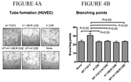
- FIGS. 4A-4B FGF2 mutants are defective in inducing tube formation and suppress tube formation induced by WT FGF2 in HUVECs.
- FIG. 4B The number of branching points was counted per field from the images.
- FIGS. 5A-5B FGF2 mutants suppress WT FGF2-induced vessel sprouting from aorta ring.
- FIG. 5A Isolated rat aortic ring was embedded in collagen gels in DMEM containing FGF2 WT (50 ng/ml) or mutants (each 50 ng/ml), or the mixture of WT FGF2 (50 ng/ml) and mutants (each 2.5 ⁇ g/ml) and cultured for 10 days. Representative images of 3 independent experiments are shown.
- FIGS. 6A-6B FGF2 mutants suppress WT FGF2-induced angiogenesis.
- FIG. 6A Hydrogel containing WT FGF2 (100 ng/ml), FGF2 mutants (100 ng/ml) or the mixture of WT FGF2 (100 ng/ml) and excess FGF2 mutants (5 ⁇ m/ml) were implanted subcutaneously into the back of rat, respectively. The epidermis, dermis and subcutaneous tissue were removed 2 weeks after implantation and tissue sections were stained for von Willebrand factor. Representative images are shown.
- FIG. 6B Three samples were obtained from each condition. The von Willebrand factor positive cells were counted. Data are shown as means +/ ⁇ SE.
- Inflammation is a refers to an organism's immune response to irritation, toxic substances, pathogens, or other stimuli. The response can involve innate immune components and/or adaptive immunity. Inflammation is generally characterized as either chronic or acute. Acute inflammation is characterized by redness, pain, heat, swelling, and/or loss of function due to infiltration of plasma proteins and leukocytes to the affected area. Chronic inflammation is characterized by persistent inflammation, tissue destruction, and attempts at repair. Monocytes, macrophages, plasma B cells, and other lymphocytes are recruited to the affected area, and angiogenesis and fibrosis occur, often leading to scar tissue.
- An “inflammatory condition” is one characterized by or involving an inflammatory response, as described above.
- a list of exemplary inflammatory conditions includes: asthma, autoimmune disease, chronic inflammation, chronic prostatitis, glomerulonephritis, hypersensitivities and allergies, skin disorders such as eczema, inflammatory bowel disease, pelvic inflammatory disease, reperfusion injury, rheumatoid arthritis, transplant rejection, and vasculitis.
- angiogenesis refers to a process during which new blood vessels are formed from pre-existing blood vessels. This process involves the migration, growth, and differentiation of endothelial cells, which line the inside wall of blood vessels. While angiogenesis is a normal and vital process in growth and development, as well as in wound healing and in the formation of granulation tissue, it is also a fundamental step in the transition of tumors from a benign state to a malignant one. Anti-angiogenic agents are therefore often used as therapeutics in cancer treatment.
- FGF2 refers to a member of the fibroblast growth factor (FGF) family.
- FGF2 protein refers to a full-length FGF2 polypeptide sequence, including the human FGF2 (GenBank Accession No. NP_001997.5, encoded by GenBank Accession No. NM_002006.4), its polymorphic variants and species orthologs or homologs.
- An “FGF2 polynucleotide” refers to a nucleic acid sequence from the gene encoding the FGF2 protein and may include both the coding and non-coding regions.
- FGF2 cDNA,” “FGF2 mRNA,” “FGF2 coding sequence,” and their variations refer to a nucleic acid sequence that encodes an FGF2 polypeptide.
- an FGF2 dominant negative mutant or “an FGF2 mutant” as used herein refers to an FGF2 antagonist compound in the form of a mutated FGF2 or a fragment thereof, which suppresses FGF2-induced cellular signaling by way of its interaction with integrins (such as integrin ⁇ v ⁇ 3 or ⁇ 5 ⁇ 1) and FGF receptor (FGFR) in a manner that imposes an inhibitory or disruptive effect on the specific binding among wild-type FGF2, FGFR, and integrin, thus inhibiting downstream events normally triggered by FGF2 signaling, for example, FGF2-mediated ERK1/2 phosphorylation, DNA synthesis, cellular proliferation, and angiogenesis.
- integrins such as integrin ⁇ v ⁇ 3 or ⁇ 5 ⁇ 1
- FGFR FGF receptor
- one or more amino acid residues predicted to interact with integrin are mutated, either by deletion or by substitution with a different amino acid (e.g., the K119E/R120E and K125E mutations), resulting in the mutant having decreased or even abolished capability to bind integrin such as ⁇ v ⁇ 3 or ⁇ 5 ⁇ 1.
- FGF2 dominant negative mutants can be identified based on their deficiency compared to the wild-type FGF2 in decreased integrin binding, as well as in signaling functions (failure to activate ERK1/2 and DNA synthesis for example) in test cells (e.g., NIH3T3 cells). They can also be identified by their capability to suppress FGF2 signaling induced by wild-type FGF2 in test cells such as endothelial cells, in addition to their anti-angiogenic activity, e.g., suppressing tube formation in vitro, sprouting in aorta ring assays ex vivo, or angiogenesis in vivo.
- An FGF2 dominant negative mutant may be initially generated based on the wild-type FGF2 amino acid sequence (i.e., SEQ ID NO:1) with certain amino acid residue(s) mutated, it may further includes one or more heterologous amino acid sequences (derived from a source other than FGF2 protein) at its N-terminus and/or C-terminus.
- an FGF2 dominant negative mutant may optionally include one or more additional heterologous amino acid sequence(s) of 1, 2, 3, 4, 5, 6, 7, 8, 9, 10, 11, 12, 13, 14, 15, 16, 17, 18, 19, 20, 25, 30, 35, 40, 45, or up to 50 amino acids at C- and/or N-terminus of the K119E/R120E sequence.
- heterologous peptide sequences can be of a varying nature, for example, any one of the “tags” known and used in the field of recombinant proteins: a peptide tag such as an AviTag, a peptide allowing biotinylation by the enzyme BirA and so the protein can be isolated by streptavidin, a Calmodulin-tag, a peptide bound by the protein calmodulin, a polyglutamate tag, a peptide binding efficiently to anion-exchange resin such as Mono-Q, an E-tag, a peptide recognized by an antibody, a FLAG-tag, a peptide recognized by an antibody, an HA-tag, a peptide recognized by an antibody, a His-tag, 5-10 histidines (SEQ ID NO: 2) bound by a nickel or cobalt chelate, a Myc-tag, a short peptide recognized by an antibody, an S-tag, an SBP-tag, a peptide that specifically binds to str
- FGF2 dominant negative mutants may also include one or more D-amino acids or include chemical modifications such as glycosylation, PEGylation, crosslinking, and the like.
- an “increase” or a “decrease” refers to a detectable positive or negative change in quantity from a comparison control, e.g., an established standard control (such as an average level of ERK1/2 activation or DNA synthesis induced by wild-type FGF2).
- An increase is a positive change that is typically at least 10%, or at least 20%, or 50%, or 100%, and can be as high as at least 2-fold or at least 5-fold or even 10-fold of the control value.
- a decrease is a negative change that is typically at least 10%, or at least 20%, 30%, or 50%, or even as high as at least 80% or 90% of the control value.
- a composition “consisting essentially of an FGF2 dominant negative mutant” is one that includes an FGF2 mutant that inhibits specific binding among wild-type FGF2, FGFR, and integrin (such as integrin ⁇ v ⁇ 3 or ⁇ 5 ⁇ 1) but no other compounds that contribute significantly to the inhibition of the binding.
- Such compounds may include inactive excipients, e.g., for formulation or stability of a pharmaceutical composition, or active ingredients that do not significantly contribute to the inhibition of FGF2-integrin binding.
- Exemplary compositions consisting essentially of an FGF2 dominant negative mutant include therapeutics, medicaments, and pharmaceutical compositions.
- an “effective amount” or a “therapeutically effective amount” means the amount of a compound that, when administered to a subject or patient for treating a disorder, is sufficient to prevent, reduce the frequency of, or alleviate the symptoms of the disorder.
- the effective amount will vary depending on a variety of the factors, such as a particular compound used, the disease and its severity, the age, weight, and other factors of the subject to be treated. Amelioration of a symptom of a particular condition by administration of a pharmaceutical composition described herein refers to any lessening, whether permanent or temporary, that can be associated with the administration of the pharmaceutical composition.
- the amount of an FGF2 dominant negative mutant is considered therapeutically effective for treating a condition involving undesired angiogenesis (such as tumor vascularization) when treatment results in eliminated symptoms, delayed onset of symptoms, or reduced frequency or severity of symptoms such as tumor growth, metastasis, etc.
- a “subject,” or “subject in need of treatment,” as used herein, refers to an individual who seeks medical attention due to risk of, or actual sufferance from, a condition involving an undesirable angiogenesis or cancer cell proliferation.
- the term subject can include both animals, especially mammals, and humans.
- Subjects or individuals in need of treatment include those that demonstrate symptoms of undesirable angiogenesis and/or tumor cell proliferation or are at risk of later developing these conditions and/or symptoms.
- nucleic acid or “polynucleotide” refers to deoxyribonucleotides or ribonucleotides and polymers thereof in either single- or double-stranded form. Unless specifically limited, the term encompasses nucleic acids containing known analogues of natural nucleotides which have similar binding properties as the reference nucleic acid and are metabolized in a manner similar to naturally occurring nucleotides. Unless otherwise indicated, a particular nucleic acid sequence also implicitly encompasses conservatively modified variants thereof (e.g., degenerate codon substitutions) and complementary sequences as well as the sequence explicitly indicated.
- degenerate codon substitutions may be achieved by generating sequences in which the third position of one or more selected (or all) codons is substituted with mixed-base and/or deoxyinosine residues (Batzer et al., Nucleic Acid Res., 19:5081 (1991); Ohtsuka et al., J. Biol. Chem., 260:2605-2608 (1985); and Cassol et al., (1992); Rossolini et al., Mol. Cell. Probes, 8:91-98 (1994)).
- the terms nucleic acid and polynucleotide are used interchangeably with gene, cDNA, and mRNA encoded by a gene.
- polypeptide “peptide,” and “protein” are used interchangeably herein to refer to a polymer of amino acid residues.
- the terms apply to amino acid polymers in which one or more amino acid residue is an artificial chemical mimetic of a corresponding naturally occurring amino acid, as well as to naturally occurring amino acid polymers and non-naturally occurring amino acid polymers.
- the terms encompass amino acid chains of any length, including full length proteins (i.e., antigens), wherein the amino acid residues are linked by covalent peptide bonds.
- amino acid refers to naturally occurring and synthetic amino acids, as well as amino acid analogs and amino acid mimetics that function in a manner similar to the naturally occurring amino acids.
- Naturally occurring amino acids are those encoded by the genetic code, as well as those amino acids that are later modified, e.g., hydroxyproline, ⁇ -carboxyglutamate, and O-phosphoserine.
- Amino acid analogs refers to compounds that have the same basic chemical structure as a naturally occurring amino acid, i.e., an ⁇ carbon that is bound to a hydrogen, a carboxyl group, an amino group, and an R group, e.g., homoserine, norleucine, methionine sulfoxide, methionine methyl sulfonium. Such analogs have modified R groups (e.g., norleucine) or modified peptide backbones, but retain the same basic chemical structure as a naturally occurring amino acid.
- Amino acid mimetics refers to chemical compounds that have a structure that is different from the general chemical structure of an amino acid, but that functions in a manner similar to a naturally occurring amino acid.
- Amino acids may be referred to herein by either their commonly known three letter symbols or by the one-letter symbols recommended by the IUPAC-IUB Biochemical Nomenclature Commission. Nucleotides, likewise, may be referred to by their commonly accepted single-letter codes.
- an “expression cassette” is a nucleic acid construct, generated recombinantly or synthetically, with a series of specified nucleic acid elements that permit transcription of a particular polynucleotide sequence in a host cell.
- An expression cassette may be part of a plasmid, viral genome, or nucleic acid fragment.
- an expression cassette includes a polynucleotide to be transcribed, operably linked to a promoter.
- “Operably linked” in this context means two or more genetic elements, such as a polynucleotide coding sequence and a promoter, placed in relative positions that permit the proper biological functioning of the elements, such as the promoter directing transcription of the coding sequence.
- an expression cassette Other elements that may be present in an expression cassette include those that enhance transcription (e.g., enhancers) and terminate transcription (e.g., terminators), as well as those that confer certain binding affinity or antigenicity to the recombinant protein produced from the expression cassette.
- enhancers e.g., enhancers
- terminate transcription e.g., terminators
- heterologous refers to the two elements such as two polynucleotide sequences (e.g., a promoter and a polypeptide-encoding sequence) or polypeptide sequences (e.g., a first amino acid sequence (such as one set forth in SEQ ID NO:1 with mutation or mutations) and a second peptide sequence serving as a fusion partner with the first amino acid sequence) that are not naturally found in the same relative position.
- a “heterologous promoter” of a gene refers to a promoter that is not naturally operably linked to that gene.
- a “heterologous polypeptide/amino acid sequence” or “heterologous polynucleotide” to an amino acid sequence or its encoding sequence is one derived from a non-FGF2 origin or derived from FGF2 but not naturally connected to the first FGF2-derived sequence (e.g., one set forth in SEQ ID NO:1) in the same fashion.
- the fusion of an FGF2-derived amino acid sequence (or its coding sequence) with a heterologous polypeptide (or polynucleotide sequence) does not result in a longer polypeptide or polynucleotide sequence that can be found naturally in FGF2.
- FGF1 integrin-binding defective FGF1 mutant
- R50E suppresses angiogenesis and tumor growth, suggesting that R50E has potential as a therapeutic.
- FGF1 is unstable, and R50E had to be expressed in cancer cells for xenograft study, since injected R50E may rapidly disappear from circulation. This study was aimed to determine whether an antagonist can be developed for the more stable FGF2.
- FGF2 is widely involved in important biological processes such as stem cell proliferation and angiogenesis.
- FGF2 mutants suppressed FGF2 signaling induced by WT FGF2 in endothelial cells, indicating that these FGF2 mutants are antagonists.
- the FGF2 mutants effectively suppressed tube formation in vitro, sprouting in aorta ring assays ex vivo, and angiogenesis in vivo.
- the positions of amino acids critical for integrin binding are different between FGF1 and FGF2, suggesting that they do not interact with integrins in the same manner.
- the newly developed FGF2 mutants have potential as anti-angiogenic agents and useful tools for studying the role of integrins in FGF2 signaling.
- nucleic acids sizes are given in either kilobases (kb) or base pairs (bp). These are estimates derived from agarose or acrylamide gel electrophoresis, from sequenced nucleic acids, or from published DNA sequences.
- kb kilobases
- bp base pairs
- proteins sizes are given in kilodaltons (kDa) or amino acid residue numbers. Proteins sizes are estimated from gel electrophoresis, from sequenced proteins, from derived amino acid sequences, or from published protein sequences.
- Oligonucleotides that are not commercially available can be chemically synthesized, e.g., according to the solid phase phosphoramidite triester method first described by Beaucage & Caruthers, Tetrahedron Lett. 22: 1859-1862 (1981), using an automated synthesizer, as described in Van Devanter et. al., Nucleic Acids Res. 12: 6159-6168 (1984). Purification of oligonucleotides is performed using any art-recognized strategy, e.g., native acrylamide gel electrophoresis or anion-exchange HPLC as described in Pearson & Reanier, J. Chrom. 255: 137-149 (1983).
- polypeptide of interest e.g., an FGF2 dominant negative mutant polypeptide
- synthetic oligonucleotides can be verified after cloning or subcloning using, e.g., the chain termination method for sequencing double-stranded templates of Wallace et al., Gene 16: 21-26 (1981).
- polynucleotide sequences encoding human FGF2 is known as GenBank Accession No. NM_002006.4.
- the corresponding amino acid sequence is known as GenBank Accession No. NP_001997.5.
- These polynucleotide sequences may be obtained from a commercial supplier or by amplification methods such as polymerase chain reaction (PCR).
- a polynucleotide sequence encoding an FGF2 polypeptide can be isolated from a cDNA or genomic DNA library using standard cloning techniques such as PCR, where homology-based primers can often be derived from a known nucleic acid sequence encoding an integrin polypeptide. This approach is particularly useful for identifying variants, orthologs, or homologs of FGF2. Most commonly used techniques for this purpose are described in standard texts, e.g., Sambrook and Russell, supra.
- cDNA libraries suitable for obtaining a coding sequence for a human FGF2 polypeptide may be commercially available or can be constructed.
- the general methods of isolating mRNA, making cDNA by reverse transcription, ligating cDNA into a recombinant vector, transfecting into a recombinant host for propagation, screening, and cloning are well known (see, e.g., Gubler and Hoffman, Gene, 25: 263-269 (1983); Ausubel et al., supra).
- the segment can be further used as a probe to isolate the full length polynucleotide sequence encoding the gene of interest (e.g., human FGF2) from the cDNA library.
- the gene of interest e.g., human FGF2
- a general description of appropriate procedures can be found in Sambrook and Russell, supra.
- a similar procedure can be followed to obtain a sequence encoding a human FGF2 from a human genomic library, which may be commercially available or can be constructed according to various art-recognized methods.
- degenerate oligonucleotides can be designed as primer sets and PCR can be performed under suitable conditions (see, e.g., White et al., PCR Protocols: Current Methods and Applications, 1993; Griffin and Griffin, PCR Technology, CRC Press Inc. 1994) to amplify a segment of nucleotide sequence from a cDNA or genomic library.
- the sequence can be modified and then subcloned into a vector, for instance, an expression vector, so that a recombinant polypeptide (e.g., an FGF2 dominant negative mutant polypeptide) can be produced from the resulting construct.
- a recombinant polypeptide e.g., an FGF2 dominant negative mutant polypeptide
- Further modifications to the coding sequence e.g., nucleotide substitutions, may be subsequently made to alter the characteristics of the polypeptide.
- the amino acid sequence of an FGF2 polypeptide may be modified in order to achieve the dominant negative phenotype that inhibits FGF2 cellular signaling, angiogenesis, and cancer cell proliferation, as determined by the in vitro or in vivo methods known in the field as well as described herein. Possible modifications to the amino acid sequence may include conservative substitutions; deletion or addition of one or more amino acid residues (e.g., addition at one terminal of the polypeptide of a tag sequence such as 6 ⁇ His (SEQ ID NO: 3) to facilitate purification or identification) at either or both of the N- and C-termini.
- a variety of mutation-generating protocols are established and described in the art, and can be readily used to modify a polynucleotide sequence encoding an integrin ⁇ fragment polypeptide. See, e.g., Zhang et al., Proc. Natl. Acad. Sci. USA, 94: 4504-4509 (1997); and Stemmer, Nature, 370: 389-391 (1994).
- the procedures can be used separately or in combination to produce variants of a set of nucleic acids, and hence variants of encoded polypeptides. Kits for mutagenesis, library construction, and other diversity-generating methods are commercially available.
- Mutational methods of generating diversity include, for example, site-directed mutagenesis (Botstein and Shortle, Science, 229: 1193-1201 (1985)), mutagenesis using uracil-containing templates (Kunkel, Proc. Natl. Acad. Sci. USA, 82: 488-492 (1985)), oligonucleotide-directed mutagenesis (Zoller and Smith, Nucl. Acids Res., 10: 6487-6500 (1982)), phosphorothioate-modified DNA mutagenesis (Taylor et al., Nucl.
- the polynucleotide sequence encoding an FGF2 dominant negative mutant polypeptide can be further altered to coincide with the preferred codon usage of a particular host.
- the preferred codon usage of one strain of bacterial cells can be used to derive a polynucleotide that encodes an FGF2 mutant and includes the codons favored by this strain.
- the frequency of preferred codon usage exhibited by a host cell can be calculated by averaging frequency of preferred codon usage in a large number of genes expressed by the host cell (e.g., calculation service is available from web site of the Kazusa DNA Research Institute, Japan). This analysis is preferably limited to genes that are highly expressed by the host cell.
- the coding sequences are verified by sequencing and are then subcloned into an appropriate expression vector for recombinant production of the FGF2 dominant negative mutant polypeptides.
- FGF2 protein has been established (e.g., GenBank Accession No. NP_001997.5).
- Polypeptides of known sequences especially those of relatively short length such as FGF2 amino acid sequence set forth in SEQ ID NO:1, may be synthesized by solid-phase peptide synthesis methods using procedures similar to those described by Merrifield et al., J. Am. Chem. Soc., 85:2149-2156 (1963); Barany and Merrifield, Solid - Phase Peptide Synthesis, in The Peptides: Analysis, Synthesis, Biology Gross and Meienhofer (eds.), Academic Press, N.Y., vol. 2, pp.
- N- ⁇ -protected amino acids having protected side chains are added stepwise to a growing polypeptide chain linked by its C-terminal and to a solid support, i.e., polystyrene beads.
- the peptides are synthesized by linking an amino group of an N- ⁇ -deprotected amino acid to an ⁇ -carboxy group of an N- ⁇ -protected amino acid that has been activated by reacting it with a reagent such as dicyclohexylcarbodiimide. The attachment of a free amino group to the activated carboxyl leads to peptide bond formation.
- the most commonly used N- ⁇ -protecting groups include Boc, which is acid labile, and Fmoc, which is base labile.
- halomethyl resins such as chloromethyl resin or bromomethyl resin
- hydroxymethyl resins such as phenol resins, such as 4-( ⁇ [2,4-dimethoxyphenyl]-Fmoc-aminomethyl)phenoxy resin
- tert-alkyloxycarbonyl-hydrazidated resins such as tert-alkyloxycarbonyl-hydrazidated resins, and the like.
- the C-terminal N- ⁇ -protected amino acid is first attached to the solid support.
- the N- ⁇ -protecting group is then removed.
- the deprotected ⁇ -amino group is coupled to the activated ⁇ -carboxylate group of the next N- ⁇ -protected amino acid.
- the process is repeated until the desired peptide is synthesized.
- the resulting peptides are then cleaved from the insoluble polymer support and the amino acid side chains deprotected. Longer peptides can be derived by condensation of protected peptide fragments.
- a polypeptide of interest e.g., an FGF2 dominant negative mutant polypeptide
- an FGF2 dominant negative mutant polypeptide can be produced using routine techniques in the field of recombinant genetics, relying on the polynucleotide sequences encoding the polypeptide disclosed herein.
- a strong promoter to direct transcription e.g., in Sambrook and Russell, supra, and Ausubel et al., supra.
- Bacterial expression systems for expressing recombinant polypeptides are available in, e.g., E. coli, Bacillus sp., Salmonella , and Caulobacter . Kits for such expression systems are commercially available.
- the eukaryotic expression vector is an adenoviral vector, an adeno-associated vector, or a retroviral vector.
- the promoter used to direct expression of a heterologous nucleic acid depends on the particular application.
- the promoter is optionally positioned about the same distance from the heterologous transcription start site as it is from the transcription start site in its natural setting. As is known in the art, however, some variation in this distance can be accommodated without loss of promoter function.
- the expression vector typically includes a transcription unit or expression cassette that contains all the additional elements required for the expression of the desired polypeptide in host cells.
- a typical expression cassette thus contains a promoter operably linked to the nucleic acid sequence encoding the polypeptide and signals required for efficient polyadenylation of the transcript, ribosome binding sites, and translation termination.
- the nucleic acid sequence encoding the desired polypeptide is typically linked to a cleavable signal peptide sequence to promote secretion of the recombinant polypeptide by the transformed cell.
- signal peptides include, among others, the signal peptides from tissue plasminogen activator, insulin, and neuron growth factor, and juvenile hormone esterase of Heliothis virescens .
- an appropriate anchoring sequence is used in concert with the coding sequence. Additional elements of the cassette may include enhancers and, if genomic DNA is used as the structural gene, introns with functional splice donor and acceptor sites.
- the expression cassette should also contain a transcription termination region downstream of the structural gene to provide for efficient termination.
- the termination region may be obtained from the same gene as the promoter sequence or may be obtained from different genes.
- the particular expression vector used to transport the genetic information into the cell is not particularly critical. Any of the conventional vectors used for expression in eukaryotic or prokaryotic cells may be used. Standard bacterial expression vectors include plasmids such as pBR322 based plasmids, pSKF, pET23D, and fusion expression systems such as GST and LacZ. Epitope tags can also be added to recombinant proteins to provide convenient methods of isolation, e.g., c-myc.
- Expression vectors containing regulatory elements from eukaryotic viruses are typically used in eukaryotic expression vectors, e.g., SV40 vectors, papilloma virus vectors, and vectors derived from Epstein-Barr virus.
- eukaryotic vectors include pMSG, pAV009/A + , pMTO10/A + , pMAMneo-5, baculovirus pDSVE, and any other vector allowing expression of proteins under the direction of the SV40 early promoter, SV40 later promoter, metallothionein promoter, murine mammary tumor virus promoter, Rous sarcoma virus promoter, polyhedrin promoter, or other promoters shown effective for expression in eukaryotic cells.
- Some expression systems have markers that provide gene amplification such as thymidine kinase, hygromycin B phosphotransferase, and dihydrofolate reductase.
- markers that provide gene amplification such as thymidine kinase, hygromycin B phosphotransferase, and dihydrofolate reductase.
- high yield expression systems not involving gene amplification are also suitable, such as a baculovirus vector in insect cells, with a polynucleotide sequence encoding the desired polypeptide under the direction of the polyhedrin promoter or other strong baculovirus promoters.
- the elements that are typically included in expression vectors also include a replicon that functions in E. coli , a gene encoding antibiotic resistance to permit selection of bacteria that harbor recombinant plasmids, and unique restriction sites in nonessential regions of the plasmid to allow insertion of eukaryotic sequences.
- the particular antibiotic resistance gene chosen is not critical, any of the many resistance genes known in the art are suitable.
- the prokaryotic sequences are optionally chosen such that they do not interfere with the replication of the DNA in eukaryotic cells, if necessary. Similar to antibiotic resistance selection markers, metabolic selection markers based on known metabolic pathways may also be used as a means for selecting transformed host cells.
- the expression vector further comprises a sequence encoding a secretion signal, such as the E. coli OppA (Periplasmic Oligopeptide Binding Protein) secretion signal or a modified version thereof, which is directly connected to 5′ of the coding sequence of the protein to be expressed.
- a secretion signal such as the E. coli OppA (Periplasmic Oligopeptide Binding Protein) secretion signal or a modified version thereof, which is directly connected to 5′ of the coding sequence of the protein to be expressed.
- This signal sequence directs the recombinant protein produced in cytoplasm through the cell membrane into the periplasmic space.
- the expression vector may further comprise a coding sequence for signal peptidase 1, which is capable of enzymatically cleaving the signal sequence when the recombinant protein is entering the periplasmic space.
- Standard transfection methods are used to produce bacterial, mammalian, yeast, insect, or plant cell lines that express large quantities of a recombinant polypeptide, which are then purified using standard techniques (see, e.g., Colley et al., J. Biol. Chem. 264: 17619-17622 (1989); Guide to Protein Purification, in Methods in Enzymology , vol. 182 (Deutscher, ed., 1990)). Transformation of eukaryotic and prokaryotic cells are performed according to standard techniques (see, e.g., Morrison, J. Bact. 132: 349-351 (1977); Clark-Curtiss & Curtiss, Methods in Enzymology 101: 347-362 (Wu et al., eds, 1983).
- Any of the well-known procedures for introducing foreign nucleotide sequences into host cells may be used. These include the use of calcium phosphate transfection, polybrene, protoplast fusion, electroporation, liposomes, microinjection, plasma vectors, viral vectors and any of the other well-known methods for introducing cloned genomic DNA, cDNA, synthetic DNA, or other foreign genetic material into a host cell (see, e.g., Sambrook and Russell, supra). It is only necessary that the particular genetic engineering procedure used be capable of successfully introducing at least one gene into the host cell capable of expressing the recombinant polypeptide.
- the host cells are then cultured in an appropriate scale for the purpose of purifying the recombinant polypeptide.
- inclusion bodies typically involves the extraction, separation and/or purification of inclusion bodies by disruption of bacterial cells, e.g., by incubation in a buffer of about 100-150 ⁇ g/ml lysozyme and 0.1% Nonidet P40, a non-ionic detergent.
- the cell suspension can be ground using a Polytron grinder (Brinkman Instruments, Westbury, N.Y.). Alternatively, the cells can be sonicated on ice. Alternate methods of lysing bacteria are described in Ausubel et al. and Sambrook and Russell, both supra, and will be apparent to those of skill in the art.
- the cell suspension is generally centrifuged and the pellet containing the inclusion bodies resuspended in buffer which does not dissolve but washes the inclusion bodies, e.g., 20 mM Tris-HCl (pH 7.2), 1 mM EDTA, 150 mM NaCl and 2% Triton-X 100, a non-ionic detergent. It may be necessary to repeat the wash step to remove as much cellular debris as possible.
- the remaining pellet of inclusion bodies may be resuspended in an appropriate buffer (e.g., 20 mM sodium phosphate, pH 6.8, 150 mM NaCl).
- an appropriate buffer e.g., 20 mM sodium phosphate, pH 6.8, 150 mM NaCl.
- Other appropriate buffers will be apparent to those of skill in the art.
- the inclusion bodies are solubilized by the addition of a solvent that is both a strong hydrogen acceptor and a strong hydrogen donor (or a combination of solvents each having one of these properties).
- a solvent that is both a strong hydrogen acceptor and a strong hydrogen donor or a combination of solvents each having one of these properties.
- the proteins that formed the inclusion bodies may then be renatured by dilution or dialysis with a compatible buffer.
- Suitable solvents include, but are not limited to, urea (from about 4 M to about 8 M), formamide (at least about 80%, volume/volume basis), and guanidine hydrochloride (from about 4 M to about 8 M).
- Some solvents that are capable of solubilizing aggregate-forming proteins may be inappropriate for use in this procedure due to the possibility of irreversible denaturation of the proteins, accompanied by a lack of immunogenicity and/or activity.
- SDS sodium dodecyl sulfate
- 70% formic acid may be inappropriate for use in this procedure due to the possibility of irreversible denaturation of the proteins, accompanied by a lack of immunogenicity and/or activity.
- guanidine hydrochloride and similar agents are denaturants, this denaturation is not irreversible and renaturation may occur upon removal (by dialysis, for example) or dilution of the denaturant, allowing re-formation of the immunologically and/or biologically active protein of interest.
- the protein can be separated from other bacterial proteins by standard separation techniques.
- purifying recombinant polypeptides from bacterial inclusion body see, e.g., Patra et al., Protein Expression and Purification 18: 182-190 (2000).
- the periplasmic fraction of the bacteria can be isolated by cold osmotic shock in addition to other methods known to those of skill in the art (see e.g., Ausubel et al., supra).
- the bacterial cells are centrifuged to form a pellet. The pellet is resuspended in a buffer containing 20% sucrose.
- the bacteria are centrifuged and the pellet is resuspended in ice-cold 5 mM MgSO 4 and kept in an ice bath for approximately 10 minutes.
- the cell suspension is centrifuged and the supernatant decanted and saved.
- the recombinant proteins present in the supernatant can be separated from the host proteins by standard separation techniques well known to those of skill in the art.
- a recombinant polypeptide When a recombinant polypeptide is expressed in host cells in a soluble form, its purification can follow the standard protein purification procedure described below. This standard purification procedure is also suitable for purifying polypeptides obtained from chemical synthesis (e.g., an integrin ⁇ fragment).
- an initial salt fractionation can separate many of the unwanted host cell proteins (or proteins derived from the cell culture media) from the recombinant protein of interest.
- the preferred salt is ammonium sulfate.
- Ammonium sulfate precipitates proteins by effectively reducing the amount of water in the protein mixture. Proteins then precipitate on the basis of their solubility. The more hydrophobic a protein is, the more likely it is to precipitate at lower ammonium sulfate concentrations.
- a typical protocol is to add saturated ammonium sulfate to a protein solution so that the resultant ammonium sulfate concentration is between 20-30%. This will precipitate the most hydrophobic proteins.
- the precipitate is discarded (unless the protein of interest is hydrophobic) and ammonium sulfate is added to the supernatant to a concentration known to precipitate the protein of interest.
- the precipitate is then solubilized in buffer and the excess salt removed if necessary, through either dialysis or diafiltration.
- Other methods that rely on solubility of proteins, such as cold ethanol precipitation, are well known to those of skill in the art and can be used to fractionate complex protein mixtures.
- a protein of greater and lesser size can be isolated using ultrafiltration through membranes of different pore sizes (for example, Amicon or Millipore membranes).
- the protein mixture is ultrafiltered through a membrane with a pore size that has a lower molecular weight cut-off than the molecular weight of a protein of interest, e.g., an integrin ⁇ fragment polypeptide.
- the retentate of the ultrafiltration is then ultrafiltered against a membrane with a molecular cut off greater than the molecular weight of the protein of interest.
- the recombinant protein will pass through the membrane into the filtrate.
- the filtrate can then be chromatographed as described below.
- proteins of interest can also be separated from other proteins on the basis of their size, net surface charge, hydrophobicity, or affinity for ligands.
- antibodies raised against an FGF2 mutant can be conjugated to column matrices and the corresponding polypeptide immunopurified. All of these methods are well known in the art.
- inflammation is generally characterized by redness, swelling, pain, and occasional loss of function.
- symptoms vary among tissues, so that some inflammatory conditions are not easily detectable (e.g., atherosclerosis).
- Undesirable cell proliferation is often determined by way of detecting a benign or malignant growth, including an abnormal expansion of a particular cell or tissue type, such as various types of tumors and cancers.
- Angiogenesis involves the formation of new blood vessels from the existing old vessels, a biological process critical for wound healing and tumorigenesis.
- inflammatory response can play a role in the healing process by destroying, diluting, and isolating injurious agents and stimulating repair of the affected tissue
- inflammatory responses can also be harmful.
- inflammation results in leakage of plasma from the blood vessels. Although this leakage can have beneficial effects, it causes pain and when uncontrolled can lead to loss of function and death (such as adult respiratory distress syndrome).
- Anaphylactic shock, arthritis, and gout are among the conditions that are characterized by uncontrolled or inappropriate inflammation.
- an inflammatory response is typically initiated by endothelial cells producing molecules that attract and detain inflammatory cells (e.g., myeloid cells such as neutrophils, eosinophils, and basophils) at the site of injury or irritation.
- inflammatory cells e.g., myeloid cells such as neutrophils, eosinophils, and basophils
- the inflammatory cells then are transported through the endothelial barrier into the surrounding tissue.
- the result is accumulation of inflammatory cells, in particular neutrophils. Such accumulation is easily detectable by one of skill.
- Adaptive immune cells are often involved in inflammatory conditions. These cells release cytokines and antibodies in response to the source of the irritation. Thus, an inflammatory response can also be detected by detecting a change in the level of inflammatory cytokines, e.g., in a localized region of irritation or in the serum or plasma of an individual. It will be appreciated by those of skill in the art that each of these symptoms can be detected in an individual for the purposes of diagnosis. Further, a subject undergoing therapy for an inflammatory condition can be monitored, for instance, by detecting any changes in severity of the symptoms. Such inflammatory conditions include rheumatoid arthritis, Alzheimer's disease, multiple sclerosis, and atherosclerosis.
- the present invention also provides pharmaceutical compositions comprising an effective amount of an FGF2 dominant negative mutant polypeptide for inhibiting a pro-inflammatory signal, a pro-proliferation signal, or a pro-angiogenesis signal, therefore useful in both prophylactic and therapeutic applications designed for various diseases and conditions involving undesired inflammation, cell proliferation, and/or angiogenesis.
- Pharmaceutical compositions of the invention are suitable for use in a variety of drug delivery systems. Suitable formulations for use in the present invention are found in Remington's Pharmaceutical Sciences , Mack Publishing Company, Philadelphia, Pa., 17th ed. (1985). For a brief review of methods for drug delivery, see, Langer, Science 249: 1527-1533 (1990).
- the pharmaceutical compositions of the present invention can be administered by various routes, e.g., oral, subcutaneous, transdermal, intramuscular, intravenous, or intraperitoneal.
- the routes of administering the pharmaceutical compositions include systemic or local delivery to a subject suffering from a condition exacerbated by inflammation at daily doses of about 0.01-5000 mg, preferably 5-500 mg, of an FGF2 mutant polypeptide for a 70 kg adult human per day.
- the appropriate dose may be administered in a single daily dose or as divided doses presented at appropriate intervals, for example as two, three, four, or more subdoses per day.
- inert and pharmaceutically acceptable carriers are used.
- the pharmaceutical carrier can be either solid or liquid.
- Solid form preparations include, for example, powders, tablets, dispersible granules, capsules, cachets, and suppositories.
- a solid carrier can be one or more substances that can also act as diluents, flavoring agents, solubilizers, lubricants, suspending agents, binders, or tablet disintegrating agents; it can also be an encapsulating material.
- the carrier is generally a finely divided solid that is in a mixture with the finely divided active component, e.g., an FGF2 mutant polypeptide.
- the active ingredient the mutant polypeptide
- the carrier having the necessary binding properties in suitable proportions and compacted in the shape and size desired.
- a low-melting wax such as a mixture of fatty acid glycerides and cocoa butter is first melted and the active ingredient is dispersed therein by, for example, stirring. The molten homogeneous mixture is then poured into convenient-sized molds and allowed to cool and solidify.
- Powders and tablets preferably contain between about 5% to about 70% by weight of the active ingredient.
- Suitable carriers include, for example, magnesium carbonate, magnesium stearate, talc, lactose, sugar, pectin, dextrin, starch, tragacanth, methyl cellulose, sodium carboxymethyl cellulose, a low-melting wax, cocoa butter, and the like.
- compositions can include the formulation of the active compound of an FGF2 mutant polypeptide with encapsulating material as a carrier providing a capsule in which the mutant (with or without other carriers) is surrounded by the carrier, such that the carrier is thus in association with the compound.
- encapsulating material as a carrier providing a capsule in which the mutant (with or without other carriers) is surrounded by the carrier, such that the carrier is thus in association with the compound.
- cachets can also be included. Tablets, powders, cachets, and capsules can be used as solid dosage forms suitable for oral administration.
- Liquid pharmaceutical compositions include, for example, solutions suitable for oral or parenteral administration, suspensions, and emulsions suitable for oral administration.
- Sterile water solutions of the active component e.g., an FGF2 mutant polypeptide
- sterile solutions of the active component in solvents comprising water, buffered water, saline, PBS, ethanol, or propylene glycol are examples of liquid compositions suitable for parenteral administration.
- the compositions may contain pharmaceutically acceptable auxiliary substances as required to approximate physiological conditions, such as pH adjusting and buffering agents, tonicity adjusting agents, wetting agents, detergents, and the like.
- Sterile solutions can be prepared by dissolving the active component (e.g., an FGF2 mutant polypeptide) in the desired solvent system, and then passing the resulting solution through a membrane filter to sterilize it or, alternatively, by dissolving the sterile compound in a previously sterilized solvent under sterile conditions.
- the resulting aqueous solutions may be packaged for use as is, or lyophilized, the lyophilized preparation being combined with a sterile aqueous carrier prior to administration.
- the pH of the preparations typically will be between 3 and 11, more preferably from 5 to 9, and most preferably from 7 to 8.
- compositions containing the FGF2 mutant can be administered for prophylactic and/or therapeutic treatments.
- compositions are administered to a patient already suffering from a condition that may be exacerbated by an undesirable inflammatory reaction/cell proliferation/angiogenesis in an amount sufficient to prevent, cure, reverse, or at least partially slow or arrest the symptoms of the condition and its complications.
- An amount adequate to accomplish this is defined as a “therapeutically effective dose.” Amounts effective for this use will depend on the severity of the disease or condition and the weight and general state of the patient, but generally range from about 0.1 mg to about 2,000 mg of the mutant polypeptide per day for a 70 kg patient, with dosages of from about 5 mg to about 500 mg of the mutant polypeptide per day for a 70 kg patient being more commonly used.
- compositions containing an FGF2 mutant polypeptide are administered to a patient susceptible to or otherwise at risk of developing a disease or condition involving an undesirable inflammatory response, cell proliferation, and/or angiogenesis in an amount sufficient to delay or prevent the onset of the symptoms.
- Such an amount is defined to be a “prophylactically effective dose.”
- the precise amounts of the inhibitor again depend on the patient's state of health and weight, but generally range from about 0.1 mg to about 2,000 mg of the mutant polypeptide for a 70 kg patient per day, more commonly from about 5 mg to about 500 mg for a 70 kg patient per day.
- compositions can be carried out with dose levels and pattern being selected by the treating physician.
- the pharmaceutical formulations should provide a quantity of a compound sufficient to effectively inhibit the undesirable inflammatory response/cellular proliferation/angiogenesis mediated by FGF2 signaling in the patient, either therapeutically or prophylactically.
- a variety of inflammatory conditions or undesirable cell proliferation/angiogenesis can be treated by therapeutic approaches that involve introducing into a cell a nucleic acid encoding an FGF2 dominant negative mutant polypeptide (e.g., K119E/R120E or K125E) such that the expression of the mutant leads to reduced or abolished FGF2 signaling in the cell.
- FGF2 dominant negative mutant polypeptide e.g., K119E/R120E or K125E
- Those amenable to treatment by this approach include a broad spectrum of conditions involving undesirable inflammation, cell proliferation, and/or angiogenesis.
- an inhibitory nucleic acid of the invention can be incorporated into a vector.
- vectors used for such purposes include expression plasmids capable of directing the expression of the FGF2 mutants in the target cell.
- the vector is a viral vector system wherein the polynucleotide is incorporated into a viral genome that is capable of transfecting the target cell.
- the inhibitory nucleic acid can be operably linked to expression and control sequences that can direct transcription of sequence in the desired target host cells. Thus, one can achieve reduced FGF2 signaling under appropriate conditions in the target cell.
- viral vector systems useful in the introduction and expression of an inhibitory nucleic acid include, for example, naturally occurring or recombinant viral vector systems.
- suitable viral vectors include replication competent, replication deficient, and conditionally replicating viral vectors.
- viral vectors can be derived from the genome of human or bovine adenoviruses, vaccinia virus, herpes virus, adeno-associated virus, minute virus of mice (MVM), HIV, Sindbis virus, and retroviruses (including but not limited to Rous sarcoma virus), and MoMLV.
- the inhibitory nucleic acid is inserted into such vectors to allow packaging of the gene construct, typically with accompanying viral DNA, followed by infection of a sensitive host cell and expression of the gene of interest.
- viral envelopes used for packaging gene constructs that include the inhibitory nucleic acid can be modified by the addition of receptor ligands or antibodies specific for a receptor to permit receptor-mediated endocytosis into specific cells (see, e.g., WO 93/20221, WO 93/14188, and WO 94/06923).
- Retroviral vectors may also be useful for introducing the inhibitory nucleic acid of the invention into target cells or organisms.
- Retroviral vectors are produced by genetically manipulating retroviruses.
- the viral genome of retroviruses is RNA.
- this genomic RNA is reverse transcribed into a DNA copy which is integrated into the chromosomal DNA of transduced cells with a high degree of stability and efficiency.
- the integrated DNA copy is referred to as a provirus and is inherited by daughter cells as is any other gene.
- the wild type retroviral genome and the proviral DNA have three genes: the gag, the pol and the env genes, which are flanked by two long terminal repeat (LTR) sequences.
- LTR long terminal repeat
- the gag gene encodes the internal structural (nucleocapsid) proteins; the pol gene encodes the RNA directed DNA polymerase (reverse transcriptase); and the env gene encodes viral envelope glycoproteins.
- the 5′ and 3′ LTRs serve to promote transcription and polyadenylation of virion RNAs.
- Adjacent to the 5′ LTR are sequences necessary for reverse transcription of the genome (the tRNA primer binding site) and for efficient encapsulation of viral RNA into particles (the Psi site) (see, Mulligan, In: Experimental Manipulation of Gene Expression , Inouye (ed), 155-173 (1983); Mann et al., Cell 33:153-159 (1983); Cone and Mulligan, Proceedings of the National Academy of Sciences, U.S.A., 81:6349-6353 (1984)).
- retroviral vectors The design of retroviral vectors is well known to those of ordinary skill in the art. In brief, if the sequences necessary for encapsidation (or packaging of retroviral RNA into infectious virions) are missing from the viral genome, the result is a cis acting defect which prevents encapsidation of genomic RNA. However, the resulting mutant is still capable of directing the synthesis of all virion proteins. Retroviral genomes from which these sequences have been deleted, as well as cell lines containing the mutant genome stably integrated into the chromosome are well known in the art and are used to construct retroviral vectors.
- the retroviral vector particles are prepared by recombinantly inserting the desired inhibitory nucleic acid sequence into a retrovirus vector and packaging the vector with retroviral capsid proteins by use of a packaging cell line.
- the resultant retroviral vector particle is incapable of replication in the host cell but is capable of integrating into the host cell genome as a proviral sequence containing the desired nucleotide sequence.
- the patient is capable of producing, for example, the inhibitory nucleic acid, thus eliminating or reducing unwanted inflammatory conditions.
- Packaging cell lines that are used to prepare the retroviral vector particles are typically recombinant mammalian tissue culture cell lines that produce the necessary viral structural proteins required for packaging, but which are incapable of producing infectious virions.
- the defective retroviral vectors that are used lack these structural genes but encode the remaining proteins necessary for packaging.
- To prepare a packaging cell line one can construct an infectious clone of a desired retrovirus in which the packaging site has been deleted. Cells comprising this construct will express all structural viral proteins, but the introduced DNA will be incapable of being packaged.
- packaging cell lines can be produced by transforming a cell line with one or more expression plasmids encoding the appropriate core and envelope proteins. In these cells, the gag, pol, and env genes can be derived from the same or different retroviruses.
- a number of packaging cell lines suitable for the present invention are also available in the prior art. Examples of these cell lines include Crip, GPE86, PA317 and PG13 (see Miller et al., J. Virol. 65:2220-2224 (1991)). Examples of other packaging cell lines are described in Cone and Mulligan Proceedings of the National Academy of Sciences, USA, 81:6349-6353 (1984); Danos and Mulligan Proceedings of the National Academy of Sciences, USA, 85:6460-6464 (1988); Eglitis et al. (1988), supra; and Miller (1990), supra.
- the inhibitory nucleic acid is generally formulated in a suitable buffer, which can be any pharmaceutically acceptable buffer, such as phosphate buffered saline or sodium phosphate/sodium sulfate, Tris buffer, glycine buffer, sterile water, and other buffers known to the ordinarily skilled artisan such as those described by Good et al. Biochemistry 5:467 (1966).
- a suitable buffer such as phosphate buffered saline or sodium phosphate/sodium sulfate, Tris buffer, glycine buffer, sterile water, and other buffers known to the ordinarily skilled artisan such as those described by Good et al. Biochemistry 5:467 (1966).
- compositions can further include a stabilizer, an enhancer, and/or other pharmaceutically acceptable carriers or vehicles.
- a pharmaceutically acceptable carrier can contain a physiologically acceptable compound that acts, for example, to stabilize the inhibitory nucleic acids of the invention and any associated vector.
- a physiologically acceptable compound can include, for example, carbohydrates, such as glucose, sucrose or dextrans, antioxidants, such as ascorbic acid or glutathione, chelating agents, low molecular weight proteins or other stabilizers or excipients.
- Other physiologically acceptable compounds include wetting agents, emulsifying agents, dispersing agents or preservatives, which are particularly useful for preventing the growth or action of microorganisms.
- Various preservatives are well known and include, for example, phenol and ascorbic acid. Examples of carriers, stabilizers or adjuvants can be found in Remington's Pharmaceutical Sciences, Mack Publishing Company, Philadelphia, Pa., 17th ed. (1985).
- the formulations containing an inhibitory nucleic acid can be delivered to any tissue or organ using any delivery method known to the ordinarily skilled artisan.
- the nucleic acid is formulated in mucosal, topical, and/or buccal formulations, particularly mucoadhesive gel and topical gel formulations.
- Exemplary permeation enhancing compositions, polymer matrices, and mucoadhesive gel preparations for transdermal delivery are disclosed in U.S. Pat. No. 5,346,701.
- the formulations containing the inhibitory nucleic acid are typically administered to a cell.
- the cell can be provided as part of a tissue or as an isolated cell, such as in tissue culture.
- the cell can be provided in vivo, ex vivo, or in vitro.
- the formulations can be introduced into the tissue of interest in vivo or ex vivo by a variety of methods.
- the inhibitory nucleic acid is introduced into cells by such methods as microinjection, calcium phosphate precipitation, liposome fusion, ultrasound, electroporation, or biolistics.
- the nucleic acid is taken up directly by the tissue of interest.
- the inhibitory nucleic acid is administered ex vivo to cells or tissues explanted from a patient, then returned to the patient.
- ex vivo administration of therapeutic gene constructs include Nolta et al., Proc Natl. Acad. Sci. USA 93 (6):2414-9 (1996); Koc et al., Seminars in Oncology 23 (1):46-65 (1996); Raper et al., Annals of Surgery 223 (2):116-26 (1996); Dalesandro et al., J. Thorac. Cardi. Surg., 11 (2):416-22 (1996); and Makarov et al., Proc. Natl. Acad. Sci. USA 93 (1):402-6 (1996).
- Effective dosage of the formulations will vary depending on many different factors, including means of administration, target site, physiological state of the patient, and other medicines administered. Thus, treatment dosages will need to be titrated to optimize safety and efficacy.
- the physician should evaluate the particular nucleic acid used, the disease state being diagnosed; the age, weight, and overall condition of the patient, circulating plasma levels, vector toxicities, progression of the disease, and the production of anti-vector antibodies.
- the size of the dose also will be determined by the existence, nature, and extent of any adverse side-effects that accompany the administration of a particular vector.
- doses ranging from about 10 ng-1 g, 100 ng-100 mg, 1 ⁇ g-10 mg, or 30-300 ⁇ g inhibitory nucleic acid per patient are typical. Doses generally range between about 0.01 and about 50 mg per kilogram of body weight, preferably between about 0.1 and about 5 mg/kg of body weight or about 10 8 -10 10 or 10 12 viral particles per injection. In general, the dose equivalent of a naked nucleic acid from a vector is from about 1 ⁇ g-100 ⁇ g for a typical 70 kg patient, and doses of vectors which include a retroviral particle are calculated to yield an equivalent amount of an inhibitory nucleic acid.
- kits for suppressing FGF2-induced cellular signaling or treating a condition involving undesirable inflammatory responses and/or angiogenesis including cancer cell proliferation by inhibiting the specific binding between FGF2 and integrin according to the method of the present invention typically include a first container that contains a pharmaceutical composition having an effective amount of an FGF2 dominant negative mutant, optionally with a second container containing an anti-cancer agent, such as another anti-angiogenic agent, such as bevacizumab (Avastin), itraconazole, carboxyamidotriazole, TNP-470, CM101, IFN- ⁇ , IL-12, platelet factor-4, suramin, SU5416, thrombospondin, VEGFR antagonists, angiostatin, endostatin, 2-methoxyestradiol, tecogalan, tetrathiomolybdate, thalidomide, prolactin, ⁇ V ⁇ 3 inhibitors, linomide, ramucirumab,
- kits will also include informational material containing instructions on how to dispense the pharmaceutical composition, including description of the type of patients who may be treated (e.g., a person suffering from cancer or at risk of developing secondary metastasis), the schedule (e.g., dose and frequency of administration) and route of administration, and the like.
- informational material containing instructions on how to dispense the pharmaceutical composition, including description of the type of patients who may be treated (e.g., a person suffering from cancer or at risk of developing secondary metastasis), the schedule (e.g., dose and frequency of administration) and route of administration, and the like.
- the fibroblast growth factor (FGF) family consists of 22 related polypeptides that are expressed in almost all tissues and are multifunctional. Some FGFs, like FGF1 and FGF2, have potent angiogenic activity and are implicated as promoters of angiogenesis, the formation of new blood vessels, in cancer and chronic inflammatory diseases [1-3]. FGFs also increase the motility and invasiveness of a variety of cell types. The biological effects of FGFs are mediated by four structurally related receptor tyrosine kinases: Fibroblast growth factor receptor-1 (FGFR1), FGFR2, FGFR3, and FGFR4. The binding of FGF to its receptor results in receptor dimerization and subsequent autophosphorylation of specific tyrosine residues within the intracellular domain.
- FGFR1 Fibroblast growth factor receptor-1
- FGFR2 Fibroblast growth factor receptor-1
- FGFR3 structurally related receptor tyrosine kinases
- Integrins are a family of cell adhesion receptors that recognize extracellular matrix (ECM) ligands and cell surface ligands [4]. Integrins are transmembrane ⁇ - ⁇ heterodimers, and at least 18 ⁇ and 8 ⁇ subunits are known [5]. Integrins are involved in signal transduction upon ligand binding and their functions are in turn regulated by signals from within the cell [5]. Previous studies found that antagonists to integrin ⁇ v ⁇ 3 suppressed angiogenesis induced by FGF2 [6], suggesting that this integrin is involved in FGF2 signaling (FGF-integrin crosstalk).
- ECM extracellular matrix
- An FGF1 mutant (the Arg-50 to Glu mutant, R50E), which is defective in integrin binding but still binds to heparin and FGFR1, is defective in inducing DNA synthesis, cell proliferation, cell migration, and chemotaxis, suggesting that the direct integrin binding to FGF1 is critical for FGF1 signaling [13].
- the present inventors proposed a model in which integrin and FGFR bind to FGF1 simultaneously and make a ternary complex on the cell surface. They discovered that R50E is a dominant-negative antagonist of FGF1.
- R50E suppressed DNA synthesis and cell proliferation induced by WT FGF1 [13], and suppressed angiogenesis in vitro and in vivo [14].
- FGF1 decoy FGF1 decoy
- R50E had to be expressed in cancer cells to demonstrate its antagonistic effects on tumor growth and angiogenesis since it is believed that R50E is quickly removed from circulation if R50E is systematically injected into mice in xenograft experiments [14].
- FGF2 is widely involved in important biological processes such as stem cell proliferation and angiogenesis partly due to its stability [1]. It has been reported that integrin ⁇ v ⁇ 3 binds to immobilized FGF2 and promote endothelial cell adhesion and spreading [15]. Also, anti- ⁇ v ⁇ 3 monoclonal and polyclonal antibodies specifically inhibit cell proliferation and up regulation of the urokinase-type plasminogen activator induced by soluble FGF2 in GM 7373 cells grown on tissue culture plastic [15]. It is unclear, however, how FGF2 interacts with integrins.
- the goal of this study is to determine how integrins bind to FGF2 and to determine if integrin-binding defective FGF2 is dominant-negative as in the case of FGF1.
- the present inventors developed FGF2 mutants that are defective in integrin binding and found that such FGF2 mutants act as antagonists of FGF2 signaling (FGF2 decoys). Notably, these FGF2 mutants effectively suppressed angiogenesis.
- FGF2 mutants can serve as cancer therapeutics by virtue of their anti-angiogenic activity.
- NIH3T3 embryonic mouse fibroblasts were obtained from American Type Culture Collection (ATCC) and were maintained in DMEM supplemented with 10% FCS and antibiotics.
- Human umbilical endothelial cells (HUVEC) were purchased from Sanko-junyaku (Tokyo, Japan) and were routinely cultured in EGM-2 Bullet kit (Lonza Basel, Switzerland) supplemented with 2% FCS.
- K562 Erythroleukemia cells and K562 cells that express recombinant ⁇ v ⁇ 3 were described before [11].
- Recombinant soluble ⁇ v ⁇ 3 was synthesized as described [16].
- FGF2 A fragment of cDNA encoding human FGF2 (SEQ ID NO:1, PALPEDGGSGAFPPGHFKDPKRLYCKNGGFFLRIHPDGRVDGVREKSDPHIKLQLQA EERGVVSIKGVCANRYLAMKEDGRLLASKCVTDECFFFERLESNNYNTYRSRKYTS WYVALKRTGQYKLGSKTGPGQKAILFLPMSAKS) was amplified by PCR using full-length human FGF2 cDNA as a template and subcloned into the BamHI/EcoRI site of PET28a+. Protein was synthesized in E. coli BL21 and purified by Ni-NTA affinity chromatography. WT and mutant FGF2 migrated as single bands in SDS-PAGE (data not shown).
- Kollman “united-atom” charges were used.
- AutoDock 3.05 uses a Lamarckian genetic algorithm (LGA) that couples a typical Darwinian genetic algorithm for global searching with the Solis and Wets algorithm for local searching.
- the LGA parameters were defined as follows: the initial population of random individuals had a size of 50 individuals; each docking was terminated with a maximum number of 1 ⁇ 10 6 energy evaluations or a maximum number of 27,000 generations, whichever came first; mutation and crossover rates were set at 0.02 and 0.80, respectively.
- An elitism value of 1 was applied, which ensured that the top ranked individual in the population always survived into the next generation.
- a maximum of 300 iterations per local search was used.
- the probability of performing a local search on an individual was 0.06, whereas the maximum number of consecutive successes or failures before doubling or halving the search step size was 4.
- SPR Surface plasmon resonance
- BrdU Incorporation Assay DNA synthesis was measured by the cell proliferation ELISA BrdU kit (Roche Diagnostics, Basel, Switzerland). NIH3T3 cells were starved for 16 h. Cells were stimulated with either WT FGF2 or mutants on 96 well plate for 24 h and concomitantly BrdU solution was added to the culture. The mixture of WT FGF2 (5 ng/ml) and each mutant (250 ng/ml) were also tested. The amplitude of absorbance at 450 nm is proportional to the BrdU incorporation into the cells.
- Cell migration assay was performed as previously described [14]. Briefly, the membrane was placed into a 24-well cell culture plate, and the lower portion of the plate was filled with serum-free EBM-2 medium containing 5 ng/ml WT FGF2, K119E/R120E, or K125E. The mixture of WT FGF2 (5 ng/ml) and individual mutants (5 ng/ml), or the mixture were also tested. Starved HUVEC cells (6 ⁇ 10 4 cells/filter) were plated on the filter and incubated at 37° C. for 6 h. Chemotaxed cells were stained for visualization and counted.
- Endothelial Cell Tube Formation Assay was performed as described [14]. In brief, serum-starved HUVECs were plated in wells (3 ⁇ 10 4 cells/well) of 48 well plates coated with 150 ⁇ l Matrigel (BD Biosciences, San Jose, Calif.). The medium contains 5 ng/ml WT FGF2, or the mixture of WT FGF2 (5 ng/ml), individual mutants (5 ng/ml), or the mixture (5 and 250 ng/ml, respectively). Cells were incubated for 6 h at 37° C. The Number of vessel branch points of tube per field was counted from the digital images.
- Thoracic aortae were isolated from 8 week-old rats and used for aorta ring assay as described previously [14]. Briefly, aortic segments were embedded into Type I collagen (Nitta Gelatin, Osaka, Japan) that contains WT FGF2 (50 ng/ml), individual mutants (50 ng/ml), or the mixture (50 and 2500 ng/ml, respectively). Aortic ring sprouts on days 10 were photographed.
- FIG. 1 a To identify how integrin ⁇ v ⁇ 3 and FGF2 interact, docking simulation of interaction between ⁇ v ⁇ 3 (PDB code 1L5G) and FGF2 (PDB code 2FGF) was performed using autodock3 ( FIG. 1 a ). The simulation predicted that FGF2 binds to the classical RGD-binding site of ⁇ v ⁇ 3 with high affinity (docking energy ⁇ 22 kcal/mol). When the FGFR1-FGF2 complex (1FQ9) is superposed to the ⁇ v ⁇ 3-FGF2 complex, there was little or no steric hindrance, suggesting that FGF2 can simultaneously bind to FGFR1 and ⁇ v ⁇ 3.
- the predicted integrin-binding interface overlaps with the heparin-binding site (residues 119-128, KRTGQYKLGS (SEQ ID NO: 4)).
- mutations were introduced into several amino acid residues (K39, R44, K46, K119/R120, and K125 to E) within the predicted integrin-binding interface of FGF2 (Table 1). It was studied if the FGF2 mutants support adhesion of K562 cells that express recombinant integrin ⁇ v ⁇ 3 ( ⁇ v ⁇ 3-K562 cells) and parent K562 cells.
- K562 cells that are deficient in proteoglycans were chosen because FGF2 strongly binds to cell surface proteoglycans (e.g., on CHO cells).
- FGF2 supported adhesion of both ⁇ v ⁇ 3-K562 and K562 cells to the similar extent, suggesting that FGF2 binds to ⁇ 5 ⁇ 1 in addition to ⁇ v ⁇ 3, since ⁇ 5 ⁇ 1 is the only integrin expressed in K562 cells [20].
- K125 to E mutation K125E
- K119E/R120E markedly suppressed adhesion of K562 cells to FGF2 ( FIG. 1 b ).
- the R44E mutation which corresponds to R50E in FGF1, did not affect integrin binding, suggesting that FGF1 and FGF2 do not bind to integrins in the same manner.
- SPR Surface plasmon resonance
- K119E/R120E mutant did not show detectable binding to ⁇ v ⁇ 3 in SPR.
- K119E/R120E was defective in binding to heparin, but K125E of FGF2 bound to heparin (data not shown).
- These findings are consistent with the docking model and K125E and K119E/R120E in the predicted integrin-binding site of FGF2 suppress FGF2 binding to integrin ⁇ v ⁇ 3 and ⁇ 5 ⁇ 1. This does not rule out the possibility, however, that substitution of these residues indirectly affects integrin binding through global conformational changes.
- K119E/R120E and K125E FGF2 Mutants are Defective in ERK1/2 Activation and in Inducing DNA Synthesis and Suppress DNA Synthesis Induced by WT FGF2 in NIH3T3 Cells
- K119E/R120E and K125E were both defective in inducing sustained ERK1/2 activation, as in the case of the R50E mutant of FGF1, which induces transient ERK1/2 activation but is defective in sustained ERK1/2 activation [11].
- K119E/R120E and K125E could induce DNA synthesis using BrdU incorporation assays in NIH3T3 cells. It was found that K119E/R120E and K125E were both defective in inducing DNA synthesis ( FIG. 2B ), consistent with the report that DNA synthesis is related to sustained ERK1/2 activation and the ability of FGF2 to bind to integrins [21, 22]. Notably, excess FGF2 mutants suppressed WT FGF2-induced DNA synthesis ( FIG. 2B ), suggesting that K119E/R120E and K125E are dominant-negative.
- K119E/R120E and K125E FGF2 Mutants are Defective in Inducing ERK1/2 Activation and Cell Migration in Human Umbilical Endothelial Cells (HUVEC) and Suppress HUVEC Migration Induced by WT FGF2
- K119E/R120E and K125E FGF2 mutants could induce angiogenesis and if they could suppress angiogenesis induced by WT FGF2 using three different angiogenesis models.
- Tube formation induced by K119E/R120E and K125E are significantly less than that induced by WT FGF2 (at 5 ng/ml FGF2) ( FIGS. 4 a and 4 b ).
- the tube-like structures induced by K119E/R120E and K125E were thin and weak as compared with that induced by WT FGF2 ( FIG. 4A ).
- Excess K119E/R120E and K125E (at 250 ng/ml) effectively suppressed tube formation induced by WT FGF2 to the background level ( FIG. 4B ). This indicates that K119E/R120E and K125E directly affect endothelial cell tube formation.
- the DGR motif is close to or within the predicted integrin-binding interface of FGF2, but the R39E mutation did not affect integrin binding. This suggests that the DGR motif may not important for integrin binding.
- the integrin-binding defective FGF2 mutants (K119E/R120E and K125E) are defective in signaling functions (DNA synthesis, sustained ERK1/2 activation, and cell migration). Furthermore, they are dominant-negative antagonists of FGF2 signaling and potently suppressed WT FGF2-induced DNA synthesis, ERK1/2 activation, and cell migration.
- K119E and R120E mutations have been individually introduced to FGF2, but these mutations only partially reduced integrin binding, and did not affect FGF2 signaling (data not shown). Therefore, substitution of both K119 and R120, such as to Glu, simultaneously result in optimal reduction in FGF2 signaling.
- K119E/R120E and K125E FGF2 potently suppressed WT FGF2-induced angiogenesis in vitro (tube formation), ex vivo (aorta ring assays), and angiogenesis in vivo. These findings indicate that they are potent anti-angiogenic agents. K119E/R120E and K125E are comparable as inhibitors of angiogenesis. It has been proposed that the thermal stability of FGF2 is a major reason why FGF2 is involved in so many more biological processes than FGF1 and why FGF2 is a determinant factor in regulating self-renewal, differentiation, reprogramming in human pluripotent stem cells [23].
- FGF2 mutants are highly specific to FGFR compared to tyrosine kinase inhibitors, which are selective rather than specific.
- FGF2 mutants are small in size compared to IgG, and therefore FGF2 mutants have better penetrance (access to) to diseased tissues.
- K119E/R120E and K125E of FGF2 may not induce such mutations in FGFR because the FGF2 mutants bind to FGFR exactly the same way as WT FGF2, and blocking binding of FGF2 (and other members of the FGF family) to FGFR would not benefit cancer cells.
- ⁇ v ⁇ 3 integrin is highly expressed in endothelium during angiogenesis and is involved in neovascularization induced by FGF2 [25, 26].
- Previous studies found that the binding of FGF2 to FGFR is not sufficient to induce cell proliferation in endothelial cells and that an interaction of FGF2 with a cell surface integrin receptor is also required [15]. This is consistent with the observation that anti- ⁇ v ⁇ 3 antibody inhibited the angiogenic activity exerted in vivo by FGF2 without affecting neovascularization induced by vascular endothelial cell growth factor, transforming growth factor- ⁇ , or phorbol ester [26].
- integrins other than ⁇ v ⁇ 3 may bind to FGF2. If this is the case antagonists to multiple integrins are needed to block FGF2-integrin interaction. It would be necessary to address the integrin binding specificity of FGF2 in details in future studies. On the other hand, it is likely that the K119E/R120E and K125E mutations suppress binding to multiple integrins regardless of integrin species. If this is the case the FGF2 mutants may be more effective than antagonists for individual integrins. The integrin-binding defective FGF2 mutants will be useful to study the molecular details of the FGF2/FGFR/integrin crosstalk.
Landscapes
- Health & Medical Sciences (AREA)
- Life Sciences & Earth Sciences (AREA)
- Chemical & Material Sciences (AREA)
- Genetics & Genomics (AREA)
- Organic Chemistry (AREA)
- Engineering & Computer Science (AREA)
- General Health & Medical Sciences (AREA)
- Zoology (AREA)
- Medicinal Chemistry (AREA)
- Bioinformatics & Cheminformatics (AREA)
- Molecular Biology (AREA)
- Animal Behavior & Ethology (AREA)
- Pharmacology & Pharmacy (AREA)
- Public Health (AREA)
- Veterinary Medicine (AREA)
- Proteomics, Peptides & Aminoacids (AREA)
- Biomedical Technology (AREA)
- Gastroenterology & Hepatology (AREA)
- Biochemistry (AREA)
- Biophysics (AREA)
- Biotechnology (AREA)
- Wood Science & Technology (AREA)
- General Engineering & Computer Science (AREA)
- General Chemical & Material Sciences (AREA)
- Nuclear Medicine, Radiotherapy & Molecular Imaging (AREA)
- Chemical Kinetics & Catalysis (AREA)
- Toxicology (AREA)
- Immunology (AREA)
- Epidemiology (AREA)
- Physics & Mathematics (AREA)
- Plant Pathology (AREA)
- Microbiology (AREA)
- Medicines That Contain Protein Lipid Enzymes And Other Medicines (AREA)
Abstract
Description
| TABLE 1 |
| Amino acid residues of FGF2 and αvβ3 that are in the predicted interface |
| FGF2 | αv | β3 |
| Asn-27, Arg-39, Glu-78, Asp- | Met-118, Gln-145, Asp- | Tyr122, Ser-123, Met-124, Lys- |
| 79, Gly-80, Lys-110, Tyr-111, | 146, Ile-147, Asp-148, | 125, Asp-126, Asp-127, Asp- |
| Thr-112, Ser-113, Trp-114, | Ala-149, Asp-150, Gly- | 179, Met-180, Lys-181, Thr- |
| Lys-119, Arg-120, Thr-121, | 151, Phe-177, Tyr-178, | 182, Arg214, Arg-216, Asp-217, |
| Gln-123, Tyr-124, Lys-125, | Trp-179, Gln-180, Thr- | Ala-218, Asp-251, Lys-253, |
| Leu-126, Ser-128, Lys-129, | 212, Ala-213, Gln-214, | Thr-311, Glu-312, Asn-313, |
| Thr-130, Gly-131, Pro-132, | Ala-215, Ile-216, Asp-218, | Val-314, Ser-334, Met-335, |
| Gly-133, Gln-134 | Asp-219, Arg-248 | |
Amino acid residues in integrin αvβ3 and FGF2 within 6 Å to each other in the docking model were identified using Swiss-pdb viewer v. 4.1.
- 1 Lanner, F. and Rossant, J. (2010) The role of FGF/Erk signaling in pluripotent cells. Development. 137, 3351-3360
- 2 Guillemot, F. and Zimmer, C. (2011) From cradle to grave: the multiple roles of fibroblast growth factors in neural development. Neuron. 71, 574-588
- 3 Beenken, A. and Mohammadi, M. (2009) The FGF family: biology, pathophysiology and therapy. Nat Rev Drug Discov. 8, 235-253
- 4 Hynes, R. O. (2002) Integrins: bidirectional, allosteric signaling machines. Cell. 110, 673-687
- 5 Takada, Y., Ye, X. and Simon, S. (2007) The integrins. Genome Biol. 8, 215
- 6 Brooks, P. C., Clark, R. A. and Cheresh, D. A. (1994) Requirement of vascular integrin
alpha v beta 3 for angiogenesis. Science. 264, 569-571 - 7 Eliceiri, B. P. (2001) Integrin and growth factor receptor crosstalk. Circ Res. 89, 1104-1110
- 8 Kim, S. H., Turnbull, J. and Guimond, S. (2011) Extracellular matrix and cell signalling: the dynamic cooperation of integrin, proteoglycan and growth factor receptor. J Endocrinol. 209, 139-151
- 9 Odenthal, J., Takes, R. and Friedl, P. (2016) Plasticity of tumor cell invasion: governance by growth factors and cytokines. Carcinogenesis. 37, 1117-1128
- 10 Desgrosellier, J. S. and Cheresh, D. A. (2010) Integrins in cancer: biological implications and therapeutic opportunities. Nat Rev Cancer. 10, 9-22
- 11 Mori, S., Wu, C. Y., Yamaji, S., Saegusa, J., Shi, B., Ma, Z., Kuwabara, Y., Lam, K. S., Isseroff, R. R., Takada, Y. K. and Takada, Y. (2008) Direct Binding of Integrin {alpha}v{beta}3 to FGF1 Plays a Role in FGF1 Signaling. J Biol Chem. 283, 18066-18075
- 12 Mori, S. and Takada, Y. (2013) Crosstalk between Fibroblast Growth Factor (FGF) Receptor and Integrin through Direct Integrin Binding to FGF and Resulting Integrin-FGF-FGFR Ternary Complex Formation. Medical Sciences. 1, 20-36
- 13 Yamaji, S., Saegusa, J., Ieguchi, K., Fujita, M., Takada, Y. K. and Takada, Y. (2010) A novel fibroblast growth factor-1 (FGF1) mutant that acts as an FGF antagonist. PLoS One. 5, e10273
- 14 Mori, S., Tran, V., Nishikawa, K., Kaneda, T., Hamada, Y., Kawaguchi, N., Fujita, M., Takada, Y. K., Matsuura, N., Zhao, M. and Takada, Y. (2013) A Dominant-Negative FGF1 Mutant (the R50E Mutant) Suppresses Tumorigenesis and Angiogenesis. PLoS One. 8, e57927
- 15 Rusnati, M., Tanghetti, E., Dell'Era, P., Gualandris, A. and Presta, M. (1997) alphavbeta3 integrin mediates the cell-adhesive capacity and biological activity of basic fibroblast growth factor (FGF-2) in cultured endothelial cells. Mol Biol Cell. 8, 2449-2461
- 16 Saegusa, J., Akakura, N., Wu, C. Y., Hoogland, C., Ma, Z., Lam, K. S., Liu, F. T., Takada, Y. K. and Takada, Y. (2008) Pro-inflammatory secretory phospholipase A2 type IIA binds to integrins alphavbeta3 and alpha4beta1 and induces proliferation of monocytic cells in an integrin-dependent manner. J Biol Chem. 283, 26107-26115
- 17 Wu, P. L., Lee, S. C., Chuang, C. C., Mori, S., Akakura, N., Wu, W. G. and Takada, Y. (2006) Non-cytotoxic cobra cardiotoxin A5 binds to alpha(v)beta3 integrin and inhibits bone resorption. Identification of cardiotoxins as non-RGD integrin-binding proteins of the Ly-6 family. J Biol Chem. 281, 7937-7945
- 18 Fujita, M., Ieguchi, K., Cedano-Prieto, D. M., Fong, A., Wilkerson, C., Chen, J. Q., Wu, M., Lo, S. H., Cheung, A. T., Wilson, M. D., Cardiff, R. D., Borowsky, A. D., Takada, Y. K. and Takada, Y. (2013) An Integrin Binding-defective Mutant of Insulin-like Growth Factor-1 (R36E/R37E IGF1) Acts as a Dominant-negative Antagonist of the IGF1 Receptor (IGF1R) and Suppresses Tumorigenesis but Still Binds to IGF1R. J Biol Chem. 288, 19593-19603
- 19 Fujita, M., Takada, Y. K. and Takada, Y. (2012) Integrins alphavbeta3 and alpha4beta1 Act as Coreceptors for Fractalkine, and the Integrin-Binding Defective Mutant of Fractalkine Is an Antagonist of CX3CR1. J Immunol. 189, 5809-5819
- 20 Hemler, M. E., Huang, C. and Schwarz, L. (1987) The VLA protein family. Characterization of five distinct cell surface heterodimers each with a common 130,000 molecular weight beta subunit. J Biol Chem. 262, 3300-3309
- 21 Eliceiri, B. P., Klemke, R., Stromblad, S. and Cheresh, D. A. (1998) Integrin alphavbeta3 requirement for sustained mitogen-activated protein kinase activity during angiogenesis. J Cell Biol. 140, 1255-1263
- 22 Sharrocks, A. D. (2006) Cell cycle: sustained ERK signalling represses the inhibitors. Curr Biol. 16, R540-542
- 23 Chen, G., Gulbranson, D. R., Yu, P., Hou, Z. and Thomson, J. A. (2012) Thermal stability of fibroblast growth factor protein is a determinant factor in regulating self-renewal, differentiation, and reprogramming in human pluripotent stem cells. Stem Cells. 30, 623-630
- 24 Ornitz, D. M., Xu, J., Colvin, J. S., McEwen, D. G., MacArthur, C. A., Coulier, F., Gao, G. and Goldfarb, M. (1996) Receptor specificity of the fibroblast growth factor family. J Biol Chem. 271, 15292-15297
- 25 Brooks, P. C., Montgomery, A. M., Rosenfeld, M., Reisfeld, R. A., Hu, T., Klier, G. and Cheresh, D. A. (1994) Integrin
alpha v beta 3 antagonists promote tumor regression by inducing apoptosis of angiogenic blood vessels. Cell. 79, 1157-1164 - 26 Friedlander, M., Brooks, P. C., Shaffer, R. W., Kincaid, C. M., Varner, J. A. and Cheresh, D. A. (1995) Definition of two angiogenic pathways by distinct alpha v integrins. Science. 270, 1500-1502
Claims (15)
Priority Applications (1)
| Application Number | Priority Date | Filing Date | Title |
|---|---|---|---|
| US16/033,007 US11229681B2 (en) | 2017-07-14 | 2018-07-11 | Dominant-negative FGF2 antagonists |
Applications Claiming Priority (2)
| Application Number | Priority Date | Filing Date | Title |
|---|---|---|---|
| US201762532511P | 2017-07-14 | 2017-07-14 | |
| US16/033,007 US11229681B2 (en) | 2017-07-14 | 2018-07-11 | Dominant-negative FGF2 antagonists |
Publications (2)
| Publication Number | Publication Date |
|---|---|
| US20190015478A1 US20190015478A1 (en) | 2019-01-17 |
| US11229681B2 true US11229681B2 (en) | 2022-01-25 |
Family
ID=65000405
Family Applications (1)
| Application Number | Title | Priority Date | Filing Date |
|---|---|---|---|
| US16/033,007 Active US11229681B2 (en) | 2017-07-14 | 2018-07-11 | Dominant-negative FGF2 antagonists |
Country Status (1)
| Country | Link |
|---|---|
| US (1) | US11229681B2 (en) |
Citations (5)
| Publication number | Priority date | Publication date | Assignee | Title |
|---|---|---|---|---|
| EP0645451A1 (en) * | 1993-09-24 | 1995-03-29 | American Cyanamid Company | Heparin binding site structural analogues of fibroblast growth factors |
| US5604293A (en) * | 1985-09-12 | 1997-02-18 | Scios Inc. | Recombinant human basic fibroblast growth factor |
| US6083706A (en) * | 1997-02-26 | 2000-07-04 | Ciblex Corporation | Inhibitors of leaderless protein export |
| US7186526B2 (en) * | 1985-09-12 | 2007-03-06 | Scios, Inc. | Recombinant fibroblast growth factor analogs |
| US8168591B2 (en) * | 2005-10-17 | 2012-05-01 | The Regents Of The University Of California | Compositions and methods related to anti-FGF agents |
-
2018
- 2018-07-11 US US16/033,007 patent/US11229681B2/en active Active
Patent Citations (5)
| Publication number | Priority date | Publication date | Assignee | Title |
|---|---|---|---|---|
| US5604293A (en) * | 1985-09-12 | 1997-02-18 | Scios Inc. | Recombinant human basic fibroblast growth factor |
| US7186526B2 (en) * | 1985-09-12 | 2007-03-06 | Scios, Inc. | Recombinant fibroblast growth factor analogs |
| EP0645451A1 (en) * | 1993-09-24 | 1995-03-29 | American Cyanamid Company | Heparin binding site structural analogues of fibroblast growth factors |
| US6083706A (en) * | 1997-02-26 | 2000-07-04 | Ciblex Corporation | Inhibitors of leaderless protein export |
| US8168591B2 (en) * | 2005-10-17 | 2012-05-01 | The Regents Of The University Of California | Compositions and methods related to anti-FGF agents |
Non-Patent Citations (1)
| Title |
|---|
| Mori, et al. "The integrin-binding defective FGF2 mutants potently suppress FGF2 signalling and angiogenesis." Bioscience reports 37, No. 2 (2017): BSR20170173. |
Also Published As
| Publication number | Publication date |
|---|---|
| US20190015478A1 (en) | 2019-01-17 |
Similar Documents
| Publication | Publication Date | Title |
|---|---|---|
| US6911205B2 (en) | Stabilized soluble glycoprotein trimers | |
| US6455283B1 (en) | Nucleic acids encoding vascular endothelial cell growth factor-E (VEGF-E) | |
| JPH10511557A (en) | Variants of vascular endothelial cell growth factor, their use and methods of their production | |
| JPH08500845A (en) | Peptide inhibitors for cell division and cell motility | |
| WO1999054357A1 (en) | Novel cytokine-like protein | |
| JP2023055875A (en) | Polypeptides targeting hiv fusion | |
| US10663465B2 (en) | Disruption of EGFR-SAR1 interaction for cancer treatment | |
| US11229681B2 (en) | Dominant-negative FGF2 antagonists | |
| CN118510536A (en) | Relaxin-2 fusion protein analogs and methods of use thereof | |
| US20110212108A1 (en) | Neuregulin/erbb signaling and integrin | |
| WO2016115171A1 (en) | Insulin-like growth factor 2 (igf2) signaling and modulation | |
| JPH05271291A (en) | Functional polypeptide | |
| US20060019871A1 (en) | Endothelial cell apoptosis induced by fibrinogen gamma chain C-terminal fragment | |
| US10591471B2 (en) | Suppression of SPLA2-integrin binding for treating an inflammatory condition or suppressing cell proliferation | |
| US20090092595A1 (en) | Suppression of sPLA2-integrin binding for treating an inflammatory condition | |
| US20250092106A1 (en) | Vegf mutants and modulation of integrin-mediated signaling | |
| US8685403B2 (en) | Insulin-like growth factor signaling and integrin | |
| US12054517B2 (en) | Myeloid differentiation factor 2 (MD2) signaling and modulation | |
| WO2021081301A1 (en) | Computational design of alpha(v) beta (6) integrin binding proteins | |
| US20220135649A1 (en) | Dominant negative cd40l polypeptides | |
| US20250003978A1 (en) | Diagnosis and treatment of anti-pf4 induced thrombocytopenia | |
| HUE026977T2 (en) | Promiscuous HER-2/NEU CD4 T cell epitopes | |
| JP2003009865A (en) | G protein receptor HTNAD29 |
Legal Events
| Date | Code | Title | Description |
|---|---|---|---|
| FEPP | Fee payment procedure |
Free format text: ENTITY STATUS SET TO UNDISCOUNTED (ORIGINAL EVENT CODE: BIG.); ENTITY STATUS OF PATENT OWNER: SMALL ENTITY |
|
| AS | Assignment |
Owner name: THE REGENTS OF THE UNIVERSITY OF CALIFORNIA, CALIFORNIA Free format text: ASSIGNMENT OF ASSIGNORS INTEREST;ASSIGNORS:TAKADA, YOSHIKAZU;TAKADA, YOKO;MORI, SEIJI;SIGNING DATES FROM 20170808 TO 20180619;REEL/FRAME:046358/0550 Owner name: THE REGENTS OF THE UNIVERSITY OF CALIFORNIA, CALIF Free format text: ASSIGNMENT OF ASSIGNORS INTEREST;ASSIGNORS:TAKADA, YOSHIKAZU;TAKADA, YOKO;MORI, SEIJI;SIGNING DATES FROM 20170808 TO 20180619;REEL/FRAME:046358/0550 |
|
| FEPP | Fee payment procedure |
Free format text: ENTITY STATUS SET TO SMALL (ORIGINAL EVENT CODE: SMAL); ENTITY STATUS OF PATENT OWNER: SMALL ENTITY |
|
| STPP | Information on status: patent application and granting procedure in general |
Free format text: DOCKETED NEW CASE - READY FOR EXAMINATION |
|
| STPP | Information on status: patent application and granting procedure in general |
Free format text: NON FINAL ACTION MAILED |
|
| STPP | Information on status: patent application and granting procedure in general |
Free format text: RESPONSE TO NON-FINAL OFFICE ACTION ENTERED AND FORWARDED TO EXAMINER |
|
| STPP | Information on status: patent application and granting procedure in general |
Free format text: NON FINAL ACTION MAILED |
|
| STPP | Information on status: patent application and granting procedure in general |
Free format text: DOCKETED NEW CASE - READY FOR EXAMINATION |
|
| STPP | Information on status: patent application and granting procedure in general |
Free format text: NON FINAL ACTION MAILED |
|
| STPP | Information on status: patent application and granting procedure in general |
Free format text: RESPONSE TO NON-FINAL OFFICE ACTION ENTERED AND FORWARDED TO EXAMINER |
|
| STPP | Information on status: patent application and granting procedure in general |
Free format text: FINAL REJECTION MAILED |
|
| STPP | Information on status: patent application and granting procedure in general |
Free format text: RESPONSE AFTER FINAL ACTION FORWARDED TO EXAMINER |
|
| STPP | Information on status: patent application and granting procedure in general |
Free format text: NOTICE OF ALLOWANCE MAILED -- APPLICATION RECEIVED IN OFFICE OF PUBLICATIONS |
|
| STPP | Information on status: patent application and granting procedure in general |
Free format text: PUBLICATIONS -- ISSUE FEE PAYMENT RECEIVED |
|
| STPP | Information on status: patent application and granting procedure in general |
Free format text: PUBLICATIONS -- ISSUE FEE PAYMENT VERIFIED |
|
| STCF | Information on status: patent grant |
Free format text: PATENTED CASE |
|
| MAFP | Maintenance fee payment |
Free format text: PAYMENT OF MAINTENANCE FEE, 4TH YR, SMALL ENTITY (ORIGINAL EVENT CODE: M2551); ENTITY STATUS OF PATENT OWNER: SMALL ENTITY Year of fee payment: 4 |