US11083739B2 - Mito-magnolol compounds and methods of synthesis and use thereof - Google Patents
Mito-magnolol compounds and methods of synthesis and use thereof Download PDFInfo
- Publication number
- US11083739B2 US11083739B2 US16/714,461 US201916714461A US11083739B2 US 11083739 B2 US11083739 B2 US 11083739B2 US 201916714461 A US201916714461 A US 201916714461A US 11083739 B2 US11083739 B2 US 11083739B2
- Authority
- US
- United States
- Prior art keywords
- mito
- cancer
- cells
- magnolol
- melanoma
- Prior art date
- Legal status (The legal status is an assumption and is not a legal conclusion. Google has not performed a legal analysis and makes no representation as to the accuracy of the status listed.)
- Active
Links
- 0 *OC1=CC=C(CC=C)C=C1C1=C(OC[PH](C2=CC=C([Y])C=C2)(C2=CC=C([Y])C=C2)C2=CC=C([Y])C=C2)C=CC(CC=C)=C1 Chemical compound *OC1=CC=C(CC=C)C=C1C1=C(OC[PH](C2=CC=C([Y])C=C2)(C2=CC=C([Y])C=C2)C2=CC=C([Y])C=C2)C=CC(CC=C)=C1 0.000 description 5
- MCVUXLIWIZNXAI-UHFFFAOYSA-N Br.Br.C=CCC1=CC(C2=C(OC[PH](C3=CC=CC=C3)(C3=CC=CC=C3)C3=CC=CC=C3)C=CC(CC=C)=C2)=C(OCC)C=C1 Chemical compound Br.Br.C=CCC1=CC(C2=C(OC[PH](C3=CC=CC=C3)(C3=CC=CC=C3)C3=CC=CC=C3)C=CC(CC=C)=C2)=C(OCC)C=C1 MCVUXLIWIZNXAI-UHFFFAOYSA-N 0.000 description 4
- ICDSDHQDSHWMQQ-UHFFFAOYSA-N C=CCC1=CC(C2=C(OCC)C=CC(CC=C)=C2)=C(O)C=C1.[Br-] Chemical compound C=CCC1=CC(C2=C(OCC)C=CC(CC=C)=C2)=C(O)C=C1.[Br-] ICDSDHQDSHWMQQ-UHFFFAOYSA-N 0.000 description 4
- WTQHECMCOYGVRN-UHFFFAOYSA-N C=CCC1=CC=C(OCC)C(C2=C(OC)C=CC(CC=C)=C2)=C1.[Br-] Chemical compound C=CCC1=CC=C(OCC)C(C2=C(OC)C=CC(CC=C)=C2)=C1.[Br-] WTQHECMCOYGVRN-UHFFFAOYSA-N 0.000 description 3
- TYISRZVCGVXCLK-UHFFFAOYSA-N C=CCC1=CC=C(OCC)C(C2=C(OCCOCCOCCC)C=CC(CC=C)=C2)=C1.[Br-] Chemical compound C=CCC1=CC=C(OCC)C(C2=C(OCCOCCOCCC)C=CC(CC=C)=C2)=C1.[Br-] TYISRZVCGVXCLK-UHFFFAOYSA-N 0.000 description 3
- KXSUJQSEKNURPU-UHFFFAOYSA-N C=CCC1=CC=C(OCC(CC)(CC)CCCC)C(C2=C(O)C=CC(CC=C)=C2)=C1.C=CCC1=CC=C(OCC(CC)CCCCCCC)C(C2=C(O)C=CC(CC=C)=C2)=C1.C=CCC1=CC=C(OCC(CC)CCCCCCCCC)C(C2=C(O)C=CC(CC=C)=C2)=C1.C=CCC1=CC=C(OCC2=CC=C(CC)C=C2)C(C2=C(O)C=CC(CC=C)=C2)=C1.C=CCC1=CC=C(OCC2CCC(CC)CC2)C(C2=C(O)C=CC(CC=C)=C2)=C1.[Br-].[Br-].[Br-].[Br-].[Br-] Chemical compound C=CCC1=CC=C(OCC(CC)(CC)CCCC)C(C2=C(O)C=CC(CC=C)=C2)=C1.C=CCC1=CC=C(OCC(CC)CCCCCCC)C(C2=C(O)C=CC(CC=C)=C2)=C1.C=CCC1=CC=C(OCC(CC)CCCCCCCCC)C(C2=C(O)C=CC(CC=C)=C2)=C1.C=CCC1=CC=C(OCC2=CC=C(CC)C=C2)C(C2=C(O)C=CC(CC=C)=C2)=C1.C=CCC1=CC=C(OCC2CCC(CC)CC2)C(C2=C(O)C=CC(CC=C)=C2)=C1.[Br-].[Br-].[Br-].[Br-].[Br-] KXSUJQSEKNURPU-UHFFFAOYSA-N 0.000 description 2
- YCICZJSJMRMXNP-UHFFFAOYSA-N BrCBr.C=CCC1=CC=C(O)C(C2=C(O)C=CC(CC=C)=C2)=C1.C=CCC1=CC=C(O)C(C2=C(OC[PH](C3=CC=C(C)C=C3)(C3=CC=C(C)C=C3)C3=CC=C(C)C=C3)C=CC(CC=C)=C2)=C1.CC1=CC=C([PH](CBr)(C2=CC=C(C)C=C2)C2=CC=C(C)C=C2)C=C1 Chemical compound BrCBr.C=CCC1=CC=C(O)C(C2=C(O)C=CC(CC=C)=C2)=C1.C=CCC1=CC=C(O)C(C2=C(OC[PH](C3=CC=C(C)C=C3)(C3=CC=C(C)C=C3)C3=CC=C(C)C=C3)C=CC(CC=C)=C2)=C1.CC1=CC=C([PH](CBr)(C2=CC=C(C)C=C2)C2=CC=C(C)C=C2)C=C1 YCICZJSJMRMXNP-UHFFFAOYSA-N 0.000 description 1
- XHNXXBKMSHNXBE-UHFFFAOYSA-N BrCBr.C=CCC1=CC=C(O)C(C2=C(O)C=CC(CC=C)=C2)=C1.C=CCC1=CC=C(O)C(C2=C(OC[PH](C3=CC=C(C)C=C3)(C3=CC=C(OC)C=C3)C3=CC=C(OC)C=C3)C=CC(CC=C)=C2)=C1.COC1=CC=C([PH](CBr)(C2=CC=C(C)C=C2)C2=CC=C(OC)C=C2)C=C1 Chemical compound BrCBr.C=CCC1=CC=C(O)C(C2=C(O)C=CC(CC=C)=C2)=C1.C=CCC1=CC=C(O)C(C2=C(OC[PH](C3=CC=C(C)C=C3)(C3=CC=C(OC)C=C3)C3=CC=C(OC)C=C3)C=CC(CC=C)=C2)=C1.COC1=CC=C([PH](CBr)(C2=CC=C(C)C=C2)C2=CC=C(OC)C=C2)C=C1 XHNXXBKMSHNXBE-UHFFFAOYSA-N 0.000 description 1
- RNQXXXVJISAFDE-UHFFFAOYSA-N C.C=CCC1=CC=C(O)C(C2=C(O)C=CC(CC=C)=C2)=C1.C=CCC1=CC=C(O)C(C2=C(OCBr)C=CC(CC=C)=C2)=C1.C=CCC1=CC=C(O)C(C2=C(OC[PH](C3=CC=C(C)C=C3)(C3=CC=C(C(F)(F)F)C=C3)C3=CC=C(C(F)(F)F)C=C3)C=CC(CC=C)=C2)=C1.CCBr Chemical compound C.C=CCC1=CC=C(O)C(C2=C(O)C=CC(CC=C)=C2)=C1.C=CCC1=CC=C(O)C(C2=C(OCBr)C=CC(CC=C)=C2)=C1.C=CCC1=CC=C(O)C(C2=C(OC[PH](C3=CC=C(C)C=C3)(C3=CC=C(C(F)(F)F)C=C3)C3=CC=C(C(F)(F)F)C=C3)C=CC(CC=C)=C2)=C1.CCBr RNQXXXVJISAFDE-UHFFFAOYSA-N 0.000 description 1
- WEVJLDBMRJSPQV-UHFFFAOYSA-N C.C=CCC1=CC=C(O)C(C2=C(O)C=CC(CC=C)=C2)=C1.C=CCC1=CC=C(O)C(C2=C(OCBr)C=CC(CC=C)=C2)=C1.C=CCC1=CC=C(O)C(C2=C(OC[PH](C3=CC=C(Cl)C=C3)(C3=CC=C(Cl)C=C3)C3=CC=C(Cl)C=C3)C=CC(CC=C)=C2)=C1.CCBr Chemical compound C.C=CCC1=CC=C(O)C(C2=C(O)C=CC(CC=C)=C2)=C1.C=CCC1=CC=C(O)C(C2=C(OCBr)C=CC(CC=C)=C2)=C1.C=CCC1=CC=C(O)C(C2=C(OC[PH](C3=CC=C(Cl)C=C3)(C3=CC=C(Cl)C=C3)C3=CC=C(Cl)C=C3)C=CC(CC=C)=C2)=C1.CCBr WEVJLDBMRJSPQV-UHFFFAOYSA-N 0.000 description 1
- HQZYGRMVZPVCRY-UHFFFAOYSA-N C=CCC1=CC=C(O)C(C2=CC(CC=C)=CC=C2OC[PH](C2=CC=C(C(F)(F)F)C=C2)(C2=CC=C(C(F)(F)F)C=C2)C2=CC=C(C(F)(F)F)C=C2)=C1.C=CCC1=CC=C(OCC(CC)CC(C)C)C(C2=C(O)C=CC(CC=C)=C2)=C1.[Br-] Chemical compound C=CCC1=CC=C(O)C(C2=CC(CC=C)=CC=C2OC[PH](C2=CC=C(C(F)(F)F)C=C2)(C2=CC=C(C(F)(F)F)C=C2)C2=CC=C(C(F)(F)F)C=C2)=C1.C=CCC1=CC=C(OCC(CC)CC(C)C)C(C2=C(O)C=CC(CC=C)=C2)=C1.[Br-] HQZYGRMVZPVCRY-UHFFFAOYSA-N 0.000 description 1
- IXBOSHVUHBABFI-UHFFFAOYSA-N C=CCC1=CC=C(O)C(C2=CC(CC=C)=CC=C2OC[PH](C2=CC=C(Cl)C=C2)(C2=CC=C(Cl)C=C2)C2=CC=C(Cl)C=C2)=C1 Chemical compound C=CCC1=CC=C(O)C(C2=CC(CC=C)=CC=C2OC[PH](C2=CC=C(Cl)C=C2)(C2=CC=C(Cl)C=C2)C2=CC=C(Cl)C=C2)=C1 IXBOSHVUHBABFI-UHFFFAOYSA-N 0.000 description 1
- HKKBFJFGOXSECK-UHFFFAOYSA-N C=CCC1=CC=C(OCC(CC)CC(C)C)C(C2=C(O)C=CC(CC=C)=C2)=C1.[Br-] Chemical compound C=CCC1=CC=C(OCC(CC)CC(C)C)C(C2=C(O)C=CC(CC=C)=C2)=C1.[Br-] HKKBFJFGOXSECK-UHFFFAOYSA-N 0.000 description 1
- CRHMZCJRIUQMRI-UHFFFAOYSA-N C=CCC1=CC=C(OCC(CC)CC)C(C2=C(O)C=CC(CC=C)=C2)=C1.CCC(CBr)CBr.CCC(CC)CBr.[Br-] Chemical compound C=CCC1=CC=C(OCC(CC)CC)C(C2=C(O)C=CC(CC=C)=C2)=C1.CCC(CBr)CBr.CCC(CC)CBr.[Br-] CRHMZCJRIUQMRI-UHFFFAOYSA-N 0.000 description 1
- KSDPJFZMQIWVTE-UHFFFAOYSA-N C=CCC1=CC=C(OCC)C(C2=C(OCCOCCOCCO)C=CC(CC=C)=C2)=C1.OCCOCCOCCBr.[Br-] Chemical compound C=CCC1=CC=C(OCC)C(C2=C(OCCOCCOCCO)C=CC(CC=C)=C2)=C1.OCCOCCOCCBr.[Br-] KSDPJFZMQIWVTE-UHFFFAOYSA-N 0.000 description 1
- MFULDLVUHPNLAM-UHFFFAOYSA-N C=CCC1=CC=C(OCC2=CC=C(CC)C=C2)C(C2=C(O)C=CC(CC=C)=C2)=C1.CCC1=CC=C(CBr)C=C1.[Br-].[Br-] Chemical compound C=CCC1=CC=C(OCC2=CC=C(CC)C=C2)C(C2=C(O)C=CC(CC=C)=C2)=C1.CCC1=CC=C(CBr)C=C1.[Br-].[Br-] MFULDLVUHPNLAM-UHFFFAOYSA-N 0.000 description 1
- AADWLRULTWAFEJ-UHFFFAOYSA-N C=CCC1=CC=C(OCC2CCC(CC)CC2)C(C2=C(O)C=CC(CC=C)=C2)=C1.CCC1CCC(CBr)CC1.[Br-].[Br-] Chemical compound C=CCC1=CC=C(OCC2CCC(CC)CC2)C(C2=C(O)C=CC(CC=C)=C2)=C1.CCC1CCC(CBr)CC1.[Br-].[Br-] AADWLRULTWAFEJ-UHFFFAOYSA-N 0.000 description 1
Images


Classifications
-
- C—CHEMISTRY; METALLURGY
- C07—ORGANIC CHEMISTRY
- C07F—ACYCLIC, CARBOCYCLIC OR HETEROCYCLIC COMPOUNDS CONTAINING ELEMENTS OTHER THAN CARBON, HYDROGEN, HALOGEN, OXYGEN, NITROGEN, SULFUR, SELENIUM OR TELLURIUM
- C07F9/00—Compounds containing elements of Groups 5 or 15 of the Periodic Table
- C07F9/02—Phosphorus compounds
- C07F9/28—Phosphorus compounds with one or more P—C bonds
- C07F9/54—Quaternary phosphonium compounds
- C07F9/5442—Aromatic phosphonium compounds (P-C aromatic linkage)
-
- A—HUMAN NECESSITIES
- A61—MEDICAL OR VETERINARY SCIENCE; HYGIENE
- A61K—PREPARATIONS FOR MEDICAL, DENTAL OR TOILETRY PURPOSES
- A61K31/00—Medicinal preparations containing organic active ingredients
- A61K31/66—Phosphorus compounds
-
- A—HUMAN NECESSITIES
- A61—MEDICAL OR VETERINARY SCIENCE; HYGIENE
- A61K—PREPARATIONS FOR MEDICAL, DENTAL OR TOILETRY PURPOSES
- A61K45/00—Medicinal preparations containing active ingredients not provided for in groups A61K31/00 - A61K41/00
- A61K45/06—Mixtures of active ingredients without chemical characterisation, e.g. antiphlogistics and cardiaca
-
- A—HUMAN NECESSITIES
- A61—MEDICAL OR VETERINARY SCIENCE; HYGIENE
- A61P—SPECIFIC THERAPEUTIC ACTIVITY OF CHEMICAL COMPOUNDS OR MEDICINAL PREPARATIONS
- A61P35/00—Antineoplastic agents
-
- C—CHEMISTRY; METALLURGY
- C07—ORGANIC CHEMISTRY
- C07F—ACYCLIC, CARBOCYCLIC OR HETEROCYCLIC COMPOUNDS CONTAINING ELEMENTS OTHER THAN CARBON, HYDROGEN, HALOGEN, OXYGEN, NITROGEN, SULFUR, SELENIUM OR TELLURIUM
- C07F9/00—Compounds containing elements of Groups 5 or 15 of the Periodic Table
- C07F9/02—Phosphorus compounds
- C07F9/28—Phosphorus compounds with one or more P—C bonds
- C07F9/54—Quaternary phosphonium compounds
Definitions
- This invention relates generally to mitochondria-targeting cationic drugs, specifically to mito-magnolol compounds, and methods of using the compounds to overcome drug resistance including kinase resistance in cancers.
- Chemotherapy drug resistance, and metabolic reprogramming.
- Most forms of chemotherapy (conventional antitumor agents such as doxorubicin, cis-platin, targeted therapies involving kinase inhibitors such as BRAF inhibitors) and immunotherapy using the check point inhibitors invariably induce drug resistance that renders them eventually ineffective (1).
- Numerous mechanisms e.g., activation of drug efflux pumps
- OFPOS oxidative phosphorylation
- Melanoma cancer Melanoma is a very aggressive form of skin cancer accounting for the majority of skin cancer deaths.
- the existing drugs (BRAF inhibitors) provide only a short-term benefit, followed by the rapid onset of drug resistance.
- Metastatic melanoma is characterized as a metabolic disease.
- the high OXPHOS melanoma cells are sensitive to inhibitors of OXPHOS.
- the glycolytic melanoma cells are sensitive to inhibitors of kinases such as vemurafenib (as are most glycolytic phenotypes to kinase inhibitors) that decreases glycolytic flux (7).
- BRAF inhibitors cause metabolic reprogramming in a subset of melanomas and induced an increase in OXPHOS.
- Colon cancer Alterations in tumor metabolism is proposed as one of the reasons for acquired drug resistance following chemotherapy of colon cancer (8).
- Chemotherapy induced a shift in metabolism from glycolysis to OXPHOS in human colorectal tumors.
- Transcriptomics analyses revealed an increased expression of SIRT1 and PGC-1 ⁇ in colon cancer cells following chemotherapy (9).
- SIRT1 deacetylates PGC-1 ⁇ , leading to PGC-1 ⁇ activation that is essential for enhancing mitochondrial biogenesis and function (10).
- Facilitation of the SIRT1/PGC-1 ⁇ signaling pathway activates the OXPHOS program and oxidative energy metabolism in colon cancer cells (10).
- Knockdown of SIRT1 or PGC-1 ⁇ expression restored the chemotherapeutic efficacy (10).
- the chemoresistance was attributed to the increase in OXPHOS in colon cancer cells. From a therapeutic point of view, knockdown of these transcription factors in patients is not a viable option.
- Prostate cancer Drug-induced metabolic reprogramming—shifting from glycolysis to OXPHOS—was also shown in castration-resistant prostate cancer (11).
- Docetaxel (Taxotere) is being used as a “standard of care” chemotherapy for patients with castration-resistant metastatic prostate cancer (12). However, prolonged treatment with docetaxel induced acquired resistance in prostate cancer patients.
- Docetaxel-resistant PC3 cells (PC3-DR) displayed metabolic reprogramming and consequently increased OXPHOS (11).
- CML Chronic myelogenous leukemia
- Tyrosine kinases play a critical role during tumor development by promoting phosphorylation of ras-GTPase-activating protein in BCR-ABL oncogene protein (13).
- Imatinib or Gleevec inhibits phosphorylation of tyrosine kinase BCR-ABL (13).
- a major concern, however, with Gleevec is development of drug resistance in CML patients.
- the present disclosure provides mito-magnolol compounds, compositions comprising mito-magnolol compounds, kits and methods of use.
- the disclosure provides a mito-magnolol compound of formula (I)
- the disclosure provides a mito-magnolol compound of
- n is an integer between 1 and 18.
- the disclosure provides a mito-magnolol compound of
- the mito-magnolol compound is:
- the mito-magnolol compound is:
- the mito-magnolol compound is:
- n is an integer selected from 1-18, or
- n is an integer selected from 1-18.
- the disclosure provides a mito-magnolol compound of formula (II)
- n is independently selected from an integer from 1-18.
- the mito-magnolol compound is N-(2-aminoethyl)-2-aminoethyl-N-(2-aminoethyl)-2-aminoethyl-N-(2-aminoethyl)-2-aminoethyl-N-(2-aminoethyl)-2-aminoethyl-N-(2-aminoethyl)-2-oxidethyl-N-magnol
- the disclosure provides a composition comprising the mito-magnolol compound described herein and a pharmaceutically acceptable carrier.
- the disclosure provides a method of treating cancer in a subject having cancer comprising administering the mito-magnolol compound or composition described herein in a therapeutically effective amount to treat the cancer.
- the disclosure provides a method of reducing or inhibiting cancer cell growth in a subject having cancer, the method comprising administering the mito-magnolol compound or composition described herein in a therapeutically effective amount to reduce or inhibit cancer cell growth.
- the present disclosure provides a method of preventing or delaying resistance of a cancer to an anti-cancer therapy in a subject, the method comprising administering the mito-magnolol compound or composition described herein in a therapeutically effective amount to prevent or delay resistance of the cancer to the anti-cancer therapy.
- the disclosure provides a method of increasing a T cell response to an anti-cancer therapy in a cancer patient, the method comprising administering the mito-magnolol compound described herein in a therapeutically effective amount to increase the T cell response to the anti-cancer therapy.
- the disclosure provides a kit comprising at least one mito-magnolol compound described herein, a pharmaceutically acceptable carrier or diluent, and instructional material.
- the patent or application file contains at least one drawing executed in color.
- FIG. 1 shows a schematic representation of an OXPHOS inhibitor inducing synthetic lethality to tumor cells which are resistant to kinase inhibitor, Drug M.
- FIG. 2 shows NMR spectra of Mito-magnolol (Mito-MGN) and Mito-methylmagnolol (Mito Me -MGN).
- FIG. 3 shows the chemical structure of PLX4720 BRAF inhibitor.
- FIG. 4 demonstrates the effects of PLX4720 on proliferation of wild type UACC-62-WT cells and UACC-62 resistant to PLX4720 (UACC-62-R) cells.
- UACC-62-WT cells A
- UACC-62-R cells B
- C Representative images shown are the segmentation of phase contrast images (segmentation mask illustrated in black).
- D The cell confluence (as control groups reach 95% confluency) is plotted against concentration of PLX4720 inhibitor. Dashed lines represent the fitting curves used to determine the IC 50 values.
- E A two-dimensional map shows the bioenergetics in WT ( ⁇ ) and kinase inhibitor resistant ( ) cells treated with vehicle (black) or Mito-MGN (blue).
- F The effect of Mito-MGN (0.2 ⁇ M) on mitochondrial complex I and II activities (oxygen consumption rate, OCR) is shown for wild-type (red) and resistant UACC-62 (blue) melanoma cells.
- FIG. 5 demonstrates the effect of Mito-MGN on proliferation of UACC-62-WT cells and UACC-62-R cells.
- UACC-62-WT cells A or UACC-62-R cells (B) were treated with Mito-MGN as indicated. Cell proliferation was monitored in real time with the continuous presence of indicated treatments until the end of each experiment.
- C Representative images shown are the segmentation of phase contrast images (segmentation mask illustrated in black).
- D The cell confluence (as control groups reach 95% confluency) is plotted against Mito-MGN concentration. Dashed lines represent the fitting curves used to determine the IC 50 values.
- FIG. 6 shows Mito-MGN effects on oxidant production in UACC-62-WT cells and UACC-62-R cells.
- A Scheme of ROS-dependent oxidation of hydroethidine (HE) probe.
- B Effect of Mito-MGN on different oxidation products of HE probe.
- FIG. 7 shows that Mito-MGN induces the oxidation of mitochondria- and peroxide-specific antioxidative enzyme (Prx3) in wild type (WT) and resistant (R) melanoma cells.
- Prx3 mitochondria- and peroxide-specific antioxidative enzyme
- A Western blot show the redox status of cytosolic (Prx1) and mitochondrial (Prx3) peroxiredoxins.
- C Scheme showing the pathways controlling the redox state and covalent dimer formation of peroxiredoxins in cells.
- FIG. 8 demonstrates mitochondrial depolarization in response to Mito-MGN.
- A Representative TMRE quantification using flow cytometry.
- FIG. 9 is a schematic representation of Mito-magnolol inducing synthetic lethality to tumor cells resistant to kinase inhibitor (BRAF MI).
- FIG. 10 shows photomicrographs and Western blot analysis of melanoma cells UACC-62-WT in the absence and presence of Mito-MGN (A), and UACC-62-R cells in the absence and presence of Mito-MGN (B). Autophagic vacuoles are seen in Mito-MGN-treated both wild type and resistant melanoma cells.
- C Western blot of cell cycle- and autophagy-regulated proteins in Mito-MGN treated melanoma cells. UACC-62-WT and UACC-62-R cells were incubated for 24 h with 0.5 ⁇ M Mito-MGN. Data are representative of 3 separate biological replicates.
- FIG. 11 shows a proposed mechanism of activation of mitochondrial biogenesis by Mito-magnolol in T cells: Enhanced tumor cell cytotoxicity.
- FIG. 12 shows schemes for synthesis of Mito-magnolol and Mito Me -magnolol.
- A Synthesis of Mito-magnolol. Reagents and conditions: i, K 2 CO 3 , DMF, 40° C., 35%.
- B Synthesis of Mito Me -magnolol. Reagents and conditions: i, CH 3 I, DMF, 6 h, K 2 CO 3 , 35° C., 64%. K 2 CO 3 , ii DMF, 40° C., 60%.
- 2-O-Me-MGN is 2-O-methylmagnolol.
- FIG. 13 demonstrates the generic structure of mito-magnolol.
- FIG. 14 shows the chemical structure of PCL-1 molecular probe for oxidant detection in vivo.
- FIG. 15 shows computed tomography (CT) imaging of melanoma in control and Mito-MGN treated mice.
- CT computed tomography
- FIG. 16 depicts the protocol for in vivo bioluminescence imaging of tumor growth, bioenergetic status, and ROS in murine melanoma models.
- FIG. 17 demonstrates ex vivo EPR detection of oxidants in cancer.
- UACC-62-WT melanoma cells were examined by low-temperature EPR to monitor oxidants formed in control and Mito-MGN-treated cells.
- Increased levels of oxidized aconitase (Acn) provides a biomarker for in vivo Mito-MGN effect on melanoma.
- FIG. 18 demonstrates that Mito-MGN increases survival and decreases tumor size in B16-F10 melanoma.
- A Kaplan-Maier survival curves of B16-F10 engrafted mice treated on days 7, 9, 11, 14, 16, & 18 with Mito-MGN [1 mg] or with ⁇ -PD-L1 [250 ⁇ g].
- B Tumor size (mm2) over time.
- FIG. 19 demonstrates tumor infiltrating CD3+ T cells from vehicle (Veh), Mito-MGN, and anti-PD-L1 treated murine B16-F10 melanoma. T cells were detected using CD3 immunostaining (brown). Arrowheads highlight CD3+ T cells in melanoma (blue cells). Data representative of 4 separate mice.
- Tri-phenyl-phosphonium (TPP + )-conjugated mitochondria-targeted agents including mito-magnolol described herein are potent and selective inhibitors of OXPHOS in tumor cells (14-17).
- the TPP + -conjugated analogs are typically more potent, for example about 100 to 1,000 times more potent in inhibiting tumor cell proliferation.
- TPP + -containing analogs lack the toxicity associated with usual mitochondrial OXPHOS inhibitors (e.g., rotenone, cyanide), exhibiting a high therapeutic index and limited off-target effects.
- MTAs mitochondria targeted agents
- TPP + moiety conjugated to an organic molecule (magnolol) via an aliphatic side chain MTAs are targeted to mitochondria, driven by the presence of an increased negative mitochondrial membrane potential in cancer cells (18,19). MTAs are lipophilic and cationic, and diffuse across the cell membrane. The presence of OCT is not required for efficient intracellular accumulation of MTAs.
- the present disclosure provides mito-magnolol compounds (e.g., Mito-MGN), a mitochondria-targeted derivative of the naturally occurring bioactive polyphenolic molecule, magnolol.
- Mito-MGN a mitochondria-targeted derivative of the naturally occurring bioactive polyphenolic molecule
- magnolol a mitochondria-targeted derivative of the naturally occurring bioactive polyphenolic molecule
- Magnolol is the most abundant bioactive component of magnolia extract, a traditional herbal formula used effectively for centuries in East Asia for treating multiple diseases (32).
- magnolol is its ability to be modified into a single monosubstituted isomer that is easily separated and purified, enabling synthesis of large quantities of Mito-magnolol.
- the present disclosure provides novel mito-magnolol compounds modified to selectively and synergistically inhibit cancer proliferation and progression.
- TPP triphenylphosphonium
- the compound comprises mono- and bis-substituted mito-magnolol molecules.
- the disclosure provides a mito-magnolol compound of formula (I)
- X is selected from a C 1 -C 18 alkyl, phenyl and polyethylene glycol (PEG) or using other linkers (e.g., benzyl, ramification of the alkyl side chain, double bound, etc.);
- PEG polyethylene glycol
- each Y is independently selected from —H, —CF 3 , methyl (Me), Cl, and OMe, C(O)CH 3 , NO 2 , N(Me) 2 ;
- the mito-magnolol compound can be any suitable mito-magnolol compound.
- the mito-magnolol compound can be any one of the mito-magnolol compound.
- the mito-magnolol compound can be any suitable mito-magnolol compound.
- the mito-magnolol compound can be any known mito-magnolol compound.
- n is an integer selected from 1-18. In another embodiment, n is an integer selected from 6-12.
- the mito-magnolol of formula (I) comprises X is C 1 -C 18 alkyl, each Y is H and R is H.
- the mito-magnolol is:
- the mito-magnolol compound is formula (II)
- each n is selected independently from an integer from 1-18.
- the mito-magnolol of formula (II) is
- composition comprising the mito-magnolol compound described herein and a pharmaceutically acceptable carrier.
- a “pharmaceutically acceptable carrier” means any conventional pharmaceutically acceptable carrier, vehicle, or excipient that is used in the art for production and administration of compositions to a subject.
- Pharmaceutically acceptable carriers are typically non-toxic, inert, solid or liquid carriers which are physiologically balanced. Typically, phosphate buffered saline or other saline solutions are physiologically acceptable carriers.
- a pharmaceutically acceptable carrier can be selected on the basis of the selected route of administration and standard pharmaceutical practice. The compounds may be formulated into dosage forms according to standard practices in the field of pharmaceutical preparations.
- Suitable dosage forms may comprise, but are not limited to, for example, solutions, parenteral solutions, injectable solutions, troches, suppositories, or suspensions.
- the active agent may be combined with one or more solid inactive ingredients for the preparation of tablets, capsules, pills, powders, granules or other suitable oral dosage forms.
- the active agent may be combined with at least one excipient such as fillers, binders, humectants, disintegrating agents, solution retarders, absorption accelerators, wetting agents, absorbents or lubricating agents.
- Additional oral administration forms are contemplated, including, but not limited to, elixirs, liquids, solutions, suspensions, emulsions, multi-layer tablets, soft gelatin capsules, hard gelatin capsules, troches, lozenges, beads, granules, particles, microparticles, dispensible granules, cachets, among others.
- Formulations of the present technology suitable for oral administration can be presented as discrete units, such as capsules, caplets or tablets. These oral formulations also can comprise a solution or a suspension in an aqueous liquid or a non-aqueous liquid.
- the formulation can be an emulsion, such as an oil-in-water liquid emulsion or a water-in-oil liquid emulsion.
- the oils can be administered by adding the purified and sterilized liquids to a prepared enteral formula, which can then be placed in the feeding tube of a patient who is unable to swallow.
- Another oral administration may be the formation of a liquid or gel suitable for oral dosage.
- the compounds may be formulated in water, juice, or other beverage for oral consumption.
- the active agent may be mixed with a suitable carrier or diluent such as water, an oil (e.g., a vegetable oil), ethanol, saline solution (e.g., phosphate buffer saline or saline), aqueous dextrose (glucose) and related sugar solutions, glycerol, or a glycol such as propylene glycol or polyethylene glycol.
- a suitable carrier or diluent such as water, an oil (e.g., a vegetable oil), ethanol, saline solution (e.g., phosphate buffer saline or saline), aqueous dextrose (glucose) and related sugar solutions, glycerol, or a glycol such as propylene glycol or polyethylene glycol.
- Stabilizing agents, antioxidant agents and preservatives may also be added. Suitable antioxidant agents include sulfite, ascorbic acid, citric acid and its salts, and sodium EDTA.
- the pharmaceutical composition is preferably in unit dosage form.
- the preparation is divided into unit doses containing appropriate quantities of the active component.
- the unit dosage form can be a packaged preparation, the package containing discrete quantities of preparation, such as packeted tablets, capsules, and powders in vials, or ampoules.
- the unit dosage form can be a capsule, tablet, cachet, or lozenge itself, or it can be the appropriate number of any of these in packaged form.
- mito-magnolol was prepared by reacting 10-bromodecyltriphenylphosphonium bromide with magnolol in the presence of potassium carbonate in DMF ( FIG. 13A ).
- DMF dimethyl methoxymethoxylate
- 10-bromodecyltriphenylphosphonium bromide 0.42 g, 0.75 mmol
- Me-magnolol and DiMe-magnolol were prepared by reacting methyl iodide with magnolol in the presence of potassium carbonate in DMF. Williamson reaction of 2-O-methylmagnolol and 10-bromodecyltriphenylphosphonium bromide in the presence of potassium carbonate led to Mito Me -magnolol ( FIG. 13B ).
- One skilled in the art is able to modify these methods to produce other mito-magnolols contemplated herein.
- the compounds and compositions of the present disclosure may be used for methods of treating cancer, including methods of overcoming resistance to chemotherapies, for example, overcoming resistance to oncogene-targeted therapies or checkpoint inhibitors.
- the compounds and compositions comprising mito-magnolols act as potent OXPHOS inhibitors, which can be used alone or in combination with other anti-cancer therapies, including chemotherapeutic agents, to treat cancer, including drug-resistant cancer (e.g., drug resistant-melanoma), in a subject in need thereof.
- the disclosure provides methods of treating cancer, including treatment of cancers associated with increases levels of OXPHOS.
- the TPP + -conjugated mitochondria-targeted mito-magnolols selectively localize within the more negative mitochondria of cancer cells and are potent and selective inhibitors of OXPHOS in cancer cells, including, but not limited to melanoma, breast, colon, lung, and pancreas cancer cells.
- mito-magnolol potently inhibits OXPHOS and tumor cell proliferation in drug resistant melanoma.
- the mito-magnolol compounds of the present invention provide effective methods of treating cancer.
- the mito-magnolol compounds potently inhibit tumor formation.
- the cancer is a primary tumor.
- the cancer is a metastatic cancer.
- the mito-magnolol compounds or compositions described herein reduce or prevent metastasis.
- the mito-magnolol compounds are able to treat or inhibit anti-cancer (e.g., chemotherapeutic) or drug resistant cancer, for example, drug resistant melanoma.
- cancer or “tumor” we mean any abnormal proliferation of tissues, including solid and non-solid tumors.
- the composition and methods of the present invention can be utilized to treat cancers that manifest solid tumors such as skin cancer including melanoma, pancreatic cancer, breast cancer, colon cancer, lung cancer, prostate cancer, thyroid cancer, ovarian cancer, bladder cancer, and the like.
- the composition and methods of the present disclosure can also be utilized to treat non-solid tumor cancers such as non-Hodgkin's lymphoma, leukemia and the like.
- tumor refers to cancer cells that have spread to a secondary site, e.g., outside of the primary tumor tissue.
- Secondary sites include, but are not limited to, the lymphatic system, skin, distant organs (e.g., liver, stomach, pancreas, brain, etc.) and the like.
- the mito-magnolol compounds provide methods of treating a primary or secondary tumor.
- the disclosure provides a method of treating cancer in a subject having cancer comprising: administering the mito-magnolol compound described herein in a therapeutically effective amount to treat the cancer.
- the cancer is a melanoma, and in some embodiments, is a melanoma resistant to cancer therapy, for example, chemotherapy or immunotherapy.
- the melanoma is resistant to BRAF inhibitor.
- the melanoma is resistant to MEK inhibitor.
- the cancer therapy is a BRAF inhibitor.
- BRAF inhibitors are known in the art and include, but are not limited to, vemurafenib, dabrafenib, encorafenib, among others.
- the BRAF inhibitor is vemurafenib.
- the cancer therapy is a MEK inhibitor.
- MEK inhibitors include, but are not limited to, for example, trametinib (Mekinist), cobimetinib (Cotellic), and binimetinib (Mektovi), among others.
- the cancer therapy is a checkpoint inhibitor.
- Suitable checkpoint inhibitors are known in the art and include, but are not limited to, for example, a PD-1 inhibitors, PD-L1 inhibitors, CTLA-4 inhibitors, and the like.
- Suitable PD-1 inhibitors include, but are not limited to, for example, anti-PD-1 antibodies, e.g. pembrolizumab (Keytruda), Nivolumab (Opdivo), and Cemiplimab (Libtayo), among others.
- Suitable anti-PD-L1 inhibitors include, but are not limited to, for example, anti-PD-L1 antibodies, including, but not limited to, Atezolizumab (Tecentriq), Avelumab (Bavencio), and Durvalumab (Imfinzi), among others.
- anti-PD-L1 antibodies including, but not limited to, Atezolizumab (Tecentriq), Avelumab (Bavencio), and Durvalumab (Imfinzi), among others.
- the present disclosure also provides methods of reducing or inhibiting cancer cell growth in a subject having cancer, the method comprising administering the mito-magnolol compound described herein in a therapeutically effective amount to reduce or inhibit cancer cell growth.
- the cancer cell is resistant to an anti-cancer therapy or drug, for example, a BRAF inhibitor.
- the disclosure provides a method of inhibiting, preventing or delaying resistance of a cancer to an anti-cancer drug in a subject, the method comprising: administering the mito-magnolol compound or compositions described herein in a therapeutically effective amount to inhibit, prevent or delay resistance of the cancer to the anti-cancer drug.
- any of the methods described herein include administering the mito-magnolol compound in combination with one or more cancer therapies as further described herein.
- Suitable cancer therapies i.e., anti-cancer therapies
- the mito-magnolol compound is administered co-currently with the anti-cancer drug. In other embodiments, the mito-magnolol compound or composition is administered after beginning treatment with the anti-cancer drug. In other embodiments, the mito-magnolol compound or composition is administered before, during, or both before and during treatment with an anti-cancer drug.
- subject we mean mammals and non-mammals.
- “Mammals” means any member of the class Mammalia including, but not limited to, humans, non-human primates such as chimpanzees and other apes and monkey species; farm animals such as cattle, horses, sheep, goats, and swine; domestic animals such as rabbits, dogs, and cats; laboratory animals including rodents, such as rats, mice, and guinea pigs; and the like. Examples of non-mammals include, but are not limited to, birds, fish and the like.
- the term “subject” does not denote a particular age or sex. In a preferred embodiment, the subject is a human.
- treating we mean the management and care of a subject for the purpose of combating the disease, condition, or disorder. Treating includes the administration of a compound or composition described herein to reduce, prevent, ameliorate and/or improve the onset of the symptoms or complications, alleviating the symptoms or complications, or reducing or eliminating the disease, condition, or disorder.
- treating cancer in a subject includes the reducing, repressing, delaying or preventing cancer growth, reduction of tumor volume, and/or preventing, repressing, delaying or reducing metastasis of the tumor. Treating cancer in a subject also includes the reduction of the number of tumor cells within the subject.
- treatment can be characterized by at least one of the following: (a) the reducing, slowing or inhibiting the growth of cancer and cancer cells, including slowing or inhibiting the growth of metastatic cancer cells; (b) preventing the further growth of tumors; (c) reducing or preventing the metastasis of cancer cells within a subject; and (d) reducing or ameliorating at least one symptom of cancer.
- the optimum effective amount can be readily determined by one skilled in the art using routine experimentation.
- ameliorate we mean a detectable improvement or a detectable change consistent with improvement that occurs in a subject or in at least a minority of subjects, e.g., in at least about 2%, 5%, 10%, 15%, 20%, 25%, 30%, 40%, 50%, 60%, 70%, 75%, 80%, 85%, 90%, 95%, 98%, 100% or in a range about between any two of these values.
- improvement or change may be observed in treated subjects as compared to subjects not treated with the mito-magnolol compounds, where the untreated subjects have, or are subject to developing, the same or similar disease, condition, symptom or the like.
- Amelioration of a disease, condition, symptom or assay parameter may be determined subjectively or objectively, e.g., self-assessment by a subject(s), by a clinician's assessment or by conducting an appropriate assay or measurement, including, e.g., a quality of life assessment, a slowed progression of a disease(s) or condition(s), a reduced severity of a disease(s) or condition(s), or a suitable assay(s) for the level or activity(ies) of a biomolecule(s), cell(s) or by detection of cell migration within a subject.
- Amelioration may be transient, prolonged or permanent or it may be variable at relevant times during or after the mito-magnolol compound is administered to a subject or is used in an assay or other method described herein or a cited reference, e.g., within about 1 hour of the administration or use of the mito-magnolol compounds to about 3, 6, 9 months or more after a subject(s) has received the mito-magnolol compounds.
- modulation of, e.g., a symptom, level or biological activity of a molecule, replication of a pathogen, cellular response, cellular activity or the like means that the cell level or activity is detectably increased or decreased. Such increase or decrease may be observed in treated subjects as compared to subjects not treated with the mito-magnolol compounds, where the untreated subjects have, or are subject to developing, the same or similar disease, condition, symptom or the like.
- Such increases or decreases may be at least about 2%, 5%, 10%, 15%, 20%, 25%, 30%, 40%, 50%, 60%, 70%, 75%, 80%, 85%, 90%, 95%, 98%, 100%, 150%, 200%, 250%, 300%, 400%, 500%, 1000% or more or about within any range about between any two of these values.
- Modulation may be determined subjectively or objectively, e.g., by the subject's self-assessment, by a clinician's assessment or by conducting an appropriate assay or measurement, including, e.g., quality of life assessments or suitable assays for the level or activity of molecules, cells or cell migration within a subject.
- Modulation may be transient, prolonged or permanent or it may be variable at relevant times during or after the mito-magnolol compound is administered to a subject or is used in an assay or other method described herein or a cited reference, e.g., within about 1 hour of the administration or use of the mito-magnolol compounds to about 3, 6, 9 months or more after a subject(s) has received the mito-magnolol compounds.
- administering we mean any means for introducing the mito-magnolol compounds or compositions into the body, preferably into the systemic circulation. Examples include but are not limited to oral, buccal, sublingual, pulmonary, transdermal, transmucosal, as well as subcutaneous, intraperitoneal, intravenous, and intramuscular injection.
- a preferred method of administering the mito-magnolol compounds or pharmaceutical compositions of the present invention for treatment of cancer, particularly melanoma is by oral or topical administration.
- Administering also includes introducing the mito-magnolol compounds or compositions locally to the cancer, for example, but not limited to, by topical treatment or injection into the tumor site.
- an “effective amount” or “therapeutically effective amount” refers to an amount sufficient to effect beneficial or desirable biological and/or clinical results. That result can be reducing, inhibiting or preventing the growth of cancer cells, including drug-resistant or therapy resistant cancer cells, reducing, inhibiting or preventing metastasis of the cancer cells or invasiveness of the cancer cells or metastasis, or reducing, alleviating, inhibiting or preventing at least one symptoms of the cancer or metastasis thereof, or any other desired alteration of a biological system.
- An “effective treatment” refers to treatment producing a beneficial effect, e.g., amelioration of at least one symptom of a cancer.
- a beneficial effect can take the form of an improvement over baseline, i.e., an improvement over a measurement or observation made prior to initiation of therapy according to the method.
- a beneficial effect can also take the form of reducing, inhibiting or preventing further growth of cancer cells, reducing, inhibiting or preventing metastasis of the cancer cells or invasiveness of the cancer cells or metastasis or reducing, alleviating, inhibiting or preventing at least one symptoms of the cancer or metastasis thereof.
- Such effective treatment may, e.g., reduce patient pain, reduce the size or number of cancer cells, may reduce or prevent metastasis of a cancer cell, or may slow cancer or metastatic cell growth.
- the therapeutically effective amount ranges from between about 0.1-50 mg/kg.
- a therapeutically effective amount of the mito-magnolol compounds vary according to factors such as the disease state, age, sex, and weight of the subject, and the ability of the mito-magnolol compounds to elicit a desired response in the subject. Dosage regimens may be adjusted to provide the optimum therapeutic response.
- a therapeutically effective amount is also one in which any toxic or detrimental effects of the mito-magnolol compounds of the present invention are outweighed by the therapeutically beneficial effects.
- the disclosure further provides methods of increasing a T cell response to an anti-cancer therapy in a cancer patient, the method comprising administering the mito-magnolol compound or composition in a therapeutically effective amount to increase the T cell response to the therapy.
- the anti-cancer therapy is a BRAF inhibitor or an inhibitor of oncogenic kinase.
- the anti-cancer therapy is a checkpoint inhibitor.
- anti-cancer therapy refers to therapeutics that are used for the treatment of cancer, including chemotherapy, immunotherapy, among others.
- Suitable anti-cancer therapies are known in the art and depend on the type of cancer being treated. Suitable anti-cancer therapies are described herein and include, for example kinase inhibitors, including BRAF inhibitors or MEK inhibitors, checkpoint inhibitors, and chemotherapeutics.
- the mito-magnolol compounds and compositions described herein are able to provide synthetic lethality as OXPHOS inhibitors.
- the mito-magnolol compounds and compositions are able to overcome resistance to oncogene-targeted therapies.
- the concept of synthetic lethality that has been frequently used in targeted chemotherapy is schematically shown in FIG. 1 .
- Genotype-specific synthetic lethality between two genes occurs when the cell is functional with mutation of one gene but loss of both genes trigger cancer cell death
- Drug-specific synthetic lethality occurs when cells treated with an inhibitor or drug M for a mutated oncogene adapt another signaling pathway and the use of another drug (targeted inhibitor of OXPHOS) for the new adapted signaling pathway induces toxicity to mutated cancer cells ( FIG. 1 ).
- a kinase inhibitor e.g., BRAF inhibitor
- mito-magnolols described herein can be used for inhibiting OXPHOS is an example of drug-specific synthetic lethality.
- This disclosure demonstrates synthetic lethality through pharmacological mechanism targeting the adaptive potential of transformed cells to upregulate OXPHOS in cancer cells subjected to oncogenic kinase inhibition.
- the MTAs described herein through inhibiting OXPHOS can mitigate and delay onset of drug resistance to anti-cancer therapies, including chemotherapies and immunotherapies, for example, the combination therapy of metformin and vemurafenib, inhibitor of oncogenic BRAFV600E, in metastatic melanoma patients.
- chemotherapies and immunotherapies for example, the combination therapy of metformin and vemurafenib, inhibitor of oncogenic BRAFV600E, in metastatic melanoma patients.
- the Examples demonstrate the use Mito-magnolol in wild type and BRAFV600E-resistant melanoma cells.
- the compounds and compositions described herein are used in methods of reducing, suppressing, or killing BRAF inhibitor resistant cells.
- the mito-magnolol compounds and compositions described herein can be used synergistically in combination with a checkpoint inhibitor to treat cancer.
- the checkpoint inhibitor is a PD-1 or a PD-L1 checkpoint inhibitor.
- the present disclosure provides a kit comprising a pharmaceutical composition comprising the mito-magnolol compounds and instructional material.
- instructional material we mean a publication, a recording, a diagram, or any other medium of expression which is used to communicate the usefulness of the pharmaceutical composition of the invention for one of the purposes set forth herein in a human.
- the instructional material can also, for example, describe an appropriate dose of the pharmaceutical composition.
- the instructional material of the kit of the invention can, for example, be affixed to a container which contains a pharmaceutical composition of the invention or be shipped together with a container which contains the pharmaceutical composition. Alternatively, the instructional material can be shipped separately from the container with the intention that the instructional material and the pharmaceutical composition be used cooperatively by the recipient.
- the kit may further comprise one or more anti-cancer therapies to use in combination with the mito-magnolol compounds.
- This Example demonstrates methods of making mito-magnolol, and the ability of mito-magnolol to inhibit proliferation of both wild type and drug resistant melanoma in vitro.
- This Example further demonstrates that mito-magnolol compounds can stimulate ROS and cause oxidation of mitochondrial antioxidative enzyme in BRAFV600E inhibitor-resistant melanoma cells.
- Mito-magnolol was prepared by reacting 10-bromodecyltriphenylphosphonium bromide with magnolol in the presence of potassium carbonate in DMF ( FIG. 12A ). To a mixture of magnolol (0.2 g, 0.75 mmol) and anhydrous potassium carbonate (0.22 g, 1.5 mmol) in DMF (20 mL) was added 10-bromodecyltriphenylphosphonium bromide (0.42 g, 0.75 mmol) at 0° C. The mixture was stirred at 35° C. for 24 hours. The residue was taken up into water and extracted with CH2C12.
- FIG. 2 shows the NMR of Mito-magnolol.
- Me-magnolol and DiMe-magnolol were prepared by reacting methyl iodide with magnolol in the presence of potassium carbonate in DMF. Williamson reaction of 2-O-methylmagnolol and 10-bromodecyl-triphenylphosphonium bromide in the presence of potassium carbonate led to Mito Me -magnolol ( FIG. 12B ).
- FIG. 2 shows the NMR of Mito Me -magnolol.
- FIG. 13 gives the generic structure of Mito-MGN derivatives that can be prepared using this procedure.
- the MitoPEG-MGN compounds of the present invention can be synthesized according to the following reactions:
- the MitoPhen-MGN compound of the present invention can be synthesized according to the following reactions:
- the MitoCy-MGN compound of the present invention can be synthesized according to the following reactions:
- the Mito Me -MGN compounds of the present invention can be synthesized according to the following reactions:
- the MitO OMe -MGN compounds of the present invention can be synthesized according to the following reactions:
- the Mito Cl -MGN compounds of the present invention can be synthesized according to the following reactions:
- the Mito CF3 -MGN compounds of the present invention can be synthesized according to the following reactions:
- Mito n -MGN2 compounds of the present invention can be synthesized according to the following reactions:
- PLX4720-resistant melanoma cell line Mutations in the BRAF kinase, especially the mutation BRAFV600E, contribute to melanoma. This mutation is not effectively targeted by inhibitors of wild type BRAF.
- PLX4720 FIG. 3
- BRAFV600E Raf Kinase inhibitor V
- PLX4720 was found to be much less effective against wild type BRAF and induces cell cycle arrest and apoptosis in melanoma cells expressing BRAF mutation.
- FIGS. 4A and 4B show the effect of PLX4720 on proliferation of UACC-62-WT and UACC-62-R cells.
- FIG. 4C shows the phase contrast images obtained after six days of treatment with 0.5-10 ⁇ M PLX4720 of the WT and resistant cells.
- FIG. 4D shows the IC 50 values (concentration at which 50% of cell proliferation is inhibited).
- Metabolic reprogramming in drug-resistant melanoma cells Enhanced OXPHOS.
- Melanoma cells harboring BRAF oncogenic mutations use aerobic glycolysis (the Warburg effect) to meet their energy demands.
- cells with acquired resistance to BRAF inhibitors exhibit metabolic reprogramming, i.e., decreased glycolysis and a compensatory activation of mitochondrial OXPHOS ( FIG. 4E , red arrow from WT to R).
- One reason for the acquired resistance to PLX4720 is the adaptive change from glycolysis to OXPHOS.
- OCR oxygen consumption rate
- ECAR extracellular acidification rate
- PPR proton production rate
- Mito-MGN inhibits mitochondrial complex I activity.
- a Seahorse XF96 Analyzer to measure OCR, a readout of mitochondrial respiration, and ECAR, a surrogate marker for glycolysis, in real time.
- OCR is measured after adding substrates and inhibitors of complexes I-IV.
- Mito-MGN inhibited complex I activity in both WT and resistant cells ( FIG. 4F ).
- the IC 50 to inhibit complex I-mediated respiration (1 h Mito-MGN treatment) was 0.35 ⁇ M for UACC-62-WT cells, and 0.66 ⁇ M for UACC-62-R.
- FIGS. 5A and 5B show the effect of Mito-magnolol on proliferation of UACC-62-WT and UACC-62-R cells. Cell proliferation was monitored in real time for cells incubated continuously under treatment conditions as shown.
- FIG. 5C shows the phase contrast images obtained after six days of treatment of WT and PLX4720-resistant cells in the presence of 0.1-0.3 ⁇ M of Mito-magnolol. As can be seen, both cell lines were extraordinarly sensitive to Mito-magnolol treatment.
- FIG. 5D shows the IC 50 values of Mito-magnolol in WT and resistant cells. Results show that Mito-magnolol is effective in inhibiting the proliferation of drug resistant melanoma cells ( FIG. 5 ).
- the vemurafenib-resistant melanoma cells exhibited increased oxidative stress and reactive oxygen species.
- One of the new strategies that was proposed to enhance the killing of vemurafenib-resistant cells involved the use of pro-oxidants or pro-oxidative drugs such as elesclomal (22). It was also suggested that mitochondrial pro-oxidants may have a clinical potential for treatment of drug resistant melanomas.
- Antioxidants compounds or drugs that quench formation of oxidants and reactive oxygen species reversed the effect of pro-oxidative drugs in vemurafenib-resistant melanoma cells.
- Mito-magnolol is a structurally modified analog of magnolol, a naturally occurring bioactive polyphenol that has been shown to have an antioxidative and anti-inflammatory property (23,24).
- Mito-magnolol is synthesized by conjugating a triphenylphosphonium moiety to the phenolic hydroxyl group of magnolol. Magnolol elicits antioxidant behavior in both chemical and cellular systems. Magnolol inhibits oxidant-induced lipid peroxidation, a pro-oxidative process.
- Mito-Q a triphenylphosphonium based modification of a naturally occurring antioxidant co-enzyme Q, shows potent antioxidant property in mitochondria (25).
- Mito-magnolol stimulates ROS and causes oxidation of mitochondrial antioxidative enzyme in BRAFV600E inhibitor-resistant melanoma cells.
- UACC-62-WT and UACC-62-R cells were preincubated with Mito-magnolol for 24 h and then treated with hydroethidine.
- Cell lysates were analyzed by HPLC and the superoxide-specific hydroxylation product (2-OH-E + ) and other two-electron and one-electron oxidation products (E + and E + -E + ) were determined.
- Mito-magnolol induces significantly higher amount of 2-OH-E + and E + and E + -E + in both WT and resistant cells.
- Hydroethidine (HE) was used to measure intracellular O 2 . ⁇ formation.
- 2-Hydroxyethidium (2-OH-E + ) the diagnostic marker product of O 2 . ⁇ /HE reaction ( FIG. 6A ) increased in Mito-MGN treated melanoma cells ( FIG. 6B ).
- Mito-MGN induced the iron or peroxidase-dependent oxidation of HE as evidenced by enhanced intracellular formation of E + -E + and E + ( FIG. 6B ).
- Prx Peroxiredoxins
- H 2 O 2 hydrogen peroxide
- Prx3 mitochondrial
- Prx1 cytosolic compartments
- FIGS. 7A and 7B show the scheme of oxidation of reduced thiol group in Prx3 to an oxidized form in the dimeric form.
- Mito-MGN-induced ROS halts melanoma proliferation through induction of mitophagy.
- ROS at high ⁇ M concentrations have detrimental effects on lipids, proteins, and DNA, whereas at nM concentrations, ROS exerts potent signaling effects (11, 41, 65).
- OXPHOS inhibition by Mito-MGN increases ROS levels, which in turn stimulates mitophagy.
- TRE Tetramethylrhodamine ethyl ester
- PD-1 blockade immunotherapy has revolutionized treatment of some cancers (melanoma, lung cancer), many patients did not respond favorably to immunotherapy.
- the lack of response was attributed to an overall exhaustion of T cells (in the immunosuppressive tumor microenvironment) caused by mitochondrial dysfunction and decreased energy metabolism.
- T cells in the immunosuppressive tumor microenvironment
- ROS mitochondrial membrane potential
- mitochondrial stimulation in T cells likely supports the antitumor activity (26). It was rationalized that unresponsive T cells may be activated using combinatorial therapies involving PD-1 blockade therapy and mitochondrial ROS stimulators.
- Mito-MGN seem to inhibit tumor bioenergetics, stimulate mitochondrial ROS and redox signaling, and mitigate tumorigenesis in human UACC-62 and B16 melanoma models.
- ⁇ CT micro-computed tomography
- BLI bioluminescence imaging
- PCL-1 peroxy-caged luciferin
- Mito-MGN The efficacy of Mito-MGN in a murine model of melanoma was tested using cells sensitive and resistant to BRAF inhibitors as tools to probe the anti-tumor effects of targeted OXPHOS inhibitors.
- the objective of this experiment is to use human melanoma xenograft and metastatic B16.F10 syngeneic orthograft models to investigate functional effects of Mito-MGN on tumor growth and metastasis in vivo.
- UACC or B16 tumor-bearing mice will be treated via intra-tumoral injection with 0.1, 0.5, 1, and 5 mg Mito-MGN.
- Immunohistochemistry will be used to investigate tumor mitophagy, proliferation, and cell death. BLI will be performed as illustrated in FIG. 17 and described in our prior publications (6, 10, 60, 77-79).
- BLI is based on the ATP-dependent, luciferase-catalyzed oxidation of luciferin accompanied by light emission proportional to the number of cancer cells ( FIG. 16 , Tumor size imaging).
- UACC-62 and B16 melanoma cells will be engineered to express firefly luciferase. Because luc-catalyzed oxidation of luciferin requires ATP as a co-factor, this reaction can also be used to monitor changes in ATP level, when the tumor size is not a variable. Mice will be imaged first for tumor size and again the next day for ATP, a few hours after administering Mito-MGN ( FIG.
- a cumulative group size of 12 mice will provide 80% power to detect a difference of 1.27 radiance luminescence units (6.57 to 5.30), as calculated from our pilot studies and analyzed with a one-way ANOVA and a Tukey's multi-comparison test (a set at 0.05). Melanoma has a slightly higher incidence in men relative to women (1, 2), so our preclinical studies will include equal numbers of male and female mice.
- Example 4 In Vivo Tumor Progression in Mito-MGN Treated Melanoma and Stimulation of ROS In Vivo
- mice or B16 cells engrafted to C57BL/6 mice will be treated 3 times per week with 0.1, 0.5, 1, and 5 mg intratumoral doses of Mito-MGN. These doses were rationally selected based on the in vitro data and our prior reports using other mitochondria-targeted agents (8, 10, 17).
- Mice treated with vehicle will be the control.
- BLI and ⁇ CT will be used for in vivo measurement of tumor growth and metastasis.
- mice will be sacrificed, and the primary tumor as well as the metastatic target organs (liver, lung, and brain) removed to measure overall tumor burden. Metastasis will be quantified by luminescence detection and wet weight of those organs. The incidence of metastasized tumors in control compared to treated mice will be recorded as a ratio of luminescent tumors in each organ and stated as overall tumor burden.
- mice will be engrafted with mKeima transduced UACC-62 or B16 cells and treated with Mito-MGN. Tumors will be excised and phagosome acidification, as a measure of in vivo mitophagy, will be quantified using immunofluorescence microscopy (71). A portion of the excised tumor will be fixed in zinc formalin for histological analyses. Immunohistochemistry will be performed using anti-Ki67, anti-cleaved caspase-3, or anti-Parkin to visualize and quantify tumor cell growth, death, and mitophagy, respectively.
- EPR analysis of mitochondrial complexes in mouse melanoma To determine if Mito-MGN treatment stimulates ROS in vivo, low-temperature EPR spectroscopy will be employed. Mitochondria exhibit multiple EPR signals, depending on the local redox environments of the individual mitochondrial complexes. EPR spectra (recorded at liquid helium temperatures) of mitochondria can be modeled as the sum of a set of simulated basis spectra from the redox centers of mitochondrial complexes I-IV and aconitase (49).
- FIG. 17 illustrates pilot EPR data of melanoma cells. The relative signal peaks indicate expression levels, the local and global redox states, and the integrity of the electron transport chain in control (black line) and Mito-MGN treated (red line) melanoma cells.
- increased ROS-mediated oxidative stress is indicated by increased aconitase (Acn) signal in the melanoma cells in vitro and tissue ex vivo.
- Multi-modal treatment approaches including those targeting metabolism are an increasingly powerful strategy to treat cancer (28, 29).
- We will therefore examine the potential for Mito-MGN to provide additive or synergistic benefit to standard-of-care BRAF inhibitor chemotherapy.
- Mito-MGN can synergize with the traditional BRAF inhibitor, vemurafenib, to abrogate melanoma progression in vivo. Tumor size and metastasis will be measured at study end (day 35).
- BRAF inhibitor sensitive B16 or UACC-62-WT melanoma engrafted mice will first be treated 3 ⁇ weekly starting on day 2 with Mito-MGN, at an initial 1 mg per tumor dose, followed by a weekly injection with BRAF inhibitor (500 nM) starting on day 3. Subsequent experiments will test a range of Mito-MGN doses from 0.1, 0.5 and 5 mg. Next, we will reverse the sequence of drug administration in which melanoma engrafted mice will first receive weekly vemurafenib starting on day 2 with Mito-MGN 3 ⁇ per week starting on day 3.
- Mito-MGN will abrogate the growth and progression of both BRAF inhibitor resistant and sensitive melanoma in vivo and block growth and metastasis across a range of non-metastatic (F0, F1) and metastatic (F10) B16 melanoma.
- Dual targeting of glycolysis using BRAF inhibitors plus Mito-MGN to inhibit OXPHOS is expected to strongly inhibit melanoma progression in vivo.
- the in vivo toxicity of Mito-MGN will be monitored using histopathologic assessment by a board-certified pathologist and serum quantification of hepatic, kidney, and cardiac biomarkers using veterinary assays.
- Melanoma is a variably immunogenic tumor with sensitivity to immune-targeted therapies (53, 82, 83). Melanoma lymphocyte infiltrate is highly associated with improved survival (84). Despite the prevalence of immune cells, anti-tumor immunity may be prevented by upregulation of checkpoint inhibitor ligands on tumor cells (85) that suppress anti-tumor effector CD8 + cytotoxic T lymphocytes (CTL) (85, 86). Paradoxically, while there are unproductive immune responses in the tumor microenvironment, there is an accumulation of tumor-reactive helper T cells and CTLs. As illustrated by Chen and Mellman (52), immune responses to cancer require effective infiltration of tumor reactive cells into the tumor and activated T cell-mediated killing of tumor cells.
- Mito-MGN will promote anti-melanoma immune responses. Not to be bound by any theory, but we believe that Mito-MGN will promote anti-tumor immunity by inhibiting tumor cell OXPHOS and increasing ROS and mitophagy, which will in turn activate tumor killing independent of checkpoint protein expression.
- mice orthotopically engrafted with B16.F10 cells and treated with 1 mg/tumor Mito-MGN or 250 ⁇ g neutralizing anti-PD-L1 antibody showed increased survival ( FIG. 18A ) and decreased melanoma growth in vivo ( FIG. 18 ).
- Mito-MGN efficiency was equal to anti-PD-L1 in abrogating tumor progression and size ( FIG. 18B-C ).
- Our data suggest the exciting potential for Mito-MGN to either synergize with checkpoint inhibitors such as PD-L1 or to improve immunotherapy independent of tumor checkpoint protein expression.
- the B16 orthotopic syngeneic melanoma model will be used to investigate functional effects of Mito-MGN on immune cell proliferation and activation in melanoma.
- Flow cytometry will be used to comprehensively quantify lymphoid and myeloid cell populations within tumors of Mito-MGN treated and control mice. Immunohistochemistry will be used to visualize localization of intra-tumoral T cells and myeloid cells.
- apoptosis of melanoma cells cultured with tumor-reactive T cells will be measured using high-throughput IncuCyte cytotoxicity assays. Bioenergetic metabolism of immune cells will be measured using the Seahorse Extracellular Analyzer. Tumor cell levels of checkpoint inhibitor proteins will be measured by flow cytometry.
- Mito-MGN Effect of Mito-MGN on T cell activation and melanoma cell killing.
- We will first profile and quantify the lymphoid and myeloid cell subsets within melanomas of mice treated with 0.1, 0.5, 1, and 5 mg Mito-MGN or vehicle control. Mice treated with magnolol will serve as a separate control. Mice will be euthanized on days 7, 12, or 19 and bone marrow, spleen, and the tumor excised and immune cells dissociated. One half of the tumor will be fixed for immunohistochemical analysis and morphometric enumeration of immune cell localization.
- Immune cell subsets will be comprehensively profiled using a battery of lymphoid and myeloid markers from the remaining half of the tumor tissue (Table 1). Leukocyte proliferative capacity will be assessed by staining CD45 + cells for Ki67. Levels of tumor cell expressed PD-L1 will be measured from dissociated
- CD45 ⁇ cells CD45 ⁇ cells.
- T cell activation will be quantified by enumeration of Granzyme B, IFN ⁇ , and PD-1 staining.
- that portion of the tumor not used for flow cytometry will be digested to isolate and expand T infiltrating lymphocytes (TIL) ex vivo.
- TILs will be incubated 10:1 with anti-CD3/28 antibody-loaded and irradiated K562 artificial antigen presenting cells engineered to express CD32 and CD137L.
- TIL-K562 cells will be co-cultured in full growth medium supplemented with the proliferative cytokines IL2, IL7, and IL15. After 7-10 days in culture expanded TILs will be incubated at 5:1, 10:1, or 20:1 with 1 ⁇ 10 5 B16 cells and tumor cell killing measured using an IncuCyte caspase-3/7 assay. IFN ⁇ ELISPOT will be measured as a second readout for activated T cell killing. T cells from non-tumor bearing mice will be a control.
- Mitochondrial ROS e.g., H 2 O2
- H 2 O2 Mitochondrial ROS
- PEG-catalase PEG-catalase
- SOD mimetics SOD mimetics
- Effector T cells use aerobic glycolysis while memory T cells rely on OXPHOS (88, 103).
- Activators of AMPK and inhibitors of mTORC1 signaling stimulate effector T cells (104).
- the dynamic bioenergetic metabolism of T cells may impact mitochondrial membrane potential (105).
- Mito-MGN activates CTLs by inhibiting OXPHOS metabolic reprogramming.
- LCMS will quantify Mito-MGN localization, or in separate experiments, ATP levels, within na ⁇ ve CD8 + T cells and TILs from melanoma tumors.
- Mito-MGN effects in combination with checkpoint blockade. While we hypothesize that Mito-MGN stimulates T cell proliferation into the tumor mass, it remains possible that cellular mechanisms within the tumor parenchyma functionally prevent tumor cell killing by CTLs, an in vivo caveat our in vitro killing assays may not detect.
- Mito-MGN will reverse T cell exhaustion and work independently and/or cooperatively with checkpoint inhibitors such as anti-PD-L1 in anti-melanoma immune responses.
- Initial experiments will group mice into Mito-MGN alone, anti-PD-L1 antibody at 250 ⁇ g only, or combinations of Mito-MGN with anti-PD-L1 treatment arms.
- the negative control groups will receive vehicle or 250 ⁇ g isotype control antibody.
- Mito-MGN will be administered three times per week by intra-tumoral injection starting on day 1 post-implantation while anti-PD-L1 will be administered on days 7, 11, and 16.
- T cell-cancer interactions will utilize our published intravital microscopy approach to visualize and quantify TIL movement in the melanoma microenvironment in real time, using Imaris software to identify and track individual TILs and calculate speed and contact time with melanoma cells (108-111). It is also possible that any elevation in T cells within the tumor may reflect Mito-MGN stimulating the mobilization of tumor reactive, as well as non-tumor reactive, T cells, into the peripheral blood, where they can then traffic into the melanoma tumor. To assess this potential, we will use flow cytometry to quantify T cells within the peripheral blood and, use IFN ⁇ ELISPOT to determine the tumor reactivity of any circulating T cells.
- Organoid-T cell co-culture systems show increasing utility and provide an approach with which to examine tumor-immune interactions in a more physiologically relevant microenvironment.
Landscapes
- Health & Medical Sciences (AREA)
- Chemical & Material Sciences (AREA)
- General Health & Medical Sciences (AREA)
- Life Sciences & Earth Sciences (AREA)
- Organic Chemistry (AREA)
- Medicinal Chemistry (AREA)
- Pharmacology & Pharmacy (AREA)
- Animal Behavior & Ethology (AREA)
- Public Health (AREA)
- Veterinary Medicine (AREA)
- Epidemiology (AREA)
- Nuclear Medicine, Radiotherapy & Molecular Imaging (AREA)
- General Chemical & Material Sciences (AREA)
- Chemical Kinetics & Catalysis (AREA)
- Biochemistry (AREA)
- Molecular Biology (AREA)
- Pharmaceuticals Containing Other Organic And Inorganic Compounds (AREA)
Abstract
Description

-
- X is selected from a C1-C18 alkyl, phenyl and polyethylene glycol (PEG);
- Each Y is independently selected from —H, —CF3, methyl (Me), Cl, OMe, C(O)CH3, NO2, N(Me)2; and
- R is selected from H, C1-C18 alkyl, phenyl, benzyl and polyethylene glycol (PEG).


wherein n is an integer selected from 1-18.


wherein

In another example, the mito-magnolol compound can be

wherein n is an integer selected from 1-18. In another embodiment, n is an integer selected from 6-12.


wherein each n is selected independently from an integer from 1-18.
In one embodiment, the mito-magnolol of formula (II) is


| TABLE 1 |
| Toxicity |
| ALT [IU/L] | AP [IU/L] | BUN [mg/dL] | |||
| ref (22-77) | ref (45-222) | ref (12-28) | |||
| Control | 60.4 ± 22.2 | 52.2 ± 4.9 | 24.4 ± 1.6 | ||
| Mito-MGN | 58.7 ± 12.7 | 36.7 ± 10.8 | 24.5 ± 1.9 | ||
| Serum levels assayed on |
|||||
| ALT: alanine transaminase; AP: alkaline phosphatase; BUN: Blood urea nitrogen. | |||||
| Mean ± SE, n = 4-5 | |||||
| TABLE 1 |
| Flow Cytometry |
| Cell subset | Associated markers |
| Pan-leukocyte marker | CD45 |
| Activated CTL | CD3+, CD8+ CD69+ CD44+ granzyme B+ |
| Helper T cell | CD3+ CD4+ |
| Treg | CD3+ CD4+ foxp3+ |
| TAM | CD11b+ Ly6G+ Ly6Clow |
| Granulocytic MDSC | CD11b+ Ly6G− Ly6Chigh |
| NK cell | CD3− NK1.1+ |
| CT:, cytotixic T lymphocytes, Treg, regulatory T cell, TAM, Tumor-associated macrophage; MDSC, myeloid-derived suppressor cell; NK, Natural Killer; Italicized markers: intracellular | |
- 1. Batus M, Waheed S, Ruby C, Petersen L, Bines S D, Kaufman H L. 2013. Optimal management of metastatic melanoma: current strategies and future directions. Am J Clin Dermatol 14: 179-94
- 2. Miller K D, Siegel R L, Lin C C, Mariotto A B, Kramer J L, Rowland J H, Stein K D, Alteri R, Jemal A. 2016. Cancer treatment and survivorship statistics, 2016. CA Cancer J Clin 66: 271-89
- 3. Domingues B, Lopes J M, Soares P, Populo H. 2018. Melanoma treatment in review. Immunotargets Ther 7: 35-49
- 4. Tsao H, Goel V, Wu H, Yang G, Haluska F G. 2004. Genetic interaction between NRAS and BRAF mutations and PTEN/MMAC1 inactivation in melanoma. J Invest Dermatol 122: 337-41
- 5. Abildgaard C, Dahl C, Basse A L, Ma T, Guldberg P. 2014. Bioenergetic modulation with dichloroacetate reduces the growth of melanoma cells and potentiates their response to BRAFV600E inhibition. J Transl Med 12: 247
- 6. Cheng G, Zielonka J, Dranka B P, McAllister D, Mackinnon A C, Jr., Joseph J, Kalyanaraman B. 2012. Mitochondria-targeted drugs synergize with 2-deoxyglucose to trigger breast cancer cell death. Cancer Res 72: 2634-44
- 7. Cheng G, Zielonka J, McAllister D M, Mackinnon A C, Jr., Joseph J, Dwinell M B, Kalyanaraman B. 2013. Mitochondria-targeted vitamin E analogs inhibit breast cancer cell energy metabolism and promote cell death. BMC. Cancer 13: 285
- 8. Cheng G, Zielonka J, McAllister D, Tsai S, Dwinell M B, Kalyanaraman B. 2014. Profiling and targeting of cellular bioenergetics: inhibition of pancreatic cancer cell proliferation. Br. J. Cancer 111: 85-93
- 9. Cheng G, Zielonka J, McAllister D, Hardy M, Ouari O, Joseph J, Dwinell M B, Kalyanaraman B. 2015. Antiproliferative effects of mitochondria-targeted cationic antioxidants and analogs: Role of mitochondrial bioenergetics and energy-sensing mechanism. Cancer Lett 365: 96-106
- 10. Cheng G, Zielonka J, Ouari O, Lopez M, McAllister D, Boyle K, Barrios C S, Weber J J, Johnson B D, Hardy M, Dwinell M B, Kalyanaraman B. 2016. Mitochondria-Targeted Analogues of Metformin Exhibit Enhanced Antiproliferative and Radiosensitizing Effects in Pancreatic Cancer Cells. Cancer Res 76: 3904-15
- 11. Kalyanaraman B, Cheng G, Hardy M, Ouari O, Lopez M, Joseph J, Zielonka J, Dwinell M B. 2018. A review of the basics of mitochondrial bioenergetics, metabolism, and related signaling pathways in cancer cells: Therapeutic targeting of tumor mitochondria with lipophilic cationic compounds. Redox Biol 14: 316-27
- 12. Boyle K A, Van Wickle J, Hill R B, Marchese A, Kalyanaraman B, Dwinell M B. 2018. Mitochondria-targeted drugs stimulate mitophagy and abrogate colon cancer cell proliferation. J Biol Chem 293: 14891-904
- 13. Nadakavukaren K K, Nadakavukaren J J, Chen L B. 1985. Increased rhodamine 123 uptake by carcinoma cells. Cancer Res 45: 6093-9
- 14. Modica-Napolitano J S, Aprille J R. 1987. Basis for the selective cytotoxicity of rhodamine 123. Cancer Res 47: 4361-5
- 15. Hong S K, Starenki D, Wu P K, Park J I. 2017. Suppression of B-Raf(V600E) melanoma cell survival by targeting mitochondria using triphenyl-phosphonium-conjugated nitroxide or ubiquinone. Cancer Biol Ther 18: 106-14
- 16. Smith R A, Adlam V J, Blaikie F H, Manas A R, Porteous C M, James A M, Ross M F, Logan A, Cocheme H M, Trnka J, Prime T A, Abakumova I, Jones B A, Filipovska A, Murphy M P. 2008. Mitochondria-targeted antioxidants in the treatment of disease. Ann. N. Y. Acad. Sci 1147: 105-11
- 17. Cheng G, Lopez M, Zielonka J, Hauser A D, Joseph J, McAllister D, Rowe J J, Sugg S L, Williams C L, Kalyanaraman B. 2011. Mitochondria-targeted nitroxides exacerbate fluvastatin-mediated cytostatic and cytotoxic effects in breast cancer cells. Cancer Biol. Ther 12: 707-17
- 18. Mukhopadhyay P, Horvath B, Zsengeller Z, Batkai S, Cao Z, Kechrid M, Holovac E, Erdelyi K, Tanchian G, Liaudet L, Stillman I E, Joseph J, Kalyanaraman B, Pacher P. 2012. Mitochondrial reactive oxygen species generation triggers inflammatory response and tissue injury associated with hepatic ischemia-reperfusion: Therapeutic potential of mitochondrially targeted antioxidants. Free Radic. Biol. Med 53: 1123-38
- 19. Chandran K, Aggarwal D, Migrino R Q, Joseph J, McAllister D, Konorev E A, Antholine W E, Zielonka J, Srinivasan S, Avadhani N G, Kalyanaraman B. 2009. Doxorubicin inactivates myocardial cytochrome c oxidase in rats: cardioprotection by Mito-Q. Biophys. J96: 1388-98
- 20. Gide T N, Wilmott J S, Scolyer R A, Long G V. 2018. Primary and Acquired Resistance to Immune Checkpoint Inhibitors in Metastatic Melanoma. Clin Cancer Res 24: 1260-70
- 21. Vazquez F, Lim J H, Chim H, Bhalla K, Girnun G, Pierce K, Clish C B, Granter S R, Widlund H R, Spiegelman B M, Puigserver P. 2013. PGC1alpha expression defines a subset of human melanoma tumors with increased mitochondrial capacity and resistance to oxidative stress. Cancer Cell 23: 287-301
- 22. Roesch A, Vultur A, Bogeski I, Wang H, Zimmermann K M, Speicher D, Korbel C, Laschke M W, Gimotty P A, Philipp S E, Krause E, Patzold S, Villanueva J, Krepler C, Fukunaga-Kalabis M, Hoth M, Bastian B C, Vogt T, Herlyn M. 2013. Overcoming intrinsic multidrug resistance in melanoma by blocking the mitochondrial respiratory chain of slow-cycling JARID1B (high) cells. Cancer Cell 23: 811-25
- 23. Hardeman K N, Peng C, Paudel B B, Meyer C T, Luong T, Tyson D R, Young J D, Quaranta V, Fessel J P. 2017. Dependence On Glycolysis Sensitizes BRAF-mutated Melanomas For Increased Response To Targeted BRAF Inhibition. Sci Rep 7: 42604
- 24. Haq R, Shoag J, Andreu-Perez P, Yokoyama S, Edelman H, Rowe G C, Frederick D T, Hurley A D, Nellore A, Kung A L, Wargo J A, Song J S, Fisher D E, Arany Z, Widlund H R. 2013. Oncogenic BRAF regulates oxidative metabolism via PGClalpha and MITF. Cancer Cell 23: 302-15
- 25. Bengsch F, Knoblock D M, Liu A, McAllister F, Beatty G L. 2017. CTLA-4/CD80 pathway regulates T cell infiltration into pancreatic cancer. Cancer Immunol Immunother 66: 1609-17
- 26. Molina J R, Sun Y, Protopopova M, Gera S, Bandi M, Bristow C, McAfoos T, Morlacchi P, Ackroyd J, Agip A A, Al-Atrash G, Asara J, Bardenhagen J, Carrillo C C, Carroll C, Chang E, Ciurea S, Cross J B, Czako B, Deem A, Daver N, de Groot J F, Dong J W, Feng N, Gao G, Gay J, Do M G, Greer J, Giuliani V, Han J, Han L, Henry V K, Hirst J, Huang S, Jiang Y, Kang Z, Khor T, Konoplev S, Lin Y H, Liu G, Lodi A, Lofton T, Ma H, Mahendra M, Matre P, Mullinax R, Peoples M, Petrocchi A, Rodriguez-Canale J, Serreli R, Shi T, Smith M, Tabe Y, Theroff J, Tiziani S, Xu Q, Zhang Q, Muller F, DePinho R A, Toniatti C, Draetta G F, Heffernan T P, Konopleva M, Jones P, Di Francesco M E, Marszalek J R. 2018. An inhibitor of oxidative phosphorylation exploits cancer vulnerability. Nat Med 24: 1036-46
- 27. Weinberg F, Hamanaka R, Wheaton W W, Weinberg S, Joseph J, Lopez M, Kalyanaraman B, Mutlu G M, Budinger G R, Chandel N S. 2010. Mitochondrial metabolism and ROS generation are essential for Kras-mediated tumorigenicity. Proc. Natl. Acad. Sci. U. S. A 107: 8788-93
- 28. Luengo A, Gui D Y, Vander Heiden M G. 2017. Targeting Metabolism for Cancer Therapy. Cell Chem Biol 24: 1161-80
- 29. Vander Heiden M G, DeBerardinis R J. 2017. Understanding the Intersections between Metabolism and Cancer Biology. Cell 168: 657-69
- 30. Weinberg S E, Chandel N S. 2015. Targeting mitochondria metabolism for cancer therapy. Nat Chem Biol 11: 9-15
- 31. Zielonka J, Kalyanaraman B. 2018. Small-molecule luminescent probes for the detection of cellular oxidizing and nitrating species. Free Radic Biol Med 128: 3-22
- 32. Sarrica A, Kirika N, Romeo M, Salmona M, Diomede L. 2018. Safety and Toxicology of Magnolol and Honokiol. Planta Med 84: 1151-64
- 33. Zielonka J, Joseph J, Sikora A, Hardy M, Ouari O, Vasquez-Vivar J, Cheng G, Lopez M, Kalyanaraman B. 2017. Mitochondria-Targeted Triphenylphosphonium-Based Compounds: Syntheses, Mechanisms of Action, and Therapeutic and Diagnostic Applications. Chem Rev 117: 10043-120
- 34. Sena L A, Li S, Jairaman A, Prakriya M, Ezponda T, Hildeman D A, Wang C R, Schumacker P T, Licht J D, Perlman H, Bryce P J, Chandel N S. 2013. Mitochondria are required for antigen-specific T cell activation through reactive oxygen species signaling. Immunity 38: 225-36
- 35. Weinberg S E, Sena L A, Chandel N S. 2015. Mitochondria in the regulation of innate and adaptive immunity. Immunity 42: 406-17
- 36. Schlie K, Westerback A, DeVorkin L, Hughson L R, Brandon J M, MacPherson S, Gadawski I, Townsend K N, Poon V I, Elrick M A, Cote H C, Abraham N, Wherry E J, Mizushima N, Lum J J. 2015. Survival of effector CD8+ T cells during influenza infection is dependent on autophagy. J Immunol 194:4277-86
- 37. Chamoto K, Chowdhury P S, Kumar A, Sonomura K, Matsuda F, Fagarasan S, Honjo T. 2017. Mitochondrial activation chemicals synergize with surface receptor PD-1 blockade for T cell-dependent antitumor activity. Proc Natl Acad Sci USA 114: E761-e70
- 38. Hosios A M, Vander Heiden M G. 2018. The redox requirements of proliferating mammalian cells. J Biol Chem 293: 7490-8
- 39. Beier U H, Angelin A, Akimova T, Wang L, Liu Y, Xiao H, Koike M A, Hancock S A, Bhatti T R, Han R, Jiao J, Veasey S C, Sims C A, Baur J A, Wallace D C, Hancock W W. 2015. Essential role of mitochondrial energy metabolism in Foxp3(+) T-regulatory cell function and allograft survival. Faseb j 29: 2315-26
- 40. Goffaux G, Hammami I, Jolicoeur M. 2017. A Dynamic Metabolic Flux Analysis of Myeloid-Derived Suppressor Cells Confirms Immunosuppression-Related Metabolic Plasticity. Sci Rep 7: 9850
- 41. Le Bourgeois T, Strauss L, Aksoylar H I, Daneshmandi S, Seth P, Patsoukis N, Boussiotis V A. 2018. Targeting T Cell Metabolism for Improvement of Cancer Immunotherapy. Front Oncol 8: 237
- 42. Kaelin W G, Jr. 2005. The concept of synthetic lethality in the context of anticancer therapy. Nat Rev Cancer 5: 689-98
- 43. Brunen D, Bernards R. 2017. Drug therapy: Exploiting synthetic lethality to improve cancer therapy. Nat Rev Clin Oncol 14: 331-2
- 44. Gopal Y N, Rizos H, Chen G, Deng W, Frederick D T, Cooper Z A, Scolyer R A, Pupo G, Komurov K, Sehgal V, Zhang J, Patel L, Pereira C G, Broom B M, Mills G B, Ram P, Smith P D, Wargo J A, Long G V, Davies M A. 2014. Inhibition of mTORC1/2 overcomes resistance to MAPK pathway inhibitors mediated by PGClalpha and oxidative phosphorylation in melanoma. Cancer Res 74: 7037-47
- 45. Schockel L, Glasauer A, Basit F, Bitschar K, Truong H, Erdmann G, Algire C, Hagebarth A, Willems P H, Kopitz C, Koopman W J, Heroult M. 2015. Targeting mitochondrial complex I using BAY 87-2243 reduces melanoma tumor growth. Cancer Metab 3: 11
- 46. Liberman E A, Topaly V P, Tsofina L M, Jasaitis A A, Skulachev V P. 1969. Mechanism of coupling of oxidative phosphorylation and the membrane potential of mitochondria. Nature 222: 1076-8
- 47. Lichtshtein D, Kaback H R, Blume A J. 1979. Use of a lipophilic cation for determination of membrane potential in neuroblastoma-glioma hybrid cell suspensions. Proc Natl Acad Sci USA 76: 650-4
- 48. Hanahan D, Weinberg R A. 2011. Hallmarks of cancer: the next generation. Cell 144: 646-74
- 49. Cheng G, Zielonka M, Dranka B, Kumar S N, Myers C R, Bennett B, Garces A M, Dias Duarte Machado L G, Thiebaut D, Ouari O, Hardy M, Zielonka J, Kalyanaraman B. 2018. Detection of mitochondria-generated reactive oxygen species in cells using multiple probes and methods: Potentials, pitfalls, and the future. J Biol Chem 293: 10363-80
- 50. Kalyanaraman B, Cheng G, Zielonka J, Bennett B. 2018. Low-Temperature EPR Spectroscopy as a Probe-Free Technique for Monitoring Oxidants Formed in Tumor Cells and Tissues: Implications in Drug Resistance and OXPHOS-Targeted Therapies. Cell Biochem Biophys 2018 Sep. 26. doi: 10.1007/s12013-018-0858-1. [Epub ahead of print]
- 51. Brahmer J R, Drake C G, Wollner I, Powderly J D, Picus J, Sharfman W H, Stankevich E, Pons A, Salay T M, McMiller T L, Gilson M M, Wang C, Selby M, Taube J M, Anders R, Chen L, Korman A J, Pardoll D M, Lowy I, Topalian SL. 2010. Phase I study of single-agent anti-programmed death-1 (MDX-1106) in refractory solid tumors: safety, clinical activity, pharmacodynamics, and immunologic correlates. J Clin Oncol 28: 3167-75
- 52. Chen D S, Mellman I. 2013. Oncology meets immunology: the cancer-immunity cycle. Immunity 39: 1-10
- 53. Hamid O, Robert C, Daud A, Hodi F S, Hwu W J, Kefford R, Wolchok J D, Hersey P, Joseph R W, Weber J S, Dronca R, Gangadhar T C, Patnaik A, Zarour H, Joshua A M, Gergich K, Elassaiss-Schaap J, Algazi A, Mateus C, Boasberg P, Tumeh P C, Chmielowski B, Ebbinghaus S W, Li X N, Kang S P, Ribas A. 2013. Safety and tumor responses with lambrolizumab (anti-PD-1) in melanoma. N Engl J Med 369: 134-44
- 54. Bryant K G, Chae Y C, Martinez R L, Gordon J C, Elokely K M, Kossenkov A V, Grant S, Childers W E, Abou-Gharbia M, Altieri D C. 2017. A Mitochondrial-targeted purine-based HSP90 antagonist for leukemia therapy. Oncotarget 8: 112184-98
- 55. Ozsvari B, Sotgia F, Lisanti M P. 2018. Exploiting mitochondrial targeting signal(s), TPP and bis-TPP, for eradicating cancer stem cells (CSCs). Aging (Albany N.Y.) 10: 229-40
- 56. Titova E, Shagieva G, Ivanova O, Domnina L, Domninskaya M, Strelkova O, Khromova N, Kopnin P, Chernyak B, Skulachev V, Dugina V. 2018. Mitochondria-targeted antioxidant SkQ1 suppresses fibrosarcoma and rhabdomyosarcoma tumour cell growth. Cell Cycle 17: 1797-811
- 57. Hardy M, Zielonka J, Karoui H, Sikora A, Michalski R, Podsiadly R, Lopez M, Vasquez-Vivar J, Kalyanaraman B, Ouari O. 2018. Detection and Characterization of Reactive Oxygen and Nitrogen Species in Biological Systems by Monitoring Species-Specific Products. Antioxid Redox Signal 28: 1416-32
- 58. Nakamura K, Yoshikawa N, Yamaguchi Y, Kagota S, Shinozuka K, Kunitomo M. 2002. Characterization of mouse melanoma cell lines by their mortal malignancy using an experimental metastatic model. Life Sci 70: 791-8
- 59. Melnikova V O, Bolshakov S V, Walker C, Ananthaswamy H N. 2004. Genomic alterations in spontaneous and carcinogen-induced murine melanoma cell lines. Oncogene 23: 2347-56
- 60. Roy I, McAllister D, Gorse E, Dixon K, Piper C T, Zimmerman N P, Getschman A E, Tsai S, Engle D D, Evans D B, Volkman B F, Kalyanaraman B, Dwinell M B. 2015. Pancreatic Cancer Cell Migration and Metastasis Is Regulated by Chemokine-Biased Agonism and Bioenergetic Signaling. Cancer Res 75: 3529-42
- 61. Pan J Y L, Cheng G, Zielonka J, Zhang Q, Bajzikova M, Xiong D, Tsaih S-W, Hardy M, Flister M, Olsen C M, Wang Y, Vang O, Neuzil J, Myers C R, Kalyanaraman B, You M. 2018. Mitochondria-Targeted Honokiol Confers a Striking Inhibitory Effect on Lung Cancer via Inhibiting Complex I Activity. iScience 3: 192-207
- 62. Wood Z A, Poole L B, Karplus P A. 2003. Peroxiredoxin evolution and the regulation of hydrogen peroxide signaling. Science 300: 650-3
- 63. Cox A G, Winterbourn C C, Hampton M B. 2009. Mitochondrial peroxiredoxin involvement in antioxidant defence and redox signalling. Biochem J 425: 313-25
- 64. Myers C R. 2016. Enhanced targeting of mitochondrial peroxide defense by the combined use of thiosemicarbazones and inhibitors of thioredoxin reductase. Free Radic Biol Med 91: 81-92
- 65. Holmstrom K M, Finkel T. 2014. Cellular mechanisms and physiological consequences of redox-dependent signalling. Nat Rev Mol Cell Biol 15: 411-21
- 66. Andreyev A Y, Kushnareva Y E, Murphy A N, Starkov A A. 2015. Mitochondrial ROS Metabolism: 10 Years Later. Biochemistry (Mosc) 80: 517-31
- 67. Johnson L V, Walsh M L, Bockus B J, Chen L B. 1981. Monitoring of relative mitochondrial membrane potential in living cells by fluorescence microscopy. J Cell Biol 88: 526-35
- 68. Hardie D G, Ashford M L. 2014. AMPK: regulating energy balance at the cellular and whole body levels.
Physiology.(Bethesda.) 29: 99-107 - 69. Allavena G, Boyd C, Oo K S, Maellaro E, Zhivotovsky B, Kaminskyy V O. 2016. Suppressed translation and ULK1 degradation as potential mechanisms of autophagy limitation under prolonged starvation. Autophagy 12: 2085-97
- 70. Katayama H, Kogure T, Mizushima N, Yoshimori T, Miyawaki A. 2011. A sensitive and quantitative technique for detecting autophagic events based on lysosomal delivery. Chem Biol 18: 1042-52
- 71. Sun N, Malide D, Liu J, Rovira, II, Combs C A, Finkel T. 2017. A fluorescence-based imaging method to measure in vitro and in vivo mitophagy using mt-Keima. Nat Protoc 12: 1576-87
- 72. Das S, Alhasson F, Dattaroy D, Pourhoseini S, Seth R K, Nagarkatti M, Nagarkatti P S, Michelotti G A, Diehl A M, Kalyanaraman B, Chatterjee S. 2015. NADPH Oxidase-Derived Peroxynitrite Drives Inflammation in Mice and Human Nonalcoholic Steatohepatitis via TLR4-Lipid Raft Recruitment. Am J Pathol 185: 1944-57
- 73. Chandrashekaran V, Seth R K, Dattaroy D, Alhasson F, Ziolenka J, Carson J, Berger F G, Kalyanaraman B, Diehl A M, Chatterjee S. 2017. HMGB1-RAGE pathway drives peroxynitrite signaling-induced IBD-like inflammation in murine nonalcoholic fatty liver disease. Redox Biol 13: 8-19
- 74. Deng J, Wang K, Wang M, Yu P, Mao L. 2017. Mitochondria Targeted Nanoscale Zeolitic Imidazole Framework-90 for ATP Imaging in Live Cells. J Am Chem Soc 139: 5877-82
- 75. Van de Bittner G C, Dubikovskaya E A, Bertozzi C R, Chang C J. 2010. In vivo imaging of hydrogen peroxide production in a murine tumor model with a chemoselective bioluminescent reporter. Proc Natl Acad Sci USA 107: 21316-21
- 76. Zielonka J, Podsiadly R, Zielonka M, Hardy M, Kalyanaraman B. 2016. On the use of peroxy-caged luciferin (PCL-1) probe for bioluminescent detection of inflammatory oxidants in vitro and in vivo—Identification of reaction intermediates and oxidant-specific minor products. Free Radic Biol Med 99: 32-42
- 77. Wendt M K, Cooper A N, Dwinell M B. 2008. Epigenetic silencing of CXCL12 increases the metastatic potential of mammary carcinoma cells. Oncogene 27: 1461-71
- 78. Drury L J, Ziarek J J, Gravel S, Veldkamp C T, Takekoshi T, Hwang S T, Heveker N, Volkman B F, Dwinell M B. 2011. Monomeric and dimeric CXCL12 inhibit metastasis through distinct CXCR4 interactions and signaling pathways. Proc. Natl. Acad. Sci. U S. A 108: 17655-60
- 79. Roy I, Zimmerman N P, Mackinnon A C, Tsai S, Evans D B, Dwinell M B. 2014. CXCL12 chemokine expression suppresses human pancreatic cancer growth and metastasis. PLoS. ONE 9: e90400
- 80. Wendt M K, Drury L J, Vongsa R A, Dwinell M B. 2008. Constitutive CXCL12 expression induces anoikis in colorectal carcinoma cells. Gastroenterology 135: 508-17
- 81. Brandt K E, Falls K C, Schoenfeld J D, Rodman S N, Gu Z, Zhan F, Cullen J J, Wagner B A, Buettner G R, Allen B G, Berg D J, Spitz D R, Fath M A. 2018. Augmentation of intracellular iron using iron sucrose enhances the toxicity of pharmacological ascorbate in colon cancer cells. Redox Biol 14: 82-7
- 82. Gajewski T F. 2006. Identifying and overcoming immune resistance mechanisms in the melanoma tumor microenvironment. Clin Cancer Res 12: 2326s-30s
- 83. Hodi F S, O'Day S J, McDermott D F, Weber R W, Sosman J A, Haanen J B, Gonzalez R, Robert C, Schadendorf D, Hassel J C, Akerley W, van den Eertwegh A J, Lutzky J, Lorigan P, Vaubel J M, Linette G P, Hogg D, Ottensmeier C H, Lebbe C, Peschel C, Quirt I, Clark J I, Wolchok J D, Weber J S, Tian J, Yellin M J, Nichol G M, Hoos A, Urba W J. 2010. Improved survival with ipilimumab in patients with metastatic melanoma. N Engl J Med 363: 711-23
- 84. Mihm M C, Jr., Clemente C G, Cascinelli N. 1996. Tumor infiltrating lymphocytes in lymph node melanoma metastases: a histopathologic prognostic indicator and an expression of local immune response. Lab Invest 74: 43-7
- 85. Chen D S, Mellman I. 2017. Elements of cancer immunity and the cancer-immune set point. Nature 541: 321-30
- 86. Gajewski T F, Schreiber H, Fu Y X. 2013. Innate and adaptive immune cells in the tumor microenvironment. Nat Immunol 14: 1014-22
- 87. Pearce E L, Walsh M C, Cejas P J, Harms G M, Shen H, Wang L S, Jones R G, Choi Y. 2009. Enhancing CD8 T-cell memory by modulating fatty acid metabolism. Nature 460: 103-7
- 88. Gubser P M, Bantug G R, Razik L, Fischer M, Dimeloe S, Hoenger G, Durovic B, Jauch A, Hess C. 2013. Rapid effector function of memory CD8+ T cells requires an immediate-early glycolytic switch. Nat Immunol 14: 1064-72
- 89. Eikawa S, Nishida M, Mizukami S, Yamazaki C, Nakayama E, Udono H. 2015. Immune-mediated antitumor effect by
type 2 diabetes drug, metformin. Proc Natl Acad Sci USA 112: 1809-14 - 90. Pereira F V, Melo A C L, Low J S, de Castro I A, Braga T T, Almeida D C, Batista de Lima A G U, Hiyane M I, Correa-Costa M, Andrade-Oliveira V, Origassa C S T, Pereira R M, Kaech S M, Rodrigues E G, Camara NOS. 2018. Metformin exerts antitumor activity via induction of multiple death pathways in tumor cells and activation of a protective immune response. Oncotarget 9: 25808-25
- 91. Li L, Wang L, Li J, Fan Z, Yang L, Zhang Z, Zhang C, Yue D, Qin G, Zhang T, Li F, Chen X, Ping Y, Wang D, Gao Q, He Q, Huang L, Li H, Huang J, Zhao X, Xue W, Sun Z, Lu J, Yu J J, Zhao J, Zhang B, Zhang Y. 2018. Metformin-Induced Reduction of CD39 and CD73 Blocks Myeloid-Derived Suppressor Cell Activity in Patients with Ovarian Cancer. Cancer Res 78: 1779-91
- 92. Cha J H, Yang W H, Xia W, Wei Y, Chan L C, Lim S O, Li C W, Kim T, Chang S S, Lee H H, Hsu J L, Wang H L, Kuo C W, Chang W C, Hadad S, Purdie C A, McCoy A M, Cai S, Tu Y, Litton J K, Mittendorf E A, Moulder S L, Symmans W F, Thompson A M, Piwnica-Worms H, Chen C H, Khoo K H, Hung M C. 2018. Metformin Promotes Antitumor Immunity via Endoplasmic-Reticulum-Associated Degradation of PD-L1. Mol Cell 71: 606-20.e7
- 93. Ziegler P K, Bollrath J, Pallangyo C K, Matsutani T, Canli O, De Oliveira T, Diamanti M A, Muller N, Gamrekelashvili J, Putoczki T, Horst D, Mankan A K, Oner M G, Muller S, Muller-Hocker J, Kirchner T, Slotta-Huspenina J, Taketo M M, Reinheckel T, Drose S, Lamer A C, Wels W S, Ernst M, Greten T F, Arkan M C, Korn T, Wirth D, Greten F R. 2018. Mitophagy in Intestinal Epithelial Cells Triggers Adaptive Immunity during Tumorigenesis. Cell 174: 88-101.e16
- 94. Ma X H, Piao S F, Dey S, McAfee Q, Karakousis G, Villanueva J, Hart L S, Levi S, Hu J, Zhang G, Lazova R, Klump V, Pawelek J M, Xu X, Xu W, Schuchter L M, Davies M A, Herlyn M, Winkler J, Koumenis C, Amaravadi R K. 2014. Targeting ER stress-induced autophagy overcomes BRAF inhibitor resistance in melanoma. J Clin Invest 124: 1406-17
- 95. Kaminski M M, Sauer S W, Kaminski M, Opp S, Ruppert T, Grigaravicius P, Grudnik P, Grone H J, Krammer P H, Gulow K. 2012. T cell activation is driven by an ADP-dependent glucokinase linking enhanced glycolysis with mitochondrial reactive oxygen species generation. Cell Rep 2: 1300-15
- 96. Puleston D J, Simon A K. 2014. Autophagy in the immune system. Immunology 141: 1-8
- 97. Janji B, Viry E, Moussay E, Paggetti J, Arakelian T, Mgrditchian T, Messai Y, Noman M Z, Van Moer K, Hasmim M, Mami-Chouaib F, Berchem G, Chouaib S. 2016. The multifaceted role of autophagy in tumor evasion from immune surveillance. Oncotarget 7: 17591-607
- 98. Cunha L D, Yang M, Carter R, Guy C, Harris L, Crawford J C, Quarato G, Boada-Romero E, Kalkavan H, Johnson M D L, Natarajan S, Turnis M E, Finkelstein D, Opferman J T, Gawad C, Green D R. 2018. LC3-Associated Phagocytosis in Myeloid Cells Promotes Tumor Immune Tolerance. Cell 75: 429-41
- 99. Dowling S D, Macian F. 2018. Autophagy and T cell metabolism. Cancer Lett 419: 20-6
- 100. Mocholi E, Dowling S D, Botbol Y, Gruber R C, Ray A K, Vastert S, Shafit-Zagardo B, Coffer P J, Macian F. 2018. Autophagy Is a Tolerance-Avoidance Mechanism that Modulates TCR-Mediated Signaling and Cell Metabolism to Prevent Induction of T Cell Anergy. Cell Rep 24: 1136-50
- 101. Wang J M, Deng X, Gong W, Su S. 1998. Chemokines and their role in tumor growth and metastasis. J Immunol. Methods 220: 1-17
- 102. Owens K M, Aykin-Burns N, Dayal D, Coleman M C, Domann F E, Spitz D R. 2012. Genomic instability induced by mutant succinate dehydrogenase subunit D (SDHD) is mediated by O2(-*) and H2O2. Free Radic Biol Med 52: 160-6
- 103. Buck M D, O'Sullivan D, Pearce E L. 2015. T cell metabolism drives immunity. J Exp Med 212: 1345-60
- 104. Araki K, Turner A P, Shaffer V O, Gangappa S, Keller S A, Bachmann M F, Larsen C P, Ahmed R. 2009. mTOR regulates memory CD8 T-cell differentiation. Nature 460: 108-12
- 105. Sukumar M, Liu J, Mehta G U, Patel S J, Roychoudhuri R, Crompton J G, Klebanoff C A, Ji Y, Li P, Yu Z, Whitehill G D, Clever D, Eil R L, Palmer D C, Mitra S, Rao M, Keyvanfar K, Schrump D S, Wang E, Marincola F M, Gattinoni L, Leonard W J, Muranski P, Finkel T, Restifo N P. 2016. Mitochondrial Membrane Potential Identifies Cells with Enhanced Stemness for Cellular Therapy. Cell Metab 23: 63-76
- 106. Zha Y, Blank C, Gajewski T F. 2004. Negative regulation of T-cell function by PD-1. Crit Rev Immunol 24: 229-37
- 107. Blank C, Kuball J, Voelkl S, Wiendl H, Becker B, Walter B, Majdic O, Gajewski T F, Theobald M, Andreesen R, Mackensen A. 2006. Blockade of PD-L1 (B7-H1) augments human tumor-specific T cell responses in vitro. Int J Cancer 119: 317-27
- 108. Zimmerman N P, Vongsa R A, Faherty S L, Salzman N H, Dwinell M B. 2011. Targeted intestinal epithelial deletion of the chemokine receptor CXCR4 reveals important roles for extracellular-regulated kinase-1/2 in restitution. Lab Invest 91: 1040-55
- 109. Hwang S, Zimmerman N P, Agle K A, Turner J R, Kumar S N, Dwinell M B. 2012. E-cadherin is critical for collective sheet migration and is regulated by the chemokine CXCL12 protein during restitution. J. Biol. Chem 287: 22227-40
- 110. Gropper Y, Feferman T, Shalit T, Salame T M, Porat Z, Shakhar G. 2017. Culturing CTLs under Hypoxic Conditions Enhances Their Cytolysis and Improves Their Anti-tumor Function. Cell Rep 20: 2547-55
- 111. Bentolila N Y, Barnhill R L, Lugassy C, Bentolila L A. 2018. Intravital Imaging of Human Melanoma Cells in the Mouse Ear Skin by Two-Photon Excitation Microscopy. Methods Mol Biol 1755: 223-32
- 112. Tsai S, McOlash L, Palen K, Johnson B, Duris C, Yang Q, Dwinell M B, Hunt B, Evans D B, Gershan J, James M A. 2018. Development of primary human pancreatic cancer organoids, matched stromal and immune cells and 3D tumor microenvironment models. BMC Cancer 18: 335
- 113. Spoerri L, Beaumont K A, Anfosso A, Haass N K. 2017. Real-Time Cell Cycle Imaging in a 3D Cell Culture Model of Melanoma. Methods Mol Biol 1612: 401-16
- 114. Muller I, Kulms D. 2018. A 3D Organotypic Melanoma Spheroid Skin Model. J Vis Exp 2018 May 18;(135). doi: 10.3791/57500.
Claims (21)
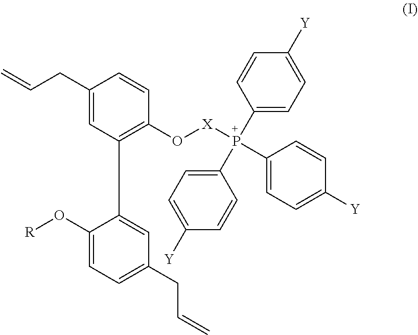


Priority Applications (3)
| Application Number | Priority Date | Filing Date | Title |
|---|---|---|---|
| US16/714,461 US11083739B2 (en) | 2018-12-14 | 2019-12-13 | Mito-magnolol compounds and methods of synthesis and use thereof |
| US17/099,268 US11897910B2 (en) | 2015-06-11 | 2020-11-16 | Mito-honokiol compounds and methods of synthesis and use thereof |
| US17/397,672 US11612610B2 (en) | 2018-12-14 | 2021-08-09 | Mito-magnolol compounds and methods of synthesis and use thereof |
Applications Claiming Priority (2)
| Application Number | Priority Date | Filing Date | Title |
|---|---|---|---|
| US201862779795P | 2018-12-14 | 2018-12-14 | |
| US16/714,461 US11083739B2 (en) | 2018-12-14 | 2019-12-13 | Mito-magnolol compounds and methods of synthesis and use thereof |
Related Parent Applications (2)
| Application Number | Title | Priority Date | Filing Date |
|---|---|---|---|
| PCT/US2016/036827 Continuation-In-Part WO2016201188A1 (en) | 2015-06-11 | 2016-06-10 | Mito-honokiol compounds and methods of synthesis and use thereof |
| US15/735,405 Continuation-In-Part US10836782B2 (en) | 2015-06-11 | 2016-06-10 | Mito-honokiol compounds and methods of synthesis and use thereof |
Related Child Applications (2)
| Application Number | Title | Priority Date | Filing Date |
|---|---|---|---|
| US17/099,268 Continuation-In-Part US11897910B2 (en) | 2015-06-11 | 2020-11-16 | Mito-honokiol compounds and methods of synthesis and use thereof |
| US17/397,672 Continuation US11612610B2 (en) | 2018-12-14 | 2021-08-09 | Mito-magnolol compounds and methods of synthesis and use thereof |
Publications (2)
| Publication Number | Publication Date |
|---|---|
| US20200222433A1 US20200222433A1 (en) | 2020-07-16 |
| US11083739B2 true US11083739B2 (en) | 2021-08-10 |
Family
ID=71077530
Family Applications (2)
| Application Number | Title | Priority Date | Filing Date |
|---|---|---|---|
| US16/714,461 Active US11083739B2 (en) | 2015-06-11 | 2019-12-13 | Mito-magnolol compounds and methods of synthesis and use thereof |
| US17/397,672 Active US11612610B2 (en) | 2018-12-14 | 2021-08-09 | Mito-magnolol compounds and methods of synthesis and use thereof |
Family Applications After (1)
| Application Number | Title | Priority Date | Filing Date |
|---|---|---|---|
| US17/397,672 Active US11612610B2 (en) | 2018-12-14 | 2021-08-09 | Mito-magnolol compounds and methods of synthesis and use thereof |
Country Status (2)
| Country | Link |
|---|---|
| US (2) | US11083739B2 (en) |
| WO (1) | WO2020124019A1 (en) |
Families Citing this family (1)
| Publication number | Priority date | Publication date | Assignee | Title |
|---|---|---|---|---|
| EP4637823A1 (en) * | 2022-12-20 | 2025-10-29 | The Medical College of Wisconsin, Inc. | Polyphenolic boronates and use thereof |
Family Cites Families (1)
| Publication number | Priority date | Publication date | Assignee | Title |
|---|---|---|---|---|
| CN108883082B (en) * | 2015-06-11 | 2021-10-26 | 威斯康星州医药大学股份有限公司 | Silo-honokiol compounds and methods of synthesis and use thereof |
-
2019
- 2019-12-13 US US16/714,461 patent/US11083739B2/en active Active
- 2019-12-13 WO PCT/US2019/066354 patent/WO2020124019A1/en not_active Ceased
-
2021
- 2021-08-09 US US17/397,672 patent/US11612610B2/en active Active
Non-Patent Citations (133)
| Title |
|---|
| Abildgaard et al., 2014. Bioenergetic modulation with dichloroacetate reduces the growth of melanoma cells and potentiates their response to BRAFV600E inhibition. J Transl Med 12: 247. |
| Allavena et al., 2016. Suppressed translation and ULK1 degradation as potential mechanisms of autophagy limitation under prolonged starvation. Autophagy 12: 2085-97. |
| Andreyev et al., 2015. Mitochondrial ROS Metabolism: 10 Years Later. Biochemistry (Mosc) 80: 517-31. |
| Araki et al., 2009. mTOR regulates memory CD8 T-cell differentiation. Nature 460: 108-12. |
| Asin-Cayuela et al., Fine-tuning the hydrophobicity of a mitochondria-targeted antioxidant. FEBS Lett. Jul. 30, 2004;571 (1-3):9-16. |
| Batus et al., 2013. Optimal management of metastatic melanoma: current strategies and future directions. Am J Clin Dermatol 14: 179-94. |
| Beier et al., 2015. Essential role of mitochondrial energy metabolism in Foxp3(+) T-regulatory cell function and allograft survival. Faseb j 29: 2315-26. |
| Bengsch et al., 2017. CTLA-4/CD80 pathway regulates T cell infiltration into pancreatic cancer. Cancer Immunol Immunother 66: 1609-17. |
| Bentolila et al., 2018. Intravital Imaging of Human Melanoma Cells in the Mouse Ear Skin by Two-Photon Excitation Microscopy. Methods Mol Biol 1755: 223-32. |
| Berridge et al., Effects of mitochondrial gene deletion on tumorigenicity of metastatic melanoma: reassessing the Warburg effect. Rejuvenation Res. Apr.-Jun. 2010;13(2-3):139-41. |
| Blank et al., 2006. Blockade of PD-L1 (B7-H1) augments human tumor-specific T cell responses in vitro. Int J Cancer 119: 317-27. |
| Boyle et al., 2018. Mitochondria-targeted drugs stimulate mitophagy and abrogate colon cancer cell proliferation. J Biol Chem 293: 14891-904. |
| Brahmer et al., 2010. Phase I study of single-agent anti-programmed death-1 (MDX-1106) in refractory solid tumors: safety, clinical activity, pharmacodynamics, and immunologic correlates. J Clin Oncol 28: 3167-75. |
| Brandt et al., 2018. Augmentation of intracellular iron using iron sucrose enhances the toxicity of pharmacological ascorbate in colon cancer cells. Redox Biol 14: 82-7. |
| Brunen D, Bernards R. 2017. Drug therapy: Exploiting synthetic lethality to improve cancer therapy. Nat Rev Clin Oncol 14: 331-2. |
| Bryant et al., 2017. A Mitochondrial-targeted purine-based HSP90 antagonist for leukemia therapy. Oncotarget 8: 112184-98. |
| Buck et al., 2015. T cell metabolism drives immunity. J Exp Med 212: 1345-60. |
| Cancer [online], [retrieved on Jul. 6, 2007]. Retrieved from the internet, URL http://www.nlm.nih.gov/medlineplus/cancer.html>. * |
| Cancer and Metastasis Reviews (1998), 17(1), 91-106. * |
| Cha et al., 2018. Metformin Promotes Antitumor Immunity via Endoplasmic-Reticulum-Associated Degradation of PD-L1. Mol Cell 71: 606-20.e7. |
| Chamoto et al., Mitochondrial activation chemicals synergize with surface receptor PD-1 blockade for T cell-dependent antitumor activity. Proc Natl Acad Sci U S A. Jan. 31, 2017;114(5):E761-E70. |
| Chandran et al., 2009. Doxorubicin inactivates myocardial cytochrome c oxidase in rats: cardioprotection by Mito-Q. Biophys. J 96: 1388-98. |
| Chandrashekaran et al., 2017. HMGB1-RAGE pathway drives peroxynitrite signaling-induced IBD-like inflammation in murine nonalcoholic fatty liver disease. Redox Biol 13: 8-19. |
| Chen et al., 2013. Oncology meets immunology: the cancer-immunity cycle. Immunity 39: 1-10. |
| Chen et al., 2017. Elements of cancer immunity and the cancer-immune set point. Nature 541: 321-30. |
| Cheng et al., 2011. Mitochondria-targeted nitroxides exacerbate fluvastatin-mediated cytostatic and cytotoxic effects in breast cancer cells. Cancer Biol. Ther 12: 707-17. |
| Cheng et al., 2015. Antiproliferative effects of mitochondria-targeted cationic antioxidants and analogs: Role of mitochondrial bioenergetics and energy-sensing mechanism. Cancer Lett 365: 96-106. |
| Cheng et al., 2016. Mitochondria-Targeted Analogues of Metformin Exhibit Enhanced Antiproliferative and Radiosensitizing Effects in Pancreatic Cancer Cells. Cancer Res 76: 3904-15. |
| Cheng et al., 2018. Detection of mitochondria-generated reactive oxygen species in cells using multiple probes and methods: Potentials, pitfalls, and the future. J Biol Chem 293: 10363-80. |
| Cheng et al., Mitochondria-targeted drugs synergize with 2-deoxyglucose to trigger breast cancer cell death. Cancer Res. May 15, 2012;72(10):2634-44. |
| Cheng et al., Mitochondria-targeted vitamin E analogs inhibit breast cancer cell energy metabolism and promote cell death. BMC Cancer. Jun. 13, 2013;13:285. |
| Cheng et al., Profiling and targeting of cellular bioenergetics: inhibition of pancreatic cancer cell proliferation. Br J Cancer. Jul. 8, 2014;111(1):85-93. |
| Chuang et al., Magnolia polyphenols attenuate oxidative and inflammatory responses in neurons and microglial cells. J Neuroinflammation. Jan. 29, 2013;10:15. doi: 10.1186/1742-2094-10-15. |
| Corazao-Rozas et al., Mitochondrial oxidative stress is the Achille's heel of melanoma cells resistant to Braf-mutant inhibitor. Oncotarget. Nov. 2013;4(11):1986-98. |
| Cox et al., 2009. Mitochondrial peroxiredoxin involvement in antioxidant defence and redox signalling. Biochem J 425: 313-25. |
| Cunha et al., 2018. LC3-Associated Phagocytosis in Myeloid Cells Promotes Tumor Immune Tolerance. Cell 75: 429-41. |
| Das et al., 2015. NADPH Oxidase-Derived Peroxynitrite Drives Inflammation in Mice and Human Nonalcoholic Steatohepatitis via TLR4-Lipid Raft Recruitment. Am J Pathol 185: 1944-57. |
| Deng et al., 2017. Mitochondria Targeted Nanoscale Zeolitic Imidazole Framework-90 for ATP Imaging in Live Cells. J Am Chem Soc 139: 5877-82. |
| Domingues et al., 2018. Melanoma treatment in review. Immunotargets Ther 7: 35-49. |
| Dowling et al., 2018. Autophagy and T cell metabolism. Cancer Lett 419: 20-6. |
| Drury et al., 2011. Monomeric and dimeric CXCL12 inhibit metastasis through distinct CXCR4 interactions and signaling pathways. Proc. Natl. Acad. Sci. U. S. A 108: 17655-60. |
| Eikawa et al., 2015. Immune-mediated antitumor effect by type 2 diabetes drug, metformin. Proc Natl Acad Sci U S A 112: 1809-14. |
| Fanciulli et al., Energy Metabolism of Human LoVo Colon Carcinoma Cells: Correlation to Drug Resistance and Influence of Lonidamine. Clin Cancer Res. Apr. 2000;6(4):1590-7. |
| Gajewski et al., 2013. Innate and adaptive immune cells in the tumor microenvironment. Nat Immunol 14: 1014-22. |
| Gajewski. 2006. Identifying and overcoming immune resistance mechanisms in the melanoma tumor microenvironment. Clin Cancer Res 12: 2326s-30s. |
| Gide et al., 2018. Primary and Acquired Resistance to Immune Checkpoint Inhibitors in Metastatic Melanoma. Clin Cancer Res 24: 1260-70. |
| Goffaux et al., 2017. A Dynamic Metabolic Flux Analysis of Myeloid-Derived Suppressor Cells Confirms Immunosuppression-Related Metabolic Plasticity. Sci Rep 7: 9850. |
| Gopal et al., 2014. Inhibition of mTORC1/2 overcomes resistance to MAPK pathway inhibitors mediated by PGC1alpha and oxidative phosphorylation in melanoma. Cancer Res 74: 7037-47. |
| Gottesman et al., Multidrug resistance in cancer: role of ATP-dependent transporters. Nat Rev Cancer. Jan. 2002;2 (1):48-58. |
| Gottschalk et al., Imatinib (STI571)-mediated changes in glucose metabolism in human leukemia BCR-ABL-positive cells. Clin Cancer Res. Oct. 1, 2004;10(19):6661-8. |
| Gropper et al., 2017. Culturing CTLs under Hypoxic Conditions Enhances Their Cytolysis and Improves Their Anti-tumor Function. Cell Rep 20: 2547-55. |
| Gubser et al., 2013. Rapid effector function of memory CD8+ T cells requires an immediate-early glycolytic switch. Nat Immunol 14: 1064-72. |
| Hamid et al., 2013. Safety and tumor responses with lambrolizumab (anti-PD-1) in melanoma. N Engl J Med 369: 134-44. |
| Hanahan et al., 2011. Hallmarks of cancer: the next generation. Cell 144: 646-74. |
| Haq et al., Molecular pathways: BRAF induces bioenergetic adaptation by attenuating oxidative phosphorylation. Clin Cancer Res. May 1, 2014;20(9):2257-63. |
| Haq et al., Oncogenic BRAF regulates oxidative metabolism via PGC1α and MITF. Cancer Cell. Mar. 18, 2013;23 (3):302-15. |
| Hardeman et al., 2017. Dependence on Glycolysis Sensitizes BRAF-mutated Melanomas for Increased Response to Targeted BRAF Inhibition. Sci Rep 7: 42604. |
| Hardie et al., 2014. AMPK: regulating energy balance at the cellular and whole body levels. Physiology. (Bethesda. ) 29: 99-107. |
| Hardy et al., 2018. Detection and Characterization of Reactive Oxygen and Nitrogen Species in Biological Systems by Monitoring Species-Specific Products. Antioxid Redox Signal 28: 1416-32. |
| Hodi et al., 2010. Improved survival with ipilimumab in patients with metastatic melanoma. N Engl J Med 363: 711-23. |
| Holmstrom et al., 2014. Cellular mechanisms and physiological consequences of redox-dependent signalling. Nat Rev Mol Cell Biol 15: 411-21. |
| Hong et al., 2017. Suppression of B-Raf(V600E) melanoma cell survival by targeting mitochondria using triphenyl-phosphonium-conjugated nitroxide or ubiquinone. Cancer Biol Ther 18: 106-14. |
| Hosios et al., 2018. The redox requirements of proliferating mammalian cells. J Biol Chem 293: 7490-8. |
| Hwang et al., 2012. E-cadherin is critical for collective sheet migration and is regulated by the chemokine CXCL12 protein during restitution. J. Biol. Chem 287: 22227-40. |
| Ippolito et al., Metabolic shift toward oxidative phosphorylation in docetaxel resistant prostate cancer cells. Oncotarget. Sep. 20, 2016;7(38):61890-904. |
| Janji et al., 2016. The multifaceted role of autophagy in tumor evasion from immune surveillance. Oncotarget 7: 17591-607. |
| Johnson et al., 1981. Monitoring of relative mitochondrial membrane potential in living cells by fluorescence microscopy. J Cell Biol 88: 526-35. |
| Kaelin. The concept of synthetic lethality in the context of anticancer therapy. Nat Rev Cancer. Sep. 2005;5(9):689-98. |
| Kalyanaraman et al., 2018. A review of the basics of mitochondrial bioenergetics, metabolism, and related signaling pathways in cancer cells: Therapeutic targeting of tumor mitochondria with lipophilic cationic compounds. Redox Biol 14: 316-27. |
| Kalyanaraman et al., 2018. Low-Temperature EPR Spectroscopy as a Probe-Free Technique for Monitoring Oxidants Formed in Tumor Cells and Tissues: Implications in Drug Resistance and OXPHOS-Targeted Therapies. Cell Biochem Biophys Sep. 26, 2018. doi: 10.1007/s12013-018-0858-1. [Epub ahead of print]. |
| Kaminski et al., 2012. T cell activation is driven by an ADP-dependent glucokinase linking enhanced glycolysis with mitochondrial reactive oxygen species generation. Cell Rep 2: 1300-15. |
| Katayama et al., 2011. A sensitive and quantitative technique for detecting autophagic events based on lysosomal delivery. Chem Biol 18: 1042-52. |
| Kim et al., Current and upcoming mitochondrial targets for cancer therapy. Semin Cancer Biol. Dec. 2017;47:154-67. |
| Le Bourgeois et al., 2018. Targeting T Cell Metabolism for Improvement of Cancer Immunotherapy. Front Oncol 8: 237. |
| Li et al., 2018. Metformin-Induced Reduction of CD39 and CD73 Blocks Myeloid-Derived Suppressor Cell Activity in Patients with Ovarian Cancer. Cancer Res 78: 1779-91. |
| Liberman et al., 1969. Mechanism of coupling of oxidative phosphorylation and the membrane potential of mitochondria. Nature 222: 1076-8. |
| Lichtshtein et al., 1979. Use of a lipophilic cation for determination of membrane potential in neuroblastoma-glioma hybrid cell suspensions. Proc Natl Acad Sci U S A 76: 650-4. |
| Luengo et al., 2017. Targeting Metabolism for Cancer Therapy. Cell Chem Biol 24: 1161-80. |
| Ma et al., 2014. Targeting ER stress-induced autophagy overcomes BRAF inhibitor resistance in melanoma. J Clin Invest 124: 1406-17. |
| Melnikova et al., 2004. Genomic alterations in spontaneous and carcinogen-induced murine melanoma cell lines. Oncogene 23: 2347-56. |
| Mihm et al., 1996. Tumor infiltrating lymphocytes in lymph node melanoma metastases: a histopathologic prognostic indicator and an expression of local immune response. Lab Invest 74: 43-7. |
| Miller et al., 2016. Cancer treatment and survivorship statistics, 2016. CA Cancer J Clin 66: 271-89. |
| Mocholi et al., 2018. Autophagy Is a Tolerance-Avoidance Mechanism that Modulates TCR-Mediated Signaling and Cell Metabolism to Prevent Induction of T Cell Anergy. Cell Rep 24: 1136-50. |
| Modica-Napolitano et al., 1987. Basis for the selective cytotoxicity of rhodamine 123. Cancer Res 47: 4361-5. |
| Molina et al., 2018. An inhibitor of oxidative phosphorylation exploits cancer vulnerability. Nat Med 24: 1036-46. |
| Mukhopadhyay et al., 2012. Mitochondrial reactive oxygen species generation triggers inflammatory response and tissue injury associated with hepatic ischemia-reperfusion: Therapeutic potential of mitochondrially targeted antioxidants. Free Radic. Biol. Med 53: 1123-38. |
| Muller et al., 2018. A 3D Organotypic Melanoma Spheroid Skin Model. J Vis Exp May 18, 2018;(135). doi: 10.3791/57500. |
| Murphy et al., Targeting antioxidants to mitochondria by conjugation to lipophilic cations. Annu Rev Pharmacol Toxicol. 2007;47:629-56. |
| Myers. 2016. Enhanced targeting of mitochondrial peroxide defense by the combined use of thiosemicarbazones and inhibitors of thioredoxin reductase. Free Radic Biol Med 91: 81-92. |
| Nadakavukaren et al., 1985. Increased rhodamine 123 uptake by carcinoma cells. Cancer Res 45: 6093-9. |
| Nakamura et al., 2002. Characterization of mouse melanoma cell lines by their mortal malignancy using an experimental metastatic model. Life Sci 70: 791-8. |
| Owens et al., 2012. Genomic instability induced by mutant succinate dehydrogenase subunit D (SDHD) is mediated by O2(-*) and H2O2. Free Radic Biol Med 52: 160-6. |
| Ozsvari et al., 2018. Exploiting mitochondrial targeting signal(s), TPP and bis-TPP, for eradicating cancer stem cells (CSCs). Aging (Albany NY) 10: 229-40. |
| Pan et al., 2018. Mitochondria-Targeted Honokiol Confers a Striking Inhibitory Effect on Lung Cancer via Inhibiting Complex I Activity. iScience 3: 192-207. |
| Pearce et al., 2009. Enhancing CD8 T-cell memory by modulating fatty acid metabolism. Nature 460: 103-7. |
| Pereira et al., 2018. Metformin exerts antitumor activity via induction of multiple death pathways in tumor cells and activation of a protective immune response. Oncotarget 9: 25808-25. |
| Petrylak et al., Docetaxel and estramustine compared with mitoxantrone and prednisone for advanced refractory prostate cancer. N Engl J Med. Oct. 7, 2004;351(15):1513-20. |
| Pollak. Targeting oxidative phosphorylation: why, when, and how. Cancer Cell. Mar. 18, 2013;23(3):263-4. |
| Puleston et al., 2014. Autophagy in the immune system. Immunology 141: 1-8. |
| Roesch et al., 2013. Overcoming intrinsic multidrug resistance in melanoma by blocking the mitochondrial respiratory chain of slow-cycling JARID1B(high) cells. Cancer Cell 23: 811-25. |
| Roy et al., 2014. CXCL12 chemokine expression suppresses human pancreatic cancer growth and metastasis. PLoS. ONE 9: e90400. |
| Roy et al., 2015. Pancreatic Cancer Cell Migration and Metastasis Is Regulated by Chemokine-Biased Agonism and Bioenergetic Signaling. Cancer Res 75: 3529-42. |
| Sarrica et al., 2018. Safety and Toxicology of Magnolol and Honokiol. Planta Med 84: 1151-64. |
| Schlie et al., 2015. Survival of effector CD8+ T cells during influenza infection is dependent on autophagy. J Immunol 194: 4277-86. |
| Schockel et al., 2015. Targeting mitochondrial complex I using BAY 87-2243 reduces melanoma tumor growth. Cancer Metab 3: 11. |
| Science (1999), vol. 286, 531-537. * |
| Sena et al., 2013. Mitochondria are required for antigen-specific T cell activation through reactive oxygen species signaling. Immunity 38: 225-36. |
| Smith et al., 2008. Mitochondria-targeted antioxidants in the treatment of disease. Ann. N. Y. Acad. Sci 1147: 105-11. |
| Spoerri et al., 2017. Real-Time Cell Cycle Imaging in a 3D Cell Culture Model of Melanoma. Methods Mol Biol 1612: 401-16. |
| Sukumar et al., 2016. Mitochondrial Membrane Potential Identifies Cells with Enhanced Stemness for Cellular Therapy. Cell Metab 23: 63-76. |
| Sun et al., 2017. A fluorescence-based imaging method to measure in vitro and in vivo mitophagy using mt-Keima. Nat Protoc 12: 1576-87. |
| Tan et al., The Role of PGC1α in Cancer Metabolism and its Therapeutic Implications. Mol Cancer Ther. May 2016;15 (5):774-82. |
| Tian et al., Magnolol Alleviates Inflammatory Responses and Lipid Accumulation by AMP-Activated Protein Kinase-Dependent Peroxisome Proliferator-Activated Receptor α Activation. Front Immunol. Feb. 5, 2018;9:147. doi: 10.3389/fimmu.2018.00147. eCollection 2018. |
| Titova et al., 2018. Mitochondria-targeted antioxidant SkQ1 suppresses fibrosarcoma and rhabdomyosarcoma tumour cell growth. Cell Cycle 17: 1797-811. |
| Tsai et al., 2018. Development of primary human pancreatic cancer organoids, matched stromal and immune cells and 3D tumor microenvironment models. BMC Cancer 18: 335. |
| Tsao et al., 2004. Genetic interaction between NRAS and BRAF mutations and PTEN/MMAC1 inactivation in melanoma. J Invest Dermatol 122: 337-41. |
| Van de Bittner et al., 2010. In vivo imaging of hydrogen peroxide production in a murine tumor model with a chemoselective bioluminescent reporter. Proc Natl Acad Sci U S A 107: 21316-21. |
| Vander Heiden et al., 2017. Understanding the Intersections between Metabolism and Cancer Biology. Cell 168: 657-69. |
| Vazquez et al., PGC1α expression defines a subset of human melanoma tumors with increased mitochondrial capacity and resistance to oxidative stress. Cancer Cell. Mar. 18, 2013;23(3):287-301. |
| Vellinga et al., SIRT1/PGC1α-Dependent Increase in Oxidative Phosphorylation Supports Chemotherapy Resistance of Colon Cancer. Clin Cancer Res. Jun. 15, 2015;21(12):2870-9. |
| Wang et al., 1998. Chemokines and their role in tumor growth and metastasis. J. Immunol. Methods 220: 1-17. |
| Weinberg et al., 2015. Mitochondria in the regulation of innate and adaptive immunity. Immunity 42: 406-17. |
| Weinberg et al., 2015. Targeting mitochondria metabolism for cancer therapy. Nat Chem Biol 11: 9-15. |
| Weinberg et al., Mitochondrial metabolism and ROS generation are essential for Kras-mediated tumorigenicity. Proc Natl Acad Sci U S A. May 11, 2010;107(19):8788-93. |
| Wendt et al., 2008. Constitutive CXCL12 expression induces anoikis in colorectal carcinoma cells. Gastroenterology 135: 508-17. |
| Wendt et al., 2008. Epigenetic silencing of CXCL12 increases the metastatic potential of mammary carcinoma cells. Oncogene 27: 1461-71. |
| Wood et al., 2003. Peroxiredoxin evolution and the regulation of hydrogen peroxide signaling. Science 300: 650-3. |
| Zha et al., 2004. Negative regulation of T-cell function by PD-1. Crit Rev Immunol 24: 229-37. |
| Ziegler et al., 2018. Mitophagy in Intestinal Epithelial Cells Triggers Adaptive Immunity during Tumorigenesis. Cell 174: 88-101.e16. |
| Zielonka et al., 2016. On the use of peroxy-caged luciferin (PCL-1) probe for bioluminescent detection of inflammatory oxidants in vitro and in vivo—Identification of reaction intermediates and oxidant-specific minor products. Free Radic Biol Med 99: 32-42. |
| Zielonka et al., 2018. Small-molecule luminescent probes for the detection of cellular oxidizing and nitrating species. Free Radic Biol Med 128: 3-22. |
| Zielonka et al., Mitochondria-Targeted Triphenylphosphonium-Based Compounds: Syntheses, Mechanisms of Action, and Therapeutic and Diagnostic Applications. Chem Rev. Aug. 9, 2017;117(15):10043-120. |
| Zimmerman et al., 2011. Targeted intestinal epithelial deletion of the chemokine receptor CXCR4 reveals important roles for extracellular-regulated kinase-1/2 in restitution. Lab Invest 91: 1040-55. |
Also Published As
| Publication number | Publication date |
|---|---|
| US11612610B2 (en) | 2023-03-28 |
| US20200222433A1 (en) | 2020-07-16 |
| US20220211727A1 (en) | 2022-07-07 |
| WO2020124019A1 (en) | 2020-06-18 |
Similar Documents
| Publication | Publication Date | Title |
|---|---|---|
| Tufail et al. | Breast cancer: molecular mechanisms of underlying resistance and therapeutic approaches | |
| Xiong et al. | Target profiling of an iridium (III)-based immunogenic cell death inducer unveils the engagement of unfolded protein response regulator BiP | |
| Wagner et al. | Dose intensification of TRAIL-inducing ONC201 inhibits metastasis and promotes intratumoral NK cell recruitment | |
| US11897910B2 (en) | Mito-honokiol compounds and methods of synthesis and use thereof | |
| Lin et al. | Targeting activated Akt with GDC-0068, a novel selective Akt inhibitor that is efficacious in multiple tumor models | |
| Miao et al. | Endoplasmic reticulum-targeting AIE photosensitizers to boost immunogenic cell death for immunotherapy of bladder carcinoma | |
| US20230093147A1 (en) | Methods and compositions relating to improved combination therapies | |
| US20230061048A1 (en) | Selection of patients for combination therapy | |
| US20150246033A1 (en) | Methods for inhibiting tie-2 kinase useful in the treatment of cancer | |
| KR20210063475A (en) | Methods for inhibiting tie2 kinase useful in the treatment of cancer | |
| Shen et al. | Sinoporphyrin sodium-mediated sonodynamic therapy inhibits RIP3 expression and induces apoptosis in the H446 small cell lung cancer cell line | |
| US11612610B2 (en) | Mito-magnolol compounds and methods of synthesis and use thereof | |
| Brincks et al. | Triptolide enhances the tumoricidal activity of TRAIL against renal cell carcinoma | |
| Li et al. | Nanodelivery of arsenic trioxide induces macrophage-governed cGAS-STING signaling to remodel immune microenvironment in hepatocellular carcinoma | |
| Rajpurohit et al. | Moving towards dawn: KRas signaling and treatment in pancreatic ductal adenocarcinoma | |
| WO2022051420A1 (en) | Methods and compositions for targeting cytosolic dsdna signaling in chromosomally unstable cancers | |
| Bieber et al. | Personalized treatment options in dermatology | |
| WO2024097994A1 (en) | Methods for the detection and treatment of non-small-cell lung cancer | |
| KR20170071393A (en) | Composition for diagnosis of radioresistance and use thereof | |
| US11298338B2 (en) | Compositions and methods for cancer therapy | |
| ES2939357T3 (en) | Selection of patients for combination therapy | |
| JP7541512B2 (en) | Prodrugs and their medical uses | |
| Samishina et al. | Programmed death-ligand 1 signaling pathway involves in bladder cancer growth and progression | |
| Martelo | Efficacy of RANK pathway and CDK4/6 inhibition in immune-competent luminal breast cancer models | |
| Niavarani | Targeting metastatic triple negative breast cancer with an autologous cancer cell vaccine |
Legal Events
| Date | Code | Title | Description |
|---|---|---|---|
| FEPP | Fee payment procedure |
Free format text: ENTITY STATUS SET TO UNDISCOUNTED (ORIGINAL EVENT CODE: BIG.); ENTITY STATUS OF PATENT OWNER: SMALL ENTITY |
|
| FEPP | Fee payment procedure |
Free format text: ENTITY STATUS SET TO SMALL (ORIGINAL EVENT CODE: SMAL); ENTITY STATUS OF PATENT OWNER: SMALL ENTITY |
|
| AS | Assignment |
Owner name: MEDICAL COLLEGE OF WISCONSIN, INC., WISCONSIN Free format text: ASSIGNMENT OF ASSIGNORS INTEREST;ASSIGNORS:KALYANARAMAN, BALARAMAN;ZIELONKA, JACEK MICHAL;CHENG, GANG;SIGNING DATES FROM 20200204 TO 20200210;REEL/FRAME:051773/0959 Owner name: AIX-MARSEILLE UNIVERSITE, FRANCE Free format text: ASSIGNMENT OF ASSIGNORS INTEREST;ASSIGNORS:HARDY, MICAEL JOEL;OUARI, OLIVIER;SIGNING DATES FROM 20200204 TO 20200205;REEL/FRAME:051774/0069 |
|
| FEPP | Fee payment procedure |
Free format text: PETITION RELATED TO MAINTENANCE FEES GRANTED (ORIGINAL EVENT CODE: PTGR); ENTITY STATUS OF PATENT OWNER: SMALL ENTITY |
|
| STPP | Information on status: patent application and granting procedure in general |
Free format text: RESPONSE TO NON-FINAL OFFICE ACTION ENTERED AND FORWARDED TO EXAMINER |
|
| STPP | Information on status: patent application and granting procedure in general |
Free format text: NOTICE OF ALLOWANCE MAILED -- APPLICATION RECEIVED IN OFFICE OF PUBLICATIONS |
|
| STPP | Information on status: patent application and granting procedure in general |
Free format text: AWAITING TC RESP., ISSUE FEE NOT PAID |
|
| STPP | Information on status: patent application and granting procedure in general |
Free format text: NOTICE OF ALLOWANCE MAILED -- APPLICATION RECEIVED IN OFFICE OF PUBLICATIONS |
|
| STPP | Information on status: patent application and granting procedure in general |
Free format text: PUBLICATIONS -- ISSUE FEE PAYMENT VERIFIED |
|
| STCF | Information on status: patent grant |
Free format text: PATENTED CASE |
|
| FEPP | Fee payment procedure |
Free format text: SURCHARGE FOR LATE PAYMENT, SMALL ENTITY (ORIGINAL EVENT CODE: M2554); ENTITY STATUS OF PATENT OWNER: SMALL ENTITY |
|
| MAFP | Maintenance fee payment |
Free format text: PAYMENT OF MAINTENANCE FEE, 4TH YR, SMALL ENTITY (ORIGINAL EVENT CODE: M2551); ENTITY STATUS OF PATENT OWNER: SMALL ENTITY Year of fee payment: 4 |