US11058721B2 - Composition comprising a substantially pure population of multipotent stromal cells encapsulated in platelet-poor plasma (PPP) - Google Patents
Composition comprising a substantially pure population of multipotent stromal cells encapsulated in platelet-poor plasma (PPP) Download PDFInfo
- Publication number
- US11058721B2 US11058721B2 US16/478,002 US201816478002A US11058721B2 US 11058721 B2 US11058721 B2 US 11058721B2 US 201816478002 A US201816478002 A US 201816478002A US 11058721 B2 US11058721 B2 US 11058721B2
- Authority
- US
- United States
- Prior art keywords
- ppp
- cells
- root canal
- msc
- mscs
- Prior art date
- Legal status (The legal status is an assumption and is not a legal conclusion. Google has not performed a legal analysis and makes no representation as to the accuracy of the status listed.)
- Active, expires
Links
Images


Classifications
-
- A—HUMAN NECESSITIES
- A61—MEDICAL OR VETERINARY SCIENCE; HYGIENE
- A61K—PREPARATIONS FOR MEDICAL, DENTAL OR TOILETRY PURPOSES
- A61K35/00—Medicinal preparations containing materials or reaction products thereof with undetermined constitution
- A61K35/12—Materials from mammals; Compositions comprising non-specified tissues or cells; Compositions comprising non-embryonic stem cells; Genetically modified cells
- A61K35/14—Blood; Artificial blood
- A61K35/16—Blood plasma; Blood serum
-
- A—HUMAN NECESSITIES
- A61—MEDICAL OR VETERINARY SCIENCE; HYGIENE
- A61K—PREPARATIONS FOR MEDICAL, DENTAL OR TOILETRY PURPOSES
- A61K35/00—Medicinal preparations containing materials or reaction products thereof with undetermined constitution
- A61K35/12—Materials from mammals; Compositions comprising non-specified tissues or cells; Compositions comprising non-embryonic stem cells; Genetically modified cells
- A61K35/28—Bone marrow; Haematopoietic stem cells; Mesenchymal stem cells of any origin, e.g. adipose-derived stem cells
-
- A—HUMAN NECESSITIES
- A61—MEDICAL OR VETERINARY SCIENCE; HYGIENE
- A61K—PREPARATIONS FOR MEDICAL, DENTAL OR TOILETRY PURPOSES
- A61K35/00—Medicinal preparations containing materials or reaction products thereof with undetermined constitution
- A61K35/12—Materials from mammals; Compositions comprising non-specified tissues or cells; Compositions comprising non-embryonic stem cells; Genetically modified cells
- A61K35/48—Reproductive organs
- A61K35/51—Umbilical cord; Umbilical cord blood; Umbilical stem cells
-
- A—HUMAN NECESSITIES
- A61—MEDICAL OR VETERINARY SCIENCE; HYGIENE
- A61P—SPECIFIC THERAPEUTIC ACTIVITY OF CHEMICAL COMPOUNDS OR MEDICINAL PREPARATIONS
- A61P1/00—Drugs for disorders of the alimentary tract or the digestive system
- A61P1/02—Stomatological preparations, e.g. drugs for caries, aphtae, periodontitis
Definitions
- the present invention relates to a root canal filler and a dental tissue regeneration method by using the root canal filler.
- the dental pulp When dental caries is deep enough to reach dental pulp, pulpectomy is usually performed for treatment of the caries.
- the dental pulp not only has a function to block external stimulus by reparative dentin formation, but also functions to inhibit further invasion of bacterial by sense and prevent tooth fracture caused by chewing a hard material with the sense of occlusion.
- the dental pulp can maintain protein and water in dentin by metabolism, and additionally keep the tensile strength and other properties of dentin.
- the dental pulp is also known to have an infection defence mechanism by immune system.
- NiTi alloy rotary files are used popularly in endodontics, because of morphological complexity of the root canal.
- complete pulpectomy, enlargement of root canal and root canal filling are almost impossible.
- pulpectomy often leads to periapical periodontitis, and has high possibility of resultant loss of the tooth.
- most of the reports on pulp regeneration in the emptied root canal are those in immature teeth.
- no method or root canal filler for dental tissue regeneration have been developed yet. There is thus an unmet need for development of a method to preserve the dental pulp as long as possible for longevity of teeth.
- mesenchymal stem cells hold great promise in the dental regeneration field. They could be derived from a large variety of sources such as the bone marrow (BM), the adipose tissue, the dental pulp, the gingival tissue or the umbilical cord and placenta. MSCs are pluripotent cells and have the ability to differentiate into different types of cells (osteogenic, chondrogenic and adipogenic). Despite the various sources of MSCs, bone marrow-MSCs have been predominantly used extensively for cell therapy, however, one of the major drawbacks of using bone marrow derive MSCs (BM-MSCs) is the yield, which is relatively low, therefore harvesting a large volume of BM-MSCs is difficult. In this sense, according to different studies, in order to perform a clinical trial a large number of cells are required sharing a stable phenotype in order to obtain successful results. This objective is difficult to achieve with BM-MSCs.
- BM-MSCs bone marrow derive MSCs
- DPSCs dental pulp stem cells
- SHED exfoliated deciduous teeth
- PDLSCs periodontal ligament stem cells
- SCAP stem cells from apical papilla
- DFPCs dental follicle progenitor cells
- Placental MSCs have been obtained from multiple placental areas including both maternal and foetal origins as well as from the umbilical cord blood (UCB), amniotic fluids, amniotic membrane, Wharton's jelly, chorionic membrane, chorionic villi and decidua.
- PPP Platelet-Poor Plasma
- PPP is blood plasma with very low number of platelets ( ⁇ 10 ⁇ 10 3 / ⁇ L).
- PPP was recommended for use in platelet aggregation studies to both adjust the Platelet-rich plasma concentration, and to serve as a control.
- PPP may have elevated levels of fibrinogen, which has the ability to form a fibrin-rich clot once activated. Wound healing requires cell migration and attachment, which is facilitated by this fibrin clot Methods to prepare PPP are well known in the art, in this sense, an exemplary methodology is as follows:
- An object of the present invention which was made to solve the problems above, is to provide a novel and creative root canal filler for regeneration of dental tissue by filling a scaffold into the root canal of a mature tooth with complete apical closure after pulpectomy and a dental tissue regeneration method by using such a root canal filler.
- Said root canal filler is made of mesenchymal stem cells (MSCs) encapsulated or grown in Platelet-Poor Plasma.
- MSCs mesenchymal stem cells
- the present invention describes that there is a significant increase in the number of cells after 12 days of implantation compared to day 1 (pre-implantation) as shown in FIGS. 2 (E), (F) and (G) of MSCs encapsulated in PPP.
- the present invention thus indicates the usefulness of PPP as a support and vehicle for MSCs, in particular for the preparation of a root canal filler for the regeneration of dental tissue, in particular for the generation of pulp.
- FIG. 1 (A) Histology of MSCs grown in PPP for 1, 7 and 14 days. Cells were then counted and compared among the two different sources MSCs (mesenchymal stem cells) (UC-MSCs (umbilical cord stem cells) and DPSCs (dental pulp stem cells)). (B) Electron microscopy analysis (EM) of PPP with or without cultured MSCs and day 1 or 21 post-culture showing porosity changes.
- MSCs meenchymal stem cells
- DPSCs dental pulp stem cells
- FIG. 2 (A) Quantification of cell proliferation between UC-MSC incubated at different time points. At day 3 and 7, an increase in the proliferation of UC-MSC at 37° C. compared 4° C. was observed (p ⁇ 0.05). (B, C and D) In vitro release of different proteins (VEGF, FGF and DMP-1) between 3 different donors of PPP and UC-MSC. The results have shown a higher protein release for the donor UC-MSC (374-5) and donor 0850 for PPP with a, p ⁇ 0.05. (E, F and G) Proliferation rate of encapsulated UC-MSC in PPP in vivo.
- FIG. 3 The proliferation between DPSC and UC-MSC was investigated using an Alamar Blue cell proliferation assay. A significant increase in the proliferation of DPSC at day 3 and day 9 with 1.2 and 1.1 fold increases respectively was observed (P ⁇ 0.0001). (B+C) indicates that UC-MSC possess a higher migration potential in comparison to DPSC for the different time points analyzed.
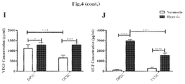
- FIG. 4 (A, B, C, D) Image analysis of the tube formation evaluated at 5 hours post-culture initiation showed a higher angiogenic capacity evidenced by a more extensive network of capillary-like structures for DPSC as compared to UC-MSC. (E) Images were taken and the results have shown a higher tubular structure for the HUVECS incubated with the conditioned medium under hypoxic conditions vs normoxic. (F, G, H) There was a significant difference between the formation of total tube lengths, total loops and total branching points between hypoxia DPSC and hypoxia UC-MSC. (I, J) The quantification of angiogenic factors revealed a non-significant difference in the VEGF release for DPSC compared to UC-MSC after 8 hours of incubation under hypoxic conditions.
- FIG. 5 (A) All plugs generated vessels around and inside the implant. (B) The image analysis of the implants showed a non-significant difference in the vessel formation between DPSC and UC-MSC. (C) The results have shown a 6.2 fold change higher hemoglobin content of DPSC compared to Matrigel (negative control) (P ⁇ 0.0001) and 5.1 fold change difference between UC-MSC and Matrigel (negative control).
- FIG. 6 (A) UC-MSC or DPSC were encapsulated in PPP scaffolds and cultured for various time points in vitro. (B) The results have shown that DPSC and UC-MSC were attached and proliferated on the PPP scaffold at various time points. The proliferation of DPSC or UC-MSC encapsulated in PPP was evaluated with alamarBlue. The results have shown an 1.5 (P ⁇ 0.05), 3.1 (P ⁇ 0.0001) and 3 fold change (P ⁇ 0.0001) respectively increase in the proliferation between DPSC+PPP vs PPP alone at day 3, day 7 and day 14.
- FIG. 7 An ELISA of different protein factors was used such as VEGF, FGF, and DMP-1 in order to investigate the release profile of PPP with or without cells.
- B There was a 1.3 fold increase (P ⁇ 0.01) in the protein expression of the encapsulated VEGF (EF) between PPP+DPSC and PPP+UC-MSC after 24 hours of incubation.
- C Also, a 2.1 fold increase of (P ⁇ 0.0001) increase in the protein expression of the encapsulated FGF (EF) after 24 hours between PPP+DPSC and PPP+UC-MSC.
- FIG. 8 (A) The dentin-discs/PPP scaffolds were implanted subcutaneously for 30 days. (B) Images were taken of the implants prior removal and an evaluation of the vessel formation around the dentin dentin-discs/PPP was performed. (C) The results have shown a 1.6 fold increase (P ⁇ 0.01) in vessels formation around the dentin-disc for the PPP+DPSC compared to PPP alone. (D) The hematoxylin and eosin results have shown that DPSC or UC-MSC were well incorporated in the PPP and migrated towards the dentin wall forming a dense layer of new mineralized dentin-like tissue.
- FIG. 9 (A) Both cell sources (UC-MSC and DPSC) showed a positive expression of the common MSC markers such as CD105, CD90, CD73, CD34, CD45, CD19 and HLA-DR and (B) also DPSC and UC-MSC were induced to differentiate into mesodermal tissues (adipopogenic, chondrogenic and osteogenic) lineages.
- the invention in the present embodiment relates to a dental tissue regeneration method for regeneration of dental tissue in root canal, characterized by injecting a population of multipotent stromal cells encapsulated in Platelet-Poor Plasma (PPP) into the apical side of the root canal after pulpectomy or enlargement and cleaning of an infected root canal.
- the dental tissues to be regenerated are, for example, blood vessel, nerve, dental pulp, dentin and others in root canal.
- the dental pulp is removed and disinfected; the apical portion of the root is cut out open (apicoectomized); and a root canal filler is transplanted.
- a synthetic filler or blood clot is not transplanted into the disinfected hollow root canal, but a scaffold superior in biocompatibility without causing adverse effects and in low immunogenicity with multipotent stromal cells, which mimics dental pulp tissue, is used.
- the root canal filler is preferably filled to 1 ⁇ 4 to 2 ⁇ 5 of the apical part of the root canal, more preferably 1 ⁇ 3 of the apical part.
- an extracellular matrix is prepared, in particular PPP.
- the extracellular matrix is the so-called scaffold, a matrix for cell attachment.
- the root canal filler is prepared by growing or encapsulating multipotent stromal cells including dental pulp stem cells or umbilical cord stem cells in the extracellular matrix (PPP).
- the cells including dental pulp stem cells are attached to the apical part of the root canal of the root canal filler.
- a typical example of the method of producing the root canal filler is provided in the examples.
- a tooth with pulpitis is subjected to extract in the dental tissue regeneration method in the present embodiment.
- the targeted tooth is then subjected to pulpectomy.
- the targeted tooth is a tooth in which microbial infection reaches the coronal pulp or the radicular pulp because of caries, pulpitis etc.
- Pulpectomy is an operation to remove the whole dental pulp present in the tooth.
- the width of the apical foramen i.e., the diameter of root canal
- the width of the apical foramen is desirably 0.7 mm or more and 1.5 mm or less.
- the width of the root canal is less than 0.7 mm, blood vessel and nerve do not penetrate easily from the apical periodontal tissue, and it may be difficult to fill the root canal filler, while when the width of the root canal is more than 1.5 mm, enlargement of the root canal may lead to application of a load more than needed on the targeted tooth, thus causing tooth fracture.
- the root canal filler (a composition comprising a population of multipotent stromal cells encapsulated in Platelet-Poor Plasma (PPP)) is inserted into the apical side of the root canal for example with tweezers.
- PPP Platelet-Poor Plasma
- Such composition might be injected for example with Pipetman or syringe.
- the cells included in the composition may be the autologous cells extracted from the animal subjected to the treatment for dental tissue regeneration or the allogeneic cells extracted from an animal different from the animal subjected to the treatment for dental tissue regeneration.
- the multipotent stromal cells or mesenchymal stem cells (MSC) useful in the present invention could be derived from a large variety of sources such as the bone marrow (BM), the adipose tissue, the dental pulp, the gingival tissue or the umbilical cord and placenta.
- BM bone marrow
- MSCs are multipotent cells and have the ability to differentiate into different types of cells (osteogenic, chondrogenic and adipogenic).
- MSCs especially useful in the present invention are different human dental stem cells such as: dental pulp stem cells (DPSCs), stem cells from exfoliated deciduous teeth (SHED), periodontal ligament stem cells (PDLSCs), stem cells from apical papilla (SCAP), and dental follicle progenitor cells (DFPCs).
- DPSCs dental pulp stem cells
- SHED exfoliated deciduous teeth
- PDLSCs periodontal ligament stem cells
- SCAP apical papilla
- DFPCs dental follicle progenitor cells
- P-MSCs Placental MSCs obtained from multiple placental areas including both maternal and foetal origins as well as from the umbilical cord blood (UCB), amniotic fluids, amniotic membrane, Wharton's jelly, chorionic membrane, chorionic villi and decidua.
- the content of the MSCs in the root canal filler is preferably 1 ⁇ 10 3 cell/ ⁇ l or more and 1 ⁇ 10 7 cell/ ⁇ l or less. It is because a stem cell content of less than 1 ⁇ 10 3 cell/ ⁇ l may lead to insufficient regeneration of the dental tissue in root canal. On the other hand, a dental pulp stem cell content of more than 1 ⁇ 10 6 cell/ ⁇ l, in particular more than 1 ⁇ 10 7 cell/ ⁇ l may cause unexpected adverse reactions to the targeted tooth.
- gelatin may be injected to the region above the root canal filler and the root canal is capped with a resin. Then, the extracted tooth is replanted into the odontectomized cavity.
- the dental tissue in the root canal is regenerated.
- the regenerated dental tissues are, for example, blood vessel and dental pulp tissue in the root canal.
- the resin is removed once; a morphogen such as BMP or a growth/differentiation factor is applied on the tooth-crown dental pulp; and the root canal is capped with the resin.
- Dentin is also regenerated, when the morphogen or the growth/differentiation factor is applied on the tooth-crown dental pulp.
- Tissues that can be regenerated are not limited thereto, and nerve regeneration is also accelerated.
- the targeted tooth is a tooth in which microbial infection reaches coronal pulp or radicular pulp because of caries, pulpitis, etc. in embodiment 1 described above, but it is not limited thereto, and the targeted teeth also include a tooth of which the sense of occlusion is weakened by deterioration in nerve function. It is possible in such a case to improve the occlusion sense by regenerating the dental pulp, by injecting a root canal filler after pulpectomy.
- the targeted teeth also include a tooth in which microbial infection reaches apical periodontal tissue (tooth in which microbes reach the dental pulp and additionally to dentin of root canal wall and apical periodontal tissue).
- a second embodiment of the invention refers to a composition comprising a population of multipotent stromal cells encapsulated in Platelet-Poor Plasma (PPP), wherein preferably the population of multipotent stromal cells are selected from the group consisting of dental pulp stem cells (DPSCs), stem cells from exfoliated deciduous teeth (SHED), periodontal ligament stem cells (PDLSCs), stem cells from apical papilla (SCAP), dental follicle progenitor cells (DFPCs), Placental MSCs (P-MSCs), bone marrow MSCs (mesenchymal stem cells), adipose tissue derived MSCs and umbilical cord blood (UCB) MSCs.
- DPSCs dental pulp stem cells
- SHED exfoliated deciduous teeth
- PDLSCs periodontal ligament stem cells
- SCAP dental follicle progenitor cells
- P-MSCs Placental MSCs
- bone marrow MSCs meenchy
- the population of multipotent stromal cells is able to secrete paracrine factors including extracellular vesicles, cytokines and growth factors, comprising or consisting of VEGF, FGF and or DMP-1.
- the multipotent stromal cells are encapsulated in a biomaterial scaffold, wherein the growth factors are gradually secreted. So, a preferred embodiment of the invention refers to multipotent stromal cells encapsulated in a biomaterial scaffold, wherein the cells secrete the growth factors which are absorbed by the scaffold which in turn secretes them to the exterior, therefore achieving the liberation of the secreted growth factors to the external medium.
- the multipotent stromal cells without the scaffold, collect the secretome, concentrate it, or isolate specific vesicles/molecules and then directly encapsulate them (the secretome) in the scaffolds.
- a third embodiment of the invention refers to a pharmaceutical composition which comprises the composition according to the second embodiment of the invention and optionally a pharmaceutically acceptable carrier/vehicle and one or more excipients.
- a fourth embodiment of the invention refers to the pharmaceutical composition of the third embodiment of the invention, for use in therapy or medicine, in particular for use in dental tissue regeneration in a method of regenerating dental tissue, in particular pulp tissue, in a root canal, more particularly the method comprises pulpectomizing or enlarging and cleaning of a root canal infected with periapical disease; injecting the composition according to any of claims 1 to 4 , into at least an apical area of the root canal.
- width of the root canal in the apical area is adjusted to a particular size, by enlargement of the root canal before insertion of the composition into the apical area of the root canal.
- the concentration of the multipotent stromal cells of any of the above mentioned compositions is from 1 ⁇ 10 3 cells/ ⁇ l to 1 ⁇ 10 7 cells/ ⁇ l, more preferably about 1 ⁇ 10 6 cells/ ⁇ l.
- a fifth embodiment of the invention refers to the above defined composition for use in the regeneration of defect of bone or connective tissue, preferably cartilage tissue.
- a sixth embodiment of the invention refers to the above defined composition for use in the regeneration of the skin or in the treatment of skin diseases or injuries, preferably acute and chronic ulcers.
- a seventh embodiment of the invention refers to the above defined composition for use in the regeneration of periodontal tissue or in the treatment of periodontal diseases, preferably periodontitis.
- An eight embodiment of the invention refers to a method for preparing a composition which comprises a substantially pure population of multipotent stromal cells encapsulated in Platelet-Poor Plasma (PPP), wherein the method comprises growing a cell population of multipotent stromal cells in platelet-poor plasma (PPP)
- PPP Platelet-Poor Plasma
- the method comprises the following steps:
- the method comprises:
- UC-MSC umbilical cord stem cells
- PPP Plasma Poor in Platelets
- a sample was taken of the original plate using a pouch of 5 mm diameter and transferred to a 96 well plate.
- the proliferation rate was measured using the WST-1 methods following the manufacturer's instruction (Roche Applied Science, USA).
- the absorbance was measured using a plate reader (Tecan, USA) at 450 nm with a reference wavelength at 570 nm.
- VEGF Vascular endothelial growth factor
- FGF basic fibroblast growth factor
- DMP-1 Dental Matrix protein
- DPSC and UC-MSC were characterized by migration, proliferation potential, immunophenotypic profile, and the capacity to differentiate into adipocytes, chondrocytes and osteoblasts.
- DPSC or UC-MSC were incubated with the following antibodies: CD105, CD90, CD73, CD34, CD45, CD19 and HLA-DR (BD, USA) for 20 minutes at 4° C. in dark area, then were washed with 4 ml of PBS 1 ⁇ , centrifuged at 1800 rpm for 6 minutes and the supernatant was removed. Data were collected using a FACS Canto II Flow cytometer (BD Biosciences, San Jose, Calif.) and analysed with FlowJo analysis software.
- Adipogenic differentiation (50.0000 cells) cells were incubated with medium containing ( ⁇ -MEM, 10% FBS, Penn strep (1%), Dexamethasone (0.11 mM), Insulin (10 mg/ml), Indometacin (0.02 mg/ml) for 4 weeks. Then the cells were washed with 1 ml of PBS 1 ⁇ and stained with 1 m of Oil Red in isopropanol 60% v/v for 1 hour at room temperature.
- osteogenic differentiation 70.000 cells were incubated with medium containing ⁇ -MEM, 10% FBS, Penn Strep (1%), Dexamethasone (0.1 mM), ⁇ -Glycerophosphate (10 mM) and ascorbate-2 phosphate (50 mg/ml) (Sigma, USA) for 4 weeks. Then the cells were washed with 1 ml of PBS 1 ⁇ and stained with 1 m of Alizarin Red 40 mM in NaH 2 PO 4 (0.1M pH 4.3) (Sigma, USA). Finally, for chondrocyte differentiation 60.000 cells were plated in a 10 ⁇ l drop in the middle of a 4 well plate for the creation of the micromass.
- Cells were incubated with medium containing ( ⁇ -MEM, 10% FBS, Penn Strep (1%), Dexamethasone (0.1 mM), Insulin (5 mg/ml), TGF- ⁇ 1 (10 ng/ml), Ascorbate-2-phosphate (50 mg/ml) for 4 weeks. Then the cells were washed with 1 ml of PBS 1 ⁇ and stained with 1 m Safranina O (0.1%) Images were taken with an inverted microscope (Olympus CKX41).
- the cell migration was evaluated with a scratch assay.
- Cells 350.000
- proliferation ⁇ -MEM 10% FBS and 1% Penn Strep
- a scratch was made with a 10 ⁇ l pipet tip (Thermoscientific, USA). Images were taken at various time points (0, 4, 8, 12 and 24 hours) using an inverted microscope until the complete closure of the gap. The images were analyzed with the Wimscratch Software (Wimasis, Germany).
- Example 8 In Vitro Tube Formation Assay and Measurement of Angiogenic Factors
- DPSC or UC-MSC The angiogenic potential of DPSC or UC-MSC was evaluated based on their capacity to form tube-like structures in vitro (Total Branching Points, Total tube Length, Total Loops).
- DPSC or UC-MSC (60.000 in total) were seeded on a pre-coated 24 well plate (Nunc, USA) with standard Matrigel matrix (BD Biosciences, USA) and incubated for 5 hours at 37° C., 5% CO 2 with EGM medium (Lonza, USA). Additionally, to determine the angiogenic potential of MSC conditioned media (CM), 500.000 cells (DPSC or UC-MSC) were incubated under hypoxic (1% O 2 ) or normoxic conditions for 48 hours.
- hypoxic 1% O 2
- HUVEC human umbilical vein endothelial cells
- EGM-2 endothelial growth medium
- ⁇ -MEM negative control
- the Matrigel plug assay was performed in an 8-week-old NOD.Cg-Prkdc scid Il2rg tm1wjl /SzJ (NSG) mice (Jackson Laboratories, Bar Harbor, Me.). All in vivo studies received approval by the Universidad de Los Andes ethical committee for animal experimentation. Additionally the authors have completed and complied with the Animal Research: Reporting of In Vivo Experiments guidelines/checklist for preclinical animal studies.
- mice Specifically 3.000.000 cells were mixed with 250 ⁇ l of Matrigel HC GFR (BD, USA) with 50 ng/ml VEGF (R&D Systems, USA) and injected subcutaneously using a syringe 23G in both flanks of the mouse (2 Matrigel plugs per mouse-6.000.000 cells).
- the mice 24 mice in total) were divided into 4 different groups: 1) Matrigel alone 2) UC-MSC+Matrigel, 3) DPSC+Matrigel and 4) HUVECS+Matrigel (positive control).
- the mice After 14 days post-implantation, the mice were euthanized and the plugs were removed. Images were taken of the implanted plugs and the quantity of new vessels formed around the implants was quantified with image J.
- the matrigel implants were homogenized and Hemoglobin content of the implant was determined by Drabkins reagent kit (Sigma, USA).
- PPP 500 ml was purchased from Clinica Universidad de los Andes, Chile. To avoid several freeze-thaw cycles, PPP was aliquoted it in falcon tubes (50 ml) and maintained at ⁇ 80° C. until further used.
- a PPP scaffold 5 ml of PPP total
- 1 ⁇ 10 6 cells were mixed with 3800 ⁇ l of PPP, 875 ⁇ l ⁇ -MEM (10% FBS and 1% Penn Strep), 75 ⁇ l of tranexamic acid and 250 ⁇ l of 2% Calcium chloride and transferred in a 6 well plate (Nunc, USA) and placed at 37° C. until further use.
- Cells (1 ⁇ 10 6 ) were mixed with 760 ⁇ l of PPP, 175 ⁇ l ⁇ -MEM (10% FBS and 1% Penn Strep), 15 ⁇ l of tranexamic acid and 50 ⁇ l ⁇ l of 2% Calcium chloride in a 1.5 centrifuge tube. Upon coagulation, PPP was detached and lifted carefully using a pair of tweezers and placed into the canal space of each root a fragment and kept in a 6 well plate with proliferation medium ( ⁇ -MEM) at 37° C. until further use.
- proliferation medium ⁇ -MEM
- the mice were anesthetized using zivofluoran 15 mg/kg and the implants were place in a subcutaneous pocket.
- Each mouse received two Dentin-Discs/PPP Scaffolds and incubated for 4 weeks.
- the mice were euthanized using CO 2 and images were taken of the implant discs while still positioned on the mouse skin in order to quantify new vessels formed around Dentin-Discs/PPP Scaffolds using image J as described previously.
- UC-MSCs and DPSCs dental pulp stem cells extracted from the tissue were cultured with DMEM (10% FBS and 1% Pen strep) until reaching passage 3-4.
- DMEM 50% FBS and 1% Pen strep
- MSCs from both sources showed similar fibroblast-like characteristics as other mesenchymal stem cells used in our laboratory.
- DPSCs showed higher proliferation rate as counted in H&E staining of PPP ( FIG. 1A ) as day 7 and 14 post-culture.
- Electonic microscopy showed a change in the PPP structure post-culture independently of the source of MSCs used. An increased porosity was detected starting at day 7 in PPP with MSCs in comparison to the condition where cells were not added ( FIG. 1B ).
- VEGF vascular endothelial growth factor
- the amount of protein released was measured with an ELISA for VEGF, FGF and DMP-1.
- 3 different donor of UC-MSCs were seeded in 6 well plates and cultured for 24 hours with 2% FBS (see FIG. 1 )
- Example 18 Proliferation Capacity of UC-MSCs Encapsulated or Grown in PPP after 12 Days In Vivo
- UC-MSCs In order to evaluate the proliferation capacity of different doses of encapsulated cells in PPP, we have tested different concentrations of UC-MSCs (250.000, 500.000, 1.000.000 and 2.000.000).
- the UC-MSCs were injected in vivo for 12 days. After 12 days, the implants were removed and processed for histology. The number of cells were counted using image J. The results show a significant increase in the number of cells after 12 days of implantation compared to day 1 (pre-implantation) as shown in FIGS. 2 (E),(F) and (G).
- Both cell sources showed a positive expression of the common MSC markers such as CD105, CD90, CD73, CD34, CD45, CD19 and HLA-DR ( FIG. 9A ) and also DPSC and UC-MSC were induced to differentiate into mesodermal tissues (adipopogenic, chondrogenic and osteogenic) lineages ( FIG. 9B ). No apparent differences were observed between DPSC or UC-MSC and also their multipotency (osteogenic, chondrogenic, adipogenic).
- DPSC and UC-MSC have shown previously similar fibroblast-like characteristics. Additionally, the proliferation between the DPSC and UC-MSC was investigated using an Alamar Blue cell proliferation assay. A significant increase in the proliferation of DPSC at day 3 and day 9 with 1.2 and 1.1 fold increase respectively (P ⁇ 0.0001) ( FIG. 3 A).
- Example 20 UC-MSC Exhibit a Superior Migratory Capacity in a Wound Scratch Assay
- Example 21 DPSC and UC-MSC were Able to Form a High Number of Tube-Like Structures
- the angiogenic ability designated by the ability of DPSC and UC-MSC to form tubular network was investigated in vitro in a semi-solid medium (Matrigel).
- the in vitro angiogenesis was evaluated with following characteristics: 1) total branching points, 2) total tube length and 3) total loops ( FIG. 4 A).
- Image analysis of the tube formation evaluated at 5 hours post-culture initiation, showed a high angiogenic capacity evidenced by an extensive network of capillary-like structures for DPSC and UC-MSC. Specifically a 1.3 total branching points increase (P ⁇ 0.001), 1.15 fold change total tube length (P ⁇ 0.01) and a 2.5 fold change for total loops (P ⁇ 0.001) ( FIG. 4 B+C+D).
- CM conditioned media
- DPSC DPSC
- UC-MSC conditioned media
- HUVECS conditioned media
- Alpha-MEM basic media
- EGM angiogenic media
- Example 22 Angiogenic Potential of UC-MSC and DPSC In Vivo
- a Matrigel plug was implanted in NSG mice. After 15 days the implants were collected and photographs were taken. As shown in FIG. 5 all plugs generated vessels around and inside the implant. The image analysis pf the implants showed a non-significant difference in the vessel formation between DPSC and UC-MSC ( FIG. 5 B). Additionally, the implants were extracted and analyzed for their hemoglobin content. The results have shown a 6.2 fold change higher hemoglobin content of DPSC compared to Matrigel (negative control) (P ⁇ 0.0001) and 5.1 fold change difference between UC-MSC and Matrigel (negative control) ( FIG. 5 C).
- Example 23 Cytocompatibility of DPSC or UC-MSC Encapsulated on PPP Scaffolds and Ultrastactural Analysis
- UC-MSC or DPSC were encapsulated in PPP scaffolds and cultured for various time points in vitro ( FIG. 6A ).
- the results have shown that DPSC and UC-MSC were attached and proliferated on the PPP scaffold at various time points.
- the proliferation of DPSC or UC-MSC encapsulated in PPP was evaluated with alamarBlue.
- the results have shown a 1.5 (P ⁇ 0.05), 3.1 (P ⁇ 0.0001) and 3 fold change (P ⁇ 0.0001) respectively increase in the proliferation between DPSC+PPP vs PPP alone at day 3, day 7 and day 14.
- 1.8 fold change (P ⁇ 0.0001) and 2.8 fold change (P ⁇ 0.0001) increase was observed between UC-MSC+PPP vs PPP alone at day 7 and 14.
- a significant different 1 fold (P ⁇ 0.0001) was observed between UC-MSC+PPP and DPSC+PPP at day 14 ( FIG. 6B ).
- FIG. 6C Histological sections ( FIG. 6C ) of UC-MSC or DPSC have revealed an increase in the cell number for DPSC and UC-MSC which coincide with the proliferation results.
- PPP encapsulated with either DPSC or UC-MSC has shown an apparent increase in the porosity after 14 days of incubation.
- FIG. 7A In order to investigate the release profile of PPP with or without cells an ELISA of different protein factors were used such as VEGF, FGF, and DMP-1 as shown in FIG. 7A .
- the results have shown that there was a 1.3 fold increase (P ⁇ 0.01) in the protein expression of the encapsulated VEGF (EF) between PPP+DPSC and PPP+UC-MSC after 24 hours of incubation ( FIG. 7B ).
- a 2.1 fold increase of (P ⁇ 0.0001) increases in the protein expression of the encapsulated FGF (EF) after 24 hours between PPP+DPSC and PPP+UC-MSC ( FIG. 7C ).
- Example 25 In Vivo Implantation of Dentin-Disc/PPP Scaffold and Evaluation of Dentin and Angiogenic Formation
- the dentin-discs/PPP scaffolds were implanted subcutaneously for 30 days ( FIG. 8A ). Images were taken of the implants prior removal ( FIG. 8B ) and an evaluation of the vessel formation around the dentin dentin-discs/PPP was performed with image J. The results have shown a 1.6 fold increase (P ⁇ 0.01) in vessels formation around the dentin-disc for the PPP+DPSC compared to PPP alone ( FIG. 8C ). After imaging, the dentin-discs/PPP scaffolds were removed, decalcified and processed for histology. The hematoxylin and eosin results ( FIG. 8A ).
- FIG. 8D have shown that DPSC or UC-MSC were well incorporated in the PPP and migrated towards the dentin wall forming a dense layer of new mineralized dentin-like tissue
- Immunohistochemical staining as shown in FIG. 8E revealed a positive staining for dentin sialoprotien (DSPP) which implies the formation of new dentin and odontoblast formation for both DPSC and UC-MSC.
- DSPP dentin sialoprotien
- the amount of DSPP expression image J was used. The results have shown a 28.1 fold change (P ⁇ 0.0001) increase for PPP+DPSC compared to PPP alone and 28.5 fold change (P ⁇ 0.0001) difference for PPP+UC-MSC compared to PPP ( FIG. 8F ).
- HLA Human leukocyte antigen
Landscapes
- Health & Medical Sciences (AREA)
- Life Sciences & Earth Sciences (AREA)
- Cell Biology (AREA)
- Developmental Biology & Embryology (AREA)
- Immunology (AREA)
- Hematology (AREA)
- Veterinary Medicine (AREA)
- Engineering & Computer Science (AREA)
- Public Health (AREA)
- Pharmacology & Pharmacy (AREA)
- General Health & Medical Sciences (AREA)
- Animal Behavior & Ethology (AREA)
- Chemical & Material Sciences (AREA)
- Medicinal Chemistry (AREA)
- Biomedical Technology (AREA)
- Epidemiology (AREA)
- Zoology (AREA)
- Virology (AREA)
- Biotechnology (AREA)
- Reproductive Health (AREA)
- Bioinformatics & Cheminformatics (AREA)
- Chemical Kinetics & Catalysis (AREA)
- General Chemical & Material Sciences (AREA)
- Nuclear Medicine, Radiotherapy & Molecular Imaging (AREA)
- Organic Chemistry (AREA)
- Medicines Containing Material From Animals Or Micro-Organisms (AREA)
- Micro-Organisms Or Cultivation Processes Thereof (AREA)
- Materials For Medical Uses (AREA)
Abstract
Description
-
- Within 2 hours of blood collection, centrifuge capped citrate tube for 15 minutes at an RCF (relative centrifugal force) of 3000-3500 g;
- Using a plastic transfer pipet, remove the top ¾ of plasma and place it in a plastic centrifuge tube with cap;
- Centrifuge the plasma (in the plastic centrifuge tube) for another 15 minutes at 3000-3500 g;
- Using a plastic transfer pipet, remove the top ¾ into a plastic tube. Do not disturb the plasma in the bottom of the spun tube, where any residual platelets will be;
- Aliquots with visible red cells or hemolysis (pink plasma) are not acceptable; and
- Freeze plasma immediately. Samples for most laboratory assays should be frozen within 4 hours of collection.
- Plasma-derived fractions have been used as source of growth factors; however, limited knowledge concerning their biologic effects and cytocompatibility have limited their application as supporting scaffold.
-
- 1. Mixing the stromal cells with PPP;
- 2. Adding an anti-fibrinolytic agent such as tranexamic acid;
- 3. Adding a crosslinking agent to start the gelification process;
- 4. Incubate at 37° C. for 3 to 5 minutes and verify that the mixed has gelified.
-
- 1. Mixing approximately 1×10⁶ stromal cells in a saline composition with approx. 760 μl of PPP;
- 2. Adding 15 μl of tranexamic acid;
- 3. Adding 50 μl of 2% Calcium chloride to start the gelification process;
- 4. Incubate at 37° C. for 3 to 5 minutes and verify that the mixed has gelified.
Claims (7)
Applications Claiming Priority (4)
| Application Number | Priority Date | Filing Date | Title |
|---|---|---|---|
| EP17151688 | 2017-01-16 | ||
| EP17151688.3 | 2017-01-16 | ||
| EP17151688 | 2017-01-16 | ||
| PCT/IB2018/050243 WO2018131003A1 (en) | 2017-01-16 | 2018-01-15 | Composition comprising a substantially pure population of multipotent stromal cells encapsulated in platelet-poor plasma (ppp) |
Publications (2)
| Publication Number | Publication Date |
|---|---|
| US20190365804A1 US20190365804A1 (en) | 2019-12-05 |
| US11058721B2 true US11058721B2 (en) | 2021-07-13 |
Family
ID=57821879
Family Applications (1)
| Application Number | Title | Priority Date | Filing Date |
|---|---|---|---|
| US16/478,002 Active 2038-06-19 US11058721B2 (en) | 2017-01-16 | 2018-01-15 | Composition comprising a substantially pure population of multipotent stromal cells encapsulated in platelet-poor plasma (PPP) |
Country Status (5)
| Country | Link |
|---|---|
| US (1) | US11058721B2 (en) |
| EP (1) | EP3568141B1 (en) |
| JP (1) | JP2020505345A (en) |
| CL (1) | CL2019001989A1 (en) |
| WO (1) | WO2018131003A1 (en) |
Families Citing this family (13)
| Publication number | Priority date | Publication date | Assignee | Title |
|---|---|---|---|---|
| CN109952341A (en) | 2016-06-17 | 2019-06-28 | 洛斯安第斯大学 | Gelatin polymers derived from naturally derived cold-adapted marine species and uses thereof |
| EP3801028A4 (en) | 2018-05-30 | 2022-04-13 | Direct Biologics LLC | FROZEN OR POWDERED GROWTH FACTOR AND EXTRACELLULAR VESICLE ADDITIVE WITH A MESENCHYMAL STEM CELL (MSC) PREPARATION AND METHOD OF USE |
| WO2020061627A1 (en) * | 2018-09-26 | 2020-04-02 | CK Cell Technologies Pty Ltd | Mesenchymal stem cells and products therefrom |
| US12569517B2 (en) | 2019-02-07 | 2026-03-10 | Direct Biologics, Llc | Method for treating osteoarthritis with mesenchymal stem cell exosomes |
| PH12021552289A1 (en) | 2019-03-28 | 2022-07-18 | Kowa Co | Non-cellular root canal filler and non-cellular dental tissue regeneration promotion kit |
| US10881693B2 (en) | 2019-04-09 | 2021-01-05 | Combangio, Inc. | Processes for making and using a mesenchymal stem cell derived secretome |
| US20220202871A1 (en) * | 2019-04-29 | 2022-06-30 | Direct Biologics, Llc | Method for the treatment of periodontal disease using characterized mesenchymal stem cell growth factors and exosomes |
| US12213995B2 (en) | 2019-07-18 | 2025-02-04 | Direct Biologics, Llc | Preparations comprising mesenchymal stem cells and cannabinoids and methods of their use |
| WO2021207282A2 (en) | 2020-04-07 | 2021-10-14 | Combangio, Inc. | Lyophilized mesenchymal stem cell derived secretome and uses thereof |
| KR20230004709A (en) | 2020-04-22 | 2023-01-06 | 다이렉트 바이오로직스 엘엘씨 | Methods and compositions for treating inflammatory conditions associated with infectious diseases |
| IT202000017746A1 (en) * | 2020-07-22 | 2022-01-22 | ALGO BIOTECHNOLOGIES srl | PHARMACEUTICAL COMPOSITION COMPRISING MEDIA CONDITIONED BY SECRETOMA OF MESENCHIMAL CELLS OF THE ORAL CAVITY |
| IT202100005438A1 (en) * | 2021-03-09 | 2022-09-09 | Pharmaexceed S R L | SCAFFOLD FOR BONE REGENERATION AND RELATED MANUFACTURING METHOD |
| US12502407B2 (en) | 2024-04-25 | 2025-12-23 | Direct Biologics, Llc | Treatment of fistula with bone marrow mesenchymal stem cell derived extracellular vesicles |
Citations (5)
| Publication number | Priority date | Publication date | Assignee | Title |
|---|---|---|---|---|
| US8920791B2 (en) * | 2008-03-12 | 2014-12-30 | Japan Health Sciences Foundation | Root canal filler and dental tissue regeneration method |
| US20180304502A1 (en) | 2015-10-14 | 2018-10-25 | Juan Pablo Acevedo | Automated fabrication of layer-by-layer tissue engineered complex tubes |
| US20180303878A1 (en) | 2015-10-14 | 2018-10-25 | Cells For Cells, S.P.A. | Treatment for infection composed of menstrual stem cells |
| US20190038675A1 (en) | 2015-10-13 | 2019-02-07 | Maroun Khoury | Anti-angiogenic therapy based on exosomes derived from menstrual stem cells |
| US20190194460A1 (en) | 2016-06-17 | 2019-06-27 | Universidad De Los Andes | Gelatin polymer derived from natural sources of cold-adapted marine species and uses thereof |
-
2018
- 2018-01-15 JP JP2019538139A patent/JP2020505345A/en active Pending
- 2018-01-15 EP EP18701598.7A patent/EP3568141B1/en active Active
- 2018-01-15 US US16/478,002 patent/US11058721B2/en active Active
- 2018-01-15 WO PCT/IB2018/050243 patent/WO2018131003A1/en not_active Ceased
-
2019
- 2019-07-15 CL CL2019001989A patent/CL2019001989A1/en unknown
Patent Citations (5)
| Publication number | Priority date | Publication date | Assignee | Title |
|---|---|---|---|---|
| US8920791B2 (en) * | 2008-03-12 | 2014-12-30 | Japan Health Sciences Foundation | Root canal filler and dental tissue regeneration method |
| US20190038675A1 (en) | 2015-10-13 | 2019-02-07 | Maroun Khoury | Anti-angiogenic therapy based on exosomes derived from menstrual stem cells |
| US20180304502A1 (en) | 2015-10-14 | 2018-10-25 | Juan Pablo Acevedo | Automated fabrication of layer-by-layer tissue engineered complex tubes |
| US20180303878A1 (en) | 2015-10-14 | 2018-10-25 | Cells For Cells, S.P.A. | Treatment for infection composed of menstrual stem cells |
| US20190194460A1 (en) | 2016-06-17 | 2019-06-27 | Universidad De Los Andes | Gelatin polymer derived from natural sources of cold-adapted marine species and uses thereof |
Non-Patent Citations (6)
Also Published As
| Publication number | Publication date |
|---|---|
| JP2020505345A (en) | 2020-02-20 |
| EP3568141A1 (en) | 2019-11-20 |
| CL2019001989A1 (en) | 2019-12-06 |
| US20190365804A1 (en) | 2019-12-05 |
| EP3568141B1 (en) | 2020-12-16 |
| WO2018131003A1 (en) | 2018-07-19 |
Similar Documents
| Publication | Publication Date | Title |
|---|---|---|
| US11058721B2 (en) | Composition comprising a substantially pure population of multipotent stromal cells encapsulated in platelet-poor plasma (PPP) | |
| Sequeira et al. | Regeneration of pulp-dentin complex using human stem cells of the apical papilla: In vivo interaction with two bioactive materials | |
| Wang et al. | The use of platelet-rich fibrin combined with periodontal ligament and jaw bone mesenchymal stem cell sheets for periodontal tissue engineering | |
| US8920791B2 (en) | Root canal filler and dental tissue regeneration method | |
| Li et al. | Platelet‐rich fibrin promotes periodontal regeneration and enhances alveolar bone augmentation | |
| EP2682136B1 (en) | Tooth root canal filling material containing mesenchymal stem cells and use in a method of treatment for dental tissue regeneration | |
| Xu et al. | Adipose tissue–derived microvascular fragments as vascularization units for dental pulp regeneration | |
| CN113318274B (en) | Hydrogel and preparation method and application thereof | |
| US20110002895A1 (en) | Composition for autotransplantation or allotransplantation using dental pulp stem cell, and use of the composition | |
| CN102596270B (en) | Non-exodontia canal filling material and the dental tissue renovation process of non-exodontia | |
| Chen et al. | Human umbilical cord mesenchymal stem cells: a new therapeutic option for tooth regeneration | |
| CN102046194A (en) | Pharmaceuticals, dental materials and screening methods | |
| Angelopoulos et al. | Delivery of affordable and scalable encapsulated allogenic/autologous mesenchymal stem cells in coagulated platelet poor plasma for dental pulp regeneration | |
| Tan et al. | Regeneration of dentin–pulp-like tissue using an injectable tissue engineering technique | |
| JP2021072925A (en) | Root canal filler | |
| Zhao et al. | G-CSF/laponite/collagen composite inducing stem cells of apical papilla homing for dental pulp regeneration | |
| JP2007181511A (en) | Cell composition for transplantation | |
| Francia et al. | Establishing and implementing a simplified protocol for the expansion and culture of human dental pulp stem cells (hDPSCs) | |
| EP3695803B1 (en) | Kit and method for improved bone regeneration | |
| AlMaimouni | Application of Dynamic Hydrogels for Dental Pulp Regeneration: A Comparative Study on Cellular Viability, Inflammatory Modulation, and Differentiation Potential | |
| MERCKX | The Potential of Dental Pulp Stromal Cells and Their Derivatives for Angiogenesis and Cancer Therapy | |
| Hilkens et al. | Research Article The Angiogenic Potential of DPSCs and SCAPs in an In Vivo Model of Dental Pulp Regeneration |
Legal Events
| Date | Code | Title | Description |
|---|---|---|---|
| FEPP | Fee payment procedure |
Free format text: ENTITY STATUS SET TO UNDISCOUNTED (ORIGINAL EVENT CODE: BIG.); ENTITY STATUS OF PATENT OWNER: SMALL ENTITY |
|
| FEPP | Fee payment procedure |
Free format text: ENTITY STATUS SET TO SMALL (ORIGINAL EVENT CODE: SMAL); ENTITY STATUS OF PATENT OWNER: SMALL ENTITY |
|
| STPP | Information on status: patent application and granting procedure in general |
Free format text: APPLICATION DISPATCHED FROM PREEXAM, NOT YET DOCKETED |
|
| STPP | Information on status: patent application and granting procedure in general |
Free format text: NOTICE OF ALLOWANCE MAILED -- APPLICATION RECEIVED IN OFFICE OF PUBLICATIONS |
|
| STPP | Information on status: patent application and granting procedure in general |
Free format text: PUBLICATIONS -- ISSUE FEE PAYMENT RECEIVED |
|
| STPP | Information on status: patent application and granting procedure in general |
Free format text: PUBLICATIONS -- ISSUE FEE PAYMENT VERIFIED |
|
| AS | Assignment |
Owner name: CELLS FOR CELLS S.A., CHILE Free format text: ASSIGNMENT OF ASSIGNORS INTEREST;ASSIGNOR:KHOURY, MAROUN;REEL/FRAME:056428/0946 Effective date: 20210525 Owner name: UNIVERSIDAD DE LOS ANDES, CHILE Free format text: ASSIGNMENT OF ASSIGNORS INTEREST;ASSIGNORS:BRIZUELA, CLAUDIA;ANGELOPOULOS, IOANNIS;SIGNING DATES FROM 20210526 TO 20210527;REEL/FRAME:056429/0051 |
|
| STCF | Information on status: patent grant |
Free format text: PATENTED CASE |
|
| MAFP | Maintenance fee payment |
Free format text: PAYMENT OF MAINTENANCE FEE, 4TH YR, SMALL ENTITY (ORIGINAL EVENT CODE: M2551); ENTITY STATUS OF PATENT OWNER: SMALL ENTITY Year of fee payment: 4 |