US10952903B2 - Tympanic membrane repair device - Google Patents
Tympanic membrane repair device Download PDFInfo
- Publication number
- US10952903B2 US10952903B2 US15/911,895 US201815911895A US10952903B2 US 10952903 B2 US10952903 B2 US 10952903B2 US 201815911895 A US201815911895 A US 201815911895A US 10952903 B2 US10952903 B2 US 10952903B2
- Authority
- US
- United States
- Prior art keywords
- patch
- optionally
- perforation
- exemplary embodiments
- tympanic membrane
- Prior art date
- Legal status (The legal status is an assumption and is not a legal conclusion. Google has not performed a legal analysis and makes no representation as to the accuracy of the status listed.)
- Expired - Fee Related, expires
Links
Images


Classifications
-
- A61F11/004—
-
- A—HUMAN NECESSITIES
- A61—MEDICAL OR VETERINARY SCIENCE; HYGIENE
- A61F—FILTERS IMPLANTABLE INTO BLOOD VESSELS; PROSTHESES; DEVICES PROVIDING PATENCY TO, OR PREVENTING COLLAPSING OF, TUBULAR STRUCTURES OF THE BODY, e.g. STENTS; ORTHOPAEDIC, NURSING OR CONTRACEPTIVE DEVICES; FOMENTATION; TREATMENT OR PROTECTION OF EYES OR EARS; BANDAGES, DRESSINGS OR ABSORBENT PADS; FIRST-AID KITS
- A61F2/00—Filters implantable into blood vessels; Prostheses, i.e. artificial substitutes or replacements for parts of the body; Appliances for connecting them with the body; Devices providing patency to, or preventing collapsing of, tubular structures of the body, e.g. stents
- A61F2/02—Prostheses implantable into the body
- A61F2/18—Internal ear or nose parts, e.g. ear-drums
-
- A—HUMAN NECESSITIES
- A61—MEDICAL OR VETERINARY SCIENCE; HYGIENE
- A61F—FILTERS IMPLANTABLE INTO BLOOD VESSELS; PROSTHESES; DEVICES PROVIDING PATENCY TO, OR PREVENTING COLLAPSING OF, TUBULAR STRUCTURES OF THE BODY, e.g. STENTS; ORTHOPAEDIC, NURSING OR CONTRACEPTIVE DEVICES; FOMENTATION; TREATMENT OR PROTECTION OF EYES OR EARS; BANDAGES, DRESSINGS OR ABSORBENT PADS; FIRST-AID KITS
- A61F11/00—Methods or devices for treatment of the ears or hearing sense; Non-electric hearing aids; Methods or devices for enabling ear patients to achieve auditory perception through physiological senses other than hearing sense; Protective devices for the ears, carried on the body or in the hand
- A61F11/20—Ear surgery
-
- A—HUMAN NECESSITIES
- A61—MEDICAL OR VETERINARY SCIENCE; HYGIENE
- A61F—FILTERS IMPLANTABLE INTO BLOOD VESSELS; PROSTHESES; DEVICES PROVIDING PATENCY TO, OR PREVENTING COLLAPSING OF, TUBULAR STRUCTURES OF THE BODY, e.g. STENTS; ORTHOPAEDIC, NURSING OR CONTRACEPTIVE DEVICES; FOMENTATION; TREATMENT OR PROTECTION OF EYES OR EARS; BANDAGES, DRESSINGS OR ABSORBENT PADS; FIRST-AID KITS
- A61F2/00—Filters implantable into blood vessels; Prostheses, i.e. artificial substitutes or replacements for parts of the body; Appliances for connecting them with the body; Devices providing patency to, or preventing collapsing of, tubular structures of the body, e.g. stents
- A61F2/02—Prostheses implantable into the body
- A61F2/18—Internal ear or nose parts, e.g. ear-drums
- A61F2002/183—Ear parts
Definitions
- the present invention in some embodiments thereof, relates to the field of Tympanoplasty and, more particularly, but not exclusively, to a device and a method for repairing a perforation in a Tympanic membrane.
- Tympanoplasty is a surgical treatment for repairing a perforation in the tympanic membrane (commonly known as “eardrum”) and defects in one or more of the ossicular bones.
- Perforations in the eardrum may be the result of a birth defect or may be attributed to ear/nose/throat infections, physical ear injury, exposure to high noise levels, aging, among other contributing factors.
- U.S. Pat. Nos. 5,501,700 and 5,643,300 to Hirata relates to “an eardrum perforation patch and an eardrum undersurface scraper for application of the patch.
- the eardrum perforation patch comprises double plates connected together with a coupler and these elements are made of artificial material.
- the plates comprise a supporting piece and a closure piece. After being inserted into the tympanic perforation the closure piece pinches the perforation margin and it stays steadily in place until it changes to a new part of the tympanic membrane.
- the eardrum undersurface scraper comprises a shaft, a neck and a head with a blade edge.
- the neck is curved at an angle of more than 90 degrees, twisted and rotated, so that the blade edge gains access to target points behind the tympanic membrane and scrapes and entirely rakes out the mucosal barrier on the undersurface grafting bed of the tympanic membrane through the tympanic perforation.
- the combination of the eardrum perforation patch and the eardrum undersurface scraper is useful in repairing the tympanic perforation”.
- U.S. Pat. No. 5,236,455 to Wilk et al. relate to “a device for repairing a tympanic membrane comprises a patch provided on one side with an adhesive layer, and an elongate tubular applicator member having a proximal end and a distal end.
- the patch is removably attached via suction to the distal end of the tubular applicator member so that the adhesive layer faces away from the elongate applicator member.
- the proximal end of the tubular applicator member is temporarily closed or sealed to maintain the suction force, thereby bolding the tympanic patch to the distal end of the applicator tube while the tube is being inserted through the auditory canal.
- the proximal end of the tube is released to pressurize the tube channel with ambient air. This pressurization releases the patch and allows it to adhere to the tympanic membrane”.
- U.S. Pat. No. 4,641,651 to Card discloses “an ossicular replacement prosthesis is configured to have its tympanic membrane-facing end self-attached to a cartilage plug forced there against. Self-attachment is achieved either by a prosthesis projection penetrating the plug, a prosthesis portion enclosing at least part of the plug, or adhesive attachment between the plug and prosthesis.
- a cartilage punch is provided which removes a cartilage plug of uniform thickness from a patient's tragus, retains the plug after removal and then forcefully urges the plug against the prosthesis to effect self-attachment.
- the punch includes a reciprocatable annular cutting member and a stage surface.
- Tragal cartilage is inserted between the cutting member and the stage, and the cartilage plug is cut and retained in the cutting member by forcing the cutting member toward the stage to pierce the tragel cartilage.
- the prosthesis is then placed on the stage so that the retained cartilage plug can be forced thereagainst by again translating the cutting member toward the stage”.
- a tympanic membrane prosthesis includes, in combination, a generally flat, planar membrane sized to overlay a tear or perforation in the tympanic membrane and having at least one preformed perforation, and at least one mechanical fixation device for fixing the membrane to the tympanic membrane of a patient.
- Each mechanical fixation device is a tack component including a sharp, piercing distal end, an enlarged proximal end and a shaft extending therebetween.
- a tack insertion device is provided to guide the tack to and through the tear covering membrane.”
- a method for repairing a perforation in a tympanic membrane comprising attaching a single patch to an undersurface of the tympanic membrane covering the perforation.
- the method includes inserting the patch through the perforation.
- the method includes pulling the patch in a proximal direction against the undersurface.
- the method includes attaching the patch with a biocompatible adhesive.
- the method includes debriding peripheral tissue surrounding the perforation.
- the method includes rotating a cutting edge against the peripheral tissue.
- the method includes punching a cutting edge through the peripheral tissue.
- the method includes extracting the peripheral tissue.
- the method includes monitoring the repair of the perforation with a micro-otoscope.
- a device for attaching a patch to an undersurface of a tympanic membrane comprising a patch guiding mechanism including a rod for inserting the patch through a perforation in the tympanic membrane, and a debridement mechanism including a cutting edge for cutting tissue surrounding the perforation.
- the rod includes a distal end adapted to accommodate mechanical coupling of the patch.
- the distal end includes a spring-loaded clamp for grasping the patch.
- the cutting edge is a circular cutting edge.
- the cutting edge is rotatable.
- the device includes a patch release mechanism for reversely releasing the patch following attachment to the undersurface.
- the patch release mechanism includes a hollow tube slidably fitting over the distal end.
- the hollow tube is adapted to exert a radial force on the distal end of the rod.
- the patch release mechanism includes a release lever for proximally pulling the hollow tube.
- the device includes a device alignment mechanism for substantially axially aligning the device when inserted in the auditory canal.
- the device alignment mechanism includes an alignment receptacle for aligning the patch guiding mechanism with the perforation.
- the device alignment mechanism includes a speculum.
- the device includes an adhesive application mechanism for administering an adhesive through the device to the patch.
- the adhesive application mechanism includes an adhesive insertion adapter through which the adhesive is poured into the device.
- the adhesive insertion adapter is attached to a hollow rod in the patch guiding mechanism having a conduit fluidly connecting the adhesive insertion adapter with the patch.
- the patch includes a surface texture conducive to epithelial cell growth.
- the patch includes hyaluronic acid.
- a diameter, of the patch is in a range from 2 mm-10 mm.
- the patch includes a flexible material.
- the patch includes a biodegradable material.
- the device is configured for disposing following a single use.
- a kit for repairing a perforation in a tympanic membrane comprising a device for attaching a patch to an undersurface of the tympanic membrane, and a patch.
- the kit includes an adhesive for attaching the patch to the undersurface.
- the kit includes an adhesive applicator for applying the adhesive to the device.
- the kit includes a micro-otoscope.
- the kit includes a single-use, disposable device.
- FIG. 1 schematically illustrates an exemplary tympanic membrane repair device, according to an embodiment of the present invention
- FIG. 2 schematically illustrates the tympanic membrane repair device of FIG. 1 positioned in an ear, inside the auditory canal, according to some exemplary embodiments of the present invention
- FIG. 3 schematically illustrates an exemplary tympanic membrane repair device for applying a patch to a perforation on a tympanic membrane, according to some embodiments of the present invention
- FIGS. 4A-4C schematically illustrate perspective views of the various mechanisms in the device of FIG. 3 , according to some embodiments of the present invention
- FIG. 5 schematically illustrates a perspective view of the tympanic membrane repair device of FIG. 3 in a debridement configuration, according to some exemplary embodiments of the present invention
- FIG. 6 schematically illustrates a perspective view of the tympanic membrane repair device of FIG. 3 in a patch release configuration, according to some exemplary embodiments of the present invention
- FIGS. 7A-7C schematically illustrate partial perspective views of the device in FIG. 3 in a patch guiding configuration, in a debridement configuration, and in a patch release configuration, respectively, according to some exemplary embodiments of the invention
- FIG. 8 illustrates a flow chart of a non-limiting method of repairing a perforation in a tympanic membrane by adhering a flexible patch to an undersurface of the membrane, according to some exemplary embodiments of the present invention
- FIGS. 9A and 9B, 10A and 10B, 11A and 11B, and 12 schematically illustrate the operation of the tympanic membrane repair device in FIG. 3 , according to some exemplary embodiments of the present invention.
- FIG. 13 schematically illustrates a tympanic membrane repair kit, according to some exemplary embodiments of the invention.
- the present invention in some embodiments thereof, relates to the field of Tympanoplasty and, more particularly, but not exclusively, to a device and a method for repairing a perforation in a Tympanic membrane.
- An aspect of some embodiments of the present invention relates to a method for relatively rapidly repairing a perforation in a tympanic membrane including debriding peripheral membrane tissue surrounding the perforation and adhering, a flexible patch to the membrane's undersurface covering the perforation.
- An undersurface of the tympanic membrane is the surface of the membrane bounding on the middle ear.
- the method can allow, in some exemplary embodiments, for repairing the perforation in an estimated period of time ranging from 5 to 20 minutes, from the time the device is introduced by the physician into an auditory canal of a patient.
- the method can further allow, in some exemplary embodiments, for repairing all types of tympanic membrane perforations excluding large marginal perforations (where the perforation includes a border/margin of the tympanic membrane).
- the patch is guided down the auditory canal of the patient and inserted through the perforation.
- debridement increases a distance from a center of the perforation to a border of the perforation by at least, 1 mm.
- Debridement removes the injured or infected tissue surrounding the perforation, resulting in faster epithelial tissue growth across the patch and natural tissue sealing of the perforation.
- debridement of the peripheral tissue is performed following inserting the patch through the perforation. Alternatively, debridement is performed prior to inserting the patch through the perforation.
- the method includes using a same device for introducing the patch through the perforation, attaching the patch to the undersurface, and performing the debridement.
- the device is inserted into the auditory canal only once during the whole procedure.
- a separate cutting device is used for performing debridement.
- the cutting device is introduced together with the single device.
- the method does not require administering a painkiller and/or other medication to the patient.
- the painkiller and/or other medication are locally administered.
- the device is also used for applying the painkiller and/or medication.
- the method includes using a micro-otoscope or other suitable instrument known in the art for monitoring the procedure.
- the micro-otoscope is mechanically coupled to the device.
- the micro-otoscope is separate from the device.
- the method includes using a guidewire for guiding the device with the patch through the auditory canal and through the perforation into the middle ear, obviating a use of monitoring equipment.
- the patch is of a biocompatible material and is attached to the undersurface of the membrane with a biocompatible adhesive.
- the adhesive is applied to the patch following inserting the patch through the perforation.
- the adhesive is “instant” adhering type glue such as, for example, Dermabond by EthiconTM.
- an amount of glue applied to the patch is between, 20-150 microliters.
- the adhesive is applied prior to inserting the patch through the perforation.
- the adhesive is applied prior to guiding the patch down the auditory canal.
- the adhesive is applied during production of the patch.
- the patch is self-adhering.
- the patch includes a surface texture for promoting epithelial tissue growth for closing the perforation.
- the patch is biodegradable.
- the patch includes Alloderm® and/or hyaluronic acid.
- the patch is not biodegradable and remains implanted in the patient's ear.
- the patch may be of a diameter in a range from 2 mm-10 mm, for example 3 mm, 4 mm, 5 mm, 6 min, 8 mm, 9 mm.
- the diameter of the patch is greater than a size of the perforation by 1 mm or more, for example, 2 mm, 3 mm, 4 mm, 6 mm, or greater.
- the patch includes a non-circular shape.
- the non-circular patch includes an area similar to that of the previously mentioned circular patch.
- the patch covers an area of the undersurface not less than 110% of the size of the perforation, for example, 112%, 115%, 125%, 150%, 200%, or more.
- the patch overlays the borders of the perforation by at least, 1 mm, 1.2 mm, 1.5 mm, 2 mm, or greater.
- the patch is single-sized and is cut by the physician according to a size of the perforation.
- the single-sized patch is of a large size for allowing several patches to be cut from the single-sized patch.
- the single-sized patch may have a diameter ranging from 3 mm-20 mm, for example, 4 mm, 6 mm, 8 mm, 10 mm, 12 mm, 15 mm, 18 mm, 19 mm.
- the single-sized patch includes an area the same as that of the circular single-sized patch.
- An aspect of some embodiments of the present invention relates to a tympanic membrane repair device insertable through an auditory canal of a patient for adhering a flexible patch to the membrane's undersurface covering a perforation, and for performing debridement of peripheral membrane tissue surrounding the perforation.
- the device is adapted to flatly attach the patch to the undersurface.
- the diameter of the perforation is increased by the debridement.
- the device is used for guiding the flexible patch through the perforation into the middle ear.
- a single device for performing these actions is potentially advantageous over techniques known in the art as only one device is inserted a single time into the patient's auditory canal. This allows for the repair to be performed quickly, substantially saving on physician time and possible associated medical costs, and potentially reduces patient discomfort.
- the device includes a patch guiding mechanism which secures the patch to the device while being guided down the auditory canal and through the perforation.
- the patch guiding mechanism secures the patch while being adhered to the undersurface of the tympanic membrane.
- the patch is supported by a gel while being adhered to the undersurface.
- the patch guiding mechanism includes a spring-loaded arrangement for clasping the patch.
- the patch guiding mechanism includes a male/female type fastening mechanism for securing the patch.
- the patch guiding mechanism includes a hook-and-fastener type of fastening mechanism.
- the patch guiding mechanism is connected to a vacuum device adapted to apply a sucking force to the patch for securing the patch.
- the patch is secured to the device prior to insertion into the auditory canal.
- the patch is pre-attached to the patch guiding mechanism at the device manufacturer.
- the patch guiding mechanism comes as a kit with the patch, the patch guiding mechanism being replaceable following one-time use.
- the device includes a patch release mechanism for releasing the patch once attached to the undersurface of the tympanic membrane.
- the patch is released by the physician activating a patch release lever included in the patch release mechanism.
- the patch is released by releasing the clamping action of the spring-loading clamping mechanism.
- the patch is released by a pulling action exerted on the patch while being attached, or already attached, to the undersurface.
- the device includes a debridement mechanism for cutting away tympanic membrane tissue surrounding the perforation prior to adhering the patch.
- the debridement mechanism includes a circular cutting edge (blade) for performing the cutting.
- cutting is performed rotating the circular cutting edge so that the surrounding membrane is progressively cut along a circumference of the enlarged perforation.
- cutting is performed by punching through the tissue, cutting the surrounding membrane at once.
- the circular cutting edge is of a diameter ranging from 2 mm-19 mm, for example, 3 mm, 5 mm, 8 mm, 12 mm, 15 mm, 18 mm.
- an anvil-like arrangement is included for supporting the membrane while being punched through by the blade.
- the cut tissue is fitted within the periphery of the circular cutting edge and is removed from the auditory canal together with the device.
- the cutting edge is non-circular.
- the device includes a cover for maintaining the cutting edge unexposed until the debridement is to be performed.
- the blade is introduced through the device and is remotely operated by the physician for cutting the tissue.
- the blade includes a scissor-like arrangement for effecting the cutting of the surrounding membrane.
- heating may be used for cutting the surrounding membrane.
- the debridement mechanism includes an arrangement for extracting the cut tissue from the auditory canal, such as, for example, a grasping mechanism.
- the cut tissue is not removed, and is left inside the middle ear.
- the device includes a device alignment mechanism for allowing the physician to properly align the device inside the patient's ear and for anchoring the device while operated by the physician.
- the alignment mechanism includes a speculum fitted over the device and insertable into the ear.
- the speculum provides radial alignment of the device within the auditory canal.
- the speculum are axially positioned within the auditory canal.
- the device does not include an alignment mechanism and is aligned by the physician while holding the device.
- the physician uses a second hand to align the device.
- the device includes an adhesive application mechanism for introducing the adhesive into the device from outside the ear and for conducting the adhesive to the patch.
- the device includes an adhesive insertion adaptor for attaching a container with the adhesive for administering the adhesive to the patch.
- the container includes a premeasured amount of the adhesive required for applying to the patch.
- the device includes a container which is filled with the premeasured amount of adhesive.
- the container in the device is replaceable.
- the device includes a conduit for adhesive flow to the patch.
- the patch is self-adhering and the adhesive insertion adapter, the container, the conduit, or the adhesive are not required.
- the device may be used for treating a condition where a retraction pocket has been created in the tympanic membrane.
- the debridement mechanism is used to cut the retraction pocket and surrounding tissue leaving an opening through which the patch is inserted and attached to the undersurface.
- An aspect of some embodiments of the present invention relates to a tympanic membrane repair device which is made for a one-time (single) use.
- the device is disposable following the one-time use.
- the device is manufactured using relative inexpensive components and materials, for example, using plastic components and mass-production plastic molding techniques.
- the device is pre-sterilized during manufacture, so that in-situ sterilization is not required.
- the device does not require cleaning or removal of traces of adhesive possibly remaining in the device from the application of the adhesive to the patch.
- An aspect of some embodiments of the present invention relates to a tympanic membrane repair kit including a single use tympanic membrane repair device.
- the repair kit includes a multiple use device.
- the repair kit includes one or more patches.
- the repair kit includes the adhesive.
- the repair kit includes an adhesive applicator.
- the patches are self-adhering and the adhesive and the adhesive applicator are not required.
- the repair kit includes an otoscope separately insertable into the auditory canal from the tympanic membrane repair device. Additionally, or alternatively, the separate otoscope is attachable to the device for insertion into the auditory canal together with the device.
- FIG. 1 schematically illustrates an exemplary tympanic membrane repair device 100 , according to an embodiment of the present invention.
- tympanic membrane repair device 100 is used by a physician for guiding a flexible patch down an auditory canal and through a perforation in the tympanic membrane into the middle ear of a patient.
- device 100 is used by the physician for attaching the patch to an undersurface of the tympanic membrane.
- device 100 is used by the physician for performing debridement of tissue peripherally surrounding the perforation.
- tympanic membrane repair device 100 includes a patch guiding mechanism 102 which secures the patch as it is guided down the auditory canal and through the perforation in the tympanic membrane.
- patch guiding mechanism 102 secures the patch while being adhered to the undersurface.
- tympanic membrane repair device 100 includes a debridement mechanism 104 for cutting away membrane tissue peripherally surrounding the perforation.
- debridement mechanism 104 is a separate cutting device which is introduced together with device 100 for accessing the debridement area.
- debridement mechanism 104 is a separate cutting device which is introduced separately from device 100 for accessing the debridement area.
- debridement mechanism 104 is introduced through device 100 .
- tympanic membrane repair device 100 includes a patch release mechanism 106 for releasing the patch from patch guiding mechanism 102 following attachment to the undersurface of the tympanic membrane.
- tympanic membrane repair device 100 includes a device alignment mechanism 108 for assisting the physician to properly align and anchor device 100 when inserted in the auditory canal.
- device alignment mechanism 108 includes alignment/anchoring components known in the art and suitable to be fitted onto device 100 .
- tympanic repair device 100 includes an adhesive application mechanism 110 which serves for applying the adhesive to the patch prior to adhering to the undersurface.
- adhesive application mechanism 110 includes a receptacle for receiving an adhesive applied exteriorly to the ear and a conduit for conducting the adhesive through device 100 to the patch.
- adhesive application mechanism 110 includes a pre-filled container with adhesive and a conduit leading to the patch.
- the patch is self-adhering and adhesive application mechanism 110 is not included in device 100 .
- FIG. 2 schematically illustrates tympanic membrane repair device 100 positioned in an ear 10 , inside the auditory canal 12 , according to some exemplary embodiments of the present invention.
- Device 100 is used to repair a perforation 14 in a tympanic membrane 16 . Repair is done by using device 100 to introduce a patch 112 through perforation 14 for adhering the patch to an undersurface 18 of tympanic membrane 16 .
- device 100 is used to perform debridement on perforation 14 prior to adhering patch 112 to undersurface 18 .
- the adhesive is introduced from outside ear 10 and flows through device 100 onto patch 112 .
- patch 112 is self-adhering.
- FIG. 3 schematically illustrates an exemplary tympanic membrane repair device 200 for applying a patch 212 to a perforation on a tympanic membrane, according to some embodiments of the present invention.
- tympanic membrane repair device 200 includes a patch guiding mechanism 202 , a debridement mechanism 204 , a patch release mechanism 206 , a device alignment mechanism 208 , and an adhesive application mechanism 210 .
- device 200 including device mechanisms 202 - 210 are similar to device 100 including device mechanisms 102 - 110 .
- device 200 includes an elongated, substantially cylindrical shape for facilitating partial insertion of the device into the patient's auditory canal while a portion of the device remains external to the auditory canal.
- the portion of device 200 external to the auditory canal includes components of the various device mechanisms 202 , 204 , 206 , and 210 which are acted upon by the physician for operating the device.
- the physician operates device 200 using only one hand.
- device 200 is configured with device mechanisms 202 - 210 distributed along a longitudinal axis 201 of the device allowing the mechanisms to be independently operated by the physician.
- device 200 includes a telescopic configuration with the device mechanisms 202 , 206 and 204 concentrically overlaid one on the other (in the same order), allowing the physician to independently displace debridement mechanism 204 and patch release mechanism 206 along longitudinal axis 201 relative to patch guiding mechanism 202 .
- FIGS. 4A and 4B schematically illustrate perspective views of the various mechanisms in the device
- FIG. 4C schematically illustrates a perspective, sectional view of tympanic membrane repair device 200 ; according to some exemplary embodiments of the present invention.
- patch guiding mechanism 202 includes a hollow rod 214 concentrically extending along longitudinal axis 201 , and having a distal end 216 , a proximal end 218 , and a throughbore 220 interconnecting the proximal end and the distal end.
- rod 214 may be a solid rod when patch 212 is a self-adhering patch.
- rod 214 is of a diameter ranging from 1 mm-10 mm, for example, 2 mm, 3 mm, 4 mm, 5 mm, 7 mm, 8 mm, 9 mm.
- distal end 212 is configured for attaching patch 212 to the distal end.
- distal end 216 may include a spring-loaded clamping mechanism which forcedly clamps onto a projecting button 224 on patch 212 when a radial force is applied to the distal end, and which releases the projecting button when the radial force is removed.
- an adhesive insertion adaptor 226 included in adhesive application mechanism 210 is attached to proximal end 218 .
- adhesive insertion adaptor 226 includes an opening 228 and a conduit 230 extending through the adaptor.
- the adhesive for adhering patch 212 is introduced into device 200 through opening 228 and flows through conduit 230 into throughbore 220 and out the distal end onto patch 212 .
- debridement mechanism 204 includes a proximal rotary knob 242 , a distal cutting blade 244 , and a cylindrical tube 246 interconnecting the knob and the blade.
- cylindrical tube 246 slidably fits over tube 232 in patch release mechanism 206 for moving debridement mechanism in the proximal and distal direction.
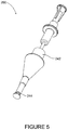
- FIG. 5 schematically illustrates a perspective view of tympanic membrane repair device 200 in a debridement configuration, according to some exemplary embodiments of the present invention.
- slidingly pushing rotary knob 242 towards patch 212 nears blade 244 to the tympanic membrane.
- rotating rotary knob 242 rotates blade 244 cutting the peripheral tissue surrounding the perforation.
- the cut tissue remains inside a periphery of blade 244 .
- pulling rotary knob 242 in a proximal direction retrieves blade 244 following debridement.
- the cut tissue is retrieved with blade 244 .
- patch release mechanism 206 includes a hollow tube 232 which slidably fits over rod 214 .
- hollow tube 232 is of a diameter ranging from 1.5 mm-11 mm, for example, 2.5 mm, 3.5 mm, 4.5 mm, 6.5 mm, 7.5 mm., 9.5 mm.
- hollow tube 232 includes a proximal release lever 234 , and a blade protector 236 distally positioned near a distal end 222 .
- release lever 234 includes a cylindrical shape and has a spring cavity 238 for accommodating a coil spring 240 proximally located on rod 214 .
- a cavity opening 235 is adapted to receive a distal portion 227 of adhesive insertion adaptor 226 for slidingly fitting into spring cavity 238 when release lever 234 is pulled in a proximal direction.
- coil spring 240 is a compression spring and compresses inside spring cavity 238 as release lever 234 is pulled in the proximal direction and distal portion 227 slides into spring cavity 238 .
- coil spring 240 exerts a pushing force on hollow tube 232 in a direction towards distal end 216 .
- distal end 222 overlays distal 216 applying a radial force on distal end 216 .
- distal end 216 closes clasping button 224 on patch 212 .
- FIG. 6 schematically illustrates a perspective view of tympanic membrane repair device 200 in a patch release configuration, according to some exemplary embodiments of the present invention.
- pulling release lever 234 in the proximal direction retrieves hollow tube 232 in the proximal direction, and distal end 222 from over distal end 216 .
- the radial force applied on distal end 216 for clasping button 224 is removed, releasing patch 212 .
- device alignment mechanism 208 serves to maintain device 200 aligned and anchored in the auditory canal.
- device alignment mechanism 208 is funnel shaped and includes an alignment receptacle 209 for slidingly accommodating cylindrical tube 246 and thereby maintaining device 200 aligned with the perforation.
- a minimum diameter of alignment mechanism 208 ranges from 2 mm-15 mm, for example, 3 mm, 5 mm, 7 mm, 9 mm, 11 mm, 13 mm.
- device alignment mechanism 208 includes a speculum or similar type of device known in the art.
- device alignment mechanism 208 is fitted into the opening to the auditory canal.
- a distance between a distal end of alignment mechanism 208 and patch 212 ranges from 10 mm-35 mm, for example, 15 mm, 20 mm, 25 mm, 30 mm.
- FIGS. 7A-7C schematically illustrate partial perspective views of device 200 in a patch guiding configuration, in a debridement configuration, and in a patch release configuration, respectively, according to some exemplary embodiments of the invention.
- patch 212 is secured in position by the clamping action of distal end 216 due to the spring-loaded clamping mechanism, with distal end 222 applying the radial force (pressing) on distal end 216 .
- Blade 244 is retrieved.
- device 200 is guided down the auditory canal and patch 212 inserted through the perforation in the tympanic membrane.
- cylindrical tube 246 is moved in the distal direction for nearing blade 244 to the debridement area in the tympanic membrane.
- distal end 222 has been slidingly moved in the proximal direction, uncovering distal end 216 .
- the radial force applied by distal end 222 on distal end 216 is removed.
- Distal end 216 opens due to the spring-loaded clamping mechanism releasing button 224 on patch 212 .
- patch 212 has been adhered to the tympanic membrane.
- An annular recess 225 is formed between patch 212 and button 224 where a button 224 diameter is greater than a diameter of the annular recess.
- FIG. 8 illustrates a flow chart of a non-limiting method of repairing a perforation in a tympanic membrane by adhering a flexible patch to an undersurface of the membrane, according to some exemplary embodiments of the present invention.
- FIGS. 9A-11 schematically illustrate the operation of tympanic membrane repair device 200 while performing the method, according to some exemplary embodiments of the present invention.
- a physician optionally performs an otoscopy to locate the perforation and determines its size and selects a patch 212 of a suitable dimension and attaches it to distal end 216 .
- the physician performs the otoscopy for making a perforation.
- the patch is selected so that it is at least 1 mm larger than the largest radius of the perforation.
- attachment is done by pulling on release lever 234 so that distal end 222 moves in a proximal direction away from distal end 216 .
- Distal end 216 opens and button 224 in patch 212 is inserted into the distal end.
- release lever 234 is returned to its original “closed” position, distal end 222 moving distally over 216 pressing distal end 216 closed over button 222 , securing patch 212 .
- patch 212 is pre-attached to distal end 216 . The physician then inserts device 200 into the patient's ear first introducing the end of the device to which patch 212 is attached.
- the physician guides patch 212 down the auditory canal and through the perforation in the tympanic membrane.
- the physician utilizes a micro-otoscope to monitor the medical procedure.
- the micro-otoscope is mechanically attached to device 200 .
- the micro-otoscope is separately inserted into the auditory canal.
- a position of device 200 is adjusted by the physician and is maintained aligned/anchored by device alignment mechanism 208 which is fitted into the opening of the auditory canal.
- the physician performs debridement of the tissue surrounding the perforation.
- debridement of the tissue surrounding the perforation is done.
- a first step in debridement includes moving rotary knob in a distal direction A for nearing blade 244 to the membrane.
- rotary knob 242 is rotated in a clockwise direction B for cutting away the peripheral tissue.
- cutting of the tissue may be done by rotating rotary knob 242 in a counterclockwise direction.
- cutting is done by a reciprocating motion both in a clockwise direction and a counterclockwise direction.
- cutting of the tissue may not require rotating rotary knob 242 .
- pushing rotary knob 242 in the distal direction also results in cutting of the tissue.
- the cut tissue is fitted within the periphery of the cutting edge of blade 244 .
- the physician inserts a cutting instrument for debriding, for example, a blade, scissors, or other instrument known in the art suitable for performing debridement of the perforation.
- the physician aligns device 200 so that patch 212 is proximal to the area of the perforation, on the undersurface of the tympanic membrane.
- the physician applies adhesive to patch 212 .
- adhesive container 248 includes a capsular container.
- adhesive container 248 includes a syringe and a plunger which is pushed in the distal direction, as shown by arrow C, for administering adhesive 250 .
- the capsular container is fittedly accommodated in the syringe.
- the adhesive is an “instant” glue.
- adhesive 250 is introduced into device 200 and flows through the device and out distal end 216 , spreading over the surface of patch 212 , as shown by arrows D.
- steps 1203 and 1204 are interchangeable.
- patch 212 is self-adhering and there is no need for the adhesive insertion adapter 226 or for the administering of the adhesive into device 200 .
- patch 212 is attached to the undersurface of the tympanic membrane sealing the perforation.
- device 200 is pulled in a proximal direction for attaching patch 212 to the undersurface.
- patch 212 adheres “instantly” to the undersurface by the instant glue.
- patch release lever 234 is pulled in a proximal direction E.
- distal end 222 is pulled in the proximal direction E uncovering distal end 216 , which opens as the radial force is removed from the spring-loaded clamping mechanism.
- opening of distal end 216 releases button 224 on patch 212 , releasing the patch.
- device 200 is pulled in the proximal direction after waiting a predetermined amount of time for adhering of patch 212 to the undersurface, the patch detaching due to the resistance of the undersurface to the pulling.
- the physician retrieves device 200 from the auditory canal.
- device 200 is retrieved only after waiting a predetermined amount of time following attachment of patch 212 to the undersurface for complete adhesion.
- biodegradable foam is introduced into the middle ear for supporting patch 212 as it adheres to the tympanic membrane.
- kit 1300 is a single use kit which is disposed of following one-time usage.
- Kit 1300 includes a tympanic membrane repair device 1301 , which may be similar to that shown in FIG. 1 at 100 , or in FIG. 3 at 200 .
- kit 1300 includes one or more patch 1302 , for example, 2, 3, 4, 5 patches.
- patch 1302 includes patches of different sizes, for example, having a diameter in a range from 2 mm-10 mm, for example 3 mm, 4 mm, 5 mm, 6 mm, 8 mm, 9 mm.
- the patch is a single-sized patch.
- kit 1300 includes an adhesive and an adhesive applicator 1303 .
- compositions, method or structure may include additional ingredients, steps and/or parts, but only if the additional ingredients, steps and/or parts do not materially alter the basic and novel characteristics of the claimed composition, method or structure.
- a compound or “at least one compound” may include a plurality of compounds, including mixtures thereof.
- range format is merely for convenience and brevity and should not be construed as an inflexible limitation on the scope of the invention. Accordingly, the description of a range should be considered to have specifically disclosed all the possible subranges as well as individual numerical values within that range. For example, description of a range such as from 1 to 6 should be considered to have specifically disclosed subranges such as from 1 to 3, from 1 to 4, from 1 to 5, from 2 to 4, from 2 to 6, from 3 to 6 etc., as well as individual numbers within that range, for example, 1, 2, 3, 4, 5, and 6. This applies regardless of the breadth of the range.
- a numerical range is indicated herein, it is meant to include any cited numeral (fractional or integral) within the indicated range.
- the phrases “ranging/ranges between” a first indicate number and a second indicate number and “ranging/ranges from” a first indicate number “to” a second indicate number are used herein interchangeably and are meant to include the first and second, indicated numbers and all the fractional and integral numerals therebetween.
- method refers to manners, means, techniques and procedures for accomplishing a given task including, but not limited to, those manners, means, techniques and procedures either known to, or readily developed from known manners, means, techniques and procedures by practitioners of the chemical, pharmacological, biological, biochemical and medical arts.
Landscapes
- Health & Medical Sciences (AREA)
- Life Sciences & Earth Sciences (AREA)
- Animal Behavior & Ethology (AREA)
- Otolaryngology (AREA)
- Veterinary Medicine (AREA)
- Public Health (AREA)
- General Health & Medical Sciences (AREA)
- Engineering & Computer Science (AREA)
- Biomedical Technology (AREA)
- Heart & Thoracic Surgery (AREA)
- Vascular Medicine (AREA)
- Transplantation (AREA)
- Oral & Maxillofacial Surgery (AREA)
- Cardiology (AREA)
- Pulmonology (AREA)
- Surgery (AREA)
- Physics & Mathematics (AREA)
- Acoustics & Sound (AREA)
- Biophysics (AREA)
- Psychology (AREA)
- Prostheses (AREA)
Abstract
Description
Claims (9)
Priority Applications (1)
| Application Number | Priority Date | Filing Date | Title |
|---|---|---|---|
| US15/911,895 US10952903B2 (en) | 2011-03-08 | 2018-03-05 | Tympanic membrane repair device |
Applications Claiming Priority (4)
| Application Number | Priority Date | Filing Date | Title |
|---|---|---|---|
| US201161450175P | 2011-03-08 | 2011-03-08 | |
| PCT/IL2012/050076 WO2012120513A1 (en) | 2011-03-08 | 2012-03-08 | Tympanic membrane repair device |
| US201314003802A | 2013-09-08 | 2013-09-08 | |
| US15/911,895 US10952903B2 (en) | 2011-03-08 | 2018-03-05 | Tympanic membrane repair device |
Related Parent Applications (2)
| Application Number | Title | Priority Date | Filing Date |
|---|---|---|---|
| US14/003,802 Continuation US9907701B2 (en) | 2011-03-08 | 2012-03-08 | Tympanic membrane repair device |
| PCT/IL2012/050076 Continuation WO2012120513A1 (en) | 2011-03-08 | 2012-03-08 | Tympanic membrane repair device |
Publications (2)
| Publication Number | Publication Date |
|---|---|
| US20180193197A1 US20180193197A1 (en) | 2018-07-12 |
| US10952903B2 true US10952903B2 (en) | 2021-03-23 |
Family
ID=45976980
Family Applications (2)
| Application Number | Title | Priority Date | Filing Date |
|---|---|---|---|
| US14/003,802 Expired - Fee Related US9907701B2 (en) | 2011-03-08 | 2012-03-08 | Tympanic membrane repair device |
| US15/911,895 Expired - Fee Related US10952903B2 (en) | 2011-03-08 | 2018-03-05 | Tympanic membrane repair device |
Family Applications Before (1)
| Application Number | Title | Priority Date | Filing Date |
|---|---|---|---|
| US14/003,802 Expired - Fee Related US9907701B2 (en) | 2011-03-08 | 2012-03-08 | Tympanic membrane repair device |
Country Status (5)
| Country | Link |
|---|---|
| US (2) | US9907701B2 (en) |
| EP (1) | EP2683331B1 (en) |
| DK (1) | DK2683331T3 (en) |
| ES (1) | ES2533830T3 (en) |
| WO (1) | WO2012120513A1 (en) |
Cited By (1)
| Publication number | Priority date | Publication date | Assignee | Title |
|---|---|---|---|---|
| US12508010B2 (en) | 2020-04-10 | 2025-12-30 | C2Dx, Inc. | Medical devices for repairing perforations in tissue, methods of manufacturing medical devices, and methods of implanting a medical device |
Families Citing this family (10)
| Publication number | Priority date | Publication date | Assignee | Title |
|---|---|---|---|---|
| ES2533830T3 (en) | 2011-03-08 | 2015-04-15 | Mor Research Applications Ltd. | Tympanic membrane repair device |
| US8961603B2 (en) * | 2012-06-20 | 2015-02-24 | Enteroptyx | Suction grasper for ossicular prosthesis |
| WO2016005946A2 (en) * | 2014-07-10 | 2016-01-14 | Otology Solutions Ltd | Tympanic membrane repair device |
| CA2980504C (en) | 2015-03-20 | 2024-01-30 | Massachusetts Eye And Ear Infirmary | Artificial tympanic membrane devices and uses |
| EP3349703A4 (en) * | 2015-09-17 | 2019-07-31 | Tymcure Ltd | TYMPANOPLASTY STAMP APPLICATOR |
| US10799341B2 (en) | 2016-09-16 | 2020-10-13 | Massachusetts Eye And Ear Infirmary | Ear canal grafts |
| US12569334B2 (en) | 2019-05-02 | 2026-03-10 | Massachusetts Eye And Ear Infirmary | Winged grafts for tympanic membrane repair and augmentation |
| CN215503745U (en) * | 2020-03-12 | 2022-01-14 | 新加坡国立大学 | Device and a kit for repairing the region of the ear via a natural orifice |
| CN112089502B (en) * | 2020-09-23 | 2024-01-30 | 中国水产科学研究院北戴河中心实验站 | Method for rapidly picking up juvenile otoliths of takifugu |
| US20240307231A1 (en) * | 2023-03-13 | 2024-09-19 | Ear Tech, LLC | Tympanostomy tube insertion device |
Citations (17)
| Publication number | Priority date | Publication date | Assignee | Title |
|---|---|---|---|---|
| GB108797A (en) | 1916-12-12 | 1917-08-23 | Edward Baum | An Artificial Ear Drum. |
| US4641651A (en) | 1983-09-22 | 1987-02-10 | Card George W | Cartilage punch and modified prosthesis in tympanoplasty |
| US5236455A (en) | 1992-02-10 | 1993-08-17 | Wilk Peter J | Tympanic patch, applicator, and related method |
| US5254133A (en) | 1991-04-24 | 1993-10-19 | Seid Arnold S | Surgical implantation device and related method of use |
| CN2162943Y (en) | 1993-03-28 | 1994-04-27 | 王庆华 | Tympanic membrane vent pipe imbedding and repairing device |
| US5501700A (en) | 1993-01-08 | 1996-03-26 | Hirata; Keisuke | Eardrum perforation patch and eardrum undersurface scraper |
| US6309419B1 (en) | 1999-03-26 | 2001-10-30 | Johns Hopkins University | Tympanic membrane prosthesis with mechanical fixation |
| US20020151974A1 (en) | 2001-02-23 | 2002-10-17 | Bonassar Lawrence J. | Tympanic membrane patch |
| US20030078597A1 (en) * | 1999-04-16 | 2003-04-24 | Blatter Duane D. | Intraluminally directed anvil apparatus and related methods and systems |
| US20040181185A1 (en) | 2003-03-14 | 2004-09-16 | Yi-Chang Lee | Device for removing diseased surface tissues |
| JP2006240441A (en) | 2005-03-02 | 2006-09-14 | Sumitomo Metal Ind Ltd | Body reinforcement member |
| US20070028927A1 (en) * | 2003-12-12 | 2007-02-08 | Slattery William H Iii | Surgical instrument set and procedure for implanting sound transducer proximate to patient's outer ear canal |
| KR20080040516A (en) | 2006-11-03 | 2008-05-08 | 아주대학교산학협력단 | Artificial chitosan patch for eardrum regeneration |
| US20080262468A1 (en) | 2007-04-19 | 2008-10-23 | Acclarent, Inc. | System and Method for the Simultaneous Bilateral Treatment of Target Tissues Within the Ears Using a Guide Block Structure |
| US20090299379A1 (en) | 2008-05-28 | 2009-12-03 | Yeshayahu Katz | Myringotomy instrument |
| US20090297533A1 (en) | 2008-05-23 | 2009-12-03 | Otonomy, Inc. | Controlled release immunomodulator compositions and methods for the treatment of otic disorders |
| WO2012120513A1 (en) | 2011-03-08 | 2012-09-13 | Mor Research Applications Ltd. | Tympanic membrane repair device |
-
2012
- 2012-03-08 ES ES12715440.9T patent/ES2533830T3/en active Active
- 2012-03-08 DK DK12715440.9T patent/DK2683331T3/en active
- 2012-03-08 EP EP12715440.9A patent/EP2683331B1/en active Active
- 2012-03-08 WO PCT/IL2012/050076 patent/WO2012120513A1/en not_active Ceased
- 2012-03-08 US US14/003,802 patent/US9907701B2/en not_active Expired - Fee Related
-
2018
- 2018-03-05 US US15/911,895 patent/US10952903B2/en not_active Expired - Fee Related
Patent Citations (18)
| Publication number | Priority date | Publication date | Assignee | Title |
|---|---|---|---|---|
| GB108797A (en) | 1916-12-12 | 1917-08-23 | Edward Baum | An Artificial Ear Drum. |
| US4641651A (en) | 1983-09-22 | 1987-02-10 | Card George W | Cartilage punch and modified prosthesis in tympanoplasty |
| US5254133A (en) | 1991-04-24 | 1993-10-19 | Seid Arnold S | Surgical implantation device and related method of use |
| US5236455A (en) | 1992-02-10 | 1993-08-17 | Wilk Peter J | Tympanic patch, applicator, and related method |
| US5501700A (en) | 1993-01-08 | 1996-03-26 | Hirata; Keisuke | Eardrum perforation patch and eardrum undersurface scraper |
| US5643300A (en) | 1993-01-08 | 1997-07-01 | Hirata; Keisuke | Eardrum undersurface scraper |
| CN2162943Y (en) | 1993-03-28 | 1994-04-27 | 王庆华 | Tympanic membrane vent pipe imbedding and repairing device |
| US6309419B1 (en) | 1999-03-26 | 2001-10-30 | Johns Hopkins University | Tympanic membrane prosthesis with mechanical fixation |
| US20030078597A1 (en) * | 1999-04-16 | 2003-04-24 | Blatter Duane D. | Intraluminally directed anvil apparatus and related methods and systems |
| US20020151974A1 (en) | 2001-02-23 | 2002-10-17 | Bonassar Lawrence J. | Tympanic membrane patch |
| US20040181185A1 (en) | 2003-03-14 | 2004-09-16 | Yi-Chang Lee | Device for removing diseased surface tissues |
| US20070028927A1 (en) * | 2003-12-12 | 2007-02-08 | Slattery William H Iii | Surgical instrument set and procedure for implanting sound transducer proximate to patient's outer ear canal |
| JP2006240441A (en) | 2005-03-02 | 2006-09-14 | Sumitomo Metal Ind Ltd | Body reinforcement member |
| KR20080040516A (en) | 2006-11-03 | 2008-05-08 | 아주대학교산학협력단 | Artificial chitosan patch for eardrum regeneration |
| US20080262468A1 (en) | 2007-04-19 | 2008-10-23 | Acclarent, Inc. | System and Method for the Simultaneous Bilateral Treatment of Target Tissues Within the Ears Using a Guide Block Structure |
| US20090297533A1 (en) | 2008-05-23 | 2009-12-03 | Otonomy, Inc. | Controlled release immunomodulator compositions and methods for the treatment of otic disorders |
| US20090299379A1 (en) | 2008-05-28 | 2009-12-03 | Yeshayahu Katz | Myringotomy instrument |
| WO2012120513A1 (en) | 2011-03-08 | 2012-09-13 | Mor Research Applications Ltd. | Tympanic membrane repair device |
Non-Patent Citations (6)
| Title |
|---|
| "EpiDisc Tympanic Membrane Perforation Patch Kit", Medtronic, Nov. 14, 2014. |
| "EpiFilm and EpiDisc Otologic Laminae", Ear Packing Products Medtronic, Nov. 14, 2014. |
| "Procedures and Techniques for Tympanoplasty and Ossicular Reconstruction", Medtronic, Nov. 14, 2014. |
| Communication under rule 71(3) EPC from the European patent office regarding Application No. 12715440.9; dated Jul. 16, 2014, 6 pages. |
| Notification concerning transmittal of international preliminary report on patentability regarding PCT/IL2012/050076; dated Sep. 19, 2013, 8 pages. |
| Notification of transmittal of the international search report and the written opinion of the international searching authority regarding PCT/IL2012/050076; dated Jun. 6, 2012, 13 pages. |
Cited By (1)
| Publication number | Priority date | Publication date | Assignee | Title |
|---|---|---|---|---|
| US12508010B2 (en) | 2020-04-10 | 2025-12-30 | C2Dx, Inc. | Medical devices for repairing perforations in tissue, methods of manufacturing medical devices, and methods of implanting a medical device |
Also Published As
| Publication number | Publication date |
|---|---|
| US9907701B2 (en) | 2018-03-06 |
| WO2012120513A1 (en) | 2012-09-13 |
| US20130345722A1 (en) | 2013-12-26 |
| EP2683331B1 (en) | 2014-12-31 |
| US20180193197A1 (en) | 2018-07-12 |
| ES2533830T3 (en) | 2015-04-15 |
| EP2683331A1 (en) | 2014-01-15 |
| DK2683331T3 (en) | 2015-04-07 |
Similar Documents
| Publication | Publication Date | Title |
|---|---|---|
| US10952903B2 (en) | Tympanic membrane repair device | |
| CN110381860B (en) | Systems and methods for delivering and positioning surgical implants | |
| CN108697437B (en) | Minimally invasive tissue collection device | |
| US20060161179A1 (en) | Follicular transplantation device and method | |
| WO2010028486A1 (en) | Elastic band ligation device and method for treatment of hemorrhoids | |
| WO2009045912A3 (en) | Vertebrally-mounted tissue retractor and method for use in spinal surgery | |
| CN102438530A (en) | Biopsy device | |
| CN107872966B (en) | Wound closure devices and methods | |
| KR20180110103A (en) | Pixel array medical system, apparatus and method | |
| US20210282754A1 (en) | Biopsy system with end deploy needle | |
| US10966842B2 (en) | Surgical extraction device for bone implant tips | |
| US11806222B2 (en) | Apparatus for cutting a material and a method for cutting a negative pressure wound therapy dressing | |
| EP3193753A1 (en) | Woven retention devices, systems, packaging, and related methods | |
| KR20200042877A (en) | Pixel array medical systems, devices and methods | |
| US11559326B2 (en) | Disposable circumcision apparatus and method of use of this apparatus | |
| US6309419B1 (en) | Tympanic membrane prosthesis with mechanical fixation | |
| CN114938979B (en) | Pressure relief device implant kit and method of use thereof | |
| EP3087951B1 (en) | Breast implant introducer | |
| US12447063B2 (en) | Apparatus for cutting a material and a method for cutting a negative pressure wound therapy dressing | |
| CN105640615B (en) | Endoscopic surgery knife with pulling device | |
| RU183426U1 (en) | CUTTER FOR FACILITY OF THE CARTILAGE TRANSPLANT FOR STEPEDOPLASTY FROM THE PATIENT'S AERIAL Shell | |
| CN108852432A (en) | For the membrane body laying device in minimally invasive endoscope-assistant surgery | |
| WO2012109312A2 (en) | Device and method for bone cement removal | |
| UA99792U (en) | Gavrilenko Aspiration Aspirator |
Legal Events
| Date | Code | Title | Description |
|---|---|---|---|
| AS | Assignment |
Owner name: MOR RESEARCH APPLICATIONS LTD., ISRAEL Free format text: ASSIGNMENT OF ASSIGNORS INTEREST;ASSIGNOR:MARGULIS, ARIEL;REEL/FRAME:045107/0951 Effective date: 20120408 |
|
| FEPP | Fee payment procedure |
Free format text: ENTITY STATUS SET TO UNDISCOUNTED (ORIGINAL EVENT CODE: BIG.); ENTITY STATUS OF PATENT OWNER: SMALL ENTITY |
|
| FEPP | Fee payment procedure |
Free format text: ENTITY STATUS SET TO SMALL (ORIGINAL EVENT CODE: SMAL); ENTITY STATUS OF PATENT OWNER: SMALL ENTITY |
|
| STPP | Information on status: patent application and granting procedure in general |
Free format text: DOCKETED NEW CASE - READY FOR EXAMINATION |
|
| STPP | Information on status: patent application and granting procedure in general |
Free format text: NON FINAL ACTION MAILED |
|
| STPP | Information on status: patent application and granting procedure in general |
Free format text: RESPONSE TO NON-FINAL OFFICE ACTION ENTERED AND FORWARDED TO EXAMINER |
|
| STPP | Information on status: patent application and granting procedure in general |
Free format text: NON FINAL ACTION MAILED |
|
| STPP | Information on status: patent application and granting procedure in general |
Free format text: FINAL REJECTION MAILED |
|
| STPP | Information on status: patent application and granting procedure in general |
Free format text: DOCKETED NEW CASE - READY FOR EXAMINATION |
|
| STPP | Information on status: patent application and granting procedure in general |
Free format text: NOTICE OF ALLOWANCE MAILED -- APPLICATION RECEIVED IN OFFICE OF PUBLICATIONS |
|
| STPP | Information on status: patent application and granting procedure in general |
Free format text: PUBLICATIONS -- ISSUE FEE PAYMENT VERIFIED |
|
| STCF | Information on status: patent grant |
Free format text: PATENTED CASE |
|
| FEPP | Fee payment procedure |
Free format text: MAINTENANCE FEE REMINDER MAILED (ORIGINAL EVENT CODE: REM.); ENTITY STATUS OF PATENT OWNER: SMALL ENTITY |
|
| LAPS | Lapse for failure to pay maintenance fees |
Free format text: PATENT EXPIRED FOR FAILURE TO PAY MAINTENANCE FEES (ORIGINAL EVENT CODE: EXP.); ENTITY STATUS OF PATENT OWNER: SMALL ENTITY |
|
| STCH | Information on status: patent discontinuation |
Free format text: PATENT EXPIRED DUE TO NONPAYMENT OF MAINTENANCE FEES UNDER 37 CFR 1.362 |
|
| FP | Lapsed due to failure to pay maintenance fee |
Effective date: 20250323 |