US10794977B2 - System and method for normalized reference database for MR images via autoencoders - Google Patents
System and method for normalized reference database for MR images via autoencoders Download PDFInfo
- Publication number
- US10794977B2 US10794977B2 US15/629,779 US201715629779A US10794977B2 US 10794977 B2 US10794977 B2 US 10794977B2 US 201715629779 A US201715629779 A US 201715629779A US 10794977 B2 US10794977 B2 US 10794977B2
- Authority
- US
- United States
- Prior art keywords
- imaging data
- data
- normalized reference
- reference data
- image
- Prior art date
- Legal status (The legal status is an assumption and is not a legal conclusion. Google has not performed a legal analysis and makes no representation as to the accuracy of the status listed.)
- Active, expires
Links
Images


Classifications
-
- G—PHYSICS
- G01—MEASURING; TESTING
- G01R—MEASURING ELECTRIC VARIABLES; MEASURING MAGNETIC VARIABLES
- G01R33/00—Arrangements or instruments for measuring magnetic variables
- G01R33/20—Arrangements or instruments for measuring magnetic variables involving magnetic resonance
- G01R33/44—Arrangements or instruments for measuring magnetic variables involving magnetic resonance using nuclear magnetic resonance [NMR]
- G01R33/48—NMR imaging systems
- G01R33/54—Signal processing systems, e.g. using pulse sequences ; Generation or control of pulse sequences; Operator console
- G01R33/56—Image enhancement or correction, e.g. subtraction or averaging techniques, e.g. improvement of signal-to-noise ratio and resolution
- G01R33/5608—Data processing and visualization specially adapted for MR, e.g. for feature analysis and pattern recognition on the basis of measured MR data, segmentation of measured MR data, edge contour detection on the basis of measured MR data, for enhancing measured MR data in terms of signal-to-noise ratio by means of noise filtering or apodization, for enhancing measured MR data in terms of resolution by means for deblurring, windowing, zero filling, or generation of gray-scaled images, colour-coded images or images displaying vectors instead of pixels
-
- G—PHYSICS
- G01—MEASURING; TESTING
- G01R—MEASURING ELECTRIC VARIABLES; MEASURING MAGNETIC VARIABLES
- G01R33/00—Arrangements or instruments for measuring magnetic variables
- G01R33/20—Arrangements or instruments for measuring magnetic variables involving magnetic resonance
- G01R33/44—Arrangements or instruments for measuring magnetic variables involving magnetic resonance using nuclear magnetic resonance [NMR]
- G01R33/48—NMR imaging systems
- G01R33/54—Signal processing systems, e.g. using pulse sequences ; Generation or control of pulse sequences; Operator console
- G01R33/56—Image enhancement or correction, e.g. subtraction or averaging techniques, e.g. improvement of signal-to-noise ratio and resolution
- G01R33/5602—Image enhancement or correction, e.g. subtraction or averaging techniques, e.g. improvement of signal-to-noise ratio and resolution by filtering or weighting based on different relaxation times within the sample, e.g. T1 weighting using an inversion pulse
Definitions
- MR magnetic resonance
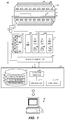
- FIG. 1 is a block diagram of an MRA system according to some embodiments
- FIG. 2 is an illustrative example of an autoencoder
- FIG. 3 is a flow diagram of a process according to some embodiments.
- FIG. 4 is a flow diagram of another process according to some embodiments.
- FIG. 5 is a flow diagram of a process, according to some embodiments.
- FIG. 6 is a system diagram of a task network including some aspects of a normalization network, according to some embodiments.
- the net aligned moment, M z may be rotated, or “tipped” into the x-y plane to produce a net transverse magnetic moment M t , which is rotating, or spinning, in the x-y plane at the Larmor frequency.
- a signal is emitted by the excited spins after the excitation field B 1 is terminated. The emitted signals are detected, digitized and processed to reconstruct an image using one of many well-known MR reconstruction techniques.
- FIG. 1 illustrates magnetic resonance imaging (MRI) system 100 according to some embodiments.
- MRI system 100 includes MRI chassis 110 that defines a bore 115 in which patient 120 is disposed.
- MRI chassis 110 includes polarizing main magnet 125 , gradient coils 130 , and RF coil 135 arranged about bore 115 .
- polarizing main magnet 125 generates the uniform magnetic field B 0 mentioned above and RF coil 135 emits the excitation field B 1 .
- Gradient coils 130 produce magnetic field gradients G x , G y , and G z which are used for position-encoding NMR signals.
- the magnetic field gradients G x , G y , and G z distort the main magnetic field in a predictable way so that the Larmor frequency of nuclei within the main magnetic field varies as a function of position. Accordingly, an excitation field B 1 which is near a particular Larmor frequency will tip the net aligned moment M z of those nuclei located at field positions which correspond to the particular Larmor frequency, and signals will be emitted only by those nuclei after the excitation field B 1 is terminated.
- Gradient coils 130 may consist of three windings, for example, each of which is supplied with current by an amplifier 140 a - 140 c in order to generate a linear gradient field in its respective Cartesian direction (i.e., x, y, or z).
- Each amplifier 140 a - 140 c includes a digital-analog converter 145 a - 145 c that is controlled by a sequence controller 150 to generate desired gradient pulses at proper times.
- Sequence controller 150 also controls the generation of RF pulses by RF system 155 .
- RF system 155 is responsive to a scan prescription and direction from sequence controller 150 to produce RF pulses of the desired frequency, phase, and pulse amplitude waveform.
- the generated RF pulses may be applied to the whole of RF coil 135 or to one or more local coils or coil arrays.
- RF coil 135 converts the RF pulses emitted by RF amplifier 160 , via multiplexer 162 , into a magnetic alternating field to excite the nuclei and align the nuclear spins of the object to be examined or the region of the object to be examined.
- the RF pulses to be produced by RF system 155 are represented digitally as complex numbers. Sequence controller 150 supplies these numbers in real and imaginary parts to digital-analog converters 164 a - 164 b in RF system 155 to create corresponding analog pulse sequences. Transmission channel 165 modulates the pulse sequences with a radio-frequency carrier signal having a base frequency corresponding to the resonance frequency of the nuclear spins in the volume to be imaged.
- RF coil 135 both emits the radio-frequency pulse to excite nuclear spins and scans the alternating field which is produced because of the precessing nuclear spins, i.e. the nuclear spin echo signals.
- the received signals are received by multiplexer 162 , amplified by RF amplifier 170 and demodulated in receiving channel 171 of RF system 155 in a phase-sensitive manner.
- Analog-digital converters 172 a and 172 b convert the demodulated signals into a real part and an imaginary part.
- Computing system 175 receives the real and imaginary parts and reconstructs an image therefrom per known techniques.
- System 175 may comprise any general-purpose or dedicated computing system. Accordingly, system 175 includes one or more processing units 176 (e.g., processors, processor cores, execution threads, etc.) configured to execute processor-executable program code to cause system 175 to operate as described herein, and storage device 178 for storing the program code.
- Storage device 178 may comprise one or more fixed disks, solid-state random access memory, and/or removable media (e.g., a thumb drive) mounted in a corresponding interface (e.g., a USB port).
- Storage device 178 stores program code of control program 180 .
- One or more processing units 146 may execute control program 180 to cause system 175 to perform any one or more of the processes described herein.
- one or more processing units 146 may execute control program 180 to cause system 175 to receive the real and imaginary parts of a received RF signal via MR system interface 182 and reconstruct an image therefrom according to known techniques. Such an image may be stored among acquired images 185 of storage device 178 .
- Control program 180 may also be executed to process one or more reconstructed images as described herein, and to store a processed image among processed images 186 of storage device 178 .
- One or more processing units 146 may also execute control program 180 to provide instructions to sequence controller 150 via MR system interface 182 .
- sequence controller 150 may be instructed to initiate the desired pulse sequences and corresponding scanning of k-space (i.e., acquired signal data).
- sequence controller 150 may be instructed to control the switching of magnetic field gradients via amplifiers 140 a - 140 c at appropriate times, the transmission of radio-frequency pulses having a defined phase and amplitude via RF system 155 and RF amplifier 160 , and the reception of the resulting magnetic resonance signals.
- Acquired images 185 and/or processed images 186 may be provided to terminal 190 via UI interface 184 of system 175 .
- UI interface 184 may also receive input from terminal 190 , which may be used to provide commands to control program 180 to control sequence controller 150 and other elements of system 100 .
- Terminal 190 may simply comprise a display device and an input device coupled to system 175 .
- terminal 190 is a separate computing device such as, but not limited to, a desktop computer, a laptop computer, a tablet computer, and a smartphone.
- Storage device 178 may also store data and other program code for providing additional functionality and/or which are necessary for operation of system 175 , such as device drivers, operating system files, etc.
- storage device 178 may comprise an instance of a database (e.g., a database node of a distributed database system).
- MR images may contain significant (i.e., non-trivial) intensity variations across different patients and different scanners.
- intensity variations might, for example, even exist in MR images generated by MR systems of a same model number located in different hospitals.
- Intensity variances may be attributable to differences in protocols, scanner calibrations, technician biases/preferences, etc. that can cause differences in the MR images obtained by the two different scanners. These differences may cause a problem in interpreting and evaluating the MR images since the distribution of intensities of the images is not consistent in the data obtained from different patients and different scanners at different times and operated by different technicians.
- MR image data may be obtained any scanner and processed according to the processes disclosed herein to obtain MR images with normalized intensities such that, for example, the normalized MR images can be evaluated relative to a consistent intensity distribution.
- MR images with normalized intensities of a brain can be accurately viewed/interpreted knowing (i.e., given) the intensity of white matter in the MR images is between a given intensity range and grey matter will be within a second (different) intensity range.
- variances in intensities due to different scanners, calibrations, technicians, times, patients, scanned organs and structural anatomies, and combinations thereof might be minimized and different images with a similar/same intensity distribution can be compared by the use of the MR images with normalized intensities.
- FIG. 2 is an illustrative example of an autoencoder 200 , that may be used in some embodiments of the present disclosure.
- autoencoder 200 is a variational autoencoder (VAE). It is noted however that autoencoder need not be a VAE type of autoencoder.
- VAE 200 might be used by a system or in a process to generate normalized imaging data in accordance with some embodiments related to a neural network.
- VAE 200 may be described as a hidden layer in the neural network.
- VAE 200 comprises multiple layers, including an encoder 205 , a latent space 215 of random variables (z), and a decoder 220 .
- the input to encoder 210 in the example of FIG. 2 is MP (multiparametric) MR images 205 (i.e., a sequence of functional forms of imaging such as, for example, T1, T2, FLAIR, etc.).
- the MP images 205 include a sequence of images or data points (x) and its output from encoder 210 is a hidden representation (z), with certain weights and biases ( ⁇ ).
- Encoder 210 encodes the MP images into the latent space (z) 215 .
- the encoder may be represented by q ⁇ (z
- Decoder 220 receives, as an input, the hidden representation (z).
- the latent space focuses on having two random variables, independent layers that represent the mean and STD of the input intensity distribution.
- Decoder 220 learns to reconstruct an image based on the statistics of the latent space 215 . Decoder 220 outputs the parameters to a probability distribution of the data, with weights and biases ⁇ .
- the encoder may be represented by q ⁇ (x
- the output of decoder in the example of FIG. 3 is normalized reference database (NRD) image 225 representations of the MP images 205 .
- NTD normalized reference database
- MP images 205 include a large-scale dataset of images acquired using a single specific scanner.
- the NRD image or reference database is generated with statistical information on, for example, each tissue/organ in the MP scans.
- a representation of each tissue/organ can be computed using multiple scans of multiple individuals (e.g., volunteers), normalizing the representation data, and averaging over it over the total number of scans to create the NRD images 225 .
- the NRD may be considered as the set of images that an atlas is averaged from, such atlas containing shape information and intensity distribution information of labeled structural or anatomical regions (i.e., tissue/organs).
- NRD image 225 representations are normalized for the set of MP images 205 so that a reference level of intensities can be obtained, generated, or otherwise determined.
- the reference level of intensities of the NRD images can be, in some instances, uses a standard or measure against which other MR images can be compared or evaluated for normal and/or problematic intensities.
- the NRD images or data can be used to establish a “normal” range of image intensities, where other intensities outside of the “normal/healthy” range in a MR image might indicate a problem with the scanned tissue or organ represented in the MR image.
- a normalized database of MR images for a particular anatomical region might be used to establish a range of expected intensities for “normal/healthy” tissue in the region. Intensity values outside of the expected intensity range might indicate a tumor, lesion, or other abnormality in the subject tissue/organ region of the MR image.
- the present disclosure relates to a system and a process to normalize MR image data, and create a reference database of tissue/organ MR images for comparison/evaluation of MR images to the reference database.
- advances in deep-learning generative models may be used to facilitate evaluating and reconstructing images based on a provided, specific target.
- the target is a normalized database of MR images.
- an autoencoder such as a VAE (though not limited to a VAE) may be used in a process and a system to learn a transformation ⁇ (X, ⁇ ) that takes MR images as an input and match those input images to the normalized database of MR images (i.e., the target).
- FIG. 3 is an illustrative depiction of a system 300 , in accordance with some embodiments herein.
- System 300 may include more or other components than those explicitly depicted in FIG. 3 . While FIG. 3 is shown as comprising a number of discrete components, FIG. 3 may represent a logical embodiment of a system, where some functions might be implemented by one or more devices and/or multiple functionalities might be executed by one device.
- FIG. 3 includes a VAE 310 that receives MP images 305 as an input thereto. VAE 310 may facilitate learning a transformation, ⁇ (X, ⁇ , ⁇ ) from an MR image (e.g., from any scanner) to an image normalized to a reference database (i.e., NRD image 315 ).
- a score X (x 1 , . . . , x n ) with x i representing a contrast image out of n possible, i.e. T1, T2, FLAIR, etc.
- X is the multiparametric image
- images are encoded into continuous latent variables q ⁇ (z
- the transformation from MP image 305 to NRD image 315 may be accomplished in, for example, two ways herein.
- a normalized database may be created and (1) a neural network using a VAE (e.g., 310 ) may be used to learn the transformation and (2) a deep-learning based neural network 320 that identifies attributes of the MP image, such as the acquisition source (i.e., scanner) of the image, which can be used by the VAE to further refine the creation of the normalized database (e.g., using the scanner model classification 325 ).
- a neural network using a VAE e.g., 310
- a deep-learning based neural network 320 that identifies attributes of the MP image, such as the acquisition source (i.e., scanner) of the image, which can be used by the VAE to further refine the creation of the normalized database (e.g., using the scanner model classification 325 ).
- MP image 305 is fed to deep-learning neural network 320 that learns (or at least attempts to learn) where the MP image originates.
- deep neural network 320 may determine the vendor and magnetic field strength of the MP image.
- Scanner model classification module 325 may generate scanner-specific information that may be provided to VAE 310 as priors to the VAE to improve the intensity matching thereof.
- a classification approach to discriminate scanner types and vendors to learn a second or an inverse transformation to match intensities to the NRD is a derived operation.
- a classification metric can be derived by learning or recognizing which scanners/vendors are input to the network or system 300 . That is, a second transformation herein may be used to go from NRD to a specific scanner type/vendor (i.e., determine model- or scanner-specific information), where a specific scanner type/vendor corresponds to a specific distribution type.
- scanner information may be used as prior as well. After training is done, NRD image will include features that are independent to scanner variations and will thus reflect the best image.
- the transformation learning process can be implemented in at least one of an organ-based form and an intensity-based form.
- organ-based form a NRD database for a specific organ is generated and the intensity distribution of images of the same organ (e.g., the brain) can be matched to the reference database images to a brain-specific NRD.
- the intensity distribution of a subject image may, without spatial information or context, be mapped or correlated to that of the NRD reference database.
- training may be accomplished using a large database of MR images acquired from multiple scanners with multiple magnetic fields (1.5 T, 3 T, 7 T) from different vendors.
- transformations might be separated based on at least one of a scanner vendor, magnetic field strength, and receive coil array. Examples can include transformations for different configurations of MR scanners such as Siemens/1.5 T, Siemens/3 T, GE/1.5 T, GE/3 T, etc.
- FIG. 4 is a flow diagram of a process 400 , according to some embodiments herein.
- Process 400 is related to a method that might be executed by, for example, system 300 in FIG. 3 where MP images 305 are processed through an encoder (e.g., VAE 310 ) to generate a normalized reference database including NRD images 315 .
- MR imaging data from a first scanner device is received.
- the MR imaging data may include data pertaining to a large dataset of different structural or anatomical regions.
- the MR imaging data may comprise sequences of functional images, as opposed to singular images.
- Operation 410 includes generating normalized reference data based on the MR imaging data received at operation 405 .
- the normalized reference data may be labeled or otherwise identifiable for each MR scan.
- a transformation is learned or otherwise determined, based on the normalized reference data of operation 410 .
- the transformation may operate to match, map, or otherwise correlate a set (i.e., one or more) of input MR images to the normalized reference data determined at operation 410 .
- a record of the transformed imaging data may be stored.
- the stored data may be further processed, viewed, or reported, and can be configured as any now known or future developed data structure.
- FIG. 5 is a flow diagram of a process 500 , according to some embodiments herein.
- Process 500 in some embodiments, is related to a method that might be executed by, for example, system 300 in FIG. 3 where MP images 305 might be processed through a neural/learning network (e.g., deep neural network 320 ) and a classification operation (e.g., scanner model classification module 325 ).
- a neural/learning network e.g., deep neural network 320
- a classification operation e.g., scanner model classification module 325 .
- MR imaging data from a first scanner device is received and the MP images are forwarded to an encoder at operation 510 .
- the scanner-specific information if available for the input MP images may, in some instances, be used to improve an accuracy of an intensity matching operation, the efficiency of performing such an operation, combinations thereof, and for other purposes.
- the determination of operation 515 may be executed, in some instances, by a query of a database (not shown in FIG. 5 ). In the instance there is scanner-specific information available corresponding to the input MP images, process 500 proceeds to operation 520 where such scanner-specific information is obtained.
- the scanner-specific information may be used in a transformation operation 525 . Again, use of the scanner-specific information in transformation operation 525 may benefit an accuracy, efficiency, and other aspects thereof.
- process 500 may advance directly to the transformation operation 525 .
- the normalized images determined or otherwise generated at operation 525 may be stored in a data storage facility or device, such as, for example, a database.
- the normalized data may be used in other processes, including but not limited to medical treatment plans, data analytics, data visualizations, and other tasks.
- the MR imaging data may include data pertaining to a large dataset of different structural or anatomical regions.
- the MR imaging data may comprise sequences of functional images, as opposed to singular images.
- a normalized image generator in accordance with some aspects herein might be used in or otherwise comprise a part of a larger task (e.g., quantification) network.
- FIG. 6 is an illustrative example where some aspects of the present disclosure discussed hereinabove are incorporated or embedded in a system 600 .
- System 600 may include a combined network 605 comprising a normalization network 610 and a task oriented network 620 .
- Normalization network 610 may operate to generate NRD images 615 given an input of MP images 625 , in accordance with some aspects herein.
- Task oriented network 620 may operate to generate a prediction 630 given an input NRD image 615 .
- Task oriented network 620 might be a deep-learning neural network directed to segmentation or region classification based on normalized MR images.
- task network 620 might be designed to extract images of a brain (or liver) from an image of a head (torso body part).
Landscapes
- Physics & Mathematics (AREA)
- Engineering & Computer Science (AREA)
- Radiology & Medical Imaging (AREA)
- Health & Medical Sciences (AREA)
- General Health & Medical Sciences (AREA)
- Nuclear Medicine, Radiotherapy & Molecular Imaging (AREA)
- Signal Processing (AREA)
- High Energy & Nuclear Physics (AREA)
- Condensed Matter Physics & Semiconductors (AREA)
- General Physics & Mathematics (AREA)
- Computer Vision & Pattern Recognition (AREA)
- Artificial Intelligence (AREA)
- Magnetic Resonance Imaging Apparatus (AREA)
Abstract
Description
Claims (16)
Priority Applications (1)
| Application Number | Priority Date | Filing Date | Title |
|---|---|---|---|
| US15/629,779 US10794977B2 (en) | 2016-06-23 | 2017-06-22 | System and method for normalized reference database for MR images via autoencoders |
Applications Claiming Priority (2)
| Application Number | Priority Date | Filing Date | Title |
|---|---|---|---|
| US201662353740P | 2016-06-23 | 2016-06-23 | |
| US15/629,779 US10794977B2 (en) | 2016-06-23 | 2017-06-22 | System and method for normalized reference database for MR images via autoencoders |
Publications (2)
| Publication Number | Publication Date |
|---|---|
| US20170371017A1 US20170371017A1 (en) | 2017-12-28 |
| US10794977B2 true US10794977B2 (en) | 2020-10-06 |
Family
ID=60677398
Family Applications (1)
| Application Number | Title | Priority Date | Filing Date |
|---|---|---|---|
| US15/629,779 Active 2037-11-30 US10794977B2 (en) | 2016-06-23 | 2017-06-22 | System and method for normalized reference database for MR images via autoencoders |
Country Status (1)
| Country | Link |
|---|---|
| US (1) | US10794977B2 (en) |
Cited By (1)
| Publication number | Priority date | Publication date | Assignee | Title |
|---|---|---|---|---|
| US20210209773A1 (en) * | 2017-12-20 | 2021-07-08 | Al Analysis. Inc. | Methods and systems that normalize images, generate quantitative enhancement maps, and generate synthetically enhanced images |
Families Citing this family (11)
| Publication number | Priority date | Publication date | Assignee | Title |
|---|---|---|---|---|
| US10373055B1 (en) * | 2016-05-20 | 2019-08-06 | Deepmind Technologies Limited | Training variational autoencoders to generate disentangled latent factors |
| US10733788B2 (en) | 2018-03-15 | 2020-08-04 | Siemens Healthcare Gmbh | Deep reinforcement learning for recursive segmentation |
| US10878570B2 (en) | 2018-07-17 | 2020-12-29 | International Business Machines Corporation | Knockout autoencoder for detecting anomalies in biomedical images |
| US11403511B2 (en) | 2018-08-23 | 2022-08-02 | Apple Inc. | Unsupervised annotation using dual network system with pre-defined structure |
| US11893498B2 (en) | 2018-09-18 | 2024-02-06 | Insilico Medicine Ip Limited | Subset conditioning using variational autoencoder with a learnable tensor train induced prior |
| US11593660B2 (en) * | 2018-09-18 | 2023-02-28 | Insilico Medicine Ip Limited | Subset conditioning using variational autoencoder with a learnable tensor train induced prior |
| US11995854B2 (en) * | 2018-12-19 | 2024-05-28 | Nvidia Corporation | Mesh reconstruction using data-driven priors |
| CA3124400A1 (en) * | 2018-12-21 | 2020-06-25 | Nova Scotia Health Authority | Systems and methods for generating cancer prediction maps from multiparametric magnetic resonance images using deep learning |
| US10824794B2 (en) | 2019-04-08 | 2020-11-03 | Paypal, Inc. | Process for creating a fixed length representation of a variable length input |
| US11494695B2 (en) | 2019-09-27 | 2022-11-08 | Google Llc | Training neural networks to generate structured embeddings |
| EP4052175A4 (en) * | 2019-10-31 | 2023-11-29 | President And Fellows Of Harvard College | IMAGE PROCESSING FOR ORGANISMS SIZE AND SHAPE STANDARDIZATION |
Citations (5)
| Publication number | Priority date | Publication date | Assignee | Title |
|---|---|---|---|---|
| US20150302599A1 (en) * | 2011-12-05 | 2015-10-22 | The John Hopkins University | System and method of automatically detecting tissue abnormalities |
| US20160093050A1 (en) * | 2014-09-30 | 2016-03-31 | Samsung Electronics Co., Ltd. | Image registration device, image registration method, and ultrasonic diagnosis apparatus having image registration device |
| US20170100078A1 (en) * | 2015-10-13 | 2017-04-13 | IMPAC Medical Systems, Inc | Pseudo-ct generation from mr data using a feature regression model |
| US20170337682A1 (en) * | 2016-05-18 | 2017-11-23 | Siemens Healthcare Gmbh | Method and System for Image Registration Using an Intelligent Artificial Agent |
| US20180325461A1 (en) * | 2015-05-29 | 2018-11-15 | Northwestern University | Systems and Methods for Producing Quantitatively Calibrated Grayscale Values in Magnetic Resonance Images |
-
2017
- 2017-06-22 US US15/629,779 patent/US10794977B2/en active Active
Patent Citations (5)
| Publication number | Priority date | Publication date | Assignee | Title |
|---|---|---|---|---|
| US20150302599A1 (en) * | 2011-12-05 | 2015-10-22 | The John Hopkins University | System and method of automatically detecting tissue abnormalities |
| US20160093050A1 (en) * | 2014-09-30 | 2016-03-31 | Samsung Electronics Co., Ltd. | Image registration device, image registration method, and ultrasonic diagnosis apparatus having image registration device |
| US20180325461A1 (en) * | 2015-05-29 | 2018-11-15 | Northwestern University | Systems and Methods for Producing Quantitatively Calibrated Grayscale Values in Magnetic Resonance Images |
| US20170100078A1 (en) * | 2015-10-13 | 2017-04-13 | IMPAC Medical Systems, Inc | Pseudo-ct generation from mr data using a feature regression model |
| US20170337682A1 (en) * | 2016-05-18 | 2017-11-23 | Siemens Healthcare Gmbh | Method and System for Image Registration Using an Intelligent Artificial Agent |
Cited By (2)
| Publication number | Priority date | Publication date | Assignee | Title |
|---|---|---|---|---|
| US20210209773A1 (en) * | 2017-12-20 | 2021-07-08 | Al Analysis. Inc. | Methods and systems that normalize images, generate quantitative enhancement maps, and generate synthetically enhanced images |
| US11562494B2 (en) * | 2017-12-20 | 2023-01-24 | AI Analysis, Inc. | Methods and systems that normalize images, generate quantitative enhancement maps, and generate synthetically enhanced images |
Also Published As
| Publication number | Publication date |
|---|---|
| US20170371017A1 (en) | 2017-12-28 |
Similar Documents
| Publication | Publication Date | Title |
|---|---|---|
| US10794977B2 (en) | System and method for normalized reference database for MR images via autoencoders | |
| US10713785B2 (en) | Image quality assessment system and method | |
| KR101674848B1 (en) | Nuclear magnetic resonance (NMR) fingerprinting | |
| JP4863610B2 (en) | Method for creating a standard measurement protocol for a tomographic imaging system, a computer readable recording medium recording a computer program, and a planning method for positioning an imaging range of an actual object in the tomographic imaging system | |
| EP3333583B1 (en) | Method for identifying an organ structure of an investigated object in magnetic resonance image data | |
| KR101652387B1 (en) | Method to generate image data | |
| US8744154B2 (en) | System and method for acquiring magnetic resonance imaging (MRI) data | |
| US10736538B2 (en) | Method and computer differentiating correlation patterns in functional magnetic resonance imaging | |
| CN108324277B (en) | Method for classifying magnetic resonance measurement data acquired by means of magnetic resonance fingerprinting | |
| US11125847B2 (en) | System and method for out-of-view artifact suppression for magnetic resonance fingerprinting | |
| CN113012246A (en) | Method and processing system for generating synthetic image data and providing trained functions | |
| US10761167B2 (en) | System and method for generating a magnetic resonance fingerprinting dictionary using semi-supervised learning | |
| CN107440719A (en) | Method for showing Quantitative MRI Measurement view data | |
| CN119732670A (en) | Magnetic resonance imaging method, neural network, generation method thereof and readable medium | |
| US10663547B2 (en) | Automatic detection and setting of magnetic resonance protocols based on read-in image data | |
| JP2021518228A (en) | Anomaly detection using magnetic resonance fingerprinting | |
| US11385311B2 (en) | System and method for improved magnetic resonance fingerprinting using inner product space | |
| US12205198B2 (en) | Method and system for generating magnetic resonance image, and computer readable storage medium | |
| CN113327224B (en) | System and method for automatic field of view (FOV) definition | |
| US10825169B2 (en) | Method and apparatus for functional magnetic resonance imaging | |
| US10362961B2 (en) | System and method for neutral contrast magnetic resonance imaging of calcifications | |
| US12318184B2 (en) | Magnetic resonance imaging of an organ structure | |
| EP3798661B1 (en) | Mri method to determine a susceptibility distribution of an examination subject | |
| WO2011069411A1 (en) | Methods and systems for estimating longitudinal relaxation times in mri | |
| US12394043B2 (en) | Workflow management for labeling the subject anatomy |
Legal Events
| Date | Code | Title | Description |
|---|---|---|---|
| AS | Assignment |
Owner name: SIEMENS MEDICAL SOLUTIONS USA, INC., PENNSYLVANIA Free format text: ASSIGNMENT OF ASSIGNORS INTEREST;ASSIGNORS:ODRY, BENJAMIN L.;CETINGUL, HASAN ERTAN;MAILHE, BORIS;AND OTHERS;REEL/FRAME:042793/0825 Effective date: 20170622 |
|
| AS | Assignment |
Owner name: SIEMENS HEALTHCARE GMBH, GERMANY Free format text: ASSIGNMENT OF ASSIGNORS INTEREST;ASSIGNOR:SIEMENS MEDICAL SOLUTIONS USA, INC.;REEL/FRAME:043007/0482 Effective date: 20170627 |
|
| STPP | Information on status: patent application and granting procedure in general |
Free format text: NON FINAL ACTION MAILED |
|
| STPP | Information on status: patent application and granting procedure in general |
Free format text: RESPONSE TO NON-FINAL OFFICE ACTION ENTERED AND FORWARDED TO EXAMINER |
|
| STPP | Information on status: patent application and granting procedure in general |
Free format text: FINAL REJECTION MAILED |
|
| STPP | Information on status: patent application and granting procedure in general |
Free format text: ADVISORY ACTION MAILED |
|
| STPP | Information on status: patent application and granting procedure in general |
Free format text: DOCKETED NEW CASE - READY FOR EXAMINATION |
|
| STPP | Information on status: patent application and granting procedure in general |
Free format text: NON FINAL ACTION MAILED |
|
| STPP | Information on status: patent application and granting procedure in general |
Free format text: RESPONSE TO NON-FINAL OFFICE ACTION ENTERED AND FORWARDED TO EXAMINER |
|
| STPP | Information on status: patent application and granting procedure in general |
Free format text: NOTICE OF ALLOWANCE MAILED -- APPLICATION RECEIVED IN OFFICE OF PUBLICATIONS |
|
| STPP | Information on status: patent application and granting procedure in general |
Free format text: PUBLICATIONS -- ISSUE FEE PAYMENT VERIFIED |
|
| STCF | Information on status: patent grant |
Free format text: PATENTED CASE |
|
| AS | Assignment |
Owner name: SIEMENS HEALTHINEERS AG, GERMANY Free format text: ASSIGNMENT OF ASSIGNORS INTEREST;ASSIGNOR:SIEMENS HEALTHCARE GMBH;REEL/FRAME:066267/0346 Effective date: 20231219 Owner name: SIEMENS HEALTHINEERS AG, GERMANY Free format text: ASSIGNMENT OF ASSIGNOR'S INTEREST;ASSIGNOR:SIEMENS HEALTHCARE GMBH;REEL/FRAME:066267/0346 Effective date: 20231219 |
|
| MAFP | Maintenance fee payment |
Free format text: PAYMENT OF MAINTENANCE FEE, 4TH YEAR, LARGE ENTITY (ORIGINAL EVENT CODE: M1551); ENTITY STATUS OF PATENT OWNER: LARGE ENTITY Year of fee payment: 4 |