US10751185B2 - Treatment of skeletal voids with implantable substrate hydrated with bone marrow concentrate - Google Patents
Treatment of skeletal voids with implantable substrate hydrated with bone marrow concentrate Download PDFInfo
- Publication number
- US10751185B2 US10751185B2 US15/168,199 US201615168199A US10751185B2 US 10751185 B2 US10751185 B2 US 10751185B2 US 201615168199 A US201615168199 A US 201615168199A US 10751185 B2 US10751185 B2 US 10751185B2
- Authority
- US
- United States
- Prior art keywords
- printed
- implantable device
- mesh
- screw
- bone
- Prior art date
- Legal status (The legal status is an assumption and is not a legal conclusion. Google has not performed a legal analysis and makes no representation as to the accuracy of the status listed.)
- Active, expires
Links
Images



Classifications
-
- A—HUMAN NECESSITIES
- A61—MEDICAL OR VETERINARY SCIENCE; HYGIENE
- A61L—METHODS OR APPARATUS FOR STERILISING MATERIALS OR OBJECTS IN GENERAL; DISINFECTION, STERILISATION OR DEODORISATION OF AIR; CHEMICAL ASPECTS OF BANDAGES, DRESSINGS, ABSORBENT PADS OR SURGICAL ARTICLES; MATERIALS FOR BANDAGES, DRESSINGS, ABSORBENT PADS OR SURGICAL ARTICLES
- A61L27/00—Materials for grafts or prostheses or for coating grafts or prostheses
- A61L27/50—Materials characterised by their function or physical properties, e.g. injectable or lubricating compositions, shape-memory materials, surface modified materials
- A61L27/56—Porous materials, e.g. foams or sponges
-
- A—HUMAN NECESSITIES
- A61—MEDICAL OR VETERINARY SCIENCE; HYGIENE
- A61F—FILTERS IMPLANTABLE INTO BLOOD VESSELS; PROSTHESES; DEVICES PROVIDING PATENCY TO, OR PREVENTING COLLAPSING OF, TUBULAR STRUCTURES OF THE BODY, e.g. STENTS; ORTHOPAEDIC, NURSING OR CONTRACEPTIVE DEVICES; FOMENTATION; TREATMENT OR PROTECTION OF EYES OR EARS; BANDAGES, DRESSINGS OR ABSORBENT PADS; FIRST-AID KITS
- A61F2/00—Filters implantable into blood vessels; Prostheses, i.e. artificial substitutes or replacements for parts of the body; Appliances for connecting them with the body; Devices providing patency to, or preventing collapsing of, tubular structures of the body, e.g. stents
- A61F2/02—Prostheses implantable into the body
- A61F2/28—Bones
- A61F2/2846—Support means for bone substitute or for bone graft implants, e.g. membranes or plates for covering bone defects
-
- A—HUMAN NECESSITIES
- A61—MEDICAL OR VETERINARY SCIENCE; HYGIENE
- A61F—FILTERS IMPLANTABLE INTO BLOOD VESSELS; PROSTHESES; DEVICES PROVIDING PATENCY TO, OR PREVENTING COLLAPSING OF, TUBULAR STRUCTURES OF THE BODY, e.g. STENTS; ORTHOPAEDIC, NURSING OR CONTRACEPTIVE DEVICES; FOMENTATION; TREATMENT OR PROTECTION OF EYES OR EARS; BANDAGES, DRESSINGS OR ABSORBENT PADS; FIRST-AID KITS
- A61F2/00—Filters implantable into blood vessels; Prostheses, i.e. artificial substitutes or replacements for parts of the body; Appliances for connecting them with the body; Devices providing patency to, or preventing collapsing of, tubular structures of the body, e.g. stents
- A61F2/02—Prostheses implantable into the body
- A61F2/30—Joints
- A61F2/44—Joints for the spine, e.g. vertebrae or spinal discs
- A61F2/4455—Joints for the spine, e.g. vertebrae or spinal discs for the fusion of spinal bodies, e.g. intervertebral fusion of adjacent spinal bodies, e.g. fusion cages
- A61F2/4465—Joints for the spine, e.g. vertebrae or spinal discs for the fusion of spinal bodies, e.g. intervertebral fusion of adjacent spinal bodies, e.g. fusion cages having a circular or kidney shaped cross-section substantially perpendicular to the axis of the spine
-
- A—HUMAN NECESSITIES
- A61—MEDICAL OR VETERINARY SCIENCE; HYGIENE
- A61F—FILTERS IMPLANTABLE INTO BLOOD VESSELS; PROSTHESES; DEVICES PROVIDING PATENCY TO, OR PREVENTING COLLAPSING OF, TUBULAR STRUCTURES OF THE BODY, e.g. STENTS; ORTHOPAEDIC, NURSING OR CONTRACEPTIVE DEVICES; FOMENTATION; TREATMENT OR PROTECTION OF EYES OR EARS; BANDAGES, DRESSINGS OR ABSORBENT PADS; FIRST-AID KITS
- A61F2/00—Filters implantable into blood vessels; Prostheses, i.e. artificial substitutes or replacements for parts of the body; Appliances for connecting them with the body; Devices providing patency to, or preventing collapsing of, tubular structures of the body, e.g. stents
- A61F2/02—Prostheses implantable into the body
- A61F2/30—Joints
- A61F2/46—Special tools for implanting artificial joints
- A61F2/4601—Special tools for implanting artificial joints for introducing bone substitute, for implanting bone graft implants or for compacting them in the bone cavity
-
- A—HUMAN NECESSITIES
- A61—MEDICAL OR VETERINARY SCIENCE; HYGIENE
- A61L—METHODS OR APPARATUS FOR STERILISING MATERIALS OR OBJECTS IN GENERAL; DISINFECTION, STERILISATION OR DEODORISATION OF AIR; CHEMICAL ASPECTS OF BANDAGES, DRESSINGS, ABSORBENT PADS OR SURGICAL ARTICLES; MATERIALS FOR BANDAGES, DRESSINGS, ABSORBENT PADS OR SURGICAL ARTICLES
- A61L27/00—Materials for grafts or prostheses or for coating grafts or prostheses
- A61L27/02—Inorganic materials
- A61L27/12—Phosphorus-containing materials, e.g. apatite
-
- A—HUMAN NECESSITIES
- A61—MEDICAL OR VETERINARY SCIENCE; HYGIENE
- A61L—METHODS OR APPARATUS FOR STERILISING MATERIALS OR OBJECTS IN GENERAL; DISINFECTION, STERILISATION OR DEODORISATION OF AIR; CHEMICAL ASPECTS OF BANDAGES, DRESSINGS, ABSORBENT PADS OR SURGICAL ARTICLES; MATERIALS FOR BANDAGES, DRESSINGS, ABSORBENT PADS OR SURGICAL ARTICLES
- A61L27/00—Materials for grafts or prostheses or for coating grafts or prostheses
- A61L27/14—Macromolecular materials
- A61L27/20—Polysaccharides
-
- A—HUMAN NECESSITIES
- A61—MEDICAL OR VETERINARY SCIENCE; HYGIENE
- A61L—METHODS OR APPARATUS FOR STERILISING MATERIALS OR OBJECTS IN GENERAL; DISINFECTION, STERILISATION OR DEODORISATION OF AIR; CHEMICAL ASPECTS OF BANDAGES, DRESSINGS, ABSORBENT PADS OR SURGICAL ARTICLES; MATERIALS FOR BANDAGES, DRESSINGS, ABSORBENT PADS OR SURGICAL ARTICLES
- A61L27/00—Materials for grafts or prostheses or for coating grafts or prostheses
- A61L27/14—Macromolecular materials
- A61L27/22—Polypeptides or derivatives thereof, e.g. degradation products
-
- A—HUMAN NECESSITIES
- A61—MEDICAL OR VETERINARY SCIENCE; HYGIENE
- A61L—METHODS OR APPARATUS FOR STERILISING MATERIALS OR OBJECTS IN GENERAL; DISINFECTION, STERILISATION OR DEODORISATION OF AIR; CHEMICAL ASPECTS OF BANDAGES, DRESSINGS, ABSORBENT PADS OR SURGICAL ARTICLES; MATERIALS FOR BANDAGES, DRESSINGS, ABSORBENT PADS OR SURGICAL ARTICLES
- A61L27/00—Materials for grafts or prostheses or for coating grafts or prostheses
- A61L27/14—Macromolecular materials
- A61L27/22—Polypeptides or derivatives thereof, e.g. degradation products
- A61L27/222—Gelatin
-
- A—HUMAN NECESSITIES
- A61—MEDICAL OR VETERINARY SCIENCE; HYGIENE
- A61L—METHODS OR APPARATUS FOR STERILISING MATERIALS OR OBJECTS IN GENERAL; DISINFECTION, STERILISATION OR DEODORISATION OF AIR; CHEMICAL ASPECTS OF BANDAGES, DRESSINGS, ABSORBENT PADS OR SURGICAL ARTICLES; MATERIALS FOR BANDAGES, DRESSINGS, ABSORBENT PADS OR SURGICAL ARTICLES
- A61L27/00—Materials for grafts or prostheses or for coating grafts or prostheses
- A61L27/14—Macromolecular materials
- A61L27/22—Polypeptides or derivatives thereof, e.g. degradation products
- A61L27/225—Fibrin; Fibrinogen
-
- A—HUMAN NECESSITIES
- A61—MEDICAL OR VETERINARY SCIENCE; HYGIENE
- A61L—METHODS OR APPARATUS FOR STERILISING MATERIALS OR OBJECTS IN GENERAL; DISINFECTION, STERILISATION OR DEODORISATION OF AIR; CHEMICAL ASPECTS OF BANDAGES, DRESSINGS, ABSORBENT PADS OR SURGICAL ARTICLES; MATERIALS FOR BANDAGES, DRESSINGS, ABSORBENT PADS OR SURGICAL ARTICLES
- A61L27/00—Materials for grafts or prostheses or for coating grafts or prostheses
- A61L27/14—Macromolecular materials
- A61L27/22—Polypeptides or derivatives thereof, e.g. degradation products
- A61L27/227—Other specific proteins or polypeptides not covered by A61L27/222, A61L27/225 or A61L27/24
-
- A—HUMAN NECESSITIES
- A61—MEDICAL OR VETERINARY SCIENCE; HYGIENE
- A61L—METHODS OR APPARATUS FOR STERILISING MATERIALS OR OBJECTS IN GENERAL; DISINFECTION, STERILISATION OR DEODORISATION OF AIR; CHEMICAL ASPECTS OF BANDAGES, DRESSINGS, ABSORBENT PADS OR SURGICAL ARTICLES; MATERIALS FOR BANDAGES, DRESSINGS, ABSORBENT PADS OR SURGICAL ARTICLES
- A61L27/00—Materials for grafts or prostheses or for coating grafts or prostheses
- A61L27/14—Macromolecular materials
- A61L27/22—Polypeptides or derivatives thereof, e.g. degradation products
- A61L27/24—Collagen
-
- A—HUMAN NECESSITIES
- A61—MEDICAL OR VETERINARY SCIENCE; HYGIENE
- A61L—METHODS OR APPARATUS FOR STERILISING MATERIALS OR OBJECTS IN GENERAL; DISINFECTION, STERILISATION OR DEODORISATION OF AIR; CHEMICAL ASPECTS OF BANDAGES, DRESSINGS, ABSORBENT PADS OR SURGICAL ARTICLES; MATERIALS FOR BANDAGES, DRESSINGS, ABSORBENT PADS OR SURGICAL ARTICLES
- A61L27/00—Materials for grafts or prostheses or for coating grafts or prostheses
- A61L27/36—Materials for grafts or prostheses or for coating grafts or prostheses containing ingredients of undetermined constitution or reaction products thereof, e.g. transplant tissue, natural bone, extracellular matrix
- A61L27/38—Materials for grafts or prostheses or for coating grafts or prostheses containing ingredients of undetermined constitution or reaction products thereof, e.g. transplant tissue, natural bone, extracellular matrix containing added animal cells
- A61L27/3804—Materials for grafts or prostheses or for coating grafts or prostheses containing ingredients of undetermined constitution or reaction products thereof, e.g. transplant tissue, natural bone, extracellular matrix containing added animal cells characterised by specific cells or progenitors thereof, e.g. fibroblasts, connective tissue cells, kidney cells
- A61L27/3834—Cells able to produce different cell types, e.g. hematopoietic stem cells, mesenchymal stem cells, marrow stromal cells, embryonic stem cells
-
- A—HUMAN NECESSITIES
- A61—MEDICAL OR VETERINARY SCIENCE; HYGIENE
- A61L—METHODS OR APPARATUS FOR STERILISING MATERIALS OR OBJECTS IN GENERAL; DISINFECTION, STERILISATION OR DEODORISATION OF AIR; CHEMICAL ASPECTS OF BANDAGES, DRESSINGS, ABSORBENT PADS OR SURGICAL ARTICLES; MATERIALS FOR BANDAGES, DRESSINGS, ABSORBENT PADS OR SURGICAL ARTICLES
- A61L27/00—Materials for grafts or prostheses or for coating grafts or prostheses
- A61L27/36—Materials for grafts or prostheses or for coating grafts or prostheses containing ingredients of undetermined constitution or reaction products thereof, e.g. transplant tissue, natural bone, extracellular matrix
- A61L27/38—Materials for grafts or prostheses or for coating grafts or prostheses containing ingredients of undetermined constitution or reaction products thereof, e.g. transplant tissue, natural bone, extracellular matrix containing added animal cells
- A61L27/3839—Materials for grafts or prostheses or for coating grafts or prostheses containing ingredients of undetermined constitution or reaction products thereof, e.g. transplant tissue, natural bone, extracellular matrix containing added animal cells characterised by the site of application in the body
- A61L27/3843—Connective tissue
- A61L27/3847—Bones
-
- A—HUMAN NECESSITIES
- A61—MEDICAL OR VETERINARY SCIENCE; HYGIENE
- A61F—FILTERS IMPLANTABLE INTO BLOOD VESSELS; PROSTHESES; DEVICES PROVIDING PATENCY TO, OR PREVENTING COLLAPSING OF, TUBULAR STRUCTURES OF THE BODY, e.g. STENTS; ORTHOPAEDIC, NURSING OR CONTRACEPTIVE DEVICES; FOMENTATION; TREATMENT OR PROTECTION OF EYES OR EARS; BANDAGES, DRESSINGS OR ABSORBENT PADS; FIRST-AID KITS
- A61F2/00—Filters implantable into blood vessels; Prostheses, i.e. artificial substitutes or replacements for parts of the body; Appliances for connecting them with the body; Devices providing patency to, or preventing collapsing of, tubular structures of the body, e.g. stents
- A61F2/02—Prostheses implantable into the body
- A61F2/28—Bones
- A61F2002/2817—Bone stimulation by chemical reactions or by osteogenic or biological products for enhancing ossification, e.g. by bone morphogenetic or morphogenic proteins [BMP] or by transforming growth factors [TGF]
-
- A—HUMAN NECESSITIES
- A61—MEDICAL OR VETERINARY SCIENCE; HYGIENE
- A61F—FILTERS IMPLANTABLE INTO BLOOD VESSELS; PROSTHESES; DEVICES PROVIDING PATENCY TO, OR PREVENTING COLLAPSING OF, TUBULAR STRUCTURES OF THE BODY, e.g. STENTS; ORTHOPAEDIC, NURSING OR CONTRACEPTIVE DEVICES; FOMENTATION; TREATMENT OR PROTECTION OF EYES OR EARS; BANDAGES, DRESSINGS OR ABSORBENT PADS; FIRST-AID KITS
- A61F2/00—Filters implantable into blood vessels; Prostheses, i.e. artificial substitutes or replacements for parts of the body; Appliances for connecting them with the body; Devices providing patency to, or preventing collapsing of, tubular structures of the body, e.g. stents
- A61F2/02—Prostheses implantable into the body
- A61F2/28—Bones
- A61F2002/2825—Femur
-
- A—HUMAN NECESSITIES
- A61—MEDICAL OR VETERINARY SCIENCE; HYGIENE
- A61F—FILTERS IMPLANTABLE INTO BLOOD VESSELS; PROSTHESES; DEVICES PROVIDING PATENCY TO, OR PREVENTING COLLAPSING OF, TUBULAR STRUCTURES OF THE BODY, e.g. STENTS; ORTHOPAEDIC, NURSING OR CONTRACEPTIVE DEVICES; FOMENTATION; TREATMENT OR PROTECTION OF EYES OR EARS; BANDAGES, DRESSINGS OR ABSORBENT PADS; FIRST-AID KITS
- A61F2/00—Filters implantable into blood vessels; Prostheses, i.e. artificial substitutes or replacements for parts of the body; Appliances for connecting them with the body; Devices providing patency to, or preventing collapsing of, tubular structures of the body, e.g. stents
- A61F2/02—Prostheses implantable into the body
- A61F2/28—Bones
- A61F2/2846—Support means for bone substitute or for bone graft implants, e.g. membranes or plates for covering bone defects
- A61F2002/285—Fixation appliances for attaching bone substitute support means to underlying bone
-
- A—HUMAN NECESSITIES
- A61—MEDICAL OR VETERINARY SCIENCE; HYGIENE
- A61F—FILTERS IMPLANTABLE INTO BLOOD VESSELS; PROSTHESES; DEVICES PROVIDING PATENCY TO, OR PREVENTING COLLAPSING OF, TUBULAR STRUCTURES OF THE BODY, e.g. STENTS; ORTHOPAEDIC, NURSING OR CONTRACEPTIVE DEVICES; FOMENTATION; TREATMENT OR PROTECTION OF EYES OR EARS; BANDAGES, DRESSINGS OR ABSORBENT PADS; FIRST-AID KITS
- A61F2/00—Filters implantable into blood vessels; Prostheses, i.e. artificial substitutes or replacements for parts of the body; Appliances for connecting them with the body; Devices providing patency to, or preventing collapsing of, tubular structures of the body, e.g. stents
- A61F2/02—Prostheses implantable into the body
- A61F2/30—Joints
- A61F2002/30001—Additional features of subject-matter classified in A61F2/28, A61F2/30 and subgroups thereof
- A61F2002/30003—Material related properties of the prosthesis or of a coating on the prosthesis
- A61F2002/3006—Properties of materials and coating materials
- A61F2002/30062—(bio)absorbable, biodegradable, bioerodable, (bio)resorbable, resorptive
-
- A—HUMAN NECESSITIES
- A61—MEDICAL OR VETERINARY SCIENCE; HYGIENE
- A61F—FILTERS IMPLANTABLE INTO BLOOD VESSELS; PROSTHESES; DEVICES PROVIDING PATENCY TO, OR PREVENTING COLLAPSING OF, TUBULAR STRUCTURES OF THE BODY, e.g. STENTS; ORTHOPAEDIC, NURSING OR CONTRACEPTIVE DEVICES; FOMENTATION; TREATMENT OR PROTECTION OF EYES OR EARS; BANDAGES, DRESSINGS OR ABSORBENT PADS; FIRST-AID KITS
- A61F2/00—Filters implantable into blood vessels; Prostheses, i.e. artificial substitutes or replacements for parts of the body; Appliances for connecting them with the body; Devices providing patency to, or preventing collapsing of, tubular structures of the body, e.g. stents
- A61F2/02—Prostheses implantable into the body
- A61F2/30—Joints
- A61F2002/30001—Additional features of subject-matter classified in A61F2/28, A61F2/30 and subgroups thereof
- A61F2002/30316—The prosthesis having different structural features at different locations within the same prosthesis; Connections between prosthetic parts; Special structural features of bone or joint prostheses not otherwise provided for
- A61F2002/30535—Special structural features of bone or joint prostheses not otherwise provided for
- A61F2002/30593—Special structural features of bone or joint prostheses not otherwise provided for hollow
-
- A—HUMAN NECESSITIES
- A61—MEDICAL OR VETERINARY SCIENCE; HYGIENE
- A61F—FILTERS IMPLANTABLE INTO BLOOD VESSELS; PROSTHESES; DEVICES PROVIDING PATENCY TO, OR PREVENTING COLLAPSING OF, TUBULAR STRUCTURES OF THE BODY, e.g. STENTS; ORTHOPAEDIC, NURSING OR CONTRACEPTIVE DEVICES; FOMENTATION; TREATMENT OR PROTECTION OF EYES OR EARS; BANDAGES, DRESSINGS OR ABSORBENT PADS; FIRST-AID KITS
- A61F2/00—Filters implantable into blood vessels; Prostheses, i.e. artificial substitutes or replacements for parts of the body; Appliances for connecting them with the body; Devices providing patency to, or preventing collapsing of, tubular structures of the body, e.g. stents
- A61F2/02—Prostheses implantable into the body
- A61F2/30—Joints
- A61F2002/30001—Additional features of subject-matter classified in A61F2/28, A61F2/30 and subgroups thereof
- A61F2002/30621—Features concerning the anatomical functioning or articulation of the prosthetic joint
- A61F2002/30622—Implant for fusing a joint or bone material
-
- A—HUMAN NECESSITIES
- A61—MEDICAL OR VETERINARY SCIENCE; HYGIENE
- A61L—METHODS OR APPARATUS FOR STERILISING MATERIALS OR OBJECTS IN GENERAL; DISINFECTION, STERILISATION OR DEODORISATION OF AIR; CHEMICAL ASPECTS OF BANDAGES, DRESSINGS, ABSORBENT PADS OR SURGICAL ARTICLES; MATERIALS FOR BANDAGES, DRESSINGS, ABSORBENT PADS OR SURGICAL ARTICLES
- A61L2300/00—Biologically active materials used in bandages, wound dressings, absorbent pads or medical devices
- A61L2300/40—Biologically active materials used in bandages, wound dressings, absorbent pads or medical devices characterised by a specific therapeutic activity or mode of action
- A61L2300/412—Tissue-regenerating or healing or proliferative agents
-
- A—HUMAN NECESSITIES
- A61—MEDICAL OR VETERINARY SCIENCE; HYGIENE
- A61L—METHODS OR APPARATUS FOR STERILISING MATERIALS OR OBJECTS IN GENERAL; DISINFECTION, STERILISATION OR DEODORISATION OF AIR; CHEMICAL ASPECTS OF BANDAGES, DRESSINGS, ABSORBENT PADS OR SURGICAL ARTICLES; MATERIALS FOR BANDAGES, DRESSINGS, ABSORBENT PADS OR SURGICAL ARTICLES
- A61L2300/00—Biologically active materials used in bandages, wound dressings, absorbent pads or medical devices
- A61L2300/40—Biologically active materials used in bandages, wound dressings, absorbent pads or medical devices characterised by a specific therapeutic activity or mode of action
- A61L2300/412—Tissue-regenerating or healing or proliferative agents
- A61L2300/414—Growth factors
-
- A—HUMAN NECESSITIES
- A61—MEDICAL OR VETERINARY SCIENCE; HYGIENE
- A61L—METHODS OR APPARATUS FOR STERILISING MATERIALS OR OBJECTS IN GENERAL; DISINFECTION, STERILISATION OR DEODORISATION OF AIR; CHEMICAL ASPECTS OF BANDAGES, DRESSINGS, ABSORBENT PADS OR SURGICAL ARTICLES; MATERIALS FOR BANDAGES, DRESSINGS, ABSORBENT PADS OR SURGICAL ARTICLES
- A61L2430/00—Materials or treatment for tissue regeneration
- A61L2430/02—Materials or treatment for tissue regeneration for reconstruction of bones; weight-bearing implants
Definitions
- the claimed invention relates generally to medical devices and procedures and more particularly to devices and methods for treating defects in the tissue of a living being.
- tissue regeneration As tissue (e.g., bone) graft scaffolds, for use in trauma and spinal applications, and for the delivery of drugs and growth factors.
- Bone and soft tissue repair is necessary to treat a variety of medical (e.g., orthopedic) conditions.
- medical e.g., orthopedic
- implants may take many forms (e.g. plugs, putties, rods, dowels, wedges, screws, plates, etc.) which are placed into the tissue.
- implants can be rigid, flexible, deformable, or flowable and can be prepared in a variety of shapes and sizes.
- the defect site is typically preconditioned by forming a depression, channel, or other feature (e.g., pre-tapped hole) therein in preparation for the application of the implant.
- a depression, channel, or other feature e.g., pre-tapped hole
- An accurately configured implant that substantially fills the defect site will enhance the integration of natural bone and tissue to provide better healing over time. For example, when repairing defects in bone, intimate load carrying contact often is desired between the natural bone and the bone substitute material to promote bone remodeling and regeneration leading to incorporation of the graft by host bone.
- Bone graft materials include autografts (the use of bone from the patient), allografts (the use of cadaver bone), and a variety of other artificial or synthetic bone substitute materials.
- Autografts are typically comprised of cancellous bone and/or cortical bone.
- Cancellous bone grafts essentially provide minimal structural integrity. Bone strength increases as the implant incorporates surrounding cells and new bone is deposited. For cortical bone, the graft initially provides some structural strength. However, as the graft is incorporated by the host bone, nonviable bone is removed by resorption, significantly reducing the strength of the graft.
- autograft bone may result in severe patient pain and other complications at the harvest site, and there are limitations to the amount of autograft bone that can be harvested from the patient.
- Allografts are similar to autografts in that they are comprised of cancellous and/or cortical bone with greater quantities and sizes being typically available. Disadvantages of allografts include limited supplies of materials and the potential for transmission of disease. The disadvantages of the existing products creates a need for a better devices and methods for treating defects in the tissue of a living being.
- bone is the most commonly transplanted tissue and autografts/allografts are used in approximately 2.2 million orthopedic procedures annually.
- autograft and allograft materials are used as bone substitutes carries a number of possible complications.
- bone graft material is limited to patient sample availability, and thus is not a suitable candidate material for larger bone defects.
- an iliac crest bone graft involves a surgical procedure to recover cortical/cancellous bone from the patient's iliac crest. Such procedures are associated with chronic pain at the site of graft harvest and a limited volume of autograft, since the iliac crest usually doesn't completely regenerate after harvesting.
- Allografts although more widely available and without the same complications associated with sample harvesting, can result in other complications to the patient, notably disease transmission.
- Over 96% of FDA recalled allograft tissues were musculoskeletal allografts as a result of contamination, improper donor evaluation, and recipient infections. Additionally, allograft materials have been shown to lack the osteoinductive capacities of autograft samples. Therefore, there exists a need for the development of a synthetic alternative for bone grafts. When considering choices for this type of tissue replacement, a number of key material parameters need to be evaluated.
- the material would need to be non-toxic, non-immunogenic, capable of bonding with the host bone, capable of supporting in-growth of new bone into the graft, and biodegradable.
- the graft itself would need also to have adequate surface area contact between the graft and recipient site. While this could be accomplished by modifying the graft site with a reamer, burr or bone shaver, use of these instruments can cause heat generation, which may result in tissue necrosis.
- a device and process in which the substrate closely mimics natural bone tissue is deployed in such a manner as to take into consideration the biology of tissue remodeling at the site of injury.
- Wound healing in response to injury involves the coordination of a large number of complex cellular and molecular events within the body. This response is defined by the need for cells to respond to signals from the pathologic site, mobilize and migrate to the site of injury, secrete trophic factors, possibly proliferate, promote formation of blood vessels, and, eventually, promote synthesis of extracellular matrix to restore the structure and function of the damaged tissue. These cellular processes are driven by a wide variety of proteins, growth factors, and cytokines that act to control cellular functions. The contribution of cells is often overlooked in biomaterials-based approaches for orthopedic healing, but ultimately cells present at the treatment site, whether transplanted or recruited endogenously, are responsible for new tissue generation and remodeling.
- Autologous bone grafts are successful because they are comprised of a number of components necessary for tissue regeneration: progenitor cells from the bone marrow, an extracellular matrix to support cellular growth, and osteogenic proteins and growth factors. In order to successfully create new tissue, all three factors need to be integrated, combining both autologous and synthetic materials in order to create an implantable device that elicits normal tissue restoration and achieves full repair.
- 3D printed orthopedic devices and implants made of inorganic materials and manufactured using 3D-printing technology have been made.
- these devices and implants lack appropriate surface features and internal structure to optimally support bony in-growth.
- granular inorganic materials such as, for example, tri-calcium phosphate, and hydroxyapatite
- the porosities and voids are subject to a wide range of dimensional outcomes (e.g., see FIG. 1 ). Due to the varying levels of porosity, among other features, block forms made of these materials typically may not be strong enough to function as structural implants.
- non-human, donor-derived substrates to support the repair, reconstruction, and replacement of damaged or diseased tissues.
- These devices can be composed of a variety of materials including titanium.
- the inventive concept addresses the inadequacies of current devices by offering a composition, method of use, and means of manufacture to provide for an optimal microenvironment to promote bony growth and repair.
- HA hydroxyapatite
- monolithic scaffolds are utilized, usually conforming to the pre-determined shape of defects created in animal models. In clinical application, this type of scaffold would be impractical. An implanted scaffold would need to fill the entire volume of the defect, but having a pre-formed scaffold would limit its application to defects of specific sizes or require the physician to modify the graft or the graft site.
- a moldable formulation may reduce surgical time as well as avoid additional bone loss or trauma to the surrounding tissue area. It would therefore be advantageous for the scaffolding material to be moldable to irregular geometries present in most clinical cases in order to completely fill the defect and bridge the native bone.
- An embodiment of the invention is directed to a filler for repair or regeneration of bone tissue.
- the filler incorporates the use of a scaffold matrix that comprises a porous granule, a cellular component, and an organic biopolymer.
- Other embodiments may include a growth factor and/or cytokine component.
- An embodiment of the invention includes using a growth factor comprising at least one of vascular endothelial growth factor and plate-derived growth factor.
- a cytokine is also added to the filler.
- An embodiment of the invention includes using a growth factor comprising an autologous growth factor that has been concentrated from a biological fluid.
- the concentrated biological fluid comprises at least one of a platelet poor fraction of blood and/or bone marrow.
- An embodiment of the invention includes a porous granule that has a porosity between about 50% and about 95% by volume.
- the porous granule comprises a porosity between about 3% and about 15% by volume.
- An embodiment of the invention uses a porous granule that is formed from a composition selected from the group consisting of calcium phosphate, mono calcium phosphate, tricalcium phosphate, tetra calcium phosphate, octacalcium phosphate, hydroxyapatite, carbonate apatite, fluoro apatite.
- the porous granule also comprises one or more of silicon, strontium, magnesium, or a sinterable metal powder like titanium.
- An embodiment of the invention uses an organic biopolymer comprising one or more of collagen, gelatin, fibrinogen, vitronectin, fibronectin, albumin, peptides, chitin, alginate, cellulose, carboxymethycellulose.
- An embodiment of the invention uses a cellular component comprising autologous bone marrow.
- the autologous bone marrow is filtered to remove at least one of the red blood cells and extracellular components.
- An embodiment of the invention uses a cellular component comprising autologous adipose tissue.
- the autologous adipose tissue comprises progenitor cells.
- An embodiment of the invention uses a cellular component comprising concentrated autologous progenitor cells.
- the cellular component comprises concentrated allogeneic progenitor cells.
- An embodiment of the invention includes the use of a clotting agent.
- An embodiment of the invention is directed to a method that incorporates the use of concentrated autologous cells, including progenitor cells, which are known to support the body's natural response to injury and promote bone healing, delivered directly to the site of injury while seeded on a biomimetic substrate.
- concentrated autologous cells including progenitor cells, which are known to support the body's natural response to injury and promote bone healing
- the highest concentration of progenitor cells is delivered to the site of injury in combination with an osteogenic implant in the smallest volume possible, minimizing the implantation space while maximizing the regenerative capabilities of the implant.
- This method will be applicable for any defect within the body. This will include defects within the skeletal system including spine, pelvis, and extremities.
- An embodiment of the invention is directed to a 3D-printed titanium implant that includes features that are conducive to the attachment and proliferation of cells commonly found in the body, especially those found in the bone marrow and adipose tissue.
- the cells can include hematopoietic stem cells, mesenchymal stem cells and other progenitor cells that have been shown to play an important role in creating a biologically-supportive microenvironment.
- the inventive device will contain portions that resemble what is typically referred to as “cancellous” bone, as well as “cortical” bone. These zones will be connected by elements that render the entire implant capable of being weight-bearing, making the implant suitable for use as a bone replacement or bone spacer. Typical sites of use would be in the spine or extremities when a fusion of adjacent bony structures is desirable.
- the 3D-printed titanium implant is contacted with a patient's own autologous cell preparation, typically obtained from the patient's concentrated bone marrow or adipose tissue.
- the cell preparation fills internal pores of the device prior to being placed at the site of pathology.
- the 3D-printed implantable device includes features that facilitate attachment of various cells found in bone marrow (or other cell-containing preparation) to pores within a mesh-like structure of the 3D-printed implantable device, which subsequently fosters continued viability of the cells over an extended period of time.
- the pores of the 3D-printed implantable device can also facilitate various cell types to expand in number within the microenvironment provided by the mesh-like structure of the 3D-printed implantable device.
- the 3D-printed implantable device can be described as having features similar to that of a substrate made of, such as, for example, hydroxyapatite, and thus also can be considered as a “substrate” and not just an implant.
- the 3D-printed implantable device is made by the use of sinterable materials, such as, for example, titanium and the like, that can be dispensed by 3D-printing technology.
- the design of the 3D-printed implantable device is optimized for creating a microenvironment that is conducive to the attachment and proliferation of various types of progenitor cells.
- the 3D-printed implantable device includes features that create a porosity that mimics pore sizes, connectedness, and pore-size distribution commonly found in cancellous bone and/or cortical bone.
- a surface texture of the 3D-printed implantable device can also be adjusted or modified to enhance the attachment of cells critical to the production of bony tissues.
- the 3D-printed implantable device possesses certain physical features that facilitate its insertion into a site of pathology. These features enable secure attachment of implanting tools for handling and positioning the device during deployment at the site of pathology.
- a further aspect of the design of the 3D-printed implantable device accommodates in-situ loading of channels, tunnels and pores of the 3D-printed implantable device with biological preparations, including autologous or allogeneic proteins, fluids, and cells. This dynamic loading feature will facilitate the optimal loading of the 3D-printed implantable device, further enhancing the formation of bony tissues.
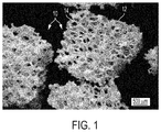
- FIG. 1 is a scanning electron microscope (SEM) image of porous hydroxyapatite granules
- FIG. 2 is an enhanced image showing mesenchymal stem cells proliferated on a porous hydroxyapatite scaffold in vitro;
- FIG. 3 is an image of a thirteen week test article implant site
- FIG. 4 is an image of a thirteen week control article implant site
- FIG. 5 is an isometric view of a 3D-printed implantable device
- FIG. 6 is a top view of the 3D-printed implantable device of FIG. 5 ;
- FIG. 7 is a vertical-sectioned view of the 3D-printed implantable device of FIG. 5 ;
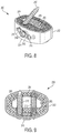
- FIG. 8 is an isometric view of a 3D-printed implantable device.
- FIG. 9 is a top view of the 3D-printed implantable device of FIG. 9 .
- the extracellular matrix of hard tissues is composed of two distinct phases, an organic phase and an inorganic phase.
- the inorganic portion of bone is comprised mainly of hydroxyapatite while the organic phase is primarily comprised of type I collagen and smaller amounts of other proteins.
- Structurally, although hard, bone is a porous material, with porosities of 50-90% in trabecular bone and 3-12% in cortical bone.
- Calcium phosphate based materials are a popular synthetic bone graft material because they have been shown to demonstrate an ability to incorporate within natural bone, as well as have osteoconductive properties.
- Two of the most widely researched materials of this type are hydroxyapatite and ⁇ -tricalcium phosphate ( ⁇ -TCP).
- Hydroxyapatite has high biocompatibility, good bioaffinity, has been shown to stimulate osteoconduction, and can be further integrated/replaced by the host bone after implantation.
- Porosity of hydroxyapatite has been shown to be incredibly important in osteogenic differentiation on both a micro and macro level of organization. In combination with rigid plating to provide mechanical support, hydroxyapatite materials have demonstrated an ability to fully incorporate and patients reported significant decreases in pain.
- the scaffold must have a high surface area and a high degree of surface contact with the bone for the implantation to be successful.
- Moldable formulations may be preferred as compared to rigid scaffolds in that they can take the shape of any treatment space.
- particle formulations combined with a delivery vehicle have been investigated.
- a microparticle based system it is possible to capitalize on the osteogenic properties of the scaffold material, but use it in a moldable formulation.
- particulate based formulations have a much higher surface area, which may alter the degradation rates of the materials resulting in faster implant incorporation.
- the particles can be used to incorporate an extra dimension of porosity in that the spaces between tightly packed particles can be used to create interconnected pores throughout the implant.
- Collagen (type I) is the most abundant extracellular protein of bone, the structure of which has been shown to be important for cell attachment, proliferation and differentiation.
- Gelatin a derivative of collagen, is biocompatible and biodegradable and has been widely investigated as a carrier material for other biological agents in bone applications. Further, incorporation of gelatin has been shown to increase cell adhesion as well as proliferation of cells. This effect has been demonstrated in a variety of ways, most notably in simple coating procedures. When combined with hydroxyapatite particles, particles incorporating gelatin showed significantly enhanced cell binding as compared to hydroxyapatite particles alone.
- Other naturally derived biopolymers have been investigated as scaffold-based materials including alginate, chitosan, and fibrin.
- VEGF Vascular endothelial growth factor
- PDGF Plate-derived growth factor
- PDGF vascular endothelial growth factor
- the presence of growth factors and cytokines within the matrix will encourage recruitment of additional host cells within the defect and help to further reduce the time necessary for tissue formation and repair of the pathology.
- the inventive process and methods are an improvement on the art of using a bone void filler in that it combines all necessary factors in the healing cascade in a concentrated manner, maximizing the regenerative capabilities of the implanted device.
- the scaffold material offers the ability to not only offer a substrate that the concentrated cells will preferentially bind to, but also be adsorbed as new tissue is formed, allowing for complete repair of tissue.
- the addition of autologous growth factors will recruit other necessary cells from the surrounding host tissue, thus further augmenting the healing cascade.
- a cellular component is defined as a fluid containing cells, some of which are known to be progenitors of bone-forming cells that are capable of making extracellular matrix resembling native bone tissue, with a composition that is not naturally occurring in the body.
- inventive device include the following:
- the inorganic matrix part of the scaffold material is defined as a porous particle such that it mimics the inorganic portion of natural bony tissue. Examples include the following:
- the organic matrix part of the scaffold material is defined as an organic biopolymer either natural or synthetic that would act to aid in cell retention as well as render the scaffold in a moldable format.
- the organic matrix can be made up of a single biopolymer or a mixture of biopolymers. Examples include the following:
- Growth factors and cytokines are proteins that can be found autologously in blood and bone marrow but delivered in a concentrated form not found naturally within the body. Examples include the following:
- a clotting agent may be added at the discretion of the physician to the device in order to preferentially alter the handling characteristics of the implant.
- FIG. 1 is an SEM image of porous hydroxyapatite granules.
- a granule 10 is shown comprising a scaffold 12 .
- the scaffold 12 forms a plurality of voids 14 throughout the granule 10 .
- the plurality of voids 14 increases a surface area of the granule 10 , which increase facilitates one or more of faster implant incorporation, better handling properties, increased cell affinity/bioactivity, and an overall increase in porosity of in an area of application.
- FIG. 2 is an enhanced image showing mesenchymal stem cells 26 proliferated on a porous hydroxyapatite scaffold 20 in vitro.
- the porous hydroxyapatite scaffold 20 comprises a structure of a granule 22 .
- the granule 22 may be similar to the granule 10 shown in FIG. 1 .
- the porous hydroxyapatite scaffold 20 comprises a plurality of voids 24 , which plurality of voids 24 increases a surface area of the granule 22 .
- Each of the plurality of mesenchymal stem cells 26 comprises a nucleus 28 and fibrillar actin 30 .
- the nuclei 28 are shown in FIG. 2 as white spots disposed on the porous hydroxyapatite scaffold 20 .
- the fibrillar actin 28 is indicated in FIG. 2 by light gray areas surrounding the nuclei 26 .
- the increased surface area provided to the granule 22 by the plurality of voids 24 results in an increased proliferation of mesenchymal stem cells 26 compared to other non-void-containing granules.
- Each implant section was analyzed by a pathologist for local tissue reaction following ISO 10993-6 guidelines, as well as any osteoconductivity, bone growth, and/or bone development within the defect.
- the test article was compared to the control article.
- the histopathology data was used to evaluate both local tissue reactions as well as bone formation.
- FIGS. 3 and 4 demonstrate histology results for a test article and a control article after a 13 week period, respectively.
- FIG. 3 shows that implanting porous hydroxyapatite granules was as effective as the predicate control article at promoting new bone development in a defect by having similar tissue responses surrounding the implant sites and within the implant sites as the control article at all durations. Implanting porous hydroxyapatite granules also had faster resorption and increased new bone formation within the implant sites compared to the control article.
- Rabbit condyles were scanned using micro-computer tomography (MicroCT) to visualize new bone formation.
- the specimens were processed to obtain 3D images in addition to quantitative measurements of bone and material volumes, densities, and trabecular features. All samples were scanned on a high-resolution, volumetric microCT scanner.
- BV bone volume
- TV total volume
- Trabecular morphometric analysis was also performed within the region of interest to determine connectivity density, structure model index, trabecular number, trabecular thickness, trabecular spacing, bone surface, bone surface per bone volume (BS/BV) and mean bone density.
- 3D-printing technology and, for example, sinterable titanium powder provides a means to create a structurally-stable implant that itself is conducive to supporting the in-growth of bone, by sustaining various populations of cells within its pores during the first few weeks after being implanted in the body.
- Titanium is known to support the development of bony tissues and is capable of interacting with the emerging bony tissue, but other materials also have similar bone-nurturing properties. Controlling the distribution of pores, the size range of pores and the overall physical geometry of the implants all contribute to a biologically-supportive microenvironment, which in turn promotes the formation of bony tissue.
- the 3D-printed portion can be combined with a non-3D-printed device, such as, for example, femoral rings from human cadaveric donors or other non-natural structures, to create a composite implant with an optimal biologically-supportive microenvironment and the rigidity of a cortical bone.
- a non-3D-printed device such as, for example, femoral rings from human cadaveric donors or other non-natural structures
- FIGS. 5-8 an embodiment of a 3D-printed implantable device 100 is shown.
- the embodiment shown in FIGS. 5-8 is adapted to be inserted between two vertebrae.
- the device 100 may be shaped differently as desired.
- FIG. 5 an isometric view of the 3D-printed implantable device 100 is shown.
- the device 100 includes an outer body 102 and a mesh 104 .
- the outer body 102 is generally oval-shaped.
- the outer body 102 may take on a variety of other shapes depending upon the intended application of the device 100 .
- the outer body 102 includes an angled bore 106 and an angled bore 108 .
- Each of the angled bores 106 and 108 extends from the outer body 102 through the mesh 104 and is adapted to receive a screw 110 and a screw 112 , respectively.
- the angled bore 106 is angled so that the screw 110 extends out of a top surface of the device 100 and the angled bore 108 is angled so that the screw 112 extends out of a bottom surface of the device 100 .
- the angle of the bores 106 and 108 can be adjusted as desired.
- each of the screws 110 and 112 is adapted to screw into an upper and lower vertebra, respectively.
- the device 100 includes an anti-backout plate 114 .
- the anti-backout plate 114 includes two wing portions 116 that can be positioned to cover heads of each of the screws 110 and 112 to prevent the screws 110 and 112 from backing out of the angled bores 106 and 108 .
- the anti-backout plate 114 may be secured to the device 100 via a screw 118 .
- the anti-backout plate 114 may be rotated such that the wing portions 116 do not cover the angled bores 106 and 108 to permit the screws 110 and 112 to be inserted into the angled bores 106 and 108 .
- additional angled bores may be added as desired.
- the device 100 could be configured to include two bores that are oriented to direct screws into an upper vertebra and a two bores that are oriented to direct screws into a lower vertebra.
- the anti-backout plate could be configured with four wing portions so that heads of each of the four screws are covered.
- the outer body 102 may also include one or more features that facilitate handling and placing of the device 100 .
- FIG. 5 shows a pair of indentations 120 that facilitate griping of the device 100 by a tool, such as, for example, a pair of pliers and the like, to grip the device 100 to assist with handling and positioning the device 100 .
- the mesh 104 is fills a space within the outer body 102 .
- the mesh 104 is designed to create a plurality of voids within the device 100 that are adapted to receive and hold a material, such as, for example, a filler and/or cell preparation. Because the device 100 is 3D printed, the mesh 104 may take on a variety of shapes and designs depending on the intended application. A porosity of the mesh 104 can be changed based on various design considerations. In some embodiments, the mesh 104 has a porosity of between 50%-95% by volume. As shown in FIGS. 5-8 , the mesh 104 comprises a lattice-type design.
- the device 100 includes features that create a porosity that mimics pore sizes, connectedness, and pore-size distribution commonly found in cancellous bone and/or cortical bone.
- a surface texture of the 3D-printed implantable device can also be adjusted or modified to enhance the attachment of cells critical to the production of bony tissues.
- the lattice-type design of the mesh 104 comprises a straight-through design when viewed from the top. Such a design may facilitate tissue growth through the mesh 104 that fuses the two bones or bony ends together.
- the mesh 104 may not include a straight-through design when viewed from the top.
- successive layers of the lattice may be offset so that voids are not lined up, or in other embodiments the mesh may be comprised of layers having different patterns that result in voids not aligning with one another.
- FIG. 7 a sectioned view of the device 100 about line A-A of FIG. 6 is shown.
- FIG. 7 shows that the outer body 102 is made up of a relatively thin wall compared to an overall width of the device 100 .
- the thickness of the outer body 102 may be changed depending on various design considerations. For example, in some embodiments, it may be desirable to increase the thickness of the outer body 102 to increase the strength of the outer body 102 . In some embodiments, the thickness of the outer body 102 may be increased around the various bores of the device 100 to provide additional strength in those areas.
- FIG. 7 shows that the mesh 104 includes a plurality of voids. In a typical embodiment, the plurality of voids comprises uniformly shaped voids.
- the mesh 104 may be designed to include non-uniformly shaped voids.
- FIG. 7 also shows a bore 122 that extends from the outer body 102 into the mesh 104 .
- the bore 122 is adapted to receive the screw 118 to secure the anti-backout plate 114 to the device 100 .
- FIG. 8 is an isometric view of a 3D-printed implantable device 200 .
- the device 200 is similar to the device 100 , but the device 200 includes a pocket 201 formed through the device 200 .
- the device 200 includes an outer body 202 and a mesh 204 .
- the device 200 includes an angled bore 206 and an angled bore 208 .
- the device 200 may include additional angled bores as desired.
- the angled bores 206 and 208 are adapted to receive screws 210 and 212 , respectively.
- the screws 210 and 212 are adapted to secure the device 200 to an upper and a lower vertebra, respectively.
- the device 200 includes an anti-backout plate 214 .
- the anti-backout plate 214 includes two wing portions 216 that may be positioned over heads of the screws 210 and 212 to prevent the screws 210 and 212 from backing out of the upper and lower vertebrae.
- the anti-backout plate 214 is secured to the device 200 with a screw 218 .
- the device 200 includes a bore adapted to receive the screw 218 that is similar to the bore 122 of the device 100 .
- the pocket 201 provides an interior space within the device 200 into which a material, such as, for example, a filler and/or cell preparation, can be inserted.
- An interior wall 203 of the device 200 surrounds a periphery of the pocket 201 .
- the interior wall 203 includes one or more perforations 205 .
- the one or more perforations allows for in-growth of the material disposed within the pocket 201 into the mesh 204 .
- a size and shape of the one or more perforations 205 may be varied based on various design considerations.
- a filler may be inserted into the mesh 204 and the pocket 201 .
- a first filler may be inserted into the mesh 204 and a second filler may be inserted into the pocket 201 .
- FIG. 9 is a top view of the device 200 .
- the mesh 204 similar to the mesh 104 , comprises a straight-through design when viewed from the top. Such a design may facilitate cell growth between two bones or bony ends resulting in formation of a bony union.
- the mesh 204 may not include a straight-through design when viewed from the top. In such a design, successive layers of the lattice may be offset so that voids are not lined up, or in other embodiments the mesh may be comprised of layers having different patterns that result in voids not aligning with one another to create a straight-through design.
- the mesh 104 has porosity of between 50%-95% by volume.
- compositions and methods disclosed and claimed herein can be made and executed without undue experimentation in light of the present disclosure. While the compositions and methods of this disclosure have been described in terms of preferred embodiments, it will be apparent to those of skill in the art that variations may be applied to the compositions and methods and in the steps or in the sequence of steps of the method described herein without departing from the concept, spirit and scope of the disclosure. More specifically, it will be apparent that certain agents which are both chemically and physiologically related may be substituted for the agents described herein while the same or similar results would be achieved. All such similar substitutes and modifications apparent to those skilled in the art are deemed to be within the spirit, scope and concept of the disclosure as defined by the appended claims.
Landscapes
- Health & Medical Sciences (AREA)
- Life Sciences & Earth Sciences (AREA)
- Chemical & Material Sciences (AREA)
- Transplantation (AREA)
- Veterinary Medicine (AREA)
- Oral & Maxillofacial Surgery (AREA)
- Animal Behavior & Ethology (AREA)
- General Health & Medical Sciences (AREA)
- Public Health (AREA)
- Medicinal Chemistry (AREA)
- Dermatology (AREA)
- Epidemiology (AREA)
- Engineering & Computer Science (AREA)
- Biomedical Technology (AREA)
- Orthopedic Medicine & Surgery (AREA)
- Vascular Medicine (AREA)
- Cell Biology (AREA)
- Cardiology (AREA)
- Heart & Thoracic Surgery (AREA)
- Zoology (AREA)
- Chemical Kinetics & Catalysis (AREA)
- Botany (AREA)
- Neurology (AREA)
- Dispersion Chemistry (AREA)
- Inorganic Chemistry (AREA)
- Urology & Nephrology (AREA)
- Hematology (AREA)
- Developmental Biology & Embryology (AREA)
- Biophysics (AREA)
- Physical Education & Sports Medicine (AREA)
- Prostheses (AREA)
- Materials For Medical Uses (AREA)
- Molecular Biology (AREA)
Abstract
Description
-
- a. Autologous bone marrow concentrated and/or filtered to remove red blood cells, while retaining growth factors and mononuclear cells at a concentration greater than 1.1×.
- b. Autologous bone marrow concentrated and/or filtered to remove all other extracellular components at a concentration greater than 1.1×.
- c. Autologous adipose tissue filtered, enzymatically digested, and/or concentrated to isolate known progenitor cells at a concentration greater than 1.1× natural cell concentration.
- d. Autologous progenitor cells that have been isolated and expanded ex vivo to be 1.1× or more the concentration of the cells found in native tissue.
- e. Allogeneic progenitor cells that are 1.1× or more the concentration of the cells found in native tissue.
- f. Any combination of the above.
-
- a. The inventive matrix can be formulated in a variety of formats, including a granule form, a powder form, a strip form and a block form.
- b. Porosity will mimic that found in bone ranging from 50-95% or 3-15% porosity.
- c. Granule composition may include calcium phosphate, mono calcium phosphate, tricalcium phosphate, tetra calcium phosphate, octacalcium phosphate, hydroxyapatite, carbonate apatite, fluoro apatite, or any combination thereof.
- d. The granule composition may also contain materials to mimic the ionic characteristics of bone, this may include (but is not limited to): silicon, strontium, or magnesium.
-
- a. The biopolymer may be selected from one of the following: collagen, gelatin, fibrinogen, vitronectin, fibronectin, albumin, peptides, chitin, alginate, cellulose, carboxymethycellulose or any combination thereof
-
- a. The growth factors and cytokines can be autologous and concentrated from biological fluids including the platelet poor fraction of blood or bone marrow
- b. The growth factors and cytokines can be synthetically derived and incorporated within the scaffold at the discretion of the physician
Claims (19)
Priority Applications (1)
| Application Number | Priority Date | Filing Date | Title |
|---|---|---|---|
| US15/168,199 US10751185B2 (en) | 2014-10-23 | 2016-05-30 | Treatment of skeletal voids with implantable substrate hydrated with bone marrow concentrate |
Applications Claiming Priority (3)
| Application Number | Priority Date | Filing Date | Title |
|---|---|---|---|
| US201462067815P | 2014-10-23 | 2014-10-23 | |
| US14/921,826 US10183095B2 (en) | 2014-10-23 | 2015-10-23 | Treatment of skeletal voids with implantable substrate hydrated with bone marrow concentrate |
| US15/168,199 US10751185B2 (en) | 2014-10-23 | 2016-05-30 | Treatment of skeletal voids with implantable substrate hydrated with bone marrow concentrate |
Related Parent Applications (1)
| Application Number | Title | Priority Date | Filing Date |
|---|---|---|---|
| US14/921,826 Continuation-In-Part US10183095B2 (en) | 2014-10-23 | 2015-10-23 | Treatment of skeletal voids with implantable substrate hydrated with bone marrow concentrate |
Publications (2)
| Publication Number | Publication Date |
|---|---|
| US20160270920A1 US20160270920A1 (en) | 2016-09-22 |
| US10751185B2 true US10751185B2 (en) | 2020-08-25 |
Family
ID=56923425
Family Applications (1)
| Application Number | Title | Priority Date | Filing Date |
|---|---|---|---|
| US15/168,199 Active 2036-05-09 US10751185B2 (en) | 2014-10-23 | 2016-05-30 | Treatment of skeletal voids with implantable substrate hydrated with bone marrow concentrate |
Country Status (1)
| Country | Link |
|---|---|
| US (1) | US10751185B2 (en) |
Cited By (4)
| Publication number | Priority date | Publication date | Assignee | Title |
|---|---|---|---|---|
| US11116645B2 (en) * | 2019-04-30 | 2021-09-14 | Life Spine, Inc. | ALIF spine implant with cam screws for inhibiting bone anchor backout |
| US11147679B2 (en) * | 2018-02-05 | 2021-10-19 | Paragon Advanced Technologies, Inc. | Bone fixation device |
| US12053212B2 (en) | 2020-05-01 | 2024-08-06 | FloSpine, LLC | Implant assembly including screw locking mechanism |
| US12653680B1 (en) | 2020-01-08 | 2026-06-16 | Restor3D, Inc. | Sheet based triply periodic minimal surface implants for promoting osseointegration and methods for producing same |
Families Citing this family (42)
| Publication number | Priority date | Publication date | Assignee | Title |
|---|---|---|---|---|
| US10842645B2 (en) * | 2008-08-13 | 2020-11-24 | Smed-Ta/Td, Llc | Orthopaedic implant with porous structural member |
| US10492921B2 (en) | 2015-04-29 | 2019-12-03 | Institute for Musculoskeletal Science and Education, Ltd. | Implant with arched bone contacting elements |
| US10449051B2 (en) | 2015-04-29 | 2019-10-22 | Institute for Musculoskeletal Science and Education, Ltd. | Implant with curved bone contacting elements |
| US10709570B2 (en) | 2015-04-29 | 2020-07-14 | Institute for Musculoskeletal Science and Education, Ltd. | Implant with a diagonal insertion axis |
| CN114259328A (en) | 2015-04-29 | 2022-04-01 | 肌肉骨骼科学教育研究所有限公司 | Coiled implants and systems and methods of use |
| US10034768B2 (en) * | 2015-09-02 | 2018-07-31 | Globus Medical, Inc. | Implantable systems, devices and related methods |
| US20180338838A1 (en) * | 2016-02-03 | 2018-11-29 | Globus Medical, Inc. | Intervertebral Spinal Implants, Associated Instruments, And Methods Thereof |
| US11478286B2 (en) * | 2016-06-03 | 2022-10-25 | Paragon Advanced Technologies, Inc. | Bone fixation devices |
| WO2018053403A1 (en) | 2016-09-16 | 2018-03-22 | Mirus Llc | Interbody fusion devices and related methods of manufacture |
| US11033394B2 (en) | 2016-10-25 | 2021-06-15 | Institute for Musculoskeletal Science and Education, Ltd. | Implant with multi-layer bone interfacing lattice |
| US10478312B2 (en) | 2016-10-25 | 2019-11-19 | Institute for Musculoskeletal Science and Education, Ltd. | Implant with protected fusion zones |
| FR3061650B1 (en) * | 2017-01-06 | 2020-10-02 | Kisco Int | INTERSOMATIC IMPLANT WITH GRAFT RETENTION |
| WO2018165571A1 (en) * | 2017-03-10 | 2018-09-13 | Life Spine, Inc. (A Delaware Corporation) | 3-d printed orthopedic implants |
| US10667924B2 (en) | 2017-03-13 | 2020-06-02 | Institute for Musculoskeletal Science and Education, Ltd. | Corpectomy implant |
| US10512549B2 (en) | 2017-03-13 | 2019-12-24 | Institute for Musculoskeletal Science and Education, Ltd. | Implant with structural members arranged around a ring |
| US10064726B1 (en) * | 2017-04-18 | 2018-09-04 | Warsaw Orthopedic, Inc. | 3D printing of mesh implants for bone delivery |
| EP3459502B1 (en) * | 2017-09-20 | 2024-05-22 | Stryker European Operations Holdings LLC | Spinal implants |
| US10744001B2 (en) | 2017-11-21 | 2020-08-18 | Institute for Musculoskeletal Science and Education, Ltd. | Implant with improved bone contact |
| US10940015B2 (en) | 2017-11-21 | 2021-03-09 | Institute for Musculoskeletal Science and Education, Ltd. | Implant with improved flow characteristics |
| US10517739B2 (en) | 2018-05-08 | 2019-12-31 | Globus Medical, Inc. | Intervertebral spinal implant |
| US10682238B2 (en) | 2018-05-08 | 2020-06-16 | Globus Medical, Inc. | Intervertebral spinal implant |
| US10744003B2 (en) | 2018-05-08 | 2020-08-18 | Globus Medical, Inc. | Intervertebral spinal implant |
| AU2019213392B2 (en) * | 2018-08-09 | 2024-11-28 | Vb Spine Llc | Interbody implants and optimization features thereof |
| CN108888389A (en) * | 2018-09-14 | 2018-11-27 | 北京爱康宜诚医疗器材有限公司 | Articular surface fusion device |
| US11298244B2 (en) | 2019-01-31 | 2022-04-12 | K2M, Inc. | Interbody implants and instrumentation |
| US11039931B2 (en) | 2019-02-01 | 2021-06-22 | Globus Medical, Inc. | Intervertebral spinal implant |
| USD955579S1 (en) * | 2019-04-26 | 2022-06-21 | Warsaw Orthopedic, Inc. | Surgical implant |
| USD948048S1 (en) * | 2019-04-26 | 2022-04-05 | Warsaw Orthopedic, Inc. | Surgical implant |
| CN110063820B (en) * | 2019-06-03 | 2023-10-27 | 华中科技大学同济医学院附属协和医院 | 3D printed intervertebral fusion device with interface bone ingrowth activity and its preparation method |
| US11534307B2 (en) | 2019-09-16 | 2022-12-27 | K2M, Inc. | 3D printed cervical standalone implant |
| US11844704B2 (en) | 2019-09-18 | 2023-12-19 | Restor3D, Inc. | Instruments and method for ankle replacement surgery |
| USD920517S1 (en) | 2020-01-08 | 2021-05-25 | Restor3D, Inc. | Osteotomy wedge |
| US11957600B2 (en) | 2020-02-18 | 2024-04-16 | Mirus Llc | Anterior lumbar interbody fusion device with bidirectional screws |
| USD942624S1 (en) * | 2020-11-13 | 2022-02-01 | Mirus Llc | Spinal implant |
| USD942011S1 (en) * | 2020-11-13 | 2022-01-25 | Mirus Llc | Spinal implant |
| USD944400S1 (en) * | 2020-11-13 | 2022-02-22 | Mirus Llc | Spinal implant |
| USD942623S1 (en) * | 2020-11-13 | 2022-02-01 | Mirus Llc | Spinal implant |
| US20230120830A1 (en) * | 2021-10-20 | 2023-04-20 | Bone Solutions, Inc. | Spinal Implant with a Magnesium-Phosphate Three-Dimensional Porosity Structure |
| USD1122446S1 (en) | 2023-05-10 | 2026-04-14 | Restor3D, Inc. | Talus implant |
| USD1124342S1 (en) | 2023-05-10 | 2026-04-28 | Restor3D, Inc. | Talus implant |
| US12539220B2 (en) | 2023-05-30 | 2026-02-03 | Medtronic Navigation, Inc. | Systems and methods for bone graft mixture guidance based on navigation information |
| US12690986B1 (en) | 2023-08-08 | 2026-07-28 | Restor3D, Inc. | Total ankle replacement system and processes for making and using the same |
Citations (14)
| Publication number | Priority date | Publication date | Assignee | Title |
|---|---|---|---|---|
| US20010039456A1 (en) * | 2000-03-22 | 2001-11-08 | Boyer Michael L. | Tissue forms |
| US20030078668A1 (en) * | 1999-05-05 | 2003-04-24 | Michelson Gary K. | Interbody spinal fusion implants with single-lock for locking opposed screws |
| US20050112397A1 (en) * | 2003-07-24 | 2005-05-26 | Rolfe Jonathan L. | Assembled non-random foams |
| US20100185289A1 (en) * | 2009-01-20 | 2010-07-22 | Incite Innovation Llc | Interbody fusion device and method of operation |
| US20110040334A1 (en) * | 2001-07-12 | 2011-02-17 | Kaes David R | Intervertebral implant with movement resistant structure |
| US20120185047A1 (en) * | 2011-01-17 | 2012-07-19 | Cibor, Inc. | Reinforced carbon fiber/carbon foam intervertebral spine fusion device |
| US20120191188A1 (en) * | 2011-01-20 | 2012-07-26 | Huang meng-feng | Spinal implant with bone engaging projections |
| US20120277870A1 (en) * | 2011-04-29 | 2012-11-01 | Wolters Madeline C | Spinal Interbody Implant With Bone Screw Retention |
| US20130116793A1 (en) * | 2010-07-23 | 2013-05-09 | Privelop-Spine Ag | Surgical implant |
| US20140012384A1 (en) * | 2011-07-12 | 2014-01-09 | Spinesmith Partners, L.P. | Interbody fusion device and associated methods |
| US20140107786A1 (en) * | 2012-10-11 | 2014-04-17 | Rhausler, Inc. | Fusion cage implant with lattice structure |
| US20140288649A1 (en) * | 2013-03-15 | 2014-09-25 | 4Web, Inc. | Traumatic bone fracture repair systems and methods |
| US20150010607A1 (en) * | 2012-02-07 | 2015-01-08 | Lifenet Health | Liquefaction of bone matrix |
| US10064737B2 (en) * | 2015-12-07 | 2018-09-04 | Industrial Technology Research Institute | Implant device for osseous integration |
-
2016
- 2016-05-30 US US15/168,199 patent/US10751185B2/en active Active
Patent Citations (14)
| Publication number | Priority date | Publication date | Assignee | Title |
|---|---|---|---|---|
| US20030078668A1 (en) * | 1999-05-05 | 2003-04-24 | Michelson Gary K. | Interbody spinal fusion implants with single-lock for locking opposed screws |
| US20010039456A1 (en) * | 2000-03-22 | 2001-11-08 | Boyer Michael L. | Tissue forms |
| US20110040334A1 (en) * | 2001-07-12 | 2011-02-17 | Kaes David R | Intervertebral implant with movement resistant structure |
| US20050112397A1 (en) * | 2003-07-24 | 2005-05-26 | Rolfe Jonathan L. | Assembled non-random foams |
| US20100185289A1 (en) * | 2009-01-20 | 2010-07-22 | Incite Innovation Llc | Interbody fusion device and method of operation |
| US20130116793A1 (en) * | 2010-07-23 | 2013-05-09 | Privelop-Spine Ag | Surgical implant |
| US20120185047A1 (en) * | 2011-01-17 | 2012-07-19 | Cibor, Inc. | Reinforced carbon fiber/carbon foam intervertebral spine fusion device |
| US20120191188A1 (en) * | 2011-01-20 | 2012-07-26 | Huang meng-feng | Spinal implant with bone engaging projections |
| US20120277870A1 (en) * | 2011-04-29 | 2012-11-01 | Wolters Madeline C | Spinal Interbody Implant With Bone Screw Retention |
| US20140012384A1 (en) * | 2011-07-12 | 2014-01-09 | Spinesmith Partners, L.P. | Interbody fusion device and associated methods |
| US20150010607A1 (en) * | 2012-02-07 | 2015-01-08 | Lifenet Health | Liquefaction of bone matrix |
| US20140107786A1 (en) * | 2012-10-11 | 2014-04-17 | Rhausler, Inc. | Fusion cage implant with lattice structure |
| US20140288649A1 (en) * | 2013-03-15 | 2014-09-25 | 4Web, Inc. | Traumatic bone fracture repair systems and methods |
| US10064737B2 (en) * | 2015-12-07 | 2018-09-04 | Industrial Technology Research Institute | Implant device for osseous integration |
Cited By (8)
| Publication number | Priority date | Publication date | Assignee | Title |
|---|---|---|---|---|
| US11147679B2 (en) * | 2018-02-05 | 2021-10-19 | Paragon Advanced Technologies, Inc. | Bone fixation device |
| US11766337B2 (en) | 2018-02-05 | 2023-09-26 | Paragon Advanced Technologies, Inc. | Bone fixation device |
| US11116645B2 (en) * | 2019-04-30 | 2021-09-14 | Life Spine, Inc. | ALIF spine implant with cam screws for inhibiting bone anchor backout |
| US20210401587A1 (en) * | 2019-04-30 | 2021-12-30 | Life Spine, Inc. | Spine implant with cam screws for inhibiting bone anchor backout |
| US11951019B2 (en) * | 2019-04-30 | 2024-04-09 | Life Spine, Inc. | Spine implant with cam screws for inhibiting bone anchor backout |
| US12653680B1 (en) | 2020-01-08 | 2026-06-16 | Restor3D, Inc. | Sheet based triply periodic minimal surface implants for promoting osseointegration and methods for producing same |
| US12667461B1 (en) * | 2020-01-08 | 2026-06-30 | Restor3D, Inc. | Sheet based triply periodic minimal surface implants for promoting osseointegration and methods for producing same |
| US12053212B2 (en) | 2020-05-01 | 2024-08-06 | FloSpine, LLC | Implant assembly including screw locking mechanism |
Also Published As
| Publication number | Publication date |
|---|---|
| US20160270920A1 (en) | 2016-09-22 |
Similar Documents
| Publication | Publication Date | Title |
|---|---|---|
| US20160270920A1 (en) | Treatment of skeletal voids with implantable substrate hydrated with bone marrow concentrate | |
| Anderson et al. | Critical size defect in the goat's os ilium: A model to evaluate bone grafts and substitutes | |
| Henkel et al. | Bone regeneration based on tissue engineering conceptions—a 21st century perspective | |
| KR102248576B1 (en) | Solid substrates for promoting cell and tissue growth | |
| JP5795577B2 (en) | Solid form for tissue repair | |
| TWI394597B (en) | Biodegradable scaffold bone graft for orthopaedic use | |
| Parizi et al. | Human platelet rich plasma plus Persian Gulf coral effects on experimental bone healing in rabbit model: radiological, histological, macroscopical and biomechanical evaluation | |
| Brandt et al. | Nanocrystalline hydroxyapatite for bone repair: an animal study | |
| US8916228B2 (en) | Bi-layered bone-like scaffolds | |
| Nik Md Noordin Kahar et al. | A review of bioceramics scaffolds for bone defects in different types of animal models: HA and β-TCP | |
| JP6116484B2 (en) | Compositions and methods for spinal fusion | |
| JP2000508911A (en) | Bone regeneration and enhancement using mesenchymal stem cells | |
| EP3530295A1 (en) | Demineralized bone matrix having improved handling characteristics | |
| Damron | Use of 3D β-tricalcium phosphate (Vitoss®) scaffolds in repairing bone defects | |
| Louisia et al. | Coral grafting supplemented with bone marrow | |
| CA2468506A1 (en) | Implant, method of making the same and use thereof for the treating of bones defects | |
| KR20180127641A (en) | Composite medical implants and methods for their use and manufacturing | |
| Faratzis et al. | Effect of autologous platelet-rich plasma in combination with a biphasic synthetic graft material on bone healing in critical-size cranial defects | |
| US20250032673A1 (en) | Bone repair composition and kit | |
| US10183095B2 (en) | Treatment of skeletal voids with implantable substrate hydrated with bone marrow concentrate | |
| Tibeică et al. | Bone regeneration influence in the success of periimplant surgery | |
| US20220168356A1 (en) | Allografts containing viable cells and methods therof | |
| US11426489B2 (en) | Biomaterial compositions, implants, and methods of making the same | |
| Ding et al. | Efficacy of a small cell‐binding peptide coated hydroxyapatite substitute on bone formation and implant fixation in sheep | |
| KR20180127640A (en) | Composite medical implants and methods for their use and manufacturing |
Legal Events
| Date | Code | Title | Description |
|---|---|---|---|
| AS | Assignment |
Owner name: SPINESMITH PARTNERS, L.P., TEXAS Free format text: ASSIGNMENT OF ASSIGNORS INTEREST;ASSIGNORS:DAWSON, EILEEN;DUNWORTH, KEVIN;SAND, THEODORE;AND OTHERS;SIGNING DATES FROM 20160720 TO 20160923;REEL/FRAME:039844/0175 |
|
| AS | Assignment |
Owner name: BLUEARC MEZZANINE PARTNERS I, L.P., GEORGIA Free format text: SECURITY INTEREST;ASSIGNOR:SPINESMITH HOLDINGS, LLC;REEL/FRAME:042071/0370 Effective date: 20170315 |
|
| AS | Assignment |
Owner name: AMERICAN BANK, N.A., TEXAS Free format text: SECURITY INTEREST;ASSIGNOR:SPINESMITH HOLDINGS, LLC;REEL/FRAME:044882/0096 Effective date: 20171206 |
|
| STPP | Information on status: patent application and granting procedure in general |
Free format text: DOCKETED NEW CASE - READY FOR EXAMINATION |
|
| AS | Assignment |
Owner name: SPINESMITH HOLIDNGS, LLC, TEXAS Free format text: RELEASE BY SECURED PARTY;ASSIGNOR:AMERICAN BANK;REEL/FRAME:048853/0105 Effective date: 20190328 |
|
| STPP | Information on status: patent application and granting procedure in general |
Free format text: NON FINAL ACTION MAILED |
|
| STPP | Information on status: patent application and granting procedure in general |
Free format text: RESPONSE TO NON-FINAL OFFICE ACTION ENTERED AND FORWARDED TO EXAMINER |
|
| STPP | Information on status: patent application and granting procedure in general |
Free format text: FINAL REJECTION MAILED |
|
| STPP | Information on status: patent application and granting procedure in general |
Free format text: DOCKETED NEW CASE - READY FOR EXAMINATION |
|
| STPP | Information on status: patent application and granting procedure in general |
Free format text: NOTICE OF ALLOWANCE MAILED -- APPLICATION RECEIVED IN OFFICE OF PUBLICATIONS |
|
| STPP | Information on status: patent application and granting procedure in general |
Free format text: PUBLICATIONS -- ISSUE FEE PAYMENT VERIFIED |
|
| STCF | Information on status: patent grant |
Free format text: PATENTED CASE |
|
| MAFP | Maintenance fee payment |
Free format text: PAYMENT OF MAINTENANCE FEE, 4TH YR, SMALL ENTITY (ORIGINAL EVENT CODE: M2551); ENTITY STATUS OF PATENT OWNER: SMALL ENTITY Year of fee payment: 4 |