US10722191B2 - Digital X-ray diagnosis and evaluation of dental disease - Google Patents
Digital X-ray diagnosis and evaluation of dental disease Download PDFInfo
- Publication number
- US10722191B2 US10722191B2 US15/829,766 US201715829766A US10722191B2 US 10722191 B2 US10722191 B2 US 10722191B2 US 201715829766 A US201715829766 A US 201715829766A US 10722191 B2 US10722191 B2 US 10722191B2
- Authority
- US
- United States
- Prior art keywords
- dental
- density
- density value
- decay
- cursor
- Prior art date
- Legal status (The legal status is an assumption and is not a legal conclusion. Google has not performed a legal analysis and makes no representation as to the accuracy of the status listed.)
- Active
Links
Images


Classifications
-
- A—HUMAN NECESSITIES
- A61—MEDICAL OR VETERINARY SCIENCE; HYGIENE
- A61B—DIAGNOSIS; SURGERY; IDENTIFICATION
- A61B6/00—Apparatus or devices for radiation diagnosis; Apparatus or devices for radiation diagnosis combined with radiation therapy equipment
- A61B6/50—Apparatus or devices for radiation diagnosis; Apparatus or devices for radiation diagnosis combined with radiation therapy equipment specially adapted for specific body parts; specially adapted for specific clinical applications
- A61B6/51—Apparatus or devices for radiation diagnosis; Apparatus or devices for radiation diagnosis combined with radiation therapy equipment specially adapted for specific body parts; specially adapted for specific clinical applications for dentistry
-
- A61B6/14—
-
- A—HUMAN NECESSITIES
- A61—MEDICAL OR VETERINARY SCIENCE; HYGIENE
- A61B—DIAGNOSIS; SURGERY; IDENTIFICATION
- A61B6/00—Apparatus or devices for radiation diagnosis; Apparatus or devices for radiation diagnosis combined with radiation therapy equipment
- A61B6/46—Arrangements for interfacing with the operator or the patient
- A61B6/461—Displaying means of special interest
- A61B6/463—Displaying means of special interest characterised by displaying multiple images or images and diagnostic data on one display
-
- A—HUMAN NECESSITIES
- A61—MEDICAL OR VETERINARY SCIENCE; HYGIENE
- A61B—DIAGNOSIS; SURGERY; IDENTIFICATION
- A61B6/00—Apparatus or devices for radiation diagnosis; Apparatus or devices for radiation diagnosis combined with radiation therapy equipment
- A61B6/46—Arrangements for interfacing with the operator or the patient
- A61B6/467—Arrangements for interfacing with the operator or the patient characterised by special input means
-
- A—HUMAN NECESSITIES
- A61—MEDICAL OR VETERINARY SCIENCE; HYGIENE
- A61B—DIAGNOSIS; SURGERY; IDENTIFICATION
- A61B6/00—Apparatus or devices for radiation diagnosis; Apparatus or devices for radiation diagnosis combined with radiation therapy equipment
- A61B6/50—Apparatus or devices for radiation diagnosis; Apparatus or devices for radiation diagnosis combined with radiation therapy equipment specially adapted for specific body parts; specially adapted for specific clinical applications
- A61B6/505—Apparatus or devices for radiation diagnosis; Apparatus or devices for radiation diagnosis combined with radiation therapy equipment specially adapted for specific body parts; specially adapted for specific clinical applications for diagnosis of bone
-
- A—HUMAN NECESSITIES
- A61—MEDICAL OR VETERINARY SCIENCE; HYGIENE
- A61B—DIAGNOSIS; SURGERY; IDENTIFICATION
- A61B6/00—Apparatus or devices for radiation diagnosis; Apparatus or devices for radiation diagnosis combined with radiation therapy equipment
- A61B6/52—Devices using data or image processing specially adapted for radiation diagnosis
- A61B6/5211—Devices using data or image processing specially adapted for radiation diagnosis involving processing of medical diagnostic data
- A61B6/5217—Devices using data or image processing specially adapted for radiation diagnosis involving processing of medical diagnostic data extracting a diagnostic or physiological parameter from medical diagnostic data
-
- A—HUMAN NECESSITIES
- A61—MEDICAL OR VETERINARY SCIENCE; HYGIENE
- A61B—DIAGNOSIS; SURGERY; IDENTIFICATION
- A61B6/00—Apparatus or devices for radiation diagnosis; Apparatus or devices for radiation diagnosis combined with radiation therapy equipment
- A61B6/58—Testing, adjusting or calibrating thereof
- A61B6/582—Calibration
-
- G—PHYSICS
- G06—COMPUTING OR CALCULATING; COUNTING
- G06T—IMAGE DATA PROCESSING OR GENERATION, IN GENERAL
- G06T7/00—Image analysis
- G06T7/0002—Inspection of images, e.g. flaw detection
- G06T7/0012—Biomedical image inspection
-
- A—HUMAN NECESSITIES
- A61—MEDICAL OR VETERINARY SCIENCE; HYGIENE
- A61B—DIAGNOSIS; SURGERY; IDENTIFICATION
- A61B6/00—Apparatus or devices for radiation diagnosis; Apparatus or devices for radiation diagnosis combined with radiation therapy equipment
- A61B6/58—Testing, adjusting or calibrating thereof
- A61B6/582—Calibration
- A61B6/583—Calibration using calibration phantoms
-
- G—PHYSICS
- G06—COMPUTING OR CALCULATING; COUNTING
- G06T—IMAGE DATA PROCESSING OR GENERATION, IN GENERAL
- G06T2207/00—Indexing scheme for image analysis or image enhancement
- G06T2207/30—Subject of image; Context of image processing
- G06T2207/30004—Biomedical image processing
- G06T2207/30036—Dental; Teeth
Definitions
- FIGS. 5-15 are images that illustrate use of a software algorithm for diagnosing and evaluating decay from the digital dental x-ray image.
- FIGS. 19-21 are charts that illustrate optical density plots along the DEJ corresponding to FIGS. 16-18 , respectively.
- FIG. 67 is a schematic diagram of a composite calibration block.
- Conversion to digital dental x-ray imaging and analysis may be employed for improving the accuracy of dental disease diagnosis, and may offer other practical benefits as well.
- the use of digital imaging and analysis may reduce the time required for making a diagnosis, benefiting both dentist and patient. Images may be retrieved electronically for viewing or analysis quickly and remotely without searching for x-ray films. Patient may be more accepting of a diagnosis made by digital methodologies because they can better visualize their own condition.
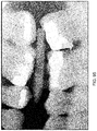
- FIGS. 1-4 Several exemplary dental x-ray images are shown in FIGS. 1-4 , illustrating the difficulty of accurately diagnosing decay from such images.
- FIGS. 1 and 2 show teeth # 3 , # 4 , and # 5 (specific teeth may be referred to hereinafter by number only). It is difficult to discern from these images the extent of interproximal decay between these teeth, or if decay is even present. Likewise, in FIGS. 3 and 4 , it is difficult to evaluate interproximal decay (if any) between # 30 and # 31 .
- These direct digital images were “enhanced” for visual interpretation on a display monitor. Critical data is often washed out by the x-ray capture and image display process, making decay difficult to detect or evaluate.
- FIG. 18 analysis of an area of more advanced decay is illustrated.
- the corresponding density curves in FIG. 21 exhibit extensive dips in both enamel and dentin density curves.
- the width of the affected area i.e. the length of the dip along the DEJ
- the width of the affected area is larger in the dentin. This arises from differing pathophysiology of the respective decay processes in enamel and dentin.
- the decay process in enamel is diet-driven: bacteria rely on external nutrients (such as dietary sugars), and the acids produced by their metabolic activity drives the decay process.
- FIGS. 43-51 An example of analysis of an x-ray of pediatric teeth is illustrated in FIGS. 43-51 .
- FIGS. 53-60 use of edge detection algorithms in combination with pattern recognition functions for mapping the entire DEJ contour is illustrated in FIGS. 53-60 .
- Two points near the DEJ are marked by the user ( FIGS. 53 and 54 ).
- the software algorithm locates the DEJ contour ( FIG. 55 ), and computes numerical decay values along the DEJ ( FIG. 56 ). This is repeated for other teeth in the x-ray image ( FIG. 57 ).
- numerical decay values are displayed as the user moves a cursor over the image ( FIGS. 58-60 ).
- Other approaches may be employed as well.
- the DEJ could be manually traced by a user.
- dental pathologies may be simulated by suitably configured composite calibration structures.
- Such other dental pathologies include but are not limited to periodontal disease, endodontic disease, bone densities around implants or implant failures, progression of diseases by comparing prior images and data derived from those images, and potentially the identification of tumors or lesions that might contain cancer in the bone around teeth.
- a software algorithm for analyzing digital dental radiographs may provide a more objective and more accurate means for diagnosing and evaluating periodontal disease.
- the software algorithm may enable the clinician to make more appropriate treatment recommendations, and may elicit more ready acceptance of those treatment recommendations by the patient.
Landscapes
- Health & Medical Sciences (AREA)
- Life Sciences & Earth Sciences (AREA)
- Engineering & Computer Science (AREA)
- Medical Informatics (AREA)
- Nuclear Medicine, Radiotherapy & Molecular Imaging (AREA)
- General Health & Medical Sciences (AREA)
- Physics & Mathematics (AREA)
- Radiology & Medical Imaging (AREA)
- Molecular Biology (AREA)
- Veterinary Medicine (AREA)
- Pathology (AREA)
- High Energy & Nuclear Physics (AREA)
- Biomedical Technology (AREA)
- Heart & Thoracic Surgery (AREA)
- Biophysics (AREA)
- Surgery (AREA)
- Animal Behavior & Ethology (AREA)
- Optics & Photonics (AREA)
- Public Health (AREA)
- Dentistry (AREA)
- Oral & Maxillofacial Surgery (AREA)
- Human Computer Interaction (AREA)
- Computer Vision & Pattern Recognition (AREA)
- Orthopedic Medicine & Surgery (AREA)
- Physiology (AREA)
- Quality & Reliability (AREA)
- General Physics & Mathematics (AREA)
- Theoretical Computer Science (AREA)
- Apparatus For Radiation Diagnosis (AREA)
- Dental Tools And Instruments Or Auxiliary Dental Instruments (AREA)
Abstract
Description
Claims (16)
Priority Applications (1)
| Application Number | Priority Date | Filing Date | Title |
|---|---|---|---|
| US15/829,766 US10722191B2 (en) | 2006-01-12 | 2017-12-01 | Digital X-ray diagnosis and evaluation of dental disease |
Applications Claiming Priority (5)
| Application Number | Priority Date | Filing Date | Title |
|---|---|---|---|
| US75882906P | 2006-01-12 | 2006-01-12 | |
| US11/652,822 US8417010B1 (en) | 2006-01-12 | 2007-01-12 | Digital x-ray diagnosis and evaluation of dental disease |
| US13/858,805 US9339245B2 (en) | 2006-01-12 | 2013-04-08 | Digital x-ray diagnosis and evaluation of dental disease |
| US15/156,271 US9839402B2 (en) | 2006-01-12 | 2016-05-16 | Digital X-ray diagnosis and evaluation of dental disease |
| US15/829,766 US10722191B2 (en) | 2006-01-12 | 2017-12-01 | Digital X-ray diagnosis and evaluation of dental disease |
Related Parent Applications (1)
| Application Number | Title | Priority Date | Filing Date |
|---|---|---|---|
| US15/156,271 Continuation US9839402B2 (en) | 2006-01-12 | 2016-05-16 | Digital X-ray diagnosis and evaluation of dental disease |
Publications (2)
| Publication Number | Publication Date |
|---|---|
| US20180085073A1 US20180085073A1 (en) | 2018-03-29 |
| US10722191B2 true US10722191B2 (en) | 2020-07-28 |
Family
ID=47999297
Family Applications (4)
| Application Number | Title | Priority Date | Filing Date |
|---|---|---|---|
| US11/652,822 Expired - Fee Related US8417010B1 (en) | 2006-01-12 | 2007-01-12 | Digital x-ray diagnosis and evaluation of dental disease |
| US13/858,805 Active US9339245B2 (en) | 2006-01-12 | 2013-04-08 | Digital x-ray diagnosis and evaluation of dental disease |
| US15/156,271 Expired - Fee Related US9839402B2 (en) | 2006-01-12 | 2016-05-16 | Digital X-ray diagnosis and evaluation of dental disease |
| US15/829,766 Active US10722191B2 (en) | 2006-01-12 | 2017-12-01 | Digital X-ray diagnosis and evaluation of dental disease |
Family Applications Before (3)
| Application Number | Title | Priority Date | Filing Date |
|---|---|---|---|
| US11/652,822 Expired - Fee Related US8417010B1 (en) | 2006-01-12 | 2007-01-12 | Digital x-ray diagnosis and evaluation of dental disease |
| US13/858,805 Active US9339245B2 (en) | 2006-01-12 | 2013-04-08 | Digital x-ray diagnosis and evaluation of dental disease |
| US15/156,271 Expired - Fee Related US9839402B2 (en) | 2006-01-12 | 2016-05-16 | Digital X-ray diagnosis and evaluation of dental disease |
Country Status (1)
| Country | Link |
|---|---|
| US (4) | US8417010B1 (en) |
Cited By (5)
| Publication number | Priority date | Publication date | Assignee | Title |
|---|---|---|---|---|
| US10937108B1 (en) | 2020-01-17 | 2021-03-02 | Pearl Inc. | Computer vision-based claims processing |
| US20210073977A1 (en) * | 2019-09-05 | 2021-03-11 | Pearl Inc. | Systems and methods for automated medical image annotation |
| US11389131B2 (en) | 2018-06-27 | 2022-07-19 | Denti.Ai Technology Inc. | Systems and methods for processing of dental images |
| US11676701B2 (en) | 2019-09-05 | 2023-06-13 | Pearl Inc. | Systems and methods for automated medical image analysis |
| US11776677B2 (en) | 2021-01-06 | 2023-10-03 | Pearl Inc. | Computer vision-based analysis of provider data |
Families Citing this family (15)
| Publication number | Priority date | Publication date | Assignee | Title |
|---|---|---|---|---|
| US9111372B2 (en) * | 2006-08-11 | 2015-08-18 | Visionary Technologies, Inc. | System and method for object identification and anomaly detection |
| US10022202B2 (en) | 2013-03-15 | 2018-07-17 | Triagenics, Llc | Therapeutic tooth bud ablation |
| WO2014143014A1 (en) | 2013-03-15 | 2014-09-18 | Triagenics, Llc | Therapeutic tooth bud ablation |
| CA2761652C (en) | 2009-05-11 | 2019-10-01 | Leigh E. Colby | Therapeutic tooth bud ablation |
| US12514679B2 (en) | 2009-05-11 | 2026-01-06 | TriAgenics, Inc. | Therapeutic tooth bud ablation |
| CA2799266A1 (en) * | 2010-05-13 | 2011-11-17 | Stephen Abrams | Method of processing and displaying oral health diagnostic data |
| US9438264B1 (en) * | 2015-09-10 | 2016-09-06 | Realtek Semiconductor Corp. | High-speed capacitive digital-to-analog converter and method thereof |
| CN105451660B (en) * | 2013-07-19 | 2020-08-21 | Axion 日本株式会社 | Panoramic image photographing apparatus and image diagnosis method used in the same |
| US10792004B2 (en) * | 2015-11-13 | 2020-10-06 | Rutgers, The State University Of New Jersey | Differential diagnosis of periapical diseases based on results of image analysis |
| EP3465495A1 (en) * | 2016-05-30 | 2019-04-10 | 3Shape A/S | Predicting the development of a dental condition |
| US11049606B2 (en) | 2018-04-25 | 2021-06-29 | Sota Precision Optics, Inc. | Dental imaging system utilizing artificial intelligence |
| US11823376B2 (en) | 2018-05-16 | 2023-11-21 | Benevis Informatics, Llc | Systems and methods for review of computer-aided detection of pathology in images |
| US11464466B2 (en) | 2018-07-11 | 2022-10-11 | Novodynamics, Inc. | Methods and systems for periodontal disease screening |
| EP4470491A3 (en) | 2020-10-26 | 2025-01-22 | TriAgenics, Inc. | Ablation probe systems |
| CN115239786B (en) * | 2022-07-14 | 2026-01-06 | 四川大学 | A method for measuring enamel thickness at any point on the surface of the maxillary anterior teeth |
Citations (25)
| Publication number | Priority date | Publication date | Assignee | Title |
|---|---|---|---|---|
| US5113424A (en) | 1991-02-04 | 1992-05-12 | University Of Medicine & Dentistry Of New Jersey | Apparatus for taking radiographs used in performing dental subtraction radiography with a sensorized dental mouthpiece and a robotic system |
| US5222021A (en) | 1989-07-20 | 1993-06-22 | General Electric Cgr S.A. | Method to correct the measurement of the bone density in a scanner using a calibration phantom having two inserts |
| US5235628A (en) | 1990-11-26 | 1993-08-10 | Wisconsin Alumni Research Foundation | Calibration phantom for bone mineral measurement on the lumbar spine |
| US5331550A (en) | 1991-03-05 | 1994-07-19 | E. I. Du Pont De Nemours And Company | Application of neural networks as an aid in medical diagnosis and general anomaly detection |
| US5335260A (en) | 1992-11-25 | 1994-08-02 | Arnold Ben A | Calibration phantom and improved method of quantifying calcium and bone density using same |
| US5493601A (en) | 1993-12-24 | 1996-02-20 | Agfa-Gevaert | Radiographic calibration phantom |
| US5570182A (en) | 1994-05-27 | 1996-10-29 | Regents Of The University Of California | Method for detection of dental caries and periodontal disease using optical imaging |
| US5742700A (en) | 1995-08-10 | 1998-04-21 | Logicon, Inc. | Quantitative dental caries detection system and method |
| US6302582B1 (en) | 1998-12-22 | 2001-10-16 | Bio-Imaging Technologies, Inc. | Spine phantom simulating cortical and trabecular bone for calibration of dual energy x-ray bone densitometers |
| US20020085673A1 (en) | 1999-10-08 | 2002-07-04 | Dentsply Research & Development Corp. | Automatic exposure control for dental panoramic and cephalographic x-ray equipment |
| US20030112921A1 (en) | 2000-10-11 | 2003-06-19 | Philipp Lang | Methods and devices for analysis of x-ray images |
| US6690761B2 (en) | 2000-10-11 | 2004-02-10 | Imaging Therapeutics, Inc. | Methods and devices for analysis of X-ray images |
| US6736776B2 (en) | 2002-10-11 | 2004-05-18 | Interactive Diagnostic Imaging, Llc | Method for diagnosing and interpreting dental conditions |
| US20050010106A1 (en) | 2003-03-25 | 2005-01-13 | Imaging Therapeutics, Inc. | Methods for the compensation of imaging technique in the processing of radiographic images |
| US20050032720A1 (en) | 2003-08-06 | 2005-02-10 | Regenacorp, Inc. | Method and composition for treating peridontal disease |
| US6868172B2 (en) | 2001-10-03 | 2005-03-15 | Eastman Kodak Company | Method for registering images in a radiography application |
| US20050078802A1 (en) | 2000-08-29 | 2005-04-14 | Philipp Lang | Calibration devices and methods of use thereof |
| US20050100866A1 (en) | 1999-07-23 | 2005-05-12 | Teraview Limited | Radiation probe and detecting tooth decay |
| US6904123B2 (en) | 2000-08-29 | 2005-06-07 | Imaging Therapeutics, Inc. | Methods and devices for quantitative analysis of x-ray images |
| US20060263825A1 (en) | 2003-04-01 | 2006-11-23 | Proactive Oral Solutions, Inc. | Caries risk test for predicting and assessing the risk of disease |
| US20070025607A1 (en) | 2003-07-31 | 2007-02-01 | Yoshitomo Takaishi | Bone mineral density evaluation device and bone mineral density evaluation method |
| US7343305B2 (en) | 2001-05-03 | 2008-03-11 | University Of Florida Research Foundation, Inc. | Method and system for recording carious lesions |
| US7488109B2 (en) | 2003-03-27 | 2009-02-10 | Wright State University | Osteoporosis screening using radiographic absorptiometry of the mandible |
| US20100239689A1 (en) | 2003-09-19 | 2010-09-23 | Yukiyo Sekimoto | Method of inhibiting alveolar bone resorption and periodontal membrane loss and composition for internal use to be used therein |
| US8073521B2 (en) | 2003-09-19 | 2011-12-06 | Imatx, Inc. | Method for bone structure prognosis and simulated bone remodeling |
-
2007
- 2007-01-12 US US11/652,822 patent/US8417010B1/en not_active Expired - Fee Related
-
2013
- 2013-04-08 US US13/858,805 patent/US9339245B2/en active Active
-
2016
- 2016-05-16 US US15/156,271 patent/US9839402B2/en not_active Expired - Fee Related
-
2017
- 2017-12-01 US US15/829,766 patent/US10722191B2/en active Active
Patent Citations (29)
| Publication number | Priority date | Publication date | Assignee | Title |
|---|---|---|---|---|
| US5222021A (en) | 1989-07-20 | 1993-06-22 | General Electric Cgr S.A. | Method to correct the measurement of the bone density in a scanner using a calibration phantom having two inserts |
| US5235628A (en) | 1990-11-26 | 1993-08-10 | Wisconsin Alumni Research Foundation | Calibration phantom for bone mineral measurement on the lumbar spine |
| US5113424A (en) | 1991-02-04 | 1992-05-12 | University Of Medicine & Dentistry Of New Jersey | Apparatus for taking radiographs used in performing dental subtraction radiography with a sensorized dental mouthpiece and a robotic system |
| US5331550A (en) | 1991-03-05 | 1994-07-19 | E. I. Du Pont De Nemours And Company | Application of neural networks as an aid in medical diagnosis and general anomaly detection |
| US5335260A (en) | 1992-11-25 | 1994-08-02 | Arnold Ben A | Calibration phantom and improved method of quantifying calcium and bone density using same |
| US5493601A (en) | 1993-12-24 | 1996-02-20 | Agfa-Gevaert | Radiographic calibration phantom |
| US5570182A (en) | 1994-05-27 | 1996-10-29 | Regents Of The University Of California | Method for detection of dental caries and periodontal disease using optical imaging |
| US5742700A (en) | 1995-08-10 | 1998-04-21 | Logicon, Inc. | Quantitative dental caries detection system and method |
| US6302582B1 (en) | 1998-12-22 | 2001-10-16 | Bio-Imaging Technologies, Inc. | Spine phantom simulating cortical and trabecular bone for calibration of dual energy x-ray bone densitometers |
| US20050100866A1 (en) | 1999-07-23 | 2005-05-12 | Teraview Limited | Radiation probe and detecting tooth decay |
| US20020085673A1 (en) | 1999-10-08 | 2002-07-04 | Dentsply Research & Development Corp. | Automatic exposure control for dental panoramic and cephalographic x-ray equipment |
| US6904123B2 (en) | 2000-08-29 | 2005-06-07 | Imaging Therapeutics, Inc. | Methods and devices for quantitative analysis of x-ray images |
| US20050226374A1 (en) | 2000-08-29 | 2005-10-13 | Philipp Lang | Methods and devices for quantitative analysis of x-ray images |
| US20050078802A1 (en) | 2000-08-29 | 2005-04-14 | Philipp Lang | Calibration devices and methods of use thereof |
| US20040081287A1 (en) | 2000-10-11 | 2004-04-29 | Imaging Therapeutics, Inc. | Methods and devices for analysis of x-ray images |
| US6811310B2 (en) | 2000-10-11 | 2004-11-02 | Imaging Therapeutics, Inc. | Methods and devices for analysis of X-ray images |
| US20040062358A1 (en) | 2000-10-11 | 2004-04-01 | Imaging Therapeutics, Inc. | Methods and devices for analysis of X-ray images |
| US6690761B2 (en) | 2000-10-11 | 2004-02-10 | Imaging Therapeutics, Inc. | Methods and devices for analysis of X-ray images |
| US20030112921A1 (en) | 2000-10-11 | 2003-06-19 | Philipp Lang | Methods and devices for analysis of x-ray images |
| US7343305B2 (en) | 2001-05-03 | 2008-03-11 | University Of Florida Research Foundation, Inc. | Method and system for recording carious lesions |
| US6868172B2 (en) | 2001-10-03 | 2005-03-15 | Eastman Kodak Company | Method for registering images in a radiography application |
| US6736776B2 (en) | 2002-10-11 | 2004-05-18 | Interactive Diagnostic Imaging, Llc | Method for diagnosing and interpreting dental conditions |
| US20050010106A1 (en) | 2003-03-25 | 2005-01-13 | Imaging Therapeutics, Inc. | Methods for the compensation of imaging technique in the processing of radiographic images |
| US7488109B2 (en) | 2003-03-27 | 2009-02-10 | Wright State University | Osteoporosis screening using radiographic absorptiometry of the mandible |
| US20060263825A1 (en) | 2003-04-01 | 2006-11-23 | Proactive Oral Solutions, Inc. | Caries risk test for predicting and assessing the risk of disease |
| US20070025607A1 (en) | 2003-07-31 | 2007-02-01 | Yoshitomo Takaishi | Bone mineral density evaluation device and bone mineral density evaluation method |
| US20050032720A1 (en) | 2003-08-06 | 2005-02-10 | Regenacorp, Inc. | Method and composition for treating peridontal disease |
| US20100239689A1 (en) | 2003-09-19 | 2010-09-23 | Yukiyo Sekimoto | Method of inhibiting alveolar bone resorption and periodontal membrane loss and composition for internal use to be used therein |
| US8073521B2 (en) | 2003-09-19 | 2011-12-06 | Imatx, Inc. | Method for bone structure prognosis and simulated bone remodeling |
Non-Patent Citations (5)
| Title |
|---|
| Kang, Byung-Cheol et al., "Computer-aided proximal caries diagnosis: correlation with clinical examination and histology," Korean Journal of Oral and Maxillofacial Radiology, 2002, 32 pp. 187-194. |
| Smith, Kevin E., DMD, "Carries Detection: At Best an Inexact Science, Part II," The Global Dental News Journal, 2000, 13 pages, http://www.global-dental.com. |
| Smith, Kevin E., DMD, "Carries Detection: At Best an Inexact Science, Part One," The Global Dental News Journal, 1999, 9 pages, http://www.global-dental.com. |
| Umar, Hikmet, DMD, MSIS, "Capabilities of Computerized Clinical Decision Support Systems: The Implications for the Practicing Dental Professional," The Journal of Contemporary Dental Practice, vol. 3, No. 1, Feb. 15, 2002, 17 pages. |
| Zhero, Natalia Ivanivna, Abstract of Ukrainian Patent No. UA9186U entitled Method for Differential Diagnosis of Destructive Forms of Periodonitis, Sep. 15, 2005, 1 page. |
Cited By (10)
| Publication number | Priority date | Publication date | Assignee | Title |
|---|---|---|---|---|
| US11389131B2 (en) | 2018-06-27 | 2022-07-19 | Denti.Ai Technology Inc. | Systems and methods for processing of dental images |
| US12251253B2 (en) | 2018-06-27 | 2025-03-18 | Denti.Ai Technology Inc. | Systems and methods for processing of dental images |
| US20210073977A1 (en) * | 2019-09-05 | 2021-03-11 | Pearl Inc. | Systems and methods for automated medical image annotation |
| US10984529B2 (en) * | 2019-09-05 | 2021-04-20 | Pearl Inc. | Systems and methods for automated medical image annotation |
| US11676701B2 (en) | 2019-09-05 | 2023-06-13 | Pearl Inc. | Systems and methods for automated medical image analysis |
| US10937108B1 (en) | 2020-01-17 | 2021-03-02 | Pearl Inc. | Computer vision-based claims processing |
| US11055789B1 (en) | 2020-01-17 | 2021-07-06 | Pearl Inc. | Systems and methods for insurance fraud detection |
| US11328365B2 (en) | 2020-01-17 | 2022-05-10 | Pearl Inc. | Systems and methods for insurance fraud detection |
| US11587184B2 (en) | 2020-01-17 | 2023-02-21 | Pearl Inc. | Computer vision-based claims processing |
| US11776677B2 (en) | 2021-01-06 | 2023-10-03 | Pearl Inc. | Computer vision-based analysis of provider data |
Also Published As
| Publication number | Publication date |
|---|---|
| US20130295524A1 (en) | 2013-11-07 |
| US9839402B2 (en) | 2017-12-12 |
| US8417010B1 (en) | 2013-04-09 |
| US20160256121A1 (en) | 2016-09-08 |
| US9339245B2 (en) | 2016-05-17 |
| US20180085073A1 (en) | 2018-03-29 |
Similar Documents
| Publication | Publication Date | Title |
|---|---|---|
| US10722191B2 (en) | Digital X-ray diagnosis and evaluation of dental disease | |
| Chavda et al. | Comparing the in vivo diagnostic accuracy of digital periapical radiography with cone-beam computed tomography for the detection of vertical root fracture | |
| Alqerban et al. | Comparison of 6 cone-beam computed tomography systems for image quality and detection of simulated canine impaction-induced external root resorption in maxillary lateral incisors | |
| Tugnait et al. | The usefulness of radiographs in diagnosis and management of periodontal diseases: a review | |
| US11045156B2 (en) | Method for periodontal disease measurement | |
| Benn | A computer‐assisted method for making linear radiographic measurements using stored regions of interest | |
| Yoon et al. | Detection of proximal caries using quantitative light-induced fluorescence-digital and laser fluorescence: a comparative study | |
| Chifor et al. | Identification of the anatomical elements used in periodontal diagnosis on 40 MHz periodontal ultrasonography | |
| Koç et al. | Ability to detect endodontic complications using three different cone beam computed tomography units with and without artefact reduction modes: an ex vivo study | |
| Ruetters et al. | Ex vivo comparison of CBCT and digital periapical radiographs for the quantitative assessment of periodontal defects | |
| Heaven et al. | The use of a computer-based image analysis program for the diagnosis of approximal caries from bitewing radiographs | |
| Fan et al. | The feasibility of ultrasonography for the measurement of periodontal and peri‐implant phenotype: A systematic review and meta‐analysis | |
| Kloukos et al. | Gingival thickness assessment at mandibular incisors of orthodontic patients with ultrasound and cone-beam CT. A cross-sectional study | |
| Chifor et al. | Computer-assisted identification of the gingival sulcus and periodontal epithelial junction on high-frequency ultrasound images | |
| Anbiaee et al. | Evaluation of panoramic radiography diagnostic accuracy in the assessment of interdental alveolar bone loss using cbct | |
| van der Stelt et al. | Computer-aided interpretation and quantification of angular periodontal bone defects on dental radiographs | |
| EP1592348A2 (en) | Dental and orthopedic densitometry modeling system and method | |
| Lu et al. | A novel cone-beam CT scanning technique for measuring periodontal soft tissues in the esthetic area | |
| Haiter-Neto et al. | Linear and logarithmic subtraction for detecting enamel subsurface demineralization | |
| JP5441141B1 (en) | Periodontitis index creation method, creation device, creation program, recording medium recording the same, periodontitis diagnosis method, diagnosis device, diagnosis program, and recording medium recorded the same | |
| Kamburoğlu et al. | In vitro assessment of periapical lesions created in sheep mandibles by using high resolution ultrasonography and cone beam computed tomography | |
| Baraka et al. | Comparison of two CBCT analysis techniques with conventional periapical radiographs in assessment of tertiary dentin after indirect pulp capping in young permanent teeth | |
| Rodrigues et al. | Accuracy assessment of human and AI-assisted bitewing radiography and NIRI-based methods for interproximal caries detection: a histological validation | |
| Verdonschot et al. | Applicability of an image analysis system in alveolar bone loss measurement | |
| Singh et al. | Novel 3-dimensional classification of cervical abrasion using CBCT: A comprehensive analysis |
Legal Events
| Date | Code | Title | Description |
|---|---|---|---|
| FEPP | Fee payment procedure |
Free format text: ENTITY STATUS SET TO UNDISCOUNTED (ORIGINAL EVENT CODE: BIG.); ENTITY STATUS OF PATENT OWNER: SMALL ENTITY |
|
| FEPP | Fee payment procedure |
Free format text: ENTITY STATUS SET TO SMALL (ORIGINAL EVENT CODE: SMAL); ENTITY STATUS OF PATENT OWNER: SMALL ENTITY |
|
| AS | Assignment |
Owner name: OREGON DENTAL, INC., OREGON Free format text: ASSIGNMENT OF ASSIGNORS INTEREST;ASSIGNOR:COLBY, LEIGH E.;REEL/FRAME:044679/0651 Effective date: 20180111 |
|
| STPP | Information on status: patent application and granting procedure in general |
Free format text: FINAL REJECTION MAILED |
|
| STPP | Information on status: patent application and granting procedure in general |
Free format text: DOCKETED NEW CASE - READY FOR EXAMINATION |
|
| STPP | Information on status: patent application and granting procedure in general |
Free format text: NON FINAL ACTION MAILED |
|
| STPP | Information on status: patent application and granting procedure in general |
Free format text: NOTICE OF ALLOWANCE MAILED -- APPLICATION RECEIVED IN OFFICE OF PUBLICATIONS |
|
| STPP | Information on status: patent application and granting procedure in general |
Free format text: PUBLICATIONS -- ISSUE FEE PAYMENT VERIFIED |
|
| STCF | Information on status: patent grant |
Free format text: PATENTED CASE |
|
| AS | Assignment |
Owner name: OREGON DENTAL, INC., MINNESOTA Free format text: CHANGE OF ADDRESS;ASSIGNOR:OREGON DENTAL, INC.;REEL/FRAME:062387/0718 Effective date: 20220112 |
|
| MAFP | Maintenance fee payment |
Free format text: PAYMENT OF MAINTENANCE FEE, 4TH YR, SMALL ENTITY (ORIGINAL EVENT CODE: M2551); ENTITY STATUS OF PATENT OWNER: SMALL ENTITY Year of fee payment: 4 |