US10663405B2 - Raman probe and bio-component analyzing apparatus using the same - Google Patents

Raman probe and bio-component analyzing apparatus using the same Download PDF

Info

Publication number
US10663405B2
US10663405B2 US16/281,432 US201916281432A US10663405B2 US 10663405 B2 US10663405 B2 US 10663405B2 US 201916281432 A US201916281432 A US 201916281432A US 10663405 B2 US10663405 B2 US 10663405B2
Authority
US
United States
Prior art keywords
light
concave part
light source
skin
raman
Prior art date
Legal status (The legal status is an assumption and is not a legal conclusion. Google has not performed a legal analysis and makes no representation as to the accuracy of the status listed.)
Active
Application number
US16/281,432
Other versions
US20190257761A1 (en
Inventor
Woochang LEE
Ho Jun CHANG
Current Assignee (The listed assignees may be inaccurate. Google has not performed a legal analysis and makes no representation or warranty as to the accuracy of the list.)
Samsung Electronics Co Ltd
Original Assignee
Samsung Electronics Co Ltd
Priority date (The priority date is an assumption and is not a legal conclusion. Google has not performed a legal analysis and makes no representation as to the accuracy of the date listed.)
Filing date
Publication date
Application filed by Samsung Electronics Co Ltd filed Critical Samsung Electronics Co Ltd
Assigned to SAMSUNG ELECTRONICS CO., LTD reassignment SAMSUNG ELECTRONICS CO., LTD ASSIGNMENT OF ASSIGNORS INTEREST (SEE DOCUMENT FOR DETAILS). Assignors: CHANG, HO JUN, LEE, WOOCHANG
Publication of US20190257761A1 publication Critical patent/US20190257761A1/en
Application granted granted Critical
Publication of US10663405B2 publication Critical patent/US10663405B2/en
Active legal-status Critical Current
Anticipated expiration legal-status Critical

Links

Images

Classifications

    • GPHYSICS
    • G01MEASURING; TESTING
    • G01NINVESTIGATING OR ANALYSING MATERIALS BY DETERMINING THEIR CHEMICAL OR PHYSICAL PROPERTIES
    • G01N21/00Investigating or analysing materials by the use of optical means, i.e. using sub-millimetre waves, infrared, visible or ultraviolet light
    • G01N21/62Systems in which the material investigated is excited whereby it emits light or causes a change in wavelength of the incident light
    • G01N21/63Systems in which the material investigated is excited whereby it emits light or causes a change in wavelength of the incident light optically excited
    • G01N21/65Raman scattering
    • AHUMAN NECESSITIES
    • A61MEDICAL OR VETERINARY SCIENCE; HYGIENE
    • A61BDIAGNOSIS; SURGERY; IDENTIFICATION
    • A61B5/00Measuring for diagnostic purposes; Identification of persons
    • A61B5/0059Measuring for diagnostic purposes; Identification of persons using light, e.g. diagnosis by transillumination, diascopy, fluorescence
    • A61B5/0075Measuring for diagnostic purposes; Identification of persons using light, e.g. diagnosis by transillumination, diascopy, fluorescence by spectroscopy, i.e. measuring spectra, e.g. Raman spectroscopy, infrared absorption spectroscopy
    • AHUMAN NECESSITIES
    • A61MEDICAL OR VETERINARY SCIENCE; HYGIENE
    • A61BDIAGNOSIS; SURGERY; IDENTIFICATION
    • A61B5/00Measuring for diagnostic purposes; Identification of persons
    • A61B5/145Measuring characteristics of blood in vivo, e.g. gas concentration or pH-value ; Measuring characteristics of body fluids or tissues, e.g. interstitial fluid or cerebral tissue
    • A61B5/14532Measuring characteristics of blood in vivo, e.g. gas concentration or pH-value ; Measuring characteristics of body fluids or tissues, e.g. interstitial fluid or cerebral tissue for measuring glucose, e.g. by tissue impedance measurement
    • AHUMAN NECESSITIES
    • A61MEDICAL OR VETERINARY SCIENCE; HYGIENE
    • A61BDIAGNOSIS; SURGERY; IDENTIFICATION
    • A61B5/00Measuring for diagnostic purposes; Identification of persons
    • A61B5/145Measuring characteristics of blood in vivo, e.g. gas concentration or pH-value ; Measuring characteristics of body fluids or tissues, e.g. interstitial fluid or cerebral tissue
    • A61B5/1455Measuring characteristics of blood in vivo, e.g. gas concentration or pH-value ; Measuring characteristics of body fluids or tissues, e.g. interstitial fluid or cerebral tissue using optical sensors, e.g. spectral photometrical oximeters
    • AHUMAN NECESSITIES
    • A61MEDICAL OR VETERINARY SCIENCE; HYGIENE
    • A61BDIAGNOSIS; SURGERY; IDENTIFICATION
    • A61B5/00Measuring for diagnostic purposes; Identification of persons
    • A61B5/68Arrangements of detecting, measuring or recording means, e.g. sensors, in relation to patient
    • A61B5/6801Arrangements of detecting, measuring or recording means, e.g. sensors, in relation to patient specially adapted to be attached to or worn on the body surface
    • A61B5/6843Monitoring or controlling sensor contact pressure
    • GPHYSICS
    • G01MEASURING; TESTING
    • G01JMEASUREMENT OF INTENSITY, VELOCITY, SPECTRAL CONTENT, POLARISATION, PHASE OR PULSE CHARACTERISTICS OF INFRARED, VISIBLE OR ULTRAVIOLET LIGHT; COLORIMETRY; RADIATION PYROMETRY
    • G01J3/00Spectrometry; Spectrophotometry; Monochromators; Measuring colours
    • G01J3/02Details
    • G01J3/0205Optical elements not provided otherwise, e.g. optical manifolds, diffusers, windows
    • G01J3/0208Optical elements not provided otherwise, e.g. optical manifolds, diffusers, windows using focussing or collimating elements, e.g. lenses or mirrors; performing aberration correction
    • GPHYSICS
    • G01MEASURING; TESTING
    • G01JMEASUREMENT OF INTENSITY, VELOCITY, SPECTRAL CONTENT, POLARISATION, PHASE OR PULSE CHARACTERISTICS OF INFRARED, VISIBLE OR ULTRAVIOLET LIGHT; COLORIMETRY; RADIATION PYROMETRY
    • G01J3/00Spectrometry; Spectrophotometry; Monochromators; Measuring colours
    • G01J3/02Details
    • G01J3/0205Optical elements not provided otherwise, e.g. optical manifolds, diffusers, windows
    • G01J3/021Optical elements not provided otherwise, e.g. optical manifolds, diffusers, windows using plane or convex mirrors, parallel phase plates, or particular reflectors
    • GPHYSICS
    • G01MEASURING; TESTING
    • G01JMEASUREMENT OF INTENSITY, VELOCITY, SPECTRAL CONTENT, POLARISATION, PHASE OR PULSE CHARACTERISTICS OF INFRARED, VISIBLE OR ULTRAVIOLET LIGHT; COLORIMETRY; RADIATION PYROMETRY
    • G01J3/00Spectrometry; Spectrophotometry; Monochromators; Measuring colours
    • G01J3/02Details
    • G01J3/0205Optical elements not provided otherwise, e.g. optical manifolds, diffusers, windows
    • G01J3/0216Optical elements not provided otherwise, e.g. optical manifolds, diffusers, windows using light concentrators or collectors or condensers
    • GPHYSICS
    • G01MEASURING; TESTING
    • G01JMEASUREMENT OF INTENSITY, VELOCITY, SPECTRAL CONTENT, POLARISATION, PHASE OR PULSE CHARACTERISTICS OF INFRARED, VISIBLE OR ULTRAVIOLET LIGHT; COLORIMETRY; RADIATION PYROMETRY
    • G01J3/00Spectrometry; Spectrophotometry; Monochromators; Measuring colours
    • G01J3/02Details
    • G01J3/0205Optical elements not provided otherwise, e.g. optical manifolds, diffusers, windows
    • G01J3/0218Optical elements not provided otherwise, e.g. optical manifolds, diffusers, windows using optical fibers
    • GPHYSICS
    • G01MEASURING; TESTING
    • G01JMEASUREMENT OF INTENSITY, VELOCITY, SPECTRAL CONTENT, POLARISATION, PHASE OR PULSE CHARACTERISTICS OF INFRARED, VISIBLE OR ULTRAVIOLET LIGHT; COLORIMETRY; RADIATION PYROMETRY
    • G01J3/00Spectrometry; Spectrophotometry; Monochromators; Measuring colours
    • G01J3/02Details
    • G01J3/0205Optical elements not provided otherwise, e.g. optical manifolds, diffusers, windows
    • G01J3/0227Optical elements not provided otherwise, e.g. optical manifolds, diffusers, windows using notch filters
    • GPHYSICS
    • G01MEASURING; TESTING
    • G01JMEASUREMENT OF INTENSITY, VELOCITY, SPECTRAL CONTENT, POLARISATION, PHASE OR PULSE CHARACTERISTICS OF INFRARED, VISIBLE OR ULTRAVIOLET LIGHT; COLORIMETRY; RADIATION PYROMETRY
    • G01J3/00Spectrometry; Spectrophotometry; Monochromators; Measuring colours
    • G01J3/02Details
    • G01J3/027Control of working procedures of a spectrometer; Failure detection; Bandwidth calculation
    • GPHYSICS
    • G01MEASURING; TESTING
    • G01JMEASUREMENT OF INTENSITY, VELOCITY, SPECTRAL CONTENT, POLARISATION, PHASE OR PULSE CHARACTERISTICS OF INFRARED, VISIBLE OR ULTRAVIOLET LIGHT; COLORIMETRY; RADIATION PYROMETRY
    • G01J3/00Spectrometry; Spectrophotometry; Monochromators; Measuring colours
    • G01J3/02Details
    • G01J3/0291Housings; Spectrometer accessories; Spatial arrangement of elements, e.g. folded path arrangements
    • GPHYSICS
    • G01MEASURING; TESTING
    • G01JMEASUREMENT OF INTENSITY, VELOCITY, SPECTRAL CONTENT, POLARISATION, PHASE OR PULSE CHARACTERISTICS OF INFRARED, VISIBLE OR ULTRAVIOLET LIGHT; COLORIMETRY; RADIATION PYROMETRY
    • G01J3/00Spectrometry; Spectrophotometry; Monochromators; Measuring colours
    • G01J3/02Details
    • G01J3/10Arrangements of light sources specially adapted for spectrometry or colorimetry
    • GPHYSICS
    • G01MEASURING; TESTING
    • G01JMEASUREMENT OF INTENSITY, VELOCITY, SPECTRAL CONTENT, POLARISATION, PHASE OR PULSE CHARACTERISTICS OF INFRARED, VISIBLE OR ULTRAVIOLET LIGHT; COLORIMETRY; RADIATION PYROMETRY
    • G01J3/00Spectrometry; Spectrophotometry; Monochromators; Measuring colours
    • G01J3/28Investigating the spectrum
    • G01J3/30Measuring the intensity of spectral lines directly on the spectrum itself
    • G01J3/32Investigating bands of a spectrum in sequence by a single detector
    • GPHYSICS
    • G01MEASURING; TESTING
    • G01JMEASUREMENT OF INTENSITY, VELOCITY, SPECTRAL CONTENT, POLARISATION, PHASE OR PULSE CHARACTERISTICS OF INFRARED, VISIBLE OR ULTRAVIOLET LIGHT; COLORIMETRY; RADIATION PYROMETRY
    • G01J3/00Spectrometry; Spectrophotometry; Monochromators; Measuring colours
    • G01J3/28Investigating the spectrum
    • G01J3/44Raman spectrometry; Scattering spectrometry ; Fluorescence spectrometry
    • G01J3/4412Scattering spectrometry
    • GPHYSICS
    • G02OPTICS
    • G02BOPTICAL ELEMENTS, SYSTEMS OR APPARATUS
    • G02B27/00Optical systems or apparatus not provided for by any of the groups G02B1/00 - G02B26/00, G02B30/00
    • G02B27/30Collimators
    • GPHYSICS
    • G02OPTICS
    • G02BOPTICAL ELEMENTS, SYSTEMS OR APPARATUS
    • G02B5/00Optical elements other than lenses
    • G02B5/08Mirrors
    • GPHYSICS
    • G02OPTICS
    • G02BOPTICAL ELEMENTS, SYSTEMS OR APPARATUS
    • G02B6/00Light guides; Structural details of arrangements comprising light guides and other optical elements, e.g. couplings
    • G02B6/0001Light guides; Structural details of arrangements comprising light guides and other optical elements, e.g. couplings specially adapted for lighting devices or systems
    • G02B6/0005Light guides; Structural details of arrangements comprising light guides and other optical elements, e.g. couplings specially adapted for lighting devices or systems the light guides being of the fibre type
    • AHUMAN NECESSITIES
    • A61MEDICAL OR VETERINARY SCIENCE; HYGIENE
    • A61BDIAGNOSIS; SURGERY; IDENTIFICATION
    • A61B2562/00Details of sensors; Constructional details of sensor housings or probes; Accessories for sensors
    • A61B2562/02Details of sensors specially adapted for in-vivo measurements
    • A61B2562/0247Pressure sensors
    • AHUMAN NECESSITIES
    • A61MEDICAL OR VETERINARY SCIENCE; HYGIENE
    • A61BDIAGNOSIS; SURGERY; IDENTIFICATION
    • A61B5/00Measuring for diagnostic purposes; Identification of persons
    • A61B5/145Measuring characteristics of blood in vivo, e.g. gas concentration or pH-value ; Measuring characteristics of body fluids or tissues, e.g. interstitial fluid or cerebral tissue
    • A61B5/14546Measuring characteristics of blood in vivo, e.g. gas concentration or pH-value ; Measuring characteristics of body fluids or tissues, e.g. interstitial fluid or cerebral tissue for measuring analytes not otherwise provided for, e.g. ions, cytochromes
    • AHUMAN NECESSITIES
    • A61MEDICAL OR VETERINARY SCIENCE; HYGIENE
    • A61BDIAGNOSIS; SURGERY; IDENTIFICATION
    • A61B5/00Measuring for diagnostic purposes; Identification of persons
    • A61B5/44Detecting, measuring or recording for evaluating the integumentary system, e.g. skin, hair or nails
    • A61B5/441Skin evaluation, e.g. for skin disorder diagnosis
    • A61B5/443Evaluating skin constituents, e.g. elastin, melanin, water
    • GPHYSICS
    • G01MEASURING; TESTING
    • G01JMEASUREMENT OF INTENSITY, VELOCITY, SPECTRAL CONTENT, POLARISATION, PHASE OR PULSE CHARACTERISTICS OF INFRARED, VISIBLE OR ULTRAVIOLET LIGHT; COLORIMETRY; RADIATION PYROMETRY
    • G01J3/00Spectrometry; Spectrophotometry; Monochromators; Measuring colours
    • G01J3/02Details
    • G01J3/10Arrangements of light sources specially adapted for spectrometry or colorimetry
    • G01J2003/102Plural sources
    • G01J2003/104Monochromatic plural sources

Definitions

  • the following description relates to a Raman probe and a bio-component analyzing apparatus using the same.
  • Non-invasive glucose sensors using spectroscopic analysis techniques such as Raman spectroscopy, may improve convenience of diabetic patients who need to draw blood regularly, or people at risk of metabolic diseases.
  • such non-invasive analysis techniques may be used to predict a signal of a blood component by analyzing interstitial fluid present in a dermal layer based on each individual skin spectrum.
  • an incident path e.g., dead skin cells, epidermis layer, dermis layer, etc.
  • a signal derived from biomolecules e.g., lipid, protein, etc.
  • a target material e.g., glucose
  • One or more example embodiments provide a Raman probe and a bio-component analyzing apparatus using the same.
  • a Raman probe including: a probe head having a concave part configured to receive skin of an object being inserted into the concave part when the probe head comes into contact with the skin of the object; a light source part configured to emit light onto the skin inserted into the concave part; and a light collector formed above the concave part and configured to collect Raman scattered light from the skin inserted into the concave part.
  • the light source part may be disposed on a side of at least one of the light collector and the concave part.
  • the light source part may be further configured to emit light onto the skin, which is inserted into the concave part, at a predetermined angle with respect to a vertical axis of the light collector.
  • the light source part may include: a light source configured to emit the light; and a filter configured to allow a specific wavelength of the light to pass through among a plurality of wavelengths of the light emitted by the light source.
  • the light source part may further include a reflection mirror configured to reflect the light emitted by the light source to direct the light toward a desired position.
  • the light collector may include: a first lens configured to collect the Raman scattered light; and a filter configured to remove a light component having a same wavelength band as the light emitted by the light source part among the collected Raman scattered light.
  • the first lens may be one of a collection lens and a collimating lens.
  • the filter may be one of a notch filter and a long-pass filter.
  • the light collector may further include: a second lens configured to focus the light having passed through the filter; and a fiber configured to transmit the focused light to an outside.
  • the Raman probe may further include a pressure measurer configured to measure contact pressure of the skin.
  • the pressure measurer may be formed at the probe head.
  • the Raman probe may further include a notification information generator configured to determine whether the skin is inserted into the concave part based on the measured contact pressure, and to generate notification information based on the determination.
  • a notification information generator configured to determine whether the skin is inserted into the concave part based on the measured contact pressure, and to generate notification information based on the determination.
  • the Raman probe may further include an output interface configured to output the generated notification information.
  • FIG. 1 is a block diagram illustrating an example of a Raman probe
  • FIG. 2 is a diagram illustrating an example of a structure of a Raman probe
  • FIG. 3 is a diagram illustrating another example of a structure of a Raman probe
  • FIG. 4 is a block diagram illustrating another example of a Raman probe
  • FIG. 5 is a block diagram illustrating an example of a bio-component analyzing apparatus.
  • FIG. 6 is a block diagram illustrating another example of a bio-component analyzing apparatus.
  • Process steps described herein may be performed differently from a specified order, unless a specified order is clearly stated in the context of the disclosure. That is, each step may be performed in a specified order, substantially at the same time, in a reverse order, or in any other order.
  • components that will be described in the specification are discriminated merely according to functions mainly performed by the components. That is, two or more components which will be described later can be integrated into a single component. Furthermore, a single component which will be explained later can be separated into two or more components. Moreover, each component which will be described can additionally perform some or all of a function executed by another component in addition to the main function thereof. Some or all of the main function of each component which will be explained can be carried out by another component.
  • Each component may be implemented as hardware (e.g., a circuit, a microchip, a processor, etc.), software (e.g., instructions, code, a program, an application, firmware, etc.), or a combination of both.
  • FIG. 1 is a block diagram illustrating an example of a Raman probe.
  • the Raman probe 100 includes a probe head 110 , a light source part 120 , and a light collector 130 .
  • the probe head 110 may come into contact with the skin of an object (e.g., human body).
  • the probe head 110 may include a concave part (e.g., a recess or an indentation), into which the skin of the object may be inserted when the probe head 110 comes into contact with the skin of the object.
  • the light source part 120 may emit light onto the skin of the object.
  • the light source part 120 may include a light source.
  • the light source may emit light of a predetermined wavelength, e.g., visible light or infrared light, onto a sample (e.g., analysis target).
  • a sample e.g., analysis target
  • the light source is not limited thereto, and wavelengths of light emitted by the light source may vary depending on the purpose of measurement and an analysis target.
  • the light source is not necessarily a single light emitting body, but may be an array of a plurality of light emitting bodies.
  • the light source is configured as an array of a plurality of light emitting bodies
  • the plurality of light emitting bodies may emit light of different wavelengths according to the purpose of measurement, or all the light emitting bodies may emit light of the same wavelength.
  • the light source may be a light emitting diode (LED), a laser diode, or the like.
  • LED light emitting diode
  • laser diode or the like.
  • the light source part 120 may further include a filter (e.g., clean-up filter, band-pass filter, etc.) for selecting light of a specific wavelength (e.g., a specific wavelength band) and/or an optical element (e.g., reflection mirror, etc.) for directing the emitted light toward a desired position on the skin of an object.
  • a filter e.g., clean-up filter, band-pass filter, etc.
  • an optical element e.g., reflection mirror, etc.
  • the light collector 130 may collect Raman scattered light from the skin inserted into the concave part.
  • the light collector 130 may include a filter (e.g., notch filter, long-pass filter, etc.), a lens (e.g., collection lens, collimating lens, focusing lens, etc.), a fiber, a waveguide, a grating, and the like.
  • the light collector 130 may be formed above the concave part to collect Raman scattered light from the skin inserted into the concave part; and the light source part 120 may be formed on a side of the light collector 130 or a side of the concave part to minimize interference between the emitted light and Raman scattered light.
  • FIG. 1 illustrates the Raman probe 100 including one light source part 120
  • the Raman probe 100 is not limited thereto. That is, the Raman probe 100 may include two or more light source parts 120 , in which case the two or more light source parts 120 may be formed on a side of the light collector 130 or a side of the concave part to surround the light collector 130 or the concave part.
  • FIG. 2 is a diagram illustrating an example of a structure of a Raman probe.
  • the Raman probe 200 of FIG. 2 may be an example of the Raman probe 100 of FIG. 1 .
  • the Raman probe 200 includes a probe head 110 , a light source part 120 , and a light collector 130 .
  • the probe head 110 may come into contact with skin 10 of an object (e.g., human body).
  • the probe head 110 may include a concave part 111 , into which the skin 10 of the object may be inserted when the probe head 110 comes into contact with the skin 10 of the object.
  • a cross-section of the concave part 111 may be formed in an arch shape or a semi-circular shape.
  • the light source part 120 may be formed on a side of the light lector 130 and may emit light onto the skin 10 of the object inserted into the concave part 111 .
  • the light source part 120 may include a light source 121 , a filter 122 , and a reflection mirror 123 .
  • the light source 121 may emit light of a specific wavelength, e.g., visible light or infrared light.
  • the filter 122 may allow light of a specific wavelength to pass among lights emitted by the light source 121 .
  • the filter 122 may be a clean-up filter or a band-pass filter.
  • the reflection mirror 123 may reflect light emitted by the light source 121 to direct the emitted light toward a desired position.
  • the reflection minor 123 may allow the light emitted by the light source to be emitted onto the skin 10 , inserted into the concave part 111 , at a predetermined angle (e.g., angle of 50 degrees or greater) with respect to a vertical axis of the light collector 130 .
  • the light collector 130 may be formed concave part 111 to collect Raman scattered light from the skin 10 inserted into the concave part 111 .
  • the light collector 130 may include a first lens 131 , a filter 132 , a second lens 133 , and a fiber 134 .
  • the first lens 131 may collect Raman scattered light from the skin 10 inserted into the concave part 111 .
  • the first lens 131 may be a collection lens or a collimating lens.
  • the filter 132 may remove a light component having the same wavelength band as the light emitted by the light source part 120 .
  • the filter 132 may be a notch filter or a long-pass filter.
  • the second lens 133 may focus light, having passed through the filter 132 , onto the fiber 134 , and the fiber 134 may transmit the focused light to the outside (e.g., other device, other component, etc.).
  • the second lens 133 may be a focusing lens.
  • FIG. 3 is a diagram illustrating another example of a structure of a Raman probe.
  • the Raman probe 300 of FIG. 3 may be another example of the Raman probe 100 of FIG. 1 .
  • the Raman probe 300 includes a probe head 110 , a light source part 120 , and a light collector 130 .
  • the probe head 110 may come into contact with skin 10 of an object.
  • the probe head 110 may include a concave part 111 , into which the skin 10 of the object may be inserted when the probe head 110 comes into contact with the skin 10 of the object. 1 n this case, the cross-section of the concave part 111 may be formed in a trapezoidal shape.
  • the hollow space of the concave part 111 may be shaped like a bowl with its radius increasing towards the skin 10 .
  • the light source part 120 may be formed on a side surface of the concave part 111 and may emit light onto the skin 10 of the object inserted into the concave part 111 .
  • the light source part 120 may include alight source 121 and a filter 122 .
  • the light source 121 may emit light of a specific wavelength, e.g., visible light or infrared light, onto the skin 10 inserted into the concave part 111 .
  • the light source 121 may emit light onto the skin 10 , inserted into the concave part 111 , at a predetermined angle (e.g., angle of 50 degrees or greater) with respect to a vertical axis of the light collector 130 .
  • the filter 122 may allow light of a specific wavelength to pass among lights emitted by the light source 121 .
  • the filter 122 may be a clean-up filter or a band-pass filter.
  • the light collector 130 may be formed above the concave part 111 to collect Raman scattered light from the skin inserted into the concave part 111 .
  • the light collector 130 may include a first lens 131 , a filter 132 , a second lens 133 , and a fiber 134 .
  • the first lens 131 may collect Raman scattered light from the skin 10 inserted into the concave part 111 .
  • the first lens 131 may be a collection collimating lens.
  • the filter 132 may remove a light component having the same wavelength band as the light emitted by the light source 120 .
  • the filter 132 may be a notch filter or a long-pass filter.
  • the second lens 133 may focus light, having passed through the filter 132 , onto the fiber 134 , and the fiber 134 may transmit the focused light to the outside.
  • the second lens 133 may be a focusing lens.
  • FIG. 4 is a block diagram illustrating another example of a Raman probe.
  • the Raman probe 400 includes a probe head 110 , a light source part 120 , a light collector 130 , a pressure measurer 410 , a notification information generator 420 , and an output interface 430 .
  • the probe head 110 , the light source part 120 , and the light collector 130 are described above with reference to FIGS. 1 to 3 , such that detailed description thereof will be omitted.
  • the pressure measurer 410 may be formed at the probe head 110 , and may measure contact pressure between the skin of an object and the probe head 110 .
  • the notification information generator 420 may determine whether the skin of an object is inserted into the concave part 111 based on the contact pressure measured by the pressure measurer 410 . In one embodiment, the notification information generator 420 may determine whether the contact pressure, measured by the pressure measurer 410 , exceeds a predetermined threshold value. In response to the contact pressure exceeding the predetermined threshold value, the notification information generator 420 may determine that the skin of the object is inserted into the concave part 111 . By contrast, in response to the contact pressure being less than or equal to the predetermined threshold value, the notification information generator 420 may determine that the skin of the object is not inserted into the concave part 111 . In this case, the predetermined threshold value may vary depending on the position of the pressure measurer 410 .
  • the predetermined threshold value may be set to a relatively high value as compared to a case where the pressure measurer 410 is formed at the concave part 111 .
  • the notification information generator 420 may generate notification information according to a result of determination on whether the skin is inserted into the concave part 111 . For example, upon determining that the skin is not inserted into the concave part 111 , the notification information generator 420 may generate notification information e. “press the probe against the skin”, etc.) for inducing the skin to be properly inserted into the concave part 111 . Further, upon determining that the skin is inserted into the concave part 111 , the notification information generator 420 may generate notification information (e.g., “measurement is ready to be taken,” etc.) for indicating that measurement is ready to start.
  • notification information e.g., “measurement is ready to be taken,” etc.
  • the output interface 430 may output the notification information, generated by the notification information generator 420 , to the outside.
  • the output interface 430 may output the generated notification information by using at least one of an acoustic method, a visual method, and a tactile method.
  • the output interface 430 may include a display, a speaker, a vibrator, and the like.
  • FIG. 5 is a block diagram illustrating an example of a bio-component analyzing at s.
  • the bio-component analyzing apparatus 500 includes a Raman probe 510 , a spectrometer 520 , a photodetector 530 , a Raman spectrum generator 540 , and a bio-component estimator 550 .
  • the Raman probe 510 is the same as the Raman probes 100 and 400 described above with reference to FIGS. 1 to 4 , such that detailed description thereof will be omitted.
  • the spectrometer 520 may spatially separate the Raman red light, collected by the Raman probe 510 , into different wavelengths.
  • the spectrometer 520 may include a prism, a grating, and the like.
  • the photodetector 530 may receive the Raman scattered light which is separated into wavelengths by the spectrometer 520 .
  • the photodetector 530 may be a photo diode, a photo transistor (PTr), a charge-coupled device (CCD), or the like.
  • the photodetector 530 is not necessarily a single device, and may be an array of a plurality of devices.
  • the Raman spectrum generator 540 may generate a Raman spectrum based on the light received by the photodetector 530 .
  • the bio-component estimator 550 may estimate a bio-component of an object by analyzing the Raman spectrum.
  • the bio-component include: blood components such as blood glucose, cholesterol, triglyceride, protein, uric acid, and the like; and skin components such as collagen, keratin, elastin, and the like.
  • FIG. 6 is a block diagram illustrating another example of a bio-component analyzing apparatus.
  • the bio-component analyzing apparatus 600 includes a Raman probe 510 , a spectrometer 520 , a photodetector 530 , a Raman spectrum generator 540 , a bio-component estimator 550 , an input interface 610 , a storage 620 , a communicator 630 , and an output interface 640 .
  • the Raman probe 510 , the spectrometer 520 , the photodetector 530 , the Raman spectrum generator 540 , and the bio-component estimator 550 are described above with reference to FIG. 5 , such that detailed description thereof will be omitted.
  • the input interface 610 may receive input of various operation signals from a user.
  • the input interface 610 may include a keypad, a dome switch, a touch pad (e.g., static pressure/capacitance), a jog wheel, a jog switch, a hardware (H/W) button, and the like.
  • the touch pad which forms a layer structure with a display, may be called a touch screen.
  • the storage 620 may store programs or commands for operation of the bio-component analyzing apparatus 600 , and may store data input to and output from the bio-component analyzing apparatus 600 . Further, the storage 620 may store a Raman spectrum of an object which is generated by the Raman spectrum generator 540 , and bio-information of an object which is estimated by the bio-component estimator 550 .
  • the storage 620 may include at least one storage medium of a flash memory type memory, a hard disk type memory, a multimedia card micro type memory, a card type memory (e.g., an Secure Digital (SD) memory, an xD-Picture Card memory, etc.), a random access memory (RAM), a static random access memory (SRAM), a read-only memory (ROM), an electrically erasable programmable read-only memory (EEPROM), a programmable read-only memory (PROM), a magnetic memory, a magnetic disk, and an optical disc, and the like.
  • the bio-component analyzing apparatus 600 may operate an external storage medium, such as web storage and the like, which performs a storage function of the storage 620 on the Internet.
  • the communicator 630 may perform communication with an external device. For example, the communicator 630 may transmit, to the external device, data input by a user through the input interface 610 , the Raman spectrum of an object which is generated by the Raman spectrum generator 540 , and the bio-information of an object which is estimated by the bio-component estimator 550 , and the like; or may receive, from the external device, various data useful for obtaining a Raman spectrum and/or estimating bio-information.
  • the external device may be medical equipment using the data input by a user through the input interface 610 , the Raman spectrum of an object which is generated by the Raman spectrum generator 540 , the bio-information of an object which is estimated by the bio-component estimator 550 , and the like, a printer to print out results, or a display to display the results.
  • the external device may be a digital TV, a desktop computer, a cellular phone, a smartphone, a tablet personal computer (PC), a laptop computer, a personal digital assistant (PDA), a portable multimedia player (PMP), a navigation, an MP3 player, a digital camera, a wearable device, and the like, but is not limited thereto.
  • the communicator 630 may communicate with an external device by using Bluetooth communication, Bluetooth Low Energy (BLE) communication, Near-Field Communication (NFC), wireless local area network (WLAN) communication, Zigbee communication, Infrared Data Association (IrDA) communication, Wi-Fi Direct (WFD) communication, Ultra-Wideband (UWB) communication, Ant+ communication, Wi-Fi communication, Radio Frequency Identification (RFID) communication, 3G communication, 4G communication, 5G communication, and the like.
  • BLE Bluetooth Low Energy
  • NFC Near-Field Communication
  • WLAN wireless local area network
  • Zigbee communication Zigbee communication
  • IrDA Infrared Data Association
  • Wi-Fi Direct (WFD) communication Wi-Fi Direct
  • UWB Ultra-Wideband
  • Ant+ communication Wi-Fi communication
  • RFID Radio Frequency Identification
  • the output interface 640 may output the data input by a user through the input interface 610 , the Raman spectrum of an object which is generated by the Raman spectrum generator 540 , the bio-information of an object which is estimated by the bio-component estimator 550 , and the like.
  • the output interface 640 may output the data input by a user through the input interface 610 , the Raman spectrum of an object which is generated by the Raman spectrum generator 540 , the bio-information of an object which is estimated by the bio-component estimator 550 , and the like, by using at least one of an acoustic method, a visual method, and a tactile method.
  • the output interface 640 may include a display, a speaker, a vibrator, and the like.
  • the present disclosure can be realized as a computer-readable code written on a computer-readable recording medium. Code and code segments needed for realizing the present disclosure can be easily deduced by computer programmers of ordinary skill in the art.
  • the computer-readable recording medium may be any type of recording device in which data is stored in a computer-readable manner. Examples of the computer-readable recording medium include a ROM, a RAM, a compact disc read-only memory (CD-ROM), a magnetic tape, a floppy disc, an optical disc, and the like. Further, the computer-readable recording medium can be distributed over a plurality of computer systems connected to a network so that a computer-readable recording medium is written thereto and executed therefrom in a decentralized manner.

Landscapes

  • Physics & Mathematics (AREA)
  • Spectroscopy & Molecular Physics (AREA)
  • Health & Medical Sciences (AREA)
  • Life Sciences & Earth Sciences (AREA)
  • General Physics & Mathematics (AREA)
  • Pathology (AREA)
  • General Health & Medical Sciences (AREA)
  • Optics & Photonics (AREA)
  • Medical Informatics (AREA)
  • Public Health (AREA)
  • Biomedical Technology (AREA)
  • Heart & Thoracic Surgery (AREA)
  • Biophysics (AREA)
  • Molecular Biology (AREA)
  • Surgery (AREA)
  • Animal Behavior & Ethology (AREA)
  • Veterinary Medicine (AREA)
  • Engineering & Computer Science (AREA)
  • Nuclear Medicine, Radiotherapy & Molecular Imaging (AREA)
  • Chemical & Material Sciences (AREA)
  • Analytical Chemistry (AREA)
  • Biochemistry (AREA)
  • Immunology (AREA)
  • Emergency Medicine (AREA)
  • Investigating, Analyzing Materials By Fluorescence Or Luminescence (AREA)

Abstract

Provided are a Raman probe and a bio-component analyzing apparatus using the same. The Raman probe according to an embodiment of the present disclosure may include: a probe head having a concave part configured to receive skin of an object being inserted into the concave part when the probe head comes into contact with the skin of the object; a light source part configured to emit light onto the skin inserted into the concave part; and a light collector formed above the concave part and configured to collect Raman scattered light from the skin inserted into the concave part. The light source part may be disposed on a side of at least one of the light collector and the concave part.

Description

CROSS-REFERENCE TO RELATED APPLICATION
This application claims priority from Korean Patent Application No. 10-2018-0020575, filed on Feb. 21, 2018, in the Korean Intellectual Property Office, the entire disclosure of which is incorporated herein by reference.
BACKGROUND 1. Field
The following description relates to a Raman probe and a bio-component analyzing apparatus using the same.
2. Description of the Related Art
Non-invasive glucose sensors, using spectroscopic analysis techniques such as Raman spectroscopy, may improve convenience of diabetic patients who need to draw blood regularly, or people at risk of metabolic diseases. Particularly, such non-invasive analysis techniques may be used to predict a signal of a blood component by analyzing interstitial fluid present in a dermal layer based on each individual skin spectrum. However, a considerable amount of incident light disappears while passing along an incident path (e.g., dead skin cells, epidermis layer, dermis layer, etc.), and a signal derived from biomolecules (e.g., lipid, protein, etc.) is mixed therewith, such that the obtained skin Raman spectrum has greater background noise compared to a signal of a target material (e.g., glucose).
SUMMARY
One or more example embodiments provide a Raman probe and a bio-component analyzing apparatus using the same.
According to an aspect of an example embodiment, there is provided a Raman probe including: a probe head having a concave part configured to receive skin of an object being inserted into the concave part when the probe head comes into contact with the skin of the object; a light source part configured to emit light onto the skin inserted into the concave part; and a light collector formed above the concave part and configured to collect Raman scattered light from the skin inserted into the concave part. The light source part may be disposed on a side of at least one of the light collector and the concave part.
The light source part may be further configured to emit light onto the skin, which is inserted into the concave part, at a predetermined angle with respect to a vertical axis of the light collector.
The light source part may include: a light source configured to emit the light; and a filter configured to allow a specific wavelength of the light to pass through among a plurality of wavelengths of the light emitted by the light source.
The light source part may further include a reflection mirror configured to reflect the light emitted by the light source to direct the light toward a desired position.
The light collector may include: a first lens configured to collect the Raman scattered light; and a filter configured to remove a light component having a same wavelength band as the light emitted by the light source part among the collected Raman scattered light.
The first lens may be one of a collection lens and a collimating lens.
The filter may be one of a notch filter and a long-pass filter.
The light collector may further include: a second lens configured to focus the light having passed through the filter; and a fiber configured to transmit the focused light to an outside.
The Raman probe may further include a pressure measurer configured to measure contact pressure of the skin.
The pressure measurer may be formed at the probe head.
The Raman probe may further include a notification information generator configured to determine whether the skin is inserted into the concave part based on the measured contact pressure, and to generate notification information based on the determination.
The Raman probe may further include an output interface configured to output the generated notification information.
BRIEF DESCRIPTION OF THE DRAWINGS
The above and/or other aspects will become apparent and more readily appreciated from the following description of exemplary embodiments, taken in conjunction with the accompanying drawings in which:
FIG. 1 is a block diagram illustrating an example of a Raman probe;
FIG. 2 is a diagram illustrating an example of a structure of a Raman probe;
FIG. 3 is a diagram illustrating another example of a structure of a Raman probe;
FIG. 4 is a block diagram illustrating another example of a Raman probe;
FIG. 5 is a block diagram illustrating an example of a bio-component analyzing apparatus; and
FIG. 6 is a block diagram illustrating another example of a bio-component analyzing apparatus.
DETAILED DESCRIPTION
Hereinafter, example embodiments of the present disclosure will be described in detail with reference to the accompanying drawings. It should be noted that, in the drawings, the same reference symbols refer to same parts although illustrated in other drawings. In the following description, a detailed description of known functions and configurations incorporated herein will be omitted when it may obscure the subject matter of the present invention.
Process steps described herein may be performed differently from a specified order, unless a specified order is clearly stated in the context of the disclosure. That is, each step may be performed in a specified order, substantially at the same time, in a reverse order, or in any other order.
Further, the terms used throughout this specification are defined in consideration of the functions according to exemplary embodiments, and can be varied according to a purpose of a user or manager, or precedent and so on. Therefore, definitions of the terms should be made on the basis of the overall context.
It will be understood that, although the terms “first,” “second,” etc. may be used herein to describe various elements, these elements should not be limited by these terms. These terms do not necessarily imply order, preference, or precedence and are only used to distinguish one element from another. Any references to singular may include plural unless expressly stated otherwise in the present specification, it should be understood that the terms, such as “including” or “having,” etc., are intended to indicate the existence of the features, numbers, steps, actions, components, parts, or combinations thereof disclosed in the specification, and are not intended to preclude the possibility that one or more other features, numbers, steps, actions components, parts, or combinations thereof may existor may be added.
Further, components that will be described in the specification are discriminated merely according to functions mainly performed by the components. That is, two or more components which will be described later can be integrated into a single component. Furthermore, a single component which will be explained later can be separated into two or more components. Moreover, each component which will be described can additionally perform some or all of a function executed by another component in addition to the main function thereof. Some or all of the main function of each component which will be explained can be carried out by another component. Each component may be implemented as hardware (e.g., a circuit, a microchip, a processor, etc.), software (e.g., instructions, code, a program, an application, firmware, etc.), or a combination of both.
Throughout the drawings and the detailed description, unless otherwise described, the same drawing reference numerals will be understood to refer to the same elements, features, and structures. The relative size and depiction of these elements may be exaggerated for clarity, illustration, and convenience. The word “exemplary” used herein to mean “serving as an example or illustration.” Any aspect or design described herein as “exemplary” is not necessarily to be construed as preferred or advantageous over other aspects or designs.
FIG. 1 is a block diagram illustrating an example of a Raman probe.
Referring to FIG. 1, the Raman probe 100 includes a probe head 110, a light source part 120, and a light collector 130.
The probe head 110 may come into contact with the skin of an object (e.g., human body). The probe head 110 may include a concave part (e.g., a recess or an indentation), into which the skin of the object may be inserted when the probe head 110 comes into contact with the skin of the object.
The light source part 120 may emit light onto the skin of the object. To this end, the light source part 120 may include a light source. In one embodiment, the light source may emit light of a predetermined wavelength, e.g., visible light or infrared light, onto a sample (e.g., analysis target). However, the light source is not limited thereto, and wavelengths of light emitted by the light source may vary depending on the purpose of measurement and an analysis target. Further, the light source is not necessarily a single light emitting body, but may be an array of a plurality of light emitting bodies. In the case where the light source is configured as an array of a plurality of light emitting bodies, the plurality of light emitting bodies may emit light of different wavelengths according to the purpose of measurement, or all the light emitting bodies may emit light of the same wavelength. In one embodiment, the light source may be a light emitting diode (LED), a laser diode, or the like. However, this is merely exemplary, and the light source is not limited thereto.
In one embodiment, the light source part 120 may further include a filter (e.g., clean-up filter, band-pass filter, etc.) for selecting light of a specific wavelength (e.g., a specific wavelength band) and/or an optical element (e.g., reflection mirror, etc.) for directing the emitted light toward a desired position on the skin of an object.
The light collector 130 may collect Raman scattered light from the skin inserted into the concave part. To this end, the light collector 130 may include a filter (e.g., notch filter, long-pass filter, etc.), a lens (e.g., collection lens, collimating lens, focusing lens, etc.), a fiber, a waveguide, a grating, and the like.
The light collector 130 may be formed above the concave part to collect Raman scattered light from the skin inserted into the concave part; and the light source part 120 may be formed on a side of the light collector 130 or a side of the concave part to minimize interference between the emitted light and Raman scattered light.
Although FIG. 1 illustrates the Raman probe 100 including one light source part 120, the Raman probe 100 is not limited thereto. That is, the Raman probe 100 may include two or more light source parts 120, in which case the two or more light source parts 120 may be formed on a side of the light collector 130 or a side of the concave part to surround the light collector 130 or the concave part.
FIG. 2 is a diagram illustrating an example of a structure of a Raman probe. The Raman probe 200 of FIG. 2 may be an example of the Raman probe 100 of FIG. 1.
Referring to FIG. 2, the Raman probe 200 includes a probe head 110, a light source part 120, and a light collector 130.
The probe head 110 may come into contact with skin 10 of an object (e.g., human body). The probe head 110 may include a concave part 111, into which the skin 10 of the object may be inserted when the probe head 110 comes into contact with the skin 10 of the object. In this case, a cross-section of the concave part 111 may be formed in an arch shape or a semi-circular shape.
The light source part 120 may be formed on a side of the light lector 130 and may emit light onto the skin 10 of the object inserted into the concave part 111. To this end, the light source part 120 may include a light source 121, a filter 122, and a reflection mirror 123.
The light source 121 may emit light of a specific wavelength, e.g., visible light or infrared light.
The filter 122 may allow light of a specific wavelength to pass among lights emitted by the light source 121. In one embodiment, the filter 122 may be a clean-up filter or a band-pass filter.
The reflection mirror 123 may reflect light emitted by the light source 121 to direct the emitted light toward a desired position. In one embodiment, the reflection minor 123 may allow the light emitted by the light source to be emitted onto the skin 10, inserted into the concave part 111, at a predetermined angle (e.g., angle of 50 degrees or greater) with respect to a vertical axis of the light collector 130.
The light collector 130 may be formed concave part 111 to collect Raman scattered light from the skin 10 inserted into the concave part 111. To this end, the light collector 130 may include a first lens 131, a filter 132, a second lens 133, and a fiber 134.
The first lens 131 may collect Raman scattered light from the skin 10 inserted into the concave part 111. In one embodiment, the first lens 131 may be a collection lens or a collimating lens.
The filter 132 may remove a light component having the same wavelength band as the light emitted by the light source part 120. In one embodiment, the filter 132 may be a notch filter or a long-pass filter.
The second lens 133 may focus light, having passed through the filter 132, onto the fiber 134, and the fiber 134 may transmit the focused light to the outside (e.g., other device, other component, etc.). In one embodiment, the second lens 133 may be a focusing lens.
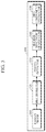
FIG. 3 is a diagram illustrating another example of a structure of a Raman probe. The Raman probe 300 of FIG. 3 may be another example of the Raman probe 100 of FIG. 1.
Referring to FIG. 3, the Raman probe 300 includes a probe head 110, a light source part 120, and a light collector 130.
The probe head 110 may come into contact with skin 10 of an object. The probe head 110 may include a concave part 111, into which the skin 10 of the object may be inserted when the probe head 110 comes into contact with the skin 10 of the object. 1 n this case, the cross-section of the concave part 111 may be formed in a trapezoidal shape. Thus, the hollow space of the concave part 111 may be shaped like a bowl with its radius increasing towards the skin 10.
The light source part 120 may be formed on a side surface of the concave part 111 and may emit light onto the skin 10 of the object inserted into the concave part 111. To this end, the light source part 120 may include alight source 121 and a filter 122.
The light source 121 may emit light of a specific wavelength, e.g., visible light or infrared light, onto the skin 10 inserted into the concave part 111. In one embodiment, the light source 121 may emit light onto the skin 10, inserted into the concave part 111, at a predetermined angle (e.g., angle of 50 degrees or greater) with respect to a vertical axis of the light collector 130.
The filter 122 may allow light of a specific wavelength to pass among lights emitted by the light source 121. In one embodiment, the filter 122 may be a clean-up filter or a band-pass filter.
The light collector 130 may be formed above the concave part 111 to collect Raman scattered light from the skin inserted into the concave part 111. To this end, the light collector 130 may include a first lens 131, a filter 132, a second lens 133, and a fiber 134.
The first lens 131 may collect Raman scattered light from the skin 10 inserted into the concave part 111. In one embodiment, the first lens 131 may be a collection collimating lens.
The filter 132 may remove a light component having the same wavelength band as the light emitted by the light source 120. In one embodiment, the filter 132 may be a notch filter or a long-pass filter.
The second lens 133 may focus light, having passed through the filter 132, onto the fiber 134, and the fiber 134 may transmit the focused light to the outside. In one embodiment, the second lens 133 may be a focusing lens.
FIG. 4 is a block diagram illustrating another example of a Raman probe.
Referring to FIG. 4, the Raman probe 400 includes a probe head 110, a light source part 120, a light collector 130, a pressure measurer 410, a notification information generator 420, and an output interface 430. Here, the probe head 110, the light source part 120, and the light collector 130 are described above with reference to FIGS. 1 to 3, such that detailed description thereof will be omitted.
The pressure measurer 410 may be formed at the probe head 110, and may measure contact pressure between the skin of an object and the probe head 110. There is no restriction on the position of the probe head 110 at which the pressure measurer 410 is formed, as long as the position does not affect emission of light and collection of Raman scattered light. That is, as long as emission of light and collection of Raman scattered light are not affected, the pressure measurer 410 may be formed at a concave part 111, or may be formed in a region of the probe head 110 other than the concave part 111.
The notification information generator 420 may determine whether the skin of an object is inserted into the concave part 111 based on the contact pressure measured by the pressure measurer 410. In one embodiment, the notification information generator 420 may determine whether the contact pressure, measured by the pressure measurer 410, exceeds a predetermined threshold value. In response to the contact pressure exceeding the predetermined threshold value, the notification information generator 420 may determine that the skin of the object is inserted into the concave part 111. By contrast, in response to the contact pressure being less than or equal to the predetermined threshold value, the notification information generator 420 may determine that the skin of the object is not inserted into the concave part 111. In this case, the predetermined threshold value may vary depending on the position of the pressure measurer 410. That is, in the case where the pressure measurer 410 is formed in a region of the probe head 110 other than the concave part 111, the predetermined threshold value may be set to a relatively high value as compared to a case where the pressure measurer 410 is formed at the concave part 111.
The notification information generator 420 may generate notification information according to a result of determination on whether the skin is inserted into the concave part 111. For example, upon determining that the skin is not inserted into the concave part 111, the notification information generator 420 may generate notification information e. “press the probe against the skin”, etc.) for inducing the skin to be properly inserted into the concave part 111. Further, upon determining that the skin is inserted into the concave part 111, the notification information generator 420 may generate notification information (e.g., “measurement is ready to be taken,” etc.) for indicating that measurement is ready to start.
The output interface 430 may output the notification information, generated by the notification information generator 420, to the outside. In one embodiment, the output interface 430 may output the generated notification information by using at least one of an acoustic method, a visual method, and a tactile method. To this end, the output interface 430 may include a display, a speaker, a vibrator, and the like.
FIG. 5 is a block diagram illustrating an example of a bio-component analyzing at s. Referring to FIG. 5, the bio-component analyzing apparatus 500 includes a Raman probe 510, a spectrometer 520, a photodetector 530, a Raman spectrum generator 540, and a bio-component estimator 550. Here, the Raman probe 510 is the same as the Raman probes 100 and 400 described above with reference to FIGS. 1 to 4, such that detailed description thereof will be omitted.
The spectrometer 520 may spatially separate the Raman red light, collected by the Raman probe 510, into different wavelengths. To this end, the spectrometer 520 may include a prism, a grating, and the like.
The photodetector 530 may receive the Raman scattered light which is separated into wavelengths by the spectrometer 520. In one embodiment, the photodetector 530 may be a photo diode, a photo transistor (PTr), a charge-coupled device (CCD), or the like. The photodetector 530 is not necessarily a single device, and may be an array of a plurality of devices.
The Raman spectrum generator 540 may generate a Raman spectrum based on the light received by the photodetector 530.
The bio-component estimator 550 may estimate a bio-component of an object by analyzing the Raman spectrum. Here, examples of the bio-component include: blood components such as blood glucose, cholesterol, triglyceride, protein, uric acid, and the like; and skin components such as collagen, keratin, elastin, and the like.
FIG. 6 is a block diagram illustrating another example of a bio-component analyzing apparatus.
Referring to FIG. 6, the bio-component analyzing apparatus 600 includes a Raman probe 510, a spectrometer 520, a photodetector 530, a Raman spectrum generator 540, a bio-component estimator 550, an input interface 610, a storage 620, a communicator 630, and an output interface 640. Here, the Raman probe 510, the spectrometer 520, the photodetector 530, the Raman spectrum generator 540, and the bio-component estimator 550 are described above with reference to FIG. 5, such that detailed description thereof will be omitted.
The input interface 610 may receive input of various operation signals from a user. In one embodiment, the input interface 610 may include a keypad, a dome switch, a touch pad (e.g., static pressure/capacitance), a jog wheel, a jog switch, a hardware (H/W) button, and the like. Particularly, the touch pad, which forms a layer structure with a display, may be called a touch screen.
The storage 620 may store programs or commands for operation of the bio-component analyzing apparatus 600, and may store data input to and output from the bio-component analyzing apparatus 600. Further, the storage 620 may store a Raman spectrum of an object which is generated by the Raman spectrum generator 540, and bio-information of an object which is estimated by the bio-component estimator 550.
The storage 620 may include at least one storage medium of a flash memory type memory, a hard disk type memory, a multimedia card micro type memory, a card type memory (e.g., an Secure Digital (SD) memory, an xD-Picture Card memory, etc.), a random access memory (RAM), a static random access memory (SRAM), a read-only memory (ROM), an electrically erasable programmable read-only memory (EEPROM), a programmable read-only memory (PROM), a magnetic memory, a magnetic disk, and an optical disc, and the like. Further, the bio-component analyzing apparatus 600 may operate an external storage medium, such as web storage and the like, which performs a storage function of the storage 620 on the Internet.
The communicator 630 may perform communication with an external device. For example, the communicator 630 may transmit, to the external device, data input by a user through the input interface 610, the Raman spectrum of an object which is generated by the Raman spectrum generator 540, and the bio-information of an object which is estimated by the bio-component estimator 550, and the like; or may receive, from the external device, various data useful for obtaining a Raman spectrum and/or estimating bio-information.
In this case, the external device may be medical equipment using the data input by a user through the input interface 610, the Raman spectrum of an object which is generated by the Raman spectrum generator 540, the bio-information of an object which is estimated by the bio-component estimator 550, and the like, a printer to print out results, or a display to display the results. In addition, the external device may be a digital TV, a desktop computer, a cellular phone, a smartphone, a tablet personal computer (PC), a laptop computer, a personal digital assistant (PDA), a portable multimedia player (PMP), a navigation, an MP3 player, a digital camera, a wearable device, and the like, but is not limited thereto.
The communicator 630 may communicate with an external device by using Bluetooth communication, Bluetooth Low Energy (BLE) communication, Near-Field Communication (NFC), wireless local area network (WLAN) communication, Zigbee communication, Infrared Data Association (IrDA) communication, Wi-Fi Direct (WFD) communication, Ultra-Wideband (UWB) communication, Ant+ communication, Wi-Fi communication, Radio Frequency Identification (RFID) communication, 3G communication, 4G communication, 5G communication, and the like. However, this is merely exemplary and is not intended to be limiting.
The output interface 640 may output the data input by a user through the input interface 610, the Raman spectrum of an object which is generated by the Raman spectrum generator 540, the bio-information of an object which is estimated by the bio-component estimator 550, and the like. In one embodiment, the output interface 640 may output the data input by a user through the input interface 610, the Raman spectrum of an object which is generated by the Raman spectrum generator 540, the bio-information of an object which is estimated by the bio-component estimator 550, and the like, by using at least one of an acoustic method, a visual method, and a tactile method. To this end, the output interface 640 may include a display, a speaker, a vibrator, and the like.
The present disclosure can be realized as a computer-readable code written on a computer-readable recording medium. Code and code segments needed for realizing the present disclosure can be easily deduced by computer programmers of ordinary skill in the art. The computer-readable recording medium may be any type of recording device in which data is stored in a computer-readable manner. Examples of the computer-readable recording medium include a ROM, a RAM, a compact disc read-only memory (CD-ROM), a magnetic tape, a floppy disc, an optical disc, and the like. Further, the computer-readable recording medium can be distributed over a plurality of computer systems connected to a network so that a computer-readable recording medium is written thereto and executed therefrom in a decentralized manner.
The present disclosure has been described herein with regard to example embodiments. However, it will be obvious to those skilled in the art that various modifications can be made without departing from the present disclosure. Therefore, it is to be understood that that the scope of the present disclosure is not limited to the above-mentioned example embodiments, but is intended to include various modifications and equivalents included within the spirit and scope of the appended claims.

Claims (11)

What is claimed is:
1. A Raman probe, comprising:
a probe head having a concave part configured to receive skin of an object being inserted into the concave part when the probe head comes into contact with the skin of the object;
a light source part disposed on a side of the concave part and configured to emit light onto the skin through the side of the concave part;
a pressure measurer formed at the probe head and configured to measure contact pressure of the skin; and
a light collector formed above a center of the concave part and configured to collect Raman scattered light when the Raman scattered light is scattered from the skin and then passes through the center of the concave part.
2. The Raman probe of claim 1, wherein the light source part is further configured to emit the light onto the skin, which is inserted into the concave part, at a predetermined angle with respect to a vertical axis of the light collector.
3. The Raman probe of claim 1, wherein the light source part comprises:
a light source configured to emit the light; and
a filter configured to allow a specific wavelength of the light to pass through among a plurality of wavelengths of the light emitted by the light source.
4. The Raman probe of claim 3, wherein the light source part further comprises a reflection mirror configured to reflect the light emitted by the light source to direct the light toward a desired position.
5. The Raman probe of claim 1, wherein the light collector comprises:
a first lens configured to collect the Raman scattered light; and
a filter configured to remove a light component having a same wavelength band as the light emitted by the light source part among the collected Raman scattered light.
6. The Raman probe of claim 5, wherein the first lens is one of a collection lens and a collimating lens.
7. The Raman probe of claim 5, wherein the filter is one of a notch filter and a long-pass filter.
8. The Raman probe of claim 5, wherein the light collector further comprises:
a second lens configured to focus the light having passed through the filter; and
a fiber configured to transmit the focused light to an outside.
9. The Raman probe of claim 1 further comprising a notification information generator configured to determine whether the skin is inserted into the concave part based on the measured contact pressure, and to generate notification information based on the determination.
10. The Raman probe of claim 9, further comprising an output interface configured to output the generated notification information.
11. The Raman probe of claim 1, wherein the light source part comprises a first light source configured to emit the light onto the skin through the side of the concave part, and a second light source configured to emit another light onto the skin through another side of the concave part, and
wherein the light collector is disposed directly above the center of concave part, and between the first light source and the second light source.
US16/281,432 2018-02-21 2019-02-21 Raman probe and bio-component analyzing apparatus using the same Active US10663405B2 (en)

Applications Claiming Priority (2)

Application Number Priority Date Filing Date Title
KR10-2018-0020575 2018-02-21
KR1020180020575A KR102574086B1 (en) 2018-02-21 2018-02-21 Raman probe

Publications (2)

Publication Number Publication Date
US20190257761A1 US20190257761A1 (en) 2019-08-22
US10663405B2 true US10663405B2 (en) 2020-05-26

Family

ID=67617785

Family Applications (1)

Application Number Title Priority Date Filing Date
US16/281,432 Active US10663405B2 (en) 2018-02-21 2019-02-21 Raman probe and bio-component analyzing apparatus using the same

Country Status (2)

Country Link
US (1) US10663405B2 (en)
KR (1) KR102574086B1 (en)

Cited By (1)

* Cited by examiner, † Cited by third party
Publication number Priority date Publication date Assignee Title
US20220172387A1 (en) * 2019-03-26 2022-06-02 Sony Group Corporation Image processing apparatus, image processing method, and image processing program

Families Citing this family (2)

* Cited by examiner, † Cited by third party
Publication number Priority date Publication date Assignee Title
KR102908182B1 (en) * 2019-12-13 2026-01-05 삼성전자주식회사 Compact raman sensor, and apparatus for estimating bio-component using the same
CN113405658A (en) * 2020-03-17 2021-09-17 谱钜科技股份有限公司 Spectrometer

Citations (19)

* Cited by examiner, † Cited by third party
Publication number Priority date Publication date Assignee Title
US3757770A (en) * 1971-02-22 1973-09-11 Bio Tel Western Physiological pressure sensing and telemetry means employing a diode connected transistor transducer
US5593899A (en) * 1993-02-25 1997-01-14 Trustees Of The University Of Pennsylvania Device and method for measuring tissue oxygenation through the skin using oxygen dependent quenching of phosphorescence
US6223063B1 (en) 1998-01-27 2001-04-24 Lightouch Medical, Inc. Method and device for tissue modulation
US20020188223A1 (en) * 2001-06-08 2002-12-12 Edward Perez Devices and methods for the expression of bodily fluids from an incision
US20040175826A1 (en) 2003-03-03 2004-09-09 Technion Research & Development Foundation Ltd. Cultured cartilage/bone cells/tissue, method of generating same and uses thereof
JP2004290226A (en) 2003-03-25 2004-10-21 Olympus Corp Glucose concentration measuring apparatus
US6882872B2 (en) 2000-02-07 2005-04-19 Matsushita Electric Industrial Co., Ltd. Biological information detecting probe, biological information measuring apparatus, fabrication method for biological information detecting probe, and method of measuring biological information
US20050248758A1 (en) * 2004-05-07 2005-11-10 Carron Keith T Raman spectrometer
US20070017666A1 (en) 2005-07-19 2007-01-25 Goenka Lakhi N Energy management system for a hybrid-electric vehicle
US7598483B2 (en) 2005-02-02 2009-10-06 Panasonic Corporation Optical element and optical measurement device using the optical element
US7873397B2 (en) 2007-06-19 2011-01-18 Richard Higgins Spectroscopic optical system
US8315681B2 (en) 2005-11-30 2012-11-20 Toshiba Medical Systems Corporation Method for noninvasive measurement of glucose and apparatus for noninvasive measurement of glucose
JP5095468B2 (en) 2008-03-28 2012-12-12 テルモ株式会社 Blood component measuring device
US20130229651A1 (en) 2010-09-20 2013-09-05 Nederlandse Organisatie Voor Toegepast- Natuurwetenschappelijk Onderzoek Tno Optical probe comprising transparent monolithic body with refracting and reflecting surface parts
US9036970B2 (en) 2009-10-08 2015-05-19 Commissariat A L'energie Atomique Et Aux Energies Alternatives Method and device for diffuse excitation in imaging
US20170119290A1 (en) * 2015-10-31 2017-05-04 Lujing Cai Method and apparatus of non-invasive biological sensing using controlled suction device
US9717425B2 (en) 2008-07-03 2017-08-01 Masimo Corporation Noise shielding for a noninvaise device
US20180064069A1 (en) * 2015-03-18 2018-03-08 Agri Advanced Technologies Gmbh Apparatus for locking in position at least one living bird for recording characteristic vibrational spectra and for sexing
EP3485802A1 (en) * 2017-11-17 2019-05-22 Samsung Electronics Co., Ltd. Bio-information measuring apparatus and bio-information measuring method

Patent Citations (20)

* Cited by examiner, † Cited by third party
Publication number Priority date Publication date Assignee Title
US3757770A (en) * 1971-02-22 1973-09-11 Bio Tel Western Physiological pressure sensing and telemetry means employing a diode connected transistor transducer
US5593899A (en) * 1993-02-25 1997-01-14 Trustees Of The University Of Pennsylvania Device and method for measuring tissue oxygenation through the skin using oxygen dependent quenching of phosphorescence
US6223063B1 (en) 1998-01-27 2001-04-24 Lightouch Medical, Inc. Method and device for tissue modulation
US6882872B2 (en) 2000-02-07 2005-04-19 Matsushita Electric Industrial Co., Ltd. Biological information detecting probe, biological information measuring apparatus, fabrication method for biological information detecting probe, and method of measuring biological information
US20020188223A1 (en) * 2001-06-08 2002-12-12 Edward Perez Devices and methods for the expression of bodily fluids from an incision
US20040175826A1 (en) 2003-03-03 2004-09-09 Technion Research & Development Foundation Ltd. Cultured cartilage/bone cells/tissue, method of generating same and uses thereof
JP2004290226A (en) 2003-03-25 2004-10-21 Olympus Corp Glucose concentration measuring apparatus
US20050248758A1 (en) * 2004-05-07 2005-11-10 Carron Keith T Raman spectrometer
US7598483B2 (en) 2005-02-02 2009-10-06 Panasonic Corporation Optical element and optical measurement device using the optical element
US20070017666A1 (en) 2005-07-19 2007-01-25 Goenka Lakhi N Energy management system for a hybrid-electric vehicle
US8315681B2 (en) 2005-11-30 2012-11-20 Toshiba Medical Systems Corporation Method for noninvasive measurement of glucose and apparatus for noninvasive measurement of glucose
US7873397B2 (en) 2007-06-19 2011-01-18 Richard Higgins Spectroscopic optical system
JP5095468B2 (en) 2008-03-28 2012-12-12 テルモ株式会社 Blood component measuring device
US9717425B2 (en) 2008-07-03 2017-08-01 Masimo Corporation Noise shielding for a noninvaise device
US9036970B2 (en) 2009-10-08 2015-05-19 Commissariat A L'energie Atomique Et Aux Energies Alternatives Method and device for diffuse excitation in imaging
US20130229651A1 (en) 2010-09-20 2013-09-05 Nederlandse Organisatie Voor Toegepast- Natuurwetenschappelijk Onderzoek Tno Optical probe comprising transparent monolithic body with refracting and reflecting surface parts
US20180064069A1 (en) * 2015-03-18 2018-03-08 Agri Advanced Technologies Gmbh Apparatus for locking in position at least one living bird for recording characteristic vibrational spectra and for sexing
US20170119290A1 (en) * 2015-10-31 2017-05-04 Lujing Cai Method and apparatus of non-invasive biological sensing using controlled suction device
EP3485802A1 (en) * 2017-11-17 2019-05-22 Samsung Electronics Co., Ltd. Bio-information measuring apparatus and bio-information measuring method
US20190150746A1 (en) * 2017-11-17 2019-05-23 Samsung Electronics Co., Ltd. Bio-information measuring apparatus and bio-information measuring method

Cited By (1)

* Cited by examiner, † Cited by third party
Publication number Priority date Publication date Assignee Title
US20220172387A1 (en) * 2019-03-26 2022-06-02 Sony Group Corporation Image processing apparatus, image processing method, and image processing program

Also Published As

Publication number Publication date
US20190257761A1 (en) 2019-08-22
KR20190100695A (en) 2019-08-29
KR102574086B1 (en) 2023-09-04

Similar Documents

Publication Publication Date Title
EP3981329A1 (en) Apparatus and method for estimating analyte concentration, and signal measuring apparatus
US11898910B2 (en) Compact Raman sensor and apparatus for estimating bio-component
US10684226B2 (en) Raman probe, Raman spectrum obtaining apparatus, and method of obtaining Raman spectrum and detecting distribution of target material using Raman probe
KR102574088B1 (en) Apparatus and method for estimating analyte concentration, Apparatus and method for generating analyte concentration estimation model
KR20200014523A (en) Apparatus and method for measuring bio-information
EP4184147B1 (en) Apparatus and method for estimating bio-information
KR102461187B1 (en) Raman probe, Raman spectrum obtaining apparatus, Raman spectrum obtaining method and target material distribution probing method using raman probe
US10663405B2 (en) Raman probe and bio-component analyzing apparatus using the same
KR20190043967A (en) Apparatus and method for obtaining individualized unit spectrum, apparatus and method for estimating biological component
US11627895B2 (en) Apparatus and method for estimating analyte concentration, and apparatus and method for generating analyte concentration estimation model
KR20230154147A (en) Apparatus and method for estimating cardiovascular information
US11439327B2 (en) Apparatus and method for measuring Raman spectrum
US10983001B2 (en) Raman probe and biological component analyzing apparatus using the same
CN114526830A (en) Apparatus and method for estimating body temperature and medical device
US10883876B2 (en) Compact spectrometer unit and bio-signal measuring apparatus
KR20200009869A (en) Apparatus and method for processing spectrum
KR20210022319A (en) Apparatus and method for estimating bio-information
US12171584B2 (en) Apparatus and method for estimating body component
KR20210151525A (en) Apparatus and method for estimating bio-information
CN112472076B (en) Lens, light source device having lens, and device for estimating analyte concentration

Legal Events

Date Code Title Description
AS Assignment

Owner name: SAMSUNG ELECTRONICS CO., LTD, KOREA, REPUBLIC OF

Free format text: ASSIGNMENT OF ASSIGNORS INTEREST;ASSIGNORS:LEE, WOOCHANG;CHANG, HO JUN;REEL/FRAME:048398/0749

Effective date: 20190214

FEPP Fee payment procedure

Free format text: ENTITY STATUS SET TO UNDISCOUNTED (ORIGINAL EVENT CODE: BIG.); ENTITY STATUS OF PATENT OWNER: LARGE ENTITY

STPP Information on status: patent application and granting procedure in general

Free format text: NON FINAL ACTION MAILED

STPP Information on status: patent application and granting procedure in general

Free format text: RESPONSE TO NON-FINAL OFFICE ACTION ENTERED AND FORWARDED TO EXAMINER

STPP Information on status: patent application and granting procedure in general

Free format text: NOTICE OF ALLOWANCE MAILED -- APPLICATION RECEIVED IN OFFICE OF PUBLICATIONS

STPP Information on status: patent application and granting procedure in general

Free format text: PUBLICATIONS -- ISSUE FEE PAYMENT VERIFIED

STCF Information on status: patent grant

Free format text: PATENTED CASE

MAFP Maintenance fee payment

Free format text: PAYMENT OF MAINTENANCE FEE, 4TH YEAR, LARGE ENTITY (ORIGINAL EVENT CODE: M1551); ENTITY STATUS OF PATENT OWNER: LARGE ENTITY

Year of fee payment: 4