US10641776B2 - Methods for measuring binding and cellular engagement of ligands with target proteins - Google Patents
Methods for measuring binding and cellular engagement of ligands with target proteins Download PDFInfo
- Publication number
- US10641776B2 US10641776B2 US15/932,335 US201815932335A US10641776B2 US 10641776 B2 US10641776 B2 US 10641776B2 US 201815932335 A US201815932335 A US 201815932335A US 10641776 B2 US10641776 B2 US 10641776B2
- Authority
- US
- United States
- Prior art keywords
- protein
- target
- binding
- compound
- denaturation
- Prior art date
- Legal status (The legal status is an assumption and is not a legal conclusion. Google has not performed a legal analysis and makes no representation as to the accuracy of the status listed.)
- Active, expires
Links
- 238000000034 method Methods 0.000 title claims abstract description 139
- 230000027455 binding Effects 0.000 title claims abstract description 115
- 238000009739 binding Methods 0.000 title claims abstract description 114
- 108090000623 proteins and genes Proteins 0.000 title claims description 214
- 102000004169 proteins and genes Human genes 0.000 title claims description 206
- 239000003446 ligand Substances 0.000 title description 53
- 230000001413 cellular effect Effects 0.000 title description 9
- 229920002521 macromolecule Polymers 0.000 claims abstract description 144
- 238000004925 denaturation Methods 0.000 claims abstract description 141
- 230000036425 denaturation Effects 0.000 claims abstract description 138
- 108090000765 processed proteins & peptides Proteins 0.000 claims abstract description 113
- 150000001875 compounds Chemical class 0.000 claims abstract description 95
- 239000012634 fragment Substances 0.000 claims abstract description 71
- 108020001507 fusion proteins Proteins 0.000 claims abstract description 71
- 102000004190 Enzymes Human genes 0.000 claims abstract description 68
- 108090000790 Enzymes Proteins 0.000 claims abstract description 68
- 102000037865 fusion proteins Human genes 0.000 claims abstract description 67
- 238000002372 labelling Methods 0.000 claims abstract description 66
- 239000000203 mixture Substances 0.000 claims abstract description 58
- 238000003174 enzyme fragment complementation Methods 0.000 claims abstract description 34
- 239000012530 fluid Substances 0.000 claims abstract description 24
- 150000003384 small molecules Chemical class 0.000 claims abstract description 23
- 238000010438 heat treatment Methods 0.000 claims description 52
- 108010005774 beta-Galactosidase Proteins 0.000 claims description 51
- 102000005936 beta-Galactosidase Human genes 0.000 claims description 47
- 239000000758 substrate Substances 0.000 claims description 30
- 102000004196 processed proteins & peptides Human genes 0.000 claims description 29
- 230000004927 fusion Effects 0.000 claims description 25
- 238000001816 cooling Methods 0.000 claims description 23
- 229920001184 polypeptide Polymers 0.000 claims description 21
- 150000001413 amino acids Chemical class 0.000 claims description 15
- 102000001805 Bromodomains Human genes 0.000 claims description 14
- 108050009021 Bromodomains Proteins 0.000 claims description 14
- -1 small molecule compound Chemical class 0.000 claims description 10
- 230000000295 complement effect Effects 0.000 claims description 7
- 238000005259 measurement Methods 0.000 claims description 7
- HEDRZPFGACZZDS-UHFFFAOYSA-N Chloroform Chemical compound ClC(Cl)Cl HEDRZPFGACZZDS-UHFFFAOYSA-N 0.000 claims description 6
- 102000004157 Hydrolases Human genes 0.000 claims description 6
- 108090000604 Hydrolases Proteins 0.000 claims description 6
- 102000001253 Protein Kinase Human genes 0.000 claims description 6
- 239000003960 organic solvent Substances 0.000 claims description 6
- 108060006633 protein kinase Proteins 0.000 claims description 6
- 230000002934 lysing effect Effects 0.000 claims description 5
- 230000005855 radiation Effects 0.000 claims description 5
- FWMNVWWHGCHHJJ-SKKKGAJSSA-N 4-amino-1-[(2r)-6-amino-2-[[(2r)-2-[[(2r)-2-[[(2r)-2-amino-3-phenylpropanoyl]amino]-3-phenylpropanoyl]amino]-4-methylpentanoyl]amino]hexanoyl]piperidine-4-carboxylic acid Chemical compound C([C@H](C(=O)N[C@H](CC(C)C)C(=O)N[C@H](CCCCN)C(=O)N1CCC(N)(CC1)C(O)=O)NC(=O)[C@H](N)CC=1C=CC=CC=1)C1=CC=CC=C1 FWMNVWWHGCHHJJ-SKKKGAJSSA-N 0.000 claims description 4
- 102000011787 Histone Methyltransferases Human genes 0.000 claims description 3
- 108010036115 Histone Methyltransferases Proteins 0.000 claims description 3
- 238000010790 dilution Methods 0.000 claims description 3
- 239000012895 dilution Substances 0.000 claims description 3
- 125000003158 alcohol group Chemical group 0.000 claims description 2
- 230000002706 hydrostatic effect Effects 0.000 claims description 2
- 239000003112 inhibitor Substances 0.000 abstract description 64
- 238000003556 assay Methods 0.000 description 108
- 210000004027 cell Anatomy 0.000 description 108
- 238000012360 testing method Methods 0.000 description 30
- IAZDPXIOMUYVGZ-UHFFFAOYSA-N Dimethylsulphoxide Chemical compound CS(C)=O IAZDPXIOMUYVGZ-UHFFFAOYSA-N 0.000 description 21
- 230000000694 effects Effects 0.000 description 21
- 230000003993 interaction Effects 0.000 description 19
- 231100000673 dose–response relationship Toxicity 0.000 description 18
- 238000005119 centrifugation Methods 0.000 description 16
- 238000001514 detection method Methods 0.000 description 15
- 101000823316 Homo sapiens Tyrosine-protein kinase ABL1 Proteins 0.000 description 14
- 238000002474 experimental method Methods 0.000 description 14
- 239000000284 extract Substances 0.000 description 14
- 102100022596 Tyrosine-protein kinase ABL1 Human genes 0.000 description 13
- 102100021975 CREB-binding protein Human genes 0.000 description 12
- 238000002844 melting Methods 0.000 description 12
- 230000008018 melting Effects 0.000 description 12
- 102000039446 nucleic acids Human genes 0.000 description 12
- 108020004707 nucleic acids Proteins 0.000 description 12
- 150000007523 nucleic acids Chemical class 0.000 description 12
- 101000896987 Homo sapiens CREB-binding protein Proteins 0.000 description 11
- 238000000670 ligand binding assay Methods 0.000 description 11
- 230000036515 potency Effects 0.000 description 11
- 238000002849 thermal shift Methods 0.000 description 11
- 239000000523 sample Substances 0.000 description 10
- ZBNZXTGUTAYRHI-UHFFFAOYSA-N Dasatinib Chemical compound C=1C(N2CCN(CCO)CC2)=NC(C)=NC=1NC(S1)=NC=C1C(=O)NC1=C(C)C=CC=C1Cl ZBNZXTGUTAYRHI-UHFFFAOYSA-N 0.000 description 9
- 238000003505 heat denaturation Methods 0.000 description 9
- 230000004568 DNA-binding Effects 0.000 description 8
- 101000979342 Homo sapiens Nuclear factor NF-kappa-B p105 subunit Proteins 0.000 description 8
- 239000002067 L01XE06 - Dasatinib Substances 0.000 description 8
- 102100023050 Nuclear factor NF-kappa-B p105 subunit Human genes 0.000 description 8
- 229960002448 dasatinib Drugs 0.000 description 8
- 229940079593 drug Drugs 0.000 description 8
- 239000003814 drug Substances 0.000 description 8
- 238000011534 incubation Methods 0.000 description 8
- 238000004020 luminiscence type Methods 0.000 description 8
- QOECJCJVIMVJGX-UHFFFAOYSA-N 2-cyclohexyl-6-methoxy-N-(1-propan-2-yl-4-piperidinyl)-7-[3-(1-pyrrolidinyl)propoxy]-4-quinazolinamine Chemical compound N1=C(C2CCCCC2)N=C2C=C(OCCCN3CCCC3)C(OC)=CC2=C1NC1CCN(C(C)C)CC1 QOECJCJVIMVJGX-UHFFFAOYSA-N 0.000 description 7
- 108010043121 Green Fluorescent Proteins Proteins 0.000 description 7
- 102000004144 Green Fluorescent Proteins Human genes 0.000 description 7
- 230000002776 aggregation Effects 0.000 description 7
- 238000004220 aggregation Methods 0.000 description 7
- 238000005516 engineering process Methods 0.000 description 7
- 239000005090 green fluorescent protein Substances 0.000 description 7
- 238000000159 protein binding assay Methods 0.000 description 7
- 239000000243 solution Substances 0.000 description 7
- 101000744394 Homo sapiens Oxidized purine nucleoside triphosphate hydrolase Proteins 0.000 description 6
- 108010052285 Membrane Proteins Proteins 0.000 description 6
- 102000016397 Methyltransferase Human genes 0.000 description 6
- 108060004795 Methyltransferase Proteins 0.000 description 6
- 102100039792 Oxidized purine nucleoside triphosphate hydrolase Human genes 0.000 description 6
- 210000000170 cell membrane Anatomy 0.000 description 6
- 238000006243 chemical reaction Methods 0.000 description 6
- 210000004962 mammalian cell Anatomy 0.000 description 6
- 239000000463 material Substances 0.000 description 6
- 230000036961 partial effect Effects 0.000 description 6
- 210000001519 tissue Anatomy 0.000 description 6
- GEPYBHCJBORHCE-SFHVURJKSA-N 4-[(2s)-1-[2-[2-(3-chloro-4-methoxyphenyl)ethyl]-5-(3,5-dimethyl-1,2-oxazol-4-yl)benzimidazol-1-yl]propan-2-yl]morpholine Chemical compound C1=C(Cl)C(OC)=CC=C1CCC1=NC2=CC(C3=C(ON=C3C)C)=CC=C2N1C[C@H](C)N1CCOCC1 GEPYBHCJBORHCE-SFHVURJKSA-N 0.000 description 5
- 108090000204 Dipeptidase 1 Proteins 0.000 description 5
- 108700040121 Protein Methyltransferases Proteins 0.000 description 5
- 102000055027 Protein Methyltransferases Human genes 0.000 description 5
- 238000004458 analytical method Methods 0.000 description 5
- 238000013459 approach Methods 0.000 description 5
- 102000006635 beta-lactamase Human genes 0.000 description 5
- 239000000872 buffer Substances 0.000 description 5
- 239000003596 drug target Substances 0.000 description 5
- 230000002255 enzymatic effect Effects 0.000 description 5
- 238000003173 enzyme complementation Methods 0.000 description 5
- UYBRROMMFMPJAN-UHFFFAOYSA-N ethyl n-[6-[3-(methanesulfonamido)-4-methylphenyl]-3-methyl-[1,2,4]triazolo[4,3-b]pyridazin-8-yl]carbamate Chemical compound N=1N2C(C)=NN=C2C(NC(=O)OCC)=CC=1C1=CC=C(C)C(NS(C)(=O)=O)=C1 UYBRROMMFMPJAN-UHFFFAOYSA-N 0.000 description 5
- 238000000338 in vitro Methods 0.000 description 5
- 230000003834 intracellular effect Effects 0.000 description 5
- 230000008569 process Effects 0.000 description 5
- 239000011541 reaction mixture Substances 0.000 description 5
- 230000002829 reductive effect Effects 0.000 description 5
- 238000000926 separation method Methods 0.000 description 5
- 239000000126 substance Substances 0.000 description 5
- 102000018697 Membrane Proteins Human genes 0.000 description 4
- 230000008901 benefit Effects 0.000 description 4
- WQZGKKKJIJFFOK-FPRJBGLDSA-N beta-D-galactose Chemical compound OC[C@H]1O[C@@H](O)[C@H](O)[C@@H](O)[C@H]1O WQZGKKKJIJFFOK-FPRJBGLDSA-N 0.000 description 4
- 150000001720 carbohydrates Chemical class 0.000 description 4
- 235000014633 carbohydrates Nutrition 0.000 description 4
- 239000003153 chemical reaction reagent Substances 0.000 description 4
- 230000001419 dependent effect Effects 0.000 description 4
- 239000013604 expression vector Substances 0.000 description 4
- 230000006870 function Effects 0.000 description 4
- 239000002609 medium Substances 0.000 description 4
- 239000002773 nucleotide Substances 0.000 description 4
- 125000003729 nucleotide group Chemical group 0.000 description 4
- 229920000642 polymer Polymers 0.000 description 4
- 108020001580 protein domains Proteins 0.000 description 4
- 230000006916 protein interaction Effects 0.000 description 4
- 230000035945 sensitivity Effects 0.000 description 4
- 238000012546 transfer Methods 0.000 description 4
- 239000013598 vector Substances 0.000 description 4
- MHFUWOIXNMZFIW-WNQIDUERSA-N (2s)-2-hydroxypropanoic acid;n-[4-[4-(4-methylpiperazin-1-yl)-6-[(5-methyl-1h-pyrazol-3-yl)amino]pyrimidin-2-yl]sulfanylphenyl]cyclopropanecarboxamide Chemical compound C[C@H](O)C(O)=O.C1CN(C)CCN1C1=CC(NC2=NNC(C)=C2)=NC(SC=2C=CC(NC(=O)C3CC3)=CC=2)=N1 MHFUWOIXNMZFIW-WNQIDUERSA-N 0.000 description 3
- 101000623895 Bos taurus Mucin-15 Proteins 0.000 description 3
- 108020004414 DNA Proteins 0.000 description 3
- 108010057466 NF-kappa B Proteins 0.000 description 3
- 102000003945 NF-kappa B Human genes 0.000 description 3
- 108091028043 Nucleic acid sequence Proteins 0.000 description 3
- YWEGXZZAORIRQR-UHFFFAOYSA-N SCH51344 Chemical compound OCCOCCNC1=C2C=C(OC)C=CC2=NC2=NNC(C)=C21 YWEGXZZAORIRQR-UHFFFAOYSA-N 0.000 description 3
- 108700019146 Transgenes Proteins 0.000 description 3
- 210000004899 c-terminal region Anatomy 0.000 description 3
- 230000003197 catalytic effect Effects 0.000 description 3
- 230000008859 change Effects 0.000 description 3
- 239000003599 detergent Substances 0.000 description 3
- 239000013024 dilution buffer Substances 0.000 description 3
- HKSZLNNOFSGOKW-UHFFFAOYSA-N ent-staurosporine Natural products C12=C3N4C5=CC=CC=C5C3=C3CNC(=O)C3=C2C2=CC=CC=C2N1C1CC(NC)C(OC)C4(C)O1 HKSZLNNOFSGOKW-UHFFFAOYSA-N 0.000 description 3
- 230000006355 external stress Effects 0.000 description 3
- 238000001914 filtration Methods 0.000 description 3
- 238000000684 flow cytometry Methods 0.000 description 3
- 230000008642 heat stress Effects 0.000 description 3
- KTUFNOKKBVMGRW-UHFFFAOYSA-N imatinib Chemical compound C1CN(C)CCN1CC1=CC=C(C(=O)NC=2C=C(NC=3N=C(C=CN=3)C=3C=NC=CC=3)C(C)=CC=2)C=C1 KTUFNOKKBVMGRW-UHFFFAOYSA-N 0.000 description 3
- 230000000670 limiting effect Effects 0.000 description 3
- 239000012139 lysis buffer Substances 0.000 description 3
- 238000004949 mass spectrometry Methods 0.000 description 3
- 239000012528 membrane Substances 0.000 description 3
- 239000002207 metabolite Substances 0.000 description 3
- 238000012544 monitoring process Methods 0.000 description 3
- 230000009871 nonspecific binding Effects 0.000 description 3
- 230000003389 potentiating effect Effects 0.000 description 3
- 230000004850 protein–protein interaction Effects 0.000 description 3
- 230000003252 repetitive effect Effects 0.000 description 3
- 238000013207 serial dilution Methods 0.000 description 3
- 230000006641 stabilisation Effects 0.000 description 3
- 238000011105 stabilization Methods 0.000 description 3
- HKSZLNNOFSGOKW-FYTWVXJKSA-N staurosporine Chemical compound C12=C3N4C5=CC=CC=C5C3=C3CNC(=O)C3=C2C2=CC=CC=C2N1[C@H]1C[C@@H](NC)[C@@H](OC)[C@]4(C)O1 HKSZLNNOFSGOKW-FYTWVXJKSA-N 0.000 description 3
- CGPUWJWCVCFERF-UHFFFAOYSA-N staurosporine Natural products C12=C3N4C5=CC=CC=C5C3=C3CNC(=O)C3=C2C2=CC=CC=C2N1C1CC(NC)C(OC)C4(OC)O1 CGPUWJWCVCFERF-UHFFFAOYSA-N 0.000 description 3
- 238000001262 western blot Methods 0.000 description 3
- MIAKOEWBCMPCQR-YBXAARCKSA-N (2s,3r,4s,5r,6r)-2-(4-aminophenoxy)-6-(hydroxymethyl)oxane-3,4,5-triol Chemical compound C1=CC(N)=CC=C1O[C@H]1[C@H](O)[C@@H](O)[C@@H](O)[C@@H](CO)O1 MIAKOEWBCMPCQR-YBXAARCKSA-N 0.000 description 2
- XRILCFTWUCUKJR-INFSMZHSSA-N 2'-3'-cGAMP Chemical compound C([C@H]([C@H]1O)O2)OP(O)(=O)O[C@H]3[C@@H](O)[C@H](N4C5=NC=NC(N)=C5N=C4)O[C@@H]3COP(O)(=O)O[C@H]1[C@@H]2N1C=NC2=C1NC(N)=NC2=O XRILCFTWUCUKJR-INFSMZHSSA-N 0.000 description 2
- ZTOBILYWTYHOJB-WBCGDKOGSA-N 3',6'-bis[[(2s,3r,4s,5r,6r)-3,4,5-trihydroxy-6-(hydroxymethyl)oxan-2-yl]oxy]spiro[2-benzofuran-3,9'-xanthene]-1-one Chemical compound O[C@@H]1[C@@H](O)[C@@H](O)[C@@H](CO)O[C@H]1OC1=CC=C2C3(C4=CC=CC=C4C(=O)O3)C3=CC=C(O[C@H]4[C@@H]([C@@H](O)[C@@H](O)[C@@H](CO)O4)O)C=C3OC2=C1 ZTOBILYWTYHOJB-WBCGDKOGSA-N 0.000 description 2
- OPIFSICVWOWJMJ-AEOCFKNESA-N 5-bromo-4-chloro-3-indolyl beta-D-galactoside Chemical compound O[C@@H]1[C@@H](O)[C@@H](O)[C@@H](CO)O[C@H]1OC1=CNC2=CC=C(Br)C(Cl)=C12 OPIFSICVWOWJMJ-AEOCFKNESA-N 0.000 description 2
- 102100029893 Bromodomain-containing protein 9 Human genes 0.000 description 2
- 238000007426 Cellular thermal shift assay Methods 0.000 description 2
- 102000005636 Cyclic AMP Response Element-Binding Protein Human genes 0.000 description 2
- 108010045171 Cyclic AMP Response Element-Binding Protein Proteins 0.000 description 2
- 239000006144 Dulbecco’s modified Eagle's medium Substances 0.000 description 2
- 238000002965 ELISA Methods 0.000 description 2
- 101000794032 Homo sapiens Bromodomain-containing protein 9 Proteins 0.000 description 2
- 239000005517 L01XE01 - Imatinib Substances 0.000 description 2
- STECJAGHUSJQJN-USLFZFAMSA-N LSM-4015 Chemical compound C1([C@@H](CO)C(=O)OC2C[C@@H]3N([C@H](C2)[C@@H]2[C@H]3O2)C)=CC=CC=C1 STECJAGHUSJQJN-USLFZFAMSA-N 0.000 description 2
- 108060001084 Luciferase Proteins 0.000 description 2
- 108090000854 Oxidoreductases Proteins 0.000 description 2
- 102000004316 Oxidoreductases Human genes 0.000 description 2
- 102000003992 Peroxidases Human genes 0.000 description 2
- 108010026552 Proteome Proteins 0.000 description 2
- 108020004511 Recombinant DNA Proteins 0.000 description 2
- 108010076089 accutase Proteins 0.000 description 2
- 239000002253 acid Substances 0.000 description 2
- 230000003281 allosteric effect Effects 0.000 description 2
- 230000004075 alteration Effects 0.000 description 2
- 238000002820 assay format Methods 0.000 description 2
- 230000004071 biological effect Effects 0.000 description 2
- 229910052799 carbon Inorganic materials 0.000 description 2
- 230000008614 cellular interaction Effects 0.000 description 2
- 239000003593 chromogenic compound Substances 0.000 description 2
- 238000010367 cloning Methods 0.000 description 2
- 230000001086 cytosolic effect Effects 0.000 description 2
- UQLDLKMNUJERMK-UHFFFAOYSA-L di(octadecanoyloxy)lead Chemical compound [Pb+2].CCCCCCCCCCCCCCCCCC([O-])=O.CCCCCCCCCCCCCCCCCC([O-])=O UQLDLKMNUJERMK-UHFFFAOYSA-L 0.000 description 2
- 238000002022 differential scanning fluorescence spectroscopy Methods 0.000 description 2
- KTEIFNKAUNYNJU-LBPRGKRZSA-N ent-crizotinib Chemical compound O([C@@H](C)C=1C(=C(F)C=CC=1Cl)Cl)C(C(=NC=1)N)=CC=1C(=C1)C=NN1C1CCNCC1 KTEIFNKAUNYNJU-LBPRGKRZSA-N 0.000 description 2
- 238000011156 evaluation Methods 0.000 description 2
- 238000001943 fluorescence-activated cell sorting Methods 0.000 description 2
- 239000007850 fluorescent dye Substances 0.000 description 2
- 239000001963 growth medium Substances 0.000 description 2
- 230000001744 histochemical effect Effects 0.000 description 2
- 229960002411 imatinib Drugs 0.000 description 2
- 238000003018 immunoassay Methods 0.000 description 2
- 229910017053 inorganic salt Inorganic materials 0.000 description 2
- 230000005865 ionizing radiation Effects 0.000 description 2
- 238000000111 isothermal titration calorimetry Methods 0.000 description 2
- 210000003292 kidney cell Anatomy 0.000 description 2
- 150000002632 lipids Chemical class 0.000 description 2
- 238000010369 molecular cloning Methods 0.000 description 2
- 108700043045 nanoluc Proteins 0.000 description 2
- 238000005457 optimization Methods 0.000 description 2
- 230000026731 phosphorylation Effects 0.000 description 2
- 238000006366 phosphorylation reaction Methods 0.000 description 2
- 108091033319 polynucleotide Proteins 0.000 description 2
- 102000040430 polynucleotide Human genes 0.000 description 2
- 239000013641 positive control Substances 0.000 description 2
- 239000002244 precipitate Substances 0.000 description 2
- 238000001556 precipitation Methods 0.000 description 2
- 238000002360 preparation method Methods 0.000 description 2
- 238000001498 protein fragment complementation assay Methods 0.000 description 2
- 238000010188 recombinant method Methods 0.000 description 2
- 238000012216 screening Methods 0.000 description 2
- 108091006024 signal transducing proteins Proteins 0.000 description 2
- 102000034285 signal transducing proteins Human genes 0.000 description 2
- 230000011664 signaling Effects 0.000 description 2
- 230000009870 specific binding Effects 0.000 description 2
- 238000010186 staining Methods 0.000 description 2
- 238000005406 washing Methods 0.000 description 2
- 108091032973 (ribonucleotides)n+m Proteins 0.000 description 1
- TYMLOMAKGOJONV-UHFFFAOYSA-N 4-nitroaniline Chemical compound NC1=CC=C([N+]([O-])=O)C=C1 TYMLOMAKGOJONV-UHFFFAOYSA-N 0.000 description 1
- CTNPALGJUAXMMC-PMFHANACSA-N 5-[(z)-(5-fluoro-2-oxo-1h-indol-3-ylidene)methyl]-n-[(2s)-2-hydroxy-3-morpholin-4-ylpropyl]-2,4-dimethyl-1h-pyrrole-3-carboxamide Chemical compound C([C@@H](O)CNC(=O)C=1C(C)=C(\C=C/2C3=CC(F)=CC=C3NC\2=O)NC=1C)N1CCOCC1 CTNPALGJUAXMMC-PMFHANACSA-N 0.000 description 1
- 102000002260 Alkaline Phosphatase Human genes 0.000 description 1
- 108020004774 Alkaline Phosphatase Proteins 0.000 description 1
- 102100029895 Bromodomain-containing protein 4 Human genes 0.000 description 1
- 101710126815 Bromodomain-containing protein 4 Proteins 0.000 description 1
- 108010040163 CREB-Binding Protein Proteins 0.000 description 1
- OKTJSMMVPCPJKN-UHFFFAOYSA-N Carbon Chemical compound [C] OKTJSMMVPCPJKN-UHFFFAOYSA-N 0.000 description 1
- 102100026735 Coagulation factor VIII Human genes 0.000 description 1
- 108020004705 Codon Proteins 0.000 description 1
- 241000699800 Cricetinae Species 0.000 description 1
- 108010025905 Cystine-Knot Miniproteins Proteins 0.000 description 1
- 108020005199 Dehydrogenases Proteins 0.000 description 1
- 241000588724 Escherichia coli Species 0.000 description 1
- LFQSCWFLJHTTHZ-UHFFFAOYSA-N Ethanol Chemical compound CCO LFQSCWFLJHTTHZ-UHFFFAOYSA-N 0.000 description 1
- 201000003542 Factor VIII deficiency Diseases 0.000 description 1
- 102000006575 G-Protein-Coupled Receptor Kinases Human genes 0.000 description 1
- 108010008959 G-Protein-Coupled Receptor Kinases Proteins 0.000 description 1
- 102000002464 Galactosidases Human genes 0.000 description 1
- 108010093031 Galactosidases Proteins 0.000 description 1
- 102000053187 Glucuronidase Human genes 0.000 description 1
- 108010060309 Glucuronidase Proteins 0.000 description 1
- 229930186217 Glycolipid Natural products 0.000 description 1
- 102000003886 Glycoproteins Human genes 0.000 description 1
- 108090000288 Glycoproteins Proteins 0.000 description 1
- 208000009292 Hemophilia A Diseases 0.000 description 1
- 102100034523 Histone H4 Human genes 0.000 description 1
- 108010033040 Histones Proteins 0.000 description 1
- 101000911390 Homo sapiens Coagulation factor VIII Proteins 0.000 description 1
- 101000945096 Homo sapiens Ribosomal protein S6 kinase alpha-5 Proteins 0.000 description 1
- 108090000144 Human Proteins Proteins 0.000 description 1
- 102000003839 Human Proteins Human genes 0.000 description 1
- UFHFLCQGNIYNRP-UHFFFAOYSA-N Hydrogen Chemical compound [H][H] UFHFLCQGNIYNRP-UHFFFAOYSA-N 0.000 description 1
- 102000008394 Immunoglobulin Fragments Human genes 0.000 description 1
- 108010021625 Immunoglobulin Fragments Proteins 0.000 description 1
- 102000004310 Ion Channels Human genes 0.000 description 1
- 108090000862 Ion Channels Proteins 0.000 description 1
- 206010022998 Irritability Diseases 0.000 description 1
- 102000004195 Isomerases Human genes 0.000 description 1
- 108090000769 Isomerases Proteins 0.000 description 1
- FBOZXECLQNJBKD-ZDUSSCGKSA-N L-methotrexate Chemical compound C=1N=C2N=C(N)N=C(N)C2=NC=1CN(C)C1=CC=C(C(=O)N[C@@H](CCC(O)=O)C(O)=O)C=C1 FBOZXECLQNJBKD-ZDUSSCGKSA-N 0.000 description 1
- 239000002137 L01XE24 - Ponatinib Substances 0.000 description 1
- 102000003960 Ligases Human genes 0.000 description 1
- 108090000364 Ligases Proteins 0.000 description 1
- 239000005089 Luciferase Substances 0.000 description 1
- 102000043136 MAP kinase family Human genes 0.000 description 1
- 108091054455 MAP kinase family Proteins 0.000 description 1
- 241000124008 Mammalia Species 0.000 description 1
- 108010063312 Metalloproteins Proteins 0.000 description 1
- 102000010750 Metalloproteins Human genes 0.000 description 1
- 108010085220 Multiprotein Complexes Proteins 0.000 description 1
- 102000007474 Multiprotein Complexes Human genes 0.000 description 1
- 101710135898 Myc proto-oncogene protein Proteins 0.000 description 1
- 102100038895 Myc proto-oncogene protein Human genes 0.000 description 1
- 108010063372 N-Glycosyl Hydrolases Proteins 0.000 description 1
- 102000010722 N-Glycosyl Hydrolases Human genes 0.000 description 1
- 206010028980 Neoplasm Diseases 0.000 description 1
- 102000007399 Nuclear hormone receptor Human genes 0.000 description 1
- 108020005497 Nuclear hormone receptor Proteins 0.000 description 1
- 108091005804 Peptidases Proteins 0.000 description 1
- 102000035195 Peptidases Human genes 0.000 description 1
- 108091093037 Peptide nucleic acid Proteins 0.000 description 1
- 108700020962 Peroxidase Proteins 0.000 description 1
- 102000045595 Phosphoprotein Phosphatases Human genes 0.000 description 1
- 108700019535 Phosphoprotein Phosphatases Proteins 0.000 description 1
- 108090001050 Phosphoric Diester Hydrolases Proteins 0.000 description 1
- 102000004861 Phosphoric Diester Hydrolases Human genes 0.000 description 1
- 108091000080 Phosphotransferase Proteins 0.000 description 1
- 206010035226 Plasma cell myeloma Diseases 0.000 description 1
- 239000002202 Polyethylene glycol Substances 0.000 description 1
- 229920001213 Polysorbate 20 Polymers 0.000 description 1
- 239000004365 Protease Substances 0.000 description 1
- 101150040459 RAS gene Proteins 0.000 description 1
- 101150076031 RAS1 gene Proteins 0.000 description 1
- 102100033645 Ribosomal protein S6 kinase alpha-5 Human genes 0.000 description 1
- 229940124639 Selective inhibitor Drugs 0.000 description 1
- 108010022394 Threonine synthase Proteins 0.000 description 1
- 101710150448 Transcriptional regulator Myc Proteins 0.000 description 1
- 102000004357 Transferases Human genes 0.000 description 1
- 108090000992 Transferases Proteins 0.000 description 1
- 241000700605 Viruses Species 0.000 description 1
- 230000009471 action Effects 0.000 description 1
- 239000000556 agonist Substances 0.000 description 1
- 238000010640 amide synthesis reaction Methods 0.000 description 1
- 230000003321 amplification Effects 0.000 description 1
- 238000012436 analytical size exclusion chromatography Methods 0.000 description 1
- 210000004102 animal cell Anatomy 0.000 description 1
- 239000005557 antagonist Substances 0.000 description 1
- 230000001093 anti-cancer Effects 0.000 description 1
- 239000000427 antigen Substances 0.000 description 1
- NETXMUIMUZJUTB-UHFFFAOYSA-N apabetalone Chemical compound C=1C(OC)=CC(OC)=C(C(N2)=O)C=1N=C2C1=CC(C)=C(OCCO)C(C)=C1 NETXMUIMUZJUTB-UHFFFAOYSA-N 0.000 description 1
- 238000003149 assay kit Methods 0.000 description 1
- 230000001580 bacterial effect Effects 0.000 description 1
- 108010002833 beta-lactamase TEM-1 Proteins 0.000 description 1
- 239000011230 binding agent Substances 0.000 description 1
- 239000013060 biological fluid Substances 0.000 description 1
- 230000008827 biological function Effects 0.000 description 1
- 230000008236 biological pathway Effects 0.000 description 1
- 238000001574 biopsy Methods 0.000 description 1
- 239000008280 blood Substances 0.000 description 1
- 210000004369 blood Anatomy 0.000 description 1
- 229940125763 bromodomain inhibitor Drugs 0.000 description 1
- 201000011510 cancer Diseases 0.000 description 1
- 230000015556 catabolic process Effects 0.000 description 1
- 238000000423 cell based assay Methods 0.000 description 1
- 238000004113 cell culture Methods 0.000 description 1
- 230000010261 cell growth Effects 0.000 description 1
- 239000013592 cell lysate Substances 0.000 description 1
- 230000006037 cell lysis Effects 0.000 description 1
- 239000008004 cell lysis buffer Substances 0.000 description 1
- 239000006285 cell suspension Substances 0.000 description 1
- 230000033077 cellular process Effects 0.000 description 1
- 239000007795 chemical reaction product Substances 0.000 description 1
- 239000003795 chemical substances by application Substances 0.000 description 1
- 210000004978 chinese hamster ovary cell Anatomy 0.000 description 1
- 238000003776 cleavage reaction Methods 0.000 description 1
- 238000005345 coagulation Methods 0.000 description 1
- 230000015271 coagulation Effects 0.000 description 1
- 230000002860 competitive effect Effects 0.000 description 1
- 238000004590 computer program Methods 0.000 description 1
- 238000012790 confirmation Methods 0.000 description 1
- 238000010276 construction Methods 0.000 description 1
- 238000007796 conventional method Methods 0.000 description 1
- 238000004132 cross linking Methods 0.000 description 1
- 239000003431 cross linking reagent Substances 0.000 description 1
- 239000000287 crude extract Substances 0.000 description 1
- LJOSBOOJFIRCSO-AWEZNQCLSA-N cs-m2721 Chemical compound N([C@@H](CC(O)=O)C1=NN=C(N1C=1SC(C)=C(C)C=11)C)=C1C1=CC=C(Cl)C=C1 LJOSBOOJFIRCSO-AWEZNQCLSA-N 0.000 description 1
- 230000001186 cumulative effect Effects 0.000 description 1
- 230000001351 cycling effect Effects 0.000 description 1
- 230000009089 cytolysis Effects 0.000 description 1
- 210000000172 cytosol Anatomy 0.000 description 1
- 230000006378 damage Effects 0.000 description 1
- 238000007405 data analysis Methods 0.000 description 1
- 230000007423 decrease Effects 0.000 description 1
- 238000006731 degradation reaction Methods 0.000 description 1
- 238000013461 design Methods 0.000 description 1
- 238000011161 development Methods 0.000 description 1
- 102000004419 dihydrofolate reductase Human genes 0.000 description 1
- 238000010494 dissociation reaction Methods 0.000 description 1
- 230000005593 dissociations Effects 0.000 description 1
- 238000009510 drug design Methods 0.000 description 1
- 230000009977 dual effect Effects 0.000 description 1
- 238000004520 electroporation Methods 0.000 description 1
- 238000011067 equilibration Methods 0.000 description 1
- 210000003527 eukaryotic cell Anatomy 0.000 description 1
- 238000000799 fluorescence microscopy Methods 0.000 description 1
- 108091006047 fluorescent proteins Proteins 0.000 description 1
- 102000034287 fluorescent proteins Human genes 0.000 description 1
- 230000002538 fungal effect Effects 0.000 description 1
- 150000008195 galaktosides Chemical class 0.000 description 1
- 210000003976 gap junction Anatomy 0.000 description 1
- 238000001415 gene therapy Methods 0.000 description 1
- 238000007429 general method Methods 0.000 description 1
- 238000010353 genetic engineering Methods 0.000 description 1
- 150000004676 glycans Chemical class 0.000 description 1
- 238000009998 heat setting Methods 0.000 description 1
- 238000013537 high throughput screening Methods 0.000 description 1
- 210000005260 human cell Anatomy 0.000 description 1
- 229910052739 hydrogen Inorganic materials 0.000 description 1
- 239000001257 hydrogen Substances 0.000 description 1
- 230000007062 hydrolysis Effects 0.000 description 1
- 238000006460 hydrolysis reaction Methods 0.000 description 1
- 230000002209 hydrophobic effect Effects 0.000 description 1
- 238000003384 imaging method Methods 0.000 description 1
- 210000001822 immobilized cell Anatomy 0.000 description 1
- 238000010166 immunofluorescence Methods 0.000 description 1
- 230000006872 improvement Effects 0.000 description 1
- 238000001727 in vivo Methods 0.000 description 1
- 230000005764 inhibitory process Effects 0.000 description 1
- 230000002427 irreversible effect Effects 0.000 description 1
- 210000003734 kidney Anatomy 0.000 description 1
- 101150066555 lacZ gene Proteins 0.000 description 1
- 150000002605 large molecules Chemical class 0.000 description 1
- 108020001756 ligand binding domains Proteins 0.000 description 1
- 210000004185 liver Anatomy 0.000 description 1
- 238000011068 loading method Methods 0.000 description 1
- 238000013507 mapping Methods 0.000 description 1
- 238000013178 mathematical model Methods 0.000 description 1
- 230000001404 mediated effect Effects 0.000 description 1
- 229960000485 methotrexate Drugs 0.000 description 1
- 238000000520 microinjection Methods 0.000 description 1
- 238000012986 modification Methods 0.000 description 1
- 230000004048 modification Effects 0.000 description 1
- 230000009149 molecular binding Effects 0.000 description 1
- 238000001823 molecular biology technique Methods 0.000 description 1
- 239000003068 molecular probe Substances 0.000 description 1
- 239000000178 monomer Substances 0.000 description 1
- 150000002772 monosaccharides Chemical class 0.000 description 1
- 201000000050 myeloid neoplasm Diseases 0.000 description 1
- UPSFMJHZUCSEHU-JYGUBCOQSA-N n-[(2s,3r,4r,5s,6r)-2-[(2r,3s,4r,5r,6s)-5-acetamido-4-hydroxy-2-(hydroxymethyl)-6-(4-methyl-2-oxochromen-7-yl)oxyoxan-3-yl]oxy-4,5-dihydroxy-6-(hydroxymethyl)oxan-3-yl]acetamide Chemical compound CC(=O)N[C@@H]1[C@@H](O)[C@H](O)[C@@H](CO)O[C@H]1O[C@H]1[C@H](O)[C@@H](NC(C)=O)[C@H](OC=2C=C3OC(=O)C=C(C)C3=CC=2)O[C@@H]1CO UPSFMJHZUCSEHU-JYGUBCOQSA-N 0.000 description 1
- 239000013642 negative control Substances 0.000 description 1
- 108020004017 nuclear receptors Proteins 0.000 description 1
- 238000003199 nucleic acid amplification method Methods 0.000 description 1
- 238000007899 nucleic acid hybridization Methods 0.000 description 1
- 230000030648 nucleus localization Effects 0.000 description 1
- 238000002515 oligonucleotide synthesis Methods 0.000 description 1
- 230000003287 optical effect Effects 0.000 description 1
- 238000004806 packaging method and process Methods 0.000 description 1
- 230000035515 penetration Effects 0.000 description 1
- 239000000816 peptidomimetic Substances 0.000 description 1
- 108091005706 peripheral membrane proteins Proteins 0.000 description 1
- 108040007629 peroxidase activity proteins Proteins 0.000 description 1
- 102000020233 phosphotransferase Human genes 0.000 description 1
- 239000013612 plasmid Substances 0.000 description 1
- 229920001223 polyethylene glycol Polymers 0.000 description 1
- 238000006116 polymerization reaction Methods 0.000 description 1
- 239000002157 polynucleotide Substances 0.000 description 1
- 235000010486 polyoxyethylene sorbitan monolaurate Nutrition 0.000 description 1
- 239000000256 polyoxyethylene sorbitan monolaurate Substances 0.000 description 1
- 229920001282 polysaccharide Polymers 0.000 description 1
- 239000005017 polysaccharide Substances 0.000 description 1
- PHXJVRSECIGDHY-UHFFFAOYSA-N ponatinib Chemical compound C1CN(C)CCN1CC(C(=C1)C(F)(F)F)=CC=C1NC(=O)C1=CC=C(C)C(C#CC=2N3N=CC=CC3=NC=2)=C1 PHXJVRSECIGDHY-UHFFFAOYSA-N 0.000 description 1
- 229960001131 ponatinib Drugs 0.000 description 1
- 125000002924 primary amino group Chemical group [H]N([H])* 0.000 description 1
- 238000012545 processing Methods 0.000 description 1
- 239000000047 product Substances 0.000 description 1
- 238000003157 protein complementation Methods 0.000 description 1
- 238000001742 protein purification Methods 0.000 description 1
- 238000000746 purification Methods 0.000 description 1
- 239000012521 purified sample Substances 0.000 description 1
- ZKDXRFMOHZVXSG-HNNXBMFYSA-N purvalanol B Chemical compound C=12N=CN(C(C)C)C2=NC(N[C@@H](CO)C(C)C)=NC=1NC1=CC=C(C(O)=O)C(Cl)=C1 ZKDXRFMOHZVXSG-HNNXBMFYSA-N 0.000 description 1
- 238000010833 quantitative mass spectrometry Methods 0.000 description 1
- ZAHRKKWIAAJSAO-UHFFFAOYSA-N rapamycin Natural products COCC(O)C(=C/C(C)C(=O)CC(OC(=O)C1CCCCN1C(=O)C(=O)C2(O)OC(CC(OC)C(=CC=CC=CC(C)CC(C)C(=O)C)C)CCC2C)C(C)CC3CCC(O)C(C3)OC)C ZAHRKKWIAAJSAO-UHFFFAOYSA-N 0.000 description 1
- 102000016914 ras Proteins Human genes 0.000 description 1
- 230000007115 recruitment Effects 0.000 description 1
- 230000009467 reduction Effects 0.000 description 1
- 238000011160 research Methods 0.000 description 1
- HSSLDCABUXLXKM-UHFFFAOYSA-N resorufin Chemical compound C1=CC(=O)C=C2OC3=CC(O)=CC=C3N=C21 HSSLDCABUXLXKM-UHFFFAOYSA-N 0.000 description 1
- 230000004044 response Effects 0.000 description 1
- 238000012552 review Methods 0.000 description 1
- 210000003705 ribosome Anatomy 0.000 description 1
- 230000007017 scission Effects 0.000 description 1
- QFJCIRLUMZQUOT-HPLJOQBZSA-N sirolimus Chemical compound C1C[C@@H](O)[C@H](OC)C[C@@H]1C[C@@H](C)[C@H]1OC(=O)[C@@H]2CCCCN2C(=O)C(=O)[C@](O)(O2)[C@H](C)CC[C@H]2C[C@H](OC)/C(C)=C/C=C/C=C/[C@@H](C)C[C@@H](C)C(=O)[C@H](OC)[C@H](O)/C(C)=C/[C@@H](C)C(=O)C1 QFJCIRLUMZQUOT-HPLJOQBZSA-N 0.000 description 1
- 229960002930 sirolimus Drugs 0.000 description 1
- 229940126586 small molecule drug Drugs 0.000 description 1
- 239000002689 soil Substances 0.000 description 1
- 125000006850 spacer group Chemical group 0.000 description 1
- 238000001228 spectrum Methods 0.000 description 1
- 230000000707 stereoselective effect Effects 0.000 description 1
- 230000009897 systematic effect Effects 0.000 description 1
- 230000008685 targeting Effects 0.000 description 1
- 238000013518 transcription Methods 0.000 description 1
- 230000035897 transcription Effects 0.000 description 1
- 108091008023 transcriptional regulators Proteins 0.000 description 1
- 238000013519 translation Methods 0.000 description 1
- 230000005945 translocation Effects 0.000 description 1
- 108091005703 transmembrane proteins Proteins 0.000 description 1
- 102000035160 transmembrane proteins Human genes 0.000 description 1
- 102000027257 transmembrane receptors Human genes 0.000 description 1
- 108091008578 transmembrane receptors Proteins 0.000 description 1
- 230000007306 turnover Effects 0.000 description 1
- XLYOFNOQVPJJNP-UHFFFAOYSA-N water Substances O XLYOFNOQVPJJNP-UHFFFAOYSA-N 0.000 description 1
- 238000001086 yeast two-hybrid system Methods 0.000 description 1
Images


Classifications
-
- G—PHYSICS
- G01—MEASURING; TESTING
- G01N—INVESTIGATING OR ANALYSING MATERIALS BY DETERMINING THEIR CHEMICAL OR PHYSICAL PROPERTIES
- G01N33/00—Investigating or analysing materials by specific methods not covered by groups G01N1/00 - G01N31/00
- G01N33/48—Biological material, e.g. blood, urine; Haemocytometers
- G01N33/50—Chemical analysis of biological material, e.g. blood, urine; Testing involving biospecific ligand binding methods; Immunological testing
- G01N33/58—Chemical analysis of biological material, e.g. blood, urine; Testing involving biospecific ligand binding methods; Immunological testing involving labelled substances
- G01N33/581—Chemical analysis of biological material, e.g. blood, urine; Testing involving biospecific ligand binding methods; Immunological testing involving labelled substances with enzyme label (including co-enzymes, co-factors, enzyme inhibitors or substrates)
-
- G—PHYSICS
- G01—MEASURING; TESTING
- G01N—INVESTIGATING OR ANALYSING MATERIALS BY DETERMINING THEIR CHEMICAL OR PHYSICAL PROPERTIES
- G01N33/00—Investigating or analysing materials by specific methods not covered by groups G01N1/00 - G01N31/00
- G01N33/48—Biological material, e.g. blood, urine; Haemocytometers
- G01N33/50—Chemical analysis of biological material, e.g. blood, urine; Testing involving biospecific ligand binding methods; Immunological testing
- G01N33/53—Immunoassay; Biospecific binding assay; Materials therefor
- G01N33/536—Immunoassay; Biospecific binding assay; Materials therefor with immune complex formed in liquid phase
- G01N33/542—Immunoassay; Biospecific binding assay; Materials therefor with immune complex formed in liquid phase with steric inhibition or signal modification, e.g. fluorescent quenching
-
- H05K999/99—
-
- G—PHYSICS
- G01—MEASURING; TESTING
- G01N—INVESTIGATING OR ANALYSING MATERIALS BY DETERMINING THEIR CHEMICAL OR PHYSICAL PROPERTIES
- G01N2333/00—Assays involving biological materials from specific organisms or of a specific nature
- G01N2333/90—Enzymes; Proenzymes
- G01N2333/914—Hydrolases (3)
- G01N2333/924—Hydrolases (3) acting on glycosyl compounds (3.2)
- G01N2333/938—Hydrolases (3) acting on glycosyl compounds (3.2) acting on beta-galactose-glycoside bonds, e.g. beta-galactosidase
-
- G—PHYSICS
- G01—MEASURING; TESTING
- G01N—INVESTIGATING OR ANALYSING MATERIALS BY DETERMINING THEIR CHEMICAL OR PHYSICAL PROPERTIES
- G01N2500/00—Screening for compounds of potential therapeutic value
- G01N2500/04—Screening involving studying the effect of compounds C directly on molecule A (e.g. C are potential ligands for a receptor A, or potential substrates for an enzyme A)
Definitions
- the present invention relates generally to methods of monitoring binding of a compound to a target macromolecule, wherein the target macromolecule is either in vitro or expressed in a transfected cell, and the cell is not isolated or separated from the target macromolecule.
- the present invention also relates to a novel method of use of enzyme fragment complementation and using an enzyme fragment complementation assay that is used in a thermal shift assay.
- the binding assays help in determining a target which should be safe and druggable.
- the multibillion dollar pharmaceutical industry depends on these assays to find a drug compound that has an ability to bind to its target protein to perform its function.
- a number of protein-ligand interaction assays have been developed and discussed in the past such as labeled and label-free ligand binding assays, structure-based ligand binding assays, thermodynamic ligand binding assays and whole cell ligand binding assays.
- fluorescence based ligand binding assay wherein a fluorescently labeled ligand binds to a target macromolecule.
- the assay is susceptible to different fluorescence interference and thus leads to undesirable alterations in the binding characteristics of the ligand.
- a radioactively labeled binding assay is popular for membrane based targets; however, the assay suffers from high cost along with hazards of handling high levels of radioactivity and thus comes with many restrictions for the lab and lab personnel working with the assay.
- NMR based analysis has also been applied to analyze the detailed structure of proteins and thus to assist in structure based drug design but suffers from a high cost of the assay and a long time required to analyze the spectra.
- a previously developed thermal shift assay also called differential scanning fluorimetry (DSF) is a thermal-denaturation assay that measures the thermal stability of a target protein and a subsequent increase in protein melting temperature upon binding of a ligand to the protein.
- the thermal stability change is measured by performing a thermal denaturation curve in the presence of a fluorescent dye, such as Sypro Orange.
- a fluorescent dye such as Sypro Orange.
- Such methods also involve a step of centrifugation and oil dispensing.
- U.S. Pat. No. 6,020,141 “Microplate thermal shift assay for ligand development and multi-variable protein chemistry optimization,” issued Feb. 1, 2000 to Pantoliano et al., discloses a thermal shift assay that comprises contacting the target molecule with one molecule of a multiplicity of different molecules in each of a multiplicity of containers, simultaneously heating the multiplicity of containers, and measuring in each of the containers a physical change associated with the thermal denaturation of the target molecule.
- Molina et al. “Monitoring Drug Target Engagement in Cells and Tissues Using the Cellular Thermal Shift Assay,” Science 5 Jul. 2013: Vol. 341 no. 6141 pp. 84-87 discloses a method that takes advantage of a shift in protein thermal stability caused by drug binding to a target protein. The method was used to monitor drug target engagement in cancer cells and in mouse livers and kidneys. However, according to the authors, the method is not likely to work for highly inhomogenous proteins or for proteins in which unfolding of the ligand-binding domain does not promote aggregation.
- the present invention comprises a method of measuring binding of a test compound to a target macromolecule. Further, the present invention discloses a homogeneous method of measuring binding of a test compound to a target protein of interest under heat denaturation of a macromolecule having a defined, native secondary and, optionally, tertiary structure, wherein the denaturation disrupts the secondary and/or tertiary structure of the target protein, but not a labeling peptide attached to it. The method may therefore be carried out in a homogenous format, without centrifugation or filtration steps between addition of the sample and readout of a result.
- the method will cause aggregation and a lack of accessibility of the labeling peptide attached to the target macromolecule; the labeling peptide may conveniently be added at the N or C terminus of the protein-based macromolecule.
- the labeling peptide may conveniently be added at the N or C terminus of the protein-based macromolecule.
- a number of short, relatively low temperature heat pulses are preferred to a single heat step; similarly, it is not preferred to completely denature a protein under study.
- the present method may be carried out with the use of a whole cell, engineered to express a fusion protein containing the target macromolecule and the labeling peptide expressed in the cell from a vector introduced into the cell.
- the cell is a eukaryotic or mammalian cell.
- the compound of interest is incubated with a whole cell preparation and is heated with the whole cells, using heating conditions described below; some or all of the cells in the preparation are lysed at the step where a second label reacts with the labeling peptide, also as described below.
- the invention comprises a step wherein detecting a signal indicates a differential between (i) denatured and (ii) non-denatured target macromolecules and thereby a differential between target macromolecules not bound to the compound and target macromolecules bound to the compound, respectively.
- the method comprises preparing a chimeric molecule that is a fusion protein comprising a labeling peptide and a protein target macromolecule.
- the method comprises preparing a fusion protein comprising a nucleotide binding domain, labeling peptide and a target macromolecule.
- the nucleotide binding domain may be a DNA binding domain.
- the target macromolecule may be a protein, a peptide, a carbohydrate or a lipid molecule.
- the present invention is a method for measuring binding between a compound and a target macromolecule, comprising: (a) preparing a fluid mixture comprising (i) an intact viable cell expressing a chimeric protein that comprises a target macromolecule linked to a labeling peptide and (ii) a compound being measured for binding to the target macromolecule, wherein said target macromolecule is subject to denaturation; (b) incubating the fluid mixture of step (a) under conditions permitting binding of said compound to said target macromolecule; (c) partially denaturing target macromolecules in the fluid mixture, after incubating in step (b), under conditions that produce a combined mixture of (i) denatured chimeric molecules not bound to the compound and (ii) non-denatured chimeric molecules bound to the compound; and (d) contacting the combined mixture with a second label that binds to and reacts with the labeling peptide in the chimeric molecule to form a detectible signal, wherein, (e) the detectible signal in step (d
- the present invention is a method as described above wherein said partially denaturing comprises heating the fluid mixture as prepared in step (b) in a step that is one of (a) multiple heating steps and at least one cooling step between heating steps and (b) a single heating step.
- the present invention is a method as described above wherein the target macromolecule is a protein.
- the present invention is a method wherein the protein further comprises an inactive exogenous polypeptide (“IEP”) linked to the protein in a distal end from the labeling peptide. The protein would have the arrangement IEP-target protein-labeling peptide.
- the present invention is a method as described above wherein the labeling peptide is between 10 and 100 amino acids in length.
- the present invention comprises a method wherein the labeling peptide is an enzyme fragment and the second label is a complementary enzyme fragment which combines with the labeling peptide to create an active enzyme (i.e. creating enzyme complementation) further comprising the step of lysing the viable cell.
- the present invention comprises a method wherein the labeling peptide is an enzyme donor (“ED”) active in enzyme fragment complementation of ⁇ -galactosidase and is fused to a terminus of a protein that is the target macromolecule.
- the present invention comprises a method as described above wherein the ED is one of SEQ ID NO: 1, SEQ ID NO: 2, or SEQ ID NO: 3.
- the present invention is a method as described above wherein the labeling peptide is an epitope tag. In other embodiments, the present invention is a method as described above wherein the compound is a small molecule. In other embodiments, the present invention is a method as described above wherein the small molecule is one that binds to an active site on the target macromolecule.
- the present invention is a method as described above wherein the step of treating the fluid mixture comprises a step of heating the fluid mixture to a temperature that is one of between (a) 25° C. and 100° C., and (b) 30° C. and 60 C.
- the present invention is a method as described above wherein a heating step comprises multiple heating steps for a defined period of time between 0.1 and 5 minutes.
- said heating step comprises applying heat to the mixture between 40° C. and 60° C., and further comprises multiple steps of heating for a time of 0.1 to 5 minutes.
- the present invention is a method as described above wherein multiple steps of heating comprises an individual cooling step between individual heating steps, of between 10 seconds and 2 minutes in duration. In other embodiments, the present invention is a method as described above comprising between three and ten cooling steps. In other embodiments, the present invention is a method as described above wherein said temperature of the mixture during a cooling step remains greater than a temperature in a previous cooling step. In other embodiments, the present invention is a method as described above wherein said cooling steps comprise actively cooling the mixture.
- the present invention is a method as described above wherein the step of denaturation comprises at least one of (a) heating, (b) hydrostatic pressure, (c) an organic solvent and (d) radiation, whereby the cell remains intact after denaturation.
- the present invention is a method as described above wherein the organic solvent is alcohol or chloroform.
- the present invention comprises a method as described further above, having steps (a) through (e), wherein steps (a) through (e) are repeated in mixtures containing different dilutions of said compound.
- the present invention comprises a method as described further above, having steps (a) through (e), wherein repeated steps (a) through (e) may be used to calculate a binding constant (K D ) of binding of compound to the target macromolecule.
- the present invention comprises a method as described above wherein the target macromolecule is a protein which is one of a bromodomain protein, a protein kinase, a hydrolase, or a histone methyltransferase.
- the present invention is a method for measuring a binding property between a small molecule compound and a target protein, comprising: (a) preparing a fluid mixture comprising a cell expressing a chimeric protein that is a fusion of a target protein and a labeling peptide that is a ⁇ -galactosidase enzyme fragment of between 10 and 100 amino acids in length, said fluid mixture further containing a small molecule compound being measured for binding to the target protein; (b) incubating the fluid mixture of step (a) under conditions permitting binding of said small molecule compound to target proteins in the cell; (c) heating the fluid mixture of step (b) under conditions that cause denaturation of proteins not bound to the compound and also cause less denaturation of proteins bound to the compound, whereby one may detect a signal from proteins bound to the compound in step (c) by adding to the mixture a second label that is a ⁇ -galactosidase fragment that reacts with the labeling peptide on protein not denatured during the heating
- the present invention is a method as described above wherein the assay mixture is prepared through steps (a) to (c) in a single container. This may be referred to as a homogeneous assay, not requiring any separation steps.
- the present invention is a method as described above wherein replicate samples containing different concentrations of compound are prepared.
- the present invention is a method as described above wherein the target macromolecule is one of a bromodomain protein and an enzyme.
- the present invention comprises a kit.
- the present invention comprises a kit comprising instructions for measuring binding of a potential ligand to a target protein, comprising components I through III, wherein component I is a target protein fused at a terminus to a first ⁇ -galactosidase enzyme fragment, component II is a ⁇ -galactosidase enzyme fragment that is complementary to the first ⁇ -galactosidase enzyme fragment, and component III is a ⁇ -galactosidase enzyme substrate, said instructions comprising instructions for: (a) preparing a fluid reaction mixture comprising a cell containing (i) a target protein fused at a terminus to a first ⁇ -galactosidase enzyme fragment (component I), and (ii) the potential ligand to the target protein; (b) heating the reaction mixture and cell of step (a) to cause at least some fraction of the target protein population to denature; (c) measuring, in the reaction mixture, after step (b),
- the present invention comprises a kit wherein the target protein is a bromodomain protein and an enzyme wherein the enzyme may be one of a protein kinase, or a histone methyltransferase.
- the present invention comprises a kit wherein the first ⁇ -galactosidase enzyme fragment is essentially identical to one of SEQ ID NO: 1, SEQ ID NO: 2, or SEQ ID NO: 3.
- the present invention comprises a kit wherein said substrate is chromogenic, fluorescent, or chemiluminescent and which generates a signal in the presence of an active ⁇ -galactosidase but not inactive ⁇ -galactosidase.
- FIG. 1 is a schematic representation of the present assay showing how a tagged macromolecule (e.g. a fusion protein and labeling peptide) is in native form (i.e. native secondary and tertiary structure) protected from denaturation in the presence of a test compound that binds to the tagged native macromolecule; the protection therefore enables detection of the binding with an enzyme complementation assay.
- a tagged macromolecule e.g. a fusion protein and labeling peptide
- native form i.e. native secondary and tertiary structure
- FIG. 2 is a schematic representation of the assay principle as in FIG. 1 , except that in this situation the test compound does not bind to the tagged native macromolecule, resulting in denaturation of the tagged native macromolecule and further making the tag (labeling peptide) inaccessible for complementation.
- FIG. 3 shows a graph of a fusion protein of BRD4(1) -ProLabelTM enzyme donor (ED) in a crude cell extract incubated with an indicator (substrate) and then heated at about 45° C. for 30 seconds.
- An inhibitor that binds to the BRD4 (1) is added at different concentrations between 0 and 100 uM. It can be seen that a dose-response protection of the fusion/BRD4(1) is achieved by binding of the inhibitor.
- the construct used is NF ⁇ B DNA Binding Domain—Linker-BRD/1(1)—Linker ED.
- the NF ⁇ B DNA binding domain sequence in the fusion protein does not play an active role in these examples.
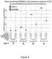
- FIG. 4 is a plot comparing a non-inhibitor DMSO (labeled “2”) with JQ1, (6S)-4-(4-Chlorophenyl)-2,3,9-trimethyl-6H-thieno[3,2-f][1,2,4]triazolo[4,3-a][1,4]diazepine-6-acetic acid, 1,1-dimethylethyl ester, known to be a selective BET bromodomain inhibitor.
- the enzyme complementation ability of the BRD4(1) is reduced by a significant amount at temperatures between 45 and 55° C., while the presence of JQ1 at 10 uM protects BRD4(1) from denaturation and enzyme complementation is reduced to a much lesser extent.
- FIG. 5 is a plot that also shows protection of BRD4(1) at 45, 50 and 55° C. in the presence or absence of JQ1 binding to the BRD4(1). As shown at the arrow, the protection from heat denaturation at 45° C. is more significant than at 25, 50, or 55° C. The incubation at 25° C. was run as a control, not to indicate thermal denaturation.
- FIG. 6 is a plot of the RLU (relative luminescence units) showing that little difference is seen whether or not centrifugation is carried out, as shown by the closely spaced spots at 602 and 604.
- FIG. 7 is a pair of bar graphs showing dose response relationships in the ligand binding assay with potent BRD4(1) inhibitor JQ1 (top figure) and a known, less potent BRD4(1) inhibitor GW334556X (bottom figure).
- BRD4(1)-ProLabelTM enzyme donor (ED) fusion protein in a crude cell extract was incubated with an indicator and heated to a target temperature in a PCR thermal cycler machine.
- the asterisks indicate the K D (dissociation constant) concentration as measured by the BROMOscan® Technology Platform available from DiscoveRx Corp. This shows that the present assay can determine dose-response relationships. The coefficient of variance observed here was less than 1%. The assay is also very reproducible.
- GW334556X is further described in Chung et al. J. Med. Chem. (2012) 55: 576.
- FIG. 8A, 8B is pair of graphs showing an example of a pulse-denaturation protocol.
- FIG. 8A shows a standard denaturation profile of 1 cycle, with a high temperature (45° C.) over a time period of 3 minutes;
- FIG. 8B shows a pulse denaturation profile of 25 pulses at a reduced temperature of 40° C., where each pulse represents a brief denaturation pulse time of 7 seconds (i.e. a period of heating that is 7 seconds), where a PCR thermal cycler is reset to a lower temperature (e.g. ambient temperature) after a heating pulse.
- a lower temperature e.g. ambient temperature
- FIG. 9 is a graph showing a mathematical model calculating an assay window versus the number of denaturation cycles. It shows that the assay window grows exponentially with cycle number.
- “Assay window” is defined as luminescence signal in the presence of known ligand divided by luminescence signal in the absence of known ligand after x cycles.
- the model assumes that a low fraction of folded protein denatures each cycle and cannot refold/reactivate on subsequent cycles.
- the model assumes that, in each cycle, the known ligand rescues 50% of the protein from denaturation.
- the model also assumes that compound binding returns to room temperature levels each cycle during temperature shift from set point to room temp.
- the degree to which compound binding protects the protein from denaturation and the total amount of denaturation for the no compound control denaturation/naturation indicates the assay window.
- An assay window may be estimated from data presented here by comparing the base line signal (RLU) to the maximum signal in a given experiment.
- FIG. 10 is a graph comparing a “standard” (constant heat) protocol with a pulse protocol.
- the assay window for the pulse denaturation is shown in black circles.
- the pulse system results, over time, in an increased assay window in comparison to the standard denaturation, which is shown in white circles.
- Thermal melting assay windows for both “standard” and “pulse” protocols were measured for the BRD9-Bromosporine interaction.
- Bromosporine is N-[(6-3-Methanesulfonamido-4-methylphenyl)-3-methyl-[1,2,4]triazolo[4,3-b]pyridazin-8-yl]carbamate, commercially available from Tocris Biosciences.
- Protein samples were exposed to a standard single heat denaturation step (“standard”) or to repetitive 0.5 minute heat pulses.
- standard single heat denaturation step
- a 45° C. denaturation temperature was used for both protocols.
- Total denaturation time is the total amount of time that the protein sample was exposed to 45° C., so one single 3 minute step with the standard protocol (i.e. no pulsing, but a single 3 minute heating step at a constant temperature) is equivalent to 6 repetitive 0.5 minute steps with the pulse protocol. That is, six 0.5 heat pulses may be compared to one 3 minute heating step.
- FIG. 11 is a graph showing different signals in RLU (relative luminosity units) comparing pulse denaturation versus standard denaturation for binding of SGC-CBP30 (a CREBBP/EP300-selective chemical probe), present at different concentrations, to CREBBP (Gene ID 1387, NCBI).
- SGC-CBP30 is commercially available from Tocris Biosciences, and is 8-(3-chloro-4-methoxy-phenethyl)-4-(3,5-dimethyl-isoxazol-4-yl)-9-(2-(morpholin-4-yl)-Dose response curves were measured for CREBBP with SGC-CBP30 with the “standard protocol” at 45° C.
- FIG. 12 is a graph showing dose response curves for seven inhibitors of the ABL1 protein kinase.
- the dose response curves were obtained using ED-tagged ABL1 and the inhibitors in a pulse denaturation protocol. It shows, for example, that one compound tested, dasatinib (N-(2-chloro-6-methylphenyl)-2-[[6-[4-(2-hydroxyethyl)-1-piperazinyl]-2-methyl-4-pyrimidinyl]amino]-5thiazolecarboxamide, monohydrate), binds to the test protein, ABL1, to a significant degree at a 1 nM concentration, whereas other compounds, such as imatinib (4-[(4-methylpiperazin-1-yl)methyl]-N-[4-methyl-3-[(4-pyridin-3-ylpyrimidin-2-yl)amino]phenyl]benzamide), do not bind at 1 nM to a significant effect, as measured by the present heat
- FIG. 13 is a graph showing a dose response curve for UNC-0638 (2-Cyclohexyl-6-methoxy-N-[1-(1-methylethyl)-4-piperidinyl]-7-[3-(1-pyrrolidinyl)propoxy]-4-quinazolinamine, CAS 1255580-76-7), an inhibitor of the G9a protein methyltransferase.
- the dose response curve was obtained using ED-tagged G9a and the inhibitor in a pulse denaturation protocol.
- FIG. 14 is a schematic representation of an in-cell embodiment of the present assay, showing a cell 1106 transfected with an enzyme fragment tagged protein of interest; the cell has been modified to overexpress a fusion protein comprising a peptide label (PL small enzyme fragment of ⁇ galactosidase) and a target macromolecule.
- the cell is incubated with a potential ligand 1104, which can cross the plasma membrane of the cell.
- the compound is being evaluated for binding to the protein of interest.
- the cell and ligand are then subjected to pulse denaturation as described above.
- the tagged macromolecule e.g.
- a fusion protein/nucleic acid and labeling peptide in the cell binds to the test, compound, it is protected from denaturation, as shown at 1108, and the PL can react with the EA (enzyme acceptor, i.e. large fragment of ⁇ -galactosidase to form an active ⁇ -galactosidase enzyme that, as shown here, will produce a luminescent readout (“light”).
- EA enzyme acceptor, i.e. large fragment of ⁇ -galactosidase to form an active ⁇ -galactosidase enzyme that, as shown here, will produce a luminescent readout (“light”).
- EA enzyme acceptor, i.e. large fragment of ⁇ -galactosidase to form an active ⁇ -galactosidase enzyme that, as shown here, will produce a luminescent readout (“light”).
- the tagged macromolecule did not bind to the test ligand (or if the test compound cannot cross
- the binding and non-binding status shown at 1108 and 1110, respectively, are present in a state of disequilibrium, and a binding curve can be constructed with different concentrations of test compound that are bound to the tagged macromolecule expressed in the recombinant cell.
- the present methods may also be used to evaluate the ability of a test compound to cross the plasma membrane of an engineered cell as described. This provides information regarding bioavailability as well as binding properties.
- FIG. 15 shows an exemplary in-cell pulse assay using inhibitors Dasatinib and VX-680 tested for binding to ABL1 tyrosine kinase.
- FIG. 16 shows an exemplary in-cell pulse assay using inhibitor JQ1 on BRD4(1) bromodomain.
- FIG. 17 shows an exemplary in-cell pulse assay using MTH1 hydrolase domain and inhibitor SCH 51344 using two preliminary denaturation protocols indicated in the legend.
- FIG. 18 shows an exemplary in-cell pulse assay using G9a methyltransferase catalytic domain and inhibitor UNC0638.
- any range set forth is intended to include any sub-range within the stated range, unless otherwise stated.
- a range of 2 minutes to 8 minutes includes 3-4 minutes, 2-7 minutes, etc.
- a temperature range of 40-45° C. includes 41-45° C., 42-43° C., etc.
- the term “about” has its ordinary meaning of approximately and may be determined in context by experimental variability. In case of doubt, the term “about” means plus or minus 5% of a stated numerical value.
- peptide refers to any polymer compound produced by amide formation between an ⁇ -carboxyl group of one amino acid and an ⁇ -amino group of another group.
- a peptide may be a labeling peptide, of relatively small size, having little or no secondary structure (i.e. a loop) linked to a macromolecule and for detecting the fusion.
- protein refers to polypeptides of specific sequence of more than about 50 residues. While all proteins are peptides, the term “peptide” generally refers to a fragment of a protein; the term “fusion protein” is used to refer to both fusion proteins and fusions with peptides, such as a fusion with a labeling peptide, e.g. an ED.
- fusion protein is used to refer to both fusion proteins and fusions with peptides, such as a fusion with a labeling peptide, e.g. an ED.
- the protein under study need not be a full length protein sequence.
- the target macromolecule may in fact be a protein that has been truncated to isolate a domain under study, modified for easier handling, etc. Thus a protein fragment in the present assay is referred to for simplicity as a “protein”.
- fusion protein refers to a protein created through genetic engineering from two or more proteins/peptides. In general, this is achieved by creating a “fusion gene”, a nucleic acid that encodes the fusion protein. For example, a fusion gene that encodes a fusion protein may be made by removing the stop codon from a first DNA sequence encoding the first protein, then appending a DNA sequence encoding the second protein in frame. The resulting fusion gene sequence will then be expressed by a cell as a single fusion protein. Fusion proteins may include a linker (or “spacer”) sequence which can promote appropriate folding and activity of each domain of the fusion protein.
- Fusion proteins may also include epitope tags (labeling peptide) for identification (e.g., in western blots, immunofluorescence, etc.) and/or purification.
- epitope tags labeling peptide
- Non-limiting examples of epitope tags in current use include: HA, myc, FLAG, and 6-HIS. These known epitope tags are relatively short peptides that can be specifically detected by monoclonal antibodies, i.e. a second label that binds to the epitope tag attached to the target macromolecule.
- the fusion protein will be “chimeric” if the molecule contains two sequences that are not normally found together in the same polypeptide chain.
- a chimeric molecule may also contain a fusion of two different polymers, such as a single polypeptide chain comprising the target macromolecule and the labeling peptide.
- the chimeric molecule may also contain a labeling peptide chemically linked to the target macromolecule.
- a chimeric molecule as defined may contain a target macromolecule linked or fused to a labeling peptide, and further wherein said target macromolecule is fused or linked to a third polypeptide that is used to increase the effective concentration of the target macromolecule when partial denaturation and detection are carried out.
- a protein used in the assay will often be a human protein of interest as a drug target, and will be prepared by recombinant methods in an active form and containing known protein binding sites.
- protein specifically includes fragments of proteins that are not full length proteins, but contain only a fragment of sufficient structure to have the requisite secondary and tertiary structure and have the binding site to the compound of interest.
- target macromolecule refers to various macromolecules that can be denatured. That is, they have a secondary or tertiary structure that can be eliminated by heat, or, alternatively, other agents. They are, for example DNA, RNA, and/or proteins (which includes protein fragments such as protein domains).
- the “macromolecule” may be of a relatively small MW compared to a full length protein, provided that it has a native three dimensional structure that is rigidly defined by cross linking, hydrogen binding or the like.
- knottin small peptides have a rigid, defined tertiary structure that could be measured by the present assay.
- macromolecule refers to a polynucleotide, polypeptide or a complex carbohydrate having a defined tertiary structure.
- glycans often present as glycoproteins or glycolipids, form highly complex structures. In mammals ten monosaccharides are utilized in building glycoconjugates in the form of oligo- (up to about a dozen monomers) and polysaccharides.
- the present “macromolecule” is one that can be denatured by destroying in significant part such three dimensional structure.
- a “target” macromolecule is a macromolecule capable of binding specifically to a third molecule, typically a small molecule or other pharmaceutical drug candidate. The target macromolecule used in the present assays may be purified, present in a cell extract, or in other forms.
- a target macromolecule may have a specific binding partner in nature, and a small molecule is intended to target this binding.
- small molecule is art-recognized and refers to a composition which has a molecular weight of less than about 2000 amu, or less than about 1000 amu, and even less than about 500 amu.
- Small molecules may be, for example, nucleic acids, peptides, polypeptides, peptide nucleic acids, peptidomimetics, carbohydrates, lipids or other organic (carbon containing) or inorganic molecules.
- Many pharmaceutical companies have extensive libraries of chemical and/or biological mixtures, often fungal, bacterial, or algal extracts, which can be screened with any of the assays of the invention.
- small organic molecule refers to a synthetic or purified natural small molecule that is often identified as being an organic or medicinal compound, and does not include molecules that contain nucleic acids, peptides or polypeptides.
- binding refers to the binding of small molecules, proteins or compounds to the proteins in a cell or in a solution.
- binding partner or “member of a binding pair” refer to molecules that specifically bind other molecules to form a binding complex such as antibody-antigen, lectin-carbohydrate, nucleic acid-nucleic acid, biotin-avidin, etc.
- the binding is predominantly mediated by non-covalent (e.g. ionic, hydrophobic, etc.) interactions and is between a small molecule and its target and/or two proteins that specifically bind to each other during a cellular process.
- enzyme fragment complementation involves the use of one enzyme fragment, which may be referred to as a labeling peptide or an ED (enzyme donor), which is not enzymatically active until it is complexed with another enzyme fragment, termed an EA, or enzyme acceptor, or second label.
- EFA enzyme acceptor
- EFC or labeling peptide is not limited to a ⁇ -galactosidase system.
- EFC is a generic term to describe the combination of enzyme fragments to form active enzyme, followed by detection of that activity by measurement of a hydrolysis product, generally by colorimetric, fluorometric or chemiluminescent methods. It has the advantage of providing an amplification step, due to enzyme turnover, as part of the detection system.
- EFC assays based either on dihydrofolate reductase or beta-lactamase have been used to quantify the effects of the drug rapamycin on its target in living cells (Remy, I. and Michnick, S. W., Clonal Selection and In Vivo Quantitation of Protein Interactions with Protein Fragment Complementation Assays. Proc Natl Acad Sci USA, 96: 5394-5399, 1999; Galarneau, A., Primeau, M., Trudeau, L.-E. and Michnick, S. W., A Protein fragment Complementation Assay based on TEM1 ß-lactamase for detection of protein-protein interactions, Nature Biotech.
- EFC useful here is Promega NanoBiTTM.
- This technology employs a NanoLuc, a small (19 kDa) luciferase enzyme engineered for structural stability and the ability to generate an intense, steady, bioluminescent signal. Details are described in Dixon et al., “NanoLuc Complementation Reporter Optimized for Accurate Measurement of Protein Interactions in Cells,” ACS Chem. Biol., Just Accepted Manuscript, Publication Date (Web): Nov. 16, 2015.
- NanoBiT subunits i.e., 1.3 kDa peptide, 18 kDa polypeptide
- weakly associate so that their assembly into a luminescent complex is dictated by the interaction characteristics of the target proteins onto which they are appended.
- labeling peptide means a peptide having essentially no secondary structure (i.e. random coil) and which functions as a label for detection of a protein or protein fragment (e.g. target peptide) fused thereto and having essentially no effect on the stability of the target peptide to which it is fused.
- the labeling peptide will generally be less than 100 amino acids in length. It may itself function as a label, or it may provide an epitope for antibody recognition. As explained below, the labeling peptide is selected so as not to affect the stability of a fusion partner of the labeling peptide.
- ED means an enzyme donor fragment for use in a ⁇ -galactosidase enzyme fragment complementation assay. Examples of EDs are given below. An ED that is “essentially identical” to one of SEQ ID NOs: 1-3 will be 100% identical except for up to two amino acid alterations.
- EA as is known in the art, means an enzyme acceptor fragment for use in a ⁇ -galactosidase enzyme fragment complementation assay.
- the term “denaturation” is used in its conventional sense to refer to a process in which proteins, nucleic acids or other macromolecules or macromolecular structures (e.g. ribosomes) lose their quaternary structure, tertiary structure and or secondary structure, at least in part. Loss of this native state in the present method occurs by application of some external stress or compound such as a strong acid or base, a concentrated inorganic salt, an organic solvent (e.g., alcohol or chloroform), radiation or heat.
- the term used here specifically includes partial denaturation, where only a fraction of the molecules (e.g. proteins) in a mixture are denatured.
- protein “melting,” which refers to protein denaturation is also used herein. As is known, a melting temperature (Tm) may be determined from a protein denaturation study. See, for details, US 2013/0217137.
- pulse denaturation refers to a process in which proteins or nucleic acids denature by application of more than one cycle of a short heat pulse which is followed by short re-equilibrium at room temperature or below room temperature.
- a “mild temperature” is considered to be a temperature at least about 5° C. less than the melting temperature of the macromolecule being heated.
- a protein melting point can be determined by known methods, as in e.g. US 20140057368, referred to below.
- a pulse denaturation protocol may cause a small amount of denaturation in a given step (5%-10%). However, the denaturation is cumulative over multiple cycles, and may reach 80% or more denaturation after the pulses are applied.
- a pulse denaturation protocol may comprise 10-70 pulses at 37-50° C. for 5-10 seconds, separated by 15-20 second cooling pulses.
- the pulse denaturation may be carried out under conditions where the fraction of the protein denatured in one cycle does not refold when cooled or upon additional pulse denaturation steps.
- protein denaturation refers to denaturation of proteins involving the disruption and possible destruction of both the secondary and tertiary structures.
- Denaturation disrupts the normal alpha helix and beta sheets in a protein and uncoils it into a random shape resulting in precipitation or coagulation of the protein. Denaturation of proteins results in loss of their biological function and/or activity. Denaturation may occur by some external stress or compound such as a strong acid or base, a concentrated inorganic salt, an organic solvent, radiation, heat or cold. Denaturation can be complete or partial but, for the present purposes, is sufficient to cause insolubility. Pressure denaturation of proteins is described, e.g. in Johnson et al., “Pressure and Protein Denaturation,” J. Biol. Chem. 1946, 163:689-698. Frick, “Effect of Ionizing Radiation on Protein Denaturation,” Nature 169, 965-966 (7 Jun. 1952) describes the use of ionizing radiation to denature solutions of proteins.
- active exogenous polypeptide or “inactive exogenous polypeptide sequence” refers to a sequence engineered to be expressed in a chimeric protein used in the present methods that is added to provide additional sequence to the fusion protein without participating in or affecting the binding events in the assay. It is used to modulate the denaturation of the chimeric protein.
- This polypeptide may be of any length, e.g. 20-1000 amino acids long and is exogenous in the sense that it is not part of the fusion protein unless artificially coded, and, furthermore, it does not participate in binding of the peptide label to the second label. In addition, it does not bind to the compound of interest.
- thermal stability refers to a quality of a macromolecule such as a protein to resist irreversible change in its chemical or physical structure at a high relative temperature.
- thermal shift assay refers to an assay based on the principles that a purified protein will denature and unfold at a particular temperature and that the binding of a ligand to a protein will thermally stabilize the protein. Further details on carrying out various thermal shift assays may be found in Nordlund, US 20140057368, “Methods for determining ligand binding to a target protein using a thermal shift assay.” As described there, such assays may be carried out with non-purified samples.
- homogenous is used in its standard sense, to refer to an assay format and method that does not require a separation step. This allows measurement of results by a simple mix and read procedure, without the need to process samples by separation or washing steps during the assay.
- CETSA Cellular Thermal Shift Assay
- the present methods can be used to measure other binding interactions, such as described in Huber et al., “Proteome-wide drug and metabolite interaction mapping by thermal-stability profiling,” Nature Methods 12, 1055-1057 (2015). There, the authors show that in combination with quantitative mass spectrometry, the approach allows for the systematic survey of protein engagement by cellular metabolites and drugs. The authors profiled the targets of the drugs methotrexate and (S)-crizotinib and the metabolite 2′3′-cGAMP in intact cells and identified the 2′3′-cGAMP cognate transmembrane receptor STING, involved in immune signaling.
- the present invention relates to a patent application by the present inventors, entitled “Homogeneous Thermal Ligand Binding Assay”, cited herein as a related patent application. It describes a biochemical (in vitro, not intracellular) method for using recombinant target proteins fused to a short “Enzyme Donor” (ED) peptide derived from beta-galactosidase for detection of ligand-protected target protein after heat denaturation.
- ED Enzyme Donor
- ProLabelTM enzyme donor or PL can be detected at femtomolar concentrations in homogeneous assays by the simple addition of the beta-galactosidase Enzyme Acceptor (EA) protein, which complements ED in trans leading to robust beta-galactosidase enzyme activity (Enzyme Fragment Complementation (EFC)).
- EA beta-galactosidase Enzyme Acceptor
- EFC Endogen Fragment Complementation
- This detection system is not only sensitive but also has a linear dynamic range of 4-5 orders of magnitude.
- ED-labeled proteins that denature/aggregate due to thermally-induced unfolding are significantly less competent to complement EA and are thus less active in this format.
- the impractical centrifugation step required in the Science paper cited above is not required, nor are immuno-assay or mass spectroscopy-based readouts.
- the invention is distinguished from the use of GFP fusions (see citation above) that simply act as a detection tag, since the ED tag in our system is both a detection tag and also reports on the folded state of the protein.
- target macromolecules such as proteins
- the methods described herein can be applied to target macromolecules, such as proteins, in a cellular milieu. That is, the target macromolecule can be expressed within a cell (or cells in a tissue), preferably a mammalian cell, and then the compound can be exposed to the cell in an intact state, i.e. crossing the intact plasma membrane.
- the present method of heat denaturation in the presence of a test compound can be carried out while the target molecule and compound are contained in a living cell.
- the term “in” a cell is used broadly to refer to a target macromolecule that is entirely inside a cell, e.g. in the cytosol, nucleus, internal membrane, etc. or a macromolecule that is attached to the plasma membrane of the cell.
- the target molecule may have an extracellular domain.
- the present method which is being developed commercially under the name “InCELL Pulse”TM system, comprises heating of cells to produce a thermal shift in enzyme complementation, as exemplified in FIGS. 14-18 .
- the InCELL Pulse system can quantitatively detect intracellular binding of small molecule ligands to cytosolic target protein domains expressed as ED-enzyme fragment (e.g. PL) fusions, and has been demonstrated with multiple target classes, including kinases, bromodomain proteins, methyltransferases, and hydrolases. This method can also be applied to integral membrane proteins, for which cellular penetration of the test ligand may or may not be required to achieve target engagement.
- ED-enzyme fragment e.g. PL
- the present Examples 13-16 use single heat pulses as opposed to a series of short heat pulses (pulse denaturation), which is used in previous examples herein.
- the cell-free biochemical application of this technology e.g. Examples 8-11
- the cell-free biochemical application of this technology has been demonstrated by use of an N-terminal fusion of the target protein to the NF ⁇ B DNA binding domain (Examples not shown).
- the addition of an extra amino acid sequence (termed below an “inactive exogenous polypeptide”) enhances assay performance in some cases. It is expected that the NF ⁇ B moiety can be more critical for the cell-free assays (e.g. Examples 8-11) than for the cellular assays (e.g.
- the inactive exogenous polypeptide may be linked to an N terminus of the target macromolecule (protein) and the labeling peptide may be linked to the C terminus of the target macromolecule.
- the present method monitors and measures a binding property between a compound and a target macromolecule. It exploits the known ability of ligands such as small molecules to protect macromolecules such as proteins from denaturation and aggregation due to unfolding and shows that this method can be applied to evaluation of ligands within a whole cell, e.g. a mammalian cell that has been transfected with a chimeric protein.
- ligands such as small molecules to protect macromolecules such as proteins from denaturation and aggregation due to unfolding
- Prior art methods have required high-g-force centrifugation steps and insensitive readouts.
- the present methods exemplified by the use of ⁇ -galactosidase enzyme fragment complementation, utilize a small peptide (less than 100 amino acids) fused to the macromolecule.
- the small peptide may be the known enzyme donor, the ProLabelTM enzyme donor (“ED”) commercially available from DiscoveRx Corporation, Fremont Calif. Enzyme fragment complementation provides a sensitive, facile generic readout. Moreover, the assay may be carried out in a homogeneous format, meaning that no physical separation of the reagents is required, eliminating the need for filtration, decanting, centrifugation, etc. The present invention is widely applicable to a number of ligand binding assays.
- the ligand may typically be a small molecule that is under study for binding properties to a target molecule, which will be a macromolecule that is susceptible to thermal denaturation and resultant loss of buoyancy or stability in a fluid.
- the binding property may be measured in the presence of a competitor or under different target macromolecules.
- BRD4 bromodomain-containing protein 4
- the present methods may also be carried out with a series of heating pulses separated by cooling.
- the heat applied to the above described mixture of proteins and (potential) ligands serves, as described above to denature the protein; the resulting activity may be measured as shown in FIGS. 1 and 2 .
- the described methods may include a pulsed denaturation protocol where protein samples (+/ ⁇ ligand) are subjected to several cycles (e.g. 10-200 cycles) of a short heat pulse at mild temperature which is followed by short re-equilibration at room temperature or below.
- These “pulse” protocols are easily carried out by using standard programmable thermal cycler instrumentation. This method avoids the need for high denaturation temperatures and long denaturation times and therefore increases the sensitivity of the method for detection of inhibitor binding.
- T m melting temperature
- Proteins are one of the most studied and most targeted macromolecules in the pharmaceutical industry. A number of approaches have been designed and invented to study proteins, their structure, their chemistry, protein-protein interactions, protein-test compound interactions, and biological pathways where proteins participate, and to determine small molecule binding to proteins of interest. However, all these processes and measurements require proteins to be stable and active as proteins are susceptible to degradation or aggregation by a number of factors.
- Macromolecules such as proteins denature when exposed to high temperatures, leading to their precipitation and aggregation. Binding of a compound to a macromolecule can increase its thermal stability and hence can be explored for a binding assay wherein one can measure a target macromolecule binding to a compound by measuring its thermal stability.
- a compound that binds at an active or allosteric site of a macromolecule such as a protein will form a complex and influence its thermal stability. This will lead to the compound being stable even at higher temperatures.
- a thermal shift assay measures the thermal stability of a target protein and a subsequent increase in protein melting temperature upon binding of a ligand to the protein.
- a number of assays have been designed to study the thermal stability of proteins and investigate buffer conditions, ligands, co-factors, drugs and other compounds affecting this stability to identify protein complexes and further characterize them.
- An alternative format has also been disclosed to measure ligand binding to a target protein using a thermal shift assay.
- the assay works on both purified and non-purified protein samples even at a low concentration.
- the assay requires that denatured, precipitated proteins be removed by centrifugation and stabilized proteins be quantified by low dynamic range immune-assays such as Western blot or ELISA.
- the present invention discloses a homogeneous binding assay to determine ligand macromolecule interactions, and further, how these interactions result in stabilization of the macromolecule. More specifically, the present invention discloses a homogeneous binding assay to determine ligand protein interactions, and further, how these interactions result in stabilization of the protein under heat stress.
- a macromolecule 101 having a secondary or tertiary structure is linked chemically to a tag 102 , which may be an enzyme fragment and may be a fusion protein with a protein macromolecule.
- a test compound 104 is mixed in a fluid environment with the tagged native macromolecule under buffer and physical conditions that will allow the test compound to bind to the native macromolecule in a specific recognition being evaluated. Non-specific binding is minimized by the buffer conditions.
- the buffer may contain low concentrations of detergent (0.05% Tween 20). Nonspecific binding is also minimized because of all the other proteins in the crude extract that discourage non-specific binding.
- the binding of interest is generally of specific high affinity binding.
- the test compound may be binding to a specific binding site designed in nature to receive and bind a ligand. During this incubation, the test compound is allowed to bind to the chimeric molecule comprising the target macromolecule linked to the labeling peptide, as shown at 106 . As shown at arrow 108 , the heat denaturation step is applied to the construct in the mixture. Then, as shown at arrow 110 , a complementary enzyme fragment 112 is added to the mixture and will complement the tag 102 on macromolecules in which denaturation was minimized by the binding of the test compound. As shown at 114 , an enzyme substrate or substrate mixture is added to the active enzyme found in the mixture, and a luminescent reaction is read using standard optical methods.
- FIG. 2 the corollary of the schematic of FIG. 1 is shown.
- the test compound 104 does not bind to the macromolecule 101 .
- the heat denaturation treatment in 208 causes the macromolecules in the mixture to form an aggregated mass 210 . Aggregation increases with the degree of denaturation.
- the enzyme fragment 112 is added at 110 , no, or minimal active enzyme is produced, as indicated at step 214 .
- the denaturation of a portion or all of the macromolecules effectively sequesters their tag 102 , reducing the signal from the mixture.
- the process of FIG. 1 and FIG. 2 may both occur at the same time in the same mixture and reaction.
- the degree of binding may be measured under different concentrations and/or in the absence or presence of binding inhibitors.
- the “test compound” may be a known binder, and the evaluation is that of a test inhibitor.
- the present invention provides an assay system for detecting macromolecule-ligand interactions following heat stress and denaturation, using EFC or fluorescent protein complementation.
- the macromolecule tested generally is a large molecule and is typically created by polymerization of smaller subunits (amino acids and nucleotides).
- Macromolecules of interest include polynucleic acids, proteins and carbohydrates, having defined three dimensional structures.
- the present macromolecules denature, i.e. lose their quaternary structure (sub units), tertiary structure and/or secondary structure by application of some external stress, such as an external compound, radiation or heat.
- the target macromolecule may be a full length protein or a protein domain of interest to compound binding, such as by a small molecule agonist or antagonist of the target macromolecule.
- the target macromolecule is prepared in chimeric form by recombinant DNA methods.
- the chimeric protein may include a peptide label located at the amino or carboxy terminus of the target macromolecule.
- the target macromolecule may further be fused to an inactive exogenous polypeptide sequence that does not have any binding activity in the assay, but provides additional amino acid sequence (mass) for the target macromolecule to be partially denatured. For example, NF- ⁇ B DNA binding domain, described in Table 1 of U.S. Pat. No.
- 9,110,054 (SEQ ID NO: 5 in that document), entitled “Detectable nucleic acid tag,” may be conveniently fused to the target macromolecule at a terminus opposite to the terminus containing the labeling peptide.
- the NF- ⁇ B binding amino acid sequence binds to specific DNA sequences, and is not known to bind to any small molecule drug candidates.
- Other inactive exogenous polypeptide sequences may be cloned into the target polypeptide and used, provided that they are known to not interact with the test compound, the labeling peptide, or the target macromolecule.
- a labeling peptide is attached to the target macromolecule which aids in the detection of macromolecule stability, compound binding to the macromolecule, inhibitors of compound macromolecule binding, and allosteric modulators, among other applications.
- a number of labeling moieties can be used to detect ligand macromolecule binding.
- ⁇ -lactamase-complementing reporter subunits as derived from ⁇ -lactamase can be constructed and utilized. Activity of the complementing ⁇ -lactamase can be detected using substrates for ⁇ -lactamase developed in the art which include a fluorescent donor moiety and a quencher, wherein the attached group is hydrolyzed off after the substrate enters the cell. Fluorescence energy transfer between the donor and quencher then can be monitored as an indicator of ⁇ -lactamase activity, as described in PCT WO 96/30540 published Oct. 3, 1996.
- Green fluorescent protein The protein is isolated from a marine organism and exhibits bright green fluorescence when exposed to light in the blue to UV range. GFP is tagged with a protein of interest, making a fusion protein which, upon binding to a compound that affects thermal stability of the protein, can then be measured using fluorescence microscopy.
- a chromogenic peptide substrate can also be used wherein the enzymatic cleavage of the peptide p-nitro-aniline amide linkage in the chromogenic peptide substrate results in release of the chromophore p-nitroaniline.
- the reaction can be monitored spectrophotometrically.
- labeling moieties include those in a ras-based recruitment system (RRS and SOS), a fusion-protein based system such as a yeast two hybrid system and the like.
- the labeling moiety can be coupled to the macromolecule of interest using any suitable method.
- the labeling moiety may be linked to the macromolecule of interest either directly or via a linker.
- Enzymes capable of catalyzing conversion of a substrate to a detectable reaction product, either directly or indirectly, such as beta-glucuronidase, alkaline phosphatase, peroxidase, luciferase and beta-galactosidase may also be used as labeling moieties.
- Beta-galactosidase ( ⁇ -gal) is encoded by the E. Coli lacZ gene and can act as a labeling peptide.
- the enzyme activity can be monitored by different methods including live-cell flow cytometry and histochemical staining with a chromogenic substrate.
- the ⁇ -gal enzymes and its fragments (See U.S. Pat. No. 4,708,929) are required to have a number of characteristics.
- the fragments are substantially inactive individually, in that there is little, if any, background with only one fragment present in the presence of substrate.
- the fragments have sufficient affinity for each other, that in the absence of other binding, e.g. by entities fused to the fragments, the fragments will combine to provide an active enzyme.
- the small fragment (“ED” or “PL”) may be designed artificially and will not interfere with the biological activity of the gene or protein to which it is fused.
- the resulting fusion protein as has been determined here, will fold properly and retain sites of activity, including enzyme activity, binding activity to other proteins, translocation capability, etc.
- the ED will usually be at least about 37, usually at least about 40 amino acids, and usually not more than about 110, more usually not more than about 90.
- the ⁇ -galactosidase complementation system here is one that is made up of two or more ⁇ -galactosidase fragments or variants thereof.
- the complementation system includes a first and second fragment of ⁇ -galactosidase (e.g., an ⁇ and ⁇ fragment).
- the complementation system may include more than two ⁇ -galactosidase fragments, such as a first, second and third ⁇ -galactosidase fragment (e.g., an ⁇ , ⁇ and ⁇ fragment).
- the small fragment of ⁇ -gal also the signal producing peptide
- ED enzyme donor
- the signal producing peptide is one of a pair of fragments of an enzyme that is reconstituted when the two fragments, the enzyme donor (“ED”) and the enzyme acceptor (“EA”), complex together.
- the ED will be a fragment of an enzyme that can be complemented with another fragment, the EA, to form an active enzyme.
- the ED fragment of the fusion protein will complex with the EA fragment owing to the affinity of the fragments for each other.
- the complementing fragments are high affinity fragments.
- High affinity components are generally two fragments of an enzyme with the fragments having sufficiently high affinity such that they can spontaneously bind to each other and reform a fully functional enzyme or enzyme subunit.
- at least 5% of enzymatic activity of the native enzyme is achieved when mixed under appropriate conditions in solution, sometimes about 10, 20, 30, 40, 50, 60, 70, 80, 90, 95, 99, or 100% of the enzymatic activity of the native enzyme. Determination of such activity can be performed using routine methods with the concentration at which the complemented enzyme and parental enzyme are compared typically being the same with a range of, e.g., between 10 ⁇ 3 to 10 ⁇ 6 M.
- High affinity components permit monitoring of the presence, absence, or increase of the complementing fragments as they form a complex with the interacting partner. For example, if one complementing fragment is present in the fusion protein (fused to the target macromolecule) and forms a complex with the test compound then the incubation with the second fragment after lysis of the cell will result in detectable enzymatic activity, thus permitting the analysis of cellular interactions. Accordingly, if the amount of high affinity components increases in the assay system, then the amount of detectable enzymatic activity will increase proportionally. Typically, large increases in the amount of activity are detectable up to a 1:1 reporter component ratio. See e.g., the experimental section below, as well as U.S. patent application Ser. No. 11/132,764 filed on May 18, 2005 for a review of such a rational approach as employed with an initial high affinity ⁇ -galactosidase complementation reporter system.
- the a peptide employed is one that complements the a peptide robustly in mammalian cells.
- the high affinity minimal a peptide permits sensitive, accurate analysis of cellular interaction events between various intracellular entities with only a minimum of interactions required for detection.
- Exemplary a peptides (enzyme donors) include
- a range of methods are available to measure the enzyme activity of ⁇ -galactosidase which include live cell flow cytometry and histochemical staining with the chromogenic substrate 5-bromo-4-chloro-3-indolyl ⁇ -D-galactopyranoside (X-Gal). See e.g., Nolan et al., Proc. Natl. Acad. Sci., USA, 85: 2603-2607 (1988); and Lojda, Z., Enzyme Histochemistry: A laboratory Manual, Springer, Berlin, (1979). Vital substrates for ⁇ -gal, which can be used in living cells, are also encompassed by the presently disclosed methods and materials.
- a fluorogenic substrate resorufin ⁇ -galactosidase bis-aminopropyl polyethylene glycol 1900 (RGPEG) has been described. Minden (1996) BioTechniques 20(1): 122-129. This compound can be delivered to cells by microinjection, electroporation or a variety of bulk-loading techniques. Once inside a cell, the substrate is unable to escape through the plasma membrane or by gap junctions.
- Another vital substrate that can be used in the practice of the presently disclosed methods and materials is fluorescein di- ⁇ -D-galactopyranoside (FDG), which is especially well-suited for analyses by fluorescence-activated cell sorting (FACS) and flow cytometry. Nolan et al., Proc. Natl. Acad. Sci, USA, 85:2603-2607 (1988) and Rotman et al. (1963) Proc. Natl. Acad. Sci, USA 50:1-6.
- the active reconstituted ⁇ -galactosidase may also be detected using a chemiluminescence assay.
- a chemiluminescence assay For example, cells containing ⁇ -galactosidase fusions are lysed (with or without contacting with a crosslinking agent) in a mixture of buffers containing Galacton Plus substrate from a Galactolight Plus assay kit (Tropix, Bedford Mass.). Bronstein et al, J. Biolumin. Chemilumin., 4:99-111 (1989). After addition of Light Emission Accelerator solution, luminescence is measured in a luminometer or a scintillation counter.
- a macromolecule of interest is fused to a labeling peptide to form a fusion protein.
- a fusion protein includes a single continuous linear polymer of amino acids which includes the full or partial sequences of two or more macromolecules or two or more distinct proteins. Methods for the construction of fusion proteins are known in the art.
- a fusion protein gene construct may also include a single continuous linear polymer of nucleotides which encodes the full or partial sequences of two or more distinct proteins in the same uninterrupted reading frame.
- the fusion gene construct of the invention is introduced into the cells to assay for ligand binding following denaturation.
- the fusion gene construct may be introduced into cells by any method of nucleic acid transfer known in the art. Different fusion gene constructs encoding unique fusion proteins may be present on separate nucleic acid molecules or on the same nucleic acid molecule.
- the fusion protein may also comprise a target macromolecule fused to a ⁇ -gal fragment as a labeling peptide.
- the fusion protein as used in the present assay comprises a protein of interest and a labeling peptide, which, after treatment, is contacted with a second label (enzyme fragment, antibody, etc.) to generate a signal.
- Target Protein Use of a Protein as a Target Macromolecule (Target Protein)
- the protein used as a target macromolecule can be any conceivable polypeptide or protein that may be of interest, such as to study or otherwise characterize. Proteins of interest may include transferase, oxidoreductase, hydrolase, ligase, and isomerase, along with kinases, phosphatases, carboxylases, phosphodiesterases, dehydrogenases, oxidases, peroxidases, proteases, signaling proteins, metalloproteins, cytoplasmic proteins and nuclear localization proteins.
- the target protein may be obtained from any source such as a natural occurring source, e.g., cells, tissues, biological fluids, tissue biopsies, soil, water, etc.
- the bromodomain protein family is the family described in the SCOP database, scop.mrc-lmb.cam.ac.uk/scop-1.75/dataiscop.b.b.ec.b.b.html.
- BET bromodomain proteins
- BRD1-BRD4 bromodomain proteins
- related proteins as described at Filippakopoulos et al., “Selective inhibition of BET bromodomains,” Nature 468: 1067-1073 (23 Dec. 2010).
- Target proteins may also include cell membrane proteins, defined as proteins that interact with biological membranes and comprise integral membrane proteins and peripheral membrane proteins. Target proteins may also include signaling proteins that govern basic cellular activities and coordinate cell actions.
- the present invention contemplates the use of a cell engineered to carry a transgene expressing (or over expressing) a fusion protein of a target macromolecule and a labeling peptide as described herein.
- the labelling peptide may be a PL at the C terminus of the protein of interest.
- the host cells used in the examples here are HEK293 (human embryonic kidney cells). This cell line may be obtained from the ATCC and other vendors. Methods for engineering HLK cells to express transgenes are known. For example, see Doering et al., “Directed Engineering of a High-expression Chimeric Transgene as a Strategy for Gene Therapy of Hemophilia A,” Mol Ther. 2009 July; 17(7): 1145-1154 and US 20130344537, “Mammalian expression vectors and uses thereof,” published Dec. 26, 2013 for details on creation of the cells that can be used here.
- Other cells can be engineered for use in the present methods. These include CHO
- kits for use in practicing one or more of the above described applications.
- kits at least include a cell that expresses, either constitutively or inducibly, a fusion protein that includes a protein of interest and a ⁇ -galactosidase fragment, as reviewed above.
- kits include elements for making such cells, e.g., nucleic acids encoding a fusion protein present on vectors and/or nucleic acids encoding a ⁇ -galactosidase fragment to which proteins of interest can be fused using standard molecular biology techniques, as reviewed above.
- the kits may further include one or more additional components which find use in practicing certain embodiments of the invention, including but not limited to enzyme substrates, cell growth media, etc.
- the kit may include (a) a cell comprising an expression vector for expressing a fusion protein comprising a protein of interest fused to an ED (enzyme donor) fragment of ⁇ -galactosidase (component I), (b) an EA (enzyme acceptor) fragment of ⁇ -galactosidase (component II) for adding to a cell after it has been incubated with a test compound and (c) a ⁇ -galactosidase substrate (component III) for detecting binding of the EA to the ED in the fusion protein, and, optionally, a lysis buffer for lysing cells prior to or during addition of the EA.
- the kit may further comprise an expression vector containing multiple cloning sites permitting the user to insert a selected protein of interest.
- the kit may further comprise an expression vector for expressing a fusion protein that contains exogenous sequence fused to an inserted target protein.
- the exogenous sequence may encode any protein known not to interact in the assay, such as, for example, a plant or non-mammalian virus protein.
- the kit may further contain positive and negative controls relevant to the protein of interest.
- the subject kits may further include instructions for practicing the subject methods and intact cells for use with the in-cell embodiments.
- These instructions may be present in the subject kit in a variety of forms, one or more of which may be present in the kit.
- One form in which these instructions may be present is as printed information on a suitable medium or substrate, e.g., a piece or pieces of paper on which the information is printed, in the packaging of the kit, in a package insert etc.
- Yet another means would be a computer readable medium, e.g., diskette, CD, etc., on which the information has been recorded.
- Yet another means that may be present is a website address which may be used via the internet to access the information at a removed site. Any convenient means may be present in the kits.
- the present working examples demonstrate that the thermal denaturation of a BRD4(1)-ED fusion can be measured in a facile, sensitive, and precise manner by using enzyme fragment complementation (EFC) ( FIGS. 4 and 5 ). Furthermore, the observed denaturation can be rescued by the presence of small molecule BRD4(1) inhibitors in a dose-dependent manner ( FIGS. 3-7 ).
- the method is homogeneous and does not require wash steps or centrifugation ( FIG. 6 ) and is thus amenable to high throughput applications; and the resultant data have high precision ( FIGS. 3 and 7 ).
- the method can be used to distinguish molecules with different potencies for BRD4(1) as well ( FIG. 7 ).
- the present experiment was done to study binding of a ligand to BRD4(1) fusion protein under heat stress and if the ligand binding protects the fusion protein from denaturation.
- a fusion protein [BRD4(1)-ED] which also contains an NF ⁇ B DNA binding domain at the N-terminus, was constructed with BRD4(1) and a ProLabelTM enzyme donor (ED) fragment.
- the fusion protein in crude cell extract was incubated with a known potent inhibitor (JQ1) in a concentration ranging from 0-100 micromolar. The incubation was for 1 hour at room temperature. The samples were then heated to 45° C. for 30s. EA was subsequently added into the samples (along with luminescent substrate for beta-galactosidase) and complementation was measured by measuring luminescence.
- JQ1 potent inhibitor
- JQ1 potent inhibitor
- EA was subsequently added into the samples (along with luminescent substrate for beta-galactosidase) and complementation was measured by measuring luminescence.
- binding of the inhibitor to BRD4(1) fusion protein and binding could rescue the fusion protein from denaturation in a dose-dependent manner. Exquisite precision was obtained for
- the present experiment was done to study denaturation/aggregation of BRD4(1) in the presence and absence of an inhibitor.
- the inhibitor as used in the present experiment is JQ1.
- two separate samples were prepared wherein one sample comprises BRD4(1)(fusion) and no inhibitor whereas another sample comprises BRD4(1)(fusion) and an inhibitor.
- the inhibitor as used in the present experiment is JQ1 and was used at a concentration of 10 micromolar. Following incubation, samples were exposed to increased temperatures (25° C., 45° C., 50° C. and 55° C. respectively). The samples were then centrifuged to separate the precipitate. As can be seen from FIG. 4 , JQ1 bound to the fusion protects BRD4(1) from denaturation as compared to BRD4(1) with no inhibitor binding.
- a fusion protein was constructed comprising NF ⁇ B-BRD4(1)-ED.
- the fusion protein from cell lysate was incubated with JQ1 (10 ⁇ M).
- the samples were then exposed to several temperatures (25° C., 45° C., 50° C. and 55° C. respectively).
- fold soluble BRD4(1) was maximally rescued at 45° C., and proteins slowly started retaining the denatured state at higher temperatures.
- the present experiment was done to study the importance of centrifugation to separate denatured fusion protein in a composition also containing soluble, bound fusion protein, and how it affects the final readout.
- a fusion protein was constructed comprising NF ⁇ B-BRD4(1)-ProLabelTM enzyme donor(ED). The fusion protein was then incubated with a ligand and the luminescent readout was done in the presence of a detergent-containing cell lysis and in PBS. The samples were then kept at room temperature or exposed to 45° C. elevated temperature. Each sample was divided into 2 wherein one part was centrifuged and other part of the sample was not centrifuged.
- the present experiment was done to determine the dose-dependence of ligand protein binding following the ligand binding assay as described in the present application.
- a fusion protein was constructed as described above.
- the fusion protein was then incubated with a ligand (JQ1 and GW334556X) respectively, as shown in FIG. 7 .
- the incubation at the indicated concentrations was at room temperature for 1 hour with 1:100 diluted extract (about 10 nM BRD4(1) concentration).
- the samples were then heated to 45° C. for 3 minutes followed by adding EA and a luminescent beta-galactosidase substrate (Flash Substrate) to measure complementation.
- the error bars indicate 3X standard deviation (99.7% confidence interval). Interactions were detected with high statistical significance when compounds were screened at ⁇ 3X the K D concentration.
- Inhibitor K D values measured by another method are indicated by * (align * to x-axis concentration value).
- the BROMOscan® assay panel has been developed by DiscoveRx Corporation, Fremont, Calif.
- the BROMOscan® platform measures the interactions between test compounds and a panel of bromodomain assays. See more at: discoverx.com/technologies-platforms/competitive-binding-technology/bromoscan-technology-platform#sthash.ZjaxX6mR.dpuf.
- the methods of Examples 1-5 can be implemented by a number of heating steps.
- the following examples pertain to a protocol in which the macromolecules (e.g. proteins) are heated at a temperature that does not, in a single heating step, cause significant denaturation (melting) but that, in a series of heating steps, does.
- the steps are applied such that one heating step, which is not effective to melt the protein, is followed by a series of heating steps that, cumulatively, do cause denaturation (in the absence of a ligand).
- a thermoelectric heating and cooling device may be employed and preprogrammed to carry out predefined heating and cooling (non-heating) steps.
- the preferred thermoelectric heating and cooling device is based on a Peltier Junction.
- the methods described below may comprise a number of heating steps, separated by cooling steps, and a series of one or more heating temperatures. These numbers can be determined using the teachings herein.
- the method may be carried out with a heating step based on the properties of the macromolecule that is the binding target. Once a temperature T and a time t are determined, T may be reduced in increments, preferably below the melting temperature of the macromolecule, and the time t can be subdivided into a series of heating steps whose total, at least initially, is total time.
- Cooling steps may be based on the equipment used to return the reaction mixture to ambient or near ambient temperature ( ⁇ 25° C.). Possible, non-limiting, combinations are 10-70 pulses at 3/ ⁇ 50° C. for 5-10 seconds, with a 15 second to 2 minute cooling interval. Other examples are given below.
- the volume of the reaction mixture should also be considered, where larger volumes suggest the use of more and longer pulses.
- a standard denaturation profile comprises a single cycle of a high temperature pulse followed by an extended denaturation time for protein denaturation.
- a pulse denaturation profile comprises several cycles of a heat pulse at a mild temperature which is followed by a brief denaturation and a resetting to a lower temperature ( FIG. 8B ).
- the temperatures in FIGS. 8A and 8B represent heat settings.
- the temperature in the mixture will, as is known, follow a curve.
- the cooling steps in FIG. 8B will result in a ramp up of the mixture over time, according to the embodiment. That is, the existing temperature of the mixture during a cooling step remains greater than a temperature in the mixture that may be existent and measured during a previous cooling step.
- the temperature for a standard denaturation protocol may be 45° C. or higher whereas the temperature for a pulsed denaturation may be 40° C. or lower than 40° C.
- Denaturation of macromolecules runs in minutes in a standard denaturation protocol whereas in a pulse denaturation protocol there is a brief denaturation step running in seconds followed by reduction of the temperature to room temperature or even lower before the next heat pulse.
- FIG. 9 shows results of a mathematical modeling showing advantages of pulse denaturation.
- the Y axis on the graph shows an assay window which is defined as luminescence signal in the presence of known ligand divided by luminescence signal in the absence of known ligand after X no. of cycle.
- the X axis on the graph shows the number of denaturation cycles wherein the denaturation is done by pulse heating.
- the model is based on an assumption that a low fraction of folded protein denatures during each cycle and cannot refold or reactivate on subsequent cycles, while incubating the protein with a known ligand rescues half or more than half of the protein from denaturation.
- the protein ligand binding level returns to room temperature or below level during each cycle when the temperature is brought down to room temperature or lower.
- the mathematical modeling graph shows that multiple cycles of gentle denaturation are expected to produce a robust assay window even though the denaturation temperature is below the melting temperature.
- FIG. 10 shows a dose response curve for BRD9-bromosporine binding following standard and pulsed denaturation protocols.
- Cell extracts containing the ED tagged BRD9 were diluted in PBST and incubated for 1 hour at room temp. with 25 uM Bromosporine or with 0.1% DMSO/0.9% MEG (49.5 ul cell extract+0.5 ul 2.5 mM Bromosporine or 0.5 ul 10% DMSO/90% MEG).
- the samples were then subjected to heat denaturation at 45° C. either with the pulse method or with the standard (one step) denaturation protocol. In the first case the samples were repeatedly exposed to 45° C.
- Soluble protein was then quantified by EFC (heat denaturation renders the ED inaccessible for complementation to EA).
- EFC reactions were set up as follows: 5 ul cell extract was incubated for 30 min with 10 ul EA, 10 ul EA Dilution Buffer, 20 ul Flash lysis buffer, 20 ul Flash Substrate, and 135 ul PBS. Assay windows were calculated by dividing the RLU signal of the protein with bromosporine by the RLU of the protein with DMSO/MEG.
- the assay window improved with pulse denaturation (6 repetitive pulses, with a total of 3 minutes of heating) giving improved binding assay results without extended heating.
- FIG. 11 shows a dose response curve for CREBBP/SGC-CBP-30 binding following standard and pulsed denaturation protocols.
- CREBBP is CREB binding protein having the official symbol CREBBP
- SGC-CBP-30 is a small molecule that is a selective inhibitor of CREBBP, having IUPAC name (S)-4-(1-(2-(3-chloro-4-methoxyphenethyl)-5-(3,5-dimethylisoxazol-4-yl)-1H-benzo[d]imidazol-1-yl)propan-2-yl)morpholine.
- Example 10 Study of ABL1 Binding to a Range of Small Molecule Inhibitors Using Pulsed Denaturation
- FIG. 12 shows pulse denaturation technology applied to a protein kinase (ABL1) to test a range of small molecule inhibitors known from the literature to bind this target with a wide range of affinities.
- EC 50 dose response curves for the seven inhibitors shown on the graph were measured against ED-tagged ABL1.
- the overall procedure used was similar to that described for Example 9 and used a pulse denaturation sequence of 30 cycles of: 7 seconds at 42° C. followed by 60 seconds at 25° C.
- the affinity of the inhibitors for ABL1 decreases from left to right on the graph; the leftmost curve identifies the inhibitor of highest affinity and the rightmost curve identifies the one of lowest affinity.
- the inhibitors are dasatinib, ponatinib, imatinib, VX-680, staurosporine, SU-14813, and purvalanol B.
- the potency rank order for these seven inhibitors is in good agreement with published values (Davis et al. Nat Biotechnol. 2011 Oct. 30; 29(11):1046-51. doi: 10.1038/nbt.1990.).
- the inhibitors can be compared as to their degree of binding to ABL1.
- a maximal signal is seen for dasatinib but a minimal signal is seen for staurosporine. This means that at this concentration, dasatinib protects ABL1 against denaturation to a much greater degree than staurosporine does, a result that reflects the higher affinity of dasatinib for ABL1.
- FIG. 13 shows a denaturation protocol applied to a protein methyltransferase (G9a) to test the known inhibitor UNC-0638 in a dose response curve.
- UNC-0638 was tested against ED-tagged G9a (with NF ⁇ B inactive exogenous polypeptide sequence), and the overall procedure used was similar to that described for Example 9.
- This proof of concept G9a study was performed using a single denaturation step of 3 minutes at 50° C.
- the increase in the assay signal starting at about 10 2 nM and continuing through 10 4 nM shows that the protection against denaturation of G9a increases in a dose-dependent manner with the concentration of the inhibitor.
- the protection results from inhibitor binding, the associated EC 50 value of which may be derived from the curve.
- This example along with the other examples disclosed herein, shows that the assay may be used to measure binding of compounds to a wide variety of macromolecules.
- This example shows that a single heat pulse at 50° C. for 3 minutes yields a useful assay with a methyltransferase enzyme and indicates the broad applicability of the method.
- Example 12 Kit for Measuring Inhibitors of BRD4(1)
- the exemplary kit is prepared as a 96-well plate format kit for the in vitro biochemical assessment of BRD4(1) inhibitor potency.
- the assay uses enzyme fragment complementation (EFC) and pulse denaturation technologies to measure ligand-dependent thermal stabilization of target proteins. This approach enables the measurement of quantitative inhibitor apparent EC 50 values.
- the kit provides sufficient reagents to perform four 96-well plate experiments (16 ⁇ 12 point dose-response curves in duplicate) and is optimized for the measurement of inhibitor EC 50 values.
- the streamlined and rapid assay protocol does not require cumbersome sample processing, plate transfer, or centrifugation steps.
- the assay provided in this kit is not optimized for single concentration screens of compound collections.
- the kit may be used to validate screening hits identified by related or orthogonal methods; monitor compound potency improvements during lead optimization; obtain rapid results (less than 4 hours start to finish), with less than 60 minutes of hands-on time; and measure inhibitor EC 50 values over a broad potency range (high picomolar to millimolar).
- the kit includes a fusion of the protein of interest with a beta galactosidase enzyme fragment (“PL”); a positive control, dilution buffer and two enzyme substrate reagents— ⁇ galactosidase fragment complementing PL; and ⁇ galactoside substrate.
- PL beta galactosidase enzyme fragment
- Step 1 of the exemplary kit instructions is to prepare the test and control compounds in serial dilutions. One prepares a series of dilutions. Then the BRD4-PL reagent is added and the mixture is incubated. Step 2 is to set up a compound binding reaction. Step 3 is instructions for a pulse denaturation protocol.
- the pulse denaturation protocol is summarized as follows: a. Transfer PCR plate to a thermocycler and perform 40 pulse denaturation cycles. One denaturation cycle is defined as: 7 seconds at 40° C. followed by 60 seconds at 25° C. b. After the cycling program is complete, the plate can be read-out by EFC immediately or stored at ⁇ 80° C. if the readout is to be performed at a later time.
- thermocycler's heated lid is ideally set to 40° C., but some instruments have a default setting of 95° C., which is also acceptable.
- Step 4 of the instructions describes the EFC detection protocol in each well of the assay plate.
- Step 5 comprises instructions for reading the samples on a luminescence plate reader at 1 second/well and data analysis which will show a dose response of the compound(s) tested.
- the following examples describe use of an in-cell method (InCELL PulseTM method) as described above.
- the examples use a target macromolecule (protein or protein domain or portion) that is linked to labeling peptide, here the ⁇ -galactosidase enzyme fragment termed ProLabelTM enzyme donor.
- ProLabelTM enzyme donor the ⁇ -galactosidase enzyme fragment
- These fusion proteins are subjected to denaturation while still in the cell where they have been expressed.
- the vectors for expressing the fusion proteins in the cell to be treated e.g. heated) can be prepared by conventional recombinant methods, described, e.g. in Horecka at al., “Analysis of intracellular modifications,” US20060019285, published 26 Jan. 2006, which is also, as noted at the last paragraph here.
- the chimeric molecule may be prepared in any convenient cell.
- the same cell is used for partial denaturation in the assay, and is a mammalian cell.
- Example 13 In-Cell Method Using a Kinase and its Inhibitor: FIG. 15
- Inhibitor EC 50 (nM) Assay Window Dasatinib 2.8 +/ ⁇ 0.5 5.2 +/ ⁇ 0.38 VX-680 570 +/ ⁇ 230 3.6 +/ ⁇ 0.058 Published dasatinib cell potency against ABL1-driven cells: IC 50 1 nM Blood. 2006; 108(7):2332-2338.
- Assay Protocol ABL1 assay is performed essentially as follows: 1. Discard growth medium from 10 cm plate with HEK293 cells transfected with plasmid expressing ABL1-PL fusion and rinse cells with PBS. 2.
- Pulse temp. (° C.) Pulse time (sec.) control No pulse 48 180 50 180 50 90 52 90 10.
- Add 60 ul of Enzyme Fragment Complementation (EFC) mix (containing EA, EA dilution buffer, lysis buffer and substrate); 11. Read plates after 30 min incubation at RT.
- EFC Enzyme Fragment Complementation
- the assay metrics shown in Example 13 are for the condition where the Pulse temperature was 48° C. and the denaturation time was 90 seconds.
- Example 14 In-Cell Method Using a Bromodomain Protein and its Inhibitor: FIG. 16
- Example 15 In-Cell Method Using a Hydrolase and its Inhibitor: FIG. 17
- MTH1 assay is performed essentially as follows: MTH1 hydrolase catalytic domain expressed with a C-terminal PL tag and also fused at the N-terminus to the DNA binding domain of NF- ⁇ B was incubated for 3 hours with inhibitor at 37° C. followed by denaturation step as specified in Example 13.
- Example 16 In-Cell Method Using G9a Methyltransferase and its Inhibitor: FIG. 18
- Assay Protocol G9a assay is performed essentially as described for the previous Example. The G9a catalytic domain is expressed with a C-terminal PL tag. A 3 hour incubation with inhibitor at 37° C. is followed by a single 3 minute denaturation step at 48° C.
Landscapes
- Health & Medical Sciences (AREA)
- Life Sciences & Earth Sciences (AREA)
- Engineering & Computer Science (AREA)
- Immunology (AREA)
- Molecular Biology (AREA)
- Biomedical Technology (AREA)
- Chemical & Material Sciences (AREA)
- Hematology (AREA)
- Urology & Nephrology (AREA)
- Food Science & Technology (AREA)
- Biochemistry (AREA)
- Cell Biology (AREA)
- Biotechnology (AREA)
- Medicinal Chemistry (AREA)
- Physics & Mathematics (AREA)
- Analytical Chemistry (AREA)
- Microbiology (AREA)
- General Health & Medical Sciences (AREA)
- General Physics & Mathematics (AREA)
- Pathology (AREA)
- Measuring Or Testing Involving Enzymes Or Micro-Organisms (AREA)
- Peptides Or Proteins (AREA)
- Investigating Or Analysing Biological Materials (AREA)
- Proteomics, Peptides & Aminoacids (AREA)
Abstract
Description
-
- No protein purification required
- No fluorescent labels or antibodies
- Target protein present at <10 nM (in vitro embodiment)
- Exploits DiscoveRx's proprietary Enzyme Fragment Complementation (EFC) technology
2. BRD4(1) working example - Highly precise (median % CV (coefficient of variance ˜0.5); outstanding signal to noise ratio
- Correct rank order for high and low potency inhibitors
3. Applications - High throughput screening (384-well compatible)
- Hit confirmation & potency rank order
4. Target classes - Bromodomains; proteins of pharmaceutical interest, such as kinases, G protein-coupled receptors, methyltransferases, RAS, MAPK, and MSK1 signaling molecules, nuclear receptors, ion channels, methyltransferase, nucleoside hydrolase, etc. Preferred target classes are human drug targets.
| (Wild-type ED) | |
| SEQ ID NO. 1: | |
| MGVITDSLAVVLQRRDWENPGVTQLNRLAAHPPFASWRNSEE | |
| ARTDRPSQQL | |
| (ProLabel™ enzyme donor, or “PL” ED) | |
| SEQ ID NO. 2: | |
| NSLAVVLQRRDWENPGVTQLNRLAAHPPFASWRNSEEARTDR, | |
| and | |
| (modified ED, W34Y), | |
| SEQ ID NO. 3: | |
| MGVITDSLAVVLQRRDWENPGVTQLNRLAAHPPFASYRNSEE | |
| ARTDRPSQQL. |
- Day 1 : Transfect HEK293 cells with vector expressing ProLabel™-tagged target protein
- Day 2 (all following steps): Detach transfected cells using Accutase® cell detachment solution, count, and dilute to 100,000 cells/ml
- Add 1 uL of a 50× compound stock or DMSO ctril to each well of black per PCR plate
- To each plate with comound/DMSO add 50 μl of cell suspension (5,000 cells)
- Incubate at 37° C. (e.g. for 4 hours)
- Using thermocycler, perform denaturation step on cells in plate (e.g. 3 minutes at 45° C.)
- Add 60 μl EFC reagents (EA, cell lysis buffer, and substrate, i.e. chemiluminescent betaglactosidase substrate), incubate at room temp. for 30 min
- Read out well signals using luminometer
| Inhibitor | EC50 (nM) | Assay Window | ||
| Dasatinib | 2.8 +/− 0.5 | 5.2 +/− 0.38 | ||
| VX-680 | 570 +/− 230 | 3.6 +/− 0.058 | ||
Published dasatinib cell potency against ABL1-driven cells: IC50=1 nM
Blood. 2006; 108(7):2332-2338. Assay uses ABL1 kinase domain expressed with a C=terminal PL tag (SEQ ID NO: 2).
Assay Protocol
ABL1 assay is performed essentially as follows:
1. Discard growth medium from 10 cm plate with HEK293 cells transfected with plasmid expressing ABL1-PL fusion and rinse cells with PBS. 2. Detach cells with 1 ml Accutase® cell detachment solution. 3. Dilute cells with 10
| Pulse temp. (° C.) | Pulse time (sec.) | ||
| control | No pulse | ||
| 48 | 180 | ||
| 50 | 180 | ||
| 50 | 90 | ||
| 52 | 90 | ||
10. Add 60 ul of Enzyme Fragment Complementation (EFC) mix (containing EA, EA dilution buffer, lysis buffer and substrate); 11. Read plates after 30 min incubation at RT.
| Inhibitor | EC50 (nM) | | ||
| JQ1 | ||||
| 390 +/− 23 | 3.5 +/− 0.14 | |||
Published JQ1 cell potency for reducing c-Myc expression: IC50≈100 nM, see Ciceri et al., “Dual kinase-bromodomain inhibitors for rationally designed polypharmacology,” Nat Chem Biol. 2014 April; 10(4):305-12. doi: 10.1038/nchembio.1471. Epub 2014 Mar. 2.
Assay Protocol
BRD4(1) assay is performed essentially as described in previous example 13.
| Temp/Time | EC50 (nM) | | ||
| 45° C./1.5 min. | 370 | 1.9 | ||
| 48° C./3.0 min. | 4600 | 9.7 | ||
Published SCH 51344 MTH1 potency: Cellular IC50<20 uM;
In vitro EC50=50 nM.
Assay Protocol
MTH1 assay is performed essentially as follows:
MTH1 hydrolase catalytic domain expressed with a C-terminal PL tag and also fused at the N-terminus to the DNA binding domain of NF-κB was incubated for 3 hours with inhibitor at 37° C. followed by denaturation step as specified in Example 13.
| Inhibitor | EC50 (nM) | | ||
| UNC0538 | ||||
| 13 | 5.0 | |||
Published UNC0538 cell potency for reducing H3K9me2: IC50≈81 nM;
For details on methyltransferase and its inhibitor, see Vedadi et al., “A chemical probe selectively inhibits G9a and GLP methyltransferase activity in cells,” Nature Chemical Biology 7, 566-574 (2011).
Assay Protocol
G9a assay is performed essentially as described for the previous Example. The G9a catalytic domain is expressed with a C-terminal PL tag.
A 3 hour incubation with inhibitor at 37° C. is followed by a single 3 minute denaturation step at 48° C.
Claims (23)
Priority Applications (1)
| Application Number | Priority Date | Filing Date | Title |
|---|---|---|---|
| US15/932,335 US10641776B2 (en) | 2014-08-21 | 2018-02-16 | Methods for measuring binding and cellular engagement of ligands with target proteins |
Applications Claiming Priority (5)
| Application Number | Priority Date | Filing Date | Title |
|---|---|---|---|
| US201462040294P | 2014-08-21 | 2014-08-21 | |
| US201462087671P | 2014-12-04 | 2014-12-04 | |
| US14/830,328 US9618516B2 (en) | 2014-08-21 | 2015-08-19 | Homogenous thermal shift ligand binding assay |
| PCT/US2016/012356 WO2017030606A1 (en) | 2014-08-21 | 2016-01-06 | Methods for measuring binding and cellular engagement of ligands with target proteins |
| US15/932,335 US10641776B2 (en) | 2014-08-21 | 2018-02-16 | Methods for measuring binding and cellular engagement of ligands with target proteins |
Related Parent Applications (1)
| Application Number | Title | Priority Date | Filing Date |
|---|---|---|---|
| PCT/US2016/012356 Continuation WO2017030606A1 (en) | 2014-08-21 | 2016-01-06 | Methods for measuring binding and cellular engagement of ligands with target proteins |
Publications (2)
| Publication Number | Publication Date |
|---|---|
| US20180275133A1 US20180275133A1 (en) | 2018-09-27 |
| US10641776B2 true US10641776B2 (en) | 2020-05-05 |
Family
ID=55348113
Family Applications (2)
| Application Number | Title | Priority Date | Filing Date |
|---|---|---|---|
| US14/830,328 Active US9618516B2 (en) | 2014-08-21 | 2015-08-19 | Homogenous thermal shift ligand binding assay |
| US15/932,335 Active 2035-08-27 US10641776B2 (en) | 2014-08-21 | 2018-02-16 | Methods for measuring binding and cellular engagement of ligands with target proteins |
Family Applications Before (1)
| Application Number | Title | Priority Date | Filing Date |
|---|---|---|---|
| US14/830,328 Active US9618516B2 (en) | 2014-08-21 | 2015-08-19 | Homogenous thermal shift ligand binding assay |
Country Status (6)
| Country | Link |
|---|---|
| US (2) | US9618516B2 (en) |
| EP (1) | EP3183580B1 (en) |
| JP (1) | JP6953398B2 (en) |
| CN (1) | CN108368535A (en) |
| CA (1) | CA2995351C (en) |
| WO (2) | WO2016028804A1 (en) |
Families Citing this family (9)
| Publication number | Priority date | Publication date | Assignee | Title |
|---|---|---|---|---|
| EP3254113B1 (en) | 2015-02-05 | 2024-10-09 | Promega Corporation | Luciferase-based thermal shift assays |
| WO2017180911A1 (en) * | 2016-04-13 | 2017-10-19 | Arizona Board Of Regents On Behalf Of The University Of Arizona | Methods and systems for detecting or monitoring aggregation related to amyotrophic lateral sclerosis |
| CN109609535B (en) * | 2016-09-22 | 2021-02-12 | 北京九强生物技术股份有限公司 | Prokaryotic expression vector of escherichia coli beta galactosidase receptor |
| CN110095610A (en) * | 2018-01-31 | 2019-08-06 | 中国科学院上海生命科学研究院 | A kind of high throughput protein analysis method and its applicable library |
| CN108827922B (en) * | 2018-06-13 | 2021-01-08 | 中国农业科学院农业质量标准与检测技术研究所 | Pesticide residue visualization rapid detection technology based on multiple bionic recognition |
| CN113785072A (en) * | 2019-03-28 | 2021-12-10 | 欧陆迪斯卡沃爱克斯制品有限责任公司 | Promoter region analysis method and cells for carrying out the method |
| CN115052991B (en) * | 2019-09-11 | 2025-08-12 | 美国控股实验室公司 | Methods and systems for rapid detection of microorganisms using recombinant infectious agents to express indicator subunits |
| WO2023108488A1 (en) * | 2021-12-15 | 2023-06-22 | 深圳先进技术研究院 | Method for screening foxp3-inhibiting small molecule drugs |
| WO2024147939A1 (en) * | 2023-01-02 | 2024-07-11 | The Board Of Regents Of The University Of Oklahoma | Improved high-throughput hydrogen-deuterium exchange-mass spectrometry system and method |
Citations (8)
| Publication number | Priority date | Publication date | Assignee | Title |
|---|---|---|---|---|
| US20020068298A1 (en) | 1999-12-09 | 2002-06-06 | Pharmacia & Upjohn Company Global Intellectual Property | Methods of identifying compounds that bind to target species under isothermal denaturing conditions |
| US20040137480A1 (en) | 2001-08-30 | 2004-07-15 | Eglen Richard M. | Monitoring intracellular proteins |
| US6828099B2 (en) | 1998-02-02 | 2004-12-07 | Odyssey Thera Inc. | Protein fragment complementation assay (PCA) for the detection of protein-protein, protein-small molecule and protein nucleic acid interactions based on the E. coli TEM-1 β-Lactamase |
| WO2010037718A1 (en) | 2008-09-30 | 2010-04-08 | Ge Healthcare Uk Limited | Methods and compounds for testing binding of a ligand to a g protein-coupled receptor |
| WO2012143714A1 (en) | 2011-04-18 | 2012-10-26 | Evitraproteoma Ab | Methods for determining ligand binding to a target protein using a thermal shift assay |
| WO2013119579A1 (en) | 2012-02-06 | 2013-08-15 | Discoveryx Corporation | Detection of intracellular binding events by measuring protein abundance |
| US20140045194A1 (en) | 2011-06-23 | 2014-02-13 | Discoverx Corporation | Monitoring protein trafficking using beta-galactosidase reporter fragment complementation |
| US20150133336A1 (en) | 2011-04-18 | 2015-05-14 | Pelago Bioscience AB | Methods for determining ligand binding to a target protein using a thermal shift assay |
Family Cites Families (13)
| Publication number | Priority date | Publication date | Assignee | Title |
|---|---|---|---|---|
| US3161542A (en) | 1961-12-29 | 1964-12-15 | Ibm | Peltier heating and cooling of substrates and masks |
| US4708929A (en) | 1984-10-29 | 1987-11-24 | Microgenics Corporation | Methods for protein binding enzyme complementation assays |
| US5741657A (en) | 1995-03-20 | 1998-04-21 | The Regents Of The University Of California | Fluorogenic substrates for β-lactamase and methods of use |
| EP0914608A1 (en) | 1996-05-09 | 1999-05-12 | 3-Dimensional Pharmaceuticals, Inc. | Microplate thermal shift assay and apparatus for ligand development and multi-variable protein chemistry optimization |
| US6893827B1 (en) * | 2000-02-07 | 2005-05-17 | Applera Corporation | Receptor function assay for G-protein coupled receptors and orphan receptors by reporter enzyme mutant complementation |
| US20030186221A1 (en) | 2002-04-02 | 2003-10-02 | Lockhart David J. | Phage display affinity filter and forward screen |
| US7833741B2 (en) | 2002-08-07 | 2010-11-16 | Ambit Biosciences Corporation | Uncoupling of DNA insert propagation and expression of protein for phage display |
| JP4991291B2 (en) | 2003-06-20 | 2012-08-01 | ディスカバーエクス コーポレイション | Assays and kits for detecting protein binding |
| EP2400030B1 (en) | 2004-05-18 | 2016-01-20 | The Board of Trustees of the Leland Stanford Junior University | Detection of protein translocation by beta-galactosidase reporter fragment complementation |
| MX2009000044A (en) | 2006-06-30 | 2009-01-23 | Ambit Bios Corp | Detectable nucleic acid tag. |
| TWI478937B (en) | 2008-01-15 | 2015-04-01 | Abbvie Inc | Improved mammalian expression vectors and uses thereof |
| US8211655B2 (en) * | 2009-02-12 | 2012-07-03 | Discoverx Corporation | Wild-type receptor assays |
| EP3492921B1 (en) | 2012-01-27 | 2021-05-19 | Life Technologies Corporation | Compositions and kits comprising dipyrrometheneboron difluoride compounds |
-
2015
- 2015-08-18 EP EP15834042.2A patent/EP3183580B1/en active Active
- 2015-08-18 WO PCT/US2015/045736 patent/WO2016028804A1/en not_active Ceased
- 2015-08-19 US US14/830,328 patent/US9618516B2/en active Active
-
2016
- 2016-01-06 CN CN201680054321.7A patent/CN108368535A/en active Pending
- 2016-01-06 WO PCT/US2016/012356 patent/WO2017030606A1/en not_active Ceased
- 2016-01-06 CA CA2995351A patent/CA2995351C/en active Active
- 2016-01-06 JP JP2018507586A patent/JP6953398B2/en active Active
-
2018
- 2018-02-16 US US15/932,335 patent/US10641776B2/en active Active
Patent Citations (9)
| Publication number | Priority date | Publication date | Assignee | Title |
|---|---|---|---|---|
| US6828099B2 (en) | 1998-02-02 | 2004-12-07 | Odyssey Thera Inc. | Protein fragment complementation assay (PCA) for the detection of protein-protein, protein-small molecule and protein nucleic acid interactions based on the E. coli TEM-1 β-Lactamase |
| US20020068298A1 (en) | 1999-12-09 | 2002-06-06 | Pharmacia & Upjohn Company Global Intellectual Property | Methods of identifying compounds that bind to target species under isothermal denaturing conditions |
| US20040137480A1 (en) | 2001-08-30 | 2004-07-15 | Eglen Richard M. | Monitoring intracellular proteins |
| WO2010037718A1 (en) | 2008-09-30 | 2010-04-08 | Ge Healthcare Uk Limited | Methods and compounds for testing binding of a ligand to a g protein-coupled receptor |
| WO2012143714A1 (en) | 2011-04-18 | 2012-10-26 | Evitraproteoma Ab | Methods for determining ligand binding to a target protein using a thermal shift assay |
| US20150133336A1 (en) | 2011-04-18 | 2015-05-14 | Pelago Bioscience AB | Methods for determining ligand binding to a target protein using a thermal shift assay |
| US20140045194A1 (en) | 2011-06-23 | 2014-02-13 | Discoverx Corporation | Monitoring protein trafficking using beta-galactosidase reporter fragment complementation |
| WO2013119579A1 (en) | 2012-02-06 | 2013-08-15 | Discoveryx Corporation | Detection of intracellular binding events by measuring protein abundance |
| US8945853B2 (en) | 2012-02-06 | 2015-02-03 | Discoverx Corporation | Detection of intracellular binding events by measuring protein abundance |
Non-Patent Citations (9)
| Title |
|---|
| Auld et al. (2015) "Examining Ligand-Based Stabilization of Proteins in Cells with MEK1 Kinase Inhibitors" Assay and Drug Develop. Tech., 13(5):266-276. |
| Eglen et al. (2003) "Beta Galactosidase Enzyme Fragment Complementation as a Novel Technology for High Throughput Screening" Combinatorial Chemistry and High Throughput Screening, 6(4):381-387. |
| Graham et al. J of Biomolecular Screening, 2001, 6(6):401-411. * |
| Kaniskan et al. (2015) "A Potent, Selective and Cell-Active Allosteric Inhibitor of Protein Arginine Methyltransferase? 3 (PRMT3)" Angewandte Chemie. Int. Ed., 54(17):5166-5170. |
| Koch et al. (2006) "Direct selection of antibodies from complex libraries with the protein fragment complementation assay" J. Mol. Biol., 357:427-441. |
| Patel et al. (2008) "ATLAS-A High-Throughput Affinity-Based Screening Technology for Soluble Proteins: Technology Application Using p38 MAP Kinase" Assay and Drug Develop. Tech., 6(1):55-68. |
| Patel et al. (2008) "ATLAS—A High-Throughput Affinity-Based Screening Technology for Soluble Proteins: Technology Application Using p38 MAP Kinase" Assay and Drug Develop. Tech., 6(1):55-68. |
| PathHunter Pathway Assay Cell Line & eXpress kit (2015) URL, http://web.archive.org/web/20150622015347/https:// www.cosmobio.co.jp/product/detail/drx_ph_pathway.asp?entry_id=10153. |
| Schulze et al. (2014) "Cell-Based Protein Stabilization Assays for the Detection of Interactions between Small-Molecule Inhibitors and BRD4" J. of Biomol. Screening, 20(2):180-189. |
Also Published As
| Publication number | Publication date |
|---|---|
| EP3183580A4 (en) | 2018-04-25 |
| EP3183580C0 (en) | 2025-03-26 |
| WO2016028804A1 (en) | 2016-02-25 |
| JP6953398B2 (en) | 2021-10-27 |
| CA2995351C (en) | 2023-02-21 |
| US20180275133A1 (en) | 2018-09-27 |
| CA2995351A1 (en) | 2017-02-23 |
| EP3183580B1 (en) | 2025-03-26 |
| EP3183580A1 (en) | 2017-06-28 |
| WO2017030606A1 (en) | 2017-02-23 |
| US20160054322A1 (en) | 2016-02-25 |
| US9618516B2 (en) | 2017-04-11 |
| JP2018527921A (en) | 2018-09-27 |
| CN108368535A (en) | 2018-08-03 |
Similar Documents
| Publication | Publication Date | Title |
|---|---|---|
| US10641776B2 (en) | Methods for measuring binding and cellular engagement of ligands with target proteins | |
| Inglese et al. | High-throughput screening assays for the identification of chemical probes | |
| Fernández-Suárez et al. | Protein− protein interaction detection in vitro and in cells by proximity biotinylation | |
| Kumar et al. | GloSensor assay for discovery of GPCR-selective ligands | |
| Yang et al. | Evolution of tools and methods for monitoring autophagic flux in mammalian cells | |
| Eglen | Functional G protein-coupled receptor assays for primary and secondary screening | |
| Koudelkova et al. | Novel FRET-based Src biosensor reveals mechanisms of Src activation and its dynamics in focal adhesions | |
| TWI647237B (en) | Enzyme assay with repetitive fluorophore | |
| EP3337905B1 (en) | Methods for measuring binding and cellular engagement of ligands with target proteins | |
| Bergendahl et al. | Luminescence resonance energy transfer-based high-throughput screening assay for inhibitors of essential protein-protein interactions in bacterial RNA polymerase | |
| Babuta et al. | Calcium‐binding protein EhCaBP3 is recruited to the phagocytic complex of Entamoeba histolytica by interacting with A rp2/3 complex subunit 2 | |
| Madiraju et al. | TR-FRET-based high-throughput screening assay for identification of UBC13 inhibitors | |
| Ma et al. | Legionella effectors SidC/SdcA ubiquitinate multiple small GTPases and SNARE proteins to promote phagosomal maturation | |
| Davies et al. | Detection of PCNA modifications in Saccharomyces cerevisiae | |
| US9388449B2 (en) | Monitoring protein trafficking using beta-galactosidase reporter fragment complementation | |
| KR102059091B1 (en) | Methods and compounds for increasing sensitivity of botulinum assays | |
| Cohen et al. | Analysis of Arf GTP‐binding protein function in cells | |
| Chen et al. | Near-Infrared Luciferase Complementation Assay with Enhanced Bioluminescence for Studying Protein–Protein Interactions and Drug Evaluation Under Physiological Conditions | |
| Chuykin et al. | Analysis of planar cell polarity complexes by proximity biotinylation in xenopus embryos | |
| US8426138B2 (en) | Detection of sub-cellular compartment localization of a molecule using a reduced affinity enzyme complementation reporter system | |
| Hou et al. | NMR binding and functional assays for detecting inhibitors of S. aureus MnaA | |
| Gardner et al. | Subcellular localization of GPCR kinases differentially modulate biased signaling at CXCR3 | |
| US20060008849A1 (en) | Methods for measuring chloride channel conductivity | |
| Passos et al. | Methods for addressing the protein-protein interaction between histone deacetylase 6 and ubiquitin | |
| Rodrigues et al. | Using reporter gene technologies to detect changes in cAMP as a result of GPCR activation |
Legal Events
| Date | Code | Title | Description |
|---|---|---|---|
| FEPP | Fee payment procedure |
Free format text: ENTITY STATUS SET TO SMALL (ORIGINAL EVENT CODE: SMAL); ENTITY STATUS OF PATENT OWNER: LARGE ENTITY |
|
| AS | Assignment |
Owner name: EUROFINS DISCOVERX CORPORATION, CALIFORNIA Free format text: CHANGE OF NAME;ASSIGNOR:DISCOVERX CORPORATION;REEL/FRAME:045997/0382 Effective date: 20180122 |
|
| FEPP | Fee payment procedure |
Free format text: ENTITY STATUS SET TO UNDISCOUNTED (ORIGINAL EVENT CODE: BIG.); ENTITY STATUS OF PATENT OWNER: LARGE ENTITY |
|
| FEPP | Fee payment procedure |
Free format text: ENTITY STATUS SET TO SMALL (ORIGINAL EVENT CODE: SMAL); ENTITY STATUS OF PATENT OWNER: LARGE ENTITY Free format text: ENTITY STATUS SET TO UNDISCOUNTED (ORIGINAL EVENT CODE: BIG.); ENTITY STATUS OF PATENT OWNER: LARGE ENTITY |
|
| STPP | Information on status: patent application and granting procedure in general |
Free format text: DOCKETED NEW CASE - READY FOR EXAMINATION |
|
| AS | Assignment |
Owner name: DISCOVERX CORPORATION, CALIFORNIA Free format text: ASSIGNMENT OF ASSIGNORS INTEREST;ASSIGNORS:TREIBER, DANIEL K.;MENICHELLI, ELENA;REEL/FRAME:047022/0205 Effective date: 20160531 |
|
| STPP | Information on status: patent application and granting procedure in general |
Free format text: NON FINAL ACTION MAILED |
|
| AS | Assignment |
Owner name: EUROFINS DISCOVERX CORPORATION, CALIFORNIA Free format text: CHANGE OF NAME;ASSIGNOR:DISCOVERX CORPORATION;REEL/FRAME:049429/0384 Effective date: 20180122 |
|
| STPP | Information on status: patent application and granting procedure in general |
Free format text: RESPONSE TO NON-FINAL OFFICE ACTION ENTERED AND FORWARDED TO EXAMINER |
|
| STPP | Information on status: patent application and granting procedure in general |
Free format text: NON FINAL ACTION MAILED |
|
| STPP | Information on status: patent application and granting procedure in general |
Free format text: RESPONSE TO NON-FINAL OFFICE ACTION ENTERED AND FORWARDED TO EXAMINER |
|
| STPP | Information on status: patent application and granting procedure in general |
Free format text: NOTICE OF ALLOWANCE MAILED -- APPLICATION RECEIVED IN OFFICE OF PUBLICATIONS |
|
| STCF | Information on status: patent grant |
Free format text: PATENTED CASE |
|
| MAFP | Maintenance fee payment |
Free format text: PAYMENT OF MAINTENANCE FEE, 4TH YEAR, LARGE ENTITY (ORIGINAL EVENT CODE: M1551); ENTITY STATUS OF PATENT OWNER: LARGE ENTITY Year of fee payment: 4 |