US10548993B2 - Metal-encapsulated carbonaceous dots - Google Patents
Metal-encapsulated carbonaceous dots Download PDFInfo
- Publication number
- US10548993B2 US10548993B2 US15/500,691 US201515500691A US10548993B2 US 10548993 B2 US10548993 B2 US 10548993B2 US 201515500691 A US201515500691 A US 201515500691A US 10548993 B2 US10548993 B2 US 10548993B2
- Authority
- US
- United States
- Prior art keywords
- nanoparticle
- dots
- acid
- nanoparticles
- dtpa
- Prior art date
- Legal status (The legal status is an assumption and is not a legal conclusion. Google has not performed a legal analysis and makes no representation as to the accuracy of the status listed.)
- Active, expires
Links
Images


Classifications
-
- A—HUMAN NECESSITIES
- A61—MEDICAL OR VETERINARY SCIENCE; HYGIENE
- A61K—PREPARATIONS FOR MEDICAL, DENTAL OR TOILETRY PURPOSES
- A61K49/00—Preparations for testing in vivo
- A61K49/06—Nuclear magnetic resonance [NMR] contrast preparations; Magnetic resonance imaging [MRI] contrast preparations
- A61K49/18—Nuclear magnetic resonance [NMR] contrast preparations; Magnetic resonance imaging [MRI] contrast preparations characterised by a special physical form, e.g. emulsions, microcapsules, liposomes
- A61K49/1818—Nuclear magnetic resonance [NMR] contrast preparations; Magnetic resonance imaging [MRI] contrast preparations characterised by a special physical form, e.g. emulsions, microcapsules, liposomes particles, e.g. uncoated or non-functionalised microparticles or nanoparticles
- A61K49/1821—Nuclear magnetic resonance [NMR] contrast preparations; Magnetic resonance imaging [MRI] contrast preparations characterised by a special physical form, e.g. emulsions, microcapsules, liposomes particles, e.g. uncoated or non-functionalised microparticles or nanoparticles coated or functionalised microparticles or nanoparticles
- A61K49/1824—Nuclear magnetic resonance [NMR] contrast preparations; Magnetic resonance imaging [MRI] contrast preparations characterised by a special physical form, e.g. emulsions, microcapsules, liposomes particles, e.g. uncoated or non-functionalised microparticles or nanoparticles coated or functionalised microparticles or nanoparticles coated or functionalised nanoparticles
- A61K49/1827—Nuclear magnetic resonance [NMR] contrast preparations; Magnetic resonance imaging [MRI] contrast preparations characterised by a special physical form, e.g. emulsions, microcapsules, liposomes particles, e.g. uncoated or non-functionalised microparticles or nanoparticles coated or functionalised microparticles or nanoparticles coated or functionalised nanoparticles having a (super)(para)magnetic core, being a solid MRI-active material, e.g. magnetite, or composed of a plurality of MRI-active, organic agents, e.g. Gd-chelates, or nuclei, e.g. Eu3+, encapsulated or entrapped in the core of the coated or functionalised nanoparticle
- A61K49/1866—Nuclear magnetic resonance [NMR] contrast preparations; Magnetic resonance imaging [MRI] contrast preparations characterised by a special physical form, e.g. emulsions, microcapsules, liposomes particles, e.g. uncoated or non-functionalised microparticles or nanoparticles coated or functionalised microparticles or nanoparticles coated or functionalised nanoparticles having a (super)(para)magnetic core, being a solid MRI-active material, e.g. magnetite, or composed of a plurality of MRI-active, organic agents, e.g. Gd-chelates, or nuclei, e.g. Eu3+, encapsulated or entrapped in the core of the coated or functionalised nanoparticle the nanoparticle having a (super)(para)magnetic core coated or functionalised with a peptide, e.g. protein, polyamino acid
-
- A—HUMAN NECESSITIES
- A61—MEDICAL OR VETERINARY SCIENCE; HYGIENE
- A61B—DIAGNOSIS; SURGERY; IDENTIFICATION
- A61B5/00—Measuring for diagnostic purposes; Identification of persons
- A61B5/0033—Features or image-related aspects of imaging apparatus, e.g. for MRI, optical tomography or impedance tomography apparatus; Arrangements of imaging apparatus in a room
- A61B5/0035—Features or image-related aspects of imaging apparatus, e.g. for MRI, optical tomography or impedance tomography apparatus; Arrangements of imaging apparatus in a room adapted for acquisition of images from more than one imaging mode, e.g. combining MRI and optical tomography
-
- A—HUMAN NECESSITIES
- A61—MEDICAL OR VETERINARY SCIENCE; HYGIENE
- A61B—DIAGNOSIS; SURGERY; IDENTIFICATION
- A61B5/00—Measuring for diagnostic purposes; Identification of persons
- A61B5/0059—Measuring for diagnostic purposes; Identification of persons using light, e.g. diagnosis by transillumination, diascopy, fluorescence
- A61B5/0071—Measuring for diagnostic purposes; Identification of persons using light, e.g. diagnosis by transillumination, diascopy, fluorescence by measuring fluorescence emission
-
- A—HUMAN NECESSITIES
- A61—MEDICAL OR VETERINARY SCIENCE; HYGIENE
- A61B—DIAGNOSIS; SURGERY; IDENTIFICATION
- A61B5/00—Measuring for diagnostic purposes; Identification of persons
- A61B5/05—Detecting, measuring or recording for diagnosis by means of electric currents or magnetic fields; Measuring using microwaves or radio waves
- A61B5/055—Detecting, measuring or recording for diagnosis by means of electric currents or magnetic fields; Measuring using microwaves or radio waves involving electronic [EMR] or nuclear [NMR] magnetic resonance, e.g. magnetic resonance imaging
-
- A—HUMAN NECESSITIES
- A61—MEDICAL OR VETERINARY SCIENCE; HYGIENE
- A61K—PREPARATIONS FOR MEDICAL, DENTAL OR TOILETRY PURPOSES
- A61K49/00—Preparations for testing in vivo
- A61K49/06—Nuclear magnetic resonance [NMR] contrast preparations; Magnetic resonance imaging [MRI] contrast preparations
- A61K49/18—Nuclear magnetic resonance [NMR] contrast preparations; Magnetic resonance imaging [MRI] contrast preparations characterised by a special physical form, e.g. emulsions, microcapsules, liposomes
- A61K49/1818—Nuclear magnetic resonance [NMR] contrast preparations; Magnetic resonance imaging [MRI] contrast preparations characterised by a special physical form, e.g. emulsions, microcapsules, liposomes particles, e.g. uncoated or non-functionalised microparticles or nanoparticles
- A61K49/1821—Nuclear magnetic resonance [NMR] contrast preparations; Magnetic resonance imaging [MRI] contrast preparations characterised by a special physical form, e.g. emulsions, microcapsules, liposomes particles, e.g. uncoated or non-functionalised microparticles or nanoparticles coated or functionalised microparticles or nanoparticles
- A61K49/1824—Nuclear magnetic resonance [NMR] contrast preparations; Magnetic resonance imaging [MRI] contrast preparations characterised by a special physical form, e.g. emulsions, microcapsules, liposomes particles, e.g. uncoated or non-functionalised microparticles or nanoparticles coated or functionalised microparticles or nanoparticles coated or functionalised nanoparticles
- A61K49/1827—Nuclear magnetic resonance [NMR] contrast preparations; Magnetic resonance imaging [MRI] contrast preparations characterised by a special physical form, e.g. emulsions, microcapsules, liposomes particles, e.g. uncoated or non-functionalised microparticles or nanoparticles coated or functionalised microparticles or nanoparticles coated or functionalised nanoparticles having a (super)(para)magnetic core, being a solid MRI-active material, e.g. magnetite, or composed of a plurality of MRI-active, organic agents, e.g. Gd-chelates, or nuclei, e.g. Eu3+, encapsulated or entrapped in the core of the coated or functionalised nanoparticle
-
- G—PHYSICS
- G01—MEASURING; TESTING
- G01R—MEASURING ELECTRIC VARIABLES; MEASURING MAGNETIC VARIABLES
- G01R33/00—Arrangements or instruments for measuring magnetic variables
- G01R33/20—Arrangements or instruments for measuring magnetic variables involving magnetic resonance
- G01R33/44—Arrangements or instruments for measuring magnetic variables involving magnetic resonance using nuclear magnetic resonance [NMR]
- G01R33/48—NMR imaging systems
- G01R33/54—Signal processing systems, e.g. using pulse sequences ; Generation or control of pulse sequences; Operator console
- G01R33/56—Image enhancement or correction, e.g. subtraction or averaging techniques, e.g. improvement of signal-to-noise ratio and resolution
- G01R33/5601—Image enhancement or correction, e.g. subtraction or averaging techniques, e.g. improvement of signal-to-noise ratio and resolution involving use of a contrast agent for contrast manipulation, e.g. a paramagnetic, super-paramagnetic, ferromagnetic or hyperpolarised contrast agent
Definitions
- the subject matter disclosed herein generally relates to nanoparticles containing a metal encapsulated in a carbonaceous shell, and methods of making and using thereof
- Gadolinium(III)-based contrast probes have been widely used in clinical MRI. So far, there are at least nine formulations of Gd-containing contrast agents approved for human use in the U.S., and they are assisting more than 10 million magnetic resonance imaging (MRI) scans per year. Free Gd is known to have a high toxicity profile, hence clinically used Gadolinium agents are all in the form of Gd-chelator complexes. Despite the complexation, however, these contrast agents are found to cause severe nephrogenic systemic fibrosis (NSF), especially for patients with renal diseases or poor renal functions (Perazella, Curr. Drug Safety 2008, 3(1):67-75; Chrysochou et al., Clin. J. Am. Soc.
- NSF severe nephrogenic systemic fibrosis
- Gd(III) is to load or imbed Gd(III) into a nanoparticle capsule/carrier that can suppress the Gd release while maintaining the T 1 -shortening capacity.
- this approach include Gd 2 O 3 nanoparticles (Bridot et al., J. Am. Chem. Soc. 2007, 129(16):5076-5084), Gd-loaded silica nanoparticles (Vivero-Escoto et al., Small 2013, 9(20):3523-3531), and Gd-doped Fe 3 O 4 nanoparticles (Zhou et al., Adv. Mater. 2012, 24(46):6223-6228).
- the subject matter described herein in one aspect, relates to compositions and methods for preparing and using such compositions.
- the disclosed subject matter relates to a nanoparticle comprising a metal encapsulated in an amorphous carbon shell.
- the disclosed subject matter relates to a nanoparticle comprising gadolinium encapsulated in an amorphous carbon shell.
- the amorphous carbon shell of the disclosed nanoparticles can comprise carboxyl groups.
- the average diameter of the disclosed nanoparticle can be from about 2 nm to about 34 nm.
- the subject matter described herein also relates to methods of making the disclosed nanoparticles comprise calcining a metal with a chelator. Methods of functionalizing the disclosed nanoparticles are also disclosed. Methods of using the disclosed nanoparticles, e.g., for imaging are also disclosed.
- FIG. 1 displays characterizations of Gd@C-dots.
- Panel (a) is a schematic illustration of Gd@C-dots.
- Panel (b) is a TEM and Panel (c) is a HRTEM image of 12 nm Gd@C-dots.
- Panel (d) shows elemental mapping (Gd) of Gd@C-dots.
- Panel (e) is a STEM image of a single 12 nm Gd@C-dot.
- Panel (f) is an EDX line profile across the nanoparticle in Panel (e). Points “A” and “B” corresponded to those labeled respectively in Panel (e).
- Panel (g) shows the DLS result of Gd@C-dots.
- Panel (h) shows the Zeta potential of Gd@C-dots.
- FIG. 2 displays the optical and magnetic properties of Gd@C-dots.
- Panel (a) is the absorption and photoluminescence spectra of Gd@C-dots.
- Panel (b) shows a photostability study. Gd@C-dots were under continuous irradiation by UV light (30 W, 254 nm) and the photoluminescence (ex/em 360/425 nm) intensity was monitored over time.
- Panel (c) shows a comparison of photostability among FITC, CdSe/ZnS QDs, and Gd@C-dots. The three solutions were under continuous irradiation by UV light (30 W, 254 nm) for different amounts of time.
- Panel (d) shows T 1 -weight MR images of Gd@C-dot agarose samples of different Gd concentrations.
- Panel (e) shows the linear correlation between R 1 (T 1 ⁇ 1 and Gd concentration, based on readings from Panel (d).
- the r 1 relaxivity which is the slope of the curve, was determined to be 5.88 mM ⁇ 1 s ⁇ 1 .
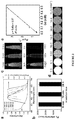
- FIG. 3 displays cytotoxicity and cell targeting data.
- Panel (a) shows the photoluminescence intensity (ex/em 360/425 nm) change when Gd@C-dots were incubated in buffers of different pH values.
- Panel (b) shows Gd release from Gd@C-dots over time. The nanoparticles were incubated in solutions with pH 5 or 7.4. #: The overall Gd concentrations in the solutions.
- Panel (c) shows cell viability, evaluated by MTT assays with U87MG cells. 2.5 mM Ca(II) was added in the incubation medium.
- Panel (d) displays a cell targeting study.
- RGD-Gd@C-dots were incubated with U87MG cells for 30 min and the cells were then imaged under a fluorescence microscope (scale bar, 10 ⁇ m).
- cells were incubated with Gd@C-dots at the same Gd concentration or with RGD-Gd@C-dots in the presence of free c(RGDyK) (30 ⁇ ).
- Panel (e) shows T 1 -weighted MR images of cell pellets, where cells had been incubated with either RGD-Gd@C-dots or Gd@C-dots.
- FIG. 4 panel (a), shows T 1 -weighted MR images acquired at different time points after injection of Gd@C-dots or RGD-Gd@C-dots.
- Panel (b) shows the signal change in the bladder (bl) and liver (lv), based on region of interest (ROI) analysis on images from panel (a).
- Panel (c) shows a photoluminescence analysis on urine samples taken 60 min after the injection of RGD-Gd@C-dots.
- FIG. 5 panel (a), shows T 1 -weighted transverse MR images.
- Gd@C-dots or RGD@C-dots (3.2 mg/kg) were intravenously injected into U87MG tumor bearing mice. Images were acquired at 0, 10, 30, 45, 60 and 240 min. For both types of nanoparticles, strong signals in the bladder were observed soon after the particle injection, indicating fast renal clearance.
- Panel (b) shows T 1 -weighted coronal MR images. Significant signal enhancement was observed in tumors of animals injected with RGD@Gd-dots.
- Panel (c) shows the relative signal change at different time points, based imaging results from panel (b).
- Panel (d) shows an immunofluorescence histology study with tumor samples.
- FIG. 6 shows the size distribution analysis of Gd@C-dots (panel (a)), RGD-Gd@C-dots (panel (b)), and Gd@C-dots in FBS (panel (c)) for 24 h.
- FIG. 7 shows the Zeta potentials of Gd@C-dots and RGD-Gd@C-dots.
- FIG. 8 shows the FTIR spectra of Gd@C-dots and DTPA.
- FIG. 9 shows the fluorescent spectra of metal-containing C-dots.
- Ranges can be expressed herein as from “about” one particular value, and/or to “about” another particular value. When such a range is expressed, another embodiment includes from the one particular value and/or to the other particular value. Similarly, when values are expressed as approximations, by use of the antecedent “about,” it will be understood that the particular value forms another embodiment. It will be further understood that the endpoints of each of the ranges are significant both in relation to the other endpoint, and independently of the other endpoint. Unless stated to the contrary “about” a particular value means within 5% of the particular value, e.g., within 2% or 1% of the particular value.
- amorphous is meant non-crystalline and without structural order over a long range, e.g., a majority of the nanoparticle.
- An amorphous shell can contain some ordered structure over a short range atomic length scale, but the majority of the shell is not ordered and non-crystalline.
- M@C-dots metal encapsulated carbon dots
- Gd@C-dots Gd encapsulated carbon dots
- nanocarriers/nanocapsules carbon has low-toxicity and is highly biologically inert.
- the disclosed nanoparticle M@C-dots can remain intact even in harsh biological environments, therefore precluding the risk of metal release to the surroundings (Cao et al., Theranostics 2012, 2(3):295-301).
- gadolinium stemming from the inert carbon coating, the disclosed nanoparticles are immune to the issue of Gd leaking that is often observed with complex-based Gd agents. Leakage of other metals from the disclosed nanoparticles is also expected.
- the disclosed nanoparticles can have an average size of about 12 nm in diameter.
- the disclosed nanoparticles can have an average size of from about 2 nm to about 34, from about 4 nm to about 32 nm, from about 6 nm to about 30 nm, from 8 to about 28 nm, from about 10 nm to about 24 nm, from about 12 nm to about 22 nm, from about 14 nm to about 20 nm, from about 26 nm to about 18 nm, from about 8 nm to about 16 nm, from about 10 nm to about 14 nm, from about 12 nm to about 20 nm, or from about 14 nm to about 18 nm.
- the disclosed nanoparticles can have an average size of 2, 4, 6, 8, 9, 10, 11, 12, 13, 14, 15, 16, 17, 18, 19, 20, 21, 22, 23, 24, 25, 26, 28, 30, 32, or 34 nm, where any of the stated values can form an upper or lower endpoint of a range.
- the disclosed nanoparticles for example, the Gd@C-dots
- r 1 relaxivity is about 5.88 mM ⁇ 1 s ⁇ 1 (on per Gd basis).
- the r 1 relaxivity can be tuned up to 50 mM ⁇ 1 s ⁇ 1 by adjusting the size and composition of Gd@C-dots.
- the disclosed Gd@C dots can have r 1 relaxivity from about 0 to about 50 mM ⁇ 1 s ⁇ 1 , e.g., about 0, 5, 10, 15, 20, 25, 30, 35, 40, 45, or 50 mM ⁇ 1 s ⁇ 1 , where any of the stated values can form an upper or lower end point of a range.
- the disclosed Gd@C-dots can have a r 1 relaxivity from about 0 to about 10, from about 5 to about 15, from about 10 to about 20, from about 15 to about 25, from about 20 to about 30, from about 25 to about 35, from about 30 to about 40, from about 35 to about 45, from about 40 to about 50, from about 0 to about 25, or from about 25 to about 50 mM ⁇ 1 s ⁇ 1 .
- the disclosed Gd@C-dots also possess bright photoluminescence, boasting a high quantum yield and excellent photostability that is even superior to quantum dots.
- the QY is found to be 19.7% for 12 nm Gd@C-dots but can be tuned up to 80% by adjusting the size and composition of Gd@C-dots.
- the QY of the disclosed Gd@C-dots is comparable to the highest reported QYs of C-dots (Bhunia et al., Sci. Rep. 2013, 3, 1473).
- the discloed Gd@C-dots can have a QY of from about 0 to about 80, from about 15 to about 65, from about 30 to about 80, from about 0 to about 35, from about 15 to about 75, from about 0 to about 20, or from about 20 to about 35%.
- nanoparticle M@C-dots can be efficiently excreted via urine, further minimizing toxicity risks.
- an imaging probe can home efficiently to the diseased area (e.g., a tumor), with the unbound probes rapidly excreted from the host.
- the disclosed nanoparticles are not endohedral fullerenes, which are much smaller than the disclosed M@C-dots. That is, endohedral fullerenes are usually less than about 1 nm. C 60 , for example, has a diameter of 0.7 nm. Also, the fullerene structure is highly ordered, where as the disclosed nanoparticles have an amorphous shell that functionalized with carboxyl groups.
- the surface of the disclosed nanoparticles contains carboxyl groups that can be used to functionalize the surface of the nanoparticles.
- the carbonyl groups are electrophiles that can be used in nucleophilic substitution reactions or carbodiimide coupling reactions with any desirable functionalizing reagent.
- the functionalizing reagent can contain a targeting moiety that can be used to direct the functionalized nanoparticles to specific locations in the patient.
- M@C-dot nanoparticles functionalized with a targeting moiety.
- RGD-peptides and cyclic RGD-peptides when coupled to the disclosed M@C-dots, can direct the nanoparticles to target tumors.
- EGFR targeting peptides can be coupled/attached to the disclosed nanoparticles.
- Different types of antibodies such as Herceptin, Avastin, and Erbitux, etc., can be coupled to the particle surface for facilitating particle targeting to tumors.
- Small molecule drugs such as doxorubicin, methotrexate or paclitaxel or their derivatives and can also be coupled to the surface of the nanoparticles, and in these cases the particles are used as drug carriers. Functionalizing the surface of the nanoparticles can also be used to assist the passage of the M@C-dots across certain cell membranes.
- the particle surface can be coated with a layer of positively charged polymer such as polyethylenimine and the resulting conjugates can be used as carriers for gene delivery (e.g., siRNA) due to assisting gene therapeutics passing through negatively charged cell membranes.
- a layer of positively charged polymer such as polyethylenimine
- the resulting conjugates can be used as carriers for gene delivery (e.g., siRNA) due to assisting gene therapeutics passing through negatively charged cell membranes.
- Suitable reagents for initiating a carbodiimide-mediate coupling to the carboxyl of the disclosed nanoparticles are commercially available.
- Specific examples of such reagents include, but are not limited to, water soluble carbodiimides such as 1-ethyl-3-(3-dimethylaminopropyl)-carbodiimide hydrochloride and 1-cyclohexyl-3-(2-morpholinoethyl)-carbodiimide-metho-p-toluene sulfonate, alcohol and water soluble N-ethyoxycarbonyl-2-ethoxy-1,2-dihydroquinoline, and organic soluble N,N′-dicyclohexylcarbodiimide.
- the disclosed M@C-dots can be prepared by calcining a metal with a chelator.
- An exemplary chelator that can be calcined with the metal is diethylenetriaminepentacetate (DTPA).
- DTPA can form complexes with a wide range of transition metals, especially Gd.
- metal-encapsulated M@C-dots can be produced. This method was tested with complexes formed between DTPA and Mn 2+ , Ni 2+ , Co 2+ , Zn 2+ , Fe 3+ , Nd 3+ , Y 3+ , and Eu 3+ . In all the scenarios, metal-containing C-dots ( FIG. 9 ) were obtained, indicating the generality of the method.
- chelator can be calcined with the metal to produce the disclosed nanoparticles.
- chelators include, but are not limited to, 1,4,7-triazacyclononane-1,4,7-triacetic acid (NOTA), 1,4,7,10-tetraazacyclodode-cane-1,4,7,10-tetraacetic acid (DOTA), 1,4,8,11-tetraazacyclododenane-1,4,8,11-tetraacetic acid (TETA), 2,2′-(1,4,8,11-tetraazabicyclo[6.6.2]hexadecane-4,11-diyl)diacetic acid (CB-TE2A), 3,6,9,15-Tetraazabicyclo[9.3.1]pentadeca-1(15),11,13-triene-3,6,9-triacetic acid (PCTA), pendetide (GYK-DTPA), cyclohexyldiethylenetriaminepentaacetic acid (CH
- compositions which comprise one or more of the disclosed nanoparticles together with one or more pharmaceutically acceptable carriers thereof and optionally one or more other therapeutic ingredients.
- the carrier(s) must be “acceptable” in the sense of being compatible with the other ingredients of the formulation and not deleterious to the recipient thereof Proper formulation is dependent upon the route of administration chosen. Any of the well-known techniques, carriers, and excipients can be used as suitable and as understood in the art; e.g., in Remington: The Science and Practice of Pharmacy, 21 st Ed., Gennaro, Ed., Lippencott Williams & Wilkins (2003).
- compositions and formulations disclosed herein can be manufactured in any manner known in the art, e.g., by means of conventional mixing, dissolving, granulating, dragee-making, levigating, emulsifying, encapsulating, entrapping or compression processes.
- a nanoparticle as disclosed herein can be incorporated into a variety of formulations for therapeutic administration, including solid, semi-solid, or liquid forms.
- the formulations include those suitable for oral, parenteral (including subcutaneous, intradermal, intramuscular, intravenous, intraarticular, and intramedullary), intraperitoneal, transmucosal, transdermal, rectal and topical (including dermal, buccal, sublingual and intraocular) administration although the most suitable route can depend upon for example the condition and disorder of the recipient.
- the formulations can conveniently be presented in unit dosage form and can be prepared by any of the methods well known in the art of pharmacy.
- these methods include the step of bringing into association a compound or a pharmaceutically acceptable salt thereof (“active ingredient”) with the carrier which constitutes one or more accessory ingredients.
- active ingredient a compound or a pharmaceutically acceptable salt thereof
- the formulations are prepared by uniformly and intimately bringing into association the active ingredient with liquid carriers or finely divided solid carriers or both and then, if necessary, shaping the product into the desired formulation.
- the disclosed nanoparticles can be formulated for parenteral administration by injection, e.g., by bolus injection or continuous infusion.
- Formulations for injection can be presented in unit dosage form, e.g., in ampoules or in multi-dose containers, with an added preservative.
- the compositions can take such forms as suspensions, solutions or emulsions in oily or aqueous vehicles, and can contain formulatory agents such as suspending, stabilizing and/or dispersing agents.
- the formulations can be presented in unit-dose or multi-dose containers, for example sealed ampoules and vials, and can be stored in powder form or in a freeze-dried (lyophilized) condition requiring only the addition of the sterile liquid carrier, for example, saline or sterile pyrogen-free water, immediately prior to use.
- sterile liquid carrier for example, saline or sterile pyrogen-free water
- Extemporaneous injection solutions and suspensions can be prepared from sterile powders, granules and tablets of the kind previously described.
- Formulations for parenteral administration include aqueous and non-aqueous (oily) sterile injection solutions of the active compounds which can contain antioxidants, buffers, bacteriostats and solutes which render the formulation isotonic with the blood of the intended recipient; and aqueous and non-aqueous sterile suspensions which can include suspending agents and thickening agents.
- Suitable lipophilic solvents or vehicles include fatty oils such as sesame oil, or synthetic fatty acid esters, such as ethyl oleate or triglycerides, or liposomes.
- Aqueous injection suspensions can contain substances which increase the viscosity of the suspension, such as sodium carboxymethyl cellulose, sorbitol, or dextran.
- the suspension can also contain suitable stabilizers or agents which increase the solubility of the compounds to allow for the preparation of highly concentrated solutions.
- the disclosed nanoparticles have properties that can afford them uses as optical, MRI, fluorescence, photoacoustic imaging probes.
- the disclosed nanoparticles can also be used in therapy (drug delivery, gene delivery, photodynamic therapy), catalysis, energy, and electronics applications.
- the disclosed nanoparticles can be used a MRI/fluorescence dual modal imaging probes.
- the disclosed nanoparticles, or formulations containing them can be used as imaging agents to visualize cancerous tissues, e.g., tumors.
- a method of detecting cancer in vivo comprising administering a nanoparticle (e.g., Gd@C-dot) as disclosed herein to an individual and detecting a fluorescent signal and/or magnetic resonance signal.
- a region of interest in the individual can be imaged using a fluorescence reflectance imaging system (such as the F-Pro from Bruker), which is fitted with multiple band pass filters for excitation and emission.
- a fluorescence reflectance imaging system such as the F-Pro from Bruker
- the disclosed nanoparticles can also be used as MR imaging probes.
- the nanoparticles disclosed herein can be used to detect/image a variety of other cancers.
- cancer types detectable by the compounds and compositions disclosed herein include bladder cancer, brain cancer, breast cancer, colorectal cancer, cervical cancer, gastrointestinal cancer, genitourinary cancer, head and neck cancer, lung cancer, ovarian cancer, pancreatic cancer, prostate cancer, renal cancer, skin cancer, and testicular cancer.
- Further examples include cancer and/or tumors of the anus, bile duct, bone, bone marrow, bowel (including colon and rectum), eye, gall bladder, kidney, mouth, larynx, esophagus, stomach, testis, cervix, mesothelioma, neuroendocrine, penis, skin, spinal cord, thyroid, vagina, vulva, uterus, liver, muscle, blood cells (including lymphocytes and other immune system cells).
- Specific cancers contemplated for imaging include carcinomas, Karposi's sarcoma, melanoma, mesothelioma, soft tissue sarcoma, pancreatic cancer, lung cancer, leukemia (acute lymphoblastic, acute myeloid, chronic lymphocytic, chronic myeloid, and other), and lymphoma (Hodgkin's and non-Hodgkin's), and multiple myeloma.
- UV-Vis absorption spectra were recorded on a Shimdzu 2450 UV-Vis spectrometer.
- Photoluminescence (PL) measurements were performed on a Hitachi F-7000 fluorometer.
- Fourier transform infrared (FT-IR) spectra were recorded on a Nicolet iS10 FT-IR Spectrometer.
- the PL quantum yield (QY) was estimated using quinine sulfate in 0.1 M H 2 SO 4 (literature quantum yield: 58% at 354 nm excitation) as a reference standard, which was freshly prepared to reduce the measurement error.
- TEM and HR-TEM samples were prepared by dispersing the sample onto carbon-coated copper grids with the excess solvent evaporated.
- the TEM/HR-TEM overview images were recorded using a FEI Tecnai20 transmission electron microscope operating at 200 kV.
- Energy Dispersive Spectroscopy (EDS) and element mapping was characterized using Hitachi HD2000 Dedicated Scanning Transmission Electron Microscope (STEM).
- Dynamic light scattering (DLS) analysis was performed on a Zetasizer Nano S90 size analyzer (Malvern Corp, U.K.). Fluorescence images were acquired on an Olympus X71 fluorescence microscope (ex/em: 360/420 nm).
- Gadolinium-DTPA gadopentetic acid which is a complex of gadolinium with diethylenetriaminepentacetate
- Gadolinium-DTPA gadopentetic acid which is a complex of gadolinium with diethylenetriaminepentacetate
- the raw products were dispersed in water and subjected to centrifugation using centrifugal filter units (Millipore filter units: MWCO 100K, 3K), which removed aggregations of nanoparticles and unreacted precursors, respectively. The soluble portion through the filter was collected.
- the yielded Gd@C-dots were spherical, with an average size of about 12 nm and relatively narrow size distribution ( FIG.
- Gd@C-dots showed a broad absorption band between 200 to 500 nm, with a shoulder appearing at about 280 nm ( FIG. 2 , panel (a)).
- the spectrum resembles those published previously of pure C-dots (Chen et al., Theranostics 2013, 3(9):650-657; Bhunia et al., Sci. Rep. 2013, 3, 1473; Sun et al., J. Am. Chem. Soc. 2006, 128(24):7756-7757; Ding et al., Acc. Chem. Res. 2014, 47, 20-30).
- the Gd@C-dots are also highly fluorescent, and can be excited by light of a wide range of wavelengths to emit strong photoluminescence ( FIG. 2 , panel (a)). Such wavelength-dependent fluorescence is also similar to conventional C-dots (Bourlinos et al., Small 2008, 4(4):455-458; Li et al., Angew. Chem., Int. Ed. 2013, 52(31):8151-8255). Impressively, there was no drop of photoluminescence intensity of Gd@C-dots even after 24 hours of continuous UV illumination ( FIG. 2 , panel (b)).
- Gd@C-dots were also subjected to a cytotoxicity study with U87MG cells using standard MTT assays. The cells were first seeded in 96-well plates (1 ⁇ 10 4 cells per well). After 24 h, Gd@C-dots at different concentrations were added. Incubation was carried out for 24 h with or without 2.5 mM CaCl 2 . To assess the particles' stability against transmetallation, which is the major cause of toxicity for conventional Gd contrast agents (Corot et al., J. Magn. Reson. Imaging 1998, 8(3):695-702), 2.5 mM Ca(II) was added into the incubation medium.
- Carboxyl groups on the Gd@C-dots surface offer a facile means to tether functional bio-species.
- c(RGDyK) a tumor targeting peptide
- a cyclic RGD derivative, c(RGDyK) holds strong binding affinity toward integrin ⁇ v ⁇ 3 , a biomarker that is seen overexpressed on neoplastic blood vessels and many types of cancer cells (Ye et al., Theranostics 2011, 1:102-126).
- Gd@C-dots were dispersed in a borate buffer (pH 8.3).
- U87MG cells which are integrin ⁇ v ⁇ 3 positive.
- U87MG cells were grown in a petri dish of a sterile glass bottom at 37° C. in 5% CO 2 . Cells were incubated in 1 mL media containing Gd@C-dots (50 ⁇ g) with and without the presence of c(RGDyK) (1 mg) for 1 hour. Cells were washed three times with PBS (pH 7.4), and then imaged under an Olympus X71 fluorescence microscope. For MRI studies, 10 5 cells treated under similar conditions were collected in 300 ⁇ L PCR tubes and subjected to T 1 -weighted MRI.
- FIG. 3 , panel (e) shows a T 1 -weighted MR image of 105 U87MG cells that had been incubated with either RGD-Gd@C-dot or Gd@C-dots. Compared to the control, significantly enhanced signals were observed in cells incubated with RGD@C-dots ( FIG. 3 , panel (e)).
- Transverse and coronal T 1 -weighted MR images were acquired at 10, 30, 45 min, 60 min and 4 h post the nanoparticle injection (p.i.).
- RGD-Gd@C-dots were also tested as tumor imaging probes in U87MG tumor-bearing mice.
- Tumor models were developed in 5-6 week athymic nude mice (Harlan) by subcutaneous implantation of 10 6 human glioblastoma U87MG cells suspended in 100 ⁇ L of serum-free DMEM to the right lower flank of a mouse. Imaging studies were conducted 3-4 weeks later. Specifically, the tumor-bearing mice were intravenously injected with Gd@C-dots and RGD-Gd@C-dots (3.2 mg Gd/kg).
- SBR signal-to-background ratio
- ROI regions of interest
- SI Signal intensity
- pre and post ROIs were determined manually on each image as reproducible as possible.
- 3-5 ROIs were selected to measure the SI of the liver, kidney, brain and muscle.
- Gd@C-dots at the same Gd dose were injected.
Landscapes
- Health & Medical Sciences (AREA)
- Engineering & Computer Science (AREA)
- Life Sciences & Earth Sciences (AREA)
- Nanotechnology (AREA)
- Chemical & Material Sciences (AREA)
- Nuclear Medicine, Radiotherapy & Molecular Imaging (AREA)
- General Health & Medical Sciences (AREA)
- Physics & Mathematics (AREA)
- Veterinary Medicine (AREA)
- Public Health (AREA)
- Animal Behavior & Ethology (AREA)
- Radiology & Medical Imaging (AREA)
- Biomedical Technology (AREA)
- Biophysics (AREA)
- Molecular Biology (AREA)
- Medical Informatics (AREA)
- Heart & Thoracic Surgery (AREA)
- Surgery (AREA)
- Pathology (AREA)
- Epidemiology (AREA)
- Proteomics, Peptides & Aminoacids (AREA)
- High Energy & Nuclear Physics (AREA)
- Condensed Matter Physics & Semiconductors (AREA)
- General Physics & Mathematics (AREA)
- Signal Processing (AREA)
- Medicines Containing Antibodies Or Antigens For Use As Internal Diagnostic Agents (AREA)
Abstract
Description
(QY)sm=(QY)st×[(PL area /OD)sm/(PL area /OD)st]×η2 sm/η2 st
Claims (20)
Priority Applications (1)
| Application Number | Priority Date | Filing Date | Title |
|---|---|---|---|
| US15/500,691 US10548993B2 (en) | 2014-07-31 | 2015-07-30 | Metal-encapsulated carbonaceous dots |
Applications Claiming Priority (3)
| Application Number | Priority Date | Filing Date | Title |
|---|---|---|---|
| US201462031821P | 2014-07-31 | 2014-07-31 | |
| US15/500,691 US10548993B2 (en) | 2014-07-31 | 2015-07-30 | Metal-encapsulated carbonaceous dots |
| PCT/US2015/042787 WO2016019090A1 (en) | 2014-07-31 | 2015-07-30 | Metal-encapsulated carbonaceous dots |
Publications (2)
| Publication Number | Publication Date |
|---|---|
| US20170216464A1 US20170216464A1 (en) | 2017-08-03 |
| US10548993B2 true US10548993B2 (en) | 2020-02-04 |
Family
ID=55218300
Family Applications (1)
| Application Number | Title | Priority Date | Filing Date |
|---|---|---|---|
| US15/500,691 Active 2036-10-13 US10548993B2 (en) | 2014-07-31 | 2015-07-30 | Metal-encapsulated carbonaceous dots |
Country Status (2)
| Country | Link |
|---|---|
| US (1) | US10548993B2 (en) |
| WO (1) | WO2016019090A1 (en) |
Families Citing this family (7)
| Publication number | Priority date | Publication date | Assignee | Title |
|---|---|---|---|---|
| US10245373B2 (en) | 2014-12-01 | 2019-04-02 | Carefusion 2200, Inc. | Pump cassettes with positioning feature and infusion pump systems |
| CN106278920B (en) * | 2016-08-03 | 2018-12-11 | 河南大学 | Nitrilotriacetic acid Holmium complex as cadmium ion fluorescent probe and preparation method thereof |
| CN109388085B (en) * | 2018-09-21 | 2020-06-16 | 高新兴创联科技有限公司 | Application method of computer platform based on self-wheel running special equipment |
| CN109233828B (en) * | 2018-11-02 | 2021-07-27 | 广西医科大学 | A kind of preparation method and application of gadolinium-based fluorescent carbon dots |
| CN109735330B (en) * | 2019-01-16 | 2022-11-22 | 河南师范大学 | A kind of iron ion doped carbon point, preparation method and application thereof |
| KR102235374B1 (en) * | 2019-09-19 | 2021-04-01 | 경북대학교 산학협력단 | Imaging agent using carbon-coated gadolinium oxide and method of the imaging agent |
| CN112724961B (en) * | 2020-07-02 | 2022-07-01 | 中国人民解放军63653部队 | A kind of preparation method of white light emitting carbon quantum dots |
Citations (9)
| Publication number | Priority date | Publication date | Assignee | Title |
|---|---|---|---|---|
| US20020127224A1 (en) | 2001-03-02 | 2002-09-12 | James Chen | Use of photoluminescent nanoparticles for photodynamic therapy |
| US20030053951A1 (en) * | 2001-07-26 | 2003-03-20 | Millennium Pharmaceuticals, Inc. | Use of non-invasive imaging technologies to monitor in vivo gene-expression |
| US20070178308A1 (en) * | 2006-01-27 | 2007-08-02 | Konica Minolta Medical & Graphic, Inc. | Germanium nanoparticles and biosubstance labeling agent by use thereof |
| WO2007095454A2 (en) | 2006-02-10 | 2007-08-23 | Board Of Supervisors Of Louisiana State University And Agricultural And Mechanical College | Carbon-encased metal nanoparticles and sponges, methods of synthesis, and methods of use |
| US7887771B2 (en) | 2005-10-06 | 2011-02-15 | Headwaters Technology Innovation, Llc | Carbon nanorings manufactured from templating nanoparticles |
| US20120323112A1 (en) * | 2011-06-17 | 2012-12-20 | The Board Of Trustees Of The Leland Stanford Junior University | Nanoparticles for accoustic imaging, methods of making, and methods of accoustic imaging |
| US20130112605A1 (en) | 2010-07-26 | 2013-05-09 | Waters Technologies Corporation | Superficially porous materials comprising a substantially nonporous core having narrow particle size distribution; process for the preparation thereof; and use thereof for chromatographic separations |
| US20130289520A1 (en) | 2010-04-23 | 2013-10-31 | Children's Hospital Boston | Targeted and light-activated cytosolic drug delivery |
| US20140044648A1 (en) | 2012-07-06 | 2014-02-13 | University Of Central Florida Research Foundation, Inc. | Activatable imaging contrast agents |
Family Cites Families (1)
| Publication number | Priority date | Publication date | Assignee | Title |
|---|---|---|---|---|
| JP6297269B2 (en) * | 2012-06-28 | 2018-03-20 | ローム アンド ハース エレクトロニック マテリアルズ エルエルシーRohm and Haas Electronic Materials LLC | Polymer composition, photoresist comprising the polymer composition, and coated article comprising the photoresist |
-
2015
- 2015-07-30 WO PCT/US2015/042787 patent/WO2016019090A1/en not_active Ceased
- 2015-07-30 US US15/500,691 patent/US10548993B2/en active Active
Patent Citations (9)
| Publication number | Priority date | Publication date | Assignee | Title |
|---|---|---|---|---|
| US20020127224A1 (en) | 2001-03-02 | 2002-09-12 | James Chen | Use of photoluminescent nanoparticles for photodynamic therapy |
| US20030053951A1 (en) * | 2001-07-26 | 2003-03-20 | Millennium Pharmaceuticals, Inc. | Use of non-invasive imaging technologies to monitor in vivo gene-expression |
| US7887771B2 (en) | 2005-10-06 | 2011-02-15 | Headwaters Technology Innovation, Llc | Carbon nanorings manufactured from templating nanoparticles |
| US20070178308A1 (en) * | 2006-01-27 | 2007-08-02 | Konica Minolta Medical & Graphic, Inc. | Germanium nanoparticles and biosubstance labeling agent by use thereof |
| WO2007095454A2 (en) | 2006-02-10 | 2007-08-23 | Board Of Supervisors Of Louisiana State University And Agricultural And Mechanical College | Carbon-encased metal nanoparticles and sponges, methods of synthesis, and methods of use |
| US20130289520A1 (en) | 2010-04-23 | 2013-10-31 | Children's Hospital Boston | Targeted and light-activated cytosolic drug delivery |
| US20130112605A1 (en) | 2010-07-26 | 2013-05-09 | Waters Technologies Corporation | Superficially porous materials comprising a substantially nonporous core having narrow particle size distribution; process for the preparation thereof; and use thereof for chromatographic separations |
| US20120323112A1 (en) * | 2011-06-17 | 2012-12-20 | The Board Of Trustees Of The Leland Stanford Junior University | Nanoparticles for accoustic imaging, methods of making, and methods of accoustic imaging |
| US20140044648A1 (en) | 2012-07-06 | 2014-02-13 | University Of Central Florida Research Foundation, Inc. | Activatable imaging contrast agents |
Non-Patent Citations (84)
Also Published As
| Publication number | Publication date |
|---|---|
| WO2016019090A1 (en) | 2016-02-04 |
| US20170216464A1 (en) | 2017-08-03 |
Similar Documents
| Publication | Publication Date | Title |
|---|---|---|
| US10548993B2 (en) | Metal-encapsulated carbonaceous dots | |
| Ashokan et al. | A molecular receptor targeted, hydroxyapatite nanocrystal based multi-modal contrast agent | |
| Zhang et al. | Glutathione-capped fluorescent gold nanoclusters for dual-modal fluorescence/X-ray computed tomography imaging | |
| Iqbal et al. | Silica-coated super-paramagnetic iron oxide nanoparticles (SPIONPs): a new type contrast agent of T 1 magnetic resonance imaging (MRI) | |
| Wang et al. | Two-photon graphene quantum dot modified Gd 2 O 3 nanocomposites as a dual-mode MRI contrast agent and cell labelling agent | |
| Ma et al. | Biocompatible composite nanoparticles with large longitudinal relaxivity for targeted imaging and early diagnosis of cancer | |
| US20170151351A1 (en) | Gd-ENCAPSULATED CARBON DOTS AND METHODS OF MAKING AND USING THEREOF | |
| Cormode et al. | Inorganic nanocrystals as contrast agents in MRI: synthesis, coating and introduction of multifunctionality | |
| Yeh et al. | Tumor targeting and MR imaging with lipophilic cyanine-mediated near-infrared responsive porous Gd silicate nanoparticles | |
| Chen et al. | Cy5. 5 conjugated MnO nanoparticles for magnetic resonance/near-infrared fluorescence dual-modal imaging of brain gliomas | |
| Yang et al. | Tumor-targeted biodegradable multifunctional nanoparticles for cancer theranostics | |
| JP6227760B2 (en) | Fluorescent solid lipid nanoparticle composition and production method thereof | |
| WO2011057216A1 (en) | Bioconjugation of calcium phosphosilicate nanoparticles for selective targeting of cells in vivo | |
| US20110158901A1 (en) | Chitosan-based nanoparticles and methods for making and using the same | |
| WO2010021519A2 (en) | T1-t2 dual modal mri contrast agents | |
| Jang et al. | In vivo magnetic resonance and fluorescence dual imaging of tumor sites by using dye-doped silica-coated iron oxide nanoparticles | |
| US9474809B2 (en) | Activatable nanoprobes for intracellular drug delivery | |
| Yin et al. | Fluorescent oligo (p-phenyleneethynylene) contained amphiphiles-encapsulated magnetic nanoparticles for targeted magnetic resonance and two-photon optical imaging in vitro and in vivo | |
| US10201622B2 (en) | Tumour-targeted theranostic | |
| US20180161461A1 (en) | Rare Earth Oxide Particles and Use Thereof in Particular In Imaging | |
| Xie et al. | pH-responsive MTX-BSA@ MnO2-Cy5. 5 for NIRF/MR imaging guided chemotherapy of anaplastic thyroid carcinoma | |
| Lee et al. | Amphiphilic hyaluronic acid-based nanoparticles for tumor-specific optical/MR dual imaging | |
| Li et al. | Synthesis of a UCNPs@ SiO 2@ gadofullerene nanocomposite and its application in UCL/MR bimodal imaging | |
| Ma et al. | Folic acid-targeted magnetic Tb-doped CeF 3 fluorescent nanoparticles as bimodal probes for cellular fluorescence and magnetic resonance imaging | |
| Xiao et al. | Facile synthesis of Silicon quantum dot-Gadolinium: A potential fluorescent/T1-T2 multimodal imaging agent |
Legal Events
| Date | Code | Title | Description |
|---|---|---|---|
| AS | Assignment |
Owner name: UNIVERSITY OF GEORGIA RESEARCH FOUNDATION, INC, GEORGIA Free format text: ASSIGNMENT OF ASSIGNORS INTEREST;ASSIGNORS:XIE, JIN;CHEN, HONGMIN;WANG, GEOFFREY D.;REEL/FRAME:042205/0173 Effective date: 20140804 Owner name: UNIVERSITY OF GEORGIA RESEARCH FOUNDATION, INC, GE Free format text: ASSIGNMENT OF ASSIGNORS INTEREST;ASSIGNORS:XIE, JIN;CHEN, HONGMIN;WANG, GEOFFREY D.;REEL/FRAME:042205/0173 Effective date: 20140804 |
|
| STPP | Information on status: patent application and granting procedure in general |
Free format text: DOCKETED NEW CASE - READY FOR EXAMINATION |
|
| AS | Assignment |
Owner name: NATIONAL INSTITUTES OF HEALTH (NIH), U.S. DEPT. OF HEALTH AND HUMAN SERVICES (DHHS), U.S. GOVERNMENT, MARYLAND Free format text: CONFIRMATORY LICENSE;ASSIGNOR:UNIVERSITY OF GEORGIA;REEL/FRAME:043936/0374 Effective date: 20170920 Owner name: NATIONAL INSTITUTES OF HEALTH (NIH), U.S. DEPT. OF Free format text: CONFIRMATORY LICENSE;ASSIGNOR:UNIVERSITY OF GEORGIA;REEL/FRAME:043936/0374 Effective date: 20170920 |
|
| STPP | Information on status: patent application and granting procedure in general |
Free format text: NON FINAL ACTION MAILED |
|
| STPP | Information on status: patent application and granting procedure in general |
Free format text: NOTICE OF ALLOWANCE MAILED -- APPLICATION RECEIVED IN OFFICE OF PUBLICATIONS |
|
| STPP | Information on status: patent application and granting procedure in general |
Free format text: PUBLICATIONS -- ISSUE FEE PAYMENT VERIFIED |
|
| STCF | Information on status: patent grant |
Free format text: PATENTED CASE |