US10497123B1 - Isolation of aneurysm and parent vessel in volumetric image data - Google Patents
Isolation of aneurysm and parent vessel in volumetric image data Download PDFInfo
- Publication number
- US10497123B1 US10497123B1 US16/515,075 US201916515075A US10497123B1 US 10497123 B1 US10497123 B1 US 10497123B1 US 201916515075 A US201916515075 A US 201916515075A US 10497123 B1 US10497123 B1 US 10497123B1
- Authority
- US
- United States
- Prior art keywords
- mask
- aneurysm
- voxel
- generating
- refined
- Prior art date
- Legal status (The legal status is an assumption and is not a legal conclusion. Google has not performed a legal analysis and makes no representation as to the accuracy of the status listed.)
- Active
Links
Images


Classifications
-
- G—PHYSICS
- G06—COMPUTING OR CALCULATING; COUNTING
- G06T—IMAGE DATA PROCESSING OR GENERATION, IN GENERAL
- G06T7/00—Image analysis
- G06T7/10—Segmentation; Edge detection
- G06T7/11—Region-based segmentation
-
- A—HUMAN NECESSITIES
- A61—MEDICAL OR VETERINARY SCIENCE; HYGIENE
- A61B—DIAGNOSIS; SURGERY; IDENTIFICATION
- A61B6/00—Apparatus or devices for radiation diagnosis; Apparatus or devices for radiation diagnosis combined with radiation therapy equipment
- A61B6/50—Apparatus or devices for radiation diagnosis; Apparatus or devices for radiation diagnosis combined with radiation therapy equipment specially adapted for specific body parts; specially adapted for specific clinical applications
- A61B6/504—Apparatus or devices for radiation diagnosis; Apparatus or devices for radiation diagnosis combined with radiation therapy equipment specially adapted for specific body parts; specially adapted for specific clinical applications for diagnosis of blood vessels, e.g. by angiography
-
- A—HUMAN NECESSITIES
- A61—MEDICAL OR VETERINARY SCIENCE; HYGIENE
- A61B—DIAGNOSIS; SURGERY; IDENTIFICATION
- A61B6/00—Apparatus or devices for radiation diagnosis; Apparatus or devices for radiation diagnosis combined with radiation therapy equipment
- A61B6/52—Devices using data or image processing specially adapted for radiation diagnosis
- A61B6/5211—Devices using data or image processing specially adapted for radiation diagnosis involving processing of medical diagnostic data
- A61B6/5217—Devices using data or image processing specially adapted for radiation diagnosis involving processing of medical diagnostic data extracting a diagnostic or physiological parameter from medical diagnostic data
-
- A—HUMAN NECESSITIES
- A61—MEDICAL OR VETERINARY SCIENCE; HYGIENE
- A61B—DIAGNOSIS; SURGERY; IDENTIFICATION
- A61B6/00—Apparatus or devices for radiation diagnosis; Apparatus or devices for radiation diagnosis combined with radiation therapy equipment
- A61B6/52—Devices using data or image processing specially adapted for radiation diagnosis
- A61B6/5258—Devices using data or image processing specially adapted for radiation diagnosis involving detection or reduction of artifacts or noise
-
- G—PHYSICS
- G06—COMPUTING OR CALCULATING; COUNTING
- G06T—IMAGE DATA PROCESSING OR GENERATION, IN GENERAL
- G06T11/00—2D [Two Dimensional] image generation
- G06T11/60—Editing figures and text; Combining figures or text
-
- G—PHYSICS
- G06—COMPUTING OR CALCULATING; COUNTING
- G06T—IMAGE DATA PROCESSING OR GENERATION, IN GENERAL
- G06T7/00—Image analysis
- G06T7/10—Segmentation; Edge detection
- G06T7/187—Segmentation; Edge detection involving region growing; involving region merging; involving connected component labelling
-
- G—PHYSICS
- G06—COMPUTING OR CALCULATING; COUNTING
- G06T—IMAGE DATA PROCESSING OR GENERATION, IN GENERAL
- G06T7/00—Image analysis
- G06T7/10—Segmentation; Edge detection
- G06T7/194—Segmentation; Edge detection involving foreground-background segmentation
-
- G—PHYSICS
- G06—COMPUTING OR CALCULATING; COUNTING
- G06T—IMAGE DATA PROCESSING OR GENERATION, IN GENERAL
- G06T2207/00—Indexing scheme for image analysis or image enhancement
- G06T2207/10—Image acquisition modality
- G06T2207/10116—X-ray image
-
- G—PHYSICS
- G06—COMPUTING OR CALCULATING; COUNTING
- G06T—IMAGE DATA PROCESSING OR GENERATION, IN GENERAL
- G06T2207/00—Indexing scheme for image analysis or image enhancement
- G06T2207/30—Subject of image; Context of image processing
- G06T2207/30004—Biomedical image processing
- G06T2207/30101—Blood vessel; Artery; Vein; Vascular
-
- G—PHYSICS
- G06—COMPUTING OR CALCULATING; COUNTING
- G06T—IMAGE DATA PROCESSING OR GENERATION, IN GENERAL
- G06T2210/00—Indexing scheme for image generation or computer graphics
- G06T2210/12—Bounding box
-
- G—PHYSICS
- G06—COMPUTING OR CALCULATING; COUNTING
- G06T—IMAGE DATA PROCESSING OR GENERATION, IN GENERAL
- G06T2211/00—Image generation
- G06T2211/40—Computed tomography
- G06T2211/404—Angiography
Definitions
- the present disclosure generally relates to digital image data processing, and more particularly to isolation of aneurysm and its parent vessel in volumetric image data.
- An aneurysm is a localized, blood-filled dilation or bulge of a blood vessel caused by disease or weakening of the vessel wall.
- Aneurysms can occur anywhere where there are blood vessels, although they are most common in arteries. Aneurysms most commonly occur in arteries at the base of the brain, in the circle of Willis and in the aorta. Rupture and blood clotting are the risks involved with aneurysms. Particularly, if the patient has elevated blood pressure, this bulge in the blood vessel can burst and lead to hemorrhage and possibly death at any time. The risk of death is high except for rupture in the extremities. The larger an aneurysm becomes, the more likely it is to burst.
- X-ray C-arms are routinely used in medicine to acquire three-dimensional (3D) digital images for diagnostic assessment, and for guidance of interventional therapeutic procedures such as stent placement or coiling of aneurysms.
- Aneurysm analysis performed in 3D image space is often affected by the limitations of the aneurysm segmentation technique. In complicated aneurysm cases, the detection of aneurysm is not 100% accurate. This causes incomplete aneurysm detection, or aneurysm overflow (leak) outside the region of interest, which further results in inaccurate measurements.
- FIG. 1 shows examples of aneurysm segmentation leak generated by a traditional segmentation technique.
- the gray portions 102 a - h in the images represent the aneurysm detected by the segmentation technique.
- the aneurysm 102 a - h has been detected incorrectly as leaking (or overflowing) to adjacent vessels outside the region of interest.
- the framework generates a refined mask by performing region growing starting at an aneurysm dome point to eliminate vessels that are indirectly connected to an aneurysm or parent vessel in volumetric image data.
- a final mask may be generated based at least in part on the refined mask by eliminating any kissing vessel from the refined mask. The final mask may then be used for segmenting the aneurysm and the parent vessel in the volumetric image data.
- FIG. 1 shows examples of aneurysm segmentation leak generated by a traditional technique
- FIG. 2 is a block diagram illustrating an exemplary system
- FIG. 3 shows an exemplary method performed by a computer system
- FIG. 4 a shows an exemplary original volume mask
- FIG. 4 b shows an exemplary refined volume mask generated by the region growing process
- FIG. 5 shows an exemplary aneurysm mask
- FIG. 6 shows an exemplary parent vessel mask
- FIG. 7 shows an exemplary combined mask
- FIG. 8 shows an exemplary ‘crown’
- FIGS. 9 a - f illustrate the steps for cleaning up the mask
- FIG. 10 a shows an exemplary original volumetric image data
- FIG. 10 b shows the final output mask generated after performing the previous clean up steps
- FIG. 11 a shows an exemplary final mask
- FIG. 11 b shows an exemplary segmentation output
- FIG. 12 a shows an exemplary image generated by traditional segmentation methods
- FIG. 12 b shows an exemplary image generated by the present framework
- FIG. 13 a shows another exemplary image generated by traditional segmentation methods
- FIG. 13 b shows another exemplary image generated by the present framework.
- x-ray image may mean a visible x-ray image (e.g., displayed on a video screen) or a digital representation of an x-ray image (e.g., a file corresponding to the pixel output of an x-ray detector).
- in-treatment x-ray image may refer to images captured at any point in time during a treatment delivery phase of an interventional or therapeutic procedure, which may include times when the radiation source is either on or off. From time to time, for convenience of description, CT imaging data (e.g., cone-beam CT imaging data) may be used herein as an exemplary imaging modality.
- data from any type of imaging modality including but not limited to x-ray radiographs, MRI, PET (positron emission tomography), PET-CT, SPECT, SPECT-CT, MR-PET, 3D ultrasound images or the like may also be used in various implementations.
- imaging modality including but not limited to x-ray radiographs, MRI, PET (positron emission tomography), PET-CT, SPECT, SPECT-CT, MR-PET, 3D ultrasound images or the like may also be used in various implementations.
- sequences of instructions designed to implement the methods can be compiled for execution on a variety of hardware platforms and for interface to a variety of operating systems.
- implementations of the present framework are not described with reference to any particular programming language. It will be appreciated that a variety of programming languages may be used.
- the term “image” refers to multi-dimensional data composed of discrete image elements (e.g., pixels for 2D images and voxels for 3D images).
- the image may be, for example, a medical image of a subject collected by computer tomography, magnetic resonance imaging, ultrasound, or any other medical imaging system known to one of skill in the art.
- the image may also be provided from non-medical contexts, such as, for example, remote sensing systems, electron microscopy, etc.
- an image can be thought of as a function from R 3 to R, or a mapping to R 3
- the present methods are not limited to such images, and can be applied to images of any dimension, e.g., a 2D picture or a 3D volume.
- the domain of the image is typically a 2- or 3-Dimensional rectangular array, wherein each pixel or voxel can be addressed with reference to a set of 2 or 3 mutually orthogonal axes.
- digital and “digitized” as used herein will refer to images or volumes, as appropriate, in a digital or digitized format acquired via a digital acquisition system or via conversion from an analog image.
- pixels for picture elements, conventionally used with respect to 2D imaging and image display, and “voxels” for volume image elements, often used with respect to 3D imaging, can be used interchangeably.
- the 3D volume image is itself synthesized from image data obtained as pixels on a 2D sensor array and displays as a 2D image from some angle of view.
- 2D image processing and image analysis techniques can be applied to the 3D volume image data.
- techniques described as operating upon pixels may alternately be described as operating upon the 3D voxel data that is stored and represented in the form of 2D pixel data for display.
- techniques that operate upon voxel data can also be described as operating upon pixels.
- variable x is used to indicate a subject image element at a particular spatial location or, alternately considered, a subject pixel.
- subject pixel or “subject voxel” are used to indicate a particular image element as it is operated upon using techniques described herein.
- One aspect of the present framework isolates an aneurysm and its parent vessel in volumetric (or three-dimensional) image data.
- the framework starts by cleaning up the original input volumetric image data to remove vessels that are indirectly connected to the aneurysm or to the parent vessels. The clean-up may be performed using a region growing technique starting from an aneurysm dome point. Once the input image data is cleaned up, a final version of the volume mask is generated. This final mask contains only the aneurysm dome and parent vessel along the centerline.
- the final mask does not contain any “kissing vessel” artifact.
- a “kissing vessel” as used herein generally refers to any unwanted artifact that appears to be a vessel touching or connected to the aneurysm in the mask, but is actually not a vessel where the aneurysm originated.
- the final mask may be passed to an aneurysm segmentation unit.
- the aneurysm segmentation unit runs on a cleaner version of input mask that does not contain any kissing vessels.
- the aneurysm segmentation unit generates segmentation results that are much more accurate and shows minimal amount of leak outside the actual aneurysm.
- FIG. 2 is a block diagram illustrating an exemplary system 200 .
- the system 200 includes a computer system 201 for implementing the framework as described herein.
- computer system 201 operates as a standalone device.
- computer system 201 may be connected (e.g., using a network) to other machines, such as imaging device 230 and workstation 234 .
- computer system 201 may operate in the capacity of a server (e.g., thin-client server, such as Syngo® by Siemens Healthcare), a client user machine in server-client user network environment, or as a peer machine in a peer-to-peer (or distributed) network environment.
- a server e.g., thin-client server, such as Syngo® by Siemens Healthcare
- client user machine in server-client user network environment
- peer-to-peer or distributed network environment.
- computer system 201 comprises a processor device or central processing unit (CPU) 204 coupled to one or more non-transitory computer-readable media 206 (e.g., computer storage or memory device), display device 208 (e.g., monitor) and various input devices 209 (e.g., mouse, touchpad or keyboard) via an input-output interface 221 .
- Computer system 201 may further include support circuits such as a cache, a power supply, clock circuits and a communications bus.
- Various other peripheral devices such as additional data storage devices and printing devices, may also be connected to the computer system 201 .
- Non-transitory computer-readable media 206 may include random access memory (RAM), read-only memory (ROM), magnetic floppy disk, flash memory, and other types of memories, or a combination thereof.
- the computer-readable program code is executed by CPU 204 to process data acquired by, for example, imaging device 230 .
- the computer system 201 is a general-purpose computer system that becomes a specific-purpose computer system when executing the computer-readable program code.
- the computer-readable program code is not intended to be limited to any particular programming language and implementation thereof. It will be appreciated that a variety of programming languages and coding thereof may be used to implement the teachings of the disclosure contained herein.
- the same or different computer-readable media 206 may be used for storing image datasets, knowledge base, individual patient data, database of previously treated patients (e.g., training data), and so forth. Such data may also be stored in external storage or other memories.
- the external storage may be implemented using a database management system (DBMS) managed by the CPU 204 and residing on a memory, such as a hard disk, RAM, or removable media.
- DBMS database management system
- the external storage may be implemented on one or more additional computer systems.
- the external storage may include a data warehouse system residing on a separate computer system, a picture archiving and communication system (PACS), or any other now known or later developed hospital, medical institution, medical office, testing facility, pharmacy or other medical patient record storage system.
- PPS picture archiving and communication system
- the imaging device 230 may be a radiology scanner, such as an X-ray or a CT scanner, for acquiring image data.
- the imaging device 230 may be, for example, a flat-panel based X-ray scanner that includes at least one pair of X-ray source and X-ray detector.
- the imaging device 230 may include a rotating CT gantry covering at least one pair of X-ray source and X-ray detector.
- the imaging device 230 is an MR projection scanner.
- the imaging device 230 is a rotating optical CT gantry covering at least one pair of light source and optical detector.
- Other types of imaging device 230 such as angular sampling ultrasound, may also be used.
- the workstation 234 may include a computer and appropriate peripherals, such as a keyboard and display device, and can be operated in conjunction with the entire system 200 .
- the workstation 234 may communicate with the imaging device 230 so that the image data collected by the imaging device 230 can be rendered at the workstation 234 and viewed on a display device.
- the workstation 234 may communicate directly with the computer system 201 to display processed image data and/or output image processing results.
- the workstation 234 may include a graphical user interface to receive user input via an input device (e.g., keyboard, mouse, touch screen voice or video recognition interface, etc.) to manipulate visualization and/or processing of the image data.
- an input device e.g., keyboard, mouse, touch screen voice or video recognition interface, etc.
- FIG. 3 shows an exemplary method 300 performed by a computer system. It should be understood that the steps of the method 300 may be performed in the order shown or a different order. Additional, different, or fewer steps may also be provided. Further, the method 300 may be implemented with the system 200 of FIG. 2 , a different system, or a combination thereof.
- image processing unit 207 receives volumetric (3D) image data of an aneurysm in a parent vessel.
- the volumetric image data may be generated by, for example, digital subtraction angiography (DSA) based on raw images acquired by imaging device 230 .
- DSA digital subtraction angiography
- the aneurysm is located in the parent vessel, and can be filled with a contrast agent or medium for observing its propagation over time.
- the parent vessel may be found in, for example, a patient's or subject's brain, heart, leg, arm, and so forth.
- the parent vessel may be a cerebral vascular structure containing a cerebral aneurysm.
- the volumetric image data may include other vessels that are indirectly connected to the parent vessel.
- image processing unit 207 eliminates vessels that are indirectly connected to the aneurysm or parent vessel to generate a refined mask.
- image processing unit 207 eliminates the unconnected vessels by performing region growing starting at an aneurysm dome point.
- the aneurysm dome point is any point on or inside the rounded surface of an unruptured aneurysm.
- Image processing unit 207 may enable selection of the aneurysm dome point in the volumetric image data via a user interface presented at workstation 234 .
- Region growing may be performed radially starting from the selected aneurysm dome point towards the surface of a sphere centered at the aneurysm dome point. Region growing may be terminated in response to reaching the extents of a predefined bounding box, or if foreground (or non-background) voxels cannot be found in the immediate neighborhood, whichever comes first.
- the extents of the bounding box may be defined by the aneurysm dome point and two centerline end points plus a predetermined offset (e.g., 20%). Region growing occurs only for foreground (or non-background) voxels that have intensity values greater than or equal to the currently predefined threshold.
- neighboring voxels may be grown around it (e.g., 26 neighboring voxels in a 3 ⁇ 3 ⁇ 3 neighborhood).
- Region growing for each voxel stops if at least one neighboring voxel is a background (or invalid) voxel with intensity value less than the currently predefined threshold. This helps to direct region growing towards voxels that have valid intensity values and are connected, and filters out (or invalidates) voxels representing vessel parts that are not connected.
- the region growing process outputs a refined mask that contains the aneurysm, parent vessel and all immediately connected vessels.
- FIG. 4 a shows an exemplary original volume mask 402 .
- the volume mask includes the aneurysm 404 in the parent vessel 406 and kissing vessel 408 .
- FIG. 4 b shows an exemplary refined volume mask 412 generated by the region growing process.
- the refined volume mask 412 includes only the aneurysm 404 and the parent vessel 406 . Vessels that are not connected to the aneurysm 404 and the parent vessel 406 have been removed. However, a kissing vessel (not shown) may still appear in the refined volume mask 412 in some cases.
- image processing unit 207 generates an aneurysm mask based on the refined mask.
- the aneurysm mask contains only the aneurysm portion of the volumetric image data. Voxels that represent the aneurysm portion are saved as foreground values in the aneurysm mask, while voxels that do not represent the aneurysm portion are set to background values.
- FIG. 5 shows an exemplary aneurysm mask 502 .
- the aneurysm mask may be generated by checking each voxel of the refined mask to ensure there is a connected straight path from the voxel to the aneurysm dome point. More particularly, for each test voxel of the refined mask, a ray is generated from the voxel to the aneurysm dome point. If the ray from the test voxel hits the dome point without hitting a background voxel, the test voxel is saved as a foreground (or valid) voxel in the aneurysm mask.
- test voxel is marked as a background (or invalid) voxel in the aneurysm mask. This test eliminates a majority or all voxels that are not part of the aneurysm.
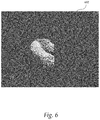
- image processing unit 207 generates a parent vessel mask based on the refined mask.
- the parent vessel mask contains only the parent vessel portion of the volumetric data. Voxels that represent the parent vessel portion are saved as foreground values in the parent vessel mask, while voxels that do not represent the parent vessel portion are set to background values.
- FIG. 6 shows an exemplary parent vessel mask 602 .
- the parent vessel mask may be generated by checking each voxel of the refined mask to ensure there is a connected straight path from a centerline point of the parent vessel to generate a parent vessel mask. More particularly, for each test voxel of the refined mask, rays are generated from the voxel to all centerline points of the parent vessel. If the ray from the test voxel hits the centerline points without hitting a background voxel, the test voxel is saved as foreground voxels in the parent vessel mask.
- test voxel is marked as a background voxel in the parent vessel mask. This test eliminates a majority or all voxels that are not part of the parent vessel.
- image processing unit 207 combines the aneurysm mask and the parent vessel mask to generate a combined mask.
- the aneurysm and parent vessel masks may be combined by marking a voxel at a particular voxel location in the combined mask as foreground (or valid) if the voxel at the same location in either the aneurysm mask or parent vessel mask is marked as foreground.
- a combined mask that contains both aneurysm and part of parent vessel is obtained.
- FIG. 7 shows an exemplary combined mask 702 .
- image processing unit 207 eliminates any kissing vessel with a large connection with the aneurysm in the combined mask.
- Kissing vessels with small connection e.g., 2 voxels wide
- some kissing vessels have large connections (e.g., >5 voxels) with the aneurysm.
- a ‘crown’ detection and elimination process may be performed.
- the ‘crown’ refers to the edge of the kissing vessel that makes a crown shape with the aneurysm. Such ‘crown’ shape information may be used to erase the kissing vessel portion from the aneurysm surface.
- the ‘crown’ may be detected by finding all voxels that do not have a straight path connection to the aneurysm dome point, but have a neighbor that does. These clusters of invalid voxels mark the edge of possible kissing vessels, and are removed from the mask.
- FIG. 8 shows an exemplary “crown” 802 .
- the “crown” 802 is a cluster of invalid voxels with valid neighbors that are detected between an aneurysm 804 and a kissing vessel 806 . Rays 810 from the aneurysm dome point 808 may be generated to check for the invalid voxels in the combined mask.
- FIGS. 9 a - f illustrate the steps for cleaning up the mask. More particularly, FIG. 9 a shows an exemplary refined mask 902 .
- FIG. 9 b shows an exemplary aneurysm mask 904 generated based on the refined mask 902 .
- FIG. 9 c shows an exemplary parent vessel mask 906 generated based on the refined mask 902 .
- FIG. 9 d shows an exemplary combined mask 908 generated by combining the aneurysm mask 904 and parent vessel mask 906 .
- the combined mask 908 still includes a portion (i.e., ‘crown’) of the kissing vessel 910 .
- FIG. 9 e illustrates the detection of the crown 915 .
- Rays are generated from the aneurysm dome point 914 in all directions through each voxel of the combined mask 908 to determine which voxels are directly hit (i.e., valid).
- Voxels that are indirectly hit by the rays but have a valid neighboring voxel are extracted as invalid voxels 915 .
- the invalid voxels 915 look like a “crown”, which indicates the border of the kissing vessel portion.
- the center point of this “crown” 915 is determined, and used with the dome point 914 to determine the borderline.
- an iterative process may be performed to determine the borderline of the kissing vessel and aneurysm surface.
- the iterative process starts with calculating the center point of the “crown” 915 .
- a cut plane is determined based on the direction from the center point to the aneurysm dome point.
- the cut plane is orthogonal to that direction.
- the number of crown points that are on either side of the cut plane is determined. If the percentage of crown points that are above (or on the side of the cut plane further away from the aneurysm dome point) is more than or equal to a predetermined threshold value (e.g., >90%), then the cut plane is assigned as the borderline and the iterative process stops.
- a predetermined threshold value e.g., >90%
- the center point is moved towards the aneurysm point by a predetermined distance to generate a new center point, and the cut plane is recalculated based on the new center point. All voxels that lie above the borderline are marked as background voxels (i.e., removed) to generate a final mask 916 , as shown in FIG. 9 f
- the final mask 916 does not contain the ‘crown’ or border of the kissing vessel portion 910 .
- FIG. 10 a shows an exemplary original volumetric image data 1002 .
- FIG. 10 b shows the final output mask 1004 generated after performing the previous clean up steps.
- the final output mask contains only the aneurysm dome 1006 and the parent vessel 1008 . Small and large kissing vessels were removed from the volumetric image data.
- image processing unit 207 outputs the final mask for segmentation of the aneurysm and the parent vessel.
- the final mask may be provided to a segmentation unit to perform the final segmentation of the volumetric image data.
- FIG. 11 a shows an exemplary final mask 1102 .
- FIG. 11 b shows an exemplary segmentation output 1104 .
- the segmentation output 1104 is more accurate than those generated by traditional segmentation methods, and shows minimal leak outside the actual aneurysm 1106 .
- FIG. 12 a shows an exemplary image 1202 generated by traditional segmentation methods.
- the segmented portions ( 1204 , 1206 ) are shown in a uniform color (e.g., orange).
- the segmentation output includes both the aneurysm in the parent vessel 1204 as well as a neighboring kissing vessel 1206 .
- FIG. 12 b shows an exemplary image 1207 generated by the present framework described herein.
- the segmented portion 1208 includes only the actual aneurysm in the parent vessel, without leaking into the kissing vessel 1210 .
- FIG. 13 a shows another exemplary image 1302 generated by traditional segmentation methods.
- the segmented portions ( 1304 , 1306 ) are shown in a uniform color (e.g., orange).
- the segmentation output includes both the aneurysm in the parent vessel 1304 as well as a neighboring kissing vessel 1306 .
- FIG. 13 b shows another exemplary image 1307 generated by the present framework described herein.
- the segmented portion 1308 is more accurate, and includes only the actual aneurysm in the parent vessel, without leaking into the kissing vessel 1310 .
Landscapes
- Engineering & Computer Science (AREA)
- Physics & Mathematics (AREA)
- Health & Medical Sciences (AREA)
- Computer Vision & Pattern Recognition (AREA)
- General Physics & Mathematics (AREA)
- Theoretical Computer Science (AREA)
- Life Sciences & Earth Sciences (AREA)
- Medical Informatics (AREA)
- General Health & Medical Sciences (AREA)
- Radiology & Medical Imaging (AREA)
- Nuclear Medicine, Radiotherapy & Molecular Imaging (AREA)
- Animal Behavior & Ethology (AREA)
- Veterinary Medicine (AREA)
- Biophysics (AREA)
- Optics & Photonics (AREA)
- Pathology (AREA)
- High Energy & Nuclear Physics (AREA)
- Biomedical Technology (AREA)
- Heart & Thoracic Surgery (AREA)
- Molecular Biology (AREA)
- Surgery (AREA)
- Public Health (AREA)
- Vascular Medicine (AREA)
- Dentistry (AREA)
- Oral & Maxillofacial Surgery (AREA)
- Physiology (AREA)
- Apparatus For Radiation Diagnosis (AREA)
- Quality & Reliability (AREA)
- Surgical Instruments (AREA)
- Prostheses (AREA)
- Magnetic Resonance Imaging Apparatus (AREA)
Abstract
Description
Claims (20)
Priority Applications (1)
| Application Number | Priority Date | Filing Date | Title |
|---|---|---|---|
| US16/515,075 US10497123B1 (en) | 2017-05-16 | 2019-07-18 | Isolation of aneurysm and parent vessel in volumetric image data |
Applications Claiming Priority (2)
| Application Number | Priority Date | Filing Date | Title |
|---|---|---|---|
| US15/597,026 US10402976B2 (en) | 2017-05-16 | 2017-05-16 | Isolation of aneurysm and parent vessel in volumetric image data |
| US16/515,075 US10497123B1 (en) | 2017-05-16 | 2019-07-18 | Isolation of aneurysm and parent vessel in volumetric image data |
Related Parent Applications (1)
| Application Number | Title | Priority Date | Filing Date |
|---|---|---|---|
| US15/597,026 Continuation US10402976B2 (en) | 2017-05-16 | 2017-05-16 | Isolation of aneurysm and parent vessel in volumetric image data |
Publications (2)
| Publication Number | Publication Date |
|---|---|
| US20190362496A1 US20190362496A1 (en) | 2019-11-28 |
| US10497123B1 true US10497123B1 (en) | 2019-12-03 |
Family
ID=62152466
Family Applications (2)
| Application Number | Title | Priority Date | Filing Date |
|---|---|---|---|
| US15/597,026 Expired - Fee Related US10402976B2 (en) | 2017-05-16 | 2017-05-16 | Isolation of aneurysm and parent vessel in volumetric image data |
| US16/515,075 Active US10497123B1 (en) | 2017-05-16 | 2019-07-18 | Isolation of aneurysm and parent vessel in volumetric image data |
Family Applications Before (1)
| Application Number | Title | Priority Date | Filing Date |
|---|---|---|---|
| US15/597,026 Expired - Fee Related US10402976B2 (en) | 2017-05-16 | 2017-05-16 | Isolation of aneurysm and parent vessel in volumetric image data |
Country Status (3)
| Country | Link |
|---|---|
| US (2) | US10402976B2 (en) |
| EP (1) | EP3404613A3 (en) |
| CN (1) | CN108876794B (en) |
Families Citing this family (13)
| Publication number | Priority date | Publication date | Assignee | Title |
|---|---|---|---|---|
| US11484322B2 (en) | 2018-01-03 | 2022-11-01 | Aneuclose Llc | Aneurysm neck bridge with a closeable opening or lumen through which embolic material is inserted into the aneurysm sac |
| US11464518B2 (en) | 2008-05-01 | 2022-10-11 | Aneuclose Llc | Proximal concave neck bridge with central lumen and distal net for occluding cerebral aneurysms |
| US11583289B2 (en) | 2008-05-01 | 2023-02-21 | Aneuclose Llc | Aneurysm-occluding mesh ribbon with a series of loops or segments having distal-to-proximal variation in size, shape, and/or orientation |
| US11357511B2 (en) | 2008-05-01 | 2022-06-14 | Aneuclose Llc | Intrasacular aneurysm occlusion device with globular first configuration and bowl-shaped second configuration |
| US11471164B2 (en) | 2008-05-01 | 2022-10-18 | Aneuclose Llc | Methods of occluding a cerebral aneurysm by inserting embolic members or material into an intrasacular implant |
| US11918423B2 (en) * | 2018-10-30 | 2024-03-05 | Corindus, Inc. | System and method for navigating a device through a path to a target location |
| CN109872328B (en) * | 2019-01-25 | 2021-05-07 | 腾讯科技(深圳)有限公司 | Brain image segmentation method, device and storage medium |
| EP3928294B1 (en) * | 2019-02-21 | 2025-12-03 | Koninklijke Philips N.V. | Methods and systems for segmentation and rendering of inverted ultrasound data |
| CN109907732B (en) * | 2019-04-09 | 2022-12-02 | 广州新脉科技有限公司 | A method and system for assessing the risk of intracranial aneurysm rupture |
| US12412246B2 (en) * | 2019-10-17 | 2025-09-09 | Nikon Corporation | Image processing method, image processing device, and image processing program |
| CN111223089B (en) * | 2020-01-17 | 2023-11-03 | 强联智创(北京)科技有限公司 | Aneurysm detection method and device and computer readable storage medium |
| EP4318393A4 (en) * | 2021-04-23 | 2025-01-08 | Wuhan United Imaging Healthcare Surgical Technology Co., Ltd. | Surgical path processing system, method, apparatus and device, and storage medium |
| KR102806707B1 (en) * | 2023-09-06 | 2025-05-20 | 주식회사 메디픽셀 | Method for generating aneurysm region and electronic device thereof |
Citations (2)
| Publication number | Priority date | Publication date | Assignee | Title |
|---|---|---|---|---|
| US20080249755A1 (en) * | 2007-04-03 | 2008-10-09 | Siemens Corporate Research, Inc. | Modeling Cerebral Aneurysms in Medical Images |
| US20130066219A1 (en) * | 2011-09-09 | 2013-03-14 | Jingfeng Jiang | Method for Assessing The Efficacy of a Flow-Diverting Medical Device in a Blood Vessel |
Family Cites Families (7)
| Publication number | Priority date | Publication date | Assignee | Title |
|---|---|---|---|---|
| US7310435B2 (en) * | 2003-11-25 | 2007-12-18 | General Electric Company | Method and apparatus for extracting multi-dimensional structures using dynamic constraints |
| EP2279490B1 (en) * | 2008-04-16 | 2012-03-21 | Université de Lausanne | Automatic detection and accurate segmentation of abdominal aortic aneurysm |
| US9846765B2 (en) * | 2012-11-09 | 2017-12-19 | Siemens Healthcare Gmbh | System and method for patient specific modeling of liver tumor ablation |
| EP3767630A1 (en) * | 2014-01-17 | 2021-01-20 | Arterys Inc. | Methods for four dimensional (4d) flow magnetic resonance imaging |
| CN104978726A (en) * | 2014-04-03 | 2015-10-14 | 上海联影医疗科技有限公司 | Blood vessel extraction method |
| US9367667B2 (en) * | 2014-04-30 | 2016-06-14 | Siemens Aktiengesellschaft | Method and system for advanced aneurysm analysis |
| US10037603B2 (en) * | 2015-05-04 | 2018-07-31 | Siemens Healthcare Gmbh | Method and system for whole body bone removal and vascular visualization in medical image data |
-
2017
- 2017-05-16 US US15/597,026 patent/US10402976B2/en not_active Expired - Fee Related
-
2018
- 2018-05-11 EP EP18171871.9A patent/EP3404613A3/en not_active Withdrawn
- 2018-05-16 CN CN201810467147.XA patent/CN108876794B/en active Active
-
2019
- 2019-07-18 US US16/515,075 patent/US10497123B1/en active Active
Patent Citations (2)
| Publication number | Priority date | Publication date | Assignee | Title |
|---|---|---|---|---|
| US20080249755A1 (en) * | 2007-04-03 | 2008-10-09 | Siemens Corporate Research, Inc. | Modeling Cerebral Aneurysms in Medical Images |
| US20130066219A1 (en) * | 2011-09-09 | 2013-03-14 | Jingfeng Jiang | Method for Assessing The Efficacy of a Flow-Diverting Medical Device in a Blood Vessel |
Non-Patent Citations (5)
| Title |
|---|
| Azadek Firouzian, et al., "Intracranial aneurysm segmentation in 3D CT angiography: Method and quantitative validation with and without prior noise filtering", European Journal of Radiology, Elsevier Science, NL, vol. 79, No. 2, Feb. 17, 2010, pp. 299-304. |
| Bernd F. Tomandl, et al, "CT Angiography of Intracranial Aneurysms: A Focus on Postprocesing", Radiographics, vol. 24, No. 3, May 1, 2004, pp. 637-655. |
| H Akhoondali, et al, "Rapid Automatic Segmentation and Visualization of Teeth in CT-Scan Data", Journal of Applied Sciences, vol. 9, No. 11, Jan. 1, 2009, pp. 2031-2044. |
| Nikravanshalmani Alireza, et al., "Segmentation and Separation of Cerebral Aneurysms: A multi-phase approach", 2013 8th International Symposium on Image and Signal Processing and Analysis (ISPA), University of Trieste and University of Zagreb, Sep. 4, 2013, pp. 505-510. |
| Search Report for Corresponding European Application No. 18171871.0, dated Sep. 20, 2018. |
Also Published As
| Publication number | Publication date |
|---|---|
| US10402976B2 (en) | 2019-09-03 |
| CN108876794A (en) | 2018-11-23 |
| CN108876794B (en) | 2022-04-12 |
| EP3404613A3 (en) | 2019-04-03 |
| US20180336676A1 (en) | 2018-11-22 |
| EP3404613A2 (en) | 2018-11-21 |
| US20190362496A1 (en) | 2019-11-28 |
Similar Documents
| Publication | Publication Date | Title |
|---|---|---|
| US10497123B1 (en) | Isolation of aneurysm and parent vessel in volumetric image data | |
| US9684980B2 (en) | Prior image based three dimensional imaging | |
| US9401047B2 (en) | Enhanced visualization of medical image data | |
| US9754390B2 (en) | Reconstruction of time-varying data | |
| US9547894B2 (en) | Apparatus for, and method of, processing volumetric medical image data | |
| US9471987B2 (en) | Automatic planning for medical imaging | |
| US20160321427A1 (en) | Patient-Specific Therapy Planning Support Using Patient Matching | |
| US20100135562A1 (en) | Computer-aided detection with enhanced workflow | |
| US10083511B2 (en) | Angiographic roadmapping mask | |
| US10460508B2 (en) | Visualization with anatomical intelligence | |
| US9691157B2 (en) | Visualization of anatomical labels | |
| US9786069B2 (en) | Refined reconstruction of time-varying data | |
| US20180092608A1 (en) | Reconstruction of Flow Data | |
| US10977792B2 (en) | Quantitative evaluation of time-varying data | |
| US8817014B2 (en) | Image display of a tubular structure | |
| Qin et al. | Three Dimensional Reconstruction of Blood Vessels and Evaluation of Vascular Stenosis Based on DSA | |
| US20100054568A1 (en) | Method and apparatus for selecting a volume of interest in one or more image data sets |
Legal Events
| Date | Code | Title | Description |
|---|---|---|---|
| AS | Assignment |
Owner name: SIEMENS HEALTHCARE GMBH, GERMANY Free format text: ASSIGNMENT OF ASSIGNORS INTEREST;ASSIGNOR:SIEMENS MEDICAL SOLUTIONS USA, INC.;REEL/FRAME:049785/0735 Effective date: 20170517 Owner name: SIEMENS MEDICAL SOLUTIONS USA, INC., PENNSYLVANIA Free format text: ASSIGNMENT OF ASSIGNORS INTEREST;ASSIGNORS:DUTTA, KOMAL;BAKER, SCOTT;SIGNING DATES FROM 20170502 TO 20170503;REEL/FRAME:049785/0701 |
|
| FEPP | Fee payment procedure |
Free format text: ENTITY STATUS SET TO UNDISCOUNTED (ORIGINAL EVENT CODE: BIG.); ENTITY STATUS OF PATENT OWNER: LARGE ENTITY |
|
| STCF | Information on status: patent grant |
Free format text: PATENTED CASE |
|
| MAFP | Maintenance fee payment |
Free format text: PAYMENT OF MAINTENANCE FEE, 4TH YEAR, LARGE ENTITY (ORIGINAL EVENT CODE: M1551); ENTITY STATUS OF PATENT OWNER: LARGE ENTITY Year of fee payment: 4 |
|
| AS | Assignment |
Owner name: SIEMENS HEALTHINEERS AG, GERMANY Free format text: ASSIGNMENT OF ASSIGNORS INTEREST;ASSIGNOR:SIEMENS HEALTHCARE GMBH;REEL/FRAME:066267/0346 Effective date: 20231219 Owner name: SIEMENS HEALTHINEERS AG, GERMANY Free format text: ASSIGNMENT OF ASSIGNOR'S INTEREST;ASSIGNOR:SIEMENS HEALTHCARE GMBH;REEL/FRAME:066267/0346 Effective date: 20231219 |