US10401349B2 - Compositions and methods for disease diagnosis using single cell analysis - Google Patents
Compositions and methods for disease diagnosis using single cell analysis Download PDFInfo
- Publication number
- US10401349B2 US10401349B2 US15/891,542 US201815891542A US10401349B2 US 10401349 B2 US10401349 B2 US 10401349B2 US 201815891542 A US201815891542 A US 201815891542A US 10401349 B2 US10401349 B2 US 10401349B2
- Authority
- US
- United States
- Prior art keywords
- cell
- small volume
- cells
- reporter
- dye
- Prior art date
- Legal status (The legal status is an assumption and is not a legal conclusion. Google has not performed a legal analysis and makes no representation as to the accuracy of the status listed.)
- Active
Links
Images


Classifications
-
- G—PHYSICS
- G01—MEASURING; TESTING
- G01N—INVESTIGATING OR ANALYSING MATERIALS BY DETERMINING THEIR CHEMICAL OR PHYSICAL PROPERTIES
- G01N33/00—Investigating or analysing materials by specific methods not covered by groups G01N1/00 - G01N31/00
- G01N33/48—Biological material, e.g. blood, urine; Haemocytometers
- G01N33/50—Chemical analysis of biological material, e.g. blood, urine; Testing involving biospecific ligand binding methods; Immunological testing
- G01N33/5005—Chemical analysis of biological material, e.g. blood, urine; Testing involving biospecific ligand binding methods; Immunological testing involving human or animal cells
- G01N33/5091—Chemical analysis of biological material, e.g. blood, urine; Testing involving biospecific ligand binding methods; Immunological testing involving human or animal cells for testing the pathological state of an organism
-
- C—CHEMISTRY; METALLURGY
- C12—BIOCHEMISTRY; BEER; SPIRITS; WINE; VINEGAR; MICROBIOLOGY; ENZYMOLOGY; MUTATION OR GENETIC ENGINEERING
- C12Q—MEASURING OR TESTING PROCESSES INVOLVING ENZYMES, NUCLEIC ACIDS OR MICROORGANISMS; COMPOSITIONS OR TEST PAPERS THEREFOR; PROCESSES OF PREPARING SUCH COMPOSITIONS; CONDITION-RESPONSIVE CONTROL IN MICROBIOLOGICAL OR ENZYMOLOGICAL PROCESSES
- C12Q1/00—Measuring or testing processes involving enzymes, nucleic acids or microorganisms; Compositions therefor; Processes of preparing such compositions
- C12Q1/02—Measuring or testing processes involving enzymes, nucleic acids or microorganisms; Compositions therefor; Processes of preparing such compositions involving viable microorganisms
- C12Q1/04—Determining presence or kind of microorganism; Use of selective media for testing antibiotics or bacteriocides; Compositions containing a chemical indicator therefor
-
- C—CHEMISTRY; METALLURGY
- C12—BIOCHEMISTRY; BEER; SPIRITS; WINE; VINEGAR; MICROBIOLOGY; ENZYMOLOGY; MUTATION OR GENETIC ENGINEERING
- C12Q—MEASURING OR TESTING PROCESSES INVOLVING ENZYMES, NUCLEIC ACIDS OR MICROORGANISMS; COMPOSITIONS OR TEST PAPERS THEREFOR; PROCESSES OF PREPARING SUCH COMPOSITIONS; CONDITION-RESPONSIVE CONTROL IN MICROBIOLOGICAL OR ENZYMOLOGICAL PROCESSES
- C12Q1/00—Measuring or testing processes involving enzymes, nucleic acids or microorganisms; Compositions therefor; Processes of preparing such compositions
- C12Q1/02—Measuring or testing processes involving enzymes, nucleic acids or microorganisms; Compositions therefor; Processes of preparing such compositions involving viable microorganisms
- C12Q1/18—Testing for antimicrobial activity of a material
-
- G—PHYSICS
- G01—MEASURING; TESTING
- G01N—INVESTIGATING OR ANALYSING MATERIALS BY DETERMINING THEIR CHEMICAL OR PHYSICAL PROPERTIES
- G01N15/00—Investigating characteristics of particles; Investigating permeability, pore-volume or surface-area of porous materials
- G01N15/10—Investigating individual particles
- G01N15/14—Optical investigation techniques, e.g. flow cytometry
- G01N15/1429—Signal processing
- G01N15/1433—Signal processing using image recognition
-
- G01N15/1463—
-
- G—PHYSICS
- G01—MEASURING; TESTING
- G01N—INVESTIGATING OR ANALYSING MATERIALS BY DETERMINING THEIR CHEMICAL OR PHYSICAL PROPERTIES
- G01N21/00—Investigating or analysing materials by the use of optical means, i.e. using sub-millimetre waves, infrared, visible or ultraviolet light
- G01N21/62—Systems in which the material investigated is excited whereby it emits light or causes a change in wavelength of the incident light
- G01N21/63—Systems in which the material investigated is excited whereby it emits light or causes a change in wavelength of the incident light optically excited
- G01N21/64—Fluorescence; Phosphorescence
- G01N21/6428—Measuring fluorescence of fluorescent products of reactions or of fluorochrome labelled reactive substances, e.g. measuring quenching effects, using measuring "optrodes"
-
- G—PHYSICS
- G01—MEASURING; TESTING
- G01N—INVESTIGATING OR ANALYSING MATERIALS BY DETERMINING THEIR CHEMICAL OR PHYSICAL PROPERTIES
- G01N33/00—Investigating or analysing materials by specific methods not covered by groups G01N1/00 - G01N31/00
- G01N33/48—Biological material, e.g. blood, urine; Haemocytometers
- G01N33/50—Chemical analysis of biological material, e.g. blood, urine; Testing involving biospecific ligand binding methods; Immunological testing
- G01N33/5005—Chemical analysis of biological material, e.g. blood, urine; Testing involving biospecific ligand binding methods; Immunological testing involving human or animal cells
- G01N33/5008—Chemical analysis of biological material, e.g. blood, urine; Testing involving biospecific ligand binding methods; Immunological testing involving human or animal cells for testing or evaluating the effect of chemical or biological compounds, e.g. drugs, cosmetics
- G01N33/5011—Chemical analysis of biological material, e.g. blood, urine; Testing involving biospecific ligand binding methods; Immunological testing involving human or animal cells for testing or evaluating the effect of chemical or biological compounds, e.g. drugs, cosmetics for testing antineoplastic activity
-
- G—PHYSICS
- G01—MEASURING; TESTING
- G01N—INVESTIGATING OR ANALYSING MATERIALS BY DETERMINING THEIR CHEMICAL OR PHYSICAL PROPERTIES
- G01N33/00—Investigating or analysing materials by specific methods not covered by groups G01N1/00 - G01N31/00
- G01N33/48—Biological material, e.g. blood, urine; Haemocytometers
- G01N33/50—Chemical analysis of biological material, e.g. blood, urine; Testing involving biospecific ligand binding methods; Immunological testing
- G01N33/58—Chemical analysis of biological material, e.g. blood, urine; Testing involving biospecific ligand binding methods; Immunological testing involving labelled substances
- G01N33/582—Chemical analysis of biological material, e.g. blood, urine; Testing involving biospecific ligand binding methods; Immunological testing involving labelled substances with fluorescent label
-
- G—PHYSICS
- G01—MEASURING; TESTING
- G01N—INVESTIGATING OR ANALYSING MATERIALS BY DETERMINING THEIR CHEMICAL OR PHYSICAL PROPERTIES
- G01N15/00—Investigating characteristics of particles; Investigating permeability, pore-volume or surface-area of porous materials
- G01N15/10—Investigating individual particles
- G01N2015/1006—Investigating individual particles for cytology
-
- G—PHYSICS
- G01—MEASURING; TESTING
- G01N—INVESTIGATING OR ANALYSING MATERIALS BY DETERMINING THEIR CHEMICAL OR PHYSICAL PROPERTIES
- G01N15/00—Investigating characteristics of particles; Investigating permeability, pore-volume or surface-area of porous materials
- G01N15/10—Investigating individual particles
- G01N15/14—Optical investigation techniques, e.g. flow cytometry
- G01N15/1404—Handling flow, e.g. hydrodynamic focusing
- G01N2015/1406—Control of droplet point
-
- G—PHYSICS
- G01—MEASURING; TESTING
- G01N—INVESTIGATING OR ANALYSING MATERIALS BY DETERMINING THEIR CHEMICAL OR PHYSICAL PROPERTIES
- G01N15/00—Investigating characteristics of particles; Investigating permeability, pore-volume or surface-area of porous materials
- G01N15/10—Investigating individual particles
- G01N15/14—Optical investigation techniques, e.g. flow cytometry
- G01N2015/1481—Optical analysis of particles within droplets
-
- G—PHYSICS
- G01—MEASURING; TESTING
- G01N—INVESTIGATING OR ANALYSING MATERIALS BY DETERMINING THEIR CHEMICAL OR PHYSICAL PROPERTIES
- G01N2201/00—Features of devices classified in G01N21/00
- G01N2201/06—Illumination; Optics
- G01N2201/062—LED's
- G01N2201/0621—Supply
-
- G—PHYSICS
- G01—MEASURING; TESTING
- G01N—INVESTIGATING OR ANALYSING MATERIALS BY DETERMINING THEIR CHEMICAL OR PHYSICAL PROPERTIES
- G01N2800/00—Detection or diagnosis of diseases
- G01N2800/26—Infectious diseases, e.g. generalised sepsis
Definitions
- This disclosure relates generally to cellular biology and more particularly to compositions and methods for rapid and sensitive identification of disease-causing cells and the rapid and sensitive characterization of their response to the drugs used to treat them.
- DCCs Disease-Causing Cells
- methods for identifying DCCs and characterizing their response to treatment typically require either multiplication of the target cells and/or target-dependent amplification of the target cells' molecular contents and/or products, depending on the application.
- nucleic acid amplification (e.g., PCR-based) tests have become the preferred method for fast pathogen identification.
- Culture based methods are the currently preferred method for antimicrobial susceptibility testing because the methods assess the microbial phenotype, providing high clinical validity.
- NATs require complicated sample workflow steps which usually include cell lysis followed by nucleic acid concentration step that also removes PCR inhibitors from the samples.
- Cell lysis creates an asymmetry in the requirements for nucleic acid extraction efficiency.
- Mycobacteria and fungi possess a very thick cell wall compared to gram-negative bacteria and thus are far more difficult to lyse, usually requiring a mechanical lysis step to efficiently disrupt their cell wall. Consequently, NATs that utilize simple chemical lysis methods often lack sensitivity for these tougher pathogens.
- cell lysis reagents are extremely inhibitory to PCR reaction because they are designed to efficiently denature proteins and PCR utilizes proteins to perform the amplification reaction.
- NATs require expensive assay development methods because they rely on pathogen-specific reporter molecules (primers and probes) that must be designed specifically for each target. Each NAT must therefore include an expensive molecular R&D process which involves primer/probe design and screening for each target.
- NAT assay includes more than one target (multiplex assay), requiring more than one reporter species (primer pair)
- the different target and/or reporter species can interact nonspecifically with one another, causing either false positives when the reporter amplifies nonspecifically against the other reagents or false-negatives when the target amplification reaction is inhibited by a non-specific interaction with another species. Consequently, this limits how many targets can be identified within a single NAT. This becomes particularly relevant with the issue of drug resistance because, in the case of gram-negative bacteria and mycobacteria, there are numerous mutations (each mutation being a target) indicative of resistance. NATs can only interrogate a small fraction of those mutations within a single test.
- MRSA Methicillin-resistant Staphylococcus aureus
- NATs that include two or more targets (multiplexed) cannot simultaneously quantify those targets accurately and precisely. This is because the same nonspecific interactions between reporter species described above also cause variances in the PCR signal output and quantitative PCR relies on reproducible reaction results in order to correlate the generated amplification curves to the initial target concentration. This is a significant limitation because it prevents the use of multiplex NATs for the diagnosis of infections from non-sterile fluids since humans are often colonized by the same pathogens that can cause an infection. In non-sterile fluids, therefore, the number of pathogens present in the clinical sample (the pathogen load) is what determines whether a bacterial species is causing an infection or “peacefully” colonizing the fluid.
- the pathogen load for any bacteria present in the sample must exceed 10 3 CFU/mL to be considered the source of an infection.
- the threshold is 10 5 CFU/mL.
- DCCs disease causing cells
- DCCs are defined herein as either host cells, such as malignant or disease-associated cells of the host from which the sample is taken (e.g., cancer cells), or acquired cells (e.g., fungal or bacterial), which include any microflora associated with, involved in, implicated in, or indicative of a disease or pathology.
- diseases include, but are not limited to cancer and infections.
- Embodiments of the invention are directed to methods that identify pathogens by recording and interpreting a time-dependent signal produced by unique cellular metabolism, respiration, and/or permeability attributes of isolated cells. In certain aspects of the current invention does not require lysis or washing.
- metabolism refers to the set of life-sustaining chemical transformations or processes within a cell. The three main purposes of metabolism are the conversion of food/fuel to energy to run cellular processes; the conversion of food/fuel to building blocks for proteins, lipids, nucleic acids, and some carbohydrates; and the elimination of nitrogenous wastes.
- Respiration as referred to herein is cellular respiration, a set of metabolic reactions and processes that take place in a cell or organisms to convert biochemical energy from nutrients into adenosine triphosphate (ATP), and then release waste products.
- ATP adenosine triphosphate
- intact cells are used, only whole cells need to be manipulated rather than nucleic acid molecules, which are much more difficult manipulate due to their small size and propensity for charge-based interactions with different materials.
- the cells can be incubated at a single temperature, typically a relatively low temperature in the range of 25 to 45° C., obviating the need for thermal cycling equipment required for most NATs, which reduces cost and workflow complexity of the currently described invention.
- the invention avoids significant issues that can arise with fluid evaporation and/or bubbles that can disrupt the integrity of the reaction and/or the fluorescent readout.
- a reporter is a molecule, peptide, protein, or other compound that varies in fluorescence emission, absorption, and/or reflectance in relation to a variation in environment or condition, such as reduction-oxidation (redox) state.
- redox reduction-oxidation
- the methods described herein produce metabolic, respiratory, or drug-susceptibility profiles that can be used to identify or phenotype a target cell in a sample.
- this method uses a mammalian cell's or microbial cell's unique metabolism, respiration, and/or permeability characteristics to distinguish between different cellular and microbial species, those same characteristics can be used to determine whether the cell or microbe is susceptible to a particular compound, cytotoxic compound, or antimicrobial, since a compound or an effective drug will alter the cellular metabolism, respiration, and/or permeability characteristics.
- methods of the invention can interrogate the interactions of a compound(s) or conditions on a cell, be it (i) a normal cell for determining toxicity of a compound or condition, or (ii) a pathogenic or disease-related cell for determining therapeutic efficacy of a compound or condition.
- This method can account for all resistance mechanisms that may confer resistance to a particular cell and, therefore, offers higher clinical validity than that of NATs, which can only account for a fraction of the resistance mechanism. This is why, despite being much faster, NATs have not been able to replace culture-based methods for drug-susceptibility testing. NATs typically produce a result in about an hour whereas existing culture-based drug-susceptibility methods typically require over 48 hrs.
- the currently described methods produce phenotypic susceptibility results at significantly faster turn-around-times ( ⁇ 4 to 6 hrs) than culture-based phenotypic methods.
- the increase in speed compared to culture is accomplished, in part, by the rapid signal concentration made possible at sub-nanoliter volumes which are orders of magnitude smaller than the milli- and microliter volumes typically used by other methods.
- confinement of each cell or microbe into sub-nanoliter droplets enables reporters (e.g., fluorescent molecules) to be rapidly detected or rapidly concentrate to detectable levels.
- this method can detect single cells, the method is as sensitive as NATs which are more sensitive the culture methods.
- the described methods offers the analytical validity of NATs combined with the clinical validity of culture based methods.
- individual cells or microbes are isolated or partitioned into separate droplets, enabling quantification to become the simple matter of counting those droplets that produced a signal indicating the presence of a target cell or microbe.
- the shape or change of the reporter signal over time e.g., a waveform
- a waveform which is one of the characteristics relied upon for pathogen identification and characterization, is orthogonal to the method for quantifying the number of pathogens—one does not affect the other.
- the reporter or cell viability reagents used in the invention are inexpensive compared to the materials used in NATs. And because of their simple structure, they can be easily lyophilized and solubilized.
- a variety of reporters may be used with the systems and methods disclosed herein.
- the at least one small molecule metabolic reporter can be a fluorophore, a protein labeled fluorophore, a protein comprising a photooxidizable cofactor, a protein comprising another intercalated fluorophore, a mitochondrial vital stain or dye, a dye exhibiting at least one of a redox potential, a membrane localizing dye, a dye with energy transfer properties, and/or a pH indicating dye.
- the reporter can be or include a resazurin dye, a tetrazolium dye, coumarin dye, an anthraquinone dye, a cyanine dye, an azo dye, a xanthene dye, an arylmethine dye, a pyrene derivative dye, a ruthenium bipyridyl complex dye or a derivative thereof.
- “derivative” is understood to mean a chemically modified form of a dye that maintains some of the detection characteristics, e.g., fluorescent sensitivity to redox state, of the compound from which it is derived. Certain embodiments utilize a resazurin-based dye which is very inexpensive compared to PCR reagents, such as expensive enzymatic components.
- Some compounds or antimicrobials are only effective against certain cells or microbes (indeed, this is why, in the absence of definitive drug-susceptibility information, it is important to identify the causative pathogen to guide therapy), for example, a microbe's response to a particular antimicrobial aids in identifying the microbe. This sensitivity is one reason bacterial culture methods utilize antibiotics in the growth media to identifying the growing bacteria. Certain embodiments of the invention provide a method for rapid and sensitive identification of disease-causing cells and the rapid and sensitive characterization of their response to the drugs used to treat them.
- target cells are re-suspended with a reporter (e.g., a resazurin-based dye), poising agents or test compounds (optional), and cell nutrients (e.g., growth media), the suspension is compartmentalized into droplets and organized into a two-dimensional array where they are incubated and their fluorescence is monitored over time using an imaging system.
- a reporter e.g., a resazurin-based dye
- poising agents or test compounds optionalal
- cell nutrients e.g., growth media
- the resorufin is reversibly reduced to hydroresorufin, which is a non-fluorescent molecule. If the redox potential rises above the same certain threshold, the hydroresorufin is oxidized back to resorufin and fluoresces again.
- the amount of fluorescence emitted from a droplet can undulate over time depending on the changing environment or conditions (e.g., redox potential) of the droplet. Because cells have different metabolic or redox characteristics, cells will produce characteristic fluorescent undulations (signatures) that can be used to identify which cell type is in the droplet.
- a characteristic signature can also be generated by stressing the cell with an environmental stressor or condition, such as an antimicrobial drug or potential therapeutic. By combining the information contained within one or more of these signatures, single cells contained within each droplet can be identified and characterized.
- the cells in a test sample are divided into two populations, one population (the test population) includes an environmental stressor or test condition, and in the other population (the control population) the environmental stressor or test condition is excluded.
- the two populations are observed over time.
- the cells in the test sample may be identified by population characteristics and/or from the signatures in a control population. Identification may be further aided by the signatures generated in the test population, but the primary purpose of the test population is to characterize the response of the cells to the environmental stressor or test condition by comparing the test population signatures to the control signatures.
- Particular embodiments are directed to methods for identifying and characterizing a disease causing cell (DCC) in a sample or diagnosing a disease associated with a disease causing cell.
- the methods can comprise (a) contacting a sample suspected of containing one or more target disease causing cells (DCCs) with at least one reporter (e.g., a viability or reporter dye) forming a sample mixture; (b) partitioning the sample mixture into partitions comprising at most one target DCC or natural DCC aggregate per partition; (c) incubating the partitions for a period of time at a specified temperature or series of temperatures; (d) monitoring optical characteristics of the partitions during the incubation time; (e) constructing a waveform (signal over time) for each partition based on the optical characteristics over time; and (f) evaluating the sample using information provided by partition waveforms.
- DCCs disease causing cell
- a natural DCC aggregate is an association of two or more cells having the same phenotype (i.e., a homogenous aggregate), the association of which is not readily dissociated by the processing conditions.
- the DCC aggregate is an aggregate of microbes having the same phenotype.
- the partitions are droplets in an immiscible fluid. Upon mixing of the target-containing solution and the immiscible fluid, they form phases—an aqueous drop or partition, which holds the target material in solution, and a non-aqueous phase made up of the immiscible fluid.
- the immiscible fluid can be a fluorocarbon comprising a fluorosurfactant or hydrocarbon oils such as mineral oil, or silicone oils.
- the droplets can be between 0.1 pL and 10 nL. In a further aspect the droplets are at least, at most, or about 0.1.
- the methods can further comprise arranging the droplets in a two-dimensional array.
- the two-dimensional array is a static two-dimensional array. Partitioning of the sample can be done by Laplace pressure gradients or by using shear stress methods, as well as other methods for drop or partition formation.
- the optical characteristics of the partitions can be performed using a detector, such as a camera or the like.
- the optical characteristics include fluorescence of the partition(s).
- the monitoring of the optical characteristics of the partitions further comprises illuminating or exposing the partition with electromagnetic radiation, such as light.
- the electromagnetic radiation comprises an excitation wavelength that is compatible with the reporter, i.e., illuminating or irradiating a partition with an appropriate source.
- the source provides light including an excitation wavelength of 500, 525, 550, 575, 600, 625, 650, 675, to 700 nm, including all values and ranges there between.
- the source will be selected so that the electromagnetic radiation excites one or more reporter in the samples, e.g., dyes or other compounds.
- the light source can be a light emitting diode (LED).
- Reporters can include a “viability dye” or “reporter dye”, the viability or reporter dye is a moiety that detects changes in the environment surrounding an isolated cell due to a cell's viability, respiration, or metabolic activity; or is a detectable protein that is expressed under specific conditions (e.g., green fluorescent protein or luciferase).
- a cell can be transfected or engineered to express a reporter protein.
- the reporter can be detected using any method known in the art appropriate to the reporter employed, for example light emission or absorbance of a fluorophore or a colorimetric dye.
- the signal from the reporter is detected by optical microscopy, camera, or other detector/sensor as appropriate.
- the reporter is a fluorescent dye.
- the reporter is resazurin, a resazurin-based dye, or a dye that is a derivative of or structurally related to resazurin (7-Hydroxy-3H-phenoxazin-3-one 10-oxide).
- Resazurin is a non-toxic, cell permeable compound that, in its oxidized state, is blue in color and virtually non-fluorescent. When in contact with living cells, resazurin is reduced to resorufin, a compound that is red in color and highly fluorescent, and can be detected fluorimetrically or colorimetrically.
- a resazurin-based dye is a dye that contains a resazurin structure in addition to other modifying groups.
- a viability dye is tetrazolium, a tetrazolium-based dye, or a dye that is a derivative of or structurally related to tetrazolium.
- a tetrazolium-based dye is a dye that contains a tetrazolium moiety and may contain other modifying groups that do not disrupt the five membered tetrazolium ring.
- the incubating of the partitions is at a constant temperature (isothermal).
- the temperature can be controlled and can be stepped or ramped up using a particular interval or rate, such as stepping up from 25 to 37° C. or increasing at a rate of 2 to 10° C. per minute.
- temperature can be decreased at a particular interval or rate, such as decreasing at an interval of 5 to 10° C. or a rate of 2 to 10° C. per minute.
- the temperature(s) are in the range of 20 to 45° C., 30 to 40° C., or 35 to 38° C., including all values and ranges there between.
- partitions are incubated at 37° C.
- the partitions comprise a single cell, microbe, or cellular or microbial aggregation.
- the partition may contain 2, 3, 4, or more cell or microbe types.
- the methods can further comprise classifying a microbe by species, genus, family, order, class, phylum, kingdom, or a combination thereof. The classification can be based on the characteristics of one or more waveforms under one or more conditions.
- the microbe is a bacteria. Certain aspects of the invention can include classifying the bacteria by gram-stain group or other classification criteria recognized for microbes, including bacteria.
- a partition may contain more than one target type (species, genus, etc.) but that an environmental stressor or condition may kill all but one target type, which can be identified using its signature or waveform.
- the methods can further comprise dividing the sample into a control sample and at least one test sample prior to partitioning.
- Each test sample can be treated or processed in a manner that differs from the control.
- at least one test sample is contacted with a stressor, cytotoxic, anticancer, antimicrobial compound or condition.
- individual test samples can be exposed to different concentrations compounds or variations in conditions.
- a test sample can be exposed to a variety of temperatures, environments, or chemicals that may or may not alter the phenotype of the cells contained in the test sample.
- a DCC is a pathologic or pathogenic cell, such as a cancer cell.
- the methods include evaluating the sample (control and test samples) using the partition waveform (i.e., signal detected over time).
- evaluating includes comparing the partition waveform to a library of stored or predetermined waveforms (e.g., waveform reference).
- Certain embodiments are directed to methods for detecting and characterizing DCCs, such as microbes or cancer cells, in a sample comprising (a) contacting a sample comprising microbes with a reporter, e.g., a viability dye, forming a sample mixture; (b) dividing the sample mixture into at least two portions or samples that include a control sample and at least one test sample; (d) introducing a test compound/substance or an antimicrobial drug to the at least one test sample; (e) partitioning each of the control sample and at least one test sample into partitions forming control sample partitions and test sample partitions, wherein the partitions comprise at most one target microbe or a natural aggregation of microbes; (f) incubating the partitions over time at a specific temperature or temperatures; (g) monitoring optical characteristics of the partitions during the incubation time, wherein the optical characteristics include the amount of optical signal produced by interaction of the reporter with the microbe in the partition; (h) constructing an optical signal waveform for each partition
- test compound can include a small molecule, peptide, a nanoparticle, or a protein.
- a test substance can include bacteriophage and other engineered therapeutics.
- the test compound/substance can be a therapeutic identified as a therapy or engineered as a therapy for other disease conditions, such as cancer (e.g., chemotherapeutic or anti-cancer compound or substance).
- Cancer diagnosis is very similar to infectious disease diagnosis in that disease causing cells can mutate rapidly and can develop resistance to the drugs used to treat them.
- cancer cells exhibit different morphological, metabolic, and respiratory characteristics than healthy cells. Specifically, cancer cells are known to exhibit different oxidation-reduction characteristics from healthy cells. Furthermore, cancer cells are often much larger than healthy cells which also influences the amount of fluorescence generated during respiration. Because, certain aspects of this method are sensitive to oxidation-reduction (redox) changes that occur during cellular respiration, those differences, as well as others, may be used to distinguish cancer cells from healthy cells.
- redox oxidation-reduction
- cancer cells often bypass apoptosis pathways and are therefore able to remain viable for longer periods of time when they no longer reside in specific tissues.
- the cellular respiration would last longer for cancer cells compared to healthy cells, which could be observed as signal accumulates for longer periods of time for cancer cells compared to healthy cells.
- the waveforms for normal cells will differentiate them from pathogenic or cancer cells.
- partition refers to a volume of fluid (e.g., liquid or gas) that is a separated portion of a bulk volume.
- a bulk volume may be partitioned into any suitable number (e.g., 10 2 , 10 3 , 10 4 , 10 5 , 10 6 , 10 7 , etc.) of smaller volumes or partitions.
- Partitions may be separated by a physical barrier or by physical forces (e.g., surface tension, hydrophobic repulsion, etc.).
- Partitions generated from the larger volume may be substantially uniform in size (monodisperse) or may have non-uniform sizes (polydisperse).
- Partitions may be produced by any suitable manner, including emulsion, microfluidics, and microspray methods.
- partitions are droplets.
- the term “droplet” refers to a small volume of liquid which is immiscible with its surroundings (e.g., gases, liquids, surfaces, etc.).
- a droplet may reside upon a surface, be encapsulated by a fluid with which it is immiscible, such as the continuous phase of an emulsion, a gas, or a combination thereof.
- a droplet is typically spherical or substantially spherical in shape, but may be non-spherical. The shape of an otherwise spherical or substantially spherical droplet may be altered by deposition onto a surface.
- a droplet may be a “simple droplet” or a “compound droplet,” wherein one droplet encapsulates one or more additional smaller droplets.
- the volume of a droplet and/or the average volume of a set of droplets provided herein is typically less than about one microliter, for example droplet volume can be about 1 ⁇ L, 0.1 ⁇ L, 10 pL, 1 pL, 100 nL, 10 nL, 1 nL, 100 fL, 10 fL, 1 fL, including all values and ranges there between.
- the diameter of a droplet and/or the average diameter of a set of droplets provided herein is typically less than about one millimeter, for example 1 mm, 100 ⁇ m, 10 ⁇ m, to 1 ⁇ m, including all values and ranges there between.
- Droplets may be formed by any suitable technique, including emulsification, microfluidics, etc., and may be monodisperse, substantially monodisperse (differing by less than 5%), or polydisperse.
- the words “comprising” (and any form of comprising, such as “comprise” and “comprises”), “having” (and any form of having, such as “have” and “has”), “including” (and any form of including, such as “includes” and “include”) or “containing” (and any form of containing, such as “contains” and “contain”) are inclusive or open-ended and do not exclude additional, unrecited elements or method steps.
- FIG. 1 is a schematic of a preferred embodiment of the invention applied to generally to disease causing cells.
- FIG. 2 is a schematic of a preferred embodiment of the invention which as a method pathogen identification and antimicrobial susceptibility testing by monitoring the fluorescence over time of individual cells and/or monoclonal cell clusters isolated in subnanoliter droplets with a redox-sensitive viability dye.
- FIG. 3 is an illustration of a two-dimensional droplet array.
- FIG. 4 is an illustration of a droplet monolayer being imaged by a camera.
- the droplet monolayer provides for good thermal conductivity and temperature control.
- FIG. 5 is an image a droplet array containing E. coli cells.
- FIG. 6 is a schematic providing one explanation of as to fluorescence variation according to the redox environment established by an isolated cell.
- FIG. 7 illustrates a waveform derived from an E. coli cell in a 268 pL droplet.
- FIG. 8 illustrates multiple waveforms from an array comprising a multiple cell types ( E. coli and S. epidermis ) partitioned into microdroplets. The waveforms are clearly distinguishable.
- FIG. 9 provides a comparison of fluorescence relative to volume.
- the fluorescence waveform generated by a single bacterium incubated in pico-liter droplets rises much faster than in larger volumes, as expected, but also unexpectedly collapses due to a different redox potential in the picodroplet.
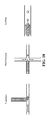
- FIG. 10 illustrates one method of using shear stress for droplet generation.
- FIG. 11 illustrates two methods of using Laplace pressure gradients for droplet generation.
- FIG. 12 illustrates the distinction between waveform monitoring as compared to a discreet value at particular time.
- the redox potential causes fluorescence to vary depending on the bacterial species
- traditional endpoint analysis does not provide distinguishable viability information.
- FIG. 13 illustrates that waveform information provides an increased sensitivity when detecting drug-susceptibility.
- FIG. 14 illustrates stored waveforms representing four bacterial species and their correlation to observed waveforms from the same species (median correlation coefficient) and, thus, can be used to identify the bacteria that generated the observed waveforms.
- Stored waveforms also correlate well according to gram stain gram ( B. subtilis and S. epidermidis are gram positives, E. coli and S. maltophilia are gram negatives).
- invention is not intended to refer to any particular embodiment or otherwise limit the scope of the disclosure. Although one or more of these embodiments may be preferred, the embodiments disclosed should not be interpreted, or otherwise used, as limiting the scope of the disclosure, including the claims.
- discussion has broad application, and the discussion of any embodiment is meant only to be exemplary of that embodiment, and not intended to intimate that the scope of the disclosure, including the claims, is limited to that embodiment.
- the invention generally relates to methods for disease diagnosis using single-cell analysis.
- the following sections discuss general considerations for test samples, compartmentalization/partitioning, cell viability and viability or reporter dyes, disease-causing cell aggregates/aggregation of microbes, signal detection, and multiplexing.
- a test sample comprising at least one target cell is combined with a viability or reporter dye and partitioned into droplets such that a statistically significant number of droplets contain no more than one target cell or aggregation of cells (some microbial species tend to aggregate into cell clusters or chains).
- a viability or reporter dye will be reduced from a non-fluorescent molecule to a fluorescent molecule in the presence of a viable cell and then further reduced to a non-fluorescent molecule if the redox potential in the droplet drops below a certain amount, typically ⁇ 100 mV.
- the fluorescent signature generated in each droplet is monitored over time and used to identify and characterize the cell contained within. Further details on the processes of the invention are provided below.
- Target cells in the test sample include bacteria, fungi, plant cells, animal cells, or cells from any other cellular organism.
- the cells may be cultured cells or cells obtained directly from naturally occurring sources.
- the cells may be obtained directly from an organism or from a biological sample obtained from an organism, e.g., from sputum, saliva, urine, blood, cerebrospinal fluid, seminal fluid, stool, and tissue. Any tissue or body fluid specimen.
- the test sample includes cells that are isolated from a biological sample comprising a variety of other components, such as non-target cells (background cells), viruses, proteins, and cell-free nucleic acids.
- the cells may be infected with a virus or another intracellular pathogen.
- the test sample comprises cells suspended in a nutrient medium that enables them to replicate and/or remain viable.
- the nutrient media may be defined media with known quantities or all ingredients or an undefined media where the nutrients are complex ingredients such yeast extract or casein hydrolysate, which contain a mixture of many chemical species of unknown proportions, including a carbon source such as glucose, water, various salts, amino acids and nitrogen.
- the target cells in the test sample comprise pathogens and the nutrient media comprises a commonly used nutrient broth (liquid media) for culturing pathogens such as lysogeny broth, nutrient broth or tryptic soy broth.
- the media may be supplemented with a blood serum or synthetic serum to facilitate the growth of fastidious organisms.
- the methods of the invention involve combining a test sample comprising at least one target cell with a viability or reporter dye and then partitioning the test sample into droplets such that no droplet contains more than one target cell or cell aggregates.
- the number of droplets can vary from hundreds to millions depending on the application and droplet volumes can also vary between 1 pL to 100 nL depending on the application, but preferably between 25-500 pL.
- the methods described herein are compatible with any droplet generation method. Exemplary methods for droplet generation are shown in FIG. 10 and FIG. 11 .
- the methods for droplet formation differ, all the methods disperse an aqueous phase, the test sample in this case, into an immiscible phase, also referred to as the continuous phase, so that each droplet is surrounded by an immiscible carrier fluid.
- the immiscible phase is an oil wherein the oil comprises a surfactant.
- the immiscible phase is a fluorocarbon oil comprising a fluoro-surfactant.
- Laplace pressure is the differential pressure between the inside and outside of a curved surface, such as the difference in pressure between the inside and outside of a droplet.
- An aqueous phase containing cells or microbes can be introduced into a device having a reservoir of a continuous phase (i.e., immiscible fluid) forming an aqueous “tongue” in an appropriate device.
- the device can incorporate height variation(s) into a microchannel that subject the immiscible interfaces to a difference in curvature between the portion of the aqueous phase that has not encountered the height variation and the portion of the aqueous phase downstream of the height variation.
- a critical curvature is reached for the portion of the aqueous phase downstream of the height variation beyond which the two portions cannot remain in static equilibrium, breaking of the aqueous phase into a droplet, as the downstream portion detaches from the tongue formed by introduction of the aqueous phase into a continuous phase, the size of the drops being determined by the device geometry.
- the height variation can be accomplished with a single step change in the height of a microchannel (step emulsification), multiple steps (multi-step emulsification), and a ramp or similarly gradual gradients of confinement.
- the at least one small molecule metabolic reporter can be a fluorophore, a protein labeled fluorophore, a protein comprising a photooxidizable cofactor, a protein comprising another intercalated fluorophore, a mitochondrial vital stain or dye, a dye exhibiting at least one of a redox potential, a membrane localizing dye, a dye with energy transfer properties, a pH indicating dye.
- the reporter can be or include a resazurin dye, a tetrazolium dye, coumarin dye, an anthraquinone dye, a cyanine dye, an azo dye, a xanthene dye, an arylmethine dye, a pyrene derivative dye, a ruthenium bipyridyl complex dye or derivatives thereof. Certain embodiments utilize a resazurin-based dye.
- Cell viability dyes which are also included in the term reporter used herein, are used as analysis reagents to identify and characterize individual cells or pathogens encapsulated within droplets. Viability dyes have been used since the 1950's for cell viability purposes.
- reagents are typically employed in samples that are significantly greater than 1 microliter in volume and/or are used as an endpoint assay to indicate the presence of viable cells.
- aspects of the invention use a viability dye in droplets that are between 1 pL and 100 nL, and more specifically 25-500 pL.
- the optical signal generated by the viability dye is concentrated by the small droplet volume and measured and recorded over an incubation time.
- this results in an optical signature that is rapidly generated and has information about the characteristics of the cell encapsulated within the droplets.
- an additional signature can be generated by monitoring the optical signal of the droplets containing a cell over time.
- optical signatures from the cell with and without the environmental stressor can be used to determine the identity and/or characteristics of the cell. Furthermore, the differences between the optical signatures obtained from a species of cells exposed to a drug compared to the optical signatures for same species of target cells that are not exposed to the drug can be used to determine the phenotypic drug resistance profile for the target cells obtained from a test sample. Because these signatures are generated from individual cells encapsulated in droplets, they represent information about the individual characteristics of each cell as opposed to an average characteristic of a population of cells that is generated from a bulk sample containing many cells.
- the methods of the invention are compatible with any viability or reporter dye that can be used with live cells (does not require cell lysis).
- the viability dye is a resazurin-based dye or derivative thereof.
- non-fluorescent resazurin When blue, non-fluorescent resazurin is irreversibly reduced to pink and highly fluorescent resorufin ( FIG. 6 ) it produces a fluorescent signal and a colorimetric shift (from blue to pink).
- the fluorescence is used because it offers better sensitivity over colorimetric signal changes. The limited-diffusion confinement within a sub-nanoliter volume of secreted fluorescent molecules quickly concentrates to detectable signal levels and is then detected by the methods described below.
- resorufin is reversibly reduced to non-fluorescent hydroresorufin ( FIG. 6 ) if the redox environment dips below a particular redox threshold, usually around ⁇ 100 mV.
- a particular redox threshold usually around ⁇ 100 mV.
- the combination of irreversible reduction from resazurin to resorufin and the reversible reduction of resorufin to hydroresorufin and oxidation of hydroresorufin back to resorufin depending on the redox potential of the droplet are what create the unique fluorescence signature over time in droplets that are small enough volume such that redox changes occur quickly in the presence of a single cell or cell aggregate.
- resazurin-based dyes examples include AlamarBlueTM (various), PrestoBlueTM (Thermo Fisher Scientific), Cell-titer BlueTM (Promega), or Resazurin sodium salt powder.
- Dyes that are structurally related to resazurin and can be also be used in the method are: 10-acetyl-3, 7-dihydroxyphenoxazine (also known as Amplex RedTM) 7-ethoxyresorufin, and 1,3-dichloro-7-hydroxy-9,9-dimethylacridine-2(9H)-one (DDAO dye).
- dyes that rely on tetrazolium-reduction can be used as the cell viability indicator.
- examples include INT, MTT, XTT, MTS, TTC or tetrazolium chloride, NBT, and the WST series.
- DCC Cell Aggregates.
- a preferred application of the invention is towards the diagnosis of microbial infections by identifying the microbes causing the infection and whether or not they are resistant to antimicrobial drugs.
- the DCCs can be single-celled microbes.
- some droplets may comprise an aggregate of cells of the same microbial species (homogenous aggregate) rather than a single microbe.
- the shape of the curve may be affected by the number of cells in the aggregate.
- the stored signature waveforms and call logic that are used to classify the compartmentalized cells can account for such aggregates the same way they can account for single cells.
- the embodiment includes antimicrobial susceptibility testing the mixture comprising the antimicrobial drug will exhibit the same cell aggregation characteristics as the mixture that excludes the antimicrobial drug and the comparison will still be accurate. Therefore, while the method of the invention generally comprises isolation of single-cells in each droplet, it necessarily accommodates the case of a single cell species in a homogenous aggregate isolated in the droplet rather individual cells.
- the target DCCs typically do not aggregate if they are circulating tumor cells. If the cancer cells are obtained from tissue, the tissue is typically disintegrated into individual cells prior to analysis. Therefore, each droplet will contain at most one cell; however, in some instances a cancer aggregate may also be analyzed using the described methods.
- the droplets Once the droplets have been generated, they must be presented for analysis by an optical system, sensor, or sensor array.
- the droplets are presented in a two-dimensional array ( FIGS. 3, 4, and 5 ) so that good thermal control can be maintained and the droplet signals can be measured simultaneously (at a single instance in time) for many droplets.
- the reporter In the droplets containing target cells, the reporter will produce a concentrated fluorescent signal that will rise above the background droplets that do not contain cells ( FIGS. 3, 4, and 5 ).
- the concentrated signal of the droplet enables single cell identification in comparable time standard PCR techniques which are the gold standard for fast identification.
- the signal is detected by exciting a reduced reporter with a specific wavelength of light and collecting the bandpass-filtered, Stokes-shifted light with a camera as shown in FIG. 4 .
- imaging techniques are that they can image a droplet array that remains stationary and can therefore easily be monitored over time. Cytometry based methods typically employ endpoint detection instead of real time detection because of the difficulty in keeping track of the moving droplets over time.
- Another advantage to imaging the array is that all the droplets experience the same reaction conditions at the time of analysis. Therefore, droplet signals can be compared at equivalent time points which is important since signals vary over time. With a cytometry approach, droplets pass by the detector at different times. Therefore, some droplets are incubated longer than others at the time of analysis.
- test sample there may be different target cell species in the test sample.
- FIG. 8 is a schematic illustration showing two different cell types in the droplets and a graph of the fluorescent signals generated by two different bacterial species, E. coli and S. epidermidis , that were in the same test sample.
Landscapes
- Health & Medical Sciences (AREA)
- Life Sciences & Earth Sciences (AREA)
- Chemical & Material Sciences (AREA)
- Engineering & Computer Science (AREA)
- Immunology (AREA)
- Organic Chemistry (AREA)
- Physics & Mathematics (AREA)
- Molecular Biology (AREA)
- Analytical Chemistry (AREA)
- General Health & Medical Sciences (AREA)
- Biochemistry (AREA)
- Biomedical Technology (AREA)
- Proteomics, Peptides & Aminoacids (AREA)
- Wood Science & Technology (AREA)
- Zoology (AREA)
- Microbiology (AREA)
- Biotechnology (AREA)
- Hematology (AREA)
- Urology & Nephrology (AREA)
- Pathology (AREA)
- General Physics & Mathematics (AREA)
- Toxicology (AREA)
- Bioinformatics & Cheminformatics (AREA)
- General Engineering & Computer Science (AREA)
- Genetics & Genomics (AREA)
- Biophysics (AREA)
- Food Science & Technology (AREA)
- Cell Biology (AREA)
- Medicinal Chemistry (AREA)
- Tropical Medicine & Parasitology (AREA)
- Signal Processing (AREA)
- Dispersion Chemistry (AREA)
- Physiology (AREA)
- Chemical Kinetics & Catalysis (AREA)
- Optics & Photonics (AREA)
- Nuclear Medicine, Radiotherapy & Molecular Imaging (AREA)
- Measuring Or Testing Involving Enzymes Or Micro-Organisms (AREA)
- Computer Vision & Pattern Recognition (AREA)
- Apparatus Associated With Microorganisms And Enzymes (AREA)
Abstract
Description
Claims (20)
Priority Applications (1)
| Application Number | Priority Date | Filing Date | Title |
|---|---|---|---|
| US15/891,542 US10401349B2 (en) | 2016-10-12 | 2018-02-08 | Compositions and methods for disease diagnosis using single cell analysis |
Applications Claiming Priority (4)
| Application Number | Priority Date | Filing Date | Title |
|---|---|---|---|
| US201662407311P | 2016-10-12 | 2016-10-12 | |
| US15/466,377 US9851345B1 (en) | 2016-10-12 | 2017-03-22 | Compositions and methods for disease diagnosis using single cell analysis |
| US15/833,629 US10488404B2 (en) | 2016-10-12 | 2017-12-06 | Compositions and methods for disease diagnosis using single cell analysis |
| US15/891,542 US10401349B2 (en) | 2016-10-12 | 2018-02-08 | Compositions and methods for disease diagnosis using single cell analysis |
Related Parent Applications (1)
| Application Number | Title | Priority Date | Filing Date |
|---|---|---|---|
| US15/833,629 Continuation US10488404B2 (en) | 2016-10-12 | 2017-12-06 | Compositions and methods for disease diagnosis using single cell analysis |
Publications (2)
| Publication Number | Publication Date |
|---|---|
| US20180164293A1 US20180164293A1 (en) | 2018-06-14 |
| US10401349B2 true US10401349B2 (en) | 2019-09-03 |
Family
ID=60674644
Family Applications (8)
| Application Number | Title | Priority Date | Filing Date |
|---|---|---|---|
| US15/466,377 Active US9851345B1 (en) | 2016-10-12 | 2017-03-22 | Compositions and methods for disease diagnosis using single cell analysis |
| US15/833,629 Active 2037-03-24 US10488404B2 (en) | 2016-10-12 | 2017-12-06 | Compositions and methods for disease diagnosis using single cell analysis |
| US15/891,542 Active US10401349B2 (en) | 2016-10-12 | 2018-02-08 | Compositions and methods for disease diagnosis using single cell analysis |
| US15/891,649 Active US10488406B2 (en) | 2016-10-12 | 2018-02-08 | Compositions and methods for disease diagnosis using single cell analysis |
| US15/891,616 Active US10488405B2 (en) | 2016-10-12 | 2018-02-08 | Compositions and methods for disease diagnosis using single cell analysis |
| US16/656,962 Active US11061018B2 (en) | 2016-10-12 | 2019-10-18 | Compositions and methods for disease diagnosis using single cell analysis |
| US17/372,957 Active 2037-05-23 US11959910B2 (en) | 2016-10-12 | 2021-07-12 | Compositions and methods for disease diagnosis using single cell analysis |
| US18/602,861 Active US12529089B2 (en) | 2016-10-12 | 2024-03-12 | Compositions and methods for disease diagnosis using single cell analysis |
Family Applications Before (2)
| Application Number | Title | Priority Date | Filing Date |
|---|---|---|---|
| US15/466,377 Active US9851345B1 (en) | 2016-10-12 | 2017-03-22 | Compositions and methods for disease diagnosis using single cell analysis |
| US15/833,629 Active 2037-03-24 US10488404B2 (en) | 2016-10-12 | 2017-12-06 | Compositions and methods for disease diagnosis using single cell analysis |
Family Applications After (5)
| Application Number | Title | Priority Date | Filing Date |
|---|---|---|---|
| US15/891,649 Active US10488406B2 (en) | 2016-10-12 | 2018-02-08 | Compositions and methods for disease diagnosis using single cell analysis |
| US15/891,616 Active US10488405B2 (en) | 2016-10-12 | 2018-02-08 | Compositions and methods for disease diagnosis using single cell analysis |
| US16/656,962 Active US11061018B2 (en) | 2016-10-12 | 2019-10-18 | Compositions and methods for disease diagnosis using single cell analysis |
| US17/372,957 Active 2037-05-23 US11959910B2 (en) | 2016-10-12 | 2021-07-12 | Compositions and methods for disease diagnosis using single cell analysis |
| US18/602,861 Active US12529089B2 (en) | 2016-10-12 | 2024-03-12 | Compositions and methods for disease diagnosis using single cell analysis |
Country Status (4)
| Country | Link |
|---|---|
| US (8) | US9851345B1 (en) |
| EP (1) | EP3526341A4 (en) |
| CN (1) | CN110177882A (en) |
| WO (1) | WO2018069866A1 (en) |
Families Citing this family (11)
| Publication number | Priority date | Publication date | Assignee | Title |
|---|---|---|---|---|
| US9851345B1 (en) | 2016-10-12 | 2017-12-26 | Viasphere, Llc | Compositions and methods for disease diagnosis using single cell analysis |
| KR20210106007A (en) | 2017-02-02 | 2021-08-27 | 패스트 코포레이션 | Analyzing and using motility kinematics of microorganisms |
| JP7709375B2 (en) * | 2018-06-13 | 2025-07-16 | パターン バイオサイエンス インコーポレイテッド | Compositions and methods for cell phenotypic assessment of a sample using confined volume arrays - Patents.com |
| CN108830261B (en) * | 2018-07-20 | 2021-11-30 | 北京汉能华科技股份有限公司 | Equipment fault diagnosis method and device based on image recognition |
| JP2021533828A (en) * | 2018-08-17 | 2021-12-09 | ベクトン・ディキンソン・アンド・カンパニーBecton, Dickinson And Company | Antimicrobial susceptibility test using microdroplets |
| FR3088342A1 (en) * | 2018-11-12 | 2020-05-15 | Millidrop Instruments Sas | METHOD FOR ISOLATING AND ANALYZING MICROORGANISMS CONTAINED IN A SAMPLE |
| ES3058259T3 (en) | 2019-08-20 | 2026-03-09 | Pattern Bioscience Inc | Methods for screening and subsequent processing of samples taken from non-sterile sites |
| WO2021067896A1 (en) * | 2019-10-04 | 2021-04-08 | Genetirate, Inc. | Automated organism sorting device and method of use |
| CN112905593B (en) * | 2021-03-04 | 2024-02-02 | 天九共享网络科技集团有限公司 | Report generation method, report generation device, report generation medium and electronic equipment |
| US20240401020A1 (en) * | 2021-10-06 | 2024-12-05 | Pattern Bioscience, Inc. | Methods for preventing inter-droplet transfer of a fluorescent product in aqueous droplets by forming a soluble complex with cyclodextrin |
| WO2025209977A1 (en) * | 2024-04-02 | 2025-10-09 | F. Hoffmann-La Roche Ag | Systems and methods for digital cell culture analysis of pathogen and host interactions |
Citations (7)
| Publication number | Priority date | Publication date | Assignee | Title |
|---|---|---|---|---|
| US5501959A (en) | 1989-01-17 | 1996-03-26 | Alamar Biosciences Laboratory, Inc. | Antibiotic and cytotoxic drug susceptibility assays using resazurin and poising agents |
| US6174670B1 (en) | 1996-06-04 | 2001-01-16 | University Of Utah Research Foundation | Monitoring amplification of DNA during PCR |
| US20060259249A1 (en) | 2004-03-03 | 2006-11-16 | Rangarajan Sampath | Rapid identification of microbial agents |
| US20100227767A1 (en) | 2007-07-26 | 2010-09-09 | Boedicker James Q | Stochastic confinement to detect, manipulate, and utilize molecules and organisms |
| US8622987B2 (en) | 2008-06-04 | 2014-01-07 | The University Of Chicago | Chemistrode, a plug-based microfluidic device and method for stimulation and sampling with high temporal, spatial, and chemical resolution |
| US8895255B1 (en) | 2003-07-12 | 2014-11-25 | Accelerate Diagnostics, Inc. | Sensitive and rapid determination of antimicrobial susceptibility |
| US9851345B1 (en) | 2016-10-12 | 2017-12-26 | Viasphere, Llc | Compositions and methods for disease diagnosis using single cell analysis |
Family Cites Families (6)
| Publication number | Priority date | Publication date | Assignee | Title |
|---|---|---|---|---|
| US4643968A (en) * | 1981-01-29 | 1987-02-17 | Massachusetts Institute Of Technology | Process for determining metabolism and growth of cells under various conditions |
| EP1111044A4 (en) | 1998-09-08 | 2003-06-04 | Takara Shuzo Co | DNA SYNTHESIS PROCESS |
| SE0004297D0 (en) | 2000-11-23 | 2000-11-23 | Gyros Ab | Device for thermal cycling |
| DE10145568A1 (en) | 2001-09-14 | 2003-04-03 | Knoell Hans Forschung Ev | Process for the cultivation and analysis of microbial single cell cultures |
| AT505106A1 (en) * | 2007-03-27 | 2008-10-15 | Arc Austrian Res Centers Gmbh | DEVICE, ESPECIALLY BIO-CHIP, FOR THE IDENTIFICATION OF MICRO-ORGANISMS |
| KR20180040511A (en) * | 2014-11-05 | 2018-04-20 | 캘리포니아 인스티튜트 오브 테크놀로지 | Microfluidic measurements of the response of an organism to a drug |
-
2017
- 2017-03-22 US US15/466,377 patent/US9851345B1/en active Active
- 2017-10-12 CN CN201780063296.3A patent/CN110177882A/en active Pending
- 2017-10-12 EP EP17860574.7A patent/EP3526341A4/en active Pending
- 2017-10-12 WO PCT/IB2017/056326 patent/WO2018069866A1/en not_active Ceased
- 2017-12-06 US US15/833,629 patent/US10488404B2/en active Active
-
2018
- 2018-02-08 US US15/891,542 patent/US10401349B2/en active Active
- 2018-02-08 US US15/891,649 patent/US10488406B2/en active Active
- 2018-02-08 US US15/891,616 patent/US10488405B2/en active Active
-
2019
- 2019-10-18 US US16/656,962 patent/US11061018B2/en active Active
-
2021
- 2021-07-12 US US17/372,957 patent/US11959910B2/en active Active
-
2024
- 2024-03-12 US US18/602,861 patent/US12529089B2/en active Active
Patent Citations (7)
| Publication number | Priority date | Publication date | Assignee | Title |
|---|---|---|---|---|
| US5501959A (en) | 1989-01-17 | 1996-03-26 | Alamar Biosciences Laboratory, Inc. | Antibiotic and cytotoxic drug susceptibility assays using resazurin and poising agents |
| US6174670B1 (en) | 1996-06-04 | 2001-01-16 | University Of Utah Research Foundation | Monitoring amplification of DNA during PCR |
| US8895255B1 (en) | 2003-07-12 | 2014-11-25 | Accelerate Diagnostics, Inc. | Sensitive and rapid determination of antimicrobial susceptibility |
| US20060259249A1 (en) | 2004-03-03 | 2006-11-16 | Rangarajan Sampath | Rapid identification of microbial agents |
| US20100227767A1 (en) | 2007-07-26 | 2010-09-09 | Boedicker James Q | Stochastic confinement to detect, manipulate, and utilize molecules and organisms |
| US8622987B2 (en) | 2008-06-04 | 2014-01-07 | The University Of Chicago | Chemistrode, a plug-based microfluidic device and method for stimulation and sampling with high temporal, spatial, and chemical resolution |
| US9851345B1 (en) | 2016-10-12 | 2017-12-26 | Viasphere, Llc | Compositions and methods for disease diagnosis using single cell analysis |
Non-Patent Citations (7)
| Title |
|---|
| Boedicker et al., "Detecting Bacteria and determining their susceptibility to antibiotics by stochastic confinement in nanoliter droplets using plug-based microfluidics," Lab Chip, 8(8):1265-1272, (2008). |
| Chen et al., "Characterization of Dye Leakage in Microfluidic Droplets," 17th International Conference on Miniaturized Systems for Chemistry and Life Science, pp. 1947-1949, (2013). |
| International Search Report and Written Opinion issued in International Patent Application No. PCT/IB17/56326, dated Mar. 28, 2018. |
| Invitation to Pay Additional Fees issued in International Application No. PCT/IB2017/56326, dated Feb. 12, 2018. |
| Scheler et al., "Dodecylresorufin (C12R) Outperforms Resorufin in Microdroplet Bacterial Assays," ACS Appl. Mater. Interfaces, 8:11318-11325, (2016). |
| Scheler et al., "Fast Quantification of Aerobic Bacteria Using Droplet Microfluidics,"19th International Conference on Miniaturized Systems for Chemistry and Life Science, pp. 398-400, (2015). |
| Shemesh et al., "Stationary nanoliter droplet array with a substrate of choice for single adherent/nonadherent cell incubation and analysis," PNAS, 111(31):11293-11298, (2014). |
Also Published As
| Publication number | Publication date |
|---|---|
| US20180164293A1 (en) | 2018-06-14 |
| US20200256854A1 (en) | 2020-08-13 |
| WO2018069866A1 (en) | 2018-04-19 |
| US20180172674A1 (en) | 2018-06-21 |
| US10488404B2 (en) | 2019-11-26 |
| US20180172675A1 (en) | 2018-06-21 |
| US20180106786A1 (en) | 2018-04-19 |
| EP3526341A1 (en) | 2019-08-21 |
| US12529089B2 (en) | 2026-01-20 |
| US20240425896A1 (en) | 2024-12-26 |
| US10488406B2 (en) | 2019-11-26 |
| US20220137029A1 (en) | 2022-05-05 |
| US11959910B2 (en) | 2024-04-16 |
| US11061018B2 (en) | 2021-07-13 |
| EP3526341A4 (en) | 2020-05-27 |
| US10488405B2 (en) | 2019-11-26 |
| CN110177882A (en) | 2019-08-27 |
| US9851345B1 (en) | 2017-12-26 |
Similar Documents
| Publication | Publication Date | Title |
|---|---|---|
| US12529089B2 (en) | Compositions and methods for disease diagnosis using single cell analysis | |
| Dietvorst et al. | Current and near-future technologies for antibiotic susceptibility testing and resistant bacteria detection | |
| US12036556B2 (en) | Methods and apparatus for forming 2-dimensional drop arrays | |
| Kim et al. | Recent developments of chip-based phenotypic antibiotic susceptibility testing | |
| US20150118707A1 (en) | Method and device for detecting metabollically active cells | |
| Wu et al. | Applications and challenges for single-bacteria analysis by flow cytometry | |
| WO1989010566A1 (en) | Process for forming and using microdroplets | |
| Jiang et al. | Digital antimicrobial susceptibility testing using the MilliDrop technology | |
| JP7709375B2 (en) | Compositions and methods for cell phenotypic assessment of a sample using confined volume arrays - Patents.com | |
| Hogmander et al. | Luminometric label array for counting and differentiation of bacteria | |
| US20240401020A1 (en) | Methods for preventing inter-droplet transfer of a fluorescent product in aqueous droplets by forming a soluble complex with cyclodextrin | |
| Pacocha | High-throughput and precise methods for bacteria counting, identification and antibiotic susceptibility testing | |
| JP2023532780A (en) | Method for assessing metabolic activity of non-cancer cells | |
| Ou | Quantitative fluorescence study of microbiological systems using a fibre-based spectroscopic device |
Legal Events
| Date | Code | Title | Description |
|---|---|---|---|
| AS | Assignment |
Owner name: VIASPHERE, LLC, TEXAS Free format text: ASSIGNMENT OF ASSIGNORS INTEREST;ASSIGNORS:ARAB, NICOLAS;JOHNSON, ROSS;REEL/FRAME:045284/0980 Effective date: 20170322 |
|
| FEPP | Fee payment procedure |
Free format text: ENTITY STATUS SET TO UNDISCOUNTED (ORIGINAL EVENT CODE: BIG.); ENTITY STATUS OF PATENT OWNER: SMALL ENTITY |
|
| FEPP | Fee payment procedure |
Free format text: ENTITY STATUS SET TO SMALL (ORIGINAL EVENT CODE: SMAL); ENTITY STATUS OF PATENT OWNER: SMALL ENTITY |
|
| STPP | Information on status: patent application and granting procedure in general |
Free format text: DOCKETED NEW CASE - READY FOR EXAMINATION |
|
| STPP | Information on status: patent application and granting procedure in general |
Free format text: NON FINAL ACTION MAILED |
|
| STPP | Information on status: patent application and granting procedure in general |
Free format text: NOTICE OF ALLOWANCE MAILED -- APPLICATION RECEIVED IN OFFICE OF PUBLICATIONS |
|
| AS | Assignment |
Owner name: KLARIS CORPORATION, TEXAS Free format text: CHANGE OF NAME;ASSIGNOR:VIASPHERE, LLC,;REEL/FRAME:049751/0194 Effective date: 20180319 |
|
| STPP | Information on status: patent application and granting procedure in general |
Free format text: PUBLICATIONS -- ISSUE FEE PAYMENT RECEIVED |
|
| STPP | Information on status: patent application and granting procedure in general |
Free format text: PUBLICATIONS -- ISSUE FEE PAYMENT VERIFIED |
|
| STCF | Information on status: patent grant |
Free format text: PATENTED CASE |
|
| AS | Assignment |
Owner name: PATTERN BIOSCIENCE, INC., TEXAS Free format text: CHANGE OF NAME;ASSIGNOR:KLARIS CORPORATION;REEL/FRAME:053560/0544 Effective date: 20200104 |
|
| MAFP | Maintenance fee payment |
Free format text: PAYMENT OF MAINTENANCE FEE, 4TH YR, SMALL ENTITY (ORIGINAL EVENT CODE: M2551); ENTITY STATUS OF PATENT OWNER: SMALL ENTITY Year of fee payment: 4 |