US10296604B2 - Method and device for providing reference information for scan protocol - Google Patents
Method and device for providing reference information for scan protocol Download PDFInfo
- Publication number
- US10296604B2 US10296604B2 US14/819,368 US201514819368A US10296604B2 US 10296604 B2 US10296604 B2 US 10296604B2 US 201514819368 A US201514819368 A US 201514819368A US 10296604 B2 US10296604 B2 US 10296604B2
- Authority
- US
- United States
- Prior art keywords
- image
- basic information
- scanned
- pilot image
- scan protocol
- Prior art date
- Legal status (The legal status is an assumption and is not a legal conclusion. Google has not performed a legal analysis and makes no representation as to the accuracy of the status listed.)
- Active, expires
Links
Images


Classifications
-
- G06F17/30277—
-
- A—HUMAN NECESSITIES
- A61—MEDICAL OR VETERINARY SCIENCE; HYGIENE
- A61B—DIAGNOSIS; SURGERY; IDENTIFICATION
- A61B6/00—Apparatus or devices for radiation diagnosis; Apparatus or devices for radiation diagnosis combined with radiation therapy equipment
- A61B6/02—Arrangements for diagnosis sequentially in different planes; Stereoscopic radiation diagnosis
- A61B6/03—Computed tomography [CT]
- A61B6/032—Transmission computed tomography [CT]
-
- A—HUMAN NECESSITIES
- A61—MEDICAL OR VETERINARY SCIENCE; HYGIENE
- A61B—DIAGNOSIS; SURGERY; IDENTIFICATION
- A61B6/00—Apparatus or devices for radiation diagnosis; Apparatus or devices for radiation diagnosis combined with radiation therapy equipment
- A61B6/48—Diagnostic techniques
- A61B6/488—Diagnostic techniques involving pre-scan acquisition
-
- A—HUMAN NECESSITIES
- A61—MEDICAL OR VETERINARY SCIENCE; HYGIENE
- A61B—DIAGNOSIS; SURGERY; IDENTIFICATION
- A61B6/00—Apparatus or devices for radiation diagnosis; Apparatus or devices for radiation diagnosis combined with radiation therapy equipment
- A61B6/52—Devices using data or image processing specially adapted for radiation diagnosis
- A61B6/5211—Devices using data or image processing specially adapted for radiation diagnosis involving processing of medical diagnostic data
-
- A—HUMAN NECESSITIES
- A61—MEDICAL OR VETERINARY SCIENCE; HYGIENE
- A61B—DIAGNOSIS; SURGERY; IDENTIFICATION
- A61B6/00—Apparatus or devices for radiation diagnosis; Apparatus or devices for radiation diagnosis combined with radiation therapy equipment
- A61B6/54—Control of apparatus or devices for radiation diagnosis
- A61B6/542—Control of apparatus or devices for radiation diagnosis involving control of exposure
- A61B6/544—Control of apparatus or devices for radiation diagnosis involving control of exposure dependent on patient size
-
- G—PHYSICS
- G06—COMPUTING OR CALCULATING; COUNTING
- G06F—ELECTRIC DIGITAL DATA PROCESSING
- G06F16/00—Information retrieval; Database structures therefor; File system structures therefor
- G06F16/50—Information retrieval; Database structures therefor; File system structures therefor of still image data
- G06F16/53—Querying
- G06F16/532—Query formulation, e.g. graphical querying
-
- G06F19/321—
-
- G—PHYSICS
- G16—INFORMATION AND COMMUNICATION TECHNOLOGY [ICT] SPECIALLY ADAPTED FOR SPECIFIC APPLICATION FIELDS
- G16H—HEALTHCARE INFORMATICS, i.e. INFORMATION AND COMMUNICATION TECHNOLOGY [ICT] SPECIALLY ADAPTED FOR THE HANDLING OR PROCESSING OF MEDICAL OR HEALTHCARE DATA
- G16H30/00—ICT specially adapted for the handling or processing of medical images
- G16H30/20—ICT specially adapted for the handling or processing of medical images for handling medical images, e.g. DICOM, HL7 or PACS
-
- A—HUMAN NECESSITIES
- A61—MEDICAL OR VETERINARY SCIENCE; HYGIENE
- A61B—DIAGNOSIS; SURGERY; IDENTIFICATION
- A61B6/00—Apparatus or devices for radiation diagnosis; Apparatus or devices for radiation diagnosis combined with radiation therapy equipment
- A61B6/54—Control of apparatus or devices for radiation diagnosis
- A61B6/545—Control of apparatus or devices for radiation diagnosis involving automatic set-up of acquisition parameters
Definitions
- a doctor To perform CT scan examination, a doctor generally sets a scan protocol based on basic information of a patient, such as the gender, a body part to be scanned and a scanning mode.
- a scan protocol includes a scanning voltage, a scanning current, a scanning duration and a scanning irradiation dose, and the like.
- a scan image noise of the fat patient is lager than that of the thin patient; and if a high irradiation dose is applied to both the fat patient and the thin patient, the irradiation dose is wasted for the thin patient and the thin patient is subjected to excessive and unnecessary radiation.
- a high irradiation dose should be applied to the fat patient, and a low irradiation dose should be applied to the thin patient.
- the set irradiation dose may be deficient such that definition of a CT image is negatively affected, or the set irradiation dose may be excessive such that the patient is subjected to redundant radiation.
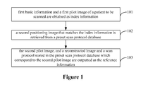
- FIG. 1 is a flow chart of a method for providing reference information for a scan protocol according to an example of the present disclosure
- FIG. 2 is a flow chart of a method for providing reference information for a scan protocol according to another example of the present disclosure
- FIG. 3 is a scan image of chest, which is taken as a pilot image according to the present disclosure
- FIG. 4 is a graph of CT scan values corresponding to the pilot image in FIG. 3 of the present disclosure.
- FIG. 5 is a schematic structural diagram of a device for providing reference information for a scan protocol according to an example of the present disclosure.
- FIG. 6 is a schematic structural diagram of a scan protocol database provided in the present disclosure.
- the present disclosure may make a doctor more accurately set a scan protocol for the patient to be scanned by providing the doctor with a second pilot image, a reconstructed image and a scan protocol of a scanned patient as reference information, where the scanned patient has similar physical condition to the patient to be scanned.
- FIG. 1 is a flow chart of a method for providing reference information for a scan protocol according to a first example of the present disclosure. The method includes blocks 101 - 103 .
- a first basic information and a first pilot image of a patient to be scanned are obtained as index information.
- a doctor should input the first basic information based on the physical condition of the patient to be scanned before CT scanning.
- the first basic information may affect the setting of the scan protocol.
- the first basic information may include the gender of the patient to be scanned, a to-be-scanned body part of the patient, and a scanning mode for the patient to be scanned.
- the scanning mode may include a tomography scan and a spiral scan.
- the first basic information may further include any one or any combination of parameters such as age, height, weight and Body Mass Index (BMI) of the patient to be scanned.
- BMI Body Mass Index
- the first pilot image of the patient to be scanned refers to the pilot image which is obtained by performing one scan such that a doctor may determine a body part of the patient to be scanned.
- the pilot image is different from a reconstructed image obtained finally after a CT scan.
- the pilot image is mainly used to determine a start point and a termination point of the CT scan so as to determine a scan scope.
- the first basic information and the first pilot image of the patient to be scanned are parameters greatly influencing the scan protocol.
- the first basic information and the first pilot image are used as index information to search for scan information of a scanned patient having similar physical condition to the patient to be scanned in a preset scan protocol database, and wherein the scan information of the scanned patient is taken as reference information.
- a second pilot image that matches the index information is retrieved from a preset scan protocol database.
- scan information of a large number of scanned patients is stored in the preset scan protocol database.
- the scan information includes second basic information, the second pilot image, a second reconstructed image and a second scan protocol of the scanned patient. Additionally, a correspondence among the second basic information, the second pilot image, the second reconstructed image and the second scan protocol of each scanned patient is also stored in the preset scan protocol database.
- the scan protocol database may be preset based on acquired scan information of a large number of scanned patients.
- the scan protocol database may be gradually accumulated by storing the first basic information and the first pilot image of each patient to be scanned, a first scan protocol set based on the reference information and a first reconstructed image obtained by scanning each patient to be scanned according to the first scan protocol into the scan protocol database.
- the first reconstructed image is obtained after scanning the patient to be scanned according to the first scan protocol; and after the first basic information, the first pilot image, the first scan protocol and the first reconstructed image are stored in the preset scan protocol database, the first basic information is taken as second basic information, the first pilot image is taken as a second pilot image, the first scan protocol is taken as a second scan protocol, and the first reconstructed image is taken as a second reconstructed image.
- first and second herein are only used to distinguish scan information of the patient to be scanned from scan information of the scanned patient, and is not used to indicate an order.
- the retrieving a second pilot image that matches the index information from a preset scan protocol database may include:
- the physical condition of the scanned patient is similar to that of the patient to be scanned.
- the second pilot image, and a reconstructed image and a scan protocol stored in the preset scan protocol database which correspond to the second pilot image are outputted as the reference information.
- the second basic information of the scanned patient and the second pilot image of the scanned patient respectively match the first basic information and the first pilot image of the patient to be scanned, it means that the physical condition of the scanned patient in the preset scan protocol database is similar to that of the patient to be scanned.
- the second pilot image, the second reconstructed image and the second scan protocol of the scanned patient in the preset scan protocol database who has the similar physical condition to the patient to be scanned are outputted as the reference information.
- the second reconstructed image and the second scan protocol of each scanned patient Since the correspondence among the second basic information, the second pilot image, the second reconstructed image and the second scan protocol of each scanned patient is stored in the preset scan protocol database, if a second pilot image which matches the first pilot image of the patient to be scanned is retrieved, the second reconstructed image and the second scan protocol corresponding to the second pilot image may be obtained.
- the outputted reference may also include a dose report besides the second pilot image, and the second reconstructed image and the second scan protocol in the preset scan protocol database which correspond to the second pilot image.
- the doctor may obtain an irradiation dose value of the scanned patient from the dose report directly, rather than convert an irradiation dose parameter in the second scan protocol to the irradiation dose value.
- the dose report is more intuitive than the irradiation dose parameter in the second scan protocol.
- multiple second pilot images matching the index information may be retrieved from a preset scan protocol database, it means there are multiple scanned patients whose physical conditions are similar to that of the patient to be scanned.
- Multiple pieces of reference information may be outputted in a descending order of matching degrees between the first pilot image of the patient to be scanned and the multiple second pilot images; or multiple pieces of reference information may be outputted in an ascending order or in a descending order of irradiation dose parameters in multiple second scan protocols; or an outputting order of multiple pieces of reference information may be set in other manners according to actual needs.
- one or more scanned patients may be selected, according to a self-set condition, from the multiple scanned patients whose physical conditions are similar to that of the patient to be scanned, and reference information of the selected one or more scanned patients is output, which is not limited specifically here.
- the second pilot image, the second constructed image and the second scan protocol of one or more scanned patients who have similar physical condition to the patient to be scanned are outputted for the doctor as the reference information.
- the doctor may set the first scan protocol of the patient to be scanned, especially set an irradiation dose parameter in the first scan protocol according actual needs.
- the irradiation dose parameter set by the doctor based on the reference information can ensure a clear reconstructed image for one body part to be scanned, the irradiation dose parameter is set appropriately such that the irradiation dose is neither deficient nor excessive, improving accuracy of the scan protocol which is set by the doctor for the patient to be scanned.
- a matching second pilot image which is retrieved from the preset scan protocol database may not change, that is, same reference information is outputted for different doctors; accordingly, different doctors may set the first scan protocol for the patient to be scanned based on the same reference information. Therefore, subjective effects of different doctors on setting the scan protocol, especially the irradiation dose parameter in the scan protocol, for the same patient to be scanned can be eliminated to some extent.
- the present disclosure has following advantageous effect.
- the first basic information and the first pilot image of the patient to be scanned are obtained as index information; the second pilot image that matches the index information is retrieved from the preset scan protocol database; and the second pilot image, and the second reconstructed image and the second scan protocol in the preset scan protocol database which correspond to the second pilot image are outputted as reference information.
- the physical condition of the scanned patient corresponding to the second pilot image is similar to that of the patient to be scanned, and the second pilot image, and the second reconstructed image and the second scan protocol in the preset scan protocol database which correspond to the second pilot image are outputted as the reference information, for helping the doctor in determining the scan protocol of the patient to be scanned, preventing the irradiation dose from being deficient or excessive, and improving accuracy of the scan protocol which is set by the doctor for the patient to be scanned.
- FIG. 2 is a flow chart of a method for providing reference information for a scan protocol according to a second example of the present disclosure. Compared with the first example described above, in this example, a specific implementation of retrieving a second pilot image that matches the index information from a preset scan protocol database is provided.
- the method according to the second example includes following blocks 201 to 208 .
- a scan protocol database is preset, where a correspondence among second basic information, a second pilot image, a second reconstructed image and a second scan protocol of each scanned patient is stored in the preset scan protocol database.
- Scan information of multiple scanned patients is stored in the preset scan protocol database.
- Scan information of each scanned patient includes the second basic information, the second pilot image, the second reconstructed image and the second scan protocol.
- the correspondence among the second basic information, the second pilot image, the second reconstructed image and the second scan protocol of each scanned patient is stored in the preset scan protocol database.
- the preset scan protocol database is preset by obtaining scan information of a large number of scanned patients. Since the scan information of a large number of scanned patients is stored in the preset scan protocol database, it is easier to retrieve a second pilot image of a scanned patient matching a first pilot image of a patient to be scanned, on the premise that the second basic information of the scanned patient is matched with first basic information of the patient to be scanned. In other words, it is easier to find a scanned patient whose physical condition is similar to that of the patient to be scanned and the scan information of the found scanned patient is taken as the reference information.
- the first basic information and the first pilot image of the patient to be scanned are obtained as index information.
- the first basic information includes the gender of the patient to be scanned, a to-be-scanned body part of the patient, and a scanning mode for the patient to be scanned.
- the first basic information has greater influence on the setting of the scan protocol, and thus has relative high referential value. Further, the first basic information may also include any one or any combination of parameters such as age, height, weight and BMI of the patient to be scanned.
- the first pilot image is mainly used to determine a scan scope of the patient to be scanned.
- second basic information which is matched with the first basic information is searched for in the preset scan protocol database.
- a first step of retrieving the scanned patient whose physical condition is similar to that of the patient to be scanned in the scan image database is to search for second basic information which is matched with the first basic information of the patient to be scanned.
- the first basic information and the second basic information may include the gender, a body part to be scanned and a scanning mode.
- the searching for the second basic information which is matched with the first basic information in the preset scan protocol database may include:
- the second basic information is matched with the first basic information, if the gender, the body part to be scanned and the scanning mode in the first basic information are the same as those in the second basic information respectively.
- the execution order to determine whether the gender, the body part to be scanned and the scanning mode in the first basic information are the same as those in the second basic information respectively is not limited specifically, and it may be set according to actual need. In generally, the execution order may be performed as follows.
- block 1 it is determined whether the gender in the first basic information is the same as the gender in the second basic information. If the gender in the first basic information is the same as the gender in the second basic information, block 2 is performed; or it is determined that the first basic information is not matched with the second basic information if the gender in the first basic information is not the same as the gender in the second basic information.
- block 2 it is determined whether the body part to be scanned in the first basic information is the same as the body part to be scanned in the second basic information. If the body part to be scanned in the first basic information is the same as the body part to be scanned in the second basic information, block 3 is performed; or it is determined that the first basic information is not matched with the second basic information if the body part to be scanned in the first basic information is not the same as the body part to be scanned in the second basic information.
- the scanning mode in the first basic information is the same as the scanning mode in the second basic information; If the scanning mode in the first basic information is the same as the scanning mode in the second basic information, it is determined that the first basic information is matched with the second basic information; or it is determined that the first basic information is not matched with the second basic information if the scanning mode in the first basic information is not the same as the scanning mode in the second basic information.
- the searching for second basic information which is matched with the first basic information in the preset scan protocol database may further include:
- the execution order of above determination procedures is not limited specifically, and it may be set according to actual need. It is determined that the first basic information is matched with the second basic information, if the gender, the body part to be scanned and the scanning mode in the first basic information are respectively same as those in the second basic information, and the age, the height, the weight and BMI in the first basic information of the patient to be scanned are also same as those in the second basic information of the scanned patient respectively.
- a second step of retrieving the scanned patient whose physical condition is similar to that of the patient to be scanned in the scan image database is to obtain the second pilot image that corresponds to the second basic information which is matched with the first basic information of the patient to be scanned.
- the second pilot image corresponds to the second basic information which is matched with the first basic information of the patient to be scanned.
- Second pilot images which correspond to the second basic information of the multiple scanned patients may be obtained respectively, i.e., the second pilot images of the multiple scanned patients may be obtained.
- block 205 it is determined whether the second pilot image matches the first pilot image. If the second pilot image matches the first pilot image, block 206 is performed; or else block 208 is performed.
- a third step of retrieving the scanned patient whose physical condition is similar to that of the patient to be scanned in the scan image database is to determine whether the second pilot image of each scanned patient matches the first pilot image one by one. If a second pilot image matches the first pilot image, it may be considered that physical condition of a scanned patient who has the second pilot image is similar to that of the patient to be scanned.
- the determining whether the second pilot image matches the first pilot image may include block A and block B.
- an image deviation between a location parameter of a preset characteristic location in the second pilot image and a location parameter of a preset characteristic location in the first pilot image is calculated.
- the preset characteristic location greatly affects the quality of the reconstructed image as the irradiation dose varies.
- the preset characteristic location is usually preset in the pilot image according to experience. In general, there is little difference between the preset characteristic location in the first pilot image of the patient to be scanned and the preset characteristic location in the second pilot image of the scanned patient.
- FIG. 3 is a scan image of chest which is used as a pilot image. Lung tissue is segmented out from the pilot image by performing image segmentation on the pilot image, and the top of the lung lobe is selected as the preset characteristic location.
- the preset characteristic location is selected based on the principle of human anatomy. In this example, it assumes that the selected preset characteristic location is located near the collarbones and shoulders.
- the density and size of the preset characteristic location are larger than those of other locations; if a low irradiation dose is adopted in a chest CT scan, strip artifacts may be caused due to lack of irradiation, which has great influence on the quality of the reconstructed image of the CT scan.
- the location parameter of the preset characteristic location may include a size and an attenuation mean value.
- the size refers to the length between a head index point and a tail index point within the preset characteristic location, where CT scan values are greater than a threshold value.
- FIG. 4 is a graph of CT scan values corresponding to the pilot image of FIG. 3 .
- a straight line A is the preset characteristic location and a threshold T is preset. Points of the graph in FIG. 4 are traversed from a start point to a central point to find the head index point at which the CT scan value firstly exceeds the threshold T, and points of the graph in FIG.
- the head index point is C
- the tail index point is D
- the length between C and D is the size in the location parameter of the preset characteristic location.
- the sum of the mean of the CT scan values at points between C and D in FIG. 4 and 1024 HU is the attenuation mean value.
- the above approach may be used to calculate a first size and a first attenuation mean value in the location parameter of the preset characteristic location in the first pilot image of the patient to be scanned, and also may be used to calculate a second size and a second attenuation mean value in the location parameter of the preset characteristic location in the second pilot image of each of multiple scanned patients.
- block A may include:
- the first size and the first attenuation mean value of the preset characteristic location in the first pilot image of the patient to be scanned are calculated with the above approach, where the first size is represented by L 0 , and the first attenuation mean value is represented by A 0 .
- the second size and the second attenuation mean value of the preset characteristic location in the first pilot image of each patient to be scanned is calculated with the above approach, where the second size is represented by L, and the second attenuation mean value is represented by A.
- the image deviation E between the first pilot image of the patient to be scanned and the second pilot image of each scanned patient may be calculated using formula (3):
- the image deviation E is the sum of sub-deviation between respective items of the location parameter of the first pilot image and respective items of the location parameter of the second pilot image.
- the location parameter may include the size and the attenuation mean value, and may also include other item. If the location parameter includes other item, a sub-deviation of the other item may be calculated and added.
- block B is performed to determine whether the image deviation is less than the preset threshold. It means that the second pilot image matches the first pilot image and the physical condition of the patient to be scanned is similar to that of the scanned patient, if the image deviation is less than the preset threshold. It means that the second pilot image does not match the first pilot image and there is a relative great difference between the physical condition of the patient to be scanned and the physical condition of the scanned patient if the image deviation is not less than the preset threshold.
- the second pilot image, and a second reconstructed image and a second scan protocol in the preset scan protocol database which correspond to the second pilot image are outputted as reference information.
- the second pilot image of each scanned patient that matched with the first pilot image of a patient to be scanned, and the second reconstructed image and the second scan protocol in the preset scan protocol database which correspond to the second pilot image may be outputted as the reference information.
- one or more of the second pilot image of the selected one or more scanned patients according to actual needs, and the second reconstructed image and the second scan protocol in the preset scan protocol database which correspond to the second pilot image may be outputted as reference information.
- a selection condition may be preset by a doctor, and a scan protocol of a scanned patient is automatically selected as a scan protocol of the patient to be scanned. First, the preset selection condition is obtained, and then the scan protocol of a patient to be scanned is determined from the reference information according to the preset selection condition.
- the selection condition may be the irradiation dose parameter of the scan protocol.
- a scan protocol having a maximum dose parameter or a minimum dose parameter is selected as the scan protocol of the patient to be scanned. It is understood that the selection condition may be set in other ways, which are not detailed here.
- a first scan protocol which is set for the patient to be scanned based on the reference information is obtained; the patient to be scanned is scanned according to the first scan protocol to obtain a first reconstructed image; and then the first basic information, the first pilot image, the first scan protocol and the first reconstructed image of the patient to be scanned are stored in the preset scan protocol database.
- a CT scan may be performed on the patient to be scanned according to the first scan protocol to obtain the first reconstructed image of the patient to be scanned, and then the first basic information, the first pilot image, the first scan protocol and the first reconstructed image of the patient to be scanned are stored in the preset scan protocol database in order to enrich the data continually.
- the first basic information is taken as second basic information
- the first pilot image is taken as a second pilot image
- the first scan protocol is taken as a second scan protocol
- the first reconstructed image is taken as a second reconstructed image.
- a notification of failing in finding a second pilot image which matches the first pilot image is outputted.
- the notification of failing in finding a second pilot image which matches the first pilot image is outputted, and the doctor may manually set a scan protocol based on the specific condition of the patient to be scanned.
- the present disclosure also has the following advantageous effect.
- the scan information such as the second pilot image, the reconstructed image and the second scan protocol of the scanned patient who has the similar physical condition to the patient to be scanned may be retrieved and outputted as reference information for setting a scan protocol for the patient to be scanned.
- the first basic information, the first pilot image, the first scan protocol and the first reconstructed image of the patient to be scanned are stored in the preset scan protocol database, thereby enriching the data in the preset scan protocol database continually.
- FIG. 5 is a schematic structural diagram of a device for providing reference information for a scan protocol according to a third example of the present disclosure.
- the device corresponds to the method according to the first example.
- the device includes a processor and a non-transitory computer readable storage medium, where instructions are stored in the non-transitory computer readable storage medium.
- the processor may communicate with the non-transitory computer readable storage medium through an internal bus.
- the processor may read the instructions to obtain first basic information and a first pilot image of a patient to be scanned as index information;
- the processor reads the instructions to retrieve a second pilot image that matches the index information from a preset scan protocol database is to:
- the second pilot image matches the first pilot image, and determine the second pilot image as a second pilot image which matches the index information if the second pilot image matches the first pilot image.
- the processor may read the instructions further to output the second pilot image, and a second reconstructed image and a second scan protocol in the preset scan protocol database which correspond to the second pilot image as reference information.
- the third example is similar to the first example. Details of the third example are not given here and can be known with reference to the description of the first example.
- a device for providing reference information for a scan protocol is provided according to a fourth example of the present disclosure.
- the device corresponds to the method according to the second example. Similar to the third example, the device according to the fourth example also includes a processor and a non-transitory computer readable storage medium, where instructions are stored in the non-transitory computer readable storage medium, and the processor may communicate with the non-transitory computer readable storage medium through an internal bus.
- the processor may read the instructions to preset a scan protocol database, where a correspondence among second basic information, a second pilot image, a second reconstructed image and a second scan protocol of each scanned patient is stored in the preset scan protocol database.
- the preset scan protocol database includes two parts.
- One part is an original scan protocol sub-database 601 , in which scan data of a number of scanned patients is stored in advance.
- Scan data of each scanned patient includes the basic information, the pilot image, the scan protocol and the reconstructed image of the scanned patient.
- the other part is an updated scan protocol database 602 .
- Scan data of each patient to be scanned is stored in the updated scan protocol database.
- the scan data of each patient to be scanned includes the basic information, the pilot image, the scan protocol and the reconstructed image of the patient to be scanned.
- the processor may read the instructions further to: obtain first basic information and a first pilot image of a patient to be scanned as index information; and
- the processor reads the instructions to search, in the preset scan protocol database, for second basic information which is matched with the first basic information is to:
- the second basic information which is matched with the first basic information if the gender, the body part to be scanned and the scanning mode in the first basic information are the same as those in the second basic information respectively.
- the processor may read the instructions further to: obtain a second pilot image corresponding to the second basic information;
- the second pilot image matches the first pilot image, and determine the second pilot image as a second pilot image which matches the index information if the second pilot image matches the first pilot image.
- the processor reads the instructions to determine whether the second pilot image matches the first pilot image is to:
- the processor reads the instructions to calculate an image deviation between a location parameter of a preset characteristic location in the second pilot image and a location parameter of a preset characteristic location in the first pilot image is to:
- the processor may read the instructions further to output the second pilot image, and a second reconstructed image and a second scan protocol in the preset scan protocol database which correspond to the second pilot image as reference information.
- the processor may read the instructions further to:
- the fourth example is similar to the second example. Details of the fourth example are not given here and can be known with reference to the description of the second example.
Landscapes
- Health & Medical Sciences (AREA)
- Engineering & Computer Science (AREA)
- Life Sciences & Earth Sciences (AREA)
- Medical Informatics (AREA)
- General Health & Medical Sciences (AREA)
- Radiology & Medical Imaging (AREA)
- Public Health (AREA)
- Nuclear Medicine, Radiotherapy & Molecular Imaging (AREA)
- Physics & Mathematics (AREA)
- Surgery (AREA)
- Biomedical Technology (AREA)
- Heart & Thoracic Surgery (AREA)
- Molecular Biology (AREA)
- Optics & Photonics (AREA)
- Animal Behavior & Ethology (AREA)
- High Energy & Nuclear Physics (AREA)
- Biophysics (AREA)
- Veterinary Medicine (AREA)
- Pathology (AREA)
- Theoretical Computer Science (AREA)
- Pulmonology (AREA)
- Computer Vision & Pattern Recognition (AREA)
- Primary Health Care (AREA)
- Epidemiology (AREA)
- Mathematical Physics (AREA)
- Data Mining & Analysis (AREA)
- Databases & Information Systems (AREA)
- General Engineering & Computer Science (AREA)
- General Physics & Mathematics (AREA)
- Apparatus For Radiation Diagnosis (AREA)
- Computer Networks & Wireless Communication (AREA)
Abstract
Description
Claims (10)
Applications Claiming Priority (3)
| Application Number | Priority Date | Filing Date | Title |
|---|---|---|---|
| CN201410385398 | 2014-08-06 | ||
| CN201410385398.5 | 2014-08-06 | ||
| CN201410385398.5A CN104224218B (en) | 2014-08-06 | 2014-08-06 | A kind of method and device of the reference information that scan protocols is provided |
Publications (2)
| Publication Number | Publication Date |
|---|---|
| US20160042012A1 US20160042012A1 (en) | 2016-02-11 |
| US10296604B2 true US10296604B2 (en) | 2019-05-21 |
Family
ID=52213756
Family Applications (1)
| Application Number | Title | Priority Date | Filing Date |
|---|---|---|---|
| US14/819,368 Active 2037-06-28 US10296604B2 (en) | 2014-08-06 | 2015-08-05 | Method and device for providing reference information for scan protocol |
Country Status (2)
| Country | Link |
|---|---|
| US (1) | US10296604B2 (en) |
| CN (1) | CN104224218B (en) |
Families Citing this family (17)
| Publication number | Priority date | Publication date | Assignee | Title |
|---|---|---|---|---|
| CN105078496A (en) * | 2014-04-30 | 2015-11-25 | 上海西门子医疗器械有限公司 | Method for scanning double positioning images and computed tomography device |
| CN106691491A (en) * | 2017-02-28 | 2017-05-24 | 赛诺威盛科技(北京)有限公司 | CT (computed tomography) positioning system implemented by using visible light and infrared light and CT positioning method |
| EP3441004B1 (en) * | 2017-08-11 | 2019-12-11 | Siemens Healthcare GmbH | Method for recording an image dataset with an x-ray imaging system and x-ray imaging system |
| WO2019200351A1 (en) * | 2018-04-13 | 2019-10-17 | General Electric Company | Systems and methods for an imaging system express mode |
| EP3809970A4 (en) * | 2018-05-28 | 2021-06-30 | Shanghai United Imaging Healthcare Co., Ltd. | SYSTEMS AND METHODS FOR ACQUISITION OF X-RAY IMAGES |
| CN108670279A (en) * | 2018-05-28 | 2018-10-19 | 上海联影医疗科技有限公司 | Shoot method, Medical Imaging System, computer and the storage medium of medical image |
| EP3784134A4 (en) | 2018-05-28 | 2021-06-16 | Shanghai United Imaging Healthcare Co., Ltd. | SYSTEMS AND METHODS FOR DETERMINING EXAMINATION PARAMETERS |
| CN109276269A (en) * | 2018-08-22 | 2019-01-29 | 上海联影医疗科技有限公司 | Obtain method, apparatus, medical supply and the storage medium of scan protocols |
| CN109350100A (en) * | 2018-09-27 | 2019-02-19 | 上海联影医疗科技有限公司 | Medical imaging procedure, medical imaging devices and computer readable storage medium |
| CN110464326B (en) | 2019-08-19 | 2022-05-10 | 上海联影医疗科技股份有限公司 | Scanning parameter recommendation method, system, device and storage medium |
| CN112820383B (en) * | 2019-11-15 | 2023-04-28 | 上海联影医疗科技股份有限公司 | Medical imaging method, system and storage medium |
| JP7583530B2 (en) * | 2020-03-03 | 2024-11-14 | キヤノンメディカルシステムズ株式会社 | Medical information processing device, medical image diagnostic device, and medical information processing method |
| CN111528879A (en) * | 2020-05-06 | 2020-08-14 | 上海联影医疗科技有限公司 | A method and system for medical image acquisition |
| CN111493917A (en) * | 2020-04-23 | 2020-08-07 | 上海联影医疗科技有限公司 | Image scanning protocol interaction device |
| CN113393500B (en) * | 2021-05-28 | 2023-04-25 | 上海联影医疗科技股份有限公司 | Spine scanning parameter acquisition method, device, equipment and storage medium |
| CN113645307A (en) * | 2021-08-18 | 2021-11-12 | 中国人民解放军东部战区总医院 | Nuclear magnetic resonance system network shared positioning database system and its establishment and use method |
| CN115662605A (en) * | 2022-10-13 | 2023-01-31 | 东软医疗系统股份有限公司 | Scanning method and device |
Citations (10)
| Publication number | Priority date | Publication date | Assignee | Title |
|---|---|---|---|---|
| US20060153436A1 (en) | 2005-01-13 | 2006-07-13 | Gabriel Haras | Method for determining acquisition parameters for a medical tomography device, and an associated apparatus |
| US20060193437A1 (en) * | 2005-01-31 | 2006-08-31 | Dieter Boeing | Method and apparatus for controlling an imaging modality |
| US7378660B2 (en) * | 2005-09-30 | 2008-05-27 | Cardiovascular Imaging Technologies L.L.C. | Computer program, method, and system for hybrid CT attenuation correction |
| US20090074143A1 (en) | 2007-09-14 | 2009-03-19 | Kabushiki Kaisha Toshiba | X-ray ct apparatus, scan plan assistance apparatus and method for scan plan assistance |
| US20110150312A1 (en) * | 2008-07-11 | 2011-06-23 | Mitsubishi Precision Co., Ltd. | Biodata model preparation method and apparatus, data structure of biodata model and data storage device of biodata model, and load dispersion method and apparatus of 3d data model |
| US20110222749A1 (en) * | 2006-07-06 | 2011-09-15 | The Gov't Of The U.S.A. As Represented By The Secretary Of The Dept. Of Health & Human Services | Hybrid segmentation of anatomical structure |
| US20130051704A1 (en) | 2011-08-31 | 2013-02-28 | Fujifilm Corporation | Exposure condition decision support system and method |
| US20130101079A1 (en) | 2011-10-19 | 2013-04-25 | David M. Hough | Method For Controlling Radiation Dose and Intravenous Contrast Dose In Computed Tomography Imaging |
| US20150103969A1 (en) * | 2013-10-14 | 2015-04-16 | Siemens Aktiengesellschaft | Reconstruction of image data by means of contour data |
| JP2015130909A (en) | 2014-01-09 | 2015-07-23 | 株式会社日立メディコ | X-ray ct apparatus and photographing condition setting method |
-
2014
- 2014-08-06 CN CN201410385398.5A patent/CN104224218B/en active Active
-
2015
- 2015-08-05 US US14/819,368 patent/US10296604B2/en active Active
Patent Citations (13)
| Publication number | Priority date | Publication date | Assignee | Title |
|---|---|---|---|---|
| JP2006192270A (en) | 2005-01-13 | 2006-07-27 | Siemens Ag | Method and apparatus for determining imaging parameters of a medical tomography apparatus |
| US20060153436A1 (en) | 2005-01-13 | 2006-07-13 | Gabriel Haras | Method for determining acquisition parameters for a medical tomography device, and an associated apparatus |
| US20060193437A1 (en) * | 2005-01-31 | 2006-08-31 | Dieter Boeing | Method and apparatus for controlling an imaging modality |
| CN100581472C (en) | 2005-01-31 | 2010-01-20 | 西门子公司 | Method and apparatus for controlling image mode |
| US7378660B2 (en) * | 2005-09-30 | 2008-05-27 | Cardiovascular Imaging Technologies L.L.C. | Computer program, method, and system for hybrid CT attenuation correction |
| US20110222749A1 (en) * | 2006-07-06 | 2011-09-15 | The Gov't Of The U.S.A. As Represented By The Secretary Of The Dept. Of Health & Human Services | Hybrid segmentation of anatomical structure |
| US20090074143A1 (en) | 2007-09-14 | 2009-03-19 | Kabushiki Kaisha Toshiba | X-ray ct apparatus, scan plan assistance apparatus and method for scan plan assistance |
| US20110150312A1 (en) * | 2008-07-11 | 2011-06-23 | Mitsubishi Precision Co., Ltd. | Biodata model preparation method and apparatus, data structure of biodata model and data storage device of biodata model, and load dispersion method and apparatus of 3d data model |
| US20130051704A1 (en) | 2011-08-31 | 2013-02-28 | Fujifilm Corporation | Exposure condition decision support system and method |
| JP2013048746A (en) | 2011-08-31 | 2013-03-14 | Fujifilm Corp | Imaging condition determination support apparatus and imaging condition determination support method |
| US20130101079A1 (en) | 2011-10-19 | 2013-04-25 | David M. Hough | Method For Controlling Radiation Dose and Intravenous Contrast Dose In Computed Tomography Imaging |
| US20150103969A1 (en) * | 2013-10-14 | 2015-04-16 | Siemens Aktiengesellschaft | Reconstruction of image data by means of contour data |
| JP2015130909A (en) | 2014-01-09 | 2015-07-23 | 株式会社日立メディコ | X-ray ct apparatus and photographing condition setting method |
Non-Patent Citations (1)
| Title |
|---|
| The First Office Action dated Dec. 28, 2015 regarding the Chinese priority patent application (Appl. No. 201410385398.5). |
Also Published As
| Publication number | Publication date |
|---|---|
| CN104224218B (en) | 2016-10-05 |
| US20160042012A1 (en) | 2016-02-11 |
| CN104224218A (en) | 2014-12-24 |
Similar Documents
| Publication | Publication Date | Title |
|---|---|---|
| US10296604B2 (en) | Method and device for providing reference information for scan protocol | |
| US10748649B2 (en) | Similar case retrieval apparatus, similar case retrieval method, non-transitory computer-readable storage medium, similar case retrieval system, and case database | |
| CN111325759B (en) | Blood vessel segmentation method, device, computer equipment and readable storage medium | |
| US8380013B2 (en) | Case image search apparatus, method and computer-readable recording medium | |
| CN111932554B (en) | Lung vessel segmentation method, equipment and storage medium | |
| JP2021520568A (en) | Tissue nodule detection method and its model Training method, equipment, equipment, system, and its computer program | |
| Schneider et al. | Normative data for 8 neuropsychological tests in older blacks and whites from the atherosclerosis risk in communities (ARIC) study | |
| KR101684998B1 (en) | method and system for diagnosis of oral lesion using medical image | |
| US20170154167A1 (en) | A system and a related method for automatically selecting a hanging protocol for a medical study | |
| US12387054B2 (en) | Information saving apparatus, method, and program and analysis record generation apparatus, method, and program for recognizing correction made in image analysis record | |
| CN113327235A (en) | Image processing method and device based on CT image sarcopenia evaluation | |
| US9678988B2 (en) | Image processing apparatus and image processing method | |
| JP2014039852A (en) | Information processor, information processing method and program | |
| CN106021915A (en) | Big data-based automatic diagnosis and treatment-oriented medical data analysis system and apparatus | |
| Renker et al. | Iterative image reconstruction: a realistic dose-saving method in cardiac CT imaging? | |
| US12148166B2 (en) | Updating boundary segmentations | |
| CN108648164A (en) | Ultrasound image optimization method, apparatus and computer readable storage medium | |
| WO2020007026A1 (en) | Segmentation model training method and apparatus, and computer-readable storage medium | |
| Chacón et al. | Computational assessment of stomach tumor volume from multi-slice computerized tomography images in presence of type 2 cancer | |
| Chua et al. | Using multiple imputation to efficiently correct cerebral MRI whole brain lesion and atrophy data in patients with multiple sclerosis | |
| Annoni et al. | Pre-TAVI aortic annulus sizing: comparison between manual and semi-automated new generation software measurements in operators with different experience | |
| JP2021114171A (en) | Medical image processing apparatus, program, and medical image processing system | |
| EP2343687A2 (en) | Tomographic image generating apparatus, tomographic image generating method, and program for generating tomographic images | |
| EP3910645B1 (en) | Image retrieval | |
| JP2018102358A (en) | MEDICAL IMAGE GENERATION METHOD, MEDICAL IMAGE GENERATION DEVICE, AND MEDICAL IMAGE GENERATION PROGRAM |
Legal Events
| Date | Code | Title | Description |
|---|---|---|---|
| AS | Assignment |
Owner name: SHENYANG NEUSOFT MEDICAL SYSTEMS CO., LTD., CHINA Free format text: ASSIGNMENT OF ASSIGNORS INTEREST;ASSIGNORS:LOU, SHANSHAN;ZHENG, HAN;REEL/FRAME:036262/0886 Effective date: 20141027 |
|
| STPP | Information on status: patent application and granting procedure in general |
Free format text: PUBLICATIONS -- ISSUE FEE PAYMENT RECEIVED |
|
| STPP | Information on status: patent application and granting procedure in general |
Free format text: PUBLICATIONS -- ISSUE FEE PAYMENT VERIFIED |
|
| STCF | Information on status: patent grant |
Free format text: PATENTED CASE |
|
| AS | Assignment |
Owner name: NEUSOFT MEDICAL SYSTEMS CO., LTD., CHINA Free format text: CHANGE OF NAME;ASSIGNOR:SHENYANG NEUSOFT MEDICAL SYSTEMS CO., LTD.;REEL/FRAME:052398/0489 Effective date: 20200329 |
|
| MAFP | Maintenance fee payment |
Free format text: PAYMENT OF MAINTENANCE FEE, 4TH YEAR, LARGE ENTITY (ORIGINAL EVENT CODE: M1551); ENTITY STATUS OF PATENT OWNER: LARGE ENTITY Year of fee payment: 4 |