US10227560B2 - Biomimetic support for three-dimensional cell culturing, method for manufacturing same, and use thereof - Google Patents
Biomimetic support for three-dimensional cell culturing, method for manufacturing same, and use thereof Download PDFInfo
- Publication number
- US10227560B2 US10227560B2 US15/326,638 US201515326638A US10227560B2 US 10227560 B2 US10227560 B2 US 10227560B2 US 201515326638 A US201515326638 A US 201515326638A US 10227560 B2 US10227560 B2 US 10227560B2
- Authority
- US
- United States
- Prior art keywords
- cells
- collagen
- cell
- cancer
- support
- Prior art date
- Legal status (The legal status is an assumption and is not a legal conclusion. Google has not performed a legal analysis and makes no representation as to the accuracy of the status listed.)
- Active, expires
Links
- 238000000034 method Methods 0.000 title claims abstract description 53
- 238000012258 culturing Methods 0.000 title claims description 52
- 238000004519 manufacturing process Methods 0.000 title abstract description 18
- 230000003592 biomimetic effect Effects 0.000 title description 5
- 229920001436 collagen Polymers 0.000 claims abstract description 335
- 102000008186 Collagen Human genes 0.000 claims abstract description 332
- 108010035532 Collagen Proteins 0.000 claims abstract description 332
- 239000000203 mixture Substances 0.000 claims abstract description 100
- 238000002156 mixing Methods 0.000 claims abstract description 21
- 210000004027 cell Anatomy 0.000 claims description 566
- 229940072056 alginate Drugs 0.000 claims description 167
- 229920000615 alginic acid Polymers 0.000 claims description 167
- 206010028980 Neoplasm Diseases 0.000 claims description 106
- 201000011510 cancer Diseases 0.000 claims description 102
- FHVDTGUDJYJELY-UHFFFAOYSA-N 6-{[2-carboxy-4,5-dihydroxy-6-(phosphanyloxy)oxan-3-yl]oxy}-4,5-dihydroxy-3-phosphanyloxane-2-carboxylic acid Chemical compound O1C(C(O)=O)C(P)C(O)C(O)C1OC1C(C(O)=O)OC(OP)C(O)C1O FHVDTGUDJYJELY-UHFFFAOYSA-N 0.000 claims description 87
- 235000010443 alginic acid Nutrition 0.000 claims description 87
- 229920000936 Agarose Polymers 0.000 claims description 51
- 206010060862 Prostate cancer Diseases 0.000 claims description 42
- 208000000236 Prostatic Neoplasms Diseases 0.000 claims description 42
- 238000001879 gelation Methods 0.000 claims description 34
- 206010033128 Ovarian cancer Diseases 0.000 claims description 29
- 206010061535 Ovarian neoplasm Diseases 0.000 claims description 29
- 208000003950 B-cell lymphoma Diseases 0.000 claims description 28
- 206010042971 T-cell lymphoma Diseases 0.000 claims description 25
- 208000027585 T-cell non-Hodgkin lymphoma Diseases 0.000 claims description 25
- 206010058467 Lung neoplasm malignant Diseases 0.000 claims description 12
- 201000005202 lung cancer Diseases 0.000 claims description 12
- 208000020816 lung neoplasm Diseases 0.000 claims description 12
- 230000001939 inductive effect Effects 0.000 claims description 8
- 201000005787 hematologic cancer Diseases 0.000 claims description 6
- 208000024200 hematopoietic and lymphoid system neoplasm Diseases 0.000 claims description 6
- 239000007787 solid Substances 0.000 claims description 5
- 206010023825 Laryngeal cancer Diseases 0.000 claims description 4
- 206010023841 laryngeal neoplasm Diseases 0.000 claims description 4
- 206010004593 Bile duct cancer Diseases 0.000 claims description 2
- 206010005949 Bone cancer Diseases 0.000 claims description 2
- 208000018084 Bone neoplasm Diseases 0.000 claims description 2
- 206010006187 Breast cancer Diseases 0.000 claims description 2
- 208000026310 Breast neoplasm Diseases 0.000 claims description 2
- 206010009944 Colon cancer Diseases 0.000 claims description 2
- 208000000461 Esophageal Neoplasms Diseases 0.000 claims description 2
- 208000032612 Glial tumor Diseases 0.000 claims description 2
- 206010018338 Glioma Diseases 0.000 claims description 2
- 208000008839 Kidney Neoplasms Diseases 0.000 claims description 2
- 208000034578 Multiple myelomas Diseases 0.000 claims description 2
- 206010030155 Oesophageal carcinoma Diseases 0.000 claims description 2
- 206010061902 Pancreatic neoplasm Diseases 0.000 claims description 2
- 206010035226 Plasma cell myeloma Diseases 0.000 claims description 2
- 208000015634 Rectal Neoplasms Diseases 0.000 claims description 2
- 206010038389 Renal cancer Diseases 0.000 claims description 2
- 208000004337 Salivary Gland Neoplasms Diseases 0.000 claims description 2
- 206010061934 Salivary gland cancer Diseases 0.000 claims description 2
- 208000005718 Stomach Neoplasms Diseases 0.000 claims description 2
- 208000000389 T-cell leukemia Diseases 0.000 claims description 2
- 208000028530 T-cell lymphoblastic leukemia/lymphoma Diseases 0.000 claims description 2
- 208000024770 Thyroid neoplasm Diseases 0.000 claims description 2
- 206010062129 Tongue neoplasm Diseases 0.000 claims description 2
- 208000002495 Uterine Neoplasms Diseases 0.000 claims description 2
- 208000029742 colonic neoplasm Diseases 0.000 claims description 2
- 201000004101 esophageal cancer Diseases 0.000 claims description 2
- 206010017758 gastric cancer Diseases 0.000 claims description 2
- 208000003906 hydrocephalus Diseases 0.000 claims description 2
- 201000010982 kidney cancer Diseases 0.000 claims description 2
- 201000007270 liver cancer Diseases 0.000 claims description 2
- 208000014018 liver neoplasm Diseases 0.000 claims description 2
- 208000015486 malignant pancreatic neoplasm Diseases 0.000 claims description 2
- 201000008968 osteosarcoma Diseases 0.000 claims description 2
- 201000002528 pancreatic cancer Diseases 0.000 claims description 2
- 208000008443 pancreatic carcinoma Diseases 0.000 claims description 2
- 206010038038 rectal cancer Diseases 0.000 claims description 2
- 201000001275 rectum cancer Diseases 0.000 claims description 2
- 201000011549 stomach cancer Diseases 0.000 claims description 2
- 201000002510 thyroid cancer Diseases 0.000 claims description 2
- 201000006134 tongue cancer Diseases 0.000 claims description 2
- 206010046766 uterine cancer Diseases 0.000 claims description 2
- 239000002121 nanofiber Substances 0.000 abstract description 237
- 241000251468 Actinopterygii Species 0.000 abstract description 222
- 239000002131 composite material Substances 0.000 abstract description 145
- XLYOFNOQVPJJNP-UHFFFAOYSA-N water Substances O XLYOFNOQVPJJNP-UHFFFAOYSA-N 0.000 abstract description 36
- 229920001059 synthetic polymer Polymers 0.000 abstract description 30
- 238000001523 electrospinning Methods 0.000 abstract description 23
- 239000003960 organic solvent Substances 0.000 abstract description 11
- 229920001610 polycaprolactone Polymers 0.000 description 222
- 239000000017 hydrogel Substances 0.000 description 193
- 239000000243 solution Substances 0.000 description 138
- 238000004113 cell culture Methods 0.000 description 105
- 102000007469 Actins Human genes 0.000 description 52
- 108010085238 Actins Proteins 0.000 description 52
- FWBHETKCLVMNFS-UHFFFAOYSA-N 4',6-Diamino-2-phenylindol Chemical group C1=CC(C(=N)N)=CC=C1C1=CC2=CC=C(C(N)=N)C=C2N1 FWBHETKCLVMNFS-UHFFFAOYSA-N 0.000 description 45
- 238000012604 3D cell culture Methods 0.000 description 44
- 238000001000 micrograph Methods 0.000 description 39
- 210000004292 cytoskeleton Anatomy 0.000 description 35
- 238000004458 analytical method Methods 0.000 description 34
- 239000002246 antineoplastic agent Substances 0.000 description 32
- 238000010186 staining Methods 0.000 description 32
- LOKCTEFSRHRXRJ-UHFFFAOYSA-I dipotassium trisodium dihydrogen phosphate hydrogen phosphate dichloride Chemical compound P(=O)(O)(O)[O-].[K+].P(=O)(O)([O-])[O-].[Na+].[Na+].[Cl-].[K+].[Cl-].[Na+] LOKCTEFSRHRXRJ-UHFFFAOYSA-I 0.000 description 31
- 239000002953 phosphate buffered saline Substances 0.000 description 31
- 230000002441 reversible effect Effects 0.000 description 28
- 239000011550 stock solution Substances 0.000 description 26
- 108091003079 Bovine Serum Albumin Proteins 0.000 description 25
- 108700031361 Brachyury Proteins 0.000 description 25
- 239000012091 fetal bovine serum Substances 0.000 description 23
- AOJJSUZBOXZQNB-TZSSRYMLSA-N Doxorubicin Chemical compound O([C@H]1C[C@@](O)(CC=2C(O)=C3C(=O)C=4C=CC=C(C=4C(=O)C3=C(O)C=21)OC)C(=O)CO)[C@H]1C[C@H](N)[C@H](O)[C@H](C)O1 AOJJSUZBOXZQNB-TZSSRYMLSA-N 0.000 description 22
- 229940041181 antineoplastic drug Drugs 0.000 description 22
- 230000012010 growth Effects 0.000 description 21
- UCSJYZPVAKXKNQ-HZYVHMACSA-N streptomycin Chemical compound CN[C@H]1[C@H](O)[C@@H](O)[C@H](CO)O[C@H]1O[C@@H]1[C@](C=O)(O)[C@H](C)O[C@H]1O[C@@H]1[C@@H](NC(N)=N)[C@H](O)[C@@H](NC(N)=N)[C@H](O)[C@H]1O UCSJYZPVAKXKNQ-HZYVHMACSA-N 0.000 description 20
- 230000010261 cell growth Effects 0.000 description 19
- -1 poly(ε-caprolactone) Polymers 0.000 description 18
- 210000001519 tissue Anatomy 0.000 description 18
- 230000036210 malignancy Effects 0.000 description 17
- 208000002154 non-small cell lung carcinoma Diseases 0.000 description 17
- 208000029729 tumor suppressor gene on chromosome 11 Diseases 0.000 description 17
- 230000003833 cell viability Effects 0.000 description 16
- 229940098773 bovine serum albumin Drugs 0.000 description 15
- 239000003814 drug Substances 0.000 description 15
- 239000000499 gel Substances 0.000 description 15
- 239000011148 porous material Substances 0.000 description 15
- 230000035899 viability Effects 0.000 description 14
- 210000000130 stem cell Anatomy 0.000 description 13
- 241001465754 Metazoa Species 0.000 description 12
- 229930012538 Paclitaxel Natural products 0.000 description 12
- 238000010240 RT-PCR analysis Methods 0.000 description 12
- 239000012153 distilled water Substances 0.000 description 12
- 230000000694 effects Effects 0.000 description 12
- 229960001592 paclitaxel Drugs 0.000 description 12
- RCINICONZNJXQF-MZXODVADSA-N taxol Chemical compound O([C@@H]1[C@@]2(C[C@@H](C(C)=C(C2(C)C)[C@H](C([C@]2(C)[C@@H](O)C[C@H]3OC[C@]3([C@H]21)OC(C)=O)=O)OC(=O)C)OC(=O)[C@H](O)[C@@H](NC(=O)C=1C=CC=CC=1)C=1C=CC=CC=1)O)C(=O)C1=CC=CC=C1 RCINICONZNJXQF-MZXODVADSA-N 0.000 description 12
- UXVMQQNJUSDDNG-UHFFFAOYSA-L Calcium chloride Chemical compound [Cl-].[Cl-].[Ca+2] UXVMQQNJUSDDNG-UHFFFAOYSA-L 0.000 description 11
- 241000237858 Gastropoda Species 0.000 description 11
- 239000001110 calcium chloride Substances 0.000 description 11
- 229910001628 calcium chloride Inorganic materials 0.000 description 11
- 238000009826 distribution Methods 0.000 description 11
- 229960004679 doxorubicin Drugs 0.000 description 11
- 229940079593 drug Drugs 0.000 description 11
- 238000001727 in vivo Methods 0.000 description 11
- HEDRZPFGACZZDS-UHFFFAOYSA-N Chloroform Chemical compound ClC(Cl)Cl HEDRZPFGACZZDS-UHFFFAOYSA-N 0.000 description 10
- 239000006144 Dulbecco’s modified Eagle's medium Substances 0.000 description 10
- TWRXJAOTZQYOKJ-UHFFFAOYSA-L Magnesium chloride Chemical compound [Mg+2].[Cl-].[Cl-] TWRXJAOTZQYOKJ-UHFFFAOYSA-L 0.000 description 10
- 229930182555 Penicillin Natural products 0.000 description 10
- JGSARLDLIJGVTE-MBNYWOFBSA-N Penicillin G Chemical compound N([C@H]1[C@H]2SC([C@@H](N2C1=O)C(O)=O)(C)C)C(=O)CC1=CC=CC=C1 JGSARLDLIJGVTE-MBNYWOFBSA-N 0.000 description 10
- VFLDPWHFBUODDF-FCXRPNKRSA-N curcumin Chemical compound C1=C(O)C(OC)=CC(\C=C\C(=O)CC(=O)\C=C\C=2C=C(OC)C(O)=CC=2)=C1 VFLDPWHFBUODDF-FCXRPNKRSA-N 0.000 description 10
- 201000005243 lung squamous cell carcinoma Diseases 0.000 description 10
- 229940049954 penicillin Drugs 0.000 description 10
- 229960005322 streptomycin Drugs 0.000 description 10
- 230000004083 survival effect Effects 0.000 description 10
- 206010059866 Drug resistance Diseases 0.000 description 9
- 239000012981 Hank's balanced salt solution Substances 0.000 description 9
- 206010027476 Metastases Diseases 0.000 description 9
- OKKJLVBELUTLKV-UHFFFAOYSA-N Methanol Chemical compound OC OKKJLVBELUTLKV-UHFFFAOYSA-N 0.000 description 9
- 230000015572 biosynthetic process Effects 0.000 description 9
- 230000003993 interaction Effects 0.000 description 9
- 239000002609 medium Substances 0.000 description 9
- 230000009401 metastasis Effects 0.000 description 9
- 229920005615 natural polymer Polymers 0.000 description 9
- 230000035755 proliferation Effects 0.000 description 9
- 102100032912 CD44 antigen Human genes 0.000 description 8
- 101800004490 Endothelin-1 Proteins 0.000 description 8
- 102100033902 Endothelin-1 Human genes 0.000 description 8
- 101000868273 Homo sapiens CD44 antigen Proteins 0.000 description 8
- 210000004748 cultured cell Anatomy 0.000 description 8
- 238000009792 diffusion process Methods 0.000 description 8
- 238000005516 engineering process Methods 0.000 description 8
- 239000001963 growth medium Substances 0.000 description 8
- 238000011160 research Methods 0.000 description 8
- 206010025323 Lymphomas Diseases 0.000 description 7
- 102100025246 Neurogenic locus notch homolog protein 2 Human genes 0.000 description 7
- 239000000872 buffer Substances 0.000 description 7
- 230000021164 cell adhesion Effects 0.000 description 7
- 238000006243 chemical reaction Methods 0.000 description 7
- 239000003153 chemical reaction reagent Substances 0.000 description 7
- 230000003013 cytotoxicity Effects 0.000 description 7
- 231100000135 cytotoxicity Toxicity 0.000 description 7
- 238000004925 denaturation Methods 0.000 description 7
- 230000036425 denaturation Effects 0.000 description 7
- 230000001747 exhibiting effect Effects 0.000 description 7
- 239000007788 liquid Substances 0.000 description 7
- 239000000463 material Substances 0.000 description 7
- 230000035945 sensitivity Effects 0.000 description 7
- 238000004088 simulation Methods 0.000 description 7
- JUJBNYBVVQSIOU-UHFFFAOYSA-M sodium;4-[2-(4-iodophenyl)-3-(4-nitrophenyl)tetrazol-2-ium-5-yl]benzene-1,3-disulfonate Chemical compound [Na+].C1=CC([N+](=O)[O-])=CC=C1N1[N+](C=2C=CC(I)=CC=2)=NC(C=2C(=CC(=CC=2)S([O-])(=O)=O)S([O-])(=O)=O)=N1 JUJBNYBVVQSIOU-UHFFFAOYSA-M 0.000 description 7
- 238000012360 testing method Methods 0.000 description 7
- 239000003656 tris buffered saline Substances 0.000 description 7
- IAZDPXIOMUYVGZ-UHFFFAOYSA-N Dimethylsulphoxide Chemical compound CS(C)=O IAZDPXIOMUYVGZ-UHFFFAOYSA-N 0.000 description 6
- 102100031181 Glyceraldehyde-3-phosphate dehydrogenase Human genes 0.000 description 6
- 108700037638 Neurogenic locus notch homolog protein 1 Proteins 0.000 description 6
- 102100023181 Neurogenic locus notch homolog protein 1 Human genes 0.000 description 6
- 108700037064 Neurogenic locus notch homolog protein 2 Proteins 0.000 description 6
- 102000005789 Vascular Endothelial Growth Factors Human genes 0.000 description 6
- 108010019530 Vascular Endothelial Growth Factors Proteins 0.000 description 6
- 230000004663 cell proliferation Effects 0.000 description 6
- 239000006059 cover glass Substances 0.000 description 6
- 231100000433 cytotoxic Toxicity 0.000 description 6
- 230000001472 cytotoxic effect Effects 0.000 description 6
- 230000003247 decreasing effect Effects 0.000 description 6
- 238000011161 development Methods 0.000 description 6
- 230000018109 developmental process Effects 0.000 description 6
- 108020004445 glyceraldehyde-3-phosphate dehydrogenase Proteins 0.000 description 6
- 150000002500 ions Chemical class 0.000 description 6
- 239000003550 marker Substances 0.000 description 6
- 238000005259 measurement Methods 0.000 description 6
- 238000012758 nuclear staining Methods 0.000 description 6
- 230000008261 resistance mechanism Effects 0.000 description 6
- QKNYBSVHEMOAJP-UHFFFAOYSA-N 2-amino-2-(hydroxymethyl)propane-1,3-diol;hydron;chloride Chemical compound Cl.OCC(N)(CO)CO QKNYBSVHEMOAJP-UHFFFAOYSA-N 0.000 description 5
- 238000001157 Fourier transform infrared spectrum Methods 0.000 description 5
- 101150092640 HES1 gene Proteins 0.000 description 5
- 102000016971 Proto-Oncogene Proteins c-kit Human genes 0.000 description 5
- 108010014608 Proto-Oncogene Proteins c-kit Proteins 0.000 description 5
- 239000012980 RPMI-1640 medium Substances 0.000 description 5
- 102000013127 Vimentin Human genes 0.000 description 5
- 108010065472 Vimentin Proteins 0.000 description 5
- 238000002835 absorbance Methods 0.000 description 5
- 239000007864 aqueous solution Substances 0.000 description 5
- 230000034303 cell budding Effects 0.000 description 5
- 230000024245 cell differentiation Effects 0.000 description 5
- 238000012512 characterization method Methods 0.000 description 5
- 238000010382 chemical cross-linking Methods 0.000 description 5
- 238000002512 chemotherapy Methods 0.000 description 5
- 239000002299 complementary DNA Substances 0.000 description 5
- 229940109262 curcumin Drugs 0.000 description 5
- 235000012754 curcumin Nutrition 0.000 description 5
- 239000004148 curcumin Substances 0.000 description 5
- VFLDPWHFBUODDF-UHFFFAOYSA-N diferuloylmethane Natural products C1=C(O)C(OC)=CC(C=CC(=O)CC(=O)C=CC=2C=C(OC)C(O)=CC=2)=C1 VFLDPWHFBUODDF-UHFFFAOYSA-N 0.000 description 5
- 239000000835 fiber Substances 0.000 description 5
- 238000001943 fluorescence-activated cell sorting Methods 0.000 description 5
- 229910001629 magnesium chloride Inorganic materials 0.000 description 5
- PYWVYCXTNDRMGF-UHFFFAOYSA-N rhodamine B Chemical compound [Cl-].C=12C=CC(=[N+](CC)CC)C=C2OC2=CC(N(CC)CC)=CC=C2C=1C1=CC=CC=C1C(O)=O PYWVYCXTNDRMGF-UHFFFAOYSA-N 0.000 description 5
- 238000007619 statistical method Methods 0.000 description 5
- 238000003860 storage Methods 0.000 description 5
- 210000005048 vimentin Anatomy 0.000 description 5
- 238000012605 2D cell culture Methods 0.000 description 4
- 101001046870 Homo sapiens Hypoxia-inducible factor 1-alpha Proteins 0.000 description 4
- 241000713869 Moloney murine leukemia virus Species 0.000 description 4
- 101100284799 Mus musculus Hesx1 gene Proteins 0.000 description 4
- 102100025247 Neurogenic locus notch homolog protein 3 Human genes 0.000 description 4
- 108010070047 Notch Receptors Proteins 0.000 description 4
- 108010029756 Notch3 Receptor Proteins 0.000 description 4
- 108010029741 Notch4 Receptor Proteins 0.000 description 4
- 102000001753 Notch4 Receptor Human genes 0.000 description 4
- FAPWRFPIFSIZLT-UHFFFAOYSA-M Sodium chloride Chemical compound [Na+].[Cl-] FAPWRFPIFSIZLT-UHFFFAOYSA-M 0.000 description 4
- 238000000692 Student's t-test Methods 0.000 description 4
- 229920004890 Triton X-100 Polymers 0.000 description 4
- 239000007853 buffer solution Substances 0.000 description 4
- 230000005754 cellular signaling Effects 0.000 description 4
- 238000010586 diagram Methods 0.000 description 4
- 230000004069 differentiation Effects 0.000 description 4
- 210000002919 epithelial cell Anatomy 0.000 description 4
- 230000007705 epithelial mesenchymal transition Effects 0.000 description 4
- ZMMJGEGLRURXTF-UHFFFAOYSA-N ethidium bromide Chemical compound [Br-].C12=CC(N)=CC=C2C2=CC=C(N)C=C2[N+](CC)=C1C1=CC=CC=C1 ZMMJGEGLRURXTF-UHFFFAOYSA-N 0.000 description 4
- 229910052739 hydrogen Inorganic materials 0.000 description 4
- 239000001257 hydrogen Substances 0.000 description 4
- 238000013508 migration Methods 0.000 description 4
- 238000010899 nucleation Methods 0.000 description 4
- 238000002360 preparation method Methods 0.000 description 4
- 239000002904 solvent Substances 0.000 description 4
- 239000006228 supernatant Substances 0.000 description 4
- 230000008961 swelling Effects 0.000 description 4
- 238000012353 t test Methods 0.000 description 4
- CSCPPACGZOOCGX-UHFFFAOYSA-N Acetone Chemical compound CC(C)=O CSCPPACGZOOCGX-UHFFFAOYSA-N 0.000 description 3
- 230000004544 DNA amplification Effects 0.000 description 3
- YMWUJEATGCHHMB-UHFFFAOYSA-N Dichloromethane Chemical compound ClCCl YMWUJEATGCHHMB-UHFFFAOYSA-N 0.000 description 3
- LFQSCWFLJHTTHZ-UHFFFAOYSA-N Ethanol Chemical class CCO LFQSCWFLJHTTHZ-UHFFFAOYSA-N 0.000 description 3
- QTANTQQOYSUMLC-UHFFFAOYSA-O Ethidium cation Chemical compound C12=CC(N)=CC=C2C2=CC=C(N)C=C2[N+](CC)=C1C1=CC=CC=C1 QTANTQQOYSUMLC-UHFFFAOYSA-O 0.000 description 3
- 238000005033 Fourier transform infrared spectroscopy Methods 0.000 description 3
- 102100022875 Hypoxia-inducible factor 1-alpha Human genes 0.000 description 3
- 102000011782 Keratins Human genes 0.000 description 3
- 108010076876 Keratins Proteins 0.000 description 3
- ZMXDDKWLCZADIW-UHFFFAOYSA-N N,N-Dimethylformamide Chemical compound CN(C)C=O ZMXDDKWLCZADIW-UHFFFAOYSA-N 0.000 description 3
- 229930040373 Paraformaldehyde Natural products 0.000 description 3
- 108010006785 Taq Polymerase Proteins 0.000 description 3
- 239000013504 Triton X-100 Substances 0.000 description 3
- 238000003556 assay Methods 0.000 description 3
- 230000034994 death Effects 0.000 description 3
- 231100000673 dose–response relationship Toxicity 0.000 description 3
- 210000003386 epithelial cell of thymus gland Anatomy 0.000 description 3
- 239000012530 fluid Substances 0.000 description 3
- 238000011534 incubation Methods 0.000 description 3
- 230000005012 migration Effects 0.000 description 3
- 230000003287 optical effect Effects 0.000 description 3
- 229920002866 paraformaldehyde Polymers 0.000 description 3
- 239000002861 polymer material Substances 0.000 description 3
- 108090000623 proteins and genes Proteins 0.000 description 3
- 239000000523 sample Substances 0.000 description 3
- 238000009987 spinning Methods 0.000 description 3
- 239000000126 substance Substances 0.000 description 3
- 229940124597 therapeutic agent Drugs 0.000 description 3
- 230000001988 toxicity Effects 0.000 description 3
- 231100000419 toxicity Toxicity 0.000 description 3
- 238000005406 washing Methods 0.000 description 3
- 102000010834 Extracellular Matrix Proteins Human genes 0.000 description 2
- 108010037362 Extracellular Matrix Proteins Proteins 0.000 description 2
- WZUVPPKBWHMQCE-UHFFFAOYSA-N Haematoxylin Chemical compound C12=CC(O)=C(O)C=C2CC2(O)C1C1=CC=C(O)C(O)=C1OC2 WZUVPPKBWHMQCE-UHFFFAOYSA-N 0.000 description 2
- 102100034343 Integrase Human genes 0.000 description 2
- 229920001410 Microfiber Polymers 0.000 description 2
- 101000978776 Mus musculus Neurogenic locus notch homolog protein 1 Proteins 0.000 description 2
- 101000577200 Mus musculus Neurogenic locus notch homolog protein 2 Proteins 0.000 description 2
- 102000014736 Notch Human genes 0.000 description 2
- 239000004698 Polyethylene Substances 0.000 description 2
- 239000002202 Polyethylene glycol Substances 0.000 description 2
- 229920000954 Polyglycolide Polymers 0.000 description 2
- 108010092799 RNA-directed DNA polymerase Proteins 0.000 description 2
- QNVSXXGDAPORNA-UHFFFAOYSA-N Resveratrol Natural products OC1=CC=CC(C=CC=2C=C(O)C(O)=CC=2)=C1 QNVSXXGDAPORNA-UHFFFAOYSA-N 0.000 description 2
- 101150047834 SNAI2 gene Proteins 0.000 description 2
- WYURNTSHIVDZCO-UHFFFAOYSA-N Tetrahydrofuran Chemical compound C1CCOC1 WYURNTSHIVDZCO-UHFFFAOYSA-N 0.000 description 2
- LUKBXSAWLPMMSZ-OWOJBTEDSA-N Trans-resveratrol Chemical compound C1=CC(O)=CC=C1\C=C\C1=CC(O)=CC(O)=C1 LUKBXSAWLPMMSZ-OWOJBTEDSA-N 0.000 description 2
- 239000004480 active ingredient Substances 0.000 description 2
- 238000000246 agarose gel electrophoresis Methods 0.000 description 2
- 230000001093 anti-cancer Effects 0.000 description 2
- 210000004204 blood vessel Anatomy 0.000 description 2
- 210000000988 bone and bone Anatomy 0.000 description 2
- BQRGNLJZBFXNCZ-UHFFFAOYSA-N calcein am Chemical compound O1C(=O)C2=CC=CC=C2C21C1=CC(CN(CC(=O)OCOC(C)=O)CC(=O)OCOC(C)=O)=C(OC(C)=O)C=C1OC1=C2C=C(CN(CC(=O)OCOC(C)=O)CC(=O)OCOC(=O)C)C(OC(C)=O)=C1 BQRGNLJZBFXNCZ-UHFFFAOYSA-N 0.000 description 2
- 230000005907 cancer growth Effects 0.000 description 2
- 210000000845 cartilage Anatomy 0.000 description 2
- 238000002701 cell growth assay Methods 0.000 description 2
- 210000000170 cell membrane Anatomy 0.000 description 2
- 230000012292 cell migration Effects 0.000 description 2
- 239000007795 chemical reaction product Substances 0.000 description 2
- 238000003501 co-culture Methods 0.000 description 2
- 238000007796 conventional method Methods 0.000 description 2
- 238000001816 cooling Methods 0.000 description 2
- 230000001054 cortical effect Effects 0.000 description 2
- 238000002784 cytotoxicity assay Methods 0.000 description 2
- 231100000263 cytotoxicity test Toxicity 0.000 description 2
- 238000000354 decomposition reaction Methods 0.000 description 2
- 229960005542 ethidium bromide Drugs 0.000 description 2
- 210000002744 extracellular matrix Anatomy 0.000 description 2
- 238000000684 flow cytometry Methods 0.000 description 2
- MHMNJMPURVTYEJ-UHFFFAOYSA-N fluorescein-5-isothiocyanate Chemical compound O1C(=O)C2=CC(N=C=S)=CC=C2C21C1=CC=C(O)C=C1OC1=CC(O)=CC=C21 MHMNJMPURVTYEJ-UHFFFAOYSA-N 0.000 description 2
- 238000000799 fluorescence microscopy Methods 0.000 description 2
- 230000006870 function Effects 0.000 description 2
- 239000011521 glass Substances 0.000 description 2
- 238000007490 hematoxylin and eosin (H&E) staining Methods 0.000 description 2
- 210000004408 hybridoma Anatomy 0.000 description 2
- 125000002887 hydroxy group Chemical group [H]O* 0.000 description 2
- 238000005286 illumination Methods 0.000 description 2
- 238000012744 immunostaining Methods 0.000 description 2
- 230000006698 induction Effects 0.000 description 2
- 230000001788 irregular Effects 0.000 description 2
- 239000003658 microfiber Substances 0.000 description 2
- 239000011259 mixed solution Substances 0.000 description 2
- 239000003068 molecular probe Substances 0.000 description 2
- 239000002062 molecular scaffold Substances 0.000 description 2
- 229940126619 mouse monoclonal antibody Drugs 0.000 description 2
- 210000003205 muscle Anatomy 0.000 description 2
- 230000009871 nonspecific binding Effects 0.000 description 2
- 210000000056 organ Anatomy 0.000 description 2
- 230000000704 physical effect Effects 0.000 description 2
- 239000005015 poly(hydroxybutyrate) Substances 0.000 description 2
- 229920000218 poly(hydroxyvalerate) Polymers 0.000 description 2
- 229920000747 poly(lactic acid) Polymers 0.000 description 2
- 229920001606 poly(lactic acid-co-glycolic acid) Polymers 0.000 description 2
- 229920001223 polyethylene glycol Polymers 0.000 description 2
- 229920000642 polymer Polymers 0.000 description 2
- 229920002451 polyvinyl alcohol Polymers 0.000 description 2
- 230000001172 regenerating effect Effects 0.000 description 2
- 238000012827 research and development Methods 0.000 description 2
- 229940016667 resveratrol Drugs 0.000 description 2
- 235000021283 resveratrol Nutrition 0.000 description 2
- 229940043267 rhodamine b Drugs 0.000 description 2
- 239000003161 ribonuclease inhibitor Substances 0.000 description 2
- 238000012216 screening Methods 0.000 description 2
- 210000003491 skin Anatomy 0.000 description 2
- 239000011780 sodium chloride Substances 0.000 description 2
- 238000003239 susceptibility assay Methods 0.000 description 2
- 230000002992 thymic effect Effects 0.000 description 2
- 239000001226 triphosphate Substances 0.000 description 2
- 235000011178 triphosphate Nutrition 0.000 description 2
- UNXRWKVEANCORM-UHFFFAOYSA-N triphosphoric acid Chemical compound OP(O)(=O)OP(O)(=O)OP(O)(O)=O UNXRWKVEANCORM-UHFFFAOYSA-N 0.000 description 2
- 230000004614 tumor growth Effects 0.000 description 2
- 210000003932 urinary bladder Anatomy 0.000 description 2
- UJPMYEOUBPIPHQ-UHFFFAOYSA-N 1,1,1-trifluoroethane Chemical compound CC(F)(F)F UJPMYEOUBPIPHQ-UHFFFAOYSA-N 0.000 description 1
- RYHBNJHYFVUHQT-UHFFFAOYSA-N 1,4-Dioxane Chemical compound C1COCCO1 RYHBNJHYFVUHQT-UHFFFAOYSA-N 0.000 description 1
- KZEVSDGEBAJOTK-UHFFFAOYSA-N 1-(2,4,6,7-tetrahydrotriazolo[4,5-c]pyridin-5-yl)-2-[5-[2-[[3-(trifluoromethoxy)phenyl]methylamino]pyrimidin-5-yl]-1,3,4-oxadiazol-2-yl]ethanone Chemical compound N1N=NC=2CN(CCC=21)C(CC=1OC(=NN=1)C=1C=NC(=NC=1)NCC1=CC(=CC=C1)OC(F)(F)F)=O KZEVSDGEBAJOTK-UHFFFAOYSA-N 0.000 description 1
- HMUNWXXNJPVALC-UHFFFAOYSA-N 1-[4-[2-(2,3-dihydro-1H-inden-2-ylamino)pyrimidin-5-yl]piperazin-1-yl]-2-(2,4,6,7-tetrahydrotriazolo[4,5-c]pyridin-5-yl)ethanone Chemical compound C1C(CC2=CC=CC=C12)NC1=NC=C(C=N1)N1CCN(CC1)C(CN1CC2=C(CC1)NN=N2)=O HMUNWXXNJPVALC-UHFFFAOYSA-N 0.000 description 1
- IXPNQXFRVYWDDI-UHFFFAOYSA-N 1-methyl-2,4-dioxo-1,3-diazinane-5-carboximidamide Chemical compound CN1CC(C(N)=N)C(=O)NC1=O IXPNQXFRVYWDDI-UHFFFAOYSA-N 0.000 description 1
- VZSRBBMJRBPUNF-UHFFFAOYSA-N 2-(2,3-dihydro-1H-inden-2-ylamino)-N-[3-oxo-3-(2,4,6,7-tetrahydrotriazolo[4,5-c]pyridin-5-yl)propyl]pyrimidine-5-carboxamide Chemical compound C1C(CC2=CC=CC=C12)NC1=NC=C(C=N1)C(=O)NCCC(N1CC2=C(CC1)NN=N2)=O VZSRBBMJRBPUNF-UHFFFAOYSA-N 0.000 description 1
- IPJDHSYCSQAODE-UHFFFAOYSA-N 5-chloromethylfluorescein diacetate Chemical compound O1C(=O)C2=CC(CCl)=CC=C2C21C1=CC=C(OC(C)=O)C=C1OC1=CC(OC(=O)C)=CC=C21 IPJDHSYCSQAODE-UHFFFAOYSA-N 0.000 description 1
- 102100024222 B-lymphocyte antigen CD19 Human genes 0.000 description 1
- 241001474374 Blennius Species 0.000 description 1
- ZUBDGKVDJUIMQQ-VVTNISDDSA-N CC[C@H](C)[C@H](NC(=O)[C@H](CC(O)=O)NC(=O)[C@H](CC(C)C)NC(=O)[C@H](Cc1c[nH]cn1)NC(=O)[C@@H]1CSSC[C@H](N)C(=O)N[C@@H](CO)C(=O)N[C@H]2CSSC[C@H](NC(=O)[C@H](CCC(O)=O)NC(=O)[C@H](CCCCN)NC(=O)[C@H](CC(O)=O)NC(=O)[C@H](CCSC)NC(=O)[C@H](CC(C)C)NC(=O)[C@H](CO)NC(=O)[C@H](CO)NC2=O)C(=O)N[C@@H](C(C)C)C(=O)N[C@H](Cc2ccc(O)cc2)C(=O)N[C@H](Cc2ccccc2)C(=O)N1)C(=O)N[C@@H]([C@@H](C)CC)C(=O)N[C@@H](Cc1c[nH]c2ccccc12)C(O)=O Chemical compound CC[C@H](C)[C@H](NC(=O)[C@H](CC(O)=O)NC(=O)[C@H](CC(C)C)NC(=O)[C@H](Cc1c[nH]cn1)NC(=O)[C@@H]1CSSC[C@H](N)C(=O)N[C@@H](CO)C(=O)N[C@H]2CSSC[C@H](NC(=O)[C@H](CCC(O)=O)NC(=O)[C@H](CCCCN)NC(=O)[C@H](CC(O)=O)NC(=O)[C@H](CCSC)NC(=O)[C@H](CC(C)C)NC(=O)[C@H](CO)NC(=O)[C@H](CO)NC2=O)C(=O)N[C@@H](C(C)C)C(=O)N[C@H](Cc2ccc(O)cc2)C(=O)N[C@H](Cc2ccccc2)C(=O)N1)C(=O)N[C@@H]([C@@H](C)CC)C(=O)N[C@@H](Cc1c[nH]c2ccccc12)C(O)=O ZUBDGKVDJUIMQQ-VVTNISDDSA-N 0.000 description 1
- 101150017002 CD44 gene Proteins 0.000 description 1
- OYPRJOBELJOOCE-UHFFFAOYSA-N Calcium Chemical compound [Ca] OYPRJOBELJOOCE-UHFFFAOYSA-N 0.000 description 1
- 238000000116 DAPI staining Methods 0.000 description 1
- 238000000305 Fourier transform infrared microscopy Methods 0.000 description 1
- 101150112014 Gapdh gene Proteins 0.000 description 1
- SXRSQZLOMIGNAQ-UHFFFAOYSA-N Glutaraldehyde Chemical compound O=CCCCC=O SXRSQZLOMIGNAQ-UHFFFAOYSA-N 0.000 description 1
- 101000980825 Homo sapiens B-lymphocyte antigen CD19 Proteins 0.000 description 1
- 101000925493 Homo sapiens Endothelin-1 Proteins 0.000 description 1
- 101001066129 Homo sapiens Glyceraldehyde-3-phosphate dehydrogenase Proteins 0.000 description 1
- 101000958041 Homo sapiens Musculin Proteins 0.000 description 1
- 101000803403 Homo sapiens Vimentin Proteins 0.000 description 1
- 238000004566 IR spectroscopy Methods 0.000 description 1
- 241000124008 Mammalia Species 0.000 description 1
- 101100013786 Mus musculus Gapdh gene Proteins 0.000 description 1
- 101000819572 Mus musculus Glyceraldehyde-3-phosphate dehydrogenase Proteins 0.000 description 1
- 229920002732 Polyanhydride Polymers 0.000 description 1
- 229920001710 Polyorthoester Polymers 0.000 description 1
- 239000004793 Polystyrene Substances 0.000 description 1
- 229920002125 Sokalan® Polymers 0.000 description 1
- 108010073929 Vascular Endothelial Growth Factor A Proteins 0.000 description 1
- 238000010521 absorption reaction Methods 0.000 description 1
- 230000001464 adherent effect Effects 0.000 description 1
- 239000011543 agarose gel Substances 0.000 description 1
- 229920003232 aliphatic polyester Polymers 0.000 description 1
- 150000001413 amino acids Chemical class 0.000 description 1
- 230000001640 apoptogenic effect Effects 0.000 description 1
- 230000006907 apoptotic process Effects 0.000 description 1
- 239000012298 atmosphere Substances 0.000 description 1
- 230000027455 binding Effects 0.000 description 1
- 239000000560 biocompatible material Substances 0.000 description 1
- 229920002988 biodegradable polymer Polymers 0.000 description 1
- 239000004621 biodegradable polymer Substances 0.000 description 1
- 230000004071 biological effect Effects 0.000 description 1
- 239000012620 biological material Substances 0.000 description 1
- 230000033228 biological regulation Effects 0.000 description 1
- 230000037237 body shape Effects 0.000 description 1
- 238000009835 boiling Methods 0.000 description 1
- 239000011575 calcium Substances 0.000 description 1
- 229910052791 calcium Inorganic materials 0.000 description 1
- 239000006143 cell culture medium Substances 0.000 description 1
- 230000030833 cell death Effects 0.000 description 1
- 230000005859 cell recognition Effects 0.000 description 1
- 238000012200 cell viability kit Methods 0.000 description 1
- 230000017455 cell-cell adhesion Effects 0.000 description 1
- 230000001413 cellular effect Effects 0.000 description 1
- 238000005119 centrifugation Methods 0.000 description 1
- 239000003795 chemical substances by application Substances 0.000 description 1
- 238000001218 confocal laser scanning microscopy Methods 0.000 description 1
- 210000002808 connective tissue Anatomy 0.000 description 1
- 239000000470 constituent Substances 0.000 description 1
- 239000011162 core material Substances 0.000 description 1
- 238000004132 cross linking Methods 0.000 description 1
- 210000000805 cytoplasm Anatomy 0.000 description 1
- 230000003111 delayed effect Effects 0.000 description 1
- 150000002009 diols Chemical class 0.000 description 1
- 238000001035 drying Methods 0.000 description 1
- 238000005538 encapsulation Methods 0.000 description 1
- 230000002708 enhancing effect Effects 0.000 description 1
- YQGOJNYOYNNSMM-UHFFFAOYSA-N eosin Chemical compound [Na+].OC(=O)C1=CC=CC=C1C1=C2C=C(Br)C(=O)C(Br)=C2OC2=C(Br)C(O)=C(Br)C=C21 YQGOJNYOYNNSMM-UHFFFAOYSA-N 0.000 description 1
- 238000011156 evaluation Methods 0.000 description 1
- 238000002474 experimental method Methods 0.000 description 1
- 102000034240 fibrous proteins Human genes 0.000 description 1
- 108091005899 fibrous proteins Proteins 0.000 description 1
- 229930182830 galactose Natural products 0.000 description 1
- 150000004676 glycans Chemical class 0.000 description 1
- 102000048851 human CD44 Human genes 0.000 description 1
- 102000047486 human GAPDH Human genes 0.000 description 1
- 102000046949 human MSC Human genes 0.000 description 1
- 102000057393 human VIM Human genes 0.000 description 1
- 230000002209 hydrophobic effect Effects 0.000 description 1
- 229920001600 hydrophobic polymer Polymers 0.000 description 1
- 238000000338 in vitro Methods 0.000 description 1
- 230000009545 invasion Effects 0.000 description 1
- 230000002147 killing effect Effects 0.000 description 1
- 238000012423 maintenance Methods 0.000 description 1
- 230000003211 malignant effect Effects 0.000 description 1
- 239000000155 melt Substances 0.000 description 1
- 238000002844 melting Methods 0.000 description 1
- 230000008018 melting Effects 0.000 description 1
- 238000000386 microscopy Methods 0.000 description 1
- 238000004264 monolayer culture Methods 0.000 description 1
- 230000000877 morphologic effect Effects 0.000 description 1
- QNILTEGFHQSKFF-UHFFFAOYSA-N n-propan-2-ylprop-2-enamide Chemical compound CC(C)NC(=O)C=C QNILTEGFHQSKFF-UHFFFAOYSA-N 0.000 description 1
- 231100000252 nontoxic Toxicity 0.000 description 1
- 230000003000 nontoxic effect Effects 0.000 description 1
- 229920002113 octoxynol Polymers 0.000 description 1
- 230000008520 organization Effects 0.000 description 1
- 229910000489 osmium tetroxide Inorganic materials 0.000 description 1
- 239000012285 osmium tetroxide Substances 0.000 description 1
- 230000003204 osmotic effect Effects 0.000 description 1
- 238000002135 phase contrast microscopy Methods 0.000 description 1
- 239000008363 phosphate buffer Substances 0.000 description 1
- 238000000053 physical method Methods 0.000 description 1
- 239000002745 poly(ortho ester) Substances 0.000 description 1
- 239000004632 polycaprolactone Substances 0.000 description 1
- 229920006149 polyester-amide block copolymer Polymers 0.000 description 1
- 229920001282 polysaccharide Polymers 0.000 description 1
- 239000005017 polysaccharide Substances 0.000 description 1
- 229920002223 polystyrene Polymers 0.000 description 1
- 229920002635 polyurethane Polymers 0.000 description 1
- 239000004814 polyurethane Substances 0.000 description 1
- 239000000843 powder Substances 0.000 description 1
- 230000008569 process Effects 0.000 description 1
- 238000004393 prognosis Methods 0.000 description 1
- 230000002062 proliferating effect Effects 0.000 description 1
- 108020003175 receptors Proteins 0.000 description 1
- 102000005962 receptors Human genes 0.000 description 1
- 230000001105 regulatory effect Effects 0.000 description 1
- 239000013557 residual solvent Substances 0.000 description 1
- 238000003757 reverse transcription PCR Methods 0.000 description 1
- 230000028327 secretion Effects 0.000 description 1
- 235000010413 sodium alginate Nutrition 0.000 description 1
- 239000000661 sodium alginate Substances 0.000 description 1
- 229940005550 sodium alginate Drugs 0.000 description 1
- 239000000758 substrate Substances 0.000 description 1
- YLQBMQCUIZJEEH-UHFFFAOYSA-N tetrahydrofuran Natural products C=1C=COC=1 YLQBMQCUIZJEEH-UHFFFAOYSA-N 0.000 description 1
- 231100000331 toxic Toxicity 0.000 description 1
- 230000002588 toxic effect Effects 0.000 description 1
- 238000013518 transcription Methods 0.000 description 1
- 230000035897 transcription Effects 0.000 description 1
- 229940070710 valerate Drugs 0.000 description 1
- 238000005303 weighing Methods 0.000 description 1
Images


Classifications
-
- C—CHEMISTRY; METALLURGY
- C12—BIOCHEMISTRY; BEER; SPIRITS; WINE; VINEGAR; MICROBIOLOGY; ENZYMOLOGY; MUTATION OR GENETIC ENGINEERING
- C12N—MICROORGANISMS OR ENZYMES; COMPOSITIONS THEREOF; PROPAGATING, PRESERVING, OR MAINTAINING MICROORGANISMS; MUTATION OR GENETIC ENGINEERING; CULTURE MEDIA
- C12N5/00—Undifferentiated human, animal or plant cells, e.g. cell lines; Tissues; Cultivation or maintenance thereof; Culture media therefor
- C12N5/0062—General methods for three-dimensional culture
-
- G—PHYSICS
- G01—MEASURING; TESTING
- G01N—INVESTIGATING OR ANALYSING MATERIALS BY DETERMINING THEIR CHEMICAL OR PHYSICAL PROPERTIES
- G01N33/00—Investigating or analysing materials by specific methods not covered by groups G01N1/00 - G01N31/00
- G01N33/48—Biological material, e.g. blood, urine; Haemocytometers
- G01N33/50—Chemical analysis of biological material, e.g. blood, urine; Testing involving biospecific ligand binding methods; Immunological testing
- G01N33/5005—Chemical analysis of biological material, e.g. blood, urine; Testing involving biospecific ligand binding methods; Immunological testing involving human or animal cells
- G01N33/5008—Chemical analysis of biological material, e.g. blood, urine; Testing involving biospecific ligand binding methods; Immunological testing involving human or animal cells for testing or evaluating the effect of chemical or biological compounds, e.g. drugs, cosmetics
- G01N33/5011—Chemical analysis of biological material, e.g. blood, urine; Testing involving biospecific ligand binding methods; Immunological testing involving human or animal cells for testing or evaluating the effect of chemical or biological compounds, e.g. drugs, cosmetics for testing antineoplastic activity
-
- G—PHYSICS
- G01—MEASURING; TESTING
- G01N—INVESTIGATING OR ANALYSING MATERIALS BY DETERMINING THEIR CHEMICAL OR PHYSICAL PROPERTIES
- G01N33/00—Investigating or analysing materials by specific methods not covered by groups G01N1/00 - G01N31/00
- G01N33/48—Biological material, e.g. blood, urine; Haemocytometers
- G01N33/50—Chemical analysis of biological material, e.g. blood, urine; Testing involving biospecific ligand binding methods; Immunological testing
- G01N33/53—Immunoassay; Biospecific binding assay; Materials therefor
- G01N33/543—Immunoassay; Biospecific binding assay; Materials therefor with an insoluble carrier for immobilising immunochemicals
- G01N33/54366—Apparatus specially adapted for solid-phase testing
-
- C—CHEMISTRY; METALLURGY
- C12—BIOCHEMISTRY; BEER; SPIRITS; WINE; VINEGAR; MICROBIOLOGY; ENZYMOLOGY; MUTATION OR GENETIC ENGINEERING
- C12M—APPARATUS FOR ENZYMOLOGY OR MICROBIOLOGY; APPARATUS FOR CULTURING MICROORGANISMS FOR PRODUCING BIOMASS, FOR GROWING CELLS OR FOR OBTAINING FERMENTATION OR METABOLIC PRODUCTS, i.e. BIOREACTORS OR FERMENTERS
- C12M25/00—Means for supporting, enclosing or fixing the microorganisms, e.g. immunocoatings
- C12M25/02—Membranes; Filters
-
- C—CHEMISTRY; METALLURGY
- C12—BIOCHEMISTRY; BEER; SPIRITS; WINE; VINEGAR; MICROBIOLOGY; ENZYMOLOGY; MUTATION OR GENETIC ENGINEERING
- C12M—APPARATUS FOR ENZYMOLOGY OR MICROBIOLOGY; APPARATUS FOR CULTURING MICROORGANISMS FOR PRODUCING BIOMASS, FOR GROWING CELLS OR FOR OBTAINING FERMENTATION OR METABOLIC PRODUCTS, i.e. BIOREACTORS OR FERMENTERS
- C12M25/00—Means for supporting, enclosing or fixing the microorganisms, e.g. immunocoatings
- C12M25/14—Scaffolds; Matrices
-
- C—CHEMISTRY; METALLURGY
- C12—BIOCHEMISTRY; BEER; SPIRITS; WINE; VINEGAR; MICROBIOLOGY; ENZYMOLOGY; MUTATION OR GENETIC ENGINEERING
- C12N—MICROORGANISMS OR ENZYMES; COMPOSITIONS THEREOF; PROPAGATING, PRESERVING, OR MAINTAINING MICROORGANISMS; MUTATION OR GENETIC ENGINEERING; CULTURE MEDIA
- C12N2501/00—Active agents used in cell culture processes, e.g. differentation
- C12N2501/90—Polysaccharides
-
- C—CHEMISTRY; METALLURGY
- C12—BIOCHEMISTRY; BEER; SPIRITS; WINE; VINEGAR; MICROBIOLOGY; ENZYMOLOGY; MUTATION OR GENETIC ENGINEERING
- C12N—MICROORGANISMS OR ENZYMES; COMPOSITIONS THEREOF; PROPAGATING, PRESERVING, OR MAINTAINING MICROORGANISMS; MUTATION OR GENETIC ENGINEERING; CULTURE MEDIA
- C12N2501/00—Active agents used in cell culture processes, e.g. differentation
- C12N2501/998—Proteins not provided for elsewhere
-
- C—CHEMISTRY; METALLURGY
- C12—BIOCHEMISTRY; BEER; SPIRITS; WINE; VINEGAR; MICROBIOLOGY; ENZYMOLOGY; MUTATION OR GENETIC ENGINEERING
- C12N—MICROORGANISMS OR ENZYMES; COMPOSITIONS THEREOF; PROPAGATING, PRESERVING, OR MAINTAINING MICROORGANISMS; MUTATION OR GENETIC ENGINEERING; CULTURE MEDIA
- C12N2533/00—Supports or coatings for cell culture, characterised by material
- C12N2533/50—Proteins
- C12N2533/54—Collagen; Gelatin
-
- C—CHEMISTRY; METALLURGY
- C12—BIOCHEMISTRY; BEER; SPIRITS; WINE; VINEGAR; MICROBIOLOGY; ENZYMOLOGY; MUTATION OR GENETIC ENGINEERING
- C12N—MICROORGANISMS OR ENZYMES; COMPOSITIONS THEREOF; PROPAGATING, PRESERVING, OR MAINTAINING MICROORGANISMS; MUTATION OR GENETIC ENGINEERING; CULTURE MEDIA
- C12N2533/00—Supports or coatings for cell culture, characterised by material
- C12N2533/70—Polysaccharides
- C12N2533/74—Alginate
-
- C—CHEMISTRY; METALLURGY
- C12—BIOCHEMISTRY; BEER; SPIRITS; WINE; VINEGAR; MICROBIOLOGY; ENZYMOLOGY; MUTATION OR GENETIC ENGINEERING
- C12N—MICROORGANISMS OR ENZYMES; COMPOSITIONS THEREOF; PROPAGATING, PRESERVING, OR MAINTAINING MICROORGANISMS; MUTATION OR GENETIC ENGINEERING; CULTURE MEDIA
- C12N2533/00—Supports or coatings for cell culture, characterised by material
- C12N2533/70—Polysaccharides
- C12N2533/76—Agarose, agar-agar
Definitions
- the present invention relates to a method of preparing nanofiber scaffolds prepared by electrospinning in a mixture solution comprising hydrophilic natural polymers of fish collagen and hydrophobic synthetic polymers, as active ingredients and a use thereof, and a method of biomimetic three-dimensional cell culture using a hydrogel support comprising agarose, collagen and alginate, as active ingredients and a use thereof.
- a tissue engineering has recently attracted an attention as a convergence technology which is intended to be used for clinical treatment by maintaining, enhancing, restoring or replacing vital functions through a combinatory use of cells, engineering materials and appropriate physiological/biochemical factors etc.
- tissue engineering The most fundamental technique of this tissue engineering is to attach cells separated from a biological tissue to porous scaffolds made of a biodegradable polymer and to transplant them in vivo or to culture them for a certain period of time in vitro to produce new biological tissues.
- porous scaffolds made of a biodegradable polymer
- transplant them in vivo or to culture them for a certain period of time in vitro to produce new biological tissues.
- bio-environmental simulation technology as well as biomaterials, cells, and biologically active factors is essential to develop artificial tissues/organs for the advance of tissue engineering
- cell culture methods have been based on culturing cells on a two-dimensional (2D) surface of substrates composed of polystyrene or glass so as to support the growth of adherent cells.
- 2D cell culture and three-dimensional (3D) cell culture show overall morphological differences, and phenomena occurring in the actual tissue environment such as expression of receptor, gene transcription regulation, cell migration and apoptosis, are different from those which occur through conventional 2D cell culture.
- the present invention provides a method of preparing a nanofiber support by electrospinning a mixture solution of fish collagen which is a hydrophilic natural polymer and poly- ⁇ -caprolactone (PCL) which is a hydrophobic polymer, and a biomimetic model for highly functional and highly efficient 3D cell culture by using the same.
- the present invention also provides a method and a use of 3D cell culturing having extraordinarily high cell culture efficiency using a novel, specially manufactured, agarose-collagen-alginate composite hydrogel support, by which the problems of conventional natural polymer materials can be solved.
- the present invention provides a method of preparing a composite nanofiber support comprising: a first step of dissolving a synthetic polymer in an organic solvent; a second step of dissolving a fish collagen in water to prepare an aqueous fish collagen solution; a third step of adding the aqueous fish collagen solution prepared in the second step to the synthetic polymer solution prepared in the first step, followed by mixing homogeneously; and a fourth step of electrospinning a mixture solution prepared in the third step to prepare a nanofiber support.
- the present invention provides a method of culturing three-dimensional cell comprising: a first step of dissolving a synthetic polymer in an organic solvent; a second step of dissolving a fish collagen in water to prepare an aqueous fish collagen solution; a third step of adding the aqueous fish collagen solution prepared in the second step to the synthetic polymer solution prepared in the first step, followed by mixing homogeneously; a fourth step of electrospinning a mixture solution prepared in the third step to prepare a nanofiber support; and a step of culturing cell on the nanofiber support prepared in the fourth step.
- the present invention provides a method of culturing three-dimensional cell comprising: a step of mixing and culturing an alginate solution and cells; a step of adding a collagen solution to the alginate-cell mixture to prevent cell detachment; and a step of adding the agarose solution to the alginate-cell-collagen mixture and allowing the mixture to stand, thereby inducing gelation.
- the present invention provides a method of measuring cell activity comprising: a step of mixing and culturing an alginate solution and cells; a step of adding a collagen solution to the alginate-cell mixture to prevent cell detachment; a step of adding an agarose solution to the alginate-cell-collagen mixture and allowing the mixture to stand and induce gelation, and culturing the cells three-dimensionally; and analyzing growth, proliferation, death, differentiation and migration of cells cultured three-dimensionally.
- the present invention provides a method of measuring sensitivity to anticancer drugs comprising: a step of mixing and culturing an alginate solution and cells; a step of adding a collagen solution to the alginate-cell mixture to prevent cell detachment; a step of adding an agarose solution to the alginate-cell-collagen mixture and allowing the mixture to stand and induce gelation, and culturing the cells three-dimensionally; and a step of analyzing cell viability by adding an anticancer drug to cells cultured three-dimensionally.
- the fish collagen/synthetic polymer nanofiber support according to the present invention is a composite nanofiber support prepared by electrospinning a solution prepared by adding a natural polymer, fish collagen to a conventional synthetic polymer material and it can exhibit higher mechanical stability than the nanofiber support made of the conventional synthetic polymer materials.
- the fish collagen/synthetic polymer nanofiber scaffold of the present invention not only provides a cell culture environment much similar to a living body to remarkably improve 3D cell culture efficiency such as cell adhesion ability, cell viability and cell distribution efficiency and but also shows excellent biomimetic function, indicating its suitability as a 3D cell culture support.
- the concentration of the solvent can be easily controlled, a suitable 3D environment can be provided according to the cell to be cultured. Therefore, the 3D cell culture method using the fish collagen/synthetic polymer nanofiber support of the present invention is suitable as a 3D cell culture model which can be used in research for drug sensitivity test, cancer cell formation, growth, metastasis and death, tissue engineering, and regenerative medicine for restoring or replacing a part or whole of tissues such as bones, cartilage, blood vessels, bladder, skin or muscles.
- the agarose-collagen-alginate composite hydrogel support according to the present invention solves the problem of cytotoxicity induced by chemical crosslinking, by inducing gelation by a physical method without chemical crosslinking using ions, and provides high transparency property that allows easy observation of cells by phase-contrast microscope without need of cell staining during culture.
- the agarose-collagen-alginate composite hydrogel of the present invention uses marine-derived collagen (hereinafter, referred to as marine collagen), thereby exhibiting the same cell growth effect as a composite hydrogel support using the conventional animal collagen and is economically superior to animal collagen and has high cell affinity, biocompatibility and low cytotoxicity and provides a safe and efficient cell culture method for cells during cell culture.
- the method of 3D cell culture using the agarose-collagen-alginate composite hydrogel support constructs provide a cell culture environment much similar to that of a living body, and has superior spheroid forming ability, drug diffusion ability and cell growth ability. Therefore, the biomimetic 3D cell culture method using the composite hydrogel support according to the present invention can be used for biological activity analysis through observation of cell growth, proliferation, death, differentiation and migration. In addition, it can be used for studies of cancer development, malignant alternation, invasion and metastasis, susceptibility to anticancer drugs and cancer prognosis analysis or evaluation, and can also be effectively used in research for development of therapeutic agents having excellent bioavailability.
- FIG. 1 is a photograph ( ⁇ 400) showing the fish collagen/PCL nanofiber and confirming the complete mixing of fish collagen and PCL according to the present invention by a confocal laser scanning microscope after rhodamine staining.
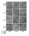
- FIG. 2 is a photograph ( ⁇ 400) of a confocal laser scanning microscope showing the cell adhesion by F-actin staining on F c 0.1 wt %-S c 20 wt %, F c 1 wt %-S c 20 wt % and F c 2%-S c 20 wt % fish collagen/PCL nanofiber support, prepared from different concentration of fish collagen (Fc) with the fish collagen stock solution (S c ) 20%.
- the green is F-actin and the blue color is DAPI.
- FIG. 3 is a scanning electron microscope image showing the microstructure of fish collagen/PCL nanofiber support prepared from F c 2 wt %, F c 3 wt %, F c 4 wt %, F c 5 wt % and F c 10 wt % using fish collagen stock solution of S c 50 wt %.
- FIG. 4 is a scanning electron microscope image showing the microstructure of fish collagen/PCL nanofiber support prepared from F c 2 wt %, F c 3 wt %, F c 4 wt %, F c 5 wt % and F c 10 wt % using fish collagen stock solution of S c 70 wt %.
- FIG. 5 is a scanning electron microscope image showing the microstructure of fish collagen/PCL nanofiber support prepared from F c 2 wt % using fish collagen stock solution (S c ) having concentrations of 20 wt %, 30 wt %, 40 wt % and 50 wt %.
- FIG. 6 is a scanning electron microscope image showing the microstructure of fish collagen/PCL nanofiber support prepared from F c 4 wt % using fish collagen stock solution (S c ) having concentrations of 40 wt % and 50 wt %.
- FIG. 7 is confocal laser scanning micrographs of human lung squamous cell carcinoma cell line (NCI-H1703) seeded onto the electrospun PCL nanofiber support (F c 0%; PCL 100%) after 1 day of 3D cell culture.
- Z-stack images of different planes showing from surface to bottom with 1 ⁇ m plane thickness (A), a reconstructed 3D projection image (B), and an enlarged image (C) are shown.
- Cells were stained with FITC-phalloidin (green) for F-actin cytoskeleton and DAPI (blue) for nucleus ( ⁇ 400). The green is F-actin and the blue color is DAPI ( ⁇ 400).
- FIG. 8 is confocal laser scanning micrographs of human lung squamous cell carcinoma cell line (NCI-H1703) seeded onto the electrospun fish collagen/PCL nanofiber support (F c 2 wt %-S c 30 wt %) after 1 day of 3D cell culture.
- Z-stack images of different planes showing from surface to bottom with 1 ⁇ m plane thickness (A), a reconstructed 3D projection image (B), and an enlarged image (C) are shown.
- Cells were stained with FITC-phalloidin (green) for F-actin cytoskeleton and DAPI (blue) for nucleus ( ⁇ 400).
- FIG. 9 is confocal laser scanning micrographs of human lung squamous cell carcinoma cell line (NCI-H1703) seeded onto the electrospun fish collagen/PCL nanofiber support (F c 2 wt %-S c 50 wt %) after 1 day of 3D cell culture.
- Z-stack images of different planes showing from surface to bottom with 1 ⁇ m plane thickness (A), a reconstructed 3D projection image (B), and an enlarged image (C) are shown.
- Cells were stained with FITC-phalloidin (green) for F-actin cytoskeleton and DAPI (blue) for nucleus ( ⁇ 400).
- FIG. 10 is confocal laser scanning micrographs of human lung squamous cell carcinoma cell line (NCI-H1703) seeded onto the electrospun fish collagen/PCL nanofiber support (F c 2 wt %-S c 70 wt %) after 1 day of 3D cell culture.
- Z-stack images of different planes showing from surface to bottom with 1 ⁇ m plane thickness (A), a reconstructed 3D projection image (B), and an enlarged image (C) are shown.
- Cells were stained with FITC-phalloidin (green) for F-actin cytoskeleton and DAPI (blue) for nucleus ( ⁇ 400).
- FIG. 11 is confocal laser scanning micrographs of human lung squamous cell carcinoma cell line (NCI-H1703) seeded onto the electrospun fish collagen/PCL nanofiber support (F c 5 wt %-S c 50 wt %) after 1 day of 3D cell culture.
- Z-stack images of different planes showing from surface to bottom with 1 ⁇ m plane thickness (A), a reconstructed 3D projection image (B), and an enlarged image (C) are shown.
- Cells were stained with FITC-phalloidin (green) for F-actin cytoskeleton and DAPI (blue) for nucleus ( ⁇ 400).
- FIG. 12 is confocal laser scanning micrographs of human lung squamous cell carcinoma cell line (NCI-H1703) seeded onto the electrospun fish collagen/PCL nanofiber support (F c 5 wt %-S c 70 wt %) after 1 day of 3D cell culture.
- Z-stack images of different planes showing from surface to bottom with 1 ⁇ m plane thickness (A), a reconstructed 3D projection image (B), and an enlarged image (C) are shown.
- Cells were stained with FITC-phalloidin (green) for F-actin cytoskeleton and DAPI (blue) for nucleus ( ⁇ 400).
- FIG. 13 is confocal laser scanning micrographs of human lung squamous cell carcinoma cell line (NCI-H1703) seeded onto the electrospun fish collagen/PCL nanofiber support (F c 10 wt %-S c 50 wt %) after 1 day of 3D cell culture.
- Z-stack images of different planes showing from surface to bottom with 1 ⁇ m plane thickness (A), a reconstructed 3D projection image (B), and an enlarged image (C) are shown.
- Cells were stained with FITC-phalloidin (green) for F-actin cytoskeleton and DAPI (blue) for nucleus ( ⁇ 400).
- FIG. 14 is confocal laser scanning micrographs of human lung squamous cell carcinoma cell line (NCI-H1703) seeded onto the electrospun fish collagen/PCL nanofiber support (F c 10 wt %-S c 70 wt %) after 1 day of 3D cell culture.
- Z-stack images of different planes showing from surface to bottom with 1 ⁇ m plane thickness (A), a reconstructed 3D projection image (B), and an enlarged image (C) are shown.
- Cells were stained with FITC-phalloidin (green) for F-actin cytoskeleton and DAPI (blue) for nucleus ( ⁇ 400).
- FIG. 15 is confocal laser scanning micrographs of human lung squamous cell carcinoma cell line (NCI-H1703) seeded onto the electrospun fish collagen/PCL nanofiber support (F c 15 wt %-S c 70 wt %) after 1 day of 3D cell culture.
- Z-stack images of different planes showing from surface to bottom with 1 ⁇ m plane thickness (A), a reconstructed 3D projection image (B), and an enlarged image (C) are shown.
- Cells were stained with FITC-phalloidin (green) for F-actin cytoskeleton and DAPI (blue) for nucleus ( ⁇ 400).
- FIG. 16 is confocal laser scanning micrographs of human lung squamous cell carcinoma cell line (NCI-H1703) seeded onto the electrospun fish collagen/PCL nanofiber support (F c 20 wt %-S c 70 wt %) after 1 day of 3D cell culture.
- Z-stack images of different planes showing from surface to bottom with 1 ⁇ m plane thickness (A), a reconstructed 3D projection image (B), and an enlarged image (C) are shown.
- Cells were stained with FITC-phalloidin (green) for F-actin cytoskeleton and DAPI (blue) for nucleus ( ⁇ 400).
- FIG. 17 is confocal laser scanning micrographs of human prostate cancer cell line (DU-145) seeded onto the electrospun PCL nanofiber support (F c 0%; PCL 100%) after 1 day of 3D cell culture.
- Z-stack images of different planes showing from surface to bottom with 1 ⁇ m plane thickness (A), a reconstructed 3D projection image (B), and an enlarged image (C) are shown.
- Cells were stained with FITC-phalloidin (green) for F-actin cytoskeleton and DAPI (blue) for nucleus ( ⁇ 400).
- FIG. 18 is confocal laser scanning micrographs of human prostate cancer cell line (DU-145) seeded onto the electrospun fish collagen/PCL nanofiber support (F c 2 wt %-S c 30 wt %) after 1 day of 3D cell culture.
- Z-stack images of different planes showing from surface to bottom with 1 ⁇ m plane thickness (A), a reconstructed 3D projection image (B), and an enlarged image (C) are shown.
- Cells were stained with FITC-phalloidin (green) for F-actin cytoskeleton and DAPI (blue) for nucleus ( ⁇ 400).
- FIG. 19 is confocal laser scanning micrographs of human prostate cancer cell line (DU-145) seeded onto the electrospun fish collagen/PCL nanofiber support (F c 2 wt %-S c 50 wt %) after 1 day of 3D cell culture.
- Z-stack images of different planes showing from surface to bottom with 1 ⁇ m plane thickness (A), a reconstructed 3D projection image (B), and an enlarged image (C) are shown.
- Cells were stained with FITC-phalloidin (green) for F-actin cytoskeleton and DAPI (blue) for nucleus ( ⁇ 400).
- FIG. 20 is confocal laser scanning micrographs of human prostate cancer cell line (DU-145) seeded onto the electrospun fish collagen/PCL nanofiber support (F c 2 wt %-S c 70 wt %) after 1 day of 3D cell culture.
- Z-stack images of different planes showing from surface to bottom with 1 ⁇ m plane thickness (A), a reconstructed 3D projection image (B), and an enlarged image (C) are shown.
- Cells were stained with FITC-phalloidin (green) for F-actin cytoskeleton and DAPI (blue) for nucleus ( ⁇ 400).
- FIG. 21 is confocal laser scanning micrographs of human prostate cancer cell line (DU-145) seeded onto the electrospun fish collagen/PCL nanofiber support (F c 5 wt %-S c 50 wt %) after 1 day of 3D cell culture.
- Z-stack images of different planes showing from surface to bottom with 1 ⁇ m plane thickness (A), a reconstructed 3D projection image (B), and an enlarged image (C) are shown.
- Cells were stained with FITC-phalloidin (green) for F-actin cytoskeleton and DAPI (blue) for nucleus ( ⁇ 400).
- FIG. 22 is confocal laser scanning micrographs of human prostate cancer cell line (DU-145) seeded onto the electrospun fish collagen/PCL nanofiber support (F c 5 wt %-S c 70 wt %) after 1 day of 3D cell culture.
- Z-stack images of different planes showing from surface to bottom with 1 ⁇ m plane thickness (A), a reconstructed 3D projection image (B), and an enlarged image (C) are shown.
- Cells were stained with FITC-phalloidin (green) for F-actin cytoskeleton and DAPI (blue) for nucleus ( ⁇ 400).
- FIG. 23 is confocal laser scanning micrographs of human prostate cancer cell line (DU-145) seeded onto the electrospun fish collagen/PCL nanofiber support (F c 10 wt %-S c 50 wt %) after 1 day of 3D cell culture.
- Z-stack images of different planes showing from surface to bottom with 1 ⁇ m plane thickness (A), a reconstructed 3D projection image (B), and an enlarged image (C) are shown.
- Cells were stained with FITC-phalloidin (green) for F-actin cytoskeleton and DAPI (blue) for nucleus ( ⁇ 400).
- FIG. 24 is confocal laser scanning micrographs of human prostate cancer cell line (DU-145) seeded onto the electrospun fish collagen/PCL nanofiber support (F c 10 wt %-S c 70 wt %) after 1 day of 3D cell culture.
- Z-stack images of different planes showing from surface to bottom with 1 ⁇ m plane thickness (A), a reconstructed 3D projection image (B), and an enlarged image (C) are shown.
- Cells were stained with FITC-phalloidin (green) for F-actin cytoskeleton and DAPI (blue) for nucleus ( ⁇ 400).
- FIG. 25 is confocal laser scanning micrographs of human prostate cancer cell line (DU-145) seeded onto the electrospun fish collagen/PCL nanofiber support (F c 15 wt %-S c 70 wt %) after 1 day of 3D cell culture.
- Z-stack images of different planes showing from surface to bottom with 1 ⁇ m plane thickness (A), a reconstructed 3D projection image (B), and an enlarged image (C) are shown.
- Cells were stained with FITC-phalloidin (green) for F-actin cytoskeleton and DAPI (blue) for nucleus ( ⁇ 400).
- FIG. 26 is confocal laser scanning micrographs of human prostate cancer cell line (DU-145) seeded onto the electrospun fish collagen/PCL nanofiber support (F c 20 wt %-S c 70 wt %) after 1 day of 3D cell culture.
- Z-stack images of different planes showing from surface to bottom with 1 ⁇ m plane thickness (A), a reconstructed 3D projection image (B), and an enlarged image (C) are shown.
- Cells were stained with FITC-phalloidin (green) for F-actin cytoskeleton and DAPI (blue) for nucleus ( ⁇ 400).
- FIG. 27 is confocal laser scanning micrographs of mouse B cell lymphoma cell line (A20) seeded onto the electrospun PCL nanofiber support (F c 0%; PCL 100%) after 1 day of 3D cell culture.
- Z-stack images of different planes showing from surface to bottom with 1 ⁇ m plane thickness (A), a reconstructed 3D projection image (B), and an enlarged image (C) are shown.
- Cells were stained with FITC-phalloidin (green) for F-actin cytoskeleton and DAPI (blue) for nucleus ( ⁇ 400).
- FIG. 28 is confocal laser scanning micrographs of mouse B cell lymphoma cell line (A20) seeded onto the electrospun fish collagen/PCL nanofiber support (F c 2 wt %-S c 30 wt %) after 1 day of 3D cell culture.
- Z-stack images of different planes showing from surface to bottom with 1 ⁇ m plane thickness (A), a reconstructed 3D projection image (B), and an enlarged image (C) are shown.
- Cells were stained with FITC-phalloidin (green) for F-actin cytoskeleton and DAPI (blue) for nucleus ( ⁇ 400).
- FIG. 29 is confocal laser scanning micrographs of mouse B cell lymphoma cell line (A20) seeded onto the electrospun fish collagen/PCL nanofiber support (F c 2 wt %-S c 50 wt %) after 1 day of 3D cell culture.
- Z-stack images of different planes showing from surface to bottom with 1 ⁇ m plane thickness (A), a reconstructed 3D projection image (B), and an enlarged image (C) are shown.
- Cells were stained with FITC-phalloidin (green) for F-actin cytoskeleton and DAPI (blue) for nucleus ( ⁇ 400).
- FIG. 30 is confocal laser scanning micrographs of mouse B cell lymphoma cell line (A20) seeded onto the electrospun fish collagen/PCL nanofiber support (F c 2 wt %-S c 70 wt %) after 1 day of 3D cell culture.
- Z-stack images of different planes showing from surface to bottom with 1 ⁇ m plane thickness (A), a reconstructed 3D projection image (B), and an enlarged image (C) are shown.
- Cells were stained with FITC-phalloidin (green) for F-actin cytoskeleton and DAPI (blue) for nucleus ( ⁇ 400).
- FIG. 31 is confocal laser scanning micrographs of mouse B cell lymphoma cell line (A20) seeded onto the electrospun fish collagen/PCL nanofiber support (F c 5 wt %-S c 50 wt %) after 1 day of 3D cell culture.
- Z-stack images of different planes showing from surface to bottom with 1 ⁇ m plane thickness (A), a reconstructed 3D projection image (B), and an enlarged image (C) are shown.
- Cells were stained with FITC-phalloidin (green) for F-actin cytoskeleton and DAPI (blue) for nucleus ( ⁇ 400).
- FIG. 32 is confocal laser scanning micrographs of mouse B cell lymphoma cell line (A20) seeded onto the electrospun fish collagen/PCL nanofiber support (F c 5 wt %-S c 70 wt %) after 1 day of 3D cell culture.
- Z-stack images of different planes showing from surface to bottom with 1 ⁇ m plane thickness (A), a reconstructed 3D projection image (B), and an enlarged image (C) are shown.
- Cells were stained with FITC-phalloidin (green) for F-actin cytoskeleton and DAPI (blue) for nucleus ( ⁇ 400).
- FIG. 33 is confocal laser scanning micrographs of mouse B cell lymphoma cell line (A20) seeded onto the electrospun fish collagen/PCL nanofiber support (F c 10 wt %-S c 70 wt %) after 1 day of 3D cell culture.
- Z-stack images of different planes showing from surface to bottom with 1 ⁇ m plane thickness (A), a reconstructed 3D projection image (B), and an enlarged image (C) are shown.
- Cells were stained with FITC-phalloidin (green) for F-actin cytoskeleton and DAPI (blue) for nucleus ( ⁇ 400).
- FIG. 34 is confocal laser scanning micrographs of mouse B cell lymphoma cell line (A20) seeded onto the electrospun fish collagen/PCL nanofiber support (F b 15 wt %-S c 70 wt %) after 1 day of 3D cell culture.
- Z-stack images of different planes showing from surface to bottom with 1 ⁇ m plane thickness (A), a reconstructed 3D projection image (B), and an enlarged image (C) are shown.
- Cells were stained with FITC-phalloidin (green) for F-actin cytoskeleton and DAPI (blue) for nucleus ( ⁇ 400).
- FIG. 35 is confocal laser scanning micrographs of mouse B cell lymphoma cell line (A20) seeded onto the electrospun fish collagen/PCL nanofiber support (F c 20 wt %-S c 70 wt %) after 1 day of 3D cell culture.
- Z-stack images of different planes showing from surface to bottom with 1 ⁇ m plane thickness (A), a reconstructed 3D projection image (B), and an enlarged image (C) are shown.
- Cells were stained with FITC-phalloidin (green) for F-actin cytoskeleton and DAPI (blue) for nucleus ( ⁇ 400).
- FIG. 36 is confocal laser scanning micrographs of mouse T cell lymphoma cell line (EL4) seeded onto the electrospun PCL nanofiber support (F c 0%; PCL 100%) after 1 day of 3D cell culture.
- Z-stack images of different planes showing from surface to bottom with 1 ⁇ m plane thickness (A), a reconstructed 3D projection image (B), and an enlarged image (C) are shown.
- Cells were stained with FITC-phalloidin (green) for F-actin cytoskeleton and DAPI (blue) for nucleus ( ⁇ 400).
- FIG. 37 is confocal laser scanning micrographs of mouse T cell lymphoma cell line (EL4) seeded onto the electrospun fish collagen/PCL nanofiber support (F c 2 wt %-S c 30 wt %) after 1 day of 3D cell culture.
- Z-stack images of different planes showing from surface to bottom with 1 ⁇ m plane thickness (A), a reconstructed 3D projection image (B), and an enlarged image (C) are shown.
- Cells were stained with FITC-phalloidin (green) for F-actin cytoskeleton and DAPI (blue) for nucleus ( ⁇ 400).
- FIG. 38 is confocal laser scanning micrographs of mouse T cell lymphoma cell line (EL4) seeded onto the electrospun fish collagen/PCL nanofiber support (F c 2 wt %-S c 50 wt %) after 1 day of 3D cell culture.
- Z-stack images of different planes showing from surface to bottom with 1 ⁇ m plane thickness (A), a reconstructed 3D projection image (B), and an enlarged image (C) are shown.
- Cells were stained with FITC-phalloidin (green) for F-actin cytoskeleton and DAPI (blue) for nucleus ( ⁇ 400).
- FIG. 39 is confocal laser scanning micrographs of mouse T cell lymphoma cell line (EL4) seeded onto the electrospun fish collagen/PCL nanofiber support (F c 2 wt %-S c 70 wt %) after 1 day of 3D cell culture.
- Z-stack images of different planes showing from surface to bottom with 1 ⁇ m plane thickness (A), a reconstructed 3D projection image (B), and an enlarged image (C) are shown.
- Cells were stained with FITC-phalloidin (green) for F-actin cytoskeleton and DAPI (blue) for nucleus ( ⁇ 400).
- FIG. 40 is confocal laser scanning micrographs of mouse T cell lymphoma cell line (EL4) seeded onto the electrospun fish collagen/PCL nanofiber support (F c 5 wt %-S c 50 wt %) after 1 day of 3D cell culture.
- Z-stack images of different planes showing from surface to bottom with 1 ⁇ m plane thickness (A), a reconstructed 3D projection image (B), and an enlarged image (C) are shown.
- Cells were stained with FITC-phalloidin (green) for F-actin cytoskeleton and DAPI (blue) for nucleus ( ⁇ 400).
- FIG. 41 is confocal laser scanning micrographs of mouse T cell lymphoma cell line (EL4) seeded onto the electrospun fish collagen/PCL nanofiber support (F c 5 wt %-S c 70 wt %) after 1 day of 3D cell culture.
- Z-stack images of different planes showing from surface to bottom with 1 ⁇ m plane thickness (A), a reconstructed 3D projection image (B), and an enlarged image (C) are shown.
- Cells were stained with FITC-phalloidin (green) for F-actin cytoskeleton and DAPI (blue) for nucleus ( ⁇ 400).
- FIG. 42 illustrates an analysis result of an effect of the PCL nanofiber support and the F c 2 wt %-S c 50 wt % fish collagen/PCL nanofiber support on survival of mouse B-cell lymphoma cells during cell culture thereof by a confocal laser scanning microscopic observation ( ⁇ 400) of the Live/Dead cells after seeding the mouse B cell lymphoma cell and culturing for 2 days.
- FIG. 43 illustrates an analysis result of an effect of the PCL nanofiber support and the F c 2 wt %-S c 50 wt % fish collagen/PCL nanofiber support on survival of mouse B-cell lymphoma cells during cell culture thereof by a confocal laser scanning microscopic observation ( ⁇ 400) of the Live/Dead cells after seeding the mouse B cell lymphoma cell and culturing for 6 days.
- FIG. 44 shows a result of diffusion degree of anti-cancer agents after treating 1 ⁇ M doxorubicin, a standard anti-cancer drug against lymphoma, to 3D culture model of mouse T lymphoma cells using F c 2 wt %-Sc 50 wt % fish collagen/PCL nanofiber support according to the present invention, for evaluating the diffusion ability of the drug to reach cancer cells in a 3D environment.
- FIG. 45 shows the sensitivity of anticancer drugs by measuring the cell viability of human lung cancer cells (NCI-H1703, A) and prostate cancer cells (DU-145, B) cultured in a conventional two-dimensional environment and three-dimensional environment using F c 4 wt %-S c 50 wt % fish collagen/PCL nanofiber support according to the present invention.
- FIG. 46 shows expression levels of Hes 1 and Notch genes related to malignancy by RT-PCR of human prostate cancer cells (DU-145, B) cultured in a conventional two-dimensional environment and three-dimensional environment using F c 4 wt %-S c 50 wt % fish collagen/PCL nanofiber support according to the present invention.
- FIG. 47 shows expression levels of snail, slug, endothelin-1, CD44, VEGF, and HIF-1 ⁇ genes related to malignancy by RT-PCR of human prostate cancer cells (DU-145, B) cultured in a conventional two-dimensional environment and three-dimensional environment using F c 4 wt %-S c 50 wt % fish collagen/PCL nanofiber support according to the present invention.
- FIG. 48 shows flow cytometric analysis of measuring the cancer stem cell-forming ability and the differentiation ability of mouse T cell lymphoma cell line cultured three-dimensionally in F c 2 wt %-S c 50 wt % fish collagen/PCL nanofiber support according to the present invention.
- FIG. 49 is a schematic diagram showing a composition of agarose-collagen-alginate composite hydrogel according to the present invention.
- FIG. 50 is a schematic diagram showing a production process of agarose-collagen-alginate composite hydrogel according to the present invention.
- FIG. 51 is a WST-1 analysis result of cytotoxic effect of CaCl 2 after treating a mouse normal cell, CREC and cancer cell lines, EL4, R182 and A2780 cells with CaCl 2 having various concentrations (10, 50, 100, 200 and 300 mM) for 18 to 24 hours.
- FIG. 52 is photographs by a phase-contrast microscope to investigate three-dimensional cell growth environment after culturing the A2780 cells in agarose-alginate composite hydrogel in which the final concentration of the agarose is 0.125 wt % and the final concentration of the alginate is 1 wt % in mixed solution prepared from 0.5, 1 and 1.5 wt % agarose solution (referred to as hereinafter “0.125 wt % agarose-1 wt % alginate composite hydrogel”).
- FIG. 53 is photographs by a phase-contrast microscope to investigate EL4 cell growth after culturing EL4 cells three-dimensionally in pure agarose gels having concentrations of 0.5, 1, 1.5 and 2 wt % for 2, 5 and 10 days.
- FIGS. 54 and 55 are photographs by a phase-contrast microscope to investigate the detached cells from the hydrogels.
- EL4 cells were seeded in the 0.125% agarose-1% alginate composite hydrogel containing 0.05% rat tail collagen (RC) and in 0.125% agarose-1% alginate composite hydrogels containing different concentrations of marine collagen at 0.05, 0.1, 0.2, 0.5, 1, 2, 5 and 10%. The encapsulation efficiency of EL4 cells among these hydrogels was observed.
- FIG. 54 is a phase contrast microscopic image of non-encapsulated cells
- FIG. 55 is a bar graph showing the numbers of non-encapsulated EL4 cells.
- FIG. 56 is photographs by a phase-contrast microscope to investigate cell growth after culturing EL4 cells for 18 to 24 hours in supports containing 0.125 wt % agarose-collagen-1 wt % alginate composite hydrogel, in which both conventional animal collagen and marine collagen were used.
- a marine collagen was attempted firstly according to the present invention, as a source of collagen.
- FIG. 57 is photographs by a phase-contrast microscope to investigate cell growth after culturing A2780 cells three-dimensionally in supports containing 0.125 wt % agarose-collagen-1 wt % alginate composite hydrogel, in which the conventional animal collagen and marine collagen were used.
- a marine collagen was attempted firstly according to the present invention, as a source of collagen.
- FIG. 58 is photographs by a phase-contrast microscope to confirm the transparency of agarose-collagen-alginate composite hydrogel support according to the present invention.
- FIG. 59 is photographs by a scanning electron microscope to observe the cross section of hydrogel supports composed with 1 wt % of pure alginate, 0.125 wt % of pure agarose, 7.5 or 10 wt % of pure marine collagen, 0.125 wt % agarose-1 wt % alginate composite hydrogel support and 0.125 wt % agarose-7.5 wt % marine collagen-1 wt % alginate and 0.125 wt % agarose-10 wt % marine collagen-1 wt % alginate composite hydrogel support.
- FIG. 60 is FT-IR analysis result of hydrogel supports composed with 1 wt % of pure alginate, 0.125 wt % of pure agarose, 7.5 or 10 wt % of pure marine collagen, 0.125 wt % agarose-1 wt % alginate composite hydrogel support and 0.125 wt % agarose-7.5 wt % marine collagen-1 wt % alginate and 0.125 wt % agarose-10 wt % marine collagen-1 wt % alginate composite hydrogel support.
- FIG. 61 is a schematic diagram of the molecular structure of each component of the hydrogel material.
- FIGS. 61A, 61B and 61C are schematic diagrams of the molecular structure of agarose, marine collagen and alginate, respectively.
- FIG. 62 is a graph showing the swelling behavior of 0.125 wt % agarose-7.5 wt % marine collagen-1 wt % alginate and 0.125 wt % agarose-10 wt % marine collagen-1 wt % alginate composite hydrogel supports in distilled water and phosphate buffered saline (PBS) according to the invention.
- PBS phosphate buffered saline
- FIG. 63 is results by a light microscope (a), H&E staining (b), DAPI staining (c) and F-actin staining (d) after culturing human ovarian cancer cells for 7 days in 0.125 wt % agarose-7.5 wt % marine collagen-1 wt % alginate composite hydrogel support according to the present invention.
- FIG. 64 is results by a phase contrast microscope to observe cell spheroids formed after culturing EL4 cells for 1, 3 and 5 days in 0.125 wt % agarose-10 wt % marine collagen-1 wt % alginate composite hydrogel support according to the present invention.
- FIG. 65 is results observing spheroid budding phenomena that play an important role in cell spheroid formation, after culturing A2780 cells in 0.125 wt % agarose-7.5 wt % marine collagen-1 wt % alginate composite hydrogel support according to the present invention.
- FIGS. 65A and 65B are results by a phase difference microscope and H&E staining, respectively.
- FIG. 66 is a result confirming the diffusion ability of the drug to reach the tumor in the three-dimensional environment by using three-dimensional culture model of human ovarian cancer cells A2780 in 0.125 wt % agarose-7.5 wt % marine collagen-1 wt % alginate composite hydrogel support according to the present invention.
- FIG. 67 is a result of the cell proliferation ability after culturing human ovarian cancer A2780 for 1, 3, 5 and 7 days in the two-dimensional culture environment and the three-dimensional environment using 0.125 wt % agarose-7.5 wt % marine collagen-1 wt % alginate composite hydrogel support according to the present invention, respectively.
- FIG. 68 is a result by a CFSE staining to confirm the cell proliferation after three-dimensionally culturing CREC and EL4 cells in 0.125 wt % agarose-10 wt % marine collagen-1 wt % alginate composite hydrogel support according to the invention.
- FIGS. 69 and 70 are results showing the cytotoxicity of the agarose-marine collagen-alginate composite hydrogel support according to the present invention.
- FIG. 69 is a result by a confocal laser microscopic observation of Live/Dead cells after seeding human ovarian cancer cells A2780 in 0.125 wt % agarose-7.5 wt % marine collagen-1 wt % alginate composite hydrogel support according to the present invention and culturing them for 7 and 10 days.
- FIG. 69 is a result by a confocal laser microscopic observation of Live/Dead cells after seeding human ovarian cancer cells A2780 in 0.125 wt % agarose-7.5 wt % marine collagen-1 wt % alginate composite hydrogel support according to the present invention and culturing them for 7 and 10 days.
- FIG. 71 shows expression levels of Notch genes related to malignancy of the cancer cell by RT-PCR after culturing EL4 cells two dimensionally and three-dimensionally in 0.125 wt % agarose-10 wt % marine collagen-1 wt % alginate composite hydrogel support according to the present invention, respectively.
- FIG. 72 shows expression levels of snail and vimentin which are malignancy markers of the cancer cells, CD44 which is an important marker of cancer stem cells, and endothelin-1 which plays a key role in growth and metastasis of tumors after culturing human ovarian cancer cells two dimensionally and three-dimensionally in 0.125 wt % agarose-7.5 wt % marine collagen-1 wt % alginate composite hydrogel support according to the present invention, respectively.
- FIG. 73 is a result identifying the cell viability of anticancer drugs after culturing lymphoma cells (EL4) and human ovarian cancer cells (A2780) two dimensionally and three-dimensionally in 0.125 wt % agarose-10 wt % marine collagen-1 wt % alginate composite hydrogel support according to the present invention, respectively.
- FIG. 73 is a result identifying the cell viability against anticancer drugs after culturing lymphoma cells (EL4) and human ovarian cancer cells (A2780) two dimensionally and three-dimensionally in 0.125 wt % agarose-10 wt % marine collagen-1 wt % alginate composite hydrogel support according to the present invention, respectively.
- FIG. 74 is a result identifying the cell viability of ovarian cancer cells against a standard anticancer agent, paclitaxel and curcumin, which have anticancer effect against various cancer cells, after culturing human ovarian cancer cells (A2780) two dimensionally and three-dimensionally in 0.125 wt % agarose-7.5 wt % marine collagen-1 wt % alginate composite hydrogel support according to the present invention, respectively.
- FIG. 75 is results observing the cells by phase contrast microscopy and fluorescence microscopy in human ovarian cancer cells (A2780) cultured three-dimensionally in 0.125 wt % agarose-7.5 wt % marine collagen-1 wt % alginate composite hydrogel support according to the present invention after immunostaining with antibodies against cytokeratin which is a marker specifically expressed in the epithelial cells.
- FIG. 76 shows flow cytometric analysis of measuring the cancer stem cell-forming ability and the differentiation ability of EL4 cells cultured three-dimensionally in 0.125 wt % agarose-10 wt % marine collagen-1 wt % alginate composite hydrogel support according to the present invention.
- the present invention provides a method of preparing a composite nanofiber support comprising: a first step of dissolving a synthetic polymer in an organic solvent; a second step of dissolving a fish collagen in water to prepare an aqueous fish collagen solution; a third step of adding the aqueous fish collagen solution prepared in the second step to the synthetic polymer solution prepared in the first step, followed by mixing homogeneously; and a fourth step of electrospinning a mixture solution prepared in the third step to prepare a nanofiber support.
- the synthetic polymer solution in the first step may be prepared by dissolving 8 to 10 parts by weight of a synthetic polymer, based on 100 parts by weight of the organic solvent. More preferably, the synthetic polymer solution of 8.8 wt % may be prepared, but is not limited thereto.
- addition range of the synthetic polymer is out of the range, electrospinning may not be performed, or it may be difficult to produce homogeneous nanofibers during electrospinning.
- the synthetic polymer may be one or more selected from the group consisting of poly( ⁇ -caprolactone) (PCL), poly(lactic acid) (PLA), poly(glycolic acid) (PGA), poly(hydroxy valerate) (PHV), poly (lactic-co-glycolic acid) (PLGA), poly-hydroxy butyrate (PHB), poly (hydroxy butyrate-co-valerate (PHBV), polyanhydride, polyorthoester, poly (vinyl alcohol) (PVA), polyethylene glycol (PEG), polyurethane, polyacrylic acid (PAA), poly-N-isopropylacrylamide, diol/biacid-based aliphatic polyester and polyester-amide/polyester-urethane. More preferably, the synthetic polymer may be poly( ⁇ -caprolactone) (PCL), but is not limited thereto.
- the poly( ⁇ -caprolactone) (PCL) is a synthetic polymer having a semi-crystalline powder form with a low melting point, and it melts easily compared to other polymers.
- the poly( ⁇ -caprolactone) (PCL) is a non-toxic and biocompatible material that exhibits very slow decomposition rates and is widely used as a material for scaffolds for tissue engineering.
- the organic solvent may be selected from the group consisting of chloroform, hexafluoroisopropane, tetrahydrofuran, dimethylformamide, dichloromethane, acetone, dioxane, trifluoroethane and mixture thereof.
- the aqueous fish collagen solution of the second step may be prepared by dissolving fish collagen of 10 to 70 parts by weight based on 100 parts by weight of water.
- the fish collagen does not dissolve homogeneously in water or a homogeneous fish collagen-PCL mixture solution cannot be obtained or homogeneous nanofibers cannot be produced.
- the collagen may be collagen extracted from marine organisms, more preferably fish collagen, but is not limited thereto.
- Collagen is a fibrous protein and a natural polymer which is the main component of the extracellular matrix in the connective tissue. This collagen plays a role in regulating the cell recognition and cell adhesion and proliferation, supports the tissues and organs, maintains the body shape surrounding the body surface and regenerates tissues. It can be processed into various forms such as a sponge, a fiber, a gel, a solution, a core material and a tubular material.
- Fish collagen is a natural polymer substance extracted from marine fish species. The collagen extracted from these marine organisms can undergo easily lower molecular peptidization than collagen extracted from mammals, and has excellent absorption into human body. In addition, the amino acid composition of collagen extracted from marine organisms is different from that of human collagen and can be easily distinguished by special antibodies.
- collagen has a disadvantage in that it has a very low mechanical strength and is difficult to control the decomposition rate and gelation is difficult by using collagen alone.
- the water is the most common solvent in nature and is more economical than organic solvents and does not show toxicity.
- the present invention demonstrates for the first time that water can be used among solvents that constitute a mixture solution used in the manufacture of nanofibers using electrospinning. Specifically, it is possible to prepare a mixture solution for electrospinning by dissolving a natural polymer in water without toxicity instead of an organic solvent exhibiting toxicity to the cells.
- a mixture solution for electrospinning by dissolving a natural polymer in water without toxicity instead of an organic solvent exhibiting toxicity to the cells.
- other natural polymer in addition to fish collagen which can use water as a solvent may apply to the manufacture of nanofibers by electrospinning.
- the mixture solution of the third step is prepared by adding the synthetic polymer solution of the first step so as to comprise the synthetic polymer of 6.3 to 8.5 parts by weight based on 100 parts by weight of the mixture solution. More specifically, poly( ⁇ -caprolactone) (PCL) solution 8.8 wt % prepared in the first step may be added in an amount of 71.4 to 97.1 parts by weight based on 100 parts by weight of the mixture solution, but it is not limited thereto.
- the fish collagen solution may be added in an amount of 2 to 20 parts by weight based on 100 parts by weight of the mixture solution, but it is not limited thereto.
- the electrospinning may not be properly performed or it may be difficult to produce homogeneous nanofibers during electrospinning.
- the mixture solution of the third step is prepared by adding water so as to comprise water of 0.9 to 10 parts by weight based on 100 parts by weight of the mixture solution, but it is not limited thereto.
- the present invention provides a method of culturing three-dimensional cell comprising: a first step of dissolving a synthetic polymer in an organic solvent; a second step of dissolving a fish collagen in water to prepare an aqueous fish collagen solution; a third step of adding the aqueous fish collagen solution prepared in the second step to the synthetic polymer solution prepared in the first step, followed by mixing homogeneously; a fourth step of electrospinning a mixture solution prepared in the third step to prepare a nanofiber support; and a step of culturing cell on the nanofiber support prepared in the fourth step.
- the present invention provides a method of culturing three-dimensional cell comprising: a step of mixing and culturing an alginate solution and cells; a step of adding a collagen solution to a cultured mixture to prevent cell detachment; and a step of adding the agarose solution to a mixture including collagen and allowing the mixture to stand, thereby inducing gelation.
- the method of three-dimensional cell culture may comprise mixing the alginate solution and the cells, and culturing at a temperature of 25 to 37° C. for 5 to 30 minutes; adding the collagen solution to the cultured mixture to prevent cell detachment at a temperature of 25 to 37° C. for 5 to 30 minutes; and adding the agarose solution to a mixture including collagen and allowing the mixture to stand for 5 to 30 minutes at a temperature of 4 to 25° C., thereby inducing gelation.
- the incubation temperature and time range are the most preferable temperature and time range for minimizing the influence on the cells.
- the collagen solution In the step of adding the collagen solution to the cultured mixture so as to prevent cell detachment, if the temperature and the time do not fall within the range, the collagen is not homogeneously mixed or the cell culture efficiency is lowered.
- the step of adding the agarose solution is added and allowing the mixture to stand, thereby inducing gelation, if the temperature and the time do not fall within the range, gelation may not be performed or cells may be damaged or killed.
- the collagen may be marine-derived collagen, but is not limited thereto.
- the alginate may be added in an amount of 0.5 to 2 parts by weight, more preferably 1 part by weight based on 100 parts by weight of the cultured mixture, but is not limited thereto.
- the collagen may be added in an amount of 1 to 30 parts by weight, more preferably 7.5 to 10 parts by weight based on 100 parts by weight of the mixture, but is not limited thereto.
- the cells are not be encapsulated so that the cells escape from the gel or affect the growth ability of the cells.
- the agarose is added in an amount of 0.1 to 0.5 parts by weight, more preferably 0.125 parts by weight based on 100 parts by weight of the mixture, but is not limited thereto.
- the agarose is a polysaccharide composed of galactose extracted from seaweed. It is mainly used as a cell culture medium and can be used in the form of a hydrogel because the hardness of the structure can be adjusted, so that it is possible to seed cells evenly inside the scaffold. Moreover, agarose can induce gelation by changing temperature without ion crosslinking or chemical crosslinking. However, if the agarose solution comprises in amount out of the above range, gelation may not be performed or cells may be damaged or killed.
- the alginate, collagen and agarose added to the mixture may be added in the form of a storage solution, but are not limited thereto.
- the alginate storage solution for preparing the mixture solution may be prepared by dissolving alginate in an amount of 1 to 10 parts by weight based on 100 parts by weight of distilled water.
- the agarose storage solution may be prepared by dissolving agarose in an amount of 1 to 3 parts by weight based on 100 parts by weight of distilled water.
- the collagen storage solution may be prepared by dissolving collagen in an amount of 20 to 30 parts by weight based on 100 parts by weight of distilled water, but is not limited thereto.
- the cells that can be cultured by the three-dimensional cell culture method of the present invention can be selected from normal cells and cancer cells.
- the cancer cells are solid cancer cell selected from the group consisting of ovarian cancer, breast cancer, gastric cancer, colon cancer, rectal cancer, lung cancer, liver cancer, prostate cancer, renal cancer, pancreatic cancer, thyroid cancer, uterine cancer, laryngeal cancer, hydrocephalus, laryngeal cancer, guinea, salivary gland cancer, esophageal cancer, osteosarcoma, tongue cancer, biliary cancer, glioma, bone cancer and unknown cause cancer, and the cell is a blood cancer cell selected from the group consisting of B cell lymphoma, T cell lymphoma, leukemia, and multiple myeloma, but is not limited thereto.
- the present invention provides a method of measuring cell activity comprising: a step of mixing and culturing an alginate solution and cells; a step of adding a collagen solution to a cultured mixture to prevent cell detachment; a step of adding an agarose solution to a mixture containing collagen and allowing the mixture to stand and induce gelation, and culturing the cells three-dimensionally; and analyzing growth, proliferation, death, differentiation and migration of cells cultured three-dimensionally.
- the present invention provides a method of measuring sensitivity of anticancer drugs comprising: a step of mixing and culturing an alginate solution and cells; a step of adding a collagen solution to a cultured mixture to prevent cell detachment; a step of adding an agarose solution to a mixture containing collagen and allowing the mixture to stand and induce gelation, and culturing the cells three-dimensionally; and a step of analyzing cell viability by adding an anticancer drug to cells cultured three-dimensionally.
- PCL polycaprolactone
- the needle of the electrospining was connected to a wire mesh type collector (0.5 mm in wire diameter, 1 ⁇ 1 mm mesh size) under the conditions of 27 gauge, a scattering distance of 8 cm, a voltage of 11 kV, a fluid velocity of 0.5 mL/hr, temperature 21 ⁇ 22° C. and humidity of 40 ⁇ 45% and the spinning time was 60 to 120 minutes, preferably approximately 90 minutes.
- a PCL nanofiber support was prepared by drying in an oven of 60° C. for 10 seconds to remove the residual solvent contained in the mat.
- the S C 20 wt % fish collagen stock solution was added to the 8.8 wt % PCL solution so as to be the final concentration of fish collagen of 0.1, 1 and 2 wt %, and the mixture was homogeneously mixed for 3 hours using a magnetic stirrer to prepare fish collagen/PCL mixture solution.
- the fish collagen/PCL mixture solution of 0.1, 1 and 2 wt % prepared in the above was electrospun by using an electrospinning apparatus.
- the needles of electrospinning were connected to a wire mesh type collector (0.5 mm in wire diameter, 1 ⁇ 1 mm mesh size) under the conditions of 27 gauge, a scattering distance of 8 cm, a voltage of 9 kV, a fluid velocity of 0.4 mL/hr, temperature 21 ⁇ 22° C. and humidity of 40 ⁇ 45%.
- the spinning time was 60 to 90 minutes, preferably approximately 70 minutes.
- rhodamine B (Sigma) was added to a fish collagen aqueous solution of S C 20 wt % and dissolved uniformly in a test tube mixer. Thereafter, a fish collagen aqueous solution of S C 20 wt % containing rhodamine B was added to 8.8 wt % PCL solution so that the final concentration of fish collagen was 1% by weight, and the mixture was stirred to prepare a rhodamine-1% fish collagen/PCL mixture solution.
- a nanofiber support was prepared by electrospinning the rhodamine-1 wt % fish collagen/PCL solution prepared in the above under electrospinning conditions of Examples 1-2.
- the prepared rhodamine-1 wt % fish collagen/PCL nanofiber support was placed on a slide glass, sealed with a cover glass, and the degree of mixing was analyzed using a confocal laser scanning microscope.
- Thymic deep cortex or cortical reticular epithelial cell (hereafter, referred to as CREC), a type of normal mouse thymic epithelial cell (TEC) provided from Dr. Barbara B. Knowles (The Jackson Laboratory, Bar Harbor, Me., USA) was cultured and incubated in Dulbecco's Modified Eagle Medium (DMEM, HyClone, Logan, Utah, USA) containing 10% (v/v) fetal bovine serum (FBS; Gibco, Invitrogen), 100 IU/mL penicillin (Gibco, USA) and 100 ⁇ g/mL streptomycin (Gibco, Invitrogen) at 37° C. in a 5% CO 2 atmosphere. The culture medium was replaced with new one every 2-3 days.
- DMEM Dulbecco's Modified Eagle Medium
- FBS fetal bovine serum
- penicillin Gibco, Invitrogen
- streptomycin (Gibco, Invitrogen)
- the PCL nanofiber support prepared in Example 1 and the fish collagen/PCL nanofiber support having final concentrations of 0.1, 1 and 2 wt % were placed in 96-well plates respectively and CREC cells were inoculated at 5 ⁇ 10 4 cells per a well and cultured for 6 hours.
- the support having the cultured cells was washed twice with phosphate buffered saline (PBS) and fixed with 4% paraformaldehyde fixation solution at room temperature for 20 minutes. Thereafter, the cells were washed twice with PBS, and were infiltrated into 0.1% Triton-X100 solution at room temperature for 5 minutes and then washed twice with PBS.
- PBS phosphate buffered saline
- the cells were treated with 0.2% bovine serum albumin (BSA) solution at room temperature for 20 minutes and then washed twice with PBS.
- BSA bovine serum albumin
- actin-detecting Phalloidin-FITC (Promega, Madison, Wis., USA) was diluted to 1:150 in 0.2% BSA solution and reacted at room temperature for 40 minutes, and then was washed with tris-buffered saline (TBS), the support was placed on a cover glass and was contrast-stained with DAPI (Vector Lab, Inc., Burlingame, Calif., USA) for nuclear staining of the cells, and encapsulated. The cell adhesion of the cells for the support was measured by a laser confocal microscope.
- 1 wt % fish collagen-PCL nanofiber support exhibited similar adhesion to the PCL nanofiber support, and the 1 and 2 wt % fish collagen support significantly increased the cell adhesion than PCL nanofiber support, and the higher the concentration of fish collagen, the better the cell culture efficiency of support. Therefore, the following Examples confirmed that three-dimensional culture of cells optimized at a concentration of at least 2 wt % of fish collagen.
- Fish collagen was added distilled water to prepare fish collagen stock solutions (S c ) having the concentrations of S c 10 wt %, S c 20 wt %, S c 30 wt % S c 40 wt % S c 50 wt % S c 60 wt % S c 70 wt % S c 80 wt % S c 90 wt % and S c 100 wt %, and were named as S c 10%, S c 20%, S c 30% S c 40% S c 50% S c 60% S c 70% S c 80% S c 90% and S c 100%.
- the fish collagen stock solutions to be added to the mixture solution were prepared as S c 10%, S c 20%, S c 30%, S c 40%, S c 50% and S c 70%.
- PCL polycaprolacton, average Mn 80,000, Sigma
- fish collagen stock solutions having each concentration were added to the produced 8.8 wt % PCL solution to prepare fish collagen/PCL mixture solutions having final concentration of fish collagen in the mixed solution (FC) of F c 2 wt %, F c 3 wt %, F c 4 wt %, F c 5 wt %, F c 10 wt %, F c 15 wt %, F c 20 wt %, F c 30 wt %, F c 40 wt % and F c 50 wt %, and were named as F c 2%, F c 3%, F c 4%, F c 5%, F c 10%, F c 15%, F c 20%, F c 30%, F c 40% and F c 50%, respectively.
- FC mixed solution
- the nanofiber support was not homogeneously produced or electrospun when it contained 1.1 mL or more of water based on the total volume of the mixture solution of 10 mL (number in parentheses in italics). Accordingly, it was confirmed that the nanofiber support was successfully prepared when the amount of water contained based on the total volume of the mixture solution of 10 mL was 0.09 to 1 mL (number in parentheses in italics).
- the mixture solution prepared according to the amount of water in Table 1, the amount of fish collagen in Table 2, and the amount of the 8.8 wt % PCL synthetic polymer solution in Table 3 based on a total volume of 10 mL of the mixture solution was electrospun to prepare a nanofiber support.
- the needles of electrospinning were connected to a wire mesh type collector (0.5 mm in diameter, 1 ⁇ 1 mm mesh size) under the conditions of 27 gauge, a scattering distance of 8 cm, a voltage of 9 kV, a fluid velocity of 0.4 mL/hr, temperature 21 ⁇ 22° C. and humidity of 40 ⁇ 45%.
- the spinning time was 60 to 90 minutes, preferably approximately 70 minutes.
- each nanofiber support was fixed with 2.5% glutaraldehyde fixation liquid for 24 hours at 4° C. and then additionally fixed with 1% osmium tetroxide at room temperature for 1 hour.
- the Fixed support samples were washed 2-3 times with 0.1 M phosphate buffer (pH 7.4) and dehydrated by an ethanol series. Each of the dehydrated nanofiber support samples was frozen at ⁇ 80° C.
- the fibers constituting the fish collagen/PCL nanofiber support were a nano/micro hybrid fiber composed of nanofibers having a nanometer diameter and micro fibers having a micrometer diameter. Therefore, even when a mixture solution of a PCL solution dissolved in an organic solvent such as chloroform and a fish collagen solution dissolved in water is electrospun into a polymer solution mixed with an organic solvent and water, a nano/micro hybrid fiber, It was confirmed that the fish collagen/PCL mat could be successfully obtained.
- the average diameters of the F c 2% nanofibers were 1.47 ⁇ 0.6 ⁇ m (S c 20%), 1.31 ⁇ 0.7 ⁇ m (S c 30%), 1.28 ⁇ 0.7 ⁇ m (S c 40%) and 1.01 ⁇ 0.4 ⁇ m (S c 50%), respectively.
- the average diameters of the F c 4% nanofibers were 1.53 ⁇ 1.0 ⁇ m (S c 40%) and 0.96 ⁇ 0.5 ⁇ m (S c 50%), respectively.
- FIGS. 5 and 6 it was confirmed that as the concentration of F c 2% and F c 4% stock solution increased, the size of the pores decreased.
- NCI-H1703 A human non-small cell lung cancer cells (NCI-H1703) were purchased from the American Type Culture Collection (ATCC, Manassas, Va., USA) and a human prostate cancer cell ⁇ 145 were purchased from Korean Cell Line Bank. NCI-H1703 and DU-145 cell lines were cultured and maintained in RPMI-1640 medium (HyClone, Logan, Utah, USA) containing 10% (v/v) fetal bovine serum (FBS; Gibco, Invitrogen), 100 IU/mL penicillin (Gibco, Invitrogen) and 100 ug/mL streptomycin (Gibco, Invitrogen) at 37° C. under atmospheric conditions containing 5% CO 2 . The culture medium was changed with new one every 2-3 days.
- the fish collagen/PCL nanofiber support was prepared according to the method of the fish collagen/PCL nanofiber support in the Example 1 and the fish collagen/PCL nanofiber support was placed on a 96-well plate and human non-small cell lung cancer (NCI-H1703) and human prostate cancer (DU-145) cells inoculated at 5 ⁇ 10 4 cells/well, respectively and cultured.
- NCI-H1703 human non-small cell lung cancer
- DU-145 human prostate cancer
- NCI-H1703 human non-small cell lung cancer
- DU-145 prostate cancer cell line
- the cell-cultured fish collagen/PCL composite nanofiber support was fixed with 4% paraformaldehyde fixation liquid for 20 minutes at room temperature. Then, the cells were washed twice with PBS, infiltrated into 0.1% Triton-x 100 solution at room temperature for 5 minutes, and washed twice with PBS. To remove nonspecific staining, the cells were treated with 0.2% bovine serum albumin (BSA) solution at room temperature for 20 minutes and then washed twice with PBS.
- BSA bovine serum albumin
- actin-detecting Phalloidin-FITC (Promega, Madison, Wis., USA) was diluted to 1:150 with 0.2% BSA solution and reacted for 40 minutes and washed with tris-buffered saline (TBS) three times to place on a slide.
- TBS tris-buffered saline
- the cell were contrast-stained with DAPI (Vector Lab, Inc., Burlingame, Calif., USA) for nuclear staining of cells, covered with a cover glass and encapsulated to identify three-dimensional distribution of the cells in each nanofiber support by a confocal laser scanning microscope (FV1000-IX81, Olympus, Japan).
- FIG. 7 which is result of three-dimensional culture of human non-small cell lung cancer line (NCI-H1703) cells in PCL nanofiber support (F c 0%; PCL 100%)
- FIG. 17 which is a result of three-dimensional culture of human prostate cancer cell
- human non-small cell lung cancer cells (NCI-H1703) and human prostate cancer cell lines (DU-145) cells were found to extend from the surface of the nanofiber support into a depth of about 55 ⁇ m and 28 ⁇ m, respectively.
- FIGS. 8, 9 and 10 as a result of three-dimensional culture of human non-small cell lung cancer (NCI-H1703), FIGS. 18, 19 and 20 as a result of three-dimensional culture of human prostate cancer cell line (DU-145) in fish collagen/PCL nanofiber support, it was confirmed that human lung cancer cells were introduced to the depth of about 42 ⁇ m from the surface of F c 2%-S c 30% fish collagen/PCL nanofiber support ( FIG. 8 ). It was confirmed that human prostate cancer cells were introduced to the depth of about 58 ⁇ m from the surface of the nanofiber support ( FIG. 18 ). Also, as for F F c 2%-S c 50% fish collagen/PCL nanofiber support, human lung cancer cells ( FIG.
- FIG. 9 human prostate cancer cell lines (DU-145) ( FIG. 19 ) were introduced to the depth of about 36 ⁇ m and about at least 100 ⁇ m from the surface of the nanofiber support, respectively.
- human lung cancer cells FIG. 10
- human prostate cancer cell lines FIG. 20
- FIGS. 8C, 9C and 10C as a result of three-dimensional culture of human non-small cell lung cancer (NCI-H1703), FIGS.
- FIGS. 11 and 12 as a result of three-dimensional culture of human non-small cell lung cancer (NCI-H1703), FIGS. 21 and 22 , as a result of three-dimensional culture of human prostate cancer cell line (DU-145) in F c 5%-S c 50% and F c 5%-S c 70% fish collagen/PCL nanofiber support, it was confirmed that human lung cancer cells were introduced to the depth of about 20 ⁇ m from the surface of F c 5%-S c 50% fish collagen/PCL nanofiber support ( FIG. 11 ) and human prostate cancer cells were introduced to the depth of about 38 ⁇ m from the surface of the nanofiber support ( FIG. 21 ).
- F c 5%-S c 70% fish collagen/PCL nanofiber support human lung cancer cells ( FIG. 12 ) and human prostate cancer cell lines (DU-145) ( FIG. 22 ) were introduced to the depth of about 24 ⁇ m and about 20 ⁇ m from the surface of the nanofiber support, respectively.
- FIGS. 11C and 12C as a result of three-dimensional culture of human non-small cell lung cancer (NCI-H1703), FIGS.
- FIGS. 13 and 14 as a result of three-dimensional culture of human non-small cell lung cancer (NCI-H1703), FIGS. 23 and 24 , as a result of three-dimensional culture of human prostate cancer cell line (DU-145) in F c 10%-S c 50% and F c 10%-S c 70% fish collagen/PCL nanofiber support, it was confirmed that human lung cancer cells were introduced to the depth of about 22 ⁇ m from the surface of F c 10%-S c 50% fish collagen/PCL nanofiber support ( FIG. 13 ) and human prostate cancer cells were introduced to the depth of about 22 ⁇ m from the surface of the nanofiber support ( FIG. 23 ).
- F c 10%-S c 70% fish collagen/PCL nanofiber support human lung cancer cells ( FIG. 14 ) and human prostate cancer cell lines (DU-145) ( FIG. 24 ) were introduced to the depth of about 26 ⁇ m and about 22 ⁇ m from the surface of the nanofiber support, respectively.
- FIGS. 13C and 14C as a result of three-dimensional culture of human non-small cell lung cancer (NCI-H1703), FIGS.
- FIGS. 15 and 16 as a result of three-dimensional culture of human non-small cell lung cancer (NCI-H1703), FIGS. 25 and 26 , as a result of three-dimensional culture of human prostate cancer cell line (DU-145) in F c 15%-S c 70% and F c 20%-S c 70% fish collagen/PCL nanofiber support, it was confirmed that human lung cancer cells were introduced to the depth of about 18 ⁇ m from the surface of F c 15%-S c 70% fish collagen/PCL nanofiber support ( FIG. 15 ) and human prostate cancer cells were introduced to the depth of about 20 ⁇ m from the surface of the nanofiber support ( FIG. 25 ).
- F c 20%-S c 70% fish collagen/PCL nanofiber support human lung cancer cells ( FIG. 16 ) and human prostate cancer cell lines (DU-145) ( FIG. 26 ) were introduced to the depth of about 26 ⁇ m and about 24 ⁇ m from the surface of the nanofiber support, respectively. From the above results, it was confirmed that the pores between the F c 15%-S c 70% and F c 20%-Sc70 ⁇ fish collagen/PCL nanofiber supports are narrower than those between the F c 2%-S c 30% and F c 2%-S c 50% so that the distribution is relatively shallow. However, as a result of checking each cross section at 1 ⁇ m intervals, it was homogeneously distributed as in the cells of FIGS.
- FIGS. 15C and 16C as a result of three-dimensional culture of human non-small cell lung cancer (NCI-H1703), FIGS. 25C and 26C , as a result of three-dimensional culture of human prostate cancer cell line (DU-145) in F c 15%-S c 70% and F c 20%-S c 70% fish collagen/PCL nanofiber support, most cells have culture characteristics related to the survival, proliferation and interaction of the cells to be adhered to grow, and unlike the cell culture characteristics of the PCL nanofiber support (F c 0%) of FIG. 7C , no cells were found to be scattered independently.
- PCL nanofiber support F c 0%
- the F c 15%-S c 70% and F c 20%-S c 70% fish collagen/PCL nanofiber support provide an ideal three-dimensional cell growth environment, thereby exhibiting high three-dimensional cell culture efficiency.
- the fish collagen/PCL nanofiber support comprising fish collagen having a final fish collagen concentration (F c ) of F c 2% or more in the F c 2%, F c 5%, F c 10%, F c 15% and F c 20% fish collagen/PCL mixture solution, has been demonstrated that it can be effectively used for three-dimensional culture of non-small cell lung cancer (NCI-H1703) and human prostate cancer cell line (DU-145).
- F c 2%-S c 30% to F c 2%-S c 50% fish collagen/PCL nanofiber supports can be usefully used for three-dimensional culture of cells among fish collagen/PCL nanofiber support containing at least F c 2%.
- Mouse T cell lymphoma cells (EL4) and mouse B cell lymphoma cells (A20) were purchased from the American Type Culture Collection (ATCC, Manassas, Va., USA). EL4 cell lines were cultured and maintained in Dulbecco's Modified Eagle Medium (DMEM, HyClone, Logan, Utah, USA) containing 10% (v/v) fetal bovine serum (FBS; Gibco, Invitrogen), 100 IU/ml penicillin (Gibco, Invitrogen) and 100 ⁇ g/mL streptomycin (Gibco, Invitrogen) at 37° C. under 5% CO 2 -containing atmospheric conditions.
- DMEM Dulbecco's Modified Eagle Medium
- FBS Gibco, Invitrogen
- penicillin Gibco, Invitrogen
- streptomycin Gibco, Invitrogen
- the A20 cell line was cultured and maintained in RPMI-1640 medium (HyClone, Logan, Utah, USA) containing 10% v/v fetal bovine serum (FBS; Gibco, Invitrogen), 100 IU/mL penicillin (Gibco, Invitrogen) and 100 ⁇ g/mL streptomycin (Gibco, Invitrogen) at 37° C. under 5% CO 2 -containing atmospheric conditions. The culture medium was replaced with new one every 2-3 days.
- FBS v/v fetal bovine serum
- penicillin Gibco, Invitrogen
- streptomycin Gibco, Invitrogen
- the fish collagen/PCL nanofiber support was prepared according to the method of the fish collagen/PCL nanofiber support in the Example 1 and the fish collagen/PCL nanofiber support was placed on a 96-well plate and Mouse T cell lymphoma cell line (EL4) and mouse B cell lymphoma cell line (A20) cells inoculated at 4 ⁇ 10 5 cells/well, respectively and cultured.
- EL4 Mouse T cell lymphoma cell line
- A20 mouse B cell lymphoma cell line
- F-actin fibrous actin staining of mouse B cell lymphoma cell line (A20) and mouse T cell lymphoma cell line (EL4) cultured three-dimensionally for 1 day and 3 days using a fish collagen/PCL nanofiber support
- fish collagen/PCL composite nanofiber support having cultured cells was fixed with 4% paraformaldehyde fixation liquid for 20 minutes at room temperature. Then, the cells were washed twice with PBS, then infiltrated into a 0.1% triton-X 100 solution at room temperature for 5 minutes, and washed twice with PBS. To remove nonspecific staining, the cells were treated with 0.2% bovine serum albumin (BSA) solution at room temperature for 20 minutes and then washed twice with PBS.
- BSA bovine serum albumin
- actin-detecting Phalloidin-FITC (Promega, Madison, Wis., USA) was diluted to 1:150 with 0.2% BSA solution and the cells were reacted for 40 minutes and washed with tris-buffered saline three times and contrast-stained with DAPI (Vector Lab, Inc., Burlingame, Calif., USA) for nuclear staining of cells, covered with a cover glass to encapsulated to measure by a confocal laser scanning microscope FV1000-IX81, Olympus, Japan).
- FIGS. 27C and 36C showing the cross sections at 1 ⁇ m intervals, the number of cells distributed in the mouse B cell lymphoma cell line (A20) cells and the mouse T cell lymphoma cell line (EL4) F-actin was considerably small and was not homogeneously distributed along the cell membrane and the expression was greatly reduced or accumulated irregularly in the local region.
- the cell size was also smaller than that of normal cells and showed very irregular size and shape.
- the PCL nanofiber support does not provide an environment suitable for three-dimensional culture of blood cancer cells.
- FIGS. 28, 29 and 30 as a result of three-dimensional culture of mouse B cell lymphoma cell line (A20), FIGS. 37, 38 and 39 as a result of three-dimensional culture of mouse T cell lymphoma cell line (EL4) in fish collagen/PCL nanofiber support, it was confirmed that mouse B cell lymphoma cells were introduced to the depth of about 66 ⁇ m from the surface of F c 2%-S c 30% fish collagen/PCL nanofiber support ( FIG. 28 ). It was confirmed that mouse T cell lymphoma cells were introduced to the depth of about 60 ⁇ m from the surface of the nanofiber support ( FIG. 37 ).
- B cell lymphoma cell line ( FIG. 29 ) and mouse T cell lymphoma cell line ( FIG. 38 ) were introduced to the depth of about 70 ⁇ m and about 80 ⁇ m from the surface of the nanofiber support, respectively.
- the F c 2%-S c 30% and F c 2%-S c 50% fish collagen/PCL nanofiber supports provide an ideal three-dimensional cell growth environment, thereby exhibiting high three-dimensional cell culture efficiency.
- 31C, 32C, 33C, 34C and 35C were homogeneously distributed like the cells of Fc2%-Sc30% and Fc2%-Sc50% fish collagen/PCL nanofiber support, and the size, shape and F-actin staining characteristics of the cells were similar to those of normal cells.
- the cell density was low as F c 2%-S c 70% fish collagen/PCL nanofiber support.
- Mouse T cell lymphoma cells were found to reach 30 ⁇ m and 20 ⁇ m depth respectively in F c 5%-S c 50% ( FIG. 40 ) and F c 5%-S c 70% ( FIG. 41 ) fish collagen/PCL nanofiber supports, but in F c 10%-S c 70%, F c 15%-S c 70%, F c 20-S c 70% fish collagen/PCL nanofiber supports, the depth from the surface of the support is not large and the number of cells was also very small.
- fish collagen having a final fish collagen concentration (F c ) of F c 2% to F c 5% in F c 2%, F c 5%, F c 10%, F c 15% and F c 20% fish collagen/PCL mixture solutions, it was confirmed that the fish collagen/PCL nanofiber supports provide a culture environment suitable for three-dimensional cell culture, more preferably F c 2%-S c 30% to F c 2%-S c 50% fish collagen/PCL nanofiber supports are more suitable.
- Mouse B cell lymphoma cells were purchased from the American Type Culture Collection (ATCC, Manassas, Va., USA). The A20 cell line was cultured and maintained in RPMI-1640 medium (Gibco, Invitrogen (HyClone, Logan, Utah, USA) containing 10% v/v fetal bovine serum (FBS; Gibco, Invitrogen), 100 IU/mL penicillin (Gibco, Invitrogen) and 100 ⁇ g/mL streptomycin at 37° C. under atmospheric conditions containing 5% CO 2 . The culture medium was replaced with new one every 1-2 days.
- RPMI-1640 medium Gibco, Invitrogen (HyClone, Logan, Utah, USA) containing 10% v/v fetal bovine serum (FBS; Gibco, Invitrogen), 100 IU/mL penicillin (Gibco, Invitrogen) and 100 ⁇ g/mL streptomycin at 37° C. under atmospheric conditions containing 5% CO
- the Live/Dead staining was performed to confirm the effect of the support on the survival of A20 cells in the culture of the fish collagen/PCL nanofiber support according to the present invention.
- A20 cells were inoculated in a fish collagen/PCL composite nanofiber support as in Example 6-2 and cultured for 2 days and 6 days.
- Each nanofiber support was washed once with PBS, diluted with 2 ⁇ L of calcein AM and 0.5 ⁇ L of ethidium (EthD-1) (LIVE/DEAD Viability Kit, Molecular Probes) in 1 ml of PBS to stain in the dark and analyzed by confocal laser scanning microscopy (FV1000-IX81, Olympus, Japan).
- Example 7 In-Vivo Mimetic Model Conformity Analysis of Three-Dimensional Cultured Cancer Cells Using Fish Collagen/PCL Nanofiber Support
- NCI-H1703 Chemotherapy-sensitive human non-small cell lung cancer cells (NCI-H1703) were purchased from the American Type Culture Collection (ATCC, Manassas, Va., USA) and human prostate cancer cell (DU-145) were purchased from Korean Cell Line Bank. NCI-H1703 cell line and DU-145 cell line were cultured and maintained in in RPMI-1640 medium (HyClone, Logan, Utah, USA) containing 10% (v/v) fetal bovine serum (FBS; Gibco, Invitrogen), 100 IU/mL penicillin (Gibco, Invitrogen) and 10 ⁇ g/mL streptomycin (Gibco, Invitrogen) at 37° C. under atmospheric conditions containing 5% CO 2 .
- FBS fetal bovine serum
- Gibco, Invitrogen 100 IU/mL penicillin
- streptomycin Gabco, Invitrogen
- Mouse T cell lymphoma cells were purchased from the American Type Culture Collection (ATCC, Manassas, Va., USA). EL4 cell lines were cultured in Dulbecco's Modified Eagle Medium (DMEM, HyClone, Logan, Utah, USA) containing 10% (v/v) fetal bovine serum (FBS; Gibco, Invitrogen), 100 IU/ml penicillin (Gibco, Invitrogen) and 100 ⁇ g/mL streptomycin (Gibco, Invitrogen) at 37° C. under atmospheric conditions containing 5% CO 2 . The culture medium was replaced with new one every 1-2 days.
- DMEM Dulbecco's Modified Eagle Medium
- FBS fetal bovine serum
- penicillin Gibco, Invitrogen
- streptomycin Gibco, Invitrogen
- F c 2%-S c 50% fish collagen/PCL nanofiber support was prepared, placed in a 96-well plate and EL4 cells were inoculated at 1 ⁇ 105 cells/well and cultured for 3 days.
- doxorubicin Cell Signaling Technology, Danvers, Mass., USA
- doxorubicin Cell Signaling Technology, Danvers, Mass., USA
- washed with PBS washed with PBS, and the nucleus was stained with DAPI solution to confirm the distribution of doxorubicin in the support was confirmed by confocal laser microscopy (FV1000-IX81, Olympus, Tokyo, Japan).
- the results are expressed as mean ⁇ standard deviation (SD) for each analysis.
- SD standard deviation
- doxorubicin was diffused into most regions where EL4 cells, which are mouse T lymphoma cells, were distributed in the support.
- the three-dimensional cancer cell culture model using the fish collagen/PCL nanofiber support according to the present invention can be useful for screening for susceptibility testing of various drugs such as anticancer drugs.
- NCI-H1703 and DU-145 cells cultured in the conventional two-dimensional culture condition and NCI-H1703 and DU-145 cells cultured three-dimensionally in the nanofiber support were treated with 3 ⁇ M doxorubicin (Cell Signaling Technology, Danvers, Mass., USA) and 3 ⁇ M paclitaxel (T7402, Sigma) for 24 and 48 hours, respectively and with WST-1 reagent and the absorbance was measured at 450 nm using a microplate reader. The results are expressed as mean ⁇ standard deviation (SD) for each analysis. Statistical analysis was performed using a two-tailed t-test and statistical significance was evaluated based on P ⁇ 0.05.
- SD standard deviation
- the cell viability was 50.2% (P ⁇ 0.01) as compared with the control group in the two-dimensional environment and was 93.5% (P ⁇ 0.01) as compared with the control group in the three-dimensional environment in FIG. 45B .
- the viability was 14.6% (P ⁇ 0.01) as compared with the control group in the two-dimensional environment and was 64.2% (P ⁇ 0.01) as compared with the control group in the three-dimensional environment.
- the three-dimensional environment exhibit effectively drug resistance to anticancer agents than the two-dimensional environment, and the three-dimensional cancer cell culture model using fish collagen/PCL nanofiber support can be used for drug resistance mechanism research and anticancer drug susceptibility test.
- F c 4%-S c 50% fish collagen/PCL nanofiber support prepared in the same manner as in Example 1 was placed in a 96-well plate and DU-145 cells were inoculated at a concentration of 5 ⁇ 10 4 cells/well and cultured for 2 days.
- DU-145 cells were cultured in the same condition in two-dimensional manner as a control.
- a semi-quantitative reverse transcription-polymerase chain reaction analysis (RT-PCR) was performed.
- single-strand cDNA was synthesized from 1 ⁇ g of total RNA using 25 ⁇ L of buffer solution containing (DT) 0.5 ⁇ g oligo (dT) 12-18, Oligo (dT) primer, 50 mM Tris-HCl (pH 8.3), 75 mM KCl, 3 mM MgCl 2 , 40 mM DTT, 0.5 mM deoxynucleotide triphosphate (dNTP) mixture, 10 units RNase inhibitor and 200 units Moloney murine leukemia virus (MMLV) reverse transcriptase (Promega, Madison, Wis., USA).
- DT 0.5 ⁇ g oligo (dT) 12-18
- Oligo (dT) primer 50 mM Tris-HCl (pH 8.3)
- 75 mM KCl 75 mM KCl
- 3 mM MgCl 2 3 mM MgCl 2
- 40 mM DTT 0.5 mM deoxynucleo
- PCR reaction of human Hes1 and Notch-1, -2, -3, and 4 was performed by using 25 ⁇ l of a buffer solution containing 10 ⁇ mol of the primer and 1 ⁇ l cDNA sample, 20 mM Tris-HCl (pH 8.4), 50 mM KCl, 1.5 mM MgCl 2 , 0.1% Triton X-100, 0.2 mM dNTP mixture liquid and 5 units of Taq DNA polymerase (Promega) to carry out Initial denaturation at 94° C. for 5 minutes, at 72° C. for 45 sec and at 72° C.
- cancer cells of snails and slugs which are makers closely related to malignancies of cancer cells, especially epithelial-mesenchymal transition (EMT), VEGF which is a vascular endothelial growth factor of cancer cells, VEGF secretion factor HIF-1 ⁇ , CD44, which is an important marker of cancer stem cell, and endothelin-1, which plays a key role in tumor growth and metastasis, was higher in the three-dimensional culture than the two-dimensional culture.
- EMT epithelial-mesenchymal transition
- HIF-1 ⁇ vascular endothelial growth factor of cancer cells
- CD44 which is an important marker of cancer stem cell
- endothelin-1 which plays a key role in tumor growth and metastasis
- the culture environment of cancer cells such as growth, progression, metastasis and malignancy of cancer can exhibit characteristics very similar to a living body. Because the three-dimensional cell culture method using the nanofiber support of the present invention provides in-vivo simulation environment which cannot be provided in the conventional two-dimensional cell culture method, it is useful for development of therapeutic agent for cancer cells as well as the research on cancer cell, growth, metastasis and malignancy of cancer cells.
- Example 8 In-Vivo Simulation Induction/Growth and Cancer Cell Differentiation Suitability Analysis for Three-Dimensionally Cultured Cancer Cells Using Fish Collagen/PCL Nanofiber Support
- Mouse T cell lymphoma cells were purchased from the American Type Culture Collection (ATCC, Manassas, Va., USA).
- EL4 cell lines were cultured in Dulbecco's Modified Eagle Medium (DMEM, HyClone, Logan, Utah, USA) containing 10% (v/v) fetal bovine serum (FBS; Gibco, Invitrogen), 100 IU/mL penicillin (Gibco, Invitrogen) and 100 ⁇ g/mL streptomycin (Gibco, Invitrogen) at 37° C. under atmospheric conditions containing 5% CO 2 . The culture medium was replaced with new one every 2-3 days.
- DMEM Dulbecco's Modified Eagle Medium
- FBS fetal bovine serum
- penicillin Gibco, Invitrogen
- streptomycin Gibco, Invitrogen
- F c 2%-S c 50% nanofiber support was prepared in the same manner as in Example 1 and was placed in a 96-well plate and EL4 cells were inoculated at a concentration of 2 ⁇ 10 5 cells/well and cultured for 2 days.
- the cultured cells were separated and washed twice with Hank's Balanced Salt Solution (HBSS, GibcoLife Technologies), resuspended in 3 mL of FACS buffer (0.1% BSA, 0.1% sodium chloride, 500 mL of HBSS) and centrifuged at 4° C. and 1500 rpm for 4 minutes and the supernatant was removed and the cell was resuspended and pre-incubated with anti-Fc ⁇ RII/III (2.4G2, hybridoma supernatant) to avoid nonspecific binding with antibodies.
- HBSS Hank's Balanced Salt Solution
- the primary fluorescent-labeled primary anti-mouse monoclonal antibody such as anti-CD4 antibody conjugated with FITC or PE (FITC-CD4 or PE-CD4), APC-conjugated anti-CD8 antibody (APC-CD8), c-kit-conjugated anti-c-kit antibody (c-kit-PE-CY7-c-kit) and APC-conjugated anti-sca-1 antibody (APC-sca-1) was reacted at a concentration of 5-10 ⁇ g/mL for 30 minutes under ice-cooling. Background fluorescence was determined by cells reacted with isotype-matched nonreactive antibodies.
- the cells were washed twice with HBSS, the cells were resuspended in 500 ⁇ L of FACS buffer and the expression of cell surface molecules was analyzed by Dot-plot method using a flow cytometer. The obtained results were analyzed using FlowJo software (Tree Star, Ashland, Oreg., USA).
- the ratio of c-kit + sca-1 + cells corresponding to cancer stem cells among the EL4 cells cultured for 2 days under the 2D environment was 0.97%; however the ratio of c-kit + sca-1 + cancer stem cells in EL4 cell cultured for 2 days under the 3D environment increased to 4.45%.
- the culture environment of cancer cells related to the formation and growth of cancer stem cells is similar to that of a living body. Because the three-dimensional cell culture method using the nanofiber support of the present invention provides in-vivo simulation environment which cannot be provided in the conventional two-dimensional cell culture method, it can be used as a culture model useful for researches of cancer stem cell and anticancer drug resistance mechanism and development of highly efficient chemotherapy.
- a chemotherapy-sensitive human ovarian cancer cell line A2780 was purchased from the Sigma-Aldrich (Saint Louis, Mo., USA) and the mouse T cell lymphoma cell line (EL4) was purchased from American Type Culture Collection (ATCC, Manassas, Va., USA).
- EL4 mouse T cell lymphoma cell line
- a chemotherapy-sensitive human ovarian cancer cell line R182 cells were provided from Dr. J. Shah (Memorial Sloan-Kettering Cancer Center, New York, N.Y., USA) and a thymic deep cortex or cortical reticular epithelial cell (CREC) which is one of normal mouse thymic epithelial cell is provided from Dr. Barbara B.
- R182 cells and A2780 cell lines were cultured and maintained in RPMI-1640 medium (HyClone, Logan, Utah, USA) containing 10% (v/v) fetal bovine serum (FBS; Gibco, Invitrogen), 100 IU/mL penicillin (Gibco, Invitrogen) and 100 ⁇ g/mL streptomycin (Gibco, Invitrogen) at 37° C. under atmospheric conditions containing 5% CO 2 .
- the culture medium was replaced with new one every 2-3 days.
- CREC and EL4 cell lines were cultured and maintained in Dulbecco's Modified Eagle Medium (DMEM, HyClone, Logan, Utah, USA) containing 10% (v/v) fetal bovine serum (FBS; Gibco, Invitrogen), 100 IU/mL penicillin (Gibco, Invitrogen) and 100 ⁇ g/mL streptomycin (Gibco, Invitrogen) at 37° C. under atmospheric conditions containing 5% CO 2 . The culture medium was replaced with new one every 2-3 days.
- DMEM Dulbecco's Modified Eagle Medium
- FBS fetal bovine serum
- penicillin Gibco, Invitrogen
- streptomycin Gibco, Invitrogen
- the hydrogel support was prepared in the same manner as in FIG. 49 .
- stock solution was prepared by dissolving sodium alginate (Sigma-Aldrich) in distilled water for 16 hours at a concentration of 3% by weight and sterilized and agarose (Affymetrix, Cleveland, Ohio, USA) was added to sterilized distilled-water to be an amount of 2% by weight and heated at 100° C. until boiling to make agarose stock solution.
- agarose Affymetrix, Cleveland, Ohio, USA
- marine organism derived collagen hereinafter referred to as “marine collagen”, Geltech, Busan, Korea
- marine collagen marine organism derived collagen
- Each of the solutions prepared by the above method was mixed at a temperature of 25° C. to 37° C.
- the cells are mixed with 33.3 ⁇ L of 3 wt % alginate stock solution and with 30 ⁇ L or 40 ⁇ L of 25 wt % marine collagen stock solution and finally 6.25 ⁇ L of 2 wt % agarose stock solution to be final concentrations of alginate of 1 wt %, marine collagen of 7.5 wt % or 10 wt % and agarose of 0.125 wt % (hereinafter referred to as “0.125 wt % agarose-7.5 wt % marine collagen-1 wt % alginate” or “0.125 wt % agarose-10 wt % marine collagen-1 wt % alginate”).
- the mixture obtained in the above allowed to stand at 4° C. for 5 to 30 minutes, more preferably at 5 to 10 minutes.
- the three-dimensional cell culture using the hydrogel support of the present invention can be prepared by directly introducing the mixture into a culture container to be used for cell culture, inducing gelation of the mixture and culturing the cell, or transferring the gel to another container, i.e. as the method of FIG. 50 , in which the end of a 1 mL syringe was cut and then the mixture was injected therein to induce gelation and transferred the gel into a 24-well plate using a piston of a syringe to culture the cells.
- the hydrogel of the present invention can prepare the gel according to the size and shape of the cell culture container, and the three-dimensional cell culture is possible by using the prepared hydrogel support.
- the cytotoxicity assay was performed by the conventional method [H. Lee et al., Regul. Pept. (2008), 147, 72-81].
- the cells of Reference Example 1 were dispensed in a 96-well plate at a concentration of 1 ⁇ 10 4 cells/well for R182 and A2780 cells together with 0.1 mL of 10% FBS containing cell medium, and a concentration of 1 ⁇ 10 5 cells/well for R182 and A2780 cells, and the cells were cultured for 24 hours and then cultured for 12-16 hours in the absence of FBS.
- the cultured cells were treated with various concentrations of calcium chloride (CaCl 2 , Sigma-Aldrich) at concentrations of 10, 50, 100, 200 and 300 mM and cultured for 18 to 24 hours to perform WST-1 assay (EZ-Cytox Cell Viability Assay kit, Daeillab Service, Seoul, Korea) for cell cytotoxicity assay.
- CaCl 2 calcium chloride
- WST-1 assay EZ-Cytox Cell Viability Assay kit, Daeillab Service, Seoul, Korea
- WST-1 reagent of 10 ⁇ L was added to each well and incubated for 2 hours in a humidified incubator at 37° C. and 5% CO 2 and the absorbance was measured at 450 nm using a microplate reader (Tecan, M, Switzerland) to calculate the percent growth rate.
- the human ovarian cancer cell, R182 cells showed 34.1% (P ⁇ 0.001), 26.4% (P ⁇ 0.001), 21.1% (P ⁇ 0.001), 21.4% (P ⁇ 0.001), 24.9% (P ⁇ 0.001) and 25.6% (P ⁇ 0.001) at 50 mM, 100 mM, 150 mM, 200 mM, 250 mM and 300 mM of CaCl 2 , respectively and very strong cytotoxic effects at all concentrations above 50 mM.
- the other types of human ovarian cancer cells, A2780 cells showed 74.9%, 65.9%, 41.5% (P ⁇ 0.05), 37.9% (P ⁇ 0.05), 39.5% (P ⁇ 0.05) and 43.7% (P ⁇ 0.05) at 50 mM, 100 mM, 150 mM, 200 mM, 250 mM and 300 mM of CaCl 2 , respectively and namely dose-dependent cytotoxic effect.
- CaCl 2 shows a significant cytotoxic effect on various types of solid cancer and blood cancer cells as well as normal cells at a general concentration. From the above, because CaCl 2 , which is widely used as a hardening agent in the gelation process, i.e. an essential step in the production of an alginate-based hydrogel cell culture support as a general material of hydrogel-based cell culture support, acts as strong toxic factor for both normal and cancer cells, it is very important to develop a method for gelation of a hydrogel which can reduce or eliminate such side effects.
- agarose sole hydrogel, agarose-alginate composite hydrogel and agarose-collagen-alginate composite hydrogel were prepared in the same manner as in Example 9, and their characteristics were analyzed.
- the agarose sole hydrogel was prepared at concentrations of 0.5, 1, 1.5, and 2 wt %, which are conventionally used, and the agarose-alginate composite hydrogel was prepared by mixing 3 wt % alginate stock solution and 2 wt % agarose stock solution to be the concentration of 1 wt % alginate and 0.125 wt % agarose, and agarose-collagen-alginate composite hydrogel were prepared in the same manner as in Example 9.
- Example 9 For cell culture in each hydrogel support prepared by the above method, as Example 9, A2780 cells were added into 0.5, 1 and 1.5 wt % agarose sole hydrogel and 0.125 wt % agarose-1 wt % alginate complex a hydrogel support, placed in a 96-well plate to induce the gelation at 4° C. for 5 to 10 minutes and incubated in a medium, and the cell morphology was observed by a phase contrast microscope on days 1, 3 and 5 after the culture. In addition, EL4 cells were added to 0.5, 1.5, and 2 wt % agarose sole hydrogel supports, and placed in 96-well plates to induce the gelation at 4° C. for 5-10 minutes and incubated in a medium, and the cell morphology was observed by a phase contrast microscope on days 2, 5 and 10 after the culture.
- the cell growth was slower than 0.5 wt % agarose, and the number of dying cells was more increased and the 1.5 wt % agarose also showed a decrease in the cell growth and the number of dead cells.
- the growth of cells was hardly observed from day 2 after culturing and all the cells were observed to be dying over time.
- the survival and growth of A2780 and EL4 cells in the agarose-alginate composite hydrogel support was superior to that of the agarose sole hydrogel support, and the agarose-collagen-alginate composite hydrogel support exhibited a more stable cell binding force than the agarose-alginate composite hydrogel support.
- the cell culture using the agarose-collagen-alginate composite hydrogel support is superior to agarose sole hydrogel in the cell survival and growth, and solves the cell detachment phenomenon of the agarose-alginate composite hydrogel support having weak mechanical strength, thereby confirming very suitable for three-dimensional cell culture.
- 0.125 wt % agarose-0.05 wt % rat tail collagen-1.0 wt % alginate and 0.125 wt % agarose-(0.05, 0.1, 0.2, 0.5, 1, 2, 5, and 10 wt %) marine collagen-1.0 wt % alginate composite hydrogel support was prepared and EL4 cells of 5 ⁇ 10 4 cells/gel were included in the preparation of each of these hydrogel supports. Subsequently, 80 ⁇ L of the mixture solution was introduced into a 1 mL syringe to induce the gelation at 4° C. for 5 to 10 minutes and cultured in a 48-well plate, and observed by a phase contrast microscope on day 1, 2, 3 and 4 after culturing in FIG. 56 .
- 0.125 wt % agarose-0.05 wt % rat tail collagen-1.0 wt % alginate and 0.125 wt % agarose-(0.05, 0.1, 1, 3, 5, 7.5, 10 and 15 wt %) marine collagen-1.0 wt % alginate composite hydrogel support was prepared, and A2780 cells were included in the preparation of each of these hydrogel supports. Subsequently, the mixture solution was introduced into a 96-well plate to induce the gelation 4° C. for 5 to 10 minutes and a medium was added to culture and cells were observed by a phase contrast microscope on day 1 and 3 after culturing in FIG. 57 .
- EL4 cells were added to 0.125 wt % agarose-10 wt % marine collagen-1 wt % alginate composite hydrogel support prepared by the method of Example 9, hydrogel support prepared by chemical cross-linking of 100 mM CaCl 2 , and a 96-well conventional plate for two-dimensional cell culture, at 4 ⁇ 10 4 cells/gel, respectively and were cultured to measure and compare the transparency of each support by a phase contrast microscope.
- the agarose-marine collagen-alginate composite hydrogel support (d) provides very high transparency to observe the morphology of EL4 cells using a phase contrast microscope easily. Therefore, the agarose-marine collagen-alginate composite hydrogel support of the present invention can observe by a phase contrast microscope various types of cells, which is impossible in an alginate-based hydrogel or a nanofiber-based support crosslinked with ions such as calcium in the three-dimensional cell culture.
- the hydrogel supports of 7 experimental groups prepared in Example 14-1 were dispensed in a 1 mL syringe at 100 ⁇ L, respectively to induce the gelation at 4° C. for 5 to 10 minutes and the cells were frozen at ⁇ 20° C. for 24 hours and dried by a vacuum freeze dryer (FDS series, ilShinBioBase) for 24 hours and surface-treated with ion sputter (E-1010, Hitachi, Japan) and observed by a scanning electron microscope (JSM-6490LV, JEOL, Japan) under an accelerating voltage of 20 kV.
- FDS series ilShinBioBase
- the hydrogel supports of 7 experimental groups prepared in Example 14-1 were dispensed in a 1 mL syringe at 100 ⁇ L, respectively to induce the gelation at 4° C. for 5 to 10 minutes and the cells were frozen at ⁇ 20° C. for 24 hours and dried by a vacuum freeze dryer (FDS series, ilShinBioBase) for 24 hours and surface-treated with ion sputter (E-1010, Hitachi, Japan) and observed by an infrared spectroscopy system (FT-IR Microscopy System).
- FDS series ilShinBioBase
- ion sputter E-1010, Hitachi, Japan
- the FT-IR spectrum of the 0.125 wt % agarose-1 wt % alginate composite hydrogel support showed the property of the parent molecule, and no significant additional band was present.
- the FT-IR spectrum of the 0.125 wt % agarose-7.5 wt % marine collagen-1 wt % alginate composite hydrogel support showed the properties of parent molecules, in particular 7.5 wt % marine collagen and no significant additional band was present.
- the FT-IR spectrum of 0.125 wt % agarose-10 wt % marine collagen-1 wt % alginate composite hydrogel support showed 10 wt % marine collagen characteristics as a parent molecule and no significant additional band was present.
- This FT-IR spectrum of 0.125 wt % agarose-10 wt % marine collagen-1 wt % alginate composite hydrogel support represents 10 wt % of marine collagen as its main component.
- the hydrogel supports of seven experimental groups prepared in Example 14-1 were dispensed into a 1 mL syringe at 100 ⁇ L, induced to the gelation at 4° C. for 5 to 10 minutes and dried at 40° C. for 24 hours.
- the weight of the dried agarose-marine collagen-alginate composite hydrogel support was weighed, immersed in phosphate buffered saline (PBS) and distilled water at 37° C., respectively to weigh at intervals of time for obtaining water content. The weighing was performed at room temperature after the solution flowing down was removed.
- both 0.125 wt % agarose-7.5 wt % marine collagen-1 wt % alginate composite hydrogel and 0.125 wt % agarose-10 wt % marine collagen-1 wt % alginate composite hydrogel support showed slightly delayed swelling degree in PBS than that in distilled water. This is probably because the osmotic pressure of agarose-marine collagen-alginate composite hydrogel support acts largely in distilled water than PBS.
- Example 9 0.125 wt % agarose-10 wt % marine collagen-1 wt % alginate composite hydrogel support was prepared, and cells were added to the 96-well plate and induced to the gelation at 4° C. for 5-10 minutes and then cultured in a medium.
- the cell-cultured composite hydrogel support was washed twice with PBS and fixed with 100% methanol fixation solution at ⁇ 20° C. for 20 minutes. Then, the cells were washed twice with PBS, and were infiltrated into 0.1% Triton-x 100 solution at room temperature for 5 minutes and washed twice with PBS.
- the cells were treated with 1% bovine serum albumin (BSA) solution at room temperature for 1 hour and then washed twice with PBS. After that, actin-detecting Phalloidin-FITC (Promega, Madison, Wis., USA) was diluted to 1:50 in 0.2% BSA solution and reacted at 4° C. for 18 hours and washed with tris-buffered saline (TBS) three times.
- BSA bovine serum albumin
- TBS tris-buffered saline
- H-E staining was performed on human ovarian cancer cells (A2780) cultured three-dimensionally for 7 days using an agarose-marine collagen-alginate composite hydrogel support.
- the cell-cultured composite hydrogel support was washed once with PBS and fixed in 100% methanol fixation at ⁇ 20° C. for 20 minutes.
- the composite hydrogel support was nuclear-stained with hematoxylin for 5-10 minutes, washed with water and the cytoplasm was stained for 5-7 minutes in eosin. After washing with 100% ethanol, a part of the composite hydrogel support was peeled off, placed on a slide, covered with a cover glass to encapsulated and observed by an optical microscope.
- A2780 and R182 cells were cultured in agarose-marine collagen-alginate omposite hydrogel support for 7 days and the total hydrogel was subjected to hematoxylin-eosin staining and observed by an optical microscope. At the same time, unstained cells were observed by a phase contrast microscope.
- the degree of the drug diffusion is measured by the three-dimensional culture model of human ovarian cancer cell (A2780) using an agarose-marine collagen-alginate composite hydrogel support, a solution using an agarose-marine collagen-alginate composite hydrogel support.
- the cells were cultured until spheroids were formed in the agarose-marine collagen-alginate composite hydrogel support, and were treated with the anticancer drug doxorubicin (Cell Signaling Technology, Danvers, Mass., USA) at 1 ⁇ M for 24 hours.
- the hydrogel support was washed with PBS and the nuclear staining was performed with DAPI solution, and the distribution of doxorubicin in the spheroid was confirmed by confocal laser microscope (FV1000-IX81, Olympus, Japan).
- the three-dimensional cancer cell culture model using the agarose-marine collagen-alginate composite hydrogel support can be very useful for the screening method for the sensitivity assay of various drugs including anticancer drugs.
- Example 16 Analysis of Cell Growth Ability by Three-Dimensional Cell Culture Using Agarose-Marine Collagen-Alginate Composite Hydrogel Support
- Example 9 0.125 wt % agarose-7.5 wt % marine collagen-1 wt % alginate composite hydrogel support was prepared and A2780 cells were added to 96-well plate, induced to the gelation at 4° C. for 5-10 minutes and cultured in the medium.
- the conventional two-dimensional cell culture was also performed as a control for three-dimensional cell culture.
- A2780 cells, a human ovarian cancer cell were cultured at 5 ⁇ 10 5 cells/gel in agarose-marine collagen-alginate composite hydrogel support, and the formation of spheroid of cells was observed by a phase contrast microscope after 0, 1, 3, 5 and 7 days. The spheroid sizes were measured on day 5 and 7.
- the WST-1 reagent was treated for 2 hours and the absorbance at 450 nm was measured using a microplate reader to calculate the percent growth rate.
- FIGS. 67A and 67B it was confirmed that the cells proliferated more rapidly in the two-dimensional environment at the initial stage of culture, but proliferated more rapidly in the three-dimensional environment after five days of culture. Also, as shown in FIG. 67C , it was confirmed that the size of the cell spheroid was more increased in the three-dimensional environment at day 7 group than day 5 group.
- the three-dimensional cell culture method using the agarose-marine collagen-alginate composite hydrogel support is more suitable for forming a three-dimensional cell culture environment more similar to a living body than the two-dimensional cell culture, and the cell support according to the present invention can be used to construct a model suitable for cell culture studies.
- CFSE staining was performed to confirm cell proliferating ability in an agarose-marine collagen-alginate composite hydrogel support.
- EL4 and CREC cells were treated with CellTrackerTM Green CMFDA (Invitrogen, Carlsbad, Calif., USA) at 37° C. for 8 minutes for staining and then washed twice with PBS.
- the cells labeled with CFSE were cultured on composite hydrogel support for 0, 1, 2, and 3 days, and cell viability was observed by a fluorescence microscope.
- the cells cultured in the agarose-marine collagen-alginate composite hydrogel support showed a decrease in the number of CFSE-positive cells as the culture time increased. Therefore, it was confirmed that the culture in the agarose-marine collagen-alginate composite hydrogel support according to the present invention induced the proliferation of EL4 cells.
- the agarose-marine collagen-alginate composite hydrogel support of the present invention not only provides a three-dimensional environment suitable for the proliferation of CREC and EL4 cells, but also can be used as a very useful means for studying cell proliferation.
- Example 9 0.125 wt % agarose-7.5 wt % marine collagen-1 wt % alginate composite hydrogel support was prepared, cells were added and placed in a 96-well plate, induced the gelation at 4° C. for 5-10 minutes and cultured in a medium.
- human ovarian cancer cells (A2780) were cultured in marine collagen-alginate composite hydrogel support prepared by the Example 16-1 for 7 days and 10 days, and EL4 and CREC cells were cultured alone or co-cultured for 2 weeks.
- 2 ⁇ L calcein AM and 0.5 ⁇ L EthD-1 (LIVE/DEAD Viability Kit, Molecular Probes) were diluted in 1 mL PBS and stained in the dark for 30 minutes, and analyzed by a microscope (FV1000-IX81, Olympus, Japan).
- 0.125 wt % agarose-10 wt % marine collagen-1 wt % alginate composite hydrogel support was prepared in the same manner as in Example 9, and cells were added and the mixture solution was added to a 1 mL syringe with 80 ⁇ L and induced the gelation at 4° C. for 5-10 minutes, and then cultured in a 48-well plate.
- 0.125 wt % agarose-7.5 wt % marine collagen-1 wt % alginate composite hydrogel support was prepared in the same manner as in Example 9, cells were added, and the cells were added to 96-well plate at 4° C. for 5-10 minutes, and then cultured in the medium.
- the conventional two-dimensional cell culture was also performed as a control for three-dimensional cell culture.
- EL4 cells were cultured in agarose-marine collagen-alginate composite hydrogel support in two-dimensional and three-dimensional environments and the degree of expression of notch closely related to the malignancy of cancer cells was confirmed by RT-PCR analysis.
- EL4 cells were cultured in agarose-marine collagen-alginate composite hydrogel support for 2 weeks in order to observe the expression of Notch by reverse transcription-polymerase chain reaction (hereinafter “RT-PCR”) assay.
- RT-PCR reverse transcription-polymerase chain reaction
- a single-strand cDNA was synthesized by using a buffer containing 0.5 ⁇ g oligo (dT) 12-18, Oligo (dT) primer, 50 mM Tris-HCl (pH 8.3), 75 mM KCl, 3 mM MgCl 2 , 40 mM DTT, 0.5 mM deoxynucleotide triphosphate (dNTP) mixture, 10 units RNase inhibitor and 200 units MMLV (Moloney murine leukemia virus) and reverse transcriptase (Promega, Madison, Wis., USA) from 2 ⁇ g of total RNA.
- dNTP deoxynucleotide triphosphate
- PCR reactions for mouse Notch-1, Notch-2 and GAPDH gene amplification were performed using the mouse Notch-1 forward and reverse primers of SEQ ID Nos. 25 and 26 in Table 1, the mouse Notch-2 forward and reverse primer of SEQ ID Nos. 27 and 28, or 0.4 ⁇ mol mouse GAPDH forward and reverse primer of SEQ ID Nos. 29 and 30, 1 ⁇ L cDNA sample, 25 ⁇ l of buffer solution containing 20 mM Tris-HCl (pH 8.4), 50 mM KCl, 1.5 mM MgCl 2 , 0.1% Triton X-100, 0.2 mM dNTP mixture liquid and 5 unit Taq DNA polymerase (Promega), the initial denaturation was performed at 94° C. for 30 sec, at 60° C. for 30 sec and at 72° C. for 30 sec in 35 cycles. In the case of GAPDH, the reaction was carried out in 30 cycles.
- the PCR reactions for human snail, vimentin, CD44 and GAPDH gene amplification consisted of the human snail forward and reverse primers of SEQ ID Nos. 31 and 32; human vimentin forward and reverse primers of SEQ ID Nos. 33 and 34; human CD44 forward and reverse primers of SEQ ID Nos. 35 and 36; human endothelin-1 forward and reverse primers of SEQ ID Nos. 37 and 38; or 0.4 pmol of the human GAPDH forward and reverse primers of SEQ ID Nos.
- the initial denaturation was performed at 94° C. for 5 minutes, followed by 27 cycles at 94° C. for 30 seconds, 60° C. for 30 seconds, and 72° C. for 30 seconds. Finally, endothelin-1 was subjected to initial denaturation at 94° C. for 5 minutes, followed initial denaturation at 94° C. for 30 seconds, at 65° C. for 30 seconds, and at 72° C. for 30 seconds for 27 cycles.
- reaction product samples produced by 2% agarose gel electrophoresis were stained with EtBr (Ethidium bromide) and analyzed under UV light illumination.
- the three-dimensional cancer cell culture model using the agarose-marine collagen-alginate composite hydrogel support according to the present invention can effectively simulate a living body (in-vivo) environment.
- the malignancies of the cancer cells particularly the expression of the snail and the vimentin, markers closely related to the epithelial-mesenchymal transition (EMT) CD19, an important marker for cancer stem cells, and endothelin-1, which plays a key role in tumor growth and metastasis, was confirmed.
- EMT epithelial-mesenchymal transition
- the cancer cells cultured in the three-dimensional environment increase the expression of the marker related to the malignancy of the cancer, than two-dimensional environment, as shown in FIG. 72 .
- the culture environment of cancer cells such as growth, progression, metastasis and malignancy of cancer in the three-dimensional cancer cell culture based on agarose-marine collagen-alginate composite hydrogel support, is very similar to those of living body. Because the three-dimensional cell culture method using the nanofiber support of the present invention provides in-vivo simulation environment which cannot be provided in the conventional two-dimensional cell culture method, it is useful for development of therapeutic agent for cancer cells as well as the research on cancer cell, growth, metastasis and malignancy of cancer cells.
- Example 19 Measurement of Anticancer Drug Susceptibility of Cancer Cells Cultured Three-Dimensional in Agarose-Marine Collagen-Alginate Composite Hydrogel Support: Analysis of Suitability as In-Vivo Simulation Anticancer Susceptibility Measurement Model
- 0.125 wt % agarose-10 wt % marine collagen-1 wt % alginate composite hydrogel support was prepared in the same manner as in Example 9, and cells were added, and the mixture liquid was added to a 1 mL syringe with 80 ⁇ L to induce the gelation at 4° C. for 5-10 minutes, and then cultured in a 48-well plate.
- 0.125 wt % agarose-7.5 wt % marine collagen-1 wt % alginate composite hydrogel support was prepared in the same manner as in Example 9, the cells were added to 96-well plate to induce the gelation at 4° C. for 5-10 minutes, and then cultured in the medium.
- the conventional two-dimensional cell culture was also performed as a control for three-dimensional cell culture.
- EL4 and human ovarian cancer cell A2780 were cultured in an agarose-marine collagen-alginate composite hydrogel support in a two-dimensional and three-dimensional environment and an efficacy test for an anticancer agent was performed.
- EL4 cells cultured in two-dimensional culture conditions and EL4 cells cultured three-dimensionally in an agarose-marine collagen-alginate composite hydrogel support were treated with 5 ⁇ M doxorubicin (5927, cell signaling) and 100 ⁇ M resveratrol and the human ovarian cancer cell A2780 was treated with paclitaxel at a concentration of 0.5 ⁇ M for 24 hours, and then with WST-1 reagent, and absorbance was measured at 450 nm by a microplate reader to calculate percent growth rate.
- FIG. 73A when EL4 cells as a mouse lymphoma cell were treated with 5 ⁇ M doxorubicin, the viability was 58.6% (P ⁇ 0.001) in the two-dimensional environment as compared with the control group, and cell viability was 71.4% (P ⁇ 0.01) compared to the control group.
- FIG. 73B when the cell was treated with 100 ⁇ M resveratrol, the viability was 50.2% (P ⁇ 0.001) in the two-dimensional environment as compared with the control group, and the cell viability was increased to 93.5% in the three-dimensional environment compared to the control group.
- the three-dimensional cancer cell culture model using the agarose-marine collagen-alginate composite hydrogel support according to the present invention can be very useful for studying the drug resistance mechanism against the anticancer agent and the sensitivity to the anticancer agent.
- A2780 cells were cultured in an agarose-marine collagen-alginate composite hydrogel support in a two- and three-dimensional environment, and paclitaxel and curcumin efficacy tests for ovarian cancer were conducted.
- the viability was 12.3% (P ⁇ 0.01) in the case of the treatment of 10 ⁇ M paclitaxel and 6.4% (P ⁇ 0.001) in the case of the treatment of 20 ⁇ M paclitaxel in the two-dimensional environment.
- the viability was 40.2% (P ⁇ 0.001) in the case of the treatment of 10 ⁇ M paclitaxel and 51.4% (P ⁇ 0.05) in the case of the treatment of 20 ⁇ M paclitaxel in the three-dimensional environment.
- the cell viability was 42.5% (P ⁇ 0.001) at 30 ⁇ M and 36.7% (P ⁇ 0.001) at 50 ⁇ M in the two-dimensional environment and the cell viability was 95.0% at 30 ⁇ M and 82.4% at 50 ⁇ M in the two-dimensional environment.
- Human ovarian cancer cells were cultured in an agarose-marine collagen-alginate composite hydrogel support in a three-dimensional environment, and immunostaining was performed with an antibody against cytokeratin, which is a marker specifically expressed in epithelial cells.
- the cells were treated with 1% BSA solution for 1 hour at room temperature and then stained with plate-cytochokeratin antibody (Sigma-Aldrich) diluted to 1:50 for 18 hours at 4° C. After washing once with PBS, nuclear staining was performed with DAPI solution at room temperature for 30 minutes and observed by a fluorescence microscope. At the same time, unstained cells were observed by a phase contrast microscope.
- plate-cytochokeratin antibody Sigma-Aldrich
- the three-dimensional cancer cell culture model using the agarose-marine collagen-alginate composite hydrogel support according to the present invention can be very useful in studying the drug resistance mechanism against the anticancer agent and the sensitivity to the anticancer agent.
- Example 20 Measurement of Cancer Stem Cell Formation Ability and Cancer Cell Differentiation Ability in Cancer Cell Cultured Three-Dimensionally in Agarose-Marine Collagen-Alginate Composite Hydrogel Support: Induction and Growth of In-Vivo Simulation Cancer Stem Cell and its Suitability as Cancer Cell Differentiation Model
- 0.125 wt % agarose-10 wt % marine collagen-1 wt % alginate composite hydrogel support was prepared at the final concentration in the same manner as in Example 9, EL4 cells were added, and 80 ⁇ L of the mixture solution was added to a 1 mL syringe to induce the gelation at 4° C. for 5-10 minutes, and the cells were placed in a 48-well plate and cultured.
- EL4 cells were cultured for 10 days in a three-dimensional (3D) agarose-marine collagen-alginate composite hydrogel support and a 96-well plate in a two-dimensional environment (2D), and then analyzed using a flow cytometer (FACS canto II, flow cytometer BD Biosciences).
- HBSS Hank's Balanced Salt Solution
- FACS buffer 0.1% BSA, 0.1% sodium chloride, 500 mL HBSS
- the cells were resuspended and the cells were pre-incubated with anti-Fc ⁇ RII/III (2.4G2, hybridoma supernatant) to avoid nonspecific binding with the antibody.
- the cells were incubated in HBSS containing 2% FBS by reacting with the objective primary anti-mouse monoclonal antibody, i.e. anti-CD4 antibody conjugated with FITC or PE (FITC-CD4 or PE-CD4), APC-conjugated anti-CD8 antibody (APC-CD8), PE-CY7-conjugated anti-c-kit antibody (PE-CY7-c-kit) and APC-conjugated anti-sca-1 antibody (APC-sca-1) at 5-10 ⁇ g/mL for 30 minutes under ice cooling.
- the objective primary anti-mouse monoclonal antibody i.e. anti-CD4 antibody conjugated with FITC or PE (FITC-CD4 or PE-CD4), APC-conjugated anti-CD8 antibody (APC-CD8), PE-CY7-conjugated anti-c-kit antibody (PE-CY7-c-kit) and APC-conjugated anti-sca-1 antibody (APC-
- the cells were then washed twice with HBSS, resuspended in 500 ⁇ L of FACS buffer, and analyzed for expression of cell surface molecules by a flow cytometer using the Dot-plot method. The obtained results were analyzed using FlowJo software (Tree Star, Ashland, Oreg., USA).
- the three-dimensional cell culture method using the agarose-marine collagen-alginate composite hydrogel support according to the present invention provides an in-vivo environment that could not be provided by the conventional two-dimensional cell culture method, is useful for the research on the anticancer drug resistance mechanism and the development of the highly efficient chemotherapy method.
Landscapes
- Health & Medical Sciences (AREA)
- Life Sciences & Earth Sciences (AREA)
- Engineering & Computer Science (AREA)
- Biomedical Technology (AREA)
- Immunology (AREA)
- Chemical & Material Sciences (AREA)
- Biotechnology (AREA)
- Molecular Biology (AREA)
- Urology & Nephrology (AREA)
- Hematology (AREA)
- Bioinformatics & Cheminformatics (AREA)
- Microbiology (AREA)
- Biochemistry (AREA)
- Cell Biology (AREA)
- General Health & Medical Sciences (AREA)
- Genetics & Genomics (AREA)
- Organic Chemistry (AREA)
- Zoology (AREA)
- Wood Science & Technology (AREA)
- Pathology (AREA)
- Medicinal Chemistry (AREA)
- Food Science & Technology (AREA)
- General Physics & Mathematics (AREA)
- Physics & Mathematics (AREA)
- Analytical Chemistry (AREA)
- General Engineering & Computer Science (AREA)
- Toxicology (AREA)
- Tropical Medicine & Parasitology (AREA)
- Micro-Organisms Or Cultivation Processes Thereof (AREA)
Abstract
Disclosed is a method for manufacturing a composite nanofiber support and a fish collagen/synthetic polymer nanofiber support manufactured by the method, wherein the method comprises: a first step for dissolving a synthetic polymer in an organic solvent; a second step for dissolving a fish collagen in water to prepare an aqueous fish collagen solution; a third step for adding the aqueous fish collagen solution prepared in the second step to the synthetic polymer solution prepared in the first step, followed by mixing; and a fourth step for electrospinning the mixture solution prepared in the third step to manufacture a nanofiber support.
Description
This application is a National Stage Application of PCT International Patent Application No. PCT/KR2015/007394 filed on Jul. 16, 2015, under 35 U.S.C. § 371, which claims priority to Korean Patent Application Nos. 10-2014-0089858 filed on Jul. 16, 2014, and 10-2014-0098995 filed on Aug. 1, 2014, which are all hereby incorporated by reference in their entirety.
The present invention relates to a method of preparing nanofiber scaffolds prepared by electrospinning in a mixture solution comprising hydrophilic natural polymers of fish collagen and hydrophobic synthetic polymers, as active ingredients and a use thereof, and a method of biomimetic three-dimensional cell culture using a hydrogel support comprising agarose, collagen and alginate, as active ingredients and a use thereof.
A tissue engineering has recently attracted an attention as a convergence technology which is intended to be used for clinical treatment by maintaining, enhancing, restoring or replacing vital functions through a combinatory use of cells, engineering materials and appropriate physiological/biochemical factors etc.
The most fundamental technique of this tissue engineering is to attach cells separated from a biological tissue to porous scaffolds made of a biodegradable polymer and to transplant them in vivo or to culture them for a certain period of time in vitro to produce new biological tissues. As it is used in various fields such as restoring or replacing a part or whole of tissues such as bones, cartilage, blood vessels, bladder, skin or muscles, etc., advancement of bio-environmental simulation technology as well as biomaterials, cells, and biologically active factors is essential to develop artificial tissues/organs for the advance of tissue engineering,
So far, cell culture methods have been based on culturing cells on a two-dimensional (2D) surface of substrates composed of polystyrene or glass so as to support the growth of adherent cells. However, since a monolayer cell culture method of culturing on the 2D surface cannot accurately reflect the biotissue environment of the three-dimensionally grown cells attached to the extracellular matrix, there are many differences between two-dimensional and the body tissue environment of the cell. As a result, 2D cell culture and three-dimensional (3D) cell culture show overall morphological differences, and phenomena occurring in the actual tissue environment such as expression of receptor, gene transcription regulation, cell migration and apoptosis, are different from those which occur through conventional 2D cell culture.
In addition, because the same cell type is generally cultured in the conventional 2D cell culture, it is difficult to observe interactions between different cell types. Although some attempts have been made to solve these problems partially through 2D co-culture, this 2D co-culture cannot fully mirror the cellular environment in living tissues.
In order to solve the problems of the 2D cell culture, there is a need for research and development of a 3D cell culture method taking account of the spatial organization of the cells. As a method of constructing a simulated cell culture environment for a living cell to solve the problem, research and development have been carried out in order to build a 3D cell culture environment by culturing cells in a porous biocompatible scaffold. As a results, studies on how to manufacture scaffolds using synthetic polymers and natural polymers and on precise cell culture methods specific to the tissues to be studied have been progressed.
However, to date, methods for the production of scaffolds most suitable for regenerating living tissues are not yet known.
In order to solve the above-mentioned problems, the present invention provides a method of preparing a nanofiber support by electrospinning a mixture solution of fish collagen which is a hydrophilic natural polymer and poly-ε-caprolactone (PCL) which is a hydrophobic polymer, and a biomimetic model for highly functional and highly efficient 3D cell culture by using the same. The present invention also provides a method and a use of 3D cell culturing having extraordinarily high cell culture efficiency using a novel, specially manufactured, agarose-collagen-alginate composite hydrogel support, by which the problems of conventional natural polymer materials can be solved.
The present invention provides a method of preparing a composite nanofiber support comprising: a first step of dissolving a synthetic polymer in an organic solvent; a second step of dissolving a fish collagen in water to prepare an aqueous fish collagen solution; a third step of adding the aqueous fish collagen solution prepared in the second step to the synthetic polymer solution prepared in the first step, followed by mixing homogeneously; and a fourth step of electrospinning a mixture solution prepared in the third step to prepare a nanofiber support.
The present invention provides a method of culturing three-dimensional cell comprising: a first step of dissolving a synthetic polymer in an organic solvent; a second step of dissolving a fish collagen in water to prepare an aqueous fish collagen solution; a third step of adding the aqueous fish collagen solution prepared in the second step to the synthetic polymer solution prepared in the first step, followed by mixing homogeneously; a fourth step of electrospinning a mixture solution prepared in the third step to prepare a nanofiber support; and a step of culturing cell on the nanofiber support prepared in the fourth step.
The present invention provides a method of culturing three-dimensional cell comprising: a step of mixing and culturing an alginate solution and cells; a step of adding a collagen solution to the alginate-cell mixture to prevent cell detachment; and a step of adding the agarose solution to the alginate-cell-collagen mixture and allowing the mixture to stand, thereby inducing gelation.
The present invention provides a method of measuring cell activity comprising: a step of mixing and culturing an alginate solution and cells; a step of adding a collagen solution to the alginate-cell mixture to prevent cell detachment; a step of adding an agarose solution to the alginate-cell-collagen mixture and allowing the mixture to stand and induce gelation, and culturing the cells three-dimensionally; and analyzing growth, proliferation, death, differentiation and migration of cells cultured three-dimensionally.
The present invention provides a method of measuring sensitivity to anticancer drugs comprising: a step of mixing and culturing an alginate solution and cells; a step of adding a collagen solution to the alginate-cell mixture to prevent cell detachment; a step of adding an agarose solution to the alginate-cell-collagen mixture and allowing the mixture to stand and induce gelation, and culturing the cells three-dimensionally; and a step of analyzing cell viability by adding an anticancer drug to cells cultured three-dimensionally.
The fish collagen/synthetic polymer nanofiber support according to the present invention is a composite nanofiber support prepared by electrospinning a solution prepared by adding a natural polymer, fish collagen to a conventional synthetic polymer material and it can exhibit higher mechanical stability than the nanofiber support made of the conventional synthetic polymer materials. In addition, as a result of cell culture using the fish collagen/synthetic polymer nanofiber scaffold of the present invention, it was confirmed that the fish collagen/synthetic polymer nanofiber scaffold of the present invention not only provides a cell culture environment much similar to a living body to remarkably improve 3D cell culture efficiency such as cell adhesion ability, cell viability and cell distribution efficiency and but also shows excellent biomimetic function, indicating its suitability as a 3D cell culture support. Furthermore, since the concentration of the solvent can be easily controlled, a suitable 3D environment can be provided according to the cell to be cultured. Therefore, the 3D cell culture method using the fish collagen/synthetic polymer nanofiber support of the present invention is suitable as a 3D cell culture model which can be used in research for drug sensitivity test, cancer cell formation, growth, metastasis and death, tissue engineering, and regenerative medicine for restoring or replacing a part or whole of tissues such as bones, cartilage, blood vessels, bladder, skin or muscles.
In addition, the agarose-collagen-alginate composite hydrogel support according to the present invention solves the problem of cytotoxicity induced by chemical crosslinking, by inducing gelation by a physical method without chemical crosslinking using ions, and provides high transparency property that allows easy observation of cells by phase-contrast microscope without need of cell staining during culture. In addition, the agarose-collagen-alginate composite hydrogel of the present invention uses marine-derived collagen (hereinafter, referred to as marine collagen), thereby exhibiting the same cell growth effect as a composite hydrogel support using the conventional animal collagen and is economically superior to animal collagen and has high cell affinity, biocompatibility and low cytotoxicity and provides a safe and efficient cell culture method for cells during cell culture.
It is confirmed that the method of 3D cell culture using the agarose-collagen-alginate composite hydrogel support constructs provide a cell culture environment much similar to that of a living body, and has superior spheroid forming ability, drug diffusion ability and cell growth ability. Therefore, the biomimetic 3D cell culture method using the composite hydrogel support according to the present invention can be used for biological activity analysis through observation of cell growth, proliferation, death, differentiation and migration. In addition, it can be used for studies of cancer development, malignant alternation, invasion and metastasis, susceptibility to anticancer drugs and cancer prognosis analysis or evaluation, and can also be effectively used in research for development of therapeutic agents having excellent bioavailability.
The present invention provides a method of preparing a composite nanofiber support comprising: a first step of dissolving a synthetic polymer in an organic solvent; a second step of dissolving a fish collagen in water to prepare an aqueous fish collagen solution; a third step of adding the aqueous fish collagen solution prepared in the second step to the synthetic polymer solution prepared in the first step, followed by mixing homogeneously; and a fourth step of electrospinning a mixture solution prepared in the third step to prepare a nanofiber support.
The synthetic polymer solution in the first step may be prepared by dissolving 8 to 10 parts by weight of a synthetic polymer, based on 100 parts by weight of the organic solvent. More preferably, the synthetic polymer solution of 8.8 wt % may be prepared, but is not limited thereto.
If the addition range of the synthetic polymer is out of the range, electrospinning may not be performed, or it may be difficult to produce homogeneous nanofibers during electrospinning.
The synthetic polymer may be one or more selected from the group consisting of poly(ε-caprolactone) (PCL), poly(lactic acid) (PLA), poly(glycolic acid) (PGA), poly(hydroxy valerate) (PHV), poly (lactic-co-glycolic acid) (PLGA), poly-hydroxy butyrate (PHB), poly (hydroxy butyrate-co-valerate (PHBV), polyanhydride, polyorthoester, poly (vinyl alcohol) (PVA), polyethylene glycol (PEG), polyurethane, polyacrylic acid (PAA), poly-N-isopropylacrylamide, diol/biacid-based aliphatic polyester and polyester-amide/polyester-urethane. More preferably, the synthetic polymer may be poly(ε-caprolactone) (PCL), but is not limited thereto.
The poly(ε-caprolactone) (PCL) is a synthetic polymer having a semi-crystalline powder form with a low melting point, and it melts easily compared to other polymers. In addition, the poly(ε-caprolactone) (PCL) is a non-toxic and biocompatible material that exhibits very slow decomposition rates and is widely used as a material for scaffolds for tissue engineering.
The organic solvent may be selected from the group consisting of chloroform, hexafluoroisopropane, tetrahydrofuran, dimethylformamide, dichloromethane, acetone, dioxane, trifluoroethane and mixture thereof.
The aqueous fish collagen solution of the second step may be prepared by dissolving fish collagen of 10 to 70 parts by weight based on 100 parts by weight of water.
If the addition amount of the fish collagen is out of the range, the fish collagen does not dissolve homogeneously in water or a homogeneous fish collagen-PCL mixture solution cannot be obtained or homogeneous nanofibers cannot be produced.
The collagen may be collagen extracted from marine organisms, more preferably fish collagen, but is not limited thereto.
Collagen is a fibrous protein and a natural polymer which is the main component of the extracellular matrix in the connective tissue. This collagen plays a role in regulating the cell recognition and cell adhesion and proliferation, supports the tissues and organs, maintains the body shape surrounding the body surface and regenerates tissues. It can be processed into various forms such as a sponge, a fiber, a gel, a solution, a core material and a tubular material. Fish collagen is a natural polymer substance extracted from marine fish species. The collagen extracted from these marine organisms can undergo easily lower molecular peptidization than collagen extracted from mammals, and has excellent absorption into human body. In addition, the amino acid composition of collagen extracted from marine organisms is different from that of human collagen and can be easily distinguished by special antibodies.
However, collagen has a disadvantage in that it has a very low mechanical strength and is difficult to control the decomposition rate and gelation is difficult by using collagen alone.
The water is the most common solvent in nature and is more economical than organic solvents and does not show toxicity. The present invention demonstrates for the first time that water can be used among solvents that constitute a mixture solution used in the manufacture of nanofibers using electrospinning. Specifically, it is possible to prepare a mixture solution for electrospinning by dissolving a natural polymer in water without toxicity instead of an organic solvent exhibiting toxicity to the cells. Hereafter, other natural polymer in addition to fish collagen which can use water as a solvent may apply to the manufacture of nanofibers by electrospinning.
The mixture solution of the third step is prepared by adding the synthetic polymer solution of the first step so as to comprise the synthetic polymer of 6.3 to 8.5 parts by weight based on 100 parts by weight of the mixture solution. More specifically, poly(ε-caprolactone) (PCL) solution 8.8 wt % prepared in the first step may be added in an amount of 71.4 to 97.1 parts by weight based on 100 parts by weight of the mixture solution, but it is not limited thereto. The fish collagen solution may be added in an amount of 2 to 20 parts by weight based on 100 parts by weight of the mixture solution, but it is not limited thereto.
If the amount of the synthetic polymer solution and the fish collagen aqueous solution added in the production of the mixture solution of the third step is out of the range, the electrospinning may not be properly performed or it may be difficult to produce homogeneous nanofibers during electrospinning.
The mixture solution of the third step is prepared by adding water so as to comprise water of 0.9 to 10 parts by weight based on 100 parts by weight of the mixture solution, but it is not limited thereto.
Also, the present invention provides a method of culturing three-dimensional cell comprising: a first step of dissolving a synthetic polymer in an organic solvent; a second step of dissolving a fish collagen in water to prepare an aqueous fish collagen solution; a third step of adding the aqueous fish collagen solution prepared in the second step to the synthetic polymer solution prepared in the first step, followed by mixing homogeneously; a fourth step of electrospinning a mixture solution prepared in the third step to prepare a nanofiber support; and a step of culturing cell on the nanofiber support prepared in the fourth step.
In addition, the present invention provides a method of culturing three-dimensional cell comprising: a step of mixing and culturing an alginate solution and cells; a step of adding a collagen solution to a cultured mixture to prevent cell detachment; and a step of adding the agarose solution to a mixture including collagen and allowing the mixture to stand, thereby inducing gelation.
More specifically, the method of three-dimensional cell culture may comprise mixing the alginate solution and the cells, and culturing at a temperature of 25 to 37° C. for 5 to 30 minutes; adding the collagen solution to the cultured mixture to prevent cell detachment at a temperature of 25 to 37° C. for 5 to 30 minutes; and adding the agarose solution to a mixture including collagen and allowing the mixture to stand for 5 to 30 minutes at a temperature of 4 to 25° C., thereby inducing gelation.
In the step of mixing and culturing the alginate solution and the cells, the incubation temperature and time range are the most preferable temperature and time range for minimizing the influence on the cells.
In the step of adding the collagen solution to the cultured mixture so as to prevent cell detachment, if the temperature and the time do not fall within the range, the collagen is not homogeneously mixed or the cell culture efficiency is lowered.
In addition, in the step of adding the agarose solution is added and allowing the mixture to stand, thereby inducing gelation, if the temperature and the time do not fall within the range, gelation may not be performed or cells may be damaged or killed.
The collagen may be marine-derived collagen, but is not limited thereto.
The alginate may be added in an amount of 0.5 to 2 parts by weight, more preferably 1 part by weight based on 100 parts by weight of the cultured mixture, but is not limited thereto.
The collagen may be added in an amount of 1 to 30 parts by weight, more preferably 7.5 to 10 parts by weight based on 100 parts by weight of the mixture, but is not limited thereto.
If the collagen solution comprises in amount out of the above range, the cells are not be encapsulated so that the cells escape from the gel or affect the growth ability of the cells.
Also, the agarose is added in an amount of 0.1 to 0.5 parts by weight, more preferably 0.125 parts by weight based on 100 parts by weight of the mixture, but is not limited thereto.
The agarose is a polysaccharide composed of galactose extracted from seaweed. It is mainly used as a cell culture medium and can be used in the form of a hydrogel because the hardness of the structure can be adjusted, so that it is possible to seed cells evenly inside the scaffold. Moreover, agarose can induce gelation by changing temperature without ion crosslinking or chemical crosslinking. However, if the agarose solution comprises in amount out of the above range, gelation may not be performed or cells may be damaged or killed.
The alginate, collagen and agarose added to the mixture may be added in the form of a storage solution, but are not limited thereto.
The alginate storage solution for preparing the mixture solution may be prepared by dissolving alginate in an amount of 1 to 10 parts by weight based on 100 parts by weight of distilled water. The agarose storage solution may be prepared by dissolving agarose in an amount of 1 to 3 parts by weight based on 100 parts by weight of distilled water. The collagen storage solution may be prepared by dissolving collagen in an amount of 20 to 30 parts by weight based on 100 parts by weight of distilled water, but is not limited thereto.
The cells that can be cultured by the three-dimensional cell culture method of the present invention can be selected from normal cells and cancer cells.
The cancer cells are solid cancer cell selected from the group consisting of ovarian cancer, breast cancer, gastric cancer, colon cancer, rectal cancer, lung cancer, liver cancer, prostate cancer, renal cancer, pancreatic cancer, thyroid cancer, uterine cancer, laryngeal cancer, hydrocephalus, laryngeal cancer, guinea, salivary gland cancer, esophageal cancer, osteosarcoma, tongue cancer, biliary cancer, glioma, bone cancer and unknown cause cancer, and the cell is a blood cancer cell selected from the group consisting of B cell lymphoma, T cell lymphoma, leukemia, and multiple myeloma, but is not limited thereto.
Further, the present invention provides a method of measuring cell activity comprising: a step of mixing and culturing an alginate solution and cells; a step of adding a collagen solution to a cultured mixture to prevent cell detachment; a step of adding an agarose solution to a mixture containing collagen and allowing the mixture to stand and induce gelation, and culturing the cells three-dimensionally; and analyzing growth, proliferation, death, differentiation and migration of cells cultured three-dimensionally.
In addition, the present invention provides a method of measuring sensitivity of anticancer drugs comprising: a step of mixing and culturing an alginate solution and cells; a step of adding a collagen solution to a cultured mixture to prevent cell detachment; a step of adding an agarose solution to a mixture containing collagen and allowing the mixture to stand and induce gelation, and culturing the cells three-dimensionally; and a step of analyzing cell viability by adding an anticancer drug to cells cultured three-dimensionally.
The present invention will be described more fully hereinafter with reference to the accompanying drawings, in which exemplary embodiments of the invention are shown.
1-1. Production of PCL Nanofiber Support
Chloroform was added to polycaprolactone (average Mn 80,000, Sigma (St. Louis, Mo., USA); hereinafter referred to as PCL) and uniformly dissolved for 1 hour by using a magnetic stirrer to prepare a final 8.8 wt % PCL solution and then electrospun. The needle of the electrospining was connected to a wire mesh type collector (0.5 mm in wire diameter, 1×1 mm mesh size) under the conditions of 27 gauge, a scattering distance of 8 cm, a voltage of 11 kV, a fluid velocity of 0.5 mL/hr, temperature 21˜22° C. and humidity of 40˜45% and the spinning time was 60 to 120 minutes, preferably approximately 90 minutes. Then, a PCL nanofiber support was prepared by drying in an oven of 60° C. for 10 seconds to remove the residual solvent contained in the mat.
1-2. Production of Fish Collagen/PCL Nanofiber Support
Chloroform was added to polycaprolacton (average Mn 80,000, Sigma; hereinafter referred to as PCL) and uniformly dissolved for 1 hour using a magnetic stirrer to prepare a PCL solution of final 8.8 wt %. In addition, in order to prepare fish collagen stock solution, fish collagen (Fish collagen, Geltech, Busan, Korea) was added to distilled water and mixed in a test tube mixer (Vortex, Genie-2, 60 Hz, Scientific Industries, Bohemia, N.Y., USA) to prepare 20 wt % (C 20 wt %) fish collagen aqueous solution (20 wt % fish collagen stock solution; 20 wt % stock (S) solution; S C 20 wt %; S C 20%).
Then, the S C 20 wt % fish collagen stock solution was added to the 8.8 wt % PCL solution so as to be the final concentration of fish collagen of 0.1, 1 and 2 wt %, and the mixture was homogeneously mixed for 3 hours using a magnetic stirrer to prepare fish collagen/PCL mixture solution.
The fish collagen/PCL mixture solution of 0.1, 1 and 2 wt % prepared in the above was electrospun by using an electrospinning apparatus. The needles of electrospinning, were connected to a wire mesh type collector (0.5 mm in wire diameter, 1×1 mm mesh size) under the conditions of 27 gauge, a scattering distance of 8 cm, a voltage of 9 kV, a fluid velocity of 0.4 mL/hr, temperature 21˜22° C. and humidity of 40˜45%. The spinning time was 60 to 90 minutes, preferably approximately 70 minutes.
1-3. Mixing Degree Analysis of Fish Collagen/PCL Nanofiber Support Using Rhodamine
In order to confirm the mixing of fish collagen and PCL, 0.01 g of rhodamine B (Sigma) was added to a fish collagen aqueous solution of S C 20 wt % and dissolved uniformly in a test tube mixer. Thereafter, a fish collagen aqueous solution of S C 20 wt % containing rhodamine B was added to 8.8 wt % PCL solution so that the final concentration of fish collagen was 1% by weight, and the mixture was stirred to prepare a rhodamine-1% fish collagen/PCL mixture solution.
A nanofiber support was prepared by electrospinning the rhodamine-1 wt % fish collagen/PCL solution prepared in the above under electrospinning conditions of Examples 1-2. The prepared rhodamine-1 wt % fish collagen/PCL nanofiber support was placed on a slide glass, sealed with a cover glass, and the degree of mixing was analyzed using a confocal laser scanning microscope.
As a result, as shown in FIG. 1 , it was confirmed that the nanofibers constituting the support were well stained with rhodamine.
From the above results, it was confirmed that the fish collagen aqueous solution was homogeneously mixed with PCL.
2-1. Cell Culture Using PCL and Fish Collagen/PCL Nanofiber Support
Thymic deep cortex or cortical reticular epithelial cell (hereafter, referred to as CREC), a type of normal mouse thymic epithelial cell (TEC) provided from Dr. Barbara B. Knowles (The Jackson Laboratory, Bar Harbor, Me., USA) was cultured and incubated in Dulbecco's Modified Eagle Medium (DMEM, HyClone, Logan, Utah, USA) containing 10% (v/v) fetal bovine serum (FBS; Gibco, Invitrogen), 100 IU/mL penicillin (Gibco, USA) and 100 μg/mL streptomycin (Gibco, Invitrogen) at 37° C. in a 5% CO2 atmosphere. The culture medium was replaced with new one every 2-3 days.
The PCL nanofiber support prepared in Example 1 and the fish collagen/PCL nanofiber support having final concentrations of 0.1, 1 and 2 wt % were placed in 96-well plates respectively and CREC cells were inoculated at 5×10⁴ cells per a well and cultured for 6 hours.
2-2. Analysis of Cell Adhesion of Nanofiber Support Using Fibrous Actin Staining
In order to confirm the cell adhesion of the nanofiber support, the support having the cultured cells was washed twice with phosphate buffered saline (PBS) and fixed with 4% paraformaldehyde fixation solution at room temperature for 20 minutes. Thereafter, the cells were washed twice with PBS, and were infiltrated into 0.1% Triton-X100 solution at room temperature for 5 minutes and then washed twice with PBS.
To remove nonspecific staining, the cells were treated with 0.2% bovine serum albumin (BSA) solution at room temperature for 20 minutes and then washed twice with PBS.
Thereafter, actin-detecting Phalloidin-FITC (Promega, Madison, Wis., USA) was diluted to 1:150 in 0.2% BSA solution and reacted at room temperature for 40 minutes, and then was washed with tris-buffered saline (TBS), the support was placed on a cover glass and was contrast-stained with DAPI (Vector Lab, Inc., Burlingame, Calif., USA) for nuclear staining of the cells, and encapsulated. The cell adhesion of the cells for the support was measured by a laser confocal microscope.
As a result, as shown in FIG. 2 , 1 wt % fish collagen-PCL nanofiber support exhibited similar adhesion to the PCL nanofiber support, and the 1 and 2 wt % fish collagen support significantly increased the cell adhesion than PCL nanofiber support, and the higher the concentration of fish collagen, the better the cell culture efficiency of support. Therefore, the following Examples confirmed that three-dimensional culture of cells optimized at a concentration of at least 2 wt % of fish collagen.
3-1. Production of Fish Collagen/PCL Nanofiber Support of a Concentration of 2 wt % or More
Fish collagen was added distilled water to prepare fish collagen stock solutions (Sc) having the concentrations of S c 10 wt %, S c 20 wt %, S c 30 wt % S c 40 wt % S c 50 wt % S c 60 wt % S c 70 wt % S c 80 wt % S c 90 wt % and S c 100 wt %, and were named as S c 10%, S c 20%, S c 30% S c 40% S c 50% S c 60% S c 70% S c 80% S c 90% and S c 100%. In the case of S c 100%, the PCL cannot be included at all, so it is excluded from the selection condition (marked as X in Table 1), and the concentration of at least S c 80% is excluded from the selection condition because the viscosity is too high to add the accurate content (marked as X in Table 1). In addition, in the case of high concentration of at least S c 50%, S c 60% was also excluded from the selection condition because there is no particular difference with S c 50% or S c 70%.
Accordingly, the fish collagen stock solutions to be added to the mixture solution were prepared as S c 10%, S c 20%, S c 30%, S c 40%, S c 50% and S c 70%.
In addition, chloroform was added to PCL (polycaprolacton, average Mn 80,000, Sigma) and uniformly dissolved for 1 hour using a magnetic stirrer to finally prepare 8.8 wt % PCL solution.
Thereafter, fish collagen stock solutions having each concentration were added to the produced 8.8 wt % PCL solution to prepare fish collagen/PCL mixture solutions having final concentration of fish collagen in the mixed solution (FC) of F c 2 wt %, F c 3 wt %, F c 4 wt %, F c 5 wt %, F c 10 wt %, F c 15 wt %, F c 20 wt %, F c 30 wt %, F c 40 wt % and F c 50 wt %, and were named as F c 2%, F c 3%, F c 4%, F c 5%, F c 10%, F c 15%, F c 20%, F c 30%, F c 40% and F c 50%, respectively. Referring to the Table 1, the nanofiber support was not homogeneously produced or electrospun when it contained 1.1 mL or more of water based on the total volume of the mixture solution of 10 mL (number in parentheses in italics). Accordingly, it was confirmed that the nanofiber support was successfully prepared when the amount of water contained based on the total volume of the mixture solution of 10 mL was 0.09 to 1 mL (number in parentheses in italics).
Finally, the mixture solution prepared according to the amount of water in Table 1, the amount of fish collagen in Table 2, and the amount of the 8.8 wt % PCL synthetic polymer solution in Table 3 based on a total volume of 10 mL of the mixture solution, was electrospun to prepare a nanofiber support. The needles of electrospinning, were connected to a wire mesh type collector (0.5 mm in diameter, 1×1 mm mesh size) under the conditions of 27 gauge, a scattering distance of 8 cm, a voltage of 9 kV, a fluid velocity of 0.4 mL/hr, temperature 21˜22° C. and humidity of 40˜45%. The spinning time was 60 to 90 minutes, preferably approximately 70 minutes.
| TABLE 1 | |
| Content of | |
| water based | |
| on 10 mL of | |
| mixture | Final concentration of fish collagen in mixture solution (Fc, %) |
| solution (mL) | 2 | 3 | 4 | 5 | 10 | 15 | 20 | 30 | 40 | 50 | 100 |
| Concentration | 10 | (1.8) | (2.7) | (3.6) | (4.5) | x | x | x | x | x | x | x |
| of |
20 | 0.8 | (1.2) | (1.6) | (2) | (4) | x | x | x | x | x | x |
| |
30 | 0.47 | 0.7 | 0.93 | (1.17) | (2.33) | (3.5) | (4.67) | x | x | x | x |
| |
40 | 0.3 | 0.45 | 0.6 | 0.75 | (1.5) | (2.25) | (3) | (4.5) | x | | x |
| solution | ||||||||||||
| 50 | 0.2 | 0.3 | 0.4 | 0.5 | 1 | (1.5) | (2) | (3) | (4) | x | x | |
| (Sc, %) | 60 | 0.13 | 0.2 | 0.27 | 0.33 | 0.67 | 1 | (1.33) | (2) | (2.67) | (3.33) | x |
| 70 | 0.09 | 0.13 | 0.17 | 0.21 | 0.43 | 0.64 | 0.86 | (1.29) | (1.71) | (2.14) | x | |
| 80 | x | x | x | x | x | x | x | x | x | x | x | |
| 90 | x | x | x | x | x | x | x | x | x | x | x | |
| 100 | x | x | x | x | x | x | x | x | x | x | x | |
| TABLE 2 | |
| Content of | |
| fish collagen | |
| based on | |
| 10 mL of | |
| mixture | Final concentration of fish colagen in mixture solution (Fc, %) |
| solution (mL) | 2 | 3 | 4 | 5 | 10 | 15 | 20 | 30 | 40 | 50 | 100 |
| Concentration | 10 | x | x | x | x | x | x | x | x | x | x | x |
| of |
20 | 0.2 | x | x | x | x | x | x | x | x | x | x |
| |
30 | 0.2 | 0.3 | 0.4 | x | x | x | x | x | x | x | x |
| |
40 | 0.2 | 0.3 | 0.4 | 0.5 | x | x | x | x | x | | x |
| solution | ||||||||||||
| 50 | 0.2 | 0.3 | 0.4 | 0.5 | 1.0 | x | x | x | x | x | x | |
| (Sc, %) | 60 | 0.2 | 0.3 | 0.4 | 0.5 | 1.0 | 1.5 | x | x | x | x | x |
| 70 | 0.2 | 0.3 | 0.4 | 0.5 | 1.0 | 1.5 | 2.0 | x | x | x | x | |
| 80 | x | x | x | x | x | x | x | x | x | x | x | |
| 90 | x | x | x | x | x | x | x | x | x | x | x | |
| 100 | x | x | x | x | x | x | x | x | x | x | x | |
| TABLE 3 | |
| Content of | |
| 8.8 wt % PCL | |
| solution based | |
| on 10 mL of | |
| mixture | Final concentration of fish collagen in mixture solution (Fc, %) |
| solution (mL) | 2 | 3 | 4 | 5 | 10 | 15 | 20 | 30 | 40 | 50 | 100 |
| Concentration | 10 | x | x | x | x | x | x | x | x | x | x | x |
| of |
20 | 9.00 | x | x | x | x | x | x | x | x | x | x |
| |
30 | 9.33 | 9.00 | 8.67 | x | x | x | x | x | x | x | x |
| |
40 | 9.50 | 9.25 | 9.00 | 8.75 | x | x | x | x | x | | x |
| solution | ||||||||||||
| 50 | 9.67 | 9.40 | 9.20 | 9.00 | 8.00 | x | x | x | x | x | x | |
| (Sc, %) | 60 | 9.67 | 9.50 | 9.33 | 9.17 | 8.33 | 7.50 | x | x | x | x | x |
| 70 | 9.71 | 9.57 | 9.43 | 9.29 | 8.57 | 7.86 | 7.14 | x | x | x | x | |
| 80 | x | x | x | x | x | x | x | x | x | x | x | |
| 90 | x | x | x | x | x | x | x | x | x | x | x | |
| 100 | x | x | x | x | x | x | x | x | x | x | x | |
3-2. Analysis of Structure and Diameter of Fish Collagen/PCL Nanofiber Support According to Fish Collagen Concentration
In order to confirm the surface structure of the PCL nanofiber support prepared in Example 1-1 and the fish collagen/PCL nanofiber support prepared in Example 3-1 by using a scanning electron microscope (SEM), each nanofiber support was fixed with 2.5% glutaraldehyde fixation liquid for 24 hours at 4° C. and then additionally fixed with 1% osmium tetroxide at room temperature for 1 hour. The Fixed support samples were washed 2-3 times with 0.1 M phosphate buffer (pH 7.4) and dehydrated by an ethanol series. Each of the dehydrated nanofiber support samples was frozen at −80° C. for 3 hours, freeze-dried for 12 hours using a vacuum freeze drier (FDS series, ilShinBioBase), and surface-treated by an ion sputter (E-1010, Hitachi, Tokyo, Japan), and the surface structure of the nanofiber support was analyzed by a scanning electron microscope (Hitachi) under an accelerating voltage of 20 kV. The diameter and nano/micro fiber distribution at 3000× magnification of the fish collagen/PCL nanofiber support of F c 2%-S c 50%, F c 3%-S c 50%, F c 4%-S c 50%, F c 5%-S c 50%, F c 10%-S c 50%, F c 3%-S c 70%, F c 5%-S c 70%, F c 10%-S c 70%, F c 15%-S c 70%, F c 20%-S c 70%, F c 2%-S c 20%, F c 2%-S c 30%, F c 2%-S c 40%, F c 4%-S c 40% was measured.
As a result, as shown in FIGS. 3, 4, 5 and 6 , the fibers constituting the fish collagen/PCL nanofiber support were a nano/micro hybrid fiber composed of nanofibers having a nanometer diameter and micro fibers having a micrometer diameter. Therefore, even when a mixture solution of a PCL solution dissolved in an organic solvent such as chloroform and a fish collagen solution dissolved in water is electrospun into a polymer solution mixed with an organic solvent and water, a nano/micro hybrid fiber, It was confirmed that the fish collagen/PCL mat could be successfully obtained.
In addition, as a result of the microstructure of the supports having final fish collagen concentrations of F c 2%, F c 3%, F c 4%, F c 5% and F c 10% using the S c 50% stock solution by scanning electron microscope in FIG. 3 , it was confirmed that as the final collagen concentration of the support increased, the size of the pores between the nanofibers decreased. Therefore, it was confirmed that as the final collagen concentration of the support increased, the average diameter of the nanofibers decreased.
As a result of the microstructure of the supports having final fish collagen concentrations of F c 2%, F c 3%, F c 4%, F c 5% and F c 10% using the S c 70% stock solution by scanning electron microscope, it was confirmed that as the final collagen concentration of the support increased, that the size of the pore decreased. Especially, in the case of the fish collagen/PCL nanofiber support having a high concentration of F c 5% or more in the support prepared using the S c 50% and S c 70% stock solutions, generally the pore size and the diameter of the nanofibers were too small to provide ideal environment for three-dimensional distribution of cells and thus the microstructure of the support was observed according to the concentration of the stock solution at F c 2% and F c 4%.
As a result, referring to FIG. 5 , the average diameters of the F c 2% nanofibers were 1.47±0.6 μm (S c 20%), 1.31±0.7 μm (S c 30%), 1.28±0.7 μm (S c 40%) and 1.01±0.4 μm (S c 50%), respectively. Also, referring to FIG. 6 , the average diameters of the F c 4% nanofibers were 1.53±1.0 μm (S c 40%) and 0.96±0.5 μm (S c 50%), respectively. Moreover, as shown in FIGS. 5 and 6 , it was confirmed that as the concentration of F c 2% and F c 4% stock solution increased, the size of the pores decreased.
From the above results, it was confirmed that as the concentration of the stock solution increased in the support less than F c 5%, the average diameter and the pore of the nanofibers decreased.
4-1. Cell and Cell Culture
A human non-small cell lung cancer cells (NCI-H1703) were purchased from the American Type Culture Collection (ATCC, Manassas, Va., USA) and a human prostate cancer cell −145 were purchased from Korean Cell Line Bank. NCI-H1703 and DU-145 cell lines were cultured and maintained in RPMI-1640 medium (HyClone, Logan, Utah, USA) containing 10% (v/v) fetal bovine serum (FBS; Gibco, Invitrogen), 100 IU/mL penicillin (Gibco, Invitrogen) and 100 ug/mL streptomycin (Gibco, Invitrogen) at 37° C. under atmospheric conditions containing 5% CO2. The culture medium was changed with new one every 2-3 days.
4-2. Three-Dimensional Cell Culture Using Fish Collagen/PCL Nanofiber Support
In order to compare three-dimensional culture characteristics of fish collagen/PCL nanofiber support prepared according to the concentration of fish collagen, experimental groups were set as follows:
Experimental groups was divided into 10 groups of F c 0% (PCL 100%), F c 2%-S c 30%, F c 2%-S c 50%, F c 2%-S c 70%, F c 5%-S c 50%, F c 5%-S c 70%, F c 10%-S c 50%, F c 10%-S c 70%, F c 15%-S c 70%, F c 20%-S c 70% according to the concentration (Sc) of the fish collagen stock solution used for the production of the fish collagen/PCL mixture solution and final concentration of fish collagen (Fc) in the fish collagen/PCL mixture solution used in the electrospinning. The fish collagen/PCL nanofiber support was prepared according to the method of the fish collagen/PCL nanofiber support in the Example 1 and the fish collagen/PCL nanofiber support was placed on a 96-well plate and human non-small cell lung cancer (NCI-H1703) and human prostate cancer (DU-145) cells inoculated at 5×10⁴ cells/well, respectively and cultured.
4-3. Three-Dimensional Characterization of Human Solid Cancer Cells Using Actin Staining
A F-actin staining of human non-small cell lung cancer (NCI-H1703) and prostate cancer cell line (DU-145) cultured in a three-dimensional culture for 1 day using fish collagen/PCL nanofiber support was performed.
First, the cell-cultured fish collagen/PCL composite nanofiber support was fixed with 4% paraformaldehyde fixation liquid for 20 minutes at room temperature. Then, the cells were washed twice with PBS, infiltrated into 0.1% Triton-x 100 solution at room temperature for 5 minutes, and washed twice with PBS. To remove nonspecific staining, the cells were treated with 0.2% bovine serum albumin (BSA) solution at room temperature for 20 minutes and then washed twice with PBS. Thereafter, actin-detecting Phalloidin-FITC (Promega, Madison, Wis., USA) was diluted to 1:150 with 0.2% BSA solution and reacted for 40 minutes and washed with tris-buffered saline (TBS) three times to place on a slide. The cell were contrast-stained with DAPI (Vector Lab, Inc., Burlingame, Calif., USA) for nuclear staining of cells, covered with a cover glass and encapsulated to identify three-dimensional distribution of the cells in each nanofiber support by a confocal laser scanning microscope (FV1000-IX81, Olympus, Japan).
As a result, referring to FIG. 7 , which is result of three-dimensional culture of human non-small cell lung cancer line (NCI-H1703) cells in PCL nanofiber support (F c 0%; PCL 100%) and FIG. 17 , which is a result of three-dimensional culture of human prostate cancer cell, human non-small cell lung cancer cells (NCI-H1703) and human prostate cancer cell lines (DU-145) cells were found to extend from the surface of the nanofiber support into a depth of about 55 μm and 28 μm, respectively.
From the above results, it was confirmed that the pores between the nanofibers in the PCL nanofiber support (F c 0%) were relatively wide for the cells to reach to a considerably deep region. However, referring to FIGS. 7C and 17C showing the each cross section at 1 μm intervals, human non-small cell lung cancer (NCI-H1703) cells and human prostate cancer cell lines (DU-145) are observed to have a form of a round shape which is general form of unattached cells rather than stretching. In addition, almost none of culture characteristics related to the survival, proliferation and interaction of the cells, in which the cells were adhered to grow, and most of the cells were scattered.
While, Referring FIGS. 8, 9 and 10 , as a result of three-dimensional culture of human non-small cell lung cancer (NCI-H1703), FIGS. 18, 19 and 20 as a result of three-dimensional culture of human prostate cancer cell line (DU-145) in fish collagen/PCL nanofiber support, it was confirmed that human lung cancer cells were introduced to the depth of about 42 μm from the surface of F c 2%-S c 30% fish collagen/PCL nanofiber support (FIG. 8 ). It was confirmed that human prostate cancer cells were introduced to the depth of about 58 μm from the surface of the nanofiber support (FIG. 18 ). Also, as for F F c 2%-S c 50% fish collagen/PCL nanofiber support, human lung cancer cells (FIG. 9 ) and human prostate cancer cell lines (DU-145) (FIG. 19 ) were introduced to the depth of about 36 μm and about at least 100 μm from the surface of the nanofiber support, respectively. In case of F c 2%-S c 70% fish collagen/PCL nanofiber support, human lung cancer cells (FIG. 10 ) and human prostate cancer cell lines (DU-145) (FIG. 20 ) were introduced to the depth of about 36 μm and about at least 100 μm from the surface of the nanofiber support, respectively.
From the above results, it was confirmed that the pores between the nanofibers were sufficiently large so that the F c 2%-S c 30%, F c 2%-S c 50% and F c 2%-S c 70% fish collagen/PCL nanofiber support could reach cells to a considerably deep region. In addition, referring FIGS. 8C, 9C and 10C , as a result of three-dimensional culture of human non-small cell lung cancer (NCI-H1703), FIGS. 18C, 19C and 20C as a result of three-dimensional culture of human prostate cancer cell line (DU-145), most cells cultured in F c 2%-S c 30%, F c 2%-S c 50% and F c 2%-S c 70% fish collagen/PCL nanofiber support have culture characteristics related to the survival, proliferation and interaction of the cells to be adhered to grow, and unlike the cell culture characteristics of the PCL nanofiber support (F c 0%) of FIG. 7C or FIG. 17C , no cells were found to be scattered independently.
Therefore, it was confirmed that the F c 2%-S c 30%, F c 2%-S c 50%, and F c 2%-S c 70% fish collagen/PCL nanofiber support provide an ideal three-dimensional cell growth environment, thereby exhibiting high three-dimensional cell culture efficiency.
Also, referring FIGS. 11 and 12 , as a result of three-dimensional culture of human non-small cell lung cancer (NCI-H1703), FIGS. 21 and 22 , as a result of three-dimensional culture of human prostate cancer cell line (DU-145) in F c 5%-S c 50% and F c 5%-S c 70% fish collagen/PCL nanofiber support, it was confirmed that human lung cancer cells were introduced to the depth of about 20 μm from the surface of F c 5%-S c 50% fish collagen/PCL nanofiber support (FIG. 11 ) and human prostate cancer cells were introduced to the depth of about 38 μm from the surface of the nanofiber support (FIG. 21 ). Also, as for F c 5%-S c 70% fish collagen/PCL nanofiber support, human lung cancer cells (FIG. 12 ) and human prostate cancer cell lines (DU-145) (FIG. 22 ) were introduced to the depth of about 24 μm and about 20 μm from the surface of the nanofiber support, respectively.
From the above results, it was confirmed that the pores between the F c 5%-S c 50% and F c 5%-Sc70% fish collagen/PCL nanofiber supports are narrower than those between the F c 2%-S c 30% and F c 2%-S c 50% so that the distribution is relatively shallow. However, as a result of checking each cross section at 1 μm intervals, it was homogeneously distributed as in the cells of FIGS. 8C and 9C above. In addition, referring FIGS. 11C and 12C , as a result of three-dimensional culture of human non-small cell lung cancer (NCI-H1703), FIGS. 21C and 22C , as a result of three-dimensional culture of human prostate cancer cell line (DU-145) in F c 5%-S c 50% and F c 5%-S c 70% fish collagen/PCL nanofiber support, most cells have culture characteristics related to the survival, proliferation and interaction of the cells to be adhered to grow, and unlike the cell culture characteristics of the PCL nanofiber support (F c 0%) of FIG. 7C or FIG. 17C , no cells were found to be scattered independently. Therefore, it was confirmed that the F c 5%-S c 50% and F c 5%-S c 70% fish collagen/PCL nanofiber support provide an ideal three-dimensional cell growth environment, thereby exhibiting high three-dimensional cell culture efficiency.
Also, referring FIGS. 13 and 14 , as a result of three-dimensional culture of human non-small cell lung cancer (NCI-H1703), FIGS. 23 and 24 , as a result of three-dimensional culture of human prostate cancer cell line (DU-145) in F c 10%-S c 50% and F c 10%-S c 70% fish collagen/PCL nanofiber support, it was confirmed that human lung cancer cells were introduced to the depth of about 22 μm from the surface of F c 10%-S c 50% fish collagen/PCL nanofiber support (FIG. 13 ) and human prostate cancer cells were introduced to the depth of about 22 μm from the surface of the nanofiber support (FIG. 23 ). Also, as for F c 10%-S c 70% fish collagen/PCL nanofiber support, human lung cancer cells (FIG. 14 ) and human prostate cancer cell lines (DU-145) (FIG. 24 ) were introduced to the depth of about 26 μm and about 22 μm from the surface of the nanofiber support, respectively.
From the above results, it was confirmed that the pores between the F c 10%-S c 50% and F c 10%-Sc70% fish collagen/PCL nanofiber supports are narrower than those between the F c 2%-S c 30% and F c 2%-S c 50% so that the distribution is relatively shallow. However, as a result of checking each cross section at 1 μm intervals, it was homogeneously distributed as in the cells of FIGS. 8C and 9C above. In addition, referring FIGS. 13C and 14C , as a result of three-dimensional culture of human non-small cell lung cancer (NCI-H1703), FIGS. 23C and 24C , as a result of three-dimensional culture of human prostate cancer cell line (DU-145) in F c 10%-S c 50% and F c 10%-S c 70% fish collagen/PCL nanofiber support, most cells have culture characteristics related to the survival, proliferation and interaction of the cells to be adhered to grow, and unlike the cell culture characteristics of the PCL nanofiber support (F c 0%) of FIG. 7C , no cells were found to be scattered independently. Therefore, it was confirmed that the F c 10%-S c 50% and F c 10%-S c 70% fish collagen/PCL nanofiber support provide an ideal three-dimensional cell growth environment, thereby exhibiting high three-dimensional cell culture efficiency.
Also, referring FIGS. 15 and 16 , as a result of three-dimensional culture of human non-small cell lung cancer (NCI-H1703), FIGS. 25 and 26 , as a result of three-dimensional culture of human prostate cancer cell line (DU-145) in F c 15%-S c 70% and F c 20%-S c 70% fish collagen/PCL nanofiber support, it was confirmed that human lung cancer cells were introduced to the depth of about 18 μm from the surface of F c 15%-S c 70% fish collagen/PCL nanofiber support (FIG. 15 ) and human prostate cancer cells were introduced to the depth of about 20 μm from the surface of the nanofiber support (FIG. 25 ). Also, as for F c 20%-S c 70% fish collagen/PCL nanofiber support, human lung cancer cells (FIG. 16 ) and human prostate cancer cell lines (DU-145) (FIG. 26 ) were introduced to the depth of about 26 μm and about 24 μm from the surface of the nanofiber support, respectively. From the above results, it was confirmed that the pores between the F c 15%-S c 70% and F c 20%-Sc70≡fish collagen/PCL nanofiber supports are narrower than those between the F c 2%-S c 30% and F c 2%-S c 50% so that the distribution is relatively shallow. However, as a result of checking each cross section at 1 μm intervals, it was homogeneously distributed as in the cells of FIGS. 8C and 9C above. In addition, referring FIGS. 15C and 16C , as a result of three-dimensional culture of human non-small cell lung cancer (NCI-H1703), FIGS. 25C and 26C , as a result of three-dimensional culture of human prostate cancer cell line (DU-145) in F c 15%-S c 70% and F c 20%-S c 70% fish collagen/PCL nanofiber support, most cells have culture characteristics related to the survival, proliferation and interaction of the cells to be adhered to grow, and unlike the cell culture characteristics of the PCL nanofiber support (F c 0%) of FIG. 7C , no cells were found to be scattered independently. Therefore, it was confirmed that the F c 15%-S c 70% and F c 20%-S c 70% fish collagen/PCL nanofiber support provide an ideal three-dimensional cell growth environment, thereby exhibiting high three-dimensional cell culture efficiency. From the above results, the fish collagen/PCL nanofiber support comprising fish collagen having a final fish collagen concentration (Fc) of F c 2% or more in the F c 2%, F c 5%, F c 10%, F c 15% and F c 20% fish collagen/PCL mixture solution, has been demonstrated that it can be effectively used for three-dimensional culture of non-small cell lung cancer (NCI-H1703) and human prostate cancer cell line (DU-145). In particular, it was confirmed that F c 2%-S c 30% to F c 2%-S c 50% fish collagen/PCL nanofiber supports can be usefully used for three-dimensional culture of cells among fish collagen/PCL nanofiber support containing at least F c 2%.
5-1. Cell and Cell Culture
Mouse T cell lymphoma cells (EL4) and mouse B cell lymphoma cells (A20) were purchased from the American Type Culture Collection (ATCC, Manassas, Va., USA). EL4 cell lines were cultured and maintained in Dulbecco's Modified Eagle Medium (DMEM, HyClone, Logan, Utah, USA) containing 10% (v/v) fetal bovine serum (FBS; Gibco, Invitrogen), 100 IU/ml penicillin (Gibco, Invitrogen) and 100 μg/mL streptomycin (Gibco, Invitrogen) at 37° C. under 5% CO2-containing atmospheric conditions. The A20 cell line was cultured and maintained in RPMI-1640 medium (HyClone, Logan, Utah, USA) containing 10% v/v fetal bovine serum (FBS; Gibco, Invitrogen), 100 IU/mL penicillin (Gibco, Invitrogen) and 100 μg/mL streptomycin (Gibco, Invitrogen) at 37° C. under 5% CO2-containing atmospheric conditions. The culture medium was replaced with new one every 2-3 days.
5-2. Three-Dimensional Cell Culture Using Fish Collagen/PCL Nanofiber Support
In order to compare three-dimensional culture characteristics of fish collagen/PCL nanofiber support prepared according to the concentration of fish collagen, experimental groups were set as follows:
Experimental groups was divided into 10 groups of F c 0% (PCL 100%), F c 2%-S c 30%, F c 2%-S c 50%, F c 2%-S c 70%, F c 5%-S c 50%, F c 5%-S c 70%, F c 10%-S c 50%, F c 10%-S c 70%, F c 15%-S c 70%, F c 20%-S c 70% according to the concentration (Sc) of the fish collagen stock solution used for the production of the fish collagen/PCL mixture solution and final concentration of fish collagen (Fc) in the fish collagen/PCL mixture solution used in the electrospinning. The fish collagen/PCL nanofiber support was prepared according to the method of the fish collagen/PCL nanofiber support in the Example 1 and the fish collagen/PCL nanofiber support was placed on a 96-well plate and Mouse T cell lymphoma cell line (EL4) and mouse B cell lymphoma cell line (A20) cells inoculated at 4×10⁵ cells/well, respectively and cultured.
5-3. Analysis of Three-Dimensional Distribution Characteristics of Mouse Blood Cancer Cells Using Actin Staining
For fibrous actin (F-actin) staining of mouse B cell lymphoma cell line (A20) and mouse T cell lymphoma cell line (EL4) cultured three-dimensionally for 1 day and 3 days using a fish collagen/PCL nanofiber support, fish collagen/PCL composite nanofiber support having cultured cells was fixed with 4% paraformaldehyde fixation liquid for 20 minutes at room temperature. Then, the cells were washed twice with PBS, then infiltrated into a 0.1% triton-X 100 solution at room temperature for 5 minutes, and washed twice with PBS. To remove nonspecific staining, the cells were treated with 0.2% bovine serum albumin (BSA) solution at room temperature for 20 minutes and then washed twice with PBS. Thereafter, actin-detecting Phalloidin-FITC (Promega, Madison, Wis., USA) was diluted to 1:150 with 0.2% BSA solution and the cells were reacted for 40 minutes and washed with tris-buffered saline three times and contrast-stained with DAPI (Vector Lab, Inc., Burlingame, Calif., USA) for nuclear staining of cells, covered with a cover glass to encapsulated to measure by a confocal laser scanning microscope FV1000-IX81, Olympus, Japan).
As a result of FIGS. 27 and 36 , as three-dimensional culturing the mouse B cell lymphoma cell line (A20) in a PCL nanofiber support (F c 0%; PCL 100%) and three-dimensional culture of the mouse T cell lymphoma cell line (EL4), respectively, it was confirmed that the mouse B cell lymphoma cell line (A20) and the mouse T cell lymphoma cell line (EL4) cells were introduced to the depth of about 40 μm and about 40 μm from the surface of the nanofiber support, respectively.
From the above results, it was confirmed that the pores between the nanofibers in the PCL nanofiber support (F c 0%) were relatively wide and the cells reached a considerably deep region.
However, referring to FIGS. 27C and 36C showing the cross sections at 1 μm intervals, the number of cells distributed in the mouse B cell lymphoma cell line (A20) cells and the mouse T cell lymphoma cell line (EL4) F-actin was considerably small and was not homogeneously distributed along the cell membrane and the expression was greatly reduced or accumulated irregularly in the local region. The cell size was also smaller than that of normal cells and showed very irregular size and shape.
From the above results, it was confirmed that the PCL nanofiber support does not provide an environment suitable for three-dimensional culture of blood cancer cells.
While, Referring FIGS. 28, 29 and 30 , as a result of three-dimensional culture of mouse B cell lymphoma cell line (A20), FIGS. 37, 38 and 39 as a result of three-dimensional culture of mouse T cell lymphoma cell line (EL4) in fish collagen/PCL nanofiber support, it was confirmed that mouse B cell lymphoma cells were introduced to the depth of about 66 μm from the surface of F c 2%-S c 30% fish collagen/PCL nanofiber support (FIG. 28 ). It was confirmed that mouse T cell lymphoma cells were introduced to the depth of about 60 μm from the surface of the nanofiber support (FIG. 37 ). Also, as for F c 2%-S c 50% fish collagen/PCL nanofiber support, B cell lymphoma cell line (FIG. 29 ) and mouse T cell lymphoma cell line (FIG. 38 ) were introduced to the depth of about 70 μm and about 80 μm from the surface of the nanofiber support, respectively. In case of F c 2%-S c 70% fish collagen/PCL nanofiber support, B cell lymphoma cell line (FIG. 30 ) and mouse T cell lymphoma cell line (FIG. 39 ) were introduced to the depth of about 110 μm and about 42 μm from the surface of the nanofiber support, respectively.
From the above results, it was found that F c 2%-S c 30%, F c 2%-S c 50% and F c 2%-S c 70% fish collagen/PCL nanofiber supports could reach cells to a considerably deep region and the sizes of the pores between the nanofibers were proper Is a suitable to provide an environment for migration and maintenance of cells. Also, when each cross section of 1 μm spacing was observed, the cells were homogeneously distributed at a sufficiently high density over a considerable thickness. In addition, referring to FIGS. 28C and 29C , as a result of three-dimensional culture of mouse B cell lymphoma cells and FIGS. 37C and 38C , as a result of three-dimensional culture of mouse T cell lymphoma cells, most cells of F c 2%-S c 30% and F c 2%-S c 50% nanofiber supports were distributed significantly different from those of the PCL nanofiber support (F c 0%) of FIG. 27C or 36C , and F-actin was homogeneously distributed along the cell membrane to reflect the round shape of the normal cell cortex, as well. In addition, the cells had the size similar to normal cells and showed a regular size and shape as whole.
Therefore, it was confirmed that the F c 2%-S c 30% and F c 2%-S c 50% fish collagen/PCL nanofiber supports provide an ideal three-dimensional cell growth environment, thereby exhibiting high three-dimensional cell culture efficiency.
On the other hand, in the case of F c 2%-S c 70% fish collagen/PCL nanofiber support, referring to FIGS. 30C and 39C , the size, shape and F-actin staining characteristics of cells are similar to those of normal cells; however the density between distributed cells was lower than F c 2%-S c 30% and F c 2%-S c 50% fish collagen/PCL nanofiber support. In addition, in the case of F c 5%-S c 50% (FIG. 31 ), F c 5%-S c 70% (FIG. 32 ), F c 10%-S c 70% (FIG. 33 ), F c 15%-S c 70% (FIG. 34 ) and F c 20%-S c 70% (FIG. 35 ) fish collagen/PCL nanofiber supports, it was confirmed that the mouse B cell lymphoma cells reached the depth of about 46 μm, 36 μm, 26 μm, 34 μm and 24 μm, respectively.
From the above results, it was confirmed that the pores between the F c 5%-S c 50%, F c 5%-S c 70%, F c 10%-S c 70%, F c 15%-S c 70% and F c 20%-S c 70% fish collagen/PCL nanofiber supports are narrower than those between the F c 2%-S c 30% and F c 2%-S c 50% so that the distribution is relatively shallow. However, the cells in the enlarged cross-section of FIGS. 31C, 32C, 33C, 34C and 35C were homogeneously distributed like the cells of Fc2%-Sc30% and Fc2%-Sc50% fish collagen/PCL nanofiber support, and the size, shape and F-actin staining characteristics of the cells were similar to those of normal cells.
However, the cell density was low as F c 2%-S c 70% fish collagen/PCL nanofiber support.
Mouse T cell lymphoma cells were found to reach 30 μm and 20 μm depth respectively in F c 5%-S c 50% (FIG. 40 ) and F c 5%-S c 70% (FIG. 41 ) fish collagen/PCL nanofiber supports, but in F c 10%-S c 70%, F c 15%-S c 70%, Fc 20-S c 70% fish collagen/PCL nanofiber supports, the depth from the surface of the support is not large and the number of cells was also very small.
From the above results, it was confirmed that a good three-dimensional cell growth environment could not be provided in the supports at least F c 10% concentrations (F c 10%-S c 50%, F c 10%-S c 70%, F c 15%-S c 70%, F c 20%-S c 70%).
Therefore, through the above Examples of the present invention, fish collagen having a final fish collagen concentration (Fc) of F c 2% to F c 5% in F c 2%, F c 5%, F c 10%, F c 15% and F c 20% fish collagen/PCL mixture solutions, it was confirmed that the fish collagen/PCL nanofiber supports provide a culture environment suitable for three-dimensional cell culture, more preferably F c 2%-S c 30% to F c 2%-S c 50% fish collagen/PCL nanofiber supports are more suitable.
6-1. Cell and Cell Culture
Mouse B cell lymphoma cells (A20) were purchased from the American Type Culture Collection (ATCC, Manassas, Va., USA). The A20 cell line was cultured and maintained in RPMI-1640 medium (Gibco, Invitrogen (HyClone, Logan, Utah, USA) containing 10% v/v fetal bovine serum (FBS; Gibco, Invitrogen), 100 IU/mL penicillin (Gibco, Invitrogen) and 100 μg/mL streptomycin at 37° C. under atmospheric conditions containing 5% CO2. The culture medium was replaced with new one every 1-2 days.
6-2. Three-Dimensional Cell Culture Using Fish Collagen/PCL Nanofiber Support
In order to measure the viability of three-dimensionally cultured cells in fish collagen/PCL nanofiber support, according to the concentration (Sc) of fish collagen stock used in the preparation of fish collagen/PCL mixture solution and final fish collagen concentration (Fc) using the electrospining in the fish collagen/PCL mixture solution, F c 0% (PCL100%) and F c 2%-S c 50% were used. The method of producing the fish collagen/PCL nanofiber support was performed in the same manner of manufacturing the fish collagen/PCL nanofibers of Example 1 above. Each nanofiber support was placed in a 96-well plate and A20 cells were inoculated at a concentration of 2×10⁵ cells/well and cultured.
6-3. Live/Dead Staining
The Live/Dead staining was performed to confirm the effect of the support on the survival of A20 cells in the culture of the fish collagen/PCL nanofiber support according to the present invention.
First, A20 cells were inoculated in a fish collagen/PCL composite nanofiber support as in Example 6-2 and cultured for 2 days and 6 days. Each nanofiber support was washed once with PBS, diluted with 2 μL of calcein AM and 0.5 μL of ethidium (EthD-1) (LIVE/DEAD Viability Kit, Molecular Probes) in 1 ml of PBS to stain in the dark and analyzed by confocal laser scanning microscopy (FV1000-IX81, Olympus, Japan).
As a result, the viability of A20 cells cultured on the fish collagen/PCL nanofiber support according to the present invention was much higher than that of the PCL nanofiber support, as shown in FIG. 42 on the second day of culture and FIG. 43 on the sixth day of culture.
From the above results, it was confirmed that the fish collagen/PCL nanofiber support provides a proper growth environment for three-dimensional culture of A20 cells.
7-1. Cell and Cell Culture
Chemotherapy-sensitive human non-small cell lung cancer cells (NCI-H1703) were purchased from the American Type Culture Collection (ATCC, Manassas, Va., USA) and human prostate cancer cell (DU-145) were purchased from Korean Cell Line Bank. NCI-H1703 cell line and DU-145 cell line were cultured and maintained in in RPMI-1640 medium (HyClone, Logan, Utah, USA) containing 10% (v/v) fetal bovine serum (FBS; Gibco, Invitrogen), 100 IU/mL penicillin (Gibco, Invitrogen) and 10 μg/mL streptomycin (Gibco, Invitrogen) at 37° C. under atmospheric conditions containing 5% CO2. Mouse T cell lymphoma cells (EL4) were purchased from the American Type Culture Collection (ATCC, Manassas, Va., USA). EL4 cell lines were cultured in Dulbecco's Modified Eagle Medium (DMEM, HyClone, Logan, Utah, USA) containing 10% (v/v) fetal bovine serum (FBS; Gibco, Invitrogen), 100 IU/ml penicillin (Gibco, Invitrogen) and 100 μg/mL streptomycin (Gibco, Invitrogen) at 37° C. under atmospheric conditions containing 5% CO2. The culture medium was replaced with new one every 1-2 days.
7-2. Measurement of Drug Diffusion Ability of Cancer Cells Cultured Three-Dimensionally in Fish Collagen/PCL Nanofiber Support
To measure the ability of the drug to reach cancer cells in a three-dimensional environment using a nanofiber support, F c 2%-S c 50% fish collagen/PCL nanofiber support was prepared, placed in a 96-well plate and EL4 cells were inoculated at 1×10⁵ cells/well and cultured for 3 days.
After that, doxorubicin (Cell Signaling Technology, Danvers, Mass., USA) was treated at a concentration of 1 μM for 12 and 24 hours, washed with PBS, and the nucleus was stained with DAPI solution to confirm the distribution of doxorubicin in the support was confirmed by confocal laser microscopy (FV1000-IX81, Olympus, Tokyo, Japan). The results are expressed as mean±standard deviation (SD) for each analysis. Statistical analysis was performed using a two-tailed t-test and statistical significance was evaluated based on P<0.05.
As a result, as shown in FIG. 44 , it was confirmed that doxorubicin was diffused into most regions where EL4 cells, which are mouse T lymphoma cells, were distributed in the support.
From the above results, it was confirmed that the three-dimensional cancer cell culture model using the fish collagen/PCL nanofiber support according to the present invention can be useful for screening for susceptibility testing of various drugs such as anticancer drugs.
7-3. Measurement of Anticancer Drug Susceptibility to Cancer Cells Cultured Three-Dimensionally in Fish Collagen/PCL Nanofiber Support
In order to confirm the suitability of the three-dimensional cancer cell culture model using fish collagen/PCL nanofiber support, the sensitivity of three-dimensional cancer cell culture model using fish collagen/PCL nanofiber support to anticancer drug was analyzed.
First, an F c 4%-S c 50% fish collagen/PCL nanofiber support prepared in the same manner as in Example 1, was placed in a 96-well plate and NCI-H1703 and DU-145 cells were inoculated at a concentration of 5×10⁴ cells/well. NCI-H1703 and DU-145 cells cultured in the conventional two-dimensional culture condition and NCI-H1703 and DU-145 cells cultured three-dimensionally in the nanofiber support were treated with 3 μM doxorubicin (Cell Signaling Technology, Danvers, Mass., USA) and 3 μM paclitaxel (T7402, Sigma) for 24 and 48 hours, respectively and with WST-1 reagent and the absorbance was measured at 450 nm using a microplate reader. The results are expressed as mean±standard deviation (SD) for each analysis. Statistical analysis was performed using a two-tailed t-test and statistical significance was evaluated based on P<0.05.
As a result, referring to FIG. 45A , when human non-small cell lung cancer (NCI-H1703) was treated with 3 μM doxorubicin for 24 hours, the cell viability was 55.1% (P<0.01) as compared with the control group in the two-dimensional environment and was 94.5% (P<0.01) as compared with the control group in the three-dimensional environment. After 48 hours, the viability was 40.8% (P<0.01) as compared with the control group in the two-dimensional environment and was 95.7% (P<0.01) as compared with the control group in the three-dimensional environment.
In addition, when prostate cancer cell line DU-145 was treated with 3 μM paclitaxel, the cell viability was 50.2% (P<0.01) as compared with the control group in the two-dimensional environment and was 93.5% (P<0.01) as compared with the control group in the three-dimensional environment in FIG. 45B . After 48 hours, the viability was 14.6% (P<0.01) as compared with the control group in the two-dimensional environment and was 64.2% (P<0.01) as compared with the control group in the three-dimensional environment.
Based on the above results, the three-dimensional environment exhibit effectively drug resistance to anticancer agents than the two-dimensional environment, and the three-dimensional cancer cell culture model using fish collagen/PCL nanofiber support can be used for drug resistance mechanism research and anticancer drug susceptibility test.
7-4. Analysis of Malignancy by Culturing Three-Dimensional Cancer Cells in Fish Collagen/PCL Nanofiber Support
To analyze malignancy in a three-dimensional cancer cell culture model using a fish collagen/PCL nanofiber support, F c 4%-S c 50% fish collagen/PCL nanofiber support prepared in the same manner as in Example 1 was placed in a 96-well plate and DU-145 cells were inoculated at a concentration of 5×10⁴ cells/well and cultured for 2 days. DU-145 cells were cultured in the same condition in two-dimensional manner as a control. A semi-quantitative reverse transcription-polymerase chain reaction analysis (RT-PCR) was performed.
First, total RNA was isolated from the cultured cells according to the manufacturer's instructions using trizol reagent (Ambion, Invitrogen) in each of the cultured DU-145 cells.
To analyze by RT-PCR, single-strand cDNA was synthesized from 1 μg of total RNA using 25 μL of buffer solution containing (DT) 0.5 μg oligo (dT) 12-18, Oligo (dT) primer, 50 mM Tris-HCl (pH 8.3), 75 mM KCl, 3 mM MgCl2, 40 mM DTT, 0.5 mM deoxynucleotide triphosphate (dNTP) mixture, 10 units RNase inhibitor and 200 units Moloney murine leukemia virus (MMLV) reverse transcriptase (Promega, Madison, Wis., USA).
To confirm the PCR reaction for gene amplification, the primers of Table 4 were used. PCR reaction of human Hes1 and Notch-1, -2, -3, and 4 was performed by using 25 μl of a buffer solution containing 10 μmol of the primer and 1 μl cDNA sample, 20 mM Tris-HCl (pH 8.4), 50 mM KCl, 1.5 mM MgCl2, 0.1% Triton X-100, 0.2 mM dNTP mixture liquid and 5 units of Taq DNA polymerase (Promega) to carry out Initial denaturation at 94° C. for 5 minutes, at 72° C. for 45 sec and at 72° C. for 45 sec for 35 cycles and CD44, Snail and GAPDH was performed at 94° C. for 30 sec, at 57° C. for 45 sec and at 72° C. for 45 sec and 30 cycles. In the case of human slug, HIF-1α, VEGF, and endothelin-1, initial denaturation was performed at 94° C. for 5 minutes, followed by 30 seconds at 94° C., 45 seconds at 60° C., and 45 seconds at 72° C. After the PCR reaction, the reaction product samples produced by 2% agarose gel electrophoresis were stained with EtBr (Ethidium bromide) and analyzed under UV light illumination.
As a result, as shown in FIG. 46 , three-dimensional culture of human prostate cancer cells (DU-145) using two-dimensional or fish collagen/PCL nanofiber support was performed. As a result of Hes 1 and Notch-1, -2, -3, and 4 by RT-PCR analysis, the expression of Hes 1 and Notch-1, -2, -3, and 4 was increased in cancer cells cultured in a three-dimensional environment than two-dimensional culture.
From the above results, it was confirmed that, in contrast to the two-dimensional environment, the cancer cells cultured in the three-dimensional environment increase malignancy and the three-dimensional cancer cell culture model using the fish collagen/PCL nanofiber support according to the present invention environment was well simulated as a living body (in-vivo).
In addition, as shown in FIG. 47 , the expression of cancer cells of snails and slugs, which are makers closely related to malignancies of cancer cells, especially epithelial-mesenchymal transition (EMT), VEGF which is a vascular endothelial growth factor of cancer cells, VEGF secretion factor HIF-1α, CD44, which is an important marker of cancer stem cell, and endothelin-1, which plays a key role in tumor growth and metastasis, was higher in the three-dimensional culture than the two-dimensional culture.
From the above results, in the case of three-dimensional cancer cell culture based on fish collagen/PCL nanofiber support, the culture environment of cancer cells such as growth, progression, metastasis and malignancy of cancer can exhibit characteristics very similar to a living body. Because the three-dimensional cell culture method using the nanofiber support of the present invention provides in-vivo simulation environment which cannot be provided in the conventional two-dimensional cell culture method, it is useful for development of therapeutic agent for cancer cells as well as the research on cancer cell, growth, metastasis and malignancy of cancer cells.
| TABLE 4 | ||
| SEQ | ||
| Gene | ID No. | Primer Sequence |
| Human | 1 | Forward: 5′-CCTGTCATCCCCGT |
| Hes 1 | CTACAC-3′ | |
| 2 | Reverse: 5′-CACATGGAGTCCGC | |
| CGTAA-3′ | ||
| Human | 3 | Forward: 5′-TACAAGTGCGACTG |
| Notch-1 | TGACCC-3′ | |
| 4 | Reverse: 5′-CACACGTAGCCACT | |
| GGTCAT-3′ | ||
| Human | 5 | Forward: 5′-CAACCGCAATGGAG |
| Notch-2 | GCTATG-3′ | |
| 6 | Reverse: 5′-GCGAAGGCACAATC | |
| ATCAATGTT-3′ | ||
| Human | 7 | Forward: 5′-TGGCGACCTCACTT |
| Notch-3 | ACGACT-3′ | |
| 8 | Reverse: 5′-CACTGGCAGTTATA | |
| GGTGTTGAC-3′ | ||
| Human | 9 | Forward: 5′-CCTGGCTCCTTCAA |
| Notch-4 | CTGCC-3′ | |
| 10 | Reverse: 5′-GCAAGTAGGTCCAG | |
| ACAGGT-3′ | ||
| Human | 11 | Forward: 5′-AGACCCACTCAGAT |
| Snail | GTCAA-3′ | |
| 12 | Reverse: 5′-CATAGTTAGTCACA | |
| CCTCGT-3′ | ||
| Human | 13 | Forward: 5′-GGTCAAGAAGCATT |
| Slug | TCAAC-3′ | |
| 14 | Reverse: 5′-GGTAATGTGTGGGT | |
| CCGA-3′ | ||
| Human | 15 | Forward: 5′-CGAAACCATGAACT |
| VEGF | TTCTGC-3′ | |
| 16 | Reverse: 5′-CCTCAGTGGTCACA | |
| CACTCC-3′ | ||
| Human | 17 | Forward: 5′-GGCGCGAACGACAA |
| HIF-1α | GAAAA-3′ | |
| 18 | Reverse: 5′-GTGGCAACTGATGA | |
| GCAAGC-3′ | ||
| Human | 19 | Forward: 5′-CTCCAGTGAAAGGA |
| CD44 | GCAGCA-3′ | |
| 20 | Reverse: 5′-AGCAGGGATTCTGT | |
| CTGTGC-3′ | ||
| Human | 21 | Forward: 5′-TGCTCCTGCTCTTC |
| Endothelin-1 | CCTGATGGATAAAGAGTGTGTC-3′ | |
| 22 | Reverse: 5′-GGTCACATAACGCT | |
| CTCTGGAGGGCTT-3′ | ||
| Human | 23 | Forward: 5′-TGGAGAAACCTGCC |
| GAPDH | AAGTATG-3′ | |
| 24 | Reverse: 5′-TTGTCATACCAGGA | |
| AATGAGC-3′ | ||
8-1. Cell and Cell Culture
Mouse T cell lymphoma cells (EL4) were purchased from the American Type Culture Collection (ATCC, Manassas, Va., USA).
EL4 cell lines were cultured in Dulbecco's Modified Eagle Medium (DMEM, HyClone, Logan, Utah, USA) containing 10% (v/v) fetal bovine serum (FBS; Gibco, Invitrogen), 100 IU/mL penicillin (Gibco, Invitrogen) and 100 μg/mL streptomycin (Gibco, Invitrogen) at 37° C. under atmospheric conditions containing 5% CO2. The culture medium was replaced with new one every 2-3 days.
8-2. Analysis by High-Flow Cytometer
To analyze the ability of cancer stem cell formation and cancer cell differentiation in a three-dimensional cancer cell culture model using fish collagen/PCL nanofiber support, F c 2%-S c 50% nanofiber support was prepared in the same manner as in Example 1 and was placed in a 96-well plate and EL4 cells were inoculated at a concentration of 2×10⁵ cells/well and cultured for 2 days.
The cultured cells were separated and washed twice with Hank's Balanced Salt Solution (HBSS, GibcoLife Technologies), resuspended in 3 mL of FACS buffer (0.1% BSA, 0.1% sodium chloride, 500 mL of HBSS) and centrifuged at 4° C. and 1500 rpm for 4 minutes and the supernatant was removed and the cell was resuspended and pre-incubated with anti-FcγRII/III (2.4G2, hybridoma supernatant) to avoid nonspecific binding with antibodies.
Thereafter, the primary fluorescent-labeled primary anti-mouse monoclonal antibody such as anti-CD4 antibody conjugated with FITC or PE (FITC-CD4 or PE-CD4), APC-conjugated anti-CD8 antibody (APC-CD8), c-kit-conjugated anti-c-kit antibody (c-kit-PE-CY7-c-kit) and APC-conjugated anti-sca-1 antibody (APC-sca-1) was reacted at a concentration of 5-10 μg/mL for 30 minutes under ice-cooling. Background fluorescence was determined by cells reacted with isotype-matched nonreactive antibodies.
After the cells were washed twice with HBSS, the cells were resuspended in 500 μL of FACS buffer and the expression of cell surface molecules was analyzed by Dot-plot method using a flow cytometer. The obtained results were analyzed using FlowJo software (Tree Star, Ashland, Oreg., USA).
As a result, as shown in FIG. 48 , the ratio of c-kit+sca-1+ cells corresponding to cancer stem cells among the EL4 cells cultured for 2 days under the 2D environment was 0.97%; however the ratio of c-kit+sca-1+ cancer stem cells in EL4 cell cultured for 2 days under the 3D environment increased to 4.45%.
From the above results, it was confirmed that, in the case of three-dimensional cancer cell culture based on fish collagen/PCL nanofiber support, the culture environment of cancer cells related to the formation and growth of cancer stem cells is similar to that of a living body. Because the three-dimensional cell culture method using the nanofiber support of the present invention provides in-vivo simulation environment which cannot be provided in the conventional two-dimensional cell culture method, it can be used as a culture model useful for researches of cancer stem cell and anticancer drug resistance mechanism and development of highly efficient chemotherapy.
The following reference examples are intended to provide reference examples commonly applied to Examples 9 to 20.
The types of cell lines used in the following examples are as follows. A chemotherapy-sensitive human ovarian cancer cell line A2780 was purchased from the Sigma-Aldrich (Saint Louis, Mo., USA) and the mouse T cell lymphoma cell line (EL4) was purchased from American Type Culture Collection (ATCC, Manassas, Va., USA). In addition, a chemotherapy-sensitive human ovarian cancer cell line R182 cells were provided from Dr. J. Shah (Memorial Sloan-Kettering Cancer Center, New York, N.Y., USA) and a thymic deep cortex or cortical reticular epithelial cell (CREC) which is one of normal mouse thymic epithelial cell is provided from Dr. Barbara B. Knowles (The Jackson Laboratory, Bar Harbor, Me., USA). R182 cells and A2780 cell lines were cultured and maintained in RPMI-1640 medium (HyClone, Logan, Utah, USA) containing 10% (v/v) fetal bovine serum (FBS; Gibco, Invitrogen), 100 IU/mL penicillin (Gibco, Invitrogen) and 100 μg/mL streptomycin (Gibco, Invitrogen) at 37° C. under atmospheric conditions containing 5% CO2. The culture medium was replaced with new one every 2-3 days.
In addition, CREC and EL4 cell lines were cultured and maintained in Dulbecco's Modified Eagle Medium (DMEM, HyClone, Logan, Utah, USA) containing 10% (v/v) fetal bovine serum (FBS; Gibco, Invitrogen), 100 IU/mL penicillin (Gibco, Invitrogen) and 100 μg/mL streptomycin (Gibco, Invitrogen) at 37° C. under atmospheric conditions containing 5% CO2. The culture medium was replaced with new one every 2-3 days.
All results are expressed as mean±SD (SD) for each analysis. Statistical analysis was performed using a two-tailed t-test and statistical significance was evaluated based on P<0.05.
The hydrogel support was prepared in the same manner as in FIG. 49 . To prepare a hydrogel support, stock solution was prepared by dissolving sodium alginate (Sigma-Aldrich) in distilled water for 16 hours at a concentration of 3% by weight and sterilized and agarose (Affymetrix, Cleveland, Ohio, USA) was added to sterilized distilled-water to be an amount of 2% by weight and heated at 100° C. until boiling to make agarose stock solution. In addition, marine organism derived collagen (hereinafter referred to as “marine collagen”, Geltech, Busan, Korea) was added to distilled water in an amount of 25% by weight and vortexed to prepare a marine collagen storage solution.
Each of the solutions prepared by the above method was mixed at a temperature of 25° C. to 37° C. First, the cells are mixed with 33.3 μL of 3 wt % alginate stock solution and with 30 μL or 40 μL of 25 wt % marine collagen stock solution and finally 6.25 μL of 2 wt % agarose stock solution to be final concentrations of alginate of 1 wt %, marine collagen of 7.5 wt % or 10 wt % and agarose of 0.125 wt % (hereinafter referred to as “0.125 wt % agarose-7.5 wt % marine collagen-1 wt % alginate” or “0.125 wt % agarose-10 wt % marine collagen-1 wt % alginate”). Thereafter, the mixture obtained in the above allowed to stand at 4° C. for 5 to 30 minutes, more preferably at 5 to 10 minutes.
The three-dimensional cell culture using the hydrogel support of the present invention can be prepared by directly introducing the mixture into a culture container to be used for cell culture, inducing gelation of the mixture and culturing the cell, or transferring the gel to another container, i.e. as the method of FIG. 50 , in which the end of a 1 mL syringe was cut and then the mixture was injected therein to induce gelation and transferred the gel into a 24-well plate using a piston of a syringe to culture the cells.
Therefore, the hydrogel of the present invention can prepare the gel according to the size and shape of the cell culture container, and the three-dimensional cell culture is possible by using the prepared hydrogel support.
The cytotoxicity assay was performed by the conventional method [H. Lee et al., Regul. Pept. (2008), 147, 72-81]. The cells of Reference Example 1 were dispensed in a 96-well plate at a concentration of 1×10⁴ cells/well for R182 and A2780 cells together with 0.1 mL of 10% FBS containing cell medium, and a concentration of 1×10⁵ cells/well for R182 and A2780 cells, and the cells were cultured for 24 hours and then cultured for 12-16 hours in the absence of FBS.
The cultured cells were treated with various concentrations of calcium chloride (CaCl2, Sigma-Aldrich) at concentrations of 10, 50, 100, 200 and 300 mM and cultured for 18 to 24 hours to perform WST-1 assay (EZ-Cytox Cell Viability Assay kit, Daeillab Service, Seoul, Korea) for cell cytotoxicity assay.
WST-1 reagent of 10 μL was added to each well and incubated for 2 hours in a humidified incubator at 37° C. and 5% CO2 and the absorbance was measured at 450 nm using a microplate reader (Tecan, M, Switzerland) to calculate the percent growth rate.
Also, all results were also expressed as mean±SD (SD) for each analysis. Statistical analysis was performed using a two-tailed t-test and statistical significance was evaluated based on P<0.05.
As a result, as shown in FIG. 51 , in case of the treatment of CREC cells with CaCl2, a dose-dependent cytotoxic effect exhibited 81.1% at a concentration of 10 mM, 36.8% (P<0.001) at 50 mM, 23.0% (P<0.001) at 100 mM, 9.9% (P<0.001) at 200 mM and 10.3% (P<0.001) at 300 mM. In case of the treatment of mouse T cell lymphoma cells (EL4) with CaCl2, a dose-dependent cytotoxic effect exhibited 101.3% at a concentration of 10 mM, 59.2% (P<0.001) at 50 mM, 25.4% (P<0.001) at 100 mM, 11.2% (P<0.001) at 200 mM and 9.7% (P<0.001) at 300 mM.
In addition, the human ovarian cancer cell, R182 cells showed 34.1% (P<0.001), 26.4% (P<0.001), 21.1% (P<0.001), 21.4% (P<0.001), 24.9% (P<0.001) and 25.6% (P<0.001) at 50 mM, 100 mM, 150 mM, 200 mM, 250 mM and 300 mM of CaCl2, respectively and very strong cytotoxic effects at all concentrations above 50 mM. The other types of human ovarian cancer cells, A2780 cells showed 74.9%, 65.9%, 41.5% (P<0.05), 37.9% (P<0.05), 39.5% (P<0.05) and 43.7% (P<0.05) at 50 mM, 100 mM, 150 mM, 200 mM, 250 mM and 300 mM of CaCl2, respectively and namely dose-dependent cytotoxic effect.
From the above results, it was confirmed that CaCl2 shows a significant cytotoxic effect on various types of solid cancer and blood cancer cells as well as normal cells at a general concentration. From the above, because CaCl2, which is widely used as a hardening agent in the gelation process, i.e. an essential step in the production of an alginate-based hydrogel cell culture support as a general material of hydrogel-based cell culture support, acts as strong toxic factor for both normal and cancer cells, it is very important to develop a method for gelation of a hydrogel which can reduce or eliminate such side effects.
To identify the composition of suitable materials for preparing composite hydrogel support of Example 9, agarose sole hydrogel, agarose-alginate composite hydrogel and agarose-collagen-alginate composite hydrogel were prepared in the same manner as in Example 9, and their characteristics were analyzed.
The agarose sole hydrogel was prepared at concentrations of 0.5, 1, 1.5, and 2 wt %, which are conventionally used, and the agarose-alginate composite hydrogel was prepared by mixing 3 wt % alginate stock solution and 2 wt % agarose stock solution to be the concentration of 1 wt % alginate and 0.125 wt % agarose, and agarose-collagen-alginate composite hydrogel were prepared in the same manner as in Example 9.
For cell culture in each hydrogel support prepared by the above method, as Example 9, A2780 cells were added into 0.5, 1 and 1.5 wt % agarose sole hydrogel and 0.125 wt % agarose-1 wt % alginate complex a hydrogel support, placed in a 96-well plate to induce the gelation at 4° C. for 5 to 10 minutes and incubated in a medium, and the cell morphology was observed by a phase contrast microscope on days 1, 3 and 5 after the culture. In addition, EL4 cells were added to 0.5, 1.5, and 2 wt % agarose sole hydrogel supports, and placed in 96-well plates to induce the gelation at 4° C. for 5-10 minutes and incubated in a medium, and the cell morphology was observed by a phase contrast microscope on days 2, 5 and 10 after the culture.
As a result, as shown in FIG. 52 , when the A2780 cells were cultured in agarose sole hydrogel support, the number of dead cells increased according to the lapse of the cell culture period, and in case of the agarose hydrogels of 0.5, 1 and 1.5 wt %, the number of dead cells all increased after 72 hours. In the case of the alginate-agarose composite hydrogel support, there were almost no dead cells, but the hydrogels had weak mechanical strength, so that the cells could not be stably bound to the gel escape out of the gel.
As shown in FIG. 53 , as a result of three-dimensionally culturing of EL4 cells in agarose sole hydrogel supports at various concentrations and incubation times, in the case of agarose sole hydrogel supports, as higher the concentration of agarose and longer the cell culture period, the growth of EL4 cells decreased and the number of apoptotic cells increased. At 0.5 wt % agarose, the cell growth became slow on day 2, and from day 5, the growth almost did not occur and the number of dead cells increased. Also, at 1 wt % agarose, the cell growth was slower than 0.5 wt % agarose, and the number of dying cells was more increased and the 1.5 wt % agarose also showed a decrease in the cell growth and the number of dead cells. Particularly, at 2 wt % agarose, the growth of cells was hardly observed from day 2 after culturing and all the cells were observed to be dying over time.
On the other hand, as shown in FIGS. 54 and 55 , in the agarose-collagen-alginate composite hydrogel support prepared according to the present invention, when animal collagen was added to be a final concentration of 0.05 wt % in a mixture solution, EL4 cells of about 4,500 escaped out of the gel. In case of marine collagen, the number of detached cells was 230, 20500, 18000, 17500, 11500, 5500 and 4000 at final concentrations 0.05 wt %, 0.1 wt %, 0.2 wt %, 0.5 wt %, 1 wt %, 2 wt % and 5 wt %, respectively.
From the above results, the survival and growth of A2780 and EL4 cells in the agarose-alginate composite hydrogel support was superior to that of the agarose sole hydrogel support, and the agarose-collagen-alginate composite hydrogel support exhibited a more stable cell binding force than the agarose-alginate composite hydrogel support.
Therefore, the cell culture using the agarose-collagen-alginate composite hydrogel support is superior to agarose sole hydrogel in the cell survival and growth, and solves the cell detachment phenomenon of the agarose-alginate composite hydrogel support having weak mechanical strength, thereby confirming very suitable for three-dimensional cell culture.
As described in the above Examples, in the case of the three-dimensional cell culture method using the agarose-alginate composite hydrogel support, it was confirmed that the cells were detached out of the gel. However, the problem of cell detachment was solved by a three-dimensional cell culture method using an agarose-collagen-alginate composite hydrogel support to which the collagen was added and therefore, in order to confirm the efficiency of the collagen, the effect of animal collagen and marine collagen was analyzed.
According to the method of Example 9, 0.125 wt % agarose-0.05 wt % rat tail collagen-1.0 wt % alginate and 0.125 wt % agarose-(0.05, 0.1, 0.2, 0.5, 1, 2, 5, and 10 wt %) marine collagen-1.0 wt % alginate composite hydrogel support was prepared and EL4 cells of 5×10⁴ cells/gel were included in the preparation of each of these hydrogel supports. Subsequently, 80 μL of the mixture solution was introduced into a 1 mL syringe to induce the gelation at 4° C. for 5 to 10 minutes and cultured in a 48-well plate, and observed by a phase contrast microscope on day 1, 2, 3 and 4 after culturing in FIG. 56 .
In addition, 0.125 wt % agarose-0.05 wt % rat tail collagen-1.0 wt % alginate and 0.125 wt % agarose-(0.05, 0.1, 1, 3, 5, 7.5, 10 and 15 wt %) marine collagen-1.0 wt % alginate composite hydrogel support was prepared, and A2780 cells were included in the preparation of each of these hydrogel supports. Subsequently, the mixture solution was introduced into a 96-well plate to induce the gelation 4° C. for 5 to 10 minutes and a medium was added to culture and cells were observed by a phase contrast microscope on day 1 and 3 after culturing in FIG. 57 .
As a result, as shown in Figure of EL4 cells and FIG. 57 of A2780 cells, two types of agarose-collagen-alginate composite hydrogel supports using 0.05 wt % animal collagen and 5-10 wt % marine collagen showed the same effect on the cell growth. In the case of marine collagen, marine collagen should be used at a concentration about 100 times higher than that of animal collagen in order to exhibit the same effect as animal collagen. However, since marine collagen is about 10,000 to 100,000 times cheaper than animal collagen, it was confirmed that the agarose-collagen-alginate composite hydrogel support exhibited similar effects to the composite hydrogel support using an animal collagen and could be used economically and very usefully.
In order to determine the transparency of the agarose-marine collagen-alginate composite hydrogel support, EL4 cells were added to 0.125 wt % agarose-10 wt % marine collagen-1 wt % alginate composite hydrogel support prepared by the method of Example 9, hydrogel support prepared by chemical cross-linking of 100 mM CaCl2, and a 96-well conventional plate for two-dimensional cell culture, at 4×10⁴ cells/gel, respectively and were cultured to measure and compare the transparency of each support by a phase contrast microscope.
As a result, unlike the hydrogel support prepared by a commonly used ionic chemical crosslinking method (b) and the nanofiber support prepared by electrospinning, which is widely used in tissue engineering (c), as can be seen from the two-dimensional cell culture (a), the agarose-marine collagen-alginate composite hydrogel support (d) according to the present invention provides very high transparency to observe the morphology of EL4 cells using a phase contrast microscope easily. Therefore, the agarose-marine collagen-alginate composite hydrogel support of the present invention can observe by a phase contrast microscope various types of cells, which is impossible in an alginate-based hydrogel or a nanofiber-based support crosslinked with ions such as calcium in the three-dimensional cell culture.
1. Preparation of Hydrogel Support
As experimental groups used for analyzing the structure of the support, first, 1 wt % alginate alone hydrogel, second, 0.125 wt % agarose sole hydrogel, third, 7.5 wt % marine collagen sole hydrogel, fourth, 10 wt % marine collagen sole hydrogel, fifth, 0.125 wt % agarose-1 wt % alginate composite hydrogel, sixth, 0.125 wt % agarose-7.5 wt % marine collagen-1 wt % alginate composite hydrogel seventh, 0.125 wt % agarose-10 wt % marine collagen-1 wt % alginate composite hydrogel was prepared as Example 9.
2. Analysis of Hydrogel Support Structure by Scanning Electron Microscope
The hydrogel supports of 7 experimental groups prepared in Example 14-1 were dispensed in a 1 mL syringe at 100 μL, respectively to induce the gelation at 4° C. for 5 to 10 minutes and the cells were frozen at −20° C. for 24 hours and dried by a vacuum freeze dryer (FDS series, ilShinBioBase) for 24 hours and surface-treated with ion sputter (E-1010, Hitachi, Japan) and observed by a scanning electron microscope (JSM-6490LV, JEOL, Japan) under an accelerating voltage of 20 kV.
As a result, when the cross sections of 1 wt % alginate, 0.125 wt % agarose, 7.5 and 10 wt % marine collagen sole hydrogel and 0.125 wt % agarose-1 wt % alginate composite hydrogel support were composed of an irregular pore structure as a whole, however the cross sections of 0.125 wt % agarose-7.5 wt % marine collagen-1 wt % alginate or 0.125 wt % agarose-10 wt % marine collagen-1 wt % alginate composite hydrogel support had the regular pore structure as a whole.
3. Fourier Transform Infrared Spectroscopy (FT-IR) Analysis
The hydrogel supports of 7 experimental groups prepared in Example 14-1 were dispensed in a 1 mL syringe at 100 μL, respectively to induce the gelation at 4° C. for 5 to 10 minutes and the cells were frozen at −20° C. for 24 hours and dried by a vacuum freeze dryer (FDS series, ilShinBioBase) for 24 hours and surface-treated with ion sputter (E-1010, Hitachi, Japan) and observed by an infrared spectroscopy system (FT-IR Microscopy System).
As a result, as shown in FIG. 60 , the FT-IR spectrum of the 0.125 wt % agarose-1 wt % alginate composite hydrogel support showed the property of the parent molecule, and no significant additional band was present. The FT-IR spectrum of the 0.125 wt % agarose-7.5 wt % marine collagen-1 wt % alginate composite hydrogel support showed the properties of parent molecules, in particular 7.5 wt % marine collagen and no significant additional band was present.
From the above results, it was confirmed that the main component of FT-IR spectrum of 0.125 wt % agarose-7.5 wt % marine collagen-1 wt % alginate composite hydrogel support was mainly 7.5 wt % marine collagen.
Similarly, the FT-IR spectrum of 0.125 wt % agarose-10 wt % marine collagen-1 wt % alginate composite hydrogel support showed 10 wt % marine collagen characteristics as a parent molecule and no significant additional band was present. This FT-IR spectrum of 0.125 wt % agarose-10 wt % marine collagen-1 wt % alginate composite hydrogel support represents 10 wt % of marine collagen as its main component.
From the above results, no new chemical cross-links were formed in the agarose-marine collagen-alginate composite hydrogel support and it was confirmed that even though the chemical cross-links were not formed in the agarose-marine collagen-alginate composite hydrogel support, gelation occurs due to the physical bonding such as hydrogen bonding or other interaction according to the physical properties of the constituent molecules.
This is because a hydrogen bond is formed between a proton donor and a proton acceptor in the molecule, and as shown in FIG. 61 , the interaction between marine collagen and agarose, the marine collagen and alginate occurs due to the hydrogen bond, in which the OH, —NH2 and C═O groups in the collagen can form hydrogen bonds with the OH groups in the agarose and OH and C═O in the alginate.
4. Swelling Kinetics
The hydrogel supports of seven experimental groups prepared in Example 14-1 were dispensed into a 1 mL syringe at 100 μL, induced to the gelation at 4° C. for 5 to 10 minutes and dried at 40° C. for 24 hours. The weight of the dried agarose-marine collagen-alginate composite hydrogel support was weighed, immersed in phosphate buffered saline (PBS) and distilled water at 37° C., respectively to weigh at intervals of time for obtaining water content. The weighing was performed at room temperature after the solution flowing down was removed. The water content was calculated by the following formula:
Water content (%)=[(weight of swollen gel−weight of dried gel)/weight of dried gel]×100
Water content (%)=[(weight of swollen gel−weight of dried gel)/weight of dried gel]×100
As a result, as shown in FIG. 62 , both 0.125 wt % agarose-7.5 wt % marine collagen-1 wt % alginate composite hydrogel and 0.125 wt % agarose-10 wt % marine collagen-1 wt % alginate composite hydrogel support showed slightly delayed swelling degree in PBS than that in distilled water. This is probably because the osmotic pressure of agarose-marine collagen-alginate composite hydrogel support acts largely in distilled water than PBS.
In addition, these supports exhibited considerably rapid swelling and reached equilibrium state within 2 hours.
1. Three-Dimensional Cell Culture Using Hydrogel Cell Culture Supporter
As the method of Example 9, 0.125 wt % agarose-10 wt % marine collagen-1 wt % alginate composite hydrogel support was prepared, and cells were added to the 96-well plate and induced to the gelation at 4° C. for 5-10 minutes and then cultured in a medium.
2. Actin Staining and Analysis of Ability for Forming Spheroids by Hematoxylin-Eosin Staining
To perform F-actin staining in total gel of human ovarian cancer cell A2780 cultured three-dimensionally for 7 days using agarose-marine collagen-alginate composite hydrogel support, the cell-cultured composite hydrogel support was washed twice with PBS and fixed with 100% methanol fixation solution at −20° C. for 20 minutes. Then, the cells were washed twice with PBS, and were infiltrated into 0.1% Triton-x 100 solution at room temperature for 5 minutes and washed twice with PBS.
To remove nonspecific staining, the cells were treated with 1% bovine serum albumin (BSA) solution at room temperature for 1 hour and then washed twice with PBS. After that, actin-detecting Phalloidin-FITC (Promega, Madison, Wis., USA) was diluted to 1:50 in 0.2% BSA solution and reacted at 4° C. for 18 hours and washed with tris-buffered saline (TBS) three times. The composite hydrogel support was placed on a cover glass and the cells were contrast-stained with DAPI (Vector Lab, Inc., Burlingame, Calif., USA) for nuclear staining of the cell and encapsulated and analyzed by fluorescence microscopy.
In addition, hematoxylin-eosin (H-E) staining was performed on human ovarian cancer cells (A2780) cultured three-dimensionally for 7 days using an agarose-marine collagen-alginate composite hydrogel support. First, the cell-cultured composite hydrogel support was washed once with PBS and fixed in 100% methanol fixation at −20° C. for 20 minutes. The composite hydrogel support was nuclear-stained with hematoxylin for 5-10 minutes, washed with water and the cytoplasm was stained for 5-7 minutes in eosin. After washing with 100% ethanol, a part of the composite hydrogel support was peeled off, placed on a slide, covered with a cover glass to encapsulated and observed by an optical microscope.
As shown in FIG. 63 of the results of the optical microscope (a), hematoxylin-eosin staining (b), DAPI (c) and F-actin staining (d), cell spheroid having similar sizes was formed very regularly. Therefore, it was confirmed that the three-dimensional cell culture method using the agarose-marine collagen-alginate composite hydrogel support induces cell spheroid highly effectively.
As shown in FIG. 64 , when EL4 cells were cultured for 1, 3, and 5 days, they were observed by a phase contrast microscope, it was confirmed that the formation of the cell spheroid was very regular and the cell spheroid was effectively induced as the incubation period was increased.
From the above results, it was confirmed that the three-dimensional cell culture method using the agarose-marine collagen-alginate composite hydrogel support can be very useful for studying the microenvironment and the interaction of cancer cells.
3. Analysis of the Ability of Spheroid Budding
In order to analyze the spheroid budding phenomenon, which plays an important role in the formation of the cell spheroid in the results of the above experiment, A2780 and R182 cells were cultured in agarose-marine collagen-alginate omposite hydrogel support for 7 days and the total hydrogel was subjected to hematoxylin-eosin staining and observed by an optical microscope. At the same time, unstained cells were observed by a phase contrast microscope.
As a result of the phase contrast microscope (a) and hematoxylin-eosin staining (b) in FIG. 65 , the spheroid budding phenomenon was very effectively confirmed. Therefore, it has been confirmed that the three-dimensional cell culture method using the agarose-marine collagen-alginate composite hydrogel support can be useful for the study of the spheroid budding phenomenon.
4. Analysis of Drug Diffusion Ability in Spheroids
In order to evaluate the diffusion ability of a drug reaching cancer cells in a three-dimensional environment, the degree of the drug diffusion is measured by the three-dimensional culture model of human ovarian cancer cell (A2780) using an agarose-marine collagen-alginate composite hydrogel support, a solution using an agarose-marine collagen-alginate composite hydrogel support.
First, the cells were cultured until spheroids were formed in the agarose-marine collagen-alginate composite hydrogel support, and were treated with the anticancer drug doxorubicin (Cell Signaling Technology, Danvers, Mass., USA) at 1 μM for 24 hours. The hydrogel support was washed with PBS and the nuclear staining was performed with DAPI solution, and the distribution of doxorubicin in the spheroid was confirmed by confocal laser microscope (FV1000-IX81, Olympus, Japan).
As a result, it was confirmed that doxorubicin reaches cancer cells in all regions within the spheroid as shown in FIG. 66 .
From the above results, it was confirmed that the three-dimensional cancer cell culture model using the agarose-marine collagen-alginate composite hydrogel support can be very useful for the screening method for the sensitivity assay of various drugs including anticancer drugs.
1. Three-Dimensional Cell Culture Using Hydrogel Cell Culture Support
As the method of Example 9, 0.125 wt % agarose-7.5 wt % marine collagen-1 wt % alginate composite hydrogel support was prepared and A2780 cells were added to 96-well plate, induced to the gelation at 4° C. for 5-10 minutes and cultured in the medium. In addition, the conventional two-dimensional cell culture was also performed as a control for three-dimensional cell culture.
2. Cell Growth Assay
A2780 cells, a human ovarian cancer cell were cultured at 5×10⁵ cells/gel in agarose-marine collagen-alginate composite hydrogel support, and the formation of spheroid of cells was observed by a phase contrast microscope after 0, 1, 3, 5 and 7 days. The spheroid sizes were measured on day 5 and 7. The WST-1 reagent was treated for 2 hours and the absorbance at 450 nm was measured using a microplate reader to calculate the percent growth rate.
As a result, as shown in FIGS. 67A and 67B , it was confirmed that the cells proliferated more rapidly in the two-dimensional environment at the initial stage of culture, but proliferated more rapidly in the three-dimensional environment after five days of culture. Also, as shown in FIG. 67C , it was confirmed that the size of the cell spheroid was more increased in the three-dimensional environment at day 7 group than day 5 group.
From the above results, it was confirmed that the three-dimensional cell culture method using the agarose-marine collagen-alginate composite hydrogel support is more suitable for forming a three-dimensional cell culture environment more similar to a living body than the two-dimensional cell culture, and the cell support according to the present invention can be used to construct a model suitable for cell culture studies.
3. CFSE Staining Analysis
CFSE staining was performed to confirm cell proliferating ability in an agarose-marine collagen-alginate composite hydrogel support. EL4 and CREC cells were treated with CellTracker™ Green CMFDA (Invitrogen, Carlsbad, Calif., USA) at 37° C. for 8 minutes for staining and then washed twice with PBS. The cells labeled with CFSE were cultured on composite hydrogel support for 0, 1, 2, and 3 days, and cell viability was observed by a fluorescence microscope.
As a result, it was confirmed that the cells cultured in the agarose-marine collagen-alginate composite hydrogel support showed a decrease in the number of CFSE-positive cells as the culture time increased. Therefore, it was confirmed that the culture in the agarose-marine collagen-alginate composite hydrogel support according to the present invention induced the proliferation of EL4 cells.
In addition, when EL4 and CREC cells were co-cultured, the EL4 cell proliferation was slower than EL4 cell culture alone.
From the above results, it was confirmed that the agarose-marine collagen-alginate composite hydrogel support of the present invention not only provides a three-dimensional environment suitable for the proliferation of CREC and EL4 cells, but also can be used as a very useful means for studying cell proliferation.
1. Three-Dimensional Cell Culture Using Hydrogel Cell Culture Support
As the method of Example 9, 0.125 wt % agarose-7.5 wt % marine collagen-1 wt % alginate composite hydrogel support was prepared, cells were added and placed in a 96-well plate, induced the gelation at 4° C. for 5-10 minutes and cultured in a medium.
2. Live/Dead Staining Analysis
In order to confirm the cytotoxicity of the hydrogel support of the present invention in cell culture using the agarose-marine collagen-alginate composite hydrogel support, human ovarian cancer cells (A2780) were cultured in marine collagen-alginate composite hydrogel support prepared by the Example 16-1 for 7 days and 10 days, and EL4 and CREC cells were cultured alone or co-cultured for 2 weeks. After washing the composite hydrogel support with PBS once, 2 μL calcein AM and 0.5 μL EthD-1 (LIVE/DEAD Viability Kit, Molecular Probes) were diluted in 1 mL PBS and stained in the dark for 30 minutes, and analyzed by a microscope (FV1000-IX81, Olympus, Japan).
As a result in FIG. 69 , the viability of human ovarian cancer cells cultured in the agarose-marine collagen-alginate composite hydrogel support was very high.
Also, referring to FIG. 70 , the viability of EL4 and CREC cells cultured in agarose-marine collagen-alginate composite hydrogel supports were very high.
From the above results, it was confirmed that the agarose-marine collagen-alginate composite hydrogel support of the present invention does not show cytotoxicity.
1. Three-Dimensional Cell Culture Using Hydrogel Cell Culture Support
In the case of EL4 cells, 0.125 wt % agarose-10 wt % marine collagen-1 wt % alginate composite hydrogel support was prepared in the same manner as in Example 9, and cells were added and the mixture solution was added to a 1 mL syringe with 80 μL and induced the gelation at 4° C. for 5-10 minutes, and then cultured in a 48-well plate. In the case of A2780 cells, 0.125 wt % agarose-7.5 wt % marine collagen-1 wt % alginate composite hydrogel support was prepared in the same manner as in Example 9, cells were added, and the cells were added to 96-well plate at 4° C. for 5-10 minutes, and then cultured in the medium. In addition, the conventional two-dimensional cell culture was also performed as a control for three-dimensional cell culture.
2. Semi-Quantitative Reverse Transcription-Polymerase Chain Reaction Analysis (RT-PCR)
EL4 cells were cultured in agarose-marine collagen-alginate composite hydrogel support in two-dimensional and three-dimensional environments and the degree of expression of notch closely related to the malignancy of cancer cells was confirmed by RT-PCR analysis.
EL4 cells were cultured in agarose-marine collagen-alginate composite hydrogel support for 2 weeks in order to observe the expression of Notch by reverse transcription-polymerase chain reaction (hereinafter “RT-PCR”) assay.
After that, 1.5 mL of QG buffer (Qiagen, Valencia, Calif., USA) and 2 mL of RLT buffer (Qiagen) were mixed with agarose-marine collagen-alginate composite hydrogel support and the agarose-marine collagen-alginate composite hydrogel was dissolved at room temperature and total RNA was isolated from the cultured cells according to the manufacturer's instructions using a trizol reagent (Ambion, Invitrogen).
To perform RT-PCR, a single-strand cDNA was synthesized by using a buffer containing 0.5 μg oligo (dT) 12-18, Oligo (dT) primer, 50 mM Tris-HCl (pH 8.3), 75 mM KCl, 3 mM MgCl2, 40 mM DTT, 0.5 mM deoxynucleotide triphosphate (dNTP) mixture, 10 units RNase inhibitor and 200 units MMLV (Moloney murine leukemia virus) and reverse transcriptase (Promega, Madison, Wis., USA) from 2 μg of total RNA.
PCR reactions for mouse Notch-1, Notch-2 and GAPDH gene amplification were performed using the mouse Notch-1 forward and reverse primers of SEQ ID Nos. 25 and 26 in Table 1, the mouse Notch-2 forward and reverse primer of SEQ ID Nos. 27 and 28, or 0.4 μmol mouse GAPDH forward and reverse primer of SEQ ID Nos. 29 and 30, 1 μL cDNA sample, 25 μl of buffer solution containing 20 mM Tris-HCl (pH 8.4), 50 mM KCl, 1.5 mM MgCl2, 0.1% Triton X-100, 0.2 mM dNTP mixture liquid and 5 unit Taq DNA polymerase (Promega), the initial denaturation was performed at 94° C. for 30 sec, at 60° C. for 30 sec and at 72° C. for 30 sec in 35 cycles. In the case of GAPDH, the reaction was carried out in 30 cycles.
The PCR reactions for human snail, vimentin, CD44 and GAPDH gene amplification consisted of the human snail forward and reverse primers of SEQ ID Nos. 31 and 32; human vimentin forward and reverse primers of SEQ ID Nos. 33 and 34; human CD44 forward and reverse primers of SEQ ID Nos. 35 and 36; human endothelin-1 forward and reverse primers of SEQ ID Nos. 37 and 38; or 0.4 pmol of the human GAPDH forward and reverse primers of SEQ ID Nos. 39 and 40 and 1 μL cDNA sample, 25 μl of buffer solution containing 20 mM Tris-HCl (pH 8.4), 50 mM KCl, 1.5 mM MgCl2, 0.1% Triton X-100, 0.2 mM dNTP mixture liquid and 5 unit Taq DNA polymerase (Promega), the initial denaturation of CD44, Snail and GAPDH was performed at 94° C. for 5 minutes, at 94° C. for 30 sec, 57° C. for 30 sec and at 72° C. for 30 sec in 27 cycles.
In the case of vimentin, the initial denaturation was performed at 94° C. for 5 minutes, followed by 27 cycles at 94° C. for 30 seconds, 60° C. for 30 seconds, and 72° C. for 30 seconds. Finally, endothelin-1 was subjected to initial denaturation at 94° C. for 5 minutes, followed initial denaturation at 94° C. for 30 seconds, at 65° C. for 30 seconds, and at 72° C. for 30 seconds for 27 cycles.
After the PCR reaction, the reaction product samples produced by 2% agarose gel electrophoresis were stained with EtBr (Ethidium bromide) and analyzed under UV light illumination.
As a result, as shown in FIG. 71 , the expression of both Notch-1 and Notch-2 was increased in the lymphoma cells cultured in the three-dimensional environment than the two-dimensional culture. This is the result of confirming that cancer cells cultured in three-dimensional environment are increased in malignancy unlike two-dimensional environment.
From the above results, it was confirmed that the three-dimensional cancer cell culture model using the agarose-marine collagen-alginate composite hydrogel support according to the present invention can effectively simulate a living body (in-vivo) environment.
In addition, in the human ovarian cancer cell host A2780 cells cultured in two-dimensional and three-dimensional environments, the malignancies of the cancer cells, particularly the expression of the snail and the vimentin, markers closely related to the epithelial-mesenchymal transition (EMT) CD19, an important marker for cancer stem cells, and endothelin-1, which plays a key role in tumor growth and metastasis, was confirmed.
As a result, it was confirmed that the cancer cells cultured in the three-dimensional environment increase the expression of the marker related to the malignancy of the cancer, than two-dimensional environment, as shown in FIG. 72 .
From the above results, it was confirmed that the culture environment of cancer cells such as growth, progression, metastasis and malignancy of cancer in the three-dimensional cancer cell culture based on agarose-marine collagen-alginate composite hydrogel support, is very similar to those of living body. Because the three-dimensional cell culture method using the nanofiber support of the present invention provides in-vivo simulation environment which cannot be provided in the conventional two-dimensional cell culture method, it is useful for development of therapeutic agent for cancer cells as well as the research on cancer cell, growth, metastasis and malignancy of cancer cells.
| TABLE 5 | ||
| SEQ | ||
| Gene | ID No. | Primer Sequence |
| mouse | 25 | (Forward) 5′-CCCAGCAGGTGCA |
| Notch-1 | GCCACAG-3′ | |
| 26 | (Reverse) 5′-GGTGATCTGGGAC | |
| GGCATGG-3′ | ||
| mouse | 27 | (Forward) 5′-ATGTGGACGAGTG |
| Notch-2 | TCTGTTGC-3′ | |
| 28 | (Reverse) 5′-GGAAGCATAGGCA | |
| CAGTCATC-3′ | ||
| mouse | 29 | (Forward) 5′-GAAATCCCATCAC |
| GAPDH | CATCTTCCAGG-3′ | |
| 30 | (Reverse) 5′-GAGCCCCAGCCTT | |
| CTCCATG-3′ | ||
| Human | 31 | (Forward) 5′-AGACCCACTCAGA |
| Snail | TGTCAA-3′ | |
| 32 | (Reverse) 5′-CATAGTTAGTCAC | |
| ACCTCGT-3′ | ||
| Human | 33 | (Forward) 5′-GAGGAGATGCGGG |
| vimentin | AGCTGCG-3′ | |
| 34 | (Reverse) 5′-CTGCCCTGCGTGA | |
| CGTACGT-3′ | ||
| Human | 35 | (Forward) 5′-CTCCAGTGAAAGG |
| CD44 | AGCAGCA-3′ | |
| 36 | (Reverse) 5′-AGCAGGGATTCTG | |
| TCTGTGC-3′ | ||
| Human | 37 | (Forward) 5′-TGCTCCTGCTCTT |
| endothelin-1 | CCCTGATGGATAAAGAGTGTGTC-3′ | |
| 38 | (Reverse) 5′-GGTCACATAACGC | |
| TCTCTGGAGGGCTT-3′ | ||
| Human | 39 | (Forward) 5′-TGGAGAAACCTGC |
| GAPDH | CAAGTATG-3′ | |
| 40 | (Reverse) 5′-TTGTCATACCAGG | |
| AAATGAGC-3′ | ||
1. Three-Dimensional Cell Culture Using Hydrogel Cell Culture Support
In the case of EL4 cells, 0.125 wt % agarose-10 wt % marine collagen-1 wt % alginate composite hydrogel support was prepared in the same manner as in Example 9, and cells were added, and the mixture liquid was added to a 1 mL syringe with 80 μL to induce the gelation at 4° C. for 5-10 minutes, and then cultured in a 48-well plate. In the case of A2780 cells, 0.125 wt % agarose-7.5 wt % marine collagen-1 wt % alginate composite hydrogel support was prepared in the same manner as in Example 9, the cells were added to 96-well plate to induce the gelation at 4° C. for 5-10 minutes, and then cultured in the medium. In addition, the conventional two-dimensional cell culture was also performed as a control for three-dimensional cell culture.
2. Cell Growth Assay
In order to perform a sensitivity assay for an anticancer agent, EL4 and human ovarian cancer cell A2780 were cultured in an agarose-marine collagen-alginate composite hydrogel support in a two-dimensional and three-dimensional environment and an efficacy test for an anticancer agent was performed. EL4 cells cultured in two-dimensional culture conditions and EL4 cells cultured three-dimensionally in an agarose-marine collagen-alginate composite hydrogel support were treated with 5 μM doxorubicin (5927, cell signaling) and 100 μM resveratrol and the human ovarian cancer cell A2780 was treated with paclitaxel at a concentration of 0.5 μM for 24 hours, and then with WST-1 reagent, and absorbance was measured at 450 nm by a microplate reader to calculate percent growth rate.
In addition, 10 and 20 μM paclitaxel, and 30 and 50 μM curcumin were added to A2780 cells cultured in two-dimensional condition and A2780 cells cultured three-dimensionally for 7 days in agarose-marine collagen-alginate composite hydrogel supports, and the control group was treated with DMSO at the same concentration as the experimental group. The WST-1 reagent was then treated and the absorbance at 450 nm was measured by a microplate reader to calculate the percentage growth rate.
As a result of FIG. 73A , when EL4 cells as a mouse lymphoma cell were treated with 5 μM doxorubicin, the viability was 58.6% (P<0.001) in the two-dimensional environment as compared with the control group, and cell viability was 71.4% (P<0.01) compared to the control group. As shown in FIG. 73B , when the cell was treated with 100 μM resveratrol, the viability was 50.2% (P<0.001) in the two-dimensional environment as compared with the control group, and the cell viability was increased to 93.5% in the three-dimensional environment compared to the control group.
As shown in FIG. 73C , when human ovarian cancer cell A2780 was treated with 0.5 μM paclitaxel, the viability was 60.9% (P<0.001) in the two-dimensional environment as compared with the control group. In the three-dimensional environment, and the cell viability was 94.2%.
From the above results, it was confirmed that the three-dimensional cancer cell culture model using the agarose-marine collagen-alginate composite hydrogel support according to the present invention can be very useful for studying the drug resistance mechanism against the anticancer agent and the sensitivity to the anticancer agent.
In addition, A2780 cells were cultured in an agarose-marine collagen-alginate composite hydrogel support in a two- and three-dimensional environment, and paclitaxel and curcumin efficacy tests for ovarian cancer were conducted. As the results of FIG. 74 , the viability was 12.3% (P<0.01) in the case of the treatment of 10 μM paclitaxel and 6.4% (P<0.001) in the case of the treatment of 20 μM paclitaxel in the two-dimensional environment. The viability was 40.2% (P<0.001) in the case of the treatment of 10 μM paclitaxel and 51.4% (P<0.05) in the case of the treatment of 20 μM paclitaxel in the three-dimensional environment.
Also, when curcumin was treated at a concentration of 30 and 50 μM, the cell viability was 42.5% (P<0.001) at 30 μM and 36.7% (P<0.001) at 50 μM in the two-dimensional environment and the cell viability was 95.0% at 30 μM and 82.4% at 50 μM in the two-dimensional environment.
From the above results, it was confirmed that the drug resistance to the anticancer drug effectively exhibited in the three-dimensional environment rather than the two-dimensional environment. Through this, it was confirmed that the three-dimensional cancer cell culture model using the agarose-marine collagen-alginate composite hydrogel support was excellent in assay for a drug resistance mechanism research for anticancer drugs and susceptibility to anticancer drugs.
3. Cell Morphology and Cytokeratin Staining
Human ovarian cancer cells were cultured in an agarose-marine collagen-alginate composite hydrogel support in a three-dimensional environment, and immunostaining was performed with an antibody against cytokeratin, which is a marker specifically expressed in epithelial cells.
Firstly, after three-dimensional culture of A2780 cells on the agarose-marine collagen-alginate composite hydrogel support of the present invention, 10 μM paclitaxel and 30 μM curcumin, which are anticancer agents well-known to have a high killing effect on human ovarian cancer cells in the conventional two-dimensional culture, were treated for 2 days, respectively and control group was treated with the same concentration of DMSO for 2 days. The agarose-marine collagen-alginate composite hydrogel support was then washed once with PBS and fixed with a 100% methanol fixation solution at −20° C. for 20 minutes. To remove nonspecific staining, the cells were treated with 1% BSA solution for 1 hour at room temperature and then stained with plate-cytochokeratin antibody (Sigma-Aldrich) diluted to 1:50 for 18 hours at 4° C. After washing once with PBS, nuclear staining was performed with DAPI solution at room temperature for 30 minutes and observed by a fluorescence microscope. At the same time, unstained cells were observed by a phase contrast microscope.
As a result of FIG. 75 , unlike the two-dimensional environment, the cells cultured in the three-dimensional environment effectively showed the drug resistance to the anticancer drug.
From the above results, it was confirmed that the three-dimensional cancer cell culture model using the agarose-marine collagen-alginate composite hydrogel support according to the present invention can be very useful in studying the drug resistance mechanism against the anticancer agent and the sensitivity to the anticancer agent.
1. Three-Dimensional Cell Culture Using Hydrogel Cell Culture Support
0.125 wt % agarose-10 wt % marine collagen-1 wt % alginate composite hydrogel support was prepared at the final concentration in the same manner as in Example 9, EL4 cells were added, and 80 μL of the mixture solution was added to a 1 mL syringe to induce the gelation at 4° C. for 5-10 minutes, and the cells were placed in a 48-well plate and cultured.
2. Analysis of Cell Surface Molecule Expression Ising Flow Cytometer
EL4 cells were cultured for 10 days in a three-dimensional (3D) agarose-marine collagen-alginate composite hydrogel support and a 96-well plate in a two-dimensional environment (2D), and then analyzed using a flow cytometer (FACS canto II, flow cytometer BD Biosciences).
To separate the cultured cells form the hydrogel support, 40 μm cell strainer (SPL Life Sciences) was and 2D cells were isolated using conventional methods. The separated cells were washed twice with Hank's Balanced Salt Solution (HBSS, GibcoLife Technologies) and resuspended in 3 mL of FACS buffer (0.1% BSA, 0.1% sodium chloride, 500 mL HBSS) followed by centrifugation at 4° C. and 1500 rpm for 4 minutes. After removing the supernatant, the cells were resuspended and the cells were pre-incubated with anti-FcγRII/III (2.4G2, hybridoma supernatant) to avoid nonspecific binding with the antibody. Then, the cells were incubated in HBSS containing 2% FBS by reacting with the objective primary anti-mouse monoclonal antibody, i.e. anti-CD4 antibody conjugated with FITC or PE (FITC-CD4 or PE-CD4), APC-conjugated anti-CD8 antibody (APC-CD8), PE-CY7-conjugated anti-c-kit antibody (PE-CY7-c-kit) and APC-conjugated anti-sca-1 antibody (APC-sca-1) at 5-10 μg/mL for 30 minutes under ice cooling.
Background fluorescence was determined by cells that were reacted with isotype-matched nonreactive antibodies.
The cells were then washed twice with HBSS, resuspended in 500 μL of FACS buffer, and analyzed for expression of cell surface molecules by a flow cytometer using the Dot-plot method. The obtained results were analyzed using FlowJo software (Tree Star, Ashland, Oreg., USA).
As a result of FIG. 76 , the proportion of c-kit+sca-1+ cells corresponding to cancer stem cells among the EL4 cells cultured for 10 days under the 2D environment was 4.6% The proportion of EL4 cells c-kit+sca-1+ cancer stem cells considerably increased to 31.9%. This is a result of confirming that the culture environment of cancer cells such as the formation and growth of cancer stem cells is very similar to the living body in the three-dimensional cancer cell culture based on the agarose-marine collagen-alginate composite hydrogel support.
Accordingly, the three-dimensional cell culture method using the agarose-marine collagen-alginate composite hydrogel support according to the present invention provides an in-vivo environment that could not be provided by the conventional two-dimensional cell culture method, is useful for the research on the anticancer drug resistance mechanism and the development of the highly efficient chemotherapy method.
Although the present invention has been described in detail based on particular features thereof, and it is obvious to those skilled in the art that these specific technologies are merely preferable embodiments and thus the scope of the present invention is not limited to the embodiments. Therefore, the substantial scope of the present invention is defined by the accompanying claims and equivalent thereof.
Claims (8)
1. A three-dimensional cell culturing method, comprising:
a step of mixing and culturing an alginate solution and cells;
a step of adding a collagen solution to a cultured mixture to prevent cell detachment; and
a step of adding an agarose solution to a mixture including collagen and allowing the mixture to stand, thereby inducing gelation,
wherein the collagen is added in an amount of 1 to 30 parts by weight based on 100 parts by weight of the mixture.
2. The method of claim 1 , comprising:
mixing the alginate solution and the cells and culturing at a temperature of 25 to 37° C. for 5 to 30 minutes;
adding the collagen solution to the cultured mixture to prevent cell detachment at a temperature of 25 to 37° C. for 5 to 30 minutes; and
adding the agarose solution to a mixture including collagen and allowing the mixture to stand for 5 to 30 minutes at a temperature of 4 to 25° C., thereby inducing gelation.
3. The method of claim 1 , wherein the collagen is marine-derived collagen.
4. The method of claim 1 , wherein the alginate is added in an amount of 0.5 to 2 parts by weight based on 100 parts by weight of the cultured mixture.
5. The method of claim 1 , wherein the agarose is added in an amount of 0.1 to 0.5 parts by weight based on 100 parts by weight of the mixture.
6. The method of claim 1 , wherein the cells are selected from the group consisting of normal cells and cancer cells.
7. The method of claim 6 , wherein the cancer cells are solid cancer cell selected from the group consisting of ovarian cancer, breast cancer, gastric cancer, colon cancer, rectal cancer, lung cancer, liver cancer, prostate cancer, renal cancer, pancreatic cancer, thyroid cancer, uterine cancer, laryngeal cancer, hydrocephalus, laryngeal cancer, guinea, salivary gland cancer, esophageal cancer, osteosarcoma, tongue cancer, biliary cancer, glioma, bone cancer and unknown cause cancer.
8. The method of claim 6 , wherein the cell is a blood cancer cell selected from the group consisting of B cell lymphoma, T cell lymphoma, leukemia, and multiple myeloma.
Applications Claiming Priority (5)
| Application Number | Priority Date | Filing Date | Title |
|---|---|---|---|
| KR1020140089858A KR101449906B1 (en) | 2014-07-16 | 2014-07-16 | Biomimetic three dimensional cell culture method using agarose, collagen and alginate composite hydrogel scaffold |
| KR10-2014-0089858 | 2014-07-16 | ||
| KR10-2014-0098995 | 2014-08-01 | ||
| KR20140098995A KR101473617B1 (en) | 2014-08-01 | 2014-08-01 | Electrospun fish collagen/synthetic polymer hybrid nanofiber scaffolds, method thereof and use thereof |
| PCT/KR2015/007394 WO2016010377A1 (en) | 2014-07-16 | 2015-07-16 | Biomimetic support for three-dimensional cell culturing, method for manufacturing same, and use thereof |
Related Parent Applications (1)
| Application Number | Title | Priority Date | Filing Date |
|---|---|---|---|
| PCT/KR2015/007394 A-371-Of-International WO2016010377A1 (en) | 2014-07-16 | 2015-07-16 | Biomimetic support for three-dimensional cell culturing, method for manufacturing same, and use thereof |
Related Child Applications (1)
| Application Number | Title | Priority Date | Filing Date |
|---|---|---|---|
| US16/238,537 Continuation US11162068B2 (en) | 2014-07-16 | 2019-01-03 | Biomimetic support for three-dimensional cell culturing, method for manufacturing same, and use thereof |
Publications (2)
| Publication Number | Publication Date |
|---|---|
| US20170204365A1 US20170204365A1 (en) | 2017-07-20 |
| US10227560B2 true US10227560B2 (en) | 2019-03-12 |
Family
ID=55078786
Family Applications (2)
| Application Number | Title | Priority Date | Filing Date |
|---|---|---|---|
| US15/326,638 Active 2035-08-18 US10227560B2 (en) | 2014-07-16 | 2015-07-16 | Biomimetic support for three-dimensional cell culturing, method for manufacturing same, and use thereof |
| US16/238,537 Active 2036-02-21 US11162068B2 (en) | 2014-07-16 | 2019-01-03 | Biomimetic support for three-dimensional cell culturing, method for manufacturing same, and use thereof |
Family Applications After (1)
| Application Number | Title | Priority Date | Filing Date |
|---|---|---|---|
| US16/238,537 Active 2036-02-21 US11162068B2 (en) | 2014-07-16 | 2019-01-03 | Biomimetic support for three-dimensional cell culturing, method for manufacturing same, and use thereof |
Country Status (2)
| Country | Link |
|---|---|
| US (2) | US10227560B2 (en) |
| WO (1) | WO2016010377A1 (en) |
Citations (5)
| Publication number | Priority date | Publication date | Assignee | Title |
|---|---|---|---|---|
| JP2004031135A (en) | 2002-06-26 | 2004-01-29 | Honda Motor Co Ltd | Fuel cell and control method thereof |
| US20040072338A1 (en) | 2002-07-05 | 2004-04-15 | Fuji Photo Film Co., Ltd. | Carrier for cell culture |
| US20050233442A1 (en) * | 2004-04-08 | 2005-10-20 | Fuji Photo Film Co., Ltd. | Carrier for cell culture |
| KR20070024092A (en) | 2005-08-26 | 2007-03-02 | 이화여자대학교 산학협력단 | Fibrous three-dimensional porous support for tissue regeneration using electrospinning and its manufacturing method |
| US20100120149A1 (en) | 2008-11-07 | 2010-05-13 | Korea Institute Of Science And Technology | Cell aggregate-hydrogel-polymer scaffold complex for cartilage regeneration, method for the preparation thereof and composition comprising the same |
Family Cites Families (1)
| Publication number | Priority date | Publication date | Assignee | Title |
|---|---|---|---|---|
| JP4030368B2 (en) * | 2002-07-05 | 2008-01-09 | 富士フイルム株式会社 | Cell culture carrier |
-
2015
- 2015-07-16 WO PCT/KR2015/007394 patent/WO2016010377A1/en not_active Ceased
- 2015-07-16 US US15/326,638 patent/US10227560B2/en active Active
-
2019
- 2019-01-03 US US16/238,537 patent/US11162068B2/en active Active
Patent Citations (7)
| Publication number | Priority date | Publication date | Assignee | Title |
|---|---|---|---|---|
| JP2004031135A (en) | 2002-06-26 | 2004-01-29 | Honda Motor Co Ltd | Fuel cell and control method thereof |
| US20040072338A1 (en) | 2002-07-05 | 2004-04-15 | Fuji Photo Film Co., Ltd. | Carrier for cell culture |
| US20050233442A1 (en) * | 2004-04-08 | 2005-10-20 | Fuji Photo Film Co., Ltd. | Carrier for cell culture |
| KR20070024092A (en) | 2005-08-26 | 2007-03-02 | 이화여자대학교 산학협력단 | Fibrous three-dimensional porous support for tissue regeneration using electrospinning and its manufacturing method |
| US20080233162A1 (en) | 2005-08-26 | 2008-09-25 | Seung Jin Lee | Fibrous 3-Dimensional Scaffold Via Electrospinning For Tissue Regeneration and Method For Preparing the Same |
| US20100120149A1 (en) | 2008-11-07 | 2010-05-13 | Korea Institute Of Science And Technology | Cell aggregate-hydrogel-polymer scaffold complex for cartilage regeneration, method for the preparation thereof and composition comprising the same |
| KR20100051294A (en) | 2008-11-07 | 2010-05-17 | 한국과학기술연구원 | Cell aggregate-hydrogel-polymer scaffold complex for cartilage regeneration, method for the preparation thereof and composition comprising the same |
Non-Patent Citations (4)
| Title |
|---|
| Batorsky, Anna; et al; "Encapsulation of Adult Human Mesenchymal Stem Cells Within Collagen-Agarose Microenvironments" Biotechnology and Bioengineering, 92, 492-500, 2005 (Year: 2005). * |
| Drury, Jeanie L; Mooney, David J; "Hydrogels for tissue engineering: scaffold design variables and applications" Biomaterials, 24, 4337-4351, 2003 (Year: 2003). * |
| Gang Zhou, et al., "Research on the Structure of Fish Collagen Nanofibers Influenced Cell Growth", Journal of Nanomaterials, Jun. 6, 2013, pp. 1-7, vol. 2013. |
| Hunt, Nicola; Grover, Liam M; "Cell encapsulation using biopolymer gels for regenerative medicine" Biotechnology Letters, 32, 733-742, 2010 (Year: 2010). * |
Also Published As
| Publication number | Publication date |
|---|---|
| US20170204365A1 (en) | 2017-07-20 |
| US20190119627A1 (en) | 2019-04-25 |
| US11162068B2 (en) | 2021-11-02 |
| WO2016010377A1 (en) | 2016-01-21 |
Similar Documents
| Publication | Publication Date | Title |
|---|---|---|
| Zhang et al. | Alginate/laponite hydrogel microspheres co-encapsulating dental pulp stem cells and VEGF for endodontic regeneration | |
| Choi et al. | Bioactive fish collagen/polycaprolactone composite nanofibrous scaffolds fabricated by electrospinning for 3D cell culture | |
| Sitarski et al. | 3D tissue engineered in vitro models of cancer in bone | |
| Luo et al. | Time-responsive osteogenic niche of stem cells: a sequentially triggered, dual-peptide loaded, alginate hybrid system for promoting cell activity and osteo-differentiation | |
| De Luca et al. | Relevance of 3d culture systems to study osteosarcoma environment | |
| US11873514B2 (en) | Method of screening for a substance that acts on a cell mass | |
| KR101449906B1 (en) | Biomimetic three dimensional cell culture method using agarose, collagen and alginate composite hydrogel scaffold | |
| EP2138571B1 (en) | Process for the preparation of multicellular spheroids | |
| Shin et al. | Alginate–marine collagen–agarose composite hydrogels as matrices for biomimetic 3D cell spheroid formation | |
| Wong et al. | Biomaterial substrate-derived compact cellular spheroids mimicking the behavior of pancreatic cancer and microenvironment | |
| Thein‐Han et al. | Chitosan scaffolds for in vitro buffalo embryonic stem‐like cell culture: An approach to tissue engineering | |
| Khanmohammadi et al. | Multipotency expression of human adipose stem cells in filament-like alginate and gelatin derivative hydrogel fabricated through visible light-initiated crosslinking | |
| Zhang et al. | Cellular nanofiber structure with secretory activity-promoting characteristics for multicellular spheroid formation and hair follicle regeneration | |
| Dasgupta et al. | Vitamin D3-incorporated chitosan/collagen/fibrinogen scaffolds promote angiogenesis and endothelial transition via HIF-1/IGF-1/VEGF pathways in dental pulp stem cells | |
| Ahvaz et al. | Effective combination of hydrostatic pressure and aligned nanofibrous scaffolds on human bladder smooth muscle cells: implication for bladder tissue engineering | |
| Tambrchi et al. | Polycaprolactone‐co‐polylactic acid nanofiber scaffold in combination with 5‐azacytidine and transforming growth factor‐β to induce cardiomyocyte differentiation of adipose‐derived mesenchymal stem cells | |
| Demange et al. | Survival of cord blood haematopoietic stem cells in a hyaluronan hydrogel for ex vivo biomimicry | |
| KR101473617B1 (en) | Electrospun fish collagen/synthetic polymer hybrid nanofiber scaffolds, method thereof and use thereof | |
| US20150240209A1 (en) | Methods for the preparation of fibroblasts | |
| Gao et al. | Adipose‐derived mesenchymal stem cell‐incorporated PLLA porous microspheres for cartilage regeneration | |
| Domingues et al. | From spheroids to bioprinting: a literature review on biomanufacturing strategies of 3D in vitro osteosarcoma models | |
| US11162068B2 (en) | Biomimetic support for three-dimensional cell culturing, method for manufacturing same, and use thereof | |
| Lee et al. | Application of Porcine Kidney‐Derived Extracellular Matrix as Coating, Hydrogel, and Scaffold Material for Renal Proximal Tubular Epithelial Cell | |
| Zeng et al. | Construction of cell-containing, anisotropic, three-dimensional collagen fibril scaffolds using external vibration and their influence on smooth muscle cell phenotype modulation | |
| Roh et al. | Cellular behaviour of hepatocyte‐like cells from nude mouse bone marrow‐derived mesenchymal stem cells on galactosylated poly (D, L‐lactic‐co‐glycolic acid) |
Legal Events
| Date | Code | Title | Description |
|---|---|---|---|
| AS | Assignment |
Owner name: PUSAN NATIONAL UNIVERSITY INDUSTRY-UNIVERSITY COOP Free format text: ASSIGNMENT OF ASSIGNORS INTEREST;ASSIGNORS:YOON, SIK;KWAK, JONG-YOUNG;JIN, SONG-WAN;AND OTHERS;REEL/FRAME:040978/0672 Effective date: 20170116 |
|
| STCF | Information on status: patent grant |
Free format text: PATENTED CASE |
|
| MAFP | Maintenance fee payment |
Free format text: PAYMENT OF MAINTENANCE FEE, 4TH YR, SMALL ENTITY (ORIGINAL EVENT CODE: M2551); ENTITY STATUS OF PATENT OWNER: SMALL ENTITY Year of fee payment: 4 |