US10174401B2 - Methods for binding and precipitating gold using microbial-derived metallophores and their uses - Google Patents
Methods for binding and precipitating gold using microbial-derived metallophores and their uses Download PDFInfo
- Publication number
- US10174401B2 US10174401B2 US14/760,527 US201414760527A US10174401B2 US 10174401 B2 US10174401 B2 US 10174401B2 US 201414760527 A US201414760527 A US 201414760527A US 10174401 B2 US10174401 B2 US 10174401B2
- Authority
- US
- United States
- Prior art keywords
- gold
- delftibactin
- sample
- metallophore
- aucl
- Prior art date
- Legal status (The legal status is an assumption and is not a legal conclusion. Google has not performed a legal analysis and makes no representation as to the accuracy of the status listed.)
- Expired - Fee Related, expires
Links
- 239000010931 gold Substances 0.000 title claims abstract description 359
- 229910052737 gold Inorganic materials 0.000 title claims abstract description 327
- PCHJSUWPFVWCPO-UHFFFAOYSA-N gold Chemical compound [Au] PCHJSUWPFVWCPO-UHFFFAOYSA-N 0.000 title claims abstract description 292
- 238000000034 method Methods 0.000 title claims abstract description 60
- 230000000813 microbial effect Effects 0.000 title claims abstract description 30
- 230000001376 precipitating effect Effects 0.000 title description 3
- 239000002105 nanoparticle Substances 0.000 claims abstract description 59
- 239000002207 metabolite Substances 0.000 claims abstract description 50
- 239000007787 solid Substances 0.000 claims abstract description 50
- GCNNBBXYZHJYOR-SQFVCTCFSA-N 3-[(4-amino-3-hydroxy-2-methylpentanoyl)amino]-4-[[1-[[2-[[(z)-1-[[1-[[1-[[5-(diaminomethylideneamino)-1-[(1-hydroxy-2-oxopiperidin-3-yl)amino]-1-oxopentan-2-yl]amino]-3-hydroxy-1-oxopropan-2-yl]amino]-5-[formyl(hydroxy)amino]-1-oxopentan-2-yl]amino]-1-ox Chemical group CC(N)C(O)C(C)C(=O)NC(C(O)C(O)=O)C(=O)NC(C(C)O)C(=O)NCC(=O)N\C(=C/C)C(=O)NC(CCCN(O)C=O)C(=O)NC(CO)C(=O)NC(CCCN=C(N)N)C(=O)NC1CCCN(O)C1=O GCNNBBXYZHJYOR-SQFVCTCFSA-N 0.000 claims description 218
- XEEYBQQBJWHFJM-UHFFFAOYSA-N Iron Chemical compound [Fe] XEEYBQQBJWHFJM-UHFFFAOYSA-N 0.000 claims description 67
- 150000001875 compounds Chemical class 0.000 claims description 64
- -1 gold ions Chemical class 0.000 claims description 42
- YHBAYFBDFCGZTI-HCDFXORVSA-N 4-[[1-[[2-[[(z)-1-[[5-[acetyl(hydroxy)amino]-1-[[1-[[5-(diaminomethylideneamino)-1-[(1-hydroxy-2-oxopiperidin-3-yl)amino]-1-oxopentan-2-yl]amino]-3-hydroxy-1-oxopropan-2-yl]amino]-1-oxopentan-2-yl]amino]-1-oxobut-2-en-2-yl]amino]-2-oxoethyl]amino]-3-hydro Chemical compound CC(N)C(O)C(C)C(=O)NC(C(O)C(O)=O)C(=O)NC(C(C)O)C(=O)NCC(=O)N\C(=C/C)C(=O)NC(CCCN(O)C(C)=O)C(=O)NC(CO)C(=O)NC(CCCN=C(N)N)C(=O)NC1CCCN(O)C1=O YHBAYFBDFCGZTI-HCDFXORVSA-N 0.000 claims description 40
- 229910052742 iron Inorganic materials 0.000 claims description 34
- 230000015572 biosynthetic process Effects 0.000 claims description 29
- 150000003839 salts Chemical class 0.000 claims description 25
- 239000002244 precipitate Substances 0.000 claims description 15
- 230000007613 environmental effect Effects 0.000 claims description 14
- 231100000331 toxic Toxicity 0.000 claims description 14
- 230000002588 toxic effect Effects 0.000 claims description 14
- 238000001514 detection method Methods 0.000 claims description 10
- 238000002955 isolation Methods 0.000 claims description 6
- 239000002253 acid Substances 0.000 claims description 5
- 238000005119 centrifugation Methods 0.000 claims description 4
- 238000001914 filtration Methods 0.000 claims description 4
- 238000002156 mixing Methods 0.000 claims description 3
- 239000006193 liquid solution Substances 0.000 claims description 2
- 229910003803 Gold(III) chloride Inorganic materials 0.000 description 87
- RJHLTVSLYWWTEF-UHFFFAOYSA-K gold trichloride Chemical compound Cl[Au](Cl)Cl RJHLTVSLYWWTEF-UHFFFAOYSA-K 0.000 description 82
- 239000000523 sample Substances 0.000 description 62
- 241001600125 Delftia acidovorans Species 0.000 description 57
- 238000006243 chemical reaction Methods 0.000 description 37
- 239000000243 solution Substances 0.000 description 36
- WEVYAHXRMPXWCK-UHFFFAOYSA-N Acetonitrile Chemical compound CC#N WEVYAHXRMPXWCK-UHFFFAOYSA-N 0.000 description 33
- 125000004169 (C1-C6) alkyl group Chemical group 0.000 description 26
- 230000009920 chelation Effects 0.000 description 26
- 230000012010 growth Effects 0.000 description 26
- 108010019477 S-adenosyl-L-methionine-dependent N-methyltransferase Proteins 0.000 description 24
- 235000001014 amino acid Nutrition 0.000 description 24
- 229940024606 amino acid Drugs 0.000 description 24
- 150000001413 amino acids Chemical class 0.000 description 24
- 230000001580 bacterial effect Effects 0.000 description 24
- 229910052751 metal Inorganic materials 0.000 description 24
- 239000002184 metal Substances 0.000 description 24
- 108010000785 non-ribosomal peptide synthase Proteins 0.000 description 24
- SERBHKJMVBATSJ-BZSNNMDCSA-N enterobactin Chemical compound OC1=CC=CC(C(=O)N[C@@H]2C(OC[C@@H](C(=O)OC[C@@H](C(=O)OC2)NC(=O)C=2C(=C(O)C=CC=2)O)NC(=O)C=2C(=C(O)C=CC=2)O)=O)=C1O SERBHKJMVBATSJ-BZSNNMDCSA-N 0.000 description 23
- 108010061075 Enterobactin Proteins 0.000 description 22
- SERBHKJMVBATSJ-UHFFFAOYSA-N Enterobactin Natural products OC1=CC=CC(C(=O)NC2C(OCC(C(=O)OCC(C(=O)OC2)NC(=O)C=2C(=C(O)C=CC=2)O)NC(=O)C=2C(=C(O)C=CC=2)O)=O)=C1O SERBHKJMVBATSJ-UHFFFAOYSA-N 0.000 description 22
- 229930001118 polyketide hybrid Natural products 0.000 description 19
- XLYOFNOQVPJJNP-UHFFFAOYSA-N water Substances O XLYOFNOQVPJJNP-UHFFFAOYSA-N 0.000 description 19
- 229910021578 Iron(III) chloride Inorganic materials 0.000 description 18
- RBTARNINKXHZNM-UHFFFAOYSA-K iron trichloride Chemical compound Cl[Fe](Cl)Cl RBTARNINKXHZNM-UHFFFAOYSA-K 0.000 description 18
- 239000000589 Siderophore Substances 0.000 description 17
- 238000013467 fragmentation Methods 0.000 description 17
- 238000006062 fragmentation reaction Methods 0.000 description 17
- 125000003308 polyketide hybrid group Chemical group 0.000 description 17
- 125000002496 methyl group Chemical group [H]C([H])([H])* 0.000 description 16
- 238000001556 precipitation Methods 0.000 description 16
- 108090000623 proteins and genes Proteins 0.000 description 16
- 241000894006 Bacteria Species 0.000 description 15
- OKKJLVBELUTLKV-UHFFFAOYSA-N Methanol Chemical compound OC OKKJLVBELUTLKV-UHFFFAOYSA-N 0.000 description 15
- 0 [1*]C(N)C(O)C([2*])C(=O)NC([3*])C(=O)CC([4*])C(=O)NC([5*])C(=O)CC(=[6*])C(=O)NC([7*])C(=O)CC([8*])C(=O)NC([9*])C(=O)CC1CCCN(O)C1=O Chemical compound [1*]C(N)C(O)C([2*])C(=O)NC([3*])C(=O)CC([4*])C(=O)NC([5*])C(=O)CC(=[6*])C(=O)NC([7*])C(=O)CC([8*])C(=O)NC([9*])C(=O)CC1CCCN(O)C1=O 0.000 description 15
- YCIMNLLNPGFGHC-UHFFFAOYSA-N catechol Chemical compound OC1=CC=CC=C1O YCIMNLLNPGFGHC-UHFFFAOYSA-N 0.000 description 15
- 238000004458 analytical method Methods 0.000 description 14
- 238000002474 experimental method Methods 0.000 description 14
- 108091008053 gene clusters Proteins 0.000 description 14
- 238000004519 manufacturing process Methods 0.000 description 14
- AHLPHDHHMVZTML-BYPYZUCNSA-N ornithyl group Chemical group N[C@@H](CCCN)C(=O)O AHLPHDHHMVZTML-BYPYZUCNSA-N 0.000 description 14
- 239000004098 Tetracycline Substances 0.000 description 12
- JHYVWAMMAMCUIR-UHFFFAOYSA-N Yersiniabactin Natural products CC(C)(C(O)C1CSC(N1)C1CSC(=N1)c1ccccc1O)C1=NC(C)(CS1)C(O)=O JHYVWAMMAMCUIR-UHFFFAOYSA-N 0.000 description 12
- 238000004895 liquid chromatography mass spectrometry Methods 0.000 description 12
- 230000007246 mechanism Effects 0.000 description 12
- 239000006228 supernatant Substances 0.000 description 12
- 238000012360 testing method Methods 0.000 description 12
- 229960002180 tetracycline Drugs 0.000 description 12
- 229930101283 tetracycline Natural products 0.000 description 12
- 235000019364 tetracycline Nutrition 0.000 description 12
- 150000003522 tetracyclines Chemical class 0.000 description 12
- JHYVWAMMAMCUIR-VQNLDRKJSA-N yersiniabactin Chemical compound C([C@@H](N=1)C2SC[C@H](N2)[C@@H](O)C(C)(C)C=2SC[C@@](C)(N=2)C(O)=O)SC=1C1=CC=CC=C1O JHYVWAMMAMCUIR-VQNLDRKJSA-N 0.000 description 12
- KDHHWXGBNUCREU-HOTGVXAUSA-N Ferric-aerobactin Chemical compound CC(=O)N(O)CCCC[C@@H](C(O)=O)NC(=O)CC(O)(C(O)=O)CC(=O)N[C@H](C(O)=O)CCCCN(O)C(C)=O KDHHWXGBNUCREU-HOTGVXAUSA-N 0.000 description 11
- 238000010668 complexation reaction Methods 0.000 description 11
- 239000007788 liquid Substances 0.000 description 11
- 150000002739 metals Chemical class 0.000 description 11
- 238000004627 transmission electron microscopy Methods 0.000 description 11
- 238000003556 assay Methods 0.000 description 10
- 230000033558 biomineral tissue development Effects 0.000 description 10
- 229910052733 gallium Inorganic materials 0.000 description 10
- 244000005700 microbiome Species 0.000 description 10
- 239000000126 substance Substances 0.000 description 10
- GYHNNYVSQQEPJS-UHFFFAOYSA-N Gallium Chemical compound [Ga] GYHNNYVSQQEPJS-UHFFFAOYSA-N 0.000 description 9
- AHLPHDHHMVZTML-UHFFFAOYSA-N Orn-delta-NH2 Natural products NCCCC(N)C(O)=O AHLPHDHHMVZTML-UHFFFAOYSA-N 0.000 description 9
- UTJLXEIPEHZYQJ-UHFFFAOYSA-N Ornithine Natural products OC(=O)C(C)CCCN UTJLXEIPEHZYQJ-UHFFFAOYSA-N 0.000 description 9
- 230000001851 biosynthetic effect Effects 0.000 description 9
- 210000004027 cell Anatomy 0.000 description 9
- 239000011159 matrix material Substances 0.000 description 9
- 229960003104 ornithine Drugs 0.000 description 9
- 230000001681 protective effect Effects 0.000 description 9
- 210000003705 ribosome Anatomy 0.000 description 9
- 238000004809 thin layer chromatography Methods 0.000 description 9
- 241000726119 Acidovorax Species 0.000 description 8
- VYPSYNLAJGMNEJ-UHFFFAOYSA-N Silicium dioxide Chemical compound O=[Si]=O VYPSYNLAJGMNEJ-UHFFFAOYSA-N 0.000 description 8
- 230000006154 adenylylation Effects 0.000 description 8
- 230000000694 effects Effects 0.000 description 8
- 239000000284 extract Substances 0.000 description 8
- 238000004128 high performance liquid chromatography Methods 0.000 description 8
- 150000002500 ions Chemical class 0.000 description 8
- 239000013612 plasmid Substances 0.000 description 8
- 108090000765 processed proteins & peptides Proteins 0.000 description 8
- 239000011541 reaction mixture Substances 0.000 description 8
- 230000002829 reductive effect Effects 0.000 description 8
- 235000004400 serine Nutrition 0.000 description 8
- 235000008521 threonine Nutrition 0.000 description 8
- 231100000419 toxicity Toxicity 0.000 description 8
- 230000001988 toxicity Effects 0.000 description 8
- YCCMTCQQDULIFE-UHFFFAOYSA-N 3-aminopiperidine-2-one Chemical compound NC1CCCNC1=O YCCMTCQQDULIFE-UHFFFAOYSA-N 0.000 description 7
- OKTJSMMVPCPJKN-UHFFFAOYSA-N Carbon Chemical group [C] OKTJSMMVPCPJKN-UHFFFAOYSA-N 0.000 description 7
- AYFVYJQAPQTCCC-UHFFFAOYSA-N Threonine Natural products CC(O)C(N)C(O)=O AYFVYJQAPQTCCC-UHFFFAOYSA-N 0.000 description 7
- 239000004473 Threonine Substances 0.000 description 7
- 239000002738 chelating agent Substances 0.000 description 7
- 238000001784 detoxification Methods 0.000 description 7
- 239000011347 resin Substances 0.000 description 7
- 229920005989 resin Polymers 0.000 description 7
- YYLQUHNPNCGKJQ-NHYDCYSISA-N (3R)-3-hydroxy-L-aspartic acid Chemical compound OC(=O)[C@@H](N)[C@@H](O)C(O)=O YYLQUHNPNCGKJQ-NHYDCYSISA-N 0.000 description 6
- 241000252867 Cupriavidus metallidurans Species 0.000 description 6
- 238000005481 NMR spectroscopy Methods 0.000 description 6
- MTCFGRXMJLQNBG-UHFFFAOYSA-N Serine Natural products OCC(N)C(O)=O MTCFGRXMJLQNBG-UHFFFAOYSA-N 0.000 description 6
- 230000003321 amplification Effects 0.000 description 6
- 230000000536 complexating effect Effects 0.000 description 6
- 229910001385 heavy metal Inorganic materials 0.000 description 6
- 238000011534 incubation Methods 0.000 description 6
- BDAGIHXWWSANSR-UHFFFAOYSA-N methanoic acid Natural products OC=O BDAGIHXWWSANSR-UHFFFAOYSA-N 0.000 description 6
- 238000003199 nucleic acid amplification method Methods 0.000 description 6
- 239000002245 particle Substances 0.000 description 6
- 230000009467 reduction Effects 0.000 description 6
- 238000005067 remediation Methods 0.000 description 6
- 125000004178 (C1-C4) alkyl group Chemical group 0.000 description 5
- 229920001817 Agar Polymers 0.000 description 5
- KCXVZYZYPLLWCC-UHFFFAOYSA-N EDTA Chemical compound OC(=O)CN(CC(O)=O)CCN(CC(O)=O)CC(O)=O KCXVZYZYPLLWCC-UHFFFAOYSA-N 0.000 description 5
- 102000004190 Enzymes Human genes 0.000 description 5
- 108090000790 Enzymes Proteins 0.000 description 5
- 241001478283 Variovorax Species 0.000 description 5
- GXRLFSJHBDEYLN-UHFFFAOYSA-N [H]N(C)C(=C)N Chemical compound [H]N(C)C(=C)N GXRLFSJHBDEYLN-UHFFFAOYSA-N 0.000 description 5
- 239000008272 agar Substances 0.000 description 5
- 235000003704 aspartic acid Nutrition 0.000 description 5
- 230000008901 benefit Effects 0.000 description 5
- 229910052802 copper Inorganic materials 0.000 description 5
- 239000010949 copper Substances 0.000 description 5
- 239000012154 double-distilled water Substances 0.000 description 5
- 230000003993 interaction Effects 0.000 description 5
- 230000001404 mediated effect Effects 0.000 description 5
- 239000000203 mixture Substances 0.000 description 5
- 125000002924 primary amino group Chemical group [H]N([H])* 0.000 description 5
- 239000000047 product Substances 0.000 description 5
- 238000011002 quantification Methods 0.000 description 5
- 230000035945 sensitivity Effects 0.000 description 5
- 239000000377 silicon dioxide Substances 0.000 description 5
- 241000894007 species Species 0.000 description 5
- 108700016155 Acyl transferases Proteins 0.000 description 4
- 102000057234 Acyl transferases Human genes 0.000 description 4
- 101100313763 Arabidopsis thaliana TIM22-2 gene Proteins 0.000 description 4
- ROFVEXUMMXZLPA-UHFFFAOYSA-N Bipyridyl Chemical group N1=CC=CC=C1C1=CC=CC=N1 ROFVEXUMMXZLPA-UHFFFAOYSA-N 0.000 description 4
- RYGMFSIKBFXOCR-UHFFFAOYSA-N Copper Chemical compound [Cu] RYGMFSIKBFXOCR-UHFFFAOYSA-N 0.000 description 4
- 241001302654 Escherichia coli Nissle 1917 Species 0.000 description 4
- DHMQDGOQFOQNFH-UHFFFAOYSA-N Glycine Chemical compound NCC(O)=O DHMQDGOQFOQNFH-UHFFFAOYSA-N 0.000 description 4
- 101001110310 Lentilactobacillus kefiri NADP-dependent (R)-specific alcohol dehydrogenase Proteins 0.000 description 4
- OQFSQFPPLPISGP-UHFFFAOYSA-N beta-carboxyaspartic acid Natural products OC(=O)C(N)C(C(O)=O)C(O)=O OQFSQFPPLPISGP-UHFFFAOYSA-N 0.000 description 4
- 239000003795 chemical substances by application Substances 0.000 description 4
- 230000008021 deposition Effects 0.000 description 4
- 238000000151 deposition Methods 0.000 description 4
- 125000001495 ethyl group Chemical group [H]C([H])([H])C([H])([H])* 0.000 description 4
- 230000006870 function Effects 0.000 description 4
- 238000007306 functionalization reaction Methods 0.000 description 4
- 239000002609 medium Substances 0.000 description 4
- 229910021645 metal ion Inorganic materials 0.000 description 4
- 230000004048 modification Effects 0.000 description 4
- 238000012986 modification Methods 0.000 description 4
- 230000003287 optical effect Effects 0.000 description 4
- 238000007747 plating Methods 0.000 description 4
- 230000008569 process Effects 0.000 description 4
- 230000008844 regulatory mechanism Effects 0.000 description 4
- 229930000044 secondary metabolite Natural products 0.000 description 4
- 239000002689 soil Substances 0.000 description 4
- 239000011550 stock solution Substances 0.000 description 4
- 150000003588 threonines Chemical class 0.000 description 4
- 239000003643 water by type Substances 0.000 description 4
- 125000003837 (C1-C20) alkyl group Chemical group 0.000 description 3
- 238000004038 15N-1H HMBC Methods 0.000 description 3
- OSWFIVFLDKOXQC-UHFFFAOYSA-N 4-(3-methoxyphenyl)aniline Chemical compound COC1=CC=CC(C=2C=CC(N)=CC=2)=C1 OSWFIVFLDKOXQC-UHFFFAOYSA-N 0.000 description 3
- 239000004475 Arginine Substances 0.000 description 3
- ZLAPTKXSWHCWLH-JQAMDZJQSA-N C/C=C(/NC(=O)CNC(=O)C(C)NC(=O)C(NC(=O)C(C)C(O)C(C)N)C(C)C(=O)O)C(=O)NC(CCCN(O)C(C)=O)C(=O)NC(C)C(=O)NC(CCCNC(=N)N)C(=O)NC1CCCN(C)C1=O Chemical compound C/C=C(/NC(=O)CNC(=O)C(C)NC(=O)C(NC(=O)C(C)C(O)C(C)N)C(C)C(=O)O)C(=O)NC(CCCN(O)C(C)=O)C(=O)NC(C)C(=O)NC(CCCNC(=N)N)C(=O)NC1CCCN(C)C1=O ZLAPTKXSWHCWLH-JQAMDZJQSA-N 0.000 description 3
- YGSJKOJUVMMXMW-HCBMXOAHSA-N C/C=C(/NC(=O)CNC(=O)C(C)NC(=O)C(NC(=O)C(C)C(O)C(C)N)C(C)C(=O)O)C(=O)NC(CCCN(O)C=O)C(=O)NC(C)C(=O)NC(CCCNC(=N)N)C(=O)NC1CCCN(O)C1=O Chemical compound C/C=C(/NC(=O)CNC(=O)C(C)NC(=O)C(NC(=O)C(C)C(O)C(C)N)C(C)C(=O)O)C(=O)NC(CCCN(O)C=O)C(=O)NC(C)C(=O)NC(CCCNC(=N)N)C(=O)NC1CCCN(O)C1=O YGSJKOJUVMMXMW-HCBMXOAHSA-N 0.000 description 3
- 101150105088 Dele1 gene Proteins 0.000 description 3
- YMWUJEATGCHHMB-UHFFFAOYSA-N Dichloromethane Chemical compound ClCCl YMWUJEATGCHHMB-UHFFFAOYSA-N 0.000 description 3
- 241000588724 Escherichia coli Species 0.000 description 3
- PEDCQBHIVMGVHV-UHFFFAOYSA-N Glycerine Chemical compound OCC(O)CO PEDCQBHIVMGVHV-UHFFFAOYSA-N 0.000 description 3
- 108010066420 Iron-Regulatory Proteins Proteins 0.000 description 3
- 102000018434 Iron-Regulatory Proteins Human genes 0.000 description 3
- CKLJMWTZIZZHCS-REOHCLBHSA-N L-aspartic acid Chemical compound OC(=O)[C@@H](N)CC(O)=O CKLJMWTZIZZHCS-REOHCLBHSA-N 0.000 description 3
- 238000003917 TEM image Methods 0.000 description 3
- 241001478284 Variovorax paradoxus Species 0.000 description 3
- 238000002835 absorbance Methods 0.000 description 3
- 235000004279 alanine Nutrition 0.000 description 3
- 125000000217 alkyl group Chemical group 0.000 description 3
- AVKUERGKIZMTKX-NJBDSQKTSA-N ampicillin Chemical compound C1([C@@H](N)C(=O)N[C@H]2[C@H]3SC([C@@H](N3C2=O)C(O)=O)(C)C)=CC=CC=C1 AVKUERGKIZMTKX-NJBDSQKTSA-N 0.000 description 3
- 229960000723 ampicillin Drugs 0.000 description 3
- ODKSFYDXXFIFQN-UHFFFAOYSA-N arginine Natural products OC(=O)C(N)CCCNC(N)=N ODKSFYDXXFIFQN-UHFFFAOYSA-N 0.000 description 3
- 125000004429 atom Chemical group 0.000 description 3
- 230000008859 change Effects 0.000 description 3
- 239000007795 chemical reaction product Substances 0.000 description 3
- 230000001684 chronic effect Effects 0.000 description 3
- KRKNYBCHXYNGOX-UHFFFAOYSA-N citric acid Chemical compound OC(=O)CC(O)(C(O)=O)CC(O)=O KRKNYBCHXYNGOX-UHFFFAOYSA-N 0.000 description 3
- 239000010415 colloidal nanoparticle Substances 0.000 description 3
- 238000004737 colorimetric analysis Methods 0.000 description 3
- 238000012790 confirmation Methods 0.000 description 3
- 230000003247 decreasing effect Effects 0.000 description 3
- 238000011161 development Methods 0.000 description 3
- 230000018109 developmental process Effects 0.000 description 3
- 230000029087 digestion Effects 0.000 description 3
- 238000009826 distribution Methods 0.000 description 3
- 235000019253 formic acid Nutrition 0.000 description 3
- 239000000499 gel Substances 0.000 description 3
- 230000002068 genetic effect Effects 0.000 description 3
- 125000005842 heteroatom Chemical group 0.000 description 3
- 238000003384 imaging method Methods 0.000 description 3
- 230000006872 improvement Effects 0.000 description 3
- 230000010354 integration Effects 0.000 description 3
- 239000000543 intermediate Substances 0.000 description 3
- 239000003446 ligand Substances 0.000 description 3
- 229930001119 polyketide Natural products 0.000 description 3
- 230000004044 response Effects 0.000 description 3
- 238000000926 separation method Methods 0.000 description 3
- 150000003354 serine derivatives Chemical class 0.000 description 3
- 150000003384 small molecules Chemical class 0.000 description 3
- 239000001509 sodium citrate Substances 0.000 description 3
- NLJMYIDDQXHKNR-UHFFFAOYSA-K sodium citrate Chemical compound O.O.[Na+].[Na+].[Na+].[O-]C(=O)CC(O)(CC([O-])=O)C([O-])=O NLJMYIDDQXHKNR-UHFFFAOYSA-K 0.000 description 3
- 238000002371 ultraviolet--visible spectrum Methods 0.000 description 3
- JDBLKQWADCRWMX-BYPYZUCNSA-N (2s)-5-amino-2-(hydroxyamino)pentanoic acid Chemical compound NCCC[C@H](NO)C(O)=O JDBLKQWADCRWMX-BYPYZUCNSA-N 0.000 description 2
- HNSDLXPSAYFUHK-UHFFFAOYSA-N 1,4-bis(2-ethylhexyl) sulfosuccinate Chemical compound CCCCC(CC)COC(=O)CC(S(O)(=O)=O)C(=O)OCC(CC)CCCC HNSDLXPSAYFUHK-UHFFFAOYSA-N 0.000 description 2
- UUFQTNFCRMXOAE-UHFFFAOYSA-N 1-methylmethylene Chemical group C[CH] UUFQTNFCRMXOAE-UHFFFAOYSA-N 0.000 description 2
- 238000002223 1H--13C heteronuclear multiple bond coherence Methods 0.000 description 2
- 238000001026 1H--1H correlation spectroscopy Methods 0.000 description 2
- 241001600126 Acidovorax citrulli Species 0.000 description 2
- 241000928015 Acidovorax citrulli AAC00-1 Species 0.000 description 2
- KRKNYBCHXYNGOX-UHFFFAOYSA-K Citrate Chemical compound [O-]C(=O)CC(O)(CC([O-])=O)C([O-])=O KRKNYBCHXYNGOX-UHFFFAOYSA-K 0.000 description 2
- 102000004163 DNA-directed RNA polymerases Human genes 0.000 description 2
- 108090000626 DNA-directed RNA polymerases Proteins 0.000 description 2
- 241001600129 Delftia Species 0.000 description 2
- 239000004471 Glycine Substances 0.000 description 2
- 101001018064 Homo sapiens Lysosomal-trafficking regulator Proteins 0.000 description 2
- QNAYBMKLOCPYGJ-REOHCLBHSA-N L-alanine Chemical compound C[C@H](N)C(O)=O QNAYBMKLOCPYGJ-REOHCLBHSA-N 0.000 description 2
- 239000006142 Luria-Bertani Agar Substances 0.000 description 2
- 239000004472 Lysine Substances 0.000 description 2
- KDXKERNSBIXSRK-UHFFFAOYSA-N Lysine Natural products NCCCCC(N)C(O)=O KDXKERNSBIXSRK-UHFFFAOYSA-N 0.000 description 2
- 102100033472 Lysosomal-trafficking regulator Human genes 0.000 description 2
- 235000010703 Modiola caroliniana Nutrition 0.000 description 2
- 244000038561 Modiola caroliniana Species 0.000 description 2
- 108091028043 Nucleic acid sequence Proteins 0.000 description 2
- 108010030975 Polyketide Synthases Proteins 0.000 description 2
- 238000000692 Student's t-test Methods 0.000 description 2
- 241000119294 Variovorax paradoxus S110 Species 0.000 description 2
- SBHROKVTRVTAGF-UHFFFAOYSA-N [H]C(=O)N(C)CCCC1NC(=O)CC(C)OC(=O)C(C(O)C(=O)O)NC(=O)C(CO)NC(=O)C(CCCN)NC(=O)C(C(C)O)CC(=O)C(C)C1O.[H]C(=O)N(C)CCCC1NC(=O)CC(C)OC(=O)C(C(O)C(=O)O)NC(=O)C(CO)NC(=O)C(CCCN)NC(=O)C(C(C)O)CC(=O)CC1O.[H]C(=O)N(C)CCCC1NC(=O)CC(C)OC(=O)C(C(O)C(=O)O)NC(=O)C(CO)NC(=O)C(CCCNO)NC(=O)C(C(C)O)CC(=O)C(C)C1O.[H]C(=O)N(C)CCCC1NC(=O)CC(C)OC(=O)C(C(O)C(=O)O)NC(=O)C(CO)NC(=O)C(CCCNO)NC(=O)C(C(C)O)CC(=O)CC1O Chemical compound [H]C(=O)N(C)CCCC1NC(=O)CC(C)OC(=O)C(C(O)C(=O)O)NC(=O)C(CO)NC(=O)C(CCCN)NC(=O)C(C(C)O)CC(=O)C(C)C1O.[H]C(=O)N(C)CCCC1NC(=O)CC(C)OC(=O)C(C(O)C(=O)O)NC(=O)C(CO)NC(=O)C(CCCN)NC(=O)C(C(C)O)CC(=O)CC1O.[H]C(=O)N(C)CCCC1NC(=O)CC(C)OC(=O)C(C(O)C(=O)O)NC(=O)C(CO)NC(=O)C(CCCNO)NC(=O)C(C(C)O)CC(=O)C(C)C1O.[H]C(=O)N(C)CCCC1NC(=O)CC(C)OC(=O)C(C(O)C(=O)O)NC(=O)C(CO)NC(=O)C(CCCNO)NC(=O)C(C(C)O)CC(=O)CC1O SBHROKVTRVTAGF-UHFFFAOYSA-N 0.000 description 2
- 238000010521 absorption reaction Methods 0.000 description 2
- 125000002777 acetyl group Chemical group [H]C([H])([H])C(*)=O 0.000 description 2
- 230000009471 action Effects 0.000 description 2
- 239000003242 anti bacterial agent Substances 0.000 description 2
- 229940088710 antibiotic agent Drugs 0.000 description 2
- 230000005540 biological transmission Effects 0.000 description 2
- 229910052799 carbon Inorganic materials 0.000 description 2
- 125000004432 carbon atom Chemical group C* 0.000 description 2
- 238000012512 characterization method Methods 0.000 description 2
- 239000003638 chemical reducing agent Substances 0.000 description 2
- 238000000975 co-precipitation Methods 0.000 description 2
- 230000001332 colony forming effect Effects 0.000 description 2
- 238000009833 condensation Methods 0.000 description 2
- 230000005494 condensation Effects 0.000 description 2
- 238000010276 construction Methods 0.000 description 2
- 238000005100 correlation spectroscopy Methods 0.000 description 2
- 125000004122 cyclic group Chemical group 0.000 description 2
- 230000009977 dual effect Effects 0.000 description 2
- 238000000605 extraction Methods 0.000 description 2
- 238000000855 fermentation Methods 0.000 description 2
- 230000004151 fermentation Effects 0.000 description 2
- 125000002485 formyl group Chemical group [H]C(*)=O 0.000 description 2
- 239000012634 fragment Substances 0.000 description 2
- FDWREHZXQUYJFJ-UHFFFAOYSA-M gold monochloride Chemical compound [Cl-].[Au+] FDWREHZXQUYJFJ-UHFFFAOYSA-M 0.000 description 2
- 125000002887 hydroxy group Chemical group [H]O* 0.000 description 2
- CBOIHMRHGLHBPB-UHFFFAOYSA-N hydroxymethyl Chemical compound O[CH2] CBOIHMRHGLHBPB-UHFFFAOYSA-N 0.000 description 2
- 238000005040 ion trap Methods 0.000 description 2
- 238000004949 mass spectrometry Methods 0.000 description 2
- 238000001819 mass spectrum Methods 0.000 description 2
- 239000000463 material Substances 0.000 description 2
- 238000005259 measurement Methods 0.000 description 2
- 239000012528 membrane Substances 0.000 description 2
- 230000004060 metabolic process Effects 0.000 description 2
- 125000000325 methylidene group Chemical group [H]C([H])=* 0.000 description 2
- 230000007935 neutral effect Effects 0.000 description 2
- 150000007523 nucleic acids Chemical group 0.000 description 2
- 239000006916 nutrient agar Substances 0.000 description 2
- 239000008188 pellet Substances 0.000 description 2
- 238000010647 peptide synthesis reaction Methods 0.000 description 2
- 150000003881 polyketide derivatives Chemical class 0.000 description 2
- 238000002360 preparation method Methods 0.000 description 2
- 230000000644 propagated effect Effects 0.000 description 2
- OSFBJERFMQCEQY-UHFFFAOYSA-N propylidene Chemical group [CH]CC OSFBJERFMQCEQY-UHFFFAOYSA-N 0.000 description 2
- 101150116497 sacm1l gene Proteins 0.000 description 2
- 238000002864 sequence alignment Methods 0.000 description 2
- 238000012163 sequencing technique Methods 0.000 description 2
- 150000003355 serines Chemical class 0.000 description 2
- 239000002904 solvent Substances 0.000 description 2
- 230000003595 spectral effect Effects 0.000 description 2
- 238000004885 tandem mass spectrometry Methods 0.000 description 2
- 238000006177 thiolation reaction Methods 0.000 description 2
- 230000001131 transforming effect Effects 0.000 description 2
- 238000011282 treatment Methods 0.000 description 2
- 108010082185 vicibactin Proteins 0.000 description 2
- BUNOWQMVTLLKSJ-ROLXFIACSA-N (2S)-2-amino-5-formamido-5-hydroxypentanoic acid Chemical compound OC(=O)[C@@H](N)CCC(O)NC=O BUNOWQMVTLLKSJ-ROLXFIACSA-N 0.000 description 1
- WMCZVOFALGPCPQ-WUCPZUCCSA-N (2s)-2,5-diamino-5-hydroxypentanoic acid Chemical group NC(O)CC[C@H](N)C(O)=O WMCZVOFALGPCPQ-WUCPZUCCSA-N 0.000 description 1
- GNYCTMYOHGBSBI-SVZOTFJBSA-N (3s,6r,9s,12r)-6,9-dimethyl-3-[6-[(2s)-oxiran-2-yl]-6-oxohexyl]-1,4,7,10-tetrazabicyclo[10.3.0]pentadecane-2,5,8,11-tetrone Chemical compound C([C@H]1C(=O)N2CCC[C@@H]2C(=O)N[C@H](C(N[C@H](C)C(=O)N1)=O)C)CCCCC(=O)[C@@H]1CO1 GNYCTMYOHGBSBI-SVZOTFJBSA-N 0.000 description 1
- 125000006590 (C2-C6) alkenylene group Chemical group 0.000 description 1
- 150000004055 1,2-benzoquinones Chemical class 0.000 description 1
- 150000005206 1,2-dihydroxybenzenes Chemical class 0.000 description 1
- 238000005084 2D-nuclear magnetic resonance Methods 0.000 description 1
- 108010019608 3-Oxoacyl-(Acyl-Carrier-Protein) Synthase Proteins 0.000 description 1
- 210000002925 A-like Anatomy 0.000 description 1
- DCXYFEDJOCDNAF-UHFFFAOYSA-N Asparagine Natural products OC(=O)C(N)CC(N)=O DCXYFEDJOCDNAF-UHFFFAOYSA-N 0.000 description 1
- 101100029916 Bacillus subtilis (strain 168) pksJ gene Proteins 0.000 description 1
- ZOXJGFHDIHLPTG-UHFFFAOYSA-N Boron Chemical compound [B] ZOXJGFHDIHLPTG-UHFFFAOYSA-N 0.000 description 1
- 238000009631 Broth culture Methods 0.000 description 1
- UYBORMHLJHKEEW-KCSAXWACSA-N C/C=C(/C)C(=O)NC(CCCN(O)C(C)=O)C(=O)NC(CO)C(=O)NC(CCCNC(=N)N)C(=O)NC1CCCN(C)C1=O.CNC(=O)CNC(=O)C(NC(=O)C(NC(=O)C(C)C(O)C(C)N)C(O)C(=O)O)C(C)O.CNC(=O)CNC(=O)C(NC(=O)C(NC(=O)C(C)C(O)C(C)N)C(O)C(=O)O)C(C)O.[H]C(=O)N(O)CCCC(NC(=O)/C(C)=C\C)C(=O)NC(CO)C(=O)NC(CCCNC(=N)N)C(=O)NC1CCCN(C)C1=O Chemical compound C/C=C(/C)C(=O)NC(CCCN(O)C(C)=O)C(=O)NC(CO)C(=O)NC(CCCNC(=N)N)C(=O)NC1CCCN(C)C1=O.CNC(=O)CNC(=O)C(NC(=O)C(NC(=O)C(C)C(O)C(C)N)C(O)C(=O)O)C(C)O.CNC(=O)CNC(=O)C(NC(=O)C(NC(=O)C(C)C(O)C(C)N)C(O)C(=O)O)C(C)O.[H]C(=O)N(O)CCCC(NC(=O)/C(C)=C\C)C(=O)NC(CO)C(=O)NC(CCCNC(=N)N)C(=O)NC1CCCN(C)C1=O UYBORMHLJHKEEW-KCSAXWACSA-N 0.000 description 1
- AFWTZXXDGQBIKW-UHFFFAOYSA-N C14 surfactin Natural products CCCCCCCCCCCC1CC(=O)NC(CCC(O)=O)C(=O)NC(CC(C)C)C(=O)NC(CC(C)C)C(=O)NC(C(C)C)C(=O)NC(CC(O)=O)C(=O)NC(CC(C)C)C(=O)NC(CC(C)C)C(=O)O1 AFWTZXXDGQBIKW-UHFFFAOYSA-N 0.000 description 1
- 125000003358 C2-C20 alkenyl group Chemical group 0.000 description 1
- YSJCWSWNUBWHKQ-UHFFFAOYSA-N CC(=O)C(N)C(O)C(=O)O Chemical compound CC(=O)C(N)C(O)C(=O)O YSJCWSWNUBWHKQ-UHFFFAOYSA-N 0.000 description 1
- PSQIHNMCKXPRQQ-UHFFFAOYSA-N CN1CCCC(N)C1=O Chemical compound CN1CCCC(N)C1=O PSQIHNMCKXPRQQ-UHFFFAOYSA-N 0.000 description 1
- 241001325292 Chitinophaga Species 0.000 description 1
- 241000097442 Cupriavidus metallidurans CH34 Species 0.000 description 1
- 101000979117 Curvularia clavata Nonribosomal peptide synthetase Proteins 0.000 description 1
- 150000008574 D-amino acids Chemical class 0.000 description 1
- 101000872083 Danio rerio Delta-like protein C Proteins 0.000 description 1
- UQBOJOOOTLPNST-UHFFFAOYSA-N Dehydroalanine Chemical compound NC(=C)C(O)=O UQBOJOOOTLPNST-UHFFFAOYSA-N 0.000 description 1
- 102000016680 Dioxygenases Human genes 0.000 description 1
- 108010028143 Dioxygenases Proteins 0.000 description 1
- 241000233866 Fungi Species 0.000 description 1
- 230000005526 G1 to G0 transition Effects 0.000 description 1
- 208000018522 Gastrointestinal disease Diseases 0.000 description 1
- WQZGKKKJIJFFOK-GASJEMHNSA-N Glucose Natural products OC[C@H]1OC(O)[C@H](O)[C@@H](O)[C@@H]1O WQZGKKKJIJFFOK-GASJEMHNSA-N 0.000 description 1
- WHUUTDBJXJRKMK-UHFFFAOYSA-N Glutamic acid Natural products OC(=O)C(N)CCC(O)=O WHUUTDBJXJRKMK-UHFFFAOYSA-N 0.000 description 1
- 108010051041 HC toxin Proteins 0.000 description 1
- 101000796140 Homo sapiens Ankyrin-1 Proteins 0.000 description 1
- 101000617808 Homo sapiens Synphilin-1 Proteins 0.000 description 1
- 102000006933 Hydroxymethyl and Formyl Transferases Human genes 0.000 description 1
- 108010072462 Hydroxymethyl and Formyl Transferases Proteins 0.000 description 1
- 108010035210 Iron-Binding Proteins Proteins 0.000 description 1
- 102000008133 Iron-Binding Proteins Human genes 0.000 description 1
- DCXYFEDJOCDNAF-REOHCLBHSA-N L-asparagine Chemical compound OC(=O)[C@@H](N)CC(N)=O DCXYFEDJOCDNAF-REOHCLBHSA-N 0.000 description 1
- FFEARJCKVFRZRR-BYPYZUCNSA-N L-methionine Chemical compound CSCC[C@H](N)C(O)=O FFEARJCKVFRZRR-BYPYZUCNSA-N 0.000 description 1
- OUYCCCASQSFEME-QMMMGPOBSA-N L-tyrosine Chemical compound OC(=O)[C@@H](N)CC1=CC=C(O)C=C1 OUYCCCASQSFEME-QMMMGPOBSA-N 0.000 description 1
- 108090000364 Ligases Proteins 0.000 description 1
- 102000003960 Ligases Human genes 0.000 description 1
- 238000003820 Medium-pressure liquid chromatography Methods 0.000 description 1
- 241000736262 Microbiota Species 0.000 description 1
- 101000968511 Mycobacterium tuberculosis (strain ATCC 25618 / H37Rv) Triacylglycerol lipase Proteins 0.000 description 1
- OZMJDTPATROLQC-BYPYZUCNSA-N N(5)-hydroxy-L-ornithine zwitterion Chemical compound OC(=O)[C@@H](N)CCCNO OZMJDTPATROLQC-BYPYZUCNSA-N 0.000 description 1
- KMLPEYHLAKSCGX-UHFFFAOYSA-N NC1CCCCC1=O Chemical compound NC1CCCCC1=O KMLPEYHLAKSCGX-UHFFFAOYSA-N 0.000 description 1
- 238000012565 NMR experiment Methods 0.000 description 1
- 101710141658 Nitrogen regulatory protein P-II Proteins 0.000 description 1
- MHRARRIFWSILEQ-UHFFFAOYSA-N Pederin Natural products COCC(CC1OC2C(NC(=O)C(O)C3(CC(=O)C(C)C(C)O3)OC)OCOC2C(OC)C1(C)C)OC MHRARRIFWSILEQ-UHFFFAOYSA-N 0.000 description 1
- 108090000608 Phosphoric Monoester Hydrolases Proteins 0.000 description 1
- 102000004160 Phosphoric Monoester Hydrolases Human genes 0.000 description 1
- 108010063499 Sigma Factor Proteins 0.000 description 1
- FOIXSVOLVBLSDH-UHFFFAOYSA-N Silver ion Chemical compound [Ag+] FOIXSVOLVBLSDH-UHFFFAOYSA-N 0.000 description 1
- NINIDFKCEFEMDL-UHFFFAOYSA-N Sulfur Chemical compound [S] NINIDFKCEFEMDL-UHFFFAOYSA-N 0.000 description 1
- 239000005864 Sulphur Substances 0.000 description 1
- 102100021997 Synphilin-1 Human genes 0.000 description 1
- 102000005488 Thioesterase Human genes 0.000 description 1
- 102000012463 Thioesterase domains Human genes 0.000 description 1
- 108050002018 Thioesterase domains Proteins 0.000 description 1
- 241000322535 Variovorax paradoxus EPS Species 0.000 description 1
- FYNVXASLFHXUBV-UHFFFAOYSA-K [Au+3].C(CC(O)(C(=O)[O-])CC(=O)[O-])(=O)[O-].[Na+] Chemical compound [Au+3].C(CC(O)(C(=O)[O-])CC(=O)[O-])(=O)[O-].[Na+] FYNVXASLFHXUBV-UHFFFAOYSA-K 0.000 description 1
- NGGPFQBLSJUQNJ-UNUAAEKOSA-N [H]C(=C)N(O)CCCC(NC(=O)CC/C=C/C(C)CCCCCCCC)C1OC(=O)C(CCCN(O)C(C)=O)NC(=O)C(CCCNC(N)=O)NC(=O)C(CCCN)NC(=O)C(CO)NC(=O)C(C(O)C(=O)O)NC(=O)C1C.[H]C(=C)N(O)CCCC(NC(=O)CCCCCCCCCCCCC)C1OC(=O)C(CCCN(O)C(C)=O)NC(=O)C(CCCNC(N)=O)NC(=O)C(CCCN)NC(=O)C(CO)NC(=O)C(C(O)C(=O)O)NC(=O)C1C Chemical compound [H]C(=C)N(O)CCCC(NC(=O)CC/C=C/C(C)CCCCCCCC)C1OC(=O)C(CCCN(O)C(C)=O)NC(=O)C(CCCNC(N)=O)NC(=O)C(CCCN)NC(=O)C(CO)NC(=O)C(C(O)C(=O)O)NC(=O)C1C.[H]C(=C)N(O)CCCC(NC(=O)CCCCCCCCCCCCC)C1OC(=O)C(CCCN(O)C(C)=O)NC(=O)C(CCCNC(N)=O)NC(=O)C(CCCN)NC(=O)C(CO)NC(=O)C(C(O)C(=O)O)NC(=O)C1C NGGPFQBLSJUQNJ-UNUAAEKOSA-N 0.000 description 1
- YZSLJXZNYJNIPR-UHFFFAOYSA-N [H]C(=C)N(O)CCCC(NC(=O)CCCCCCCCC)C1OC(=O)C(CCCN(O)C(C)=O)NC(=O)C(CCCNC(N)=O)NC(=O)C(CCCN)NC(=O)C(CO)NC(=O)C(C(O)C(=O)O)NC(=O)C1C.[H]C(=C)N(O)CCCC(NC(=O)CCCCCCCCCCCC)C1OC(=O)C(CCCN(O)C(C)=O)NC(=O)C(CCCNC(N)=O)NC(=O)C(CCCN)NC(=O)C(CO)NC(=O)C(C(O)C(=O)O)NC(=O)C1C.[H]C(=C)N(O)CCCC(NC(=O)CCCCCCCCCCCCCC)C1OC(=O)C(CCCN(O)C(C)=O)NC(=O)C(CCCNC(N)=O)NC(=O)C(CCCN)NC(=O)C(CO)NC(=O)C(C(O)C(=O)O)NC(=O)C1C Chemical compound [H]C(=C)N(O)CCCC(NC(=O)CCCCCCCCC)C1OC(=O)C(CCCN(O)C(C)=O)NC(=O)C(CCCNC(N)=O)NC(=O)C(CCCN)NC(=O)C(CO)NC(=O)C(C(O)C(=O)O)NC(=O)C1C.[H]C(=C)N(O)CCCC(NC(=O)CCCCCCCCCCCC)C1OC(=O)C(CCCN(O)C(C)=O)NC(=O)C(CCCNC(N)=O)NC(=O)C(CCCN)NC(=O)C(CO)NC(=O)C(C(O)C(=O)O)NC(=O)C1C.[H]C(=C)N(O)CCCC(NC(=O)CCCCCCCCCCCCCC)C1OC(=O)C(CCCN(O)C(C)=O)NC(=O)C(CCCNC(N)=O)NC(=O)C(CCCN)NC(=O)C(CO)NC(=O)C(C(O)C(=O)O)NC(=O)C1C YZSLJXZNYJNIPR-UHFFFAOYSA-N 0.000 description 1
- 238000009825 accumulation Methods 0.000 description 1
- 238000006640 acetylation reaction Methods 0.000 description 1
- 108020002494 acetyltransferase Proteins 0.000 description 1
- 102000005421 acetyltransferase Human genes 0.000 description 1
- 150000007513 acids Chemical class 0.000 description 1
- 230000001154 acute effect Effects 0.000 description 1
- 125000003295 alanine group Chemical group N[C@@H](C)C(=O)* 0.000 description 1
- 150000001299 aldehydes Chemical class 0.000 description 1
- 125000003342 alkenyl group Chemical group 0.000 description 1
- 125000002947 alkylene group Chemical group 0.000 description 1
- PNEYBMLMFCGWSK-UHFFFAOYSA-N aluminium oxide Inorganic materials [O-2].[O-2].[O-2].[Al+3].[Al+3] PNEYBMLMFCGWSK-UHFFFAOYSA-N 0.000 description 1
- 238000013459 approach Methods 0.000 description 1
- XZNUGFQTQHRASN-XQENGBIVSA-N apramycin Chemical compound O([C@H]1O[C@@H]2[C@H](O)[C@@H]([C@H](O[C@H]2C[C@H]1N)O[C@@H]1[C@@H]([C@@H](O)[C@H](N)[C@@H](CO)O1)O)NC)[C@@H]1[C@@H](N)C[C@@H](N)[C@H](O)[C@H]1O XZNUGFQTQHRASN-XQENGBIVSA-N 0.000 description 1
- 229950006334 apramycin Drugs 0.000 description 1
- QZPSXPBJTPJTSZ-UHFFFAOYSA-N aqua regia Chemical compound Cl.O[N+]([O-])=O QZPSXPBJTPJTSZ-UHFFFAOYSA-N 0.000 description 1
- 150000001483 arginine derivatives Chemical class 0.000 description 1
- 125000003118 aryl group Chemical group 0.000 description 1
- 235000009582 asparagine Nutrition 0.000 description 1
- 229960001230 asparagine Drugs 0.000 description 1
- 230000009286 beneficial effect Effects 0.000 description 1
- 231100000693 bioaccumulation Toxicity 0.000 description 1
- 238000004166 bioassay Methods 0.000 description 1
- 238000007622 bioinformatic analysis Methods 0.000 description 1
- 239000012620 biological material Substances 0.000 description 1
- 230000031018 biological processes and functions Effects 0.000 description 1
- 230000033228 biological regulation Effects 0.000 description 1
- 230000006696 biosynthetic metabolic pathway Effects 0.000 description 1
- 229910052796 boron Inorganic materials 0.000 description 1
- 244000309466 calf Species 0.000 description 1
- 239000002775 capsule Substances 0.000 description 1
- 230000001413 cellular effect Effects 0.000 description 1
- 230000033077 cellular process Effects 0.000 description 1
- 239000013522 chelant Substances 0.000 description 1
- 238000001311 chemical methods and process Methods 0.000 description 1
- 239000007806 chemical reaction intermediate Substances 0.000 description 1
- 229960005091 chloramphenicol Drugs 0.000 description 1
- WIIZWVCIJKGZOK-RKDXNWHRSA-N chloramphenicol Chemical compound ClC(Cl)C(=O)N[C@H](CO)[C@H](O)C1=CC=C([N+]([O-])=O)C=C1 WIIZWVCIJKGZOK-RKDXNWHRSA-N 0.000 description 1
- 238000013375 chromatographic separation Methods 0.000 description 1
- 230000002759 chromosomal effect Effects 0.000 description 1
- 150000001860 citric acid derivatives Chemical class 0.000 description 1
- 238000007621 cluster analysis Methods 0.000 description 1
- 239000011248 coating agent Substances 0.000 description 1
- 238000000576 coating method Methods 0.000 description 1
- 239000003283 colorimetric indicator Substances 0.000 description 1
- 230000001447 compensatory effect Effects 0.000 description 1
- 239000008139 complexing agent Substances 0.000 description 1
- 239000012141 concentrate Substances 0.000 description 1
- 238000010924 continuous production Methods 0.000 description 1
- 230000001351 cycling effect Effects 0.000 description 1
- 235000018417 cysteine Nutrition 0.000 description 1
- XUJNEKJLAYXESH-UHFFFAOYSA-N cysteine Natural products SCC(N)C(O)=O XUJNEKJLAYXESH-UHFFFAOYSA-N 0.000 description 1
- 150000001945 cysteines Chemical class 0.000 description 1
- 210000000805 cytoplasm Anatomy 0.000 description 1
- 230000002950 deficient Effects 0.000 description 1
- 230000018044 dehydration Effects 0.000 description 1
- 238000006297 dehydration reaction Methods 0.000 description 1
- 239000008367 deionised water Substances 0.000 description 1
- 101150081677 del gene Proteins 0.000 description 1
- 239000000539 dimer Substances 0.000 description 1
- 238000002592 echocardiography Methods 0.000 description 1
- 238000000132 electrospray ionisation Methods 0.000 description 1
- 238000003366 endpoint assay Methods 0.000 description 1
- 230000006353 environmental stress Effects 0.000 description 1
- 238000011156 evaluation Methods 0.000 description 1
- 108010002015 fengycin Proteins 0.000 description 1
- CUOJDWBMJMRDHN-VIHUIGFUSA-N fengycin Chemical compound C([C@@H]1C(=O)N[C@H](C(=O)OC2=CC=C(C=C2)C[C@@H](C(N[C@@H](C(=O)N[C@@H](CCC(O)=O)C(=O)N[C@H](C)C(=O)N2CCC[C@H]2C(=O)N[C@@H](CCC(N)=O)C(=O)N1)[C@@H](C)O)=O)NC(=O)[C@@H](CCCN)NC(=O)[C@H](CCC(O)=O)NC(=O)C[C@H](O)CCCCCCCCCCCCC)[C@@H](C)CC)C1=CC=C(O)C=C1 CUOJDWBMJMRDHN-VIHUIGFUSA-N 0.000 description 1
- 238000010353 genetic engineering Methods 0.000 description 1
- 239000008103 glucose Substances 0.000 description 1
- 235000013922 glutamic acid Nutrition 0.000 description 1
- 239000004220 glutamic acid Substances 0.000 description 1
- ZDXPYRJPNDTMRX-UHFFFAOYSA-N glutamine Natural products OC(=O)C(N)CCC(N)=O ZDXPYRJPNDTMRX-UHFFFAOYSA-N 0.000 description 1
- NPEWZDADCAZMNF-UHFFFAOYSA-N gold iron Chemical compound [Fe].[Au] NPEWZDADCAZMNF-UHFFFAOYSA-N 0.000 description 1
- UJLXKYSGBFPPAD-UHFFFAOYSA-N gold;2-hydroxypropane-1,2,3-tricarboxylic acid Chemical compound [Au].OC(=O)CC(O)(C(O)=O)CC(O)=O UJLXKYSGBFPPAD-UHFFFAOYSA-N 0.000 description 1
- 239000001307 helium Substances 0.000 description 1
- 229910052734 helium Inorganic materials 0.000 description 1
- SWQJXJOGLNCZEY-UHFFFAOYSA-N helium atom Chemical compound [He] SWQJXJOGLNCZEY-UHFFFAOYSA-N 0.000 description 1
- GNYCTMYOHGBSBI-UHFFFAOYSA-N helminthsporium carbonum toxin Natural products N1C(=O)C(C)NC(=O)C(C)NC(=O)C2CCCN2C(=O)C1CCCCCC(=O)C1CO1 GNYCTMYOHGBSBI-UHFFFAOYSA-N 0.000 description 1
- 201000008112 hereditary spherocytosis type 1 Diseases 0.000 description 1
- 238000005570 heteronuclear single quantum coherence Methods 0.000 description 1
- 238000004896 high resolution mass spectrometry Methods 0.000 description 1
- HNDVDQJCIGZPNO-UHFFFAOYSA-N histidine Natural products OC(=O)C(N)CC1=CN=CN1 HNDVDQJCIGZPNO-UHFFFAOYSA-N 0.000 description 1
- 150000002411 histidines Chemical class 0.000 description 1
- 230000013632 homeostatic process Effects 0.000 description 1
- 229910052739 hydrogen Inorganic materials 0.000 description 1
- 239000001257 hydrogen Substances 0.000 description 1
- 238000000126 in silico method Methods 0.000 description 1
- 238000000338 in vitro Methods 0.000 description 1
- 230000002779 inactivation Effects 0.000 description 1
- 230000002401 inhibitory effect Effects 0.000 description 1
- 230000000977 initiatory effect Effects 0.000 description 1
- 239000002198 insoluble material Substances 0.000 description 1
- 230000000968 intestinal effect Effects 0.000 description 1
- 230000003834 intracellular effect Effects 0.000 description 1
- 238000011835 investigation Methods 0.000 description 1
- 230000010438 iron metabolism Effects 0.000 description 1
- 238000005304 joining Methods 0.000 description 1
- 229960000318 kanamycin Drugs 0.000 description 1
- 229930027917 kanamycin Natural products 0.000 description 1
- SBUJHOSQTJFQJX-NOAMYHISSA-N kanamycin Chemical compound O[C@@H]1[C@@H](O)[C@H](O)[C@@H](CN)O[C@@H]1O[C@H]1[C@H](O)[C@@H](O[C@@H]2[C@@H]([C@@H](N)[C@H](O)[C@@H](CO)O2)O)[C@H](N)C[C@@H]1N SBUJHOSQTJFQJX-NOAMYHISSA-N 0.000 description 1
- 229930182823 kanamycin A Natural products 0.000 description 1
- 150000002576 ketones Chemical group 0.000 description 1
- 230000000670 limiting effect Effects 0.000 description 1
- 108020004999 messenger RNA Proteins 0.000 description 1
- 230000002503 metabolic effect Effects 0.000 description 1
- 230000037353 metabolic pathway Effects 0.000 description 1
- XDZGFZWXHTWMJQ-PIXQJZRNSA-N methanobactin Chemical compound O=C1NC(CC=2C=CC(O)=CC=2)C(=O)N2CCCC2C(OC2=O)=N\C2=C(S)/NC(CO)C(=O)NC(C(=O)NC(CCSC)C(O)=O)CSSCC1NC(=O)C(CO)NC(=O)CN\C(S)=C1\N=C(C(=O)OCC(C)C)OC1=O XDZGFZWXHTWMJQ-PIXQJZRNSA-N 0.000 description 1
- 108010025546 methanobactin Proteins 0.000 description 1
- MEBVWORBCURQLN-UHFFFAOYSA-N methanobactin Natural products CSCCC(NC(=O)C1CSSCC(NC(=O)C(CO)NC(=O)CNC(=S)c2[nH]c(nc2O)C(=O)OC(C)C)C(=O)NC(Cc3ccc(O)cc3)C(=O)N4CCC(C4)c5nc(O)c([nH]5)C(=S)NC(CO)C(=O)N1)C(=O)O MEBVWORBCURQLN-UHFFFAOYSA-N 0.000 description 1
- 229930182817 methionine Natural products 0.000 description 1
- 125000001360 methionine group Chemical class N[C@@H](CCSC)C(=O)* 0.000 description 1
- 239000013586 microbial product Substances 0.000 description 1
- 238000001000 micrograph Methods 0.000 description 1
- 238000005065 mining Methods 0.000 description 1
- 238000001823 molecular biology technique Methods 0.000 description 1
- 238000012544 monitoring process Methods 0.000 description 1
- 229910052757 nitrogen Inorganic materials 0.000 description 1
- 238000000655 nuclear magnetic resonance spectrum Methods 0.000 description 1
- 239000002773 nucleotide Substances 0.000 description 1
- 125000003729 nucleotide group Chemical group 0.000 description 1
- 150000002918 oxazolines Chemical class 0.000 description 1
- 238000005895 oxidative decarboxylation reaction Methods 0.000 description 1
- 229910052760 oxygen Inorganic materials 0.000 description 1
- ZNEZZONMADKYTB-VRCUBXEUSA-N pederin Chemical compound C1[C@@H](O)C(C)(C)[C@@H](C[C@@H](COC)OC)O[C@@H]1[C@H](OC)NC(=O)[C@@H](O)[C@]1(OC)O[C@H](C)[C@H](C)C(=C)C1 ZNEZZONMADKYTB-VRCUBXEUSA-N 0.000 description 1
- 108010001814 phosphopantetheinyl transferase Proteins 0.000 description 1
- 125000000830 polyketide group Chemical group 0.000 description 1
- 230000009024 positive feedback mechanism Effects 0.000 description 1
- 238000002203 pretreatment Methods 0.000 description 1
- 239000006041 probiotic Substances 0.000 description 1
- 230000000529 probiotic effect Effects 0.000 description 1
- 235000018291 probiotics Nutrition 0.000 description 1
- 230000002035 prolonged effect Effects 0.000 description 1
- 235000018102 proteins Nutrition 0.000 description 1
- 102000004169 proteins and genes Human genes 0.000 description 1
- 238000000425 proton nuclear magnetic resonance spectrum Methods 0.000 description 1
- 238000003908 quality control method Methods 0.000 description 1
- 230000009257 reactivity Effects 0.000 description 1
- 230000001105 regulatory effect Effects 0.000 description 1
- 238000004007 reversed phase HPLC Methods 0.000 description 1
- 238000007363 ring formation reaction Methods 0.000 description 1
- 108700027015 salmochelin Proteins 0.000 description 1
- 230000028327 secretion Effects 0.000 description 1
- 108010089727 siderophore receptors Proteins 0.000 description 1
- 238000004611 spectroscopical analysis Methods 0.000 description 1
- 238000001228 spectrum Methods 0.000 description 1
- 238000010561 standard procedure Methods 0.000 description 1
- 230000001954 sterilising effect Effects 0.000 description 1
- 238000006467 substitution reaction Methods 0.000 description 1
- 239000000758 substrate Substances 0.000 description 1
- 229910052717 sulfur Inorganic materials 0.000 description 1
- NJGWOFRZMQRKHT-UHFFFAOYSA-N surfactin Natural products CC(C)CCCCCCCCCC1CC(=O)NC(CCC(O)=O)C(=O)NC(CC(C)C)C(=O)NC(CC(C)C)C(=O)NC(C(C)C)C(=O)NC(CC(O)=O)C(=O)NC(CC(C)C)C(=O)NC(CC(C)C)C(=O)O1 NJGWOFRZMQRKHT-UHFFFAOYSA-N 0.000 description 1
- NJGWOFRZMQRKHT-WGVNQGGSSA-N surfactin C Chemical compound CC(C)CCCCCCCCC[C@@H]1CC(=O)N[C@@H](CCC(O)=O)C(=O)N[C@@H](CC(C)C)C(=O)N[C@H](CC(C)C)C(=O)N[C@@H](C(C)C)C(=O)N[C@@H](CC(O)=O)C(=O)N[C@H](CC(C)C)C(=O)N[C@@H](CC(C)C)C(=O)O1 NJGWOFRZMQRKHT-WGVNQGGSSA-N 0.000 description 1
- 230000004083 survival effect Effects 0.000 description 1
- LMBFAGIMSUYTBN-MPZNNTNKSA-N teixobactin Chemical compound C([C@H](C(=O)N[C@@H]([C@@H](C)CC)C(=O)N[C@@H](CO)C(=O)N[C@H](CCC(N)=O)C(=O)N[C@H]([C@@H](C)CC)C(=O)N[C@@H]([C@@H](C)CC)C(=O)N[C@@H](CO)C(=O)N[C@H]1C(N[C@@H](C)C(=O)N[C@@H](C[C@@H]2NC(=N)NC2)C(=O)N[C@H](C(=O)O[C@H]1C)[C@@H](C)CC)=O)NC)C1=CC=CC=C1 LMBFAGIMSUYTBN-MPZNNTNKSA-N 0.000 description 1
- 150000003549 thiazolines Chemical class 0.000 description 1
- 108020002982 thioesterase Proteins 0.000 description 1
- 230000036962 time dependent Effects 0.000 description 1
- 231100000816 toxic dose Toxicity 0.000 description 1
- 231100000133 toxic exposure Toxicity 0.000 description 1
- 230000001052 transient effect Effects 0.000 description 1
- 239000013638 trimer Substances 0.000 description 1
- OUYCCCASQSFEME-UHFFFAOYSA-N tyrosine Natural products OC(=O)C(N)CC1=CC=C(O)C=C1 OUYCCCASQSFEME-UHFFFAOYSA-N 0.000 description 1
- 125000004417 unsaturated alkyl group Chemical group 0.000 description 1
- 238000011144 upstream manufacturing Methods 0.000 description 1
- 238000003828 vacuum filtration Methods 0.000 description 1
- 230000035899 viability Effects 0.000 description 1
- WQALXABFGRSUIB-UHFFFAOYSA-N vicibactin Chemical compound CC1CC(=O)N(O)CCCC(NC(C)=O)C(=O)OC(C)CC(=O)N(O)CCCC(NC(C)=O)C(=O)OC(C)CC(=O)N(O)CCCC(N)C(=O)O1 WQALXABFGRSUIB-UHFFFAOYSA-N 0.000 description 1
- 238000011179 visual inspection Methods 0.000 description 1
- 238000003260 vortexing Methods 0.000 description 1
- 238000004876 x-ray fluorescence Methods 0.000 description 1
Images


Classifications
-
- C—CHEMISTRY; METALLURGY
- C22—METALLURGY; FERROUS OR NON-FERROUS ALLOYS; TREATMENT OF ALLOYS OR NON-FERROUS METALS
- C22B—PRODUCTION AND REFINING OF METALS; PRETREATMENT OF RAW MATERIALS
- C22B3/00—Extraction of metal compounds from ores or concentrates by wet processes
- C22B3/18—Extraction of metal compounds from ores or concentrates by wet processes with the aid of microorganisms or enzymes, e.g. bacteria or algae
-
- B—PERFORMING OPERATIONS; TRANSPORTING
- B22—CASTING; POWDER METALLURGY
- B22F—WORKING METALLIC POWDER; MANUFACTURE OF ARTICLES FROM METALLIC POWDER; MAKING METALLIC POWDER; APPARATUS OR DEVICES SPECIALLY ADAPTED FOR METALLIC POWDER
- B22F9/00—Making metallic powder or suspensions thereof
- B22F9/16—Making metallic powder or suspensions thereof using chemical processes
- B22F9/18—Making metallic powder or suspensions thereof using chemical processes with reduction of metal compounds
- B22F9/24—Making metallic powder or suspensions thereof using chemical processes with reduction of metal compounds starting from liquid metal compounds, e.g. solutions
-
- C—CHEMISTRY; METALLURGY
- C07—ORGANIC CHEMISTRY
- C07K—PEPTIDES
- C07K7/00—Peptides having 5 to 20 amino acids in a fully defined sequence; Derivatives thereof
- C07K7/02—Linear peptides containing at least one abnormal peptide link
-
- C—CHEMISTRY; METALLURGY
- C07—ORGANIC CHEMISTRY
- C07K—PEPTIDES
- C07K7/00—Peptides having 5 to 20 amino acids in a fully defined sequence; Derivatives thereof
- C07K7/04—Linear peptides containing only normal peptide links
- C07K7/06—Linear peptides containing only normal peptide links having 5 to 11 amino acids
-
- C—CHEMISTRY; METALLURGY
- C07—ORGANIC CHEMISTRY
- C07K—PEPTIDES
- C07K7/00—Peptides having 5 to 20 amino acids in a fully defined sequence; Derivatives thereof
- C07K7/50—Cyclic peptides containing at least one abnormal peptide link
- C07K7/54—Cyclic peptides containing at least one abnormal peptide link with at least one abnormal peptide link in the ring
-
- C—CHEMISTRY; METALLURGY
- C12—BIOCHEMISTRY; BEER; SPIRITS; WINE; VINEGAR; MICROBIOLOGY; ENZYMOLOGY; MUTATION OR GENETIC ENGINEERING
- C12P—FERMENTATION OR ENZYME-USING PROCESSES TO SYNTHESISE A DESIRED CHEMICAL COMPOUND OR COMPOSITION OR TO SEPARATE OPTICAL ISOMERS FROM A RACEMIC MIXTURE
- C12P21/00—Preparation of peptides or proteins
- C12P21/02—Preparation of peptides or proteins having a known sequence of two or more amino acids, e.g. glutathione
-
- C—CHEMISTRY; METALLURGY
- C22—METALLURGY; FERROUS OR NON-FERROUS ALLOYS; TREATMENT OF ALLOYS OR NON-FERROUS METALS
- C22B—PRODUCTION AND REFINING OF METALS; PRETREATMENT OF RAW MATERIALS
- C22B11/00—Obtaining noble metals
- C22B11/04—Obtaining noble metals by wet processes
-
- B22F1/0018—
-
- B—PERFORMING OPERATIONS; TRANSPORTING
- B22—CASTING; POWDER METALLURGY
- B22F—WORKING METALLIC POWDER; MANUFACTURE OF ARTICLES FROM METALLIC POWDER; MAKING METALLIC POWDER; APPARATUS OR DEVICES SPECIALLY ADAPTED FOR METALLIC POWDER
- B22F1/00—Metallic powder; Treatment of metallic powder, e.g. to facilitate working or to improve properties
- B22F1/05—Metallic powder characterised by the size or surface area of the particles
- B22F1/054—Nanosized particles
-
- B—PERFORMING OPERATIONS; TRANSPORTING
- B82—NANOTECHNOLOGY
- B82Y—SPECIFIC USES OR APPLICATIONS OF NANOSTRUCTURES; MEASUREMENT OR ANALYSIS OF NANOSTRUCTURES; MANUFACTURE OR TREATMENT OF NANOSTRUCTURES
- B82Y30/00—Nanotechnology for materials or surface science, e.g. nanocomposites
-
- C—CHEMISTRY; METALLURGY
- C12—BIOCHEMISTRY; BEER; SPIRITS; WINE; VINEGAR; MICROBIOLOGY; ENZYMOLOGY; MUTATION OR GENETIC ENGINEERING
- C12N—MICROORGANISMS OR ENZYMES; COMPOSITIONS THEREOF; PROPAGATING, PRESERVING, OR MAINTAINING MICROORGANISMS; MUTATION OR GENETIC ENGINEERING; CULTURE MEDIA
- C12N15/00—Mutation or genetic engineering; DNA or RNA concerning genetic engineering, vectors, e.g. plasmids, or their isolation, preparation or purification; Use of hosts therefor
- C12N15/09—Recombinant DNA-technology
- C12N15/11—DNA or RNA fragments; Modified forms thereof; Non-coding nucleic acids having a biological activity
- C12N15/52—Genes encoding for enzymes or proenzymes
-
- Y—GENERAL TAGGING OF NEW TECHNOLOGICAL DEVELOPMENTS; GENERAL TAGGING OF CROSS-SECTIONAL TECHNOLOGIES SPANNING OVER SEVERAL SECTIONS OF THE IPC; TECHNICAL SUBJECTS COVERED BY FORMER USPC CROSS-REFERENCE ART COLLECTIONS [XRACs] AND DIGESTS
- Y02—TECHNOLOGIES OR APPLICATIONS FOR MITIGATION OR ADAPTATION AGAINST CLIMATE CHANGE
- Y02P—CLIMATE CHANGE MITIGATION TECHNOLOGIES IN THE PRODUCTION OR PROCESSING OF GOODS
- Y02P10/00—Technologies related to metal processing
- Y02P10/20—Recycling
-
- Y02P10/234—
Definitions
- the present disclosure relates to methods for detecting gold, extracting soluble gold out of solution and/or forming gold nanoparticles using metal-chelating microbial metabolites. Also included are several novel microbial metabolites that are useful, for example, in the methods of the application.
- the existence of bacterial biofilms coating gold nuggets and discovery of bacterioform gold suggests that bacteria and specialized bacterial metabolic processes are involved in gold biomineralization (Reith et al., 2006; Reith et al., 2010; Reith et al., 2009; Reith et al., 2007).
- the present application includes a method of removing soluble gold in a sample comprising:
- the metallophore is a microbial metabolite comprising a chelation core that binds to gold ions and wherein the microbial metabolite converts the soluble gold ions to solid gold (Au 0 ).
- the solid gold that is formed comprises gold nanoparticles according, the present application further includes a method of forming gold nanoparticles comprising:
- the metallophore is a microbial metabolite comprising a chelation core that binds to gold ions and wherein the microbial metabolite converts the soluble gold ions to solid gold (Au 0 ).
- the present application also includes a method for detecting the presence of soluble gold in a sample comprising:
- the metallophore is a microbial metabolite comprising a chelation core that binds to gold ions and wherein the microbial metabolite converts the soluble gold ions to solid gold (Au 0 ).
- the metallophore is a metabolite isolated from microbial strains that exist and grow in the presence of soluble gold.
- the microbial strains biomineralize the gold producing solid gold, such as gold nanoparticles. Though the biomineralization process, the microbes are protected from the effects of toxic soluble gold.
- the metallophore is a bacterial metabolite isolated from bacteria comprising a nucleic acid sequence encoding non-ribosomal peptide synthetase (NRPS) operons such as those shown in FIG. 19 .
- NRPS non-ribosomal peptide synthetase
- the bacteria are from the genera, Delftia, Acidovorax or Variovorax .
- the metabolite is at least partially secreted by the bacteria into its surrounding environment.
- the metallophore is a compound of the Formula I:
- R 1 is selected from H and C 1-6 alkyl
- R 2 is selected from H and C 1-6 alkyl
- R 3 is (CH 2 ) n C(O)OH, unsubstituted or substituted with OH
- R 4 is selected from C 1-6 alkyl substituted with OH
- R 5 is selected from H and C 1-6 alkyl
- R 6 is selected from CH 2 , C(C 1-6 alkyl)(C 1-6 alkyl) and CHC 1-6 alkyl
- R 7 is
- R 8 is selected from C 1-6 alkyl substituted with OH;
- R 9 is
- R 10 is selected from H and C 1-6 alkyl; and n, m and p are independently selected from 1, 2, 3 and 4, or a salt thereof.
- the metallophore is a compound of the Formula II:
- R 11 and R 12 are independently selected from H, OH and C ⁇ O; R 13 is selected from H and C 1-4 alkyl; or a salt thereof.
- the metallophore is a compound of the Formula III:
- R 14 is selected from C 1-20 alkyl and C 2-20 alkenyl; or a salt thereof.
- the present application also includes an isolated compound of the Formula (I), (II) or (III), or a salt thereof.
- the present application also includes a detector for gold comprising a carrier and one or more metallophores, wherein the one or more metallophores are microbial metabolites comprising a chelation core that binds to gold ions and wherein the microbial metabolite converts the soluble gold ions to solid gold (Au 0 ).
- the one or more metallophores are one or more of the compounds of the Formula I, II or III, or a salt thereof.
- FIG. 1 shows the del gene cluster and domain architecture of NRPS-PKS hybrid assembly-line consisting of adenylation (A), thiolation (T), condensation, (C), ketosynthase (KS), acyltransferase (AT), ketoreductase (KR), and thioesterase domains. Flanking genes for heavy metal resistance (left) and iron metabolism (right) are shown. Predicted activated amino acids are indicated below their respective A domains (Table 1). The final predicted structure of the unknown del metabolite is shown.
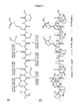
- FIG. 2 ( a ) shows the HPLC chromatogram of metabolites extracted from D. acidovorans cultures. Various fractions were separated by HPLC into a 96-well plate, and reacted with 5 mM AuCl 3 . The inset shows blackening in the wells for fractions A6 and B7-11 which indicates gold nanoparticle formation. These active wells were found to contain a common peptidic metabolite; (b) extracts of wildtype and ⁇ delG D. acidovorans analyzed by LCMS. The extracted ion chromatogram of the wildtype specific compound associated with gold precipitation is shown.
- FIG. 3 shows (a) the high resolution mass fragmentation and (b) 2D NMR spin systems for 1 H- 13 C heteronuclear multiple bond correlation (HMBC), 1 H- 1 H correlation spectroscopy (COSY) and 1 H- 15 N HMBC for delftibactin A in D 2 O.
- HMBC heteronuclear multiple bond correlation
- COSY 1 H- 1 H correlation spectroscopy
- H- 15 N HMBC for delftibactin A in D 2 O.
- FIG. 4 shows the structure of the gold interacting non-ribosomal peptide delftibactin A as one exemplary embodiment of the present application.
- FIG. 5 shows (a) pictures of increasing concentrations of delftibactin A in the presence of 2.5 mM AuCl 3 causing an increase in gold nanoparticle formation. Images were taken 30 min after the addition of delftibactin A in the following concentrations: i) 0.3125 mM ii) 0.625 mM iii) 1.25 mM iv) 2.5 mM and 5 mM; (b) a bar graph of the amount of delftibactin A and gold remaining in delftibactin A-AuCl 3 reaction supernatants.
- FIG. 8 shows (a) the time course progression of 5 mM AuCl 3 reacted with: i) water only ii) 5 mM delftibactin A iii) 5 mM AuCl 3 iv) 5 mM FeCl 3 v) 5 mM AuCl 3 +5 mM FeCl 3 vi) 5 mM delftibactin A+5 mM AuCl 3 vii) delftibactin A+5 mM AuCl 3 +5 mM FeCl 3 and viii) delftibactin A+5 mM FeCl 3 ; (b) growth curves of D.
- results are a mean of three growth curves for each condition from a single representative experiment; and c) growth curves of D. acidovorans ⁇ delG cultures inoculated 1:1000 into ACM followed by the addition of delftibactin A and/or gold as follows: i) 100 ⁇ M delftibactin A+10 ⁇ M AuCl 3 , ii) 10 ⁇ M AuCl 3 only, iii) 10 ⁇ M delftibactin A only, and iv) water only. Results are a mean of three growth curves for each condition from a single representative experiment.
- FIG. 10 is a schematic depicting the single metal binding site of delftibactin A as confirmed by gallium NMR.
- FIG. 11 shows (a) pictures of reactions of delftibactin A (5 mM) and Ga-delftibactin A (5 mM) with equimolar concentrations of AuCl 3 ; and (b) Mass spectra of free (top) and gallium-bound delftibactin (bottom).
- FIG. 12 shows (a) the structure of the acetylated analog and spin systems for 1 H- 13 C HMBC, 1 H- 1 H COSY and 1 H- 15 N HMBC in D 2 O for delftibactin B (acetylated hydroxy-ornithine delftibactin A) in a further exemplary embodiment of the present application; b) pictures of reactions demonstrating AuCl 3 (5 mM) precipitation by delftibactin B over time in the absence and presence of FeCl 3 as follows: i) water only ii) 5 mM delftibactin B iii) 5 mM AuCl 3 iv) 5 mM FeCl 3 v) 5 mM AuCl 3 +5 mM FeCl 3 vi) 5 mM delftibactin B+5 mM AuCl 3 vii) delftibactin B+5 mM AuCl 3
- FIG. 13 shows the LC-MS chromatograms from the reaction of delftibactin A and delftibactin B with equimolar (5 mM) concentrations of AuCl 3
- FIG. 14 shows pictures of the timecourse of 5 mM AuCl 3 reacted with i) water ii) 5 mM delftibactin A iii) 5 mM delftibactin-AuCl 3 reaction product and iv) 5 mM delftibactin B monitored over 2 h for AuCl 3 precipitation.
- the results demonstrate that all delftibactin species eventually co-precipitate with AuCl 3 .
- (b) fragmentation of the double charged ion of the reacted delftibactin species (m+/z 989) with diagnostic fragments shown.
- Mass loss of 44 is localized to ornithine functional group; and
- FIG. 16 shows transmission electron microscopy of delftibactin A-gold (2:1) complex after 10 minutes and reveals the presence of colloidal and octahedral gold nanoparticles, reminiscent of those seen in natural deposits. Darker arrows indicate colloidal gold. Lighter arrows indicate octahedral gold.
- FIG. 17 shows (a) pictures of the reaction of AuCl 3 (5 mM) with equimolar concentrations of delftibactin A or sodium citrate showing that delftibactin A rapidly forms complex precipitates of gold nanoparticles; (b) TEM images of the sodium citrate-gold reaction after 10 minutes; and (c) TEM images of the delftibactin-gold reaction after 10 minutes.
- FIG. 18 shows chemical structures of metabolites related to delftibactin as follows: Acidobactin A; Acidobactin B; Vacidobactin A; Vacidobactin B; Variobactin A; Variobactin B; Variobactin C; Variobactin D; and Variobactin E, as further exemplary embodiments of the present application.
- FIG. 19 is a schematic showing the genetic relationship between delftibactin A and related metabolites.
- the biosynthetic clusters were identified by BLAST searching using the delftibactin A biosynthetic gene cluster as the query. Bars represent nucleotide similarity using a MAUVE analysis. The resulting structures from each gene cluster are shown on the left.
- FIG. 20 is schematic showing several exemplary embodiments for how the metallophores of the application can be used as a biosensor of gold.
- A thin layer chromatography concept.
- B strip concept.
- C column quantification concept.
- FIG. 21 shows schematics of test strip development as exemplary embodiments delftibactin A (iii) and enterobactin (a known metallophore) (i) were applied in increasing concentrations to a C18 thin layer chromatography (TLC) plate and a 1 mL sample of 1 mM AuCl 3 was overlayed and allowed to react. The picture at the top of the figure was taken after 10 min. The picture at the bottom of the figure show delftibactin A immobilized on a C-18 thin layer chromatography slide and a solution containing gold chloride was used to develop the strip. Pictures were taken at the times indicated. Characteristic darkening can be seen where immobilized delftibactin A reacts with gold in the sample.
- TLC thin layer chromatography
- FIG. 22 shows (a) gold nanoparticle formation by delftibactins and commercial chelators.
- a solution of 10 mM AuCl 3 was reacted with equimolar concentrations of delftibactin A (i), delftibactin B (ii), EDTA (iii), bipyridyl (iv), and water (v); and (b) UV-vis spectrum of 5 ⁇ M delftibactin, 5 ⁇ M AuCl 3 , and equimolar concentrations of reacted delftibactin A and AuCl 3 after 15 min.
- FIG. 23 shows the structures of various metallophores examined for gold nanoparticle formation properties.
- Major classes of metallophore compounds are shown: hydroxamate-type (delftibactin, A, B, aerobactin), catechol-type (enterobactin), thiazoline-type (yersiniabactin), citric acid-type (aerobactin), and commercial chelators (EDTA, bipyridyl). Areas involved in metal complexation are highlighted.
- FIG. 24 shows (a) pictures of the reaction of a solution of 10 mM AuCl 3 with equimolar concentrations of delftibactin A (i), yersiniabactin (ii), aerobactin (iii), and enterobactin (iv) leading to gold nanoparticle formation; (b) transmission electron microscope images of the reactions in (a); (c) pictures of the reaction of (i) delftibactin A and (iv) enterobactin with equimolar concentrations of AuCl 3 and FeCl 3 .
- the term “comprising” and its derivatives, as used herein, are intended to be open ended terms that specify the presence of the stated features, elements, components, groups, integers, and/or steps, but do not exclude the presence of other unstated features, elements, components, groups, integers and/or steps.
- the foregoing also applies to words having similar meanings such as the terms, “including”, “having” and their derivatives.
- the term “consisting” and its derivatives, as used herein, are intended to be closed terms that specify the presence of the stated features, elements, components, groups, integers, and/or steps, but exclude the presence of other unstated features, elements, components, groups, integers and/or steps.
- the second component as used herein is chemically different from the other components or first component.
- a “third” component is different from the other, first, and second components, and further enumerated or “additional” components are similarly different.
- the compounds described herein have at least one asymmetric center. Where compounds possess more than one asymmetric center, they may exist as diastereomers. It is to be understood that all such isomers and mixtures thereof in any proportion are encompassed within the scope of the present application. It is to be further understood that while the stereochemistry of the compounds may be as shown in any given compound listed herein, such compounds may also contain certain amounts (for example, less than 20%, suitably less than 10%, more suitably less than 5%) of compounds of the present application having alternate stereochemistry. It is intended that any optical isomers, as separated, pure or partially purified optical isomers or racemic mixtures thereof are included within the scope of the present application.
- alkyl as used herein, whether it is used alone or as part of another group, means straight or branched chain, saturated alkyl groups.
- C 1-6 alkyl means an alkyl group having 1, 2, 3, 4, 5 or 6 carbon atoms.
- alkenyl as used herein means straight or branched chain, unsaturated alkyl groups, that is, a saturated carbon chain that contains one or more, for example one to three, one to two or one, double bond.
- C 2-6 alkenylene means an alkylene group having 2, 3, 4, 5 or 6 carbon atoms.
- salt as used herein means an acid addition salt or a basic addition salt.
- the formation of a desired compound salt is achieved using standard techniques. For example, the neutral compound is treated with an acid or base in a suitable solvent and the formed salt is isolated by filtration, extraction or any other suitable method.
- metalphore refers a compound that binds to or chelates metals.
- chelation core refers to the groups in a metallophore that bind to a metal through the formation of two or more separate coordination bonds.
- a “coordination bond” is a 2-center, 2-electron bond in which the two electrons derive from the same atom.
- chelating amino acid refers to an amino acid comprising a side chain with at least one chelating atom (i.e. an atom that has 2 electrons available for formation of a coordination bond).
- chelating amino acids are those comprising at least one heteroatom in the side chain wherein the heteroatom as two electrons available for formation of a chelation bond.
- the heteroatom is selected from, O, S and N.
- formylated refers to functionalization of a compound with a formyl (C(O)H) group.
- N-formulated amino acid refers to peptide amino acid (ribosomal or non-ribosomal) in which a side-chain amino functionality has been formulated.
- hydroxylated refers to functionalization of a compound with a hydroxy (OH) group.
- reference to an N-hydroxylated amino acid refers to peptide amino acid (ribosomal or non-ribosomal) in which a side-chain amino functionality has been hydroxylated.
- acetylated refers to functionalization of a compound with a acetyl (C(O)CH 3 ) group.
- C(O)CH 3 acetyl
- N-acetylated amino acid refers to peptide amino acid (ribosomal or non-ribosomal) in which a side-chain amino functionality has been acetylated.
- alkylated refers to functionalization of a compound with a C 1-6 alkyl group.
- N-alkylated amino acid refers to peptide amino acid (ribosomal or non-ribosomal) in which a side-chain amino functionality has been alkylated.
- amino acid aminoethine lactam as used herein refers to the amino acid:
- N-hydroxyornithine lactam refers to the amino acid:
- ⁇ -hydroxyaspartic acid refers to the amino acid:
- catechol refers to a compound comprising a 1,2-dihydroxybenzene group.
- isolated refers to a compound that is substantially separated from other components which naturally accompany a native compound, such as other bacterial cellular components for an isolated bacterial metabolite.
- non-ribosomal peptide refers to a class of peptide secondary metabolites usually produced by microorganisms such as bacteria and fungi.
- Non-ribosomal peptides are synthesized by non-ribosomal peptide synthetases, which, unlike the ribosomes, are independent of messenger RNA.
- Each non-ribosomal peptide synthetase can synthesize only one type of peptide.
- Non-ribosomal peptides can have a linear, cyclic and/or branched structure, can contain non-proteinogenic amino acids, including D-amino acids, carry modifications like N-methyl and N-formyl groups, or are glycosylated, acetylated halogenated or hydroxylated. Cyclization of amino acids against the peptide “backbone” is often performed, resulting in oxazolines and thiazolines, and these can be further oxidized or reduced. On occasion, dehydration is performed on serines, resulting in dehydroalanine. There are many other manipulations and variations that non-ribosomal peptides can comprise, for example polyketide extensions. Non-ribosomal peptides can be dimers or trimers of identical sequences chained together or cyclized, or even branched
- Delftibactin A and B show considerable promise in the bio-remediation of toxic soluble gold as well as in the detection of the presence of gold in a sample.
- the reduced nanoparticles formed by delftibactin A are—like delftibactin A itself—nontoxic, and are formed as part of a complex matrix, that would involve considerably less liquid handling.
- many commercially available methods for removing soluble gold including the use of activated carbon or ionic-exchange resins, are non-specific, and may result in enrichment for other contaminating metals.
- Delftibactin A is specific for a small set of metal ions including iron, gallium, and most notably gold, and would be a considerable improvement on currently available methods of remediation.
- the present application includes a method of removing soluble gold in a sample comprising:
- the metallophore is a bacterial metabolite comprising a chelation core that binds to gold ions and wherein the bacterial metabolite converts the soluble gold ions to solid gold (Au 0 ).
- the solid gold that is formed comprises gold nanoparticles
- the present application further includes a method of forming gold nanoparticles comprising:
- the metallophore is a bacterial metabolite comprising a chelation core that binds to gold ions and wherein the bacterial metabolite converts the soluble gold ions to solid gold (Au 0 ).
- the present application also includes a method for detecting the presence of soluble gold in a sample comprising:
- the metallophore is a bacterial metabolite comprising a chelation core that binds to gold ions and wherein the bacterial metabolite converts the soluble gold ions to solid gold (Au 0 ).
- the microbial metabolite comprises at least two to four, or at least, three chelating amino acids.
- the chelating amino acids are selected from:
- cysteine and S-formylated, S-acetylated and S-alkylated cysteine;
- the chelation core comprises a catechol or any other known microbial-derived chelation moiety from a metallophore that has selectivity for gold ions or is used in a process to selectively detect gold ions.
- the chelation core of the bacterial metabolite comprises at least two or more, for example, at least two to four, or at least three, chelating amino acids selected from:
- the metallophore is a non-ribosomal peptide comprising at least one N-formylated hydroxyl ornithine and optionally one or more other chemical moieties selected from N-formylated, N-acetylated, N-alkylated and N-hydroxylated ornithine; ornithine lactam and N-formylated, N-acetylated, N-alkylated and N-hydroxylated ornithine lactam; serine and O-formylated, O-acetylated and O-alkylated serine; threonine and O-formylated, O-acetylated and O-alkylated threonine and ⁇ -hydroxy aspartic acid.
- the metallophore is a non-ribosomal peptide comprising 4 to 20 amino acids. In a further embodiment, the metallophore is a non-ribosomal peptide comprising 5 to 15 amino acids. In a further embodiment, the metallophore is a non-ribosomal peptide comprising 6 to 10 amino acids. In another embodiment, the metallophore is a linear non-ribosomal peptide. In another embodiment, the metallophore is a cyclic non-ribosomal peptide.
- the metallophore is a bacterial metabolite.
- the bacterial metabolite is isolated from bacterial strains that exist and grow in the presence of soluble gold.
- the bacterial strains biomineralize the gold producing solid gold, such as gold nanoparticles. Though the biomineralization process, the bacteria are protected from the effects of toxic soluble gold.
- the bacteria are from the genera, Delftia, Acidovorax or Variovorax . In particular from D. acidovorans, A. citrulli , or V. paradoxus .
- the metabolite is at least partially secreted by the bacteria into its surrounding environment.
- metallophore is a bacterial metabolite isolated from a bacteria comprising a nucleic acid sequence encoding non-ribosomal peptide synthetase (NRPS) operons such as those shown in FIG. 19 . Accordingly, the NRPS operons comprise from initiation to termination ends:
- A is a adenylation domain
- T is a thiolation domain
- KS is a ⁇ -ketoacyl synthetase domain
- AT is an acyl transferase domain
- KR is a ketoreductase domain
- E is an epimerase domain
- TE is a termination domain
- Var 1 is a direct bond or C
- Var 2 is a direct bond or E.
- R 1 is selected from H and C 1-6 alkyl
- R 2 is selected from H and C 1-6 alkyl
- R 3 is (CH 2 ) n C(O)OH, unsubstituted or substituted with OH
- R 4 is selected from C 1-6 alkyl substituted with OH
- R 5 is selected from H and C 1-6 alkyl
- R 6 is selected from CH 2 , C(C 1-6 alkyl)(C 1-6 alkyl) and CHC 1-6 alkyl
- R 7 is
- R 8 is selected from C 1-6 alkyl substituted with OH;
- R 9 is
- R 10 is selected from H and C 1-6 alkyl; and n, m and p are independently selected from 1, 2, 3 and 4, or a salt thereof.
- R 1 is selected from CH 3 and CH 2 CH 3 . In a further embodiment R 1 is CH 3 .
- R 2 is selected from CH 3 and CH 2 CH 3 . In a further embodiment R 2 is CH 3 .
- R 3 is (CH 2 ) n C(O)OH substituted with OH. In a further embodiment, n is 1.
- R 4 is selected from C 1-4 alkyl substituted with OH. In another embodiment, R 4 is CH(OH)CH 3 .
- R 5 is selected from H and CH 3 . In another embodiment, R 5 is H.
- R 6 is selected from CH 2 , CHCH 3 and CHCH 2 CH 3 .
- R 7 is
- n 2 or 3 and R 10 is selected from H and CH 3 .
- m is 3 and R 10 is H.
- R 8 is selected from C 1-2 alkyl substituted with OH. In a further embodiment, R 8 CH 2 OH.
- R 9 is
- p is 3 or 4. In an embodiment, p is 3.
- the metallophore is delftibactin A.
- the industrially used agent for creating gold nanoparticles the industrially used agent for creating gold nanoparticles—delftibactin A does not need to be heated with the gold-containing sample to form solid gold.
- the time frame in which the soluble gold is reduced and coalesced as nanoparticles is much faster.
- Delftibactin, and analogs thereof also show considerable promise in the bio-remediation of toxic soluble gold, as the reduced nanoparticles formed by delftibactin A are—like delftibactin A itself—nontoxic, and are formed as part of a complex matrix, that would involve considerably less liquid handling.
- Delftibactin A is specific for a small set of metal ions including iron, gallium, and most notably gold, and would be a considerable improvement on currently available methods of remediation.
- the metallophore is an analog of delftibactin A which has gold complexing and solid gold forming ability.
- analogs of delftibactin A are those comprising similar gene clusters to the genes that encode the enzymes (non-ribosomal peptide synthetases or NRPS) for producing delftibactin A.
- the genes that encode the enzymes for producing delftibactin A are used as the query in a BLAST search to identify similar gene clusters in other organisms.
- confirmation that a compound isolated from another organism is an analog of delftibactin A is made by comparing the mass spectral (MS) fragmentation pattern of the analog with that of delftibactin. If the analog possesses a similar MS fragmentation pattern to the MS fragmentation pattern of delftibactin then it is likely an analog of delftibactin.
- the significance of a match in fragmentation patterns, indicating analogous structures is calculated using computational means, for example as described in Applicant's co-pending PCT Application Publication No. WO 2013/181758.
- the metallophore is a compound of the Formula II:
- R 11 and R 12 are independently selected from H, OH and C ⁇ O; R 13 is selected from H and C 1-4 alkyl; or a salt thereof.
- R 13 is H or CH 3 .
- the metallophore is a compound of the Formula III:
- R 14 is selected from C 1-20 alkyl and C 1-20 alkenyl; or a salt thereof.
- R 14 is selected from C 8-16 alkyl and C 8-16 alkenyl. In a further embodiment, R 14 is selected from C 9 alkyl, C 12 alkyl, C 13 alkyl, C 14 alkyl and C 14 alkenyl.
- the metallophore is selected from delftibactin A and delftibactin B, and acidobactin A, acidobactin B, acididobactin C, acidobactin D, variobactin A, variobactin B, variobactin C, variobactin D and variobactin E as shown in FIG. 18 , or a salt thereof.
- the metallophores for use in the methods of the present application are prepared using methods known in the art of non-ribosomal peptide synthesis or they are isolated from a microorganism, again using methods known in the art.
- the organism is cultured on a suitable medium and allowed to grow for a time sufficient for the metallophore metabolite to accumulate, for example about 1 to about 10 days, and the metallophore isolated from the medium using known techniques.
- the microorganism may be a naturally occurring microorganism or a genetically modified microorganism.
- conditions for contacting of the sample with the metallophore comprise mixing the sample and the metallophore at ambient (room temperature) for a time sufficient for the formation of solid gold, evidenced by the formation of a dark solid.
- ambient room temperature
- one or more, for example one, two or three, different metallophores are used in the methods of the application.
- the conditions for contacting of the sample with the metallophore comprise first treating the sample to liberate the soluble gold ions so that the ions can interact with the metallophore.
- pre-treatments include, for example, treatment with acid solutions (for e.g. aqua regia).
- the isolation of the solid gold or the gold nanoparticles is by any known means, for example, centrifugation or filtration.
- the presence of solid gold in a sample can be observed using, for example, colormetric detection.
- the formation of a dark precipitate upon contact of the sample with the one or more metallophores of the present application indicates that the sample contained soluble gold.
- the soluble gold is a gold ion. In a further embodiment the soluble gold is Au 3+ .
- the observation of the sample for the presence of solid gold is performed in a quantitative manner as the amount of solid gold that is formed is proportional to the amount of soluble gold that was in the sample.
- the sample is a liquid solution. In a further embodiment, the sample is an environmental sample where the presence of toxic soluble gold is undesirable. In a further embodiment, the sample is an environmental sample where the presence of gold is desired. In an embodiment, the sample does not comprise substantial amounts of iron.
- the metallophores are also used to complex soluble iron and/or gallium in a sample.
- the presence of soluble gold is observed using one of the detectors of the application (see section IV).
- the present application also includes a use a metallophore to observe the presence of soluble gold in a sample, to remove soluble gold from a sample or to form solid gold or solid gold nanoparticles, wherein the metallophore is a microbial metabolite comprising a chelation core that binds to gold ions and wherein the microbial metabolite converts the soluble gold ions to solid gold (Au 0 ).
- the present application also includes novel compounds that act as metallophores and bind gold ions, converting the gold ions into solid gold.
- novel compounds are isolated microbial metabolites.
- the present application also includes an isolated compound of Formula I:
- R 1 is selected from H and C 1-6 alkyl
- R 2 is selected from H and C 1-6 alkyl
- R 3 is (CH 2 ) n C(O)OH, unsubstituted or substituted with OH
- R 4 is selected from C 1-6 alkyl substituted with OH
- R 5 is selected from H and C 1-6 alkyl
- R 6 is selected from CH 2 , C(C 1-6 alkyl)(C 1-6 alkyl) and CHC 1-6 alkyl
- R 7 is
- R 8 is selected from C 1-6 alkyl substituted with OH;
- R 9 is
- R 10 is selected from H and C 1-6 alkyl; and n, m and p are independently selected from 1, 2, 3 and 4, or a salt thereof.
- R 1 is selected from CH 3 and CH 2 CH 3 . In a further embodiment R 1 is CH 3 .
- R 2 is selected from CH 3 and CH 2 CH 3 . In a further embodiment R 2 is CH 3 .
- R 3 is (CH 2 ) n C(O)OH substituted with OH. In a further embodiment, n is 1.
- R 4 is selected from C 1-4 alkyl substituted with OH. In another embodiment, R 4 is CH(OH)CH 3 .
- R 5 is selected from H and CH 3 . In another embodiment, R 5 is H.
- R 6 is selected from CH 2 , CHCH 3 and CHCH 2 CH 3 .
- R 7 is
- n 2 or 3 and R 10 is selected from H and CH 3 .
- m is 3 and R 10 is H.
- R 8 is selected from C 1-2 alkyl substituted with OH. In a further embodiment, R 8 CH 2 OH.
- R 9 is
- p is 3 or 4. In an embodiment, p is 3.
- the compound of Formula I is delftibactin A.
- the novel metallophore of the present application is an analog of delftibactin A which has gold chelating and solid gold forming ability.
- the analogs of delftibactin A are those comprising similar gene clusters to the genes that encode the enzymes (non-ribosomal peptide synthetases or NRPS)) for producing delftibactin A.
- the genes that encode the enzymes for producing delftibactin A are used as the query in a BLAST search to identify similar gene clusters in other organisms.
- confirmation that a compound isolated from another organism is an analog of delftibactin A is made by comparing the mass spectral (MS) fragmentation pattern of the analog with that of delftibactin. If the analog possesses a similar MS fragmentation pattern to the MS fragmentation pattern of delftibactin then it is likely an analog of delftibactin.
- the significance of a match in fragmentation patterns, indicating analogous structures is calculated using computational means, for example as described in Applicant's co-pending PCT Application Publication No. WO 2013/181758.
- the present application also includes an isolated compound of Formula II:
- R 11 and R 12 are independently selected from H, OH and C ⁇ O; R 13 is selected from H and C 1-4 alkyl; or a salt thereof.
- R 13 is H or CH 3 .
- the present application also includes an isolated compound of Formula III:
- R 14 is selected from C 1-20 alkyl and C 1-20 alkenyl; or a salt thereof.
- R 14 is selected from C 8-16 alkyl and C 8-16 alkenyl. In a further embodiment, R 14 is selected from C 9 alkyl, C 12 alkyl, C 13 alkyl, C 14 alkyl and C 14 alkenyl.
- the application includes an isolated compound selected from delftibactin A and delftibactin B, and acidobactin A, acidobactin B, acididobactin C, acidobactin D, variobactin A, variobactin B, variobactin C, variobactin D and variobactin E as shown in FIG. 18 , or a salt thereof.
- the compounds of the present application may be prepared using methods known in the art of non-ribosomal peptide synthesis or they may be isolated from a microorganism, again using methods known in the art.
- the organism is cultured on a suitable medium and allowed to grow for a time sufficient for the desired compound to accumulate, for example about 1 to about 10 days, and the desired compound isolated from the medium using known methods.
- the microorganism may be a naturally occurring microorganism or a genetically modified microorganism.
- colorimetric test strips Areas of use for colorimetric test strips include industrial and environmental surveying, analysis, and quality control. There continues to be a need for a colorimetric test strip of enhanced sensitivity for determination of gold. It is desirable that a test strip have the necessary detection capability. Stability of the indicator, uniform color development and stability of the developed color would be advantageous. Moreover, a broad range of sensitivity would be beneficial. It would be also advantageous for the test strip to be economical to manufacture.
- Colorimetric detectors useful for and method for the analysis of gold are included in the present application.
- the detector includes a carrier and a metallophore of the present application as a colorimetric indicator.
- the present application also includes a detector for gold comprising a carrier and one or more metallophores, wherein the one or more metallophores are microbial metabolites comprising a chelation core that binds to gold ions and wherein the microbial metabolites convert the soluble gold ions to solid gold (Au 0 ).
- the one or more metallophores are one or more of the compounds of the Formula I, II or III, or a salt thereof.
- the carrier is any solid support matrix including C 18 -bonded silica, silica, alumina and other stationary phase materials.
- the one or more of the compounds of Formula I, II and/or III are covalently linked to a solid support matrix, physically entrapped within a matrix (for example a sol gel matrix) or are held on or within a matrix using any known mechanism.
- the one or more of the compounds of Formula I, II and/or III are distributed on or in the matrix as a single concentration or as a gradient of concentrations to facilitate quantification of gold ions where applicable.
- the carrier is contacted with a liquid to be analyzed, and the liquid caused to flow through the carrier and to contact the one or more of the compounds of Formula I, II and/or III over a selected period of time.
- a gentle swirling action is beneficially used to cause the liquid to be in flowing contact with the compound(s).
- the carrier is evaluated for detectable color change advantageously by viewing the area of the carrier defined by the contact zone (aperature).
- the color change is compared to a standardized color chart to determine the gold concentration.
- the present application includes several ways by which the one or more of the compounds of Formula I, II and/or III are used as a detector of gold.
- the detector is based on a thin layer chromatography concept, for example as shown in FIG. 20A .
- a concentration gradient of one or more of the compounds of Formula I, II and/or III is absorbed onto a test strip and is used to determine the concentration of gold in a solution after fixed incubation period. This occurs whereby solutions with low concentrations of gold will be captured lower on the strip by the compound(s) as the liquid moves upwards and solutions with high concentrations of gold ions will saturate the compound(s) lower on the strip and move higher creating a higher band of compound-induced blackening.
- a test strip comprising one or more of the compounds of Formula I, II and/or III absorbed thereto is submerged in liquid sample to determine gold concentration, for example as shown in FIG. 20B . Varying the ratio of the compound(s) to gold causes differential darkening, allowing for easy quantification and detection of gold from a solution.
- one or more of the compounds of Formula I, II and/or III are absorbed within a chromatographic column to provide a similar colorimetric indication as the submerged test strip. This embodiment advantageously allows for increased sample volumes to be used, which can increase the signal (blackening) or decrease the limit of detection within a sample (see for example, FIG. 20C ).
- LCMS data was collected using a Bruker AmazonX ion trap mass spectrometer coupled with a Dionex UltiMate 3000 HPLC system, using a Luna C 18 column (250 mm ⁇ 4.6 mm, Phenomenex) for analytical separations, running acetonitrile and ddH 2 O as the mobile phase.
- Delftia acidovorans was ordered from the German Resource Centre for Biological Material (DSMZ, DSM No. 39). Delftia acidovorans was cultured on Acidovorax Complex Media (Pinel et al., 2008) (ACM) plates at 30° C. The ⁇ delG strain was initially grown in the presence of 30 ⁇ g/mL tetracycline. Environmentally isolated D. acidovorans strains D27L and D126L were found in soil samples collected around McMaster University from June to August 2010. Environmental isolates were identified as D.
- Wildtype and ⁇ delG Delftia acidovorans was streaked onto a Chelex treated (deferrated) Acidovorax Complex Media (ACM) agar plate and grown for 3 days at 30° C. The plate was then overlaid with 10 mL of 0.5% agar containing 10 mM AuCl 3 .
- Gold complexing comparison to other bacteria was carried out as follows. Ten microliters of an overnight culture of D. acidovorans, D. acidovorans ⁇ delG, or C. metallidurans were placed onto deferrated ACM plates and grown for 3 days at 30° C. The plates were overlaid with AuCl 3 as described above. Both images were taken after 2 h of incubation at room temperature.
- Delftibactin A NRPS and PKS genes were identified using the BLAST function of IMG (http://img.jgi.doe.gov), using the sequence of pksJ as a query. Adenylation domain specificities were assessed using NRPS Predictor (Rausch et al., 2005) or NRPS-PKS (Ansari et al., 2004), and the 10 residue codes (Stachelhaus et al., 1999) of each entry and its top scoring hit were recorded.
- the delftibactin A adenylation code and the vicibactin adenylation code were determined with NRPS Predictor (Rausch et al., 2005) and aligned manually as neither database contained domains with homologous sequences.
- a knockout plasmid for D. acidovorans was constructed by inserting a 2 kb PCR product of delG (primers 2kbNRPS2Xba2 and 2kbNRPS2Sac2) into pUC19 using Xba1 and Sac1 digest sites, ligating with T4 ligase, transforming into chemically competent DH5 ⁇ (Invitrogen), and plating on LB with 100 ⁇ g/mL ampicillin. Positive clones were identified by colony PCR with 2kbNRPS2Xba2 and 2kbNRPS2Sac2, and verified through digestion following an overnight growth and plasmid miniprep using a QIAprep Spin Miniprep Kit (Qiagen).
- a clone containing a 2 kb insert was digested with Not1 to cut in the middle of the 2 kb insert, treated with calf intestinal phosphatase (CIP), and gel extracted to remove remaining CIP.
- CIP calf intestinal phosphatase
- a tetracycline resistance cassette was amplified from pLLX13 (primers TetNotF and TetNotR), purified, digested with Not1, and ligated with the digested vector. This ligation was transformed into chemically competent DH5 ⁇ (Invitrogen) and plated on LB with 100 ⁇ g/mL ampicillin and 10 ⁇ g/mL tetracycline.
- acidovorans were centrifuged separately and washed twice with fresh LB.
- Cells were resuspended in 1 mL LB and mixed 1:1, dispensing 300 ⁇ L on nutrient agar plates and leaving to grow overnight at 30° C.
- Cells were scraped and resuspended in 3 mL LB, plating 50 ⁇ L on LB plates containing 30 ⁇ g/mL tetracycline and 100 ⁇ g/mL apramycin to remove E. coli .
- Colonies were observed and tested for growth in LB with antibiotics at 30° C. and 200 rpm, with viable cultures streaked on LB plates with 30 ⁇ g/mL tetracycline.
- D. acidovorans strains were identified as D. acidovorans strains based on 16S sequence alignment, using 16S sequences that were PCR amplified from single colonies (see above).
- 16S sequences that were PCR amplified from single colonies (see above).
- sequences and the 16S sequence for the D. acidovorans genome strain SPH1 from GenBank along with the sequence for a gold biofilm isolate of D. acidovorans (Accession no. GU013680) (Reith et al., 2010) were aligned with Geneious software version 4.8.5 (Drummond et al., 2009), using a Tamura-Nei genetic distance model, a Neighbor-Joining tree building method featuring a global alignment with free end gaps. Isolated strains were grown for 3 days at 30° C.
- the gold-delftibactin A interaction was determined through two separate experiments. First, AuCl 3 was held constant at 2.5 mM and the interaction with delftibactin A was monitored by measuring the absorption of AuCl 3 remaining in solution after precipitation by delftibactin A through comparison with a standard curve. Briefly, 2.5 mM AuCl 3 was incubated with 5, 2.5, 1.25, 0.6125, 0.3063, and 0.1531 mM delftibactin A for 1 h. Solutions were filtered with a 0.22 ⁇ M Acrodisc (Pall, USA) to remove insoluble delftibactin A-gold precipitate.
- Reaction mixtures were analyzed using a Waters Alliance 2695 RP-HPLC separations module, equipped with a Waters 2998 photodiode array and a Luna 5u C 18 column (250 ⁇ 4.60 mm, Phenomenex).
- the mobile phase was linear from 2% acetonitrile, 98% water+5 mM (NH 4 ) 2 CO 3 at 2 min to 14% acetonitrile at 18 min at a flow rate of 1 mL/min.
- Delftibactin A was reacted with AuCl 3 with a molar ratio equal to 2:1 for ten minutes. Each separate reaction of delftibactin A with AuCl 3 was examined using a Phillips CM-10 transmission electron microscopy operating at 80 kV. The whole-mount sample was absorbed and dried on a formvar-carbon coated 100-square mesh copper grid and rinsed with filter sterilized, de-ionized water to remove any salt precipitates.
- Gallium bound delftibactin A was adjusted to 10 mM and mixed 1:1 with an equimolar solution of AuCl 3 , alongside purified delftibactin A and water control. The reaction mixture was monitored at room temperature for 30 min.
- the assay was set up as follows: 50 ⁇ L of ddH 2 O containing 3.2, 1.6, 1.4, 1.2, 1.0, 0.8, 0.6, 0.4, and 0.2 mM delftibactin A was added in quadruplicate to wells within a 96-well plate. A 1.6 mM stock solution of AuCl 3 was made and 50 ⁇ L was added to each well containing delftibactin A. No-delftibactin A and no-AuCl 3 controls containing only water were also added to the 96-well plate in quadruplicate. This was incubated for 30 min at room temperature, during which time 10 mL of an overnight culture of D.
- reaction Twenty microliter reactions were set up as follows: water only, 5 mM AuCl 3 , 5 mM AuCl 3 +delftibactin A, and 5 mM AuCl 3 +delftibactin B. Reactions were initiated with the addition of delftibactin A or B. After 2 h, reactions were serially diluted to 125 ⁇ M in ACM containing D. acidovorans ⁇ delG diluted 1:1000 from an overnight culture. Optical density was monitored using a TECAN SunriseTM microplate reader at 600 nm for 36 h. Results are a mean of three growth curves for each condition from a single representative experiment.
- D. acidovorans wild type and D. acidovorans ⁇ delG were grown in deferrated ACM for 2 days at 30° C.
- 200 ⁇ L of wild type or mutant grown culture were incubated in the presence and absence of 100 ⁇ M AuCl 3 .
- the mutant culture was also complemented with 30 sM delftibactin (biological levels) in the presence of 100 ⁇ M AuCl 3 .
- metallidurans were grown on agar plates and then flooded with solutions of Au(III)—the dominant form of soluble gold found in terrestrial conditions (Reith et al., 2009; Usher et al., 2009). Following gold exposure darkened zones developed surrounding colonies of D. acidovorans but not C. metallidurans . These blackened zones suggested that D. acidovorans generated a diffusible metabolite that acts to generate reduced solid gold forms.
- the D. acidovorans genome was investigated for genes that may be associated with a unique small molecule biosynthesis pathway that is absent from C. metallidurans .
- polyketides and non-ribosomal peptides are classes of secondary metabolites that bacteria use to promote environmental fitness (Vining, 1990) but are not required for growth, and include members that function to bind metals (e.g. Fe, Cu) (Hider et al., 2010; Kim et al., 2004; Chaturvedi et al., 2012).
- This peptidic compound could be identified in supernatants in concentrations in excess of 200 ⁇ M (Table 3), enabling its isolation and structure determination by high resolution mass spectrometry ( FIG. 3 a ) and NMR spectroscopy ( FIG. 3 b ), revealing a novel linear polyketide-non-ribosomal peptide consistent with the structural prediction, which was named delftibactin A ( FIG. 4 ).
- D. acidovorans environmental isolates were also screened for their ability to produce delftibactin A, resulting in its identification in all tested isolates.
- D. acidovorans may maintain protective extracellular concentrations of delftibactin A by monitoring the loss of delftibactin A by gold co-precipitation, leading to an increase in delftibactin A production through a positive feedback mechanism (Miller et al., 2010).
- D. acidovorans supernatants treated with 10 and 30 ⁇ M AuCl 3 caused delftibactin A depletion, resulting in a compensatory increase in delftibactin A concentrations compared to an untreated control, representing a form of reactive homeostasis ( FIG. 9 ).
- Delftibactin A concentrations were also responsive to iron concentrations, but in an inverse manner (Table 3), suggesting that delftibactin A is not regulated in the same way as a typical siderophore despite the presence of relevant genes in the delftibactin A cluster.
- Metallophores are recognized to create complexes with metals and whether or not such complexation was part of the gold-delftibactin A interaction was investigated. As the gold-delftibactin A association leads to co-precipitation and formation of an insoluble material, it was examined how delftibactin A may bind metals using gallium. NMR analysis of the delftibactin-gallium complex showed the coordinating activity of delftibactin A.
- delftibactin A is capable of creating naturally occurring complex gold structures from Au 3+ on short timescales (seconds), at room temperature, neutral pH, and at rates that far exceed those observed for traditional gold nanoparticle producing agents such as citrate (Ojea-Jimenez et al., 2010) ( FIG. 17 ), providing a potential mechanism for bacterial gold biomineralization. While not wishing to be limited by theory, it is proposed that delftibactin A facilitates this biomineralization and protects D. acidovorans by chelating soluble Au 3+ and directly precipitating as a complex, or by binding and reducing gold through oxidative decarboxylation before chelating a second Au 3+ ion and precipitating as a complex.
- Example 1 discloses metabolites of D. acidovorans , delftibactin A and B, which act to extact gold from solution (Johnston et al., 2013). As compared to citrate—the industrially used agent for creating gold nanoparticles—delftibactin A does not need to be heated with the gold-containing sample. Further, the time frame in which the soluble gold is reduced and coalesced as nanoparticles is much faster.
- Delftibactin A also shows considerable promise in the bio-remediation of toxic soluble gold, as the reduced nanoparticles formed by delftibactin A are—like delftibactin A itself—nontoxic, and are formed as part of a complex matrix, that would involve considerably less liquid handling. Further, many commercially available methods for removing soluble gold, including the use of activated carbon or ionic-exchange resins, are non-specific, and may result in enrichment for other contaminating metals. Delftibactin A is specific for a small set of metal ions including iron, gallium, and most notably gold, and is a considerable improvement on currently available methods of remediation.
- delftibactin-like compounds are described in the present Example. These microbial compounds were targeted for isolation using a method coined ‘genome mining’, whereby the genes that encode the enzymes required for producing delftibactin A were used as the query in a BLAST search to identify similar gene clusters that exist in other organisms.
- a delftibactin gene cluster analysis is shown in Table 4. Using this methodology, two genera were identified that contained delftibactin-like gene clusters: Variovorax and Acidovorax. Organisms containing similar gene clusters were subsequently fermented and 9 delftibactin-like compounds were isolated.
- bacterial metabolites of the chemical scaffolds shown in FIG. 18 are cyclic non-ribosomal peptides, with chemical moieties consistent with the delftibactin family and other metallophores.
- the metabolites are predicted to have delftibactin A like properties, namely the ability to obtain solid gold from the environment or chemical and industrial processes.
- the biosynthetic genes encoding the delftibactin A non-ribosomal peptide synthetase were used to probe the GenBank database and identify related biosynthetic loci from other organisms. These biosynthetic gene homology searches revealed a series of undescribed NRPSs from organisms including: Acidovorax ( A. citrulli AAC00-1) and Variovorax ( V. paradoxus S110 and V. paradoxus EPS).
- FIG. 19 demonstrates the high genetic similarity between the delftibactin A biosynthetic gene cluster and those found in Variovorax and Acidovorax using Mauve alignment software. The highly similar gene clusters indicate their final products would also be likewise related. Using this as a guide, these organisms were obtained and fermented for delftibactin A-like molecules (shown in FIG. 18 ).
- A. citrulli AAC00-1 and V. paradoxus S110 were grown in Acidovorax Complete Media (ACM), the same media used for delftibactin A production. After 3 days of growth at 30° C., Acidobactin A and B and Vacidobactin A and B were isolated. These structures were determined by mass spectra (MS) analysis and nuclear magnetic resonance (NMR) experiments. Acidobactin C could be identified within the extract by its similar fragmentation pattern to acidobactin A and B and subsequent tandem MS analysis revealed its structure.
- ACM Acidovorax Complete Media
- Variobactin A-E An environmental isolate obtained from Cootes Paradise, near McMaster University, was determined to be a strain of V. paradoxus by 16S gene sequencing. Subsequent fermentation of this strain in Chitinophaga Defined Media showed the production of Variobactin A-E. Variobactin A's structure was determined by MS and NMR experiments. Variobactin B-E's structures were determined using MS fragmentation analysis.
- delftibactin A has been identified that are related to the gold-complexing compound, delftibactin A. These cores have common chemical moieties found in delftibactin A, including formylated, acetylated, hydroxylated ornithines, serines, threonines, and beta-hydroxy aspartic acids. In addition, a common polyketide extension is found in all cores.
- FIG. 20 shows several ways by which the metallophores of the application are used as a detector of gold.
- the detector is based on a thin layer chromatography concept, for example as shown in FIG. 20A .
- a concentration gradient of one or more of the metallophores of the application is absorbed onto a test strip and is used to determine the concentration of gold in a solution after fixed incubation period. This occurs whereby solutions with low concentrations of gold will be captured lower on the strip by the metallophores of the application as the liquid moves upwards and solutions with high concentrations of gold ions will saturate the metallophore of the application lower on the strip and move higher creating a higher band of compound-induced blackening.
- a test strip comprising one or more of the metallophores of the application absorbed thereto is submerged in liquid sample to determine gold concentration, for example as shown in FIG. 20B . Varying the ratio of the compound(s) to gold causes differential darkening, allowing for easy quantification and detection of gold from a solution.
- one or more of the metallophores of the application are absorbed within a chromatographic column to provide a similar colorimetric indication as the submerged test strip. This embodiment advantageously allows for increased sample volumes to be used, which can increase the signal (blackening) or decrease the limit of detection within a sample (see for example, FIG. 20C ).
- delftibactin A (iii) and enterobactin (a known metallophore) (i) were applied in increasing concentrations to a C18-silica thin layer chromatography (TLC) plate and a 1 mL sample of 1 mM AuCl 3 was overlayed and allowed to react. Pictures were taken after 10 min.
- delftibactin A was immobilized on a C18-silica thin layer chromatography slide. A solution containing gold chloride was used to develop the strip. Pictures were taken at the times indicated. Chararacteristic darkening can be seen where immobilized delftibactin A reacts with gold in the sample.
- Example 4 Are Other Microbial and Synthetic Metallophores Capable of Forming Ordered Gold Nanoparticles
- Delftibactin A and B were isolated as described in Example 1. Enterobactin, aerobactin and yersiniabactin were isolated from Escherichia coli Nissle 1917 (Mutaflor). Escherichia coli Nissle 1917 was isolated from a single capsule of Mutafluor on a Luria-Bertani agar plate. 6 ⁇ 1.5 L of Escherichia coli Nissle 1917 (innoculated from a single colony) was grown for 3 days at 37° C. at 200 rpm in Fernbach flasks containing M9 media supplemented with 0.5% glycerol and 0.5% glucose.
- Resin was added (HP20, DIAION), and shaken with cultures for 2 hours. Resin was collected by filtration and extracted 4 times with 500 mL of methanol. The dried extract was separated using a Combiflash (Isco) medium pressure liquid chromatography system using a 24 g RediSep (Isco) silica column. The extract was separated stepwise into seven 150 mL fractions using methanol and dichloromethane with the following methanol concentrations: 5, 15, 25, 35, 50, 70, and 100% for each fraction respectively. Fraction 1 contained yersiniabactin. Fraction 3 contained enterobactin. Fraction 7 contained aerobactin.
- Enterobactin eluted at 22.68 min (m/z 670.0).
- Transmission electron microscopy imaging was performed with a JEOL JEM 1299 EX TEMSCAN transmission electron microscope (JEOL, Peabody, Mass., USA) operated at an accelerating voltage of 80 kV.
- Sample preparation a 2 ⁇ L drop of a diluted (10 ⁇ ) siderophore-gold reaction was deposited over a formvar-coated TEM grid, and was allowed to dry under ambient conditions before being imaged. Gold nanoparticle size distribution was determined using the software, ImageJ.
- the gold nanoparticle forming capacity of delftibactin was compared to chelating substances of a non-natural origin, including common commercial metal chelators ethylenediaminetetraacetic acid (EDTA) and bipyridyl.
- EDTA ethylenediaminetetraacetic acid
- Visual inspection of chelator-Au 3+ reaction mixtures revealed that while delftibactin A and B solutions rapidly undergo a characteristic blackening and an increase in absorbance at 600 nm within the UV/Vis spectrum indicative of gold nanoparticle formation, synthetic chelating agents fail to develop a similar effect even after prolonged exposure to gold ions ( FIG. 22 ).
- chelation moieties found in delftibactin A and B and other microbial metallophores may be important for this phenomenon and based on previous studies into the mechanism of gold complexation by delftibactin, it is suggested that the N-formylated hydroxy ornithine found in the chelation core is of particular importance for gold nanoparticle formation by delftibactin A and B.
- Metallophores were isolated from the human-associated probiotic bacterium Escherichia coli Nissle 1917, which is used to prevent and treat several gastrointestinal disorders and overproduces a variety of metallophores including enterobactin (catechol), salmochelin (catechol), aerobactin (hydroxamate and citrate), and yersiniabactin (mixed-ligand) ( FIG. 23 ) (Valdebenito et al., 2006).
- the hydroxamate metallophore aerobactin which is biosynthesized from lysine and the known gold nanoparticle forming agent citric acid, was also tested. Previous work suggests that aerobactin may also be a successful gold complexing and reducing agent due to the presence of an N-acetylation as part of the hydroxamate moiety—an important feature in the delftibactin A and B chelation core.
- analysis of gold complexation assays demonstrates that aerobactin reactions fail to produce defined gold nanoparticles ( FIG. 24 a ).
- gold complexation assays were performed with the catechol siderophore enterobactin, which has the highest iron affinity of all siderophores studied to date. Enterobactin reactions showed considerable gold nanoparticle formation ( FIG. 24 a ).
- FIG. 24 e A closer analysis of the reaction mixtures using transmission electron microscopy (TEM, FIG. 24 b ), revealed that the colloidal nanoparticles produced by delftibactin A are more defined, and range in size from 10.6 to 712 nm with an average particle size of 211.2 nm as compared to the nanoparticles produced by yersiniabactin, which did not create any distinct nanoparticles.
- the colloidal nanoparticle distribution pattern for delftibactin A is shown in FIG. 24 e .
- Enterobactin generated particles range from 26 nm to 339 nm in diameter, however, the majority of the blackening that occurred in the microcentrifuge tube is not related to the defined colloidal particles as demonstrated in the TEM image ( FIG. 24 b ).
- delftibactin A also produces octahedral gold particles, which are not formed by either of the other two metallophores reaching sizes greater than several micrometers in diameter.
- enterobactin with AuCl 3 suggests there are time-dependent factors responsible for octahedral gold formation, where a slowed and directed complexation by the chelation core may allow for better growth of the larger, octahedral gold nanoparticles that are found in the nascent stages of gold nugget formation.
- Delftibactin A and B possesses a single metal binding site, as a result iron-bound delftibactin A and B is less able to precipitate gold, and delftibactin-gold complexes are incapable of binding iron.
- delftibactin-gold complexes are incapable of binding iron.
- delftibactin is depleted and can no longer sequester iron or protect from toxic gold ions. This is a fortuitous phenomenon for D.
- enterobactin reacts with gold to a similar extent as when iron is not present. This suggests the mechanism of gold nanoparticle formation caused by enterobactin is through catechol mediated redox chemistry and does not require binding of Au 3+ within its chelation core to reduce Au 3+ to gold nanoparticles. This is different from delftibactin A, where Au 3+ interacts with N-hydroxy ornithine within the chelation core and is required for gold nanoparticle formation, giving evidence that delftibactin A (and B) is a more suited to metallophore-mediated protection from gold toxicity within the environment that D. acidovorans is found.
Landscapes
- Chemical & Material Sciences (AREA)
- Organic Chemistry (AREA)
- Life Sciences & Earth Sciences (AREA)
- Engineering & Computer Science (AREA)
- Health & Medical Sciences (AREA)
- Genetics & Genomics (AREA)
- Biochemistry (AREA)
- Molecular Biology (AREA)
- General Health & Medical Sciences (AREA)
- Proteomics, Peptides & Aminoacids (AREA)
- Biophysics (AREA)
- Biotechnology (AREA)
- Wood Science & Technology (AREA)
- Zoology (AREA)
- Medicinal Chemistry (AREA)
- Microbiology (AREA)
- Manufacturing & Machinery (AREA)
- Metallurgy (AREA)
- Mechanical Engineering (AREA)
- Materials Engineering (AREA)
- Chemical Kinetics & Catalysis (AREA)
- General Chemical & Material Sciences (AREA)
- General Engineering & Computer Science (AREA)
- Bioinformatics & Cheminformatics (AREA)
- Biomedical Technology (AREA)
- General Life Sciences & Earth Sciences (AREA)
- Geochemistry & Mineralogy (AREA)
- Geology (AREA)
- Environmental & Geological Engineering (AREA)
- Physics & Mathematics (AREA)
- Plant Pathology (AREA)
- Measuring Or Testing Involving Enzymes Or Micro-Organisms (AREA)
Abstract
Description

wherein
R1 is selected from H and C1-6alkyl;
R2 is selected from H and C1-6alkyl;
R3 is (CH2)nC(O)OH, unsubstituted or substituted with OH;
R4 is selected from C1-6alkyl substituted with OH;
R5 is selected from H and C1-6alkyl;
R6 is selected from CH2, C(C1-6alkyl)(C1-6alkyl) and CHC1-6alkyl;
R7 is

R8 is selected from C1-6alkyl substituted with OH;
R9 is

R10 is selected from H and C1-6alkyl; and
n, m and p are independently selected from 1, 2, 3 and 4, or
a salt thereof.

wherein
R11 and R12 are independently selected from H, OH and C═O;
R13 is selected from H and C1-4alkyl; or
a salt thereof.

wherein:
R14 is selected from C1-20alkyl and C2-20alkenyl; or
a salt thereof.


wherein
R1 is selected from H and C1-6alkyl;
R2 is selected from H and C1-6alkyl;
R3 is (CH2)nC(O)OH, unsubstituted or substituted with OH;
R4 is selected from C1-6alkyl substituted with OH;
R5 is selected from H and C1-6alkyl;
R6 is selected from CH2, C(C1-6alkyl)(C1-6alkyl) and CHC1-6alkyl;
R7 is

R8 is selected from C1-6alkyl substituted with OH;
R9 is

R10 is selected from H and C1-6alkyl; and
n, m and p are independently selected from 1, 2, 3 and 4, or
a salt thereof.

wherein m is 2 or 3 and R10 is selected from H and CH3. In a further embodiment, m is 3 and R10 is H.

wherein p is 3 or 4. In an embodiment, p is 3.

or delftibactin B:


wherein
R11 and R12 are independently selected from H, OH and C═O;
R13 is selected from H and C1-4alkyl; or
a salt thereof.

wherein:
R14 is selected from C1-20alkyl and C1-20alkenyl; or
a salt thereof.

wherein
R1 is selected from H and C1-6alkyl;
R2 is selected from H and C1-6alkyl;
R3 is (CH2)nC(O)OH, unsubstituted or substituted with OH;
R4 is selected from C1-6alkyl substituted with OH;
R5 is selected from H and C1-6alkyl;
R6 is selected from CH2, C(C1-6alkyl)(C1-6alkyl) and CHC1-6alkyl;
R7 is

R8 is selected from C1-6alkyl substituted with OH;
R9 is

R10 is selected from H and C1-6alkyl; and
n, m and p are independently selected from 1, 2, 3 and 4, or
a salt thereof.

wherein m is 2 or 3 and R10 is selected from H and CH3. In a further embodiment, m is 3 and R10 is H.

wherein p is 3 or 4. In an embodiment, p is 3.

or delftibactin B:


wherein
R11 and R12 are independently selected from H, OH and C═O;
R13 is selected from H and C1-4alkyl; or
a salt thereof.

wherein:
R14 is selected from C1-20alkyl and C1-20alkenyl; or
a salt thereof.
| TABLE 1 |
| Adenylation domain specificities. Prediction of |
| incorporated amino acids was performed using |
| NRPSPredictor and NRPS PKS, providing a |
| probability sequence for the NRPS/PKS product |
| Adenyla- | Active | ||
| tion | Site | ||
| Domain | Residues | Substrate | Product |
| DelE A1 | DMGGYGCLFK | Alanine | HC Toxin |
| DAGGCAMVAK | Alanine | ||
| DelE A2 | DIWHISLIEK | Inactive | Nostopeptolide |
| DVWHISLIDK | Serine | ||
| DelG A1 | DLTKVCHVGK | Aspartic acid | Surfactin |
| DLTKVGHIGK | Aspartic acid | ||
| DelG A2 | DFWNIGMVHK | Threonine | Syringopeptin |
| DFWNIGMVHK | Threonine | ||
| DelG A3 | DILQLGLIWK | Glycine | Nostopeptolide |
| DILQLGLIWK | Glycine | ||
| DelH A1 | DFWNIGMVHK | Threonine | Syringopeptin |
| DFWNIGMVHK | Threonine | ||
| DelH A2 | DVWNIGLIHK | Ornithine | Fengycin |
| DVGEIGSIDK | Ornithine | ||
| DelH A3 | DVWHLSLIDK | Serine | Syringopeptin |
| DVWHLSLIDK | Serine | ||
| DelH A4 | DGEDHGAVTK | Arginine | Pederin |
| DAEDIGAITK | Arginine | ||
| DelH A5 | DGEAVGGVTK | Nb- | Vicibactin |
| hydroxyomithine | |||
| DGESSGGMTK | Nb- | ||
| hydroxyomithine | |||
| TABLE 2 |
| Primers used in construction of the ΔdelG strain |
| PCR Primer | Sequence (5′-3′) | Purpose |
| TetNotF | TTT TGC GGC CGC | Amplification of |
| TGC TGA ACC CCC | tetracycline | |
| AA | resistance cassette | |
| from pLLX13. | ||
| TetNotR | TTT TGC GGC CGC | Amplification of |
| TAT CGT TTC CAC | the tetracycline | |
| GA | resistance cassette | |
| from pLLX13. | ||
| 2kbNRPS2Xba2 | TTT TTC TAG ACG | Amplification of |
| CAT TGC TGA ACT | the 2 kb delG | |
| ACC | fragment from | |
| D. acidavorans. | ||
| 2kbNRPS2Sac2 | TTT TGA GCT CAG | Amplification of |
| CAG TTG CAC CAC | the 2 kb delG | |
| CT | fragment from | |
| D. acidavorans. | ||
| OriTF | TTT TAA GCT TTT | Amplification of |
| CCT CAA TCG CTC | an oriT from pLLX13. | |
| TTC | ||
| OriTR | TTT TAA GCT TTT | Amplification of |
| TTC GCA CGA TAT | an oriT from pLLX13. | |
| ACA | ||
| NRPS2Seq2 | GGG GTG CGG AAA | Confirmation of |
| ATG TCC TG | genomic integration | |
| of the tetracycline | ||
| resistance cassette. | ||
| TABLE 3 |
| Delftibactin production in response to [Fe3+] |
| Media [Iron] | [Delftibactin] | ||
| 0 | 205.8 ± 28.0 μM | ||
| 100 nM | 196.1 ± 30.7 |
||
| 1 μM | 149.3 ± 21.0 μM | ||
| 10 μM | 21 ± 9.4 |
||
| 100 μM | 2.2 ± 1.4 μM | ||
| 1 mM | Growth Inhibitory | ||
| TABLE 4 | ||||
| Amino | ||||
| Locus | Gene | Predicted Function | Strand | Acids |
| Daci_4765 | — | Heavy metal efflux outer | − | 485 |
| membrane component | ||||
| Daci_4764 | — | Heavy metal efflux | − | 405 |
| periplasmic component | ||||
| Daci_4763 | — | Heavy metal efflux inner | − | |
| membrane component | 1029 | |||
| Daci_4762 | — | Nitrogen regulatory protein P-II | − | 111 |
| Daci_4761 | — | RNA polymerase, sigma subunit | − | 174 |
| Daci_4760 | delA | MbtH domain protein | − | 121 |
| Daci_4759 | delB | Thioesterase | − | 247 |
| Daci_4758 | delC | Phosphopantetheinyl transferase | − | 229 |
| Daci_4757 | delD | Aspartic acid dioxygenase | − | 329 |
| Daci_4756 | delE | Nonribosomal peptide synthetase | − | 1789 |
| Daci_4755 | delF | Polyketide synthase | − | 1560 |
| Daci_4754 | delG | Nonribosomal peptide synthetase | − | 3331 |
| Daci_4753 | delH | Nonribosomal peptide synthetase | − | 6176 |
| Daci_4752 | delI | Siderophore receptor | − | 799 |
| Daci_4751 | delJ | anti-FecI sigma factor, FecR | + | 344 |
| Daci_4750 | delK | RNA polymerase, sigma | + | 199 |
| subunit | ||||
| Daci_4749 | delL | Lysine/Ornithine | + | 432 |
| N-monooxygenase | ||||
| Daci_4748 | delM | Acetyltransferase | + | 400 |
| Daci_4747 | delN | Esterase/Lipase | + | 321 |
| Daci_4746 | delO | Siderophore Export Pump | − | 571 |
| Daci_4745 | delP | N5-hydroxyornithine | − | 284 |
| formyltransferase | ||||
- Amin, S. A., et al. J. Am. Chem. Soc. 129, 478-479 (2007).
- Ansari, M. Z., Yadav, G., Gokhale, R. S. and Mohanty, D. Nucleic Acids Res. 32, W405-413 (2004).
- Berner, I., Greiner, M., Metzger, J., Jung, G., Winkelmann, G. 1991. Identification of Enterobactin and Linear Dihydroxybenzoylserine Compounds by Hplc and Ion Spray Mass-Spectrometry (Lc/Ms and Ms/Ms). Biol, Met., 4(2), 113-118.
- Chaturvedi, K. S., et al. Nat. Chem. Bio. 8, 731-736 (2012).
- Diels, L., Dong, Q., van der Lelie, D., Baeyens, W. and Mergeay, M. J. Ind. Microbiol. Biot. 14, 142-153 (1995).
- Drummond, A. J., et al. Geneious v4.7 (2009).
- Henderson, J. P., Crowley, J. R., Pinkner, J. S., Walker, J. N., Tsukayama, P., Stamm, W. E., Hooton, T. M., Hultgren, S. J. 2009. Quantitative Metabolomics Reveals an Epigenetic Blueprint for Iron Acquisition in Uropathogenic Escherichia coli. Plos Pathog., 5(2).
- Hider, R. C. and Kong, X. Nat. Prod. Rep. 27, 637-57 (2010).
- Hough, R. M., et al.
Geology 36, 571-574 (2008). - Kashefi, K., Tor, J. M., Nevin, K. P. and Lovely, D. R. Appl. Env. Microbiology 67, 3275-3279 (2001).
- Johnston, C. W., Wyatt, M. A., Li, X., Ibrahim, A., Shuster, J., Southam, G., Magarvey, N. A. 2013. Gold biomineralization by a metallophore from a gold-associated microbe. Nat. Chem. Biol., 9(4), 241-3.
- Kim, H. J., et al. Science 305, 1612-1615 (2004).
- Lengke, M., Sanpawanitchakit, C., Southam, G. 2011. Biosynthesis of Gold Nanoparticles: A Review. in: Met. Nanopart. Microbiol., (Eds.) M. Rai, N. Duran, Springer Berlin Heidelberg, pp. 37-74.
- Lin, H., Fiscchbach, M. A., Liu, D. R., Walsh, C. T. 2005. In vitro characterization of salmochelin and enterobactin trilactone hydrolases IroD, IroE, and Fes. J. Am. Chem. Soc., 127(31), 11075-11084.
- Malhotra, A., Dolma, K., Kaur, N., Rathore, Y. S., Ashish, Mayilraj, S., Choudhury, A. R. 2013. Biosynthesis of gold and silver nanoparticles using a novel marine strain of Stenotrophomonas. Biores. Technol, 142(0), 727-731.
- Miller, M. C., et al. Microbiology 156, 2226-38 (2010).
- Nies, D. H. Appl. Microbiol. Biotechnol. 51, 730-750 (1999).
- Ojea-Jiménez, I., Romero, F. M., Bastús, N. G. and Puntes, V. J. Phys. Chem. 114, 1800-1804 (2010).
- Pinel, N., Davidson, S. K. and Stahl, D. A. IJSEM 58, 2147-2157 (2008).
- Rausch, C., Weber, T., Kohlbacher, O., Wohlleben, W. and Huson, D. H. Nucleic Acids Res. 33, 5799-5808 (2005).
- Reith, F., et al. Geology 38, 843-846 (2010).
- Reith, F., et al. Proc. Natl Acad. Sci. USA 106, 17757-17762 (2009).
- Reith, F., Lengke, M. F., Falconer, D., Craw, D. and Southam, G. The
ISME Journal 1, 567-584 (2007). - Reith, F., Rogers, S. L., McPhail, D. C. and Webb, D. Science 313, 233-236 (2006).
- Salem, I. B., et al. Ann. Microbiol., 1-12 (2012).
- Schweigert, N., Zehnder, A. J. B., Eggen, R. I. L. 2001. Chemical properties of catechols and their molecular modes of toxic action in cells, from microorganisms to mammals. Environ. Microbiol., 3(2), 81-91.
- Stachelhaus, T., Mootz, H. D. and Marahiel, M. A. Chemistry &
Biology 6, 493-505 (1999). - Usher, A., McPhail, D. C. and Brugger, J. Geochim. Cosmochim. Acta 73, 3359-3380 (2009).
- Valdebenito, M., Crumbliss, A. L., Winkelmann, G., Hantke, K. 2006. Environmental factors influence the production of enterobactin, salmochelin, aerobactin, and yersiniabactin in Escherichia coli strain Nissle 1917. Int. J. Med. Microbiol., 296(8), 513-20.
- Varma, A., Chincholkar, S. B. 2007. Microbial siderophores. Springer Verlag.
- Vining, L. C. Annu. Rev. Microbiol. 44, 395-427 (1990).
- Weisburg, W. G., Barns, S. M., Pelletier, D. A. and Lane, D. J. J. Bacteriol. 173, 697-703 (1991).
Claims (19)


Priority Applications (1)
| Application Number | Priority Date | Filing Date | Title |
|---|---|---|---|
| US14/760,527 US10174401B2 (en) | 2013-01-14 | 2014-01-14 | Methods for binding and precipitating gold using microbial-derived metallophores and their uses |
Applications Claiming Priority (5)
| Application Number | Priority Date | Filing Date | Title |
|---|---|---|---|
| US201361751984P | 2013-01-14 | 2013-01-14 | |
| US201361821333P | 2013-05-09 | 2013-05-09 | |
| US201361830762P | 2013-06-04 | 2013-06-04 | |
| PCT/CA2014/050021 WO2014107814A1 (en) | 2013-01-14 | 2014-01-14 | Methods for binding and precipitating gold using microbial-derived metallophores and their uses |
| US14/760,527 US10174401B2 (en) | 2013-01-14 | 2014-01-14 | Methods for binding and precipitating gold using microbial-derived metallophores and their uses |
Publications (2)
| Publication Number | Publication Date |
|---|---|
| US20150354024A1 US20150354024A1 (en) | 2015-12-10 |
| US10174401B2 true US10174401B2 (en) | 2019-01-08 |
Family
ID=51166466
Family Applications (1)
| Application Number | Title | Priority Date | Filing Date |
|---|---|---|---|
| US14/760,527 Expired - Fee Related US10174401B2 (en) | 2013-01-14 | 2014-01-14 | Methods for binding and precipitating gold using microbial-derived metallophores and their uses |
Country Status (5)
| Country | Link |
|---|---|
| US (1) | US10174401B2 (en) |
| AU (1) | AU2014205008A1 (en) |
| CA (1) | CA2897725A1 (en) |
| WO (1) | WO2014107814A1 (en) |
| ZA (1) | ZA201505734B (en) |
Cited By (3)
| Publication number | Priority date | Publication date | Assignee | Title |
|---|---|---|---|---|
| US11197907B2 (en) * | 2017-03-28 | 2021-12-14 | Ministry Of Health, State Of Israel | Antimicrobial compounds from the genus Delftia |
| US11319613B2 (en) | 2020-08-18 | 2022-05-03 | Enviro Metals, LLC | Metal refinement |
| US20240261256A1 (en) * | 2023-01-26 | 2024-08-08 | Sheba Impact Ltd. | Methods of treating bacterial infections |
Families Citing this family (1)
| Publication number | Priority date | Publication date | Assignee | Title |
|---|---|---|---|---|
| US11471497B1 (en) | 2019-03-13 | 2022-10-18 | David Gordon Bermudes | Copper chelation therapeutics |
Citations (2)
| Publication number | Priority date | Publication date | Assignee | Title |
|---|---|---|---|---|
| US20090100969A1 (en) * | 2007-10-19 | 2009-04-23 | University Of Pittsburgh -- Of The Commonwealth System Of Higher Education | Templates for controlling synthesis of nanoparticles into discrete assemblies |
| US20120276615A1 (en) | 2009-12-30 | 2012-11-01 | Michael Seeger Pfeiffer | Recombinant bacterium capable of removing mercury (ii) species, cadmium (ii) and copper (ii) in presence of other heavy metals from polluted sites, product for the bioremediation, process of obtaining the product and method of bioremediation |
-
2014
- 2014-01-14 AU AU2014205008A patent/AU2014205008A1/en not_active Abandoned
- 2014-01-14 CA CA2897725A patent/CA2897725A1/en not_active Abandoned
- 2014-01-14 US US14/760,527 patent/US10174401B2/en not_active Expired - Fee Related
- 2014-01-14 WO PCT/CA2014/050021 patent/WO2014107814A1/en not_active Ceased
-
2015
- 2015-08-11 ZA ZA2015/05734A patent/ZA201505734B/en unknown
Patent Citations (2)
| Publication number | Priority date | Publication date | Assignee | Title |
|---|---|---|---|---|
| US20090100969A1 (en) * | 2007-10-19 | 2009-04-23 | University Of Pittsburgh -- Of The Commonwealth System Of Higher Education | Templates for controlling synthesis of nanoparticles into discrete assemblies |
| US20120276615A1 (en) | 2009-12-30 | 2012-11-01 | Michael Seeger Pfeiffer | Recombinant bacterium capable of removing mercury (ii) species, cadmium (ii) and copper (ii) in presence of other heavy metals from polluted sites, product for the bioremediation, process of obtaining the product and method of bioremediation |
Non-Patent Citations (14)
| Title |
|---|
| Fairbrother, L., et al, "Biomineralization of Gold in Biofilms of Cupriavidus metallidurans", Environ. Sci. Tech., vol. 47, 2013, 2628-2635. |
| Gadd, Geoffrey Michael, "Metals, minerals and microbes: geomicrobiology and bioremediation" Microbiology, vol. 156, 2010, 609-643. |
| Husseiny, M.l., et al,. "Biosynthesis of gold nanoparticles using Pseudomonas aeruginosa", Spectorchimica Acta Part A. Mol. Biomol. Spectrosc. 2007 (2007), vol. 67, 1003-1006. |
| International Search Report and Written Opinion of corresponding international application No. PCT/CA2014/050021 dated May 14, 2015. |
| Johnston, Chad W., et al., "Gold biomineralization by a metallophore from a gold-associated microbe", Nature Chemical Biology, vol. 9, Apr. 2013, 241-245. |
| Lengke, et al. Geochimica et Cosmochimica Acta, 2005, 69, 3759-3772. (Year: 2005). * |
| Lengke, et al. Geochimica et Cosmochimica Acta, 2006, 70, 3646-3661. (Year: 2006). * |
| Lengke, M., et al., "Biosynthesis of Gold Nanoparticles: A Review", Met. Nanopart Microbiol, 2011, 37-74. |
| Malhotra, A., et al., "Biosynthesis of gold and silver nanoparticles using a novel marine strain of Stenotrophomonas", 2013, Biores. Technol., 142, 727-731. |
| Reith et al. Geology, 2010, 38, 843-846. (Year: 2010). * |
| Reith, F. et al., "The Geomicrobiology of Gold", The ISME Journal (2007), vol. 1, 567-584. |
| Reith, F., et al. "Biomineralization of Gold: Biofilms on Bacterioform Gold", Science, vol. 313, Jul. 14, 2006, 233-236. |
| Reith, F., et al., "Mechanisms of gold biomineralization in the bacterium Cupriavidus Metallidurans" PNAS, Oct. 20, 2009, vol. 106, No. 42, 17757-17762. |
| Van Houdt, et al. Antonie van Leeuwenhoek, 2009, 96, 205-226. (Year: 2009). * |
Cited By (4)
| Publication number | Priority date | Publication date | Assignee | Title |
|---|---|---|---|---|
| US11197907B2 (en) * | 2017-03-28 | 2021-12-14 | Ministry Of Health, State Of Israel | Antimicrobial compounds from the genus Delftia |
| US11319613B2 (en) | 2020-08-18 | 2022-05-03 | Enviro Metals, LLC | Metal refinement |
| US11578386B2 (en) | 2020-08-18 | 2023-02-14 | Enviro Metals, LLC | Metal refinement |
| US20240261256A1 (en) * | 2023-01-26 | 2024-08-08 | Sheba Impact Ltd. | Methods of treating bacterial infections |
Also Published As
| Publication number | Publication date |
|---|---|
| ZA201505734B (en) | 2016-07-27 |
| WO2014107814A1 (en) | 2014-07-17 |
| AU2014205008A1 (en) | 2015-07-30 |
| CA2897725A1 (en) | 2014-07-17 |
| US20150354024A1 (en) | 2015-12-10 |
Similar Documents
| Publication | Publication Date | Title |
|---|---|---|
| Johnston et al. | Gold biomineralization by a metallophore from a gold-associated microbe | |
| Chen et al. | Involvement of type VI secretion system in secretion of iron chelator pyoverdine in Pseudomonas taiwanensis | |
| Wang et al. | Diisonitrile natural product SF2768 functions as a chalkophore that mediates copper acquisition in Streptomyces thioluteus | |
| Kreutzer et al. | Structure and biosynthetic assembly of cupriachelin, a photoreactive siderophore from the bioplastic producer Cupriavidus necator H16 | |
| Giagnoni et al. | Bacterial culturability and the viable but non-culturable (VBNC) state studied by a proteomic approach using an artificial soil | |
| Guan et al. | Effect of exogenous siderophores on iron uptake activity of marine bacteria under iron-limited conditions | |
| Wang et al. | Physiological, biochemical and proteomic insight into integrated strategies of an endophytic bacterium Burkholderia cenocepacia strain YG-3 response to cadmium stress | |
| Ahmadi et al. | Total biosynthesis and diverse applications of the nonribosomal peptide-polyketide siderophore yersiniabactin | |
| Zhao et al. | BrevicidineB, a new member of the brevicidine family, displays an extended target specificity | |
| Řezanka et al. | Siderophores: amazing metabolites of microorganisms | |
| Voß et al. | Alternaria alternata uses two siderophore systems for iron acquisition | |
| US10174401B2 (en) | Methods for binding and precipitating gold using microbial-derived metallophores and their uses | |
| Mohanty et al. | Biogenic tellurium nanorods as a novel antivirulence agent inhibiting pyoverdine production in Pseudomonas aeruginosa | |
| Hussein et al. | Zinc ions affect siderophore production by fungi isolated from the Panax ginseng rhizosphere | |
| Storey et al. | Characterization of ‘Schizokinen’; a dihydroxamate-type siderophore produced by Rhizobium leguminosarum IARI 917 | |
| Singh et al. | Recent developments in siderotyping: procedure and application | |
| Barber et al. | Small molecule natural products in human nasal/oral microbiota | |
| Wright et al. | Isolation and structural identification of the trihydroxamate siderophore vicibactin and its degradative products from Rhizobium leguminosarum ATCC 14479 bv. trifolii | |
| Li et al. | 3-Hydroxylation of the polycyclic tetramate macrolactam in the biosynthesis of antifungal HSAF from Lysobacter enzymogenes C3 | |
| Zytnick et al. | Identification of a biosynthetic gene cluster encoding a novel lanthanide chelator in Methylorubrum extorquens AM1 | |
| Liu et al. | New insights into microbial-mediated synthesis of Au@ biolayer nanoparticles | |
| Born et al. | Fe2+ chelator proferrorosamine A: a gene cluster of Erwinia rhapontici P45 involved in its synthesis and its impact on growth of Erwinia amylovora CFBP1430 | |
| Xu et al. | Photoaffinity probe-enabled discovery of sennoside A reductase in Bifidobacterium pseudocatenulatum | |
| Chakraborty et al. | Use of immunomagnetic separation for the detection of Desulfovibrio vulgaris from environmental samples | |
| Rodriguez et al. | Utilization of Fe3+-acinetoferrin analogs as an iron source by Mycobacterium tuberculosis |
Legal Events
| Date | Code | Title | Description |
|---|---|---|---|
| AS | Assignment |
Owner name: MCMASTER UNIVERSITY, CANADA Free format text: ASSIGNMENT OF ASSIGNORS INTEREST;ASSIGNORS:MAGARVEY, NATHAN;WYATT, MORGAN;JOHNSTON, CHAD;REEL/FRAME:036068/0714 Effective date: 20140113 |
|
| STCF | Information on status: patent grant |
Free format text: PATENTED CASE |
|
| CC | Certificate of correction | ||
| FEPP | Fee payment procedure |
Free format text: MAINTENANCE FEE REMINDER MAILED (ORIGINAL EVENT CODE: REM.); ENTITY STATUS OF PATENT OWNER: SMALL ENTITY |
|
| LAPS | Lapse for failure to pay maintenance fees |
Free format text: PATENT EXPIRED FOR FAILURE TO PAY MAINTENANCE FEES (ORIGINAL EVENT CODE: EXP.); ENTITY STATUS OF PATENT OWNER: SMALL ENTITY |
|
| STCH | Information on status: patent discontinuation |
Free format text: PATENT EXPIRED DUE TO NONPAYMENT OF MAINTENANCE FEES UNDER 37 CFR 1.362 |
|
| FP | Lapsed due to failure to pay maintenance fee |
Effective date: 20230108 |