US10096108B2 - Medical image segmentation method and apparatus - Google Patents
Medical image segmentation method and apparatus Download PDFInfo
- Publication number
- US10096108B2 US10096108B2 US15/387,886 US201615387886A US10096108B2 US 10096108 B2 US10096108 B2 US 10096108B2 US 201615387886 A US201615387886 A US 201615387886A US 10096108 B2 US10096108 B2 US 10096108B2
- Authority
- US
- United States
- Prior art keywords
- pixel
- centroid
- cluster
- centroids
- calculate
- Prior art date
- Legal status (The legal status is an assumption and is not a legal conclusion. Google has not performed a legal analysis and makes no representation as to the accuracy of the status listed.)
- Active, expires
Links
Images


Classifications
-
- G—PHYSICS
- G06—COMPUTING OR CALCULATING; COUNTING
- G06T—IMAGE DATA PROCESSING OR GENERATION, IN GENERAL
- G06T7/00—Image analysis
- G06T7/10—Segmentation; Edge detection
- G06T7/11—Region-based segmentation
-
- G—PHYSICS
- G06—COMPUTING OR CALCULATING; COUNTING
- G06T—IMAGE DATA PROCESSING OR GENERATION, IN GENERAL
- G06T7/00—Image analysis
- G06T7/0002—Inspection of images, e.g. flaw detection
- G06T7/0012—Biomedical image inspection
-
- A—HUMAN NECESSITIES
- A61—MEDICAL OR VETERINARY SCIENCE; HYGIENE
- A61B—DIAGNOSIS; SURGERY; IDENTIFICATION
- A61B5/00—Measuring for diagnostic purposes; Identification of persons
- A61B5/72—Signal processing specially adapted for physiological signals or for diagnostic purposes
- A61B5/7235—Details of waveform analysis
- A61B5/7264—Classification of physiological signals or data, e.g. using neural networks, statistical classifiers, expert systems or fuzzy systems
-
- A—HUMAN NECESSITIES
- A61—MEDICAL OR VETERINARY SCIENCE; HYGIENE
- A61B—DIAGNOSIS; SURGERY; IDENTIFICATION
- A61B6/00—Apparatus or devices for radiation diagnosis; Apparatus or devices for radiation diagnosis combined with radiation therapy equipment
- A61B6/52—Devices using data or image processing specially adapted for radiation diagnosis
- A61B6/5211—Devices using data or image processing specially adapted for radiation diagnosis involving processing of medical diagnostic data
- A61B6/5217—Devices using data or image processing specially adapted for radiation diagnosis involving processing of medical diagnostic data extracting a diagnostic or physiological parameter from medical diagnostic data
-
- G—PHYSICS
- G06—COMPUTING OR CALCULATING; COUNTING
- G06F—ELECTRIC DIGITAL DATA PROCESSING
- G06F18/00—Pattern recognition
- G06F18/20—Analysing
- G06F18/22—Matching criteria, e.g. proximity measures
-
- G—PHYSICS
- G06—COMPUTING OR CALCULATING; COUNTING
- G06F—ELECTRIC DIGITAL DATA PROCESSING
- G06F18/00—Pattern recognition
- G06F18/20—Analysing
- G06F18/23—Clustering techniques
-
- G—PHYSICS
- G06—COMPUTING OR CALCULATING; COUNTING
- G06F—ELECTRIC DIGITAL DATA PROCESSING
- G06F18/00—Pattern recognition
- G06F18/20—Analysing
- G06F18/23—Clustering techniques
- G06F18/232—Non-hierarchical techniques
- G06F18/2321—Non-hierarchical techniques using statistics or function optimisation, e.g. modelling of probability density functions
- G06F18/23213—Non-hierarchical techniques using statistics or function optimisation, e.g. modelling of probability density functions with fixed number of clusters, e.g. K-means clustering
-
- G06K9/4642—
-
- G06K9/52—
-
- G06K9/6215—
-
- G06K9/6218—
-
- G06K9/6223—
-
- G—PHYSICS
- G06—COMPUTING OR CALCULATING; COUNTING
- G06T—IMAGE DATA PROCESSING OR GENERATION, IN GENERAL
- G06T7/00—Image analysis
- G06T7/10—Segmentation; Edge detection
- G06T7/143—Segmentation; Edge detection involving probabilistic approaches, e.g. Markov random field [MRF] modelling
-
- G—PHYSICS
- G06—COMPUTING OR CALCULATING; COUNTING
- G06V—IMAGE OR VIDEO RECOGNITION OR UNDERSTANDING
- G06V10/00—Arrangements for image or video recognition or understanding
- G06V10/70—Arrangements for image or video recognition or understanding using pattern recognition or machine learning
- G06V10/762—Arrangements for image or video recognition or understanding using pattern recognition or machine learning using clustering, e.g. of similar faces in social networks
- G06V10/763—Non-hierarchical techniques, e.g. based on statistics of modelling distributions
-
- A—HUMAN NECESSITIES
- A61—MEDICAL OR VETERINARY SCIENCE; HYGIENE
- A61B—DIAGNOSIS; SURGERY; IDENTIFICATION
- A61B6/00—Apparatus or devices for radiation diagnosis; Apparatus or devices for radiation diagnosis combined with radiation therapy equipment
- A61B6/52—Devices using data or image processing specially adapted for radiation diagnosis
- A61B6/5211—Devices using data or image processing specially adapted for radiation diagnosis involving processing of medical diagnostic data
-
- G06K2009/4666—
-
- G—PHYSICS
- G06—COMPUTING OR CALCULATING; COUNTING
- G06T—IMAGE DATA PROCESSING OR GENERATION, IN GENERAL
- G06T2207/00—Indexing scheme for image analysis or image enhancement
- G06T2207/10—Image acquisition modality
- G06T2207/10072—Tomographic images
-
- G—PHYSICS
- G06—COMPUTING OR CALCULATING; COUNTING
- G06T—IMAGE DATA PROCESSING OR GENERATION, IN GENERAL
- G06T2207/00—Indexing scheme for image analysis or image enhancement
- G06T2207/20—Special algorithmic details
- G06T2207/20081—Training; Learning
-
- G—PHYSICS
- G06—COMPUTING OR CALCULATING; COUNTING
- G06T—IMAGE DATA PROCESSING OR GENERATION, IN GENERAL
- G06T2207/00—Indexing scheme for image analysis or image enhancement
- G06T2207/30—Subject of image; Context of image processing
- G06T2207/30004—Biomedical image processing
-
- G—PHYSICS
- G06—COMPUTING OR CALCULATING; COUNTING
- G06T—IMAGE DATA PROCESSING OR GENERATION, IN GENERAL
- G06T2207/00—Indexing scheme for image analysis or image enhancement
- G06T2207/30—Subject of image; Context of image processing
- G06T2207/30004—Biomedical image processing
- G06T2207/30024—Cell structures in vitro; Tissue sections in vitro
-
- G—PHYSICS
- G16—INFORMATION AND COMMUNICATION TECHNOLOGY [ICT] SPECIALLY ADAPTED FOR SPECIFIC APPLICATION FIELDS
- G16H—HEALTHCARE INFORMATICS, i.e. INFORMATION AND COMMUNICATION TECHNOLOGY [ICT] SPECIALLY ADAPTED FOR THE HANDLING OR PROCESSING OF MEDICAL OR HEALTHCARE DATA
- G16H50/00—ICT specially adapted for medical diagnosis, medical simulation or medical data mining; ICT specially adapted for detecting, monitoring or modelling epidemics or pandemics
- G16H50/20—ICT specially adapted for medical diagnosis, medical simulation or medical data mining; ICT specially adapted for detecting, monitoring or modelling epidemics or pandemics for computer-aided diagnosis, e.g. based on medical expert systems
Definitions
- the field of this invention is image processing. More particularly, the invention relates to a medical image segmentation method and apparatus.
- Image segmentation is one of the important topic in medical image processing. It can help the clinician to segment and extract the region of interest (pathological tissues etc.) for further analyzing and quantification, which can increase the accuracy and efficiency of clinical diagnosis.
- region of interest pathological tissues etc.
- image segmentation poses a great challenge.
- thresholding method is the most widely used because of its simple implementation and small computational load. Its algorithm is to set different threshold based on different image features and grouping the image pixels into several categories. Usually used features include grayscale values of the image, color image features, or the features transformed from the original gray scale images or color images. However, when there are many different soft tissues in one image with low contrast, the thresholding method could not segment the images. Also, it is very sensitive to noise. Therefore, it is usually used to segment blood cells or CT images but not all types of images or soft tissues.
- image segmentation method include template-based method, which needs additional contour template information. This could not be used to segment soft tissues which has a relatively large difference or deformation compared to template and those which are not included in the template.
- the present invention is to overcome the above shortages of the image segmentation method, to provide a method to segment several different soft tissues when the image contrast is low, which is suitable to segment general soft tissues in medical images.
- the present invention provides a medical image segmentation method that includes the following steps:
- Step 1 initialize of the number of clusters and determine the initial values of the cluster centroids
- Step 2 calculate the Hausdorff distance between the cluster centroids and each pixel in the image
- Step 3 calculate the membership function of each pixel based on the Hausdorff and Euclid distances between the cluster centroids and each pixel;
- Step 4 calculate the objective function, cluster the pixels of the image based on the updated membership function, and update the centroid value;
- step 2 - 4 until the difference between the two objective function values is less than the threshold value. Then the membership function from the last iteration is the final segmentation.
- the present invention adopted a fuzzy clustering method to segment medical images, which can utilize the gray scale pixel information and the contrast information from the original images, while considering the computation efficiency and the performance.
- the invention can be used to segment images with low contrast and blurred boundaries, and is suitable for all kinds of soft tissues. Besides, using the pixel difference defined by the Hausdorff distance, the invention can effectively use the neighbor information. This can categorize the boundary information with blurry pixels that is especially useful for images with blurred boundaries and low image contrast
- Another aspect of the invention is to use the image histogram to initialize the cluster centroid value that has the same distance.
- Another aspect of the invention is to calculate the Hausdorff distance between each image pixel and the centroids, which includes the following steps:
- the present invention can compare neighboring region of each image pixel with different contrast and select a proper neighbor region for calculation.
- the adaptive algorithm can resolve the discontinuity of the boundaries and those with large contrast jumps.
- Another aspect of the invention includes the several neighbor regions that comprises of the rectangular region with a width and height of 3, 5, and 7 pixels.
- Another aspect of the invention is to calculate the mutual information between the neighbor region and the centroids with
- I ⁇ i ⁇ A ⁇ ⁇ j ⁇ B ⁇ p ⁇ ( i , j ) ⁇ log ⁇ p ⁇ ( i , j ) p ⁇ ( i ) ⁇ p ⁇ ( j ) , where p(i,j) is the joint distribution function of the two images, and p(i), p(j) are the pixel distribution function for each image, respectively.
- Another aspect of the invention is to calculate the Hausdorff distance between the centroids and the selected neighbor region with the equation of
- Another aspect of the invention is to calculate the membership function of each pixel baesd on the Hausdorff and Euclid distance with the equation of
- the present invention calculates the membership function of each pixel based on the Hausdorff distance and Euclid distance.
- the clustering process is more accurate and the features of each specific image can determine the value of ⁇ . Therefore, the algorithm is robust to noise and is capable to segment blurred boundaries.
- the present invention calculates the objective function based on both the Hausdorff and Euclid distance.
- the value of ⁇ can be determined by different images, therefore enhancing the robustness of the algorithm to noise and the capability to resolve blurry boundaries.
- Another aspect of the invention includes the centroid updating equation
- an image segmentation apparatus which has features including:
- a first processing module for executing step 1 , determines the initial cluster number and calculates the initial value of each centroid
- a second processing module for executing step 2 , calculates the Hausdorff distance between each pixel and the centroids
- a third processing module for executing step 3 , calculate the membership function based on the Hausdorff distance and Euclid distance between each pixel and the centroids;
- a fourth processing module for executing step 4 , calculate the objective function, re-iterate the clustering process based on the updated membership function;
- a fifth processing module for iteration of the above steps of 2-4, until the differences of the objective function is less than the threshold value, and the last membership functions obtained provide the segmentation results.
- the second processing module includes:
- a first processing unit with each pixel located at the center of its neighbor region, calculates the mutual information between several different neighbor region and the centroids;
- a second processing unit which is used to select the neighbor region with the maximum value of the mutual information to calculate the Hausdorff distance
- a third processing unit which is used to calculate Hausdorff distance between each centroid and the neighbor region with the pixel located at the center.
- the present invention has the following advantages:
- the method can utilize the gray scale image information and the contrast value while taking into account of the balance of computation efficiency and performance. Therefore, the method can be used to segment all kinds of soft tissues and organs with low contrast and blurry boundaries. Besides, by defining the pixel difference using Hausdorff distance, the method can use the neighbor information of each pixel, especially those with low contrast, to segment the blurry boundaries.
- the present invention can compare neighboring region of each image pixel with different contrast and select a proper neighbor region for calculation.
- the adaptive algorithm can resolve the discontinuity of the boundaries and those with large contrast jumps.
- the pixel membership function and the objective function are calculated based on both the Hausdorff and Euclid distances. Therefore, the clustering of each pixel is more accurate. At the same time, the value of ⁇ can be determined and adjusted by different images, enhancing the robustness of the algorithm and the capability of resolving blurry boundaries.
- FIG. 1 is a flow chart of the medical image segmentation algorithm of the present invention.
- FIG. 2 is a flow chart to calculate the Hausdorff distance between each image pixel and the cluster centroids.
- FIG. 3 is a schematic view of the medical image segmentation apparatus of the present invention.
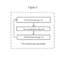
- FIG. 4 is a schematic view of the second processing module of the present invention.
- Image segmentation is a process to extract the regions of interest with different features out of an image.
- the features could be the gray scale level, color, or the textures of an image.
- the regions of interest could be a single region or different regions.
- automatic image segmentation has several challenges. For example, the influences of noise, blurry features, shades and shadows can result in erroneous segmentation. Therefore, automatic image segmentation is an improving technique.
- the boundaries between different tissues are usually not shape with blurred features.
- the segmentation of regions is very rigid and distinctive, which cannot accurately describe the boundaries that may or may not belong to a certain region. Therefore, it cannot deal with the uncertainly features in the medical images.
- the present invention adopted a segmentation method based on fuzzy clustering theory. By calculate the membership function of each pixel, the algorithm can determine the pixel belongings adaptively, therefore resulting in a better and robust segmentation.
- FIG. 1 is the flow chart of a medical image segmentation method of the present invention, which includes the following steps:
- Step 1 initialization of the cluster number and determine the initial values of the cluster centroids
- the image segmentation method of the present invention is to first determine the cluster number. This is to determine how many parts or regions of interests are to be segmented and the pixels included. The initialization of the cluster number and the centroids are determined by the specific image features and the desired segmentation results, such as the histogram of the image. Once determined, the number of the clusters is not to be changed in the following segmentation process. However, the pixels included in each cluster are constantly changing and updating, which is the process of clustering.
- the cluster centroids can be calculated.
- the cluster centroids are values that can be interpreted as an abstract mean value of a certain region of pixels. It is not a physical pixel point in an image.
- the present invention use the image histogram to calculate the initial value that has the same distance between each value.
- Step 2 calculate the Hausdorff distance between the cluster centroids and each pixel in the image
- the present invention uses Hausdorff distance, which is a definition of distance between two sets, to calculate the distance between each centroid and pixels.
- the Hausdorff distance defined pixel difference can utilize the neighbor information of each pixel. This is effective to segment and cluster the soft tissue boundaries, especially those with low image contrast.
- FIG. 2 is a flow chart to calculate the Hausdroff distance between each image pixel and the centroids, which includes the following steps:
- Step 201 select several neighbor regions with the image pixel at the center, calculate the mutual information between the neighbor region and the centroid;
- the neighbor region of one pixel is defined by a set of pixel points surrounding the pixel that is located at the center.
- the region is usually a circle or rectangle whose size determined by selection. Therefore, the computation of the pixel is determined by its neighbor region, which is called neighbor calculation.
- the present invention has selected the neighbor region that comprises of the rectangular region with a width and height of 3, 5, and 7 pixels. That is, rectangular region having 9, 25, and 49 pixels.
- I ⁇ i ⁇ A ⁇ ⁇ j ⁇ B ⁇ p ⁇ ( i , j ) ⁇ log ⁇ p ⁇ ( i , j ) p ⁇ ( i ) ⁇ p ⁇ ( j ) to calculate the mutual information between the neighbor region and the centroids, where p(i,j) is the joint distribution function of the two images, p(i) is the pixel distribution function for the neighbor region, and p(j) is the pixel distribution function for the pixel.
- Step 202 select the neighbor region that has the maximum mutual information value to calculate the Hausdorff distance
- the neighbor region that has the maximum value to calculate the Hausdorff distance. For example, in the previously stated rectangular region with a width and height of 3, 5, and 7 pixels, if the region with a width and height of 5 pixels has the largest mutual information, then the Hausdorff distance is calculated based on the 5 pixel neighbor region. By comparing different regions, the neighbor region is selected adaptively, therefore solving the problem of discontinuity and contrast jump in the boundaries.
- Step 203 Calculate the Hausdorff distances between each centroid and the neighbor region with the pixel at the center.
- the present invention calculates the Hausdorff distances between each centroid and the neighbor region with the pixel at the center.
- the equation to calculate Hausdorff distance is
- d H ⁇ ( A , B ) max ⁇ ⁇ sup p i ⁇ A ⁇ inf ⁇ p j ⁇ B ⁇ ⁇ p j - p i ⁇ ⁇ , sup p j ⁇ B ⁇ inf ⁇ p i ⁇ A ⁇ ⁇ p i - p j ⁇ ⁇ , where A is the cluster centroid, B is the pixels within the selected region.
- the present invention use the Hausdorff distance to determine the pixel differences, which can utilize the neighbor information and the soft tissues with low contrast values, therefore effectively segment and cluster the blurry boundaries.
- Step 3 calculate the membership function of each pixel based on the Hausdorff and Euclid distances between the cluster centroids and each pixel;
- membership function provides a value between 0 and 1 for each pixel, describing the pixel belongings.
- the sum of all the membership function values of each one of the pixel is 1. For example, if there exist three clusters A, B, and C, and the membership function of x is ⁇ 0.7, 0.2, 0.1 ⁇ , this represents that the probabilities of the pixel belongs to cluster A, B, and C are 70%, 20%, and 10%, respectively.
- the membership function is
- the scope of the parameters could be 30-50; for blurry and low contrast ultrasound image, the value could be 40-60; moreover, the parameter can balance the membership function and objective function representing the neighbor region information.
- m is the fuzziness value that is taken as 2 in this invention, ⁇ x i ⁇ v k ⁇ is the Euclid distance between each pixel and the centroids, ⁇ x i ⁇ v k ⁇ H is the Hausdorff distance between each pixel and the centroids.
- the membership function is calculated by both the Euclid and Hausdorff distance, by ultiziing the difference between each pixel and its neighbor region, the rigid clustering between each pixel and the soft clustering are combined, generating a more effective segmentation of the features, which is especially suitable for segmentation images with low contrast and blurry boundaries.
- Step 4 calculate the objective function, cluster the pixels of the image based on the updated membership function, and update the centroid value;
- the final cluster determination of each pixel can decided by the membership function.
- the maximum membership function value determines the corresponding final cluster. For example, if there is three clusters A, B, C. the pixel x has membership function value of ⁇ 0.7, 0.2, 0.1 ⁇ . 0.7 is the maximum membership function, therefore x belongs to cluster A. If the updated membership function values are ⁇ 0.3, 0.6, 0.1 ⁇ , then 0.6 becomes the largest value, then x belongs to B. In this manner, all the pixels can be clustered and group based on the membership functions.
- Step 5 repeat step 2 - 4 for at least twice until the difference between the two objective function values is less than the threshold value. Then the clustering has come to a constant result and the membership function from the last iteration is the final segmentation. If the difference between the two objective functions is larger than the threshold value, then repeat the above steps 2 - 4 until converge.
- the threshold value is set to 10 ⁇ 6 .
- FIG. 3 is a schematic view of a medical image segmentation apparatus of the present invention, which comprised of:
- a first processing module for executing step 1 , determines the initial cluster number and calculates the initial value of each centroid; this module is used for initializing the parameters, including cluster number and the initial values of the centroids.
- a second processing module for executing step 2 , calculates the Hausdorff distance between each pixel and the centroids; the Hausdorff distance is the distance between two point sets.
- a third processing module for executing step 3 , calculate the membership function based on the Hausdorff distance and Euclid distance between each pixel and the centroids, calculate the probability of each pixel belonging to each of the cluster.
- a fourth processing module for executing step 4 , calculate the objective function and determines whether to end the iteration or not; re-cluster the pixels based on the membership function and update the values of the centroids for the next iteration;
- a fifth processing module for iteration of the above steps of 2-4, until the differences of the objective function is less than the threshold value, and the last membership functions obtained provide the segmentation results.
- FIG. 4 shows the second processing module of the present invention, which includes:
- a first processing module calculates the mutual information between several different neighbor region and the centroids; wherein the neighbor regions are rectangular regions with a width and height of 3, 5, and 7 pixels. That is, rectangular region having 9, 25, and 49 pixels.
- a second processing module which is used to select the neighbor region with the maximum value of the mutual information to calculate the Hausdorff distance; automatically select the best neighbor region to calculate the Hausdorff distance, therefore solve the problem of discontinuities boundary and large variations of image contrast.
- a third processing module which is used to calculate Hausdorff distance between each centroid and the neighbor region with the pixel located at the center.
- the neighbor region is effectively utilized. This is especially suitable to segment the soft tissues with low image contrast and can cluster and segment the blurry boundaries.
Landscapes
- Engineering & Computer Science (AREA)
- Physics & Mathematics (AREA)
- Health & Medical Sciences (AREA)
- Theoretical Computer Science (AREA)
- Computer Vision & Pattern Recognition (AREA)
- Life Sciences & Earth Sciences (AREA)
- General Physics & Mathematics (AREA)
- Artificial Intelligence (AREA)
- Data Mining & Analysis (AREA)
- Medical Informatics (AREA)
- General Health & Medical Sciences (AREA)
- Evolutionary Computation (AREA)
- Probability & Statistics with Applications (AREA)
- Software Systems (AREA)
- Public Health (AREA)
- Nuclear Medicine, Radiotherapy & Molecular Imaging (AREA)
- Biophysics (AREA)
- Molecular Biology (AREA)
- Surgery (AREA)
- Animal Behavior & Ethology (AREA)
- Heart & Thoracic Surgery (AREA)
- Physiology (AREA)
- Veterinary Medicine (AREA)
- Biomedical Technology (AREA)
- Pathology (AREA)
- Radiology & Medical Imaging (AREA)
- Bioinformatics & Computational Biology (AREA)
- General Engineering & Computer Science (AREA)
- Bioinformatics & Cheminformatics (AREA)
- Evolutionary Biology (AREA)
- Quality & Reliability (AREA)
- Mathematical Physics (AREA)
- Fuzzy Systems (AREA)
- Psychiatry (AREA)
- Signal Processing (AREA)
- Computing Systems (AREA)
- Databases & Information Systems (AREA)
- Multimedia (AREA)
- Optics & Photonics (AREA)
- High Energy & Nuclear Physics (AREA)
Abstract
Description
where p(i,j) is the joint distribution function of the two images, and p(i), p(j) are the pixel distribution function for each image, respectively.
where A is the cluster centroid, B is the pixels within the selected region.
where xi is the gray scale pixel value, vk is the centroid, c is the amount of centroids, α is the empirical parameter determined by the image resolution and contrast, m is 2, ∥xi−vk∥ is the Euclid distance between each pixel and the centroids, ∥xi−vk∥H is the Hausdorff distance between each pixel and the centroids.
where xi is the gray scale pixel value, vk is the centroid, n is the number of the image pixels, and m is 2.
to calculate the mutual information between the neighbor region and the centroids, where p(i,j) is the joint distribution function of the two images, p(i) is the pixel distribution function for the neighbor region, and p(j) is the pixel distribution function for the pixel.
where A is the cluster centroid, B is the pixels within the selected region. The present invention use the Hausdorff distance to determine the pixel differences, which can utilize the neighbor information and the soft tissues with low contrast values, therefore effectively segment and cluster the blurry boundaries.
where xi is the gray scale pixel value, vk is the centroid, c is the amount of centroids, α is the parameter to adjusting the weighting of the Hausdorff distance that can be determined by the image contrast and resolution. For example, in MR images with high contrast and resolution, the scope of the parameters could be 30-50; for blurry and low contrast ultrasound image, the value could be 40-60; moreover, the parameter can balance the membership function and objective function representing the neighbor region information. m is the fuzziness value that is taken as 2 in this invention, ∥xi−vk∥ is the Euclid distance between each pixel and the centroids, ∥xi−vk∥∥H is the Hausdorff distance between each pixel and the centroids.
where xi is the gray scale pixel value, vk is the centroid, n is the number of the image pixels, and m is the fuzziness value. In this invention, the m value is taken as 2.
Claims (9)
Applications Claiming Priority (2)
| Application Number | Priority Date | Filing Date | Title |
|---|---|---|---|
| CN201610913874.5 | 2016-10-21 | ||
| CN201610913874.5A CN107977952A (en) | 2016-10-21 | 2016-10-21 | Medical image cutting method and device |
Publications (2)
| Publication Number | Publication Date |
|---|---|
| US20180114313A1 US20180114313A1 (en) | 2018-04-26 |
| US10096108B2 true US10096108B2 (en) | 2018-10-09 |
Family
ID=61971560
Family Applications (1)
| Application Number | Title | Priority Date | Filing Date |
|---|---|---|---|
| US15/387,886 Active 2037-07-05 US10096108B2 (en) | 2016-10-21 | 2016-12-22 | Medical image segmentation method and apparatus |
Country Status (2)
| Country | Link |
|---|---|
| US (1) | US10096108B2 (en) |
| CN (1) | CN107977952A (en) |
Cited By (1)
| Publication number | Priority date | Publication date | Assignee | Title |
|---|---|---|---|---|
| US10930386B2 (en) | 2018-12-11 | 2021-02-23 | International Business Machines Corporation | Automated normality scoring of echocardiograms |
Families Citing this family (29)
| Publication number | Priority date | Publication date | Assignee | Title |
|---|---|---|---|---|
| CN109584255B (en) * | 2018-11-07 | 2020-11-03 | 北京市遥感信息研究所 | Target contour extraction method based on fusion of panchromatic image and hyperspectral image |
| CN109685820B (en) * | 2018-11-29 | 2022-09-30 | 济南大学 | Image segmentation method based on morphological reconstruction and FCM clustering with guiding filtering |
| CN109753997B (en) * | 2018-12-19 | 2022-11-22 | 湖南科技大学 | An automatic accurate and robust segmentation method for liver tumors in CT images |
| CN109816660A (en) * | 2019-02-19 | 2019-05-28 | 闽南师范大学 | An image segmentation method, terminal device and storage medium |
| CN109993204A (en) * | 2019-02-25 | 2019-07-09 | 重庆邮电大学 | The classification of remote-sensing images algorithm of FCM cluster is selected and improved based on mRMR |
| CN110084820B (en) * | 2019-03-30 | 2023-05-26 | 重庆师范大学 | Purple soil image self-adaptive segmentation extraction method based on improved FCM algorithm |
| CN110033464B (en) * | 2019-04-15 | 2023-03-10 | 齐齐哈尔医学院 | Breast tumor ultrasonic image segmentation method based on level set algorithm |
| CN110245666B (en) * | 2019-06-10 | 2021-06-08 | 西安邮电大学 | Multi-objective interval-valued fuzzy clustering image segmentation method based on dual-membership drive |
| CN110597719B (en) * | 2019-09-05 | 2021-06-15 | 腾讯科技(深圳)有限公司 | A kind of image clustering method, device and medium for adaptation test |
| US11928430B2 (en) | 2019-09-12 | 2024-03-12 | Oracle International Corporation | Detecting unrelated utterances in a chatbot system |
| CN110827215A (en) * | 2019-10-21 | 2020-02-21 | 天津大学 | ERT image reconstruction artifact removing method based on fuzzy clustering |
| CN111047593B (en) * | 2019-10-28 | 2023-06-02 | 东北大学 | An Image Segmentation Method Based on Adaptive Distance Clustering |
| CN110853054B (en) * | 2019-10-29 | 2022-08-16 | 北京航空航天大学 | Method for improving intuitive fuzzy clustering and extracting infrared ship by using position information |
| CN110853059B (en) * | 2019-11-12 | 2023-04-07 | 陕西科技大学 | Image identification method for target ring number |
| CN111079637B (en) * | 2019-12-12 | 2023-09-08 | 武汉轻工大学 | Methods, devices, equipment and storage media for segmenting rapeseed flowers in field images |
| CN111831373A (en) * | 2020-06-09 | 2020-10-27 | 上海容易网电子商务股份有限公司 | Detection processing method for application starting state of android interactive screen |
| CN112287193B (en) * | 2020-10-30 | 2022-10-04 | 腾讯科技(深圳)有限公司 | Image segmentation method and device, computer equipment and storage medium |
| CN114764807B (en) | 2020-12-30 | 2025-04-25 | 上海联影医疗科技股份有限公司 | Coronary artery segmentation method, device, computer equipment and storage medium |
| CN112766338B (en) * | 2021-01-11 | 2023-06-16 | 明峰医疗系统股份有限公司 | Method, system and computer readable storage medium for calculating distance image |
| CN113628225B (en) * | 2021-08-24 | 2024-02-20 | 合肥工业大学 | Fuzzy C-means image segmentation method and system based on structural similarity and image region block |
| CN115797246A (en) * | 2021-09-09 | 2023-03-14 | 上海微创卜算子医疗科技有限公司 | Pathological image quality assessment, adjustment method and system, electronic equipment, medium |
| CN116563208A (en) * | 2023-03-16 | 2023-08-08 | 深圳市智影医疗科技有限公司 | A clustering-based nodule segmentation method in medical images |
| CN116309645B (en) * | 2023-03-27 | 2025-10-14 | 杭州国辰机器人科技有限公司 | An adaptive image segmentation method based on improved tangent-cotangent optimization |
| CN117036371B (en) * | 2023-06-26 | 2026-05-22 | 苏州大学 | Fuzzy clustering image segmentation method and system based on lie group theory |
| CN116883993B (en) * | 2023-09-06 | 2023-12-01 | 临沂大学 | Dried rose flower sorting method based on vision |
| CN117710385B (en) * | 2024-02-05 | 2024-05-10 | 山东建筑大学 | Image segmentation method and system based on hierarchical multi-wheel driven fuzzy clustering |
| CN119112298B (en) * | 2024-08-15 | 2025-04-18 | 中国医学科学院北京协和医院 | A bronchoscope cutter and positioning system |
| CN119323615B (en) * | 2024-12-18 | 2025-04-01 | 西安医学院第一附属医院 | A rapid reconstruction method and system for cardiac CT images |
| CN121527091B (en) * | 2026-01-16 | 2026-04-21 | 华中科技大学同济医学院附属同济医院 | Heart CT image segmentation method and system based on ultrasonic guidance |
Citations (6)
| Publication number | Priority date | Publication date | Assignee | Title |
|---|---|---|---|---|
| US20090067726A1 (en) * | 2006-07-31 | 2009-03-12 | Berna Erol | Computation of a recognizability score (quality predictor) for image retrieval |
| US20120076422A1 (en) * | 2010-09-28 | 2012-03-29 | Futurewei Technologies, Inc. | System and Method for Image Authentication |
| US20140126804A1 (en) * | 2012-11-05 | 2014-05-08 | Mitutoyo Corporation | Edge measurement video tool and interface including automatic parameter set alternatives |
| US20140254919A1 (en) * | 2013-03-06 | 2014-09-11 | Samsung Electronics Co., Ltd. | Device and method for image processing |
| US20150073765A1 (en) * | 2013-09-12 | 2015-03-12 | Siemens Corporation | System and method for prediction of respiratory motion from 3d thoracic images |
| US20170076114A1 (en) * | 2015-09-11 | 2017-03-16 | Infosys Limited | Method and system for masking of cheque image based on user access privileges |
Family Cites Families (3)
| Publication number | Priority date | Publication date | Assignee | Title |
|---|---|---|---|---|
| CN103336781B (en) * | 2013-05-29 | 2016-06-08 | 江苏大学 | A medical image clustering method |
| CN103366367B (en) * | 2013-06-19 | 2015-11-11 | 西安电子科技大学 | Based on the FCM gray-scale image segmentation method of pixel count cluster |
| CN105654453B (en) * | 2014-11-10 | 2018-09-28 | 华东师范大学 | A kind of FCM image partition methods of robustness |
-
2016
- 2016-10-21 CN CN201610913874.5A patent/CN107977952A/en active Pending
- 2016-12-22 US US15/387,886 patent/US10096108B2/en active Active
Patent Citations (6)
| Publication number | Priority date | Publication date | Assignee | Title |
|---|---|---|---|---|
| US20090067726A1 (en) * | 2006-07-31 | 2009-03-12 | Berna Erol | Computation of a recognizability score (quality predictor) for image retrieval |
| US20120076422A1 (en) * | 2010-09-28 | 2012-03-29 | Futurewei Technologies, Inc. | System and Method for Image Authentication |
| US20140126804A1 (en) * | 2012-11-05 | 2014-05-08 | Mitutoyo Corporation | Edge measurement video tool and interface including automatic parameter set alternatives |
| US20140254919A1 (en) * | 2013-03-06 | 2014-09-11 | Samsung Electronics Co., Ltd. | Device and method for image processing |
| US20150073765A1 (en) * | 2013-09-12 | 2015-03-12 | Siemens Corporation | System and method for prediction of respiratory motion from 3d thoracic images |
| US20170076114A1 (en) * | 2015-09-11 | 2017-03-16 | Infosys Limited | Method and system for masking of cheque image based on user access privileges |
Non-Patent Citations (1)
| Title |
|---|
| Dubuisson et al. ("A modified Hausdorff distance for object matching," Proceedings of 12th International Conference on Pattern Recognition, Oct. 9-13, 1994) (Year: 1994). * |
Cited By (1)
| Publication number | Priority date | Publication date | Assignee | Title |
|---|---|---|---|---|
| US10930386B2 (en) | 2018-12-11 | 2021-02-23 | International Business Machines Corporation | Automated normality scoring of echocardiograms |
Also Published As
| Publication number | Publication date |
|---|---|
| US20180114313A1 (en) | 2018-04-26 |
| CN107977952A (en) | 2018-05-01 |
Similar Documents
| Publication | Publication Date | Title |
|---|---|---|
| US20180114313A1 (en) | Medical Image Segmentation Method and Apparatus | |
| Orlando et al. | A discriminatively trained fully connected conditional random field model for blood vessel segmentation in fundus images | |
| Paik et al. | Surface normal overlap: a computer-aided detection algorithm with application to colonic polyps and lung nodules in helical CT | |
| US8855388B2 (en) | Microcalcification detection classification in radiographic images | |
| US7660451B2 (en) | System, method and apparatus for small pulmonary nodule computer aided diagnosis from computed tomography scans | |
| Wimmer et al. | A generic probabilistic active shape model for organ segmentation | |
| US7724936B2 (en) | Image generation apparatus and image generation method for detecting abnormalities | |
| US8577104B2 (en) | Liver lesion segmentation | |
| CN112991363A (en) | Brain tumor image segmentation method and device, electronic equipment and storage medium | |
| CN109753997B (en) | An automatic accurate and robust segmentation method for liver tumors in CT images | |
| Nie et al. | Two-dimensional extension of variance-based thresholding for image segmentation | |
| Li et al. | Segmentation of pulmonary nodules using a GMM fuzzy C-means algorithm | |
| CN114596320A (en) | A kind of image segmentation method and device based on ALSLCV model | |
| Ali et al. | Retinal blood vessel segmentation from retinal image using B-COSFIRE and adaptive thresholding | |
| US20080260254A1 (en) | Automatic 3-D Object Detection | |
| CN117392128B (en) | Artificial intelligent measuring method for aortic diameter | |
| Mei et al. | A curve evolution approach for unsupervised segmentation of images with low depth of field | |
| Modak et al. | Gpd-nodule: A lightweight lung nodule detection and segmentation framework on computed tomography images using uniform superpixel generation | |
| WO2017062453A1 (en) | Image segmentation of organs depicted in computed tomography images | |
| Singh et al. | An efficient combined approach for medical brain tumour segmentation | |
| JP2006325937A (en) | Image determination device, image determination method, and program therefor | |
| CN114419321B (en) | CT image heart segmentation method and system based on artificial intelligence | |
| Arifin et al. | Detection of overlapping teeth on dental panoramic radiograph | |
| Feulner et al. | Fast automatic segmentation of the esophagus from 3D CT data using a probabilistic model | |
| CN119251192A (en) | Method, device, equipment and medium for extracting image features of ultrasonic images |
Legal Events
| Date | Code | Title | Description |
|---|---|---|---|
| FEPP | Fee payment procedure |
Free format text: ENTITY STATUS SET TO MICRO (ORIGINAL EVENT CODE: MICR); ENTITY STATUS OF PATENT OWNER: MICROENTITY |
|
| STCF | Information on status: patent grant |
Free format text: PATENTED CASE |
|
| MAFP | Maintenance fee payment |
Free format text: PAYMENT OF MAINTENANCE FEE, 4TH YEAR, MICRO ENTITY (ORIGINAL EVENT CODE: M3551); ENTITY STATUS OF PATENT OWNER: MICROENTITY Year of fee payment: 4 |
|
| FEPP | Fee payment procedure |
Free format text: SURCHARGE FOR LATE PAYMENT, MICRO ENTITY (ORIGINAL EVENT CODE: M3555); ENTITY STATUS OF PATENT OWNER: MICROENTITY |
|
| MAFP | Maintenance fee payment |
Free format text: PAYMENT OF MAINTENANCE FEE, 8TH YEAR, MICRO ENTITY (ORIGINAL EVENT CODE: M3552); ENTITY STATUS OF PATENT OWNER: MICROENTITY Year of fee payment: 8 |
|
| FEPP | Fee payment procedure |
Free format text: ENTITY STATUS SET TO SMALL (ORIGINAL EVENT CODE: SMAL); ENTITY STATUS OF PATENT OWNER: SMALL ENTITY |