US10088541B2 - Method and magnetic resonance scanner for acquiring a magnetic resonance data set - Google Patents
Method and magnetic resonance scanner for acquiring a magnetic resonance data set Download PDFInfo
- Publication number
- US10088541B2 US10088541B2 US14/622,091 US201514622091A US10088541B2 US 10088541 B2 US10088541 B2 US 10088541B2 US 201514622091 A US201514622091 A US 201514622091A US 10088541 B2 US10088541 B2 US 10088541B2
- Authority
- US
- United States
- Prior art keywords
- magnetic resonance
- slices
- resonance data
- space
- acquisition
- Prior art date
- Legal status (The legal status is an assumption and is not a legal conclusion. Google has not performed a legal analysis and makes no representation as to the accuracy of the status listed.)
- Active, expires
Links
Images


Classifications
-
- G—PHYSICS
- G01—MEASURING; TESTING
- G01R—MEASURING ELECTRIC VARIABLES; MEASURING MAGNETIC VARIABLES
- G01R33/00—Arrangements or instruments for measuring magnetic variables
- G01R33/20—Arrangements or instruments for measuring magnetic variables involving magnetic resonance
- G01R33/44—Arrangements or instruments for measuring magnetic variables involving magnetic resonance using nuclear magnetic resonance [NMR]
- G01R33/48—NMR imaging systems
- G01R33/4828—Resolving the MR signals of different chemical species, e.g. water-fat imaging
-
- G—PHYSICS
- G01—MEASURING; TESTING
- G01R—MEASURING ELECTRIC VARIABLES; MEASURING MAGNETIC VARIABLES
- G01R33/00—Arrangements or instruments for measuring magnetic variables
- G01R33/20—Arrangements or instruments for measuring magnetic variables involving magnetic resonance
- G01R33/44—Arrangements or instruments for measuring magnetic variables involving magnetic resonance using nuclear magnetic resonance [NMR]
- G01R33/48—NMR imaging systems
- G01R33/4818—MR characterised by data acquisition along a specific k-space trajectory or by the temporal order of k-space coverage, e.g. centric or segmented coverage of k-space
- G01R33/482—MR characterised by data acquisition along a specific k-space trajectory or by the temporal order of k-space coverage, e.g. centric or segmented coverage of k-space using a Cartesian trajectory
-
- G—PHYSICS
- G01—MEASURING; TESTING
- G01R—MEASURING ELECTRIC VARIABLES; MEASURING MAGNETIC VARIABLES
- G01R33/00—Arrangements or instruments for measuring magnetic variables
- G01R33/20—Arrangements or instruments for measuring magnetic variables involving magnetic resonance
- G01R33/44—Arrangements or instruments for measuring magnetic variables involving magnetic resonance using nuclear magnetic resonance [NMR]
- G01R33/48—NMR imaging systems
- G01R33/483—NMR imaging systems with selection of signals or spectra from particular regions of the volume, e.g. in vivo spectroscopy
- G01R33/4833—NMR imaging systems with selection of signals or spectra from particular regions of the volume, e.g. in vivo spectroscopy using spatially selective excitation of the volume of interest, e.g. selecting non-orthogonal or inclined slices
- G01R33/4835—NMR imaging systems with selection of signals or spectra from particular regions of the volume, e.g. in vivo spectroscopy using spatially selective excitation of the volume of interest, e.g. selecting non-orthogonal or inclined slices of multiple slices
-
- G—PHYSICS
- G01—MEASURING; TESTING
- G01R—MEASURING ELECTRIC VARIABLES; MEASURING MAGNETIC VARIABLES
- G01R33/00—Arrangements or instruments for measuring magnetic variables
- G01R33/20—Arrangements or instruments for measuring magnetic variables involving magnetic resonance
- G01R33/44—Arrangements or instruments for measuring magnetic variables involving magnetic resonance using nuclear magnetic resonance [NMR]
- G01R33/48—NMR imaging systems
- G01R33/54—Signal processing systems, e.g. using pulse sequences ; Generation or control of pulse sequences; Operator console
- G01R33/543—Control of the operation of the MR system, e.g. setting of acquisition parameters prior to or during MR data acquisition, dynamic shimming, use of one or more scout images for scan plane prescription
-
- G—PHYSICS
- G01—MEASURING; TESTING
- G01R—MEASURING ELECTRIC VARIABLES; MEASURING MAGNETIC VARIABLES
- G01R33/00—Arrangements or instruments for measuring magnetic variables
- G01R33/20—Arrangements or instruments for measuring magnetic variables involving magnetic resonance
- G01R33/44—Arrangements or instruments for measuring magnetic variables involving magnetic resonance using nuclear magnetic resonance [NMR]
- G01R33/48—NMR imaging systems
- G01R33/54—Signal processing systems, e.g. using pulse sequences ; Generation or control of pulse sequences; Operator console
- G01R33/546—Interface between the MR system and the user, e.g. for controlling the operation of the MR system or for the design of pulse sequences
-
- G—PHYSICS
- G01—MEASURING; TESTING
- G01R—MEASURING ELECTRIC VARIABLES; MEASURING MAGNETIC VARIABLES
- G01R33/00—Arrangements or instruments for measuring magnetic variables
- G01R33/20—Arrangements or instruments for measuring magnetic variables involving magnetic resonance
- G01R33/44—Arrangements or instruments for measuring magnetic variables involving magnetic resonance using nuclear magnetic resonance [NMR]
- G01R33/48—NMR imaging systems
- G01R33/4818—MR characterised by data acquisition along a specific k-space trajectory or by the temporal order of k-space coverage, e.g. centric or segmented coverage of k-space
-
- G—PHYSICS
- G01—MEASURING; TESTING
- G01R—MEASURING ELECTRIC VARIABLES; MEASURING MAGNETIC VARIABLES
- G01R33/00—Arrangements or instruments for measuring magnetic variables
- G01R33/20—Arrangements or instruments for measuring magnetic variables involving magnetic resonance
- G01R33/44—Arrangements or instruments for measuring magnetic variables involving magnetic resonance using nuclear magnetic resonance [NMR]
- G01R33/48—NMR imaging systems
- G01R33/54—Signal processing systems, e.g. using pulse sequences ; Generation or control of pulse sequences; Operator console
- G01R33/56—Image enhancement or correction, e.g. subtraction or averaging techniques, e.g. improvement of signal-to-noise ratio and resolution
- G01R33/5607—Image enhancement or correction, e.g. subtraction or averaging techniques, e.g. improvement of signal-to-noise ratio and resolution by reducing the NMR signal of a particular spin species, e.g. of a chemical species for fat suppression, or of a moving spin species for black-blood imaging
-
- G—PHYSICS
- G01—MEASURING; TESTING
- G01R—MEASURING ELECTRIC VARIABLES; MEASURING MAGNETIC VARIABLES
- G01R33/00—Arrangements or instruments for measuring magnetic variables
- G01R33/20—Arrangements or instruments for measuring magnetic variables involving magnetic resonance
- G01R33/44—Arrangements or instruments for measuring magnetic variables involving magnetic resonance using nuclear magnetic resonance [NMR]
- G01R33/48—NMR imaging systems
- G01R33/54—Signal processing systems, e.g. using pulse sequences ; Generation or control of pulse sequences; Operator console
- G01R33/56—Image enhancement or correction, e.g. subtraction or averaging techniques, e.g. improvement of signal-to-noise ratio and resolution
- G01R33/561—Image enhancement or correction, e.g. subtraction or averaging techniques, e.g. improvement of signal-to-noise ratio and resolution by reduction of the scanning time, i.e. fast acquiring systems, e.g. using echo-planar pulse sequences
- G01R33/5615—Echo train techniques involving acquiring plural, differently encoded, echo signals after one RF excitation, e.g. using gradient refocusing in echo planar imaging [EPI], RF refocusing in rapid acquisition with relaxation enhancement [RARE] or using both RF and gradient refocusing in gradient and spin echo imaging [GRASE]
-
- G—PHYSICS
- G01—MEASURING; TESTING
- G01R—MEASURING ELECTRIC VARIABLES; MEASURING MAGNETIC VARIABLES
- G01R33/00—Arrangements or instruments for measuring magnetic variables
- G01R33/20—Arrangements or instruments for measuring magnetic variables involving magnetic resonance
- G01R33/44—Arrangements or instruments for measuring magnetic variables involving magnetic resonance using nuclear magnetic resonance [NMR]
- G01R33/48—NMR imaging systems
- G01R33/54—Signal processing systems, e.g. using pulse sequences ; Generation or control of pulse sequences; Operator console
- G01R33/56—Image enhancement or correction, e.g. subtraction or averaging techniques, e.g. improvement of signal-to-noise ratio and resolution
- G01R33/567—Image enhancement or correction, e.g. subtraction or averaging techniques, e.g. improvement of signal-to-noise ratio and resolution gated by physiological signals, i.e. synchronization of acquired MR data with periodical motion of an object of interest, e.g. monitoring or triggering system for cardiac or respiratory gating
- G01R33/5673—Gating or triggering based on a physiological signal other than an MR signal, e.g. ECG gating or motion monitoring using optical systems for monitoring the motion of a fiducial marker
-
- G—PHYSICS
- G01—MEASURING; TESTING
- G01R—MEASURING ELECTRIC VARIABLES; MEASURING MAGNETIC VARIABLES
- G01R33/00—Arrangements or instruments for measuring magnetic variables
- G01R33/20—Arrangements or instruments for measuring magnetic variables involving magnetic resonance
- G01R33/44—Arrangements or instruments for measuring magnetic variables involving magnetic resonance using nuclear magnetic resonance [NMR]
- G01R33/48—NMR imaging systems
- G01R33/54—Signal processing systems, e.g. using pulse sequences ; Generation or control of pulse sequences; Operator console
- G01R33/56—Image enhancement or correction, e.g. subtraction or averaging techniques, e.g. improvement of signal-to-noise ratio and resolution
- G01R33/567—Image enhancement or correction, e.g. subtraction or averaging techniques, e.g. improvement of signal-to-noise ratio and resolution gated by physiological signals, i.e. synchronization of acquired MR data with periodical motion of an object of interest, e.g. monitoring or triggering system for cardiac or respiratory gating
- G01R33/5676—Gating or triggering based on an MR signal, e.g. involving one or more navigator echoes for motion monitoring and correction
Definitions
- the invention relates to a method and magnetic resonance scanner for acquiring a magnetic resonance data set of a plurality of slices of a volume of interest of an examination object (patient).
- a trigger signal relating to the patient's respiration and indicating the start of an acquisition time window to begin a fat saturation technique and, to acquire magnetic resonance signals for the different slices in one of multiple acquisition blocks.
- These blocks include echo trains each relating to a single slice and a single portion of k-space, and all the echo trains of each individual acquisition block relate to different slices.
- a magnetic resonance image of each slice is determined by combining the magnetic resonance data acquired in different acquisition blocks and relating to a portion of k-space for that slice.
- Magnetic resonance imaging is well established as a clinical imaging method. Acquisition techniques exist in which only the magnetic resonance signal received from water bound protons (water signal) is to be contained in the magnetic resonance data set obtained, but not magnetic resonance signals arising from fat bound protons (fat signals). Various fat saturation techniques are therefore known in which, for example, a fat saturation pulse is emitted at regular intervals as a radiofrequency (RF) pulse.
- RF radiofrequency
- magnetic resonance sequences are known in which it is impossible or, because of the image quality, undesirable, to capture the entirety of k-space to be acquired after a single excitation pulse, because excessively severe signal decay occurs.
- multi-shot acquisitions in which the magnetic resonance sequence is subdivided into a number of echoes or echo trains that differ in their phase encoding portions such that different portions of k-space are acquired by each of these echo trains.
- a number of slices are frequently acquired, so that during the execution of echo trains for other slices, the signal of the previously acquired slice can relax again and a new excitation pulse can be generated in the next echo train in relation to that slice.
- the saturation pulse is not slice selective and affects the entire volume of interest. Since a number of fat saturation pulses may be required in order to achieve the desired result, it can happen that sufficient fat saturation is not yet present for the first echo trains, which is not a problem, however, as long as the other portions of k-space are acquired with longer running, sufficient fat saturation for the slices in question.
- the fat saturation technique is also restarted for each acquisition block, as cyclical continuation of the fat saturation technique is not automatically possible because of the unknown duration of the pauses between the acquisition blocks.
- the result of this is that a fat signal varying across the slices can occur.
- the effect of this is that the first slices in an acquisition block have a very low fat saturation, whereas the other slices have a more marked fat saturation. If, for example, five slices are acquired in an acquisition block, it may occur that fat saturation is not complete for the first two slices to be acquired in an acquisition block. If, for example, twenty slices are to be acquired in this way, wherein only five echo trains, for example, are possible within the acquisition time window, the effect is repeated for each slice group, so that, for example, eight out of twenty slices exhibit no or only low fat saturation. This results in a noticeable difference in image quality for different slices.
- An object of the invention is, for triggered multi-shot acquisitions of a plurality of slices with fat saturation, to provide an acquisition method that provides more uniform fat saturation for all the slices.
- This object is achieved in accordance with the invention by a method of the type described above, but wherein, for at least two, preferably all, of the acquisition blocks, a different sequence of the slices to be acquired by the echo trains is used within the acquisition block.
- the sequence of the slices within the acquisition block is varied.
- the slice sequence in accordance with the invention is varied in different acquisition blocks, in particular always between time-adjacent acquisition blocks. In this way, it is always different slices that are affected by the not-yet-complete saturation effect, so that in particular if each slice is equally subject to the not yet complete fat saturation, a homogenous image quality in terms of fat saturation is produced across all the slices. This means that slice images in which clearly insufficient fat saturation is present are no longer produced.
- the method according to the invention allows fat saturation to be improved across the slice stack for triggered acquisitions.
- the slices can be cyclically permutated in consecutive acquisition blocks or the slice sequence can be randomly selected for each acquisition block. Particularly in the case of a fairly small number of slices, a cyclical permutation proves to be the most advantageous option, so that e.g. in the case of four slices S 1 , S 2 , S 3 , S 4 , starting from the conventional slice sequence S 1 , S 2 , S 3 , S 4 , in the next acquisition block the sequence S 2 , S 3 , S 4 , S 1 can be acquired, in the one after that the sequence S 3 , S 4 , S 1 , S 2 , etc. Alternatively, however, it is possible to implement random sequences.
- the same echo train can continue to be executed for all the slices in an acquisition block, the same portion of k-space therefore being acquired for all the slices within an acquisition block.
- the contrasts in the magnetic resonance data are mainly determined by the region of the k-space center, the k-space center consequently being acquired for at least one slice initially, i.e. when fat saturation is not yet completely present.
- a departure is made from this acquisition-block-specific portion of k-space for all the slices.
- the sequence of the portions of k-space to be acquired within all the acquisition blocks can be selected such that a portion encompassing the center of k-space is acquired no earlier than the third echo train. It has been shown that fat saturation mostly needs approximately two echo trains in order to attain a sufficiently high, i.e. desired, level, so that it is advantageous to acquire the k-space center-encompassing portion of k-space to be acquired for each slice no earlier than the third echo train of an acquisition block. This consequently always ensures that echo trains which, because of their phase encoding, acquire a portion containing the center of k-space, are not used until a later point within an acquisition block.
- the portions encompassing the center of k-space are always acquired last in an acquisition block, if the number of slices is greater than the number of portions of k-space for each slice.
- the sequence of the portions of k-space within each acquisition block is selected on the basis of the proximity of the portion to the center of k-space such that portions closer to the center of k-space are acquired later than portions farther from the center of K space.
- a “portion encompassing the center of k-space,” in particular having a separation of zero means the portion closest to the center of k-space.
- peripheral regions of k-space are therefore acquired if necessary in the event of insufficient fat saturation, so that a further increase in image quality is achieved in respect of fat saturation. Altogether this means therefore that the more closely the echo train approaches the central echo, i.e.
- fat saturation pulse For the practical implementation of fat saturation it can be provided, as is generally known, that an RF pulse for fat saturation (fat saturation pulse) is emitted prior to the start of an echo train relating to a slice and a portion of k-space.
- fat saturation pulse other fat saturation techniques can basically be used which only develop their effect over time.
- the method according to the invention is particularly suitable for turbo spin-echo sequences (TSE sequences).
- the trigger signal is determined by evaluating a navigator data set and/or from breathing belt and/or breathing sensor signals.
- Different ways therefore exist of determining a trigger signal indicating a particular point in time in the respiratory cycle, this signal indicating the start of the acquisition time window. Navigators, i.e. in particular one-dimensional magnetic resonance acquisitions, are less preferred because of the additional capacity utilization of the magnetic resonance scanner; additionally or alternatively breathing belts and/or breathing sensors which can be incorporated, for example, into the patient couch of the magnetic resonance scanner with which the method according to the invention is carried out.
- the invention also relates to a magnetic resonance scanner having a control device that is designed to carry out the inventive method. All comments relating to the inventive method apply analogously to the inventive magnetic resonance scanner, which therefore also provides the advantages of the method.
- the control device thus evaluates the trigger signal and controls the components of the magnetic resonance scanner such that the echo trains are emitted in the described sequence of the slices and possibly of the portions of k-space.
- FIG. 1 shows an acquisition sequence according to the prior art.
- FIG. 2 is a schematic diagram of slice images acquired using the acquisition sequence according to FIG. 1 .
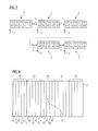
- FIG. 3 shows an acquisition sequence in a first embodiment of the method according to the invention.
- FIG. 4 shows a segmentation of k-space to be acquired.
- FIG. 5 shows an acquisition sequence of a second exemplary embodiment of the method according to the invention.
- FIG. 6 shows a schematic diagram of slice images acquired using the method according to the invention.
- FIG. 7 shows a magnetic resonance scanner according to the invention.
- respiration-triggered multi-shot acquisitions of a plurality of slices are to be carried out according to the prior art, an acquisition sequence as shown in FIG. 1 for five slices S 1 , S 2 , S 3 , S 4 , S 5 and five k-space portions K 1 , K 2 , K 3 , K 4 , K 5 is normally used.
- the trigger signal indicating a particular breathing state at which an acquisition time window 1 begins and marked by arrows 2 in FIGS. 1, 3 and 5 can be obtained optionally and/or in a combined manner by evaluating a navigator acquired using the magnetic resonance scanner, from sensor data of a breathing belt and/or from sensor data of some other breathing sensor.
- TSE sequences which are frequently employed for such multi-shot acquisitions, are used herein by way of example.
- the examination breaks down into five acquisition blocks 3 , each beginning with the application of the trigger signal, arrow 2 .
- five echo trains are played out to acquire magnetic resonance data, for each slice in a fixed sequence S 1 , S 2 , S 3 , S 4 , S 5 .
- the first acquisition block the first portion K 1 of k-space of the slices S 1 , S 2 , S 3 , S 4 , S 5 is acquired, in the second acquisition block 3 the portion K 2 , etc.
- This cyclical operation cannot be usefully continued, as the length of the pauses 5 between the acquisition blocks 3 varies and cannot be predicted.
- each slice image 6 Shown there in the form of a schematic diagram for each slice image 6 is an inner, mainly water-bound proton containing region 7 and an outer, mainly fat-bound proton containing region 8 . Because of the still barely present fat saturation, both regions 7 , 8 appear clear in the slice image 6 of the slice S 1 , i.e. signal-rich.
- FIG. 3 a first exemplary embodiment of the method according to the invention is again illustrated in FIG. 3 in the form of an acquisition sequence.
- the slice acquisition sequence differs from acquisition block 3 to acquisition block 3 .
- the slices S 1 , S 2 , S 3 , S 4 and S 5 are cyclically permutated from one acquisition block 3 to the next.
- a single portion K 1 , K 2 , K 3 , K 4 or K 5 in the first echo train of an acquisition block 3 and a single portion K 1 , K 2 , K 3 , K 4 or K 5 in the second echo train of an acquisition block 3 is acquired for each slice S 1 , S 2 , S 3 , S 4 and S 5 .
- each slice S 1 , S 2 , S 3 , S 4 and S 5 is at least similarly subject to the effect of the only subsequently occurring fat saturation. If the magnetic resonance data of all the acquisition blocks 3 is combined for each slice, this provides greatly improved quality in respect of fat saturation, in particular a uniform fat saturation.
- FIG. 4 first again shows more precisely the subdivision of k-space 9 to be acquired. This is initially split up into segments A 1 to A 5 from which an echo is acquired in each echo train, so that each segment A 1 to A 5 can be decomposed into sub-segments associated with the portions K 1 to K 5 . Finally a k-space trajectory passing through k-space 9 is decomposed accordingly.
- the portions K 1 , K 5 constitute outlying sub-segments for the relevant central segment A 3 containing the center 10 of k-space 9 , whereas the sub-segments of the portions K 2 and K 4 are closer to the center 10 of k-space 9 .
- the sub-segment of the portion K 3 lastly contains the center 10 of k-space 9 . If the knowledge that the central region of k-space 9 mainly determines the contrasts is now utilized, the sequence of portions K 1 , K 2 , K 3 , K 4 and K 5 within an acquisition block 3 can be selected such that subsequent positions within the acquisition block 3 and therefore improved fat saturation are provided for the inner regions of k-space 9 in the relevant segment A 3 .
- FIG. 5 A corresponding implementation is shown by the second exemplary embodiment for an acquisition sequence according to the present invention, as illustrated by FIG. 5 .
- the sequence of the slices S 1 , S 2 , S 3 , S 4 and S 5 is cyclically permutated as in the exemplary embodiment according to FIG. 3 , but with additionally a fixed sequence of portions K 1 , K 2 , K 3 , K 4 and K 5 of k-space 9 for all the acquisition blocks 3 , namely K 1 , K 5 , K 2 , K 4 and K 3 in this case.
- the portions K 1 , K 2 , K 3 , K 4 and K 5 are therefore sorted therein according to their proximity to the center 10 of k-space 9 , so that the portion K 5 containing the center 10 of k-space 9 is always acquired last, i.e. when maximum fat saturation has been achieved.
- FIG. 6 The effect of this change in the sequence of the slices S 1 , S 2 , S 3 , S 4 and S 5 and optimally also in the sequence of the portions K 1 , K 2 , K 3 , K 4 and K 5 is schematically illustrated in FIG. 6 .
- the slice images 6 ′ of the slices S 1 , S 2 and S 3 obtained from the combined magnetic resonance data of the acquisition blocks each show, in the region 8 , an excellent darkening which is essentially the same for all the slices.
- FIG. 7 shows a schematic diagram of an inventive magnetic resonance scanner 11 that has, as is generally known, a main magnet unit 12 defining a patient tunnel 13 .
- a main magnet unit 12 defining a patient tunnel 13 .
- an RF coil arrangement and a gradient coil arrangement are provided around the patient tunnel 13 .
- a patient bed on which the patient can be moved into the patient tunnel 13 and that can contain the breathing sensors which supply sensor data for the trigger signal.
- the operation of the magnetic resonance scanner 11 is controlled via a control device 14 that is designed to carry out the method according to the invention, i.e. in particular selecting the sequence within the acquisition blocks 3 for optimum image quality in respect of fat saturation.
Landscapes
- Physics & Mathematics (AREA)
- High Energy & Nuclear Physics (AREA)
- Condensed Matter Physics & Semiconductors (AREA)
- General Physics & Mathematics (AREA)
- Engineering & Computer Science (AREA)
- Signal Processing (AREA)
- Optics & Photonics (AREA)
- Spectroscopy & Molecular Physics (AREA)
- Magnetic Resonance Imaging Apparatus (AREA)
- Health & Medical Sciences (AREA)
- Biophysics (AREA)
- Physiology (AREA)
- Life Sciences & Earth Sciences (AREA)
- Cardiology (AREA)
- Power Engineering (AREA)
- Pulmonology (AREA)
- General Health & Medical Sciences (AREA)
- Nuclear Medicine, Radiotherapy & Molecular Imaging (AREA)
- Radiology & Medical Imaging (AREA)
Abstract
Description
Claims (10)
Applications Claiming Priority (3)
| Application Number | Priority Date | Filing Date | Title |
|---|---|---|---|
| DE102014202606 | 2014-02-13 | ||
| DE102014202606.7A DE102014202606B4 (en) | 2014-02-13 | 2014-02-13 | Creation of magnetic resonance images using acquisition blocks of different layers in different orders |
| DE102014202606.7 | 2014-02-13 |
Publications (2)
| Publication Number | Publication Date |
|---|---|
| US20150226819A1 US20150226819A1 (en) | 2015-08-13 |
| US10088541B2 true US10088541B2 (en) | 2018-10-02 |
Family
ID=53676904
Family Applications (1)
| Application Number | Title | Priority Date | Filing Date |
|---|---|---|---|
| US14/622,091 Active 2037-02-09 US10088541B2 (en) | 2014-02-13 | 2015-02-13 | Method and magnetic resonance scanner for acquiring a magnetic resonance data set |
Country Status (2)
| Country | Link |
|---|---|
| US (1) | US10088541B2 (en) |
| DE (1) | DE102014202606B4 (en) |
Cited By (2)
| Publication number | Priority date | Publication date | Assignee | Title |
|---|---|---|---|---|
| US10234524B2 (en) * | 2016-10-18 | 2019-03-19 | Siemens Healthcare Gmbh | Shifted pulses for simultaneous multi-slice imaging |
| US10989771B2 (en) * | 2018-07-26 | 2021-04-27 | Siemens Healthcare Gmbh | Method for triggered acquisition of a measured data set by a magnetic resonance system, computer program, data memory, and magnetic resonance system |
Families Citing this family (1)
| Publication number | Priority date | Publication date | Assignee | Title |
|---|---|---|---|---|
| EP3459456A1 (en) | 2017-09-26 | 2019-03-27 | Siemens Healthcare GmbH | Patient interface for supporting the breathing of a patient during a medical imaging examination |
Citations (4)
| Publication number | Priority date | Publication date | Assignee | Title |
|---|---|---|---|---|
| US6097185A (en) * | 1996-08-28 | 2000-08-01 | Hitachi Medical Corporation | Magnetic resonance imaging apparatus |
| US6577127B2 (en) | 2000-03-27 | 2003-06-10 | Koninklijke Philips Electronics N.V. | Magnetic resonance imaging method for imaging time-dependent contrast |
| US7362099B2 (en) | 2003-09-08 | 2008-04-22 | Koninklijke Philips Electronics N.V. | Randomized ordered k-space sub-sets for shared pre-pulses in MRI |
| US20140159720A1 (en) * | 2012-12-12 | 2014-06-12 | Michael Markl | Methods and Systems for Improving SNR in Multi-Slice Multi-Segment Magnetic Resonance Imaging |
-
2014
- 2014-02-13 DE DE102014202606.7A patent/DE102014202606B4/en active Active
-
2015
- 2015-02-13 US US14/622,091 patent/US10088541B2/en active Active
Patent Citations (4)
| Publication number | Priority date | Publication date | Assignee | Title |
|---|---|---|---|---|
| US6097185A (en) * | 1996-08-28 | 2000-08-01 | Hitachi Medical Corporation | Magnetic resonance imaging apparatus |
| US6577127B2 (en) | 2000-03-27 | 2003-06-10 | Koninklijke Philips Electronics N.V. | Magnetic resonance imaging method for imaging time-dependent contrast |
| US7362099B2 (en) | 2003-09-08 | 2008-04-22 | Koninklijke Philips Electronics N.V. | Randomized ordered k-space sub-sets for shared pre-pulses in MRI |
| US20140159720A1 (en) * | 2012-12-12 | 2014-06-12 | Michael Markl | Methods and Systems for Improving SNR in Multi-Slice Multi-Segment Magnetic Resonance Imaging |
Cited By (2)
| Publication number | Priority date | Publication date | Assignee | Title |
|---|---|---|---|---|
| US10234524B2 (en) * | 2016-10-18 | 2019-03-19 | Siemens Healthcare Gmbh | Shifted pulses for simultaneous multi-slice imaging |
| US10989771B2 (en) * | 2018-07-26 | 2021-04-27 | Siemens Healthcare Gmbh | Method for triggered acquisition of a measured data set by a magnetic resonance system, computer program, data memory, and magnetic resonance system |
Also Published As
| Publication number | Publication date |
|---|---|
| US20150226819A1 (en) | 2015-08-13 |
| DE102014202606B4 (en) | 2015-10-01 |
| DE102014202606A1 (en) | 2015-08-13 |
Similar Documents
| Publication | Publication Date | Title |
|---|---|---|
| US8581583B2 (en) | Method and apparatus for magnetic resonance imaging to create T1 maps | |
| US10185014B2 (en) | Method and apparatus for EPI magnetic resonance with slew rate controlled and kspace entry optimized | |
| US10393847B2 (en) | Method and apparatus for recording calibration data for a GRAPPA magnetic resonance imaging algorithm | |
| JP5854575B2 (en) | Magnetic resonance imaging system | |
| US10393840B2 (en) | Magnetic resonance apparatus and method for acquiring measurement data during simultaneous manipulation of spatially separate sub-volumes | |
| CN103356191B (en) | The functional mri method and apparatus of the brain volume fractiion of live subject | |
| DE102015221888B4 (en) | Simultaneous MRI multilayer measurement | |
| CN107865659A (en) | MR imaging apparatus and the method for obtaining MRI | |
| US10598747B2 (en) | System and method for simultaneous multislice magnetic resonance fingerprinting with variable radio frequency encoding | |
| US20140043024A1 (en) | Multiple excitation blade acquisition for motion correction in magnetic resonance imaging | |
| CN102621510A (en) | System for suppression of artifacts in MR imaging | |
| US10451698B2 (en) | Method and apparatus for parallel magnetic resonance data acquisition | |
| US10018699B2 (en) | Method and magnetic resonance apparatus for acquiring magnetic resonance data with a prospective motion correction | |
| US11009575B2 (en) | Method for simultaneous time-interleaved multislice magnetic resonance imaging | |
| US10670684B2 (en) | Free-breathing non-contrast MR angiography | |
| US10088541B2 (en) | Method and magnetic resonance scanner for acquiring a magnetic resonance data set | |
| US20180217219A1 (en) | Method and magnetic resonance apparatus for recalculating a weighting matrix in the event of patient motion | |
| US12135363B2 (en) | Shot-wise inversion time adaptation for multi-shot inversion recovery imaging | |
| US20150070015A1 (en) | Method and apparatus for magnetic resonance imaging | |
| US10725134B2 (en) | Method and magnetic resonance apparatus for optimizing the simultaneous acquisition of magnetic resonance data from multiple slabs or slices | |
| US20180246182A1 (en) | Magnetic resonance apparatus and method for parallel imaging with a reference data set for determining the weighting matrix | |
| US9846212B2 (en) | Method and apparatus for magnetic resonance imaging | |
| US20240175956A1 (en) | Method for Creating an Image Dataset, Magnetic Resonance System, Computer Program Product and Electronically Readable Data Carrier | |
| US11874353B2 (en) | Multi-shot echo planar imaging using reordered segments and recursive radio frequency pulse design giving matched slice profiles across segments | |
| US20150226824A1 (en) | Method and magnetic resonance system for generating mr images |
Legal Events
| Date | Code | Title | Description |
|---|---|---|---|
| AS | Assignment |
Owner name: SIEMENS AKTIENGESELLSCHAFT, GERMANY Free format text: ASSIGNMENT OF ASSIGNORS INTEREST;ASSIGNOR:PAUL, DOMINIK;REEL/FRAME:035682/0029 Effective date: 20150330 |
|
| STCF | Information on status: patent grant |
Free format text: PATENTED CASE |
|
| AS | Assignment |
Owner name: SIEMENS HEALTHCARE GMBH, GERMANY Free format text: ASSIGNMENT OF ASSIGNORS INTEREST;ASSIGNOR:SIEMENS AKTIENGESELLSCHAFT;REEL/FRAME:047543/0133 Effective date: 20181023 |
|
| MAFP | Maintenance fee payment |
Free format text: PAYMENT OF MAINTENANCE FEE, 4TH YEAR, LARGE ENTITY (ORIGINAL EVENT CODE: M1551); ENTITY STATUS OF PATENT OWNER: LARGE ENTITY Year of fee payment: 4 |
|
| AS | Assignment |
Owner name: SIEMENS HEALTHINEERS AG, GERMANY Free format text: ASSIGNMENT OF ASSIGNORS INTEREST;ASSIGNOR:SIEMENS HEALTHCARE GMBH;REEL/FRAME:066088/0256 Effective date: 20231219 |
|
| AS | Assignment |
Owner name: SIEMENS HEALTHINEERS AG, GERMANY Free format text: CORRECTIVE ASSIGNMENT TO CORRECT THE ASSIGNEE PREVIOUSLY RECORDED AT REEL: 066088 FRAME: 0256. ASSIGNOR(S) HEREBY CONFIRMS THE ASSIGNMENT;ASSIGNOR:SIEMENS HEALTHCARE GMBH;REEL/FRAME:071178/0246 Effective date: 20231219 |