US10085649B1 - Methods and devices for localizing the blood mass of an intracerebral hematoma - Google Patents
Methods and devices for localizing the blood mass of an intracerebral hematoma Download PDFInfo
- Publication number
- US10085649B1 US10085649B1 US15/239,722 US201615239722A US10085649B1 US 10085649 B1 US10085649 B1 US 10085649B1 US 201615239722 A US201615239722 A US 201615239722A US 10085649 B1 US10085649 B1 US 10085649B1
- Authority
- US
- United States
- Prior art keywords
- sensor
- glucose
- cannula
- blood mass
- distal end
- Prior art date
- Legal status (The legal status is an assumption and is not a legal conclusion. Google has not performed a legal analysis and makes no representation as to the accuracy of the status listed.)
- Active, expires
Links
Images


Classifications
-
- A—HUMAN NECESSITIES
- A61—MEDICAL OR VETERINARY SCIENCE; HYGIENE
- A61B—DIAGNOSIS; SURGERY; IDENTIFICATION
- A61B5/00—Measuring for diagnostic purposes; Identification of persons
- A61B5/02—Detecting, measuring or recording for evaluating the cardiovascular system, e.g. pulse, heart rate, blood pressure or blood flow
- A61B5/02007—Evaluating blood vessel condition, e.g. elasticity, compliance
-
- A—HUMAN NECESSITIES
- A61—MEDICAL OR VETERINARY SCIENCE; HYGIENE
- A61B—DIAGNOSIS; SURGERY; IDENTIFICATION
- A61B1/00—Instruments for performing medical examinations of the interior of cavities or tubes of the body by visual or photographical inspection, e.g. endoscopes; Illuminating arrangements therefor
- A61B1/313—Instruments for performing medical examinations of the interior of cavities or tubes of the body by visual or photographical inspection, e.g. endoscopes; Illuminating arrangements therefor for introducing through surgical openings, e.g. laparoscopes
-
- A—HUMAN NECESSITIES
- A61—MEDICAL OR VETERINARY SCIENCE; HYGIENE
- A61B—DIAGNOSIS; SURGERY; IDENTIFICATION
- A61B1/00—Instruments for performing medical examinations of the interior of cavities or tubes of the body by visual or photographical inspection, e.g. endoscopes; Illuminating arrangements therefor
- A61B1/00064—Constructional details of the endoscope body
- A61B1/00071—Insertion part of the endoscope body
- A61B1/0008—Insertion part of the endoscope body characterised by distal tip features
- A61B1/00087—Tools
-
- A—HUMAN NECESSITIES
- A61—MEDICAL OR VETERINARY SCIENCE; HYGIENE
- A61B—DIAGNOSIS; SURGERY; IDENTIFICATION
- A61B1/00—Instruments for performing medical examinations of the interior of cavities or tubes of the body by visual or photographical inspection, e.g. endoscopes; Illuminating arrangements therefor
- A61B1/00131—Accessories for endoscopes
- A61B1/00135—Oversleeves mounted on the endoscope prior to insertion
-
- A—HUMAN NECESSITIES
- A61—MEDICAL OR VETERINARY SCIENCE; HYGIENE
- A61B—DIAGNOSIS; SURGERY; IDENTIFICATION
- A61B1/00—Instruments for performing medical examinations of the interior of cavities or tubes of the body by visual or photographical inspection, e.g. endoscopes; Illuminating arrangements therefor
- A61B1/00163—Optical arrangements
- A61B1/00188—Optical arrangements with focusing or zooming features
-
- A—HUMAN NECESSITIES
- A61—MEDICAL OR VETERINARY SCIENCE; HYGIENE
- A61B—DIAGNOSIS; SURGERY; IDENTIFICATION
- A61B1/00—Instruments for performing medical examinations of the interior of cavities or tubes of the body by visual or photographical inspection, e.g. endoscopes; Illuminating arrangements therefor
- A61B1/04—Instruments for performing medical examinations of the interior of cavities or tubes of the body by visual or photographical inspection, e.g. endoscopes; Illuminating arrangements therefor combined with photographic or television appliances
- A61B1/05—Instruments for performing medical examinations of the interior of cavities or tubes of the body by visual or photographical inspection, e.g. endoscopes; Illuminating arrangements therefor combined with photographic or television appliances characterised by the image sensor, e.g. camera, being in the distal end portion
- A61B1/053—Instruments for performing medical examinations of the interior of cavities or tubes of the body by visual or photographical inspection, e.g. endoscopes; Illuminating arrangements therefor combined with photographic or television appliances characterised by the image sensor, e.g. camera, being in the distal end portion being detachable
-
- A—HUMAN NECESSITIES
- A61—MEDICAL OR VETERINARY SCIENCE; HYGIENE
- A61B—DIAGNOSIS; SURGERY; IDENTIFICATION
- A61B1/00—Instruments for performing medical examinations of the interior of cavities or tubes of the body by visual or photographical inspection, e.g. endoscopes; Illuminating arrangements therefor
- A61B1/06—Instruments for performing medical examinations of the interior of cavities or tubes of the body by visual or photographical inspection, e.g. endoscopes; Illuminating arrangements therefor with illuminating arrangements
-
- A—HUMAN NECESSITIES
- A61—MEDICAL OR VETERINARY SCIENCE; HYGIENE
- A61B—DIAGNOSIS; SURGERY; IDENTIFICATION
- A61B17/00—Surgical instruments, devices or methods
- A61B17/34—Trocars; Puncturing needles
- A61B17/3417—Details of tips or shafts, e.g. grooves, expandable, bendable; Multiple coaxial sliding cannulas, e.g. for dilating
- A61B17/3421—Cannulas
-
- A—HUMAN NECESSITIES
- A61—MEDICAL OR VETERINARY SCIENCE; HYGIENE
- A61B—DIAGNOSIS; SURGERY; IDENTIFICATION
- A61B5/00—Measuring for diagnostic purposes; Identification of persons
- A61B5/145—Measuring characteristics of blood in vivo, e.g. gas concentration or pH-value ; Measuring characteristics of body fluids or tissues, e.g. interstitial fluid or cerebral tissue
- A61B5/14532—Measuring characteristics of blood in vivo, e.g. gas concentration or pH-value ; Measuring characteristics of body fluids or tissues, e.g. interstitial fluid or cerebral tissue for measuring glucose, e.g. by tissue impedance measurement
-
- A—HUMAN NECESSITIES
- A61—MEDICAL OR VETERINARY SCIENCE; HYGIENE
- A61B—DIAGNOSIS; SURGERY; IDENTIFICATION
- A61B5/00—Measuring for diagnostic purposes; Identification of persons
- A61B5/145—Measuring characteristics of blood in vivo, e.g. gas concentration or pH-value ; Measuring characteristics of body fluids or tissues, e.g. interstitial fluid or cerebral tissue
- A61B5/1468—Measuring characteristics of blood in vivo, e.g. gas concentration or pH-value ; Measuring characteristics of body fluids or tissues, e.g. interstitial fluid or cerebral tissue using chemical or electrochemical methods, e.g. by polarographic means
- A61B5/1473—Measuring characteristics of blood in vivo, e.g. gas concentration or pH-value ; Measuring characteristics of body fluids or tissues, e.g. interstitial fluid or cerebral tissue using chemical or electrochemical methods, e.g. by polarographic means invasive, e.g. introduced into the body by a catheter
-
- A—HUMAN NECESSITIES
- A61—MEDICAL OR VETERINARY SCIENCE; HYGIENE
- A61B—DIAGNOSIS; SURGERY; IDENTIFICATION
- A61B5/00—Measuring for diagnostic purposes; Identification of persons
- A61B5/68—Arrangements of detecting, measuring or recording means, e.g. sensors, in relation to patient
- A61B5/6846—Arrangements of detecting, measuring or recording means, e.g. sensors, in relation to patient specially adapted to be brought in contact with an internal body part, i.e. invasive
- A61B5/6847—Arrangements of detecting, measuring or recording means, e.g. sensors, in relation to patient specially adapted to be brought in contact with an internal body part, i.e. invasive mounted on an invasive device
- A61B5/6848—Needles
-
- A—HUMAN NECESSITIES
- A61—MEDICAL OR VETERINARY SCIENCE; HYGIENE
- A61B—DIAGNOSIS; SURGERY; IDENTIFICATION
- A61B5/00—Measuring for diagnostic purposes; Identification of persons
- A61B5/68—Arrangements of detecting, measuring or recording means, e.g. sensors, in relation to patient
- A61B5/6846—Arrangements of detecting, measuring or recording means, e.g. sensors, in relation to patient specially adapted to be brought in contact with an internal body part, i.e. invasive
- A61B5/6867—Arrangements of detecting, measuring or recording means, e.g. sensors, in relation to patient specially adapted to be brought in contact with an internal body part, i.e. invasive specially adapted to be attached or implanted in a specific body part
- A61B5/6868—Brain
-
- A—HUMAN NECESSITIES
- A61—MEDICAL OR VETERINARY SCIENCE; HYGIENE
- A61B—DIAGNOSIS; SURGERY; IDENTIFICATION
- A61B5/00—Measuring for diagnostic purposes; Identification of persons
- A61B5/68—Arrangements of detecting, measuring or recording means, e.g. sensors, in relation to patient
- A61B5/6846—Arrangements of detecting, measuring or recording means, e.g. sensors, in relation to patient specially adapted to be brought in contact with an internal body part, i.e. invasive
- A61B5/6886—Monitoring or controlling distance between sensor and tissue
-
- A—HUMAN NECESSITIES
- A61—MEDICAL OR VETERINARY SCIENCE; HYGIENE
- A61B—DIAGNOSIS; SURGERY; IDENTIFICATION
- A61B5/00—Measuring for diagnostic purposes; Identification of persons
- A61B5/74—Details of notification to user or communication with user or patient; User input means
- A61B5/742—Details of notification to user or communication with user or patient; User input means using visual displays
-
- A—HUMAN NECESSITIES
- A61—MEDICAL OR VETERINARY SCIENCE; HYGIENE
- A61B—DIAGNOSIS; SURGERY; IDENTIFICATION
- A61B90/00—Instruments, implements or accessories specially adapted for surgery or diagnosis and not covered by any of the groups A61B1/00 - A61B50/00, e.g. for luxation treatment or for protecting wound edges
- A61B90/30—Devices for illuminating a surgical field, the devices having an interrelation with other surgical devices or with a surgical procedure
- A61B2090/309—Devices for illuminating a surgical field, the devices having an interrelation with other surgical devices or with a surgical procedure using white LEDs
-
- A—HUMAN NECESSITIES
- A61—MEDICAL OR VETERINARY SCIENCE; HYGIENE
- A61B—DIAGNOSIS; SURGERY; IDENTIFICATION
- A61B90/00—Instruments, implements or accessories specially adapted for surgery or diagnosis and not covered by any of the groups A61B1/00 - A61B50/00, e.g. for luxation treatment or for protecting wound edges
- A61B90/36—Image-producing devices or illumination devices not otherwise provided for
- A61B90/361—Image-producing devices, e.g. surgical cameras
- A61B2090/3614—Image-producing devices, e.g. surgical cameras using optical fibre
Definitions
- the inventions described below relate to the field of minimally invasive brain surgery.
- Stroke is a common cause of death and disabling neurologic disorder. Approximately 700,000 patients suffer from stroke in the United States every year. Hemorrhagic stroke accounts for 20% of the annual stroke population. Hemorrhagic stroke is due to a rupture of a blood vessel in the brain, causing bleeding into the brain tissue and resulting in a hematoma (a blood mass) in the brain. Prompt removal of the blood mass is necessary to limit or prevent long-term brain injury.
- the device described below for identifying the location and dimensions of a blood mass in a patient's brain includes a glucose meter, a cannula and one of more sensor wires.
- the sensor wire has a distal end and a proximal end and the proximal end of the sensor wire is operatively connected to the glucose meter and the distal end of the sensor wire is operatively connected to a glucose sensor.
- the cannula has a distal end and a proximal end and a lumen to permit access to the blood mass or other tissue of interest. In use, a surgeon extends the distal end of the sensor wire through the cannula lumen and the distal end of the sensor wire and the glucose sensor may be advanced beyond the distal end of the cannula.
- the method described below for localizing a blood mass in a patient's brain uses a glucose meter, a glucose sensor, a cannula and a sensor wire.
- the sensor wire has a distal end and a proximal end and the proximal end of the sensor wire is operatively connected to the glucose meter and the distal end of the sensor wire is operatively connected to the glucose sensor.
- the cannula has a distal end and a proximal end and a lumen to permit access to the blood mass or other tissue of interest.
- the sensor wire extends through the cannula lumen with the distal end of the sensor wire and the glucose sensor extends beyond the distal end of the cannula.
- the distal end of the cannula, the distal end of the sensor wire and the glucose sensor are inserted into the patient's brain.
- the sensor wire is advanced until the glucose meter indicates the glucose sensor is within the blood mass.
- the sensor wire is further advanced until the glucose meter indicates that the glucose sensor has passed out of the blood mass.
- the cannula is advanced over the sensor wire until the distal end of the cannula is within the blood mass and the blood mass may be removed through the cannula lumen using any suitable technique.
- FIG. 1 is an illustration of the use of a cannula and sensor wire to localize an intracerebral blood mass in the head of a patient.
- FIGS. 2 through 6 illustrate steps for localizing and removing the intracerebral blood mass from the patient of FIG. 1 .
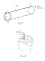
- FIG. 7 is a perspective view of a localizing cannula.
- FIG. 8 is an illustration of the use of a localizing cannula in the patient of FIG. 1 .
- FIG. 9 is an illustration of the use of a cannula and dual sensor wires to localize an intracerebral blood mass in a patient.
- FIGS. 10 through 13 illustrate steps for localizing and removing the intracerebral blood mass from a patient.
- FIG. 1 illustrates a patient 1 with an intracerebral hematoma or blood mass 2 in brain tissue 3 and a system for locating the intracerebral hematoma and inserting a cannula 4 into the hematoma.
- Cannula 4 has a distal end 4 D, a proximal end 4 P and a lumen 4 L extending from distal end 4 D to proximal end 4 P as illustrated in FIG. 7 .
- a surgeon advances one or more sensor wires such as sensor wire 5 with a glucose sensor 6 disposed on the distal tip of the sensor wire through the cannula to locate and determine the size of blood mass 2 .
- Distal end 5 D includes any suitable sensor for the detection of glucose such as sensor 6 .
- Proximal end 5 P of sensor wire 5 is operatively connected to any suitable sensor display such as glucose meter 7 with a display 8 which varies according to the glucose levels detected by sensor 6 .
- Display 8 may provide any suitable indication of glucose readings using analog, digital or audio output.
- Cannula 4 and sensor wire 5 may also include any suitable indicia such as indicia 9 to indicate their depth of insertion.
- a surgeon may localize an intracerebral blood mass using one or more sensor wires to locate and map the extent of the blood mass, and then use each sensor wire as a guide wire to guide the insertion of the cannula into the blood mass.
- FIGS. 2 through 6 illustrate steps for localizing and removing the intracerebral blood mass from the patient of FIG. 1 .
- a surgeon or other suitable person inserts the distal end of the sensor wire 5 with glucose sensor 6 to find and localize blood mass 2 .
- the surgeon advances cannula 4 and sensor wire 5 through brain tissue 3 until display 8 indicates that glucose sensor 6 has encountered glucose indicative of the presence of blood mass 2 as illustrated in FIG. 2 .
- Indicia 9 is noted to identify proximal margin 2 P of the blood mass.
- display 8 indicates the presence of glucose when glucose sensor 6 is within blood mass 2 .
- As sensor wire 5 and glucose sensor 6 are advanced through the blood mass the sensor will eventually exit the blood mass as illustrated in FIG. 4 .
- glucose sensor 6 exits the blood mass and reenters brain tissue 3 display 8 indicates a drop in glucose level and the indicia 9 on wire 5 may be noted to identify the location of the distal margin 2 D of the blood mass.
- margins 2 P and 2 D respectively are identified any suitable technique for removing the blood mass may be used. For example as shown in FIGS. 5 and 6 a surgeon may advance cannula 4 along wire 5 until distal end 4 D is between the distal and proximal margins of blood mass 2 and then remove the blood mass through the cannula.
- a surgeon may advance a catheter 10 or other suitable instrument having a smaller diameter than cannula 4 through the cannula until the distal end 10 D of the catheter is between the distal and proximal margins of the blood mass 2 . Once the surgeon has located the distal end of the device for evacuating the blood mass within blood mass 2 , the blood mass may be evacuated.
- FIG. 7 illustrates a cannula 4 , comprising a transparent cannula that conducts light from proximal end 4 P to distal end 4 D.
- Light 11 from light source 12 couples to proximal end 4 P using any suitable technique and light 11 travels from proximal end 4 P to distal end 4 D through the acrylic of cannula 4 or through one or more embedded light conductors or optical fibers such as fiber 13 .
- Light of any suitable wavelength or combination of wavelengths may be directed from the cannula or embedded fibers using lenses such as lens 14 .
- a surgeon may extract an intracerebral blood mass through bore or lumen 4 L in the cannula 4 .
- Light sources such as light emitting diodes or other suitable elements may be incorporated into the any suitable portion of the cannula to illuminate the distal end of the cannula and the operating space surrounding the distal end of the cannula to enable localization and removal of the intracerebral blood mass.
- FIGS. 9 through 12 illustrate use of a pair of sensor wires with sensor tips to determine the margins of the blood mass.
- the surgeon inserts cannula 15 into brain tissue to guide the insertion of first sensor wire 16 and second sensor wire through lumen 15 L to find and localize blood mass.
- Sensor wires 16 and 17 include any suitable glucose sensor such as first glucose sensor 18 and second glucose sensor 19 .
- First and second sensor wires 16 and 17 are operatively connected to any suitable sensor display such as glucose meter 20 with first and second displays 21 A and 21 B which varies according to the glucose levels measured by sensor first and second glucose sensors 18 and 19 respectively.
- Glucose meter 20 may provide any suitable indication of glucose readings using analog, digital or audio output.
- Cannula 15 and sensor wires 16 and 17 may also include any suitable indicia such as indicia 22 to indicate their depth of insertion.
- a surgeon inserts the distal end of a cannula, catheter or cannula such as cannula 15 into brain tissue 3 to guide the insertion of sensor wires 16 and 17 with glucose sensors 18 and 19 through lumen 15 L to find and localize blood mass 2 as illustrated in FIGS. 10, 11 and 12 .
- First sensor wire 16 is advanced until first sensor 18 detects glucose and first display 21 A indicates the presence of glucose.
- indicia 22 for first sensor wire 16 may be recorded and the first sensor wire may be immobilized or otherwise fixed relative to the position of the blood mass.
- the position of first sensor 18 is at the proximal margin 23 of blood mass 2 .
- Second sensor wire 17 is advanced until second glucose sensor 19 detects glucose and second display 21 B indicates the presence of glucose.
- Second sensor wire 17 is advanced until second display 21 B indicates that second glucose sensor has detected distal margin 24 of blood mass. Indicia 22 for second sensor wire 17 may be recorded and the second sensor wire may be immobilized or otherwise fixed relative to the position of the blood mass and second sensor wire may be secured to first sensor wire 16 .
- any suitable technique for evacuating blood mass 2 may be used.
- Cannula 15 may be advanced along the first and second sensor wires until the cannula indicia indicates that the distal end of the cannula, distal end 15 D is located between the proximal and distal margins of the blood mass.
- the sensor wires and glucose sensors may or may not be removed and then the blood mass may be evacuated through cannula lumen 15 L using any suitable device or simply suction.
- localize or localization means to find or identify the location and extent of something such as an intracerebral blood mass in brain tissue.
Landscapes
- Health & Medical Sciences (AREA)
- Life Sciences & Earth Sciences (AREA)
- Surgery (AREA)
- Physics & Mathematics (AREA)
- Veterinary Medicine (AREA)
- Animal Behavior & Ethology (AREA)
- Biomedical Technology (AREA)
- Heart & Thoracic Surgery (AREA)
- Medical Informatics (AREA)
- Molecular Biology (AREA)
- Pathology (AREA)
- Engineering & Computer Science (AREA)
- General Health & Medical Sciences (AREA)
- Public Health (AREA)
- Biophysics (AREA)
- Optics & Photonics (AREA)
- Nuclear Medicine, Radiotherapy & Molecular Imaging (AREA)
- Radiology & Medical Imaging (AREA)
- Emergency Medicine (AREA)
- Neurology (AREA)
- Chemical & Material Sciences (AREA)
- Chemical Kinetics & Catalysis (AREA)
- General Chemical & Material Sciences (AREA)
- Vascular Medicine (AREA)
- Cardiology (AREA)
- Physiology (AREA)
- Endoscopes (AREA)
- Measurement Of The Respiration, Hearing Ability, Form, And Blood Characteristics Of Living Organisms (AREA)
Abstract
Devices as described herein for localizing an intracerebral hematoma or blood mass in brain tissue include a cannula and one or more sensor wires operably connected between a glucose meter and one or more glucose sensors for determining the location and margins of an intracerebral blood mass.
Description
This application claims priority to U.S. Provisional Patent Application 62/206,115 filed Aug. 17, 2015.
The inventions described below relate to the field of minimally invasive brain surgery.
Stroke is a common cause of death and disabling neurologic disorder. Approximately 700,000 patients suffer from stroke in the United States every year. Hemorrhagic stroke accounts for 20% of the annual stroke population. Hemorrhagic stroke is due to a rupture of a blood vessel in the brain, causing bleeding into the brain tissue and resulting in a hematoma (a blood mass) in the brain. Prompt removal of the blood mass is necessary to limit or prevent long-term brain injury.
The device described below for identifying the location and dimensions of a blood mass in a patient's brain includes a glucose meter, a cannula and one of more sensor wires. The sensor wire has a distal end and a proximal end and the proximal end of the sensor wire is operatively connected to the glucose meter and the distal end of the sensor wire is operatively connected to a glucose sensor. The cannula has a distal end and a proximal end and a lumen to permit access to the blood mass or other tissue of interest. In use, a surgeon extends the distal end of the sensor wire through the cannula lumen and the distal end of the sensor wire and the glucose sensor may be advanced beyond the distal end of the cannula.
The method described below for localizing a blood mass in a patient's brain uses a glucose meter, a glucose sensor, a cannula and a sensor wire. The sensor wire has a distal end and a proximal end and the proximal end of the sensor wire is operatively connected to the glucose meter and the distal end of the sensor wire is operatively connected to the glucose sensor. The cannula has a distal end and a proximal end and a lumen to permit access to the blood mass or other tissue of interest. The sensor wire extends through the cannula lumen with the distal end of the sensor wire and the glucose sensor extends beyond the distal end of the cannula. The distal end of the cannula, the distal end of the sensor wire and the glucose sensor are inserted into the patient's brain. The sensor wire is advanced until the glucose meter indicates the glucose sensor is within the blood mass. The sensor wire is further advanced until the glucose meter indicates that the glucose sensor has passed out of the blood mass. Then the cannula is advanced over the sensor wire until the distal end of the cannula is within the blood mass and the blood mass may be removed through the cannula lumen using any suitable technique.
In use, a surgeon inserts the distal end of a cannula, catheter or cannula such as cannula 15 into brain tissue 3 to guide the insertion of sensor wires 16 and 17 with glucose sensors 18 and 19 through lumen 15L to find and localize blood mass 2 as illustrated in FIGS. 10, 11 and 12 . First sensor wire 16 is advanced until first sensor 18 detects glucose and first display 21A indicates the presence of glucose. Upon detection of glucose, indicia 22 for first sensor wire 16 may be recorded and the first sensor wire may be immobilized or otherwise fixed relative to the position of the blood mass. The position of first sensor 18 is at the proximal margin 23 of blood mass 2. Second sensor wire 17 is advanced until second glucose sensor 19 detects glucose and second display 21B indicates the presence of glucose. Second sensor wire 17 is advanced until second display 21B indicates that second glucose sensor has detected distal margin 24 of blood mass. Indicia 22 for second sensor wire 17 may be recorded and the second sensor wire may be immobilized or otherwise fixed relative to the position of the blood mass and second sensor wire may be secured to first sensor wire 16.
Once the proximal and distal margins, margins 23 and 24 respectively, are determined, any suitable technique for evacuating blood mass 2 may be used. Cannula 15 may be advanced along the first and second sensor wires until the cannula indicia indicates that the distal end of the cannula, distal end 15D is located between the proximal and distal margins of the blood mass. Upon properly locating the cannula the sensor wires and glucose sensors may or may not be removed and then the blood mass may be evacuated through cannula lumen 15L using any suitable device or simply suction.
In the context of this application, localize or localization means to find or identify the location and extent of something such as an intracerebral blood mass in brain tissue.
While the preferred embodiments of the devices and methods have been described in reference to the environment in which they were developed, they are merely illustrative of the principles of the inventions. The elements of the various embodiments may be incorporated into each of the other species to obtain the benefits of those elements in combination with such other species, and the various beneficial features may be employed in embodiments alone or in combination with each other. Other embodiments and configurations may be devised without departing from the spirit of the inventions and the scope of the appended claims.
Claims (4)
1. A method for localizing a blood mass in a patient's brain comprising the steps:
providing a glucose meter;
providing a cannula having a distal end and a proximal end and a lumen;
providing a first sensor wire having a distal end and a proximal end and the proximal end of the first sensor wire is operatively connected to the glucose meter and the distal end of the first sensor wire is operatively connected to a first glucose sensor;
inserting the distal end of the first sensor wire into the lumen;
inserting the distal end of the cannula and the first sensor wire and the first glucose sensor into the patient's brain;
advancing the first sensor wire until the glucose meter indicates the first glucose sensor is within the blood mass;
continuing to advance the first sensor wire until the glucose meter indicates that the first glucose sensor has passed out of the blood mass;
advancing the cannula over the first sensor wire until the distal end of the cannula is within the blood mass; and
removing the blood mass.
2. The method of claim 1 wherein the first sensor wire and first glucose sensor are withdrawn from the cannula after advancing the cannula over the first sensor wire and before the step of removing the blood mass.
3. A method for localizing a blood mass in a patient's brain comprising the steps:
providing a glucose meter;
providing a cannula having a distal end and a proximal end and a lumen;
providing a first sensor wire having a distal end and a proximal end and the proximal end of the first sensor wire is operatively connected to the glucose meter and the distal end of the first sensor wire is operatively connected to a first glucose sensor;
providing a second sensor wire having a distal end and a proximal end and the proximal end of the second sensor wire is operatively connected to the glucose meter and the distal end of the second sensor wire is operatively connected to a second glucose sensor;
inserting the distal end of the first and second sensor wires into the lumen;
inserting the distal end of the cannula and the first and second sensor wires and the first and second glucose sensors into the patient's brain;
advancing the first sensor wire until the glucose meter indicates the first glucose sensor is within the blood mass;
advancing the second sensor wire until the glucose meter indicates that the second glucose sensor has passed out of the blood mass;
advancing the cannula over the first and second sensor wires until the distal end of the cannula is between the first and second glucose sensors; and
removing the blood mass.
4. The method of claim 3 wherein the first and second sensor wires and the first and second glucose sensors are withdrawn from the cannula after advancing the cannula over the first and second sensor wires and before the step of removing the blood mass.
Priority Applications (1)
| Application Number | Priority Date | Filing Date | Title |
|---|---|---|---|
| US15/239,722 US10085649B1 (en) | 2015-08-17 | 2016-08-17 | Methods and devices for localizing the blood mass of an intracerebral hematoma |
Applications Claiming Priority (2)
| Application Number | Priority Date | Filing Date | Title |
|---|---|---|---|
| US201562206115P | 2015-08-17 | 2015-08-17 | |
| US15/239,722 US10085649B1 (en) | 2015-08-17 | 2016-08-17 | Methods and devices for localizing the blood mass of an intracerebral hematoma |
Publications (1)
| Publication Number | Publication Date |
|---|---|
| US10085649B1 true US10085649B1 (en) | 2018-10-02 |
Family
ID=60329218
Family Applications (3)
| Application Number | Title | Priority Date | Filing Date |
|---|---|---|---|
| US15/239,722 Active 2036-11-24 US10085649B1 (en) | 2015-08-17 | 2016-08-17 | Methods and devices for localizing the blood mass of an intracerebral hematoma |
| US15/239,632 Active 2036-12-11 US10172525B2 (en) | 2015-08-17 | 2016-08-17 | Cannula with proximally mounted camera |
| US16/240,690 Active US10398318B2 (en) | 2015-08-17 | 2019-01-04 | Cannula with proximally mounted camera |
Family Applications After (2)
| Application Number | Title | Priority Date | Filing Date |
|---|---|---|---|
| US15/239,632 Active 2036-12-11 US10172525B2 (en) | 2015-08-17 | 2016-08-17 | Cannula with proximally mounted camera |
| US16/240,690 Active US10398318B2 (en) | 2015-08-17 | 2019-01-04 | Cannula with proximally mounted camera |
Country Status (1)
| Country | Link |
|---|---|
| US (3) | US10085649B1 (en) |
Cited By (3)
| Publication number | Priority date | Publication date | Assignee | Title |
|---|---|---|---|---|
| CN113271879A (en) * | 2018-12-06 | 2021-08-17 | 回弹治疗公司 | Cannula with image control system for rotating images and proximally mounted camera |
| US12414791B2 (en) | 2021-07-07 | 2025-09-16 | Stryker Corporation | Flexible instrument delivery device |
| US12527467B2 (en) | 2020-10-08 | 2026-01-20 | Applied Medical Resources Corporation | Lighted surgical access system |
Families Citing this family (10)
| Publication number | Priority date | Publication date | Assignee | Title |
|---|---|---|---|---|
| US10085649B1 (en) * | 2015-08-17 | 2018-10-02 | Rebound Therapeutics Corporation | Methods and devices for localizing the blood mass of an intracerebral hematoma |
| AU2017232046B1 (en) * | 2016-08-17 | 2017-11-30 | Rebound Therapeutics Corporation | Cannula with proximally mounted camera |
| JP7110174B2 (en) | 2016-08-17 | 2022-08-01 | リバウンド セラピュティクス コーポレーション | Cannula with proximally mounted camera |
| US10105042B2 (en) | 2016-08-17 | 2018-10-23 | Rebound Therapeutics Corporation | Cannula with proximally mounted camera |
| CN113423349A (en) | 2019-02-08 | 2021-09-21 | 回弹治疗公司 | Bushing system with illumination |
| US12042172B2 (en) | 2019-07-03 | 2024-07-23 | Valens Recovery Solutions LLC | Medical implant delivery device |
| US11439429B2 (en) | 2019-07-11 | 2022-09-13 | New View Surgical | Cannula assembly with deployable camera |
| US11529168B2 (en) * | 2019-08-23 | 2022-12-20 | Rebound Therapeutics Corporation | Cannula with illumination |
| US12458813B2 (en) * | 2020-11-24 | 2025-11-04 | Icahn School Of Medicine At Mount Sinai | Implantable device and method of brain biomodulation |
| US12343001B2 (en) | 2021-06-11 | 2025-07-01 | Corelink, Llc | Tissue retractor and adaptor therefor |
Citations (3)
| Publication number | Priority date | Publication date | Assignee | Title |
|---|---|---|---|---|
| US20030014016A1 (en) * | 2001-07-13 | 2003-01-16 | Purdy Phillip D. | Methods and apparatuses for navigating the subaracnhnoid space |
| US20170332887A1 (en) * | 2015-08-17 | 2017-11-23 | Rebound Therapeutics Corporation | Cannula with proximally mounted camera |
| US20170332912A1 (en) * | 2016-05-17 | 2017-11-23 | Rebound Therapeutics Corporation | Methods and Devices for Color Detection to Localize the Blood Mass of an Intracerebral Hematoma |
Family Cites Families (28)
| Publication number | Priority date | Publication date | Assignee | Title |
|---|---|---|---|---|
| US5004332A (en) | 1989-04-25 | 1991-04-02 | Edwards Optical Corporation | Telemicroscope with at least one light absorbing annular baffle fitting |
| US5957832A (en) | 1993-10-08 | 1999-09-28 | Heartport, Inc. | Stereoscopic percutaneous visualization system |
| US6679833B2 (en) * | 1996-03-22 | 2004-01-20 | Sdgi Holdings, Inc. | Devices and methods for percutaneous surgery |
| US20040102804A1 (en) | 1999-08-10 | 2004-05-27 | Chin Albert K. | Apparatus and methods for endoscopic surgical procedures |
| US6361488B1 (en) | 2000-01-28 | 2002-03-26 | Endius Incorporated | Support apparatus for endoscopic surgery |
| DE102004009384B4 (en) | 2004-02-26 | 2005-12-22 | Olympus Winter & Ibe Gmbh | Video endoscopic system |
| US8480566B2 (en) * | 2004-09-24 | 2013-07-09 | Vivid Medical, Inc. | Solid state illumination for endoscopy |
| US9033870B2 (en) | 2004-09-24 | 2015-05-19 | Vivid Medical, Inc. | Pluggable vision module and portable display for endoscopy |
| US9216015B2 (en) * | 2004-10-28 | 2015-12-22 | Vycor Medical, Inc. | Apparatus and methods for performing brain surgery |
| US20080109026A1 (en) | 2004-10-28 | 2008-05-08 | Strategic Technology Assessment Group | Apparatus and Methods for Performing Brain Surgery |
| US7927272B2 (en) | 2006-08-04 | 2011-04-19 | Avantis Medical Systems, Inc. | Surgical port with embedded imaging device |
| JP4891006B2 (en) | 2006-09-06 | 2012-03-07 | オリンパス株式会社 | Endoscope device |
| EP2851020B1 (en) | 2008-01-25 | 2016-01-20 | Applied Medical Resources Corporation | Insufflating access system |
| US20100013910A1 (en) | 2008-07-21 | 2010-01-21 | Vivid Medical | Stereo viewer |
| EP3545883B1 (en) | 2008-09-29 | 2021-01-13 | Applied Medical Resources Corporation | First-entry trocar system |
| WO2011041724A2 (en) | 2009-10-01 | 2011-04-07 | Jacobsen Stephen C | Method and apparatus for viewing a body cavity |
| US20110087159A1 (en) | 2009-10-08 | 2011-04-14 | Parihar Shailendra K | Trocar Assembly |
| US8961552B2 (en) | 2010-09-21 | 2015-02-24 | Covidien Lp | Bladeless obturators and bladeless obturator members |
| US20120224263A1 (en) | 2011-03-02 | 2012-09-06 | Omnivision Technologies, Inc. | Optical Systems Utilizing Diffraction Gratings To Remove Undesirable Light From A Field Of View |
| DE102011056705A1 (en) | 2011-12-20 | 2013-06-20 | Aesculap Ag | Medical obturator and trocar |
| US9827054B2 (en) | 2014-03-14 | 2017-11-28 | Synaptive Medical (Barbados) Inc. | Intelligent positioning system and methods therefore |
| US10561302B2 (en) | 2013-03-15 | 2020-02-18 | DePuy Synthes Products, Inc. | Viewing trocar with integrated prism for use with angled endoscope |
| US20140324080A1 (en) | 2013-04-25 | 2014-10-30 | Michael P. Wallace | Intracerebral hemorrhage treatment |
| US10799146B2 (en) | 2014-03-24 | 2020-10-13 | University Of Houston System | Interactive systems and methods for real-time laparoscopic navigation |
| US9675333B2 (en) | 2014-06-19 | 2017-06-13 | Kyphon SÀRL | Cannula with flexible holder and methods of use |
| JP6393535B2 (en) | 2014-07-07 | 2018-09-19 | 京セラオプテック株式会社 | Trocar assembly |
| US10105161B2 (en) | 2014-08-15 | 2018-10-23 | Covidien Lp | Obturator having an insufflation pathway and an instrument guide |
| US10105042B2 (en) * | 2016-08-17 | 2018-10-23 | Rebound Therapeutics Corporation | Cannula with proximally mounted camera |
-
2016
- 2016-08-17 US US15/239,722 patent/US10085649B1/en active Active
- 2016-08-17 US US15/239,632 patent/US10172525B2/en active Active
-
2019
- 2019-01-04 US US16/240,690 patent/US10398318B2/en active Active
Patent Citations (12)
| Publication number | Priority date | Publication date | Assignee | Title |
|---|---|---|---|---|
| US20030014016A1 (en) * | 2001-07-13 | 2003-01-16 | Purdy Phillip D. | Methods and apparatuses for navigating the subaracnhnoid space |
| US7455666B2 (en) * | 2001-07-13 | 2008-11-25 | Board Of Regents, The University Of Texas System | Methods and apparatuses for navigating the subarachnoid space |
| US20090076357A1 (en) * | 2001-07-13 | 2009-03-19 | Purdy Phillip D | Methods and Apparatuses for Navigating the Subaracnhnoid Space |
| US7787954B2 (en) * | 2001-07-13 | 2010-08-31 | Board Of Regents, The University Of Texas System | Methods and apparatuses for navigating the subaracnhnoid space |
| US20100324397A1 (en) * | 2001-07-13 | 2010-12-23 | Purdy Phillip D | Methods and Apparatuses for Navigating the Subaracnhnoid Space |
| US8131353B2 (en) * | 2001-07-13 | 2012-03-06 | Board Of Regents, The University Of Texas System | Methods and apparatuses for navigating the subarachnoid space |
| US20120165757A1 (en) * | 2001-07-13 | 2012-06-28 | Purdy Phillip D | Sheaths |
| US8961452B2 (en) * | 2001-07-13 | 2015-02-24 | Board Of Regents, The University Of Texas System | Multi-sheath member apparatus |
| US20150367105A1 (en) * | 2001-07-13 | 2015-12-24 | Endophys Holdings, Llc | Sheath with sensing capabilities |
| US20160250451A1 (en) * | 2001-07-13 | 2016-09-01 | Endophys Holdings, Llc | Methods of using a dual-lumen sheath in intraluminal procedures |
| US20170332887A1 (en) * | 2015-08-17 | 2017-11-23 | Rebound Therapeutics Corporation | Cannula with proximally mounted camera |
| US20170332912A1 (en) * | 2016-05-17 | 2017-11-23 | Rebound Therapeutics Corporation | Methods and Devices for Color Detection to Localize the Blood Mass of an Intracerebral Hematoma |
Cited By (3)
| Publication number | Priority date | Publication date | Assignee | Title |
|---|---|---|---|---|
| CN113271879A (en) * | 2018-12-06 | 2021-08-17 | 回弹治疗公司 | Cannula with image control system for rotating images and proximally mounted camera |
| US12527467B2 (en) | 2020-10-08 | 2026-01-20 | Applied Medical Resources Corporation | Lighted surgical access system |
| US12414791B2 (en) | 2021-07-07 | 2025-09-16 | Stryker Corporation | Flexible instrument delivery device |
Also Published As
| Publication number | Publication date |
|---|---|
| US10172525B2 (en) | 2019-01-08 |
| US10398318B2 (en) | 2019-09-03 |
| US20190133459A1 (en) | 2019-05-09 |
| US20170332887A1 (en) | 2017-11-23 |
Similar Documents
| Publication | Publication Date | Title |
|---|---|---|
| US10085649B1 (en) | Methods and devices for localizing the blood mass of an intracerebral hematoma | |
| JP4842509B2 (en) | Optical guidance system for placement of invasive catheters | |
| ES2969581T3 (en) | Cannula locator device | |
| US11980444B2 (en) | Methods and devices for color detection to localize the blood mass of an intracerebral hematoma | |
| US20110190624A1 (en) | Optical system and method for localizing the fossa ovalis during trans-septal procedures | |
| US20150313630A1 (en) | Solid Introducer Needle for Catheter | |
| RU2014118621A (en) | NEEDLE GUIDING SYSTEM | |
| WO2009114324A3 (en) | Guidewires and delivery catheters having fiber optic sensing component and related systems and methods | |
| AU2019284141B2 (en) | Cannula with light-emitting optical fiber | |
| GB2567750A (en) | System and method for sensing tissue deformation | |
| KR20170059656A (en) | Indentification device of epidural space and Tuohy needle therof | |
| US20160278694A1 (en) | Device for Visual Vein Location | |
| US20260000352A1 (en) | Light-based medical device | |
| US20140200446A1 (en) | Method and apparatus for the infusion of a catheter into an artery/vein and for suspected tissue removal | |
| BR112018069746B1 (en) | CATHETER DEVICE FOR VISUALLY IDENTIFYING A BLOOD VESSEL AND METHOD FOR ASSISTANT IN THE INTRODUCTION OF A CATHETER | |
| BR112018069749B1 (en) | CANNULA LOCATION DEVICE |
Legal Events
| Date | Code | Title | Description |
|---|---|---|---|
| STCF | Information on status: patent grant |
Free format text: PATENTED CASE |
|
| FEPP | Fee payment procedure |
Free format text: ENTITY STATUS SET TO UNDISCOUNTED (ORIGINAL EVENT CODE: BIG.); ENTITY STATUS OF PATENT OWNER: LARGE ENTITY |
|
| MAFP | Maintenance fee payment |
Free format text: PAYMENT OF MAINTENANCE FEE, 4TH YEAR, LARGE ENTITY (ORIGINAL EVENT CODE: M1551); ENTITY STATUS OF PATENT OWNER: LARGE ENTITY Year of fee payment: 4 |
|
| MAFP | Maintenance fee payment |
Free format text: PAYMENT OF MAINTENANCE FEE, 8TH YEAR, LARGE ENTITY (ORIGINAL EVENT CODE: M1552); ENTITY STATUS OF PATENT OWNER: LARGE ENTITY Year of fee payment: 8 |