US10073097B2 - Diagnostic assays and methods of treating pneumonia, sepsis and systemic inflammatory response syndrome - Google Patents
Diagnostic assays and methods of treating pneumonia, sepsis and systemic inflammatory response syndrome Download PDFInfo
- Publication number
- US10073097B2 US10073097B2 US14/092,443 US201314092443A US10073097B2 US 10073097 B2 US10073097 B2 US 10073097B2 US 201314092443 A US201314092443 A US 201314092443A US 10073097 B2 US10073097 B2 US 10073097B2
- Authority
- US
- United States
- Prior art keywords
- ndmps
- neutrophil
- pneumonia
- patient
- derived microparticles
- Prior art date
- Legal status (The legal status is an assumption and is not a legal conclusion. Google has not performed a legal analysis and makes no representation as to the accuracy of the status listed.)
- Active, expires
Links
- 238000000034 method Methods 0.000 title claims abstract description 126
- 206010035664 Pneumonia Diseases 0.000 title claims abstract description 122
- 206010051379 Systemic Inflammatory Response Syndrome Diseases 0.000 title description 8
- 238000003556 assay Methods 0.000 title description 3
- 206010053879 Sepsis syndrome Diseases 0.000 title description 2
- 210000000440 neutrophil Anatomy 0.000 claims abstract description 75
- 239000011859 microparticle Substances 0.000 claims abstract description 69
- 239000012472 biological sample Substances 0.000 claims abstract description 66
- 238000001514 detection method Methods 0.000 claims abstract description 37
- 238000011002 quantification Methods 0.000 claims abstract description 36
- 239000003153 chemical reaction reagent Substances 0.000 claims abstract description 32
- 238000000684 flow cytometry Methods 0.000 claims description 52
- 239000012530 fluid Substances 0.000 claims description 44
- 239000008188 pellet Substances 0.000 claims description 26
- 238000005119 centrifugation Methods 0.000 claims description 22
- 238000001085 differential centrifugation Methods 0.000 claims description 18
- 238000000338 in vitro Methods 0.000 claims description 18
- 238000001943 fluorescence-activated cell sorting Methods 0.000 claims description 15
- 239000003242 anti bacterial agent Substances 0.000 claims description 14
- 239000006228 supernatant Substances 0.000 claims description 13
- 230000001133 acceleration Effects 0.000 claims description 12
- 230000009870 specific binding Effects 0.000 claims description 12
- 239000000126 substance Substances 0.000 claims description 10
- 102100025470 Carcinoembryonic antigen-related cell adhesion molecule 8 Human genes 0.000 claims description 8
- 101000914320 Homo sapiens Carcinoembryonic antigen-related cell adhesion molecule 8 Proteins 0.000 claims description 8
- MHMNJMPURVTYEJ-UHFFFAOYSA-N fluorescein-5-isothiocyanate Chemical group O1C(=O)C2=CC(N=C=S)=CC=C2C21C1=CC=C(O)C=C1OC1=CC(O)=CC=C21 MHMNJMPURVTYEJ-UHFFFAOYSA-N 0.000 claims description 5
- 229930186147 Cephalosporin Natural products 0.000 claims description 4
- 230000003115 biocidal effect Effects 0.000 claims description 4
- 229940124587 cephalosporin Drugs 0.000 claims description 4
- 150000001780 cephalosporins Chemical class 0.000 claims description 4
- MYSWGUAQZAJSOK-UHFFFAOYSA-N ciprofloxacin Chemical compound C12=CC(N3CCNCC3)=C(F)C=C2C(=O)C(C(=O)O)=CN1C1CC1 MYSWGUAQZAJSOK-UHFFFAOYSA-N 0.000 claims description 4
- QNRRHYPPQFELSF-CNYIRLTGSA-N Laninamivir Chemical compound OC[C@@H](O)[C@@H](OC)[C@@H]1OC(C(O)=O)=C[C@H](N=C(N)N)[C@H]1NC(C)=O QNRRHYPPQFELSF-CNYIRLTGSA-N 0.000 claims description 2
- 108010059993 Vancomycin Proteins 0.000 claims description 2
- 229940126575 aminoglycoside Drugs 0.000 claims description 2
- LSQZJLSUYDQPKJ-NJBDSQKTSA-N amoxicillin Chemical compound C1([C@@H](N)C(=O)N[C@H]2[C@H]3SC([C@@H](N3C2=O)C(O)=O)(C)C)=CC=C(O)C=C1 LSQZJLSUYDQPKJ-NJBDSQKTSA-N 0.000 claims description 2
- 229960003022 amoxicillin Drugs 0.000 claims description 2
- 229960004099 azithromycin Drugs 0.000 claims description 2
- MQTOSJVFKKJCRP-BICOPXKESA-N azithromycin Chemical compound O([C@@H]1[C@@H](C)C(=O)O[C@@H]([C@@]([C@H](O)[C@@H](C)N(C)C[C@H](C)C[C@@](C)(O)[C@H](O[C@H]2[C@@H]([C@H](C[C@@H](C)O2)N(C)C)O)[C@H]1C)(C)O)CC)[C@H]1C[C@@](C)(OC)[C@@H](O)[C@H](C)O1 MQTOSJVFKKJCRP-BICOPXKESA-N 0.000 claims description 2
- 229940041011 carbapenems Drugs 0.000 claims description 2
- 229960001668 cefuroxime Drugs 0.000 claims description 2
- JFPVXVDWJQMJEE-IZRZKJBUSA-N cefuroxime Chemical compound N([C@@H]1C(N2C(=C(COC(N)=O)CS[C@@H]21)C(O)=O)=O)C(=O)\C(=N/OC)C1=CC=CO1 JFPVXVDWJQMJEE-IZRZKJBUSA-N 0.000 claims description 2
- 229960003405 ciprofloxacin Drugs 0.000 claims description 2
- 229960002626 clarithromycin Drugs 0.000 claims description 2
- AGOYDEPGAOXOCK-KCBOHYOISA-N clarithromycin Chemical compound O([C@@H]1[C@@H](C)C(=O)O[C@@H]([C@@]([C@H](O)[C@@H](C)C(=O)[C@H](C)C[C@](C)([C@H](O[C@H]2[C@@H]([C@H](C[C@@H](C)O2)N(C)C)O)[C@H]1C)OC)(C)O)CC)[C@H]1C[C@@](C)(OC)[C@@H](O)[C@H](C)O1 AGOYDEPGAOXOCK-KCBOHYOISA-N 0.000 claims description 2
- 229960003722 doxycycline Drugs 0.000 claims description 2
- 229940124307 fluoroquinolone Drugs 0.000 claims description 2
- 229950004244 laninamivir Drugs 0.000 claims description 2
- 229960003752 oseltamivir Drugs 0.000 claims description 2
- VSZGPKBBMSAYNT-RRFJBIMHSA-N oseltamivir Chemical compound CCOC(=O)C1=C[C@@H](OC(CC)CC)[C@H](NC(C)=O)[C@@H](N)C1 VSZGPKBBMSAYNT-RRFJBIMHSA-N 0.000 claims description 2
- LSQZJLSUYDQPKJ-UHFFFAOYSA-N p-Hydroxyampicillin Natural products O=C1N2C(C(O)=O)C(C)(C)SC2C1NC(=O)C(N)C1=CC=C(O)C=C1 LSQZJLSUYDQPKJ-UHFFFAOYSA-N 0.000 claims description 2
- XRQDFNLINLXZLB-CKIKVBCHSA-N peramivir Chemical compound CCC(CC)[C@H](NC(C)=O)[C@@H]1[C@H](O)[C@@H](C(O)=O)C[C@H]1NC(N)=N XRQDFNLINLXZLB-CKIKVBCHSA-N 0.000 claims description 2
- 229960001084 peramivir Drugs 0.000 claims description 2
- 229960001225 rifampicin Drugs 0.000 claims description 2
- JQXXHWHPUNPDRT-WLSIYKJHSA-N rifampicin Chemical compound O([C@](C1=O)(C)O/C=C/[C@@H]([C@H]([C@@H](OC(C)=O)[C@H](C)[C@H](O)[C@H](C)[C@@H](O)[C@@H](C)\C=C\C=C(C)/C(=O)NC=2C(O)=C3C([O-])=C4C)C)OC)C4=C1C3=C(O)C=2\C=N\N1CC[NH+](C)CC1 JQXXHWHPUNPDRT-WLSIYKJHSA-N 0.000 claims description 2
- 229960003165 vancomycin Drugs 0.000 claims description 2
- MYPYJXKWCTUITO-LYRMYLQWSA-N vancomycin Chemical compound O([C@@H]1[C@@H](O)[C@H](O)[C@@H](CO)O[C@H]1OC1=C2C=C3C=C1OC1=CC=C(C=C1Cl)[C@@H](O)[C@H](C(N[C@@H](CC(N)=O)C(=O)N[C@H]3C(=O)N[C@H]1C(=O)N[C@H](C(N[C@@H](C3=CC(O)=CC(O)=C3C=3C(O)=CC=C1C=3)C(O)=O)=O)[C@H](O)C1=CC=C(C(=C1)Cl)O2)=O)NC(=O)[C@@H](CC(C)C)NC)[C@H]1C[C@](C)(N)[C@H](O)[C@H](C)O1 MYPYJXKWCTUITO-LYRMYLQWSA-N 0.000 claims description 2
- MYPYJXKWCTUITO-UHFFFAOYSA-N vancomycin Natural products O1C(C(=C2)Cl)=CC=C2C(O)C(C(NC(C2=CC(O)=CC(O)=C2C=2C(O)=CC=C3C=2)C(O)=O)=O)NC(=O)C3NC(=O)C2NC(=O)C(CC(N)=O)NC(=O)C(NC(=O)C(CC(C)C)NC)C(O)C(C=C3Cl)=CC=C3OC3=CC2=CC1=C3OC1OC(CO)C(O)C(O)C1OC1CC(C)(N)C(O)C(C)O1 MYPYJXKWCTUITO-UHFFFAOYSA-N 0.000 claims description 2
- 229960001028 zanamivir Drugs 0.000 claims description 2
- ARAIBEBZBOPLMB-UFGQHTETSA-N zanamivir Chemical compound CC(=O)N[C@@H]1[C@@H](N=C(N)N)C=C(C(O)=O)O[C@H]1[C@H](O)[C@H](O)CO ARAIBEBZBOPLMB-UFGQHTETSA-N 0.000 claims description 2
- 229940123424 Neuraminidase inhibitor Drugs 0.000 claims 1
- 230000000840 anti-viral effect Effects 0.000 claims 1
- XQTWDDCIUJNLTR-CVHRZJFOSA-N doxycycline monohydrate Chemical compound O.O=C1C2=C(O)C=CC=C2[C@H](C)[C@@H]2C1=C(O)[C@]1(O)C(=O)C(C(N)=O)=C(O)[C@@H](N(C)C)[C@@H]1[C@H]2O XQTWDDCIUJNLTR-CVHRZJFOSA-N 0.000 claims 1
- 239000002911 sialidase inhibitor Substances 0.000 claims 1
- 238000011282 treatment Methods 0.000 abstract description 74
- 230000004044 response Effects 0.000 abstract description 8
- 230000000977 initiatory effect Effects 0.000 abstract description 7
- 238000012544 monitoring process Methods 0.000 abstract description 6
- 206010040047 Sepsis Diseases 0.000 description 92
- 210000004027 cell Anatomy 0.000 description 85
- 241000699670 Mus sp. Species 0.000 description 73
- 239000002158 endotoxin Substances 0.000 description 37
- 208000014903 transposition of the great arteries Diseases 0.000 description 35
- 230000004913 activation Effects 0.000 description 33
- 239000003981 vehicle Substances 0.000 description 32
- 230000007423 decrease Effects 0.000 description 21
- 108060008682 Tumor Necrosis Factor Proteins 0.000 description 18
- 102000000852 Tumor Necrosis Factor-alpha Human genes 0.000 description 18
- 230000003187 abdominal effect Effects 0.000 description 18
- 229920006008 lipopolysaccharide Polymers 0.000 description 17
- 239000000523 sample Substances 0.000 description 17
- 210000004072 lung Anatomy 0.000 description 16
- 210000002540 macrophage Anatomy 0.000 description 16
- 230000001717 pathogenic effect Effects 0.000 description 16
- MZOFCQQQCNRIBI-VMXHOPILSA-N (3s)-4-[[(2s)-1-[[(2s)-1-[[(1s)-1-carboxy-2-hydroxyethyl]amino]-4-methyl-1-oxopentan-2-yl]amino]-5-(diaminomethylideneamino)-1-oxopentan-2-yl]amino]-3-[[2-[[(2s)-2,6-diaminohexanoyl]amino]acetyl]amino]-4-oxobutanoic acid Chemical compound OC[C@@H](C(O)=O)NC(=O)[C@H](CC(C)C)NC(=O)[C@H](CCCN=C(N)N)NC(=O)[C@H](CC(O)=O)NC(=O)CNC(=O)[C@@H](N)CCCCN MZOFCQQQCNRIBI-VMXHOPILSA-N 0.000 description 15
- 239000011324 bead Substances 0.000 description 15
- 241000894006 Bacteria Species 0.000 description 13
- 238000004458 analytical method Methods 0.000 description 13
- 229940088710 antibiotic agent Drugs 0.000 description 13
- 230000006907 apoptotic process Effects 0.000 description 13
- 102000004091 Caspase-8 Human genes 0.000 description 12
- 108090000538 Caspase-8 Proteins 0.000 description 12
- 241000699666 Mus <mouse, genus> Species 0.000 description 12
- 238000003745 diagnosis Methods 0.000 description 12
- FAPWRFPIFSIZLT-UHFFFAOYSA-M Sodium chloride Chemical compound [Na+].[Cl-] FAPWRFPIFSIZLT-UHFFFAOYSA-M 0.000 description 11
- 230000015572 biosynthetic process Effects 0.000 description 11
- 230000003247 decreasing effect Effects 0.000 description 11
- 208000015181 infectious disease Diseases 0.000 description 11
- 230000037361 pathway Effects 0.000 description 11
- 229940100513 Caspase 8 inhibitor Drugs 0.000 description 10
- 210000001744 T-lymphocyte Anatomy 0.000 description 10
- -1 cyanine Chemical class 0.000 description 10
- 230000037406 food intake Effects 0.000 description 10
- 239000011780 sodium chloride Substances 0.000 description 10
- 238000000692 Student's t-test Methods 0.000 description 9
- 230000028993 immune response Effects 0.000 description 9
- 230000005764 inhibitory process Effects 0.000 description 9
- 239000004816 latex Substances 0.000 description 9
- 229920000126 latex Polymers 0.000 description 9
- 239000002953 phosphate buffered saline Substances 0.000 description 9
- 230000004083 survival effect Effects 0.000 description 9
- 108010057466 NF-kappa B Proteins 0.000 description 8
- 102000003945 NF-kappa B Human genes 0.000 description 8
- 206010057249 Phagocytosis Diseases 0.000 description 8
- 230000000981 bystander Effects 0.000 description 8
- 230000001419 dependent effect Effects 0.000 description 8
- 201000010099 disease Diseases 0.000 description 8
- 208000037265 diseases, disorders, signs and symptoms Diseases 0.000 description 8
- 230000008782 phagocytosis Effects 0.000 description 8
- 230000002269 spontaneous effect Effects 0.000 description 8
- 210000001185 bone marrow Anatomy 0.000 description 7
- 238000002474 experimental method Methods 0.000 description 7
- 230000008629 immune suppression Effects 0.000 description 7
- 238000011534 incubation Methods 0.000 description 7
- 230000002458 infectious effect Effects 0.000 description 7
- 239000003112 inhibitor Substances 0.000 description 7
- 239000003550 marker Substances 0.000 description 7
- 102000004121 Annexin A5 Human genes 0.000 description 6
- 108090000672 Annexin A5 Proteins 0.000 description 6
- 102100032912 CD44 antigen Human genes 0.000 description 6
- IAZDPXIOMUYVGZ-UHFFFAOYSA-N Dimethylsulphoxide Chemical compound CS(C)=O IAZDPXIOMUYVGZ-UHFFFAOYSA-N 0.000 description 6
- 102000006354 HLA-DR Antigens Human genes 0.000 description 6
- 108010058597 HLA-DR Antigens Proteins 0.000 description 6
- 101000868273 Homo sapiens CD44 antigen Proteins 0.000 description 6
- 101001065556 Mus musculus Lymphocyte antigen 6G Proteins 0.000 description 6
- 210000001015 abdomen Anatomy 0.000 description 6
- 230000003110 anti-inflammatory effect Effects 0.000 description 6
- 238000011156 evaluation Methods 0.000 description 6
- 238000001727 in vivo Methods 0.000 description 6
- 239000007924 injection Substances 0.000 description 6
- 238000002347 injection Methods 0.000 description 6
- 230000002262 irrigation Effects 0.000 description 6
- 238000003973 irrigation Methods 0.000 description 6
- 239000002245 particle Substances 0.000 description 6
- 239000002699 waste material Substances 0.000 description 6
- FAWLNURBQMTKEB-URDPEVQOSA-N 213546-53-3 Chemical compound N([C@@H](C)C(=O)N[C@H](C(=O)N[C@@H](CC(C)C)C(=O)N[C@@H](CC(C)C)C(=O)N[C@@H](C)C(=O)N[C@@H](CC(C)C)C(=O)N[C@@H](CC(C)C)C(=O)N[C@@H](C)C(=O)N1CCC[C@H]1C(=O)N[C@H](C(=O)N[C@@H](CCC(N)=O)C(=O)N[C@@H](CCCNC(N)=N)C(=O)N[C@@H](CCCCN)C(=O)N[C@@H](CCCNC(N)=N)C(=O)N[C@@H](CCC(N)=O)C(=O)N[C@@H](CCCCN)C(=O)N[C@@H](CC(C)C)C(=O)N[C@@H](CCSC)C(=O)N1[C@@H](CCC1)C(O)=O)C(C)C)C(C)C)C(=O)[C@@H]1CCCN1C(=O)[C@H](CC(C)C)NC(=O)[C@H](CC(C)C)NC(=O)[C@H](C)NC(=O)[C@@H](NC(=O)[C@H](C)NC(=O)[C@H](C)N)C(C)C FAWLNURBQMTKEB-URDPEVQOSA-N 0.000 description 5
- VHKZGNPOHPFPER-ONNFQVAWSA-N BAY11-7085 Chemical compound CC(C)(C)C1=CC=C(S(=O)(=O)\C=C\C#N)C=C1 VHKZGNPOHPFPER-ONNFQVAWSA-N 0.000 description 5
- 102000004127 Cytokines Human genes 0.000 description 5
- 108090000695 Cytokines Proteins 0.000 description 5
- 241000192125 Firmicutes Species 0.000 description 5
- 101000914484 Homo sapiens T-lymphocyte activation antigen CD80 Proteins 0.000 description 5
- 102100027222 T-lymphocyte activation antigen CD80 Human genes 0.000 description 5
- 108060008683 Tumor Necrosis Factor Receptor Proteins 0.000 description 5
- MIFGOLAMNLSLGH-QOKNQOGYSA-N Z-Val-Ala-Asp(OMe)-CH2F Chemical compound COC(=O)C[C@@H](C(=O)CF)NC(=O)[C@H](C)NC(=O)[C@H](C(C)C)NC(=O)OCC1=CC=CC=C1 MIFGOLAMNLSLGH-QOKNQOGYSA-N 0.000 description 5
- 238000000540 analysis of variance Methods 0.000 description 5
- 230000013926 blood microparticle formation Effects 0.000 description 5
- 230000001413 cellular effect Effects 0.000 description 5
- XEYBRNLFEZDVAW-ARSRFYASSA-N dinoprostone Chemical compound CCCCC[C@H](O)\C=C\[C@H]1[C@H](O)CC(=O)[C@@H]1C\C=C/CCCC(O)=O XEYBRNLFEZDVAW-ARSRFYASSA-N 0.000 description 5
- 230000017128 negative regulation of NF-kappaB transcription factor activity Effects 0.000 description 5
- 206010034674 peritonitis Diseases 0.000 description 5
- 102000004169 proteins and genes Human genes 0.000 description 5
- 108090000623 proteins and genes Proteins 0.000 description 5
- 102000003298 tumor necrosis factor receptor Human genes 0.000 description 5
- TZCPCKNHXULUIY-RGULYWFUSA-N 1,2-distearoyl-sn-glycero-3-phosphoserine Chemical compound CCCCCCCCCCCCCCCCCC(=O)OC[C@H](COP(O)(=O)OC[C@H](N)C(O)=O)OC(=O)CCCCCCCCCCCCCCCCC TZCPCKNHXULUIY-RGULYWFUSA-N 0.000 description 4
- IYOZTVGMEWJPKR-VOMCLLRMSA-N 4-[(1R)-1-aminoethyl]-N-pyridin-4-yl-1-cyclohexanecarboxamide Chemical compound C1CC([C@H](N)C)CCC1C(=O)NC1=CC=NC=C1 IYOZTVGMEWJPKR-VOMCLLRMSA-N 0.000 description 4
- 208000028399 Critical Illness Diseases 0.000 description 4
- ZWZWYGMENQVNFU-UHFFFAOYSA-N Glycerophosphorylserin Natural products OC(=O)C(N)COP(O)(=O)OCC(O)CO ZWZWYGMENQVNFU-UHFFFAOYSA-N 0.000 description 4
- 239000012981 Hank's balanced salt solution Substances 0.000 description 4
- 241000282412 Homo Species 0.000 description 4
- 239000004793 Polystyrene Substances 0.000 description 4
- 238000010162 Tukey test Methods 0.000 description 4
- 239000000556 agonist Substances 0.000 description 4
- 230000000845 anti-microbial effect Effects 0.000 description 4
- 239000004599 antimicrobial Substances 0.000 description 4
- 230000001640 apoptogenic effect Effects 0.000 description 4
- 210000004369 blood Anatomy 0.000 description 4
- 239000008280 blood Substances 0.000 description 4
- 230000020411 cell activation Effects 0.000 description 4
- 230000000694 effects Effects 0.000 description 4
- 230000002757 inflammatory effect Effects 0.000 description 4
- 210000000265 leukocyte Anatomy 0.000 description 4
- 239000000203 mixture Substances 0.000 description 4
- 230000000242 pagocytic effect Effects 0.000 description 4
- 210000003024 peritoneal macrophage Anatomy 0.000 description 4
- 210000001539 phagocyte Anatomy 0.000 description 4
- 230000008569 process Effects 0.000 description 4
- 229940043437 protein kinase A inhibitor Drugs 0.000 description 4
- 239000012656 protein kinase A inhibitor Substances 0.000 description 4
- 108010065251 protein kinase modulator Proteins 0.000 description 4
- 208000024891 symptom Diseases 0.000 description 4
- 102000007469 Actins Human genes 0.000 description 3
- 108010085238 Actins Proteins 0.000 description 3
- 238000011740 C57BL/6 mouse Methods 0.000 description 3
- 101000848724 Homo sapiens Rap guanine nucleotide exchange factor 3 Proteins 0.000 description 3
- 102100035044 Myosin light chain kinase, smooth muscle Human genes 0.000 description 3
- 102100034584 Rap guanine nucleotide exchange factor 3 Human genes 0.000 description 3
- 238000012084 abdominal surgery Methods 0.000 description 3
- 239000005557 antagonist Substances 0.000 description 3
- 239000003443 antiviral agent Substances 0.000 description 3
- 229940121357 antivirals Drugs 0.000 description 3
- 230000001580 bacterial effect Effects 0.000 description 3
- 210000000170 cell membrane Anatomy 0.000 description 3
- 229960002986 dinoprostone Drugs 0.000 description 3
- 238000012820 exploratory laparotomy Methods 0.000 description 3
- 108091006047 fluorescent proteins Proteins 0.000 description 3
- 102000034287 fluorescent proteins Human genes 0.000 description 3
- 230000028709 inflammatory response Effects 0.000 description 3
- 230000003834 intracellular effect Effects 0.000 description 3
- 238000007912 intraperitoneal administration Methods 0.000 description 3
- 238000002955 isolation Methods 0.000 description 3
- 238000009533 lab test Methods 0.000 description 3
- 230000007246 mechanism Effects 0.000 description 3
- 210000004379 membrane Anatomy 0.000 description 3
- 239000012528 membrane Substances 0.000 description 3
- 238000002156 mixing Methods 0.000 description 3
- 230000004048 modification Effects 0.000 description 3
- 238000012986 modification Methods 0.000 description 3
- NXYCBMGKNCJXIC-CNEMSGBDSA-N n-[9-[(4ar,6r,7r,7as)-2,7-dihydroxy-2-oxo-4a,6,7,7a-tetrahydro-4h-furo[3,2-d][1,3,2]dioxaphosphinin-6-yl]purin-6-yl]benzamide Chemical compound N1=CN=C2N([C@@H]3O[C@@H]4COP(O)(=O)O[C@H]4[C@H]3O)C=NC2=C1NC(=O)C1=CC=CC=C1 NXYCBMGKNCJXIC-CNEMSGBDSA-N 0.000 description 3
- 238000001543 one-way ANOVA Methods 0.000 description 3
- 230000004768 organ dysfunction Effects 0.000 description 3
- 210000004303 peritoneum Anatomy 0.000 description 3
- 238000002360 preparation method Methods 0.000 description 3
- XEYBRNLFEZDVAW-UHFFFAOYSA-N prostaglandin E2 Natural products CCCCCC(O)C=CC1C(O)CC(=O)C1CC=CCCCC(O)=O XEYBRNLFEZDVAW-UHFFFAOYSA-N 0.000 description 3
- 230000007115 recruitment Effects 0.000 description 3
- 238000010186 staining Methods 0.000 description 3
- 229940071127 thioglycolate Drugs 0.000 description 3
- CWERGRDVMFNCDR-UHFFFAOYSA-M thioglycolate(1-) Chemical compound [O-]C(=O)CS CWERGRDVMFNCDR-UHFFFAOYSA-M 0.000 description 3
- 230000006433 tumor necrosis factor production Effects 0.000 description 3
- 230000035899 viability Effects 0.000 description 3
- IZXIZTKNFFYFOF-UHFFFAOYSA-N 2-Oxazolidone Chemical class O=C1NCCO1 IZXIZTKNFFYFOF-UHFFFAOYSA-N 0.000 description 2
- 102000000412 Annexin Human genes 0.000 description 2
- 108050008874 Annexin Proteins 0.000 description 2
- BPYKTIZUTYGOLE-IFADSCNNSA-N Bilirubin Chemical compound N1C(=O)C(C)=C(C=C)\C1=C\C1=C(C)C(CCC(O)=O)=C(CC2=C(C(C)=C(\C=C/3C(=C(C=C)C(=O)N\3)C)N2)CCC(O)=O)N1 BPYKTIZUTYGOLE-IFADSCNNSA-N 0.000 description 2
- 108090000397 Caspase 3 Proteins 0.000 description 2
- 229940124101 Caspase 3 inhibitor Drugs 0.000 description 2
- 229940122396 Caspase 9 inhibitor Drugs 0.000 description 2
- 102100029855 Caspase-3 Human genes 0.000 description 2
- 102000011727 Caspases Human genes 0.000 description 2
- 108010076667 Caspases Proteins 0.000 description 2
- 238000009007 Diagnostic Kit Methods 0.000 description 2
- 102000004190 Enzymes Human genes 0.000 description 2
- 108090000790 Enzymes Proteins 0.000 description 2
- 101001078143 Homo sapiens Integrin alpha-IIb Proteins 0.000 description 2
- 101000934372 Homo sapiens Macrosialin Proteins 0.000 description 2
- 101000946889 Homo sapiens Monocyte differentiation antigen CD14 Proteins 0.000 description 2
- 101000611183 Homo sapiens Tumor necrosis factor Proteins 0.000 description 2
- 206010058558 Hypoperfusion Diseases 0.000 description 2
- 102100025306 Integrin alpha-IIb Human genes 0.000 description 2
- 241000239218 Limulus Species 0.000 description 2
- 108010028921 Lipopeptides Proteins 0.000 description 2
- 102100025136 Macrosialin Human genes 0.000 description 2
- 108010052285 Membrane Proteins Proteins 0.000 description 2
- 102100035877 Monocyte differentiation antigen CD14 Human genes 0.000 description 2
- 101000648740 Mus musculus Tumor necrosis factor Proteins 0.000 description 2
- 108010074596 Myosin-Light-Chain Kinase Proteins 0.000 description 2
- 101710160107 Outer membrane protein A Proteins 0.000 description 2
- 229930182555 Penicillin Natural products 0.000 description 2
- 229930189077 Rifamycin Natural products 0.000 description 2
- 206010040070 Septic Shock Diseases 0.000 description 2
- 239000004098 Tetracycline Substances 0.000 description 2
- 102000004887 Transforming Growth Factor beta Human genes 0.000 description 2
- 108090001012 Transforming Growth Factor beta Proteins 0.000 description 2
- 239000000654 additive Substances 0.000 description 2
- 230000000996 additive effect Effects 0.000 description 2
- 238000013276 bronchoscopy Methods 0.000 description 2
- 239000003795 chemical substances by application Substances 0.000 description 2
- 239000000356 contaminant Substances 0.000 description 2
- 125000004122 cyclic group Chemical group 0.000 description 2
- 230000006378 damage Effects 0.000 description 2
- 230000034994 death Effects 0.000 description 2
- 231100000517 death Toxicity 0.000 description 2
- 238000011161 development Methods 0.000 description 2
- LOKCTEFSRHRXRJ-UHFFFAOYSA-I dipotassium trisodium dihydrogen phosphate hydrogen phosphate dichloride Chemical compound P(=O)(O)(O)[O-].[K+].P(=O)(O)([O-])[O-].[Na+].[Na+].[Cl-].[K+].[Cl-].[Na+] LOKCTEFSRHRXRJ-UHFFFAOYSA-I 0.000 description 2
- 210000002889 endothelial cell Anatomy 0.000 description 2
- 108010072542 endotoxin binding proteins Proteins 0.000 description 2
- 239000012091 fetal bovine serum Substances 0.000 description 2
- 239000012634 fragment Substances 0.000 description 2
- 102000057041 human TNF Human genes 0.000 description 2
- 210000002865 immune cell Anatomy 0.000 description 2
- 230000032820 leukocyte apoptotic process Effects 0.000 description 2
- 230000004807 localization Effects 0.000 description 2
- 239000003120 macrolide antibiotic agent Substances 0.000 description 2
- 229940041033 macrolides Drugs 0.000 description 2
- 238000004519 manufacturing process Methods 0.000 description 2
- 239000004005 microsphere Substances 0.000 description 2
- 210000001616 monocyte Anatomy 0.000 description 2
- 244000052769 pathogen Species 0.000 description 2
- 230000008506 pathogenesis Effects 0.000 description 2
- 150000002960 penicillins Chemical class 0.000 description 2
- 239000002831 pharmacologic agent Substances 0.000 description 2
- 238000010149 post-hoc-test Methods 0.000 description 2
- 150000007660 quinolones Chemical class 0.000 description 2
- 230000008707 rearrangement Effects 0.000 description 2
- 239000003590 rho kinase inhibitor Substances 0.000 description 2
- BTVYFIMKUHNOBZ-QXMMDKDBSA-N rifamycin s Chemical class O=C1C(C(O)=C2C)=C3C(=O)C=C1NC(=O)\C(C)=C/C=C\C(C)C(O)C(C)C(O)C(C)C(OC(C)=O)C(C)C(OC)\C=C/OC1(C)OC2=C3C1=O BTVYFIMKUHNOBZ-QXMMDKDBSA-N 0.000 description 2
- 229940081192 rifamycins Drugs 0.000 description 2
- 230000028327 secretion Effects 0.000 description 2
- 230000036303 septic shock Effects 0.000 description 2
- 230000011664 signaling Effects 0.000 description 2
- 238000004513 sizing Methods 0.000 description 2
- 238000001228 spectrum Methods 0.000 description 2
- 230000000638 stimulation Effects 0.000 description 2
- 229940124530 sulfonamide Drugs 0.000 description 2
- 150000003456 sulfonamides Chemical class 0.000 description 2
- 230000003319 supportive effect Effects 0.000 description 2
- 238000012360 testing method Methods 0.000 description 2
- 235000019364 tetracycline Nutrition 0.000 description 2
- 229940040944 tetracyclines Drugs 0.000 description 2
- 150000003522 tetracyclines Chemical class 0.000 description 2
- ZRKFYGHZFMAOKI-QMGMOQQFSA-N tgfbeta Chemical compound C([C@H](NC(=O)[C@H](C(C)C)NC(=O)CNC(=O)[C@H](CCC(O)=O)NC(=O)[C@H](CCCNC(N)=N)NC(=O)[C@H](CC(N)=O)NC(=O)[C@H](CC(C)C)NC(=O)[C@H]([C@@H](C)O)NC(=O)[C@H](CCC(O)=O)NC(=O)[C@H]([C@@H](C)O)NC(=O)[C@H](CC(C)C)NC(=O)CNC(=O)[C@H](C)NC(=O)[C@H](CO)NC(=O)[C@H](CCC(N)=O)NC(=O)[C@@H](NC(=O)[C@H](C)NC(=O)[C@H](C)NC(=O)[C@@H](NC(=O)[C@H](CC(C)C)NC(=O)[C@@H](N)CCSC)C(C)C)[C@@H](C)CC)C(=O)N[C@@H]([C@@H](C)O)C(=O)N[C@@H](C(C)C)C(=O)N[C@@H](CC=1C=CC=CC=1)C(=O)N[C@@H](C)C(=O)N1[C@@H](CCC1)C(=O)N[C@@H]([C@@H](C)O)C(=O)N[C@@H](CC(N)=O)C(=O)N[C@@H](CCC(O)=O)C(=O)N[C@@H](C)C(=O)N[C@@H](CC=1C=CC=CC=1)C(=O)N[C@@H](CCCNC(N)=N)C(=O)N[C@@H](C)C(=O)N[C@@H](CC(C)C)C(=O)N1[C@@H](CCC1)C(=O)N1[C@@H](CCC1)C(=O)N[C@@H](CCCNC(N)=N)C(=O)N[C@@H](CCC(O)=O)C(=O)N[C@@H](CCCNC(N)=N)C(=O)N[C@@H](CO)C(=O)N[C@@H](CCCNC(N)=N)C(=O)N[C@@H](CC(C)C)C(=O)N[C@@H](CC(C)C)C(O)=O)C1=CC=C(O)C=C1 ZRKFYGHZFMAOKI-QMGMOQQFSA-N 0.000 description 2
- 230000001225 therapeutic effect Effects 0.000 description 2
- ANRHNWWPFJCPAZ-UHFFFAOYSA-M thionine Chemical class [Cl-].C1=CC(N)=CC2=[S+]C3=CC(N)=CC=C3N=C21 ANRHNWWPFJCPAZ-UHFFFAOYSA-M 0.000 description 2
- FPZLLRFZJZRHSY-HJYUBDRYSA-N tigecycline Chemical class C([C@H]1C2)C3=C(N(C)C)C=C(NC(=O)CNC(C)(C)C)C(O)=C3C(=O)C1=C(O)[C@@]1(O)[C@@H]2[C@H](N(C)C)C(O)=C(C(N)=O)C1=O FPZLLRFZJZRHSY-HJYUBDRYSA-N 0.000 description 2
- 229960004089 tigecycline Drugs 0.000 description 2
- 229940046728 tumor necrosis factor alpha inhibitor Drugs 0.000 description 2
- 239000002452 tumor necrosis factor alpha inhibitor Substances 0.000 description 2
- KIUKXJAPPMFGSW-DNGZLQJQSA-N (2S,3S,4S,5R,6R)-6-[(2S,3R,4R,5S,6R)-3-Acetamido-2-[(2S,3S,4R,5R,6R)-6-[(2R,3R,4R,5S,6R)-3-acetamido-2,5-dihydroxy-6-(hydroxymethyl)oxan-4-yl]oxy-2-carboxy-4,5-dihydroxyoxan-3-yl]oxy-5-hydroxy-6-(hydroxymethyl)oxan-4-yl]oxy-3,4,5-trihydroxyoxane-2-carboxylic acid Chemical compound CC(=O)N[C@H]1[C@H](O)O[C@H](CO)[C@@H](O)[C@@H]1O[C@H]1[C@H](O)[C@@H](O)[C@H](O[C@H]2[C@@H]([C@@H](O[C@H]3[C@@H]([C@@H](O)[C@H](O)[C@H](O3)C(O)=O)O)[C@H](O)[C@@H](CO)O2)NC(C)=O)[C@@H](C(O)=O)O1 KIUKXJAPPMFGSW-DNGZLQJQSA-N 0.000 description 1
- SGKRLCUYIXIAHR-AKNGSSGZSA-N (4s,4ar,5s,5ar,6r,12ar)-4-(dimethylamino)-1,5,10,11,12a-pentahydroxy-6-methyl-3,12-dioxo-4a,5,5a,6-tetrahydro-4h-tetracene-2-carboxamide Chemical compound C1=CC=C2[C@H](C)[C@@H]([C@H](O)[C@@H]3[C@](C(O)=C(C(N)=O)C(=O)[C@H]3N(C)C)(O)C3=O)C3=C(O)C2=C1O SGKRLCUYIXIAHR-AKNGSSGZSA-N 0.000 description 1
- SLLFVLKNXABYGI-UHFFFAOYSA-N 1,2,3-benzoxadiazole Chemical compound C1=CC=C2ON=NC2=C1 SLLFVLKNXABYGI-UHFFFAOYSA-N 0.000 description 1
- VGIRNWJSIRVFRT-UHFFFAOYSA-N 2',7'-difluorofluorescein Chemical compound OC(=O)C1=CC=CC=C1C1=C2C=C(F)C(=O)C=C2OC2=CC(O)=C(F)C=C21 VGIRNWJSIRVFRT-UHFFFAOYSA-N 0.000 description 1
- MPPQGYCZBNURDG-UHFFFAOYSA-N 2-propionyl-6-dimethylaminonaphthalene Chemical compound C1=C(N(C)C)C=CC2=CC(C(=O)CC)=CC=C21 MPPQGYCZBNURDG-UHFFFAOYSA-N 0.000 description 1
- UWAUSMGZOHPBJJ-UHFFFAOYSA-N 4-nitro-1,2,3-benzoxadiazole Chemical compound [O-][N+](=O)C1=CC=CC2=C1N=NO2 UWAUSMGZOHPBJJ-UHFFFAOYSA-N 0.000 description 1
- BPVHBBXCESDRKW-UHFFFAOYSA-N 5(6)-carboxyfluorescein Chemical compound C12=CC=C(O)C=C2OC2=CC(O)=CC=C2C21OC(=O)C1=CC(C(=O)O)=CC=C21.C12=CC=C(O)C=C2OC2=CC(O)=CC=C2C11OC(=O)C2=CC=C(C(=O)O)C=C21 BPVHBBXCESDRKW-UHFFFAOYSA-N 0.000 description 1
- DVKQVRZMKBDMDH-UUOKFMHZSA-N 8-Br-cAMP Chemical compound C([C@H]1O2)OP(O)(=O)O[C@H]1[C@@H](O)[C@@H]2N1C(N=CN=C2N)=C2N=C1Br DVKQVRZMKBDMDH-UUOKFMHZSA-N 0.000 description 1
- 206010056519 Abdominal infection Diseases 0.000 description 1
- 102100024222 B-lymphocyte antigen CD19 Human genes 0.000 description 1
- 208000035143 Bacterial infection Diseases 0.000 description 1
- 108091003079 Bovine Serum Albumin Proteins 0.000 description 1
- 108050005493 CD3 protein, epsilon/gamma/delta subunit Proteins 0.000 description 1
- OYPRJOBELJOOCE-UHFFFAOYSA-N Calcium Chemical compound [Ca] OYPRJOBELJOOCE-UHFFFAOYSA-N 0.000 description 1
- 102000004039 Caspase-9 Human genes 0.000 description 1
- 108090000566 Caspase-9 Proteins 0.000 description 1
- 206010011409 Cross infection Diseases 0.000 description 1
- 108010043121 Green Fluorescent Proteins Proteins 0.000 description 1
- 102000004144 Green Fluorescent Proteins Human genes 0.000 description 1
- 101150040283 HIR2 gene Proteins 0.000 description 1
- 101000980825 Homo sapiens B-lymphocyte antigen CD19 Proteins 0.000 description 1
- 206010061218 Inflammation Diseases 0.000 description 1
- 108090001005 Interleukin-6 Proteins 0.000 description 1
- FYYHWMGAXLPEAU-UHFFFAOYSA-N Magnesium Chemical compound [Mg] FYYHWMGAXLPEAU-UHFFFAOYSA-N 0.000 description 1
- 102000018697 Membrane Proteins Human genes 0.000 description 1
- MSFSPUZXLOGKHJ-UHFFFAOYSA-N Muraminsaeure Natural products OC(=O)C(C)OC1C(N)C(O)OC(CO)C1O MSFSPUZXLOGKHJ-UHFFFAOYSA-N 0.000 description 1
- 101710198035 Myosin light chain kinase, smooth muscle Proteins 0.000 description 1
- 108010052419 NF-KappaB Inhibitor alpha Proteins 0.000 description 1
- 102100039337 NF-kappa-B inhibitor alpha Human genes 0.000 description 1
- UFWIBTONFRDIAS-UHFFFAOYSA-N Naphthalene Chemical class C1=CC=CC2=CC=CC=C21 UFWIBTONFRDIAS-UHFFFAOYSA-N 0.000 description 1
- 206010030113 Oedema Diseases 0.000 description 1
- 108010013639 Peptidoglycan Proteins 0.000 description 1
- 206010036790 Productive cough Diseases 0.000 description 1
- WDVSHHCDHLJJJR-UHFFFAOYSA-N Proflavine Chemical compound C1=CC(N)=CC2=NC3=CC(N)=CC=C3C=C21 WDVSHHCDHLJJJR-UHFFFAOYSA-N 0.000 description 1
- 102000007056 Recombinant Fusion Proteins Human genes 0.000 description 1
- 108010008281 Recombinant Fusion Proteins Proteins 0.000 description 1
- 230000005867 T cell response Effects 0.000 description 1
- 102100023935 Transmembrane glycoprotein NMB Human genes 0.000 description 1
- 102100033733 Tumor necrosis factor receptor superfamily member 1B Human genes 0.000 description 1
- 101710187830 Tumor necrosis factor receptor superfamily member 1B Proteins 0.000 description 1
- 208000032594 Vascular Remodeling Diseases 0.000 description 1
- 208000009470 Ventilator-Associated Pneumonia Diseases 0.000 description 1
- 208000027418 Wounds and injury Diseases 0.000 description 1
- ZHAFUINZIZIXFC-UHFFFAOYSA-N [9-(dimethylamino)-10-methylbenzo[a]phenoxazin-5-ylidene]azanium;chloride Chemical compound [Cl-].O1C2=CC(=[NH2+])C3=CC=CC=C3C2=NC2=C1C=C(N(C)C)C(C)=C2 ZHAFUINZIZIXFC-UHFFFAOYSA-N 0.000 description 1
- 238000009825 accumulation Methods 0.000 description 1
- DPKHZNPWBDQZCN-UHFFFAOYSA-N acridine orange free base Chemical compound C1=CC(N(C)C)=CC2=NC3=CC(N(C)C)=CC=C3C=C21 DPKHZNPWBDQZCN-UHFFFAOYSA-N 0.000 description 1
- BGLGAKMTYHWWKW-UHFFFAOYSA-N acridine yellow Chemical compound [H+].[Cl-].CC1=C(N)C=C2N=C(C=C(C(C)=C3)N)C3=CC2=C1 BGLGAKMTYHWWKW-UHFFFAOYSA-N 0.000 description 1
- 150000001251 acridines Chemical class 0.000 description 1
- 230000008382 alveolar damage Effects 0.000 description 1
- 230000033115 angiogenesis Effects 0.000 description 1
- 230000030741 antigen processing and presentation Effects 0.000 description 1
- 229940027998 antiseptic and disinfectant acridine derivative Drugs 0.000 description 1
- QVGXLLKOCUKJST-UHFFFAOYSA-N atomic oxygen Chemical compound [O] QVGXLLKOCUKJST-UHFFFAOYSA-N 0.000 description 1
- 230000003190 augmentative effect Effects 0.000 description 1
- JPIYZTWMUGTEHX-UHFFFAOYSA-N auramine O free base Chemical compound C1=CC(N(C)C)=CC=C1C(=N)C1=CC=C(N(C)C)C=C1 JPIYZTWMUGTEHX-UHFFFAOYSA-N 0.000 description 1
- 210000003719 b-lymphocyte Anatomy 0.000 description 1
- 208000022362 bacterial infectious disease Diseases 0.000 description 1
- 201000005008 bacterial sepsis Diseases 0.000 description 1
- DZBUGLKDJFMEHC-UHFFFAOYSA-N benzoquinolinylidene Natural products C1=CC=CC2=CC3=CC=CC=C3N=C21 DZBUGLKDJFMEHC-UHFFFAOYSA-N 0.000 description 1
- 238000003236 bicinchoninic acid assay Methods 0.000 description 1
- 230000000975 bioactive effect Effects 0.000 description 1
- 230000008238 biochemical pathway Effects 0.000 description 1
- 208000002352 blister Diseases 0.000 description 1
- 238000004820 blood count Methods 0.000 description 1
- 229910052791 calcium Inorganic materials 0.000 description 1
- 239000011575 calcium Substances 0.000 description 1
- CZPLANDPABRVHX-UHFFFAOYSA-N cascade blue Chemical compound C=1C2=CC=CC=C2C(NCC)=CC=1C(C=1C=CC(=CC=1)N(CC)CC)=C1C=CC(=[N+](CC)CC)C=C1 CZPLANDPABRVHX-UHFFFAOYSA-N 0.000 description 1
- 230000030833 cell death Effects 0.000 description 1
- 239000002771 cell marker Substances 0.000 description 1
- 230000003833 cell viability Effects 0.000 description 1
- 230000008859 change Effects 0.000 description 1
- 238000012512 characterization method Methods 0.000 description 1
- 238000000701 chemical imaging Methods 0.000 description 1
- 238000011109 contamination Methods 0.000 description 1
- 230000002596 correlated effect Effects 0.000 description 1
- 150000001893 coumarin derivatives Chemical class 0.000 description 1
- 229940124446 critical care medicine Drugs 0.000 description 1
- 239000013078 crystal Substances 0.000 description 1
- 210000004748 cultured cell Anatomy 0.000 description 1
- 125000001295 dansyl group Chemical group [H]C1=C([H])C(N(C([H])([H])[H])C([H])([H])[H])=C2C([H])=C([H])C([H])=C(C2=C1[H])S(*)(=O)=O 0.000 description 1
- 238000000151 deposition Methods 0.000 description 1
- 239000000428 dust Substances 0.000 description 1
- 239000000975 dye Substances 0.000 description 1
- 239000012636 effector Substances 0.000 description 1
- 238000002283 elective surgery Methods 0.000 description 1
- 230000008030 elimination Effects 0.000 description 1
- 238000003379 elimination reaction Methods 0.000 description 1
- YQGOJNYOYNNSMM-UHFFFAOYSA-N eosin Chemical compound [Na+].OC(=O)C1=CC=CC=C1C1=C2C=C(Br)C(=O)C(Br)=C2OC2=C(Br)C(O)=C(Br)C=C21 YQGOJNYOYNNSMM-UHFFFAOYSA-N 0.000 description 1
- 210000003743 erythrocyte Anatomy 0.000 description 1
- 210000001808 exosome Anatomy 0.000 description 1
- 230000034725 extrinsic apoptotic signaling pathway Effects 0.000 description 1
- 238000001125 extrusion Methods 0.000 description 1
- GNBHRKFJIUUOQI-UHFFFAOYSA-N fluorescein Chemical compound O1C(=O)C2=CC=CC=C2C21C1=CC=C(O)C=C1OC1=CC(O)=CC=C21 GNBHRKFJIUUOQI-UHFFFAOYSA-N 0.000 description 1
- 239000007850 fluorescent dye Substances 0.000 description 1
- 238000001215 fluorescent labelling Methods 0.000 description 1
- 238000013467 fragmentation Methods 0.000 description 1
- 238000006062 fragmentation reaction Methods 0.000 description 1
- 102000037865 fusion proteins Human genes 0.000 description 1
- 108020001507 fusion proteins Proteins 0.000 description 1
- PCHJSUWPFVWCPO-UHFFFAOYSA-N gold Chemical compound [Au] PCHJSUWPFVWCPO-UHFFFAOYSA-N 0.000 description 1
- 239000005090 green fluorescent protein Substances 0.000 description 1
- 230000036541 health Effects 0.000 description 1
- 230000002439 hemostatic effect Effects 0.000 description 1
- 229920005669 high impact polystyrene Polymers 0.000 description 1
- 239000004797 high-impact polystyrene Substances 0.000 description 1
- 230000013632 homeostatic process Effects 0.000 description 1
- 229920002674 hyaluronan Polymers 0.000 description 1
- 229960003160 hyaluronic acid Drugs 0.000 description 1
- KDDALCDYHZIZMH-UHFFFAOYSA-N hydron;1-(5-iodonaphthalen-1-yl)sulfonyl-1,4-diazepane;chloride Chemical compound Cl.C1=CC=C2C(I)=CC=CC2=C1S(=O)(=O)N1CCCNCC1 KDDALCDYHZIZMH-UHFFFAOYSA-N 0.000 description 1
- 210000000987 immune system Anatomy 0.000 description 1
- 230000001771 impaired effect Effects 0.000 description 1
- 230000006698 induction Effects 0.000 description 1
- 230000004054 inflammatory process Effects 0.000 description 1
- 208000014674 injury Diseases 0.000 description 1
- 239000000543 intermediate Substances 0.000 description 1
- 239000007928 intraperitoneal injection Substances 0.000 description 1
- 238000001990 intravenous administration Methods 0.000 description 1
- 239000003446 ligand Substances 0.000 description 1
- 210000004698 lymphocyte Anatomy 0.000 description 1
- 229910052749 magnesium Inorganic materials 0.000 description 1
- 239000011777 magnesium Substances 0.000 description 1
- 229940107698 malachite green Drugs 0.000 description 1
- FDZZZRQASAIRJF-UHFFFAOYSA-M malachite green Chemical compound [Cl-].C1=CC(N(C)C)=CC=C1C(C=1C=CC=CC=1)=C1C=CC(=[N+](C)C)C=C1 FDZZZRQASAIRJF-UHFFFAOYSA-M 0.000 description 1
- 239000000463 material Substances 0.000 description 1
- 238000005259 measurement Methods 0.000 description 1
- DZVCFNFOPIZQKX-LTHRDKTGSA-M merocyanine Chemical compound [Na+].O=C1N(CCCC)C(=O)N(CCCC)C(=O)C1=C\C=C\C=C/1N(CCCS([O-])(=O)=O)C2=CC=CC=C2O\1 DZVCFNFOPIZQKX-LTHRDKTGSA-M 0.000 description 1
- 244000005700 microbiome Species 0.000 description 1
- 210000000066 myeloid cell Anatomy 0.000 description 1
- 239000002811 myosin light chain kinase inhibitor Substances 0.000 description 1
- GELOGQJVGPIKAM-WTVBWJGASA-N n-[2-[[(e)-3-(4-bromophenyl)prop-2-enyl]amino]ethyl]isoquinoline-5-sulfonamide;dihydrochloride Chemical compound Cl.Cl.C1=CC(Br)=CC=C1\C=C\CNCCNS(=O)(=O)C1=CC=CC2=CN=CC=C12 GELOGQJVGPIKAM-WTVBWJGASA-N 0.000 description 1
- XJCPMUIIBDVFDM-UHFFFAOYSA-M nile blue A Chemical compound [Cl-].C1=CC=C2C3=NC4=CC=C(N(CC)CC)C=C4[O+]=C3C=C(N)C2=C1 XJCPMUIIBDVFDM-UHFFFAOYSA-M 0.000 description 1
- VOFUROIFQGPCGE-UHFFFAOYSA-N nile red Chemical compound C1=CC=C2C3=NC4=CC=C(N(CC)CC)C=C4OC3=CC(=O)C2=C1 VOFUROIFQGPCGE-UHFFFAOYSA-N 0.000 description 1
- 230000001937 non-anti-biotic effect Effects 0.000 description 1
- 150000004866 oxadiazoles Chemical class 0.000 description 1
- GHTWDWCFRFTBRB-UHFFFAOYSA-M oxazine-170 Chemical compound [O-]Cl(=O)(=O)=O.N1=C2C3=CC=CC=C3C(NCC)=CC2=[O+]C2=C1C=C(C)C(N(C)CC)=C2 GHTWDWCFRFTBRB-UHFFFAOYSA-M 0.000 description 1
- 150000004893 oxazines Chemical class 0.000 description 1
- 229910052760 oxygen Inorganic materials 0.000 description 1
- 239000001301 oxygen Substances 0.000 description 1
- 230000007310 pathophysiology Effects 0.000 description 1
- 230000002093 peripheral effect Effects 0.000 description 1
- 239000003330 peritoneal dialysis fluid Substances 0.000 description 1
- 230000026731 phosphorylation Effects 0.000 description 1
- 238000006366 phosphorylation reaction Methods 0.000 description 1
- 210000002381 plasma Anatomy 0.000 description 1
- RKCAIXNGYQCCAL-UHFFFAOYSA-N porphin Chemical compound N1C(C=C2N=C(C=C3NC(=C4)C=C3)C=C2)=CC=C1C=C1C=CC4=N1 RKCAIXNGYQCCAL-UHFFFAOYSA-N 0.000 description 1
- 230000034190 positive regulation of NF-kappaB transcription factor activity Effects 0.000 description 1
- 238000012545 processing Methods 0.000 description 1
- 229960000286 proflavine Drugs 0.000 description 1
- 230000000770 proinflammatory effect Effects 0.000 description 1
- 238000000746 purification Methods 0.000 description 1
- 150000003220 pyrenes Chemical class 0.000 description 1
- 239000000018 receptor agonist Substances 0.000 description 1
- 229940044601 receptor agonist Drugs 0.000 description 1
- 108010054624 red fluorescent protein Proteins 0.000 description 1
- 238000007634 remodeling Methods 0.000 description 1
- 230000036387 respiratory rate Effects 0.000 description 1
- PYWVYCXTNDRMGF-UHFFFAOYSA-N rhodamine B Chemical compound [Cl-].C=12C=CC(=[N+](CC)CC)C=C2OC2=CC(N(CC)CC)=CC=C2C=1C1=CC=CC=C1C(O)=O PYWVYCXTNDRMGF-UHFFFAOYSA-N 0.000 description 1
- 210000002966 serum Anatomy 0.000 description 1
- 230000035939 shock Effects 0.000 description 1
- 230000019491 signal transduction Effects 0.000 description 1
- 230000007727 signaling mechanism Effects 0.000 description 1
- DMRMZQATXPQOTP-GWTDSMLYSA-M sodium;(4ar,6r,7r,7as)-6-(6-amino-8-bromopurin-9-yl)-2-oxido-2-oxo-4a,6,7,7a-tetrahydro-4h-furo[3,2-d][1,3,2]dioxaphosphinin-7-ol Chemical compound [Na+].C([C@H]1O2)OP([O-])(=O)O[C@H]1[C@@H](O)[C@@H]2N1C(N=CN=C2N)=C2N=C1Br DMRMZQATXPQOTP-GWTDSMLYSA-M 0.000 description 1
- 208000024794 sputum Diseases 0.000 description 1
- 210000003802 sputum Anatomy 0.000 description 1
- 229910001220 stainless steel Inorganic materials 0.000 description 1
- 239000010935 stainless steel Substances 0.000 description 1
- 238000007619 statistical method Methods 0.000 description 1
- 238000007460 surgical drainage Methods 0.000 description 1
- 238000001356 surgical procedure Methods 0.000 description 1
- 239000000725 suspension Substances 0.000 description 1
- MPLHNVLQVRSVEE-UHFFFAOYSA-N texas red Chemical compound [O-]S(=O)(=O)C1=CC(S(Cl)(=O)=O)=CC=C1C(C1=CC=2CCCN3CCCC(C=23)=C1O1)=C2C1=C(CCC1)C3=[N+]1CCCC3=C2 MPLHNVLQVRSVEE-UHFFFAOYSA-N 0.000 description 1
- 238000002560 therapeutic procedure Methods 0.000 description 1
- 210000002303 tibia Anatomy 0.000 description 1
- 210000001519 tissue Anatomy 0.000 description 1
- 208000037816 tissue injury Diseases 0.000 description 1
- 108091007466 transmembrane glycoproteins Proteins 0.000 description 1
- 230000032258 transport Effects 0.000 description 1
- 210000000689 upper leg Anatomy 0.000 description 1
- 230000009278 visceral effect Effects 0.000 description 1
- 238000005406 washing Methods 0.000 description 1
- 125000001834 xanthenyl group Chemical class C1=CC=CC=2OC3=CC=CC=C3C(C12)* 0.000 description 1
- 108091005957 yellow fluorescent proteins Proteins 0.000 description 1
Images


Classifications
-
- G—PHYSICS
- G01—MEASURING; TESTING
- G01N—INVESTIGATING OR ANALYSING MATERIALS BY DETERMINING THEIR CHEMICAL OR PHYSICAL PROPERTIES
- G01N33/00—Investigating or analysing materials by specific methods not covered by groups G01N1/00 - G01N31/00
- G01N33/48—Biological material, e.g. blood, urine; Haemocytometers
- G01N33/50—Chemical analysis of biological material, e.g. blood, urine; Testing involving biospecific ligand binding methods; Immunological testing
- G01N33/53—Immunoassay; Biospecific binding assay; Materials therefor
- G01N33/569—Immunoassay; Biospecific binding assay; Materials therefor for microorganisms, e.g. protozoa, bacteria, viruses
- G01N33/56966—Animal cells
- G01N33/56972—White blood cells
-
- A—HUMAN NECESSITIES
- A61—MEDICAL OR VETERINARY SCIENCE; HYGIENE
- A61K—PREPARATIONS FOR MEDICAL, DENTAL OR TOILETRY PURPOSES
- A61K31/00—Medicinal preparations containing organic active ingredients
- A61K31/33—Heterocyclic compounds
- A61K31/395—Heterocyclic compounds having nitrogen as a ring hetero atom, e.g. guanethidine or rifamycins
- A61K31/41—Heterocyclic compounds having nitrogen as a ring hetero atom, e.g. guanethidine or rifamycins having five-membered rings with two or more ring hetero atoms, at least one of which being nitrogen, e.g. tetrazole
- A61K31/425—Thiazoles
- A61K31/429—Thiazoles condensed with heterocyclic ring systems
- A61K31/43—Compounds containing 4-thia-1-azabicyclo [3.2.0] heptane ring systems, i.e. compounds containing a ring system of the formula, e.g. penicillins, penems
- A61K31/431—Compounds containing 4-thia-1-azabicyclo [3.2.0] heptane ring systems, i.e. compounds containing a ring system of the formula, e.g. penicillins, penems containing further heterocyclic rings, e.g. ticarcillin, azlocillin, oxacillin
-
- A—HUMAN NECESSITIES
- A61—MEDICAL OR VETERINARY SCIENCE; HYGIENE
- A61K—PREPARATIONS FOR MEDICAL, DENTAL OR TOILETRY PURPOSES
- A61K31/00—Medicinal preparations containing organic active ingredients
- A61K31/33—Heterocyclic compounds
- A61K31/395—Heterocyclic compounds having nitrogen as a ring hetero atom, e.g. guanethidine or rifamycins
- A61K31/435—Heterocyclic compounds having nitrogen as a ring hetero atom, e.g. guanethidine or rifamycins having six-membered rings with one nitrogen as the only ring hetero atom
- A61K31/47—Quinolines; Isoquinolines
- A61K31/4706—4-Aminoquinolines; 8-Aminoquinolines, e.g. chloroquine, primaquine
-
- A—HUMAN NECESSITIES
- A61—MEDICAL OR VETERINARY SCIENCE; HYGIENE
- A61K—PREPARATIONS FOR MEDICAL, DENTAL OR TOILETRY PURPOSES
- A61K31/00—Medicinal preparations containing organic active ingredients
- A61K31/33—Heterocyclic compounds
- A61K31/395—Heterocyclic compounds having nitrogen as a ring hetero atom, e.g. guanethidine or rifamycins
- A61K31/495—Heterocyclic compounds having nitrogen as a ring hetero atom, e.g. guanethidine or rifamycins having six-membered rings with two or more nitrogen atoms as the only ring heteroatoms, e.g. piperazine or tetrazines
- A61K31/496—Non-condensed piperazines containing further heterocyclic rings, e.g. rifampin, thiothixene or sparfloxacin
-
- A—HUMAN NECESSITIES
- A61—MEDICAL OR VETERINARY SCIENCE; HYGIENE
- A61K—PREPARATIONS FOR MEDICAL, DENTAL OR TOILETRY PURPOSES
- A61K31/00—Medicinal preparations containing organic active ingredients
- A61K31/33—Heterocyclic compounds
- A61K31/395—Heterocyclic compounds having nitrogen as a ring hetero atom, e.g. guanethidine or rifamycins
- A61K31/54—Heterocyclic compounds having nitrogen as a ring hetero atom, e.g. guanethidine or rifamycins having six-membered rings with at least one nitrogen and one sulfur as the ring hetero atoms, e.g. sulthiame
- A61K31/542—Heterocyclic compounds having nitrogen as a ring hetero atom, e.g. guanethidine or rifamycins having six-membered rings with at least one nitrogen and one sulfur as the ring hetero atoms, e.g. sulthiame ortho- or peri-condensed with heterocyclic ring systems
- A61K31/545—Compounds containing 5-thia-1-azabicyclo [4.2.0] octane ring systems, i.e. compounds containing a ring system of the formula:, e.g. cephalosporins, cefaclor, or cephalexine
- A61K31/546—Compounds containing 5-thia-1-azabicyclo [4.2.0] octane ring systems, i.e. compounds containing a ring system of the formula:, e.g. cephalosporins, cefaclor, or cephalexine containing further heterocyclic rings, e.g. cephalothin
-
- A—HUMAN NECESSITIES
- A61—MEDICAL OR VETERINARY SCIENCE; HYGIENE
- A61K—PREPARATIONS FOR MEDICAL, DENTAL OR TOILETRY PURPOSES
- A61K31/00—Medicinal preparations containing organic active ingredients
- A61K31/65—Tetracyclines
-
- A—HUMAN NECESSITIES
- A61—MEDICAL OR VETERINARY SCIENCE; HYGIENE
- A61K—PREPARATIONS FOR MEDICAL, DENTAL OR TOILETRY PURPOSES
- A61K31/00—Medicinal preparations containing organic active ingredients
- A61K31/70—Carbohydrates; Sugars; Derivatives thereof
- A61K31/7042—Compounds having saccharide radicals and heterocyclic rings
- A61K31/7048—Compounds having saccharide radicals and heterocyclic rings having oxygen as a ring hetero atom, e.g. leucoglucosan, hesperidin, erythromycin, nystatin, digitoxin or digoxin
-
- A—HUMAN NECESSITIES
- A61—MEDICAL OR VETERINARY SCIENCE; HYGIENE
- A61K—PREPARATIONS FOR MEDICAL, DENTAL OR TOILETRY PURPOSES
- A61K31/00—Medicinal preparations containing organic active ingredients
- A61K31/70—Carbohydrates; Sugars; Derivatives thereof
- A61K31/7042—Compounds having saccharide radicals and heterocyclic rings
- A61K31/7052—Compounds having saccharide radicals and heterocyclic rings having nitrogen as a ring hetero atom, e.g. nucleosides, nucleotides
-
- G—PHYSICS
- G01—MEASURING; TESTING
- G01N—INVESTIGATING OR ANALYSING MATERIALS BY DETERMINING THEIR CHEMICAL OR PHYSICAL PROPERTIES
- G01N2333/00—Assays involving biological materials from specific organisms or of a specific nature
- G01N2333/435—Assays involving biological materials from specific organisms or of a specific nature from animals; from humans
- G01N2333/705—Assays involving receptors, cell surface antigens or cell surface determinants
- G01N2333/70503—Immunoglobulin superfamily, e.g. VCAMs, PECAM, LFA-3
-
- G—PHYSICS
- G01—MEASURING; TESTING
- G01N—INVESTIGATING OR ANALYSING MATERIALS BY DETERMINING THEIR CHEMICAL OR PHYSICAL PROPERTIES
- G01N2800/00—Detection or diagnosis of diseases
- G01N2800/12—Pulmonary diseases
-
- G—PHYSICS
- G01—MEASURING; TESTING
- G01N—INVESTIGATING OR ANALYSING MATERIALS BY DETERMINING THEIR CHEMICAL OR PHYSICAL PROPERTIES
- G01N2800/00—Detection or diagnosis of diseases
- G01N2800/26—Infectious diseases, e.g. generalised sepsis
-
- G—PHYSICS
- G01—MEASURING; TESTING
- G01N—INVESTIGATING OR ANALYSING MATERIALS BY DETERMINING THEIR CHEMICAL OR PHYSICAL PROPERTIES
- G01N2800/00—Detection or diagnosis of diseases
- G01N2800/52—Predicting or monitoring the response to treatment, e.g. for selection of therapy based on assay results in personalised medicine; Prognosis
Definitions
- the present disclosure relates to methods for diagnosing and treating pneumonia, sepsis and systemic inflammatory response syndrome, and to diagnostic kits. More specifically, the present disclosure relates to methods for diagnosing and treating pneumonia and sepsis by determining a level of neutrophil-derived microparticles (NDMP) and/or by modulating concentrations of NDMP in bronchial, and to diagnostic kits for assessing pneumonia and sepsis.
- NDMP neutrophil-derived microparticles
- Pneumonia is one of the most common nosocomial infections, causing significant morbidity and mortality in the United States for patients in the intensive care unit and extending hospitalization costing billions of dollars annually.
- the gold standard for the diagnosis of pneumonia is based upon results from a bacterial culture. This process takes approximately 72 hours. During this 72 hour waiting period, the patient is empirically administered broad-spectrum antibiotics.
- Sepsis is the leading cause of morbidity and mortality in surgical intensive care units. Despite recent advances in treatment and management, about 1.1 million hospitalized patients develop sepsis annually in the United States, with an accompanying annual mortality rate of 28.6%. Sepsis and its associated complications remain an enormous economic burden on the health care system, with total annual costs exceeding $1.4 billion USD nationally. Due to several high-profile fatalities, the high mortality rates in general, and high associated costs, sepsis continues to remain a serious concern to health-care providers.
- MPs microparticles
- lung bacterial infection and sepsis are both characterized by robust leukocyte recruitment and the release of inflammatory mediators, which in pneumonia are responsible for alveolar damage, edema, and impaired oxygen transport, and in sepsis are response fro hypoperfusion and organ dysfunction.
- inflammatory mediators which in pneumonia are responsible for alveolar damage, edema, and impaired oxygen transport, and in sepsis are response fro hypoperfusion and organ dysfunction.
- An important hallmark of immune suppression is profound immune cell apoptosis.
- One consequence of this is ingestion of apoptotic, phosphatidyl serine-expressing bodies by phagocytes and the subsequent secretion of anti-inflammatory cytokines such as TGF- ⁇ , PGE 2 and IL-10.
- leukocyte apoptosis and ingestion can lead to a reduced number of cells available for mounting an anti-microbial response as well as increasing levels of anti-inflammatory cytokines.
- This immune suppression coupled with increasing cases with antibiotic resistant bacteria, represent a significant risk for increased mortality and morbidity for patients in intensive care units.
- MPs have long been termed “platelet dust” and considered inert debris reflecting cellular activation or damage. MPs are small intact vesicles derived from cell membranes and vary in size from 0.3-1.0 ⁇ m. The most studied MPs in the blood are derived from platelets, although peripheral and tissue MPs can also arise from lymphocytes, myeloid cells, endothelial cells, and red blood cells. MPs are easily separated and distinct from exosomes and cellular debris by differential centrifugation. MPs display membrane proteins implicated in a variety of fundamental processes and thus may constitute a disseminated pool of bioactive effectors. They are thought to contribute to hemostatic and inflammatory responses, vascular remodeling and angiogenesis, cell survival, and apoptosis.
- MP generation Activation and cellular death are the proposed methods of MP generation, though the exact mechanisms of these processes are still unclear.
- remodeling of the plasma membrane can take place. This membrane modification can cause bleb formation, leading to the extrusion of MPs which incorporate surface proteins and other contents of the originating cell.
- MP release is also associated with cell apoptosis and may occur at the same time as cell fragmentation and the formation of apoptotic bodies.
- Septic shock is characterized by an overwhelming release of inflammatory mediators, which are responsible for organ dysfunction and hypoperfusion.
- inflammatory mediators which are responsible for organ dysfunction and hypoperfusion.
- sepsis persists, a shift towards an anti-inflammatory state is observed, and patients develop features consistent with immune suppression.
- 70% of septic non-survivors are still alive 3 days after the onset of the pro-inflammatory phase of septic shock.
- Approximately 80% of septic non-survivors had a continuous septic focus at time of death.
- NDMPs neutrophil-derived microparticles
- in vitro methods for assessing pneumonia or sepsis in a patient include (a) contacting at least a portion of a biological sample from the patient with reagents for detection and/or quantification of NDMPs.
- the method also includes (b) determining a level of NDMPs based on the contacting in step (a). Additionally, the method includes (c) assessing as indicating pneumonia or sepsis in the patient if the determined level of NDMPs is elevated relative to a cutoff value of the NDMPs.
- a method for managing treatment of a patient suspected of having pneumonia includes (a) assessing pneumonia in vitro in the patient by calculating a concentration of NDMPs, wherein an elevated concentration of NDMPs relative to a cutoff value of NDMPs is indicative of pneumonia. The method also includes (b) treating the patient for pneumonia where pneumonia is indicated in step (a), wherein the calculated concentration of NDMPs is elevated. Additionally, the method includes (c) placing the patient under active surveillance where pneumonia is not indicated in step (a), wherein the calculated concentration of NDMPs is not elevated.
- methods of managing treatment in a patient suffering from pneumonia or sepsis include (a) establishing a baseline concentration of NDMPs in either a bronchoalveolar lavage fluid sample or an abdominal fluid sample, respectively, from the patient, (b) initiating treatment, and (c) serially monitoring a concentration of NDMPs in the patient during the treatment, wherein the treatment is managed to decrease the concentration of NDMPs in the patient.
- methods of treating a disordered immune response for example sepsis, pneumonia or systemic inflammatory response syndrome, in a patient.
- the methods comprise modulating a concentration of neutrophil-derived microparticles (NDMP) in a sample from the patient and administering agents or initiating treatments effective to either increase or decrease generation of NDMP in the patient, depending on the modulation sought to be effectuated.
- NDMP neutrophil-derived microparticles
- FIG. 1 is a graphical representation of a flow cytometric method for analyzing microparticles wherein: (A) latex beads of a pre-determined size were used to set the forward and side scatter voltages to best distinguish particles ranging from 0.3 to 3.0 ⁇ M; and (B) representative gating demonstrated a sizing strategy to detect microparticles;
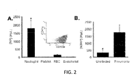
- FIG. 3 is a graphical representation of BALf microparticles isolated from pneumonic patients incubated with THP-1 cells for 24 H gated upon CD 14, CD11b surface expression determined by flow cytometry of (A) unlabeled BALf microparticles; and (B) BALf microparticles labeled with CFSE, wherein five independent samples were assayed, * p ⁇ 0.05 as determined by Student T test;
- FIG. 4 is a bar graph of fluorescently labeled NDMPs from critically ill patients incubated 18 H with THP-1 cells and compared with untreated and bystander cells analyzed for (A) HLA-Dr; (B) CD80; or (C) CD86 surface expression, wherein six independent BALf samples were assayed, *, p ⁇ 0.05 as compared to untreated and bystander and #, p ⁇ 0.05 as compared to ingested as determined by ANOVA pair-wise comparison;
- FIG. 5 is a bar graph of (+)CFSE THP-1 cells (Ingested MP) and ( ⁇ )CFSE THP-1 cells (Bystander) incubated with CFSE-labeled NDMPs for 18 H with respect to phagocytosis, wherein data are expressed as the mean (SEM) and * p ⁇ 0.05 as determined by Student's t test;
- FIG. 7 is a graphical representation of a flow cytometric method for analyzing microparticle isolated from BALf collected from uninfected and infected patients (gram positive and gram negative) wherein pathogens were identified by BALf 72 H after collection;
- FIG. 9 is a graphical representation of a flow cytometric method for analyzing NDMPs and platelet microparticles isolated from (A) peritoneal surgical waste fluid from uninfected patients; (B) peritoneal surgical waste fluid from peritonitis patients; (C) sham-operated mice; and (D) CLP-operated mice, wherein samples were analyzed using an LSR II flow cytometer and FACS Diva software;
- FIG. 11 is a graph of washes of peritoneal CLP-derived microparticles isolated and repeatedly washed using endotoxin-free saline with respect to endotoxin/MP, wherein each wash diluted the supernatent associated with MP pellet ( ⁇ 2,000-fold) and MPs were quantified by flow cytometry using True Count beads and endotoxin was determined by Limulus Amoebocyte Lysate assay;
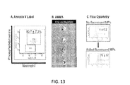
- FIG. 13 is (A) a graphic representation of microparticles isolated from septic mice analyzed for PS expression; (B) CFSE-labeled MPs incubated with macrophages for 3 H and phagocytosis determined by AMNIS; and (C) CFSE-labeled MPs incubated with macrophages for 3 H and phagocytosis determined by flow cytometry;
- FIG. 17 is a graphical representation of (A) latex beads of a predetermined size were used to set the forward and side scatter voltages to best distinguish particles ranging from 0.3 to 1.0 ⁇ M; (B) peritoneal cells isolated from septic mice 24 H after CLP incubated with PBS (100 ng/mL) for 24 H; and (C) peritoneal cells isolated from septic mice 24 H after CLP incubated with LPS (100 ng/mL) for 24 H, wherein samples were analyzed using LSR II flow cytometer and FACS Diva software;
- FIG. 20 is a bar graph of peritoneal cells collected after 24 H with TGA treatment with respect to fold difference in the number of NDMPs and cultured with the following inhibitors: (A) Myosin Light Chain Kinase Inhibitor ML7 (250 ⁇ M); (B) Caspase 3 Inhibitor Z-VAD-FMK (200 ⁇ M); (C) Caspase 8 Inhibitor Z-IETD-FMK (30 ⁇ M); and (D) Caspase 9 Inhibitor Z-LEHD-FMK (30 ⁇ M) enumerated and characterized by flow cytometry, wherein data are expressed as means ⁇ SEM, p ⁇ 0.05 as compared to control; and
- FIG. 21 is a bar graph of peritoneal cells collected after 24 H TGA treatment with respect to fold difference in the number of NDMPs and cultured with the following agonists and inhibitors: (A) cAMP analogue 8-Br-cAMP (100 ⁇ M) and PKA inhibitor H-89 (20 ⁇ M); (C) Rho Kinase Inhibitor Y27632 (30 ⁇ M); and (D) Prostaglandin E2 (1 ⁇ M).
- FIG. 22 is a graphical representation of data demonstrating that TNF- ⁇ treatment in vitro increases NDMP levels.
- Neutrophils were isolated from bone marrow as described in the examples: (A) Sizing for the microparticle populations was based upon latex beads sized between 0.5 and 3.0 ⁇ m.
- B Representative flow cytometric analysis of the forward and side scatter of a cell microparticle mixture obtained from culture. MP (microparticle gate)
- C Representative flow cytometric analysis using anti-Ly-6G (mouse neutrophil specific marker) and Annexin V (apoptosis marker) treated with vehicle.
- D with 25 ng/mL rmTNF- ⁇ .
- FIG. 23 is a bar graph showing that the production of NDMPs is additive upon TNFr1 and TNFr2 activation.
- Mouse neutrophils were isolated from bone marrow and cultured in vitro as described in the examples.
- (A) Neutrophils from WT mice were treated with either 25 ng/mL of rmTNF- ⁇ (activates both TNFr1 and TNFr2) or 25 ng/mL of rhTNF- ⁇ (TNFr1 specific). After 1 h, NDMPs were analyzed by flow cytometry. The sample size 12 mice.
- B Neutrophils from TNFr1 ⁇ / ⁇ mice treated with vehicle or 25 ng/mL of rhTNF- ⁇ .
- C Neutrophils from either WT mice or TNFr1-/0 mice were treated with 25 ng/mL of rmTNF- ⁇ . After 1 h, NDMPs were analyzed by flow cytometry. (All data are expressed in fold increase above vehicle and as means ⁇ SEM. The significance was determined using ANOVA analysis and Tukey post hoc test. *p ⁇ 0.05 compared vehicle.)
- FIG. 24 is a bar graph showing that TNFr1-specific generation of NDMPs is partially dependent upon caspase-8 activation.
- Mouse neutrophils were isolated from bone marrow from either WT or TNFr1 ⁇ / ⁇ mice and cultured in vitro as described in the examples. Neutrophils were treated as indicated with 25 ng/mL rmTNF- ⁇ or 25 ng/ml rhTNF- ⁇ , or ⁇ 30 ⁇ M of Caspase-8 inhibitor (Z-IETD-FMK). After 1 h, NDMPs were enumerated by flow cytometry. (The data are expressed in fold increase above vehicle and as means ⁇ SEM. The significance was determined using ANOVA analysis and Tukey post hoc test. *p ⁇ 0.05 compared rm/rhTNF- ⁇ alone. # p ⁇ 0.05 compared to vehicle.)
- FIG. 25 is a bar graph showing that treatment with an NF- ⁇ B inhibitor decreases NDMP production induced by either TNFr1 or TNFr2 activation.
- Mouse neutrophils were isolated from bone marrow of WT or TNFr1 ⁇ / ⁇ mice and cultured in vitro as described in the examples. Neutrophils were treated as indicated with 25 ng/mL tmTNF- ⁇ or 25 ng/mL rhTNF- ⁇ 20 ⁇ M of NF- ⁇ B inhibitor (BAY11-7085). After 1 h, NDMPs were enumerated by flow cytometry. (Data are expressed in fold increase above vehicle and as means ⁇ SEM. The significance was determined using ANOVA analysis and Tukey post hoc test. *p ⁇ 0.05 compared to rh/rmTNF- ⁇ treatment samples.)
- diagnosing refers to determining the presence and/or absence of a disease or condition based upon an evaluation of physical signs, symptoms, history, laboratory test results, and/or procedures. Specifically, in the context of pneumonia and/or sepsis, diagnosing refers to determining the presence or absence of a disease or condition based upon an evaluation of the level of NDMPs.
- a positive diagnosis refers to a determination of the presence of a disease or condition based upon an evaluation of physical signs, symptoms, history, laboratory test results, and/or procedures.
- a positive diagnosis refers to a determination of the presence of pneumonia and/or sepsis based upon an evaluation of the level of NDMPs.
- negative diagnosis refers to a determination of the absence of a disease or condition based upon an evaluation of physical signs, symptoms, history, laboratory test results, and/or procedures.
- a negative diagnosis refers to a determination of the absence of pneumonia and/or sepsis based upon an evaluation of the level of NDMPs.
- an elevated level of NDMPs in blood plasma may be from about 750 NDMPs/ ⁇ L to about 2000 NDMPs/ ⁇ L, or from about 1000 NDMPs/ ⁇ L to about 1500 NDMPs/ ⁇ L, or about 1200 NDMPs/ ⁇ L.
- the elevation of the level of NDMPs in the biological sample is statistically significant.
- the term “cutoff value” refers to a threshold value which distinguishes patients and/or subjects suffering from a disease or condition from patients and/or subjects who are not suffering from the disease or condition.
- an elevated level of NDMPs is greater than the cutoff value and a non-elevated level of NDMPs is less than or equal to the cutoff value.
- the cutoff value of NDMPs may be about 500 NDMPs/ ⁇ L.
- baseline level and/or “baseline concentration” refer to the level and/or concentration of NDMPs in a biological sample from a patient and/or subject prior to administration of a treatment and/or to the level of NDMPs in a biological sample from a patient and/or subject who is not suffering from pneumonia and/or sepsis.
- a method for diagnosing pneumonia in a patient may include: (a) obtaining a biological sample from the patient; (b) determining a level of NDMPs in the biological sample; (c) comparing the level of NDMPs determined in step (b) with a cutoff value of NDMPs; and (d) diagnosing pneumonia in the patient, wherein an elevated level of NDMPs as compared to the cutoff value correlates to a positive diagnosis of pneumonia in the patient.
- the method of diagnosing pneumonia in the patient includes obtaining a biological sample from the patient in step (a).
- the biological sample is bronchoalveolar lavage fluid (hereinafter, “BALf”).
- BALf bronchoalveolar lavage fluid
- Such fluid may be obtained via bronchoalveolar lavage which may be performed according to any bronchoalveolar lavage methods known in the field.
- bronchoalveolar lavage may be performed in accordance with Intensive Care Unit clinical practice guidelines.
- such bronchoalveolar lavage includes: (i) passing a bronchoscope through the mouth and/or nose into the lung(s); (ii) depositing (e.g., squirting) fluid suitable for such procedure into at least a portion of the lung(s); and (iii) recollecting the deposited fluid from the lung(s) for examination.
- the fluid suitable for such procedure is sterile.
- the fluid suitable for such procedure is a sterile saline solution (e.g., sterile phosphate-buffered saline).
- the patient is human.
- the method of diagnosing pneumonia in the patient includes determining a level of NDMPs in the biological sample in step (b).
- determining the level of NDMPs in the biological sample includes: (i) optionally isolating MPs via differential centrifugation; and (b) detecting and/or quantifying the NDMPs in the MPs via flow cytometry. Isolation of the MPs from the biological sample via differential centrifugation may be accomplished by performing differential centrifugation according to any methods known in the field. The differential centrifugation may be performed under conditions suitable for isolating MPs as may be known in the field.
- performing differential centrifugation includes: performing a first centrifugation of the biological sample at a first acceleration, performing a second centrifugation of a first supernatent formed from the first centrifugation at a second acceleration, and performing a third centrifugation of a second supernatent formed form the second centrifugation at a third acceleration.
- the first acceleration is from about 200 g to 1,000 g, or from about 300 g to about 900 g, or from about 400 g to about 800 g, or from about 500 g to about 700 g, or from about 450 g to about 600 g, or about 450 g.
- the second acceleration is from about 9,500 g to about 10,500 g, or from about 9,600 g to about 10,400 g, or from about 9,700 g to about 10,300 g, or from about 9,800 g to about 10,200 g, or from about 9,900 g to about 10,100 g, or from about 9,950 g to about 10,000 g, or about 9,900 g.
- the third acceleration is from about 16,500 g to about 17,500 g, or from about 16,600 g to about 17,400 g, or from about 16,700 g to about 17,300 g, or from about 16,800 g to about 17,200 g, or from about 16,900 g to about 17,100 g, or from about 16,950 g to about 17,000 g, or about 17,000 g.
- the first acceleration is about 450 g
- the second acceleration is about 9,900 g
- the third acceleration is about 17,000 g.
- the first centrifugation may be performed for a first time period
- the second centrifugation may be performed for a second time period
- the third centrifugation may be performed for a third time period.
- the first time period is from about 1 minutes to about 12 minutes, or from about 2 minutes to about 11 minutes, or from about 3 minutes to about 10 minutes, or from about 4 minutes to about 9 minutes, or from about 5 minutes to about 8 minutes, or from about 6 minutes to about 7 minutes, or about 10 minutes.
- the second time period is from about 1 minutes to about 12 minutes, or from about 2 minutes to about 11 minutes, or from about 3 minutes to about 10 minutes, or from about 4 minutes to about 9 minutes, or from about 5 minutes to about 8 minutes, or from about 6 minutes to about 7 minutes, or about 5 minutes.
- the third time period is from about 12 minutes to about 26 minutes, or from about 13 minutes to about 25 minutes, or from about 14 minutes to about 24 minutes, or from about 15 minutes to about 23 minutes, or from about 16 minutes to about 22 minutes, or from about 17 minutes to about 21, or from about 18 minutes to about 20 minutes, or about 19 minutes, or about 20 minutes.
- the first centrifugation forms a first pellet and a first supernatent
- the second centrifugation forms a second pellet and a second supernatent
- the third centrifugation forms a third pellet and a third supernatent.
- the first pellet may include cells from the biological sample
- the second pellet may include platelets from the biological sample
- the third pellet may include MPs from the biological sample.
- Detection and/or quantification of the NDMPs may be accomplished via flow cytometry which may be performed according to any methods known in the field.
- detection and/or quantification of the NDMPs is accomplished via fluorescence-activated cell sorting according to methods known in the field.
- the flow cytometry and/or fluorescence-activated cell sorting may be performed at conditions suitable for detecting and/or quantifying the NDMPs.
- performing fluorescence-activated cell sorting includes: contacting the biological sample with reagents for detection and/or quantification of NDMPs, exposing the biological sample to light of a single wavelength, and receiving scattered light and/or fluorescence emitted therefrom.
- the biological sample is contacted with reagents for detection and/or quantification of NDMPs.
- Suitable techniques for contacting the biological sample with reagents for detection and/or quantification of NDMPs include dispersing, dissolving, diffusing, or otherwise mixing the biological sample and the reagents for detection and/or quantification of NDMPS.
- the biological sample contacted with the reagents for detection and/or quantification of NDMPs are dispersed, dissolved, diffused, mixed, or otherwise provided in a fluid.
- the reagents for detection and/or quantification of NDMPs include at least one antibody having specific binding affinity to NDMPs in the biological sample. Such antibody may also have specific binding affinity to neutrophils.
- the antibody having specific binding affinity to NDMPs is anti-human CD66b.
- the antibody having specific binding affinity to NDMPs is fluorescently labeled.
- the antibody having specific binding affinity to NDMPs may be fluorescently labeled with a fluorophore.
- fluorophores which may be suitable for fluorescent labeling of the antibody include fluorescent proteins, non-protein organic fluorophores, dye families, and combinations thereof.
- suitable fluorophores may include green fluorescent protein, yellow fluorescent protein, red fluorescent proteins, fusion proteins includes such fluorescent proteins, and combinations thereof.
- suitable fluorophores may include xanthene derivatives (e.g., fluorescein, rhodamine, Oregon green, eosin, and Texas red), cyanine derivatives (e.g., cyanine, indocarbocyanine, oxacarbocyanine, thiacarbocyanine, and merocyanine), napthalene derivatives (e.g., dansyl and prodan derivatives), coumarin derivatives, oxadiazole derivatives (e.g., pryidyloxazole, nitrobenzoxadiazole, and benzoxadiazole), pyrene derivatives (e.g., cascade blue), oxazine derivatives (e.g., Nile red, Nile blue, cresyl violet, and oxazine 170), acridine derivatives (e.g., proflavin, acridine orange, and acridine derivatives (e.g., prof
- the antibody having specific binding affinity to NDMPs is fluorescently labeled with fluorescein isothiocyanate. In a further embodiment, the antibody having specific binding affinity to NDMPs is fluorescein isothiocyanate anti-human CD66b.
- the biological sample is exposed to light of a single wavelength. In a further embodiment, the biological sample is exposed to light of a single wavelength after contacting the biological sample with the reagents for detection and/or quantification of NDMPs.
- Suitable techniques for exposing the biological sample contacted with the reagents for detection and/or quantification of NDMPs include directing a beam of light of a single wavelength (e.g., laser(s) light(s)) onto a stream of fluid, wherein the fluid provides the biological sample and the reagents for detection and/or quantification of NDMPs. Such exposure scatters the light and/or excites fluorescent chemicals in the biological sample such that the fluorescent chemicals emit fluorescence.
- the fluorescent chemicals may be provided within, attached to, adhered to, associated with, and/or contacted with the biological sample.
- the fluorescent chemicals are contacted with the biological sample via a fluorescently labeled antibody having specific binding affinity to NDMPs.
- the fluorescent chemicals may be contacted with the viological sample via a fluorescein isothiocyanate labeled anti-human CD66b (e.g., fluorescein isothiocyanate anti-human CD66b).
- the light scattered and/or fluorescence emitted by the fluorescent chemicals as previously described are received.
- Suitable techniques for receiving the scattered light and/or fluorescence emitted by the fluorescent chemicals as previously described include aiming detector(s) at a point where fluid containing the biological sample and reagents for quantification and/or detection of NDMPs pass through the beam of light of a single wavelength.
- the method for diagnosing pneumonia includes comparing the level of NDMPs determined in step (b) with a cutoff value of NDMPs in step (c).
- the method for diagnosing pneumonia in the patient includes diagnosing pneumonia in the patient in step (d), wherein an elevated level of NDMPs as compared to the cutoff value correlates to a positive diagnosis of pneumonia in the patient.
- the elevated level of NDMPs is from about 750 NDMPs/ ⁇ L to about 2,500 NDMPs/ ⁇ L, or from about 1,000 NDMPs/ ⁇ L to about 2,000 NDMPs/ ⁇ L, or from about 1,250 NDMPs/ ⁇ L to about 1,750 NDMPs/ ⁇ L, or about 1,500 NDMPs/ ⁇ L.
- a non-elevated level of NDMPs as compared to the cutoff value correlates to a negative diagnosis of pneumonia.
- the method for diagnosing pneumonia also includes determining a pathogenic source of the pneumonia wherein the determined level of NDMPs is elevated.
- the pathogenic source of the pneumonia may be gram positive bacteria wherein the determined level of NDMPs is elevated relative to a threshold value.
- the pathogenic source of the pneumonia may be gram negative bacteria wherein the determined level of NDMPs is non-elevated as compared to the threshold value.
- the threshold value is greater than the cutoff value.
- a method for diagnosing sepsis includes (a) obtaining a biological sample from the patient; (b) determining a level of NDMPs in the biological sample; (c) comparing the level of NDMPs determined in step (b) with a cutoff value of NDMPs; and (d) diagnosing sepsis in the patient, wherein an elevated level of NDMPs as compared to the cutoff value correlates to a positive diagnosis of sepsis in the patient.
- the method of diagnosing sepsis in the patient includes obtaining a biological sample from the patient in step (a).
- the biological sample is obtained from infectious foci in the patient.
- the biological sample is obtained from the abdomen.
- the biological sample may be abdominal irrigation fluid and/or peritoneal waste fluid. Such fluid may be obtained via abdominal irrigation which may be performed according to any abdominal irrigation methods known in the field.
- the patient is human.
- the method of diagnosing sepsis in the patient includes determining a level of NDMPs in the biological sample in step (b).
- determining the level of NDMPs in the biological sample includes: (i) optionally isolating MPs via differential centrifugation; and (b) detecting and/or quantifying the NDMPs in the MPs via flow cytometry. Isolation of the MPs from the biological sample via differential centrifugation is as previously described with regard to diagnosing pneumonia. Detection and/or quantification of the NDMPs may be accomplished via flow cytometry which may be performed according to any methods known in the field. Such detection and/or quantification is also as previously described with regard to diagnosing pneumonia.
- the method for diagnosing sepsis includes comparing the level of NDMPs determined in step (b) with a cutoff value of NDMPs in step (c).
- the method for diagnosing sepsis in the patient includes diagnosing sepsis in the patient in step (d), wherein an elevated level of NDMPs as compared to the cutoff value correlates to a positive diagnosis of sepsis in the patient.
- a non-elevated level of NDMPs as compared to the cutoff value correlates to a negative diagnosis of sepsis.
- Embodiments of methods of diagnosing pneumonia and/or sepsis have now been described in detail. Further embodiments directed to assessing pneumonia and/or sepsis will now be described.
- in vitro methods for assessing pneumonia and/or sepsis in a patient are disclosed.
- an in vitro method for assessing pneumonia in a patient is disclosed. The method includes (a) contacting at least a portion of a biological sample from the patient with reagents for detection and/or quantification of NDMPs. The method also includes (b) determining a level of NDMPs based on the contacting in step (a). Additionally, the method includes (c) assessing as indicating pneumonia in the patient if the determined level of NDMPs is elevated relative to a cutoff value of the NDMPs.
- the method for assessing pneumonia includes contacting at least a portion of the biological sample from the patient with reagents for detection and/or quantification of NDMPs in step (a).
- the biological sample, patient, and reagents for detection and/or quantification are as previously described.
- the biological sample may be contacted with the reagents for detection and/or quantification of NDMPs via dispersing, dissolving, diffusing, or otherwise mixing the biological sample and the reagents for detection and/or quantification of NDMPS.
- MPs are isolated from the biological sample prior to the contacting in step (a) and/or prior to determining the level of NDMPs in step (b).
- the MPs may be isolated from the biological sample via techniques known in the field. For example, in one embodiment, the MPs are isolated from the biological sample via differential centrifugation as previously described. In a further embodiment, the at least a portion of the biological sample consists essentially of MPs isolated via differential centrifugation.
- the method for assessing pneumonia includes (b) determining a level of NDMPs based on the contacting in step (a). Determining the level of NDMPs based on the contacting in step (a) may be performed as previously described (e.g., via flow cytometry and/or fluorescence-activated cell sorting).
- the method for assessing pneumonia includes (c) assessing as indicating pneumonia in the patient if the determined level of NDMPs is elevated relative to a cutoff value of the NDMPs.
- an elevated level of NDMPs and the cutoff value of the NDMPs are as previously described.
- the method for assessing pneumonia also includes determining a pathogenic source of the pneumonia wherein the determined level of NDMPs is elevated.
- the pathogenic source of the pneumonia may be gram positive bacteria wherein the determined level of NDMPs is elevated relative to a threshold value.
- the pathogenic source of the pneumonia may be gram negative bacteria wherein the determined level of NDMPs is non-elevated as compared to the threshold value. The threshold value is as previously described.
- an in vitro method for assessing sepsis in a patient includes (a) contacting at least a portion of a biological sample from the patient with reagents for detection and/or quantification of NDMPs. The method also includes (b) determining a level of NDMPs based on the contacting in step (a). Additionally, the method includes (c) assessing as indicating sepsis in the patient if the determined level of NDMPs is elevated relative to a cutoff value of the NDMPs.
- the method for assessing sepsis includes contacting at least a portion of the biological sample from the patient with reagents for detection and/or quantification of NDMPs in step (a).
- the biological sample, patient, and reagents for detection and/or quantification are as previously described.
- the biological sample may be contacted with the reagents for detection and/or quantification of NDMPs via dispersing, dissolving, diffusing, or otherwise mixing the biological sample and the reagents for detection and/or quantification of NDMPS.
- MPs are isolated from the biological sample prior to the contacting in step (a) and/or prior to determining the level of NDMPs in step (b).
- the MPs may be isolated from the biological sample as previously described.
- the at least a portion of the biological sample consists essentially of MPs isolated via differential centrifugation.
- the method for assessing sepsis includes (b) determining a level of NDMPs based on the contacting in step (a). Determining the level of NDMPs based on the contacting in step (a) may be performed as previously described (e.g., via flow cytometry and/or fluorescence-activated cell sorting).
- the method for assessing sepsis includes (c) assessing as indicating pneumonia in the patient if the determined level of NDMPs is elevated relative to a cutoff value of the NDMPs.
- an elevated level of NDMPs and the cutoff value of the NDMPs are as previously described.
- Embodiments of methods of assessing pneumonia and/or sepsis have now been described in detail. Further embodiments directed to managing treatment of pneumonia and/or sepsis will now be described.
- a method for managing treatment of a patient (or subject) suspected of having pneumonia includes (a) assessing pneumonia in vitro in the patient by calculating a concentration of NDMPs, wherein an elevated concentration of NDMPs relative to a cutoff value of NDMPs is indicative of pneumonia. The method also includes (b) treating the patient for pneumonia where pneumonia is indicated in step (a), wherein the calculated concentration of NDMPs is elevated. Additionally, the method includes (c) placing the patient under active surveillance where pneumonia is not indicated in step (a), wherein the calculated concentration of NDMPs is not elevated.
- the method for managing treatment of pneumonia includes (a) assessing pneumonia in vitro in the patient by calculating a concentration of NDMPs wherein an elevated concentration of NDMPs relative to a cutoff value of NDMPs is indicative of pneumonia.
- concentration of NDMPs may be calculated as previously described with regard to determining a level of NDMPs. Additionally, an elevated concentration of NDMPs and a cutoff value of NDMPs are as previously described.
- the method for managing treatment of pneumonia also includes (b) treating the patient for pneumonia where pneumonia is indicated in step (a), wherein the calculated concentration of NDMPs is elevated.
- treatment of the patient may include administration of antibiotics, antivirals, fluids, and combinations thereof. Any suitable antibiotics and/or antivirals may be administered as treatment for pneumonia as are known in the field.
- the antibiotics are selected from the group consisting of: penicillins, cephalosporins, polymixins, rifamycins, lipiarmycins, quinolones, sulfonamides, amigoglycosides, macrolides, tetracyclines, cyclic lipopeptide s, glycylcyclines, oxazolidinones, lipiarmycins, and combinations thereof.
- antibiotics may include amoxicillin, cefuroxime, ethryomycin, flucoxacillin, doxycycline, ciprofloxacin, rifampicin, clarithromycin, azithromycin, fluoroquinolones, cephalosporins, carbapenems, aminoglycosides, vancomycin, and combinations thereof.
- the antivirals are neuramidase inhibitors.
- neuramidase inhibitors include oseltamivir, zanamivir, laninamivir, peramivir, and combinations thereof.
- the method for managing treatment of pneumonia also includes determining a pathogenic source of the pneumonia wherein the determined level of NDMPs is elevated.
- the pathogenic source of the pneumonia may be gram positive bacteria wherein the determined level of NDMPs is elevated relative to a threshold value.
- the pathogenic source of the pneumonia may be gram negative bacteria wherein the determined level of NDMPs is non-elevated as compared to the threshold value.
- the threshold value is as previously described. Accordingly, the treatment of the patient may be adapted accordingly with regard to the pathogenic source. For example, in one embodiment, narrow spectrum antibiotics may be administered wherein the pathogenic source is identified as either gram positive or gram negative bacteria.
- a method of managing treatment in a patient (or subject) suffering from pneumonia includes (a) establishing a baseline concentration of NDMPs in a biological sample (e.g., bronchoalveolar lavage fluid sample) from the patient, (b) initiating treatment, and (c) serially monitoring a concentration of NDMPs in the patient during the treatment, wherein the treatment is managed to decrease the concentration of NDMPs in the patient.
- a biological sample e.g., bronchoalveolar lavage fluid sample
- the method of managing treatment in a patient suffering from pneumonia includes (a) establishing a baseline concentration of NDMPs in a bronchoalveolar lavage fluid sample the patient.
- the baseline concentration of NDMPs is established as previously described with regard to determining a level of NDMPs and/or calculating a concentration of NDMPs.
- the patient and bronchoalveolar lavage fluid sample are as previously described.
- the baseline concentration of NDMPs in the bronchoalveolar lavage fluid sample is elevated relative to the cutoff value. The cutoff value is as previously described.
- the method of managing treatment in a patient suffering from pneumonia also includes (b) initiating treatment.
- the treatment is as previously described.
- the method of managing treatment in a patient suffering from pneumonia also includes (c) serially monitoring a concentration of NDMPs in the patient during treatment.
- the treatment is managed to decrease the concentration of NDMPs in the patient.
- the concentration of NDMPs in the patient is monitored as previously described with regard to determining a level of NDMPs and/or determining a concentration of NDMPs.
- the method for managing treatment in a patient suffering from pneumonia also includes determining a pathogenic source of the pneumonia wherein the determined level of NDMPs is elevated.
- the pathogenic source of the pneumonia may be gram positive bacteria wherein the determined level of NDMPs is elevated relative to a threshold value.
- the pathogenic source of the pneumonia may be gram negative bacteria wherein the determined level of NDMPs is non-elevated relative to the threshold value.
- the threshold value is as previously described. Accordingly, the treatment of the patient may be adapted accordingly with regard to the pathogenic source. For example, in one embodiment, narrow spectrum antibiotics may be administered wherein the pathogenic source is identified as either gram positive or gram negative bacteria.
- a method for managing treatment of a patient (or subject) suspected of having sepsis includes (a) assessing sepsis in vitro in the patient by calculating a concentration of NDMPs, wherein an elevated concentration of NDMPs relative to a cutoff value of NDMPs is indicative of sepsis. The method also includes (b) treating the patient for sepsis where sepsis is indicated in step (a), wherein the calculated concentration of NDMPs is elevated. Additionally, the method includes (c) placing the patient under active surveillance where sepsis is not indicated in step (a), wherein the calculated concentration of NDMPs is not elevated.
- the method for managing treatment of sepsis includes (a) assessing sepsis in vitro in the patient by calculating a concentration of NDMPs wherein an elevated concentration of NDMPs relative to a cutoff value of NDMPs is indicative of sepsis.
- concentration of NDMPs may be calculated as previously described with regard to determining a level of NDMPs. Additionally, an elevated concentration of NDMPs and a cutoff value of NDMPs are as previously described.
- the method for managing treatment of sepsis also includes (b) treating the patient for sepsis where sepsis is indicated in step (a), wherein the calculated concentration of NDMPs is elevated.
- treatment of the patient may include administration of antibiotics, administration of intravenous fluids, surgical drainage, and providing support for organ dysfunction.
- antibiotics any suitable antibiotics may be administered as treatment for sepsis as are known in the field.
- broad spectrum antibiotics maybe administered.
- the antibiotics are selected from the group consisting of: penicillins, cephalosporins, polymixins, rifamycins, lipiarmycins, quinolones, sulfonamides, amigoglycosides, macrolides, tetracyclines, cyclic lipopeptides, glycylcyclines, oxazolidinones, lipiarmycins, and combinations thereof.
- a method of managing treatment in a patient (or subject) suffering from sepsis includes (a) establishing a baseline concentration of NDMPs in a biological sample (e.g., abdominal wash fluid) from the patient, (b) initiating treatment, and (c) serially monitoring a concentration of NDMPs in the patient during the treatment, wherein the treatment is managed to decrease the concentration of NDMPs in the patient.
- a biological sample e.g., abdominal wash fluid
- the method of managing treatment in a patient suffering from sepsis includes (a) establishing a baseline concentration of NDMPs in an abdominal wash fluid sample the patient.
- the baseline concentration of NDMPs is established as previously described with regard to determining a level of NDMPs and/or calculating a concentration of NDMPs.
- the patient and abdominal wash fluid sample are as previously described.
- the baseline concentration of NDMPs in the abdominal wash fluid is elevated relative to the cutoff value. The cutoff value is as previously described.
- the method of managing treatment in a patient suffering from sepsis also includes (b) initiating treatment.
- the treatment is as previously described.
- the method of managing treatment in a patient suffering from sepsis also includes (c) serially monitoring a concentration of NDMPs in the patient during treatment.
- the treatment is managed to decrease the concentration of NDMPs in the patient.
- the concentration of NDMPs in the patient is monitored as previously described with regard to determining a level of NDMPs and/or determining a concentration of NDMPs.
- kits for assessing pneumonia Further embodiments directed to kits for assessing pneumonia will now be described.
- kits for assessing pneumonia and/or sepsis are disclosed.
- the kits are used to assess pneumonia and/or sepsis in patients.
- the kits may include reagents for detection and/or quantification of NDMPs.
- the kits include reagents for detection and/or quantification of NDMPs in BALf.
- the kits include reagents for detection and/or quantification of NDMPs in abdominal wash fluid.
- the kits for assessing pneumonia and/or sepsis are differential centrifugation and/or flow cytometry kits which may be used to detect and/or quantify NDMPs.
- the reagents for detection and/or quantification of NDMPs are as previously described.
- kits may also include instructions to provide guidance on utilizing the kits for assessing pneumonia and/or sepsis. More particularly, the kits may include instructions to provide guidance on detecting and/or quantifying NDMPs. Such instructions may generally include information related to performing differential centrifugation, flow cytometry (e.g., fluorescence-activated cell sorting) and/or calculating results. Such general information is as previously described.
- Tumor necrosis factor- ⁇ predominates in the initial phases of sepsis and is one of the first cytokines released into the circulation during the inflammatory response to infectious stimuli. It has diverse effects in vivo as a mediator of the immune response, in part, through activation of neutrophils.
- NDMPs are generated by administration of TNF- ⁇ alone, and have elucidated the role of TNF Receptor 1 (TNFr1) and TNF Receptor 2 (TNFr2), as well as MP generation as it relates to pathways involved in apoptosis and cellular fate decisions, namely the caspase and NF- ⁇ B pathways, thereby permitting development of methods for modulating immune response and for treating diseases relating to a disordered immune response, such as sepsis, pneumonia and systemic inflammatory response syndrome.
- TNFr1 TNF Receptor 1
- TNFr2 TNF Receptor 2
- Example 19 Treatment of neutrophils with TNF- ⁇ increases levels within 30 min. Further, activation of either TNFr1 or TNFr2 is sufficient for NDMp generation, and activation of both is additive ( FIG. 23 ). Upon the selective activation of TNFr1, NDMP numbers were sharply decreased but not entirely eliminated with addition of Caspase 8 inhibitor. In contrast, Caspase 8 inhibition did not alter NDMP numbers when only TNFr2 was activated ( FIG. 24 ). Upon activation of either TNFr1 or TNFr2, the inhibition of NF- ⁇ B activity significantly ameliorates NDMP generation ( FIG. 25 ).
- a method of treating a disordered immune response in a patient comprises modulating a concentration of neutrophil-derived microparticles (NDMP) in a sample from the patient.
- NDMP neutrophil-derived microparticles
- Modulating in accordance with the instant disclosure, means to adjust or regulate, indirectly or directly, for the purpose of achieving a therapeutic goal.
- therapeutic goals may include amelioration of severity of symptoms, restoration of normal immune system homeostasis, decrease in NDMP concentrations, elimination of infection, and the like.
- Samples comprise physiologically relevant samples, for example brachial lavage fluid in the case of a patient suffering from pneumonia and abdominal fluid in the case of a patient suffering from sepsis.
- modulating is effectuated by modulating tumor necrosis factor- ⁇ (TNF- ⁇ ) activated induction of NDMP generation in the patient.
- TNF- ⁇ tumor necrosis factor- ⁇
- one or both of TNF- ⁇ and a TNF- ⁇ receptor agonist may be administered.
- a TNF- ⁇ inhibitor may be administered.
- a TNF- ⁇ inhibitor may, for example, comprise a TNF receptor 1 (TNFr1) antagonist, a TNF receptor 2 (TNFr2) antagonist, a TNFr1 and TNFr2 antagonist, or combinations thereof.
- Caspase 8 is an enzyme known to be involved in cell apoptosis and activation.
- the present investigators determined that TNFr1 activation generates microparticles partially through a caspase-8 dependent pathway and that microparticle generation through activation of TNFr2 is independent of the caspase-8 pathway.
- inhibition of caspase-8 effectuates decreasing NDMP generation/levels and very specific embodiments are directed to administering a caspase-8 inhibitor.
- the caspase-8 inhibitor comprises Z-IETD-FMK.
- NF- ⁇ B inhibitor BAY 11-7085.
- MPs were isolated from BALf by differential centrifugation consisting of three spins.
- the first centrifugation (450 ⁇ g, 10 minutes, 4° C.) was conducted to pellet the cells.
- the cell pellet from this step may then be used to determine cell activation or apoptosis.
- the second centrifugation (9,900 ⁇ g, 5 minutes, 4° C.) was conducted to pellet platelets.
- the platelet poor supernatant was then subjected to a third spin (17,000 ⁇ g, 20 minutes, 4° C.).
- the third spin was conducted to pellet the MPs and to leave soluble proteins and membrane fragments in the supernatant.
- the resulting MPs were then analyzed by flow cytometry using LSR II Flow Cytometer and FACS Diva Software (BD Biosciences, San Jose, Calif.).
- LSR II Flow Cytometer and FACS Diva Software BD Biosciences, San Jose, Calif.
- latex beads of a pre-determined size were used to set forward and side scatter voltages to set the MP gate. Such latex beads were used to best distinguish particles ranging from 0.3 to 3.0 ⁇ M.
- FIG. 1(B) while analyzing MPs by flow cytometry, the addition of a known concentration of two populations of counting beads were included to allow enumeration of NDMP numbers.
- NDMPs express the apoptosis marker, phosphatidyl serine (hereinafter “PS”) on the extracellular leaflet of the plasma membrane. Accordingly, the active phagocytosis of NDMPs was investigated. Specifically, NDMPs were isolated from BALf of pneumonic patients. The NDMPs were then labeled with 5-(and 6)-carboxyfluorescein diacetate (hereafter “CFSE”, Invitrogen, Carlsbad, Calif.). An equal volume of 10 mM CFSE was diluted to 1:5000 in PBS just before use. MPs and CFSE were mixed in a 1:1 volume ratio and incubated for 8 minutes at room temperature on a rocker.
- CFSE 5-(and 6)-carboxyfluorescein diacetate
- fetal bovine serum hereinafter “FBS”, Invitrogen
- FBS fetal bovine serum
- sterile K5 media was added to the mixture and centrifuged at 16,000 g for 30 minutes to pellet MPs.
- CFSE-labeled NDMPs were incubated with THP-1 cells (ATCC, Manassas, Va.), a human monocytic cell line, at 37° C. in a CO 2 incubator overnight.
- THP-1 cells were collected and analyzed for macrophage activation as determined by HLA-Dr, CD80, and CD86 mean fluorescence intensity (hereinafter “MFI”).
- MFI mean fluorescence intensity
- NDMPs were labeled with CFSE as previously described and incubated with THP-1 cells.
- the use of CFSE-labeled NDMPs allowed discrimination between THP-1 cells that had ingested NDMPs versus THP-1 cells that did not ingest NDMPs (i.e., “bystander cells”).
- After 18 H cells were collected and mixed with opsonized Fluoresbrite Polychromatic Red Plain Microspheres 1.0 ⁇ M (Polysciences, Warrington, Pa.). The suspension was incubated at 37° C. for 30 minutes. After the incubation period, the phagocytosis was stopped by washing twice.
- THP-1 cells were incubated with CFSE-labeled NDMPs. After 18 H, the cultured cells were incubated with red fluorescent microspheres as previously described. Phagocytic ability was evaluated with flow cytometry.
- mice Peritoneal lavage cells from mice made septic by cecal ligation and puncture were isolated. The cells were then incubated with or without lipopolysaccharide (hereinafter “LPS”, 100 ng/mL) for 18 H. MPs from these cultures were then isolated, quantified by bicinchoninic acid assay (hereinafter, “BCA”), and analyzed by fluorescence-activated cell sorting (hereinafter “FACS”).
- LPS lipopolysaccharide
- BCA bicinchoninic acid assay
- FACS fluorescence-activated cell sorting
- BALf was obtained from 33 surgical patients with critical illness or injury undergoing diagnostic fiberoptic bronchoscopy with bronchoalveolar lavage (hereinafter “BAL”) based on an institutional protocol for the diagnosis of ventilator-associated pneumonia (hereinafter “VAP”).
- VAP ventilator-associated pneumonia
- Indications for BAL for suspected VAP were defined by ICU guidelines as the appearance of a new or changing infiltrate on chest radiograph or macroscopically purulent sputum plus at least two of the following: temperature higher than 38° C. or lower than 36° C., white blood cell count higher than 12,000/mm 3 or lower than 4000/mm 3 , 10% bands, heart rate higher than 90 beats per minutes, respiratory rate higher than 20 breaths per minute, or PaCO 2 lower than 32 mm Hg.
- Two BAL samples from lung donors were obtained and used as an uninfected control in the subset of patients with suspected pneumonia.
- Peritoneal waste fluid was collected in sterile 50-mL conical tubes from patients that underwent exploratory laparotomy or elective abdominal surgery in which abdominal irrigation was performed. Fluid was immediately placed on ice for MP analysis within 2 hours of collection time. All patients with a clinical suspicion of VAP underwent diagnostic fiberoptic bronchoscopy with BAL. All BAL procedures were performed in a uniform manner by the treating surgical intensivist as part of ICU clinical practice guidelines. The bronchoscope was advanced into the lung segment wherein radiographic changes were seen. After the bronchoscope was wedged into the appropriate lung segment, 20 mL of sterile saline was instilled into the lung as a wash.
- the waste BAL effluent was collected in a sterile specimen trap and immediately placed on ice for MP analysis within 2 hours of collection time. Abdominal fluid and BAL fluid (i.e., BALf) samples were collected as unidentifiable surgical waste that could not be linked back to the investigator.
- BALf BAL fluid
- the fluid was kept on ice until the time of processing. Fluid samples were initially filtered using a 150- ⁇ m, sterilized stainless steel mesh into a sterile 50-mL conical tube. Cells in the fluid were pelleted after a 10-minute 450 g centrifugation. The supernatant was then centrifuged for 10 minutes at 9,900 g to remove platelets. The platelet-free supernatant was centrifuged for 20 minutes at 16,000 g to pellet MPs.
- the MP pellet was resuspended in 500 ⁇ L of sterile phosphate-buffered saline (i.e., PBS) and centrifuged for 20 minutes at 16,000 g. The MP pellet was then resuspended in 100 ⁇ L of sterile PBS for analysis.
- PBS sterile phosphate-buffered saline
- CD66b (clone G10F5; BD Biosciences), CD105 (clone 43A3; BioLegend), CD41a (clone HIPS; BD Biosciences), CD235 (clone HIR2; BioLegend), CD14 (clone M5E2; BioLegend), CD11b (clone M1/70; BioLegend), HLA-Dr (clone L243; BD Biosciences), HLA-Dr (clone L243; BD Biosciences), CD80 (clone L307.4; BD Biosciences), and CD86 (clone 2331; BD Biosciences).
- MPs were isolated from peritoneal washes by differential centrifugation consisting of three steps. The first centrifugation (450 ⁇ g, 10 minutes, 4° C.) was conducted to pellet the cells. The second spin pelleted platelets (9,900 ⁇ g, 5 minutes, 4° C.). The platelet-poor supernatant was then subjected to a third spin (17,000 ⁇ g, 20 minutes, 4° C.). This pelleted the MPs and left soluble proteins and membrane fragments in the supernatant. These resulting MPs were then analyzed by flow cytometry. To ensure proper gating for the correct size of MPs, latex beads of known sizes were used to set the MP gate.
- mice For mice, a moderately severe CLP was induced and peritoneal lavages were conducted.
- de-identified excess abdominal irrigation fluid was provided from non-infected or septic patients.
- Both mouse and human samples underwent differential centrifugation as described to selectively isolate MPs. MPs were then labeled with antibodies against CD3 (T cell), CD66b or Ly-6G (human or mouse neutrophil markers, respectively), CD 105 (endothelial cell marker), CD41 (platelet marker), and CD 14 or F4/80 (human or mouse macrophage markers, respectively).
- CD3 T cell
- CD66b or Ly-6G human or mouse neutrophil markers, respectively
- CD 105 endothelial cell marker
- CD41 platelet marker
- CD 14 or F4/80 human or mouse macrophage markers, respectively.
- mice were treated with intraperitoneal injections of 3% thioglycolate (hereinafter, “TGA”), LPS, or petidylglycan (hereinafter “PepG”). Peritoneal neutrophil recruitment, NDMPs, and endotoxin associated with NDMPs were quantified.
- TGA 3% thioglycolate
- LPS LPS
- PepG petidylglycan
- treatment with TGA, LPS, and PepG resulted in neutrophil recruitment and NDMP generation.
- treatment with 3% TGA resulted in the greatest number of low endotoxin NDMPs.
- Peritoneal MPs were isolated from TGA-treated mice. Endotoxin-free saline vehicle or MPs (65,000) in saline were intraperitoneally injected as a bolus into cohorts of mice undergoing CLP. Survival was monitored for 10 days.
- mice injected with a bolus of 65,000 NDMPs exhibited increased mortality as compared to vehicle-injected mice.
- median survival times were significantly different between mice injected with CLP-derived MPs (25.5 H) and those injected with TGA-derived MPs (42 H). Accordingly, it is believed that endotoxin-free NDMPs have a negative impact upon survival during sepsis beyond adding to endotoxin levels.
- MPs were isolated from septic mice and analyzed for PS expression. Additionally, CFSE-labeled MPs were incubated with macrophages for 3H and phagocytosis was determined by AMNIS and by flow cytometry. More particularly, multispectral imaging flow cytometric analysis (i.e., Amnis ImageStream X) was used to determine the localization of fluorescently (i.e., CFSE) labeled NDMPs within phagocytes after 3 H incubation. Finally, with regard to flow cytometry, CD68 and CD11b positive macrophages were gated upon and fluorescence was observed.
- multispectral imaging flow cytometric analysis i.e., Amnis ImageStream X
- FIG. 13(A) it was observed that approximately 80% of NDMPs express extracellular PS. Additionally, as shown in FIG. 13(B) , localization of fluorescently labeled NDMPs within phagocytes was observed. Referencing FIG. 13(C) , increased fluorescence was observed in CD68 and CD11b positive macrophages, indicating either adherence or ingestion of fluorescently-tagged NDMPs. Such data indicate that NDMPs can be phagocytosed by macrophages.
- NDMPs Decrease Host Ability to Clear Bacteria During Sepsis
- mice As shown in FIG. 14(A) , a 20% decrease in phagocytic ability in mice was quantified in mice that possessed increased numbers of NDMPs. Additionally, as shown in FIG. 14(B) , a 2-fold increase in blood bacterial load was observed in mice that possessed increased NDMPs. Accordingly, it is believed that NDMPs compromise the host ability to localize an infection.
- peritoneal cells were isolated from septic mice 24 H after CLP. The isolated peritoneal cells were incubated with PBS or 100 ng/mL LPS for 24 H. Samples were analyzed using LSR II flow cytometer and FACS Diva software (BD Biosciences). As shown in FIG. 17(A) , latex beads of a predetermined size were used to set the forward and side scatter voltages to best distinguish particles ranging from 0.3 to 1.0 ⁇ M.
- cAMP was targeted as a potential signaling mechanism involved in NDMP formation.
- Peritoneal cells were isolated 24 H after CLP surgeries. The isolated peritoneal cells were incubated with either saline vehicle, 10 ⁇ M 8-Br cAMP (i.e., a membrane-permeable non-hydrolyzable cAMP analog), or 20 ⁇ M H89 (i.e., a PKA inhibitor). After 24 H, NDMPs were enumerated and characterized by flow cytometry.
- mice Male C57BL/6 mice between 6 and 8 weeks of age (about 20 to 28 g) were injected in the peritoneum with 1 mL 3% thioglycolate (Sigma, St. Louis, Mo.) or with vehicle. Peritoneal lavage was performed 24 H after injection. Peritoneal cells were pelleted after a 10-minute 450 ⁇ g centrifugation. The supernatant was then centrifuged for 10 minutes at 9,900 ⁇ g to remove platelets. The platelet-free supernatant was centrifuged for 20 minutes at 16,000 ⁇ g in order to pellet MPs. The MP pellet was resuspended in 500 ⁇ L sterile PBS and centrifuged for 20 minutes at 16,000 ⁇ g. The MP pellet was then resuspended in 100 ⁇ L sterile PBS for analysis.
- thioglycolate Sigma, St. Louis, Mo.
- mice and TGA-treated mice were stained with neutrophil specific Ly6G and annexinV antibodies. Total NDMPs from TGA-treated mice and vehicle mice were then enumerated.
- Peritoneal cells were cultured with one of the following agonists or inhibitors overnight in a CO 2 incubator: 250 ⁇ M ML7, a MLCK inhibitor, (Tocris, Bristol, UK), 200 ⁇ M Z-VAD-FMK, a caspase 3 inhibitor, (Tocris), 30 ⁇ M Z-IETD-FMK, a caspase 8 inhibitor, (Tocris), or 30 ⁇ M Z-LEHD-FMK, or a caspase 9 inhibitor, (Tocris).
- Cells and MPs were collected 24 H following incubation and analyzed using flow cytometry as previously described.
- FIG. 20(A) peritoneal cells cultured with ML7 demonstrated a 75% decrease in NDMP generation when compared to those cultured without ML7.
- FIG. 20(B) inhibition of caspase 3 with Z-VAD-FMK resulted in a 37% decrease in NDMP generation when compared to cells cultured without Z-VAD-FMK.
- FIG. 20(C) inhibition of caspase 8 with Z-IETD-FMK resulted in a 37% decrease NDMP generation when compared to cells cultured without Z-IETD-FMK.
- Peritoneal cells were isolated from peritoneal lavage 24 H after 1 mL 3% TGA injection. Peritoneal cells were incubated with one of the following agonists or inhibitors overnight in a CO 2 incubator: 100 ⁇ M 8-Bromo-cAMP, a cAMP analogue, (Tocris), 20 ⁇ M H89 dihydrochloride, a PKA inhibitor (Tocris), 100 ⁇ M 6-Bnz-cAMP, a PKA agonist, (Tocris), 100 ⁇ M 8-CPT-2Me-cAMP, EPAC, (Tocris), 200 ⁇ M Z-VAD-FMK (Tocris), 30 ⁇ M Y27632 dihydrochloride, a Rho Kinase Inhibitor, (Tocris), or 250 ⁇ M ML 7 hydrochloride (Tocris). Cells and MPs were collected 24 H following incubation and analyzed using
- peritoneal cells cultured with 8-Br exhibited decreased NDMP numbers while peritoneal cells cultured with H89 did not.
- peritoneal cells cultured with 6-Bnz-cAMP inhibited NDMP formation compared to cells cultured without 6-Bnz-cAMP.
- peritoneal cells cultured with EPAC did not affect NDMP numbers compared to cells cultured without EPAC.
- peritoneal cells cultured with Y27632 exhibited a decrease in the total number of NDMPs when compared to cells cultured without Y27632.
- peritoneal cells cultured with PGE2, known to active cAMP exhibited a decrease in the total number of NDMPs when compared to cells cultured without PGE2.
- mice between 6 and 8 weeks of age were obtained from Jackson Labs (Bar Harbor, Me.).
- Mouse and human TNF- ⁇ Recombinant Protein (rmTNF- ⁇ and rhTNF- ⁇ , respectively) were purchased from Preprotech (Rocky Hill, N.J.).
- NF- ⁇ B inhibitor (BAY 11-7085) and Caspase 8 inhibitor (Z-IETD-FMK) were purchased from R&D systems (Minneapolis, Minn.).
- the pharmacological agents were solubilized in DMSO (American Bioanalytical, Natick, Mass.). The amount of DMSO was consistent in all samples groups. Neutrophil isolation.
- HBSS Hanks' Balanced Salt Solution without calcium & Magnesium
- the purity and viability of the neutrophils (90% purity >99% viability, respectively) was determined by flow cytometry.
- Neutrophil purity was assessed by flow cytometry after cell staining with fluorescently conjugated antibodies to Ly-6G, CD19, CD3 and F4/80 with >99% of the cells negative for F4/80 and CD3.
- the main cell contaminant in this preparation was due to B cells. Further purification was not conducted by centriguation due to risk of non-specific neutrophil activation or by magnetic beads due to microparticle-sized contaminants introduced by the beads themselves.
- Isolated neutrophils from bone marrow preparation were plated and cultured for the indicated time at 37° C. with stated treatments.
- the cell/micropartocle mixture was labeled for Ly-6G and Annexin V without manipulation as previously described.
- Flow cytometry data acquisition and analysis were performed on a Beckman Coulter Epic flow cytometer using Kaluza software (Indianapolis, Ind., USA).
- Neutrophil derived microparticle numbers were assessed using the neutrophil specific marker, Ly-6G (Clone: 1A8, BD Pharmingen, San Diego, Calif., United States); and Annexin V (BD Pharmingen). Particles within the 0.3-1.0 ⁇ m range that were labeled with both Ly-6G and Annexin V were identified as NDMPs.
- FIG. 22A Using flow cytometry, forward- and side-channels were set to analyze particles using 0.5-, 0.9-, and 3.0- ⁇ m latex beads for calibration as previously described ( FIG. 22A ). The forward and side scatter gate for microparticles was then set based upon size ( FIG. 22B ).
- neutrophil and NDMP mixture were analyzed with the neutrophil-specific marker, Ly6G, and the apoptosis marker, annexin V. Vesicles within the 0.3-1.0 ⁇ m range that expressed both Ly6G and phosphatidylserine were identified as NDMPs.
- FIG. 22C is representative of NDMP generation after vehicle treatment and FIG.
- 22D is representative NDMP generation after rmTNF- ⁇ treatment.
- 25 ng/mL of rmTNF- ⁇ increased the number of NDMPs both at 30 min and at 1 h.
- Viability of neutrophils was also assessed at these time points and in all subsequent experiments and no significant difference in cell viability was found in regard to treatment (data not shown). Thus, the one-hour time point was used for the experiments described in Examples 19-22.
- TNF- ⁇ which binds to both TNFr1 and TNFr2 in mice
- rhTNF- ⁇ recombinant human TNF- ⁇
- rhTNF- ⁇ treatment generated a significantly higher number of NDMPs compared to untreated neutrophils.
- rmTNF- ⁇ generated a significantly higher number of NDMps compared to rhTNF- ⁇ ( FIG. 23A ).
- rhTNF- ⁇ was specific for TNFr1 activation
- neutrophils from TNFr1 ⁇ / ⁇ mice with either vehicle or rhTNF- ⁇ were treated ( FIG. 23B ).
- rhTNF- ⁇ produced no significant difference in NDMP generation compared to vehicle in TNFr1 ⁇ / ⁇ mice, suggesting that rhTNF- ⁇ activates only TNFr1.
- TNFr1 ⁇ / ⁇ mice were treated with rmTNF- ⁇ .
- neutrophils from the TNFr1 ⁇ / ⁇ mice treated with rmTNF- ⁇ treatment in the WT mice generated significantly more microparticles than the TNFr1 ⁇ / ⁇ mouse.
- NDMP can be produced after activation of TNFr1 alone or TNFr2 alone, but is robustly increased by the combined activation of TNFr1 and TNFr2.
- TNF- ⁇ can activate caspase 8, an enzyme known to be involved in cell apoptosis and activation.
- Genetically modified mice and specific pharmacological agents with and without caspase 8 inhibitor were utilized ( FIG. 24 ).
- Activation of TNFr1 alone (rhTNF- ⁇ ) or TNFr1 and TNFr2 (rmTNF- ⁇ ) coupled with caspase 8 inhibitor resulted in a significant decrease in NDMPs.
- rhTNF- ⁇ TNFr1 alone
- rmTNF- ⁇ TNFr1 and TNFr2
- BAY 11-7085 which inhibits the activation of Nf ⁇ -B and the phosphorylation of I ⁇ B ⁇ , was utilized. As shown in FIG. 25 , BAY 11-7085 completely abrogates NDMP generation after activation of TNFr1, TNFr2 or both. Conclusion: NDMP generation through TNFr1 and TNFR2 activation is NF- ⁇ B dependent.
- the term “substantially” is utilized herein to represent the inherent degree of uncertainty that may be attributed to any quantitative comparison, value, measurement, or other representation.
- the term “substantially” is also utilized herein to represent the degree by which a quantitative representation may vary from a stated reference without resulting in a change in the basic function of the subject matter at issue.
Landscapes
- Health & Medical Sciences (AREA)
- Life Sciences & Earth Sciences (AREA)
- Chemical & Material Sciences (AREA)
- General Health & Medical Sciences (AREA)
- Medicinal Chemistry (AREA)
- Veterinary Medicine (AREA)
- Public Health (AREA)
- Animal Behavior & Ethology (AREA)
- Epidemiology (AREA)
- Pharmacology & Pharmacy (AREA)
- Molecular Biology (AREA)
- Immunology (AREA)
- Engineering & Computer Science (AREA)
- Cell Biology (AREA)
- Hematology (AREA)
- Biomedical Technology (AREA)
- Urology & Nephrology (AREA)
- Pathology (AREA)
- Analytical Chemistry (AREA)
- Biochemistry (AREA)
- Food Science & Technology (AREA)
- General Physics & Mathematics (AREA)
- Microbiology (AREA)
- Virology (AREA)
- Tropical Medicine & Parasitology (AREA)
- Zoology (AREA)
- Biotechnology (AREA)
- Physics & Mathematics (AREA)
- Investigating Or Analysing Biological Materials (AREA)
- Measuring Or Testing Involving Enzymes Or Micro-Organisms (AREA)
Abstract
Methods for assessing pneumonia in a patient, methods for managing treatment of a patient suspected of having pneumonia, and methods of managing treatment in a patient suffering from pneumonia. The method of assessing pneumonia includes contacting a biological sample with reagents for detection and/or quantification of neutrophil-derived microparticles, determining a level of neutrophil-derived microparticles, and assessing pneumonia if the level is elevated. The method of managing treatment of a patient suspected of having pneumonia includes assessing pneumonia by calculating a concentration of neutrophil-derived microparticles, treating the patient where pneumonia is indicated, and determining treatment response by measuring a post treatment concentration of neutrophil-derived microparticles. The method of managing treatment in a patient suffering from pneumonia includes establishing a baseline concentration of neutrophil-derived microparticles, initiating treatment, and serially monitoring a concentration of neutrophil-derived microparticles during treatment.
Description
The present application claims priority to U.S. Provisional application Ser. No. 61/730,669, filed Nov. 28, 2012, and U.S. Provisional application Ser. No. 61/781,488, filed Mar. 14, 2013, the full disclosures of which are incorporated in their entirety herein by this reference.
The present disclosure relates to methods for diagnosing and treating pneumonia, sepsis and systemic inflammatory response syndrome, and to diagnostic kits. More specifically, the present disclosure relates to methods for diagnosing and treating pneumonia and sepsis by determining a level of neutrophil-derived microparticles (NDMP) and/or by modulating concentrations of NDMP in bronchial, and to diagnostic kits for assessing pneumonia and sepsis.
Pneumonia is one of the most common nosocomial infections, causing significant morbidity and mortality in the United States for patients in the intensive care unit and extending hospitalization costing billions of dollars annually. Currently, the gold standard for the diagnosis of pneumonia is based upon results from a bacterial culture. This process takes approximately 72 hours. During this 72 hour waiting period, the patient is empirically administered broad-spectrum antibiotics.
Sepsis is the leading cause of morbidity and mortality in surgical intensive care units. Despite recent advances in treatment and management, about 1.1 million hospitalized patients develop sepsis annually in the United States, with an accompanying annual mortality rate of 28.6%. Sepsis and its associated complications remain an enormous economic burden on the health care system, with total annual costs exceeding $1.4 billion USD nationally. Due to several high-profile fatalities, the high mortality rates in general, and high associated costs, sepsis continues to remain a serious concern to health-care providers.
While clinical and experimental studies have dramatically increased the understanding of the pathogenesis of both pneumonia and sepsis, the standard of care remains largely supportive, coupled with the use of antibiotics. With an increasing number of patients becoming infected with antibiotic-resistant bacteria, novel non-antibiotic treatments are urgently needed to combat pneumonia and sepsis.
During pneumonia and sepsis, exquisite control of the inflammatory response is necessary to promote an anti-microbial response and minimize tissue injury. Activated leukocytes are essential for the anti-microbial response, but can become unresponsive or undergo apoptosis over the course of these infections. In other inflammatory states, both activated and apoptotic leukocytes have been shown to generate microparticles (hereinafter “MPs”). Although MPs are normally found in the circulation of healthy individuals, changes in the quantity and type of circulating MPs have been observed in individuals with ongoing inflammation.
At the onset, lung bacterial infection and sepsis are both characterized by robust leukocyte recruitment and the release of inflammatory mediators, which in pneumonia are responsible for alveolar damage, edema, and impaired oxygen transport, and in sepsis are response fro hypoperfusion and organ dysfunction. As the disease state persists, a shift towards an anti-inflammatory state is observed, and patients can develop features consistent with immune suppression. An important hallmark of immune suppression is profound immune cell apoptosis. One consequence of this is ingestion of apoptotic, phosphatidyl serine-expressing bodies by phagocytes and the subsequent secretion of anti-inflammatory cytokines such as TGF-β, PGE2 and IL-10. Thus, leukocyte apoptosis and ingestion can lead to a reduced number of cells available for mounting an anti-microbial response as well as increasing levels of anti-inflammatory cytokines. This immune suppression, coupled with increasing cases with antibiotic resistant bacteria, represent a significant risk for increased mortality and morbidity for patients in intensive care units.
One possible inflammatory mediator of infection is MPs. MPs have long been termed “platelet dust” and considered inert debris reflecting cellular activation or damage. MPs are small intact vesicles derived from cell membranes and vary in size from 0.3-1.0 μm. The most studied MPs in the blood are derived from platelets, although peripheral and tissue MPs can also arise from lymphocytes, myeloid cells, endothelial cells, and red blood cells. MPs are easily separated and distinct from exosomes and cellular debris by differential centrifugation. MPs display membrane proteins implicated in a variety of fundamental processes and thus may constitute a disseminated pool of bioactive effectors. They are thought to contribute to hemostatic and inflammatory responses, vascular remodeling and angiogenesis, cell survival, and apoptosis.
Activation and cellular death are the proposed methods of MP generation, though the exact mechanisms of these processes are still unclear. During cell activation, remodeling of the plasma membrane can take place. This membrane modification can cause bleb formation, leading to the extrusion of MPs which incorporate surface proteins and other contents of the originating cell. MP release is also associated with cell apoptosis and may occur at the same time as cell fragmentation and the formation of apoptotic bodies.
Septic shock is characterized by an overwhelming release of inflammatory mediators, which are responsible for organ dysfunction and hypoperfusion. However, as sepsis persists, a shift towards an anti-inflammatory state is observed, and patients develop features consistent with immune suppression. Interestingly, 70% of septic non-survivors are still alive 3 days after the onset of the pro-inflammatory phase of septic shock. Approximately 80% of septic non-survivors had a continuous septic focus at time of death. Taken together, it is believed that the majority of shock-related deaths occur during this immune-compromised state in which the ability to eradicate infectious microorganisms is reduced. An important hallmark of immune suppression is profound immune cell apoptosis. One consequence of this is ingestion of apoptotic, phosphatidyl serine-expressing bodies by phagocytes and the subsequent secretion of anti-inflammatory cytokines such as TGF-β, PGE2 and IL-10. Leukocyte apoptosis and ingestion can lead to a reduced number of cells available for mounting an anti-microbial response as well as increasing levels of anti-inflammatory cytokines. This immune suppression, coupled with increasing cases with antibiotic-resistant bacteria, represent a significant risk for increased mortality and morbidity for patients in intensive care units.
The ineffectiveness of current interventions to better ameliorate the impact of sepsis upon patients in the intensive care unit demonstrates that more knowledge of the pathophysiology of sepsis is needed to develop more successful therapies. While studies have dramatically increased the understanding of sepsis pathogenesis, the standard of care remains largely supportive.
The present disclosure is based in part on the discovery that neutrophil-derived microparticles (hereinafter “NDMPs”) are increased in response to pneumonia and/or sepsis. Accordingly, in one embodiment, methods for diagnosing pneumonia and/or sepsis are disclosed
In certain embodiments, in vitro methods for assessing pneumonia or sepsis in a patient are disclosed. The methods include (a) contacting at least a portion of a biological sample from the patient with reagents for detection and/or quantification of NDMPs. The method also includes (b) determining a level of NDMPs based on the contacting in step (a). Additionally, the method includes (c) assessing as indicating pneumonia or sepsis in the patient if the determined level of NDMPs is elevated relative to a cutoff value of the NDMPs.
In another embodiment, a method for managing treatment of a patient suspected of having pneumonia is disclosed. The method includes (a) assessing pneumonia in vitro in the patient by calculating a concentration of NDMPs, wherein an elevated concentration of NDMPs relative to a cutoff value of NDMPs is indicative of pneumonia. The method also includes (b) treating the patient for pneumonia where pneumonia is indicated in step (a), wherein the calculated concentration of NDMPs is elevated. Additionally, the method includes (c) placing the patient under active surveillance where pneumonia is not indicated in step (a), wherein the calculated concentration of NDMPs is not elevated.
In other embodiments, methods of managing treatment in a patient suffering from pneumonia or sepsis are disclosed. The methods include (a) establishing a baseline concentration of NDMPs in either a bronchoalveolar lavage fluid sample or an abdominal fluid sample, respectively, from the patient, (b) initiating treatment, and (c) serially monitoring a concentration of NDMPs in the patient during the treatment, wherein the treatment is managed to decrease the concentration of NDMPs in the patient.
According to other embodiments, methods of treating a disordered immune response, for example sepsis, pneumonia or systemic inflammatory response syndrome, in a patient are provided. The methods comprise modulating a concentration of neutrophil-derived microparticles (NDMP) in a sample from the patient and administering agents or initiating treatments effective to either increase or decrease generation of NDMP in the patient, depending on the modulation sought to be effectuated.
These and other embodiments and aspects of the invention will be more clearly understood by reference to the Figures and following description, however the description and Figures are understood to illustrate by example and should not be construed to limit the scope of the invention as defined by the claims.
Skilled artisans appreciate that elements in the figures are illustrated for simplicity and clarity and are not necessarily drawn to scale. For example, the dimensions of some of the elements in the figures may be exaggerated relative to other elements, as well as conventional parts removed, to help to improve understanding of the various embodiments of the present disclosure.
The following terms are used in the present application:
As used herein, the terms “diagnosing”, “diagnosed”, and “diagnose” refer to determining the presence and/or absence of a disease or condition based upon an evaluation of physical signs, symptoms, history, laboratory test results, and/or procedures. Specifically, in the context of pneumonia and/or sepsis, diagnosing refers to determining the presence or absence of a disease or condition based upon an evaluation of the level of NDMPs.
As used herein, the term “positive diagnosis” refers to a determination of the presence of a disease or condition based upon an evaluation of physical signs, symptoms, history, laboratory test results, and/or procedures. In the context of pneumonia and/or sepsis, a positive diagnosis refers to a determination of the presence of pneumonia and/or sepsis based upon an evaluation of the level of NDMPs.
As used herein, the term “negative diagnosis” refers to a determination of the absence of a disease or condition based upon an evaluation of physical signs, symptoms, history, laboratory test results, and/or procedures. In the context of pneumonia and/or sepsis, a negative diagnosis refers to a determination of the absence of pneumonia and/or sepsis based upon an evaluation of the level of NDMPs.
In the context of NDMPs, the term “elevated level” refers to the level of NDMPs in a biological sample which is greater than a cutoff value of NDMPs. For example, in the context of pneumonia, an elevated level of NDMPs in blood plasma may be from about 750 NDMPs/μL to about 2000 NDMPs/μL, or from about 1000 NDMPs/μL to about 1500 NDMPs/μL, or about 1200 NDMPs/μL. In one embodiment, the elevation of the level of NDMPs in the biological sample is statistically significant.
As used herein, the term “cutoff value” refers to a threshold value which distinguishes patients and/or subjects suffering from a disease or condition from patients and/or subjects who are not suffering from the disease or condition. In the context of pneumonia and/or sepsis, an elevated level of NDMPs is greater than the cutoff value and a non-elevated level of NDMPs is less than or equal to the cutoff value. For example, specifically regarding pneumonia, the cutoff value of NDMPs may be about 500 NDMPs/μL.
As used herein, the terms “baseline level” and/or “baseline concentration” refer to the level and/or concentration of NDMPs in a biological sample from a patient and/or subject prior to administration of a treatment and/or to the level of NDMPs in a biological sample from a patient and/or subject who is not suffering from pneumonia and/or sepsis.
Embodiments of diagnosing and treating pneumonia and/or sepsis and modulation of an immune response will now be described.
I. Diagnosing Pneumonia and/or Sepsis
Methods of diagnosing pneumonia and/or sepsis in a patient are disclosed. In one embodiment, a method for diagnosing pneumonia in a patient is disclosed. The method may include: (a) obtaining a biological sample from the patient; (b) determining a level of NDMPs in the biological sample; (c) comparing the level of NDMPs determined in step (b) with a cutoff value of NDMPs; and (d) diagnosing pneumonia in the patient, wherein an elevated level of NDMPs as compared to the cutoff value correlates to a positive diagnosis of pneumonia in the patient.
In one embodiment, the method of diagnosing pneumonia in the patient includes obtaining a biological sample from the patient in step (a). In one particular embodiment, the biological sample is bronchoalveolar lavage fluid (hereinafter, “BALf”). Such fluid may be obtained via bronchoalveolar lavage which may be performed according to any bronchoalveolar lavage methods known in the field. For example, bronchoalveolar lavage may be performed in accordance with Intensive Care Unit clinical practice guidelines. In one embodiment, such bronchoalveolar lavage includes: (i) passing a bronchoscope through the mouth and/or nose into the lung(s); (ii) depositing (e.g., squirting) fluid suitable for such procedure into at least a portion of the lung(s); and (iii) recollecting the deposited fluid from the lung(s) for examination. In one particular embodiment, the fluid suitable for such procedure is sterile. In a further embodiment, the fluid suitable for such procedure is a sterile saline solution (e.g., sterile phosphate-buffered saline). In one particular embodiment, the patient is human.
In another embodiment, the method of diagnosing pneumonia in the patient includes determining a level of NDMPs in the biological sample in step (b). In one embodiment, determining the level of NDMPs in the biological sample includes: (i) optionally isolating MPs via differential centrifugation; and (b) detecting and/or quantifying the NDMPs in the MPs via flow cytometry. Isolation of the MPs from the biological sample via differential centrifugation may be accomplished by performing differential centrifugation according to any methods known in the field. The differential centrifugation may be performed under conditions suitable for isolating MPs as may be known in the field.
For example, in one embodiment, performing differential centrifugation includes: performing a first centrifugation of the biological sample at a first acceleration, performing a second centrifugation of a first supernatent formed from the first centrifugation at a second acceleration, and performing a third centrifugation of a second supernatent formed form the second centrifugation at a third acceleration. In one embodiment, the first acceleration is from about 200 g to 1,000 g, or from about 300 g to about 900 g, or from about 400 g to about 800 g, or from about 500 g to about 700 g, or from about 450 g to about 600 g, or about 450 g. In another embodiment, the second acceleration is from about 9,500 g to about 10,500 g, or from about 9,600 g to about 10,400 g, or from about 9,700 g to about 10,300 g, or from about 9,800 g to about 10,200 g, or from about 9,900 g to about 10,100 g, or from about 9,950 g to about 10,000 g, or about 9,900 g. In yet another embodiment, the third acceleration is from about 16,500 g to about 17,500 g, or from about 16,600 g to about 17,400 g, or from about 16,700 g to about 17,300 g, or from about 16,800 g to about 17,200 g, or from about 16,900 g to about 17,100 g, or from about 16,950 g to about 17,000 g, or about 17,000 g. In one particular embodiment, the first acceleration is about 450 g, the second acceleration is about 9,900 g, and the third acceleration is about 17,000 g.
The first centrifugation may be performed for a first time period, the second centrifugation may be performed for a second time period, and the third centrifugation may be performed for a third time period. For example, in one embodiment, the first time period is from about 1 minutes to about 12 minutes, or from about 2 minutes to about 11 minutes, or from about 3 minutes to about 10 minutes, or from about 4 minutes to about 9 minutes, or from about 5 minutes to about 8 minutes, or from about 6 minutes to about 7 minutes, or about 10 minutes. In another embodiment, the second time period is from about 1 minutes to about 12 minutes, or from about 2 minutes to about 11 minutes, or from about 3 minutes to about 10 minutes, or from about 4 minutes to about 9 minutes, or from about 5 minutes to about 8 minutes, or from about 6 minutes to about 7 minutes, or about 5 minutes. In yet another embodiment, the third time period is from about 12 minutes to about 26 minutes, or from about 13 minutes to about 25 minutes, or from about 14 minutes to about 24 minutes, or from about 15 minutes to about 23 minutes, or from about 16 minutes to about 22 minutes, or from about 17 minutes to about 21, or from about 18 minutes to about 20 minutes, or about 19 minutes, or about 20 minutes. In one embodiment, the first centrifugation forms a first pellet and a first supernatent, the second centrifugation forms a second pellet and a second supernatent, and the third centrifugation forms a third pellet and a third supernatent. The first pellet may include cells from the biological sample, the second pellet may include platelets from the biological sample, and the third pellet may include MPs from the biological sample.
Detection and/or quantification of the NDMPs may be accomplished via flow cytometry which may be performed according to any methods known in the field. In one particular embodiment, detection and/or quantification of the NDMPs is accomplished via fluorescence-activated cell sorting according to methods known in the field. The flow cytometry and/or fluorescence-activated cell sorting may be performed at conditions suitable for detecting and/or quantifying the NDMPs. For example, in one embodiment, performing fluorescence-activated cell sorting includes: contacting the biological sample with reagents for detection and/or quantification of NDMPs, exposing the biological sample to light of a single wavelength, and receiving scattered light and/or fluorescence emitted therefrom.
In one embodiment, the biological sample is contacted with reagents for detection and/or quantification of NDMPs. Suitable techniques for contacting the biological sample with reagents for detection and/or quantification of NDMPs include dispersing, dissolving, diffusing, or otherwise mixing the biological sample and the reagents for detection and/or quantification of NDMPS. In one particular embodiment, the biological sample contacted with the reagents for detection and/or quantification of NDMPs are dispersed, dissolved, diffused, mixed, or otherwise provided in a fluid. In one embodiment, the reagents for detection and/or quantification of NDMPs include at least one antibody having specific binding affinity to NDMPs in the biological sample. Such antibody may also have specific binding affinity to neutrophils. In one particular embodiment, the antibody having specific binding affinity to NDMPs is anti-human CD66b.
In another embodiment, the antibody having specific binding affinity to NDMPs is fluorescently labeled. The antibody having specific binding affinity to NDMPs may be fluorescently labeled with a fluorophore. Examples of fluorophores which may be suitable for fluorescent labeling of the antibody include fluorescent proteins, non-protein organic fluorophores, dye families, and combinations thereof. With regard to fluorescent proteins, suitable fluorophores may include green fluorescent protein, yellow fluorescent protein, red fluorescent proteins, fusion proteins includes such fluorescent proteins, and combinations thereof. With regard to non-protein organic fluorophores, suitable fluorophores may include xanthene derivatives (e.g., fluorescein, rhodamine, Oregon green, eosin, and Texas red), cyanine derivatives (e.g., cyanine, indocarbocyanine, oxacarbocyanine, thiacarbocyanine, and merocyanine), napthalene derivatives (e.g., dansyl and prodan derivatives), coumarin derivatives, oxadiazole derivatives (e.g., pryidyloxazole, nitrobenzoxadiazole, and benzoxadiazole), pyrene derivatives (e.g., cascade blue), oxazine derivatives (e.g., Nile red, Nile blue, cresyl violet, and oxazine 170), acridine derivatives (e.g., proflavin, acridine orange, and acridine yellow), arylmethine derivatives (e.g., auramine, crystal violet, and malachite green), tetrapyrrole derivatives (e.g., porphin, phtalocyanine, and bilirubin), and combinations thereof. In one particular embodiment, the antibody having specific binding affinity to NDMPs is fluorescently labeled with fluorescein isothiocyanate. In a further embodiment, the antibody having specific binding affinity to NDMPs is fluorescein isothiocyanate anti-human CD66b.
In one embodiment, the biological sample is exposed to light of a single wavelength. In a further embodiment, the biological sample is exposed to light of a single wavelength after contacting the biological sample with the reagents for detection and/or quantification of NDMPs. Suitable techniques for exposing the biological sample contacted with the reagents for detection and/or quantification of NDMPs include directing a beam of light of a single wavelength (e.g., laser(s) light(s)) onto a stream of fluid, wherein the fluid provides the biological sample and the reagents for detection and/or quantification of NDMPs. Such exposure scatters the light and/or excites fluorescent chemicals in the biological sample such that the fluorescent chemicals emit fluorescence. The fluorescent chemicals may be provided within, attached to, adhered to, associated with, and/or contacted with the biological sample. In one particular embodiment, the fluorescent chemicals are contacted with the biological sample via a fluorescently labeled antibody having specific binding affinity to NDMPs. For example, the fluorescent chemicals may be contacted with the viological sample via a fluorescein isothiocyanate labeled anti-human CD66b (e.g., fluorescein isothiocyanate anti-human CD66b).
In one embodiment, the light scattered and/or fluorescence emitted by the fluorescent chemicals as previously described are received. Suitable techniques for receiving the scattered light and/or fluorescence emitted by the fluorescent chemicals as previously described include aiming detector(s) at a point where fluid containing the biological sample and reagents for quantification and/or detection of NDMPs pass through the beam of light of a single wavelength.
In another embodiment, the method for diagnosing pneumonia includes comparing the level of NDMPs determined in step (b) with a cutoff value of NDMPs in step (c).
In yet another embodiment, the method for diagnosing pneumonia in the patient includes diagnosing pneumonia in the patient in step (d), wherein an elevated level of NDMPs as compared to the cutoff value correlates to a positive diagnosis of pneumonia in the patient. In one particular embodiment, the elevated level of NDMPs is from about 750 NDMPs/μL to about 2,500 NDMPs/μL, or from about 1,000 NDMPs/μL to about 2,000 NDMPs/μL, or from about 1,250 NDMPs/μL to about 1,750 NDMPs/μL, or about 1,500 NDMPs/μL. In another embodiment, a non-elevated level of NDMPs as compared to the cutoff value correlates to a negative diagnosis of pneumonia.
In a further embodiment, the method for diagnosing pneumonia also includes determining a pathogenic source of the pneumonia wherein the determined level of NDMPs is elevated. In one embodiment, the pathogenic source of the pneumonia may be gram positive bacteria wherein the determined level of NDMPs is elevated relative to a threshold value. In another embodiment, the pathogenic source of the pneumonia may be gram negative bacteria wherein the determined level of NDMPs is non-elevated as compared to the threshold value. In one particular embodiment, the threshold value is greater than the cutoff value.
In another embodiment, a method for diagnosing sepsis is disclosed. In one embodiment, the method includes (a) obtaining a biological sample from the patient; (b) determining a level of NDMPs in the biological sample; (c) comparing the level of NDMPs determined in step (b) with a cutoff value of NDMPs; and (d) diagnosing sepsis in the patient, wherein an elevated level of NDMPs as compared to the cutoff value correlates to a positive diagnosis of sepsis in the patient.
In one embodiment, the method of diagnosing sepsis in the patient includes obtaining a biological sample from the patient in step (a). In one particular embodiment, the biological sample is obtained from infectious foci in the patient. For example, in one embodiment, the biological sample is obtained from the abdomen. More particularly, the biological sample may be abdominal irrigation fluid and/or peritoneal waste fluid. Such fluid may be obtained via abdominal irrigation which may be performed according to any abdominal irrigation methods known in the field. In one particular embodiment, the patient is human.
In another embodiment, the method of diagnosing sepsis in the patient includes determining a level of NDMPs in the biological sample in step (b). In one embodiment, determining the level of NDMPs in the biological sample includes: (i) optionally isolating MPs via differential centrifugation; and (b) detecting and/or quantifying the NDMPs in the MPs via flow cytometry. Isolation of the MPs from the biological sample via differential centrifugation is as previously described with regard to diagnosing pneumonia. Detection and/or quantification of the NDMPs may be accomplished via flow cytometry which may be performed according to any methods known in the field. Such detection and/or quantification is also as previously described with regard to diagnosing pneumonia.
In another embodiment, the method for diagnosing sepsis includes comparing the level of NDMPs determined in step (b) with a cutoff value of NDMPs in step (c).
In yet another embodiment, the method for diagnosing sepsis in the patient includes diagnosing sepsis in the patient in step (d), wherein an elevated level of NDMPs as compared to the cutoff value correlates to a positive diagnosis of sepsis in the patient. In another embodiment, a non-elevated level of NDMPs as compared to the cutoff value correlates to a negative diagnosis of sepsis.
Embodiments of methods of diagnosing pneumonia and/or sepsis have now been described in detail. Further embodiments directed to assessing pneumonia and/or sepsis will now be described.
II. Assessing Pneumonia and/or Sepsis
In another embodiment, in vitro methods for assessing pneumonia and/or sepsis in a patient (or subject) are disclosed. In one embodiment, an in vitro method for assessing pneumonia in a patient is disclosed. The method includes (a) contacting at least a portion of a biological sample from the patient with reagents for detection and/or quantification of NDMPs. The method also includes (b) determining a level of NDMPs based on the contacting in step (a). Additionally, the method includes (c) assessing as indicating pneumonia in the patient if the determined level of NDMPs is elevated relative to a cutoff value of the NDMPs.
In one embodiment, the method for assessing pneumonia includes contacting at least a portion of the biological sample from the patient with reagents for detection and/or quantification of NDMPs in step (a). The biological sample, patient, and reagents for detection and/or quantification are as previously described. The biological sample may be contacted with the reagents for detection and/or quantification of NDMPs via dispersing, dissolving, diffusing, or otherwise mixing the biological sample and the reagents for detection and/or quantification of NDMPS. In one embodiment, MPs are isolated from the biological sample prior to the contacting in step (a) and/or prior to determining the level of NDMPs in step (b). The MPs may be isolated from the biological sample via techniques known in the field. For example, in one embodiment, the MPs are isolated from the biological sample via differential centrifugation as previously described. In a further embodiment, the at least a portion of the biological sample consists essentially of MPs isolated via differential centrifugation.
In another embodiment, the method for assessing pneumonia includes (b) determining a level of NDMPs based on the contacting in step (a). Determining the level of NDMPs based on the contacting in step (a) may be performed as previously described (e.g., via flow cytometry and/or fluorescence-activated cell sorting).
In another embodiment, the method for assessing pneumonia includes (c) assessing as indicating pneumonia in the patient if the determined level of NDMPs is elevated relative to a cutoff value of the NDMPs. In this embodiment, an elevated level of NDMPs and the cutoff value of the NDMPs are as previously described.
In a further embodiment, the method for assessing pneumonia also includes determining a pathogenic source of the pneumonia wherein the determined level of NDMPs is elevated. In one embodiment, the pathogenic source of the pneumonia may be gram positive bacteria wherein the determined level of NDMPs is elevated relative to a threshold value. In another embodiment, the pathogenic source of the pneumonia may be gram negative bacteria wherein the determined level of NDMPs is non-elevated as compared to the threshold value. The threshold value is as previously described.
In another embodiment, an in vitro method for assessing sepsis in a patient is disclosed. The method includes (a) contacting at least a portion of a biological sample from the patient with reagents for detection and/or quantification of NDMPs. The method also includes (b) determining a level of NDMPs based on the contacting in step (a). Additionally, the method includes (c) assessing as indicating sepsis in the patient if the determined level of NDMPs is elevated relative to a cutoff value of the NDMPs.
In one embodiment, the method for assessing sepsis includes contacting at least a portion of the biological sample from the patient with reagents for detection and/or quantification of NDMPs in step (a). The biological sample, patient, and reagents for detection and/or quantification are as previously described. The biological sample may be contacted with the reagents for detection and/or quantification of NDMPs via dispersing, dissolving, diffusing, or otherwise mixing the biological sample and the reagents for detection and/or quantification of NDMPS. In one embodiment, MPs are isolated from the biological sample prior to the contacting in step (a) and/or prior to determining the level of NDMPs in step (b). The MPs may be isolated from the biological sample as previously described. In a further embodiment, the at least a portion of the biological sample consists essentially of MPs isolated via differential centrifugation.
In another embodiment, the method for assessing sepsis includes (b) determining a level of NDMPs based on the contacting in step (a). Determining the level of NDMPs based on the contacting in step (a) may be performed as previously described (e.g., via flow cytometry and/or fluorescence-activated cell sorting).
In another embodiment, the method for assessing sepsis includes (c) assessing as indicating pneumonia in the patient if the determined level of NDMPs is elevated relative to a cutoff value of the NDMPs. In this embodiment, an elevated level of NDMPs and the cutoff value of the NDMPs are as previously described.
Embodiments of methods of assessing pneumonia and/or sepsis have now been described in detail. Further embodiments directed to managing treatment of pneumonia and/or sepsis will now be described.
III. Managing Treatment of Pneumonia and/or Sepsis
In another embodiment, methods for managing treatment of pneumonia and/or sepsis are disclosed. In one embodiment, a method for managing treatment of a patient (or subject) suspected of having pneumonia is disclosed. The method includes (a) assessing pneumonia in vitro in the patient by calculating a concentration of NDMPs, wherein an elevated concentration of NDMPs relative to a cutoff value of NDMPs is indicative of pneumonia. The method also includes (b) treating the patient for pneumonia where pneumonia is indicated in step (a), wherein the calculated concentration of NDMPs is elevated. Additionally, the method includes (c) placing the patient under active surveillance where pneumonia is not indicated in step (a), wherein the calculated concentration of NDMPs is not elevated.
In one embodiment, the method for managing treatment of pneumonia includes (a) assessing pneumonia in vitro in the patient by calculating a concentration of NDMPs wherein an elevated concentration of NDMPs relative to a cutoff value of NDMPs is indicative of pneumonia. The concentration of NDMPs may be calculated as previously described with regard to determining a level of NDMPs. Additionally, an elevated concentration of NDMPs and a cutoff value of NDMPs are as previously described.
In one embodiment, the method for managing treatment of pneumonia also includes (b) treating the patient for pneumonia where pneumonia is indicated in step (a), wherein the calculated concentration of NDMPs is elevated. In one embodiment, treatment of the patient may include administration of antibiotics, antivirals, fluids, and combinations thereof. Any suitable antibiotics and/or antivirals may be administered as treatment for pneumonia as are known in the field. For example, in one embodiment, the antibiotics are selected from the group consisting of: penicillins, cephalosporins, polymixins, rifamycins, lipiarmycins, quinolones, sulfonamides, amigoglycosides, macrolides, tetracyclines, cyclic lipopeptide s, glycylcyclines, oxazolidinones, lipiarmycins, and combinations thereof. Specific examples of antibiotics may include amoxicillin, cefuroxime, ethryomycin, flucoxacillin, doxycycline, ciprofloxacin, rifampicin, clarithromycin, azithromycin, fluoroquinolones, cephalosporins, carbapenems, aminoglycosides, vancomycin, and combinations thereof. In another embodiment, the antivirals are neuramidase inhibitors. Specific examples of neuramidase inhibitors include oseltamivir, zanamivir, laninamivir, peramivir, and combinations thereof.
In a further embodiment, the method for managing treatment of pneumonia also includes determining a pathogenic source of the pneumonia wherein the determined level of NDMPs is elevated. In one embodiment, the pathogenic source of the pneumonia may be gram positive bacteria wherein the determined level of NDMPs is elevated relative to a threshold value. In another embodiment, the pathogenic source of the pneumonia may be gram negative bacteria wherein the determined level of NDMPs is non-elevated as compared to the threshold value. The threshold value is as previously described. Accordingly, the treatment of the patient may be adapted accordingly with regard to the pathogenic source. For example, in one embodiment, narrow spectrum antibiotics may be administered wherein the pathogenic source is identified as either gram positive or gram negative bacteria.
In another embodiment, a method of managing treatment in a patient (or subject) suffering from pneumonia is disclosed. The method includes (a) establishing a baseline concentration of NDMPs in a biological sample (e.g., bronchoalveolar lavage fluid sample) from the patient, (b) initiating treatment, and (c) serially monitoring a concentration of NDMPs in the patient during the treatment, wherein the treatment is managed to decrease the concentration of NDMPs in the patient.
In one embodiment, the method of managing treatment in a patient suffering from pneumonia includes (a) establishing a baseline concentration of NDMPs in a bronchoalveolar lavage fluid sample the patient. The baseline concentration of NDMPs is established as previously described with regard to determining a level of NDMPs and/or calculating a concentration of NDMPs. The patient and bronchoalveolar lavage fluid sample are as previously described. In one embodiment, the baseline concentration of NDMPs in the bronchoalveolar lavage fluid sample is elevated relative to the cutoff value. The cutoff value is as previously described.
In another embodiment, the method of managing treatment in a patient suffering from pneumonia also includes (b) initiating treatment. The treatment is as previously described. In another embodiment, the method of managing treatment in a patient suffering from pneumonia also includes (c) serially monitoring a concentration of NDMPs in the patient during treatment. The treatment is managed to decrease the concentration of NDMPs in the patient. The concentration of NDMPs in the patient is monitored as previously described with regard to determining a level of NDMPs and/or determining a concentration of NDMPs.
In a further embodiment, the method for managing treatment in a patient suffering from pneumonia also includes determining a pathogenic source of the pneumonia wherein the determined level of NDMPs is elevated. In one embodiment, the pathogenic source of the pneumonia may be gram positive bacteria wherein the determined level of NDMPs is elevated relative to a threshold value. In another embodiment, the pathogenic source of the pneumonia may be gram negative bacteria wherein the determined level of NDMPs is non-elevated relative to the threshold value. The threshold value is as previously described. Accordingly, the treatment of the patient may be adapted accordingly with regard to the pathogenic source. For example, in one embodiment, narrow spectrum antibiotics may be administered wherein the pathogenic source is identified as either gram positive or gram negative bacteria.
In another embodiment, a method for managing treatment of a patient (or subject) suspected of having sepsis is disclosed. The method includes (a) assessing sepsis in vitro in the patient by calculating a concentration of NDMPs, wherein an elevated concentration of NDMPs relative to a cutoff value of NDMPs is indicative of sepsis. The method also includes (b) treating the patient for sepsis where sepsis is indicated in step (a), wherein the calculated concentration of NDMPs is elevated. Additionally, the method includes (c) placing the patient under active surveillance where sepsis is not indicated in step (a), wherein the calculated concentration of NDMPs is not elevated.
In one embodiment, the method for managing treatment of sepsis includes (a) assessing sepsis in vitro in the patient by calculating a concentration of NDMPs wherein an elevated concentration of NDMPs relative to a cutoff value of NDMPs is indicative of sepsis. The concentration of NDMPs may be calculated as previously described with regard to determining a level of NDMPs. Additionally, an elevated concentration of NDMPs and a cutoff value of NDMPs are as previously described.
In one embodiment, the method for managing treatment of sepsis also includes (b) treating the patient for sepsis where sepsis is indicated in step (a), wherein the calculated concentration of NDMPs is elevated. In one embodiment, treatment of the patient may include administration of antibiotics, administration of intravenous fluids, surgical drainage, and providing support for organ dysfunction. With regard to antibiotics, any suitable antibiotics may be administered as treatment for sepsis as are known in the field. For example, in one embodiment, broad spectrum antibiotics maybe administered. In another embodiment, the antibiotics are selected from the group consisting of: penicillins, cephalosporins, polymixins, rifamycins, lipiarmycins, quinolones, sulfonamides, amigoglycosides, macrolides, tetracyclines, cyclic lipopeptides, glycylcyclines, oxazolidinones, lipiarmycins, and combinations thereof.
In another embodiment, a method of managing treatment in a patient (or subject) suffering from sepsis is disclosed. The method includes (a) establishing a baseline concentration of NDMPs in a biological sample (e.g., abdominal wash fluid) from the patient, (b) initiating treatment, and (c) serially monitoring a concentration of NDMPs in the patient during the treatment, wherein the treatment is managed to decrease the concentration of NDMPs in the patient.
In one embodiment, the method of managing treatment in a patient suffering from sepsis includes (a) establishing a baseline concentration of NDMPs in an abdominal wash fluid sample the patient. The baseline concentration of NDMPs is established as previously described with regard to determining a level of NDMPs and/or calculating a concentration of NDMPs. The patient and abdominal wash fluid sample are as previously described. In one embodiment, the baseline concentration of NDMPs in the abdominal wash fluid is elevated relative to the cutoff value. The cutoff value is as previously described.
In another embodiment, the method of managing treatment in a patient suffering from sepsis also includes (b) initiating treatment. The treatment is as previously described. In another embodiment, the method of managing treatment in a patient suffering from sepsis also includes (c) serially monitoring a concentration of NDMPs in the patient during treatment. The treatment is managed to decrease the concentration of NDMPs in the patient. The concentration of NDMPs in the patient is monitored as previously described with regard to determining a level of NDMPs and/or determining a concentration of NDMPs.
Further embodiments directed to kits for assessing pneumonia will now be described.
IV. Kit for Assessing Pneumonia and/or Sepsis
In another embodiment, kits for assessing pneumonia and/or sepsis are disclosed. In one particular embodiment, the kits are used to assess pneumonia and/or sepsis in patients. Generally, the kits may include reagents for detection and/or quantification of NDMPs. In one embodiment, the kits include reagents for detection and/or quantification of NDMPs in BALf. In another embodiment, the kits include reagents for detection and/or quantification of NDMPs in abdominal wash fluid. In one embodiment, the kits for assessing pneumonia and/or sepsis are differential centrifugation and/or flow cytometry kits which may be used to detect and/or quantify NDMPs. The reagents for detection and/or quantification of NDMPs are as previously described.
The kits may also include instructions to provide guidance on utilizing the kits for assessing pneumonia and/or sepsis. More particularly, the kits may include instructions to provide guidance on detecting and/or quantifying NDMPs. Such instructions may generally include information related to performing differential centrifugation, flow cytometry (e.g., fluorescence-activated cell sorting) and/or calculating results. Such general information is as previously described.
V. Modulating Immune Response and Treating a Disordered Immune Response in a Patient.
Tumor necrosis factor-α (TNF-α) predominates in the initial phases of sepsis and is one of the first cytokines released into the circulation during the inflammatory response to infectious stimuli. It has diverse effects in vivo as a mediator of the immune response, in part, through activation of neutrophils.
Several papers have described generation of NDMPs following TNF-α administration; however, these papers use TNF-α in conjunction with other agents and do not address the mechanisms or the role of TNF-α in NDMP generation. The present investigators have surprisingly demonstrated that NDMPs are generated by administration of TNF-α alone, and have elucidated the role of TNF Receptor 1 (TNFr1) and TNF Receptor 2 (TNFr2), as well as MP generation as it relates to pathways involved in apoptosis and cellular fate decisions, namely the caspase and NF-κB pathways, thereby permitting development of methods for modulating immune response and for treating diseases relating to a disordered immune response, such as sepsis, pneumonia and systemic inflammatory response syndrome. (Caldwell, Charles C. et al. “Mechanisms underlying mouse TNF-α stimulated neutrophil derived microparticle generation” Biochem and Biophys Res. Comm. 437 (2013) pp 591-596, the full disclosure of which is incorporated herein by this reference)
As detailed empirically in Example 19 and illustrated in FIG. 22 , treatment of neutrophils with TNF-α increases levels within 30 min. Further, activation of either TNFr1 or TNFr2 is sufficient for NDMp generation, and activation of both is additive (FIG. 23 ). Upon the selective activation of TNFr1, NDMP numbers were sharply decreased but not entirely eliminated with addition of Caspase 8 inhibitor. In contrast, Caspase 8 inhibition did not alter NDMP numbers when only TNFr2 was activated (FIG. 24 ). Upon activation of either TNFr1 or TNFr2, the inhibition of NF-κB activity significantly ameliorates NDMP generation (FIG. 25 ).
According to one embodiment, a method of treating a disordered immune response in a patient is provided. The disordered immune response may be associated with sepsis, pneumonia or systemic inflammatory response syndrome. The method comprises modulating a concentration of neutrophil-derived microparticles (NDMP) in a sample from the patient. Modulating, in accordance with the instant disclosure, means to adjust or regulate, indirectly or directly, for the purpose of achieving a therapeutic goal. Generally, therapeutic goals may include amelioration of severity of symptoms, restoration of normal immune system homeostasis, decrease in NDMP concentrations, elimination of infection, and the like. Samples comprise physiologically relevant samples, for example brachial lavage fluid in the case of a patient suffering from pneumonia and abdominal fluid in the case of a patient suffering from sepsis.
In specific embodiments, modulating is effectuated by modulating tumor necrosis factor-α (TNF-α) activated induction of NDMP generation in the patient. Where the desired modulation is increasing NDMP generation/levels, one or both of TNF-α and a TNF-α receptor agonist may be administered. Where the desired modulation is decreasing NDMP generation/levels, a TNF-α inhibitor may be administered. A TNF-α inhibitor may, for example, comprise a TNF receptor 1 (TNFr1) antagonist, a TNF receptor 2 (TNFr2) antagonist, a TNFr1 and TNFr2 antagonist, or combinations thereof. Caspase 8 is an enzyme known to be involved in cell apoptosis and activation. The present investigators determined that TNFr1 activation generates microparticles partially through a caspase-8 dependent pathway and that microparticle generation through activation of TNFr2 is independent of the caspase-8 pathway. Hence, inhibition of caspase-8 effectuates decreasing NDMP generation/levels and very specific embodiments are directed to administering a caspase-8 inhibitor. According to even more specific embodiments, the caspase-8 inhibitor comprises Z-IETD-FMK.
The present investigators further demonstrated that NDMP generation through TNFr1 and TNFr2 is NF-κB dependent. The NF-κB pathway is activated after cellular TNF-α stimulation. Hence, inhibition of NF-κB will decrease NDMP generation/levels and another specific embodiment is directed to decreasing NDMP generation/levels by administering an NF-κB inhibitor. According to a very specific embodiment, the NF-κB inhibitor is BAY 11-7085.
The following non-limiting examples illustrate and/or elucidate specific embodiments of the present disclosure.
Experimental Protocol. MPs were isolated from BALf by differential centrifugation consisting of three spins. The first centrifugation (450×g, 10 minutes, 4° C.) was conducted to pellet the cells. The cell pellet from this step may then be used to determine cell activation or apoptosis. The second centrifugation (9,900×g, 5 minutes, 4° C.) was conducted to pellet platelets. The platelet poor supernatant was then subjected to a third spin (17,000×g, 20 minutes, 4° C.). The third spin was conducted to pellet the MPs and to leave soluble proteins and membrane fragments in the supernatant. The resulting MPs were then analyzed by flow cytometry using LSR II Flow Cytometer and FACS Diva Software (BD Biosciences, San Jose, Calif.). As shown in FIG. 1(A) , to ensure proper gating upon the correct size of MPs, latex beads of a pre-determined size were used to set forward and side scatter voltages to set the MP gate. Such latex beads were used to best distinguish particles ranging from 0.3 to 3.0 μM. Additionally, as shown in FIG. 1(B) , while analyzing MPs by flow cytometry, the addition of a known concentration of two populations of counting beads were included to allow enumeration of NDMP numbers.
Experimental Results. As shown in FIG. 2(A) , it was observed that NDMPs were predominant in the BALf. Moreover, as shown in FIG. 2(B) , significantly higher amounts of NDMPs were discovered in infected lungs as compared to non-infected lungs. In other experiments (data not shown), MPs derived from T cells or macrophages were not observed. Accordingly, NDMPs are the predominant MP generated in the lung during pneumonia.
Experimental Protocol. Studies have shown that MPs express the apoptosis marker, phosphatidyl serine (hereinafter “PS”) on the extracellular leaflet of the plasma membrane. Accordingly, the active phagocytosis of NDMPs was investigated. Specifically, NDMPs were isolated from BALf of pneumonic patients. The NDMPs were then labeled with 5-(and 6)-carboxyfluorescein diacetate (hereafter “CFSE”, Invitrogen, Carlsbad, Calif.). An equal volume of 10 mM CFSE was diluted to 1:5000 in PBS just before use. MPs and CFSE were mixed in a 1:1 volume ratio and incubated for 8 minutes at room temperature on a rocker. An equal volume of fetal bovine serum (hereinafter “FBS”, Invitrogen) was added and allowed to sit for 1 minute at room temperature. Next, an equal amount of sterile K5 media was added to the mixture and centrifuged at 16,000 g for 30 minutes to pellet MPs. CFSE-labeled NDMPs were incubated with THP-1 cells (ATCC, Manassas, Va.), a human monocytic cell line, at 37° C. in a CO2 incubator overnight. THP-1 cells were collected and analyzed for macrophage activation as determined by HLA-Dr, CD80, and CD86 mean fluorescence intensity (hereinafter “MFI”). THP-1 cells were gated upon CD14 and CD11b surface expression and CFSE fluorescence was determined by flow cytometry.
Experimental Results. As shown in FIGS. 3(A) -(B), an increase in fluorescence was observed in the FL-1 (i.e., CFSE-specific) channel from 7.17% to 61.48%. Such data indicates either increased adherence or ingestion of NDMPs.
Experimental Protocol. To determine the impact of ingestion of NDMPs by THP-1 cells, NDMPs were labeled with CFSE as previously described and incubated with THP-1 cells. The use of CFSE-labeled NDMPs allowed discrimination between THP-1 cells that had ingested NDMPs versus THP-1 cells that did not ingest NDMPs (i.e., “bystander cells”). After 18 H, cells were collected and mixed with opsonized Fluoresbrite Polychromatic Red Plain Microspheres 1.0 μM (Polysciences, Warrington, Pa.). The suspension was incubated at 37° C. for 30 minutes. After the incubation period, the phagocytosis was stopped by washing twice. Samples were run on a Becton Dickinson LSR II (BD Biosciences) to determine MFI of (+)CFSE THP-1 and (−)CFSE THP-1 cells as a measure of phagocytosis. To determine macrophage activation status, the cell surface markers HLA-Dr, CD80, and CD86 were fluorescently quantified for THP-1 cells that ingested MPs, bystander cells, and untreated THP-1 cells.
Experimental Results. As shown in FIG. 4 , THP-1 cells that ingested NDMPs had increased activation as compared to both untreated and CFSE(−) THP-1 cells. Cells that had ingested NDMPs had increased activation as seen by an increase in the MFI of HLA-Dr, CD80, and CD86 as compared with both untreated and bystander cells. Further, bystander cells exhibited decreased activation as indicated by a decrease in these surface markers. Accordingly, without being bound by the theory, it is believed that NDMPs have a divergent impact upon macrophage phenotype.
Experimental Protocol. THP-1 cells were incubated with CFSE-labeled NDMPs. After 18 H, the cultured cells were incubated with red fluorescent microspheres as previously described. Phagocytic ability was evaluated with flow cytometry.
Experimental Results. As shown in FIG. 5 , cells that had ingested NDMPs showed an increased phagocytic ability when compared with bystander cells.
Experimental Protocol. Peritoneal lavage cells from mice made septic by cecal ligation and puncture were isolated. The cells were then incubated with or without lipopolysaccharide (hereinafter “LPS”, 100 ng/mL) for 18 H. MPs from these cultures were then isolated, quantified by bicinchoninic acid assay (hereinafter, “BCA”), and analyzed by fluorescence-activated cell sorting (hereinafter “FACS”).
Experimental Results. Referencing FIGS. 6(A) -(C), a predominant increase in MPs was due to an approximate 60% increase in NDMPs from LPS-treated cells compared to spontaneous NDMP generation.
Experimental Protocol. Representative BALf were collected and MPs characterized as previously described. Pathogens were identified by BALf 72 H after collection.
Experimental Results. As shown in FIG. 7 , gram-negative bacteria generate a greater proportion of NDMPs than gram positive bacteria.
Experimental Protocol. Abdominal fluid or BALf (between 2 mL and 20 mL) was obtained from patients from the 34-bed surgical ICU of University Hospital (Cincinnati, Ohio). More specifically, abdominal fluid was obtained from 4 patients undergoing exploratory laparotomy for peritonitis and 4 patients undergoing elective abdominal surgery. All patients with a visceral perforation accompanied with a gross contamination of the abdomen were considered septic (n=3). Patients undergoing elective abdominal surgery was used as a nonseptic control (n=4). Patients were diagnosed with sepsis by the treating surgical intensivist following the American College of Chest Physicians/Society of Critical Care Medicine definition, defined as those patients meeting two of the four systemic inflammatory response syndrome criteria with an infectious process. BALf was obtained from 33 surgical patients with critical illness or injury undergoing diagnostic fiberoptic bronchoscopy with bronchoalveolar lavage (hereinafter “BAL”) based on an institutional protocol for the diagnosis of ventilator-associated pneumonia (hereinafter “VAP”). Indications for BAL for suspected VAP were defined by ICU guidelines as the appearance of a new or changing infiltrate on chest radiograph or macroscopically purulent sputum plus at least two of the following: temperature higher than 38° C. or lower than 36° C., white blood cell count higher than 12,000/mm3 or lower than 4000/mm3, 10% bands, heart rate higher than 90 beats per minutes, respiratory rate higher than 20 breaths per minute, or PaCO2 lower than 32 mm Hg. Two BAL samples from lung donors were obtained and used as an uninfected control in the subset of patients with suspected pneumonia.
Peritoneal waste fluid was collected in sterile 50-mL conical tubes from patients that underwent exploratory laparotomy or elective abdominal surgery in which abdominal irrigation was performed. Fluid was immediately placed on ice for MP analysis within 2 hours of collection time. All patients with a clinical suspicion of VAP underwent diagnostic fiberoptic bronchoscopy with BAL. All BAL procedures were performed in a uniform manner by the treating surgical intensivist as part of ICU clinical practice guidelines. The bronchoscope was advanced into the lung segment wherein radiographic changes were seen. After the bronchoscope was wedged into the appropriate lung segment, 20 mL of sterile saline was instilled into the lung as a wash. The waste BAL effluent was collected in a sterile specimen trap and immediately placed on ice for MP analysis within 2 hours of collection time. Abdominal fluid and BAL fluid (i.e., BALf) samples were collected as unidentifiable surgical waste that could not be linked back to the investigator.
Upon obtaining the abdominal fluid or BALf (between 2 mL and 20 mL), the fluid was kept on ice until the time of processing. Fluid samples were initially filtered using a 150-μm, sterilized stainless steel mesh into a sterile 50-mL conical tube. Cells in the fluid were pelleted after a 10-minute 450 g centrifugation. The supernatant was then centrifuged for 10 minutes at 9,900 g to remove platelets. The platelet-free supernatant was centrifuged for 20 minutes at 16,000 g to pellet MPs. The MP pellet was resuspended in 500 μL of sterile phosphate-buffered saline (i.e., PBS) and centrifuged for 20 minutes at 16,000 g. The MP pellet was then resuspended in 100 μL of sterile PBS for analysis.
The analysis of cell surface antigen expression was performed on abdominal fluid and BALf. Flow cytometry data acquisition and analysis were performed on an LSR II using FACS Diva software (BD Biosciences, Mountain View, Calif.). The antibodies used were as follows: CD66b (clone G10F5; BD Biosciences), CD105 (clone 43A3; BioLegend), CD41a (clone HIPS; BD Biosciences), CD235 (clone HIR2; BioLegend), CD14 (clone M5E2; BioLegend), CD11b (clone M1/70; BioLegend), HLA-Dr (clone L243; BD Biosciences), HLA-Dr (clone L243; BD Biosciences), CD80 (clone L307.4; BD Biosciences), and CD86 (clone 2331; BD Biosciences). Statistical comparisons were performed using Student's t test (two groups) or one-way ANOVA with Holm-Sidak post hoc test (more than two groups) using StatView 3.5 (SAS Institute, Cary, N.C.). The mean and standard error of the mean were calculated in experiments containing multiple data points; p<0.05 was considered statistically significant.
Experimental Results. As shown in FIGS. 8(A) -(B), an increased number of MPs were present in the abdomen of patients with peritonitis when compared with healthy controls. Additionally, as shown in FIG. 8(A) , the MPs in the abdomen of patients with peritonitis were primarily derived from neutrophils. Additionally, as shown in FIG. 8(B) , MPs were present in the BALf from patients with suspected pneumonia. Such MPs were primarily derived from NDMPs.
Experimental Protocol. MPs were isolated from peritoneal washes by differential centrifugation consisting of three steps. The first centrifugation (450×g, 10 minutes, 4° C.) was conducted to pellet the cells. The second spin pelleted platelets (9,900×g, 5 minutes, 4° C.). The platelet-poor supernatant was then subjected to a third spin (17,000×g, 20 minutes, 4° C.). This pelleted the MPs and left soluble proteins and membrane fragments in the supernatant. These resulting MPs were then analyzed by flow cytometry. To ensure proper gating for the correct size of MPs, latex beads of known sizes were used to set the MP gate. For mice, a moderately severe CLP was induced and peritoneal lavages were conducted. For humans, de-identified excess abdominal irrigation fluid was provided from non-infected or septic patients. Both mouse and human samples underwent differential centrifugation as described to selectively isolate MPs. MPs were then labeled with antibodies against CD3 (T cell), CD66b or Ly-6G (human or mouse neutrophil markers, respectively), CD 105 (endothelial cell marker), CD41 (platelet marker), and CD 14 or F4/80 (human or mouse macrophage markers, respectively).
Experimental Results. As shown in FIGS. 9(A) and 9(C) , in humans and mice, significant amounts of MPs in samples were not identified from uninfected hosts. However, as shown in FIGS. 9(B) and 9(D) , a robust increase in NDMPs was observed in infected hosts. MPs derived from other cell types were not observed (data not shown). Thus, NDMPs are the predominant MPs generated at the infectious focus during sepsis.
Experimental Protocol. The pathogenecity of NDMPs during sepsis was investigated. First, serum IL-6 levels were quantified from septic and non-septic mice at 6 H and the correlation of this with peritoneum NDMP numbers was calculated. A positive correlation (n=14, r=0.8, p=0.0007) was observed, indicating that NDMPs are associated with increased sepsis severity. Next, sepsis was inflicted on mice with exogenously added NDMPs. NDMPs were also tested for endotoxin. More specifically, CLP-derived NDMPs were tested for endotoxin using a Limulus Amoebocyte Lysate Assay. It was observed that the CLP-derived NDMPs contained significant amounts of endotoxin (˜875 EU/mL) (data not shown).
To determine whether the endotoxin was free-floating (i.e., in the wash) or bound to the MPs, we subjected CLP-derived MPs to multiple washes. After each wash, the number of MPs present as well as the endotoxin concentration associated with the MP pellet were calculated. The ratio of endotoxin associated with each MP was also calculated. Without being bound by the theory, it was postulated that if the endotoxin/MP ratio decreased after each wash, the endotoxin was free-floating.
Low-endotoxin NDMPs were generated and used to assess host response during sepsis. More specifically, mice were treated with intraperitoneal injections of 3% thioglycolate (hereinafter, “TGA”), LPS, or petidylglycan (hereinafter “PepG”). Peritoneal neutrophil recruitment, NDMPs, and endotoxin associated with NDMPs were quantified.
Experimental Results. As shown in FIG. 10 , mice injected with a bolus of NDMPs suffered increased mortality as compared to vehicle-injected mice. As shown in FIG. 11 , the endotoxin/MP ratio remained constant after repeated washes. Accordingly, it is believed that CLP-derived MPs have bound endotoxin which cannot be removed by conventional means. As shown in Table 1 below, treatment with TGA, LPS, and PepG resulted in neutrophil recruitment and NDMP generation. Moreover, treatment with 3% TGA resulted in the greatest number of low endotoxin NDMPs.
| TABLE 1 |
| Treatment with TGA, LPS, or PepG |
| Treatment: | # Neuts (×106) | # NDMPs (×103) | [Endotoxin] (EU/mL) |
| None | 0.0024 | 0.114 | 23.2 ± 15 |
| CLP | 7.3 | 64.5 | 875 ± 494 |
| 3% TGA | 6.9 | 35.1 | 26.4 ± 16.5 |
| PepG | 2.0 | 3.0 | 37.4 ± 31 |
| LPS | 0.16 | 2.4 | 131.9 ± 25.7 |
| Treatments: CLP - 75% ligation/(1) 23 gauge puncture (~50% survival), 3% TGA - 1 |
|||
Experimental Protocol. Peritoneal MPs were isolated from TGA-treated mice. Endotoxin-free saline vehicle or MPs (65,000) in saline were intraperitoneally injected as a bolus into cohorts of mice undergoing CLP. Survival was monitored for 10 days.
Experimental Results. As shown in FIG. 12 , mice injected with a bolus of 65,000 NDMPs exhibited increased mortality as compared to vehicle-injected mice. Importantly, median survival times were significantly different between mice injected with CLP-derived MPs (25.5 H) and those injected with TGA-derived MPs (42 H). Accordingly, it is believed that endotoxin-free NDMPs have a negative impact upon survival during sepsis beyond adding to endotoxin levels.
Experimental Protocol. MPs were isolated from septic mice and analyzed for PS expression. Additionally, CFSE-labeled MPs were incubated with macrophages for 3H and phagocytosis was determined by AMNIS and by flow cytometry. More particularly, multispectral imaging flow cytometric analysis (i.e., Amnis ImageStream X) was used to determine the localization of fluorescently (i.e., CFSE) labeled NDMPs within phagocytes after 3 H incubation. Finally, with regard to flow cytometry, CD68 and CD11b positive macrophages were gated upon and fluorescence was observed.
Experimental Results. As shown in FIG. 13(A) , it was observed that approximately 80% of NDMPs express extracellular PS. Additionally, as shown in FIG. 13(B) , localization of fluorescently labeled NDMPs within phagocytes was observed. Referencing FIG. 13(C) , increased fluorescence was observed in CD68 and CD11b positive macrophages, indicating either adherence or ingestion of fluorescently-tagged NDMPs. Such data indicate that NDMPs can be phagocytosed by macrophages.
Experimental Protocol. NDMPs were isolated from TGA-treated mice. A bolus of 65,000 low endotoxin NDMPs or vehicle was intraperitoneally injected into mice undergoing CLP. After 24 H, the mice were sacrificed and peritoneal cells were evaluated for phagocytic ability by flow cytometry (n=10/group). Viable bacteria in blood were also quantified by flow cytometry (n=6).
Experimental Results. As shown in FIG. 14(A) , a 20% decrease in phagocytic ability in mice was quantified in mice that possessed increased numbers of NDMPs. Additionally, as shown in FIG. 14(B) , a 2-fold increase in blood bacterial load was observed in mice that possessed increased NDMPs. Accordingly, it is believed that NDMPs compromise the host ability to localize an infection.
Experimental Protocol. There is an emerging view that T-cell responses are critical within the first hours of sepsis. As such, the impact of NDMPs on T-cells was investigated. First, a bolus of 65,000 TGA-derived NDMPs or vehicle was intraperitoneally injected into non-septic mice for 24 H. After, peritoneal non-naïve T-cells (CD62Llow) were quantified or analyzed for CD44 expression. CD44 is a transmembrane glycoprotein which binds the major in vivo ligand of T-cells, hyaluronic acid. It has been found to be important in T-cell signaling and function.
Experimental Results. As shown in FIG. 15(A) , a significant decrease in the number of peritoneal T-cells in mice injected with TGA-derived NDMPs was observed. Additionally, as shown in FIG. 15(B) , a significant decrease in the expression of CD44 was observed in mice injected with TGA-derived NDMPs. Such data indicate that increased NDMPs can contribute to T-cell immune suppression.
Experimental Protocol. To determine the impact of ingestion of NDMPs by macrophages, a bolus of 65,000 low-endotoxin TGA-derived NDMPs or vehicle was intraperitoneally injected into non-septic, naïve mice. After 24 H, peritoneal macrophages were evaluated for Cd11b and MHCII surface expression by flow cytometry. Ex vivo peritoneal macrophages were also assessed for intracellular TNF-α production after in vitro activation by 100 ng/mL LPS.
Experimental Results. As shown in FIGS. 16(A) -(B), decreased expression of CD11b and MHCII were observed in mice injected with TGA-derived NDMPs. Additionally, as shown in FIG. 16(C) , decreased TNF-α production was significantly decreased. Altogether, the data indicates that NDMP incubation with macrophages depresses antigen presentation ability and functionality.
Experimental Protocol. Peritoneal cells were isolated from septic mice 24 H after CLP. The isolated peritoneal cells were incubated with PBS or 100 ng/mL LPS for 24 H. Samples were analyzed using LSR II flow cytometer and FACS Diva software (BD Biosciences). As shown in FIG. 17(A) , latex beads of a predetermined size were used to set the forward and side scatter voltages to best distinguish particles ranging from 0.3 to 1.0 μM.
Experimental Results. Referencing FIGS. 17(B) -(C), LPS treatment was observed to enhance NDMP formation from peritoneal cells isolated from septic mice.
Experimental Protocol. Signaling intermediates required for NDMP formation are largely unexplored. cAMP was targeted as a potential signaling mechanism involved in NDMP formation. Peritoneal cells were isolated 24 H after CLP surgeries. The isolated peritoneal cells were incubated with either saline vehicle, 10 μM 8-Br cAMP (i.e., a membrane-permeable non-hydrolyzable cAMP analog), or 20 μM H89 (i.e., a PKA inhibitor). After 24 H, NDMPs were enumerated and characterized by flow cytometry.
Experimental Results. As shown in FIG. 18 , significant spontaneous NDMP generation was observed. Additionally, it was further observed that increased intracellular cAMP augmented the spontaneous generation of NDMPs. Accordingly, such data indicate that NDMP formation does not require active PKA, but that PKA activity is sufficient to increase NDMP formation.
Experimental Protocol. The ability of TGA to generate microparticles in vivo was investigated. Male C57BL/6 mice between 6 and 8 weeks of age (about 20 to 28 g) were injected in the peritoneum with 1 mL 3% thioglycolate (Sigma, St. Louis, Mo.) or with vehicle. Peritoneal lavage was performed 24 H after injection. Peritoneal cells were pelleted after a 10-minute 450×g centrifugation. The supernatant was then centrifuged for 10 minutes at 9,900×g to remove platelets. The platelet-free supernatant was centrifuged for 20 minutes at 16,000×g in order to pellet MPs. The MP pellet was resuspended in 500 μL sterile PBS and centrifuged for 20 minutes at 16,000×g. The MP pellet was then resuspended in 100 μL sterile PBS for analysis.
Analysis of cell surface antigen expression was performed on peritoneal lavage fluid. Flow cytometry data acquisition and analysis were performed on an LSR II using FACS Diva software (BD Biosciences). Antibodies used included: Ly-6G (Clone: 1A8, BioLegend), LY-6C (Clone: AL-21, BD Biosciences), CD11b (Clone: M1/70, BioLegend), Ter-119 (Clone: Ter-119, BioLegend), CD41 (Clone: MWReg30, BioLegend), CD105 (Clone: MJ7/18, BioLegend), Annexin V (BioLegend), and counting beads (Invitrogen, Grand Island, N.Y.). As shown in FIG. 19(A) , forward and side scatter flow cytometry were initially performed on the lavage to both characterize and enumerate microparticles. A gate was created using 0.3-1.1 μm beads to isolate the MPs in the sample.
Vehicle mice and TGA-treated mice were stained with neutrophil specific Ly6G and annexinV antibodies. Total NDMPs from TGA-treated mice and vehicle mice were then enumerated.
Experimental Results. As shown in FIGS. 19(B) -(C), NDMPs were observed in vehicle mice and TGA-treated mice. Additionally, as shown in FIG. 19(D) , a robust accumulation of NDMPs in the TGA-treated mice was observed in comparison with the untreated mice. Such data indicate that TGA can effectively be used to generate microparticles in vivo.
Experimental Protocol. A biochemical pathway for NDMP formation was investigated. Specifically, proteins associated with F-actin were investigated, as F-actin rearrangement is a terminal step in apoptosis. The role of myosin light chain kinase (hereinafter “MLC K”) in NDMP generation was initially investigated. Peritoneal cells were isolated from peritoneal lavage 24 H after 1 mL 3% TGA injection. Peritoneal cells were cultured with one of the following agonists or inhibitors overnight in a CO2 incubator: 250 μM ML7, a MLCK inhibitor, (Tocris, Bristol, UK), 200 μM Z-VAD-FMK, a caspase 3 inhibitor, (Tocris), 30 μM Z-IETD-FMK, a caspase 8 inhibitor, (Tocris), or 30 μM Z-LEHD-FMK, or a caspase 9 inhibitor, (Tocris). Cells and MPs were collected 24 H following incubation and analyzed using flow cytometry as previously described.
Statistical comparisons were performed using Student t Test (two groups), or one-way ANOVA with Holm-Sidak post-hoc test (more than two groups) using StatView 3.5 (SAS Institute, Cary, N.C.). The mean and standard error of the mean were calculated in experiments containing multiple data points. A value of p≤0.05 was considered statistically significant.
Experimental Results. As shown in FIG. 20(A) , peritoneal cells cultured with ML7 demonstrated a 75% decrease in NDMP generation when compared to those cultured without ML7. As shown in FIG. 20(B) , inhibition of caspase 3 with Z-VAD-FMK resulted in a 37% decrease in NDMP generation when compared to cells cultured without Z-VAD-FMK. As shown in FIG. 20(C) , inhibition of caspase 8 with Z-IETD-FMK resulted in a 37% decrease NDMP generation when compared to cells cultured without Z-IETD-FMK. As shown in FIG. 20(D) , inhibition of caspase 9 with Z-LEHD-FMK resulted in no significant difference when compared to cells cultured without Z-LEHD-FMK. These data indicate that NDMP generation is in part dependent on extrinsic apoptosis pathway through caspase 8, caspase 3, through Myosin light chain kinase, and ultimately through F-actin rearrangement.
Experimental Protocol. Elucidation of potential signaling pathways involved in NDMP formation was studied. Peritoneal cells were isolated from peritoneal lavage 24 H after 1 mL 3% TGA injection. Peritoneal cells were incubated with one of the following agonists or inhibitors overnight in a CO2 incubator: 100 μM 8-Bromo-cAMP, a cAMP analogue, (Tocris), 20 μM H89 dihydrochloride, a PKA inhibitor (Tocris), 100 μM 6-Bnz-cAMP, a PKA agonist, (Tocris), 100 μM 8-CPT-2Me-cAMP, EPAC, (Tocris), 200 μM Z-VAD-FMK (Tocris), 30 μM Y27632 dihydrochloride, a Rho Kinase Inhibitor, (Tocris), or 250 μM ML 7 hydrochloride (Tocris). Cells and MPs were collected 24 H following incubation and analyzed using flow cytometry as previously described.
Experimental Results. As shown in FIG. 21(A) , peritoneal cells cultured with 8-Br exhibited decreased NDMP numbers while peritoneal cells cultured with H89 did not. As shown in FIG. 21 (B), peritoneal cells cultured with 6-Bnz-cAMP inhibited NDMP formation compared to cells cultured without 6-Bnz-cAMP. Additionally, as shown in FIG. 21(B) , peritoneal cells cultured with EPAC did not affect NDMP numbers compared to cells cultured without EPAC. As shown in FIG. 21(C) , peritoneal cells cultured with Y27632 exhibited a decrease in the total number of NDMPs when compared to cells cultured without Y27632. As shown in FIG. 21(D) , peritoneal cells cultured with PGE2, known to active cAMP, exhibited a decrease in the total number of NDMPs when compared to cells cultured without PGE2.
The following is generally applicable to Examples 19-22.
Materials and Methods. Male C57BL/6 and TNFr1 KO (stock number: 003242) mice between 6 and 8 weeks of age were obtained from Jackson Labs (Bar Harbor, Me.). Mouse and human TNF-α Recombinant Protein (rmTNF-α and rhTNF-α, respectively) were purchased from Preprotech (Rocky Hill, N.J.). NF-κB inhibitor (BAY 11-7085) and Caspase 8 inhibitor (Z-IETD-FMK) were purchased from R&D systems (Minneapolis, Minn.). The pharmacological agents were solubilized in DMSO (American Bioanalytical, Natick, Mass.). The amount of DMSO was consistent in all samples groups. Neutrophil isolation. Mouse neutrophils from bone marrow were isolated from C57B1/6 mice as previously described. The femurs and tibias were flushed with Hanks' Balanced Salt Solution without calcium & Magnesium (HBSS) (Mediatech, Inc. Manassas, Va., USA). Bone marrow particulate was disaggregated and filtered in a 70 micron filter, then centrifuged at 400×g for 10 min at 23° C. The cells in the pellet were resuspended in HBSS, placed on a 55% Percoll gradient, and centrigued at 100×g for 30 min at 23° C. The pellet was washed once more. The final neutrophil preparation was re-suspended in HBSS. The purity and viability of the neutrophils (90% purity >99% viability, respectively) was determined by flow cytometry. Neutrophil purity was assessed by flow cytometry after cell staining with fluorescently conjugated antibodies to Ly-6G, CD19, CD3 and F4/80 with >99% of the cells negative for F4/80 and CD3. The main cell contaminant in this preparation was due to B cells. Further purification was not conducted by centriguation due to risk of non-specific neutrophil activation or by magnetic beads due to microparticle-sized contaminants introduced by the beads themselves.
Microparticle Generation and Analysis. Isolated neutrophils from bone marrow preparation were plated and cultured for the indicated time at 37° C. with stated treatments. The cell/micropartocle mixture was labeled for Ly-6G and Annexin V without manipulation as previously described. Flow cytometry data acquisition and analysis were performed on a Beckman Coulter Epic flow cytometer using Kaluza software (Indianapolis, Ind., USA). Neutrophil derived microparticle numbers were assessed using the neutrophil specific marker, Ly-6G (Clone: 1A8, BD Pharmingen, San Diego, Calif., United States); and Annexin V (BD Pharmingen). Particles within the 0.3-1.0 μm range that were labeled with both Ly-6G and Annexin V were identified as NDMPs.
Statistical Analysis. Statistical comparisons were performed using Student t Test (two groups), or one-way ANOVA with Tukey post hoc test, (more than two groups) using Prism 5.0 (GraphPad Software, La Jolla, Calif., United States). The mean and standard error of the mean were calculated in experiments containing multiple data points. A value of p<0.05 was considered statistically significant.
Using flow cytometry, forward- and side-channels were set to analyze particles using 0.5-, 0.9-, and 3.0-μm latex beads for calibration as previously described (FIG. 22A ). The forward and side scatter gate for microparticles was then set based upon size (FIG. 22B ). To enumerate NDMPs, neutrophil and NDMP mixture were analyzed with the neutrophil-specific marker, Ly6G, and the apoptosis marker, annexin V. Vesicles within the 0.3-1.0 μm range that expressed both Ly6G and phosphatidylserine were identified as NDMPs. FIG. 22C is representative of NDMP generation after vehicle treatment and FIG. 22D is representative NDMP generation after rmTNF-α treatment. As illustrated in FIG. 22E , 25 ng/mL of rmTNF-α increased the number of NDMPs both at 30 min and at 1 h. Viability of neutrophils was also assessed at these time points and in all subsequent experiments and no significant difference in cell viability was found in regard to treatment (data not shown). Thus, the one-hour time point was used for the experiments described in Examples 19-22.
To determine the role of these two TNF receptors in NDMP generation, two forms of TNF-α were utilized, (1) rmTNF-α which binds to both TNFr1 and TNFr2 in mice and, (2) recombinant human TNF-α (rhTNF-α) which binds only to TNFr1 in mice. rhTNF-α treatment generated a significantly higher number of NDMPs compared to untreated neutrophils. Further, rmTNF-α generated a significantly higher number of NDMps compared to rhTNF-α (FIG. 23A ).
To determine if rhTNF-α was specific for TNFr1 activation, neutrophils from TNFr1−/− mice with either vehicle or rhTNF-α were treated (FIG. 23B ). rhTNF-α produced no significant difference in NDMP generation compared to vehicle in TNFr1−/− mice, suggesting that rhTNF-α activates only TNFr1.
TNFr1 −/− mice were treated with rmTNF-α. As illustrated graphically in (FIG. 23C ), neutrophils from the TNFr1 −/− mice treated with rmTNF-α treatment in the WT mice generated significantly more microparticles than the TNFr1 −/− mouse. Conclusion: NDMP can be produced after activation of TNFr1 alone or TNFr2 alone, but is robustly increased by the combined activation of TNFr1 and TNFr2.
It is known that TNF-α can activate caspase 8, an enzyme known to be involved in cell apoptosis and activation. Genetically modified mice and specific pharmacological agents with and without caspase 8 inhibitor were utilized (FIG. 24 ). Activation of TNFr1 alone (rhTNF-α) or TNFr1 and TNFr2 (rmTNF-α) coupled with caspase 8 inhibitor resulted in a significant decrease in NDMPs. Significantly, there was no decrease in microparticle generation from addition of Caspase 8 inhibitor to neutrophils isolated from TNFr1 −/− mice. Conclusion: TNFr1 activation generates microparticles partially through a caspase-8 dependent pathway and microparticle generation through activation TNFr2 is independent of the caspase-8 pathway.
It is known that the NF-κB pathway is activated after cellular TNF-α stimulation. BAY 11-7085, which inhibits the activation of Nfκ-B and the phosphorylation of IκBα, was utilized. As shown in FIG. 25 , BAY 11-7085 completely abrogates NDMP generation after activation of TNFr1, TNFr2 or both. Conclusion: NDMP generation through TNFr1 and TNFR2 activation is NF-κB dependent.
It is noted that terms like “preferably,” “generally,” “commonly,” and “typically” are not utilized herein to limit the scope of the claims or to imply that certain features are critical, essential, or even important to the structure or function of the claims. Rather, these terms are merely intended to highlight alternative or additional features that may or may not be utilized in a particular embodiment of the present disclosure.
For the purposes of describing and defining the present disclosure it is noted that the term “substantially” is utilized herein to represent the inherent degree of uncertainty that may be attributed to any quantitative comparison, value, measurement, or other representation. The term “substantially” is also utilized herein to represent the degree by which a quantitative representation may vary from a stated reference without resulting in a change in the basic function of the subject matter at issue.
All documents cited are incorporated herein by reference; the citation of any document is not to be construed as an admission that it is prior art with respect to the present disclosure.
While particular embodiments of the present disclosure have been illustrated and described, it would be obvious to one skilled in the art that various other changes and modifications can be made without departing from the spirit and scope of the disclosure. It is therefore intended to cover in the appended claims all such changes and modifications that are within the scope of this disclosure.
It should be understood that every maximum numerical limitation given throughout this specification includes every lower numerical limitation, as if such lower numerical limitations were expressly written herein. Every minimum numerical limitation given throughout this specification will include every higher numerical limitation, as if such higher numerical limitations were expressly written herein. Every numerical range given throughout this specification will include every narrower numerical range that falls within such broader numerical range, as if such narrower numerical ranges were all expressly written herein.
Claims (17)
1. An in vitro method for diagnosing and treating pneumonia in a patient, the method comprising:
(a) contacting at least a portion of a biological sample from the patient consisting essentially of microparticles isolated via differential centrifugation with reagents for detection and/or quantification of neutrophil-derived microparticles;
(b) determining a level of neutrophil-derived microparticles based on the contacting in step (a);
(c) diagnosing pneumonia in the patient when the determined level of neutrophil-derived microparticles is elevated relative to a cutoff value of the neutrophil-derived microparticles; and
(d) treating the patient for pneumonia when pneumonia is indicated in step (c).
2. The method of claim 1 , wherein:
the reagents for detection and/or quantification of neutrophil-derived microparticles comprise at least one antibody having specific binding affinity to the neutrophil-derived microparticles.
3. The method of claim 1 , wherein:
the reagents for detection and/or quantification of neutrophil-derived microparticles comprise at least one antibody having specific binding affinity to the neutrophil-derived microparticles, wherein the at least one antibody is fluorescently labeled.
4. The method of claim 1 , wherein:
the reagents for detection and/or quantification of neutrophil-derived microparticles comprise at least one antibody having specific binding affinity to the neutrophil-derived microparticles, wherein the at least one antibody is anti-human CD66b.
5. The method of claim 1 , wherein:
the reagents for detection and/or quantification of neutrophil-derived microparticles comprise at least one antibody having specific binding affinity to the neutrophil-derived microparticles, wherein the at least one antibody is fluorescein isothiocyanate anti-human CD66b.
6. The method of claim 1 , wherein the level of neutrophil-derived microparticles is determined via flow cytometry.
7. The method of claim 1 , wherein the level of neutrophil-derived microparticles is determined via fluorescence-activated cell sorting.
8. The method of claim 3 , wherein determining the level of neutrophil-derived microparticles comprises:
(i) exposing the at least a portion of a biological sample from the patient contacted with reagents for detection and/or quantification of neutrophil-derived microparticles in step (a) to light of a single wavelength, wherein exposure of the light of the single wavelength thereto scatters the light and/or excites fluorescent chemicals in the at least a portion of the biological sample such that the fluorescent chemicals emit fluorescence; and
(ii) detecting the light scattered and/or the fluorescence emitted by the fluorescent chemicals in step (i).
9. The method of claim 1 , wherein an elevated level of neutrophil-derived microparticles is from about 750 neutrophil-derived microparticles/μL to about 2,500 neutrophil-derived microparticles/μL.
10. The method of claim 1 , wherein an elevated level of neutrophil-derived microparticles is from about 1,000 neutrophil-derived microparticles/μL to about 2,000 neutrophil-derived microparticles/μL.
11. The method of claim 1 , wherein an elevated level of neutrophil-derived microparticles is about 1,750 neutrophil-derived microparticles/μL.
12. The method of claim 1 , wherein the cutoff value of the neutrophil-derived microparticles is about 500 neutrophil-derived microparticles/μL.
13. The method of claim 1 , wherein the biological sample is bronchoalveolar lavage fluid.
14. The method of claim 1 , wherein the patient is human.
15. The method of claim 1 , wherein:
the differential centrifugation comprises:
(i) performing a first centrifugation of the biological sample at a first acceleration for a first time period, wherein the first centrifugation forms a first pellet and a first supernatant,
(ii) performing a second centrifugation of the first supernatant at a second acceleration for a second time period, wherein the second centrifugation forms a second pellet and a second supernatant, and
(iii) performing a third centrifugation of the second supernatant at a third acceleration for a third time period, wherein the third centrifugation forms a third pellet and a third supernatant, and wherein the third pellet comprises the microparticles.
16. The method according to claim 1 , wherein the patient is treated with an antibiotic selected from the group consisting of amoxicillin, cefuroxime, ethryomycin, flucoxacillin, doxycycline, ciprofloxacin, rifampicin, clarithromycin, azithromycin, fluoroquinolones, cephalosporins, carbapenems, aminoglycosides, vancomycin, and combinations thereof.
17. The method according to claim 1 , wherein the patient is treated with an antiviral neuraminidase inhibitor selected from the group consisting of oseltamivir, zanamivir, laninamivir, peramivir, and combinations thereof.
Priority Applications (2)
| Application Number | Priority Date | Filing Date | Title |
|---|---|---|---|
| US14/092,443 US10073097B2 (en) | 2012-11-28 | 2013-11-27 | Diagnostic assays and methods of treating pneumonia, sepsis and systemic inflammatory response syndrome |
| US16/050,004 US10598662B2 (en) | 2012-11-28 | 2018-07-31 | Diagnostic assays and methods of treating pneumonia, sepsis and systemic inflammatory response syndrome |
Applications Claiming Priority (3)
| Application Number | Priority Date | Filing Date | Title |
|---|---|---|---|
| US201261730669P | 2012-11-28 | 2012-11-28 | |
| US201361781488P | 2013-03-14 | 2013-03-14 | |
| US14/092,443 US10073097B2 (en) | 2012-11-28 | 2013-11-27 | Diagnostic assays and methods of treating pneumonia, sepsis and systemic inflammatory response syndrome |
Related Child Applications (1)
| Application Number | Title | Priority Date | Filing Date |
|---|---|---|---|
| US16/050,004 Division US10598662B2 (en) | 2012-11-28 | 2018-07-31 | Diagnostic assays and methods of treating pneumonia, sepsis and systemic inflammatory response syndrome |
Publications (2)
| Publication Number | Publication Date |
|---|---|
| US20140148402A1 US20140148402A1 (en) | 2014-05-29 |
| US10073097B2 true US10073097B2 (en) | 2018-09-11 |
Family
ID=50773809
Family Applications (2)
| Application Number | Title | Priority Date | Filing Date |
|---|---|---|---|
| US14/092,443 Active 2037-01-29 US10073097B2 (en) | 2012-11-28 | 2013-11-27 | Diagnostic assays and methods of treating pneumonia, sepsis and systemic inflammatory response syndrome |
| US16/050,004 Active US10598662B2 (en) | 2012-11-28 | 2018-07-31 | Diagnostic assays and methods of treating pneumonia, sepsis and systemic inflammatory response syndrome |
Family Applications After (1)
| Application Number | Title | Priority Date | Filing Date |
|---|---|---|---|
| US16/050,004 Active US10598662B2 (en) | 2012-11-28 | 2018-07-31 | Diagnostic assays and methods of treating pneumonia, sepsis and systemic inflammatory response syndrome |
Country Status (1)
| Country | Link |
|---|---|
| US (2) | US10073097B2 (en) |
Cited By (1)
| Publication number | Priority date | Publication date | Assignee | Title |
|---|---|---|---|---|
| DE202022104770U1 (en) | 2021-09-28 | 2022-09-06 | Chih-Cheng Chiang | Mask with quick test function |
Families Citing this family (4)
| Publication number | Priority date | Publication date | Assignee | Title |
|---|---|---|---|---|
| CN116615216A (en) * | 2020-09-23 | 2023-08-18 | 生物保护医疗公司 | Compositions and methods for identifying and treating microparticle-related diseases and disorders |
| CN112345750A (en) * | 2020-11-03 | 2021-02-09 | 上海市公共卫生临床中心 | Molecular marker for predicting and monitoring course of new coronary pneumonia and application thereof |
| IT202300000834A1 (en) * | 2023-01-20 | 2024-07-20 | Scylla Biotech Srl | SELECTIVE CASPASE-8 INHIBITORS AND THEIR USES TO ENHANCE INNATE DEFENSES |
| CN119925338B (en) * | 2025-02-10 | 2025-12-26 | 广州医科大学附属妇女儿童医疗中心 | Application of BAY 11-7085 in preventing and treating pneumonia |
Citations (1)
| Publication number | Priority date | Publication date | Assignee | Title |
|---|---|---|---|---|
| US20090192434A1 (en) * | 2006-05-12 | 2009-07-30 | Ibd Column Therapies International Ab | Method and means for treating inflammatory bowel disease |
Family Cites Families (1)
| Publication number | Priority date | Publication date | Assignee | Title |
|---|---|---|---|---|
| US8658379B2 (en) * | 2008-01-29 | 2014-02-25 | University of Pittsburgh—of the Commonwealth System of Higher Education | Follistatin-like protein-1 as a biomarker for sepsis |
-
2013
- 2013-11-27 US US14/092,443 patent/US10073097B2/en active Active
-
2018
- 2018-07-31 US US16/050,004 patent/US10598662B2/en active Active
Patent Citations (1)
| Publication number | Priority date | Publication date | Assignee | Title |
|---|---|---|---|---|
| US20090192434A1 (en) * | 2006-05-12 | 2009-07-30 | Ibd Column Therapies International Ab | Method and means for treating inflammatory bowel disease |
Non-Patent Citations (9)
| Title |
|---|
| Daniel et al., Increase of circulating neutrophil and platelet microparticles during acute vasculitis and hemodialysis. Kidney International, 2006, 69, pp. 1416-1423. * |
| Guervilly et al., High levels of circulating leukocyte microparticles are associated with better outcome in acute respiratory distress syndrome, Critical Care, 15: 2001, pp. 1-10. * |
| Introduction to Flow Cytometry: A Learning Guide, Dec. 2002, pp. 1-52. * |
| Johnson, Bobby L. et al, "Mechanisms underlying mouse TNF-α stimulated neutrophil derived micropartile generation"; Biochemical and Biophysical Research Communications 437 (2013) 591-596. |
| Prakash et al., Human Microparticles generated during sepsis in patients with critical illness are neutrophil-derived and modulate the immune response, J Trauma Acute Care Surg, vol. 73, No. 2, Aug. 2012, pp. 401-407. * |
| Priya Prakash MD et al, "Human Microparticles Generated During Sepsis in Critically Ill Patients are Neutrophil-Derived and Modulate the Immune System," Paper 5, Session 10:10 a.m., Jan. 11, 2012, Eastern Association for the Surgery of Trauma (EAST), 25th Annual Scientific Assembly, Lake Buena Vista, Florida. |
| Strassburg et al., Enhanced PMN response in chronic bronchitis and community-acquired pneumonia, Eur Respir J 2004; 24, pp. 772-778. * |
| Uriarte et al., Translational Study Linking Neutophil activation and Cytokine Responses with Conical Outcome in Patients with Community-acquired pneumonia, American Journal of Respiratory and Critical Care Medicine, vol. 185 Supp. May 2012. Abstract. * |
| Watkins et al., Diagnosis and Management of Community-Acquired Pneumonia in Adults, American Family Physician vol. 83, No. 11, Jun. 1, 2011, pp. 1299-1306. (Year: 2011). * |
Cited By (1)
| Publication number | Priority date | Publication date | Assignee | Title |
|---|---|---|---|---|
| DE202022104770U1 (en) | 2021-09-28 | 2022-09-06 | Chih-Cheng Chiang | Mask with quick test function |
Also Published As
| Publication number | Publication date |
|---|---|
| US20190056397A1 (en) | 2019-02-21 |
| US10598662B2 (en) | 2020-03-24 |
| US20140148402A1 (en) | 2014-05-29 |
Similar Documents
| Publication | Publication Date | Title |
|---|---|---|
| US10598662B2 (en) | Diagnostic assays and methods of treating pneumonia, sepsis and systemic inflammatory response syndrome | |
| Loyer et al. | Impairment of neutrophil functions and homeostasis in COVID-19 patients: association with disease severity | |
| Sun et al. | The anti-oxidative, anti-inflammatory, and protective effect of S100A8 in endotoxemic mice | |
| Conway Morris et al. | C5a mediates peripheral blood neutrophil dysfunction in critically ill patients | |
| Tőkés-Füzesi et al. | Microparticles and acute renal dysfunction in septic patients | |
| Hashiba et al. | Neutrophil extracellular traps in patients with sepsis | |
| Dolowschiak et al. | IFN-γ hinders recovery from mucosal inflammation during antibiotic therapy for Salmonella gut infection | |
| Shahbazi et al. | Pronounce expression of Tim-3 and CD39 but not PD1 defines CD8 T cells in critical Covid-19 patients | |
| Ramanathan Jr et al. | Chronic rhinosinusitis with nasal polyps is associated with decreased expression of mucosal interleukin 22 receptor | |
| Van Hout et al. | Elevated mean neutrophil volume represents altered neutrophil composition and reflects damage after myocardial infarction | |
| Laterre et al. | The safety and efficacy of stem cells for the treatment of severe community-acquired bacterial pneumonia: A randomized clinical trial | |
| Ouyang et al. | PVB exerts anti-inflammatory effects by inhibiting the activation of MAPK and NF-κB signaling pathways and ROS generation in neutrophils | |
| AL-SALIH et al. | Identify Human Cluster of differentiation 147 (CD147), a New Target of SARS-CoV-2 Invasion. | |
| Napolitano et al. | Anti-platelet factor 4 antibody-mediated disorders: an updated narrative review | |
| Brekke et al. | Neisseria meningitidis and Escherichia coli are protected from leukocyte phagocytosis by binding to erythrocyte complement receptor 1 in human blood | |
| R. Rodriguez-Orozco et al. | Recent applications of chemiluminescence assays in clinical immunology | |
| JPWO2012093681A1 (en) | How to select patients to receive sepsis medication | |
| Kotova | Monitoring of indicators of oxidative stress in patients with acute myocardial infarction | |
| Nakanishi et al. | A case of tongue cancer complicated by heparin-induced thrombocytopenia during perioperative management | |
| Wang et al. | Anti‐platelet factor 4/heparin antibodies in patients with Hantaan virus infection | |
| Yoshiyama et al. | Neutrophil-related immunoinflammatory disturbance in steroid-overdosed ulcerative colitis patients | |
| Fink et al. | Prevalence of acute critical neurological disease in children: a global epidemiological assessment (PANGEA) | |
| Xiang et al. | Activation of AIM2 inflammasome cascade by extracellular trap-laden neutrophil vesicles triggered by Fusobacterium nucleatum exacerbates ulcerative colitis | |
| Adam et al. | extracellular traps during systemic inflammation in mice | |
| Bendib et al. | Functional Ex Vivo Testing of Alveolar Monocytes in Patients with Pneumonia-Related ARDS. Cells 2021, 10, 3546 |
Legal Events
| Date | Code | Title | Description |
|---|---|---|---|
| STCF | Information on status: patent grant |
Free format text: PATENTED CASE |
|
| AS | Assignment |
Owner name: UNIVERSITY OF CINCINNATI, OHIO Free format text: ASSIGNMENT OF ASSIGNORS INTEREST;ASSIGNORS:CALDWELL, CHARLES C.;KASTEN, KEVIN R.;PRAKASH, PRIYAH S.;AND OTHERS;SIGNING DATES FROM 20190125 TO 20190129;REEL/FRAME:048596/0804 |
|
| MAFP | Maintenance fee payment |
Free format text: PAYMENT OF MAINTENANCE FEE, 4TH YR, SMALL ENTITY (ORIGINAL EVENT CODE: M2551); ENTITY STATUS OF PATENT OWNER: SMALL ENTITY Year of fee payment: 4 |