US10017460B2 - Compounds for promoting liposomal and cellular adhesion and compositions and methods of use thereof - Google Patents
Compounds for promoting liposomal and cellular adhesion and compositions and methods of use thereof Download PDFInfo
- Publication number
- US10017460B2 US10017460B2 US14/896,069 US201414896069A US10017460B2 US 10017460 B2 US10017460 B2 US 10017460B2 US 201414896069 A US201414896069 A US 201414896069A US 10017460 B2 US10017460 B2 US 10017460B2
- Authority
- US
- United States
- Prior art keywords
- cell
- liposome
- moiety
- cells
- liposomes
- Prior art date
- Legal status (The legal status is an assumption and is not a legal conclusion. Google has not performed a legal analysis and makes no representation as to the accuracy of the status listed.)
- Active
Links
- 0 [2*]C([3*])(C)C Chemical compound [2*]C([3*])(C)C 0.000 description 7
- SWQFQRIECLCLQG-UHFFFAOYSA-N CCCOCOCC Chemical compound CCCOCOCC SWQFQRIECLCLQG-UHFFFAOYSA-N 0.000 description 6
- KLKFAASOGCDTDT-UHFFFAOYSA-N CCOCOCC Chemical compound CCOCOCC KLKFAASOGCDTDT-UHFFFAOYSA-N 0.000 description 3
- SEPUGDFVKLOOLY-UHFFFAOYSA-N C#CCOCCOCCOCCOCCOCCCCCCCCCCC.C=C1C=C(CCOCCOCCOCCOCCOCCCCCCCCCCC)C(=O)O1.C=C1C=CC(=O)C(CCCCOCCOCCOCCOCCOCCCCCCCCCCC)=C1.C=CCOCCOCCOCCOCCOCCCCCCCCCCC.CCCCCCCCCCCC.CCCCCCCCCCCC.CCCCCCCCCCCC.CCCCCCCCCCCC.CCCCCCCCCCCC.CCCCCCCCCCCOCCOCCOCCOCCC1=CCC=C1.CCCCCCCCCCCOCCOCCOCCOCCOCC(=O)ON1C(=O)CCC1=O.CCCCCCCCCCCOCCOCCOCCOCCOCCNC Chemical compound C#CCOCCOCCOCCOCCOCCCCCCCCCCC.C=C1C=C(CCOCCOCCOCCOCCOCCCCCCCCCCC)C(=O)O1.C=C1C=CC(=O)C(CCCCOCCOCCOCCOCCOCCCCCCCCCCC)=C1.C=CCOCCOCCOCCOCCOCCCCCCCCCCC.CCCCCCCCCCCC.CCCCCCCCCCCC.CCCCCCCCCCCC.CCCCCCCCCCCC.CCCCCCCCCCCC.CCCCCCCCCCCOCCOCCOCCOCCC1=CCC=C1.CCCCCCCCCCCOCCOCCOCCOCCOCC(=O)ON1C(=O)CCC1=O.CCCCCCCCCCCOCCOCCOCCOCCOCCNC SEPUGDFVKLOOLY-UHFFFAOYSA-N 0.000 description 2
- BTRUONAGLTXTQZ-UHFFFAOYSA-N C=CCC(CC=O)(COCCCCCCCCCCCCC)COCCCCCCCCCCCCC.C=CCC(CC=O)(COCCOCCOCCOCCOCCCCCCCCCCC)COCCOCCOCCOCCOCCCCCCCCCCC Chemical compound C=CCC(CC=O)(COCCCCCCCCCCCCC)COCCCCCCCCCCCCC.C=CCC(CC=O)(COCCOCCOCCOCCOCCCCCCCCCCC)COCCOCCOCCOCCOCCCCCCCCCCC BTRUONAGLTXTQZ-UHFFFAOYSA-N 0.000 description 2
- WKXXNMWEAQARNV-ZOKJOEEZSA-M B.CCCCCCCCCCCC(=O)Cl.CCCCCCCCCCCC(=O)OC(C)C1=C([N+](=O)[O-])C=C(OCCCC(=O)NCCOCCOCCOCCON)C(OC)=C1.CCCCCCCCCCCC(=O)OC(C)C1=C([N+](=O)[O-])C=C(OCCCC(=O)O)C(OC)=C1.CCOC(=O)CCCBr.CCOC(=O)CCCOC1=CC([N+](=O)[O-])=C(C(C)=O)C=C1OC.CCOC(=O)CCCOC1=CC([N+](=O)[O-])=C(C(C)O)C=C1OC.CCOC(=O)CCCOC1=CC=C(C(C)=O)C=C1OC.COC1=CC(C(C)=O)=CC=C1O.COC1=CC(C(C)O)=C([N+](=O)[O-])C=C1OCCCC(=O)O.O=COO[K].O=[N+]([O-])O.[2H]CF.[KH].[NaH] Chemical compound B.CCCCCCCCCCCC(=O)Cl.CCCCCCCCCCCC(=O)OC(C)C1=C([N+](=O)[O-])C=C(OCCCC(=O)NCCOCCOCCOCCON)C(OC)=C1.CCCCCCCCCCCC(=O)OC(C)C1=C([N+](=O)[O-])C=C(OCCCC(=O)O)C(OC)=C1.CCOC(=O)CCCBr.CCOC(=O)CCCOC1=CC([N+](=O)[O-])=C(C(C)=O)C=C1OC.CCOC(=O)CCCOC1=CC([N+](=O)[O-])=C(C(C)O)C=C1OC.CCOC(=O)CCCOC1=CC=C(C(C)=O)C=C1OC.COC1=CC(C(C)=O)=CC=C1O.COC1=CC(C(C)O)=C([N+](=O)[O-])C=C1OCCCC(=O)O.O=COO[K].O=[N+]([O-])O.[2H]CF.[KH].[NaH] WKXXNMWEAQARNV-ZOKJOEEZSA-M 0.000 description 1
- YKDRKEIHHHIHDI-RCWDNTPISA-K C1CCOC1.C1CCOC1.CC1=CC=C(S(=O)(=O)O)C=C1.CC1=CC=C(S(=O)(=O)O)C=C1.CC1=CC=C(S(=O)(=O)OCCOCCOCCOCCO)C=C1.CC1=CC=C(S(=O)(=O)OCCOCCOCCOCCON2C(=O)C3=C(C=CC=C3)C2=O)C=C1.NCCOCCOCCOCCON.NN.O=C1C2=CC=CC=C2C(=O)N1CCOCCOCCOCCON1C(=O)C2=C(C=CC=C2)C1=O.O=C1C2=CC=CC=C2C(=O)N1O.O=C1C2=CC=CC=C2C(=O)N1OCCOCCOCCOCCO.O=C1[N-]C(=O)C2=CC=CC=C12.O=COO[Na].O=COO[Na].OCCOCCOCCOCCO.[2H]CF.[2H]CF.[K+] Chemical compound C1CCOC1.C1CCOC1.CC1=CC=C(S(=O)(=O)O)C=C1.CC1=CC=C(S(=O)(=O)O)C=C1.CC1=CC=C(S(=O)(=O)OCCOCCOCCOCCO)C=C1.CC1=CC=C(S(=O)(=O)OCCOCCOCCOCCON2C(=O)C3=C(C=CC=C3)C2=O)C=C1.NCCOCCOCCOCCON.NN.O=C1C2=CC=CC=C2C(=O)N1CCOCCOCCOCCON1C(=O)C2=C(C=CC=C2)C1=O.O=C1C2=CC=CC=C2C(=O)N1O.O=C1C2=CC=CC=C2C(=O)N1OCCOCCOCCOCCO.O=C1[N-]C(=O)C2=CC=CC=C12.O=COO[Na].O=COO[Na].OCCOCCOCCOCCO.[2H]CF.[2H]CF.[K+] YKDRKEIHHHIHDI-RCWDNTPISA-K 0.000 description 1
- YYNFTFUEYZJPEW-UHFFFAOYSA-N C=C1CC2C(CSC2CCCCC(=O)NCCOCCOC(CCCCCCCCCC)(OCC)OCC)N1.CCCCCCCCCC(C)C(=O)OC(C)C1=CC(OC)=C(OCCCC(=O)NCCOCCOCCC)C=C1[N+](=O)[O-].CCCCCCCCCCCOCCOCCOCCOCCOCC(=O)OC1=C(C)C=CC=C1.CCCCCCCCCCCOCCOCCOCCOCCP(=O)(OCC)OC1=CC=C(C)C=C1 Chemical compound C=C1CC2C(CSC2CCCCC(=O)NCCOCCOC(CCCCCCCCCC)(OCC)OCC)N1.CCCCCCCCCC(C)C(=O)OC(C)C1=CC(OC)=C(OCCCC(=O)NCCOCCOCCC)C=C1[N+](=O)[O-].CCCCCCCCCCCOCCOCCOCCOCCOCC(=O)OC1=C(C)C=CC=C1.CCCCCCCCCCCOCCOCCOCCOCCP(=O)(OCC)OC1=CC=C(C)C=C1 YYNFTFUEYZJPEW-UHFFFAOYSA-N 0.000 description 1
- RDESNNAQSSYHMO-UHFFFAOYSA-N C=C1CC2C(CSC2CCCCC(=O)NCCOCCOCCOCCOCCCCCCCCCCC)N1.CCCCCCCCCC(C)C(=O)OC(C)C1=CC(OC)=C(OCCCC(=O)NCCOCCOCCC)C=C1[N+](=O)[O-].CCCCCCCCCCCCC.CCCCCCCCCCCOCCOCCOCCOCCOCC(=O)OC1=C(C)C=CC=C1.CCCCCCCCCCCOCCOCCOCCOCCP(=O)(OCC)OC1=CC=C(C)C=C1 Chemical compound C=C1CC2C(CSC2CCCCC(=O)NCCOCCOCCOCCOCCCCCCCCCCC)N1.CCCCCCCCCC(C)C(=O)OC(C)C1=CC(OC)=C(OCCCC(=O)NCCOCCOCCC)C=C1[N+](=O)[O-].CCCCCCCCCCCCC.CCCCCCCCCCCOCCOCCOCCOCCOCC(=O)OC1=C(C)C=CC=C1.CCCCCCCCCCCOCCOCCOCCOCCP(=O)(OCC)OC1=CC=C(C)C=C1 RDESNNAQSSYHMO-UHFFFAOYSA-N 0.000 description 1
- CSCPPACGZOOCGX-UHFFFAOYSA-N CC(C)=O Chemical compound CC(C)=O CSCPPACGZOOCGX-UHFFFAOYSA-N 0.000 description 1
- IKHGUXGNUITLKF-UHFFFAOYSA-N CC=O Chemical compound CC=O IKHGUXGNUITLKF-UHFFFAOYSA-N 0.000 description 1
- XSKZSWFWTFVNNY-UHFFFAOYSA-N CCCCCCCCCCCC(=O)OC(C)C1=CC(OC)=C(OCCCC(=O)NCCOCCOCCC)C=C1[N+](=O)[O-] Chemical compound CCCCCCCCCCCC(=O)OC(C)C1=CC(OC)=C(OCCCC(=O)NCCOCCOCCC)C=C1[N+](=O)[O-] XSKZSWFWTFVNNY-UHFFFAOYSA-N 0.000 description 1
Images


Classifications
-
- C—CHEMISTRY; METALLURGY
- C07—ORGANIC CHEMISTRY
- C07C—ACYCLIC OR CARBOCYCLIC COMPOUNDS
- C07C235/00—Carboxylic acid amides, the carbon skeleton of the acid part being further substituted by oxygen atoms
- C07C235/02—Carboxylic acid amides, the carbon skeleton of the acid part being further substituted by oxygen atoms having carbon atoms of carboxamide groups bound to acyclic carbon atoms and singly-bound oxygen atoms bound to the same carbon skeleton
- C07C235/04—Carboxylic acid amides, the carbon skeleton of the acid part being further substituted by oxygen atoms having carbon atoms of carboxamide groups bound to acyclic carbon atoms and singly-bound oxygen atoms bound to the same carbon skeleton the carbon skeleton being acyclic and saturated
- C07C235/18—Carboxylic acid amides, the carbon skeleton of the acid part being further substituted by oxygen atoms having carbon atoms of carboxamide groups bound to acyclic carbon atoms and singly-bound oxygen atoms bound to the same carbon skeleton the carbon skeleton being acyclic and saturated having at least one of the singly-bound oxygen atoms further bound to a carbon atom of a six-membered aromatic ring, e.g. phenoxyacetamides
- C07C235/20—Carboxylic acid amides, the carbon skeleton of the acid part being further substituted by oxygen atoms having carbon atoms of carboxamide groups bound to acyclic carbon atoms and singly-bound oxygen atoms bound to the same carbon skeleton the carbon skeleton being acyclic and saturated having at least one of the singly-bound oxygen atoms further bound to a carbon atom of a six-membered aromatic ring, e.g. phenoxyacetamides having the nitrogen atoms of the carboxamide groups bound to hydrogen atoms or to acyclic carbon atoms
-
- A—HUMAN NECESSITIES
- A61—MEDICAL OR VETERINARY SCIENCE; HYGIENE
- A61K—PREPARATIONS FOR MEDICAL, DENTAL OR TOILETRY PURPOSES
- A61K9/00—Medicinal preparations characterised by special physical form
- A61K9/10—Dispersions; Emulsions
- A61K9/127—Synthetic bilayered vehicles, e.g. liposomes or liposomes with cholesterol as the only non-phosphatidyl surfactant
-
- C—CHEMISTRY; METALLURGY
- C07—ORGANIC CHEMISTRY
- C07C—ACYCLIC OR CARBOCYCLIC COMPOUNDS
- C07C217/00—Compounds containing amino and etherified hydroxy groups bound to the same carbon skeleton
- C07C217/02—Compounds containing amino and etherified hydroxy groups bound to the same carbon skeleton having etherified hydroxy groups and amino groups bound to acyclic carbon atoms of the same carbon skeleton
- C07C217/04—Compounds containing amino and etherified hydroxy groups bound to the same carbon skeleton having etherified hydroxy groups and amino groups bound to acyclic carbon atoms of the same carbon skeleton the carbon skeleton being acyclic and saturated
- C07C217/06—Compounds containing amino and etherified hydroxy groups bound to the same carbon skeleton having etherified hydroxy groups and amino groups bound to acyclic carbon atoms of the same carbon skeleton the carbon skeleton being acyclic and saturated having only one etherified hydroxy group and one amino group bound to the carbon skeleton, which is not further substituted
- C07C217/08—Compounds containing amino and etherified hydroxy groups bound to the same carbon skeleton having etherified hydroxy groups and amino groups bound to acyclic carbon atoms of the same carbon skeleton the carbon skeleton being acyclic and saturated having only one etherified hydroxy group and one amino group bound to the carbon skeleton, which is not further substituted the oxygen atom of the etherified hydroxy group being further bound to an acyclic carbon atom
-
- C—CHEMISTRY; METALLURGY
- C07—ORGANIC CHEMISTRY
- C07C—ACYCLIC OR CARBOCYCLIC COMPOUNDS
- C07C239/00—Compounds containing nitrogen-to-halogen bonds; Hydroxylamino compounds or ethers or esters thereof
- C07C239/08—Hydroxylamino compounds or their ethers or esters
- C07C239/20—Hydroxylamino compounds or their ethers or esters having oxygen atoms of hydroxylamino groups etherified
-
- C—CHEMISTRY; METALLURGY
- C07—ORGANIC CHEMISTRY
- C07C—ACYCLIC OR CARBOCYCLIC COMPOUNDS
- C07C243/00—Compounds containing chains of nitrogen atoms singly-bound to each other, e.g. hydrazines, triazanes
- C07C243/10—Hydrazines
- C07C243/12—Hydrazines having nitrogen atoms of hydrazine groups bound to acyclic carbon atoms
- C07C243/14—Hydrazines having nitrogen atoms of hydrazine groups bound to acyclic carbon atoms of a saturated carbon skeleton
-
- C—CHEMISTRY; METALLURGY
- C07—ORGANIC CHEMISTRY
- C07C—ACYCLIC OR CARBOCYCLIC COMPOUNDS
- C07C247/00—Compounds containing azido groups
- C07C247/02—Compounds containing azido groups with azido groups bound to acyclic carbon atoms of a carbon skeleton
- C07C247/04—Compounds containing azido groups with azido groups bound to acyclic carbon atoms of a carbon skeleton being saturated
-
- C—CHEMISTRY; METALLURGY
- C07—ORGANIC CHEMISTRY
- C07C—ACYCLIC OR CARBOCYCLIC COMPOUNDS
- C07C323/00—Thiols, sulfides, hydropolysulfides or polysulfides substituted by halogen, oxygen or nitrogen atoms, or by sulfur atoms not being part of thio groups
- C07C323/10—Thiols, sulfides, hydropolysulfides or polysulfides substituted by halogen, oxygen or nitrogen atoms, or by sulfur atoms not being part of thio groups containing thio groups and singly-bound oxygen atoms bound to the same carbon skeleton
- C07C323/11—Thiols, sulfides, hydropolysulfides or polysulfides substituted by halogen, oxygen or nitrogen atoms, or by sulfur atoms not being part of thio groups containing thio groups and singly-bound oxygen atoms bound to the same carbon skeleton having the sulfur atoms of the thio groups bound to acyclic carbon atoms of the carbon skeleton
- C07C323/12—Thiols, sulfides, hydropolysulfides or polysulfides substituted by halogen, oxygen or nitrogen atoms, or by sulfur atoms not being part of thio groups containing thio groups and singly-bound oxygen atoms bound to the same carbon skeleton having the sulfur atoms of the thio groups bound to acyclic carbon atoms of the carbon skeleton the carbon skeleton being acyclic and saturated
-
- C—CHEMISTRY; METALLURGY
- C07—ORGANIC CHEMISTRY
- C07C—ACYCLIC OR CARBOCYCLIC COMPOUNDS
- C07C43/00—Ethers; Compounds having groups, groups or groups
- C07C43/02—Ethers
- C07C43/03—Ethers having all ether-oxygen atoms bound to acyclic carbon atoms
- C07C43/14—Unsaturated ethers
-
- C—CHEMISTRY; METALLURGY
- C07—ORGANIC CHEMISTRY
- C07C—ACYCLIC OR CARBOCYCLIC COMPOUNDS
- C07C43/00—Ethers; Compounds having groups, groups or groups
- C07C43/02—Ethers
- C07C43/03—Ethers having all ether-oxygen atoms bound to acyclic carbon atoms
- C07C43/14—Unsaturated ethers
- C07C43/17—Unsaturated ethers containing halogen
- C07C43/172—Unsaturated ethers containing halogen containing rings other than six-membered aromatic rings
-
- C—CHEMISTRY; METALLURGY
- C07—ORGANIC CHEMISTRY
- C07C—ACYCLIC OR CARBOCYCLIC COMPOUNDS
- C07C49/00—Ketones; Ketenes; Dimeric ketenes; Ketonic chelates
- C07C49/04—Saturated compounds containing keto groups bound to acyclic carbon atoms
- C07C49/175—Saturated compounds containing keto groups bound to acyclic carbon atoms containing ether groups, groups, groups, or groups
-
- C—CHEMISTRY; METALLURGY
- C07—ORGANIC CHEMISTRY
- C07C—ACYCLIC OR CARBOCYCLIC COMPOUNDS
- C07C50/00—Quinones
- C07C50/26—Quinones containing groups having oxygen atoms singly bound to carbon atoms
- C07C50/28—Quinones containing groups having oxygen atoms singly bound to carbon atoms with monocyclic quinoid structure
-
- C—CHEMISTRY; METALLURGY
- C07—ORGANIC CHEMISTRY
- C07D—HETEROCYCLIC COMPOUNDS
- C07D207/00—Heterocyclic compounds containing five-membered rings not condensed with other rings, with one nitrogen atom as the only ring hetero atom
- C07D207/46—Heterocyclic compounds containing five-membered rings not condensed with other rings, with one nitrogen atom as the only ring hetero atom with hetero atoms directly attached to the ring nitrogen atom
-
- C—CHEMISTRY; METALLURGY
- C07—ORGANIC CHEMISTRY
- C07D—HETEROCYCLIC COMPOUNDS
- C07D307/00—Heterocyclic compounds containing five-membered rings having one oxygen atom as the only ring hetero atom
- C07D307/02—Heterocyclic compounds containing five-membered rings having one oxygen atom as the only ring hetero atom not condensed with other rings
- C07D307/34—Heterocyclic compounds containing five-membered rings having one oxygen atom as the only ring hetero atom not condensed with other rings having two or three double bonds between ring members or between ring members and non-ring members
- C07D307/56—Heterocyclic compounds containing five-membered rings having one oxygen atom as the only ring hetero atom not condensed with other rings having two or three double bonds between ring members or between ring members and non-ring members with hetero atoms or with carbon atoms having three bonds to hetero atoms with at the most one bond to halogen, e.g. ester or nitrile radicals, directly attached to ring carbon atoms
- C07D307/60—Two oxygen atoms, e.g. succinic anhydride
-
- C—CHEMISTRY; METALLURGY
- C07—ORGANIC CHEMISTRY
- C07D—HETEROCYCLIC COMPOUNDS
- C07D495/00—Heterocyclic compounds containing in the condensed system at least one hetero ring having sulfur atoms as the only ring hetero atoms
- C07D495/02—Heterocyclic compounds containing in the condensed system at least one hetero ring having sulfur atoms as the only ring hetero atoms in which the condensed system contains two hetero rings
- C07D495/04—Ortho-condensed systems
-
- C—CHEMISTRY; METALLURGY
- C07—ORGANIC CHEMISTRY
- C07F—ACYCLIC, CARBOCYCLIC OR HETEROCYCLIC COMPOUNDS CONTAINING ELEMENTS OTHER THAN CARBON, HYDROGEN, HALOGEN, OXYGEN, NITROGEN, SULFUR, SELENIUM OR TELLURIUM
- C07F9/00—Compounds containing elements of Groups 5 or 15 of the Periodic Table
- C07F9/02—Phosphorus compounds
- C07F9/28—Phosphorus compounds with one or more P—C bonds
- C07F9/38—Phosphonic acids [RP(=O)(OH)2]; Thiophosphonic acids ; [RP(=X1)(X2H)2(X1, X2 are each independently O, S or Se)]
- C07F9/40—Esters thereof
- C07F9/4071—Esters thereof the ester moiety containing a substituent or a structure which is considered as characteristic
- C07F9/4084—Esters with hydroxyaryl compounds
-
- C—CHEMISTRY; METALLURGY
- C07—ORGANIC CHEMISTRY
- C07F—ACYCLIC, CARBOCYCLIC OR HETEROCYCLIC COMPOUNDS CONTAINING ELEMENTS OTHER THAN CARBON, HYDROGEN, HALOGEN, OXYGEN, NITROGEN, SULFUR, SELENIUM OR TELLURIUM
- C07F9/00—Compounds containing elements of Groups 5 or 15 of the Periodic Table
- C07F9/02—Phosphorus compounds
- C07F9/28—Phosphorus compounds with one or more P—C bonds
- C07F9/50—Organo-phosphines
- C07F9/5022—Aromatic phosphines (P-C aromatic linkage)
-
- C—CHEMISTRY; METALLURGY
- C12—BIOCHEMISTRY; BEER; SPIRITS; WINE; VINEGAR; MICROBIOLOGY; ENZYMOLOGY; MUTATION OR GENETIC ENGINEERING
- C12N—MICROORGANISMS OR ENZYMES; COMPOSITIONS THEREOF; PROPAGATING, PRESERVING, OR MAINTAINING MICROORGANISMS; MUTATION OR GENETIC ENGINEERING; CULTURE MEDIA
- C12N15/00—Mutation or genetic engineering; DNA or RNA concerning genetic engineering, vectors, e.g. plasmids, or their isolation, preparation or purification; Use of hosts therefor
- C12N15/09—Recombinant DNA-technology
- C12N15/87—Introduction of foreign genetic material using processes not otherwise provided for, e.g. co-transformation
- C12N15/88—Introduction of foreign genetic material using processes not otherwise provided for, e.g. co-transformation using microencapsulation, e.g. using amphiphile liposome vesicle
-
- C—CHEMISTRY; METALLURGY
- C12—BIOCHEMISTRY; BEER; SPIRITS; WINE; VINEGAR; MICROBIOLOGY; ENZYMOLOGY; MUTATION OR GENETIC ENGINEERING
- C12N—MICROORGANISMS OR ENZYMES; COMPOSITIONS THEREOF; PROPAGATING, PRESERVING, OR MAINTAINING MICROORGANISMS; MUTATION OR GENETIC ENGINEERING; CULTURE MEDIA
- C12N5/00—Undifferentiated human, animal or plant cells, e.g. cell lines; Tissues; Cultivation or maintenance thereof; Culture media therefor
- C12N5/0006—Modification of the membrane of cells, e.g. cell decoration
-
- C—CHEMISTRY; METALLURGY
- C07—ORGANIC CHEMISTRY
- C07C—ACYCLIC OR CARBOCYCLIC COMPOUNDS
- C07C2601/00—Systems containing only non-condensed rings
- C07C2601/06—Systems containing only non-condensed rings with a five-membered ring
- C07C2601/10—Systems containing only non-condensed rings with a five-membered ring the ring being unsaturated
Definitions
- the present application relates to compounds containing complementary, bio-orthogonal functional groups and to their incorporation into liposomes and the use of the resulting liposomes for promoting liposomal and cellular adhesion.
- ECM extracellular matrix
- Membrane fusion processes are ubiquitous in biology and span multi-cellular communication, extracellular signaling, the reconstruction of damaged organelles, and integration of cells into complex tissues and organs. 26 As a result, there has been much interest in developing model systems to mimic biological membranes to investigate the mechanisms of fusion and for use in various biotechnological applications. For example, cells secrete and display proteins and lipids during vesicle trafficking events that either diffuse into the ECM or become components of the cell membrane after fusion. 27 Naturally, lipid vesicles provide an ideal platform for such studies and have been widely used to examine various membrane-related processes, including fusion. 26-30 In order for fusion to occur, the membranes must be brought into close proximity, followed by bilayer destabilization.
- Noncovalent cell-surface engineering strategies via cationic graft copolymer adsorption and a fluorescent cell labeling technique via cationic and aromatic lipid fusion have been previously reported. 42
- the present application describes compounds useful for incorporating chemoselective and bio-orthogonal complementary functional groups, such as ketone and oxyamine groups, and aldehyde and amine groups, into liposomes.
- the compounds of the application are amphiphatic molecules comprising a lipophilic portion, a hydrophilic portion and a functional group, wherein the functional group is bonded, optionally through a linker group, to the hydrophilic portion and is one of a complementary functional group pair.
- the presence of the hydrophilic portion in the amphiphatic molecules results in a greater amount of the amphiphatic molecules remaining at the surface of the liposome compared to amphiphatic molecules that do not contain this portion. This allows for greater numbers of functional groups to be at the surface of the liposome, available for chemical reaction, and therefore enhances the efficiency of, for example, fusion of liposomes comprising the compounds of the application.
- the compounds of the present application are compounds of Formula I:
- n is 5, 6, 7, 8, 9, 10, 11, 12, 13, 14, 15, 16, 17, 18, 19, 20, 21, 22, 23, 24, 25, 26, 27, 28 or 29;
- m is 1, 2, 3, or 4;
- p is 1, 2, 3, 4, 5, 6, 7, 8, 9 or 10;
- q is 0, 1, 2, 3, 4, 5 or 6; and
- X is one of a complementary functional group pair.
- the compounds of the application further comprise at least one of a fluorescent moiety, an electroactive moiety, a photocleavable moiety, a radioactive moiety, a chelating moiety and a spin label.
- a fluorescent moiety an electroactive moiety, a photocleavable moiety, a radioactive moiety, a chelating moiety and a spin label.
- the fluorescent moiety, electroactive moiety, photocleavable moiety, radioactive moiety, chelating moiety and/or spin label is located between the lipophilic portion and the hydrophilic portion.
- the fluorescent moiety, electroactive moiety, photocleavable moiety, radioactive moiety, chelating moiety and/or spin label is located between the hydrophilic portion and the functional group.
- the compounds of the present application comprising a fluorescent moiety, an electroactive moiety and/or a photocleavable moiety are compounds of Formula II:
- n is 5, 6, 7, 8, 9, 10, 11, 12, 13, 14, 15, 16, 17, 18, 19, 20, 21, 22, 23, 24, 25, 26, 27, 28 or 29;
- m is 1, 2, 3, or 4;
- p is 1, 2, 3, 4, 5, 6, 7, 8, 9 or 10;
- q is 0, 1, 2, 3, 4, 5 or 6;
- Q comprises at least one of a fluorescent moiety, an electroactive moiety, a photocleavable moiety, a radioactive moiety, a chelating moiety and a spin label; and
- X is one of a complementary functional group pair.
- the compounds of the present application comprising a fluorescent moiety, an electroactive moiety and/or a photocleavable moiety are compounds of Formula III:
- n is 5, 6, 7, 8, 9, 10, 11, 12, 13, 14, 15, 16, 17, 18, 19, 20, 21, 22, 23, 24, 25, 26, 27, 28 or 29;
- m is 1, 2, 3, or 4;
- p is 1, 2, 3, 4, 5, 6, 7, 8, 9 or 10;
- q is 0, 1, 2, 3, 4, 5 or 6;
- Q comprises at least one of a fluorescent moiety, an electroactive moiety, a photocleavable moiety, a radioactive moiety, a chelating moiety and a spin label; and
- X is one of a complementary functional group pair.
- the present application includes a compound of the Formula IV: R—X (IV) wherein R is selected from: H 3 C—(CH 2 ) n —[—O—(CH 2 ) m —] p —O—(CH 2 ) q —; H 3 C—(CH 2 ) n -Q-[—O—(CH 2 ) m ] p —O—(CH 2 ) q —; H 3 C—(CH 2 ) n —[—O—(CH 2 ) m ] p —O—(CH 2 ) q -Q-; n is 5, 6, 7, 8, 9, 10, 11, 12, 13, 14, 15, 16, 17, 18, 19, 20, 21, 22, 23, 24, 25, 26, 27, 28 or 29; m is 1, 2, 3, or 4; p is 1, 2, 3, 4, 5, 6, 7, 8, 9 or 10; q is 0, 1, 2, 3, 4, 5 or 6; Q comprises at least one of a fluorescent moiety, an electroactive moiety, a photocleavable moiety
- the present application also includes a compound of the Formula V:
- R 2 and R 3 are each, independently selected from H 3 C—(CH 2 ) n —[—O—(CH 2 ) m —] p —O—(CH 2 ) q —; H 3 C—(CH 2 ) n -Q-[—O—(CH 2 ) m ] p —O—(CH 2 ) q —; H 3 C—(CH 2 ) n —[—O—(CH 2 ) m ] p —O—(CH 2 ) q -Q-; H 3 C—(CH 2 ) n —O—(CH 2 ) q —; n is 5, 6, 7, 8, 9, 10, 11, 12, 13, 14, 15, 16, 17, 18, 19, 20, 21, 22, 23, 24, 25, 26, 27, 28 or 29; m is 1, 2, 3, or 4; p is 1, 2, 3, 4, 5, 6, 7, 8, 9 or 10; q is 0, 1, 2, 3 or 46; Q comprises at least one of a fluorescent moiety, an electroactive moiety, a photocle
- the present application also includes a liposome comprising amphiphatic molecules, wherein the amphiphatic molecules comprise a lipophilic portion, a hydrophilic portion and a functional group, and the functional group is bonded to the hydrophilic portion and is one of a functional group pair.
- the amphiphatic molecules are selected from one or more of a compound of Formula I, II and Formula III.
- the amphiphatic molecules are selected from one or more of a compound of Formula IV.
- the amphiphatic molecules are selected from one or more of a compound of Formula V.
- amphiphatic molecules comprising complementary functional group pairs are inserted into separate liposomes.
- liposomes When these two types of liposomes are mixed, chemical recognition occurs, producing stable bonds between the complementary functional group pairs.
- the liposomes combined in this manner react chemoselectively to form an interfacial, chemical interaction, resulting in liposome docking and adhesion.
- Adhered liposomes either fuse or form multiadherent structures.
- the present application also includes a mixture comprising a plurality of liposomes of type A and a plurality of liposomes of type B, wherein the liposomes of type A comprise a compound of the application having a functional group that is complementary to a functional group on compounds of the application comprised in the liposomes of type B to form a chemical interaction that results in adhesion of the liposomes of type A and the liposomes of type B.
- the adhesion of the liposomes of type A and the liposomes of type B results in formation of multiadherant liposomes, the partial fusion of liposomes of type A and the liposomes of type B and/or the complete fusion of the liposomes of type A and type B.
- the complementary functional group pairs are bio-orthogonal.
- the complementary functional group pair is a ketone and an oxyamine which when contacted with each other, form a stable oxime bond.
- X in the compounds of Formula I, II, and III is, independently, C(O)R 1 , wherein R 1 is C 1-2 alkyl, or X in the compounds of Formula I, II, and III is O—NH 2 .
- X in the compounds of Formula IV is C(O)R 1′ wherein R 1 is C 1-2 alkyl, or X in the compounds of Formula IV is O—NH 2 .
- X and X′ in the compounds of Formula V are both C(O)R 1 , wherein R 1 is C 1-2 alkyl, or X and X′ in the compounds of Formula V are both O—NH 2 .
- the complementary functional group pair is a aldehyde and an amine which when contacted with each other, form a stable imine bond.
- X in the compounds of Formula I, II and III is, independently, C(O)H or X in the compounds of Formula I, II and III is NH 2 .
- X and X′ in the compounds of Formula V are both C(O)H or X and X′ in the compounds of Formula V are both NH 2 .
- the liposomes further comprise any suitable amphipatic molecule, or mixture of molecules, that form liposomes.
- liposome-forming amphiphatic molecules are lipids, in particular phospholipids.
- the amphiphatic molecules are selected based on the proposed use of the liposome.
- the liposomes further comprise other functional molecules, such as, fluorescent molecules, dyes and/or other indicator molecules, so that when the liposomes of type A and type B are fused, a physical change, such as a change in fluorescence, color or smell, occurs.
- other functional molecules such as, fluorescent molecules, dyes and/or other indicator molecules
- the liposomes of the present application further comprise a nucleic acid molecule complexed with the liposomes.
- the liposomes further comprise biologically active agents, such as nucleic acids, proteins, peptides, small molecule drugs, carbohydrates and the like, and mixtures thereof, and fusion of the liposomes with any cell population results in the simultaneous delivery of the biological agents into the cells and modification of the cell's surface with at least one of a complementary functional group pair.
- biologically active agents may be entrapped within the liposome or may be incorporated into the liposome membrane.
- the present application also includes a method for promoting adhesion of liposomes comprising contacting a plurality of liposomes of type A with a plurality of liposomes of type B, wherein the liposomes of type A comprise a compound of the application having a functional group that is complementary to a functional group on compounds of the application comprised in the liposomes of type B to form a chemical interaction that results in adhesion of the liposomes of type A and the liposomes of type B.
- amphiphatic molecules comprising at least one of a fluorescent moiety, an electroactive moiety and a photocleavable moiety allows for further manipulation and monitoring of the fusion of liposomes.
- the present application also describes compounds, compositions and methods for tethering chemoselective and bio-orthogonal complementary functional groups, such as ketone and oxyamine groups or aldehyde and amine groups, from cell surfaces by liposome delivery, toward the goal of rewiring the cell's surface.
- the liposomes described above comprising at least one of a complementary functional group pair are cultured with various cell types resulting in liposome membrane fusion and the display of the complementary functional group on the cell surface in a manner such it is available for further chemical manipulation. Therefore the synthetic functional groups fused on the cell membrane serve as cell-surface receptors, providing tools for the attachment of other functional materials, biomolecules, and probes on the cell surface.
- liposome fusion to cell membranes is employed herein as a method to deliver small chemical functional groups to tailor the cell membrane for subsequent bio-orthogonal and chemoselective ligation reactions and further manipulations as described hereinbelow.
- the liposomes are combined with one or more nucleic acid molecules to form a nucleic acid-liposome complex.
- These complexes when delivered to cells result in simultaneous transfection of the cells with the nucleic acid molecule(s) and modification of the cell's surface with at least one of a complementary functional group pair.
- the transfected and cell surface modified cells can then undergo subsequent cell-cell assembly or reaction, for example, with a range of ligands, small molecules and proteins via bio-orthogonal ligation.
- the present application includes a method of modifying a cell membrane comprising contacting the cell with a liposome comprising one of more compounds of the application under conditions for incorporation of the compounds into the cell membrane.
- the present application also includes the use of one or more compounds of the application, or liposomes comprising one or more of the compounds of the application, for modifying a cell membrane.
- the present application includes a method for promoting the adhesion of cells comprising:
- the liposomes of type A comprise a functional group that reacts with a functional group comprised in the liposomes of type B to form a chemical interaction that results in the adhesion of the first and second cell populations.
- the present application also includes a method for the simultaneous transfection of one or more nucleic acid molecules into a cell and modification of the cell's membrane comprising:
- R 4 is selected from: H 3 C—(CH 2 ) n —[—O—(CH 2 ) m —] p —O—(CH 2 ) q —; H 3 C—(CH 2 ) n -Q-[—O—(CH 2 ) m ] p —O—(CH 2 ) q —; H 3 C—(CH 2 ) n —[—O—(CH 2 ) m ] p —O—(CH 2 ) q -Q-; H 3 C—(CH 2 ) s —; n is 5, 6, 7, 8, 9, 10, 11, 12, 13, 14, 15, 16, 17, 18, 19, 20, 21, 22, 23, 24, 25, 26, 27, 28 or 29; m is 1, 2, 3, or 4; p is 1, 2, 3, 4, 5, 6, 7, 8, 9 or 10; q is 0, 1, 2, 3, 4, 5
- the present application also includes the use of one or more compounds of the application, or liposomes comprising one or more of the compounds of the application, for simultaneous transfection of one or more nucleic acid molecules into a cell and modification of the cell's membrane.
- the present application includes a methodology that combines cell-surface modification, without the use of molecular biology techniques or biomolecules, and a simple, stable bio-orthogonal conjugation bottom-up approach that is capable of directing tissue formation and cell surface modification, along with optional nucleic acid transfection, and that will greatly benefit a range of medical applications.
- This platform will also find wide use in studying fundamental cell behavior and provide a range of new tools for tissue engineering and biomedical applications.
- FIG. 1 ( a ) shows a comparison of the amount of cell surface labeling between hydrophobic and hydrophilic ketone molecules and ( c ) shows a comparison of the amount of cell surface labeling between hydrophobic and hydrophilic oxyamine (—ONH 2 ) molecules, as exemplary embodiments of the present application.
- FIG. 2 shows a comparison of the amount of cell surface molecules (hydrophobic versus hydrophilic linkers) using flow cytometry in an exemplary embodiment of the present application.
- FIG. 3 shows 3D spheroid and multilayer structure cell viability, assayed using trypan blue in an exemplary embodiment of the present application.
- FIG. 4 shows a schematic of generating microtissue in flow in an exemplary embodiment of the present application.
- engineered cells can rapidly form complex assemblies in flow.
- Multiple cell lines and polymers may be incorporated to generate tubular and multiple cell type core shell (onion) type tissues.
- FIG. 5 is a schematic describing the molecular level control of tissue assembly and disassembly via a chemoselective, bio-orthogonal and photo-switchable cell surface engineering approach in an exemplary embodiment of the present application.
- FIG. 6 shows the design of liposomes to deliver chemoselective, bio-orthogonal and photo-active groups to cell surfaces and the subsequent application to assemble and disassemble tissue structures on demand in solution and on materials in exemplary embodiments of the present application.
- a photolabile oxyamine molecule is mixed with POPC and positively charged DOTAP to generate photo-active liposomes that are delivered to cell surfaces via liposome fusion.
- B) Dodecanone is mixed with POPC and DOTAP molecules to generate ketone presenting liposomes that are delivered to cell surfaces via liposome fusion.
- fibroblasts and hMSCs Two different types of cells (fibroblasts and hMSCs), tailored with photo-oxyamine (H) and ketone (I) respectively, were peeled from tissue culture plates and assembled via the oxime ligation (J, K) to form a stable multi-tissue co-culture system.
- J, K oxime ligation
- L Phase contrast microscopy of the tissue showing portions of multilayer and monolayer. The cell layers only adhered if the complementary chemistry was present on their cell surfaces.
- FIG. 7 shows the characterization of the photo-oxime reaction at cell surfaces in exemplary embodiments of the present application.
- A NMR characterization of the photo-oxyamine molecule. Each part of the molecule is designed to optimize membrane insertion, photo-cleavability and availability of the oxyamine group for intercellular ligation.
- B Flow cytometry analysis to determine the amount of photo-oxyamine molecules per cell surface. Fibroblast cells (fbs) were cultured with or without (control) photo-oxyamine containing liposomes for different time periods. The fbs were then reacted with a ketone containing fluorescent calcein dye.
- fbs were then tested against a standard bead ( ⁇ 10 7 beads/mL) with known fluorescein molecule density. Approximately 25 ⁇ 10 3 cells were counted for all samples. Samples were run in triplicate, and the mean fluorescence intensity values are displayed.
- C, D Histograms relating the number of cells counted as a function of fluorescence intensity are shown and labeled as control (without photo-oxyamine) and 12 h, 36 h, 72 h, and 96 h (with photo-oxyamine).
- E Micrograph of fbs presenting photo-oxyamine reacted with calcein. When no photo-oxyamine is present in the cell surface, no fluorescence is observed.
- FIG. 1 A cartoon describing the interfacial reaction between the surface-engineered cells and a self-assembled monolayer (SAM) on a gold substrate.
- SAM self-assembled monolayer
- Cells tailored with the photo-oxyamine group are seeded onto a SAM presenting aldehyde groups to form an interfacial oxime ligation. After washing and removing the cells, the photo-oxyamine lipid is pulled out of the cell membrane and remains ligated to the SAM via the covalent oxime bond.
- G MALDI-MS characterization of the resulting substrate shows an interfacial reaction occurs between the rewired cell surfaces and the aldehyde substrate via oxime ligation.
- FIG. 8 shows a general schematic, and optical and confocal images of chemoselective and photo-switchable tissue assembly and disassembly in exemplary embodiments of the present application.
- A Top row. Two different cell populations (hMSCs and adipogenic cells) tailored with ketones and photo-oxyamine, respectively, were conjugated together via the chemoselective oxime ligation to form a multilayer co-culture system (B, middle row).
- B middle row.
- C Bottom row. Select photo-disassembly of the tissue multilayer to a monolayer occurred upon uv exposure through a mask. After partially exposing to UV, the co-culture system was partially disassembled to form specially controlled monolayer again.
- (D) Schematic and corresponding phase contrast and optical images displaying the formation, differentiation, and release of 3D dynamic tissues using a Fb/hMSC co-culture.
- Ketone tailored hMSCs (8) were cultured on a substrate (10 5 cells/mL, 3 d) and formed a 2D monolayer as shown by the image in (E).
- Fbs presenting photo-oxyamine were then added (10 5 cells/mL, 2 d), producing a 3D multi-layered, interconnected co-culture tissue (F).
- the hMSCs differentiated into adipocytes, resulting in a 3D multi-layered co-culture of Fbs and adipocytes (G).
- FIG. 9 shows the design and characterization of a dual cell surface fluorescent receptor and reporter system in exemplary embodiments of the present application.
- D) These fluorescent liposomes were quenched via a chemoselective and bio-orthogonal reaction with dabcyl hydrazide to generate 5.
- FIG. 10 shows the characterization of changing cell fluorescence by liposome fusion, ligand conjugation and protein binding to cell surfaces in exemplary embodiments of the present application.
- FIG. 11 shows the formation and characterization of cell assemblies by a dual receptor and reporter cell surface engineering strategy in exemplary embodiments of the present application.
- FIG. 12 shows the chemical cycle for redox active, oxime conjugation and release in exemplary embodiments of the present application.
- Hydroquinone (HQ) can be chemically (20 ⁇ L, 5 ⁇ M CuSO 4 .5H 2 O in PBS at 10 mol %, 5 min) or electrochemically ( ⁇ 100 to 650 mV, pH 0, 100 mVs ⁇ 1 ) oxidized to Quinone (Q) for ligation with aminooxy (AO)-tethered ligands (R—ONH 2 ), resulting in a shift of the diagnostic reduction peak associated with the redox species (bottom cyclic voltammogram (CV)).
- HQ Hydroquinone
- the redox cycle of oxime (QOx to HQOx) is stable at pH ⁇ 5 and does not cleave.
- this redox cycle is performed at pH>5, the oxime bond is efficiently cleaved to regenerate HQ.
- the regenerated HQ can then continue the cycle for subsequent rounds of the conjugation and release of AO-tethered ligands (R—ONH 2 ).
- FIG. 13 shows fluorescent and electrochemical characterization of cyclical cell-surface tailoring and the release of ligands based on redox responsive chemoselective chemistry in exemplary embodiments of the present application.
- Fibroblasts (Fb) not fused with liposomes presenting HQ groups show no redox signal or fluorescence.
- HQ-containing liposomes (4) are added to Fbs (3 mM in tris buffer, 400 ⁇ L to 4 mL, 16 h), resulting in membrane fusion and presentation of HQ from the surface (5).
- C Activated Q-presenting Fbs (6) can be chemoselectively reacted with rhodamine-AO (7 mM in H 2 O, 100 ⁇ L to 4 mL, 30 min) for cell-surface tailoring (7 and 8). This results in stable, fluorescently labeled cells (red) and a diagnostic shift in redox signal (red trace, 252 my, 284 mV).
- FIG. 14 shows a schematic and corresponding images of 3D dynamic spheroid and multi-layered tissue assembly and disassembly via liposome fusion and chemoselective cell-surface tailoring in exemplary embodiments of the present application.
- hMSCs Human mesenchymal stem cells
- HQ groups (13) (3 mM in tris buffer, 400 ⁇ L to 4 mL, 16 h) after liposome fusion and are then activated to Q (11).
- Fbs presenting AO groups (14) are then co-cultured (1 mL, 1:1, 3 h) with Q-displaying hMSCs (11), producing (B and C) 3D spheroid assemblies, interconnected through chemoselective oxime chemistry. Mild electrochemical reduction ( ⁇ 100 mV, 10 s, pH 7.4) causes oxime cleavage and the dynamic disassembly of cells as shown in (D).
- E) Activated, Q-tethered hMSCs (11) are cultured on a substrate (10 5 cells/mL, 3 d), resulting in a 2D cell monolayer (F).
- AO-presenting Fbs (15) are added (10 5 cells/mL, 2 d) to the hMSCs (11), and a 3D interconnected multi-layered structures (G).
- a reductive potential applied to the substrate cleaves the oxime bond and induces the dynamic release of Fbs from the multi-layer, regenerating the 2D monolayer of hMSCs (H).
- the nuclei of OA-tethered Fbs (14) shown in C are stained with m-cherry for enhanced visualization.
- HMSCs (11) and Fbs (15) displayed in F-H are stained for actin (red, phalloidin) and nucleus (blue, DAPI).
- FIG. 15 shows a schematic and corresponding phase contrast images displaying the formation, differentiation, and release of 3D dynamic tissues using a Fb/hMSC co-culture in exemplary embodiments of the present application.
- A Activated, Q-tethered hMSCs (11) are cultured on a substrate (10 5 cells/mL, 3 d) and form a 2D monolayer as shown by the image in (B).
- AO-presenting Fbs (15) are then added (10 5 cells/mL, 2 d), producing a 3D multi-layered, interconnected co-culture (C).
- hMSCs differentiate into adipocytes, resulting in a 3D multi-layered co-culture of Fbs and adipocytes (D).
- a reductive potential ( ⁇ 100 mV, 10 s, pH 7.4) applied to the substrate results in oxime cleavage and the dynamic release of Fbs, leaving only the adhered adipocytes on the surface as a 2D monolayer shown by lower (E) and higher (F) magnified images.
- Adipocytes were stained for lipid vacuoles (red, Oil Red O) and nucleus (purple, Harris Hemotoxylin).
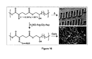
- FIG. 16 shows poly(triol ⁇ -ketoglutarate) as biodegradable, chemoselective, and mechanically tunable elastomers in an exemplary embodiment of the present application.
- the design of several elastomers based on the thermal polycondensation of ⁇ -ketoglutaric acid and one of three triols: glycerol, 1,2,4-butanetriol, or 1,2,6-hexanetriol has been performed. 74-79 By varying the curing temperature and the duration of the curing process, a wide range of mechanical properties was achieved.
- the values of the Young's modulus (0.1-657.4 MPa), ultimate stress (0.2-30.8 MPa), and ultimate strain (22-583%) encompass the mechanical properties of many biological materials, increasing the probability of success for the use of poly(triol ⁇ -ketoglutarate) as a biomaterial.
- the poly(triol ⁇ -ketoglutarate) series hydrolytically degraded in as fast as 2 days and as long as 28 days in phosphate-buffered saline solutions.
- the repeat units contain ketones, which are capable of reacting with a variety of oxyamine-terminated molecules to generate stable oxime linkages.
- the versatility and utility of these elastomers were demonstrated by creating micro-patterned structures and films for biospecific cell scaffold supports.
- FIGS. 17A and 17B show general schemes for generating liposomes presenting ketone groups and then subsequent delivery and incorporation of the ketones in bacteria membranes via liposome fusion in accordance with an exemplary embodiment of the present application.
- FIG. 18A shows a general scheme to conjugate fluorescent ligands to the engineered ketone-presenting bacterial cells via bio-orthogonal chemistry in an exemplary embodiment of the present application.
- FIGS. 18B-D show the results of the characterization of these cells with flow cytometry.
- FIG. 19A shows a general schematic of engineered ketone-presenting bacterial cells conjugating to materials presenting oxyamine groups, along with microscopic analysis of resulting materials along with controls in an exemplary embodiment of the present application.
- FIG. 19B shows a general schematic of engineered ketone presenting bacterial cells reacted with biotin to generate biotin presenting bacteria followed by immobilization on streptavidin coated materials in an exemplary embodiment of the present application. Also shown is the microscopic analysis of the resulting materials along with controls.
- FIG. 20A shows a general schematic of the preparation of biotin-presenting bacteria and subsequent conjugation to streptavidin that was conjugated to a fluorescent dye in an exemplary embodiment of the present application.
- FIGS. 20B and 20C show the flow cytometry characterization of the resulting cells.
- FIG. 21 shows a general schematic of the dual nucleic acid transfection and cell surface engineering method in an embodiment of the present application.
- FIGS. 22 a and 22 b show a general schematic and imaging of cells transfected with either DNA or siRNA via the bio-orthogonal liposome fusion method in an exemplary embodiment of the present application.
- Swiss 3T3 Albino fibroblasts were transfected with Green Fluorescent Protein (GFP) resulting in fluorescent cells.
- GFP-expressing fibroblasts lost their fluorescence upon transfection with GFP siRNA.
- FIGS. 23 a and 23 b show a general schematic and images of cells transfected with bio-orthogonal groups resulting in cell-cell assemblies.
- controls are depicted showing that transfected Red Fluorescent Protein (RFP) and GFP cells do not form cell-cell assemblies due to only the GFP cells having bio-orthogonal groups on their surface.
- RFP Red Fluorescent Protein
- FIGS. 23 a and 23 b show a general schematic and images of cells transfected with bio-orthogonal groups resulting in cell-cell assemblies.
- RFP Red Fluorescent Protein
- FIG. 24 is a general schematic of the dialdehyde conjugation strategy in accordance with one embodiment of the present application.
- FIG. 25 show a schematic of dialdehyde (compound of Formula V) liposome formation followed by ligand conjugation in an exemplary embodiment of the present application.
- the liposome conjugate was added to cells to deliver the biotin ligand to the cell surface.
- a fluorescently tagged streptavidin protein was then added and binds specifically to the biotin presenting cell.
- FIGS. 26A and 26B present flow cytometry data showing biotin delivered to fibroblast cells via the dialdehyde linker in an exemplary embodiment of the application. Fluorescent-streptavidin binding to Biotin presenting cells is measured by flow cytometry.
- A The left histogram shows the control where no biotin is added to the dialdehyde liposome followed by fusion to fibroblast cells and streptavidin exposure (no fluorescence is observed). The right histogram shows fluorescent cells due Streptavidin recognition of biotin-dialdehyde presenting cells.
- B Flow cytometry data showing the distribution between the control and biotin presenting cells.
- lipid As used in this application, the singular forms “a”, “an” and “the” include plural references unless the content clearly dictates otherwise. For example, an embodiment including “a lipid” should be understood to present certain aspects with one lipid, or two or more additional lipids.
- the second component as used herein is chemically different from the other components or first component.
- a “third” component is different from the other, first, and second components, and further enumerated or “additional” components are similarly different.
- the term “comprising” and its derivatives, as used herein, are intended to be open ended terms that specify the presence of the stated features, elements, components, groups, integers, and/or steps, but do not exclude the presence of other unstated features, elements, components, groups, integers and/or steps.
- the foregoing also applies to words having similar meanings such as the terms, “including”, “having” and their derivatives.
- the term “consisting” and its derivatives, as used herein, are intended to be closed terms that specify the presence of the stated features, elements, components, groups, integers, and/or steps, but exclude the presence of other unstated features, elements, components, groups, integers and/or steps.
- compounds of the application refers to amphiphatic molecules comprising a lipophilic portion, a hydrophilic portion and a functional group, wherein the functional group is bonded to the hydrophilic portion and is one of a functional group pair.
- the compounds of the application” or “compounds of the present application” include compounds of Formula I, compounds of Formula II and compounds of Formula III.
- the compounds of the application” or “compounds of the present application” include compounds of Formula IV.
- the compounds of the application” or “compounds of the present application” include compounds of Formula V.
- bio-orthogonal refers to non-native, non-perturbing chemical functional groups that are introduced into naturally occurring, living systems and are modified in these living systems through selective reactions that do not interfere with any other chemical moieties in the natural surroundings.
- amphiphilic refers to a compound comprising both hydrophilic (water loving) and lipophilic (fat loving) portions.
- hydrophilic portion refers to a portion of a molecule that has a tendency to interact with or be dissolved by water and other polar substances.
- the hydrophilic portion is typically charge-polarized and contains atoms that participate in hydrogen bonding.
- the hydrophilic portion may also be referred to as a polar portion.
- the hydrophilic portion is any carbon-based moiety (or organic moiety) that has a solubility in water that is more than 1 mass %.
- the hydrophilic portion is any carbon-based moiety comprising at least one neutral hydrophile group per 5 carbons, or at least one electrically charged hydrophile group per 7 carbons.
- a representative example of a hydrophilic portion is an alkylene oxide chain, such as a methylene oxide, ethylene oxide, propylene oxide or butylene oxide chain.
- lipophilic portion refers to a portion of a molecule that is soluble in fats, oils, lipids and non-polar solvents such as hexane and toluene. Lipophilic portions interact within themselves and with other substances through the London dispersion force. They have little to no capacity to form hydrogen bonds.
- a representative example of a lipophilic portion is a C6 or higher straight or branched chained alkyl or alkenyl group.
- fluorescent moiety refers to any chemical grouping that contains electrons which can absorb a photon upon exposure to light and briefly enters an excited state before either dispersing the energy non-radioactively or emitting it as a photon, but with a lower energy.
- electroactive moiety means any chemical grouping that has the ability to change electronic configuration, for example, by transferring electrons, acting as a conductor of electrons, and/or acting as an electron donor or acceptor.
- photocleavable moiety means any chemical grouping that is cleaved (i.e. bonds are broken) upon exposure to light energy.
- radioactive moiety as used herein means any chemical grouping that spontaneously emits energy in the form of particles of ionization (or radiation) from its nucleus.
- the particles of ionization include alpha particles, beta particles, and gamma rays.
- Examples of radioactive moieties include, but are not limited to, 99 Tc, 2 H, 13 C and 129 I,
- chelating moiety means any chemical grouping that forms two or more separate coordinate bonds with a single central atom. Chelating moieties are often referred to as ligands and are organic compounds containing heteroatoms, such as N, P, S and O that form two or more coordinate bonds with the central atom, which is often a metal.
- spin label means a chemical grouping that acts as a molecular reporter because it is paramagnetic (contains an unpaired electron).
- Spin labels can be detected and monitored by electron paramagnetic resonance (EPR) or electron spin resonance (ESR) spectroscopy.
- EPR electron paramagnetic resonance
- ESR electron spin resonance
- a common example of a spin label is a nitroxide (N—O) group which is usually incorporated into a heterocyclic ring (e.g. pyrrolidine), and the unpaired electron is predominantly localized to the N—O bond.
- liposomes refers to artificially prepared vesicles, the surface of which is a bilayer formed from amphiphatic molecules.
- liposomes of the application or “liposomes of the present application” as used herein refers to liposomes comprising one or more compounds of the application.
- the term “functional group” as used herein refers to a group of atoms or a single atom that will react with another group of atoms or a single atom (so called “complementary functional group”) under bio-orthogonal reaction conditions to form a chemical interaction between the two groups or atoms.
- complementary functional group pair means that the functional groups in the pair interact, or react with each other, to form a chemical interaction that is strong enough to promote the adhesion of the two types of liposomes or cells to each other.
- the chemical interaction is a covalent bond or an ionic bond. In another embodiment, the chemical interaction is a covalent bond.
- one of a complementary functional group pair refers to one member of a functional group pair.
- reacts with generally means that there is a flow of electrons or a transfer of electrostatic charge resulting in the formation of a chemical interaction.
- chemical interaction refers to the formation of either a covalent of ionic bond between the reactive functional groups.
- the chemical interaction is one that is strong enough to promote the adhesion of liposomes or cells.
- adheredhere or “adhesion” as used herein means to bring two or more entities, such as two or more liposomes or two or more cells, into close proximity to each other and to remain in contact with each other.
- the adhered liposomes remain as separate entities or, their membranes destabilize and fuse together to result in the formation of a single liposome.
- the adhered cells communicate with each other and/or divide and multiply forming, for example, tissues.
- alkyl as used herein means straight or branched chain, saturated alkyl groups.
- the number of carbon atoms in the chain is defined by the C #-# prefix preceding the term.
- C 6-30 alkyl means an alkyl group having 6, 7, 8, 9, 10, 11, 12, 13, 14, 15, 16, 17, 18, 19, 20, 21, 22, 24, 25, 26, 27, 28, 29 or 30 carbon atoms.
- alkenyl as used herein means straight or branched chain, unsaturated alkyl groups containing one or more, suitably one or three, more suitable one or two, double bonds.
- the number of carbon atoms in the chain is defined by the C #-# prefix preceding the term.
- C 6-30 alkyl means an alkenyl group having 6, 7, 8, 9, 10, 11, 12, 13, 14, 15, 16, 17, 18, 19, 20, 21, 22, 23, 24, 25, 26, 27, 28, 29 or 30 carbon atoms.
- oxyamine refers to the functional group “—O—NH 2 ”.
- amine refers to the functional group “—NH 2 ”.
- ketone refers to the functional group
- aldehyde refers to the functional group
- nucleic acid refers to both deoxyribonucleic acid (DNA) and ribonucleic acid (RNA).
- the compounds of the application are amphiphatic molecules comprising a lipophilic portion, a hydrophilic portion and a functional group, wherein the functional group is bonded to the hydrophilic portion and is one of a complementary functional group pair.
- the compounds of the present application are compounds of Formula I:
- n is 5, 6, 7, 8, 9, 10, 11, 12, 13, 14, 15, 16, 17, 18, 19, 20, 21, 22, 23, 24, 25, 26, 27, 28 or 29;
- m is 1, 2, 3, or 4;
- p is 1, 2, 3, 4, 5, 6, 7, 8, 9 or 10;
- q is 0, 1, 2, 3, 4, 5 or 6; and
- X is one of a complementary functional group pair.
- the compounds of the application further comprise at least one of a fluorescent moiety, an electroactive moiety, a photocleavable moiety, a radioactive moiety, a chelating moiety and/or a spin label.
- a fluorescent moiety an electroactive moiety, a photocleavable moiety, a radioactive moiety, a chelating moiety and/or a spin label.
- the fluorescent moiety, electroactive moiety, photocleavable moiety, radioactive moiety, chelating moiety and/or spin label is located between the lipophilic portion and the hydrophilic portion.
- the fluorescent moiety, electroactive moiety, photocleavable moiety, radioactive moiety, chelating moiety and/or spin label is located between the hydrophilic portion and the functional group.
- the compounds of the present application comprising a fluorescent moiety, an electroactive moiety, a photocleavable moiety, a radioactive moiety, a chelating moiety and/or a spin label are compounds of Formula II:
- n is 5, 6, 7, 8, 9, 10, 11, 12, 13, 14, 15, 16, 17, 18, 19, 20, 21, 22, 23, 24, 25, 26, 27, 28 or 29;
- m is 1, 2, 3, or 4;
- p is 1, 2, 3, 4, 5, 6, 7, 8, 9 or 10;
- q is 0, 1, 2, 3, 4, 5 or 6;
- Q comprises at least one of a fluorescent moiety, an electroactive moiety, a photocleavable moiety, a radioactive moiety, a chelating moiety and a spin label; and
- X is one of a complementary functional group pair.
- the compounds of the present application comprising a fluorescent moiety and/or a photocleavable moiety are compounds of Formula III:
- n is 5, 6, 7, 8, 9, 10, 11, 12, 13, 14, 15, 16, 17, 18, 19, 20, 21, 22, 23, 24, 25, 26, 27, 28 or 29;
- m is 1, 2, 3, or 4;
- p is 1, 2, 3, 4, 5, 6, 7, 8, 9 or 10;
- q is 0, 1, 2, 3, 4, 5 or 6;
- Q comprises at least one of a fluorescent moiety, an electroactive moiety, a photocleavable moiety, a radioactive moiety, a chelating moiety and a spin label; and
- X is one of a complementary functional group pair.
- the present application includes a compound of the Formula IV: R—X (IV) wherein R is selected from: H 3 C—(CH 2 ) n —[—O—(CH 2 ) m —] p —O—(CH 2 ) q ; H 3 C—(CH 2 ) n -Q-[—O—(CH 2 ) m ] p —O—(CH 2 ) q —; and H 3 C—(CH 2 ) n —[—O—(CH 2 ) m ] p —O—(CH 2 ) q -Q-; n is 5, 6, 7, 8, 9, 10, 11, 12, 13, 14, 15, 16, 17, 18, 19, 20, 21, 22, 23, 24, 25, 26, 27, 28 or 29; m is 1, 2, 3, or 4; p is 1, 2, 3, 4, 5, 6, 7, 8, 9 or 10; q is 0, 1, 2, 3, 4, 5 or 6; Q comprises at least one of a fluorescent moiety, an electroactive moiety, a photocleavable moiety
- the present application also includes a compound of the Formula V:
- R 2 and R 3 are each, independently selected from H 3 C—(CH 2 ) n —[—O—(CH 2 ) m ] p —O—(CH 2 ) q —; H 3 C—(CH 2 ) n -Q-[—O—(CH 2 ) m ] p —O—(CH 2 ) q —; H 3 C—(CH 2 ) n —[—O—(CH 2 ) m ] p —O—(CH 2 ) q -Q-; and H 3 C—(CH 2 ) n —O—(CH 2 ) q —; n is 5, 6, 7, 8, 9, 10, 11, 12, 13, 14, 15, 16, 17, 18, 19, 20, 21, 22, 23, 24, 25, 26, 27, 28 or 29; m is 1, 2, 3, or 4; p is 1, 2, 3, 4, 5, 6, 7, 8, 9 or 10; q is 0, 1, 2, 3 or 46; Q comprises at least one of a fluorescent moiety, an electroactive moiety, a photocle
- the complementary functional group pairs in the compounds of the application are bio-orthogonal and chemoselective.
- Examples of complementary, bio-orthogonal pairs of functional groups include, but are not limited to:
- ketones and oxyamines which react to form an oxime
- complementary functional group pairs that are not bio-orthogonal can be used for applications that do not involve naturally occurring living systems.
- groups include thiols and disulfides, Michael donors and Michael acceptors, activated carboxylic acids and amines, two thiols, an azide and a triaryl phosphine (Staudinger reaction substrates) and a nitro phosphate and an alcohol (grafting).
- X is selected from a functional group comprising a ketone, an oxyamine, a hydrazine, a diene, a dienophile, an azide and an alkyne.
- X is selected from a functional group comprising a ketone, an oxyamine, an aldehyde, an amine, a hydrazine, a diene, a dienophile, an azide and an alkyne.
- the complementary functional group pair in the compounds of the application is a ketone and an oxyamine which react to form an oxime. Accordingly, it is an embodiment, that X is —C(O)R 1 , wherein R 1 is C 1-2 alkyl, or O—NH 2 .
- the complementary functional group pair is a aldehyde and an amine which when contacted with each other, form a stable imine bond.
- X in the compounds of Formula I, II and III is, independently, C(O)H or X in the compounds of Formula I, II and III is NH 2 .
- X and X′ in the compounds of Formula V are, independently, C(O)H or X and X′ in the compounds of Formula V are, independently, NH 2 .
- n is 8, 9, 10, 11, 12, 13, 14, 15, 16, 17, 18, 19, 20, 21, 22, 23, 24, 25, 26, 27, 28 or 29.
- m is 2 or 3.
- p is 4, 5, 6, 7, 8, 9 or 10.
- q is 1, 2, 3 or 4.
- Q is group that comprises at least one of a fluorescent moiety, an electroactive moiety, a photocleavable moiety, a radioactive moiety, a chelating moiety and a spin label.
- Q is a group that comprises at least one of a fluorescent moiety, an electroactive moiety and a photocleavable moiety.
- the fluorescent moiety is a calcein or rhodamine or fluorescein moiety.
- the electroactive moiety hydroquinone or ferrocene).
- the photocleavable moiety is a 4-(1-hydroxyethyl)-2-methoxy-5-nitrophenoxy moiety.
- R 2 and R 3 are independently selected from: H 3 C—(CH 2 ) n —[—O—(CH 2 ) m —] p —O—(CH 2 ) q —; and H 3 C—(CH 2 ) n —O—(CH 2 ) q —.
- R 2 and R 3 in the compounds of Formula V are independently H 3 C—(CH 2 ) n —[—O—(CH 2 ) m —] p —O—(CH 2 ) q —. In a further embodiment, R 2 and R 3 in the compounds of Formula V are both H 3 C—(CH 2 ) n —[—O—(CH 2 ) m —] p —O—(CH 2 ) q —.
- n in the compounds of Formula V is 8, 9, 10, 11, 12, 13, 14, 15, 16, 17, 18, 19, 20, 21 or 22.
- m in the compounds of Formula V is 2 or 3.
- p in the compounds of Formula V is 4, 5, 6, 7, 8, 9 or 10.
- q in the compounds of Formula V is 1 or 2.
- X and X′ in the compounds of Formula V are both C(O)R 1 , wherein R 1 is C 1-2 alkyl, or X and X′ in the compounds of Formula V are both O—NH 2 .
- X and X′ in the compounds of Formula V are both C(O)H or X and X′ in the compounds of Formula V are both NH 2 .
- X and X′ in the compounds of Formula V are both C(O)H.
- r and r′ in the compounds of Formula V are 1 or 2.
- the compound of Formula I and II (or compound of Formula IV) is selected from:
- the compounds of the application in certain embodiments, have at least one asymmetric centre. Where compounds possess more than one asymmetric centre, they may exist as diastereomers. It is to be understood that all such isomers and mixtures thereof in any proportion are encompassed within the scope of the present application. It is to be further understood that while the stereochemistry of the compounds may be as shown in any given compound listed herein, such compounds may also contain certain amounts (e.g. less than 20%, suitably less than 10%, more suitably less than 5%) of compounds of the application having alternate stereochemistry.
- the present application also includes a liposome comprising one or more compounds of the application, that is one or more amphiphatic molecules, wherein the amphiphatic molecules comprise a lipophilic portion, a hydrophilic portion and a functional group, and the functional group is bonded to the hydrophilic portion and is one of a functional group pair.
- the compounds of the application are selected from one or more of a compound of Formula I, Formula II and Formula III.
- the compounds of the application are selected from one or more of a compound of Formula IV.
- the compounds of the application are selected from one or more of a compound of Formula V.
- the liposomes further comprise any suitable amphiphatic molecule, or mixture of molecules, that form liposomes.
- liposome-forming amphiphatic molecules are lipids, in particular phospholipids.
- the liposome-forming amphiphatic molecules are selected based on the proposed use of the liposome. For example, if the liposomes are to be adhered to each other, the liposome-forming amphiphatic molecule is any suitable neutral, positively charged or negatively charged amphiphatic molecule or a mixture thereof. In general, to enhance the attraction between the two entities to be adhered or fused, the charges on each entity are opposite.
- liposome-forming amphiphatic molecules are diverse and the present application is not limited to any specific type. Selection of the liposome-forming amphiphatic molecule and methods for the formation of liposomes are well within the skill of a person in the art.
- any known method for the preparation of liposomes is used to prepare the liposomes of the present application.
- the liposomes are formed by dissolving the compounds of the application in an organic solvent and thoroughly combining the resulting solution with the liposome-forming amphiphatic molecule(s), also dissolved in an organic solvent, followed by removal of all of the organic solvents.
- the dried samples are then reconstituted and brought to the desired concentration in an aqueous buffer solution, such as an aqueous buffer having a pH of about 7 to about 7.5. Sonication and warming may be used to obtain a clear solution of large unilamellar vesicles (LUVs).
- the liposomes are optionally reduced in size, for example, using extrusion methods.
- the liposome-forming amphiphatic molecule is selected from one or more of palmitoyl-oleoyl phosphatidylcholine (POPC—a neutral phospholipid), dipalmitoylphosphatidylcholine (DPPC—a neutral phospholipid), 1-palmitoyl-2-oleoyl-phophatidylglycerol (POPG, a negatively charged phospholipid) 1,2-dimyristoyl-sn-glycero-3-phosphoglycerol (DMPG, a negatively charged or anionic phospholipid) and 1,2,-dioleoyl-3-trimethylammonium-propane (DOTAP—a positively charged or cationic lipid).
- POPC palmitoyl-oleoyl phosphatidylcholine
- DPPC dipalmitoylphosphatidylcholine
- POPG 1-palmitoyl-2-oleoyl-phophatidylglycerol
- the liposome-forming amphiphatic molecule is selected from egg palmitoyl-oleoyl phosphatidylcholine (POPC—a neutral phospholipid), egg 1-palmitoyl-2-oleoyl-phophatidylglycerol (POPG, a negatively charged phospholipid) and 1,2,-dioleoyl-3-trimethylammonium-propane (DOTAP—a positively charged or cationic lipid).
- POPC egg palmitoyl-oleoyl phosphatidylcholine
- POPG egg 1-palmitoyl-2-oleoyl-phophatidylglycerol
- DOTAP 1,2,-dioleoyl-3-trimethylammonium-propane
- the amount of the compounds of the application in the liposome is about 1 mol % to about 10 mol %, or about 5 mol %. It is another embodiment, that the liposome comprises about 90 mol % to about 99 mol % of a neutral lipid and, optionally, about 1 mol % to about 5 mol % of a charged lipid.
- the liposomes further comprise fluorescent reporter molecules.
- the fluorescent reporter molecules are incorporated into the liposome-forming amphiphatic molecules. When present in the liposome-forming amphiphatic molecules, it is an embodiment that these molecules are incorporated into the liposomes in an amount of about 0.5 mol % to about 5 mol %, or about 2 mol %.
- the fluorescent phospholipids egg 1,2-diphytanoyl-sn-glycero-3-phosphoethanolamine-N-(7-nitro-2-1,3-benzoxadiazol-4-yl) (ammonium salt) (NBD-PE), and egg 1,2-dipalmitoyl-sn-glycero-3-phosphoethanolamine-N-(lissamine rhodamine B sulfonyl) (ammonium salt) (Rhod-PE), are used.
- NBD-PE egg 1,2-diphytanoyl-sn-glycero-3-phosphoethanolamine-N-(7-nitro-2-1,3-benzoxadiazol-4-yl)
- Rhod-PE egg 1,2-dipalmitoyl-sn-glycero-3-phosphoethanolamine-N-(lissamine rhodamine B sulfonyl) (ammonium salt)
- NBD-PE a fluorescence donor
- rhod-PE a fluorescence acceptor
- the liposomes further comprise other functional molecules, such as fluorescent molecules, dyes and/or other indicator molecules, so that when the liposomes are fused, a physical change, such as a change in color, fluorescence or smell, occurs.
- functional molecules may be entrapped within the liposomes or be incorporated into the liposome-forming amphiphatic molecules.
- the liposomes further comprise biologically active agents, such as nucleic acids, proteins, peptides, small molecule drugs, carbohydrates and the like, and mixtures thereof, and fusion of the liposomes with any cell population results in the simultaneous delivery of the biological agents into the cells and modification of the cell's surface with at least one of a complementary functional group pair.
- biologically active agents may be entrapped within the liposome or may be incorporated into the liposome membrane.
- the liposomes are combined with one or more nucleic acid molecules to form a nucleic acid-liposome complex.
- These complexes when delivered to cells result in simultaneous transfection of the cells with the nucleic acid molecule(s) and modification of the cell's surface with at least one of a complementary functional group pair.
- the transfected and cell surface modified cells can then undergo subsequent cell-cell assembly or reaction, for example, with a range of ligands, small molecules and proteins via bio-orthogonal ligation.
- compositions comprising one or more of the above-identified liposomes.
- the composition further comprises a solvent, diluent or carrier, such as an aqueous buffer.
- the compounds of the present application are prepared using methods known in the art. For example the various portions, including the lipophilic portion, the hydrophilic portion and the functional group are coupled together using known chemistries, including nucleophilic displacements, activation of carboxylic acids followed by displacement of activating groups by nucleophiles and cross couplings. Active functional groups are protected with protecting groups, if needed, prior to the coupling reactions and then removed after the coupling reactions.
- the fluorescent moiety, electroactive moiety, photocleavable moiety, radioactive moiety, chelating moiety and spin label are incorporated into the compounds of the application using the same methodology. Representative examples of methods for preparing the compounds of the application are shown in Schemes 1 and 2.
- Protecting groups are chemical moieties which protect or mask a reactive portion of a molecule to prevent side reactions in those reactive portions of the molecule, while manipulating or reacting a different portion of the molecule. After the manipulation or reaction is complete, the protecting group is removed under conditions that do not degrade or decompose the remaining portions of the molecule.
- the selection of a suitable protecting group can be made by a person skilled in the art. Many conventional protecting groups are known in the art, for example as described in “Protective Groups in Organic Chemistry” McOmie, J. F. W. Ed., Plenum Press, 1973, in Greene, T. W. and Wuts, P. G.
- protecting groups include, but are not limited to t-Boc, Ac, Ts, Ms, silyl ethers such as TMS, TBDMS, TBDPS, Tf, Ns, Bn, Fmoc, dimethoxytrityl, methoxyethoxymethyl ether, methoxymethyl ether, pivaloyl, p-methyoxybenzyl ether, tetrahydropyranyl, trityl, ethoxyethyl ethers, carbobenzyloxy, benzoyl and the like.
- the present application also includes a mixture comprising a plurality of liposomes of type A and a plurality of liposomes of type B, wherein the liposomes of type A comprise a compound of the application having a functional group that is complementary to a functional group on compounds of the application comprised in the liposomes of type B to form a chemical interaction that results in adhesion of the liposomes of type A and the liposomes of type B.
- the adhesion of the liposomes of type A and the liposomes of type B results in formation of multiadherent liposomes, the partial fusion of liposomes of type A and the liposomes of type B and/or the complete fusion of the liposomes of type A and type B.
- the adhesion of the liposomes of type A and the liposomes of type B results in the complete fusion of the liposomes of type A and the liposomes of type B to make larger liposomes of type “AB”.
- the present application also includes a method for promoting adhesion of liposomes comprising contacting a plurality of liposomes of type A with a plurality of liposomes of type B, wherein the liposomes of type A comprise a compound of the application having a functional group that is complementary to a functional group on compounds of the application comprised in the liposomes of type B to form a chemical interaction that results in adhesion of the liposomes of type A and the liposomes of type B.
- the application also includes a use of one or more compounds of the application to promote the adhesion of liposomes.
- kits or commercial packages for performing the method of promoting the adhesion of liposomes.
- the kit or package comprises, in separate containers, a solution of a plurality of liposomes of type A and a solution of a plurality of liposomes of type B, wherein the liposomes of type A comprise a compound of the application having a functional group that is complementary to a functional group on compounds of the application comprised in the liposomes of type B to form a chemical interaction that results in adhesion of the liposomes of type A and the liposomes of type B, along with instructions for performing the method.
- the kit or package further comprises separate means for forming bubbles with the each of the plurality of liposomes of type A and a plurality of liposomes of type B.
- Any means for forming bubbles may be used, such as any shaped device upon which a film of the solution comprising the liposomes of type A and the solution of the liposomes of type B can form and the user can apply a flow of a gas, such as air, to form bubbles. Examples of such means include the typical bubble blowing devices that are found in children's bubble forming toy products.
- the instructions include directions to form a bubble from each of the solutions of liposomes of types A and B and to bring the bubbles into contact with each other.
- each of the liposomes of type A and type B further comprise an indicator molecule, such as a dye, and contact of the bubbles of type A with the bubbles of type B results in a fused bubble having a different detectable property, such as a different colour.
- these kits and commercial packages are used or sold as novelty items or toys.
- Liposome fusion to mammalian and bacterial cell membranes was directed through the use of a charged lipids and a molecular recognition pair for chemoselective ligation using the compounds of the application.
- Applications for this strategy include, but are not limited to, small molecule delivery, cell-surface modification, tissue engineering, biologically active molecule delivery, vaccine generation, study of bacterial behavior, bacteria detection and study of bacteria pathogenicity.
- tissue engineering By employing this membrane tailoring strategy, the assembly of 3D spheroid clusters and tissue-like structures were directed after culturing two cell populations functionalized with oxyamine- and ketone-containing groups.
- this method is general, bio-orthogonal, chemically stable, and non-cytotoxic, patterned multi-layered tissue-like structures of different geometric shapes could also be fabricated without the use of 3D scaffolds to confine the cell populations. It has also been shown that this method has promising use in stem cell transplantation by co-culturing human mesenchymal stem cells (hMSCs) with fibroblasts (fbs) and inducing adipocyte differentiation while in a 3D multi-layered tissue-like structure. Also demonstrated was the inclusion of fluorescent, electroactive and photocleavable moieties in the functional group presenting molecules for further manipulation of cellular interactions. Other functional groups, such as radioactive moieties, chelating moieties and spin labels are also delivered to the cell membrane using the methods of the application.
- hMSCs human mesenchymal stem cells
- fbs fibroblasts
- adipocyte differentiation while in a 3D multi-layered tissue-like structure.
- fluorescent, electroactive and photocleavable moieties in
- the present application includes a method of modifying a cell membrane comprising contacting the cell with a liposome comprising one of more compounds of the application under conditions for incorporation of the compounds into the cell membrane.
- the present application also includes the use of one or more compounds of the application for modifying a cell membrane.
- the present application includes a method for promoting the adhesion of cells comprising:
- the liposomes of type A comprise a functional group that reacts with a functional group comprised in the liposomes of type B to form a chemical interaction that results in the adhesion of the first and second cell populations.
- a mixture of neutral, positively and/or negatively charged liposome-forming amphiphatic molecules are used depending on the cell type and corresponding membrane characteristics.
- fusion to mammalian cells types whose membranes comprise a negative charge, is promoted by incorporating positively charged lipids in to the liposome.
- the positively charged lipid enhances membrane fusion via electrostatic destabilization.
- the liposomes of the application comprise a neutral lipid, a positively charged lipid and one or more compounds of the application.
- a liposome composition is useful, for example, for fusion with cell membranes having a negative charge, such as mammalian cells, and for forming complexes with negatively charged molecules, such as nucleic acids (see below).
- a variation in the molar ratio of the neutral lipid to the positively charged lipid is made for different cells types.
- the positively charged lipids are incorporated in an amount of 1 mol % to about 5 mol %, or about 2 mol %.
- the liposomes of the application comprise a neutral lipid, a negatively charged lipid and one or more compounds of the application.
- a liposome composition is useful, for example, for fusion with bacterial cell membranes or for forming complexes with molecules having a positive charge.
- the negatively charged lipids are incorporated in an amount of 1 mol % to about 5 mol %, or about 2 mol %.
- prokaryotic and eukaryotic for example, mammals, plants, bacteria, viruses and the like, can be done using a similar strategy depending on the characteristics of the cell membrane.
- the conditions for the fusion of the liposomes with the cell populations generally involve adding an aqueous buffered solution of the liposomes to the cells in culture and incubating the cells in the presence of the liposomes for example, for 6 to about 24 hours.
- the solution of the liposomes is added at a concentration of about 0.5 to 5 mM and about 1 to about 10 mL of this solution is added to about 1 to about 10 mL of the cultured cells.
- the contacting of the fused first cell population with the fused second cell population can be done using any suitable means.
- the cell populations may be combined in solution.
- oxyamine presenting rat2 fibroblasts were combined in solution with ketone-presenting Swiss albino 3T3 fibroblasts and, upon mixing, these two cell populations formed clusters and tissue-like masses. This is a significant finding as current methods to generate these types of structures require the support of a 3D hydrogen matrix and/or assisted assembly through an external stimulus.
- one of the cell populations may be grown on a substrate and the second cell population added as a layer on top of the first population, followed by addition of alternate layers of the first and second population of cells.
- larger, dense 3D tissue-like networks are formed with geometric control.
- the 3D-tissue like networks are released from the substrate using, for example, agitation or washing, accordingly, this method provides the possibility for applications in tissue engineering and cellular transplantation.
- Another alternative is to combine the two cell populations in a continuous fashion, for example, by flowing one stream comprising the first population of cells into a second stream comprising the second population of cells, using microfluidics.
- microfluidics in combination with the surface-modified cells of the present application, will allow that rapid generation of co-culture microtissue in flow. This will provide new ways to generate complex tissues and lead to new technologies to assemble and grow tissues that complement current bioreactor methods (see for example, FIG. 4 ).
- microfluidic technology is expanded to generate complex tissues that incorporate multiple cell lineages, as well as to generate more complex microfluidic chambers to introduce multiple different surface-modified cell lines at different stages of microtissue formation to generate shelled tissue structures, for example, using tubular biodegradable polymers.
- At least one of the populations of cells is a stem cell and adhesion of a second population of a specific cell type results in induced differentiation and proliferation of the stem cells as the second cell type. This result holds great potential for areas of regenerative medicine and stem cell transplantation.
- the liposomes of type A and/or B further comprise biologically active agents, such as nucleic acids, proteins, peptides, small molecule drugs, carbohydrates and the like, and mixtures thereof, and fusion of the liposomes with the cell population results in the delivery of the biological agents into the cells.
- biologically active agents are either entrapped within the liposome and/or are incorporated into the liposome membrane.
- the liposomes of type A and/or B further comprise other functional molecules, such as fluorescent molecules, dyes and/or other indicator molecules, so that when the first and second cell populations are adhered, a physical or sensory change, such as a change in color or fluorescence occurs.
- functional molecules such as fluorescent molecules, dyes and/or other indicator molecules, so that when the first and second cell populations are adhered, a physical or sensory change, such as a change in color or fluorescence occurs.
- functional molecules are either entrapped within the liposomes and/or are incorporated into the liposome-forming amphiphatic molecules.
- the liposomes of type A and type B further comprise fluorescent reporter molecules.
- the fluorescent reporter molecules are incorporated into the liposome-forming amphiphatic molecules. When present in the liposome-forming amphiphatic molecules, it is an embodiment that these molecules are incorporated into the liposomes in an amount of about 0.5 mol % to about 5 mol %, or about 2 mol %.
- the fluorescent phospholipids egg 1,2-diphytanoyl-sn-glycero-3-phosphoethanolamine-N-(7-nitro-2-1,3-benzoxadiazol-4-yl) (ammonium salt) (NBD-PE), and/or egg 1,2-dipalmitoyl-sn-glycero-3-phosphoethanolamine-N-(lissamine rhodamine B sulfonyl) (ammonium salt) (Rhod-PE), are used.
- NBD-PE egg 1,2-diphytanoyl-sn-glycero-3-phosphoethanolamine-N-(7-nitro-2-1,3-benzoxadiazol-4-yl)
- Rhod-PE egg 1,2-dipalmitoyl-sn-glycero-3-phosphoethanolamine-N-(lissamine rhodamine B sulfonyl) (ammonium salt)
- the ability to tailor cell surfaces with new functionalities allows for many new applications and studies. For example, when these cells are contacted with other entities, including cells, and/or other molecules or substrates, comprising a complementary functional group, chemical interaction occurs resulting in the joining of the entity with the cell, providing a vast array of possible methods for manipulating the cell.
- the inclusion of at least one of a photocleavable moiety, fluorescent moiety, electroactive moiety, radioactive moiety, chelating moiety and spin label in the compounds of the application provides for further routes of cellular manipulation as described below.
- FIGS. 5-8 A general strategy has been developed that delivers photocleavable and bio-orthogonal chemistry via liposome fusion to cell surfaces for subsequent in-situ tailoring for on-demand micro-tissue assembly and disassembly ( FIGS. 5-8 ).
- a novel liposome fusion system was developed to deliver bio-orthogonal photo-active oxyamine lipid and ketone-lipid like molecules to cell membranes.
- co-culture spheroids and multilayers could be generated due to an intercellular oxime ligation. Since the ligation tether contained a photocleavable group, remote control of disassembly could be achieved upon UV light illumination ( FIGS. 6 and 7 ).
- This system was demonstrated in several cell lines to generate switchable co-culture spheroids and multi-layers.
- This photo-active cell surface engineering system was further demonstrated by conjugating and tracking cell surface ligands and applying a photocleavable oxime bond formation between cells for the spatial and temporal control of multilayer tissue assembly and disassembly for manipulating HMSC stem cell differentiation ( FIG. 8 ).
- the photo-active lipid molecule is used to generate photo-switchable engineered stem cells for temporal studies of stem cell plasticity. It has been shown that HMSC stem cells can retain the photo-active lipid and form spheroid and tissue co-cultures with fibroblasts. HMSC-fibroblast co-cultures are generated and used to study the role of interaction duration on the rate of differentiation. Since the tissues can be cleaved, and the HMSC and fibroblast cells separated upon demand, each population's genomic and proteomic profile as a function of interaction duration can be studied. Straightforward gene microarrays and mass-spectrometry proteomics analysis are compared to control populations.
- HMSC differentiation to adipocytes and osteoblasts as a function of fibroblast or other cell type association can also be studied.
- These studies delineate the role of co-culture cell-cell interactions and provide an interaction map of stem cell differentiation for a range of different cell type-stem cell studies.
- Large co-culture tissues are made between fibroblasts and then microfiche masks and ultraviolet light illumination are used to generate patterned 3D structures.
- These studies are then combined with 3D printing technology and UV light illumination to generate more complex 3D tissues.
- This methodology in conjunction with 3D printing, 3D laser imaging and bioreactor technology has the potential to generate myriad shapes of complex tissue of multiple-cell types. Programmable shapes and assemblies of any cell lines will provide new methods for a range of fundamental stem cell plasticity studies and the potential to engineer any complex tissue through automated 3D printers and lasers.
- bioreactor technologies with intercellular ligation methods provides new ways to generate large-scale complex multi-cell type tissues.
- traditional polymer scaffolds, molds or printing technologies a range of complex 3D tissues and organs are possible for an array of biomedical diagnostic and transplantation applications.
- a major theme in studying and manipulating interactions at cell surfaces is the ability to simultaneously visualize and tailor events that occur at cell membranes. These multi-modal approaches are complex and a system where docking and changing cell surfaces are observed in real-time is desirable.
- a method that can provide a beacon on a cell surface that becomes fluorescent upon specific binding allows for many opportunities to image, track and manipulate cell and tissue behaviour in space and time.
- the present application also includes a novel receptor/reporter system based on liposome fusion to incorporate a unique bio-orthogonal lipid that has the dual ability to serve as a receptor for chemoselective cell surface tailoring and as a reporter to track cell behavior ( FIG. 9 ).
- the bimodal amphiphatic molecule contains a FRET pair comprising a calcein dye joined with an efficient quencher dabcyl through an exchangeable linkage. While in a cell's membrane the non-fluorescent amphiphatic molecule can be turned on by the addition of an complementary functional group-containing ligand, which exchanges with the dabcyl quencher. This allows for the in situ simultaneous tailoring and fluorescent tracking of cells.
- the general utility of this strategy can be demonstrated by tailoring cells with small molecules and peptides for real-time analysis of cell surface modification ( FIGS. 10 and 11 ).
- Synthetic chemistry can be used to further explore the dual receptor and reporter system and then evaluate these systems for cell surface engineering applications in tissue imaging.
- a modified calcein dye has been prepared and a FRET quenched bio-orthogonal exchange version incorporated into cell membranes.
- the dual receptor reporter system is used to conjugate cell adhesive peptides (e.g. RGD-oxyamine peptide) to cell surfaces.
- cell adhesive peptides e.g. RGD-oxyamine peptide
- This methodology has been carried out on several cell lines ( FIG. 11 ).
- the preparation of complementary functional group-containing peptides, such as oxyamine-containing peptides, is known to a person skilled in the art, for example, on a peptide synthesizer.
- One feature is that only cells that become fluorescent (exchange with the dabcyl quencher) have the peptide modification.
- the RGD-complementary functional group-containing peptides are used in conjunction with the dual reporter receptor system to generate tissues containing fibroblasts and HMSC cells.
- the fibroblasts contain the dual receptor reporter amphiphatic molecule and the HMSC cells will not be modified.
- the fibroblasts Upon mixing no spheroids or tissue like structures form, however, upon introduction of the RGD-complementary functional group-containing peptide the fibroblasts become fluorescent and form spheroids with itself and HMSC cells.
- HMSC differentiation lineages e.g. adipocytes, fibroblasts, osteoblasts, and/or chondrocytes
- Engineered cells that are electroactive allow very sensitive electrochemical analytical techniques to probe and control cell behaviour. Generating electroactive cell surfaces immediately allows for cells to be manipulated by many bioanalytical methods and provides a continuous redox status of the changing local cell environment.
- Cell surfaces can be rewired via an electroactive molecule for dynamic control of cell surface ligands and cell tissue interactions ( FIGS. 12-15 ). This methodology allows for the modulation of cell-cell interactions to generate three-dimensional (3D) tissue structures applied to stem cell, cell-surface tailoring, and/or tissue engineering.
- the electroactive hydroquinone system is used to study the biophysics of membrane diffusion on a cell's surface and to conjugate and release cell-cell assemblies while adhered to a working electrode.
- Hydroquinone containing stem cells HQ-HMSCs
- HQ-HMSCs Hydroquinone containing stem cells
- a major challenge in stem cell plasticity model systems is that after co-culture tissue formation and stem cell differentiation, the cell lines are separated to study the newly differentiated stem cells.
- Co-culture tissues are generated between HMSC's and fibroblasts on working electrodes and after differentiation occurs the cells are separated by a simple electrochemical pulse that cleaves all the associations between the cells. Therefore, the electroactive engineered cells allow for co-culture tissue formation (bio-orthogonal) but also the on-demand electrical release of the cells.
- Preliminary data shows that the brief low potential electrochemical pulse does not alter cell viability.
- Generating implantable biomaterials often involves the interaction of cells with non-natural materials and polymers.
- the surface modified cells generated using a method of the present application are combined with polymers and beads that contain complementary bio-orthogonal functional groups to generate new living biomaterials and hybrid polymer-multi-layer tissues with control of 3D architecture and vasculature.
- tailored cells interact with tailored materials for example, for a range of stem cell-based biotechnology and tissue engineering applications.
- polyketoester polymers are incorporated with the surface modified cells of the present application.
- fibroblasts are generated that contain oxyamine groups these cells are seeded onto the polyketoester polymers.
- a covalent oxime ligation occurs between the cells and the polymers.
- the addition of HMSC cells with ketone groups then attaches to the fibroblast oxyamine cells.
- complex multi-cell type polymer hybrids are built. These living biomaterials are assessed for long-term stability and compatibility for implantable polymer devices.
- spheroid clusters of cells are generated based on the surface-modified cells of the present application and these clusters are seeded on to the polyketoester polymers.
- biodegradable polymer beads containing ketone groups are mixed with oxyamine presenting fibroblasts and ketone presenting HMSC cells.
- the cells covalently attach to each other and to the beads and dense tissues are formed with the beads encapsulated within the tissue. Over time the beads degrade and provide space for re-alignment or the generation of blood vessels within the tissue.
- This system is used to study tissue formation of blood vessels and as a potential method to screen drugs to inhibit the formation of these architectures. This particular strategy is general and provides new types of living biomaterial hybrids and new implantable tissue scaffolds for a range of transplantation and tissue engineering applications.
- the present application also includes cell populations whose surfaces have been modified with reactive functional groups by fusion with the liposomes of type A and/or B, compositions comprising these cell populations and all uses thereof.
- liposomes comprising one or more amphiphatic molecules having at least one of a functional group pair, are combined with one or more nucleic acid molecules to form a nucleic acid-liposome complex.
- These complexes when delivered to cells result in simultaneous transfection of the cells with the nucleic acid molecule(s) and modification of the cell's surface with at least one of a complementary functional group pair.
- the transfected and cell surface modified cells can then undergo subsequent cell-cell assembly or reaction, for example, with a range of ligands, small molecules and proteins via bio-orthogonal ligation.
- the present application also includes a method for the simultaneous transfection of one or more nucleic acid molecules into a cell and modification of the cell's membrane comprising:
- R 2 and R 3 are each, independently selected from H 3 C—(CH 2 ) n —[—O—(CH 2 ) m —] p —O—(CH 2 ) q —; H 3 C—(CH 2 ) n -Q-[—O—(CH 2 ) m ] p —O—(CH 2 ) q —; H 3 C—(CH 2 ) n —[—O—(CH 2 ) m ] p —O—(CH 2 ) q -Q-; and H 3 C—(CH 2 ) n —O—(CH 2 ) q —; n is 5, 6, 7, 8, 9, 10, 11, 12, 13, 14, 15, 16, 17, 18, 19, 20, 21, 22, 23, 24, 25, 26, 27, 28 or 29; m is 1, 2, 3, or 4; p is 1, 2, 3, 4, 5, 6, 7, 8, 9 or 10; q is 0, 1, 2, 3 or 46; Q comprises at least one of a fluorescent moiety, an electroactive moiety, a photo
- the present application also includes the use of one or more compounds of Formula V or VI, or liposomes comprising one or more of the compounds of Formula V or VI, for simultaneous transfection of one or more nucleic acid molecules into a cell and modification of the cell's membrane.
- the conditions to form a liposome-nucleic acid complex comprise contacting positively charged liposomes of the present application with the nucleic acid in, for example, aqueous solution, at a temperature of about 20° C. to about 30° C. for about 5 min to about an hour.
- the weight ratio of the liposome to nucleic acid is about 200:1 to about 75:1
- the conditions to simultaneously transfect the cell with the one or more nucleic acid molecules and incorporate the one or more compounds of Formula V or VI into the cell membrane comprise contacting the liposome-nucleic acid complex with a suitable culture medium in which the cells have been growing (for example have reached about 50% to about 90% confluency) for about one hour to about 48 hours.
- a suitable culture medium in which the cells have been growing for example have reached about 50% to about 90% confluency
- the weight ratio of the liposomes to nucleic acid is about 20:1 to about 5:1.
- the transfected and cell surface modified cells can undergo subsequent cell-cell assembly or reaction with a range of ligands, small molecules, proteins via bio-orthogonal reactions or ligation (see FIG. 22 ).
- X in the compounds of Formula V or VI is selected from a functional group comprising a ketone, an oxyamine, a hydrazine, a diene, a dienophile, an azide and an alkyne.
- X in the compounds of Formula V or VI is selected from a functional group comprising a ketone, an oxyamine, an aldehyde, an amine, a hydrazine, a diene, a dienophile, an azide and an alkyne.
- the complementary functional group pair in the compounds of Formula V or VI is a ketone and an oxyamine which react to form an oxime. Accordingly, it is an embodiment, that X is —C(O)R 1 , wherein R 1 is C 1-2 alkyl, or O—NH 2 .
- the complementary functional group pair is a aldehyde and an amine which when contacted with each other, form a stable imine bond.
- X in the compounds of Formula V or VI is, independently, C(O)H or X in the compounds of Formula V or VI is NH 2 .
- X and X′ in the compounds of Formula V or VI are, independently, C(O)H or X and X′ in the compounds of Formula V or VI are, independently, NH 2 .
- n in the compounds of Formula VI is 8, 9, 10, 11, 12, 13, 14, 15, 16, 17, 18, 19, 20, 21, 22, 23, 24, 25, 26, 27, 28 or 29.
- s in the compounds of Formula VI is 8, 9, 10, 11, 12, 13, 14, 15, 16, 17, 18, 19, 20, 21, 22, 23, 24, 25, 26, 27, 28 or 29
- m in the compounds of Formula VI is 2 or 3.
- p in the compounds of Formula VI is 4, 5, 6, 7, 8, 9 or 10.
- q in the compounds of Formula VI is 1, 2, 3 or 4.
- Q in the compounds of Formula VI is group that comprises at least one of a fluorescent moiety, an electroactive moiety, a photocleavable moiety, a radioactive moiety, a chelating moiety and a spin label.
- Q in the compounds of Formula VI is a group that comprises at least one of a fluorescent moiety, an electroactive moiety and a photocleavable moiety.
- the fluorescent moiety is a calcein or rhodamine or fluorescein moiety.
- the electroactive moiety hydroquinone or ferrocene).
- the photocleavable moiety is a 4-(1-hydroxyethyl)-2-methoxy-5-nitrophenoxy moiety.
- R 2 and R 3 are independently selected from: H 3 C—(CH 2 ) n —[—O—(CH 2 ) m —] p —O—(CH 2 ) q —; and H 3 C—(CH 2 ) n —O—(CH 2 ) q —.
- R 2 and R 3 in the compounds of Formula V are independently H 3 C—(CH 2 ) n —[—O—(CH 2 ) m —] p —O—(CH 2 ) q —. In a further embodiment, R 2 and R 3 in the compounds of Formula V are both H 3 C—(CH 2 ) n —[—O—(CH 2 ) m —] p —O—(CH 2 ) q —.
- n in the compounds of Formula V is 8, 9, 10, 11, 12, 13, 14, 15, 16, 17, 18, 19, 20, 21 or 22.
- m in the compounds of Formula V is 2 or 3.
- p in the compounds of Formula V is 4, 5, 6, 7, 8, 9 or 10.
- q in the compounds of Formula V is 1 or 2.
- X and X′ in the compounds of Formula V are both C(O)R 1 , wherein R 1 is C 1-2 alkyl, or X and X′ in the compounds of Formula V are both O—NH 2 .
- X and X′ in the compounds of Formula V are both C(O)H or X and X′ in the compounds of Formula V are both NH 2 .
- X and X′ in the compounds of Formula V are both C(O)H.
- r and r′ in the compounds of Formula V are 1 or 2.
- the compound of Formula VI is selected from:
- Egg palmitoyl-oleoyl phosphatidylcholine (egg-POPC) was purchased from Avanti Polar Lipids (Alabaster, Ala.), and all other chemicals were obtained from Sigma-Aldrich or Fisher.
- Swiss 3T3 albino mouse fibroblasts were obtained from the Tissue Culture Facility at the University of North Carolina (UNC).
- Human mesenchymal stem cells (hMSCs), basic medium, growth medium and differentiation medium were obtained from Lonza.
- the alcohol powder product (4) was finely ground and reacted with aqueous trifluoroacetic acid (TFA) at 90° C. overnight. Additional TFA was added until reaction completion was verified by thin layer chromatography (10:1, methylene chloride:acetone). The reaction mixture was cooled, filtered, and dried under vacuum overnight. The product was purified by chromatography (10:1, methylene chloride:methanol).
- TFA trifluoroacetic acid
- the photolabile precursor 5 (0.01 mol) was suspended in anhydrous DCM (1.4 mol) and stirred in a flask purged with Ar. TEA (0.03 mol) was added, and lauroyl chloride (0.025 mol) in anhydrous DCM (0.3 mol) was added dropwise at 0° C. The reaction was stirred at room temperature overnight and subsequently washed with sodium bicarbonate (5 w/v % aq.), dilute hydrochloric acid (1 v/v % aq.), and DI water. The product was purified by flash chromatography (10:1, methylene chloride:acetone).
- Liposomes were prepared as previously reported. To generate photo-oxyamine or ketone liposomes, photo-oxyamine IIa or dodecanone (2) (60 ⁇ L, 10 mM solution in CHCl 3 ) were dissolved with egg-POPC (450 ⁇ L, 10 mg/mL in CHCl 3 ) and 1,2-dioleoyl-3-trimethylammonium-propane (DOTAP, 10 ⁇ L, 10 mg/mL in CHCl 3 ) in chloroform followed by concentration under high vacuum for 4 h. The dried lipid samples were then reconstituted and brought to a final volume of 3 mL in PBS buffer, pH 7.4. The contents of the vial were warmed to 50° C. and sonicated for 20 min, in a tip sonicator, until the solution became clear, and liposomes containing photo-oxyamine or ketone groups were formed.
- DOTAP 1,2-dioleoyl-3-trimethylammonium-propane
- hMSCs Human mesenchymal stem cells
- the substrates for confocal imaging were fixed with formaldehyde (3.2% in PBS) and permeated (PBS containing 0.1% Triton X-100).
- a fluorescent dye mixture, containing phalloidin-TRITC (actin) and DAPI (nucleus) was then made in PBS containing 5% normal goat serum and 0.1% Triton X-100. Cells were incubated with the dye solution for 2 h.
- the substrates were then secured in fluorescence mounting medium (Dako, Carpinteria, Calif., USA), which enhances the visualization of cells when viewed under a fluorescent microscope with a glass cover slip.
- the substrates for adipogenic differentiation were washed by PBS and fixed in 3.2% formaldehyde for 30 minutes, followed with sterile water and 60% isopropanol for 5 minutes. Samples were then stained by Oil Red O for 5 minutes followed by Harris Hematoxylin for 1 minute.
- the substrates for collagen differentiation were fixed with formaldehyde and permeated with 0.1% Triton X-100. Monoclonal antibody of collagen I was applied for 1 h, then incubated with secondary antibody anti-mouse IgG (FITC conjugate) for 30 min, and followed with DAPI for 30 min for nucleus staining.
- the substrates for osteogenic differentiation were stained with sigmal Alkaline Phosphatase (ALP) kit (sigmal kit 85).
- ALP sigmal Alkaline Phosphatase
- RNA isolation kits Qiagen
- 1 ⁇ g of total RNA was converted to cDNA using AMV reverse transcriptase and random hexamer primers (Promega).
- the resulting cDNA was used in PCR with the following primer, LPL (sense 5′-GAG ATT TCT CTG TAT GGC ACC-3′, antisense 5′-CTG CAA ATG AGA CAC TTT CTC-3′), PPAR ⁇ 2 (sense 5′-GCT GTT ATG GGT GAA ACT CTG-3′, antisense 5′-ATA AGG TGG AGA TGC AGG CTC-3′), Collagen I (sense 5′-TGC TGG CCA ACC ATG CCT CT-3′, antisense 5′-TTG CAC AAT GCT CTG ATC-3′), Collagen II (sense 5′-ATG ACA ACC TGG CTC CCA AC-3′, antisense 5′-GCC CTA TGT CCA CAC CGA-3′), RUNX2 (sense 5′-GAT GAC ACT GCC ACC TCT GAC TT-3′, antisense 5′-CCC CCC GGC ACC ATG GGA A
- Amplification reactions were carried out for 1 minute through 30 cycles, and the reaction products were subjected to 1% agarose gel electrophoresis.
- the reaction products are 276 bp (Lpl), 351 bp (PPAR ⁇ 2), 489 bp (Collagen I), 359 bp (Collagen II), 362 bp (RUNX2), and 418 bp (ALPL) respectively.
- Fluorescence-activated cell sorting (FACS) analysis was performed to quantify the approximate number of photo-oxyamine lipids at the cell surface after membrane fusion.
- Liposomes were cultured with Fbs (3 mM in tris buffer, 400 ⁇ L added to 4 mL) to present photo-active group on cell surface (Jurkat cells).
- Ketone-conjugated fluorescein was then reacted with the surface-modified cells (0.15 mM in tris buffer, 2 h). This time course assay was conducted to determine whether the chemistry was being carried on after cell growth and division.
- a control cell population (not displaying photolabile lipids) was incubated with the ketone-fluorescein (0.15 mM in tris buffer, 2 h).
- the different cell populations were washed with PBS (3 ⁇ 5 mL), trypsinized (1 mL, 5 min, 37° C., 5% CO 2 ), centrifuged (5 min, 1000 rpm), resuspended in RPMI (without phenol red), centrifuged (5 min, 1000 rpm), and resuspended in RPMI ( ⁇ 10 7 cells/2 mL). Fluorescence measurements were calibrated using RCP-5-30 beads ( ⁇ 10 7 beads/mL, Spherotech, Inc., Lake Forest, Ill.) of known fluorescein equivalent molecule density.
- Fluorescent intensities based on number of cells counted were compared to the standard bead and control cells lacking fluorescent molecule conjugation and approximate numbers of fluorescent compound bound to the surface was calculated.
- Flow cytometry was performed using a Dako CyAn ADP (Beckman-Coulter, Brea, Calif.), and data was analyzed with Summit 4.3 software. The error bars are represented as the mean fluorescence intensity SD of 3 trials.
- Example 8 Matrix-Assisted Laser-Desorption/Ionization Mass Spectrometry (MALDI-MS)
- Gold-coated MALDI sample plates (123 ⁇ 81 mm) (Applied Biosystems, Foster City, Calif.) were prepared by electron-beam deposition (Thermionics Laboratory Inc, Hayward, Calif.) of titanium (5 nm) and then gold (12 nm).
- SAM self-assembled monolayers
- the slides were immersed in a 1 mM solution of 1:1 ratio mixture of 11-mercaptoundecanol and tetra(ethylene glycol)-terminated undecanethiol in EtOH for 12 h, rinsed with EtOH and dried, and then partially oxidized to aldehyde by mild oxidant pyridinium chlorochromate (PCC), as previously reported (S4, S5, S6). Once removed from solution, the surfaces were rinsed with EtOH and dried before use. Cells tailored with photolabile oxyamine group are seeded onto the SAM presenting aldehyde group to form oxime ligation between the cell membrane and gold substrate. After washing and removing the cells, the bonded residue on the gold substrate was traced by MALDI-MS.
- MALDI analysis was carried out using an AB SCIEX TOF/TOFTM 5800 System (Applied Biosystems, Foster City, Calif.).
- SAMs Self-assembled monolayers
- EG 4 tetra(ethylene glycol) groups
- Fbs were cultured with photo-liposomes (4 h) and then seeded ( ⁇ 10 2 cells/mL, 2 h) to the patterned aldehyde surfaces.
- Media containing 10% calf bovine serum (CBS) and 1% penicillin/streptomycin was then added, and the substrates were incubated at 37° C. in 5% CO 2 for 4 d.
- CBS calf bovine serum
- penicillin/streptomycin was then added, and the substrates were incubated at 37° C. in 5% CO 2 for 4 d.
- Substrates were then imaged by brightfield microscopy with an exposure time of 400 ms.
- FIG. 1 A depiction of the experimental hypothesis is shown in FIG. 1 and the results are shown in FIG. 2 . From the flow cytometry data ( FIG. 2A ), it can been seen that cell surface conjugation was significantly greater when the hydrophilic ketone and oxyamine were utilized for liposome fusion. Cell surface conjugation was enhanced by 6.25 ⁇ for the hydrophilic ketone and 7.4 ⁇ for the hydrophilic oxyamine compared to their corresponding hydrophobic analogs.
- FIG. 5 shows two or more different cell types were engineered via liposome fusion to present complementary and bio-orthogonal molecules on their cell surfaces.
- a stable and covalent click (oxime) type reaction occurred and tissue-like assemblies were formed.
- tissue disassembly was remotely controlled by photo-cleavage of the intercellular ligation tether.
- the bottom image in FIG. 5 is a schematic showing the interfacial molecular lock and cleave sequence for conjugating and releasing cell associations.
- the lipid lock molecule contains both a photocleavable group and an aminooxy group for bio-orthogonal oxime ligation to ketone presenting cells.
- Disassociation of cell assemblies via photo-cleavage occurs upon UV illumination
- Remote controlled tissue disassembly proceeds by a programmed photo-initiated cleavage of the intercellular ligation tether.
- the features of this method are the delivery of synthetic chemical groups to cell surfaces (via liposome fusion), the intercellular oxime click ligation bond (bio-orthogonal) and a photo-cleavage site contained within the oxyamine lipid tether.
- the complementary functional group pair was delivered to two different cell types ( FIG. 6 ).
- Two liposome populations containing the photo-oxyamine lipid (IIa) and ketone lipid (Ia) respectively were generated.
- POPC photo-oxyamine lipid
- DOTAP 1,2-dioleoyl-3-trimethylammonium-propane
- FIG. 6D-G cells that were rewired with the photo-oxyamine were seeded onto patterned materials presenting aldehyde groups.
- FIG. 6D-G Cells specifically attached to the material through the interfacial oxime bond formation bypassing non-specific associations used in regular tissue culture and native ligand-receptor based adhesion (integrin-extracellular matrix).
- UV light 365 nm, 10 mW/cm 2 5 min
- the photo-oxime was cleaved and the cells detached from the substrate.
- This strategy allows for non-adherent cells to become adhesive to tailored materials and in combination with microfluidic technology may allow for new cell sorting, cell patterning and tissue capture/release biotechnologies. 59-60
- FIG. 6H-L shows the construction of a multi-layered tissue co-culture system based on rewiring cell surfaces via liposome fusion.
- Two different types of cells hMSCs and fibroblasts, tailored with ketone and photo-oxyamine respectively, were peeled from tissue culture plates and assembled via the oxime bond formation to form a stable multi-layered tissue co-culture system. Images of the co-culture tissue showed portions of multilayer and monolayer. The cell layers only adhered if the complementary chemistry was present on their cell surfaces.
- Flow cytometry was use to quantify and characterize the amount of photo-oxyamine lipid delivered to cells via liposome fusion, for subsequent photo-oxime bond formation-induced microtissue assembly ( FIG. 7 ).
- the photo-oxyamine was designed to have 3 components that are useful for spatial and temporal control of cell interconnectivity ( FIG. 7A ).
- Liposomes containing the photo-oxyamine lipid were synthesized and then delivered to fibroblasts in culture. To measure the amount of photo-oxyamine incorporated, the cells chemoselectively reacted via oxime formation with a fluorescent calcein dye containing a ketone group. FACS analysis determined the amount of photo-oxyamine molecule present at the cell surface after various time points after liposome fusion ( FIG. 7B-E ). As expected, the cells initially had the highest amounts of photo-oxyamine molecule, which gradually decreased over time as the cells grew and divided. This decrease is due to multiple cell divisions and therefore dilution of the photo-oxyamine lipid molecule over subsequent generations of cells.
- FIG. 7F a novel mass spectrometry method was developed ( FIG. 7F ).
- Lipid transfected cells were added to SAM surfaces presenting aldehyde groups, which allow cells to attach to the surface via the interfacial oxime bond formation. Cells were then removed from the surface with agitation through a stream of PBS solution and H 2 O. Due to the covalent nature of the interfacial oxime bond between the cells and SAM surface, the lipid was essentially pulled out of the membrane and remained conjugated to the substrate. Because the substrate is conductive, MALDI mass spectrometry could be performed directly on the substrate to characterize the interfacial reaction.
- FIG. 7F Lipid transfected cells were added to SAM surfaces presenting aldehyde groups, which allow cells to attach to the surface via the interfacial oxime bond formation. Cells were then removed from the surface with agitation through a stream of PBS solution and H 2 O. Due to the covalent nature of the interfacial oxime bond
- 7G shows MALDI analysis and clearly shows the presence of the oxime molecule on the substrate. This strategy may be used to characterize a range of lipid presenting molecules on cell surfaces and as a new pull-down method of proteins if the lipid molecule spans the plasma membrane and covalently associates with cytoplasmic proteins.
- FIG. 8 To demonstrate the spatial and temporal control of tissue assembly using the intercellular photo-oxime strategy, multilayers of adipogenc and hMSC cells were generated ( FIG. 8 ). A dodecanone liposome was first added to hMSCs, which readily presented the ketone from the cell surface. Addition of adipogenic cells presenting photo-oxyamine to the hMSCs rapidly generated multilayers. In comparison, no multi-layers are formed without the complementary chemistry presented from either cell type. FIG. 4A-C shows images and confocal representations of the multilayers. Upon UV illumination through a mask, the adipogenic cells were released from the hMSCs due to the cleavage of the intercellular photo-oxime ligation tether.
- Example 12 Engineering Bacteria Cell Surfaces with Bio-Orthogonal Groups Using Liposome Fusion
- Liposomes were prepared with DPPC,DMPG and the corresponding bio-orthogonal pair in 15:1:4 ratios. 160 ul of DPPC 10 mg/ml stock, 10 ul DMPG 10 mg/ml and 55 ul of 10/mg ketone/oxyamine were mixed in chloroform. After thorough evaporation of the organic solvent, content was re-suspended in a buffer of choice and sonicated for 4 h using tip-sonicator
- FIG. 17A is a general scheme depicting liposome preparation for redecorating the surface of the cell.
- the ketone terminated lipid-like molecule (Ia) was mixed with 1,2-dipalmitoyl-sn-glycero-3-phosphocholine (DPPC) and anionic 1,2-dimyristoyl-sn-glycero-3-phospho-(1′-rac-glycerol) (DMPG) to form liposomes containing the ketone functional group.
- DPPC 1,2-dipalmitoyl-sn-glycero-3-phosphocholine
- DMPG anionic 1,2-dimyristoyl-sn-glycero-3-phospho-(1′-rac-glycerol)
- 17B shows how bacteria cells were incubated with the prepared liposome containing ketone molecules in buffer at 4.5 pH, 37° C. for 1 h.
- the liposomes adhered and fused to the bacteria delivering the bio-orthogonal lipid ketone molecule to the cell surface.
- FIG. 18A shows the engineered cell surface of the bacteria presenting bio-orthogonal groups underwent subsequent reaction with a range of molecules, ligands, proteins and probes.
- FIG. 18B shows the flow cytometry characterization of the relationship between ketone incorporated into the cell surface versus ketone concentration in the liposome. From the plot it is clear that the increase in ketone concentration in the liposome results in an increase in ketone on the cell surface.
- FIG. 18C shows the flow cytogram for cells treated with liposomes not containing ketone and then fluorescent dye FITC-ONH 2 .
- FIG. 19A shows Indium Tin Oxide materials presenting oxyamine groups.
- Ketone presenting cells were also reacted with the small molecule biotin to generate biotin presenting bacteria. These bacteria then immobilized specifically to streptavidin coated materials ( FIG. 19B ). It is well known that the small molecule biotin binds tightly to its partner protein streptavidin. Bacteria cells did not adhere to streptavidin coated materials when no biotin was present on the bacteria cell surface (control).
- the biotin ligand was presented on the bacteria cell surface via oxime conjugation and streptavidin was added to the cells and specifically bound to the biotin presenting bacteria ( FIG. 20A ). Streptavidin was conjugated with a fluorescent dye and the amount of fluorescence was determined via flow cytometry for cells presenting no biotin ( FIG. 20B ) and for cells presenting biotin ( FIG. 20C ). The data clearly show that only cells presenting the biotin ligand are fluorescent.
- O-Dodecyloxyamine was synthesized as reported in U.S. Patent Application Publication No. US 2013/0302891.
- 1-palmitoyl-2-oleoyl-sn-glycero-3-phosphocholine (POPC) and 1,2-dioleoyl-3-trimethylammonium-propane (DOTAP) were purchased from Avanti Polar Lipids (Alabaster, Ala.), and all other chemicals were obtained from Sigma-Aldrich or Fisher.
- 3T3 Swiss Abino Fibroblasts were purchased from ATCC.
- RFP Expressing Human Neonatal Dermal Fibroblasts (RFP-HNDFs) were purchased from Olaf Pharmaceuticals.
- NIH3T3/GFP cell line was purchased from Cell Biolabs, Inc.
- C3H/10T1/2 cell line was from McDermott group at York University.
- DNase-free, RNase-free and protease-free water and siRNA buffer were purchased from fisher.
- Monster Green phMGFP Vector was purchased from Promega.
- Corresponding siRNA was designed and synthesized from Life Technologies.
- Liposomes were prepared as described in as reported in U.S. Patent Application Publication No. US 2013/0302891 3T3 Swiss Abino Fibroblasts were purchased from ATCC.
- RFP Expressing Human Neonatal Dermal Fibroblasts (RFP-HNDFs) were purchased from Olaf Pharmaceuticals.
- NIH3T3/GFP cell line is purchased from Cell Biolabs, Inc. These cell lines were transferred by Cedarlanelabs (Burlington, Canada).
- C3H/10T1/2 cell line was available from McDermott group at York University, Toronto, Canada.
- DNase-free, RNase-free and protease-free water and siRNA buffer were purchased from Fisher. Monster Green phMGFP Vector was purchased from Promega. Corresponding siRNA was designed and synthesized by Life Technologies.
- Liposomes were prepared as described in Example 2. To generate functionalized liposomes presenting oxyamine or ketone, O-dodecyloxyamine or 2-dodecanone (60 ⁇ L, 10 mM solution in CHCl 3 ) were mixed with POPC (220 ⁇ L, 10 mg/mL in CHCl 3 ) and DOTAP (220 ⁇ L, 10 mg/mL in CHCl 3 ), followed by thoroughly drying with N 2 . The dried lipid samples were then suspended in 3 mL PBS buffer (pH 7.4) and sonicated with a tip sonicator (fisher scientific) for 15 min to form a clear liposome suspension (1.5 mg/mL).
- Functionalized liposomes from (b) (5 ⁇ L, 1.5 mg/mL) and phMGFP Vector (5 ⁇ L, 10 ⁇ g/mL) were mixed for 30 min at room temperature to form liposome-DNA complex, which was then added to DMEM media containing 10% FBS to reach a final volume of 100 ⁇ L (75 ⁇ g/mL liposome, 0.5 ⁇ g/mL GFP-DNA)
- liposome-DNA complex media 100 ⁇ L liposome-DNA complex media was added when cells reach 70% confluency. Cells were maintained in liposome-DNA complex media for up to 36 hours before changing media to regular media.
- Functionalized liposomes from (b) 0.5 ⁇ L, 1.5 mg/mL
- siRNA 5′-CCGUGUUCGACUACGGUAATT-3′, 5′-UUACCGUAGUCGAACACGGTT-3′
- DMEM media containing 10% FBS 7.5 ⁇ g/mL liposome, 100 nM siRNA
- 3T3 Swiss Abino Fibroblasts, RFP Expressing Human Neonatal Dermal Fibroblasts, and C3H/10T1/2 cells were cultured in Dulbecco's modified Eagle medium (DMEM) with 10% fetal bovine serum (FBS) and 1% penicillin/streptomycin.
- DMEM Dulbecco's modified Eagle medium
- FBS fetal bovine serum
- NIH3T3/GFP cells were cultured in DMEM containing 10% FBS, 0.1 mM MEM Non-Essential Amino Acids, 2 mM L-glutamine, 10 ⁇ g/mL Blasticidin, and 1% penicillin/streptomycin. These cells were incubated at 37° C. in a humidified atmosphere of 5% CO 2 , and released from tissue culture plates using 0.05% trypsin in 0.53 mM EDTA.
- the cell samples for confocal microscopy were fixed with formaldehyde (3.2% in PBS) for 20 min, rinsed with PBS, and then secured in fluorescence mounting medium (Dako, Carpinteria, Calif., USA), which enhances the visualization of cells when viewed under a fluorescent microscope, with a thin glass cover slip.
- fluorescence mounting medium Dako, Carpinteria, Calif., USA
- the mounted samples were imaged by Zeiss LSM 700 laser scanning confocal microscope and analyzed by ZEN 2010 imaging software.
- FIG. 21 A schematic showing the dual nucleic acid transfection and cell surface engineering method of the present application is provided in FIG. 21 .
- liposomes comprising a bio-orthogonal functional group, such as ketone, aldehyde, amine or oxyamine groups are prepared.
- nucleic acids are added and a complex is formed with the liposomes.
- the nucleic acid liposome complex is then delivered to mammalian cells to simultaneously transfect the cells and label the plasma membrane with the bio-orthogonal functional groups.
- the transfected and cell-surface engineered cells can then undergo further modification due to the new bio-orthogonal functional groups on the cell's surface for a range of applications.
- the method has been successfully applied using functionalized liposomes presenting oxyamine and ketone groups from O-dodecyloxyamine and 2-dodecanone.
- Swiss 3T3 Albino fibroblasts were transfected with Green Fluorescent Protein (GFP) resulting in fluorescent cells ( FIG. 22 a ) and GFP expressing fibroblasts lost their fluorescence upon transfection with GFP siRNA ( FIG. 22 b ).
- GFP Green Fluorescent Protein
- FIG. 22 b Further examples included the transfection of 10T1/2 embryonic stem cells with GFP and transfection of red fluorescent protein (RFP)-expressing epidermal fibroblasts with GFP. As time increased, the RFP cells became increasingly green due to the GFP transfection and expression.
- RFP red fluorescent protein
- Cell-cell assemblies were formed via bio-orthogonal conjugation between RFP and GFP cells when each had one of the bio-orthogonal pair on its surface ( FIG. 23 b ). In a control, transfected GFP and RFP cells did not form cell-cell assemblies when only the GFP cells had a bio-orthogonal group on its cell surface.
- the reagents and conditions used in the preparation, along with the percent yield of the product, are as follows: (i) cis-1,4-Dichloro-2-butene (1.0 eq), Na (2.2 eq), t-butanol 12 h, 80%. (ii) LiAlH 4 (5.0 eq), ⁇ 78° C., Dry THF, 2 h, NaOH/H 2 O 1M, 3 h, 95%. (iii) 1-bromododecane (3 eq), NaH (excess), Dry THF, Ar, 24 h, 80%.
- FIG. 24 shows schematically how the dialdehyde reacts with primary amine containing small molecules, ligands, probes, proteins, etc., to generate a stable covalent six member ring conjugate that can be delivered to cell surfaces via liposome fusion.
- the dialdehyde can be used in either the ethylene glycol (1) or alky-chain (2) forms for liposome formation.
- FIG. 25 depicts the incorporation of the dialdehyde lipid like compounds of Formula V into liposomes followed by ligand conjugation.
- the liposome conjugate was added to cells and used to deliver the biotin ligand to the cell surface.
- a fluorescently tagged streptavidin protein was then added and bound specifically to the biotin presenting cell FIG. 25 .
- Flow cytometry data confirmed that biotin was delivered to fibroblast cells via the dialdehyde linker ( FIGS. 26A and 26B ).
- dialdehyde in a 1.5 arrangement will react rapidly with primary amines to generate a six member ring that is covalent and stable at physiological conditions (pH 7.0, 37° C., aqueous)
- the coupling reaction is fast at physiological conditions (half life less than 5 minutes)
- the protein or ligand does not need to be manipulated (needs only a primary amine (e.g. lysine)) to conjugate proteins, ligands, molecules, etc.
- a primary amine e.g. lysine
- dialdehyde When the dialdehyde is incorporated into a liposome—it can conjugate many molecules, ligands, proteins, probes etc., to the liposome.
- Either the ethyleneglycol tail dialdehyde (Formula Va) or the long alkyl chain dialdehyde (Vb) are capable of amine conjugation.
- Liposomes comprising the dialdehyde compounds can be delivered to cells for surface presentation of ligands, proteins, etc., in minutes. This provides transient transfection without manipulating the cell genome. Many applications are possible, ranging from immunotherapy to vaccines to cancer to fundamental studies of cell behavior.
- the compounds represent new tool for chemists and cell biologists to conjugate a range of probes, proteins, ligands and small molecules to the surface of many cell types.
- this dialdehyde liposome strategy has been used to conjugate both biotin and anti-flag peptide to bacteria and swiss 3T3 albino fibroblasts.
- Biotin was recognized by streptavidin on both bacteria and fibroblasts.
- Anti-flag peptide was recognized by the flag antibody to both bacteria and fibroblasts.
Landscapes
- Chemical & Material Sciences (AREA)
- Organic Chemistry (AREA)
- Health & Medical Sciences (AREA)
- Life Sciences & Earth Sciences (AREA)
- Engineering & Computer Science (AREA)
- Genetics & Genomics (AREA)
- General Health & Medical Sciences (AREA)
- Biochemistry (AREA)
- Biomedical Technology (AREA)
- Biotechnology (AREA)
- Zoology (AREA)
- Bioinformatics & Cheminformatics (AREA)
- Wood Science & Technology (AREA)
- Molecular Biology (AREA)
- General Engineering & Computer Science (AREA)
- Microbiology (AREA)
- Cell Biology (AREA)
- Pharmacology & Pharmacy (AREA)
- Physics & Mathematics (AREA)
- Biophysics (AREA)
- Dispersion Chemistry (AREA)
- Medicinal Chemistry (AREA)
- Plant Pathology (AREA)
- Epidemiology (AREA)
- Animal Behavior & Ethology (AREA)
- Public Health (AREA)
- Veterinary Medicine (AREA)
- Medicinal Preparation (AREA)
- Acyclic And Carbocyclic Compounds In Medicinal Compositions (AREA)
Abstract
Description
wherein:
n is 5, 6, 7, 8, 9, 10, 11, 12, 13, 14, 15, 16, 17, 18, 19, 20, 21, 22, 23, 24, 25, 26, 27, 28 or 29;
m is 1, 2, 3, or 4;
p is 1, 2, 3, 4, 5, 6, 7, 8, 9 or 10;
q is 0, 1, 2, 3, 4, 5 or 6; and
X is one of a complementary functional group pair.
wherein:
n is 5, 6, 7, 8, 9, 10, 11, 12, 13, 14, 15, 16, 17, 18, 19, 20, 21, 22, 23, 24, 25, 26, 27, 28 or 29;
m is 1, 2, 3, or 4;
p is 1, 2, 3, 4, 5, 6, 7, 8, 9 or 10;
q is 0, 1, 2, 3, 4, 5 or 6;
Q comprises at least one of a fluorescent moiety, an electroactive moiety, a photocleavable moiety, a radioactive moiety, a chelating moiety and a spin label; and
X is one of a complementary functional group pair.
R—X (IV)
wherein R is selected from:
H3C—(CH2)n—[—O—(CH2)m—]p—O—(CH2)q—;
H3C—(CH2)n-Q-[—O—(CH2)m]p—O—(CH2)q—;
H3C—(CH2)n—[—O—(CH2)m]p—O—(CH2)q-Q-;
n is 5, 6, 7, 8, 9, 10, 11, 12, 13, 14, 15, 16, 17, 18, 19, 20, 21, 22, 23, 24, 25, 26, 27, 28 or 29;
m is 1, 2, 3, or 4;
p is 1, 2, 3, 4, 5, 6, 7, 8, 9 or 10;
q is 0, 1, 2, 3, 4, 5 or 6;
Q comprises at least one of a fluorescent moiety, an electroactive moiety, a photocleavable moiety, a radioactive moiety, a chelating moiety and a spin label; and
X is one of a complementary functional group pair.

wherein R2 and R3 are each, independently selected from
H3C—(CH2)n—[—O—(CH2)m—]p—O—(CH2)q—;
H3C—(CH2)n-Q-[—O—(CH2)m]p—O—(CH2)q—;
H3C—(CH2)n—[—O—(CH2)m]p—O—(CH2)q-Q-;
H3C—(CH2)n—O—(CH2)q—;
n is 5, 6, 7, 8, 9, 10, 11, 12, 13, 14, 15, 16, 17, 18, 19, 20, 21, 22, 23, 24, 25, 26, 27, 28 or 29;
m is 1, 2, 3, or 4;
p is 1, 2, 3, 4, 5, 6, 7, 8, 9 or 10;
q is 0, 1, 2, 3 or 46;
Q comprises at least one of a fluorescent moiety, an electroactive moiety, a photocleavable moiety, a radioactive moiety, a chelating moiety and a spin label; r and r′ are each independently, 1, 2 or 3; and
X and X′ are each, independently, one of a complementary functional group pair.
R4—X (VI)
wherein R4 is selected from:
H3C—(CH2)n—[—O—(CH2)m—]p—O—(CH2)q—;
H3C—(CH2)n-Q-[—O—(CH2)m]p—O—(CH2)q—;
H3C—(CH2)n—[—O—(CH2)m]p—O—(CH2)q-Q-;
H3C—(CH2)s—;
n is 5, 6, 7, 8, 9, 10, 11, 12, 13, 14, 15, 16, 17, 18, 19, 20, 21, 22, 23, 24, 25, 26, 27, 28 or 29;
m is 1, 2, 3, or 4;
p is 1, 2, 3, 4, 5, 6, 7, 8, 9 or 10;
q is 0, 1, 2, 3, 4, 5 or 6;
s is 6, 7, 8, 9, 10, 11, 12, 13, 14, 15, 16, 17, 18, 19, 20, 21, 22, 23, 24, 25, 26, 27, 28 or 30;
Q comprises at least one of a fluorescent moiety, an electroactive moiety, a photocleavable moiety, a radioactive moiety, a chelating moiety and a spin label; and
X is one of a complementary functional group pair.
wherein:
n is 5, 6, 7, 8, 9, 10, 11, 12, 13, 14, 15, 16, 17, 18, 19, 20, 21, 22, 23, 24, 25, 26, 27, 28 or 29;
m is 1, 2, 3, or 4;
p is 1, 2, 3, 4, 5, 6, 7, 8, 9 or 10;
q is 0, 1, 2, 3, 4, 5 or 6; and
X is one of a complementary functional group pair.
wherein:
n is 5, 6, 7, 8, 9, 10, 11, 12, 13, 14, 15, 16, 17, 18, 19, 20, 21, 22, 23, 24, 25, 26, 27, 28 or 29;
m is 1, 2, 3, or 4;
p is 1, 2, 3, 4, 5, 6, 7, 8, 9 or 10;
q is 0, 1, 2, 3, 4, 5 or 6;
Q comprises at least one of a fluorescent moiety, an electroactive moiety, a photocleavable moiety, a radioactive moiety, a chelating moiety and a spin label; and
X is one of a complementary functional group pair.
wherein:
n is 5, 6, 7, 8, 9, 10, 11, 12, 13, 14, 15, 16, 17, 18, 19, 20, 21, 22, 23, 24, 25, 26, 27, 28 or 29;
m is 1, 2, 3, or 4;
p is 1, 2, 3, 4, 5, 6, 7, 8, 9 or 10;
q is 0, 1, 2, 3, 4, 5 or 6;
Q comprises at least one of a fluorescent moiety, an electroactive moiety, a photocleavable moiety, a radioactive moiety, a chelating moiety and a spin label; and
X is one of a complementary functional group pair.
R—X (IV)
wherein R is selected from:
H3C—(CH2)n—[—O—(CH2)m—]p—O—(CH2)q;
H3C—(CH2)n-Q-[—O—(CH2)m]p—O—(CH2)q—; and
H3C—(CH2)n—[—O—(CH2)m]p—O—(CH2)q-Q-;
n is 5, 6, 7, 8, 9, 10, 11, 12, 13, 14, 15, 16, 17, 18, 19, 20, 21, 22, 23, 24, 25, 26, 27, 28 or 29;
m is 1, 2, 3, or 4;
p is 1, 2, 3, 4, 5, 6, 7, 8, 9 or 10;
q is 0, 1, 2, 3, 4, 5 or 6;
Q comprises at least one of a fluorescent moiety, an electroactive moiety, a photocleavable moiety, a radioactive moiety, a chelating moiety and a spin label; and
X is one of a complementary functional group pair.

wherein R2 and R3 are each, independently selected from
H3C—(CH2)n—[—O—(CH2)m]p—O—(CH2)q—;
H3C—(CH2)n-Q-[—O—(CH2)m]p—O—(CH2)q—;
H3C—(CH2)n—[—O—(CH2)m]p—O—(CH2)q-Q-; and
H3C—(CH2)n—O—(CH2)q—;
n is 5, 6, 7, 8, 9, 10, 11, 12, 13, 14, 15, 16, 17, 18, 19, 20, 21, 22, 23, 24, 25, 26, 27, 28 or 29;
m is 1, 2, 3, or 4;
p is 1, 2, 3, 4, 5, 6, 7, 8, 9 or 10;
q is 0, 1, 2, 3 or 46;
Q comprises at least one of a fluorescent moiety, an electroactive moiety, a photocleavable moiety, a radioactive moiety, a chelating moiety and a spin label; r and r′ are each independently, 1, 2 or 3; and
X and X′ are each, independently, one of a complementary functional group pair.
H3C—(CH2)n—[—O—(CH2)m—]p—O—(CH2)q—; and
H3C—(CH2)n—O—(CH2)q—.


wherein R2 and R3 are each, independently selected from
H3C—(CH2)n—[—O—(CH2)m—]p—O—(CH2)q—;
H3C—(CH2)n-Q-[—O—(CH2)m]p—O—(CH2)q—;
H3C—(CH2)n—[—O—(CH2)m]p—O—(CH2)q-Q-; and
H3C—(CH2)n—O—(CH2)q—;
n is 5, 6, 7, 8, 9, 10, 11, 12, 13, 14, 15, 16, 17, 18, 19, 20, 21, 22, 23, 24, 25, 26, 27, 28 or 29;
m is 1, 2, 3, or 4;
p is 1, 2, 3, 4, 5, 6, 7, 8, 9 or 10;
q is 0, 1, 2, 3 or 46;
Q comprises at least one of a fluorescent moiety, an electroactive moiety, a photocleavable moiety, a radioactive moiety, a chelating moiety and a spin label; r and r′ are each independently, 1, 2 or 3; and
X and X′ are each, independently, one of a complementary functional group pair; and
a compound of Formula VI:
R4—X (VI)
wherein R4 is selected from:
H3C—(CH2)n—[—O—(CH2)m—]p—O—(CH2)q—;
H3C—(CH2)n-Q-[—O—(CH2)m]p—O—(CH2)q—;
H3C—(CH2)n—[—O—(CH2)m]p—O—(CH2)q-Q-;
H3C—(CH2)s—;
n is 5, 6, 7, 8, 9, 10, 11, 12, 13, 14, 15, 16, 17, 18, 19, 20, 21, 22, 23, 24, 25, 26, 27, 28 or 29;
m is 1, 2, 3, or 4;
p is 1, 2, 3, 4, 5, 6, 7, 8, 9 or 10;
q is 0, 1, 2, 3, 4, 5 or 6;
s is 6, 7, 8, 9, 10, 11, 12, 13, 14, 15, 16, 17, 18, 19, 20, 21, 22, 23, 24, 25, 26, 27, 28 or 30;
Q comprises at least one of a fluorescent moiety, an electroactive moiety, a photocleavable moiety, a radioactive moiety, a chelating moiety and a spin label; and
X is one of a complementary functional group pair.
H3C—(CH2)n—[—O—(CH2)m—]p—O—(CH2)q—; and
H3C—(CH2)n—O—(CH2)q—.


- 1. Nelson, C. M.; Bissel, M. J. Annu. Rev. Cell Dev. Biol. 2006, 22, 287-309.
- 2. Meshel, A. S.; Wei, Q.; Adelstein, R. S.; Sheetz, M. P. Nat. Cell Biol. 2005, 7, 157-164.
- 3. Isenberg, B. C.; Williams, C.; Tranquillo, R. T. Annu. Biomed. Eng., 2006, 34, 971-985.
- 4. Hollister, S. J. (2005). Nature Mater. 4:518-524.
- 5. Gillette, B. M.; Jensen, J. A.; Tang, B.; Yang, G. J.; Bazargan-Lari, A.; Zhong, M.; Sia, S. K. Nat. Mater. 2008, 7, 636-640.
- 6. Tanaka, H.; Murphy, C. L.; Murphy, C.; Kimura, M.; Kawai, S.; Polak, J. M. J. Cell Biochem. 2004, 93, 454-462.
- 7. Gartner, Z. J.; Bertozzi, C. R. Proc. Natl. Acad. Sci. U.S.A. 2009, 106, 4606-4610.
- 8. Albrecht, D. R.; Underhill, G. H.; Wassermann, T. B.; Sah, R. L.; Bhatia, S. N. Nat.
Methods 2006, 3, 369-375. - 9. Gray, D. S.; Tan, J. L.; Voldman, J.; Chen, C. S. Biosens. Bioelectron. 2004, 19, 1765-1774.
- 10. Odde, D. J, Renn, M. J. Biotechnol. Bioeng. 2000, 67, 312-318.
- 11. Nahmias, Y.; Odde, D. J. Nat.
Protocol 2006, 1, 2288-229626. - 12. Barron, J. A.; Krizman, D. B.; Ringeisen, B. R. Annu. Biomed. Eng. 2005, 33, 121-130.
- 13. Inaba, R.; Khademhosseini, A.; Suzuki, H.; Fukuda,
J. Biomaterials 2009, 30, 3573-3577. - 14. Ringeisen, B. R.; Othon, C. M.; Barron, J. A.; Young, D.; Spargo, B. J. Biotechnol. 2006, 1, 930-948.
- 15. Chiou, P. Y.; Ohta, A. T.; Wu, M. C. Nature 2005, 436, 370-372.
- 16. Falconnet, D.; Csucs, G.; Grandin, H. M.; Textor, M. Biomaterials 2006, 27, 3044-3063.
- 17. Khademhosseini, A.; Langer, R.; Borenstein, J.; Vacanti, J. P. Proc. Natl. Acad. Sci. U.S.A. 2006, 103, 2480-2487.
- 18. Luo, W.; Chan, E. W. L.; Yousaf, M. N. J. Am. Chem. Soc. 2010, 132, 2614-2621.
- 19. Mahal, L. K.; Yarema, K. J.; Bertozi, C. R. Science 1997, 276, 1125-1128.
- 20. Prescher, J. A.; Bertozzi, C. R. Nat. Chem. Biol. 2005, 1, 13-21.
- 21. Chen, I.; Howarth, M.; Lin, W.; Ting, A. Y. Nat.
Methods 2005, 2, 99-104. - 22. Keppler, A.; Pick, H.; Arrivoli, C.; Vogel, H.; Jonhsson, K. Proc. Natl. Acad. Sci. U.S.A. 2004, 101, 9955-9959.
- 23. Miller, L. W.; Sable, J.; Goelet, P.; Sheetz, M. P.; Cornish, V. W. Angew. Chemie. Int. Ed. 2004, 43, 1672-1675.
- 24. Kellam, B.; De Bank, P. A.; Shakesheff, K. M. Chem. Soc. Rev. 2003, 32, 327-337.
- 25. Rabuka, D.; Forstner, M. B.; Gravers, J. T.; Bertozzi, C. R. J. Am. Chem. Soc. 2008, 130, 5947-5953.
- 26. Mayer, A. (2002) Membrane fusion in eukaryotic cells. Annu. Rev. Cell. Develop. Biol. 18, 289.
- 27. Rowan, A. (2006) Clamping down on exocytosis. Nat. Rev. Mol. Cell Biol. 7, 555-561.
- 28. Ellens, H., Bentz, J., and Szoka, F. C. (1985) Proton- and calcium-induced fusion and destabilization of liposomes. Biochemistry, 24, 3099-3106.
- 29. Dennison, S. M., Greenfield, N., Lenard, J., and Lentz, B. R. (2002) VSV Transmembrane domain (TMD) peptide promotes PEG-mediated fusion of liposomes in a conformationally sensitive fashion. Biochemistry, 41, 14925-14934.
- 30. Evans, K. O., and Lentz, B. R. (2002) Kinetics of lipid rearrangements during poly(ethylene glycol)-mediated fusion of highly curved unilamellar vesicles. Biochemistry, 41, 1241-1249.
- 31. Jahn, R., Lang, T., and Sudhof, T. C. (2003) Membrane fusion. Cell, 112, 519-533.
- 32. McNew, J. A., Weber, T., Parlati, F., Johnston, R. J., Melia, T. J., Sollner, T. H., and Rothman, J. E. (2000) Close is not enough: Snare-dependent membrane fusion requires an active mechanism that transduces force to membrane anchors. J. Cell Biol. 150, 105-117,
- 33. Soolner, T. H. (2004) Intracellular and viral membrane fusion: An uniting mechanism. Curr. Opin. Biol. 16, 429-435.
- 34. Parlati, F., Weber, T., McNew, J. A., Westermann, B., Sollner, T. H., and Rothman, J. E. (1999) Rapid and efficient fusion of phospholipid vesicles by the α-helical core of a SNARE complex in the absence of an N-terminal regulatory domain. Proc. Natl. Acad. Sci. U.S.A. 96, 12565-12570.
- 35. Paumet, F., Rahimian, V., and Rothman, J. E. (2004) The specificity of SNARE-dependent fusion is encoded in the SNARE motif. Proc. Natl. Acad. Sci. U.S.A. 101, 3376-3380.
- 36. Richard, A., Marchi-Artzner, V., Lalloz, M-N., Brienne, M-J., Artzner, F., Gulik-Krzywicki, T., Guedeau-Boudeville, M-A., and Lehn, J.-M. (2004) Fusogenic supramolecular vesicle systems induced by metal ion binding to amphiphilic ligands. Proc. Natl. Acad. Sci. U.S.A. 101, 15279-15284.
- 37. Marchi-Artzner, V., Gulik-Krzywicki, T., Guedeau-Boudeville, M-A., Gosse, C., Sanderson, J. M., Dedieu, J.-C., and Lehn, J-M. (2001) Selective adhesion, lipid exchange and membrane-fusion processes between vesicles of different sizes bearing complementary molecular recognition groups.
ChemPhysChem 2, 367-376. - 38. Marchi-Artzner, V., Jullien, L., Gulik-Krzywicki, T., and Lehn, J.-M. (1997) Molecular recognition induced aggregation and fusion between vesicles containing lipids bearing complementary hydrogen bonding head groups. Chem. Commun. 1, 117-118.
- 39. Paleos, C. M., and Tsiourvas, D. (2006) Interaction between complementary liposomes: A process leading to multicompartment systems formation. J. Mol. Recognition, 19, 60-67.
- 40. Chan, Y.-H. M., Lengerich, B., and Boxer, S. G. (2009) Effects of linker sequences on vesicle fusion mediated by lipid-anchored DNA oligonucleotides. Proc. Nat. Acad. Sci. U.S.A. 106, 979-984.
- 41 Gong, Y., Luo, Y., and Bong, D. (2006) Membrane activation: Selective vesicle fusion via small molecule recognition. J. Am. Chem. Soc. 128, 14430-14431.
- 42. Wilson, J. T.; Krishnamurthy, V. R.; Cui, W.; Qu, Z.; Chaikof, E. L. J. Am. Chem. Soc. 2009, 131, 18228-18229.
- 43. Csiszar, A.; Hersch, N.; Dieluweit, S.; Biehl, R.; Merkel, R.; Hoffmann, B. Bioconjugate Chem. 2010, 21, 537-543.
- 44. Pale-Grosdemange, C., Simons, E. E., Prime, K. L., and Whitesides, G. M. (1991) Formation of self-assembled monolayers by chemisorption of derivatives of oligo(ethylene glycol) of structure HS(CH2)11(OCH2CH2)mOH on gold J. Am. Chem. Soc. 113, 12-20.
- 45. Park, S., and Yousaf, M. N. (2008) Langmuir, 24, 6201-6207.
- 46. Csiszar, A., Hersch, N., Dieluweit, S., Biehl, R., Merkel, R., and Hoffmann, B. (2010) Novel fusogenic liposomes for fluorescent cell labeling and membrane modification. Bioconjugate Chem. 21, 537-543.
- 47. Beigel, M., Keren-Zur, M., Laster, Y., and Loyter, A. (1988) Poly(aspartic acid)-dependent fusion of liposomes bearing the quaternary ammonium detergent [[[(1,1,3,3-tetramethylbutyl)cresoxy]ethoxy]ethyl]dimethylbenzyl ammonium hydroxide. Biochemistry 1988, 27, 660-666.
- 48. Westcott, N. P., Pulsipher, A., Lamb, B. M., and Yousaf, M. N. (2008) Expedient generation of patterned surface aldehydes by microfluidic oxidation for chemoselective immobilization of ligands and cells. Langmuir, 24, 9237-9240.
- 49. Lamb, B. M., Barrett, D. G., Westcott, N. P., and Yousaf, M. N. (2008) Microfluidic lithography of SAMs on gold to create dynamic surfaces for directed cell migration and contiguous cell cocultures. Langmuir, 24, 8885-8889.
- 50. Harder, P., Grunze, M., Dahint, R., Whitesides, G. M., and Laibinis, P. E. (1998) Molecular conformation in Oligo(ethylene glycol)-terminated self-assembled monolayers on gold and silver surfaces determines their ability to resist protein adsorption. J. Phys. Chem. B, 102, 426-436.
- 51. Hsiao, S. C., Shum, B. J., Onoe, H., Douglas, E. S., Gartner, Z., Mathies, R. A., Bertozzi, C. R., and Francis, M. B. (2009) Direct cell surface modification with DNA for the capture of primary cells and the investigation of myotube formation on defined patterns. Langmuir, 25, 6985-6991.
- 52. Love, J. C.; Estroff, L. A.; Kriebel, J. K.; Nuzzo, R. G.; Whitesides, G. M. Chem. Rev. 2000, 105, 1103-1170.
- 53. Hsiao, S. C.; Shum, B. J.; Onoe, H.; Douglas, E. S.; Gartner, Z.; Mathies, R. A.; Bertozzi C. R.; Francis, M. B.
Langmuir 2009, 25, 6985-6991. - 54. Dutta, D.; Pulsipher, A.; Luo, W.; Mak, H.; Yousaf, M. N. Bioconjugate Chemistry 2011, 22, 2423-2433.
- 55. Dutta, D.; Pulsipher, A.; Luo, W.; Yousaf, M. N. J. Amer. Chem. Soc. 2011, 2133, 8704-8713.
- 56. Y. R. Zhao, Q. Zheng, K. Dakin, K. Xu, M. L. Martinez, W. H. Li, J. Am. Chem. Soc. 126, 4653 (2004).
- 57. M. Alvarez, A. Best, S. Pradhan-Kadam, K. Koynov, U. Jonas, M. Kreiter, Adv. Mater. 20, 4563 (2008).
- 58. A. M. Kloxin, A. M. Kasko, C. N. Salinas, K. S. Anseth. Science 324, 59 (2009).
- 59. A. Pulsipher, N. P. Westcott, W. Luo, M. N. Yousaf, J. Am. Chem. Soc. 131, 7626 (2009).
- 60. S. Nagrath et al.,
Nature 450, 1235 (2007). - 61. K. L. Schmeichel, M. J. Bissell, J. Cell Sci. 116, 2377 (2003).
- 62 C. R. Nuttelman, S. M. Henry, K. S. Anseth, Biomaterials 23, 3617 (2002).
- 63. D. R. Albrecht, G. H. Underhill, T. B. Wassermann, R. L. Sah, S. N. Bhatia, Nat.
Methods 3, 369 (2006). - 64. R. Inaba, A. Khademhosseini, H. Suzuki, J. Fukuda,
Biomaterials 30, 3573 (2009). - 65. F. Dazzi, N. J. Horwood, Curr. Opin. Oncol. 19, 650 (2007).
- 66. M. F. Pittenger, A. M. Mackay, S. C. Beck, R. K. Jaiswal, R. Douglas, J. D. Mosca, M. A. Moorman, D. W. Simonet, S. Craig, D. R. Marshak, Science 284, 143 (1999).
- 67. R. K. Jaiswal, R. K. Jaiswal, N. Jaiswal, S. P. Bruder, G. Mbalaviele, D. R. Marshak, M. F. Pittenger, J. Biol. Chem. 275, 9645 (2000).
- 68. H. B. Fana, H. F. Liva, S. L. Toh, J. C. H. Goh, Biomaterials 29, 1017 (2008).
- 69. G. M. Keller, Curr. Opin. Cell Biol. 7, 862 (1995).
- 70. M. Schuldiner, O. Yanuka, J. Itskovitz-Eldor, D. A. Melton, N. Benvenisty, Proc. Natl. Acad. Sci. USA 97, 11307 (2000).
- 71. M. M. Stevens, J. H. George, Science 310, 1135 (2005).
- 72. T. H. Petersen, E. A. Calle, L. P. Zhao, E. J. Lee, L. Q. Gui, M. B. Raredon, K. Gavrilov, T. Yi, Z. W. Zhuang, C. Breuer, E. Herzog, L. E. Niklason, Science 329, 538 (2010).
- 73. V. Mironov, R. P. Visconti, V. Kasyanov, G. Forgacs, C. J. Drake, R. R. Markwald,
Biomaterials 30, 2164 (2009). - 74. D. G. Barrett, T. Merkel, C. Luft, M. N. Yousaf, Macromolecules 43, 9660 (2010)
- 75. D. G. Barrett, M. N. Yousaf, Soft Matter, 6, 5026 (2010)
- 76. D. G. Barrett, M. N. Yousaf, Polym. Chem., 1, 296. (2010)
- 77. D. G. Barrett, M. N. Yousaf,
Biomacromolecules 9, 2029 (2008) - 78. D. G. Barrett, M. N. Yousaf, Macromolecules 41, 6347 (2008).
- 79. D. G. Barrett, M. N. Yousaf,
ChemBioChem 9, 62 (2008).
Claims (22)
R—X (IV)
H3C—(CH2)n—[—O—(CH2)m—]p—O—(CH2)q—;
H3C—(CH2)n-Q-[—O—(CH2)m]p—O—(CH2)q—; and
H3C—(CH2)n—[—O—(CH2)m]p—O—(CH2)q-Q-;
CH3(CH2)10—[OCH2CH2]3OCH2CH2ONH2 (Ic); and


H3C—(CH2)n—[—O—(CH2)m—]p—O—(CH2)q—;
H3C—(CH2)n-Q-[—O—(CH2)m]p—O—(CH2)q—;
H3C—(CH2)n—[—O—(CH2)m]p—O—(CH2)q-Q-; and
H3C—(CH2)n—O—(CH2)q—;
H3C—(CH2)n—[—O—(CH2)m]p—O—(CH2)q—; and
H3C—(CH2)n—O—(CH2)q—.
Priority Applications (1)
| Application Number | Priority Date | Filing Date | Title |
|---|---|---|---|
| US14/896,069 US10017460B2 (en) | 2013-06-12 | 2014-06-11 | Compounds for promoting liposomal and cellular adhesion and compositions and methods of use thereof |
Applications Claiming Priority (3)
| Application Number | Priority Date | Filing Date | Title |
|---|---|---|---|
| US201361834176P | 2013-06-12 | 2013-06-12 | |
| PCT/CA2014/050547 WO2014197991A1 (en) | 2013-06-12 | 2014-06-11 | Compounds for promoting liposomal and cellular adhesion and compositions and methods of use thereof |
| US14/896,069 US10017460B2 (en) | 2013-06-12 | 2014-06-11 | Compounds for promoting liposomal and cellular adhesion and compositions and methods of use thereof |
Publications (2)
| Publication Number | Publication Date |
|---|---|
| US20160130216A1 US20160130216A1 (en) | 2016-05-12 |
| US10017460B2 true US10017460B2 (en) | 2018-07-10 |
Family
ID=52021529
Family Applications (1)
| Application Number | Title | Priority Date | Filing Date |
|---|---|---|---|
| US14/896,069 Active US10017460B2 (en) | 2013-06-12 | 2014-06-11 | Compounds for promoting liposomal and cellular adhesion and compositions and methods of use thereof |
Country Status (3)
| Country | Link |
|---|---|
| US (1) | US10017460B2 (en) |
| CA (1) | CA2914448A1 (en) |
| WO (1) | WO2014197991A1 (en) |
Families Citing this family (5)
| Publication number | Priority date | Publication date | Assignee | Title |
|---|---|---|---|---|
| US9968683B2 (en) | 2014-12-17 | 2018-05-15 | Organolinx Corp. | Method for conjugating molecules |
| CZ307452B6 (en) * | 2016-11-03 | 2018-09-05 | Vysoká škola chemicko-technologická v Praze | Aminooxylipids for the construction of self-assembly liposomal systems allowing their subsequent modification by biologically functional molecules |
| CN110709065B (en) * | 2017-04-19 | 2023-02-10 | Apa先进技术有限公司 | Fusogenic liposomes, compositions, kits and uses thereof for treating cancer |
| WO2019055901A1 (en) | 2017-09-18 | 2019-03-21 | North Carolina State University | ARTIFICIAL β-CELLS AND METHODS OF USE THEREOF |
| WO2024020584A2 (en) * | 2022-07-22 | 2024-01-25 | University Of Washington | Grayscale fabrication of 4d materials |
Citations (8)
| Publication number | Priority date | Publication date | Assignee | Title |
|---|---|---|---|---|
| US4861597A (en) | 1986-05-20 | 1989-08-29 | Wako Pure Chemical Industries, Ltd. | Novel functionallized liposomes and a process for production thereof |
| US6417326B1 (en) | 1996-04-11 | 2002-07-09 | The University Of British Columbia | Fusogenic liposomes |
| US20070249060A1 (en) | 2003-03-26 | 2007-10-25 | President And Fellows Of Harvard College | Determination of Proteins and/or Other Molecules Using Mass Spectroscopy |
| US20100063135A1 (en) | 2008-09-10 | 2010-03-11 | Abbott Laboratories | Polyethylene glycol lipid conjugates and uses thereof |
| CN102051239A (en) | 2009-10-28 | 2011-05-11 | 中国石油化工股份有限公司 | Additive composition, diesel oil composition and method for improving biodiesel oxidation stability |
| US20120100077A1 (en) | 2009-07-09 | 2012-04-26 | Forschungszentrum Juelich Gmbh | Mixture of amphipathic molecules and method for modifying cell membranes by means of fusion |
| US20130302891A1 (en) | 2012-05-10 | 2013-11-14 | Muhammad Naveed Yousaf | Compositions and methods for promoting liposomal and cellular adhesion |
| WO2013173392A1 (en) | 2012-05-15 | 2013-11-21 | Concortis Biosystems, Corp | Drug-conjugates, conjugation methods, and uses thereof |
-
2014
- 2014-06-11 WO PCT/CA2014/050547 patent/WO2014197991A1/en not_active Ceased
- 2014-06-11 US US14/896,069 patent/US10017460B2/en active Active
- 2014-06-11 CA CA2914448A patent/CA2914448A1/en not_active Abandoned
Patent Citations (9)
| Publication number | Priority date | Publication date | Assignee | Title |
|---|---|---|---|---|
| US4861597A (en) | 1986-05-20 | 1989-08-29 | Wako Pure Chemical Industries, Ltd. | Novel functionallized liposomes and a process for production thereof |
| US6417326B1 (en) | 1996-04-11 | 2002-07-09 | The University Of British Columbia | Fusogenic liposomes |
| US20070249060A1 (en) | 2003-03-26 | 2007-10-25 | President And Fellows Of Harvard College | Determination of Proteins and/or Other Molecules Using Mass Spectroscopy |
| US20100063135A1 (en) | 2008-09-10 | 2010-03-11 | Abbott Laboratories | Polyethylene glycol lipid conjugates and uses thereof |
| US20120100077A1 (en) | 2009-07-09 | 2012-04-26 | Forschungszentrum Juelich Gmbh | Mixture of amphipathic molecules and method for modifying cell membranes by means of fusion |
| CN102051239A (en) | 2009-10-28 | 2011-05-11 | 中国石油化工股份有限公司 | Additive composition, diesel oil composition and method for improving biodiesel oxidation stability |
| US20130302891A1 (en) | 2012-05-10 | 2013-11-14 | Muhammad Naveed Yousaf | Compositions and methods for promoting liposomal and cellular adhesion |
| US9080144B2 (en) | 2012-05-10 | 2015-07-14 | Muhammad Naveed Yousaf | Compositions and methods for promoting liposomal and cellular adhesion |
| WO2013173392A1 (en) | 2012-05-15 | 2013-11-21 | Concortis Biosystems, Corp | Drug-conjugates, conjugation methods, and uses thereof |
Non-Patent Citations (14)
| Title |
|---|
| Alberts_2002_Lipid bilayers_https___www.ncbi.nlm.nih. * |
| Dutta et al. (Bioconjugate Chemistry, 2011, 22:8704-8713; reference cited in IDS). * |
| Dutta et al., "Engineering Cell Surfaces via Liposome Fusion", Bioconjugate Chem., 2011, 22 (12), 2423-2433. |
| Dutta et al., Synthetic Chemoselective Rewiring of Cell Surfaces: Generation of Three-Dimensional Tissue Structures, J. Am. Chem. Soc., 2011, 133 (22), 8704-8713. |
| Faiss, Simon et al., "Adhesion and rupture of liposomes mediated by electrostatic interaction monitored by thickness shear mode resonators" Eur Biophys J (2004) 33, 551-561. |
| Gong, Yun et al., "Membrane Activation: Selective Vesicle Fusion via Small Molecule Recognition", J. Am. Chem. Soc., 2006, 128 (45), 14430-14431. |
| Holland et al. Biochemistry, 1996, 35(8), 2618-2624. * |
| Holland, JW et al., "Poly(ethylene glycol)—Lipid Conjugates Regulate the Calcium-Induced Fusion of Liposomes Composed of Phosphatidylethanolamine and Phosphatidylserine", Biochemistry., 1996, 35(8), 2618-2624. |
| International Search Report and Written Opinion of PCT/CA2014/050547 dated Sep. 29, 2014. |
| Karlberg et al. Contact Dermatitis 2003: 49: 241-247. * |
| Selden et al. JACS 2012, 134(2), 765-768. * |
| Selden, Nicholas et al., "Chemically Programmed Cell Adhesion with Membrane-Anchored Oligonucleotides", J. Am. Chem. Soc., 2012, 134(2), 765-768. |
| STN International Search—dated Dec. 10, 2014. |
| Wilson, J.T. et al., "Noncovalent Cell Surface Engineering with Cationic Graft Copolymers", J. Am. Chem. Soc., 2009, 131 (51), 18228-18229. |
Also Published As
| Publication number | Publication date |
|---|---|
| US20160130216A1 (en) | 2016-05-12 |
| CA2914448A1 (en) | 2014-12-18 |
| WO2014197991A8 (en) | 2015-01-15 |
| WO2014197991A1 (en) | 2014-12-18 |
Similar Documents
| Publication | Publication Date | Title |
|---|---|---|
| Dutta et al. | Synthetic chemoselective rewiring of cell surfaces: generation of three-dimensional tissue structures | |
| US10017460B2 (en) | Compounds for promoting liposomal and cellular adhesion and compositions and methods of use thereof | |
| KR102842973B1 (en) | Bioink for 3D printing | |
| Tu et al. | Mimicking the cell: bio-inspired functions of supramolecular assemblies | |
| Luo et al. | Remote control of tissue interactions via engineered photo-switchable cell surfaces | |
| Li et al. | Lipid–oligonucleotide conjugates for bioapplications | |
| CN103596922B (en) | Amino lipid, and synthesize and purposes | |
| De Bank et al. | Surface engineering of living myoblasts via selective periodate oxidation | |
| Brinkmann et al. | About supramolecular systems for dynamically probing cells | |
| O’Brien et al. | Spheroid and tissue assembly via click chemistry in microfluidic flow | |
| Davis et al. | Coatings on mammalian cells: interfacing cells with their environment | |
| Chan et al. | Nanoneedle‐assisted delivery of site‐selective peptide‐functionalized DNA nanocages for targeting mitochondria and nuclei | |
| CN109758588B (en) | Preparation method and application of nucleic acid spherical nanoparticle medicine | |
| Elahipanah et al. | General dialdehyde click chemistry for amine bioconjugation | |
| EP3762333B1 (en) | Composite material comprising dna hydrogel and silica nanoparticles | |
| JP5775073B2 (en) | Cell membrane modification method by mixture and fusion of amphiphilic molecules | |
| O’Brien et al. | Bio-orthogonal mediated nucleic acid transfection of cells via cell surface engineering | |
| CN118979066A (en) | Nucleic acid delivery vector, nucleic acid delivery vector set, nucleic acid delivery composition and nucleic acid delivery method | |
| US9080144B2 (en) | Compositions and methods for promoting liposomal and cellular adhesion | |
| Luo et al. | A dual receptor and reporter for multi-modal cell surface engineering | |
| Kim et al. | Engineering programmable DNA particles and capsules using catechol-functionalized DNA block copolymers | |
| KR20100029062A (en) | Novel lipid-gold nanoparticle hybrid, and gene delivery method using the same | |
| Zong et al. | Synthetic intracellular environments: from basic science to applications | |
| Ding et al. | Mechanism-driven technology development for solving the intracellular delivery problem of hard-to-transfect cells | |
| CA2776618C (en) | Compositions and methods for promoting liposomal and cellular adhesion |
Legal Events
| Date | Code | Title | Description |
|---|---|---|---|
| AS | Assignment |
Owner name: ORGANOLINX CORP., CANADA Free format text: ASSIGNMENT OF ASSIGNORS INTEREST;ASSIGNOR:YOUSAF, MUHAMMAD NAVEED, MR.;REEL/FRAME:045766/0962 Effective date: 20171121 |
|
| STCF | Information on status: patent grant |
Free format text: PATENTED CASE |
|
| MAFP | Maintenance fee payment |
Free format text: PAYMENT OF MAINTENANCE FEE, 4TH YR, SMALL ENTITY (ORIGINAL EVENT CODE: M2551); ENTITY STATUS OF PATENT OWNER: SMALL ENTITY Year of fee payment: 4 |
|
| FEPP | Fee payment procedure |
Free format text: MAINTENANCE FEE REMINDER MAILED (ORIGINAL EVENT CODE: REM.); ENTITY STATUS OF PATENT OWNER: SMALL ENTITY |