RU2650770C2 - Epitopes of epidermal growth factor receptor surface antigen and use thereof - Google Patents
Epitopes of epidermal growth factor receptor surface antigen and use thereof Download PDFInfo
- Publication number
- RU2650770C2 RU2650770C2 RU2014143032A RU2014143032A RU2650770C2 RU 2650770 C2 RU2650770 C2 RU 2650770C2 RU 2014143032 A RU2014143032 A RU 2014143032A RU 2014143032 A RU2014143032 A RU 2014143032A RU 2650770 C2 RU2650770 C2 RU 2650770C2
- Authority
- RU
- Russia
- Prior art keywords
- amino acid
- acid sequence
- seq
- epitope
- egfr
- Prior art date
Links
- 102000001301 EGF receptor Human genes 0.000 title claims abstract description 118
- 108060006698 EGF receptor Proteins 0.000 title claims abstract description 118
- 239000000427 antigen Substances 0.000 title description 3
- 102000036639 antigens Human genes 0.000 title description 3
- 108091007433 antigens Proteins 0.000 title description 3
- 125000003275 alpha amino acid group Chemical group 0.000 claims abstract description 151
- FWMNVWWHGCHHJJ-SKKKGAJSSA-N 4-amino-1-[(2r)-6-amino-2-[[(2r)-2-[[(2r)-2-[[(2r)-2-amino-3-phenylpropanoyl]amino]-3-phenylpropanoyl]amino]-4-methylpentanoyl]amino]hexanoyl]piperidine-4-carboxylic acid Chemical compound C([C@H](C(=O)N[C@H](CC(C)C)C(=O)N[C@H](CCCCN)C(=O)N1CCC(N)(CC1)C(O)=O)NC(=O)[C@H](N)CC=1C=CC=CC=1)C1=CC=CC=C1 FWMNVWWHGCHHJJ-SKKKGAJSSA-N 0.000 claims abstract description 68
- 230000027455 binding Effects 0.000 claims abstract description 61
- 201000011510 cancer Diseases 0.000 claims abstract description 15
- 230000004913 activation Effects 0.000 claims abstract description 5
- 210000004027 cell Anatomy 0.000 claims description 41
- 238000000034 method Methods 0.000 claims description 40
- 108090000623 proteins and genes Proteins 0.000 claims description 30
- 239000013598 vector Substances 0.000 claims description 24
- 206010028980 Neoplasm Diseases 0.000 claims description 22
- 102000004169 proteins and genes Human genes 0.000 claims description 16
- 241000700605 Viruses Species 0.000 claims description 15
- 230000014509 gene expression Effects 0.000 claims description 15
- 241001465754 Metazoa Species 0.000 claims description 13
- 244000005700 microbiome Species 0.000 claims description 12
- 108091033319 polynucleotide Proteins 0.000 claims description 12
- 102000040430 polynucleotide Human genes 0.000 claims description 12
- 239000002157 polynucleotide Substances 0.000 claims description 12
- 239000013604 expression vector Substances 0.000 claims description 11
- 238000004091 panning Methods 0.000 claims description 11
- 150000001413 amino acids Chemical class 0.000 claims description 10
- 239000012634 fragment Substances 0.000 claims description 10
- 238000005516 engineering process Methods 0.000 claims description 8
- 238000011282 treatment Methods 0.000 claims description 7
- 102100024952 Protein CBFA2T1 Human genes 0.000 claims description 6
- 240000004808 Saccharomyces cerevisiae Species 0.000 claims description 6
- 241000588724 Escherichia coli Species 0.000 claims description 5
- 239000001963 growth medium Substances 0.000 claims description 5
- 238000012217 deletion Methods 0.000 claims description 4
- 230000037430 deletion Effects 0.000 claims description 4
- 238000003259 recombinant expression Methods 0.000 claims description 4
- 241000894006 Bacteria Species 0.000 claims description 3
- 230000005847 immunogenicity Effects 0.000 claims description 3
- 108060003951 Immunoglobulin Proteins 0.000 claims description 2
- 108010071390 Serum Albumin Proteins 0.000 claims description 2
- 102000007562 Serum Albumin Human genes 0.000 claims description 2
- 150000004676 glycans Chemical class 0.000 claims description 2
- 108060003552 hemocyanin Proteins 0.000 claims description 2
- 102000018358 immunoglobulin Human genes 0.000 claims description 2
- 239000002773 nucleotide Substances 0.000 claims description 2
- 125000003729 nucleotide group Chemical group 0.000 claims description 2
- 238000002823 phage display Methods 0.000 claims description 2
- 229920001282 polysaccharide Polymers 0.000 claims description 2
- 239000005017 polysaccharide Substances 0.000 claims description 2
- 108090000765 processed proteins & peptides Proteins 0.000 claims description 2
- 238000002702 ribosome display Methods 0.000 claims description 2
- 241001515965 unidentified phage Species 0.000 claims description 2
- 230000009261 transgenic effect Effects 0.000 claims 2
- 241000282412 Homo Species 0.000 claims 1
- 238000002955 isolation Methods 0.000 claims 1
- 238000000746 purification Methods 0.000 claims 1
- 238000011830 transgenic mouse model Methods 0.000 claims 1
- 102000009024 Epidermal Growth Factor Human genes 0.000 abstract description 34
- 230000000694 effects Effects 0.000 abstract description 10
- 239000003814 drug Substances 0.000 abstract description 6
- 230000011664 signaling Effects 0.000 abstract description 4
- 239000000126 substance Substances 0.000 abstract 1
- 101710098940 Pro-epidermal growth factor Proteins 0.000 description 31
- 229960005395 cetuximab Drugs 0.000 description 24
- 239000003446 ligand Substances 0.000 description 24
- 125000000539 amino acid group Chemical group 0.000 description 17
- 210000005253 yeast cell Anatomy 0.000 description 16
- 239000000203 mixture Substances 0.000 description 14
- DHMQDGOQFOQNFH-UHFFFAOYSA-N Glycine Chemical compound NCC(O)=O DHMQDGOQFOQNFH-UHFFFAOYSA-N 0.000 description 13
- 235000018102 proteins Nutrition 0.000 description 13
- 108020004414 DNA Proteins 0.000 description 12
- 235000001014 amino acid Nutrition 0.000 description 11
- 102000006747 Transforming Growth Factor alpha Human genes 0.000 description 10
- 101800004564 Transforming growth factor alpha Proteins 0.000 description 10
- 230000002441 reversible effect Effects 0.000 description 9
- 229960005486 vaccine Drugs 0.000 description 9
- 101800001649 Heparin-binding EGF-like growth factor Proteins 0.000 description 8
- 102100033762 Proheparin-binding EGF-like growth factor Human genes 0.000 description 8
- 229940024606 amino acid Drugs 0.000 description 8
- 230000000259 anti-tumor effect Effects 0.000 description 8
- 239000002609 medium Substances 0.000 description 8
- 238000006467 substitution reaction Methods 0.000 description 8
- 101800001382 Betacellulin Proteins 0.000 description 7
- 102100029837 Probetacellulin Human genes 0.000 description 7
- 230000002401 inhibitory effect Effects 0.000 description 7
- 238000004519 manufacturing process Methods 0.000 description 7
- 230000001225 therapeutic effect Effects 0.000 description 7
- 230000003612 virological effect Effects 0.000 description 7
- 102000007299 Amphiregulin Human genes 0.000 description 6
- 108010033760 Amphiregulin Proteins 0.000 description 6
- 101800000155 Epiregulin Proteins 0.000 description 6
- 239000004471 Glycine Substances 0.000 description 6
- 102100025498 Proepiregulin Human genes 0.000 description 6
- 238000006243 chemical reaction Methods 0.000 description 6
- 239000002131 composite material Substances 0.000 description 6
- 230000035772 mutation Effects 0.000 description 6
- 239000013612 plasmid Substances 0.000 description 6
- 101150039808 Egfr gene Proteins 0.000 description 5
- 238000010586 diagram Methods 0.000 description 5
- 201000010099 disease Diseases 0.000 description 5
- 208000037265 diseases, disorders, signs and symptoms Diseases 0.000 description 5
- 238000001943 fluorescence-activated cell sorting Methods 0.000 description 5
- 230000001939 inductive effect Effects 0.000 description 5
- 229950008001 matuzumab Drugs 0.000 description 5
- 229960001972 panitumumab Drugs 0.000 description 5
- 238000012163 sequencing technique Methods 0.000 description 5
- 230000009466 transformation Effects 0.000 description 5
- XUUXCWCKKCZEAW-YFKPBYRVSA-N Arg-Gly Chemical compound OC(=O)CNC(=O)[C@@H](N)CCCN=C(N)N XUUXCWCKKCZEAW-YFKPBYRVSA-N 0.000 description 4
- IJGRMHOSHXDMSA-UHFFFAOYSA-N Atomic nitrogen Chemical compound N#N IJGRMHOSHXDMSA-UHFFFAOYSA-N 0.000 description 4
- 206010009944 Colon cancer Diseases 0.000 description 4
- 101710113436 GTPase KRas Proteins 0.000 description 4
- LYCVKHSJGDMDLM-LURJTMIESA-N His-Gly Chemical compound OC(=O)CNC(=O)[C@@H](N)CC1=CN=CN1 LYCVKHSJGDMDLM-LURJTMIESA-N 0.000 description 4
- 108091028043 Nucleic acid sequence Proteins 0.000 description 4
- GLUBLISJVJFHQS-VIFPVBQESA-N Phe-Gly Chemical compound OC(=O)CNC(=O)[C@@H](N)CC1=CC=CC=C1 GLUBLISJVJFHQS-VIFPVBQESA-N 0.000 description 4
- 239000002671 adjuvant Substances 0.000 description 4
- 229940127089 cytotoxic agent Drugs 0.000 description 4
- 108700021358 erbB-1 Genes Proteins 0.000 description 4
- 108010036413 histidylglycine Proteins 0.000 description 4
- 239000003550 marker Substances 0.000 description 4
- 150000007523 nucleic acids Chemical group 0.000 description 4
- 229940124597 therapeutic agent Drugs 0.000 description 4
- 239000004475 Arginine Substances 0.000 description 3
- 208000001333 Colorectal Neoplasms Diseases 0.000 description 3
- 241000701022 Cytomegalovirus Species 0.000 description 3
- 101000851181 Homo sapiens Epidermal growth factor receptor Proteins 0.000 description 3
- 241000725303 Human immunodeficiency virus Species 0.000 description 3
- 239000002253 acid Substances 0.000 description 3
- 238000004458 analytical method Methods 0.000 description 3
- 230000010056 antibody-dependent cellular cytotoxicity Effects 0.000 description 3
- 239000002246 antineoplastic agent Substances 0.000 description 3
- ODKSFYDXXFIFQN-UHFFFAOYSA-N arginine Natural products OC(=O)C(N)CCCNC(N)=N ODKSFYDXXFIFQN-UHFFFAOYSA-N 0.000 description 3
- 230000015572 biosynthetic process Effects 0.000 description 3
- 230000004663 cell proliferation Effects 0.000 description 3
- 210000004978 chinese hamster ovary cell Anatomy 0.000 description 3
- 230000003247 decreasing effect Effects 0.000 description 3
- 238000010790 dilution Methods 0.000 description 3
- 239000012895 dilution Substances 0.000 description 3
- 102000052116 epidermal growth factor receptor activity proteins Human genes 0.000 description 3
- 210000003527 eukaryotic cell Anatomy 0.000 description 3
- 230000006870 function Effects 0.000 description 3
- 102000045108 human EGFR Human genes 0.000 description 3
- 238000001556 precipitation Methods 0.000 description 3
- 230000035755 proliferation Effects 0.000 description 3
- 102000005962 receptors Human genes 0.000 description 3
- 108020003175 receptors Proteins 0.000 description 3
- 230000002829 reductive effect Effects 0.000 description 3
- 239000000243 solution Substances 0.000 description 3
- NMWKYTGJWUAZPZ-WWHBDHEGSA-N (4S)-4-[[(4R,7S,10S,16S,19S,25S,28S,31R)-31-[[(2S)-2-[[(1R,6R,9S,12S,18S,21S,24S,27S,30S,33S,36S,39S,42R,47R,53S,56S,59S,62S,65S,68S,71S,76S,79S,85S)-47-[[(2S)-2-[[(2S)-4-amino-2-[[(2S)-2-[[(2S)-2-[[(2S)-2-[[(2S)-2-[[(2S)-2-amino-3-methylbutanoyl]amino]-3-methylbutanoyl]amino]-3-hydroxypropanoyl]amino]-3-(1H-imidazol-4-yl)propanoyl]amino]-3-phenylpropanoyl]amino]-4-oxobutanoyl]amino]-3-carboxypropanoyl]amino]-18-(4-aminobutyl)-27,68-bis(3-amino-3-oxopropyl)-36,71,76-tribenzyl-39-(3-carbamimidamidopropyl)-24-(2-carboxyethyl)-21,56-bis(carboxymethyl)-65,85-bis[(1R)-1-hydroxyethyl]-59-(hydroxymethyl)-62,79-bis(1H-imidazol-4-ylmethyl)-9-methyl-33-(2-methylpropyl)-8,11,17,20,23,26,29,32,35,38,41,48,54,57,60,63,66,69,72,74,77,80,83,86-tetracosaoxo-30-propan-2-yl-3,4,44,45-tetrathia-7,10,16,19,22,25,28,31,34,37,40,49,55,58,61,64,67,70,73,75,78,81,84,87-tetracosazatetracyclo[40.31.14.012,16.049,53]heptaoctacontane-6-carbonyl]amino]-3-methylbutanoyl]amino]-7-(3-carbamimidamidopropyl)-25-(hydroxymethyl)-19-[(4-hydroxyphenyl)methyl]-28-(1H-imidazol-4-ylmethyl)-10-methyl-6,9,12,15,18,21,24,27,30-nonaoxo-16-propan-2-yl-1,2-dithia-5,8,11,14,17,20,23,26,29-nonazacyclodotriacontane-4-carbonyl]amino]-5-[[(2S)-1-[[(2S)-1-[[(2S)-3-carboxy-1-[[(2S)-1-[[(2S)-1-[[(1S)-1-carboxyethyl]amino]-4-methyl-1-oxopentan-2-yl]amino]-4-methyl-1-oxopentan-2-yl]amino]-1-oxopropan-2-yl]amino]-1-oxopropan-2-yl]amino]-3-(1H-imidazol-4-yl)-1-oxopropan-2-yl]amino]-5-oxopentanoic acid Chemical compound CC(C)C[C@H](NC(=O)[C@H](CC(C)C)NC(=O)[C@H](CC(O)=O)NC(=O)[C@H](C)NC(=O)[C@H](Cc1c[nH]cn1)NC(=O)[C@H](CCC(O)=O)NC(=O)[C@@H]1CSSC[C@H](NC(=O)[C@@H](NC(=O)[C@@H]2CSSC[C@@H]3NC(=O)[C@H](Cc4ccccc4)NC(=O)[C@H](CCC(N)=O)NC(=O)[C@@H](NC(=O)[C@H](Cc4c[nH]cn4)NC(=O)[C@H](CO)NC(=O)[C@H](CC(O)=O)NC(=O)[C@@H]4CCCN4C(=O)[C@H](CSSC[C@H](NC(=O)[C@@H](NC(=O)CNC(=O)[C@H](Cc4c[nH]cn4)NC(=O)[C@H](Cc4ccccc4)NC3=O)[C@@H](C)O)C(=O)N[C@@H](CCCNC(N)=N)C(=O)N[C@@H](Cc3ccccc3)C(=O)N[C@@H](CC(C)C)C(=O)N[C@@H](C(C)C)C(=O)N[C@@H](CCC(N)=O)C(=O)N[C@@H](CCC(O)=O)C(=O)N[C@@H](CC(O)=O)C(=O)N[C@@H](CCCCN)C(=O)N3CCC[C@H]3C(=O)N[C@@H](C)C(=O)N2)NC(=O)[C@H](CC(O)=O)NC(=O)[C@H](CC(N)=O)NC(=O)[C@H](Cc2ccccc2)NC(=O)[C@H](Cc2c[nH]cn2)NC(=O)[C@H](CO)NC(=O)[C@@H](NC(=O)[C@@H](N)C(C)C)C(C)C)[C@@H](C)O)C(C)C)C(=O)N[C@@H](Cc2c[nH]cn2)C(=O)N[C@@H](CO)C(=O)NCC(=O)N[C@@H](Cc2ccc(O)cc2)C(=O)N[C@@H](C(C)C)C(=O)NCC(=O)N[C@@H](C)C(=O)N[C@@H](CCCNC(N)=N)C(=O)N1)C(=O)N[C@@H](C)C(O)=O NMWKYTGJWUAZPZ-WWHBDHEGSA-N 0.000 description 2
- 241000282693 Cercopithecidae Species 0.000 description 2
- 108020004705 Codon Proteins 0.000 description 2
- 206010052358 Colorectal cancer metastatic Diseases 0.000 description 2
- 241000702421 Dependoparvovirus Species 0.000 description 2
- 206010052804 Drug tolerance Diseases 0.000 description 2
- 101800003838 Epidermal growth factor Proteins 0.000 description 2
- 241000238631 Hexapoda Species 0.000 description 2
- 241000701044 Human gammaherpesvirus 4 Species 0.000 description 2
- HNDVDQJCIGZPNO-YFKPBYRVSA-N L-histidine Chemical compound OC(=O)[C@@H](N)CC1=CN=CN1 HNDVDQJCIGZPNO-YFKPBYRVSA-N 0.000 description 2
- COLNVLDHVKWLRT-QMMMGPOBSA-N L-phenylalanine Chemical compound OC(=O)[C@@H](N)CC1=CC=CC=C1 COLNVLDHVKWLRT-QMMMGPOBSA-N 0.000 description 2
- AYFVYJQAPQTCCC-GBXIJSLDSA-N L-threonine Chemical compound C[C@@H](O)[C@H](N)C(O)=O AYFVYJQAPQTCCC-GBXIJSLDSA-N 0.000 description 2
- QIVBCDIJIAJPQS-VIFPVBQESA-N L-tryptophane Chemical compound C1=CC=C2C(C[C@H](N)C(O)=O)=CNC2=C1 QIVBCDIJIAJPQS-VIFPVBQESA-N 0.000 description 2
- OUYCCCASQSFEME-QMMMGPOBSA-N L-tyrosine Chemical compound OC(=O)[C@@H](N)CC1=CC=C(O)C=C1 OUYCCCASQSFEME-QMMMGPOBSA-N 0.000 description 2
- 241000713666 Lentivirus Species 0.000 description 2
- 241000829100 Macaca mulatta polyomavirus 1 Species 0.000 description 2
- 102000018697 Membrane Proteins Human genes 0.000 description 2
- 108010052285 Membrane Proteins Proteins 0.000 description 2
- 241000699666 Mus <mouse, genus> Species 0.000 description 2
- 241000699670 Mus sp. Species 0.000 description 2
- 108091081024 Start codon Proteins 0.000 description 2
- AYFVYJQAPQTCCC-UHFFFAOYSA-N Threonine Natural products CC(O)C(N)C(O)=O AYFVYJQAPQTCCC-UHFFFAOYSA-N 0.000 description 2
- 239000004473 Threonine Substances 0.000 description 2
- QIVBCDIJIAJPQS-UHFFFAOYSA-N Tryptophan Natural products C1=CC=C2C(CC(N)C(O)=O)=CNC2=C1 QIVBCDIJIAJPQS-UHFFFAOYSA-N 0.000 description 2
- OPGTXAUDXWCGFI-UHFFFAOYSA-N [1-[[6-[[3-(3-dodecanoyloxytetradecanoylamino)-6-(hydroxymethyl)-5-phosphonooxy-4-(3-tetradecanoyloxytetradecanoyloxy)oxan-2-yl]oxymethyl]-2,4,5-trihydroxyoxan-3-yl]amino]-1-oxotetradecan-3-yl] hexadecanoate Chemical compound OC1C(O)C(NC(=O)CC(CCCCCCCCCCC)OC(=O)CCCCCCCCCCCCCCC)C(O)OC1COC1C(NC(=O)CC(CCCCCCCCCCC)OC(=O)CCCCCCCCCCC)C(OC(=O)CC(CCCCCCCCCCC)OC(=O)CCCCCCCCCCCCC)C(OP(O)(O)=O)C(CO)O1 OPGTXAUDXWCGFI-UHFFFAOYSA-N 0.000 description 2
- 150000007513 acids Chemical class 0.000 description 2
- 239000011543 agarose gel Substances 0.000 description 2
- 239000005557 antagonist Substances 0.000 description 2
- 230000001580 bacterial effect Effects 0.000 description 2
- FPPNZSSZRUTDAP-UWFZAAFLSA-N carbenicillin Chemical compound N([C@H]1[C@H]2SC([C@@H](N2C1=O)C(O)=O)(C)C)C(=O)C(C(O)=O)C1=CC=CC=C1 FPPNZSSZRUTDAP-UWFZAAFLSA-N 0.000 description 2
- 229960003669 carbenicillin Drugs 0.000 description 2
- 230000010261 cell growth Effects 0.000 description 2
- 230000008859 change Effects 0.000 description 2
- HVYWMOMLDIMFJA-DPAQBDIFSA-N cholesterol Chemical compound C1C=C2C[C@@H](O)CC[C@]2(C)[C@@H]2[C@@H]1[C@@H]1CC[C@H]([C@H](C)CCCC(C)C)[C@@]1(C)CC2 HVYWMOMLDIMFJA-DPAQBDIFSA-N 0.000 description 2
- 238000004587 chromatography analysis Methods 0.000 description 2
- 230000009137 competitive binding Effects 0.000 description 2
- CVSVTCORWBXHQV-UHFFFAOYSA-N creatine Chemical compound NC(=[NH2+])N(C)CC([O-])=O CVSVTCORWBXHQV-UHFFFAOYSA-N 0.000 description 2
- UQLDLKMNUJERMK-UHFFFAOYSA-L di(octadecanoyloxy)lead Chemical compound [Pb+2].CCCCCCCCCCCCCCCCCC([O-])=O.CCCCCCCCCCCCCCCCCC([O-])=O UQLDLKMNUJERMK-UHFFFAOYSA-L 0.000 description 2
- 238000001962 electrophoresis Methods 0.000 description 2
- 238000004520 electroporation Methods 0.000 description 2
- 108010030074 endodeoxyribonuclease MluI Proteins 0.000 description 2
- 229940116977 epidermal growth factor Drugs 0.000 description 2
- 238000000684 flow cytometry Methods 0.000 description 2
- 208000014829 head and neck neoplasm Diseases 0.000 description 2
- HNDVDQJCIGZPNO-UHFFFAOYSA-N histidine Natural products OC(=O)C(N)CC1=CN=CN1 HNDVDQJCIGZPNO-UHFFFAOYSA-N 0.000 description 2
- 230000028993 immune response Effects 0.000 description 2
- 230000002163 immunogen Effects 0.000 description 2
- 238000000338 in vitro Methods 0.000 description 2
- 230000001965 increasing effect Effects 0.000 description 2
- 230000006698 induction Effects 0.000 description 2
- 230000005764 inhibitory process Effects 0.000 description 2
- 230000003834 intracellular effect Effects 0.000 description 2
- 210000003292 kidney cell Anatomy 0.000 description 2
- 230000002147 killing effect Effects 0.000 description 2
- 239000002502 liposome Substances 0.000 description 2
- 239000007788 liquid Substances 0.000 description 2
- 230000001404 mediated effect Effects 0.000 description 2
- 230000000813 microbial effect Effects 0.000 description 2
- 229940035032 monophosphoryl lipid a Drugs 0.000 description 2
- 231100000350 mutagenesis Toxicity 0.000 description 2
- 229910052757 nitrogen Inorganic materials 0.000 description 2
- 102000013415 peroxidase activity proteins Human genes 0.000 description 2
- 108040007629 peroxidase activity proteins Proteins 0.000 description 2
- COLNVLDHVKWLRT-UHFFFAOYSA-N phenylalanine Natural products OC(=O)C(N)CC1=CC=CC=C1 COLNVLDHVKWLRT-UHFFFAOYSA-N 0.000 description 2
- 230000026731 phosphorylation Effects 0.000 description 2
- 238000006366 phosphorylation reaction Methods 0.000 description 2
- 210000001236 prokaryotic cell Anatomy 0.000 description 2
- 230000001105 regulatory effect Effects 0.000 description 2
- 230000010076 replication Effects 0.000 description 2
- 108091008146 restriction endonucleases Proteins 0.000 description 2
- 238000000926 separation method Methods 0.000 description 2
- 230000019491 signal transduction Effects 0.000 description 2
- 230000008054 signal transmission Effects 0.000 description 2
- 230000002195 synergetic effect Effects 0.000 description 2
- 230000002194 synthesizing effect Effects 0.000 description 2
- 230000008685 targeting Effects 0.000 description 2
- 210000004881 tumor cell Anatomy 0.000 description 2
- 230000004565 tumor cell growth Effects 0.000 description 2
- OUYCCCASQSFEME-UHFFFAOYSA-N tyrosine Natural products OC(=O)C(N)CC1=CC=C(O)C=C1 OUYCCCASQSFEME-UHFFFAOYSA-N 0.000 description 2
- 241000701161 unidentified adenovirus Species 0.000 description 2
- 241001430294 unidentified retrovirus Species 0.000 description 2
- VBEQCZHXXJYVRD-GACYYNSASA-N uroanthelone Chemical compound C([C@@H](C(=O)N[C@H](C(=O)N[C@@H](CS)C(=O)N[C@@H](CC(N)=O)C(=O)N[C@@H](CS)C(=O)N[C@H](C(=O)N[C@@H]([C@@H](C)CC)C(=O)NCC(=O)N[C@@H](CC=1C=CC(O)=CC=1)C(=O)N[C@@H](CO)C(=O)NCC(=O)N[C@@H](CC(O)=O)C(=O)N[C@@H](CCCNC(N)=N)C(=O)N[C@@H](CS)C(=O)N[C@@H](CCC(N)=O)C(=O)N[C@@H]([C@@H](C)O)C(=O)N[C@@H](CCCNC(N)=N)C(=O)N[C@@H](CC(O)=O)C(=O)N[C@@H](CC(C)C)C(=O)N[C@@H](CCCNC(N)=N)C(=O)N[C@@H](CC=1C2=CC=CC=C2NC=1)C(=O)N[C@@H](CC=1C2=CC=CC=C2NC=1)C(=O)N[C@@H](CCC(O)=O)C(=O)N[C@@H](CC(C)C)C(=O)N[C@@H](CCCNC(N)=N)C(O)=O)C(C)C)[C@@H](C)O)NC(=O)[C@H](CO)NC(=O)[C@H](CC(O)=O)NC(=O)[C@H](CC(C)C)NC(=O)[C@H](CO)NC(=O)[C@H](CCC(O)=O)NC(=O)[C@@H](NC(=O)[C@H](CC=1NC=NC=1)NC(=O)[C@H](CCSC)NC(=O)[C@H](CS)NC(=O)[C@@H](NC(=O)CNC(=O)CNC(=O)[C@H](CC(N)=O)NC(=O)[C@H](CC(C)C)NC(=O)[C@H](CS)NC(=O)[C@H](CC=1C=CC(O)=CC=1)NC(=O)CNC(=O)[C@H](CC(O)=O)NC(=O)[C@H](CC=1C=CC(O)=CC=1)NC(=O)[C@H](CO)NC(=O)[C@H](CO)NC(=O)[C@H]1N(CCC1)C(=O)[C@H](CS)NC(=O)CNC(=O)[C@H]1N(CCC1)C(=O)[C@H](CC=1C=CC(O)=CC=1)NC(=O)[C@H](CO)NC(=O)[C@@H](N)CC(N)=O)C(C)C)[C@@H](C)CC)C1=CC=C(O)C=C1 VBEQCZHXXJYVRD-GACYYNSASA-N 0.000 description 2
- 238000012447 xenograft mouse model Methods 0.000 description 2
- DIGQNXIGRZPYDK-WKSCXVIASA-N (2R)-6-amino-2-[[2-[[(2S)-2-[[2-[[(2R)-2-[[(2S)-2-[[(2R,3S)-2-[[2-[[(2S)-2-[[2-[[(2S)-2-[[(2S)-2-[[(2R)-2-[[(2S,3S)-2-[[(2R)-2-[[(2S)-2-[[(2S)-2-[[(2S)-2-[[2-[[(2S)-2-[[(2R)-2-[[2-[[2-[[2-[(2-amino-1-hydroxyethylidene)amino]-3-carboxy-1-hydroxypropylidene]amino]-1-hydroxy-3-sulfanylpropylidene]amino]-1-hydroxyethylidene]amino]-1-hydroxy-3-sulfanylpropylidene]amino]-1,3-dihydroxypropylidene]amino]-1-hydroxyethylidene]amino]-1-hydroxypropylidene]amino]-1,3-dihydroxypropylidene]amino]-1,3-dihydroxypropylidene]amino]-1-hydroxy-3-sulfanylpropylidene]amino]-1,3-dihydroxybutylidene]amino]-1-hydroxy-3-sulfanylpropylidene]amino]-1-hydroxypropylidene]amino]-1,3-dihydroxypropylidene]amino]-1-hydroxyethylidene]amino]-1,5-dihydroxy-5-iminopentylidene]amino]-1-hydroxy-3-sulfanylpropylidene]amino]-1,3-dihydroxybutylidene]amino]-1-hydroxy-3-sulfanylpropylidene]amino]-1,3-dihydroxypropylidene]amino]-1-hydroxyethylidene]amino]-1-hydroxy-3-sulfanylpropylidene]amino]-1-hydroxyethylidene]amino]hexanoic acid Chemical compound C[C@@H]([C@@H](C(=N[C@@H](CS)C(=N[C@@H](C)C(=N[C@@H](CO)C(=NCC(=N[C@@H](CCC(=N)O)C(=NC(CS)C(=N[C@H]([C@H](C)O)C(=N[C@H](CS)C(=N[C@H](CO)C(=NCC(=N[C@H](CS)C(=NCC(=N[C@H](CCCCN)C(=O)O)O)O)O)O)O)O)O)O)O)O)O)O)O)N=C([C@H](CS)N=C([C@H](CO)N=C([C@H](CO)N=C([C@H](C)N=C(CN=C([C@H](CO)N=C([C@H](CS)N=C(CN=C(C(CS)N=C(C(CC(=O)O)N=C(CN)O)O)O)O)O)O)O)O)O)O)O)O DIGQNXIGRZPYDK-WKSCXVIASA-N 0.000 description 1
- MTCFGRXMJLQNBG-REOHCLBHSA-N (2S)-2-Amino-3-hydroxypropansäure Chemical compound OC[C@H](N)C(O)=O MTCFGRXMJLQNBG-REOHCLBHSA-N 0.000 description 1
- JNYAEWCLZODPBN-JGWLITMVSA-N (2r,3r,4s)-2-[(1r)-1,2-dihydroxyethyl]oxolane-3,4-diol Chemical compound OC[C@@H](O)[C@H]1OC[C@H](O)[C@H]1O JNYAEWCLZODPBN-JGWLITMVSA-N 0.000 description 1
- YYGNTYWPHWGJRM-UHFFFAOYSA-N (6E,10E,14E,18E)-2,6,10,15,19,23-hexamethyltetracosa-2,6,10,14,18,22-hexaene Chemical compound CC(C)=CCCC(C)=CCCC(C)=CCCC=C(C)CCC=C(C)CCC=C(C)C YYGNTYWPHWGJRM-UHFFFAOYSA-N 0.000 description 1
- 102000007469 Actins Human genes 0.000 description 1
- 108010085238 Actins Proteins 0.000 description 1
- 229910017119 AlPO Inorganic materials 0.000 description 1
- DCXYFEDJOCDNAF-UHFFFAOYSA-N Asparagine Natural products OC(=O)C(N)CC(N)=O DCXYFEDJOCDNAF-UHFFFAOYSA-N 0.000 description 1
- 241000228257 Aspergillus sp. Species 0.000 description 1
- 244000063299 Bacillus subtilis Species 0.000 description 1
- 235000014469 Bacillus subtilis Nutrition 0.000 description 1
- 108091003079 Bovine Serum Albumin Proteins 0.000 description 1
- 241000701822 Bovine papillomavirus Species 0.000 description 1
- 208000003174 Brain Neoplasms Diseases 0.000 description 1
- 206010006187 Breast cancer Diseases 0.000 description 1
- 208000026310 Breast neoplasm Diseases 0.000 description 1
- UXVMQQNJUSDDNG-UHFFFAOYSA-L Calcium chloride Chemical compound [Cl-].[Cl-].[Ca+2] UXVMQQNJUSDDNG-UHFFFAOYSA-L 0.000 description 1
- 241000283707 Capra Species 0.000 description 1
- 108091026890 Coding region Proteins 0.000 description 1
- 241000699800 Cricetinae Species 0.000 description 1
- 102000012410 DNA Ligases Human genes 0.000 description 1
- 108010061982 DNA Ligases Proteins 0.000 description 1
- 241000196324 Embryophyta Species 0.000 description 1
- 102000004190 Enzymes Human genes 0.000 description 1
- 108090000790 Enzymes Proteins 0.000 description 1
- 241000701959 Escherichia virus Lambda Species 0.000 description 1
- 241000206602 Eukaryota Species 0.000 description 1
- 241000233866 Fungi Species 0.000 description 1
- WQZGKKKJIJFFOK-GASJEMHNSA-N Glucose Natural products OC[C@H]1OC(O)[C@H](O)[C@@H](O)[C@@H]1O WQZGKKKJIJFFOK-GASJEMHNSA-N 0.000 description 1
- 102000001554 Hemoglobins Human genes 0.000 description 1
- 108010054147 Hemoglobins Proteins 0.000 description 1
- 101100118549 Homo sapiens EGFR gene Proteins 0.000 description 1
- 102000001706 Immunoglobulin Fab Fragments Human genes 0.000 description 1
- 108010054477 Immunoglobulin Fab Fragments Proteins 0.000 description 1
- 108010021625 Immunoglobulin Fragments Proteins 0.000 description 1
- 102000008394 Immunoglobulin Fragments Human genes 0.000 description 1
- 241000235058 Komagataella pastoris Species 0.000 description 1
- XUJNEKJLAYXESH-REOHCLBHSA-N L-Cysteine Chemical compound SC[C@H](N)C(O)=O XUJNEKJLAYXESH-REOHCLBHSA-N 0.000 description 1
- ONIBWKKTOPOVIA-BYPYZUCNSA-N L-Proline Chemical compound OC(=O)[C@@H]1CCCN1 ONIBWKKTOPOVIA-BYPYZUCNSA-N 0.000 description 1
- QNAYBMKLOCPYGJ-REOHCLBHSA-N L-alanine Chemical compound C[C@H](N)C(O)=O QNAYBMKLOCPYGJ-REOHCLBHSA-N 0.000 description 1
- ODKSFYDXXFIFQN-BYPYZUCNSA-P L-argininium(2+) Chemical compound NC(=[NH2+])NCCC[C@H]([NH3+])C(O)=O ODKSFYDXXFIFQN-BYPYZUCNSA-P 0.000 description 1
- DCXYFEDJOCDNAF-REOHCLBHSA-N L-asparagine Chemical compound OC(=O)[C@@H](N)CC(N)=O DCXYFEDJOCDNAF-REOHCLBHSA-N 0.000 description 1
- CKLJMWTZIZZHCS-REOHCLBHSA-N L-aspartic acid Chemical compound OC(=O)[C@@H](N)CC(O)=O CKLJMWTZIZZHCS-REOHCLBHSA-N 0.000 description 1
- ZDXPYRJPNDTMRX-VKHMYHEASA-N L-glutamine Chemical compound OC(=O)[C@@H](N)CCC(N)=O ZDXPYRJPNDTMRX-VKHMYHEASA-N 0.000 description 1
- AGPKZVBTJJNPAG-WHFBIAKZSA-N L-isoleucine Chemical compound CC[C@H](C)[C@H](N)C(O)=O AGPKZVBTJJNPAG-WHFBIAKZSA-N 0.000 description 1
- ROHFNLRQFUQHCH-YFKPBYRVSA-N L-leucine Chemical compound CC(C)C[C@H](N)C(O)=O ROHFNLRQFUQHCH-YFKPBYRVSA-N 0.000 description 1
- KDXKERNSBIXSRK-YFKPBYRVSA-N L-lysine Chemical compound NCCCC[C@H](N)C(O)=O KDXKERNSBIXSRK-YFKPBYRVSA-N 0.000 description 1
- FFEARJCKVFRZRR-BYPYZUCNSA-N L-methionine Chemical compound CSCC[C@H](N)C(O)=O FFEARJCKVFRZRR-BYPYZUCNSA-N 0.000 description 1
- KZSNJWFQEVHDMF-BYPYZUCNSA-N L-valine Chemical compound CC(C)[C@H](N)C(O)=O KZSNJWFQEVHDMF-BYPYZUCNSA-N 0.000 description 1
- ROHFNLRQFUQHCH-UHFFFAOYSA-N Leucine Natural products CC(C)CC(N)C(O)=O ROHFNLRQFUQHCH-UHFFFAOYSA-N 0.000 description 1
- 206010058467 Lung neoplasm malignant Diseases 0.000 description 1
- 239000006142 Luria-Bertani Agar Substances 0.000 description 1
- KDXKERNSBIXSRK-UHFFFAOYSA-N Lysine Natural products NCCCCC(N)C(O)=O KDXKERNSBIXSRK-UHFFFAOYSA-N 0.000 description 1
- 239000004472 Lysine Substances 0.000 description 1
- 241000124008 Mammalia Species 0.000 description 1
- 102000003792 Metallothionein Human genes 0.000 description 1
- 108090000157 Metallothionein Proteins 0.000 description 1
- 241000713333 Mouse mammary tumor virus Species 0.000 description 1
- 241000221961 Neurospora crassa Species 0.000 description 1
- 206010033128 Ovarian cancer Diseases 0.000 description 1
- 206010061535 Ovarian neoplasm Diseases 0.000 description 1
- 108091000080 Phosphotransferase Proteins 0.000 description 1
- 206010035226 Plasma cell myeloma Diseases 0.000 description 1
- 229920001213 Polysorbate 20 Polymers 0.000 description 1
- ONIBWKKTOPOVIA-UHFFFAOYSA-N Proline Natural products OC(=O)C1CCCN1 ONIBWKKTOPOVIA-UHFFFAOYSA-N 0.000 description 1
- 206010060862 Prostate cancer Diseases 0.000 description 1
- 208000000236 Prostatic Neoplasms Diseases 0.000 description 1
- 108010076504 Protein Sorting Signals Proteins 0.000 description 1
- 102000004022 Protein-Tyrosine Kinases Human genes 0.000 description 1
- 108090000412 Protein-Tyrosine Kinases Proteins 0.000 description 1
- 241000588770 Proteus mirabilis Species 0.000 description 1
- 241000589774 Pseudomonas sp. Species 0.000 description 1
- 241000235070 Saccharomyces Species 0.000 description 1
- 101100221606 Saccharomyces cerevisiae (strain ATCC 204508 / S288c) COS7 gene Proteins 0.000 description 1
- 206010039491 Sarcoma Diseases 0.000 description 1
- 206010039509 Scab Diseases 0.000 description 1
- MTCFGRXMJLQNBG-UHFFFAOYSA-N Serine Natural products OCC(N)C(O)=O MTCFGRXMJLQNBG-UHFFFAOYSA-N 0.000 description 1
- 208000005718 Stomach Neoplasms Diseases 0.000 description 1
- BHEOSNUKNHRBNM-UHFFFAOYSA-N Tetramethylsqualene Natural products CC(=C)C(C)CCC(=C)C(C)CCC(C)=CCCC=C(C)CCC(C)C(=C)CCC(C)C(C)=C BHEOSNUKNHRBNM-UHFFFAOYSA-N 0.000 description 1
- KZSNJWFQEVHDMF-UHFFFAOYSA-N Valine Natural products CC(C)C(N)C(O)=O KZSNJWFQEVHDMF-UHFFFAOYSA-N 0.000 description 1
- 238000002441 X-ray diffraction Methods 0.000 description 1
- 230000009471 action Effects 0.000 description 1
- 238000001042 affinity chromatography Methods 0.000 description 1
- 235000004279 alanine Nutrition 0.000 description 1
- AZDRQVAHHNSJOQ-UHFFFAOYSA-N alumane Chemical class [AlH3] AZDRQVAHHNSJOQ-UHFFFAOYSA-N 0.000 description 1
- 125000003118 aryl group Chemical group 0.000 description 1
- 235000009582 asparagine Nutrition 0.000 description 1
- 229960001230 asparagine Drugs 0.000 description 1
- 235000003704 aspartic acid Nutrition 0.000 description 1
- 230000008901 benefit Effects 0.000 description 1
- OQFSQFPPLPISGP-UHFFFAOYSA-N beta-carboxyaspartic acid Natural products OC(=O)C(N)C(C(O)=O)C(O)=O OQFSQFPPLPISGP-UHFFFAOYSA-N 0.000 description 1
- 230000000903 blocking effect Effects 0.000 description 1
- 229940098773 bovine serum albumin Drugs 0.000 description 1
- 239000001506 calcium phosphate Substances 0.000 description 1
- 229910000389 calcium phosphate Inorganic materials 0.000 description 1
- 235000011010 calcium phosphates Nutrition 0.000 description 1
- 230000009702 cancer cell proliferation Effects 0.000 description 1
- 239000000969 carrier Substances 0.000 description 1
- 229920006317 cationic polymer Polymers 0.000 description 1
- 239000013592 cell lysate Substances 0.000 description 1
- 238000005119 centrifugation Methods 0.000 description 1
- 239000003153 chemical reaction reagent Substances 0.000 description 1
- 238000006757 chemical reactions by type Methods 0.000 description 1
- 239000003795 chemical substances by application Substances 0.000 description 1
- 238000002512 chemotherapy Methods 0.000 description 1
- 235000012000 cholesterol Nutrition 0.000 description 1
- 230000007012 clinical effect Effects 0.000 description 1
- 208000029742 colonic neoplasm Diseases 0.000 description 1
- 238000002648 combination therapy Methods 0.000 description 1
- 229960003624 creatine Drugs 0.000 description 1
- 239000006046 creatine Substances 0.000 description 1
- 239000013078 crystal Substances 0.000 description 1
- 238000012866 crystallographic experiment Methods 0.000 description 1
- 238000012258 culturing Methods 0.000 description 1
- XUJNEKJLAYXESH-UHFFFAOYSA-N cysteine Natural products SCC(N)C(O)=O XUJNEKJLAYXESH-UHFFFAOYSA-N 0.000 description 1
- 235000018417 cysteine Nutrition 0.000 description 1
- 239000002254 cytotoxic agent Substances 0.000 description 1
- 238000013461 design Methods 0.000 description 1
- 229960000633 dextran sulfate Drugs 0.000 description 1
- 238000003745 diagnosis Methods 0.000 description 1
- 238000000502 dialysis Methods 0.000 description 1
- 230000004069 differentiation Effects 0.000 description 1
- 230000029087 digestion Effects 0.000 description 1
- LOKCTEFSRHRXRJ-UHFFFAOYSA-I dipotassium trisodium dihydrogen phosphate hydrogen phosphate dichloride Chemical compound P(=O)(O)(O)[O-].[K+].P(=O)(O)([O-])[O-].[Na+].[Na+].[Cl-].[K+].[Cl-].[Na+] LOKCTEFSRHRXRJ-UHFFFAOYSA-I 0.000 description 1
- PRAKJMSDJKAYCZ-UHFFFAOYSA-N dodecahydrosqualene Natural products CC(C)CCCC(C)CCCC(C)CCCCC(C)CCCC(C)CCCC(C)C PRAKJMSDJKAYCZ-UHFFFAOYSA-N 0.000 description 1
- 238000001035 drying Methods 0.000 description 1
- 239000003623 enhancer Substances 0.000 description 1
- 230000002708 enhancing effect Effects 0.000 description 1
- 229940082789 erbitux Drugs 0.000 description 1
- 239000000835 fiber Substances 0.000 description 1
- MHMNJMPURVTYEJ-UHFFFAOYSA-N fluorescein-5-isothiocyanate Chemical compound O1C(=O)C2=CC(N=C=S)=CC=C2C21C1=CC=C(O)C=C1OC1=CC(O)=CC=C21 MHMNJMPURVTYEJ-UHFFFAOYSA-N 0.000 description 1
- 230000004927 fusion Effects 0.000 description 1
- 229930182830 galactose Natural products 0.000 description 1
- 206010017758 gastric cancer Diseases 0.000 description 1
- 238000002523 gelfiltration Methods 0.000 description 1
- 230000008571 general function Effects 0.000 description 1
- 230000002068 genetic effect Effects 0.000 description 1
- 239000008103 glucose Substances 0.000 description 1
- 125000000291 glutamic acid group Chemical group N[C@@H](CCC(O)=O)C(=O)* 0.000 description 1
- ZDXPYRJPNDTMRX-UHFFFAOYSA-N glutamine Natural products OC(=O)C(N)CCC(N)=O ZDXPYRJPNDTMRX-UHFFFAOYSA-N 0.000 description 1
- 125000003630 glycyl group Chemical group [H]N([H])C([H])([H])C(*)=O 0.000 description 1
- 230000012010 growth Effects 0.000 description 1
- 239000001257 hydrogen Substances 0.000 description 1
- 229910052739 hydrogen Inorganic materials 0.000 description 1
- 229920001477 hydrophilic polymer Polymers 0.000 description 1
- 230000002209 hydrophobic effect Effects 0.000 description 1
- 229920001600 hydrophobic polymer Polymers 0.000 description 1
- 230000016784 immunoglobulin production Effects 0.000 description 1
- 230000001024 immunotherapeutic effect Effects 0.000 description 1
- 238000003780 insertion Methods 0.000 description 1
- 230000037431 insertion Effects 0.000 description 1
- 238000011835 investigation Methods 0.000 description 1
- 238000004255 ion exchange chromatography Methods 0.000 description 1
- 238000001155 isoelectric focusing Methods 0.000 description 1
- AGPKZVBTJJNPAG-UHFFFAOYSA-N isoleucine Natural products CCC(C)C(N)C(O)=O AGPKZVBTJJNPAG-UHFFFAOYSA-N 0.000 description 1
- 229960000310 isoleucine Drugs 0.000 description 1
- 238000011031 large-scale manufacturing process Methods 0.000 description 1
- 150000002632 lipids Chemical class 0.000 description 1
- 201000005202 lung cancer Diseases 0.000 description 1
- 208000020816 lung neoplasm Diseases 0.000 description 1
- 230000036210 malignancy Effects 0.000 description 1
- 208000026037 malignant tumor of neck Diseases 0.000 description 1
- 239000011159 matrix material Substances 0.000 description 1
- 238000005259 measurement Methods 0.000 description 1
- 239000012528 membrane Substances 0.000 description 1
- 229930182817 methionine Natural products 0.000 description 1
- 230000004048 modification Effects 0.000 description 1
- 238000012986 modification Methods 0.000 description 1
- 238000010369 molecular cloning Methods 0.000 description 1
- 210000003205 muscle Anatomy 0.000 description 1
- 201000000050 myeloid neoplasm Diseases 0.000 description 1
- 230000010807 negative regulation of binding Effects 0.000 description 1
- 235000015097 nutrients Nutrition 0.000 description 1
- 230000003287 optical effect Effects 0.000 description 1
- 210000000056 organ Anatomy 0.000 description 1
- 230000002018 overexpression Effects 0.000 description 1
- 230000036961 partial effect Effects 0.000 description 1
- 239000000546 pharmaceutical excipient Substances 0.000 description 1
- 239000002953 phosphate buffered saline Substances 0.000 description 1
- 150000003904 phospholipids Chemical class 0.000 description 1
- 102000020233 phosphotransferase Human genes 0.000 description 1
- 230000008488 polyadenylation Effects 0.000 description 1
- 229920000642 polymer Polymers 0.000 description 1
- 239000000256 polyoxyethylene sorbitan monolaurate Substances 0.000 description 1
- 235000010486 polyoxyethylene sorbitan monolaurate Nutrition 0.000 description 1
- 239000000244 polyoxyethylene sorbitan monooleate Substances 0.000 description 1
- 235000010482 polyoxyethylene sorbitan monooleate Nutrition 0.000 description 1
- 229920000053 polysorbate 80 Polymers 0.000 description 1
- 229940068968 polysorbate 80 Drugs 0.000 description 1
- 230000002265 prevention Effects 0.000 description 1
- 238000000159 protein binding assay Methods 0.000 description 1
- 238000002818 protein evolution Methods 0.000 description 1
- 210000001938 protoplast Anatomy 0.000 description 1
- 230000005855 radiation Effects 0.000 description 1
- 238000001959 radiotherapy Methods 0.000 description 1
- 230000006798 recombination Effects 0.000 description 1
- 238000005215 recombination Methods 0.000 description 1
- 230000009711 regulatory function Effects 0.000 description 1
- 238000011160 research Methods 0.000 description 1
- 230000028327 secretion Effects 0.000 description 1
- HBMJWWWQQXIZIP-UHFFFAOYSA-N silicon carbide Chemical compound [Si+]#[C-] HBMJWWWQQXIZIP-UHFFFAOYSA-N 0.000 description 1
- 229910010271 silicon carbide Inorganic materials 0.000 description 1
- 238000011125 single therapy Methods 0.000 description 1
- 238000001542 size-exclusion chromatography Methods 0.000 description 1
- 229940100515 sorbitan Drugs 0.000 description 1
- 229940031439 squalene Drugs 0.000 description 1
- TUHBEKDERLKLEC-UHFFFAOYSA-N squalene Natural products CC(=CCCC(=CCCC(=CCCC=C(/C)CCC=C(/C)CC=C(C)C)C)C)C TUHBEKDERLKLEC-UHFFFAOYSA-N 0.000 description 1
- 238000010561 standard procedure Methods 0.000 description 1
- 201000011549 stomach cancer Diseases 0.000 description 1
- 238000012916 structural analysis Methods 0.000 description 1
- 239000000758 substrate Substances 0.000 description 1
- 230000001629 suppression Effects 0.000 description 1
- 238000001356 surgical procedure Methods 0.000 description 1
- 238000003786 synthesis reaction Methods 0.000 description 1
- 238000012360 testing method Methods 0.000 description 1
- 238000002560 therapeutic procedure Methods 0.000 description 1
- 210000001519 tissue Anatomy 0.000 description 1
- 230000001131 transforming effect Effects 0.000 description 1
- 238000013519 translation Methods 0.000 description 1
- QORWJWZARLRLPR-UHFFFAOYSA-H tricalcium bis(phosphate) Chemical compound [Ca+2].[Ca+2].[Ca+2].[O-]P([O-])([O-])=O.[O-]P([O-])([O-])=O QORWJWZARLRLPR-UHFFFAOYSA-H 0.000 description 1
- 230000004614 tumor growth Effects 0.000 description 1
- 239000004474 valine Substances 0.000 description 1
- 238000012795 verification Methods 0.000 description 1
- 239000013603 viral vector Substances 0.000 description 1
- 238000002424 x-ray crystallography Methods 0.000 description 1
Images

Classifications
-
- A—HUMAN NECESSITIES
- A61—MEDICAL OR VETERINARY SCIENCE; HYGIENE
- A61K—PREPARATIONS FOR MEDICAL, DENTAL OR TOILETRY PURPOSES
- A61K39/00—Medicinal preparations containing antigens or antibodies
- A61K39/0005—Vertebrate antigens
- A61K39/0011—Cancer antigens
- A61K39/001102—Receptors, cell surface antigens or cell surface determinants
- A61K39/001103—Receptors for growth factors
- A61K39/001104—Epidermal growth factor receptors [EGFR]
-
- A—HUMAN NECESSITIES
- A61—MEDICAL OR VETERINARY SCIENCE; HYGIENE
- A61K—PREPARATIONS FOR MEDICAL, DENTAL OR TOILETRY PURPOSES
- A61K39/00—Medicinal preparations containing antigens or antibodies
- A61K39/0005—Vertebrate antigens
-
- A—HUMAN NECESSITIES
- A61—MEDICAL OR VETERINARY SCIENCE; HYGIENE
- A61K—PREPARATIONS FOR MEDICAL, DENTAL OR TOILETRY PURPOSES
- A61K39/00—Medicinal preparations containing antigens or antibodies
- A61K39/0005—Vertebrate antigens
- A61K39/0011—Cancer antigens
- A61K39/00113—Growth factors
- A61K39/001131—Epidermal growth factor [EGF]
-
- A—HUMAN NECESSITIES
- A61—MEDICAL OR VETERINARY SCIENCE; HYGIENE
- A61P—SPECIFIC THERAPEUTIC ACTIVITY OF CHEMICAL COMPOUNDS OR MEDICINAL PREPARATIONS
- A61P35/00—Antineoplastic agents
-
- C—CHEMISTRY; METALLURGY
- C07—ORGANIC CHEMISTRY
- C07K—PEPTIDES
- C07K16/00—Immunoglobulins [IGs], e.g. monoclonal or polyclonal antibodies
- C07K16/18—Immunoglobulins [IGs], e.g. monoclonal or polyclonal antibodies against material from animals or humans
- C07K16/28—Immunoglobulins [IGs], e.g. monoclonal or polyclonal antibodies against material from animals or humans against receptors, cell surface antigens or cell surface determinants
- C07K16/2863—Immunoglobulins [IGs], e.g. monoclonal or polyclonal antibodies against material from animals or humans against receptors, cell surface antigens or cell surface determinants against receptors for growth factors, growth regulators
-
- C—CHEMISTRY; METALLURGY
- C07—ORGANIC CHEMISTRY
- C07K—PEPTIDES
- C07K16/00—Immunoglobulins [IGs], e.g. monoclonal or polyclonal antibodies
- C07K16/18—Immunoglobulins [IGs], e.g. monoclonal or polyclonal antibodies against material from animals or humans
- C07K16/28—Immunoglobulins [IGs], e.g. monoclonal or polyclonal antibodies against material from animals or humans against receptors, cell surface antigens or cell surface determinants
- C07K16/30—Immunoglobulins [IGs], e.g. monoclonal or polyclonal antibodies against material from animals or humans against receptors, cell surface antigens or cell surface determinants from tumour cells
- C07K16/3046—Stomach, Intestines
-
- C—CHEMISTRY; METALLURGY
- C12—BIOCHEMISTRY; BEER; SPIRITS; WINE; VINEGAR; MICROBIOLOGY; ENZYMOLOGY; MUTATION OR GENETIC ENGINEERING
- C12N—MICROORGANISMS OR ENZYMES; COMPOSITIONS THEREOF; PROPAGATING, PRESERVING, OR MAINTAINING MICROORGANISMS; MUTATION OR GENETIC ENGINEERING; CULTURE MEDIA
- C12N15/00—Mutation or genetic engineering; DNA or RNA concerning genetic engineering, vectors, e.g. plasmids, or their isolation, preparation or purification; Use of hosts therefor
- C12N15/09—Recombinant DNA-technology
- C12N15/10—Processes for the isolation, preparation or purification of DNA or RNA
- C12N15/1034—Isolating an individual clone by screening libraries
- C12N15/1037—Screening libraries presented on the surface of microorganisms, e.g. phage display, E. coli display
-
- G—PHYSICS
- G01—MEASURING; TESTING
- G01N—INVESTIGATING OR ANALYSING MATERIALS BY DETERMINING THEIR CHEMICAL OR PHYSICAL PROPERTIES
- G01N33/00—Investigating or analysing materials by specific methods not covered by groups G01N1/00 - G01N31/00
- G01N33/48—Biological material, e.g. blood, urine; Haemocytometers
- G01N33/50—Chemical analysis of biological material, e.g. blood, urine; Testing involving biospecific ligand binding methods; Immunological testing
- G01N33/53—Immunoassay; Biospecific binding assay; Materials therefor
- G01N33/574—Immunoassay; Biospecific binding assay; Materials therefor for cancer
- G01N33/57484—Immunoassay; Biospecific binding assay; Materials therefor for cancer involving compounds serving as markers for tumor, cancer, neoplasia, e.g. cellular determinants, receptors, heat shock/stress proteins, A-protein, oligosaccharides, metabolites
- G01N33/57492—Immunoassay; Biospecific binding assay; Materials therefor for cancer involving compounds serving as markers for tumor, cancer, neoplasia, e.g. cellular determinants, receptors, heat shock/stress proteins, A-protein, oligosaccharides, metabolites involving compounds localized on the membrane of tumor or cancer cells
-
- A—HUMAN NECESSITIES
- A61—MEDICAL OR VETERINARY SCIENCE; HYGIENE
- A61K—PREPARATIONS FOR MEDICAL, DENTAL OR TOILETRY PURPOSES
- A61K39/00—Medicinal preparations containing antigens or antibodies
- A61K2039/51—Medicinal preparations containing antigens or antibodies comprising whole cells, viruses or DNA/RNA
- A61K2039/53—DNA (RNA) vaccination
-
- C—CHEMISTRY; METALLURGY
- C07—ORGANIC CHEMISTRY
- C07K—PEPTIDES
- C07K2317/00—Immunoglobulins specific features
- C07K2317/10—Immunoglobulins specific features characterized by their source of isolation or production
-
- C—CHEMISTRY; METALLURGY
- C07—ORGANIC CHEMISTRY
- C07K—PEPTIDES
- C07K2317/00—Immunoglobulins specific features
- C07K2317/20—Immunoglobulins specific features characterized by taxonomic origin
- C07K2317/21—Immunoglobulins specific features characterized by taxonomic origin from primates, e.g. man
-
- C—CHEMISTRY; METALLURGY
- C07—ORGANIC CHEMISTRY
- C07K—PEPTIDES
- C07K2317/00—Immunoglobulins specific features
- C07K2317/20—Immunoglobulins specific features characterized by taxonomic origin
- C07K2317/24—Immunoglobulins specific features characterized by taxonomic origin containing regions, domains or residues from different species, e.g. chimeric, humanized or veneered
-
- C—CHEMISTRY; METALLURGY
- C07—ORGANIC CHEMISTRY
- C07K—PEPTIDES
- C07K2317/00—Immunoglobulins specific features
- C07K2317/30—Immunoglobulins specific features characterized by aspects of specificity or valency
- C07K2317/34—Identification of a linear epitope shorter than 20 amino acid residues or of a conformational epitope defined by amino acid residues
-
- C—CHEMISTRY; METALLURGY
- C07—ORGANIC CHEMISTRY
- C07K—PEPTIDES
- C07K2317/00—Immunoglobulins specific features
- C07K2317/50—Immunoglobulins specific features characterized by immunoglobulin fragments
- C07K2317/56—Immunoglobulins specific features characterized by immunoglobulin fragments variable (Fv) region, i.e. VH and/or VL
- C07K2317/567—Framework region [FR]
-
- C—CHEMISTRY; METALLURGY
- C07—ORGANIC CHEMISTRY
- C07K—PEPTIDES
- C07K2317/00—Immunoglobulins specific features
- C07K2317/70—Immunoglobulins specific features characterized by effect upon binding to a cell or to an antigen
- C07K2317/76—Antagonist effect on antigen, e.g. neutralization or inhibition of binding
-
- G—PHYSICS
- G01—MEASURING; TESTING
- G01N—INVESTIGATING OR ANALYSING MATERIALS BY DETERMINING THEIR CHEMICAL OR PHYSICAL PROPERTIES
- G01N2333/00—Assays involving biological materials from specific organisms or of a specific nature
- G01N2333/435—Assays involving biological materials from specific organisms or of a specific nature from animals; from humans
- G01N2333/705—Assays involving receptors, cell surface antigens or cell surface determinants
- G01N2333/71—Assays involving receptors, cell surface antigens or cell surface determinants for growth factors; for growth regulators
Abstract
Description
Область техники, к которой относится изобретениеFIELD OF THE INVENTION
Настоящее изобретение относится к эпитопам рецептора эпидермального фактора роста (в настоящем описании обозначен как “EGFR”) и их применению. Эпитопы, предоставленные в соответствии с одним из вариантов осуществления настоящего изобретения, приведенным в качестве примера, являются высококонсервативными и расположены в домене, тесно связанном с присоединением эпидермального фактора роста (в настоящем описании обозначен как “EGF”). Следовательно, композиции вакцины, содержащие эпитопы, антитела против данных эпитопов, или композиции, содержащие антитела, могут эффективно блокировать передачу сигналов, вызванную связыванием EGFR и EGF, и, таким образом, могут иметь важное значение и могут использоваться для лечения различных заболеваний, таких как злокачественные новообразования. Дополнительно, антитела, связывающиеся с эпитопами, могут эффективно ингибировать связывание различных лигандов EGFR, таких как, но не ограничиваясь ими, EGF, а также трансформирующий фактор роста-α (TGF-α), амфирегулин (AR), бетацеллюлин (ВТС), эпирегулин (EPR) и гепарин-связывающий EGF-подобный фактор роста (HB-EGF) с EGFR, и, таким образом, могут быть использованы для лечения различных заболеваний, вызываемых активацией EGFR.The present invention relates to epidermal growth factor receptor epitopes (herein referred to as “EGFR”) and their use. The epitopes provided in accordance with one exemplary embodiment of the present invention are highly conserved and are located in a domain closely associated with the attachment of epidermal growth factor (referred to herein as “EGF”). Therefore, vaccine compositions containing epitopes, antibodies against these epitopes, or compositions containing antibodies can effectively block signaling caused by binding of EGFR and EGF, and thus can be important and can be used to treat various diseases, such as malignant neoplasms. Additionally, antibodies that bind to epitopes can effectively inhibit the binding of various EGFR ligands, such as, but not limited to, EGF, as well as transforming growth factor-α (TGF-α), amphiregulin (AR), beta-cellulin (BTC), epiregulin (EPR) and heparin-binding EGF-like growth factor (HB-EGF) with EGFR, and thus can be used to treat various diseases caused by EGFR activation.
Также настоящее изобретение относится к способу получения антитела, специфичного в отношении эпитопов.The present invention also relates to a method for producing antibodies specific for epitopes.
Предшествующий уровень техникиState of the art
Преимущества иммунотерапевтических способов лечения злокачественных новообразований состоят в увеличении специфичности в отношении мишеневых заболеваний у пациентов по сравнению с хирургическим вмешательством, лучевой терапией и химиотерапией, в результате чего усиливаются противоопухолевые эффекты и снижаются побочные эффекты. Опухолеспецифические моноклональные антитела используются в качестве терапевтического средства, которое является эффективным для лечения опухолей благодаря нацеливанию на определенные белки, которые специфически сверхэкспрессируются в различных типах злокачественных новообразований, вызывая противоопухолевые эффекты.The advantages of immunotherapeutic methods for treating malignant neoplasms are that they increase the specificity for target diseases in patients compared to surgery, radiation therapy and chemotherapy, resulting in enhanced antitumor effects and reduced side effects. Tumor-specific monoclonal antibodies are used as a therapeutic agent that is effective for treating tumors by targeting certain proteins that are specifically overexpressed in various types of malignant neoplasms, causing antitumor effects.
Рецептор эпидермального фактора роста (EGFR) представляет собой мембранный белок типа I, имеющий молекулярную массу, равную 170 кДа, и известно, что он сверхэкспрессируется в различных типах опухолей. EGFR исследовались в течение длительного периода времени, и в настоящее время были успешно проведены кристаллографические исследования клеточного домена (Garrett TP et al., Cell, 2002, 110: 763-773) и внутриклеточного домена киназы (Stamos J et al., J Biol. Chem., 2002, 277: 46265-46272). Эти исследования дали ключевую информацию о поведении рецепторов и их лигандов. EGFR представляет собой молекулу, связанную с поверхностью клетки, которая активируется посредством связывания с ней EGF-лигандов и трансформирующего фактора роста-α (TGF-α). После связывания лигандов, рецепторы димеризуются и фосфорилируют внутриклеточный домен тирозинкиназы. В результате активируются последующие реакции сигнального каскада, индуцируя рост и пролиферацию нормальных клеток и усиливая рост опухолевых клеток. В частности, поскольку функции опухолевых клеток зависят в основном от EGFR, то понятно, что рецептор является общей мишенью для лечения из-за высокой вероятности ингибирования регуляторных функций EGFR.The epidermal growth factor receptor (EGFR) is a type I membrane protein having a molecular weight of 170 kDa and is known to be overexpressed in various types of tumors. EGFRs have been studied for a long period of time, and crystallographic studies of the cell domain (Garrett TP et al., Cell, 2002, 110: 763-773) and the intracellular kinase domain (Stamos J et al., J Biol. Chem., 2002, 277: 46265-46272). These studies provided key information on the behavior of receptors and their ligands. EGFR is a molecule bound to the surface of a cell that is activated by binding to it EGF ligands and transforming growth factor-α (TGF-α). After ligand binding, receptors dimerize and phosphorylate the intracellular tyrosine kinase domain. As a result, subsequent reactions of the signaling cascade are activated, inducing the growth and proliferation of normal cells and enhancing the growth of tumor cells. In particular, since the functions of tumor cells depend mainly on EGFR, it is understood that the receptor is a common target for treatment because of the high probability of inhibition of the regulatory functions of EGFR.
Свехэкспрессию EGFR, например, отмечают при некоторых типах злокачественных новообразований, таких как рак легких, рак молочной железы, рак ободочной кишки, рак желудка, рак мозга, рак шеи и головы, рак яичника и рак простаты. Если связывание EGF и EGFR ингибируется с помощью антител против EGFR, то можно ингибировать рост злокачественных клеток и злокачественное новообразование излечиваться, что уже было экспериментально доказано на моноклональном антителе против EGFR.Overexpression of EGFR, for example, is noted in certain types of malignancies, such as lung cancer, breast cancer, colon cancer, stomach cancer, brain cancer, neck and head cancer, ovarian cancer and prostate cancer. If the binding of EGF and EGFR is inhibited by anti-EGFR antibodies, cancer cell growth can be inhibited and the malignant neoplasm can be cured, which has already been experimentally proven with a monoclonal anti-EGFR antibody.
В то же время, хотя были сделаны попытки выяснить положения соответствующих доменов EGFR, связывающихся с EGF (S. Yokoyama et al., Cell, 2002, 110: 775-787), следует провести дополнительное исследование, ингибирует ли антитело, если такой домен используется в качестве эпитопа, активацию пути передачи сигналов EGFR за счет EGF.At the same time, although attempts have been made to elucidate the positions of the corresponding EGFR domains binding to EGF (S. Yokoyama et al., Cell, 2002, 110: 775-787), further investigation should be made whether the antibody inhibits if such a domain is used as an epitope, activation of the EGFR signaling pathway by EGF.
В последние годы, для лечения метастатического колоректального рака в клинической области используется цетуксимаб (антитело С225, название продукта: эрбитукс; ImClone, США), который представляет собой химерное антитело, получаемое посредством связывания вариабельной области антитела мыши и константной области антитела IgG1 человека (имеющей приблизительно 30% мышиной аминокислотной последовательности), и который, таким образом, ингибирует рост опухолевых клеток и фосфорилирование EGFR посредством EGF in vitro, и подавляет образование опухолей человека у “голых” мышей. Также было обнаружено, что антитело обладает синергизмом с некоторым химиотерапевтическим средством для уничтожения опухолей человека на ксенотрансплантатной мышиной модели. Однако проблема использования цетуксимаба состоит в том, что он вызывает иммунный ответ у пациентов (приблизительно 10%), и может использоваться только в виде сочетанной терапии с химиотерапевтическим средством, так как он не проявляет удовлетворительного терапевтического эффекта при его самостоятельном использовании в терапии.In recent years, cetuximab (C225 antibody, product name: Erbitux; ImClone, USA) has been used to treat metastatic colorectal cancer in the clinical field, which is a chimeric antibody obtained by binding to the variable region of a mouse antibody and the constant region of a human IgG1 antibody (having approximately 30% mouse amino acid sequence), and which thus inhibits tumor cell growth and EGFR phosphorylation by EGF in vitro, and inhibits the formation of human tumors ka at the "naked" mice. It was also found that the antibody is synergistic with some chemotherapeutic agent for killing human tumors in a xenograft mouse model. However, the problem of using cetuximab is that it elicits an immune response in patients (approximately 10%), and can only be used as a combination therapy with a chemotherapeutic agent, since it does not exhibit a satisfactory therapeutic effect when used alone in therapy.
Панитумумаб (название продукта: вектибикс; Amgen Inc. США), который является еще одним антителом, используемым для лечения метастатического колоректального рака, представляет собой полностью антитело человека, которое ингибирует рост опухолевых клеток и фосфорилирование EGFR посредством EGF in vitro, и подавляет образование опухолей человека у “голых” мышей. Также было обнаружено, что антитело обладает синергизмом с некоторым химиотерапевтическим средством для уничтожения опухолей человека на ксенотрансплантатной мышиной модели. Панитумумаб отличается от цетуксимаба тем, что является полностью антителом человека, обладает антигенсвязывающей способностью, в 10 раз превышающей аналогичную способность цетуксимаба, снижает вероятность индукции иммунного ответа, вызываемого цетуксимабом как антитело IgG2-типа, и проявляет только ингибирующий эффект на передачу сигналов при связывании EGFR и EGF вследствие ограничений IgG2, несмотря на то, что активность связывания увеличена для снижения побочных эффектов и улучшения эффективности. Следовательно, сообщалось, что панитумумаб обладает общими клиническими эффектами, аналогично цетуксимабу, поскольку он не обладает активностью антитело-зависимой клеточной цитотоксичности (ADCC) наряду с ингибирующим эффектом на передачу сигналов, и активность ADCC у цетуксимаба известна из уровня техники.Panitumumab (product name: vectibix; Amgen Inc. USA), which is another antibody used to treat metastatic colorectal cancer, is a fully human antibody that inhibits tumor cell growth and EGFR phosphorylation by EGF in vitro and inhibits the formation of human tumors in “naked” mice. It was also found that the antibody is synergistic with some chemotherapeutic agent for killing human tumors in a xenograft mouse model. Panitumumab differs from cetuximab in that it is a fully human antibody, has an antigen-binding
Матузумаб (mAb425), который был разработан совместно Merck Serono и Takeda Pharmaceuticals, до настоящего времени также не обладает удовлетворительным терапевтическим эффектом при одиночной терапии.Matuzumab (mAb425), which was developed jointly by Merck Serono and Takeda Pharmaceuticals, to date also does not have a satisfactory therapeutic effect in single therapy.
Прежде всего, все общепринятые терапевтические средства на основе антител, нацеленных на EGFR, не имеют разрешения на назначение для лечения K-ras мутантного колоректального рака. Данные, приведенные в 2009 г., подтвердили, что процентная доля пациентов, у которых два антитела демонстрируют терапевтический эффект, количественно составляет приблизительно 21%, и антитела не проявляют терапевтического эффекта у остальных 79% пациентов. Группа из 43% пациентов, у которых антитела не проявляют терапевтического эффекта, т.е. приблизительно 30% от общего числа пациентов, имеет K-ras мутант, который принадлежит к этой группе K-ras мутантного колоректального рака. Общепринятые терапевтические средства на основе антител, цетуксимаба и панитумумаба, показывают терапевтический эффект только приблизительно у 1,5% с K-ras мутантом.First of all, all generally accepted therapeutic agents based on EGFR antibodies do not have permission to prescribe mutant colorectal cancer for the treatment of K-ras. Data presented in 2009 confirmed that the percentage of patients in whom two antibodies demonstrate a therapeutic effect quantitatively is approximately 21%, and antibodies do not show a therapeutic effect in the remaining 79% of patients. A group of 43% of patients in whom antibodies do not show a therapeutic effect, i.e. approximately 30% of the total number of patients has a K-ras mutant, which belongs to this group of K-ras mutant colorectal cancer. Conventional therapeutic agents based on antibodies, cetuximab and panitumumab, show a therapeutic effect in only about 1.5% with the K-ras mutant.
Следовательно, все сильнее становится необходимость в новых анти-EGFR антителах, имеющих дифференциацию и эффективное действие, которые могут решить проблемы и ограничения общепринятых антител против EGFR благодаря более эффективному ингибированию связывания EGFR и EGF.Consequently, the need for new anti-EGFR antibodies having differentiation and effective action that can solve the problems and limitations of conventional anti-EGFR antibodies due to more effective inhibition of binding of EGFR and EGF is becoming ever stronger.
Сущность изобретенияSUMMARY OF THE INVENTION
Следовательно, объектом настоящего изобретения является аминокислотная последовательность RGDSFTH (SEQ ID NO: 2) или эпитоп EGFR, включающий указанную последовательность, и, в частности, эпитоп, имеющий аминокислотную последовательность RGDSFTHTP (SEQ ID NO: 3).Therefore, an object of the present invention is the amino acid sequence RGDSFTH (SEQ ID NO: 2) or an EGFR epitope comprising the sequence, and in particular, an epitope having the amino acid sequence RGDSFTHTP (SEQ ID NO: 3).
Еще одним объектом настоящего изобретения является способ получения эпитопа, композиция для противоопухолевых вакцин или противоопухолевой вакцины, содержащая данный эпитоп, способ получения антитела, которое способно специфически связываться с эпитопом, применение эпитопа и композиция или терапевтическое средство для профилактики и/или лечения злокачественных новообразований, содержащие антитело, полученное с помощью указанного способа.Another object of the present invention is a method for producing an epitope, a composition for an antitumor vaccine or an antitumor vaccine containing the epitope, a method for producing an antibody that is capable of specifically binding to an epitope, the use of an epitope and a composition or therapeutic agent for the prevention and / or treatment of malignant neoplasms containing an antibody obtained using this method.
Еще одним дополнительным объектом настоящего изобретения является эпитоп или полинуклеотидная последовательность, кодирующая данный эпитоп, и композиция и набор для диагностики злокачественных новообразований, содержащие указанную полинуклеотидную последовательность.Another additional object of the present invention is an epitope or polynucleotide sequence encoding this epitope, and a composition and kit for the diagnosis of malignant neoplasms containing the specified polynucleotide sequence.
Краткое описание фигурBrief Description of the Figures
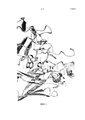
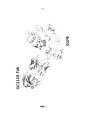
На фиг. 1 представлена диаграмма, показывающая характеристики связывания EGFR и антитела GC1118 в виде 3D-изображения.In FIG. 1 is a graph showing the binding characteristics of EGFR and GC1118 antibody in a 3D image.
На фиг. 2 представлена диаграмма, показывающая характеристики связывания EGFR и EGF в виде 3D-изображения.In FIG. 2 is a diagram showing the binding characteristics of EGFR and EGF as a 3D image.
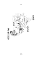
На фиг. 3 представлена диаграмма, показывающая характеристики связывания антитела GC1118 и EGFR, с которым связан EGF, если антитело GC1118 перекрывает EGFR.In FIG. 3 is a diagram showing the binding characteristics of the GC1118 antibody and the EGFR to which EGF is linked if the GC1118 antibody overlaps EGFR.
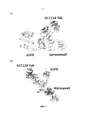
На фиг. 4 представлена диаграмма, показывающая характеристики связывания EGFR и цетуксимаба и матузумаба в виде 3D-изображения.In FIG. 4 is a graph showing the binding characteristics of EGFR and cetuximab and matuzumab as a 3D image.
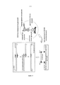
На фиг. 5 представлена диаграмма, показывающая методику синтеза вариантного гена EGFR и методику трансформации дрожжевых клеток.In FIG. 5 is a diagram showing a method for synthesizing a variant EGFR gene and a method for transforming yeast cells.
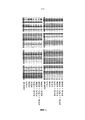
На фиг. 6 представлена диаграмма, показывающая, что соответствующие варианты EGFR синтезируются надлежащим образом посредством идентификации аминокислотной последовательности, определенной по последовательности оснований ДНК.In FIG. 6 is a diagram showing that corresponding EGFR variants are synthesized appropriately by identifying an amino acid sequence determined from a DNA base sequence.
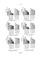
На фиг. 7 представлена диаграмма, показывающая скорость экспрессии соответствующих вариантов EGFR.In FIG. 7 is a graph showing the expression rate of the respective EGFR variants.
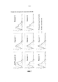
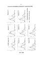
На фиг. 8 представлена диаграмма, показывающая способность к связыванию антител против соответствующих мутантов EGFR: (А) GC118 и (В) цетуксимаб (контроль).In FIG. 8 is a diagram showing the ability to bind antibodies against the corresponding EGFR mutants: (A) GC118 and (B) cetuximab (control).
На фиг. 9 представлена диаграмма, показывающая способность к конкурентному связыванию лигандов EGFR с GC118: (А) GC118 и (В) цетуксимаб (контроль).In FIG. 9 is a graph showing the ability to competitively bind EGFR ligands to GC118: (A) GC118 and (B) cetuximab (control).
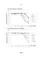
На фиг. 10 представлена диаграмма, показывающая влияние ингибирующих активностей GC118 на индукцию клеточной пролиферации.In FIG. 10 is a graph showing the effect of inhibitory activities of GC118 on the induction of cell proliferation.
Подробное описание изобретенияDETAILED DESCRIPTION OF THE INVENTION
Далее в описании настоящее изобретение будет описано подробно.Hereinafter, the present invention will be described in detail.
Авторы настоящего изобретения обнаружили, что аминокислотная последовательность SEQ ID NO: 2 или содержащая ее аминокислотная последовательность, в частности, аминокислотная последовательность RGDSFTHTP (SEQ ID NO: 3), по существу проявляет активность в отношении связывания с EGF среди аминокислотных последовательностей EGFR, и было обнаружено, что антитела с аминокислотной последовательностью в качестве эпитопа, очень эффективно ингибируют связывание EGFR и EGF и, таким образом, проявляют превосходные эффекты в отношении лечения злокачественных новообразований посредством блокирования передачи сигналов в результате связывания. Следовательно, настоящее изобретение было выполнено на основании этих фактов.The inventors of the present invention have found that the amino acid sequence of SEQ ID NO: 2 or the amino acid sequence containing it, in particular the amino acid sequence RGDSFTHTP (SEQ ID NO: 3), is substantially active in binding to EGF among the amino acid sequences of EGFR, and was found that antibodies with an amino acid sequence as an epitope very effectively inhibit the binding of EGFR and EGF and thus exhibit excellent effects in the treatment of malignant neoplasms Azings by blocking the transmission of signals by binding. Therefore, the present invention was made based on these facts.
Аминокислотная последовательность RGDSFTH, показанная как SEQ ID NO: 2, соответствует аминокислотным остаткам 353-359 аминокислотной последовательности внеклеточного домена EGFR, показанной как SEQ ID NO: 1, и аминокислотная последовательность RGDSFTHTP, показанная как SEQ ID NO: 3, соответствует аминокислотным остаткам 353-361 внеклеточного домена EGFR, показанного как SEQ ID NO: 1.The amino acid sequence RGDSFTH shown as SEQ ID NO: 2 corresponds to amino acid residues 353-359 of the amino acid sequence of the extracellular domain of EGFR shown as SEQ ID NO: 1, and the amino acid sequence RGDSFTHTP shown as SEQ ID NO: 3 corresponds to amino acid residues 353- 361 extracellular domain of EGFR shown as SEQ ID NO: 1.
Аминокислотные остатки, присутствующие в аминокислотной последовательности по настоящему изобретению, представлены посредством либо трех-, либо однобуквенных сокращений, известных из уровня техники. Кроме того, в настоящем изобретении термин “xA” относится к А аминокислоте х последовательности EGFR, показанной как SEQ ID NO: 1, и термин “xAz” означает, что А аминокислота х заменена на z. Например, термин “R353” относится к аргинину (Arg), который является 353 аминокислотным остатком аминокислотной последовательности, показанной как SEQ ID NO: 1, и термин “R353G” означает, что аргинин (Arg), который является 353 аминокислотным остатком аминокислотной последовательности, показанной как SEQ ID NO: 1, заменен на глицин (Gly).Amino acid residues present in the amino acid sequence of the present invention are represented by either three- or one-letter abbreviations known in the art. In addition, in the present invention, the term “xA” refers to an A amino acid x of the EGFR sequence shown as SEQ ID NO: 1, and the term “xAz” means that amino acid x is replaced by z. For example, the term “R353” refers to arginine (Arg), which is the 353 amino acid residue of the amino acid sequence shown as SEQ ID NO: 1, and the term “R353G” means that arginine (Arg), which is the 353 amino acid residue of the amino acid sequence, shown as SEQ ID NO: 1, replaced by glycine (Gly).
Эпитоп, имеющий аминокислотную последовательность SEQ ID NO: 2, аминокислотную последовательность, содержащую аминокислотную последовательность SEQ ID NO: 2, или аминокислотную последовательность SEQ ID NO: 3, может использоваться в комбинации с носителем для поддержания его собственной 3D-структуры или обеспечения эффективности при использовании в качестве композиции, такой как противоопухолевая вакцина. Носитель, в соответствии с одним из вариантов осуществления настоящего изобретения, приведенного в качестве примера, является биосовместимым, и все типы носителей могут быть использованы в данном случае, поскольку с ними может быть достигнут желаемый эффект. В данном случае, носитель может быть выбран из группы, состоящей из пептида, сывороточного альбумина, иммуноглобулина, гемоцианина и полисахарида, но не ограничиваясь ими.An epitope having the amino acid sequence of SEQ ID NO: 2, the amino acid sequence containing the amino acid sequence of SEQ ID NO: 2, or the amino acid sequence of SEQ ID NO: 3 can be used in combination with a carrier to maintain its own 3D structure or to ensure efficacy when used as a composition, such as an antitumor vaccine. A carrier, in accordance with one exemplary embodiment of the present invention, is biocompatible, and all types of carriers can be used in this case, since the desired effect can be achieved with them. In this case, the carrier may be selected from the group consisting of, but not limited to, a peptide, serum albumin, immunoglobulin, hemocyanin, and polysaccharide.
Эпитоп, имеющий аминокислотную последовательность SEQ ID NO: 2, аминокислотную последовательность, содержащую аминокислотную последовательность SEQ ID NO: 2, или аминокислотную последовательность SEQ ID NO: 3, может использоваться как таковой или использоваться в виде комплекса в комбинации с носителем. В этом случае, эпитоп или комплекс могут быть использованы в композиции противоопухолевой вакцины. В настоящем описании композиция вакцины может дополнительно содержать фармацевтически приемлемый адъювант или эксципиент. Может использоваться любой тип адъюванта, поскольку они предназначены для увеличения образования антитела при инъекционном введении в организм, благодаря чему достигаются цели настоящего изобретения. В частности, адъювант может представлять собой по меньшей мере один адъювант, выбранный из группы, состоящей из соли алюминия (Al(OH)3 или AlPO4), сквалена, сорбитана, полисорбата 80, СрG, липосомы, холестерина, монофосфориллипида А (MPL) и глюкопиранозиллипида А (GLA), но, не ограничиваясь ими.An epitope having the amino acid sequence of SEQ ID NO: 2, the amino acid sequence containing the amino acid sequence of SEQ ID NO: 2, or the amino acid sequence of SEQ ID NO: 3 can be used as such or used as a complex in combination with a carrier. In this case, the epitope or complex can be used in the composition of the antitumor vaccine. As used herein, a vaccine composition may further comprise a pharmaceutically acceptable adjuvant or excipient. Any type of adjuvant can be used since they are designed to increase antibody production when injected into the body, thereby achieving the objectives of the present invention. In particular, the adjuvant may be at least one adjuvant selected from the group consisting of aluminum salt (Al (OH) 3 or AlPO 4 ), squalene, sorbitan,
Полинуклеотид, кодирующий эпитоп, имеющий аминокислотную последовательность SEQ ID NO: 2, аминокислотную последовательность, содержащую аминокислотную последовательность SEQ ID NO: 2, или аминокислотную последовательность SEQ ID NO: 3 по настоящему изобретению, может использоваться в форме генетической противоопухолевой вакцины как таковой. В этом случае, полинуклеотид может использоваться сам по себе, без использования системы доставки, или он может быть доставлен в организм в составе вирусной или невирусной системы доставки. Может использоваться любой тип вирусной или невирусной системы доставки, поскольку известно, что они доступны в данной области. Конкретно, вирусная система доставки включает аденовирус, адено-ассоциированный вирус, лентивирус, ретровирус и т.п., и невирусный вектор, который может использоваться в настоящем изобретении, включает по меньшей мере один вектор, выбранный из группы, состоящей из катионного полимера, неионного полимера, липосомы, липида, фосфолипида, гидрофильного полимера, гидрофобного полимера и их комплекса, но, не ограничиваясь ими.A polynucleotide encoding an epitope having the amino acid sequence of SEQ ID NO: 2, the amino acid sequence containing the amino acid sequence of SEQ ID NO: 2, or the amino acid sequence of SEQ ID NO: 3 of the present invention can be used in the form of a genetic antitumor vaccine per se. In this case, the polynucleotide can be used on its own, without using a delivery system, or it can be delivered to the body as part of a viral or non-viral delivery system. Any type of viral or non-viral delivery system may be used since it is known that they are available in the art. Specifically, the viral delivery system includes an adenovirus, an adeno-associated virus, a lentivirus, a retrovirus and the like, and a non-viral vector that can be used in the present invention includes at least one vector selected from the group consisting of a cationic polymer, a nonionic polymer, liposomes, lipid, phospholipid, hydrophilic polymer, hydrophobic polymer and their complex, but not limited to.
Настоящее изобретение относится к рекомбинантному вектору, содержащему полинуклеотид, кодирующий эпитоп, имеющий аминокислотную последовательность SEQ ID NO: 2, аминокислотную последовательность, содержащую аминокислотную последовательность SEQ ID NO: 2, или аминокислотную последовательность SEQ ID NO: 3, к клетке-хозяину, содержащей рекомбинантный вектор, и к способу получения эпитопа, который имеет аминокислотную последовательность SEQ ID NO: 2, аминокислотную последовательность, содержащую аминокислотную последовательность SEQ ID NO: 2, или аминокислотную последовательность из SEQ ID NO: 3, используя рекомбинантный вектор или клетку-хозяина по настоящему изобретению.The present invention relates to a recombinant vector containing a polynucleotide encoding an epitope having the amino acid sequence of SEQ ID NO: 2, the amino acid sequence containing the amino acid sequence of SEQ ID NO: 2, or the amino acid sequence of SEQ ID NO: 3, to a host cell containing the recombinant vector, and to a method for producing an epitope that has the amino acid sequence of SEQ ID NO: 2, the amino acid sequence containing the amino acid sequence of SEQ ID NO: 2, or amino acid a distinct sequence from SEQ ID NO: 3 using the recombinant vector or host cell of the present invention.
В настоящем изобретении термин “рекомбинантный вектор” относится к вектору экспрессии, способному к экспрессии мишеневого белка в соответствующей клетке-хозяине, которая представляет собой генную конструкцию, содержащую незаменимый регуляторный элемент, функционально связанный для экспрессии генной вставки. В настоящем изобретении термин “функционально связанный” означает, что последовательность нуклеиновой кислоты, кодирующая желаемый белок, функционально связана с экспрессионной контрольной последовательностью нуклеиновой кислоты для выполнения общих функций. Функциональное связывание с рекомбинантным вектором может осуществляться с использованием технологий генетической рекомбинации, широко известных в данной области, и сайт-специфическое расщепление ДНК и лигирование могут легко проводиться с использованием ферментов, широко известных в данной области.In the present invention, the term “recombinant vector” refers to an expression vector capable of expressing a target protein in an appropriate host cell, which is a gene construct containing an essential regulatory element operably linked to express a gene insert. In the present invention, the term “operably linked” means that a nucleic acid sequence encoding a desired protein is operably linked to an expression control nucleic acid sequence for performing general functions. Functional binding to the recombinant vector can be carried out using genetic recombination technologies widely known in the art, and site-specific DNA digestion and ligation can be easily carried out using enzymes widely known in the art.
Подходящий вектор экспрессии, который может использоваться в настоящем изобретении, может содержать сигнальные последовательности для мембранного нацеливания или секреции в дополнение к элементам контроля экспрессии, такие как промотор, кодон инициации, стоп-кодон, сигнал полиаденилирования и энхансер. Кодон инициации и стоп-кодон, как правило, считаются частью нуклеотидной последовательности, кодирующей иммуногенный мишеневый белок, и, таким образом, должны по существу использоваться у индивида, если генную конструкцию вводят индивиду, и встраивают в рамку вместе с кодирующей последовательностью. Промотор в общем случае может быть конститутивным или индуцируемым. В прокариотических клетках присутствуют промоторы Lac, Tac, T3 и T7, но не ограничиваясь ими. Существуют промотор β-актина и промоторы, выделенные из гемоглобина человека, мышечного креатина человека и металлотионеина человека, а также промоторы, выделенные из обезьяньего вируса 40 (SV40), мышиного вируса опухоли молочных желез (MMTV), вируса иммунодефицита человека (ВИЧ) (например, промотор длинного терминального повтора (LTR) из ВИЧ), вируса Молони, цитомегаловируса (СМV), вируса Эпстейна-Барра (EBV) и вируса саркомы Рауса (RSV), присутствующих в эукариотических клетках, но, не ограничиваясь ими.A suitable expression vector that can be used in the present invention may contain signal sequences for membrane targeting or secretion in addition to expression control elements such as a promoter, initiation codon, stop codon, polyadenylation signal and enhancer. The initiation codon and stop codon are generally considered to be part of the nucleotide sequence encoding the immunogenic target protein, and thus should essentially be used in the individual if the gene construct is introduced into the individual and inserted into the frame along with the coding sequence. A promoter can generally be constitutive or inducible. In prokaryotic cells, Lac, Tac, T3, and T7 promoters are present, but are not limited to. There are β-actin promoters and promoters isolated from human hemoglobin, human muscle creatine and human metallothionein, as well as promoters isolated from monkey virus 40 (SV40), mouse mammary tumor virus (MMTV), human immunodeficiency virus (HIV) (e.g. , a promoter of long terminal repeat (LTR) from HIV), Moloni virus, cytomegalovirus (CMV), Epstein-Barr virus (EBV) and Routh sarcoma virus (RSV), present in, but not limited to, eukaryotic cells.
Вектор экспрессии может содержать маркер селекции для отбора клетки-хозяина, содержащей вектор. Маркер селекции используют для селекции клеток, трансформированных вектором. В данном случае, маркеры, придающие селектируемый фенотип, например толерантность к лекарственным средствам, ауксотрофность, толерантность к цитотоксическим средствам или экспрессию поверхностных белков, могут использоваться в качестве маркера селекции. Поскольку только клетки, экспрессирующие маркер селекции, выживают в окружении, обработанном средством для селекции, можно проводить селекцию трансформированных клеток. Также, если вектор представляет собой реплицируемый вектор экспрессии, то вектор может включать точку начала репликации, которая является определенной последовательностью нуклеиновой кислоты, от которой инициируется репликация. Различные типы векторов, таких как плазмида, вирус и космида, могут быть использованы в качестве рекомбинантных векторов экспрессии. Может использоваться любой тип рекомбинантного вектора, без конкретного ограничения, поскольку они могут функционировать для экспрессии желательного гена в различных клетках-хозяевах, таких как прокариотические клетки и эукариотические клетки, и продуцировать желаемый белок. Однако вектор, который содержит промотор сильной активности и может продуцировать в больших количествах чужеродный белок, имеющий форму, сходную белком дикого типа, в то же время сохраняя сильную интенсивность экспрессии, может предпочтительно использоваться в качестве рекомбинантного вектора.The expression vector may contain a selection marker for selecting a host cell containing the vector. A selection marker is used to select cells transformed with a vector. In this case, markers conferring a selectable phenotype, for example drug tolerance, auxotrophy, cytotoxic drug tolerance or expression of surface proteins, can be used as a selection marker. Since only cells expressing the selection marker survive in the environment treated with the selection agent, selection of transformed cells can be performed. Also, if the vector is a replicable expression vector, then the vector may include a replication start point, which is a specific nucleic acid sequence from which replication is initiated. Various types of vectors, such as plasmid, virus, and cosmid, can be used as recombinant expression vectors. Any type of recombinant vector may be used, without particular limitation, as they can function to express the desired gene in various host cells, such as prokaryotic cells and eukaryotic cells, and produce the desired protein. However, a vector that contains a strong activity promoter and can produce large quantities of a foreign protein having a form similar to a wild-type protein, while maintaining a strong expression intensity, can preferably be used as a recombinant vector.
В частности, различные комбинации хозяев/векторов экспрессии могут использоваться для экспрессии эпитопа в соответствии с одним из вариантов осуществления настоящего изобретения, приведенного в качестве примера, который имеет аминокислотную последовательность SEQ ID NO: 2, аминокислотную последовательность, содержащую аминокислотную последовательность SEQ ID NO: 2, или аминокислотную последовательность SEQ ID NO: 3. Вектор экспрессии, подходящий для хозяев-эукариотов, может содержать регуляторную последовательность для экспрессии, включающую, но без ограничения, последовательность, выделенную из SV40, вируса бычьей папилломы, аденовируса, адено-ассоциированного вируса, цитомегаловируса, лентивируса и ретровируса. Вектор экспрессии, который может использоваться в бактериальном хозяине, включает бактериальные плазмиды, полученные из Escherichia coli, такие как pET, pRSET, pBluescript, pGEX2T, вектор pUC, col E1, pCR1, pBR322, pMB9 и их производные; плазмиды, имеющие широкий диапазон хозяев, такие как RP4; фаговые ДНК, включающие различные производные фага лямбда, такие как λgt10, λgt11 и NM989; и другие ДНК фагов, такие как М13 и нитевидные одноцепочечные фаговые ДНК. Вектором, используемым для клеток насекомых, является pVL941.In particular, various combinations of expression hosts / vectors can be used to express an epitope in accordance with one exemplary embodiment of the present invention, which has the amino acid sequence of SEQ ID NO: 2, the amino acid sequence containing the amino acid sequence of SEQ ID NO: 2 , or the amino acid sequence of SEQ ID NO: 3. An expression vector suitable for eukaryotic hosts may contain a regulatory sequence for expression, including, o without limitation, the sequence isolated from SV40, bovine papilloma virus, adenovirus, adeno-associated virus, cytomegalovirus, lentivirus and retrovirus. An expression vector that can be used in a bacterial host includes bacterial plasmids derived from Escherichia coli, such as pET, pRSET, pBluescript, pGEX2T, pUC vector, col E1, pCR1, pBR322, pMB9 and their derivatives; plasmids having a wide range of hosts, such as RP4; phage DNAs including various lambda phage derivatives such as λgt10, λgt11 and NM989; and other phage DNA, such as M13 and filamentous single-stranded phage DNA. The vector used for insect cells is pVL941.
Рекомбинантный вектор вводят в клетки-хозяева для образования трансформантов. Подходящие клетки-хозяева могут включать прокариоты, такие как E. coli, Bacillus subtilis, Pseudomonas sp., Proteus mirabilis или Staphylcoccus sp., грибки, такие как Aspergillus sp., дрожжевые клетки, такие как Pichia pastoris, Saccharomyces cerevisae, Shizosaccharomyces sp. и Neurospora crassa; и другие низшие эукариоты, и клетки высших эукариотов, такие как клетки насекомых. Дополнительно, клетки-хозяева могут предпочтительно быть выделены из растений и млекопитающих, такие как клетки почки обезьяны (COS7), клетки NSO, SP2/0, клетки яичника китайского хомяка (СНО), W138, клетки почки молодого хомяка (ВНК), MDCK, линия клеток миеломы, клетки HuT 78, клетки HEK293, но без ограничения. Особенно предпочтительными являются клетки СНО.The recombinant vector is introduced into the host cells to form transformants. Suitable host cells may include prokaryotes, such as E. coli, Bacillus subtilis, Pseudomonas sp., Proteus mirabilis or Staphylcoccus sp., Fungi, such as Aspergillus sp., Yeast cells, such as Pichia pastoris, Saccharomyces cerevisae, Shizosaccharomyces. and Neurospora crassa; and other lower eukaryotes, and higher eukaryotic cells, such as insect cells. Additionally, host cells can preferably be isolated from plants and mammals, such as monkey kidney cells (COS7), NSO cells, SP2 / 0, Chinese hamster ovary cells (CHO), W138, young hamster kidney cells (BHK), MDCK, myeloma cell line, HuT 78 cells, HEK293 cells, but without limitation. Especially preferred are CHO cells.
В настоящем изобретении термин “трансформация” в клетки-хозяева охватывает любой метод введения последовательности нуклеиновой кислоты в организм, клетку, ткань или орган, и трансформация может осуществляться стандартными методиками, подходящими для клетки-хозяина, как известно в данной области. Такие методы включают, но без ограничения, электропорацию, слияние протопластов, осаждение фосфатом кальция (CaPO4), осаждение хлоридом кальция (CaCl2), встряхивание с использованием волокон карбида кремния, Agrobacteria-опосредованную трансформацию, метод ПЭГ, метод с декстрансульфатом, липофектаминовый метод и трансформацию, опосредованную сушкой/подавлением. Эпитоп, в соответствии с одним из вариантов осуществления настоящего изобретения, приведенного в качестве примера, который имеет аминокислотную последовательность SEQ ID NO: 2, аминокислотную последовательность, содержащую аминокислотную последовательность SEQ ID NO: 2, или аминокислотную последовательность SEQ ID NO: 3, может быть подвергнут крупномасштабному получению путем культивирования трансформантов, экспрессирующих рекомбинантный вектор в питательной среде. Среда и условия культивирования могут быть надлежащим образом выбраны в зависимости от клетки-хозяина. Условия, такие как температура, рН среды и время культивирования могут соответственно регулироваться для обеспечения эффективного роста клеток и получения белка в больших количествах. Как описано выше, эпитоп, имеющий аминокислотную последовательность SEQ ID NO: 2, аминокислотную последовательность, содержащую аминокислотную последовательность SEQ ID NO: 2, или аминокислотную последовательность SEQ ID NO: 3, может быть выделен рекомбинантно из среды или клеточного лизата, и может быть выделен и очищен с использованием общепринятых методов биохимического разделения (Sambrook et al., Molecular Cloning: A laboratory Manual, 2nd Ed., Col Spring Harbr Laboratory Press (1989); Deuscher, M., Guide to Protein Purificaton Methods Enzymology, Vol. 182. Academic Press. Inc., San Diego, CA (1990)). Методики биохимического разделения, которые могут быть использованы в данном случае, могут включать, но без ограничения, электрофорез, центрифугирование, гель-фильтрацию, осаждение, диализ, хроматографию (ионообменную хроматографию, аффинную хроматографию, иммуноабсорбентную хроматографию или эксклюзионную по размерам хроматографию), изоэлектрическое фокусирование и различные их модифицированные и комбинированные методы.In the present invention, the term “transformation” into host cells encompasses any method of introducing a nucleic acid sequence into an organism, cell, tissue or organ, and transformation can be carried out by standard techniques suitable for the host cell, as is known in the art. Such methods include, but are not limited to, electroporation, protoplast fusion, precipitation with calcium phosphate (CaPO 4 ), precipitation with calcium chloride (CaCl 2 ), shaking using silicon carbide fibers, Agrobacteria-mediated transformation, PEG method, dextransulfate method, lipofectamine method and transformation mediated by drying / suppression. The epitope, in accordance with one example embodiment of the present invention, which has the amino acid sequence of SEQ ID NO: 2, the amino acid sequence comprising the amino acid sequence of SEQ ID NO: 2, or the amino acid sequence of SEQ ID NO: 3, may be subjected to large-scale production by culturing transformants expressing the recombinant vector in a nutrient medium. The culture medium and conditions may be appropriately selected depending on the host cell. Conditions such as temperature, pH, and culture time can be adjusted accordingly to ensure efficient cell growth and large amounts of protein. As described above, an epitope having the amino acid sequence of SEQ ID NO: 2, the amino acid sequence containing the amino acid sequence of SEQ ID NO: 2, or the amino acid sequence of SEQ ID NO: 3, can be recombinantly isolated from the medium or cell lysate, and can be isolated and purified using conventional biochemical separation methods (Sambrook et al., Molecular Cloning: A laboratory Manual, 2nd Ed., Col Spring Harbr Laboratory Press (1989); Deuscher, M., Guide to Protein Purificaton Methods Enzymology, Vol. 182. Academic Press. Inc., San Diego, CA (1990)). Biochemical separation techniques that may be used in this case may include, but are not limited to, electrophoresis, centrifugation, gel filtration, precipitation, dialysis, chromatography (ion exchange chromatography, affinity chromatography, immunoabsorption chromatography or size exclusion chromatography), isoelectric focusing and their various modified and combined methods.
Настоящее изобретение относится к способу экспрессии эпитопа, который имеет аминокислотную последовательность SEQ ID NO: 2, аминокислотную последовательность, содержащую аминокислотную последовательность SEQ ID NO: 2, или аминокислотную последовательность SEQ ID NO: 3, на поверхности микроорганизмов или вирусов. В способе по настоящему изобретению может использоваться рекомбинантный вектор, отличающийся тем, что содержит последовательность, кодирующую индуцируемый промотор или сигнальный белок, и различные микроорганизмы или вирусы. Особенно подходящие микроорганизмы или вирусы включают, но без ограничения, рекомбинантную E. coli, рекомбинантные дрожжи и рекомбинантные бактериофаги. Технологии дисплея, широко известные в данной области, могут быть использованы для экспрессии эпитопа, имеющего аминокислотную последовательность SEQ ID NO: 2 или SEQ ID NO: 3 на поверхности микроорганизма или вируса. В частности, может быть использован, но, не ограничиваясь только им, способ связывания полинуклеотидной последовательности, кодирующей эпитоп, который имеет аминокислотную последовательность SEQ ID NO: 2, аминокислотную последовательность, содержащую аминокислотную последовательность SEQ ID NO: 2, или аминокислотную последовательность SEQ ID NO: 3, с последовательностью, кодирующей промотор или сигнальный белок для индукции экспрессии эпитопа на поверхности микробиальных или вирусных клеток, или способ делеции части гена, кодирующего белок, изначально экспрессируемый на поверхности микробиальных или вирусных клеток, и вставки полинуклеотидной последовательности, кодирующей эпитоп, который имеет аминокислотную последовательность SEQ ID NO: 2, аминокислотную последовательность, содержащую аминокислотную последовательность SEQ ID NO: 2, или аминокислотную последовательность SEQ ID NO: 3, в ген, подвергнутый частичной делеции. Эпитоп, экспрессированный на поверхностях микрорганизмов или вирусов, который имеет аминокислотную последовательность SEQ ID NO: 2, аминокислотную последовательность, содержащую аминокислотную последовательность SEQ ID NO: 2, или аминокислотную последовательность SEQ ID NO: 3, может быть выделен и очищен как таковой, и может быть использован с целью конкретного применения в соответствии с одним из вариантов осуществления настоящего изобретения, приведенного в качестве примера, и может быть использован для пэннинга антитела, специфически связывающегося с эпитопом, который имеет аминокислотную последовательность SEQ ID NO: 2, аминокислотную последовательность, содержащую аминокислотную последовательность SEQ ID NO: 2, или аминокислотную последовательность SEQ ID NO: 3, если он экспрессирован на поверхности микрорганизмов или вирусов для получения антитела.The present invention relates to a method for expressing an epitope that has the amino acid sequence of SEQ ID NO: 2, the amino acid sequence containing the amino acid sequence of SEQ ID NO: 2, or the amino acid sequence of SEQ ID NO: 3, on the surface of microorganisms or viruses. A recombinant vector can be used in the method of the present invention, characterized in that it comprises a sequence encoding an inducible promoter or signal protein, and various microorganisms or viruses. Particularly suitable microorganisms or viruses include, but are not limited to, recombinant E. coli, recombinant yeast, and recombinant bacteriophages. Display technologies widely known in the art can be used to express an epitope having the amino acid sequence of SEQ ID NO: 2 or SEQ ID NO: 3 on the surface of a microorganism or virus. In particular, a method for linking a polynucleotide sequence encoding an epitope that has the amino acid sequence of SEQ ID NO: 2, an amino acid sequence containing the amino acid sequence of SEQ ID NO: 2, or the amino acid sequence of SEQ ID NO can be used, but not limited to it. : 3, with a sequence encoding a promoter or signal protein for inducing the expression of an epitope on the surface of microbial or viral cells, or a method of deletion of a portion of a gene encoding a protein, initially expressed expressively on the surface of microbial or viral cells, and inserting a polynucleotide sequence encoding an epitope that has the amino acid sequence of SEQ ID NO: 2, the amino acid sequence containing the amino acid sequence of SEQ ID NO: 2, or the amino acid sequence of SEQ ID NO: 3, in the gene subjected to partial deletion. An epitope expressed on the surfaces of microorganisms or viruses that has the amino acid sequence of SEQ ID NO: 2, the amino acid sequence containing the amino acid sequence of SEQ ID NO: 2, or the amino acid sequence of SEQ ID NO: 3, can be isolated and purified as such, and can be used for a specific application in accordance with one embodiment of the present invention, given as an example, and can be used for panning antibodies, specifically binding communicating with an epitope that has the amino acid sequence of SEQ ID NO: 2, the amino acid sequence containing the amino acid sequence of SEQ ID NO: 2, or the amino acid sequence of SEQ ID NO: 3, if it is expressed on the surface of microorganisms or viruses to produce an antibody.
Настоящее изобретение также относится к эпитопу, имеющему аминокислотную последовательность SEQ ID NO: 2, аминокислотную последовательность, содержащую аминокислотную последовательность SEQ ID NO: 2, или аминокислотную последовательность SEQ ID NO: 3, антителу, специфически связывающегося с эпитопом, который имеет аминокислотную последовательность SEQ ID NO: 2, аминокислотную последовательность, содержащую аминокислотную последовательность SEQ ID NO: 2, или аминокислотную последовательность SEQ ID NO: 3, к применению комплекса, включающего эпитоп или полинуклеотид, кодирующий данный эпитоп, или к способу получения фрагмента такого антитела. Такое антитело может представлять собой поликлональное антитело или моноклональное антитело, и фрагмент антитела входит в объем настоящего изобретения, поскольку он сохраняет характеристики для связывания с эпитопом, который имеет аминокислотную последовательность SEQ ID NO: 2, аминокислотную последовательность, содержащую аминокислотную последовательность SEQ ID NO: 2, или аминокислотную последовательность SEQ ID NO: 3. В частности, антитело или его фрагмент в соответствии с одним из вариантов осуществления настоящего изобретения, приведенного в качестве примера, включает, но без ограничения, антитела с одиночной цепью, диатела, триатела, тетратела, Fab-фрагменты, F(ab')2-фрагменты, Fd, scFv, доменные антитела, биспецифические антитела, минитела, scAb, IgD-антитела, IgE-антитела, IgM-антитела, IgG1-антитела, IgG2-антитела, IgG3-антитела, IgG4-антитела, производные константных областей антител и искусственные антитела на основе белковых матриц, поскольку они обладают активностью связывания с эпитопом, который имеет аминокислотную последовательность SEQ ID NO: 2, аминокислотную последовательность, содержащую аминокислотную последовательность SEQ ID NO: 2, или аминокислотную последовательность SEQ ID NO: 3. Также антитела, имеющие мутации в их вариабельных областях, входят в объем настоящего изобретения, поскольку они сохраняют их характеристики в соответствии с одним из вариантов осуществления настоящего изобретения, приведенном в качестве примера. В качестве примера, мутации могут включать консервативные замены аминокислот в вариабельных областях. Консервативная замена относится к замене исходной аминокислотной последовательности на другой аминокислотный остаток, имеющий аналогичную характеристику. Например, остатки лизина, аргинина и гистидина являются сходными с точки зрения наличия у них основной боковой цепи. Также, остатки аспарагиновой кислоты и глутаминовой кислоты являются сходными с точки зрения наличия у них кислотной боковой цепи. Также, остатки глицина, аспарагина, глутамина, серина, треонина, тирозина, цистеина и триптофана являются сходными с точки зрения наличия у них незаряженной, полярной боковой цепи, а остатки аланина, валина, лейцина, треонина, изолейцина, пролина, фенилаланина и метионина являются сходными с точки зрения наличия у них неполярной боковой цепи. Дополнительно, остатки тирозина, фенилаланина, триптофана и гистидина являются сходными с точки зрения наличия у них ароматической боковой цепи. Следовательно, для специалистов в области будет очевидным, что аминокислотные замены среди группы аминокислот, имеющих сходные характеристики, как описано выше, не будут вызывать какого-либо изменения в характеристиках. В результате, способ получения антитела, имеющего мутации посредством консервативных замен в его вариабельной области входят в объем настоящего изобретения, поскольку антитело сохраняет свои характеристики.The present invention also relates to an epitope having the amino acid sequence of SEQ ID NO: 2, an amino acid sequence containing the amino acid sequence of SEQ ID NO: 2, or the amino acid sequence of SEQ ID NO: 3, an antibody specifically binding to an epitope that has the amino acid sequence of SEQ ID NO: 2, the amino acid sequence containing the amino acid sequence of SEQ ID NO: 2, or the amino acid sequence of SEQ ID NO: 3, to the use of a complex comprising an epitope or polynucleus etotide encoding a given epitope, or to a method for producing a fragment of such an antibody. Such an antibody may be a polyclonal antibody or a monoclonal antibody, and an antibody fragment is included in the scope of the present invention because it retains the characteristics for binding to an epitope that has the amino acid sequence of SEQ ID NO: 2, the amino acid sequence containing the amino acid sequence of SEQ ID NO: 2 or the amino acid sequence of SEQ ID NO: 3. In particular, an antibody or fragment thereof in accordance with one embodiment of the present invention, shown as an example, includes, but is not limited to, single chain antibodies, diabodies, triates, tetrabodies, Fab fragments, F (ab ') 2 fragments, Fd, scFv, domain antibodies, bispecific antibodies, minitel, scAb, IgD- antibodies, IgE antibodies, IgM antibodies, IgG1 antibodies, IgG2 antibodies, IgG3 antibodies, IgG4 antibodies, derivatives of constant regions of antibodies and artificial antibodies based on protein matrices, because they have binding activity to an epitope that has an amino acid sequence SEQ ID NO: 2, the amino acid sequence containing the amino acid sequence of SEQ ID NO: 2, or the amino acid sequence of SEQ ID NO: 3. Also, antibodies having mutations in their variable regions are included in the scope of the present invention, since they retain their characteristics in accordance with one of the embodiments of the present invention described in as an example. By way of example, mutations may include conservative amino acid substitutions in variable regions. Conservative substitution refers to the replacement of the original amino acid sequence with another amino acid residue having a similar characteristic. For example, residues of lysine, arginine, and histidine are similar in terms of the presence of their main side chain. Also, aspartic acid and glutamic acid residues are similar in terms of having an acid side chain. Also, the residues of glycine, asparagine, glutamine, serine, threonine, tyrosine, cysteine and tryptophan are similar in terms of the presence of their uncharged, polar side chain, and the residues of alanine, valine, leucine, threonine, isoleucine, proline, phenylalanine and methionine are similar in terms of the presence of a non-polar side chain. Additionally, residues of tyrosine, phenylalanine, tryptophan and histidine are similar in terms of the presence of an aromatic side chain. Therefore, it will be apparent to those skilled in the art that amino acid substitutions among a group of amino acids having similar characteristics, as described above, will not cause any change in characteristics. As a result, a method for producing an antibody having mutations by conservative substitutions in its variable region is included in the scope of the present invention, since the antibody retains its characteristics.
Антитело, связывающееся с эпитопом по настоящему изобретению, который имеет аминокислотную последовательность SEQ ID NO: 2, аминокислотную последовательность, содержащую аминокислотную последовательность SEQ ID NO: 2, или аминокислотную последовательность SEQ ID NO: 3, может быть получено способами, широко известными в области, к которой относится настоящее изобретение. Конкретно, антитело может быть получено путем инокуляции животному эпитопа, имеющего аминокислотную последовательность SEQ ID NO: 2, аминокислотную последовательность, содержащую аминокислотную последовательность SEQ ID NO: 2, или аминокислотную последовательность SEQ ID NO: 3, комплекса, включающего данный эпитоп, или полинуклеотида, кодирующего данный эпитоп, и продуцирования и пэннинга антитела, специфически связывающегося с эпитопом, имеющим аминокислотную последовательность SEQ ID NO: 2, аминокислотную последовательность, содержащую аминокислотную последовательность SEQ ID NO: 2, или аминокислотную последовательность SEQ ID NO: 3, из инокулированного животного.An antibody that binds to an epitope of the present invention that has the amino acid sequence of SEQ ID NO: 2, the amino acid sequence containing the amino acid sequence of SEQ ID NO: 2, or the amino acid sequence of SEQ ID NO: 3, can be obtained by methods well known in the art, to which the present invention relates. Specifically, an antibody can be obtained by inoculating an animal with an epitope having the amino acid sequence of SEQ ID NO: 2, the amino acid sequence containing the amino acid sequence of SEQ ID NO: 2, or the amino acid sequence of SEQ ID NO: 3, a complex comprising the epitope, or a polynucleotide, encoding this epitope, and the production and panning of antibodies that specifically bind to an epitope having the amino acid sequence of SEQ ID NO: 2, the amino acid sequence containing amino acids the sequence of SEQ ID NO: 2, or the amino acid sequence of SEQ ID NO: 3, from an inoculated animal.
В этом случае, животное, предпочтительно, представляет собой животное, трансформированное с целью продукции антитела, имеющего такую же последовательность, как последовательность человека, в частности, трансформированную крысу. В данном случае, может быть получено полностью антитело человека, имеющее пониженную иммуногенность, с использованием трансформированной крысы в соответствии со способами, как описано в патентах США 5569825; 5633425 и 7501552. Если животное не трансформировано для продукции антитела, имеющего такую же последовательность, как последовательность человека, может быть дополнительно осуществлена стадия гуманизации или деиммунизации антитела, полученного из животного, таким образом, что антитело становится подходящим для лечебного использования в организме, как описано в способах патентов США 5225539; 5859205; 6632927; 5693762; 6054297 и 6407213; и WO 1998/52976. Конкретно, стадия гуманизации или деиммунизации может включать стадию CDR-прививки для прививания последовательности CDR антитела, продуцируемого от животного в каркас (FR) антитела человека, и стадию блуждающей-CDR замены, вставки или делеции по меньшей мере, одной аминокислотной последовательности, для дополнительного повышения аффинности или уменьшения иммуногенности.In this case, the animal is preferably an animal transformed to produce an antibody having the same sequence as a human sequence, in particular a transformed rat. In this case, a fully human antibody having reduced immunogenicity can be obtained using a transformed rat in accordance with methods as described in US Pat. Nos. 5,569,825; 5633425 and 7501552. If the animal is not transformed to produce an antibody having the same sequence as the human sequence, the humanization or deimmunization step of an antibody derived from an animal can be further carried out so that the antibody becomes suitable for therapeutic use in the body as described in the methods of US patents 5,225,539; 5,859,205; 6,632,927; 5,693,762; 6054297 and 6407213; and WO 1998/52976. Specifically, the humanization or deimmunization step may include a CDR grafting step for grafting a CDR sequence of an antibody produced from an animal into a human antibody framework (FR), and a step-wandering-CDR substitution, insertion or deletion of at least one amino acid sequence to further enhance affinity or decreased immunogenicity.
Если непроцессированный EGFR, отличный от эпитопа, имеющего аминокислотную последовательность SEQ ID NO: 2, аминокислотную последовательность, содержащую аминокислотную последовательность SEQ ID NO: 2, или аминокислотную последовательность SEQ ID NO: 3, комплекса, включающего данный эпитоп, или полинуклеотида, кодирующего данный эпитоп, используют в качестве иммуногена, можно также применять способ пэннинга антитела, включающий первоначальный пэннинг антител, способных к связыванию с EGFR, и пэннинг антитела, специфически распознающего эпитоп, имеющий аминокислотную последовательность SEQ ID NO: 2, аминокислотную последовательность, содержащую аминокислотную последовательность SEQ ID NO: 2, или аминокислотную последовательность SEQ ID NO: 3, среди антител, первоначально подвергнутых пэннингу. В данном случае, можно также применять способ пэннинга антитела, включающий индуцирование мутаций в ключевом участке связывания эпитопа, имеющего аминокислотную последовательность SEQ ID NO: 2, аминокислотную последовательность, содержащую аминокислотную последовательность SEQ ID NO: 2, или аминокислотную последовательность SEQ ID NO: 3, и пэннинг антител, которые потеряли или уменьшили активность связывания с EGFR вследствие мутаций в ключевом сайте связывания эпитопа, имеющего аминокислотную последовательность SEQ ID NO: 2, аминокислотную последовательность, содержащую аминокислотную последовательность SEQ ID NO: 2, или аминокислотную последовательность SEQ ID NO: 3, среди антител, первоначально подвергнутых пэннингу, связывающихся с EGFR.If an unprocessed EGFR other than an epitope having the amino acid sequence of SEQ ID NO: 2, an amino acid sequence containing the amino acid sequence of SEQ ID NO: 2, or the amino acid sequence of SEQ ID NO: 3, a complex comprising the epitope, or a polynucleotide encoding the epitope are used as an immunogen, an antibody panning method can also be used, including initial panning of antibodies capable of binding to EGFR, and panning of an antibody specifically recognizing an epitope having the amino acid sequence of SEQ ID NO: 2, the amino acid sequence containing the amino acid sequence of SEQ ID NO: 2, or the amino acid sequence of SEQ ID NO: 3, among antibodies initially subjected to panning. In this case, you can also apply the method of panning antibodies, including the induction of mutations in the key site of the binding of the epitope having the amino acid sequence of SEQ ID NO: 2, the amino acid sequence containing the amino acid sequence of SEQ ID NO: 2, or the amino acid sequence of SEQ ID NO: 3, and panning for antibodies that have lost or decreased EGFR binding activity due to mutations in the key binding site of an epitope having the amino acid sequence of SEQ ID NO: 2, the amino acid sequence an amino acid sequence comprising the amino acid sequence of SEQ ID NO: 2, or the amino acid sequence of SEQ ID NO: 3, among the antibodies initially panded that bind to EGFR.
Также, антитело человека, связывающееся с эпитопом SEQ ID NO: 2 или SEQ ID NO: 3, может быть получено и подвергнуто пэннингу, с использованием технологии дисплея, широко известной в области, к которой относится настоящее изобретение. Эта технология дисплея предпочтительно представляет собой по меньшей мере одну технологию, выбранную из группы, состоящей из технологий фагового дисплея, дисплея дрожжей, дисплея бактерий и дисплея рибосом, но, не ограничиваясь ими. Получение и дисплей библиотеки может быть легко осуществлен, как описано в патентах США 5733743; 7063943; 6172197; 6348315 и 6589741. В частности, библиотеку, используемую в дисплее, предпочтительно создают, чтобы иметь последовательность антитела, полученную от человека. В частности, способ отличается тем, что включает пэннинг только антител, специфически связывающихся с эпитопом, имеющим аминокислотную последовательность SEQ ID NO: 2, аминокислотную последовательность, содержащую аминокислотную последовательность SEQ ID NO: 2, или аминокислотную последовательность SEQ ID NO: 3, с использованием эпитопа, имеющего аминокислотную последовательность SEQ ID NO: 2, аминокислотную последовательность, содержащую аминокислотную последовательность SEQ ID NO: 2, или аминокислотную последовательность SEQ ID NO: 3, или комплекса, включающего данный эпитоп.Also, a human antibody that binds to the epitope of SEQ ID NO: 2 or SEQ ID NO: 3 can be obtained and paned using display technology widely known in the field to which the present invention relates. This display technology is preferably at least one technology selected from the group consisting of, but not limited to, phage display, yeast display, bacteria display, and ribosome display technology. Obtaining and displaying the library can be easily carried out as described in US patent 5733743; 7,063,943; 6172197; 6348315 and 6589741. In particular, the library used in the display is preferably created to have an antibody sequence obtained from a human. In particular, the method is characterized in that it includes panning only of antibodies specifically binding to an epitope having the amino acid sequence of SEQ ID NO: 2, the amino acid sequence containing the amino acid sequence of SEQ ID NO: 2, or the amino acid sequence of SEQ ID NO: 3, using an epitope having the amino acid sequence of SEQ ID NO: 2, the amino acid sequence containing the amino acid sequence of SEQ ID NO: 2, or the amino acid sequence of SEQ ID NO: 3, or a complex including of the epitope.
Наконец, настоящее изобретение относится к эпитопу, имеющему аминокислотной последовательностью SEQ ID NO: 2, аминокислотную последовательность, содержащую аминокислотную последовательность SEQ ID NO: 2, или аминокислотную последовательность SEQ ID NO: 3, к комплексу, включающему данный эпитоп, или к композиции для противоопухолевых вакцин, содержащей полинуклеотид, кодирующий данный эпитоп.Finally, the present invention relates to an epitope having the amino acid sequence of SEQ ID NO: 2, the amino acid sequence containing the amino acid sequence of SEQ ID NO: 2, or the amino acid sequence of SEQ ID NO: 3, to a complex comprising the epitope, or to an antitumor composition vaccines containing a polynucleotide encoding this epitope.
Настоящее изобретение будет описано более подробно посредством примеров вариантов осуществления, приведенных ниже. Однако следует отметить, что подробное описание и конкретные примеры, указывающие на предпочтительные варианты осуществления изобретения, являются только иллюстративными, так как различные изменения и модификации в пределах объема изобретения будут понятны специалистам в данной области из этого подробного описания.The present invention will be described in more detail by way of example embodiments below. However, it should be noted that the detailed description and specific examples indicating preferred embodiments of the invention are only illustrative, as various changes and modifications within the scope of the invention will be apparent to those skilled in the art from this detailed description.
Пример 1: Идентификация структуры связывания EGFR и Fab GC1118Example 1: Identification of the binding structure of EGFR and Fab GC1118
Для идентификации структуры связывания EGFR с антиген-связывающим фрагментом (Fab) GC1118 (см. KR 10-2011-0034914 A), разработанной группой исследователей настоящего изобретения, получали данные рентгеновской дифракции кристаллической структуры композита EGFR/GC1118 Fab с использованием лучевой линии BL26B2 (Spring-8 Institution, Япония), которые индексировали и масштабировали, используя программное обеспечение HKL2000 (HKL Research Inc., США), и затем получали раннюю карту электронной плотности композита EGFR/GC1118, применяя метод молекулярной замены (MR). Для использования метода MR, были необходимы данные по 3D-структуре белка, имеющего структуру, аналогичную структуре композита EGFR/мишеневое антитело. В этом случае, структуру композита EGFR/цетуксимаб (PDB ID: IYY9) использовали в качестве структуры для композита EGFR/GC1118. Информацию по ранней фазе композита EGFR/GC1118 получали, используя программу Morlep (http://www.ysbl.york.ac.uk/-alexei/morlep.html), и задание построения модели осуществляли, используя кристаллографический объект-ориентированный набор инструментов (COOT: http://www.biop.ox.ac.uk/coot/), на основании полученной информации о ранней фазе. Затем задание по уточнению выполняли с использованием программного обеспечения Refmac5 (http://www.ccp4.ac.uk/html/refmac5.html) и программного обеспечения на базе Питон для иерархического окружения для интегрированной рентгеновской кристаллографии (PHENIX: http://www.phenix-online.org/.To identify the binding structure of EGFR to the antigen binding fragment (Fab) of GC1118 (see KR 10-2011-0034914 A) developed by a team of researchers of the present invention, X-ray diffraction data of the crystal structure of the EGFR / GC1118 Fab composite using the radiation line BL26B2 (Spring -8 Institution, Japan), which indexed and scaled using the HKL2000 software (HKL Research Inc., USA), and then received an early electron density map of the EGFR / GC1118 composite using the molecular replacement method (MR). To use the MR method, data were needed on the 3D structure of a protein having a structure similar to that of the EGFR / target antibody composite. In this case, the structure of the EGFR / cetuximab composite (PDB ID: IYY9) was used as the structure for the EGFR / GC1118 composite. Information on the early phase of the EGFR / GC1118 composite was obtained using the Morlep program (http://www.ysbl.york.ac.uk/-alexei/morlep.html), and the task of constructing the model was carried out using a crystallographic object-oriented set of tools ( COOT: http://www.biop.ox.ac.uk/coot/), based on information received about the early phase. Then, the refinement task was performed using the Refmac5 software (http://www.ccp4.ac.uk/html/refmac5.html) and the Python-based software for the hierarchical environment for integrated X-ray crystallography (PHENIX: http: // www .phenix-online.org /.
В результате, было показано, что эпитоп EGFR GC1118 был расположен в 3 домене EGFR, и, в частности, эпитоп представлял собой область короткой петли в 3 домене, которая состоит из 9 аминокислотных остатков от 353 до 361 аминокислотных остатков аминокислотной последовательности (см. SEQ ID NO: 1 и фиг. 1). Эта область петли выступает наружу, и она окружена CDR антитела. В частности, аминокислотные остатки, играющие ключевую роль в связывании между GC1118 и EGFR, представляют собой F357 и H359 EGFR. Два аминокислотных остатка окружены аминокислотными остатками области CDR антитела GC1118 и функционируют с образованием гидрофобных связей и водородных связей с аминокислотными остатками области CDR, для плотного поддержания связывания между EGFR и GC1118. R353 также способствует дополнительному связыванию с антителом через ионные связи с вариабельной областью (VH) тяжелой цепи антитела GC1118 (см. фиг. 1).As a result, it was shown that the EGFR GC1118 epitope was located in the 3 domain of EGFR, and, in particular, the epitope was a short loop region in the 3 domain, which consists of 9 amino acid residues from 353 to 361 amino acid residues of the amino acid sequence (see SEQ ID NO: 1 and Fig. 1). This loop region protrudes outward and is surrounded by the CDR of the antibody. In particular, amino acid residues that play a key role in the binding between GC1118 and EGFR are F357 and H359 EGFR. Two amino acid residues are surrounded by amino acid residues of the CDR region of the GC1118 antibody and function to form hydrophobic bonds and hydrogen bonds with the amino acid residues of the CDR region to tightly maintain binding between EGFR and GC1118. R353 also promotes additional binding to the antibody via ionic bonds to the variable region (VH) of the heavy chain of the GC1118 antibody (see FIG. 1).
В то же время, область, с которой связывается EGF, расположена между 1 и 3 доменами EGFR. В этом случае, EGF сначала связывается с 1 доменом EGFR (см. фиг. 2) для индукции структурного изменения домена EGFR, и затем связывается с 3 доменом EGFR (см. фиг. 3). В этом случае, важным аминокислотным остатком EGFR, участвующим в связывании с EGF, является D3555 3 домена (Ferguson et al., Molecular Cell, 2003, 11:507-517). В этом случае, аминокислотный остаток присутствует в петле, содержащей R353, F357 и H359, которые являются эпитопами GC1118. То есть, поскольку эпитопы EGFR, с которыми связывается GC1118, присутствуют в той же самой области, что и область, с которой связывается EGF, причем связывание антитела с эпитопами GC1118 может напрямую ингибировать связывание EGF с 3 доменом EGFR. Это дополнительно подтверждается при сравнении со структурой EGFR, связанной с EGF. Соответственно, если структура EGFR, активированная через связывание с EGF, перекрывает структуру GC1118, можно видеть, что область VH GC1118 и EGF перекрываются друг с другом (см. фиг. 3).At the same time, the region to which EGF binds is located between 1 and 3 EGFR domains. In this case, the EGF first binds to the 1 EGFR domain (see FIG. 2) to induce a structural change in the EGFR domain, and then binds to the 3 EGFR domain (see FIG. 3). In this case, an important amino acid residue of EGFR involved in binding to EGF is
При сравнении со связыванием используемого в настоящее время цетуксимаба с EGFR, цетуксимаб имеет эпитопы, расположенные в 3 домене EGFR, но участки связывания цетуксимаба были широко разбросаны на поверхности 3 домена. Следовательно, некоторые из эпитопов имели опосредованное влияние на связывание EGF (см. фиг. 4А). Матузумаб, который является еще одним антителом, также имело эпитопы, расположенные в 3 домене EGFR, но эпитопы матузумаба присутствовали в положениях, отличающихся от положений эпитопов в соответствии с одним из вариантов осуществления настоящего изобретения, приведенным в качестве примера, и расположены в области, отличной от области, с которой был связан EGF, так что эпитопы матузумаба не обладали способностью напрямую затруднять связывание EGF (см. фиг. 4В).Compared to the binding of cetuximab currently used to EGFR, cetuximab has epitopes located in the 3 domain of EGFR, but the binding sites of cetuximab were widely scattered on the surface of the 3 domain. Therefore, some of the epitopes had an indirect effect on EGF binding (see FIG. 4A). Matuzumab, which is another antibody, also had epitopes located in the 3 domain of EGFR, but the matuzumab epitopes were present at positions different from those of the epitopes in accordance with one exemplary embodiment of the present invention and are located in a region other than from the region to which EGF was bound, so the matuzumab epitopes did not have the ability to directly impede the binding of EGF (see Fig. 4B).
Пример 2: Верификация эпитопов EGFR GC1118 с использованием способа экспрессии поверхности дрожжевых клетокExample 2: Verification of EGFR GC1118 Epitopes Using a Yeast Cell Surface Expression Method
Для верификации эпитопов (аминокислотных остатков R353, F357 и H359) EGFR против GC1118, обнаруженных посредством структурной модели, соответствующие аминокислотные остатки замещали глицином, не обладающим реакционной способностью связывания, чтобы продуцировать варианты, и затем экспрессировали их на поверхностях дрожжевых клеток. Затем связывание GC1118 с вариантами EGFR, в которых эпитопы были экспрессированы на поверхностях дрожжевых клеток, подтверждали при использовании проточной цитометрии (BD FACS Calibur™, США). В результате, было подтверждено, что активность связывания с GC1118 снижалась у всех вариантов EGFR.To verify the epitopes (amino acid residues R353, F357 and H359) of the anti-GC1118 EGFRs detected by the structural model, the corresponding amino acid residues were replaced with non-binding reactive glycine to produce variants and then expressed on the surfaces of the yeast cells. Then, the binding of GC1118 to EGFR variants in which epitopes were expressed on yeast cell surfaces was confirmed using flow cytometry (BD FACS Calibur ™, USA). As a result, it was confirmed that binding activity to GC1118 was reduced in all EGFR variants.
2.1 Конструирование вариантов EGFR2.1 Designing EGFR Options
Варианты EGFR конструировали посредством замены одного или более из R353, F357 и H359 аминокислотной последовательности EGFR (см. SEQ ID NO: 1) на глицин, с использованием метода перекрывающей ПЦР, в котором генную последовательность, кодирующую внеклеточный домен EGFR человека, использовали в качестве матрицу. Результаты приведены в следующей таблице 1.EGFR variants were constructed by replacing one or more of the R353, F357, and H359 amino acid sequences of the EGFR (see SEQ ID NO: 1) with glycine using an overlapping PCR method in which the gene sequence encoding the extracellular domain of human EGFR was used as a template . The results are shown in the following table 1.
2.1.1 Реакция ПЦР для синтеза вариантов EGFR2.1.1 PCR reaction for the synthesis of EGFR variants
Два генных фрагмента (приблизительно 1150 п.о. и 720 п.о.), обычно включающие каждый из вариантов EGFR, синтезировали путем осуществления реакции ПЦР с использованием набора AccuPower™ TLA PCR PreMix (Bioneer, Корея), включающего генную последовательность EGFR человека в качестве матрицы, и набора праймеров (см. таблицу 2), сконструированных для индуцирования мутации гена EGFR. Два синтезированных генных фрагмента подвергали электрофорезу в 1% агарозном геле, и ДНК каждого из генных фрагментов очищали, используя набор Qiagen (Qiagen 28706, Германия). Последовательно, осуществляли перекрывающую реакцию ПЦР, используя ДНК набора из двух очищенных генных фрагментов в качестве одной матрицы и праймеры, имеющие аминокислотные последовательности, как показано в SEQ ID NO: 4-19 (см. таблицу 2), синтезируя, таким образом, конечные варианты генов EGFR (см. фиг. 5). Синтезированные гены расщепляли, используя ферменты рестрикции NheI/MluI (New England Biolabs, США). Термин “прямой” после названий праймеров, как приведено в таблице 2, представляет прямой праймер, и термин “обратный” представляет обратный праймер.Two gene fragments (approximately 1150 bp and 720 bp), usually including each of the EGFR variants, were synthesized by PCR using the AccuPower ™ TLA PCR PreMix kit (Bioneer, Korea), including the human EGFR gene sequence in as a matrix, and a set of primers (see table 2) designed to induce mutations of the EGFR gene. Two synthesized gene fragments were subjected to electrophoresis on a 1% agarose gel, and the DNA of each of the gene fragments was purified using a Qiagen kit (Qiagen 28706, Germany). Sequentially, an overlapping PCR reaction was performed using the DNA of a set of two purified gene fragments as a single template and primers having amino acid sequences as shown in SEQ ID NO: 4-19 (see table 2), thus synthesizing the final variants EGFR genes (see FIG. 5). The synthesized genes were digested using NheI / MluI restriction enzymes (New England Biolabs, USA). The term “forward” after the names of the primers, as shown in Table 2, represents the forward primer, and the term “reverse” represents the reverse primer.
2.1.2 Лигирование и трансформация вариантов EGFR2.1.2 Ligation and transformation of EGFR variants
Вектор экспрессии дрожжевой поверхности pCTCON (Boder, E.T. et al., Nat Biotechnol. 1997, 15(6): 553-7) расщепляли с использованием ферментов рестрикции NheI/MluI, подвергали электрофорезу в 1% агарозном геле и очищали, используя набор Qiagen. ДНК вариантов EGFR, полученных в примере 2.1.1, смешивали с pCTCON, и к ним добавляли ДНК-лигазу Т4 (New England Biolabs, США), и проводили реакцию при 25°С в течение 2 часов. Лигирующую смесь трансформировали с помощью XL1-голубых клеток E. coli (Stratagene, США) посредством электропорации с использованием Gene Pulser (Bio-Rad Laboratories, Inc., США) и инкубировали в течение часа в целом в 2 мл среды. Затем трансформированные клетки распределяли в планшет с LB-агаром, дополненным карбенициллином (Sigma, США). Далее клетки инкубировали в течение ночи при 37°С.The yeast surface expression vector pCTCON (Boder, E.T. et al., Nat Biotechnol. 1997, 15 (6): 553-7) was digested with restriction enzymes NheI / MluI, electrophoresed on a 1% agarose gel, and purified using the Qiagen kit. The DNA of the EGFR variants obtained in Example 2.1.1 was mixed with pCTCON, and T4 DNA ligase (New England Biolabs, USA) was added to them, and the reaction was carried out at 25 ° C for 2 hours. The ligation mixture was transformed with XL1-blue E. coli cells (Stratagene, USA) by electroporation using Gene Pulser (Bio-Rad Laboratories, Inc., USA) and incubated for 2 hours in total in 2 ml of medium. Then, the transformed cells were distributed on a plate with LB agar supplemented with carbenicillin (Sigma, USA). Then the cells were incubated overnight at 37 ° C.
2.1.3 Секвенирование оснований вариантов EGFR2.1.3 Sequencing of EGFR Option Bases
Колонии на планшете, культивированные в примере 2.1.2, инкубировали в течение ночи в 2 мл LB-среды, дополненной 50 мкг/мл карбенициллина, и рекомбинантную плазмиду затем отделяли, используя плазмидный мининабор Qiagen (Qiagen 27106, Германия) для определения последовательностей оснований ДНК вариантов EGFR, введенных в плазмиду. Праймеры, имеющие последовательности, как показано в SEQ ID NO: 20-22, использовали в качестве секвенирующих праймеров, используемых для секвенирования оснований (см. таблицу 3), и секвенирование оснований осуществляли и анализировали в Genotech Corporation (Тэджон, Республика Корея) в соответствии с известным методом. Последовательность оснований EGFR человека использовали в качестве контроля. После того, как были определены последовательности оснований ДНК вариантов EGFR, определенные последовательности оснований ДНК транслировали в аминокислотные последовательности с использованием веб-программы (www.expasy.org: DNA to Protein translate tool), которая транслирует последовательность оснований в аминокислотную последовательность. Результаты трансляции показаны на фиг. 6. Затем подтверждали, что все из вариантных генов EGFR, приведенных в таблице 1, были корректно введены в векторы pCTCON.Colonies on the tablet cultured in Example 2.1.2 were incubated overnight in 2 ml LB medium supplemented with 50 μg / ml carbenicillin, and the recombinant plasmid was then separated using the Qiagen plasmid miniset (Qiagen 27106, Germany) to determine DNA base sequences EGFR variants introduced into the plasmid. Primers having sequences as shown in SEQ ID NOS: 20-22 were used as sequencing primers used for base sequencing (see Table 3), and base sequencing was performed and analyzed by Genotech Corporation (Daejeon, Republic of Korea) in accordance with a known method. The human EGFR base sequence was used as a control. After the DNA base sequences of the EGFR variants were determined, specific DNA base sequences were translated into amino acid sequences using a web program (www.expasy.org: DNA to Protein translate tool) that translates the base sequence into an amino acid sequence. The translation results are shown in FIG. 6. Then it was confirmed that all of the variant EGFR genes shown in Table 1 were correctly inserted into the pCTCON vectors.
2.2. Анализ экспрессии вариантов EGFR на поверхностях дрожжевых клеток и связывания с антителом2.2. Analysis of expression of EGFR variants on yeast cell surfaces and antibody binding
2.2.1 Методика экспрессирования вариантов EGFR на поверхностях дрожжевых клеток2.2.1 Methodology for expressing EGFR variants on yeast cell surfaces
Штамм дрожжей EBY100 (S. cerevisiae) трансформировали с помощью pCTCON-EGFR, верифицированного в примере 2.1, с использованием Gene Pulser от Bio-Rad Laboratories, Inc. (фиг. 1). Трансформированные колонии инкубировали при 30°С в течение 20 часов в среде для селекции, жидкой среде SD-CAA (20 г/л глюкозы, 6,7 г/л дрожжевого азотного основания без аминокислот, 5,4 г/л Na2HPO4, 8,6 г/л NaH2PO4H2O и 5 г/л казаминокислот). Затем экспрессию варианта EGFR на поверхностях дрожжевых клеток индуцировали инкубацией трансформированных колоний при 30°С в течение 20 часов в среде для селекции, жидкой среде SD-CAA (20 г/л галактозы, 6,7 г/л дрожжевого азотного основания без аминокислот, 5,4 г/л Na2HPO4, 8,6 г/л NaH2PO4H2O и 5 г/л казаминокислот).The yeast strain EBY100 (S. cerevisiae) was transformed with pCTCON-EGFR verified in Example 2.1 using Gene Pulser from Bio-Rad Laboratories, Inc. (Fig. 1). Transformed colonies were incubated at 30 ° C for 20 hours in selection medium, SD-CAA liquid medium (20 g / L glucose, 6.7 g / L yeast nitrogen base without amino acids, 5.4 g / L Na 2 HPO 4 8.6 g / l NaH 2 PO 4 H 2 O and 5 g / l casamino acids). Then, expression of the EGFR variant on yeast cell surfaces was induced by incubating the transformed colonies at 30 ° C for 20 hours in a selection medium, SD-CAA liquid medium (20 g / L galactose, 6.7 g / L yeast nitrogen base without amino acids, 5 , 4 g / l Na 2 HPO 4 , 8.6 g / l NaH 2 PO 4 H 2 O and 5 g / l casamino acids).
2.2.2 Определение экспрессии вариантов EGFR на поверхностях дрожжевых клеток2.2.2 Determination of expression of EGFR variants on yeast cell surfaces
Экспрессию вариантов EGFR на поверхностях дрожжевых клеток подтверждали с использованием BD FACS Calibur™ (Becto Dickinson). Дрожжевые клетки, экспрессированные при плотности, равной приблизительно 1×10⁷ клеток/мл, подвергали взаимодействию с анти-с-myc 9E10 mAb (разведение 1:1000) в 0,1 мл забуференного фосфатом физраствора (PBSB, включающем 1 мг/мл BSA; pH 7,4) (при 25°С в течение 30 минут), и промывали PBSB. Дрожжевые клетки вторично подвергали взаимодействию с козьими антителами против мышиных IgG, конъюгированными с R-фикоэритрином (разведение 1:25) на льду в течение 15 минут, промывали PBSB и затем анализировали с использованием BD FACS Calibur™. Из результатов анализа видно, что варианты EGFR хорошо экспрессировались на поверхностях дрожжевых клеток (см. фиг. 7).Expression of EGFR variants on yeast cell surfaces was confirmed using BD FACS Calibur ™ (Becto Dickinson). Yeast cells expressed at a density of approximately 1 × 10 7 cells / ml were reacted with anti-c-myc 9E10 mAb (1: 1000 dilution) in 0.1 ml phosphate buffered saline (PBSB containing 1 mg / ml BSA ; pH 7.4) (at 25 ° C for 30 minutes), and washed with PBSB. Yeast cells were reacted again with goat anti-mouse IgG antibodies conjugated to R-phycoerythrin (1:25 dilution) on ice for 15 minutes, washed with PBSB and then analyzed using BD FACS Calibur ™. From the results of the analysis it can be seen that the EGFR variants were well expressed on the surfaces of yeast cells (see Fig. 7).
2.2.3 Анализ связывания между вариантами EGFR и GC1118 с использованием проточной цитометрии2.2.3 Binding Assay Between EGFR and GC1118 Variants Using Flow Cytometry
Связывание между GC1118 и вариантами EGFR, экспрессированными на поверхностях дрожжевых клеток, подтверждали с использованием BD FACS Calibur™ (Becto Dickinson). В качестве контрольного антитела использовали цетуксимаб. Дрожжевые клетки, экспрессированные при плотности, равной приблизительно 1×10⁷ клеток/мл, подвергали взаимодействию с 1 мкг каждого из антител в 0,1 мл PBSB (включающем 1 мг/мл BSA; pH 7,4) (при 25°С в течение 30 минут) и промывали PBSB. Дрожжевые клетки вторично подвергали взаимодействию с антителами против IgG человека, конъюгированными с FITC (разведение 1:50) на льду в течение 15 минут, промывали PBSB, и затем измеряли среднюю интенсивность флуоресценции с использованием BD FACS Calibur™. Результаты измерений приведены в таблице 4 и показаны на фиг. 8. Как видно из таблицы 4, было доказано, что связывание GC1118 ингибировалось у вариантов EGFR, в которых эпитопы (R353, F357 и H359, в соответствии с одним из вариантов осуществления настоящего изобретения, приведенным в качестве примера, посредством структурного анализа) были замещены глицином, и, таким образом, замененные аминокислотные остатки представляли собой эпитопы GC1118.The binding between GC1118 and EGFR variants expressed on yeast cell surfaces was confirmed using BD FACS Calibur ™ (Becto Dickinson). Cetuximab was used as a control antibody. Yeast cells expressed at a density of approximately 1 × 10 7 cells / ml were reacted with 1 μg of each of the antibodies in 0.1 ml of PBSB (including 1 mg / ml BSA; pH 7.4) (at 25 ° C. for 30 minutes) and washed with PBSB. Yeast cells were reacted a second time with anti-human IgG antibodies conjugated to FITC (1:50 dilution) on ice for 15 minutes, washed with PBSB, and then the average fluorescence intensity was measured using BD FACS Calibur ™. The measurement results are shown in table 4 and shown in FIG. 8. As can be seen from table 4, it was proved that the binding of GC1118 was inhibited in EGFR variants in which epitopes (R353, F357 and H359, in accordance with one example embodiment of the present invention, by structural analysis) were replaced glycine, and thus substituted amino acid residues, were GC1118 epitopes.
Пример 3: Анализ конкурентного связывания GC118 и лигандов с EGFRExample 3: Analysis of the competitive binding of GC118 and ligands to EGFR
Для определения, имеют ли GC118 и шесть лигандов, приведенных в таблице 5, идентичные или сходные участки связывания для EGFR, осуществляли тест на конкурентное связывание. В качестве контрольного антитела использовали цетуксимаб. Сначала, предварительную концентрацию антитела (1,5 нМ) и различные концентрации лиганда подвергали взаимодействию на планшете, покрытом 2 мкг/мл EGFR. В этом случае, лиганд присутствовал в концентрации таким образом, что молярное отношение антитела и лиганда количественно составляло 1:1, 1:10, 1:102, 1:103, 1:104 и 1:105. Полученной в результате смеси давали взаимодействовать при комнатной температуре в течение 30 минут, и затем ее промывали пять раз PBST (включающим 0,05% Твин 20; рН 7,4). Затем смесь вторично подвергали взаимодействию с антителами против IgG человека (Fc-специфичными), конъюгированными с пероксидазой (Sigma A0170, США), при комнатной температуре в течение 30 минут, и затем ее промывали пять раз PBST. Последовательно, к каждой лунке добавляли 100 мкл раствора 3,3',5,5'-тетраметилбензидина (TMB) субстрата пероксидазы для микропланшетов (KPL 50-76-03 США), и затем измеряли значения ОП (оптической плотности) при 405 нм. Было отмечено, что два реагирующих компонента (антитело и лиганд) конкурировали друг с другом, если способность к связыванию GC1118 с EGFR снижалась при возрастающей концентрации лиганда, указывая на то, что два реагирующих компонента имели идентичные эпитопы с EGFR, или имели очень близкое совпадение. Конкуренция между антителом и лигандами показана на фиг. 9 и приведена в таблице 6. В результате можно видеть, что GC1118 имеет превосходящую способность к ингибированию связывания EGF, TGF-α, BTC, EPR и HB-EGF по сравнению с цетуксимабом.To determine whether GC118 and the six ligands shown in Table 5 have identical or similar binding sites for EGFR, a competitive binding test was performed. Cetuximab was used as a control antibody. First, a preliminary antibody concentration (1.5 nM) and various ligand concentrations were reacted on a plate coated with 2 μg / ml EGFR. In this case, the ligand was present in a concentration such that the molar ratio of antibody to ligand was quantitatively 1: 1, 1:10, 1:1021:103, 1:10four and 1:105. The resulting mixture was allowed to react at room temperature for 30 minutes, and then it was washed five times with PBST (including 0.05
Пример 4: Ингибирующий эффект на пролиферацию клеток, индуцируемую лигандами EGFRExample 4: Inhibitory Effect on Cell Proliferation Induced by EGFR Ligands
Для сравнения ингибирующего эффекта GC1118 настоящего изобретения и цетуксимаба на пролиферацию раковых клеток, индуцируемую лигандами EGFR, клеточную линию колоректального рака, LS174T (ATCC, CL-188), распределяли в 96-луночном микропланшете (Nunc) таким образом, чтобы количество клеток количественно составляло 3000 клеток/лунка. Через один день, культуральную среду удаляли, и культуральные среды, не включающие бычьего сывороточного альбумина, обрабатывали GC1118 и контрольным антителом, цетуксимаб, при концентрациях, равных 0,1, 1 и 10 мкг/мл. В то же самое время, если проводили обработку антителами, культуральную среду обрабатывали лигандами EGFR по типу при концентрациях (EGF: 50 нг/мл, TGF-α: 100 нг/мл, амфирегулин: 100 нг/мл, эпирегулин: 300 нг/мл, HB-EGF: 50 нг/мл и β-целлюлин: 100 нг/мл), и затем инкубировали при 37°С в течение 3 часов в CO2-инкубаторе. Для измерения уровня пролиферации раковых клеток через 3 дня, к раковым клеткам, инкубированным в 96-луночном планшете, добавляли 40 мкл раствора MTS (CellTiter 96 Aqueous One solution Reagent, Promega), реакцию осуществляли при 37°С в течение 3 часов в CO2-инкубаторе, и затем измеряли значения ОП при 490 нм.To compare the inhibitory effect of GC1118 of the present invention and cetuximab on cancer cell proliferation induced by EGFR ligands, the colorectal cancer cell line, LS174T (ATCC, CL-188), was distributed in a 96-well microplate (Nunc) so that the number of cells was quantitatively 3000 cells / well. After one day, the culture medium was removed and culture media not containing bovine serum albumin were treated with GC1118 and a control antibody, cetuximab, at concentrations of 0.1, 1 and 10 μg / ml. At the same time, if antibody treatment was performed, the culture medium was treated with EGFR ligands of the type at concentrations (EGF: 50 ng / ml, TGF-α: 100 ng / ml, amphiregulin: 100 ng / ml, epiregulin: 300 ng / ml , HB-EGF: 50 ng / ml and β-celluline: 100 ng / ml), and then incubated at 37 ° C for 3 hours in a CO 2 incubator. To measure the proliferation of cancer cells after 3 days, 40 μl of MTS solution (CellTiter 96 Aqueous One solution Reagent, Promega) was added to cancer cells incubated in a 96-well plate, the reaction was carried out at 37 ° C for 3 hours in CO 2 incubator, and then the OD values were measured at 490 nm.
В результате, можно видеть, что GC1118 намного более эффективно ингибировало пролиферацию раковых клеток, индуцированную лигандами EGF, TGF-α, HB-EGF и BTC, по сравнению с цетуксимабом, что указывает на соответствие этих результатов результатам примера 3.As a result, it can be seen that GC1118 was much more effective in inhibiting the proliferation of cancer cells induced by the ligands EGF, TGF-α, HB-EGF and BTC, compared with cetuximab, which indicates that these results are consistent with the results of example 3.
Исходя из полученных результатов, было продемонстрировано, что антитела, имеющие эпитопы, в соответствии с одним из вариантов осуществления настоящего изобретения, приведенного в качестве примера, показали превосходящий ингибирующий эффект на пролиферацию клеток, поскольку антитела имели различные эпитопы с цетуксимабом и, таким образом, обладали способностью ингибировать связывание лиганда (см. фиг. 10). В частности, антитела, связывающиеся с эпитопами, в соответствии с одним из вариантов осуществления настоящего изобретения, приведенного в качестве примера, способны к применению для лечения различных заболеваний, таких как рак, развивающийся при активации EGFR благодаря связыванию не только с EGF, но также других лигандов EGFR, поскольку антитела эффективно ингибируют связывание различных лигандов EGFR, таких как, но, не ограничиваясь ими, EGF, а также TGF-α, AR, BTC, EPR и HB-EGF с EGFR.Based on the results obtained, it was demonstrated that antibodies having epitopes, in accordance with one exemplary embodiment of the present invention, exhibited superior inhibitory effects on cell proliferation, since the antibodies had different epitopes with cetuximab and thus had ability to inhibit ligand binding (see Fig. 10). In particular, antibodies that bind to epitopes in accordance with one exemplary embodiment of the present invention are useful for treating various diseases, such as cancer, developing upon activation of EGFR due to binding not only to EGF, but also to other EGFR ligands, because antibodies effectively inhibit the binding of various EGFR ligands, such as, but not limited to, EGF, as well as TGF-α, AR, BTC, EPR and HB-EGF with EGFR.
ПРОМЫШЛЕННАЯ ПРИМЕНИМОСТЬINDUSTRIAL APPLICABILITY
Эпитопы EGFR, предоставленные в соответствии с одним из вариантов осуществления настоящего изобретения, приведенном в качестве примера, являются высококонсервативными и расположены в домене, тесно связанном со связыванием с EGF. Следовательно, композиции, содержащие антитела против эпитопов, или композиции вакцин, содержащих эпитопы, могут эффективно блокировать передачу сигналов, вызванную связыванием EGF и EGFR, и, таким образом, могут иметь важное значение при использовании для лечения различных заболеваний, таких как злокачественные новообразования.EGFR epitopes provided in accordance with one exemplary embodiment of the present invention are highly conserved and are located in a domain closely associated with EGF binding. Therefore, compositions containing antibodies against epitopes, or compositions of vaccines containing epitopes, can effectively block signaling caused by binding of EGF and EGFR, and, thus, can be important when used for the treatment of various diseases, such as malignant neoplasms.
Claims (26)
Applications Claiming Priority (3)
| Application Number | Priority Date | Filing Date | Title |
|---|---|---|---|
| US201261616073P | 2012-03-27 | 2012-03-27 | |
| US61/616,073 | 2012-03-27 | ||
| PCT/KR2013/002550 WO2013147509A1 (en) | 2012-03-27 | 2013-03-27 | Epitopes of epidermal growth factor receptor surface antigen and use thereof |
Publications (2)
| Publication Number | Publication Date |
|---|---|
| RU2014143032A RU2014143032A (en) | 2016-05-20 |
| RU2650770C2 true RU2650770C2 (en) | 2018-04-17 |
Family
ID=49260689
Family Applications (1)
| Application Number | Title | Priority Date | Filing Date |
|---|---|---|---|
| RU2014143032A RU2650770C2 (en) | 2012-03-27 | 2013-03-27 | Epitopes of epidermal growth factor receptor surface antigen and use thereof |
Country Status (16)
| Country | Link |
|---|---|
| US (3) | US20150231220A1 (en) |
| EP (1) | EP2832363B1 (en) |
| JP (1) | JP6209206B2 (en) |
| KR (1) | KR101930019B1 (en) |
| CN (2) | CN110317259A (en) |
| BR (1) | BR112014024192A2 (en) |
| CA (1) | CA2871111C (en) |
| CO (1) | CO7160042A2 (en) |
| DK (1) | DK2832363T3 (en) |
| ES (1) | ES2694679T3 (en) |
| IN (1) | IN2014DN08778A (en) |
| MX (1) | MX356550B (en) |
| MY (1) | MY168077A (en) |
| PH (1) | PH12014502360A1 (en) |
| RU (1) | RU2650770C2 (en) |
| WO (1) | WO2013147509A1 (en) |
Families Citing this family (3)
| Publication number | Priority date | Publication date | Assignee | Title |
|---|---|---|---|---|
| EP3169699A4 (en) * | 2014-07-18 | 2018-06-20 | The University of Washington | Cancer vaccine compositions and methods of use thereof |
| KR101776879B1 (en) * | 2015-01-19 | 2017-09-08 | 주식회사 녹십자 | Pharmaceutical formulation comprising anti-egfr antibody |
| CN115226700A (en) * | 2022-08-02 | 2022-10-25 | 江苏克诺克智能科技有限公司 | Grain surface pest trapper and trapping system |
Citations (4)
| Publication number | Priority date | Publication date | Assignee | Title |
|---|---|---|---|---|
| US5459061A (en) * | 1990-01-26 | 1995-10-17 | W. Alton Jones Cell Science Center, Inc. | Hybridomas producing monoclonal antibodies which specifically bind to continuous epitope on the human EGF receptor and compete with EGF for binding to the EGF receptor |
| RU2335507C2 (en) * | 2001-06-13 | 2008-10-10 | Генмаб А/С | Human monoclonal antibodies to receptor of epidermal growth factor (egfr), method of their preparation and usage, hybridome, transfectome, transgene animal, expression vector |
| US7732157B1 (en) * | 1999-09-30 | 2010-06-08 | Tumor Biology Investment Group | Soluble epidermal growth factor receptor-like proteins and their uses in cancer detection methods |
| WO2011140391A2 (en) * | 2010-05-05 | 2011-11-10 | The Research Foundation Of State University Of New York | Egfr-related polypeptides and methods of use |
Family Cites Families (23)
| Publication number | Priority date | Publication date | Assignee | Title |
|---|---|---|---|---|
| US5225539A (en) | 1986-03-27 | 1993-07-06 | Medical Research Council | Recombinant altered antibodies and methods of making altered antibodies |
| US5530101A (en) | 1988-12-28 | 1996-06-25 | Protein Design Labs, Inc. | Humanized immunoglobulins |
| US5859205A (en) | 1989-12-21 | 1999-01-12 | Celltech Limited | Humanised antibodies |
| US7063943B1 (en) | 1990-07-10 | 2006-06-20 | Cambridge Antibody Technology | Methods for producing members of specific binding pairs |
| US6172197B1 (en) | 1991-07-10 | 2001-01-09 | Medical Research Council | Methods for producing members of specific binding pairs |
| ES2108048T3 (en) | 1990-08-29 | 1997-12-16 | Genpharm Int | PRODUCTION AND USE OF LOWER TRANSGENIC ANIMALS CAPABLE OF PRODUCING HETEROLOGICAL ANTIBODIES. |
| US7041871B1 (en) | 1995-10-10 | 2006-05-09 | Genpharm International, Inc. | Transgenic non-human animals capable of producing heterologous antibodies |
| US5633425A (en) | 1990-08-29 | 1997-05-27 | Genpharm International, Inc. | Transgenic non-human animals capable of producing heterologous antibodies |
| WO1994004679A1 (en) | 1991-06-14 | 1994-03-03 | Genentech, Inc. | Method for making humanized antibodies |
| WO1992022653A1 (en) | 1991-06-14 | 1992-12-23 | Genentech, Inc. | Method for making humanized antibodies |
| US5733743A (en) | 1992-03-24 | 1998-03-31 | Cambridge Antibody Technology Limited | Methods for producing members of specific binding pairs |
| CA2289691C (en) | 1997-04-23 | 2008-01-15 | Andreas Pluckthun | Methods for identifying nucleic acid molecules encoding (poly)peptides that interact with target molecules |
| JP2002512624A (en) | 1997-05-21 | 2002-04-23 | バイオベーション リミテッド | Method for producing non-immunogenic protein |
| EP1278851B1 (en) * | 2000-05-19 | 2006-01-18 | Scancell Limited | Humanised antibodies to the epidermal growth factor receptor |
| AUPQ841800A0 (en) * | 2000-06-28 | 2000-07-20 | Biomolecular Research Institute Limited | Truncated egf receptor |
| JP2003066677A (en) * | 2001-08-24 | 2003-03-05 | Canon Inc | Color image forming device, image correction control method and storage medium |
| CA2530172A1 (en) * | 2003-06-27 | 2005-02-10 | Abgenix, Inc. | Antibodies directed to the deletion mutants of epidermal growth factor receptor and uses thereof |
| US7767792B2 (en) * | 2004-02-20 | 2010-08-03 | Ludwig Institute For Cancer Research Ltd. | Antibodies to EGF receptor epitope peptides |
| AU2006211037B2 (en) * | 2005-02-07 | 2012-05-24 | Roche Glycart Ag | Antigen binding molecules that bind EGFR, vectors encoding same, and uses thereof |
| US20080253993A1 (en) * | 2005-03-31 | 2008-10-16 | Pharmexa A/S | Immunogenic Egfr Peptides Comprising Foreign T Cell Stimulating Epitope |
| US7985559B2 (en) * | 2007-10-19 | 2011-07-26 | Amgen Inc. | Methods of selecting epidermal growth factor receptor (EGFR) binding agents |
| EP2342220B1 (en) * | 2008-10-06 | 2021-05-12 | The University Of British Columbia | Methods and systems for predicting misfolded protein epitopes |
| KR101108642B1 (en) | 2009-09-29 | 2012-02-09 | 주식회사 녹십자 | Antibodies specifically binding to the epidermal growth factor receptor |
-
2013
- 2013-03-27 KR KR1020147027485A patent/KR101930019B1/en active IP Right Grant
- 2013-03-27 US US14/388,665 patent/US20150231220A1/en not_active Abandoned
- 2013-03-27 JP JP2015503118A patent/JP6209206B2/en active Active
- 2013-03-27 CN CN201910572748.1A patent/CN110317259A/en active Pending
- 2013-03-27 EP EP13769431.1A patent/EP2832363B1/en active Active
- 2013-03-27 DK DK13769431.1T patent/DK2832363T3/en active
- 2013-03-27 MX MX2014011597A patent/MX356550B/en active IP Right Grant
- 2013-03-27 WO PCT/KR2013/002550 patent/WO2013147509A1/en active Application Filing
- 2013-03-27 CN CN201380022281.4A patent/CN104394879A/en active Pending
- 2013-03-27 IN IN8778DEN2014 patent/IN2014DN08778A/en unknown
- 2013-03-27 ES ES13769431.1T patent/ES2694679T3/en active Active
- 2013-03-27 MY MYPI2014702810A patent/MY168077A/en unknown
- 2013-03-27 BR BR112014024192A patent/BR112014024192A2/en not_active Application Discontinuation
- 2013-03-27 CA CA2871111A patent/CA2871111C/en active Active
- 2013-03-27 RU RU2014143032A patent/RU2650770C2/en active
-
2014
- 2014-10-22 PH PH12014502360A patent/PH12014502360A1/en unknown
- 2014-10-23 CO CO14234763A patent/CO7160042A2/en unknown
-
2017
- 2017-02-06 US US15/425,413 patent/US20170145107A1/en not_active Abandoned
-
2019
- 2019-01-28 US US16/259,509 patent/US20190153117A1/en not_active Abandoned
Patent Citations (4)
| Publication number | Priority date | Publication date | Assignee | Title |
|---|---|---|---|---|
| US5459061A (en) * | 1990-01-26 | 1995-10-17 | W. Alton Jones Cell Science Center, Inc. | Hybridomas producing monoclonal antibodies which specifically bind to continuous epitope on the human EGF receptor and compete with EGF for binding to the EGF receptor |
| US7732157B1 (en) * | 1999-09-30 | 2010-06-08 | Tumor Biology Investment Group | Soluble epidermal growth factor receptor-like proteins and their uses in cancer detection methods |
| RU2335507C2 (en) * | 2001-06-13 | 2008-10-10 | Генмаб А/С | Human monoclonal antibodies to receptor of epidermal growth factor (egfr), method of their preparation and usage, hybridome, transfectome, transgene animal, expression vector |
| WO2011140391A2 (en) * | 2010-05-05 | 2011-11-10 | The Research Foundation Of State University Of New York | Egfr-related polypeptides and methods of use |
Non-Patent Citations (1)
| Title |
|---|
| HARTMANN C. et al., Peptide mimotopes recognized by antibodies cetuximab and matuzumab induce a functionally equivalent anti-EGFR immune response, Oncogene, 2010, v.29, n.32, p.4517-4527. * |
Also Published As
| Publication number | Publication date |
|---|---|
| CA2871111A1 (en) | 2013-10-03 |
| JP6209206B2 (en) | 2017-10-04 |
| BR112014024192A2 (en) | 2017-07-25 |
| EP2832363A4 (en) | 2016-03-23 |
| US20150231220A1 (en) | 2015-08-20 |
| US20190153117A1 (en) | 2019-05-23 |
| US20170145107A1 (en) | 2017-05-25 |
| CO7160042A2 (en) | 2015-01-15 |
| PH12014502360B1 (en) | 2015-01-12 |
| IN2014DN08778A (en) | 2015-05-22 |
| PH12014502360A1 (en) | 2015-01-12 |
| DK2832363T3 (en) | 2019-01-07 |
| MX356550B (en) | 2018-06-01 |
| RU2014143032A (en) | 2016-05-20 |
| CA2871111C (en) | 2022-06-28 |
| WO2013147509A1 (en) | 2013-10-03 |
| JP2015519875A (en) | 2015-07-16 |
| ES2694679T3 (en) | 2018-12-26 |
| EP2832363A1 (en) | 2015-02-04 |
| KR20140145583A (en) | 2014-12-23 |
| CN110317259A (en) | 2019-10-11 |
| KR101930019B1 (en) | 2018-12-17 |
| EP2832363B1 (en) | 2018-09-19 |
| MY168077A (en) | 2018-10-11 |
| CN104394879A (en) | 2015-03-04 |
| MX2014011597A (en) | 2015-03-10 |
Similar Documents
| Publication | Publication Date | Title |
|---|---|---|
| JP6783886B2 (en) | Anti-CTLA4 monoclonal antibody or antigen-binding fragment thereof, pharmaceutical composition and use | |
| KR102503084B1 (en) | Anti-CTLA4 and anti-PD-1 bifunctional antibodies, pharmaceutical compositions thereof and uses thereof | |
| US10221246B2 (en) | Pan-HER antibody composition | |
| EP3156421B1 (en) | Pan-her antibody composition | |
| KR20120060235A (en) | Specific binding proteins and uses thereof | |
| US20190153117A1 (en) | Epitopes of epidermal growth factor receptor surface antigen and use thereof | |
| WO2024051223A1 (en) | Pharmaceutical composition and use thereof | |
| US20240117051A1 (en) | Therapeutic combination and use thereof | |
| EA042365B1 (en) | BIFUNCTIONAL ANTIBODY AGAINST CTLA4 AND AGAINST PD-1, ITS PHARMACEUTICAL COMPOSITION AND THEIR APPLICATION |