KR20170086461A - Device and methods for delivery of simulation to a body tissue - Google Patents
Device and methods for delivery of simulation to a body tissue Download PDFInfo
- Publication number
- KR20170086461A KR20170086461A KR1020177005373A KR20177005373A KR20170086461A KR 20170086461 A KR20170086461 A KR 20170086461A KR 1020177005373 A KR1020177005373 A KR 1020177005373A KR 20177005373 A KR20177005373 A KR 20177005373A KR 20170086461 A KR20170086461 A KR 20170086461A
- Authority
- KR
- South Korea
- Prior art keywords
- tissue
- electrical
- electrode
- stimulator
- energy
- Prior art date
Links
Images


Classifications
-
- A—HUMAN NECESSITIES
- A61—MEDICAL OR VETERINARY SCIENCE; HYGIENE
- A61N—ELECTROTHERAPY; MAGNETOTHERAPY; RADIATION THERAPY; ULTRASOUND THERAPY
- A61N1/00—Electrotherapy; Circuits therefor
- A61N1/18—Applying electric currents by contact electrodes
- A61N1/32—Applying electric currents by contact electrodes alternating or intermittent currents
- A61N1/36—Applying electric currents by contact electrodes alternating or intermittent currents for stimulation
- A61N1/36014—External stimulators, e.g. with patch electrodes
- A61N1/36017—External stimulators, e.g. with patch electrodes with leads or electrodes penetrating the skin
-
- A—HUMAN NECESSITIES
- A61—MEDICAL OR VETERINARY SCIENCE; HYGIENE
- A61B—DIAGNOSIS; SURGERY; IDENTIFICATION
- A61B18/00—Surgical instruments, devices or methods for transferring non-mechanical forms of energy to or from the body
- A61B18/04—Surgical instruments, devices or methods for transferring non-mechanical forms of energy to or from the body by heating
- A61B18/08—Surgical instruments, devices or methods for transferring non-mechanical forms of energy to or from the body by heating by means of electrically-heated probes
- A61B18/082—Probes or electrodes therefor
-
- A—HUMAN NECESSITIES
- A61—MEDICAL OR VETERINARY SCIENCE; HYGIENE
- A61B—DIAGNOSIS; SURGERY; IDENTIFICATION
- A61B18/00—Surgical instruments, devices or methods for transferring non-mechanical forms of energy to or from the body
- A61B18/04—Surgical instruments, devices or methods for transferring non-mechanical forms of energy to or from the body by heating
- A61B18/12—Surgical instruments, devices or methods for transferring non-mechanical forms of energy to or from the body by heating by passing a current through the tissue to be heated, e.g. high-frequency current
- A61B18/14—Probes or electrodes therefor
- A61B18/1477—Needle-like probes
-
- A—HUMAN NECESSITIES
- A61—MEDICAL OR VETERINARY SCIENCE; HYGIENE
- A61B—DIAGNOSIS; SURGERY; IDENTIFICATION
- A61B18/00—Surgical instruments, devices or methods for transferring non-mechanical forms of energy to or from the body
- A61B18/18—Surgical instruments, devices or methods for transferring non-mechanical forms of energy to or from the body by applying electromagnetic radiation, e.g. microwaves
- A61B18/20—Surgical instruments, devices or methods for transferring non-mechanical forms of energy to or from the body by applying electromagnetic radiation, e.g. microwaves using laser
- A61B18/203—Surgical instruments, devices or methods for transferring non-mechanical forms of energy to or from the body by applying electromagnetic radiation, e.g. microwaves using laser applying laser energy to the outside of the body
-
- A—HUMAN NECESSITIES
- A61—MEDICAL OR VETERINARY SCIENCE; HYGIENE
- A61B—DIAGNOSIS; SURGERY; IDENTIFICATION
- A61B5/00—Measuring for diagnostic purposes; Identification of persons
- A61B5/05—Detecting, measuring or recording for diagnosis by means of electric currents or magnetic fields; Measuring using microwaves or radio waves
- A61B5/053—Measuring electrical impedance or conductance of a portion of the body
- A61B5/0538—Measuring electrical impedance or conductance of a portion of the body invasively, e.g. using a catheter
-
- A—HUMAN NECESSITIES
- A61—MEDICAL OR VETERINARY SCIENCE; HYGIENE
- A61N—ELECTROTHERAPY; MAGNETOTHERAPY; RADIATION THERAPY; ULTRASOUND THERAPY
- A61N1/00—Electrotherapy; Circuits therefor
- A61N1/02—Details
- A61N1/04—Electrodes
- A61N1/0404—Electrodes for external use
- A61N1/0408—Use-related aspects
- A61N1/0456—Specially adapted for transcutaneous electrical nerve stimulation [TENS]
-
- A—HUMAN NECESSITIES
- A61—MEDICAL OR VETERINARY SCIENCE; HYGIENE
- A61N—ELECTROTHERAPY; MAGNETOTHERAPY; RADIATION THERAPY; ULTRASOUND THERAPY
- A61N1/00—Electrotherapy; Circuits therefor
- A61N1/02—Details
- A61N1/04—Electrodes
- A61N1/05—Electrodes for implantation or insertion into the body, e.g. heart electrode
- A61N1/0502—Skin piercing electrodes
-
- A—HUMAN NECESSITIES
- A61—MEDICAL OR VETERINARY SCIENCE; HYGIENE
- A61N—ELECTROTHERAPY; MAGNETOTHERAPY; RADIATION THERAPY; ULTRASOUND THERAPY
- A61N1/00—Electrotherapy; Circuits therefor
- A61N1/02—Details
- A61N1/08—Arrangements or circuits for monitoring, protecting, controlling or indicating
-
- A—HUMAN NECESSITIES
- A61—MEDICAL OR VETERINARY SCIENCE; HYGIENE
- A61N—ELECTROTHERAPY; MAGNETOTHERAPY; RADIATION THERAPY; ULTRASOUND THERAPY
- A61N1/00—Electrotherapy; Circuits therefor
- A61N1/18—Applying electric currents by contact electrodes
- A61N1/32—Applying electric currents by contact electrodes alternating or intermittent currents
- A61N1/36—Applying electric currents by contact electrodes alternating or intermittent currents for stimulation
- A61N1/36014—External stimulators, e.g. with patch electrodes
- A61N1/36021—External stimulators, e.g. with patch electrodes for treatment of pain
-
- A—HUMAN NECESSITIES
- A61—MEDICAL OR VETERINARY SCIENCE; HYGIENE
- A61N—ELECTROTHERAPY; MAGNETOTHERAPY; RADIATION THERAPY; ULTRASOUND THERAPY
- A61N1/00—Electrotherapy; Circuits therefor
- A61N1/18—Applying electric currents by contact electrodes
- A61N1/32—Applying electric currents by contact electrodes alternating or intermittent currents
- A61N1/36—Applying electric currents by contact electrodes alternating or intermittent currents for stimulation
- A61N1/36014—External stimulators, e.g. with patch electrodes
- A61N1/3603—Control systems
-
- A—HUMAN NECESSITIES
- A61—MEDICAL OR VETERINARY SCIENCE; HYGIENE
- A61N—ELECTROTHERAPY; MAGNETOTHERAPY; RADIATION THERAPY; ULTRASOUND THERAPY
- A61N7/00—Ultrasound therapy
-
- A—HUMAN NECESSITIES
- A61—MEDICAL OR VETERINARY SCIENCE; HYGIENE
- A61N—ELECTROTHERAPY; MAGNETOTHERAPY; RADIATION THERAPY; ULTRASOUND THERAPY
- A61N7/00—Ultrasound therapy
- A61N7/02—Localised ultrasound hyperthermia
-
- A—HUMAN NECESSITIES
- A61—MEDICAL OR VETERINARY SCIENCE; HYGIENE
- A61B—DIAGNOSIS; SURGERY; IDENTIFICATION
- A61B18/00—Surgical instruments, devices or methods for transferring non-mechanical forms of energy to or from the body
- A61B2018/00315—Surgical instruments, devices or methods for transferring non-mechanical forms of energy to or from the body for treatment of particular body parts
- A61B2018/00452—Skin
-
- A—HUMAN NECESSITIES
- A61—MEDICAL OR VETERINARY SCIENCE; HYGIENE
- A61B—DIAGNOSIS; SURGERY; IDENTIFICATION
- A61B18/00—Surgical instruments, devices or methods for transferring non-mechanical forms of energy to or from the body
- A61B2018/00571—Surgical instruments, devices or methods for transferring non-mechanical forms of energy to or from the body for achieving a particular surgical effect
- A61B2018/00601—Cutting
-
- A—HUMAN NECESSITIES
- A61—MEDICAL OR VETERINARY SCIENCE; HYGIENE
- A61B—DIAGNOSIS; SURGERY; IDENTIFICATION
- A61B18/00—Surgical instruments, devices or methods for transferring non-mechanical forms of energy to or from the body
- A61B2018/00636—Sensing and controlling the application of energy
- A61B2018/00666—Sensing and controlling the application of energy using a threshold value
-
- A—HUMAN NECESSITIES
- A61—MEDICAL OR VETERINARY SCIENCE; HYGIENE
- A61B—DIAGNOSIS; SURGERY; IDENTIFICATION
- A61B18/00—Surgical instruments, devices or methods for transferring non-mechanical forms of energy to or from the body
- A61B2018/00636—Sensing and controlling the application of energy
- A61B2018/00773—Sensed parameters
- A61B2018/00875—Resistance or impedance
-
- A—HUMAN NECESSITIES
- A61—MEDICAL OR VETERINARY SCIENCE; HYGIENE
- A61B—DIAGNOSIS; SURGERY; IDENTIFICATION
- A61B18/00—Surgical instruments, devices or methods for transferring non-mechanical forms of energy to or from the body
- A61B18/04—Surgical instruments, devices or methods for transferring non-mechanical forms of energy to or from the body by heating
- A61B18/12—Surgical instruments, devices or methods for transferring non-mechanical forms of energy to or from the body by heating by passing a current through the tissue to be heated, e.g. high-frequency current
- A61B18/14—Probes or electrodes therefor
- A61B2018/1405—Electrodes having a specific shape
- A61B2018/1425—Needle
- A61B2018/143—Needle multiple needles
-
- A—HUMAN NECESSITIES
- A61—MEDICAL OR VETERINARY SCIENCE; HYGIENE
- A61N—ELECTROTHERAPY; MAGNETOTHERAPY; RADIATION THERAPY; ULTRASOUND THERAPY
- A61N7/00—Ultrasound therapy
- A61N2007/0004—Applications of ultrasound therapy
- A61N2007/0034—Skin treatment
-
- A—HUMAN NECESSITIES
- A61—MEDICAL OR VETERINARY SCIENCE; HYGIENE
- A61N—ELECTROTHERAPY; MAGNETOTHERAPY; RADIATION THERAPY; ULTRASOUND THERAPY
- A61N7/00—Ultrasound therapy
- A61N2007/0047—Ultrasound therapy interstitial
Abstract
본 발명은 신체 조직 내에 에너지를 전달하기 위한 시스템에 관한 것이다. 조직 천공 디바이스는 조직 표면을 천공하고 조직 표면으로부터 신체 조직 내로의 마이크로채널을 생성하도록 적응된다. 자극기는 마이크로채널 위의 또는 마이크로채널에 인접한 신체 조직에 에너지를 전달하도록 적응된다.The present invention relates to a system for transferring energy into body tissue. The tissue piercing device is adapted to perforate the tissue surface and create microchannels from the tissue surface into the body tissue. The stimulator is adapted to deliver energy to the body tissue on or adjacent to the microchannel.
Description
본 발명은 피부와 같은 조직 장벽을 통한 자극의 전달을 위한 디바이스에 관한 것이다.The present invention relates to a device for delivery of stimulation through tissue barriers such as skin.
아래의 종래기술의 공개문서들은 종래기술의 이해를 위해 관련 있는 것으로 간주된다:The following prior art public documents are considered relevant for an understanding of the prior art:
Gruzdev 외 다수에 의한 미국 특허 제7,524,317호U.S. Patent No. 7,524,317 to Gruzdev et al.
Gross에 의한 미국 특허 제8,788,045 B2호U.S. Patent No. 8,788,045 B2 by Gross
Loeb에 의한 미국 특허 제6,735,474호U.S. Patent No. 6,735,474 to Loeb
Gaunt 외 다수에 의한 미국 특허 제8,406,886호U.S. Patent No. 8,406,886 to Gaunt et al.
Libbus 외 다수에 의한 미국 특허 제8,577,458호U.S. Patent No. 8,577,458 to Libbus et al.
Avrahami에 의한 미국 특허 제6,148,232호U.S. Patent No. 6,148,232 to Avrahami
경피 전기 신경 자극(TENS)은 피부 표면 상의 표면 전극 배치 및 치료 목적으로 신경을 자극하기 위한 전류의 사용을 포함하는 비침습성 치료이다. 통증 완화, 통증 치료, 질병 치료, 만성 질병 치료 및 재활과 같은 많은 유형의 치료에서 사용하기 위해 표면 전극을 이용하여 경피 전기 자극을 제공하는 다양한 의학적 디바이스가 알려졌다. 피부와 같은 조직 장벽을 통한 전류의 침투는 조직의 저항 또는 임피던스에 의해 제한되며, 그에 따라 허용 전류(tolerable electric current)가 일반적으로 표재 신경까지만 도달할 것이다. 더 깊은 신경을 자극하기 위해서 전류를 증가시키는 것은 통증, 화상 및 과민증을 발생시킬 수 있다.Percutaneous electrical nerve stimulation (TENS) is a non-invasive therapy involving the placement of surface electrodes on skin surfaces and the use of electrical current to stimulate the nerves for therapeutic purposes. A variety of medical devices have been known that provide transdermal electrical stimulation using surface electrodes for use in many types of therapies such as pain relief, pain therapy, disease treatment, chronic disease treatment and rehabilitation. Penetration of current through tissue barrier such as skin is limited by the resistance or impedance of the tissue, so that tolerable electric current will generally reach only the superficial nerve. Increasing current to stimulate deeper nerves can cause pain, burns and hypersensitivity.
초미세 침술 바늘이 조직 내에 도입되어 신경 섬유를 전기적으로 자극하기 위해서 연성 조직 또는 근육 내에 침투하는 최소 침습 치료인 경피 전기 신경 자극(PENS)을 수행하는 의학적 디바이스가 또한 알려졌다. 침술 바늘은 정확한 치료 위치에 삽입되어야만 하며 때때로 통증 및 출혈을 발생시킨다.Medical devices for performing transcutaneous electrical nerve stimulation (PENS), a minimally invasive therapy that penetrates into soft tissues or muscles to induce micro-acupuncture needles into tissues to electrically stimulate nerve fibers, are also known. The acupuncture needle must be inserted in the correct treatment position and occasionally causes pain and bleeding.
치료적 화합물에 대한 피부의 투과성을 증가시키도록 피부의 각질층을 천공하는 다른 의학적 디바이스가 알려졌다. 이러한 디바이스는 RF 전류, 초음파, 레이저빔 및 저항성 가열 에너지 기술과 같은 에너지원을 활용한다. 이들은 심미적 치료를 위해서 그리고 또한 제약(pharmaceuticals)과 같은 고분자의 피부를 통한 전달을 가능하게 하기 위한 첫 번째 단계로서도 사용된다. 이러한 디바이스는 예를 들어, Gruzdev 외 다수에 의한 미국 특허 번호 제7,524,317호에 개시되었다.Other medical devices have been known for perforating the stratum corneum of the skin to increase the permeability of the skin to the therapeutic compound. These devices utilize energy sources such as RF current, ultrasound, laser beam and resistive heating energy technology. They are also used as a first step for aesthetic treatments and also for the delivery of polymers through the skin, such as pharmaceuticals. Such a device is disclosed, for example, in U.S. Patent No. 7,524,317 by Gruzdev et al.
Gross에 의한 미국 특허 번호 제8,788,045 B2는 피험자의 경골 신경의 1mm 내에 배치되고 경골 신경 내에 전류를 구동함으로써 다발성 신경염을 치료하도록 구동되는 전극을 포함하는 디바이스를 기술한다. 다발성 신경염은 신체의 양면, 피처링 위크니스(featuring weakness), 무감각증, 저림(pins-and-needles) 및 화상 통증 상의 대략 동일한 영역에서 말초 신경(말초 신경통)에 영향을 미치는 손상 또는 질병이다.U.S. Patent No. 8,788,045 B2 by Gross describes a device including an electrode that is disposed within 1 mm of the subject's tibial nerve and is driven to treat multiple neuritis by driving current in the tibial nerve. Polyneuritis is an impairment or disease that affects the peripheral nerves (peripheral neuralgia) in approximately the same area of the body on both sides, featuring weakness, numbness, pins-and-needles and burn pain.
Loeb에 의한 미국 특허 제6,735,474호는 회음부 피부 아래 및/또는 경골 신경 부근의 하나 이상의 배터리 또는 무선 주파수로 동력이 공급되는 마이크로-자극기를 주입 및/또는 복강경 이식하는 것을 포함하는 실금 및/또는 골반통의 치료를위한 시스템을 기술한다. 이 시스템 및 방법은 불수의적 방광 수축을 약화시키는 신경 경로를 자극함으로써 방광 비움의 무의식적인 에피소드의 발생 정도를 감소시키는 경향이 있다. 변실금의 발생빈도가 유사하게 감소되거나 제거된다.U. S. Patent No. 6,735, 474 to Loeb discloses a method of delivering incontinence and / or pelvic pain comprising implanting and / or laparoscopic implantation of a micro-stimulator powered by one or more batteries or radio frequency beneath the perineal area and / Describe the system for treatment. These systems and methods tend to reduce the incidence of unconscious episodes of bladder emptying by stimulating nerve pathways that weaken involuntary bladder contraction. The incidence of incontinence is similarly reduced or eliminated.
또한, 만성 통증을 차단하기 위해서 말초신경계 내의 감각 신경에 고주파 자극을 전달함으로써 만성 통증을 치료하기 위한 시스템이 알려졌다. 전극은 말초신경 둘레에 배치되며 심박조율기 크기의 생성기에 의해 작동된다.Also, systems for treating chronic pain by delivering high frequency stimuli to the sensory nerves within the peripheral nervous system have been known to block chronic pain. The electrodes are placed around the peripheral nerve and are activated by the pacemaker size generator.
과민성 방광(OAB) 및 요절박, 빈뇨 및 절박 요실금과 같은 연관 증상의 치료를 위해 피부를 통해 경골 신경 자극을 전달하는 최소 침습 시스템 또한 알려졌다. 이 시스템은 방광 기능을 위한 제어 센터인 천골 신경망으로 이동하는 경골 신경 전기 펄스에 경피 자극을 전달하는 데에 사용된다.Also known are minimally invasive systems that deliver tibial nerve stimulation through the skin for the treatment of overactive bladder (OAB) and related conditions such as pains, urinary frequency and urge incontinence. The system is used to deliver transcutaneous stimulation to the tibial nerve electrical pulse that travels to the sacral neural network, a control center for bladder function.
전기 신호가 전극을 통해서 리드(lead) 아래로 타깃 신경까지 경피로 전달되는 말초신경 기원의 만성 통증 치료를 위한 시스템이 개시되었다. 이러한 디바이스가 타깃화된 신경에 전기 자극을 타깃화할 수 있는 동시에, 자극 에너지의 통로는 각질층에 의해서 극적으로 감소된다.A system for the treatment of chronic pain of peripheral nerve origin has been disclosed in which an electrical signal is delivered percutaneously to the target nerve under a lead through an electrode. While such a device can target electrical stimulation to the targeted nerve, the path of stimulation energy is dramatically reduced by the stratum corneum.
미국 특허 번호 제8,406,886호는 피험자의 신경계 장애를 치료하기 위한 임플란트, 시스템 및 방법을 기술한다. 이 방법은 치료될 장애 및 주파수에 따라서 신경 임펄스를 활성화 또는 차단하기 위해 타깃 신체 조직을 전기적으로 자극하도록 전류를 라우트하는 수동 전기 전도체를 이용하는 것을 포함한다. 신경 및 근육과 같이 전기적으로 여기가능한(excitable) 신체 조직은 피부 외측에 도포되는 전극들 사이에 인가되는 전기 에너지에 의해 활성화될 수 있다. 전기 에너지, 예를 들어 전류는 양의 전극과 음의 전극 사이에서 피부를 통해 흐르며, 전극 아래에 있는 신경들과 근육들 내의 활동 전위를 유발한다. 이러한 방법은 통증을 완화하는 경피 전기 신경 자극기(TENS), 운동 목적으로 근육을 활성화하는 치료적 전기 자극기, 일상생활의 업무를 위해 근육을 활성화하는 기능적 전기 자극기 및 손상된 뼈의 재생을 촉진하는 자극기를 포함하는 서로 다른 타입의 자극기들에서 수년간 사용되어왔다.U.S. Patent No. 8,406,886 describes implants, systems and methods for treating a subject's neurological disorders. The method includes using a passive electrical conductor to route the electrical current to electrically stimulate the target body tissue to activate or block the nerve impulse, depending on the disorder and frequency to be treated. Electrically excitable body tissues such as nerves and muscles can be activated by the electrical energy applied between the electrodes applied to the outside of the skin. Electrical energy, for example current, flows through the skin between the positive and negative electrodes, causing action potentials in the nerves and muscles beneath the electrodes. These methods include transcutaneous electrical nerve stimulation (TENS) to relieve pain, therapeutic electrical stimulators to activate muscles for exercise, functional electrical stimulators to activate muscles for daily activities, and stimulators to stimulate regeneration of damaged bones Have been used in different types of stimulators for many years, including.
미국 특허 번호 제8,577,458호는 리드가 없는 심박수 모니터링을 이용하는, 만성 심장기능장애의 치료를 위해 목의 미주 신경에 전기 자극을 전달하기 위한 이식 가능한 디바이스를 기술한다. 자극 치료 리드는 목의 미주 신경 시스(sheath)의 외부 지름에 따르도록 구성된 나선형 전극 및 나선형 전극에 전기적으로 접속된 커넥터 핀들의 세트를 포함한다. 신경 자극기는 전기적 리셉터클을 포함하고 이러한 전기 리셉터클 내에 커넥터 핀들이 안전하고 전기적으로 연결된다. 신경 자극기는 또한 양방향 동작 전위를 트리거링함으로써 심장의 내인성 신경계 및 환자의 중심 반사작용 모두를 원심성으로(efferently) 활성화하도록 조절되는 자극 인가 및 자극 억제의 교번하는 사이클 내에서 나선형 전극을 통해 미주 신경을 치료적으로 자극하도록 구성된 펄스 생성기를 포함한다.U.S. Patent No. 8,577,458 describes an implantable device for delivering electrical stimulation to the vagus nerve of the neck for the treatment of chronic cardiac dysfunction utilizing leadless heart rate monitoring. The stimulation treatment lead comprises a set of connector pins electrically connected to a helical electrode and a helical electrode configured to conform to the outer diameter of the vagal nerve sheath of the neck. The nerve stimulator includes an electrical receptacle in which the connector pins are securely and electrically connected. The neurostimulator can also be used to treat vagus nerves through a spiral electrode within alternating cycles of stimulation and stimulation controlled to activate efferently both the cardiac endogenous nerve system and the central reflex of the patient by triggering a bi- Lt; / RTI > pulse generator configured to < / RTI >
Avrahami에 의한 미국 특허 번호 제6,148,232호는 각질층 에피더미디스(epidermidis)를 제거하기 위한 디바이스를 기술한다.U.S. Patent No. 6,148,232 to Avrahami describes a device for removing the stratum corneum epidermidis.
본 발명은 신체 조직 내에 에너지를 전달하기 위한 시스템 및 방법을 제공한다. 본 발명의 시스템은 치료받을 조직 표면을 천공하고 조직 표면으로부터 조직 표면 아래의 조직 내로 연장하는 마이크로채널을 생성하는 조직 천공 디바이스를 포함한다. 이 시스템은 또한 신체 조직에 에너지를 전달하는 자극기를 포함한다. 천공 디바이스는 조직 표면을 천공하기 위해서 알려진 임의의 방법에 의해서 조직 내에 마이크로채널을 생성할 수 있으며, 예를 들어 초음파 에너지, 레이저 광 에너지, 열 에너지 및 전기 에너지 중 임의의 하나 이상을 포함할 수 있다. 전기 자극, 전기 신경 자극 및 전자기복사와 같은 임의의 타입의 자극 에너지가 자극기에 의해 전달될 수 있다.The present invention provides systems and methods for transferring energy into body tissues. The system of the present invention includes a tissue punch device that punctures the tissue surface to be treated and creates microchannels extending from the tissue surface into the tissue below the tissue surface. The system also includes a stimulator that transfers energy to the body tissue. The puncturing device can create microchannels in tissue by any known method for perforating tissue surfaces and can include any one or more of, for example, ultrasonic energy, laser light energy, thermal energy, and electrical energy . Any type of stimulation energy, such as electrical stimulation, electrical nerve stimulation, and electromagnetic radiation, may be delivered by the stimulator.
본 명세서에서 사용되는 바와 같이, 마이크로채널이라는 용어는 신체 조직 내로의 전기 자극과 같은 자극의 침투를 증가시키도록 신체 조직 내에 형성된 채널을 지칭한다. 피부의 표피층인, 예를 들어 각질층은 내부 피부층에 비교하여 상대적으로 높은 저항 및 커패시턴스에 의해 특징지어진다. 저항 및 커패시턴스의 특징은 각질층 내의 마이크로채널의 형성 후에 수정 및/또는 감소된다.As used herein, the term microchannel refers to a channel formed in body tissue to increase penetration of stimuli, such as electrical stimulation into body tissue. The epidermis of the skin, for example the stratum corneum, is characterized by relatively high resistance and capacitance compared to the inner skin layer. The characteristics of the resistance and capacitance are modified and / or reduced after formation of the microchannels in the stratum corneum.
조직 천공 디바이스 및 자극기는 천공 디바이스 및 자극기로의 전기 에너지 전달을 위한 전기 회로를 포함하는 제어기의 제어 하에 있다.The tissue perforation device and the stimulator are under the control of a controller including an electrical circuit for transferring electrical energy to the perforation device and the stimulator.
본 발명의 시스템은 또한 조직 저항, 임피던스, 커패시턴스, 또는 광학 밀도와 같은 조직의 하나 이상의 전기적 또는 광학적 파라미터를 모니터하는 프로세서를 포함한다. 조직 천공 디바이스가 치료될 신체 조직의 표면에 적용되어 활성화될 때 표면 아래 신체 조직의 부피 내에 마이크로채널이 생성된다. 조직의 광학적 또는 전기적 파라미터 중 적어도 하나가 사전결정된 범위 내에 있을 때, 예를 들어 조직의 광학적 또는 전기적 파라미터 중 적어도 하나가 초기값의 적어도 사전결정된 백분율 및/또는 적응 가능한 백분율만큼 감소되었을 때, 자극이 조직에 인가될 수 있다. 본 발명인은 자극기를 통해서 원하는 효과를 획득하기 위해서 요구되는 전류 및/또는 전압 또는 광 세기를 감소시키기 위해서, 조직의 천공이 저항 및 커패시턴스와 같은 조직의 전기적 또는 광학적 파라미터를 수정하는 경향이 있음을 발견하였다. 이것은 또한 자극 중의 통증을 감소시키고 조직의 더욱 깊은 층으로의 자극의 침투를 촉진한다.The system of the present invention also includes a processor that monitors one or more electrical or optical parameters of the tissue, such as tissue resistance, impedance, capacitance, or optical density. Microchannels are created within the volume of the bodily tissue below the surface when the tissue piercing device is applied and activated on the surface of the bodily tissue to be treated. When at least one of the optical or electrical parameters of the tissue is within a predetermined range, for example when at least one of the tissue's optical or electrical parameters has been reduced by at least a predetermined percentage and / or an adaptable percentage of the initial value, May be authorized to the organization. It has been discovered that the perforation of tissue tends to modify the electrical or optical parameters of tissue, such as resistance and capacitance, in order to reduce the current and / or voltage or light intensity required to achieve the desired effect through the silver stimulator of the present invention Respectively. It also reduces pain during stimulation and promotes penetration of stimuli into deeper layers of tissue.
조직의 전기적 파라미터는 예를 들어 자극기 내의 표면 전극에 의해서 또는 천공 디바이스 내의 마이크로바늘에 의해서 측정될 수 있다.The electrical parameters of the tissue can be measured, for example, by surface electrodes in a stimulator or by microneedles in a perforation device.
조직 천공 디바이스는 치료될 조직 표면 내로 압착되는 하나의 또는 다수의 마이크로바늘을 포함할 수 있다. 일 실시예에서, 국부적인 전력 소멸로 인해서 조직 내의 마이크로바늘 둘레에 적어도 하나의 마이크로채널이 형성되도록 충분한 길이의 시간 동안 둘 이상의 마이크로바늘 사이에 전류가 인가된다. 이것은 피부 표면의 경우에서는 각질층과 같은 조직 표면의 최외곽 층 내의 마이크로바늘 둘레의 조직의 절제로 이어진다. 다른 실시예에서, 마이크로채널을 생성하기 위해 마이크로바늘 내에 초음파 펄스파가 생성된다. 또 다른 실시예에서, 조직 내에 마이크로채널을 형성하기 위해 가열 모듈이 마이크로바늘을 가열한다.The tissue perforation device can include one or more micro-needles that are squeezed into the tissue surface to be treated. In one embodiment, current is applied between two or more microneedles for a length of time sufficient to allow at least one microchannel to form around the microneedles in the tissue due to local power dissipation. This leads to the ablation of the tissue around the micro-needle in the outermost layer of the tissue surface such as the stratum corneum in the case of the skin surface. In another embodiment, an ultrasonic pulse wave is created in the micro-needle to create a microchannel. In another embodiment, a heating module heats the microneedles to form microchannels within the tissue.
조직 천공 디바이스의 다른 실시예에서, 마이크로채널을 생성하기 위해서 전자기복사가 조직을 향해 타깃화된다.In another embodiment of the tissue piercing device, electromagnetic radiation is targeted towards the tissue to create a microchannel.
본 발명의 시스템 및 발명은 아래와 같은 임의의 광범위한 의학적 및 심미적 치료에서 사용될 수 있다:The system and invention of the present invention can be used in any of a wide variety of medical and aesthetic treatments, including:
* 심미 치료. 이것은 예를 들어 안면 심미 치료일 수 있다. 마이크로채널 및 자극이 안면 피부 상에 제공될 수 있다* Aesthetic treatment. This can be, for example, a facial aesthetic treatment. Microchannels and stimulation may be provided on the facial skin
* 요통 및/또는 손목굴증후군 및/또는 어깨 통증과 같은 통증 완화. 손목굴증후군을 위해서 손목의 배면 상에 치료가 제공될 수 있다. 요통을 위해서, 통증 위치 영역의 상단 상에 치료가 제공될 수 있다 Pain relief such as back pain and / or wrist osteoarthritis and / or shoulder pain. Treatment may be provided on the back of the wrist for wrist ulceration syndrome. For low back pain, treatment may be provided on top of the pain location area
* 신체 내의 서로 다른 치료들을 위한 주변 신경 자극의 전달* Delivery of peripheral nerve stimulation for different treatments in the body
* 근육 운동 또는 근긴장도 향상. 근육 치료는 근육의 영역 내에 그리고 근육의 위에 제공될 수 있다* Muscle exercise or muscle tone improvement. Muscle care can be provided within the area of the muscles and above the muscles
* 요실금: 이것의 치료는 경골 신경, 보다 구체적으로는 후경골 신경; 또는 질을 자극함으로써 수행될 수 있다* Urinary incontinence: treatment of this tibial nerve, more specifically posterior tibial nerve; Or by stimulating the vagina
* 변실금: 이것의 치료는 경골 신경의 자극에 의해 수행될 수 있다* Fecal incontinence: treatment of this can be performed by stimulation of the tibial nerve
* 뇌졸중 이후 근육을 활성화하기 위한 자극* Stimulation to activate muscles after stroke
* 재활: 허벅지와 같은 다리 근육 또는 팔 근육이 자극받을 수 있다. 재활 치료를 위한 자극은 뼈 또는 인대 부상 이후 특정 근육 내의 근육 질량을 강화하도록 사용될 수 있다. 이것은 또한 척수 부상 및/또는 뇌졸중과 같은 뇌 장애로 인한 마비로부터 고통받는 피험자의 운동의 복원에도 사용될 수 있다. 예를 들어 일상생활의 과제를 지원하기 위한 통제와 같은 기능적인 향상을 복원할 수 있다.Rehabilitation: Leg muscles such as thighs or arm muscles can be irritated. Stimulation for rehabilitation therapy can be used to strengthen muscle mass within a particular muscle after bone or ligament injuries. It can also be used to restore the subject's movement suffering from paralysis due to brain injury such as spinal cord injury and / or stroke. For example, functional enhancements such as controls to support daily life tasks can be restored.
* 다양한 근육을 직접 또는 간접적으로 자극하는 신경들의 자극. 예를 들어, 간질, 우울증, 알츠하이머, 불안감, 비만, 폭식증, 이명, 강박장애, 또는 심부전의 치료를 위해 미주 신경 또는 그의 가지들 중 하나에 인접한 목, 안면, 가슴 또는 복부 상에 자극이 제공될 수 있다.Stimulation of nerves stimulating various muscles directly or indirectly. For example, stimulation is provided on the neck, face, chest or abdomen adjacent to one of the vagus nerve or its branches for the treatment of epilepsy, depression, Alzheimer's, anxiety, obesity, bulimia, tinnitus, obsessive compulsive disorder, .
* 미주 신경의 자극은 또한 다음과 같은 임의의 증상에서도 사용될 수 있다: 중독, 불안장애, 자폐증, 조울증, 뇌성마비, 만성 두통, 알츠하이머 질병과 연관된 인지 장애, 혼수상태, 우울증, 식이장애(예를 들어, 폭식증 및 거식증), 본태떨림, 섬유근통, 심부전, 지속성 편두통, 소아근육대간질, 편두통, 감정장애, 기면증, 비만, 강박장애, 수면장애, 이명 및 투렛 증후군, 경조 인격장애 또는 임의의 다른 기질성 수면 과잉, 긴장성 두통, 알코올성 수면장애, 약물성 수면장애, 간헐적 감정장애, 지폐장애, 강박장애, 기분저하장애, 알코올 의존 증후군, 약물 의존증, 약물의 비의존적 오용, 신경성 무식용증, 특정한 비기질성 유래 수면장애, 상세불명 식이장애, 긴장성 두통, 하루주기율동수면장애, 기질성 반응소실증, 기질성 수면장애, 본태성 또는 다른 특정 형태의 떨림, 지속성 편두통, 뇌성마비, 편두통, 급발작 및 발작성 수면, 류마티스성 심장 장애, 근육통 및 근염, 수면장애, 다식증, 감정, 파키슨병 및 두통. 자극은 다른 신경을 통해서뿐 아니라, 또는 신체 내의 특정 위치 및/또는 장기 및/또는 신경에 직접 수행될 수 있다. 미주 신경의 자극은 뇌의 감정 센터 내의 화학적 불균형을 재설정하는 것을 도울 수 있다.* Vagus nerve stimulation may also be used in any of the following conditions: addiction, anxiety disorders, autism, bipolar disorder, cerebral palsy, chronic headache, cognitive disorders associated with Alzheimer's disease, coma, depression, Migraine, emotional disorders, narcolepsy, obesity, obsessive compulsive disorder, sleep disorders, tinnitus and Tourette's syndrome, mood disorder or any other disorder or condition, including, for example, bulimia nervosa and anorexia), tremor, fibromyalgia, heart failure, persistent migraine, Alzheimer's syndrome, drug dependence, drug abuse, anorexia nervosa, a specific non-temperament, anorexia nervosa, anorexia nervosa, anxiety disorders, obsessive-compulsive disorder, Gestational-induced sleep disorders, unspecified dietary disorders, tension-type headache, day-period rhythm sleep disorder, somatic response bipolar disorder, maternal sleep disorder, intrinsic or other Persistent migraine, cerebral palsy, migraine, acute seizures and paroxysmal sleep, rheumatic heart disease, myalgia and myositis, sleep disorders, bulimia, emotions, Parkinson's disease and headache. Stimulation may be performed not only through other nerves, but also directly to specific locations within the body and / or to the organs and / or nerves. Stimulation of vagus nerves can help to reset the chemical imbalance within the emotional center of the brain.
* 전립선암과 같은 암의 치료* Treatment of cancer, such as prostate cancer
* 신경 자극을 통한 면역 체계의 제어Control of the immune system through nerve stimulation
* 미주 신경의 자극을 통한 전신 염증의 감소. 미주 신경 자극은 염증을 완화하도록 신체의 자연적인 염증 반사를 활성화하고 임상 징후 및 증상을 개선할 수 있다. 염증 반사는 신체의 면역 체계를 조절하는 신경생리학적 메커니즘이다. 이것은 감염, 조직 부상 및 염증을 감지하여 이러한 정보를 중추신경계에 전달하고, 그 다음 반사적으로 비장 및 그 외의 내장 장기들을 광범위하게 자극하는 미주 신경 및 비장 신경을 통해서 말초적으로 신경 신호를 증가시킨다. 신호는 비장 내의 T 세포로 전송되며 단핵구 및 대식세포를 포함하는 주효세포에 염증을 시작하고 지속시키는 매개자(mediator)의 생산을 감소시키도록 지시한다. 염증은 류마티스성 관절염, 염증성장질환, 건선, 당뇨병, 심장 질환 및 다수의 경화증을 포함하는 급성질환 및 만성 질환에서 중요한 역할을 한다.* Reduction of systemic inflammation through vagus nerve stimulation. Vagus nerve stimulation can activate the body's natural inflammatory reflexes to relieve inflammation and improve clinical signs and symptoms. Inflammation reflex is a neurophysiological mechanism that regulates the body's immune system. It senses infection, tissue injury and inflammation and transmits this information to the central nervous system, which in turn increases neurological signals nervously through the vagus nerve and spleen nervously stimulating the spleen and other organs. The signal is transmitted to T cells in the spleen and directs primary cells containing monocytes and macrophages to decrease the production of mediators that initiate and sustain inflammation. Inflammation plays an important role in acute and chronic diseases including rheumatoid arthritis, inflammatory growth diseases, psoriasis, diabetes, heart disease and multiple sclerosis.
* 당뇨성 신경병증, 척추 수술 후 통증 증후군, 복합부위 통증 증후군, 환상지통, 허혈 사지통, 난치성 편측 사지통 증후군, 대상포진 후 신경통 및 급성 대상포진 통증, 샤코마리투스(CMT; Charcot-Marie-Tooth) 병, 심부전 및 심근경색증, 알츠하이머 뇌졸중, 파키슨병 및 편두통을 포함하는 만성적 및 불인성 통증의 치료를 위한 효율적인 치료, 통증 완화를 위한 자극의 전달.* Charcot-Marie-Tooth (CMT), diabetic neuropathy, post-operative pain syndrome, complex regional pain syndrome, cyclic pain, ischemic dyspareunia, intractable unilateral dyspepsia syndrome, postherpetic neuralgia and acute shingles pain, Effective treatment for the treatment of chronic and non-inflammatory pain, including pain, heart failure and myocardial infarction, Alzheimer's stroke, Parkinson's disease and migraine, delivery of stimuli for pain relief.
* 당뇨성 신경병증 또는 신경성 동통, 우울증, 당뇨성 말초신경병증의 치료, 치료의 가속화, 부종 감소, 만성 당뇨성 말초신경병증 외의 원인으로부터 야기되는 통증의 감소, 허혈로 인한 만성 통증의 치료.* Treatment of diabetic neuropathy or neuropathic pain, depression, diabetic peripheral neuropathy, accelerated treatment, reduction of edema, reduction of pain caused by causes other than chronic diabetic peripheral neuropathy, treatment of chronic pain due to ischemia.
* 등 통증, 당뇨성 통증, 관절통, 섬유근육통, 두통, 반사 교감신경 이상증, 조직 손상, 천골신경근 또는 요선 신경총, 협심증 및 감각이상성 배통의 치료.* Treatment of back pain, diabetic pain, arthralgia, fibromyalgia, headache, reflex sympathetic dystrophy, tissue damage, sacral nerve or pyloric plexus, angina pectoris, and sensory abnormalities.
* 목 부위 통증, 목의 신경근병증, 목 부위 경련, 만성 목 통증, 척추 수술 후 통증 증후군, 요통, 허리 근육 경련, 요천골 근막염, 요천골 신경근병증, 무릎 관절염, 대상포진 후 신경통, 관절염, 암성 통증, 목 부위 통증, 섬유근육통, 관절통, 요통, 편두통, 수술 후 통증 및 좌골신경통의 치료.* Neuralgia of the neck, neuropathic pain of the neck, neck cramps, chronic neck pain, pain syndrome after spinal surgery, back pain, back muscle spasm, urinary sac fasciitis, osteoarthritis, postherpetic neuralgia, arthritis, cancer Pain, neck pain, fibromyalgia, arthralgia, back pain, migraine, postoperative pain and sciatica.
* 급성 통증, 수술 후 통증, 급성 및 만성 두통, 만성 요통, 심복부 통증, 고관절 골절 통증, 신경성 동통, 골반통, 측두하악골 관절(TMJ) 통증; 만성 불인성 통증의 치료, 신경 자극을 통한 근육 세기의 치료(실금)* Acute pain, postoperative pain, acute and chronic headache, chronic back pain, heartburn pain, hip fracture pain, neurogenic pain, pelvic pain, temporomandibular joint (TMJ) pain; Treatment of chronic inflammatory pain, treatment of muscular strength through nerve stimulation (incontinence)
* 상처 치유를 위한 자극. 체력과 힘을 기르도록 특정 근육 그룹을 훈련하거나 또는 운동 세션, 운동, 피트니스 및 예를 들어 뇌 또는 척수 병변을 겪는 손상된 운동 기능의 회복 후에 근육을 이완하기 위한 스포츠를 위한 자극.Stimulation for wound healing. Stimulation for sports to train specific muscle groups to train strength and strength, or to relax muscles after exercise sessions, exercise, fitness, and recovery of damaged motor functions, such as suffering brain or spinal cord lesions.
* 척추 수술 후 통증 증후군, 협심증의 관리를 위해 가장 흔히 사용되는 배면 콤먼(dorsal common)과 같은 만성 통증 완화 또는 치료.* Post-surgery pain syndrome, chronic pain relief or treatment, such as the most common dorsal common for the management of angina.
* 모발 성장 자극. 마이크로채널 및 자극이 두피 상에 제공될 수 있다.Stimulation of hair growth. Microchannels and stimuli can be provided on the scalp.
따라서 본 발명의 양태들 중 하나에서, 본 발명은 신체 조직 내에 에너지를 전달하기 위한 시스템을 제공하며, 이 시스템은:Thus, in one of the aspects of the present invention, the present invention provides a system for delivering energy into body tissue, the system comprising:
(a) 조직 표면을 천공하고 조직 표면으로부터 신체 조직 내로의 마이크로채널을 생성하도록 적응된 조직 천공 디바이스; 및(a) a tissue punch device adapted to perforate a tissue surface and produce microchannels from the tissue surface into the body tissue; And
(b) 신체 조직에 에너지를 전달하도록 적응된 자극기를 포함한다.(b) a stimulator adapted to deliver energy to the body tissue.
본 발명의 시스템은 신체 조직의 하나 이상의 전기적 또는 광학적 속성을 모니터하도록 구성된 프로세서를 더 포함할 수 있다.The system of the present invention may further comprise a processor configured to monitor one or more electrical or optical properties of the body tissue.
본 발명의 시스템에서, 조직 천공 디바이스는 적어도 하나의 마이크로바늘을 포함할 수 있다. 천공 디바이스는 하나 이상의 마이크로바늘의 하나 이상의 행렬을 포함할 수 있다. 마이크로바늘은 전도성 재료, 반전도성 재료 또는 초음파 전도성 재료로 이루어질 수 있다. 하나 이상의 마이크로바늘은 오직 마이크로바늘의 팁(tip)으로부터의 전류만을 허용하도록 전기적으로 절연된 샤프트(shaft)를 구비할 수 있다.In the system of the present invention, the tissue piercing device may comprise at least one micro needle. The puncturing device may include one or more matrices of one or more microneedles. The microneedle can be made of a conductive material, a semi-conductive material, or an ultrasonic conductive material. The at least one microneedle may have an electrically insulated shaft to allow only current from the tip of the microneedle.
조직 천공 디바이스는 초음파 에너지, 레이저 광 에너지, 열 에너지 및 전기 에너지 중 임의의 하나 이상을 활용함으로써 조직 내에 마이크로채널을 생성할 수 있다. The tissue punch device may utilize any one or more of ultrasonic energy, laser light energy, thermal energy, and electrical energy to create microchannels in tissue.
자극기는 전기 자극, 전류, 전기 신경 자극 및 전자기복사 중 임의의 하나 이상을 신체 조직에 전달하도록 적응될 수 있다.The stimulator may be adapted to deliver any one or more of electrical stimulation, current, electrical nerve stimulation, and electromagnetic radiation to the body tissue.
프로세서는 표면 전극에 의해 또는 천공 디바이스 내의 마이크로바늘에 의해 신체 조직의 하나 이상의 전기적 속성을 모니터하도록 구성될 수 있다.The processor may be configured to monitor one or more electrical properties of body tissue by surface electrodes or by micro needles in the perforation device.
프로세서는 조직 임피던스, 조직 저항 및 조직 커패시턴스로부터 선택된 조직의 하나 이상의 전기적 파라미터를 모니터할 수 있다.The processor may monitor one or more electrical parameters of tissue selected from tissue impedance, tissue resistance, and tissue capacitance.
자극기는 둘 이상의 전극을 포함할 수 있고, 이 경우 전극은 습식 표면 전극, 글루 기반 표면 전극, 히드로겔 표면 전극, 면 표면 전극, 또는 최소 침습성 전극 중 임의의 하나 이상일 수 있다. 적어도 하나의 전극은 조직 표면에 패치가 도포될 때 조직 표면의 영역을 노출시키기 위해 올려지거나 제거되도록 구성된 커버 또는 플랩을 갖는 개구(aperture)를 구비하는 패치 내에 포함될 수 있다.The stimulator may comprise more than one electrode, in which case the electrode may be any one or more of a wet surface electrode, a glue-based surface electrode, a hydrogel surface electrode, a surface surface electrode, or a minimally invasive electrode. The at least one electrode may be included in a patch having an aperture with a cover or flap configured to be raised or removed to expose an area of the tissue surface when the patch is applied to the tissue surface.
만약 조직 천공 디바이스는 하나 이상의 마이크로바늘을 포함하고 자극기는 둘 이상의 전극을 포함한다면, 마이크로바늘 및 전극은 단일 어플리케이터 내에 포함될 수 있으며, 이 경우, 전극이 적어도 하나의 마이크로바늘에 의해 형성된 적어도 하나의 마이크로채널 위의 신체 표면에 접촉하는 것을 가능하게 하도록 적어도 하나의 마이크로바늘 및 적어도 하나의 전극이 애플리케이터 내에 위치될 수 있다.If the tissue perforation device comprises more than one microneedle and the stimulator comprises more than one electrode, the microneedle and the electrode may be included in a single applicator, in which case the electrode may comprise at least one micro- At least one microneedle and at least one electrode may be positioned within the applicator to enable contact with the body surface on the channel.
자극기는 셋 이상의 전극을 포함할 수 있고 프로세서는 자극 동안 서로 다른 쌍의 전극들에 동력을 공급하도록 추가로 구성될 수 있다.The stimulator may include three or more electrodes and the processor may be further configured to power different pairs of electrodes during stimulation.
자극기는 신체 표면에 접착하도록 적응된 패치 내에 포함된 둘 이상의 전극을 포함할 수 있다.The stimulator may include two or more electrodes contained within a patch adapted to adhere to a body surface.
시스템은 수동 또는 능동의 임플란트 가능한 전도성 요소를 추가로 포함할 수 있다.The system may further include passive or active implantable conductive elements.
프로세서는 하나 이상의 모니터된 파라미터가 사전결정된 범위 또는 적응 가능한 범위 또는 동적 범위 내에 있을 때 자극기를 활성화하도록 추가로 구성될 수 있다.The processor may be further configured to activate the stimulator when the one or more monitored parameters are within a predetermined range or adaptable range or dynamic range.
시스템은 하나 이상의 조직 천공 디바이스 및 자극기를 제어하기 위한 원격 제어를 더 포함할 수 있다.The system may further include remote control for controlling one or more tissue punch devices and stimulators.
조직 천공 디바이스는 마이크로채널 내 조직을 제거하도록 구성될 수 있다.The tissue piercing device may be configured to remove tissue within the microchannel.
자극기는 마이크로채널을 열린 채로 유지하기 위해서 조직에 전류를 인가하도록 구성될 수 있다. 자극기는 둘 이상의 조직 영역을 동시에 또는 순차적으로 자극하도록 적응될 수 있다.The stimulator may be configured to apply current to tissue to maintain the microchannel open. The stimulator may be adapted to simultaneously or sequentially stimulate two or more tissue regions.
본 발명의 다른 양태에서, 본 발명은 신체 조직에 자극을 전달하기 위한 방법을 제공하며; 이 방법은:In another aspect of the invention, the present invention provides a method for delivering a stimulus to a bodily tissue; This way:
(a) 신체 조직 표면에 조직 천공 디바이스를 적용하는 단계;(a) applying a tissue punch device to the body tissue surface;
(b) 조직 표면을 천공하도록 조직 천공 디바이스를 활성화하고 표면 아래 상기 신체 조직의 부피 내에 마이크로채널을 생성하는 단계; 및(b) activating a tissue piercing device to puncture the tissue surface and creating a microchannel within the volume of the bodily tissue below the surface; And
(c) 신체 조직에 에너지를 전달하는 단계를 포함한다.(c) delivering energy to the body tissue.
본 발명의 방법에서, 생성된 마이크로채널은 신체 조직 내로의 에너지 침투를 증가시킬 수 있으며 조직의 전기적 또는 광학적 파라미터를 변화시킬 수 있다.In the method of the present invention, the resulting microchannel can increase energy penetration into the body tissue and change the electrical or optical parameters of the tissue.
본 발명을 이해하고 이것이 실제로 실행될 수 있는 방법을 보기 위해서, 이제 바람직한 실시예가 첨부된 도면을 참조하여 오직 비제한적인 예시의 방식으로 기술될 것이다:
도 1은 본 발명의 일 실시예에 따라 신체 조직 내에 에너지를 전달하기 위한 시스템을 개략적으로 도시한 도면;
도 2는 도 1의 시스템에서 사용될 수 있는 제어기를 도시한 도면;
도 3은 본 발명의 일 실시예에 따라 신체 조직 내에 자극을 전달하기 위한 방법의 흐름도;
도 4는 본 발명의 일 실시예에 따라 신체 조직 내에 에너지를 전달하기 위한 시스템에서 사용하기 위한 조직 천공 디바이스에서 사용될 수 있는 어플리케이터를 도시한 도면;
도 5는 본 발명의 일 실시예에 따라 신체 조직 내에 에너지를 전달하기 위해 사용될 수 있는 조직 천공 디바이스를 도시한 도면;
도 6은 본 발명의 제2 실시예에 따라 신체 조직 내에 에너지를 전달하기 위해 시스템 내에서 사용될 수 있는 조직 천공 디바이스를 도시한 도면;
도 7은 본 발명의 제3 실시예에 따라 신체 조직 내에 에너지를 전달하기 위한 시스템에서 사용될 수 있는 조직 천공 디바이스를 도시한 도면;
도 8은 본 발명의 일 실시예에 따라 신체 조직 내에 에너지를 전달하기 위한 시스템에서 사용될 수 있는 전기 자극기를 도시한 도면;
도 9는 본 발명에서 사용될 수 있는 예시적인 전기 신호의 하나의 사이클을 도시한 도면;
도 10은 조직 표면을 천공하고 단일 어플리케이터를 이용하여 조직 천공 및 전기 자극이 수행되는 조직 표면에 전기 자극을 인가하기 위한 시스템을 도시한 도면;
도 11a는 마이크로채널의 세 개의 클러스터 및 마이크로채널의 각 클러스터가 상응하는 전극의 접촉 영역 위에서 연장하는 조직 표면을 도시한 도면;
도 11b는 마이크로채널의 두 개의 클러스터가 생성되었고 각 전극의 접촉 영역이 마이크로채널의 상응하는 클러스터 위에서 연장하는 조직 표면을 도시한 도면;
도 11c는 마이크로채널의 하나의 클러스터가 전극의 접촉 영역 아래에 생성된 조직 표면을 도시한 도면;
도 12는 타깃화된 조직으로의 신경 임펄스를 활성화 또는 차단하기 위한 자극 에너지의 전달을 도시한 도면;
도 13은 피부 표면에 인가된 어플리케이터를 구비하는 개인을 도시한 도면;
도 14는 둘씩 짝을 지어 동시에 또는 순차적으로 활성화될 수 있는 세 개의 전극을 포함하는 도 13의 패치의 구성을 도시한 도면;
도 15는 신경성 동통 또는 과민성 방광의 치료에서의 본 발명의 이용을 도시한 도면;
도 16은 뇌전증, 고혈압과 같은 장애의 치료에서의 본 발명의 이용을 도시한 도면; 및
도 17은 패치가 조직 표면에 도포되었을 때 그 아래 조직 표면의 영역을 노출하도록 들어올릴 수 있는 플랩을 갖는, 본 발명에서 사용될 수 있는 패치를 도시한 도면.To understand the present invention and to see how it may be practiced, a preferred embodiment will now be described, by way of non-limiting example only, with reference to the accompanying drawings, in which:
1 schematically depicts a system for delivering energy into body tissue in accordance with an embodiment of the present invention;
Figure 2 illustrates a controller that may be used in the system of Figure 1;
3 is a flow diagram of a method for delivering a stimulus in a bodily tissue according to an embodiment of the present invention;
Figure 4 illustrates an applicator that may be used in a tissue punch device for use in a system for delivering energy into a bodily tissue according to an embodiment of the present invention;
Figure 5 illustrates a tissue punch device that may be used to deliver energy into body tissue in accordance with one embodiment of the present invention;
Figure 6 illustrates a tissue punch device that can be used in a system to deliver energy into body tissue in accordance with a second embodiment of the present invention;
Figure 7 illustrates a tissue punch device that may be used in a system for delivering energy into body tissue in accordance with a third embodiment of the present invention;
Figure 8 illustrates an electrical stimulator that may be used in a system for delivering energy into body tissue in accordance with one embodiment of the present invention;
Figure 9 illustrates one cycle of an exemplary electrical signal that may be used in the present invention;
10 illustrates a system for perforating a tissue surface and applying electrical stimulation to a tissue surface where tissue perforation and electrical stimulation are performed using a single applicator;
11A shows a tissue surface in which three clusters of microchannels and each cluster of microchannels extend over a contact area of a corresponding electrode;
11B shows a tissue surface in which two clusters of microchannels have been created and the contact area of each electrode extends over a corresponding cluster of microchannels;
Figure 11C shows a tissue surface where one cluster of microchannels is created below the contact area of the electrode;
Figure 12 shows the delivery of stimulation energy to activate or block neural impulses to the targeted tissue;
Figure 13 illustrates an individual with an applicator applied to the skin surface;
Figure 14 illustrates the configuration of the patch of Figure 13, including three electrodes that can be activated simultaneously or sequentially in pairs;
Figure 15 illustrates the use of the invention in the treatment of neurogenic pain or irritable bladder;
Figure 16 illustrates the use of the invention in the treatment of disorders such as epilepsy, hypertension; And
Figure 17 illustrates a patch that may be used in the present invention, with a flap that can be lifted to expose an area of the underlying tissue surface when the patch is applied to the tissue surface.
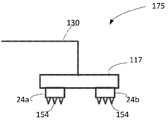
도 1은 본 발명의 일 실시예에 따라 신체 조직 내에 에너지를 전달하기 위한 시스템(100)을 개략적으로 도시한다. 시스템(100)은 조직 천공 디바이스(10) 및 자극기(12)를 포함한다. 조직 천공 디바이스(10)는 치료될 조직 표면을 천공하고 조직 표면 아래에 조직 표면으로부터 조직 내로의 마이크로채널을 생성한다. 자극기(12)는 신체 조직에 에너지를 전달한다. 조직 천공 디바이스(10) 및 자극기(12)는 제어기(16)의 제어하에 있다. 조직 천공 디바이스(10) 및 자극기(12)는 별개의 하우징 내에 포함될 수 있거나, 또는 단일의 공동 하우징 내에 포함될 수 있다.Figure 1 schematically depicts a
천공 디바이스(10)는 조직 표면을 천공하기 위해 알려진 임의의 방법에 의해 조직 내에 마이크로채널을 생성할 수 있으며, 예를 들어 초음파 에너지, 레이저 광 에너지, 열에너지 및 전기 에너지 중 임의의 하나 이상을 포함할 수 있다. 전류, 전류와 같은 전기 자극, 전기 신경 자극 및 전자기복사와 같은 임의의 타입의 자극 에너지가 자극기(12)에 의해 전달될 수 있다.The puncturing
도 2는 제어기(16)를 더욱 상세하게 도시한다. 제어기(16)는 천공 디바이스(10)에 전기 에너지를 전달하기 위한 전기 회로(136) 및 자극기(12)에 전기 에너지를 전달하기 위한 전기 회로(138)를 포함한다. 전기 회로는 공장 설정 파라미터 또는 사용자 입력 파라미터에 따라 프로세서(140)의 제어하에 있다. 제어기(16) 내의 전원(128)은 프로세서(140)에 동력을 공급함 또한 전기 회로(136, 138)에 동력을 공급한다. 키패드와 같은 사용자 입력-출력 모듈(132)은 전기 에너지의 다양한 파라미터를 입력하도록 사용자에 의해 사용될 수 있다. 입력-출력 모듈(132)은 원격 제어를 포함할 수 있다. 프로세서(140)는 또한 조직 천공의 범위를 결정하기 위해서 임피던스와 같은 조직의 일부 전기적 또는 광학적 파라미터를 모니터한다.Figure 2 shows the
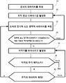
도 3은 본 발명의 이러한 양태의 일 실시예에 따른 본 발명의 시스템에 의해 신체 조직에 자극을 전달하기 위한 방법의 흐름도를 도시한다. 단계(25)에서, 조직 저항, 임피던스 또는 커패시턴스와 같은 조직의 하나 이상의 전기적 또는 광학적 파라미터의 초기 결정이 획득된다. 단계(30)에서, 조직 천공 디바이스(10)가 치료될 신체 조직의 표면에 적용되고 조직 표면을 천공하고 표면 아래의 신체 조직의 부피 내에 마이크로채널을 생성하도록 활성화된다. 신체 조직의 천공 동안에 또는 그 이후에, 조직의 전기적 또는 광학적 파라미터가 측정된다(단계(32)). 단계(34)에서, 조직의 광학적 또는 전기적 파라미터 중 적어도 하나가 적어도 단계(25)에서 결정된 초기 값의 사전결정된 백분율 및/또는 적응 가능한 백분율만큼 감소되었는지 여부가 결정된다. 조직 천공은 후속하는 자극 단계에서 조직 내의 원하는 효과를 획득하기 위해 요구되는 전류 및/또는 전압 또는 광 세기를 감소시키도록 조직의 전기적 또는 광학적 파라미터를 수정하는 경향이 있다. 이것은 또한 전기 자극 동안의 통증을 감소시키며 조직의 더욱 깊은 층으로의 자극을 침투를 촉진할 수 있다. 만약 조직의 광학적 또는 전기적 파라미터 중 적어도 하나가 단계(25)에서 결정된 초기 값의 적어도 사전결정된 또는 적응 가능한 백분율만큼 감소되지 않았다면, 프로세스는 조직 천공 디바이스의 재활성화를 통해 단계(30)로 복귀한다. 조직 천공 디바이스는 이전과 동일한 위치에서 또는 상이한 위치에서 활성화될 수 있다. 만약 조직의 광학적 또는 전기적 백분율 중 적어도 하나가 단계(25)에서 결정된 초기 값의 적어도 사전결정된 또는 적응 가능한 백분율만큼 감소되었다면, 프로세스는 자극기가 조직 표면의 천공된 영역 위의 및/또는 영역의 부분적으로 위의 및/또는 영역에 인접한 조직 표면 상에 위치되어 활성화되는 단계(36)로 이어진다. 위치결정은 또한 천공 이전에 또는 천공 동안에(단계(30)) 수행될 수 있으며, 천공 동안에 변위되거나 제거되는 것 또한 가능하다. 자극 에피소드의 종료시에 또는 자극 에피소드 동안에, 단계(38)에서 자극의 추가 에피소드가 요구되는지 여부가 결정된다. 만약 그렇다면, 단계(40)에서, 조직의 전기적 또는 광학적 속성이 다시 측정될지 여부가 결정된다. 예를 들어 원래의 마이크로채널이 밀봉되었거나 조직의 전기적 또는 광학적 파라미터가 더 이상 사전결정된 또는 적응 가능한 백분율만큼 감소되지 않으면, 자극의 제1 에피소드 후에 추가의 마이크로채널이 필요할 수 있다. 만약 그렇다면, 프로세스는 조직의 광학적 또는 전기적 속성을 측정하는 단계(32)로 복귀한다. 그렇지 않으면, 프로세스는 자극기를 활성화하는 단계(36)로 복귀한다. 만약 단계(38)에서 자극의 추가 에피소드가 필요하지 않다고 결정되면, 프로세스는 종료된다. 단계(36)에서 자극이 인가되는 동안 조직의 광학적 또는 전기적 파라미터가 주기적으로 측정될 수 있으며, 만약 감소된 파라미터가 단계(25)에서 결정된 바와 같은 초기 값으로 돌아가면 프로세스는 단계(32)로 복귀한다(도면에 도시되지 않음).Figure 3 illustrates a flow diagram of a method for delivering stimulation to body tissue by the system of the present invention in accordance with an embodiment of this aspect of the invention. In
전기적 파라미터는 자극기 내의 표면 전극 또는 천공 디바이스 내의 마이크로바늘에 의해서 측정될 수 있다.The electrical parameters can be measured by surface electrodes in a stimulator or by micro needles in a perforation device.
전기 자극의 경우에서, 생체에 적합한 전도성 재료가 자극 이전에 또는 자극 동안에 조직 표면에 도포될 수 있다.In the case of electrical stimulation, a conductive material suitable for the living body may be applied to the tissue surface prior to stimulation or during stimulation.
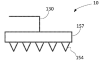
조직 천공 디바이스(10)는 도 4에 도시된 어플리케이터(157)를 포함할 수 있다. 어플리케이터(157)는 적어도 하나의 마이크로채널을 생성하기 위해 둘 이상의 마이크로바늘(154)의 하나 이상의 행렬을 포함한다. 마이크로바늘(154)은 하나 이상의 전기 접속(130)에 의해 전기 회로(136)에 접속된다. 마이크로바늘(154)은 전도성 재료 또는 반전도성 재료로 이루어질 수 있다. 마이크로바늘이 치료될 조직 표면 내에 압착되고 충분한 길이의 시간 동안 둘 이상의 마이크로바늘 사이에 전류가 인가될 때, 국부적인 전력 소멸로 인해서 조직 내의 마이크로바늘 둘레에 적어도 하나의 마이크로채널이 형성된다. 이것은 피부 표면의 경우에 각질층과 같은 조직 표면의 최외곽 층에서 마이크로바늘 둘레 조직의 절제로 이어진다.The
각각의 마이크로바늘 행렬은 조직 내의 적어도 하나의 마이크로채널 및/또는 마이크로채널들의 개별 클러스터를 생성할 수 있다. 각각의 행렬은 조직 표면의 1000㎠에 이르는 영역을 커버할 수 있다. 마이크로채널들의 하나 이상의 클러스터는 동시에 또는 순차적으로 생성될 수 있다. 두 개의 마이크로바늘은 동일한 행렬 상에 위치될 수 있다. 모든 마이크로바늘이 조직 표면에 동시에 적용될 수 있으며, 동시에 또는 순차적으로 전기적으로 활성화될 수 있다. 가깝게 위치된 마이크로바늘은 마이크로채널을 효율적으로 생성하기 위해서 동시에 우선적으로 활성화될 수 있다. 이와 다르게, 마이크로바늘의 서로 다른 조합이 조직 표면에 적용될 수 있으며 동시에 또는 순차적으로 활성화될 수 있다. 조직 표면을 천공하는 것은, 예를 들어 천공될 영역이 마이크로바늘 행렬 크기보다 더 클 때, 또는 조직 표면 상의 몇몇 이산 영역들이 천공될 때, 또는 마이크로채널들의 일부 또는 전부가 치료 중에 밀봉될 때, 단일 치료 세션에서 한 번보다 많은 횟수로 수행될 수 있다.Each microneedle matrix can create a separate cluster of at least one microchannel and / or microchannels in tissue. Each matrix can cover an area of up to 1000
마이크로바늘 길이는 예를 들어 10-400㎛일 수 있으며 지름은 예를 들어 10-400㎛일 수 있고, 이는 출혈 및 통증을 방지하는 경향이 있다. 마이크로바늘은 원뿔형 또는 원통형 또는 표피 조직 층의 천공 및/또는 절제를 가능하게 하는 임의의 다른 형태일 수 있다. 예를 들어 몇몇 개인들에서와 같이 조직 장벽의 최외곽 층이 치료 위치에서 극도로 두꺼운 경우에, 더욱 큰 마이크로바늘이 사용될 수 있다. 마이크로바늘은 약 0.01-10mm의 공간을 가질 수 있다. 마이크로바늘은 행렬 내에서 균일하게 분포될 수 있거나 또는 그렇지 않을 수 있다. 마이크로바늘들의 행렬의 광범위하게 배치된 분포는 피부의 적열 상태 및 부기를 감소시킬 수 있다.The microneedle length can be, for example, 10-400 microns and the diameter can be, for example, 10-400 microns, which tends to prevent bleeding and pain. The microneedle may be conical or cylindrical or any other form that allows perforation and / or ablation of the epidermal tissue layer. For example, as in some individuals, where the outermost layer of tissue barrier is extremely thick at the treatment site, larger micro needles may be used. The microneedles can have a space of about 0.01-10 mm. The microneedles may or may not be uniformly distributed within the matrix. The widespread distribution of the matrix of microneedles can reduce the redness and swelling of the skin.
인접한 마이크로바늘들 사이에 인가된 전류는 예를 들어 1kHz-100MHz의 주파수, 또는 0.001-1,000msec의 RF 및 지속시간 및 0.001-1,000msec의 일시정지를 갖는 직류 전류(DC) 또는 교류 전류(AC)일 수 있다. 마이크로바늘의 샤프트는 마이크로바늘의 팁(tip)으로부터의 전류만을 허용하도록, 그리고 치료될 조직의 표피층의 전류를 감소시키도록 전기적으로 절연될 수 있다.The current applied between adjacent microneedles may be a direct current (DC) or alternating current (AC) having a frequency of, for example, 1 kHz-100 MHz, or an RF and duration of 0.001-1,000 msec and a pause of 0.001-1000 msec. Lt; / RTI > The shaft of the microneedle can be electrically insulated to allow only current from the tip of the micro-needle and to reduce the current in the epidermal layer of tissue to be treated.
프로세서(140)는 조직의 천공 범위를 결정하기 위해서 임피던스와 같은 조직의 전기적 파라미터를 모니터한다. 마이크로채널의 형성은 조직 임피던스 및/또는 전도율 및/또는 전압 및/또는 전류와 같이 마이크로채널의 형성 중에 측정되는 파라미터들에 응답하여 조정될 수 있다. 이러한 파라미터들의 값은 피험자의 다양한 파라미터들에 기초하여 선택될 수 있으며, 동적일 수 있다. 도달시에 해당 마이크로채널 생성이 완료되었음을 나타내는 전도율 임계값이 명시될 수 있다. 프로세서는 마이크로바늘 중 하나의 전기적 파라미터에서의 변화에 응답하여 마이크로바늘 중 적어도 하나를 통해 인가되는 전류 또는 전압을 조정할 수 있다.
마이크로채널들의 형성 동안에 그리고 그 후에, 세포외액이 마이크로채널 내로 도입될 수 있으며, 이는 마이크로채널을 따라 임피던스를 감소시킨다. 이것은 자극, 특히 전기 자극을 위해 요구되는 전력을 감소시킬 수 있으며, 자극 에너지의 더욱 깊은 침투를 가능하게 한다.During and after formation of the microchannels, extracellular fluid can be introduced into the microchannel, which reduces the impedance along the microchannel. This can reduce the power required for stimulation, especially for electrical stimulation, and allows for a deeper penetration of stimulation energy.
도 5는 본 발명의 다른 실시예에 따른 시스템(100)에서 사용될 수 있는 조직 천공 디바이스(10a)를 도시한다. 디바이스(10a)는 초음파 펄스 발생기(312)를 포함하고 프론트엔드(314)에 전달되는 전기 신호를 생성하는 전자기기, 소프트웨어 및 피드백 메커니즘을 포함한다. 프론트엔드(314)는 전기 신호를 초음파 펄스파로 변환하는 변환기를 포함한다. 매칭 모듈(316)은 마이크로바늘 플레이트(318)와 프론트엔드(314) 사이의 적절한 물리적 접속을 가능하게 한다. 마이크로바늘 플레이트(318)는 적어도 하나의 마이크로바늘 또는 마이크로바늘들의 행렬 또는 행렬들의 클러스터를 포함한다. 마이크로바늘 플레이트(318)는 상이한 형태의 플레이트들을 이용하는 것을 가능하게 하도록 및/또는 한 피험자에서 다른 피험자로 이동될 때 제거 가능할 수 있다. 입력-출력 모듈(132)은 마이크로채널이 생성되는 동안 최소의 가열 및/또는 통증이 피험자에 의해 감지되도록 타이밍 및 에너지와 같은 천공의 선택 가능한 파라미터를 사용자가 입력하는 것을 가능하게 하도록 사용될 수 있다.Figure 5 illustrates a
마이크로바늘에 초음파 펄스파를 전달하기 위한 몇몇 동작 모드가 이용 가능하다. 예를 들어, 초음파 펄스파를 마이크로바늘 플레이트에 전달하는 단일 변환기가 사용될 수 있으며, 이때 초음파 펄스파가 조직에 선행하는 마이크로바늘로 분포되는 초음파 펄스파를 가진다. 다른 예로서, 디바이스(10a)는 다양한 마이크로바늘의 조합에서 초음파 펄스파를 생성하도록 구성된 초음파 변환기의 어레이를 포함할 수 있다. 이것은 서로 다른 조합의 마이크로바늘이 서로 다른 시간에 또는 서로 다른 파라미터를 가지고 활성화되는 것을 가능하게 한다. 마이크로채널 생성은 서로 다른 마이크로바늘의 세트에 의해 동시에 또는 순차적으로 수행될 수 있다. 전기 신호는 하나 이상의 마이크로바늘을 통해 인가될 수 있으며, 전기적 파라미터가 마이크로바늘의 초음파 에너지를 조정하기 위해서 측정될 수 있다.Several modes of operation are available for delivering the ultrasonic pulse wave to the microneedles. For example, a single transducer may be used to deliver the ultrasonic pulse wave to the microneedle plate, wherein the ultrasonic pulse wave has an ultrasonic pulse wave that is distributed in the microneedles preceding the tissue. As another example, the
초음파 펄스파의 주파수는 예를 들어 1kH-50MHz 범위 내에 있을 수 있으며, 세기는 0.1-5W/㎟의 범위 내에 있을 수 있다. 각각의 마이크로바늘에 의해서 전달되는 에너지는 0.01 내지 100 줄/채널일 수 있다. 초음파 펄스파의 주파수 및 세기는, 해부학적 차이 및 물리학적 차이로 인해 피험자마다, 그리고 동일한 피험자 안에서도 달라질 수 있다. 펄스들의 시간 간격은 조직의 과열을 감소시키거나 방지하기 위해서 1초 미만일 수 있다.The frequency of the ultrasonic pulsed wave may be in the range of, for example, 1 kH-50 MHz and the intensity may be in the range of 0.1-5 W / mm < 2 >. The energy delivered by each microneedle may be 0.01 to 100 lines / channel. The frequency and intensity of the ultrasound pulse wave may vary from subject to subject and from the same subject due to anatomical and physical differences. The time interval of the pulses may be less than one second to reduce or prevent tissue overheating.
마이크로바늘 플레이트 및 마이크로바늘(들)은 예를 들어 티타늄 또는 알루미늄, 또는 초음파 펄스파의 전달과 호환가능한 다른 금속 또는 재료로 이루어질 수 있다. 마이크로바늘은 또한 마이크로채널 생성 범위에 관한 표시를 제공하는 전기적 파라미터를 측정하고/하거나 전기 에너지를 전달하기 위해 전도성일 수 있다.The microneedle plate and microneedle (s) may be made of, for example, titanium or aluminum, or other metal or material compatible with the delivery of ultrasonic wave pulses. The microneedles may also be conductive to measure electrical parameters and / or deliver electrical energy to provide an indication of the microchannel generation range.
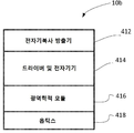
도 6은 예를 들어 본 발명의 또 다른 실시예에 따라 시스템(100) 내에서 사용될 수 있는 조직 천공 디바이스(10b)를 도시한다. 디바이스(10b)는 마이크로채널의 형성을 위해 에너지를 형성하는 레이저와 같은 전자기복사 방출기 모듈(412)을 포함한다. 전원(128)은 전자기복사 방출기 모듈(412)을 위한 에너지를 제공한다. 드라이버 및 전자기기(414)는 방출기 모듈(412)을 제어한다. 입력 출력 디바이스 모듈(132)은 전자기 펄스의 지속시간 및 세기와 같은 전자기복사 파라미터의 선택을 가능하게 한다.Figure 6 illustrates a tissue piercing device 10b that may be used in
마이크로채널은 마이크로채널을 생성하기에 충분한 에너지를 가지고 타깃화된 조직에 전자기복사 에너지를 타깃화하고 또한 복수의 마이크로채널의 생성을 가능하게 하도록 조직을 따라서 빔을 돕는 것을 가능하게 하는 광역학적 렌즈 및/또는 미러(mirror)(416) 및 옵틱스(418)의 활성화에 의해 생성될 수 있다. 마이크로채널은 1-1000㎛의 지름 및 5-5000㎛의 거리 공간을 가질 수 있다. 도 6에 도시되지 않은 표면 전극은 또한 마이크로채널 형성 범위의 표시를 제공하도록 조직의 전기적 파라미터의 측정을 위해 사용될 수 있다.The microchannel includes a photonic lens that has sufficient energy to create a microchannel to target the electromagnetic radiation to the targeted tissue and also to assist the beam along the tissue to enable generation of a plurality of microchannels, / RTI > and / or by activation of
도 7은 본 발명의 또 다른 실시예에 따라 시스템(100)에서 사용될 수 있는 조직 천공 디바이스(10c)를 도시한다. 전원(128)은 가열 모듈(468)에 에너지를 제공한다. 입력 및 출력 모듈(132)은 지속시간 및 온도와 같은 가열 파라미터의 제어를 가능하게 한다. 프로세서(140)는 가열 모듈을 포함하여 디바이스를 제어한다. 디바이스는 또한 조직의 과열을 방지하기 위해서 적어도 하나의 마이크로바늘을 포함하는 마이크로바늘 플레이트(474) 내에 온도를 측정하는 온도계를 포함할 수 있다. 마이크로바늘은 팁으로부터 소멸될 수 있으며 서로 다른 시간 간격 동안 가해질 수 있는 열을 전도하도록 설계된다. 가해진 열의 온도는 34-80℃의 범위 내에 있을 수 있다.Figure 7 illustrates a
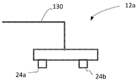
도 8은 본 발명의 일 실시예에 따라 시스템(100)에서 사용될 수 있는 전기 자극기(12a)를 도시한다. 자극기(12a)는 둘 이상의 전극(24)을 포함한다. 두 개의 전극(24a, 24b)이 도 8에 도시되었다. 이것은 단지 예시적인 것으로, 디바이스(12a)는 임의의 응용에서 원하는 바와 같은 임의의 수의 전극을 포함할 수 있다. 전극(24)은 신체 표면 상으로의 적용을 위해 적응된 표면 전극일 수 있거나, 또는 조직 표면 내에 침투를 위해 적응된 최소 침습 전극일 수 있다. 최소 침습 전극의 경우에, 천공 디바이스 내의 마이크로바늘이 최소 침습 전극으로서 천공을 위해서 또한 적응될 수 있다. 전극은 예를 들어 습식(액체/물 기반), 글루 기반 히드로겔 전극 또는 면 전극일 수 있다. 전원(128)은 전기 접속(130)을 통한 전극들(24) 양단의 교류 또는 직류 전압 신호 또는 교류 또는 직류 전류 신호를 인가하는 전기 회로에 에너지를 공급한다. 입력-출력 모듈(132)은 사용자에 의해 전극 양단의 전압, 전류, 펄스 주파수, 펄스 지속시간, 전기 임펄스의 주기성, 펄스 타입 및 전기 자극의 타이밍과 같은 전기 자극의 다양한 파라미터를 입력하도록 사용될 수 있다. 전극의 에너지 공급은 자극 동안에 서로 다른 쌍의 자극 전극들 사이에서 전환될 수 있다. 프로세서(140)는 공장 설정 파라미터 및 사용자 입력 파라미터에 따라서 전극들(24)의 에너지 공급을 위한 전기 회로를 제어한다. 프로세서는 또한 디바이스에 의해 측정되는 파라미터 및 전기 신호를 모니터하도록 사용될 수 있다. 마이크로채널의 형성에 이어 가해지는 전기 자극은, 마이크로채널의 밀봉을 방지하고 따라서 치료 기간의 연장을 가능하게 하는 것을 도울 수 있다.Figure 8 illustrates an
전기 자극은 전류 제어 또는 전압 제어될 수 있다. 전류 펄스는 임의의 응용에서 원하는 바와 같이 대칭적이거나 또는 비대칭적일 수 있다. 펄스는 전하 균형된 이상성 펄스(charged balanced bi-phasic pulse)일 수 있다. 자극은 0.01-10,000Hz의 주파수, 0.001-100mA의 전류 진폭, 1-2000μsec의 펄스폭 및 600볼트에 이르는 전압을 가질 수 있다. 도 9에 도시된 신호(108)는 사용될 수 있는 예시적인 전기 신호의 한 사이클을 도시한다.The electrical stimulation may be current controlled or voltage controlled. The current pulses may be symmetrical or asymmetric as desired in any application. The pulse may be a charged balanced bi-phasic pulse. The stimulus may have a frequency of 0.01-10,000 Hz, a current amplitude of 0.001-100 mA, a pulse width of 1-2000 μsec, and a voltage of 600 volts. The
전극들 사이의 거리는 치료될 조직의 깊이 및 위치에 의해 결정될 수 있다. 일반적으로, 신체 표면 아래의 치료될 조직이 더 깊을수록 전극들 사이에 요구되는 거리가 더욱 크다. 전극들 사이의 거리는 동적일 수 있으며 상황에 따라서 결정된다.The distance between the electrodes can be determined by the depth and location of the tissue to be treated. Generally, the deeper the tissue to be treated under the body surface, the greater the required distance between the electrodes. The distance between the electrodes can be dynamic and is determined depending on the situation.
도 10은 휴대용 어플리케이터일 수 있는 단일 어플리케이터(117)를 이용하여 조직 천공 및 전기 자극이 수행될 수 있는 조직 표면을 천공하고 그러한 조직 표면에 전기 자극을 인가하기 위한 시스템(175)을 도시한다. 어플리케이터(117)는 둘 이상의 전극(24) 및 하나 이상의 마이크로바늘(154)들의 행렬을 포함한다. 적어도 하나의 전극은 적어도 하나의 생성된 마이크로채널 위에 또는 부분적으로 위에 또는 부근에 배치된다. 두 전극들(24a, 24b)은 도 10에 도시된다. 이것은 단지 예시적인 것이며, 어플리케이터(117)는 임의의 응용에서 요구되는 바와 같은 임의의 수의 전극을 포함할 수 있다. 전극(24)은 조직 표면 상의 적용을 위해 적응되는 표면 전극 또는 최소 침습 전극일 수 있으며, 이러한 경우에 마이크로바늘(154)이 최소 침습 전극으로서의 기능을 하도록 또한 적응될 수 있다. 전원(128)은 마이크로바늘에 전기 에너지를 전달하는 전기 회로(136)뿐 아니라 공장 설정 파라미터 또는 사용자 입력 파라미터에 따라서 전기 접속(130)에 의해 전극(24)을 통해서 전기 에너지를 전달하는 전기 회로(138)에도 동력을 공급한다. 입력-출력 모듈(132)은 마이크로바늘 및/또는 전극 양단의 전압, 전류, 주파수, 지속기간, 전기 펄스의 주기성 및 전기 자극의 타이밍과 같은 천공 및 전기 자극의 다양한 파라미터를 입력하도록 사용될 수 있다. 프로세서(140)는 또한 조직의 천공 범위를 결정하도록 조직 임피던스 또는 다른 전기적 파라미터를 모니터할 수 있다. 마이크로바늘(154)은 전극에 동력을 공급하기에 앞서 천공 후에 어플리케이터로부터 제거될 수 있거나 또는 제거되지 않을 수 있다. 마이크로바늘(154)은 조직 내에 마이크로바늘을 삽입한 후에 제거되지 않을 수 있으며, 전극(24)과 함께 전기 자극 프로세스에 참여할 수 있고 따라서 전기 자극이 마이크로바늘을 통해서도 제공될 수 있다. 천공 구성과 자극 구성 사이의 전환은 기계적으로 또는 전기적으로 수행될 수 있다.Figure 10 shows a
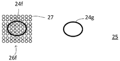
마이크로채널의 위치에 대한 자극 인가 위치는 임의의 응용에서 원하는 바와 같이 결정될 수 있다. 일반적으로, 전극들이 마이크로채널 위의 조직 표면 상에 배치될 때, 자극은 보다 효율적인 경향이 있다. 더 적은 에너지가 자극에 필요하며, 자극은 조직 내에 더 깊이 침투할 수 있다. 이것은 통증을 감소시키고, 치료의 효율성을 증가시키며, 치료 시간을 단축할 수 있다. 도 11a는 제1 예시로서, 마이크로채널들(27)의 세 개의 클러스터(26a, 26b, 26c)가 생성된 조직 표면(25)을 도시한다. 또한 조직 표면(25) 상의 세 개의 전극의 접촉 영역(24a, 24b, 24c)의 경계가 도시되었다. 이러한 예에서, 각각의 마이크로채널들의 클러스터는 상응하는 전극의 접촉 영역 위에서 연장한다. 도 11b는 제2 예로서, 마이크로채널들(27)의 두 개의 클러스터(26d, 26e)가 생성된 조직 표면(25)을 도시한다. 또한 조직 표면(25) 상의 두 개의 전극의 접촉 영역(24d, 24e)의 경계가 도시되었다. 이러한 예에서, 각각의 마이크로채널들의 클러스터는 상응하는 전극의 접촉 영역 위에서 연장한다. 마이크로채널 내의 저항이 자극 전극(24)과 접촉하는 천공되지 않은 조직 영역보다 더 낮기 때문에, 대부분의 전류가 자극 동안 마이크로채널들을 통해서 흐를 것이 예상된다. 도 11c는 제3 예로서, 마이크로채널들(27)의 하나의 클러스터(26f)가 전극의 접촉 영역(24f) 아래에 생성된 조직 표면(25)을 도시한다. 마이크로채널들은 제2 전극의 접촉 영역(24g) 아래에는 존재하지 않는다. 위의 예시들의 임의의 조합이 가능하며, 임의의 수의 전기 전극 및 마이크로채널 클러스터를 가진다.The position of stimulation applied to the position of the microchannel can be determined as desired in any application. Generally, when electrodes are placed on a tissue surface on a microchannel, the stimulation tends to be more efficient. Less energy is needed for stimulation, and stimulation can penetrate deeper into the tissue. This can reduce pain, increase the efficiency of treatment, and shorten treatment time. Figure 11A shows, as a first example, a
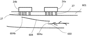
도 12는 타깃화된 조직(602)으로의 신경 임펄스를 활성화 또는 차단하도록 자극 에너지를 전달하기 위한 시스템(100)의 사용을 도시한다. 이식 가능한 전도성 요소(604)는 조직 아래에 이식되며 전도성 임플란트의 일 측면은 조직 표면 및 전극들(24)에 근접하게 위치되고 전도성 임플란트의 다른 측면은 타깃화된 조직(602)에 근접하게 위치된다. 마이크로채널들(27)은 전극들 중 적어도 하나 아래에 또는 인접하게 생성된다. 이러한 예에서, 마이크로채널들은 표면 조직인 각질(601)을 제거하며, 자극 에너지, 보다 구체적으로는 전류와 같은 전기 자극 에너지를 전극들(24)로부터 전도성 임플란트(604)를 통해 신경(602)과 같은 타깃화된 조직으로 전달하는 것을 가능하게 한다. 전도성 임플란트는 수동형일 수 있으며 자극 에너지를 저장하거나 타깃화된 조직에 전달될 에너지의 세기를 수정 또는 증폭시키는 능동 요소를 구비할 수 있다. 디바이스는 신경계의 장애를 치료하는 데에 사용될 수 있고, 이러한 경우에 타깃화된 조직은 신경일 수 있다. 하나의 전극이 임플란트 전도체(604)의 픽업 엔드(604b) 위에 위치되고 전류는 타깃 신체 조직(602)에 자극을 제공하는 전류가 임플란트의 단부(604b)로부터 단부(604a)로 전달된다. 그 다음 전류는 타깃 신체 조직을 통해 흐르며 그 다음 타깃화된 신체 조직(602)을 자극하기 위해서 신체 조직을 통해 다른 표면 전극으로 흐른다.FIG. 12 illustrates the use of
시스템의 어플리케이터는 신체 표면에 접착하도록 구성된 패치의 형태일 수 있다. 패치 타입 어플리케이터는 연장된 자극이 요구될 때 유용할 수 있다.The applicator of the system may be in the form of a patch configured to adhere to a body surface. Patch type applicators may be useful when extended stimulation is required.
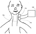
도 13은 피부 표면에 적용된 어플리케이터(210)를 구비한 개인(200)을 도시한다. 아래에 설명되는 바와 같이, 패치 어플리케이터는 예를 들어 폐쇄수면무호흡 및 코골이와 같은 수면 장애의 치료뿐 아니라 남성 발기부진, 과민성 방광, 통증 완화 또는 신경통 완화에서도 사용될 수 있다. 전기 자극은 또한 인가된 자극과 타깃 조직 사이에 위치된 이식 리드를 통해서 타깃 조직에 전달될 수도 있다(도 13에 도시되지 않음).Figure 13 shows an individual 200 with an
도 14는 패치(210)의 구성을 도시한다. 패치(210)는 쌍으로 동시에 또는 순차적으로 활성화될 수 있는 세 개의 전극들(251, 252, 253)을 포함한다. 이러한 패치는 모든 전극들 및/또는 전극들의 일부를 포함할 수 있거나, 또는 와이어에 의해서 외부 전자기기에 접속될 수 있다. 마이크로채널들은 적어도 하나의 전극의 위치 아래에 생성될 수 있다.Fig. 14 shows the configuration of the
폐쇄수면무호흡 또는 코골이의 치료를 위해서, 전류가 설하 신경을 통과하며 적절한 에너지가 혀의 전방 운동을 발생시키기 위해 인가되도록 패치 형태의 어플리케이터(210)가 도 14에 도시된 바와 같이 목의 전면 상에, 턱 아래에 또는 피험자의 턱끝밑삼각(submental triangle) 아래에 부착될 수 있다. 상기도 확장을 책임지는 가장 큰 인두 근육은 이설근이다. 이 근육은 전방 혀 운동과 안쪽 인두 벽의 경직을 책임진다. 우측 및 좌측 설하 신경은 각각 우측 및 좌측 이설근을 자극한다. 오직 한 측의 설하 신경의 전기 자극은 혀를 전방으로 당길 수 있다. 이설근의 수축은 혀를 앞으로 당김으로써 적절한 호흡을 회복하기 위해서 기도를 개방한다.For the treatment of obstructive sleep apnea or snoring, an
서로 다른 전극들의 쌍이 순차적으로 활성화될 수 있는 자극의 패턴은 근육의 주기적인 이완을 가능하게 할 수 있다. 예를 들어, 전극들(251, 252) 사이에 위치된 전극(252)은 목의 일 측면에 적용될 수 있는 전극(251)과 활성화될 수 있으며, 전극(252)은 목의 다른 측면에 적용될 수 있는 전극(253)과 활성화될 수 있다. 이와 다르게, 패치는 오직 전극들(252, 251)만을 포함할 수 있거나, 또는 전극들(252, 253)만을 포함할 수 있으며, 이러한 경우에 목의 일 측면 상의 신경만이 자극된다. 또한 오직 전극들(251, 253)만을 포함할 수도 있다. 패치(210)는 코골이 또는 무호흡과 같은 수면 장애의 표시를 검출하는 센서를 포함하는 본 발명의 시스템에서 사용될 수 있다. 이러한 시스템은 또한 자극의 비연속적인 활성화를 가능하게 하도록 수면 장애의 임의의 표시 및/또는 호흡의 리듬을 검출하는 센서도 포함할 수 있다.The pattern of stimuli, in which pairs of different electrodes can be activated sequentially, may enable periodic relaxation of the muscles. For example, the
도 15는 말초 신경통 또는 다발성 신경염, 또는 다발성 신경염 골반통 또는 과민성 방광(OAB) 및 요절박, 빈뇨 및 절박 요실금과 같은 연관 증상의 치료에서의 본 발명의 시스템의 사용을 도시한다. 이러한 시스템은 경골 신경을 자극하도록 사용된다. 경골 신경(355)은 좌골 신경의 가지이며; 발에서 끝난다. 마이크로채널은 전극들(24a, 24b) 중 적어도 하나가 부착되는 영역의 피부 내에 생성되며; 전극(24a) 및/또는 전극(24b)은 전기 에너지와 같은 최대 자극 에너지가 경골 신경으로 구동되는 방식으로 경골 신경 위에 배치된다. 마이크로채널은 각질을 제거하며 경골 신경 내로의 전기 에너지의 최대 침투를 가능하게 한다. 전극들(24a, 24b)은 도 15에 도시된 바와 같이 전류와 같은 에너지가 경골 신경을 통과하고 전류와 같은 적절한 에너지가 경골 신경(355)에 인가되도록 발목에 근접하게 부착될 수 있다. 전극들은 경골 신경이 피부 표면에 비교적 가까운 발목에 근접하게 위치될 수 있다. 전극들은 하나의 치료 에피소드에서 다른 에피소드로 제거될 수 있다. 일부 응용에 있어서, 피험자는 가정 또는 의원에서 30분간, 주에 한번 또는 두 번, 그리고 약 12주의 기간 동안에 이러한 디바이스를 이용하여 치료될 수 있다. 일부 응용에 있어서, 치료는 수개월에 이르는 기간 동안 하루에 한 회 이상 피험자에게 적용된다. 일부 응용에 있어서, 치료는 피험자에게 연속적으로 적용된다. 후경골 신경의 위치는 발가락의 움직임을 관찰하는 것과 같이 당업계에서 알려진 기술에 의해 결정될 수 있다. 전기 자극은 인가된 자극과 타깃 조직 사이에 위치된 이식된 리드를 통해서 타깃 조직에 전달될 수 있다.Figure 15 illustrates the use of the system of the present invention in the treatment of peripheral neuralgia or multiple neuritis, or multiple neuritis pelvic pain or overactive bladder (OAB) and associated conditions such as nodular pains, frequent urination and urge incontinence. This system is used to stimulate the tibial nerve. The
도 16에 도시된 바와 같이, 간질 또는 고혈압과 같은 장애의 치료를 위해서, 마이크로채널이 미주 신경(701) 위의 피부 내에 생성될 수 있으며 전극들(24)이 치료를 가능하게 하도록 미주 신경 위의 표면 상에 배치된다.As shown in FIG. 16, for the treatment of disorders such as epilepsy or hypertension, microchannels may be created in the skin above the
도 17은 시스템(100) 내에서 사용될 수 있는 패치(678a)를 도시한다. 패치(678a)는 조직 표면상에 배치되도록 적응된다. 패치는 패치가 조직 표면에 적용될 때 아래에 있는 조직 표면의 영역(682)을 노출하도록 들어올려질 수 있는 플랩(680)을 포함한다. 하나 이상의 전극(24a)(도 17에서 점선으로 표시됨)은 패치(678a)의 아랫면에 부착된다. 제2 전극(24b)은 패치(678a)의 아래면 상에 부착될 수 있거나, 또는 도 17에 도시된 바와 같이 별도의 패치(678b)의 아래면 상에 부착될 수 있다. 조직 표면에 패치(678a)를 도포한 후에, 플랩(680)은 조직 표면의 영역(682)을 노출하도록 개방될 수 있으며, 조직 천공 디바이스는 조직 표면의 노출된 영역(682) 및 영역(682) 내에 형성된 마이크로채널(27)에 적용될 수 있다. 그 다음 조직 천공 디바이스가 제거될 수 있으며 플랩(682)이 닫힌다. 이러한 방식으로, 전극(24a)은 조직 표면의 천공된 영역(682) 위에 위치되며, 자극 에너지는 천공된 조직에 직접 전달될 수 있다.FIG. 17 shows a
남성 발기부전의 치료를 위해서, 패치 형태인 어플리케이터가 해면 신경 및 등신경과 같이 남성 생식기를 자극하고 발기를 제어하는 신경 위의 표면 상에 배치될 수 있다. 이러한 치료는 또한 페니스로의 혈액 흐름을 증가시킬 수 있다.For the treatment of male erectile dysfunction, a patch-shaped applicator may be placed on the surface of the nerve that stimulates the male genitalia, such as the spongiform nerve and ischemic epidermis, and controls the erection. This treatment can also increase blood flow to the penis.
혈액 흐름을 증가시키기 위해서, 어플리케이터는 감소된 혈액 흐름의 영역 내의, 또는 신체 내의 다른 위치의 혈액 흐름에 영향을 미치는 영역 위의 조직 표면 상에 배치될 수 있다.To increase blood flow, the applicator may be placed on a tissue surface on a region of reduced blood flow, or on an area that affects blood flow at other locations within the body.
만성 부상과 같은 부상 치료를 위해서, 어플리케이터는 부상 위에 또는 부근에 배치될 수 있다. 치료는 서로 다른 타입의 전기 자극을 인가하는 것을 포함할 수 있다. 전기 자극은 고전압 펄스 전류(HVPC)를 포함할 수 있다.For wound healing, such as chronic injury, the applicator may be placed on or near the flotation. The treatment may include applying different types of electrical stimulation. Electrical stimulation may include a high voltage pulse current (HVPC).
안면 신경 마비와 같은 안면의 신경학적 장애의 치료는 안면 신경의 자극을 통해 본 발명을 이용하여 수행될 수 있다.The treatment of facial neurological disorders such as facial nerve paralysis can be performed using the present invention through stimulation of the facial nerve.
셀룰라이트 감소와 같은 심미 치료는 감소될 셀룰라이트 위의 표면을 자극함으로써 수행될 수 있다. 이러한 치료는 또한 콜라겐, 엘라스틴 및 아데노신 3인산(ATP) 생산을 자극하는 근긴장을 유지하거나 증가시킴으로써 노화의 신호를 감소시킬 수 있으며 안면 근긴장을 증가시키는 것은 피부 탄력을 향상시킬 수 있고 얼굴선, 주름 및 피부 질감 이상을 제거할 수 있다. 이것은 또한 조직으로의 혈액 흐름을 증가시킴으로써 근긴장을 유지하도록 근육 조직에 영양분을 제공할 수 있다.Aesthetic treatments such as cellulite reduction can be performed by stimulating the surface on the cellulite to be reduced. This treatment can also reduce the signs of aging by maintaining or increasing the dorsal horns that stimulate collagen, elastin and adenosine triphosphate (ATP) production, and increasing facial muscle tone can improve skin elasticity and improve facial lines, The skin texture abnormality can be removed. It can also provide nutrients to muscle tissue to maintain muscle tone by increasing blood flow to the tissue.
Claims (24)
(a) 조직 표면을 천공하고 상기 조직 표면으로부터 상기 신체 조직 내로의 마이크로채널을 생성하도록 적응된 조직 천공 디바이스; 및
(b) 상기 신체 조직에 에너지를 전달하도록 적응된 자극기를 포함하는, 시스템.A system for delivering energy into a body tissue,
(a) a tissue punch device adapted to perforate a tissue surface and generate microchannels from the tissue surface into the body tissue; And
(b) a stimulator adapted to transmit energy to the body tissue.
상기 신체 조직의 하나 이상의 전기적 또는 광학적 속성을 모니터하도록 구성된 프로세서를 더 포함하는, 시스템.The method according to claim 1,
Further comprising a processor configured to monitor one or more electrical or optical properties of the body tissue.
상기 조직 천공 디바이스는 적어도 하나의 마이크로바늘을 포함하는, 시스템.3. The method according to claim 1 or 2,
Wherein the tissue piercing device comprises at least one micro-needle.
상기 조직 천공 디바이스는 초음파 에너지, 레이저 광 에너지, 열 에너지 및 전기 에너지 중 임의의 하나 이상을 활용함으로써 상기 조직 내에 마이크로채널을 생성하는, 시스템.4. The method according to any one of claims 1 to 3,
Wherein the tissue poration device generates microchannels in the tissue by utilizing any one or more of ultrasonic energy, laser light energy, thermal energy, and electrical energy.
상기 자극기는 전기 자극, 전류, 전기 신경 자극 및 전자기복사 중 임의의 하나 이상을 상기 신체 조직에 전달하도록 적응되는, 시스템.5. The method according to any one of claims 1 to 4,
Wherein the stimulator is adapted to deliver any one or more of electrical stimulation, current, electrical nerve stimulation, and electromagnetic radiation to the body tissue.
상기 프로세서는 표면 전극에 의해 또는 상기 천공 디바이스 내의 마이크로바늘에 의해 상기 신체 조직의 하나 이상의 전기적 속성을 모니터하도록 구성된, 시스템.6. The method according to any one of claims 1 to 5,
Wherein the processor is configured to monitor at least one electrical property of the body tissue by a surface electrode or by a microneedle in the perforation device.
상기 조직 천공 디바이스는 하나 이상의 마이크로바늘의 하나 이상의 행렬을 포함하는, 시스템.The method of claim 3,
Wherein the tissue piercing device comprises one or more matrices of one or more microneedles.
상기 마이크로바늘은 전도성 재료, 반전도성 재료 또는 초음파 전도성 재료로 이루어지는, 시스템.8. The method according to claim 3 or 7,
Wherein the microneedle comprises a conductive material, a semi-conductive material, or an ultrasonic conductive material.
상기 프로세서는 조직 임피던스, 조직 저항 및 조직 커패시턴스로부터 선택된 상기 조직의 하나 이상의 전기적 파라미터를 모니터하는, 시스템.9. The method according to any one of claims 1 to 8,
Wherein the processor monitors one or more electrical parameters of the tissue selected from tissue impedance, tissue resistance, and tissue capacitance.
하나 이상의 상기 마이크로바늘은 오직 상기 마이크로바늘의 팁(tip)으로부터의 전류만을 허용하도록 전기적으로 절연된 샤프트(shaft)를 구비하는, 시스템.The method according to any one of claims 3, 7, and 8,
Wherein the at least one micro-needle is provided with an electrically insulated shaft to allow only current from the tip of the micro-needle.
상기 자극기는 둘 이상의 전극을 포함하고 상기 전극은 습식 표면 전극, 글루 기반 표면 전극, 히드로겔 표면 전극, 면 표면 전극, 또는 최소 침습성 전극 중 임의의 하나 이상인, 시스템.The method according to claim 1,
Wherein the stimulator comprises at least two electrodes and wherein the electrode is any one or more of a wet surface electrode, a glue-based surface electrode, a hydrogel surface electrode, a surface surface electrode, or a minimally invasive electrode.
적어도 하나의 전극은 상기 조직 표면에 패치가 도포될 때 상기 조직 표면의 영역을 노출시키기 위해 올려지거나 제거되도록 구성된 커버 또는 플랩을 갖는 개구(aperture)를 구비하는 상기 패치 내에 포함되는, 시스템.12. The method of claim 11,
Wherein at least one electrode is included in the patch having an aperture having a cover or flap configured to be raised or removed to expose an area of the tissue surface when the patch is applied to the tissue surface.
상기 조직 천공 디바이스는 하나 이상의 마이크로바늘을 포함하고, 상기 자극기는 둘 이상의 전극을 포함하며, 상기 마이크로바늘 및 상기 전극은 단일 어플리케이터 내에 포함되는, 시스템.The method according to claim 1,
Wherein the tissue piercing device comprises at least one microneedle, the stimulator comprises at least two electrodes, and wherein the microneedle and the electrode are contained within a single applicator.
상기 자극기는 셋 이상의 전극을 포함하고 상기 프로세서는 자극 동안 서로 다른 쌍의 전극에 동력을 공급하도록 추가로 구성되는, 시스템.The method according to claim 1,
Wherein the stimulator further comprises three or more electrodes and the processor is further configured to power different pairs of electrodes during stimulation.
상기 자극기는 신체 표면에 접착하도록 적응된 패치 내에 포함된 둘 이상의 전극을 포함하는, 시스템.The method according to claim 1,
Wherein the stimulator comprises two or more electrodes contained within a patch adapted to adhere to a body surface.
수동 또는 능동의 임플란트 가능한 전도성 요소를 더 포함하는, 시스템.16. The method according to any one of claims 1 to 15,
Further comprising a passive or active implantable conductive element.
상기 프로세서는 하나 이상의 상기 모니터된 파라미터가 사전결정된 범위 또는 적응 가능한 범위 또는 동적 범위 내에 있을 때 상기 자극기를 활성화하도록 추가로 구성되는, 시스템.17. The method according to any one of claims 1 to 16,
Wherein the processor is further configured to activate the stimulator when the one or more of the monitored parameters is within a predetermined range or adaptable range or dynamic range.
적어도 하나의 상기 마이크로바늘 및 적어도 하나의 상기 전극은 상기 적어도 하나의 마이크로바늘에 의해 형성된 적어도 하나의 마이크로채널 위의 상기 신체 표면에 상기 전극이 접촉하는 것을 가능하게 하도록 상기 어플리케이터 내에 위치되는, 시스템.14. The method of claim 13,
At least one of said micro-needles and at least one said electrode being positioned within said applicator to enable said electrode to contact said body surface on at least one micro-channel formed by said at least one micro-needle.
하나 이상의 상기 조직 천공 디바이스 및 상기 자극기를 제어하기 위한 원격 제어를 더 포함하는, 시스템.19. The method according to any one of claims 1 to 18,
Further comprising remote control for controlling the at least one tissue punch device and the stimulator.
상기 조직 천공 디바이스는 상기 마이크로채널 내의 조직을 제거하도록 구성되는, 시스템.20. The method according to any one of claims 1 to 19,
Wherein the tissue piercing device is configured to remove tissue within the microchannel.
상기 자극기는 상기 마이크로채널을 열린 채로 유지하기 위해서 상기 조직에 전류를 인가하도록 구성되는, 시스템.21. The method according to any one of claims 1 to 20,
Wherein the stimulator is configured to apply an electrical current to the tissue to maintain the microchannel open.
상기 자극기는 둘 이상의 조직 영역을 동시에 또는 순차적으로 자극하도록 적응되는, 시스템.22. The method according to any one of claims 1 to 21,
Wherein the stimulator is adapted to stimulate two or more tissue regions simultaneously or sequentially.
(a) 신체 조직 표면에 조직 천공 디바이스를 적용하는 단계;
(b) 상기 조직 표면을 천공하도록 상기 조직 천공 디바이스를 활성화하고 상기 표면 아래 상기 신체 조직의 부피 내에 마이크로채널을 생성하는 단계; 및
(c) 상기 신체 조직에 에너지를 전달하는 단계를 포함하는, 방법.As a method for delivering a stimulus to a body tissue,
(a) applying a tissue punch device to the body tissue surface;
(b) activating the tissue piercing device to puncture the tissue surface and creating microchannels within the volume of the bodily tissue beneath the surface; And
(c) transferring energy to the body tissue.
상기 생성된 마이크로채널은 상기 신체 조직 내로의 에너지 침투를 증가시키고 상기 조직의 전기적 또는 광학적 파라미터를 변화시키는, 방법.24. The method of claim 23,
Wherein the generated microchannels increase energy penetration into the body tissue and alter electrical or optical parameters of the tissue.
Applications Claiming Priority (5)
| Application Number | Priority Date | Filing Date | Title |
|---|---|---|---|
| US201462028433P | 2014-07-24 | 2014-07-24 | |
| US62/028,433 | 2014-07-24 | ||
| US201562103676P | 2015-01-15 | 2015-01-15 | |
| US62/103,676 | 2015-01-15 | ||
| PCT/IL2015/050763 WO2016103245A1 (en) | 2014-07-24 | 2015-07-23 | Device and methods for delivery of stimulation to a body tissue |
Publications (2)
| Publication Number | Publication Date |
|---|---|
| KR20170086461A true KR20170086461A (en) | 2017-07-26 |
| KR102406800B1 KR102406800B1 (en) | 2022-06-08 |
Family
ID=56149375
Family Applications (1)
| Application Number | Title | Priority Date | Filing Date |
|---|---|---|---|
| KR1020177005373A KR102406800B1 (en) | 2014-07-24 | 2015-07-23 | Device and methods for delivery of simulation to a body tissue |
Country Status (11)
| Country | Link |
|---|---|
| US (1) | US10688301B2 (en) |
| EP (2) | EP4011439A3 (en) |
| JP (1) | JP7018313B2 (en) |
| KR (1) | KR102406800B1 (en) |
| CN (1) | CN107206234B (en) |
| AU (2) | AU2015370443B2 (en) |
| BR (1) | BR112017001479B1 (en) |
| CA (1) | CA2991689C (en) |
| ES (1) | ES2922982T3 (en) |
| IL (1) | IL250250B (en) |
| WO (1) | WO2016103245A1 (en) |
Cited By (2)
| Publication number | Priority date | Publication date | Assignee | Title |
|---|---|---|---|---|
| KR20190022102A (en) * | 2017-08-25 | 2019-03-06 | 주식회사 루트로닉 | An skin treatment apparatus using rf energy and a method for skin treatment using that |
| WO2021015359A1 (en) * | 2019-07-24 | 2021-01-28 | ㈜제이시스메디칼 | Skin treatment apparatus using rf |
Families Citing this family (36)
| Publication number | Priority date | Publication date | Assignee | Title |
|---|---|---|---|---|
| CN109998483B (en) | 2013-09-16 | 2023-02-28 | 斯坦福大学董事会 | Multi-element coupler for electromagnetic energy generation |
| US20160336813A1 (en) | 2015-05-15 | 2016-11-17 | NeuSpera Medical Inc. | Midfield coupler |
| AU2015264517B2 (en) | 2014-05-18 | 2018-05-24 | NeuSpera Medical Inc. | Midfield coupler |
| CA2959330C (en) | 2014-08-26 | 2022-12-13 | Avent, Inc. | Selective nerve fiber block method and system |
| US9956404B2 (en) | 2014-11-19 | 2018-05-01 | Medtronic, Inc. | Electrical stimulation to inhibit bladder and/or bowel contraction |
| WO2017038617A1 (en) | 2015-09-04 | 2017-03-09 | Dic株式会社 | Liquid crystal composition and liquid crystal display element using same |
| US11331489B2 (en) | 2015-12-18 | 2022-05-17 | Medtronic, Inc. | High duty cycle electrical stimulation therapy |
| US10525268B2 (en) | 2016-08-23 | 2020-01-07 | Medtronic, Inc. | Delivery of independent interleaved programs to produce higher-frequency electrical stimulation therapy |
| US9913981B1 (en) * | 2016-09-03 | 2018-03-13 | Ohh-Med Medical Ltd. | Methods and devices for treating erectile dysfunction |
| US10569088B2 (en) | 2016-09-16 | 2020-02-25 | Medtronic, Inc. | Dorsal spinal column characterization with evoked potentials |
| WO2018080754A1 (en) | 2016-10-28 | 2018-05-03 | Medtronic, Inc. | High frequency stimulation based on low frequency titration gauge |
| US11045650B2 (en) | 2016-12-06 | 2021-06-29 | Medtronic, Inc. | High frequency neurostimulation for pelvic symptom control |
| TWI640291B (en) * | 2016-12-16 | 2018-11-11 | 巽晨國際股份有限公司 | Electromagnetic wave treatment device and method of use thereof |
| US11116980B2 (en) | 2017-04-07 | 2021-09-14 | Medtronic, Inc. | Complex variation of electrical stimulation therapy parameters |
| US10850095B2 (en) | 2017-08-08 | 2020-12-01 | Pulse Biosciences, Inc. | Treatment of tissue by the application of energy |
| US11590345B2 (en) | 2017-08-08 | 2023-02-28 | Pulse Biosciences, Inc. | Treatment of tissue by the application of energy |
| IT201700101375A1 (en) * | 2017-09-11 | 2019-03-11 | Maurizio Busoni | Device to stimulate skin regeneration |
| US10857347B2 (en) | 2017-09-19 | 2020-12-08 | Pulse Biosciences, Inc. | Treatment instrument and high-voltage connectors for robotic surgical system |
| US10987515B2 (en) | 2017-10-10 | 2021-04-27 | Medtronic, Inc. | Management of electrical stimulation therapy |
| CN108325063B (en) * | 2017-12-22 | 2023-12-15 | 广州析芒医疗科技有限公司 | Device with electric stimulation micro needle point array structure |
| CN110124199A (en) * | 2018-02-09 | 2019-08-16 | 钜旺生技股份有限公司 | Implanted cures material remote-wireless charging reinforced structure |
| EP3765146A2 (en) * | 2018-03-15 | 2021-01-20 | Avent, Inc. | System and method to percutaneously block painful sensations |
| US11129991B2 (en) | 2018-06-21 | 2021-09-28 | Medtronic, Inc. | ECAP based control of electrical stimulation therapy |
| EP4295898A3 (en) | 2018-06-21 | 2024-02-21 | Medtronic, Inc. | Ecap based control of electrical stimulation therapy |
| CN108904969A (en) * | 2018-07-20 | 2018-11-30 | 芜湖碧水谣医疗设备科技有限公司 | A kind of slow pain device of medical treatment neurology department |
| US11571569B2 (en) | 2019-02-15 | 2023-02-07 | Pulse Biosciences, Inc. | High-voltage catheters for sub-microsecond pulsing |
| US20210109480A1 (en) * | 2019-10-11 | 2021-04-15 | Wisconsin Alumni Research Foundation | Electrical Pulse Generator Harvesting Body Movement Energy |
| US11931582B2 (en) | 2019-10-25 | 2024-03-19 | Medtronic, Inc. | Managing transient overstimulation based on ECAPs |
| US11547855B2 (en) | 2019-10-25 | 2023-01-10 | Medtronic, Inc. | ECAP sensing for high frequency neurostimulation |
| US11857793B2 (en) | 2020-06-10 | 2024-01-02 | Medtronic, Inc. | Managing storage of sensed information |
| US11707626B2 (en) | 2020-09-02 | 2023-07-25 | Medtronic, Inc. | Analyzing ECAP signals |
| US11896828B2 (en) | 2020-10-30 | 2024-02-13 | Medtronic, Inc. | Implantable lead location using ECAP |
| EP4280990A1 (en) * | 2021-01-22 | 2023-11-29 | Ivar Edward Roth | Compositions and methods for relieving pain |
| TWI765626B (en) * | 2021-03-26 | 2022-05-21 | 國立陽明交通大學 | Intelligent bandage for chronic wound care |
| WO2023073722A1 (en) * | 2021-10-31 | 2023-05-04 | Synapstim | System and method for electrically stimulating tissue |
| CN115844630A (en) * | 2022-11-23 | 2023-03-28 | 广州优听电子科技有限公司 | A acoustoelectric stimulation auxiliary treatment equipment for alleviating tinnitus |
Citations (3)
| Publication number | Priority date | Publication date | Assignee | Title |
|---|---|---|---|---|
| JP2009529352A (en) * | 2006-01-23 | 2009-08-20 | リハブトロニクス インコーポレイテッド | A method for routing current to body tissue through embedded passive conductors |
| US20120310315A1 (en) * | 2009-03-17 | 2012-12-06 | Savage Walter T | Device and method for reducing patient transthoracic impedance for the purpose of delivering a therapeutic current |
| KR20130052737A (en) * | 2010-04-28 | 2013-05-23 | 미간 메디칼, 인코퍼레이티드 | System and method for stimulating sensory nerves |
Family Cites Families (20)
| Publication number | Priority date | Publication date | Assignee | Title |
|---|---|---|---|---|
| US5415629A (en) * | 1993-09-15 | 1995-05-16 | Henley; Julian L. | Programmable apparatus for the transdermal delivery of drugs and method |
| US6277116B1 (en) * | 1994-05-06 | 2001-08-21 | Vidaderm | Systems and methods for shrinking collagen in the dermis |
| US6735474B1 (en) | 1998-07-06 | 2004-05-11 | Advanced Bionics Corporation | Implantable stimulator system and method for treatment of incontinence and pain |
| US6148232A (en) | 1998-11-09 | 2000-11-14 | Elecsys Ltd. | Transdermal drug delivery and analyte extraction |
| US6355054B1 (en) * | 1999-11-05 | 2002-03-12 | Ceramoptec Industries, Inc. | Laser system for improved transbarrier therapeutic radiation delivery |
| US6733493B2 (en) | 2000-11-16 | 2004-05-11 | Innotech Usa, Inc. | Laser skin perforator |
| CA2553901C (en) * | 2004-01-22 | 2015-01-20 | Rehabtronics Inc. | Method of routing electrical current to bodily tissues via implanted passive conductors |
| AU2005231443B2 (en) * | 2004-04-01 | 2012-02-23 | The General Hospital Corporation | Method and apparatus for dermatological treatment and tissue reshaping |
| US8690865B2 (en) * | 2004-05-28 | 2014-04-08 | Georgia Tech Research Corporation | Methods and devices for thermal treatment |
| US20070185432A1 (en) * | 2005-09-19 | 2007-08-09 | Transport Pharmaceuticals, Inc. | Electrokinetic system and method for delivering methotrexate |
| US20070276318A1 (en) * | 2006-05-26 | 2007-11-29 | Mit, Llp | Iontosonic-microneedle applicator apparatus and methods |
| CA2671069A1 (en) * | 2006-12-01 | 2008-06-12 | Tti Ellebeau, Inc. | Systems, devices, and methods for powering and/or controlling devices, for instance transdermal delivery devices |
| JP4955634B2 (en) * | 2008-09-24 | 2012-06-20 | テルモ株式会社 | Drug administration device |
| US8615295B2 (en) | 2009-03-17 | 2013-12-24 | Cardiothrive, Inc. | External defibrillator |
| AU2010275413A1 (en) | 2009-07-23 | 2012-02-09 | Emkinetics, Inc. | Method and apparatus for magnetic induction therapy |
| AU2010280361B2 (en) * | 2009-08-04 | 2015-10-08 | Pollogen Ltd. | Cosmetic skin rejuvination |
| US8788045B2 (en) | 2010-06-08 | 2014-07-22 | Bluewind Medical Ltd. | Tibial nerve stimulation |
| KR101307775B1 (en) * | 2011-10-26 | 2013-09-12 | 한국과학기술원 | Acupuncture and Acupuncture Platform |
| US8577458B1 (en) | 2011-12-07 | 2013-11-05 | Cyberonics, Inc. | Implantable device for providing electrical stimulation of cervical vagus nerves for treatment of chronic cardiac dysfunction with leadless heart rate monitoring |
| US9962546B2 (en) * | 2013-02-21 | 2018-05-08 | Meagan Medical, Inc. | Cutaneous field stimulation with disposable and rechargeable components |
-
2015
- 2015-07-23 JP JP2017524142A patent/JP7018313B2/en active Active
- 2015-07-23 BR BR112017001479-3A patent/BR112017001479B1/en active IP Right Grant
- 2015-07-23 AU AU2015370443A patent/AU2015370443B2/en not_active Ceased
- 2015-07-23 ES ES15872090T patent/ES2922982T3/en active Active
- 2015-07-23 CA CA2991689A patent/CA2991689C/en active Active
- 2015-07-23 CN CN201580051621.5A patent/CN107206234B/en active Active
- 2015-07-23 KR KR1020177005373A patent/KR102406800B1/en active IP Right Grant
- 2015-07-23 WO PCT/IL2015/050763 patent/WO2016103245A1/en active Application Filing
- 2015-07-23 EP EP22155724.2A patent/EP4011439A3/en active Pending
- 2015-07-23 US US15/328,276 patent/US10688301B2/en active Active
- 2015-07-23 EP EP15872090.4A patent/EP3171932B1/en active Active
-
2017
- 2017-01-23 IL IL250250A patent/IL250250B/en unknown
-
2020
- 2020-07-06 AU AU2020204495A patent/AU2020204495B2/en active Active
Patent Citations (3)
| Publication number | Priority date | Publication date | Assignee | Title |
|---|---|---|---|---|
| JP2009529352A (en) * | 2006-01-23 | 2009-08-20 | リハブトロニクス インコーポレイテッド | A method for routing current to body tissue through embedded passive conductors |
| US20120310315A1 (en) * | 2009-03-17 | 2012-12-06 | Savage Walter T | Device and method for reducing patient transthoracic impedance for the purpose of delivering a therapeutic current |
| KR20130052737A (en) * | 2010-04-28 | 2013-05-23 | 미간 메디칼, 인코퍼레이티드 | System and method for stimulating sensory nerves |
Cited By (6)
| Publication number | Priority date | Publication date | Assignee | Title |
|---|---|---|---|---|
| KR20190022102A (en) * | 2017-08-25 | 2019-03-06 | 주식회사 루트로닉 | An skin treatment apparatus using rf energy and a method for skin treatment using that |
| US11241277B2 (en) | 2017-08-25 | 2022-02-08 | Lutronic Corporation | Skin treatment apparatus using RF energy and method for skin treatment using same |
| WO2021015359A1 (en) * | 2019-07-24 | 2021-01-28 | ㈜제이시스메디칼 | Skin treatment apparatus using rf |
| CN112703033A (en) * | 2019-07-24 | 2021-04-23 | 杰希思医疗公司 | Skin treatment device using RF |
| US11338134B2 (en) | 2019-07-24 | 2022-05-24 | Jeisys Medical Inc. | Skin treatment device using RF |
| CN112703033B (en) * | 2019-07-24 | 2024-03-15 | 杰希思医疗公司 | Skin treatment device using RF |
Also Published As
| Publication number | Publication date |
|---|---|
| AU2020204495A1 (en) | 2020-07-23 |
| BR112017001479B1 (en) | 2022-12-06 |
| EP4011439A3 (en) | 2022-10-12 |
| CN107206234A (en) | 2017-09-26 |
| ES2922982T3 (en) | 2022-09-22 |
| IL250250B (en) | 2022-02-01 |
| CA2991689C (en) | 2023-06-20 |
| US10688301B2 (en) | 2020-06-23 |
| CN107206234B (en) | 2021-05-11 |
| AU2015370443B2 (en) | 2020-04-09 |
| JP2017523015A (en) | 2017-08-17 |
| EP3171932A1 (en) | 2017-05-31 |
| EP4011439A2 (en) | 2022-06-15 |
| IL250250A0 (en) | 2017-03-30 |
| BR112017001479A2 (en) | 2017-12-05 |
| EP3171932B1 (en) | 2022-04-13 |
| AU2020204495B2 (en) | 2020-10-08 |
| EP3171932A4 (en) | 2018-06-20 |
| AU2015370443A1 (en) | 2017-03-16 |
| WO2016103245A1 (en) | 2016-06-30 |
| KR102406800B1 (en) | 2022-06-08 |
| US20170209695A1 (en) | 2017-07-27 |
| CA2991689A1 (en) | 2016-06-30 |
| JP7018313B2 (en) | 2022-02-10 |
Similar Documents
| Publication | Publication Date | Title |
|---|---|---|
| AU2020204495B2 (en) | Device and methods for delivery of stimulation to a body tissue | |
| US11324943B2 (en) | Devices and methods for vagal nerve stimulation | |
| JP7013023B2 (en) | Systems and methods for the treatment of bladder dysfunction | |
| US8874205B2 (en) | Device and methods for non-invasive electrical stimulation and their use for vagal nerve stimulation | |
| CA2949566C (en) | Therapeutically applicable multi-channel direct-current output device | |
| EP3171928A1 (en) | Stimulation patterns for treating dry eye | |
| WO2014168957A2 (en) | Noninvasive or percutaneous nerve stimulation | |
| JP2014526307A (en) | System and method for synchronizing stimulation of cellular function in tissue | |
| Guo et al. | Functional Electrical Stimulation |
Legal Events
| Date | Code | Title | Description |
|---|---|---|---|
| N231 | Notification of change of applicant | ||
| E902 | Notification of reason for refusal | ||
| E701 | Decision to grant or registration of patent right | ||
| GRNT | Written decision to grant |