KR20160137599A - Cancer treatment with c-met antagonists and correlation of the latter with hgf expression - Google Patents
Cancer treatment with c-met antagonists and correlation of the latter with hgf expression Download PDFInfo
- Publication number
- KR20160137599A KR20160137599A KR1020167029550A KR20167029550A KR20160137599A KR 20160137599 A KR20160137599 A KR 20160137599A KR 1020167029550 A KR1020167029550 A KR 1020167029550A KR 20167029550 A KR20167029550 A KR 20167029550A KR 20160137599 A KR20160137599 A KR 20160137599A
- Authority
- KR
- South Korea
- Prior art keywords
- hgf
- patient
- antibody
- biomarker
- cancer
- Prior art date
Links
Images


Classifications
-
- C—CHEMISTRY; METALLURGY
- C12—BIOCHEMISTRY; BEER; SPIRITS; WINE; VINEGAR; MICROBIOLOGY; ENZYMOLOGY; MUTATION OR GENETIC ENGINEERING
- C12Q—MEASURING OR TESTING PROCESSES INVOLVING ENZYMES, NUCLEIC ACIDS OR MICROORGANISMS; COMPOSITIONS OR TEST PAPERS THEREFOR; PROCESSES OF PREPARING SUCH COMPOSITIONS; CONDITION-RESPONSIVE CONTROL IN MICROBIOLOGICAL OR ENZYMOLOGICAL PROCESSES
- C12Q1/00—Measuring or testing processes involving enzymes, nucleic acids or microorganisms; Compositions therefor; Processes of preparing such compositions
- C12Q1/68—Measuring or testing processes involving enzymes, nucleic acids or microorganisms; Compositions therefor; Processes of preparing such compositions involving nucleic acids
- C12Q1/6876—Nucleic acid products used in the analysis of nucleic acids, e.g. primers or probes
- C12Q1/6883—Nucleic acid products used in the analysis of nucleic acids, e.g. primers or probes for diseases caused by alterations of genetic material
- C12Q1/6886—Nucleic acid products used in the analysis of nucleic acids, e.g. primers or probes for diseases caused by alterations of genetic material for cancer
-
- A—HUMAN NECESSITIES
- A61—MEDICAL OR VETERINARY SCIENCE; HYGIENE
- A61K—PREPARATIONS FOR MEDICAL, DENTAL OR TOILETRY PURPOSES
- A61K39/00—Medicinal preparations containing antigens or antibodies
- A61K39/395—Antibodies; Immunoglobulins; Immune serum, e.g. antilymphocytic serum
-
- A—HUMAN NECESSITIES
- A61—MEDICAL OR VETERINARY SCIENCE; HYGIENE
- A61K—PREPARATIONS FOR MEDICAL, DENTAL OR TOILETRY PURPOSES
- A61K39/00—Medicinal preparations containing antigens or antibodies
- A61K39/395—Antibodies; Immunoglobulins; Immune serum, e.g. antilymphocytic serum
- A61K39/39533—Antibodies; Immunoglobulins; Immune serum, e.g. antilymphocytic serum against materials from animals
- A61K39/39558—Antibodies; Immunoglobulins; Immune serum, e.g. antilymphocytic serum against materials from animals against tumor tissues, cells, antigens
-
- A—HUMAN NECESSITIES
- A61—MEDICAL OR VETERINARY SCIENCE; HYGIENE
- A61K—PREPARATIONS FOR MEDICAL, DENTAL OR TOILETRY PURPOSES
- A61K45/00—Medicinal preparations containing active ingredients not provided for in groups A61K31/00 - A61K41/00
-
- A—HUMAN NECESSITIES
- A61—MEDICAL OR VETERINARY SCIENCE; HYGIENE
- A61K—PREPARATIONS FOR MEDICAL, DENTAL OR TOILETRY PURPOSES
- A61K45/00—Medicinal preparations containing active ingredients not provided for in groups A61K31/00 - A61K41/00
- A61K45/06—Mixtures of active ingredients without chemical characterisation, e.g. antiphlogistics and cardiaca
-
- A—HUMAN NECESSITIES
- A61—MEDICAL OR VETERINARY SCIENCE; HYGIENE
- A61P—SPECIFIC THERAPEUTIC ACTIVITY OF CHEMICAL COMPOUNDS OR MEDICINAL PREPARATIONS
- A61P1/00—Drugs for disorders of the alimentary tract or the digestive system
- A61P1/04—Drugs for disorders of the alimentary tract or the digestive system for ulcers, gastritis or reflux esophagitis, e.g. antacids, inhibitors of acid secretion, mucosal protectants
-
- A—HUMAN NECESSITIES
- A61—MEDICAL OR VETERINARY SCIENCE; HYGIENE
- A61P—SPECIFIC THERAPEUTIC ACTIVITY OF CHEMICAL COMPOUNDS OR MEDICINAL PREPARATIONS
- A61P1/00—Drugs for disorders of the alimentary tract or the digestive system
- A61P1/16—Drugs for disorders of the alimentary tract or the digestive system for liver or gallbladder disorders, e.g. hepatoprotective agents, cholagogues, litholytics
-
- A—HUMAN NECESSITIES
- A61—MEDICAL OR VETERINARY SCIENCE; HYGIENE
- A61P—SPECIFIC THERAPEUTIC ACTIVITY OF CHEMICAL COMPOUNDS OR MEDICINAL PREPARATIONS
- A61P13/00—Drugs for disorders of the urinary system
- A61P13/12—Drugs for disorders of the urinary system of the kidneys
-
- A—HUMAN NECESSITIES
- A61—MEDICAL OR VETERINARY SCIENCE; HYGIENE
- A61P—SPECIFIC THERAPEUTIC ACTIVITY OF CHEMICAL COMPOUNDS OR MEDICINAL PREPARATIONS
- A61P21/00—Drugs for disorders of the muscular or neuromuscular system
-
- A—HUMAN NECESSITIES
- A61—MEDICAL OR VETERINARY SCIENCE; HYGIENE
- A61P—SPECIFIC THERAPEUTIC ACTIVITY OF CHEMICAL COMPOUNDS OR MEDICINAL PREPARATIONS
- A61P35/00—Antineoplastic agents
-
- C—CHEMISTRY; METALLURGY
- C07—ORGANIC CHEMISTRY
- C07K—PEPTIDES
- C07K16/00—Immunoglobulins [IGs], e.g. monoclonal or polyclonal antibodies
- C07K16/18—Immunoglobulins [IGs], e.g. monoclonal or polyclonal antibodies against material from animals or humans
- C07K16/22—Immunoglobulins [IGs], e.g. monoclonal or polyclonal antibodies against material from animals or humans against growth factors ; against growth regulators
-
- C—CHEMISTRY; METALLURGY
- C07—ORGANIC CHEMISTRY
- C07K—PEPTIDES
- C07K16/00—Immunoglobulins [IGs], e.g. monoclonal or polyclonal antibodies
- C07K16/18—Immunoglobulins [IGs], e.g. monoclonal or polyclonal antibodies against material from animals or humans
- C07K16/28—Immunoglobulins [IGs], e.g. monoclonal or polyclonal antibodies against material from animals or humans against receptors, cell surface antigens or cell surface determinants
- C07K16/2863—Immunoglobulins [IGs], e.g. monoclonal or polyclonal antibodies against material from animals or humans against receptors, cell surface antigens or cell surface determinants against receptors for growth factors, growth regulators
-
- G—PHYSICS
- G06—COMPUTING; CALCULATING OR COUNTING
- G06Q—INFORMATION AND COMMUNICATION TECHNOLOGY [ICT] SPECIALLY ADAPTED FOR ADMINISTRATIVE, COMMERCIAL, FINANCIAL, MANAGERIAL OR SUPERVISORY PURPOSES; SYSTEMS OR METHODS SPECIALLY ADAPTED FOR ADMINISTRATIVE, COMMERCIAL, FINANCIAL, MANAGERIAL OR SUPERVISORY PURPOSES, NOT OTHERWISE PROVIDED FOR
- G06Q30/00—Commerce
- G06Q30/02—Marketing; Price estimation or determination; Fundraising
- G06Q30/0241—Advertisements
- G06Q30/0251—Targeted advertisements
-
- A—HUMAN NECESSITIES
- A61—MEDICAL OR VETERINARY SCIENCE; HYGIENE
- A61K—PREPARATIONS FOR MEDICAL, DENTAL OR TOILETRY PURPOSES
- A61K39/00—Medicinal preparations containing antigens or antibodies
- A61K2039/505—Medicinal preparations containing antigens or antibodies comprising antibodies
-
- A—HUMAN NECESSITIES
- A61—MEDICAL OR VETERINARY SCIENCE; HYGIENE
- A61K—PREPARATIONS FOR MEDICAL, DENTAL OR TOILETRY PURPOSES
- A61K39/00—Medicinal preparations containing antigens or antibodies
- A61K2039/505—Medicinal preparations containing antigens or antibodies comprising antibodies
- A61K2039/507—Comprising a combination of two or more separate antibodies
-
- A—HUMAN NECESSITIES
- A61—MEDICAL OR VETERINARY SCIENCE; HYGIENE
- A61K—PREPARATIONS FOR MEDICAL, DENTAL OR TOILETRY PURPOSES
- A61K39/00—Medicinal preparations containing antigens or antibodies
- A61K2039/545—Medicinal preparations containing antigens or antibodies characterised by the dose, timing or administration schedule
-
- C—CHEMISTRY; METALLURGY
- C07—ORGANIC CHEMISTRY
- C07K—PEPTIDES
- C07K2317/00—Immunoglobulins specific features
- C07K2317/20—Immunoglobulins specific features characterized by taxonomic origin
- C07K2317/24—Immunoglobulins specific features characterized by taxonomic origin containing regions, domains or residues from different species, e.g. chimeric, humanized or veneered
-
- C—CHEMISTRY; METALLURGY
- C07—ORGANIC CHEMISTRY
- C07K—PEPTIDES
- C07K2317/00—Immunoglobulins specific features
- C07K2317/70—Immunoglobulins specific features characterized by effect upon binding to a cell or to an antigen
- C07K2317/76—Antagonist effect on antigen, e.g. neutralization or inhibition of binding
-
- C—CHEMISTRY; METALLURGY
- C12—BIOCHEMISTRY; BEER; SPIRITS; WINE; VINEGAR; MICROBIOLOGY; ENZYMOLOGY; MUTATION OR GENETIC ENGINEERING
- C12Q—MEASURING OR TESTING PROCESSES INVOLVING ENZYMES, NUCLEIC ACIDS OR MICROORGANISMS; COMPOSITIONS OR TEST PAPERS THEREFOR; PROCESSES OF PREPARING SUCH COMPOSITIONS; CONDITION-RESPONSIVE CONTROL IN MICROBIOLOGICAL OR ENZYMOLOGICAL PROCESSES
- C12Q2531/00—Reactions of nucleic acids characterised by
- C12Q2531/10—Reactions of nucleic acids characterised by the purpose being amplify/increase the copy number of target nucleic acid
- C12Q2531/113—PCR
-
- C—CHEMISTRY; METALLURGY
- C12—BIOCHEMISTRY; BEER; SPIRITS; WINE; VINEGAR; MICROBIOLOGY; ENZYMOLOGY; MUTATION OR GENETIC ENGINEERING
- C12Q—MEASURING OR TESTING PROCESSES INVOLVING ENZYMES, NUCLEIC ACIDS OR MICROORGANISMS; COMPOSITIONS OR TEST PAPERS THEREFOR; PROCESSES OF PREPARING SUCH COMPOSITIONS; CONDITION-RESPONSIVE CONTROL IN MICROBIOLOGICAL OR ENZYMOLOGICAL PROCESSES
- C12Q2561/00—Nucleic acid detection characterised by assay method
- C12Q2561/113—Real time assay
-
- C—CHEMISTRY; METALLURGY
- C12—BIOCHEMISTRY; BEER; SPIRITS; WINE; VINEGAR; MICROBIOLOGY; ENZYMOLOGY; MUTATION OR GENETIC ENGINEERING
- C12Q—MEASURING OR TESTING PROCESSES INVOLVING ENZYMES, NUCLEIC ACIDS OR MICROORGANISMS; COMPOSITIONS OR TEST PAPERS THEREFOR; PROCESSES OF PREPARING SUCH COMPOSITIONS; CONDITION-RESPONSIVE CONTROL IN MICROBIOLOGICAL OR ENZYMOLOGICAL PROCESSES
- C12Q2600/00—Oligonucleotides characterized by their use
- C12Q2600/106—Pharmacogenomics, i.e. genetic variability in individual responses to drugs and drug metabolism
-
- C—CHEMISTRY; METALLURGY
- C12—BIOCHEMISTRY; BEER; SPIRITS; WINE; VINEGAR; MICROBIOLOGY; ENZYMOLOGY; MUTATION OR GENETIC ENGINEERING
- C12Q—MEASURING OR TESTING PROCESSES INVOLVING ENZYMES, NUCLEIC ACIDS OR MICROORGANISMS; COMPOSITIONS OR TEST PAPERS THEREFOR; PROCESSES OF PREPARING SUCH COMPOSITIONS; CONDITION-RESPONSIVE CONTROL IN MICROBIOLOGICAL OR ENZYMOLOGICAL PROCESSES
- C12Q2600/00—Oligonucleotides characterized by their use
- C12Q2600/158—Expression markers
Abstract
본 발명은 암 생체표지자에 관한 것이다. 특히, 본 발명은 암의 경우의 환자 선택 및 환자 예후를 위한 생체표지자로서 HGF에 관한 것일뿐만 아니라, 치료 차원의 치료 방법, 제조 물품 및 이들의 제조 방법, 진단 키트, 검출 방법 및 이것과 관련된 광고 방법에 관한 것이다. The present invention relates to a cancer biomarker. In particular, the present invention relates not only to HGF as a biomarker for patient selection and patient prognosis in the case of cancer, but also to therapeutic-level therapeutic methods, articles of manufacture and methods for their manufacture, diagnostic kits, detection methods and therewith Advertising method.
Description
관련 출원에 대한 상호 참조Cross-reference to related application
본원은 임시 출원 제61/985,316호(2014년 4월 28일 출원) 및 임시 출원 제61/969,706호(2014년 3월 24일 출원)의 우선권을 주장하고, 이들 각각의 내용은 참조로 본원에 편입되었다.Priority is claimed on provisional applications 61 / 985,316 (filed on April 28, 2014) and provisional application 61 / 969,706 (filed March 24, 2014), each of which is incorporated herein by reference Was incorporated.
서열 목록Sequence List
본원은 ASCII 포맷으로 전자 형식으로 제출되어, 그 전체가 참조로 본원에 편입된 서열 목록을 포함한다. 상기 ASCII 카피는 2015년 3월 20일 생성된 화일명 P5805R1-WO_SL.txt 파일로 크기가 31,028 바이트이다.The present application is filed in electronic form in ASCII format and includes a sequence listing incorporated herein by reference in its entirety. The ASCII copy is a file name P5805R1-WO_SL.txt file created on March 20, 2015 and has a size of 31,028 bytes.
본 발명은 치료 차원의 치료 방법에 관한 것이다. 특히, 본 발명은 c-met 길항제를 사용한 인간 암 환자의 치료에 관한 것이다. 또한, 본 발명은 생체표지자, 예컨대 간세포 생장 인자에 관한 것이다.The present invention relates to a therapeutic method of treatment. In particular, the present invention relates to the treatment of human cancer patients using c-met antagonists. The present invention also relates to biomarkers, such as hepatocyte growth factors.
암은 여전히 인간 건강에 가장 치명적인 위협들 중 하나로 남아 있다. 미국에서, 암은 매년 거의 130만 명의 새 환자에게 영향을 미치고 있고, 심장병 다음으로 두 번째로 주요한 사망 원인으로, 사망 4건 당 약 1건을 차지한다. 또한 암은 심혈관계 질환을 넘어서 5년 내 사망 원인 1위가 될 것으로 예측된다. 사망의 대부분은 단단한 종양 때문이다. 특정 암의 의학적 치료에 유의미한 진전이 있어 왔지만, 모든 암의 경우 총 5년 생존율이 지난 20년에 걸쳐 약 10%만 개선된 바 있다. 암, 또는 악성 종양은 전이되고 통제불가하게 빠른 속도로 자라서, 시의적절한 검출 및 치료를 상당히 어렵게 만들고 있다. Cancer remains one of the most deadly threats to human health. In the United States, cancer affects nearly 1.3 million new patients each year and accounts for about one in four deaths, the second leading cause of death after heart disease. It is also expected that cancer will be the number one cause of death within five years beyond cardiovascular disease. Most of the deaths are due to solid tumors. Although significant advances have been made in the medical treatment of certain cancers, the overall 5-year survival rate for all cancers has improved by only about 10% over the past two decades. Cancer, or malignant tumors, are metastasized and grow uncontrollably at a rapid rate, making timely detection and treatment much more difficult.
신경교종이 모든 악성 뇌 및 CNS 종양의 81%를 차지한다. 교아세포종 - 세계 보건 기구(WHO) 등급 IV 성상세포종-이 악성 신경교종의 60% 내지 70%를 차지하며, 신경교종의 가장 공격적인 하위 유형으로 존재한다. 이것은 대부분 성인(진단 시 중간 나이: 64세)에서 발생하고 이것의 발생률은 미국의 경우 3.05/100,000로, 유럽의 경우 2/100,000 미만으로 추산된다. 1년 및 5년 전체 생존율이 각각 29% 및 3%로, 교아세포종의 예후는 여전히 특히 좋지 않다(Central Brain Tumor Registry of the United States(2005)(CBTRUS; http://www.cbtrus.org).Gliomas account for 81% of all malignant brain and CNS tumors. Glioblastoma - World Health Organization (WHO) Class IV astrocytoma - accounts for 60% to 70% of all malignant gliomas and exists as the most aggressive subtype of glioma. This occurs mostly in adults (mean age at diagnosis: 64 years), which is estimated to be 3.05 / 100,000 in the United States and less than 2 / 100,000 in Europe. The overall prognosis for glioblastomas is still poor, with a one- and five-year overall survival rate of 29% and 3%, respectively (CBTRUS; http://www.cbtrus.org) .
교아세포종의 치료에 어느 정도 진전이 이루어졌으나, 상기 질병은 제한적인 선택 치료로 상당히 불충족된 의학적 필요를 직면하고 있다. Although some progress has been made in the treatment of glioblastomas, the disease is faced with a significantly unsatisfactory medical need with limited selective therapy.
중피종은 여러 내부 장기를 덮고 있는 보호 내피인 중피의 세포에서 발달한 암 형태이다. 악성 중피종의 발생률은 나라마다 뚜렷한 변형을 보인다. 가장 높은 발생률을 보이는 나라에는, 호주, 벨기에 및 대영제국이 포함되고, 이들의 발생률은 약 3/100,000으로 추산된다. 증거에 의하면 석면의 노출과 중피종의 성장 사이에 관계가 있음이 시사된다. 석면의 처음 노출과 중피종의 진단 사이의 잠재 기간(latency period)은 광범위하게 다양한데, 석면의 노출의 강도의 변화의 결과일 가능성이 있다. 악성 중피종은 현재 요법의 좋지 않은 결과 때문에 심각한 건강 문제로 남아 있다. Bianchi, C. 및 Bianchi, T., Industrial Health, 45: 379-387 (2007). Mesothelioma is a form of cancer that develops in the central nervous tissue, a protective endothelium covering several internal organs. The incidence of malignant mesothelioma varies from country to country. Countries with the highest incidence include Australia, Belgium and the British Empire, and their incidence is estimated at about 3 / 100,000. Evidence suggests that there is a relationship between exposure to asbestos and growth of mesothelioma. The latency period between the initial exposure of asbestos and the diagnosis of mesothelioma varies widely, possibly as a result of changes in the intensity of exposure to asbestos. Malignant mesothelioma remains a serious health problem because of the poor outcome of current therapies. Bianchi, C. and Bianchi, T., Industrial Health, 45: 379-387 (2007).
간세포 암종(HCC, 또는 악성 간세포암로 불림)은 가장 흔한 유형의 간암이다. HCC의 대부분의 경우는 바이러스 간염 전염(B형 또는 C형 간염) 또는 간 경변증에 부차적이다. HCC는 전 세계적으로 가장 흔한 종양 중 하나이다. 이것은 여성보다 남성에게서 더 자주 발생하고, 일반적으로 50세 이상의 집단에서 관찰된다. 상기 암이 수술에 의해 완전히 제거되지 않으면, HCC는 일반적으로 3 내지 6개월 내 사망을 초래한다. (MedlinePlus (2013);http://www.nlm.nih.gov/medlineplus/ency/article/000280.htm). Hepatocellular carcinoma (HCC, or malignant hepatocellular carcinoma) is the most common type of liver cancer. Most cases of HCC are secondary to viral hepatitis (hepatitis B or C) or liver cirrhosis. HCC is one of the most common tumors in the world. This occurs more frequently in men than in women, and is generally observed in populations over 50 years of age. If the cancer is not completely removed by surgery, HCC generally causes death within 3 to 6 months. (MedlinePlus (2013); http: //www.nlm.nih.gov/medlineplus/ency/article/000280.htm).
위암은 헬리코박터 파이로리균에 의한 전염에 의해 가장 흔히 유발된다. 위암의 약 90 내지 95%는 선암종이다. 위암은 대체로 성인에게서 발생한다(진단 시 평균 연령: 69세). 위암의 발병률은 약 111명 당 1명꼴이다. 미국에서 모든 위암 환자의 경우 전체 5-년 상대 생존율은 약 29%이다(American Cancer Society (2014); http://www.cancer.org/cancer/stomachcancer/index). Stomach cancer is most commonly caused by transmission by Helicobacter pylori. About 90 to 95% of gastric cancers are adenocarcinomas. Stomach cancer usually occurs in adults (mean age at diagnosis: 69 years). The incidence of gastric cancer is about one per 111 people. The overall five-year relative survival rate for all stomach cancer patients in the United States is about 29% (American Cancer Society (2014); http://www.cancer.org/cancer/stomachcancer/index).
콩팥 세포 암종은 가장 흔한 유형의 신장암으로, 신장암의 약 90%를 차지한다. 콩팥 세포 암종은 대부분 성인에게서 발생한다(진단 시 평균 연령: 64세). 신장암 발병의 평생 위험률은 약 63명당 1명꼴이다. 신장암 진단을 받은 집단의 5년 생존율은 상기 암의 단계에 따라 달라지는데, I단계 신장암 환자의 경우 5년 생존율이 81%이고 IV단계 신장암 환자의 경우 5년 생존율이 8%이다(American Cancer Society (2015); http://www.cancer.org/cancer/kidneycancer/index). Renal cell carcinoma is the most common type of kidney cancer, accounting for about 90% of kidney cancer. Most of the renal cell carcinoma occurs in adults (mean age at diagnosis: 64 years). The lifetime risk of developing kidney cancer is about one in every 63 people. The 5-year survival rate of patients diagnosed with kidney cancer varies according to the stage of cancer. The 5-year survival rate is 81% for patients with stage I kidney cancer and 8% for 5-year survival rates for patients with stage IV renal cancer (American Cancer Society (2015); http://www.cancer.org/cancer/kidneycancer/index).
육종은 간엽에서 유래된 변형된 세포에서 발생하는 암이다. 육종은 뼈, 연골, 지방, 근육, 혈관 및 조혈 조직을 비롯한 수많은 조직에서 초래될 수 있다. 미국에서는 매년 육종이 약 15,000건의 새로 발견된다. 골육종의 5년 생존율은 약 70%이다(Longi, A., 등, Cancer Treat. Rev., 32(6); 423-36 (2006).Sarcoma is a cancer that occurs in transformed cells derived from mesenchyme. Breeding can be caused by a number of tissues including bone, cartilage, fat, muscle, blood vessels and hematopoietic tissue. In the United States, about 15,000 new breeds are found each year. The 5-year survival rate of osteosarcoma is about 70% (Longi, A., et al., Cancer Treat. Rev., 32 (6); 423-36 (2006).
출원 및 문헌들을 비롯하여 본원에 언급된 모든 참조들은 그 전체가 본원에 참조로 편입되었다. All references cited herein, including applications and documents, are hereby incorporated by reference in their entirety.
발명의 요약SUMMARY OF THE INVENTION
암 환자를 효과적으로 치료하기 위한 c-met 길항제의 사용이 제공된다. 본원은 또한 질병을 진단하는 보다 나은 방법 및 선택적으로 c-met 길항제로 질병을 치료하기 위한 보다 나은 방법을 제공한다. 상기 c-met 길항제는 암의 효과적인 치료를 위해 선택적으로 VEGF 길항제와 병용하여 사용된다.The use of c-met antagonists to effectively treat cancer patients is provided. The present invention also provides a better method of diagnosing disease and, optionally, a better method for treating a disease with a c-met antagonist. The c-met antagonist is optionally used in combination with a VEGF antagonist for effective treatment of cancer.
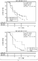
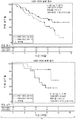
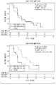
특히, 간세포 생장 인자("HGF"와 상호교환하여 사용가능함) 생체표지자가 항-c-met 길항제와 선택적으로 VEGF 길항제로의 치료가 임상적으로 의미 있는 혜택을 제공하는 환자 집단를 식별하기 위해 사용된다. 특히, 본 발명은 재발 교아세포종을 가진 개체에서 항-VEGF 항체(베바시주맙(bevacizumab))과 병용한 항-c-met 항체 MetMAb(오나투주맙(onartuzumab))의 임의 단계 II 임상실험의 데이터를 제공한다. HGF 생체표지자가 MetMAb와 베바시주맙 치료가 무진행 생존율 및 전체 생존율에 의해 평가된 임상적으로 의미 있는 혜택을 제공한 환자 집단를 식별하는데 사용되었다. 상기 임상 실험에서, MetMAb와 베바시주맙으로의 치료가 높은 수치의 HGF 생체표지자를 발현한 재발 교아세포종을 갖는 환자에게 임상적으로 의미 있는 혜택을 제공하였다. 상기 결과는 무진행 생존율(PFS) 및 전체 생존율(OS)에 의해 평가되는 효능이, 베바시주맙 단독 치료의 경우의 PFS 및 OS 데이터와 비교할 때, 특히 긍정적이었음을 보여주었다. 이와 같은 차이는 통계학적으로 유의미하였고, 베바시주맙에 MetMab의 첨가는 높은 수치의 HGF 생체표지자를 발현한 재발 교아세포종을 가진 환자에게서 무진행 생존율 및 전체 생존율 모두를 증가시켰다. 상기 임상 실험 데이터는 또한, 베바시주맙 단독으로 치료받은 환자의 진행 및 사망 위험률 대비, 베바시주맙과 병용한 MetMAb로의 치료가 낮은 수치의 HGF 생체표지자를 발현하는 재발 교아세포종을 가진 환자에게서 진행 및 사망의 위험률을 증가시켰음을 보여주었다. 상기 결과는 PFS 및 OS로 평가되는 효능이, 낮은 수치의 HGF 생체표지자를 발현한 교아세포종을 가진 환자에게서 베바시주맙 단독 치료의 경우의 PFS 및 OS 데이터와 비교할 때, MetMAb와 베바시주맙으로 치료받은 환자에게서 더 나빴음을 보여주었다. 이와 같은 차이는 통계학적으로 유의미하였다. In particular, hepatocyte growth factors (which can be used interchangeably with "HGF ") biomarkers are used to identify groups of patients in which treatment with an anti-c-met antagonist and optionally a VEGF antagonist provides clinically meaningful benefits . In particular, the present invention relates to the data of any Phase II clinical trials of the anti-c-met antibody MetMAb (onatuzumab) in combination with an anti-VEGF antibody (bevacizumab) in individuals with recurrent glioblastoma Lt; / RTI > HGF biomarkers were used to identify groups of patients who provided clinically meaningful benefits assessed by progression-free survival and overall survival for MetMAb and bevacizumab treatments. In this clinical trial, treatment with MetMAb and bevacizumab provided clinically meaningful benefits for patients with recurrent glioblastoma expressing high levels of HGF biomarkers. The results showed that the efficacy assessed by progression-free survival (PFS) and overall survival (OS) was particularly positive when compared to PFS and OS data in the case of bevacizumab monotherapy. These differences were statistically significant and the addition of MetMab to bevacizumab increased both progression-free survival and overall survival in patients with recurrent glioblastoma expressing high levels of HGF biomarkers. The clinical trial data also show that treatment with MetMAb in combination with bevacizumab versus the progression and mortality risk of patients treated with bevacizumab alone has progressed and progressed in patients with recurrent glioblastomas expressing low levels of HGF biomarkers And increased the risk of death. These results indicate that efficacy, as assessed by PFS and OS, is reduced by treatment with MetMAb and bevacizumab compared with PFS and OS data for bevacizumab alone in patients with glioblastoma expressing low levels of HGF biomarkers I was worse for the patient I received. These differences were statistically significant.
일 측면에서, 상기 환자의 암에 많은 양의 HGF 생체표지자가 있는 것으로 발견된 경우 상기 환자에게 c-met 길항제를 유효량으로 투여하는 것을 포함하는, 암 환자의 치료 방법이 제공된다.In one aspect, there is provided a method of treating a cancer patient, comprising administering to the patient an effective amount of a c-met antagonist if the cancer of the patient is found to have a large amount of a HGF biomarker.
일부 구현예에서 상기 환자의 암은 c-met을 과잉 발현한다. 일부 구현예에서, 상기 환자의 암은 c-met 증폭을 보인다. 일부 구현예에서, 상기 환자의 암은 c-met 증폭을 보이지 않는다.In some embodiments, the cancer of the patient overexpresses c-met. In some embodiments, the patient's cancer shows c-met amplification. In some embodiments, the patient's cancer does not exhibit c-met amplification.
일부 구현예에서, 상기 환자의 암은 c-met과 HGF를 모두 발현한다. 일부 구현예에서, 세포에서 분비된 HGF는 이것이 자가분비 방식으로 분비된 상기 세포의 표면에서 c-met과 결합한다. 일부 구현예에서, 상기 환자의 암은 c-met과 HGF를 모두 발현하고 자가분비 방식으로 신호를 전달한다. 일부 구현예에서, 환자의 암에서의 HGF 발현은 IHC 또는 ISH 또는 당해기술에 알려진 기타 방법을 사용하여 결정된다. In some embodiments, the patient's cancer expresses both c-met and HGF. In some embodiments, HGF secreted from the cell binds to c-met at the surface of the cell that is secreted in a self-secreted manner. In some embodiments, the cancer of the patient expresses both c-met and HGF and delivers a signal in a self-secreted fashion. In some embodiments, HGF expression in a patient's cancer is determined using IHC or ISH or other methods known in the art.
일부 구현예에서, 상기 c-met 길항제는 길항제 항-c-met 항체이다. 일부 구현예에서, 상기 항-c-met 항체는 (a) 서열 GYTFTSYWLH(서열식별번호: 1)을 포함하는 HVR1; (b) 서열 GMIDPSNSDTRFNPNFKD(서열식별번호: 2)을 포함하는 HVR2; (c) 서열 ATYRSYVTPLDY(서열식별번호: 3)을 포함하는 HVR3-HC; (d) 서열 KSSQSLLYTSSQKNYLA(서열식별번호: 4)을 포함하는 HVR1-LC; (e) 서열 WASTRES(서열식별번호: 5)를 포함하는 HVR2-LC; 및 (f) 서열 QQYYAYPWT(서열식별번호: 6)을 포함하는 HVR3-LC를 포함한다. 일부 구현예에서, 상기 항-c-met 항체는 오나투주맙 항원결정부와 결합한다. 일부 구현예에서, 상기 항-c-met 항체는 오나투주맙이다. 일부 구현예에서, 상기 항-c-met 항체의 유효량은 3주마다 15 mg/kg이다. 일부 구현예에서, 상기 항-c-met 항체의 유효량은 2주마다 10 mg/kg이다. 일부 구현예에서, 상기 c-met 길항제는 크리조티닙, 티반티닙, 카보잔티닙, MGCD-265, 피클라투주맙, 인간화된 TAK-701, 리로투무맙, 포레티닙, h224G11, DN-30, MK-2461, E7050, MK-8033, PF-4217903, AMG208, JNJ-38877605, EMD1204831, INC-280, LY-2801653, SGX-126, RP1040, LY2801653, BAY-853474 및/또는 LA480 중 하나 이상이다.In some embodiments, the c-met antagonist is an antagonist anti-c-met antibody. In some embodiments, the anti-c-met antibody comprises (a) HVR1 comprising the sequence GYTFTSYWLH (SEQ ID NO: 1); (b) HVR2 comprising the sequence GMIDPSNSDTRFNPNFKD (SEQ ID NO: 2); (c) HVR3-HC comprising the sequence ATYRSYVTPLDY (SEQ ID NO: 3); (d) HVR1-LC comprising the sequence KSSQSLLYTSSQKNYLA (SEQ ID NO: 4); (e) HVR2-LC comprising the sequence WASTRES (SEQ ID NO: 5); And (f) HVR3-LC comprising the sequence QQYYAYPWT (SEQ ID NO: 6). In some embodiments, the anti-c-met antibody binds to an onatuchwoarg antigenic determinant. In some embodiments, the anti-c-met antibody is onatuzumab. In some embodiments, the effective amount of the anti-c-met antibody is 15 mg / kg every 3 weeks. In some embodiments, the effective amount of the anti-c-met antibody is 10 mg / kg every two weeks. In some embodiments, the c-met antagonist is selected from the group consisting of: clozotinib, tibanthinib, carbozanthinib, MGCD-265, piclutujum, humanized TAK-701, lyotropum, porretinib, h224G11, Or at least one of LA480, LA480, E7050, MK-8033, PF-4217903, AMG208, JNJ-38877605, EMD1204831, INC-280, LY-2801653, SGX-126, RP1040, LY2801653, BAY-853474 and /
일부 구현예에서, 치료는 c-met 길항제과 VEGF 길항제의 병용의 유효량으로 이루어진다. 일부 구현예에서, 상기 VEGF 길항제는 항-VEGF 항체이다. 일부 구현예에서, 상기 항-VEGF 항체는 A4.6.1 항원결정부와 결합한다. 일부 구현예에서, 상기 항-VEGF 항체는 베바시주맙이다. 일부 구현예에서, 상기 항-VEGF 항체는 가변 중쇄(VH)와 가변 경쇄(VL)를 포함하되, 상기 VH는 아미노산 서열 EVQLVESGGG LVQPGGSLRL SCAASGYTFT NYGMNWVRQA PGKGLEWVGW INTYTGEPTY AADFKRRFTF SLDTSKSTAY LQMNSLRAED TAVYYCAKYP HYYGSSHWYF DVWGQGTLVT VSS(서열식별번호: 14)을 갖고 상기 VL은 아미노산 서열 DIQMTQSPSS LSASVGDRVT ITCSASQDIS NYLNWYQQKP GKAPKVLIYF TSSLHSGVPS RFSGSGSGTD FTLTISSLQP EDFATYYCQQ YSTVPWTFGQ GTKVEIKR(서열식별번호: 15)를 갖는다. 일부 구현예에서, 상기 항-VEGF 항체의 유효량은 2주마다 정맥내 투여로 10 mg/kg이다. 일부 구현예에서, 상기 항-VEGF 항체의 상기 유효량은 3주마다 정맥내 투여로 15 mg/kg이다. 일부 구현예에서, 상기 항-VEGF 항체의 유효량은 최초 정맥내로 90분에 걸쳐 투여되고, 차후에 60분 그리고 이어서 30분에 걸쳐 주입된다. 일부 구현예에서, 상기 항-VEGF 항체는 제1 주기에 상기 환자에게 두 번째로 투여된다. 일부 구현예에서, 상기 항-VEGF 항체의 추후 투여는 상기 c-met 길항제 이전 또는 이후에 이루어진다. 일부 구현예에서, 상기 VEGF 길항제는 상기 c-met 길항제와 동시에 투여된다.In some embodiments, the treatment comprises an effective amount of a combination of a c-met antagonist and a VEGF antagonist. In some embodiments, the VEGF antagonist is an anti-VEGF antibody. In some embodiments, the anti-VEGF antibody binds to the A4.6.1 antigenic determinant. In some embodiments, the anti-VEGF antibody is bevacizumab. In some embodiments, the anti-VEGF antibody comprises a variable heavy chain (VH) and a variable light chain (VL), wherein the VH comprises the amino acid sequence EVQLVESGGG LVQPGGSLRL SCAASGYTFT NYGMNWVRQA PGKGLEWVGW INTYTGEPTY AADFKRRFTF SLDTSKSTAY LQMNSLRAED TAVYYAKYPY HYYGSSHWYF DVWGQGTLVT VSS (SEQ ID NO: 14 ) And the VL has the amino acid sequence DIQMTQSPSS LSASVGDRVT ITCSASQDIS NYLNWYQQKP GKAPKVLIYF TSSLHSGVPS RFSGSGSGTD FTLTISSLQP EDFATYYCQQ YSTVPWTFGQ GTKVEIKR (SEQ ID NO: 15). In some embodiments, the effective amount of the anti-VEGF antibody is 10 mg / kg by intravenous administration every two weeks. In some embodiments, the effective amount of the anti-VEGF antibody is 15 mg / kg by intravenous administration every three weeks. In some embodiments, an effective amount of the anti-VEGF antibody is administered into the initial vein over 90 minutes, followed by 60 minutes and then over 30 minutes. In some embodiments, the anti-VEGF antibody is administered to the patient a second time in a first cycle. In some embodiments, the subsequent administration of the anti-VEGF antibody occurs before or after the c-met antagonist. In some embodiments, the VEGF antagonist is administered concurrently with the c-met antagonist.
일부 구현예에서, 상기 환자는 50세 미만이다. 일부 구현예에서, 상기 환자는 50세 이상이다. 일부 구현예에서, 상기 환자는 카르노프스키 수행 상태가 70% 내지 80%이다. 일부 구현예에서, 상기 환자는 카르노프스키 수행 상태가 90% 내지 100%이다.In some embodiments, the patient is less than 50 years old. In some embodiments, the patient is at least 50 years of age. In some embodiments, the patient has a Karnofsky performance status of 70% to 80%. In some embodiments, the patient has a Carnovsky performance status of 90% to 100%.
일부 구현예에서, 상기 환자는 높은 HGF 생체표지자를 갖지 않는 환자에 비해 더 큰 PFS 및/또는 OS를 갖는다. 일부 구현예에서, 상기 환자는 VEGF 길항제 단독으로 치료받은 환자에 비해 더 큰 PFS 및/또는 OS를 갖는다.In some embodiments, the patient has a greater PFS and / or OS than a patient who does not have a high HGF biomarker. In some embodiments, the patient has a greater PFS and / or OS than a patient treated with a VEGF antagonist alone.
일부 구현예에서, 상기 HGF 생체표지자는 HGF mRNA이고, HGF 생체표지자 mRNA 발현은 제자리 혼성화(ISH)를 사용하여 상기 환자의 샘플에서 결정된다. 일부 구현예에서, 높은 HGF 생체표지자는 2+ 및/또는 3+인 ISH 점수이다. 일부 구현예에서, 높은 HGF 생체표지자는 2+ 및 3+인 ISH 점수이다. 일부 구현예에서, 높은 HGF mRNA 생체표지자는 상기 샘플 중에 약 12개 이상의 HGF ISH 신호 양성 세포의 존재이다. 일부 구현예에서, 높은 HGF mRNA 생체표지자는 상기 샘플 중에 약 15개 이상의 HGF ISH 신호 양성 세포의 존재이다. 일부 구현예에서, 높은 HGF mRNA 생체표지자는 상기 샘플 중에 약 20개 이상의 HGF ISH 신호 양성 세포의 존재이다. 일부 구현예에서, 높은 HGF mRNA 생체표지자는 상기 샘플 중에서 약 25개 이상의 HGF ISH 신호 양성 세포의 존재이다. 일부 구현예에서, 높은 HGF mRNA 생체표지자는 상기 샘플 중에 약 30개 이상의 HGF ISH 신호 양성 세포의 존재이다. 일부 구현예에서, 높은 HGF mRNA 생체표지자는 상기 샘플 중에 약 35개 이상의 HGF ISH 신호 양성 세포의 존재이다. 일부 구현예에서, 높은 HGF mRNA 생체표지자는 상기 샘플 중에 1% 이상의 HGF ISH 신호 양성 세포이다. 일부 구현예에서, 높은 HGF mRNA 생체표지자는 상기 샘플 중에 2% 이상의 HGF ISH 신호 양성 세포이다. 일부 구현예에서, 높은 HGF mRNA 생체표지자는 상기 샘플 중에 3% 이상의 HGF ISH 신호 양성 세포이다. 일부 구현예에서, 높은 HGF mRNA 생체표지자는 상기 샘플 중에 4% 이상의 HGF ISH 신호 양성 세포이다. 일부 구현예에서, 높은 HGF mRNA 생체표지자는 상기 샘플 중에 5% 이상의 HGF ISH 신호 양성 세포이다. 일부 구현예에서, 높은 HGF mRNA 생체표지자는 상기 샘플 중에 10% 이상의 HGF ISH 신호 양성 세포이다. In some embodiments, the HGF biomarker is HGF mRNA and HGF biomarker mRNA expression is determined in the patient's sample using in situ hybridization (ISH). In some embodiments, the high HGF biomarker is an ISH score of 2+ and / or 3+. In some embodiments, high HGF biomarkers are ISH scores of 2+ and 3+. In some embodiments, the high HGF mRNA biomarker is the presence of at least about 12 HGF ISH signal positive cells in the sample. In some embodiments, the high HGF mRNA biomarker is the presence of at least about 15 HGF ISH signal positive cells in the sample. In some embodiments, the high HGF mRNA biomarker is the presence of at least about 20 HGF ISH signal positive cells in the sample. In some embodiments, the high HGF mRNA biomarker is the presence of at least about 25 HGF ISH signal positive cells in the sample. In some embodiments, the high HGF mRNA biomarker is the presence of at least about 30 HGF ISH signal positive cells in the sample. In some embodiments, the high HGF mRNA biomarker is the presence of at least about 35 HGF ISH signal positive cells in the sample. In some embodiments, the high HGF mRNA biomarker is at least 1% of the HGF ISH signal positive cells in the sample. In some embodiments, the high HGF mRNA biomarker is 2% or more HGF ISH signal positive cells in the sample. In some embodiments, the high HGF mRNA biomarker is more than 3% of the HGF ISH signal positive cells in the sample. In some embodiments, the high HGF mRNA biomarker is more than 4% of the HGF ISH signal positive cells in the sample. In some embodiments, the high HGF mRNA biomarker is more than 5% of the HGF ISH signal positive cells in the sample. In some embodiments, the high HGF mRNA biomarker is more than 10% of the HGF ISH signal positive cells in the sample.
일부 구현예에서, 상기 HGF 생체표지자 발현은 핵산 발현이고 증폭 기반 분석, RNA-seq, 유전자미세배열 분석법, SAGE, 매스어레이 기법 또는 FISH를 사용하여 상기 환자의 샘플에서 결정된다. 일부 구현예에서, 상기 증폭 기반 분석법은 폴리머라아제 연쇄반응(PCR) 기반 분석법(예컨대, 정량적 PCR, 실시간 PCR, 정량적 실시간 PCR(qRT-PCR), 역전사효소 PCR(rt-PCR), 및 역전사 정량적 PCR(rt-qPCR))이다.In some embodiments, the HGF biomarker expression is nucleic acid expression and is determined in the patient's sample using amplification-based analysis, RNA-seq, gene microarray analysis, SAGE, mass array techniques or FISH. In some embodiments, the amplification-based assays can be performed using polymerase chain reaction (PCR) based assays such as quantitative PCR, real time PCR, quantitative real time PCR (qRT-PCR), reverse transcriptase PCR (rt- PCR (rt-qPCR)).
일부 구현예에서, 상기 HGF 생체표지자는 HGF mRNA이고, HGF 생체표지자 mRNA 발현은 증폭 기반 분석법, RNA-seq, 유전자미세배열 분석법, SAGE, 매스어레이 기법 또는 FISH를 사용하여 상기 환자의 샘플에서 결정된다. 일부 구현예에서, 상기 증폭 기반 분석법은 PCR 기반 분석법(예컨대, 정량적 PCR, 실시간 PCR, 정량적 실시간 PCR(qRT-PCR), 역전사효소 PCR(rt-PCR) 및 역전사 정량적 PCR(rt-qPCR))이다. 일부 구현예에서, 상기 PCR 기반 분석법은 rt-qPCR이다. 일부 구현예에서, 높은 HGF 생체표지자는 참조 환자 집단의 상위 50%에서의 HGF 발현 수치이다. 일부 구현예에서, 높은 HGF 생체표지자는 참조 환자 집단의 상위 40%에서의 HGF 발현 수치이다. 일부 구현예에서, 높은 HGF 생체표지자는 참조 환자 집단의 상위 35%에서의 HGF 발현 수치이다. 일부 구현예에서, 높은 HGF 생체표지자는 참조 환자 집단의 상위 30%에서의 HGF 발현 수치이다. 일부 구현예에서, 높은 HGF 생체표지자는 참조 환자 집단의 상위 25%에서의 HGF 발현 수치이다. 일부 구현예에서, 높은 HGF 생체표지자는 참조 환자 집단의 상위 20%에서의 HGF 발현 수치이다.In some embodiments, the HGF biomarker is HGF mRNA and HGF biomarker mRNA expression is determined in the patient's sample using amplification-based assays, RNA-seq, gene microarray analysis, SAGE, mass array techniques or FISH . In some embodiments, the amplification based assay is a PCR based assay (e.g., quantitative PCR, real time PCR, qRT-PCR, reverse transcriptase PCR (rt-PCR) and reverse quantitative PCR (rt-qPCR)) . In some embodiments, the PCR-based assay is rt-qPCR. In some embodiments, the high HGF biomarker is a HGF expression level in the top 50% of the reference patient population. In some embodiments, the high HGF biomarker is a HGF expression level in the top 40% of the reference patient population. In some embodiments, the high HGF biomarker is a HGF expression level in the top 35% of the reference patient population. In some embodiments, the high HGF biomarker is an HGF expression level in the top 30% of the reference patient population. In some embodiments, the high HGF biomarker is a HGF expression level in the top 25% of the reference patient population. In some embodiments, the high HGF biomarker is a HGF expression level in the top 20% of the reference patient population.
일부 구현예에서, 상기 샘플은 상기 환자의 암에서 유래된 것이다. 상기 환자의 암의 샘플에는 암 세포, 림프구, 백혈구, 기질(stroma), 혈관, 연결조직, 기저판 및 기타 암 관련 세포 유형이 포함될 수 있다. 일부 구현예에서, 상기 샘플은 암 세포와 양성 기질 세포를 포함한다. 일부 구현예에서 상기 암은 교아세포종, 중피종, 간세포 암종, 콩팥 세포 암종, 위암, 육종 (예컨대, 골육종), 비소세포 폐암, 소세포 폐암, 유방암, 담낭암 또는 췌장암이다. 일부 구현예에서, 상기 암은 교아세포종, 중피종, 콩팥 세포 암종, 위암, 간세포 암종 또는 육종이다. 일부 구현예에서, 상기 암은 교아세포종이다. 일부 구현예에서, 상기 암은 이전에 치료받은 교아세포종이다. 일부 구현예에서, 상기 샘플은 교아세포종 세포와 양성 기질 세포를 포함한다. 일부 구현예에서, 상기 양성 기질 세포는 반응성 성상교세포, 신경아교 세포, 주피세포 및 내피 세포 중 하나 이상이다. 일부 구현예에서, 상기 암은 중피종이다. 일부 구현예에서, 상기 암은 이전에 치료받은 중피종이다. 일부 구현예에서, 상기 샘플은 중피종 세포와 양성 기질 세포를 포함한다. 일부 구현예에서, 상기 암은 위암이다. 일부 구현예에서, 상기 암은 이전에 치료받은 위암이다. 일부 구현예에서, 상기 암은 위암 세포와 양성 기질 세포를 포함한다. 일부 구현예에서, 상기 양성 기질 세포는 섬유아세포, 대식세포 및 내피 세포 중 하나 이상이다. 일부 구현예에서, 상기 암은 콩팥 세포 암종이다. 일부 구현예에서, 상기 암은 이전에 치료받은 콩팥 세포 암종이다. 일부 구현예에서, 상기 샘플은 콩팥 세포 암종 세포와 양성 기질 세포를 포함한다. 일부 구현예에서, 상기 암은 간세포 암종이다. 일부 구현예에서, 상기 암은 이전에 치료받은 간세포 암종이다. 일부 구현예에서, 상기 샘플은 간세포 암종 세포와 양성 기질 세포를 포함한다. 일부 구현예에서, 상기 암은 육종(예컨대, 골육종)이다. 일부 구현예에서, 상기 암은 이전에 치료받은 육종(예컨대, 이전에 치료받은 골육종)이다. 일부 구현예에서, 상기 샘플은 육종 세포와 양성 기질 세포를 포함한다. 일부 구현예에서, 상기 샘플은 환자의 종양에서 유래된 것이다. 종양 샘플에는 암 세포, 림프구, 백혈구, 기질, 혈관, 연결조직, 기저판 및 상기 종양과 관련관 기타 모든 세포 유형이 포함될 수 있다. In some embodiments, the sample is derived from the cancer of the patient. Samples of cancer of the patient may include cancer cells, lymphocytes, leukocytes, stroma, blood vessels, connective tissue, basal plates and other cancer-related cell types. In some embodiments, the sample comprises cancerous cells and benign stromal cells. In some embodiments, the cancer is a glioblastoma, mesothelioma, hepatocellular carcinoma, renal cell carcinoma, gastric cancer, sarcoma (e.g., osteosarcoma), non-small cell lung cancer, small cell lung cancer, breast cancer, gallbladder cancer or pancreatic cancer. In some embodiments, the cancer is glioblastoma, mesothelioma, renal cell carcinoma, stomach cancer, hepatocellular carcinoma or sarcoma. In some embodiments, the cancer is glioblastoma. In some embodiments, the cancer is a previously treated glioblastoma. In some embodiments, the sample comprises glioblastoma cells and benign stromal cells. In some embodiments, the benign stromal cell is at least one of a reactive astrocytic cell, a gliotic cell, a squamous cell, and an endothelial cell. In some embodiments, the cancer is mesothelioma. In some embodiments, the cancer is a previously treated mesothelioma. In some embodiments, the sample comprises mesothelioma cells and benign stromal cells. In some embodiments, the cancer is gastric cancer. In some embodiments, the cancer is a previously treated gastric cancer. In some embodiments, the cancer comprises gastric cancer cells and benign stromal cells. In some embodiments, the benign stromal cell is at least one of fibroblasts, macrophages, and endothelial cells. In some embodiments, the cancer is a kidney cell carcinoma. In some embodiments, the cancer is a previously treated renal cell carcinoma. In some embodiments, the sample comprises kidney cell carcinoma cells and benign stromal cells. In some embodiments, the cancer is hepatocellular carcinoma. In some embodiments, the cancer is a previously treated hepatocellular carcinoma. In some embodiments, the sample comprises hepatocellular carcinoma cells and benign stromal cells. In some embodiments, the cancer is sarcoma (e.g., osteosarcoma). In some embodiments, the cancer is a previously treated sarcoma (e.g., a previously treated osteosarcoma). In some embodiments, the sample comprises sarcomatous cells and benign stromal cells. In some embodiments, the sample is derived from a patient ' s tumor. Tumor samples may include cancer cells, lymphocytes, leukocytes, matrices, blood vessels, connective tissue, basal plates, and all other cell types associated with the tumor.
일부 구현예에서, 상기 샘플은 c-met 길항제로의 치료 이전에 수득된다. 일부 구현예에서, 상기 샘플은 VEGF 길항제로의 치료 이전에 수득된다. 일부 구현예에서, 상기 샘플은 암 약제로의 치료 이전에 수득된다.In some embodiments, the sample is obtained prior to treatment with a c-met antagonist. In some embodiments, the sample is obtained prior to treatment with a VEGF antagonist. In some embodiments, the sample is obtained prior to treatment with a cancer agent.
일부 구현예에서, 상기 샘플은 포르말린으로 고정되고 파라핀 포매된다. 일부 구현예에서, 상기 ISH는 혼성화-기반 신호 증폭을 사용하여 검출된다.In some embodiments, the sample is fixed in formalin and embedded in paraffin. In some embodiments, the ISH is detected using hybridization-based signal amplification.
일부 구현예에서, RNA가 상기 샘플에서 단리된다. 일부 구현예에서, RNA가 상기 포르말린으로 고정되고 파라핀 포매된 샘플에서 단리된다. 일부 구현예에서, 상기 단리된 RNA가 정제된다. 일부 구현예에서, 상기 정제된 RNA는 증폭-기반 분석법을 위한 RNA 공급원으로 사용된다. 일부 구현예에서, 상기 증폭-기반 분석법은 PCR 기반 분석법이다. 일부 구현예에서, 상기 PCR 기반 분석법은 rt-qPCR이다.In some embodiments, RNA is isolated in the sample. In some embodiments, RNA is isolated from the sample that is fixed with the formalin and is paraffin-embedded. In some embodiments, the isolated RNA is purified. In some embodiments, the purified RNA is used as an RNA source for amplification-based assays. In some embodiments, the amplification-based assay is a PCR-based assay. In some embodiments, the PCR-based assay is rt-qPCR.
일부 구현예에서 상기 암은 교아세포종, 중피종, 간세포 암종, 콩팥 세포 암종, 위암, 육종(예컨대, 골육종), 비소세포 폐암, 소세포 폐암, 유방암, 담낭암 또는 췌장암이다. 일부 구현예에서, 상기 암은 교아세포종, 중피종, 콩팥 세포 암종, 위암, 간세포 암종 또는 육종이다. 일부 구현예에서, 상기 암은 이전에 치료받은 교아세포종이다. 일부 구현예에서, 상기 암은 이전에 치료받은 중피종이다. 일부 구현예에서, 상기 암은 이전에 치료받은 콩팥 세포 암종이다. 일부 구현예에서, 상기 암은 이전에 치료받은 위암이다. 일부 구현예에서, 상기 암은 이전에 치료받은 간세포 암종이다. 일부 구현예에서, 상기 암은 이전에 치료받은 육종이다.In some embodiments, the cancer is a glioblastoma, mesothelioma, hepatocellular carcinoma, renal cell carcinoma, gastric cancer, sarcoma (e.g., osteosarcoma), non-small cell lung cancer, small cell lung cancer, breast cancer, gallbladder cancer or pancreatic cancer. In some embodiments, the cancer is glioblastoma, mesothelioma, renal cell carcinoma, stomach cancer, hepatocellular carcinoma or sarcoma. In some embodiments, the cancer is a previously treated glioblastoma. In some embodiments, the cancer is a previously treated mesothelioma. In some embodiments, the cancer is a previously treated renal cell carcinoma. In some embodiments, the cancer is a previously treated gastric cancer. In some embodiments, the cancer is a previously treated hepatocellular carcinoma. In some embodiments, the cancer is a previously treated sarcoma.
일 측면에서, 상기 환자의 암에 적은 양의 HGF 생체표지자가 있는 것으로 발견된 경우, 상기 환자에게 c-met 길항제 이외의 약제를 치료차원의 유효량으로 투여하는 것을 포함하는, 암 환자를 치료하는 방법이 제공된다.In one aspect, there is provided a method of treating a cancer patient, comprising administering to the patient an effective amount of a therapeutic agent other than a c-met antagonist, when the cancer of the patient is found to have a low amount of the HGF biomarker / RTI >
일 측면에서, 본 발명은 c-met 길항제로의 치료에 반응할 가능성이 있는 암 환자를 식별하기 위한 방법을 제공하는데, 상기 방법은 상기 환자의 암에 많은 양의 HGF 생체표지자가 있는지 여부를 확인하기 위한 단계를 포함하되, 상기 HGF 생체표지자 발현이 상기 환자가 상기 c-met 길항제로의 치료에 반응할 가능성이 있음을 시사한다.In one aspect, the invention provides a method for identifying a cancer patient who is likely to respond to treatment with a c-met antagonist, said method comprising determining whether the patient's cancer has a large amount of a HGF biomarker , Wherein said HGF biomarker expression is likely to be responsive to treatment with said c-met antagonist.
일부 구현예에서, 상기 HGF 생체표지자가 HGF mRNA이고, HGF 생체표지자 mRNA 발현이 제자리 혼성화 (ISH)을 사용하여 상기 환자의 샘플에서 결정된다. 일부 구현예에서, 높은 HGF 생체표지자는 2+ 및/또는 3+인 ISH 점수이다. 일부 구현예에서, 높은 HGF 생체표지자는 2+ 및 3+인 ISH 점수이다. 일부 구현예에서, 높은 HGF mRNA 생체표지자는 상기 샘플 중에 약 12개 이상의 HGF ISH 신호 양성 세포의 존재이다. 일부 구현예에서, 높은 HGF mRNA 생체표지자는 상기 샘플 중에 약 15개 이상의 HGF ISH 신호 양성 세포의 존재이다. 일부 구현예에서, 높은 HGF mRNA 생체표지자는 상기 샘플 중에 약 20개 이상의 HGF ISH 신호 양성 세포의 존재이다. 일부 구현예에서, 높은 HGF mRNA 생체표지자는 상기 샘플 중에 약 25개 이상의 HGF ISH 신호 양성 세포의 존재이다. 일부 구현예에서, 높은 HGF mRNA 생체표지자는 상기 샘플 중에 약 30개 이상의 HGF ISH 신호 양성 세포의 존재이다. 일부 구현예에서, 높은 HGF mRNA 생체표지자는 상기 샘플 중에 약 35개 이상의 HGF ISH 신호 양성 세포의 존재이다. 일부 구현예에서, 높은 HGF mRNA 생체표지자는 상기 샘플에서의 1% 이상의 HGF ISH 신호 양성 세포이다. 일부 구현예에서, 높은 HGF mRNA 생체표지자는 상기 샘플에서의 2% 이상의 HGF ISH 신호 양성 세포이다. 일부 구현예에서, 높은 HGF mRNA 생체표지자는 상기 샘플에서의 3% 이상의 HGF ISH 신호 양성 세포이다. 일부 구현예에서, 높은 HGF mRNA 생체표지자는 상기 샘플에서의 4% 이상의 HGF ISH 신호 양성 세포이다. 일부 구현예에서, 높은 HGF mRNA 생체표지자는 상기 샘플에서의 5% 이상의 HGF ISH 신호 양성 세포이다. 일부 구현예에서, 높은 HGF mRNA 생체표지자는 상기 샘플에서의 10% 이상의 HGF ISH 신호 양성 세포이다.In some embodiments, the HGF biomarker is HGF mRNA and HGF biomarker mRNA expression is determined in the patient's sample using in situ hybridization (ISH). In some embodiments, the high HGF biomarker is an ISH score of 2+ and / or 3+. In some embodiments, high HGF biomarkers are ISH scores of 2+ and 3+. In some embodiments, the high HGF mRNA biomarker is the presence of at least about 12 HGF ISH signal positive cells in the sample. In some embodiments, the high HGF mRNA biomarker is the presence of at least about 15 HGF ISH signal positive cells in the sample. In some embodiments, the high HGF mRNA biomarker is the presence of at least about 20 HGF ISH signal positive cells in the sample. In some embodiments, the high HGF mRNA biomarker is the presence of at least about 25 HGF ISH signal positive cells in the sample. In some embodiments, the high HGF mRNA biomarker is the presence of at least about 30 HGF ISH signal positive cells in the sample. In some embodiments, the high HGF mRNA biomarker is the presence of at least about 35 HGF ISH signal positive cells in the sample. In some embodiments, the high HGF mRNA biomarker is more than 1% of the HGF ISH signal positive cells in the sample. In some embodiments, the high HGF mRNA biomarker is 2% or more HGF ISH signal positive cells in the sample. In some embodiments, the high HGF mRNA biomarker is 3% or more HGF ISH signal positive cells in the sample. In some embodiments, the high HGF mRNA biomarker is more than 4% of the HGF ISH signal positive cells in the sample. In some embodiments, the high HGF mRNA biomarker is at least 5% HGF ISH signal positive cells in the sample. In some embodiments, the high HGF mRNA biomarker is more than 10% of the HGF ISH signal positive cells in the sample.
일부 구현예에서, 상기 HGF 생체표지자 발현은 핵산 발현이고 증폭 기반 분석법, RNA-seq, 유전자미세배열 분석법, SAGE, 매스어레이 기법 또는 FISH를 사용하여 상기 환자의 샘플에서 결정된다. 일부 구현예에서, 상기 증폭 기반 분석법은 중합효소 연쇄반응(PCR) 기반 분석법(예컨대, 정량적 PCR, 실시간 PCR, 정량적 실시간 PCR(qRT-PCR), 역전사효소 PCR(rt-PCR) 및 역전사 정량적 PCR(rt-qPCR))이다.In some embodiments, the HGF biomarker expression is nucleic acid expression and is determined in the patient's sample using amplification-based assays, RNA-seq, gene microarray analysis, SAGE, mass array techniques or FISH. In some embodiments, the amplification-based assays can be performed using polymerase chain reaction (PCR) based assays such as quantitative PCR, real time PCR, quantitative real time PCR (qRT-PCR), reverse transcriptase PCR (rt-PCR) and reverse quantitative PCR rt-qPCR)).
일부 구현예에서, 상기 HGF 생체표지자는 HGF mRNA이고, HGF 생체표지자 mRNA 발현은 증폭 기반 분석법, RNA-seq, 유전자미세배열 분석법, SAGE, 매스어레이 기법 또는 FISH를 사용하여 상기 환자의 샘플에서 결정된다. 일부 구현예에서, 상기 증폭 기반 분석법은 중합효소 연쇄반응(PCR) 기반 분석법(예컨대, 정량적 PCR, 실시간 PCR, 정량적 실시간 PCR(qRT-PCR), 역전사효소 PCR(rt-PCR) 및 역전사 정량적 PCR(rt-qPCR))이다. 일부 구현예에서, 높은 HGF 생체표지자는 참조 환자 집단의 상위 50%에서의 HGF 발현 수치이다. 일부 구현예에서, 높은 HGF 생체표지자는 참조 환자 집단의 상위 40%에서의 HGF 발현 수치이다. 일부 구현예에서, 높은 HGF 생체표지자는 참조 환자 집단의 상위 35%에서의 HGF 발현 수치이다. 일부 구현예에서, 높은 HGF 생체표지자는 참조 환자 집단의 상위 30%에서의 HGF 발현 수치이다. 일부 구현예에서, 높은 HGF 생체표지자는 참조 환자 집단의 상위 25%에서의 HGF 발현 수치이다. 일부 구현예에서, 높은 HGF 생체표지자는 참조 환자 집단의 상위 20%에서의 HGF 발현 수치이다.In some embodiments, the HGF biomarker is HGF mRNA and HGF biomarker mRNA expression is determined in the patient's sample using amplification-based assays, RNA-seq, gene microarray analysis, SAGE, mass array techniques or FISH . In some embodiments, the amplification-based assays can be performed using polymerase chain reaction (PCR) based assays such as quantitative PCR, real time PCR, quantitative real time PCR (qRT-PCR), reverse transcriptase PCR (rt-PCR) and reverse quantitative PCR rt-qPCR)). In some embodiments, the high HGF biomarker is a HGF expression level in the top 50% of the reference patient population. In some embodiments, the high HGF biomarker is a HGF expression level in the top 40% of the reference patient population. In some embodiments, the high HGF biomarker is a HGF expression level in the top 35% of the reference patient population. In some embodiments, the high HGF biomarker is an HGF expression level in the top 30% of the reference patient population. In some embodiments, the high HGF biomarker is a HGF expression level in the top 25% of the reference patient population. In some embodiments, the high HGF biomarker is a HGF expression level in the top 20% of the reference patient population.
일 측면에서, a c-met 길항제로의 치료에 반응할 가능성이 적은 암 환자를 식별하는 방법이 제공되는데, 상기 환자의 암에 적은 양의 HGF 생체표지자가 있는지 여부를 결정하는 단계를 포함하되, 상기 HGF 생체표지자 발현이 상기 환자가 상기 c-met 길항제로의 치료에 반응할 가능성이 적음을 시사한다. 일부 구현예에서, HGF 생체표지자 핵산 발현은 제자리 혼성화(ISH)를 사용하여 상기 환자의 샘플에서 결정된다. 일부 구현예에서, 낮은 HGF mRNA 생체표지자는 2+ 미만의 ISH 점수이다. 일부 구현예에서, 낮은 HGF mRNA 생체표지자는 1+ 미만의 ISH 점수이다. 일부 구현예에서, 낮은 HGF mRNA 생체표지자는 0 또는 1+인 ISH 점수이다. 일부 구현예에서, 낮은 HGF mRNA 생체표지자가 0인 ISH 점수이다. 일부 구현예에서, 낮은 HGF 생체표지자는 10개 이하의 세포에서의 HGF ISH 양성 신호의 존재이다. 일부 구현예에서, 낮은 HGF 생체표지자는 5개 이하의 세포에서의 HGF ISH 양성 신호의 존재이다. 일부 구현예에서, 낮은 HGF 생체표지자는 0개의 세포에서의 HGF ISH 양성 신호의 존재이다.In one aspect, there is provided a method of identifying a cancer patient who is less likely to respond to treatment with an a c-met antagonist, comprising determining whether the patient's cancer has a low amount of HGF biomarker, Suggesting that the HGF biomarker expression is less likely for the patient to respond to treatment with the c-met antagonist. In some embodiments, the HGF biomarker nucleic acid expression is determined in the patient's sample using in situ hybridization (ISH). In some embodiments, the low HGF mRNA biomarker is an ISH score of less than 2+. In some embodiments, the low HGF mRNA biomarker is an ISH score of less than 1+. In some embodiments, the low HGF mRNA biomarker is an ISH score of 0 or 1+. In some embodiments, the low HGF mRNA biomarker is an ISH score of zero. In some embodiments, the low HGF biomarker is the presence of an HGF ISH positive signal in no more than 10 cells. In some embodiments, the low HGF biomarker is the presence of an HGF ISH positive signal in no more than 5 cells. In some embodiments, the low HGF biomarker is the presence of an HGF ISH positive signal in zero cells.
일 측면에서, c-met 길항제로의 치료에 반응할 가능성이 적은 암 환자를 식별하는 방법이 제공되는데, 상기 환자의 암에 적은 양의 HGF 생체표지자가 있는지 여부를 결정하는 단계를 포함하되, 상기 HGF 생체표지자 발현이 상기 환자가 상기 c-met 길항제로의 치료에 반응할 가능성이 적음을 시사한다. 일부 구현예에서, HGF 생체표지자 핵산 발현은 증폭 기반 분석법, RNA-seq, 유전자미세배열 분석법, SAGE, 매스어레이 기법 또는 FISH를 사용하여 상기 환자의 샘플에서 결정된다. 일부 구현예에서, 상기 증폭 기반 분석법은 중합효소 연쇄반응(PCR) 기반 분석법(예컨대, 정량적 PCR, 실시간 PCR, 정량적 실시간 PCR(qRT-PCR), 역전사효소 PCR(rt-PCR) 및 역전사 정량적 PCR(rt-qPCR))이다. 일부 구현예에서, 낮은 HGF mRNA 생체표지자는 참조 환자 집단의 하위 50%에서의 HGF 발현 수치이다. 일부 구현예에서, 낮은 HGF mRNA 생체표지자는 참조 환자 집단의 하위 60%에서의 HGF 발현 수치이다. 일부 구현예에서, 낮은 HGF mRNA 생체표지자는 참조 환자 집단의 하위 65%에서의 HGF 발현 수치이다. 일부 구현예에서, 낮은 HGF mRNA 생체표지자는 참조 환자 집단의 하위 70%에서의 HGF 발현 수치이다. 일부 구현예에서, 낮은 HGF mRNA 생체표지자는 참조 환자 집단의 하위 75%에서의 HGF 발현 수치이다. 일부 구현예에서, 낮은 HGF mRNA 생체표지자는 참조 환자 집단의 하위 80%에서의 HGF 발현 수치이다.In one aspect, there is provided a method of identifying a cancer patient that is less likely to respond to treatment with a c-met antagonist, comprising determining whether the patient's cancer has a low amount of a HGF biomarker, HGF biomarker expression is less likely to respond to treatment with the c-met antagonist. In some embodiments, expression of the HGF biomarker nucleic acid is determined in the patient's sample using amplification-based assays, RNA-seq, gene microarray analysis, SAGE, mass array techniques or FISH. In some embodiments, the amplification-based assays can be performed using polymerase chain reaction (PCR) based assays such as quantitative PCR, real time PCR, quantitative real time PCR (qRT-PCR), reverse transcriptase PCR (rt-PCR) and reverse quantitative PCR rt-qPCR)). In some embodiments, the low HGF mRNA biomarker is a HGF expression level in the bottom 50% of the reference patient population. In some embodiments, the low HGF mRNA biomarker is a HGF expression level in the lower 60% of the reference patient population. In some embodiments, the low HGF mRNA biomarker is a HGF expression level in the lower 65% of the reference patient population. In some embodiments, the low HGF mRNA biomarker is a HGF expression level in the lower 70% of the reference patient population. In some embodiments, the low HGF mRNA biomarker is a HGF expression level in the lower 75% of the reference patient population. In some embodiments, the low HGF mRNA biomarker is a HGF expression level in the lower 80% of the reference patient population.
일부 구현예에서, 환자는 인간 환자이다. 상기 환자는 암 환자, 즉 암의 하나 이상의 증상을 앓고 있는 또는 앓을 위험이 있는 사람일 수 있다. 게다가, 상기 환자는 이전에 치료받은 암 환자일 수 있다. 상기 환자는 교아세포종 환자, 즉 교아세포종의 하나 이상의 증상을 앓고 있거나 앓을 위험이 있는 사람이다. 게다가, 상기 환자는 이전에 치료받은 교아세포종 환자일 수 있다. 일부 구현예에서, 상기 환자는 단지 한 번의 전번차 화학요법으로 치료받은 바 있다. 일부 구현예에서, 상기 환자는 이전에 테모졸로마이드로 치료받았다. 일부 구현예에서, 상기 환자는 방사선과 병용하여 테모졸로마이드로 이전에 치료받았다. 일부 구현예에서, 상기 환자는 또 다른 제제와 병용하여 테모졸로마이드로 이전에 치료받았다. 일부 구현예에서, 상기 교아세포종은 2차 교아세포종이다. 상기 환자는 중피종 환자, 즉 중피종의 하나 이상의 증상을 앓고 있거나 또는 앓을 위험이 있는 사람이다. 게다가, 상기 환자는 이전에 치료받은 중피종 환자일 수 있다. 일부 구현예에서, 상기 환자는 단지 한 번의 전번차 화학요법으로 치료받은 바 있다. 일부 구현예에서, 상기 환자는 방사선과 병용하여 화학요법으로 이전에 치료받았다. 일부 구현예에서, 상기 환자는 또 다른 제제와 병용하여 화학요법으로 이전에 치료받았다. 일부 구현예에서, 상기 중피종은 2차 중피종이다. 상기 환자는 위암 환자, 즉 위암의 하나 이상의 증상을 앓거나 또는 앓을 위험이 있는 사람일 수 있다. 게다가, 상기 환자는 이전에 치료받은 위암 환자일 수 있다. 일부 구현예에서, 상기 환자는 단지 한 번의 전번차 화학요법으로 치료받은 바 있다. 일부 구현예에서, 상기 환자는 방사선과 병용하여 화학요법으로 이전에 치료받았다. 일부 구현예에서, 상기 환자는 또 다른 제제와 병용하여 화학요법으로 이전에 치료받았다. 일부 구현예에서, 상기 위암은 2차 위암이다. 상기 환자는 콩팥 세포 암종 환자, 즉 콩팥 세포 암종의 하나 이상의 증상을 앓거나 또는 앓을 위험이 있는 사람일 수 있다. 게다가, 상기 환자는 이전에 치료받은 콩팥 세포 암종 환자일 수 있다. 일부 구현예에서, 상기 환자는 단지 한 번의 전번차 화학요법으로 치료받은 바 있다. 일부 구현예에서, 상기 환자는 화학요법으로 이전에 치료받았다. 일부 구현예에서, 상기 환자는 방사선과 병용하여 화학요법으로 이전에 치료받았다. 일부 구현예에서, 상기 환자는 또 다른 제제와 병용하여 화학요법으로 이전에 치료받았다. 일부 구현예에서, 상기 콩팥 세포 암종은 2차 콩팥 세포 암종이다. 상기 환자는 간세포 암종 환자, 즉 간세포 암종의 하나 이상의 증상을 앓거나 또는 앓을 위험이 있는 사람일 수 있다. 게다가, 상기 환자는 이전에 치료받은 간세포 암종 환자일 수 있다. 일부 구현예에서, 상기 환자는 단지 한 번의 전번차 화학요법으로 치료받은 바 있다. 일부 구현예에서, 상기 환자는 화학요법으로 이전에 치료받았다. 일부 구현예에서, 상기 환자는 방사선과 병용하여 화학요법으로 이전에 치료받았다. 일부 구현예에서, 상기 환자는 또 다른 제제와 병용하여 화학요법으로 이전에 치료받았다. 일부 구현예에서, 상기 간세포 암종은 2차 간세포 암종이다.In some embodiments, the patient is a human patient. The patient may be a cancer patient, i. E. A person suffering from one or more symptoms of cancer or at risk of being sick. In addition, the patient may be a previously treated cancer patient. The patient is a glioblastoma patient, i. E., One who suffers from one or more symptoms of glioblastoma or is at risk of being sick. In addition, the patient may be a previously treated glioblastoma patient. In some embodiments, the patient has only been treated with one of the first-line chemotherapy regimens. In some embodiments, the patient has previously been treated with temozolomide. In some embodiments, the patient has previously been treated with temozolomide in combination with radiation. In some embodiments, the patient has previously been treated with temozolomide in combination with another agent. In some embodiments, the glioblastoma is a secondary glioblastoma. The patient is a mesothelioma patient, a person who suffers from one or more symptoms of mesothelioma or is at risk of being sick. In addition, the patient may be a previously treated mesothelioma patient. In some embodiments, the patient has only been treated with one of the first-line chemotherapy regimens. In some embodiments, the patient has previously been treated with chemotherapy in conjunction with radiation. In some embodiments, the patient has previously been treated with chemotherapy in combination with another agent. In some embodiments, the mesothelioma is a secondary mesothelioma. The patient may be a gastric cancer patient, i. E., A person at risk of suffering from or at risk of having one or more symptoms of gastric cancer. In addition, the patient may be a previously treated gastric cancer patient. In some embodiments, the patient has only been treated with one of the first-line chemotherapy regimens. In some embodiments, the patient has previously been treated with chemotherapy in conjunction with radiation. In some embodiments, the patient has previously been treated with chemotherapy in combination with another agent. In some embodiments, the gastric cancer is a second gastric cancer. The patient may be a patient with renal cell carcinoma, i. E. One who has or is at risk of suffering from one or more symptoms of renal cell carcinoma. In addition, the patient may be a previously treated renal cell carcinoma patient. In some embodiments, the patient has only been treated with one of the first-line chemotherapy regimens. In some embodiments, the patient has been previously treated with chemotherapy. In some embodiments, the patient has previously been treated with chemotherapy in conjunction with radiation. In some embodiments, the patient has previously been treated with chemotherapy in combination with another agent. In some embodiments, the renal cell carcinoma is a second renal cell carcinoma. The patient may be a patient with hepatocellular carcinoma, i. E., One who has or is at risk of having one or more symptoms of hepatocellular carcinoma. In addition, the patient may be a previously treated hepatocellular carcinoma patient. In some embodiments, the patient has only been treated with one of the first-line chemotherapy regimens. In some embodiments, the patient has been previously treated with chemotherapy. In some embodiments, the patient has previously been treated with chemotherapy in conjunction with radiation. In some embodiments, the patient has previously been treated with chemotherapy in combination with another agent. In some embodiments, the hepatocellular carcinoma is secondary hepatocellular carcinoma.
일부 구현예에서, 상기 샘플은 암 환자에서 수득된 세포 또는 체액의 집합체이다. 상기 조직 또는 세포 샘플의 공급원은 신선한, 냉동된 및/또는 보존된 장기 또는 조직 샘플 또는 생검조직 또는 흡입액; 혈액 또는 모든 혈액 구성성분; 체액, 예컨대 뇌척수액, 양수, 복막액, 또는 세포간질액; 상기 개체의 임신 또는 발달의 어느 시점에서 유래된 세포에서 유래된 것과 같은 고체 조직일 수 있다. 상기 조직 샘플은 자연계에서 상기 조직과 자연스럽게 혼합되지 않는 화합물들, 예컨대 보존제, 항응고제, 완충용액, 고정액, 영양분, 항생제 등을 함유할 수 있다. 본원의 종양의 예에는, 비제한적으로, 종양 생검, 미세바늘 흡입액, 세기관지 세척액, 흉수, 객담, 소변, 수술 시료, 순환하는 종양 세포, 혈청, 혈장, 순환하는 혈장 단백질, 복수액, 종양에서 유도된 또는 종양과 유사한 특성을 보이는 1차 세포 배양액 또는 세포주, 뿐만 아니라 보존된 종양 샘플, 예컨대포르말린-고정, 파라핀-포매 종양 샘플 또는 냉동 종양 샘플이 포함된다. 종양 샘플에는 암 세포, 림프구, 백혈구, 기질, 혈관, 연결조직, 기저판 및 상기 종양과 관련된 기타 모든 세포 유형이 포함될 수 있다. 일 구현예에서, 상기 샘플은 교아세포종 종양 샘플(예컨대, 양성 기질, 예컨대, 반응성 성상교세포, 신경아교 세포, 주피세포 및/또는 내피 세포를 포함하는 교아세포종 종양 샘플)을 포함한다. 일부 구현예에서, 상기 샘플은 육안으로 해부된 교아세포종 종양 샘플(예컨대, 형태학적으로 정상인 뇌 조직이 상기 종양 샘플에서 제거되었음)을 포함한다. 일부 구현예에서, 상기 육안으로 해부된 교아세포종 종양 샘플은 양성 기질(예컨대, 반응성 성상교세포, 신경아교 세포, 주피세포 및/또는 내피 세포)를 포함한다. 일부 구현예에서, 상기 샘플은 교아세포종 생검에서 유래된 것이다. 일부 구현예에서, 상기 샘플은 교아세포종 암 절제에서 유래된 것이다. 일부 구현예에서, 상기 샘플은 상기 환자의 교아세포종 재발한 후 수득되었다. 일부 구현예에서, 상기 샘플은 상기 환자의 교아세포종이 재발하기 전에 수득되었다. 일 구현예에서 상기 샘플은 중피종 종양 샘플(예컨대, 양성 기질을 포함하는 중피종 종양 샘플)을 포함한다. 일부 구현예에서, 상기 샘플은 육안으로 해부된 중피종 종양 샘플(예컨대, 여기서 형태학적으로 정상인 중피 조직이 상기 종양 샘플에서 제거되었다)을 포함한다. 일부 구현예에서, 상기 육안으로 해부된 중피종 종양 샘플은 양성 기질을 포함한다. 일부 구현예에서, 상기 샘플은 중피종 생검에서 유래된 것이다. 일부 구현예에서, 상기 샘플은 중피종 암 절제에서 유래된 것이다. 일부 구현예에서, 상기 샘플은 상기 환자의 중피종이 재발한 후에 수득되었다. 일부 구현예에서, 상기 샘플은 상기 환자의 중피종이 재발되기 전에 수득되었다. 일 구현예에서 상기 샘플은 위암 종양 샘플(예컨대, 양성 기질, 예컨대, 섬유아세포, 대식세포 및/또는 내피 세포를 포함하는 위암 종양 샘플)을 포함한다. 일부 구현예에서, 상기 샘플은 육안으로 해부된 위암 종양 샘플(예컨대, 여기서 형태학적으로 정상적인 위 조직이 상기 종양 샘플에서 제거되었다)을 포함한다. 일부 구현예에서, 상기 육안으로 해부된 위암 종양 샘플은 양성 기질(예컨대, 섬유아세포, 대식세포 및/또는 내피 세포)를 포함한다. 일부 구현예에서, 상기 샘플은 위암 생검에서 유래된 것이다. 일부 구현예에서, 상기 샘플은 위암 절제에서 유래된 것이다. 일부 구현예에서, 상기 샘플은 상기 환자의 위암이 재발한 이후에 수득되었다. 일부 구현예에서, 상기 샘플은 상기 환자의 위암이 재발하기 이전에 수득되었다. 일 구현예에서 상기 샘플은 콩팥 세포 암종 종양 샘플(예컨대, 양성 기질을 포함하는 콩팥 세포 암종 종양 샘플)을 포함한다. 일부 구현예에서, 상기 샘플은 육안으로 해부된 콩팥 세포 암종 종양 샘플(예컨대, 여기서 형태학적으로 정상적인 신장 조직이 상기 종양 샘플에서 제거되었다)을 포함한다. 일부 구현예에서, 상기 육안으로 해부된 콩팥 세포 암종 종양 샘플은 양성 기질을 포함한다. 일부 구현예에서, 상기 샘플은 콩팥 세포 암종 생검에서 유래된 것이다. 일부 구현예에서, 상기 샘플은 콩팥 세포 암종 암 절제에서 유래된 것이다. 일부 구현예에서, 상기 샘플은 상기 환자의 콩팥 세포 암종이 재발한 이후에 수득되었다. 일부 구현예에서, 상기 샘플은 상기 환자의 콩팥 세포 암종이 재발하기 이전에 수득되었다. 일 구현예에서 상기 샘플은 간세포 암종 종양 샘플(예컨대, 양성 기질을 포함하는 간세포 암종 종양 샘플)을 포함한다. 일부 구현예에서, 상기 샘플은 육안으로 해부된 간세포 암종 종양 샘플(예컨대, 여기서 형태학적으로 정상적인 간 조직이 상기 종양 샘플에서 제거되었다)을 포함한다. 일부 구현예에서, 상기 육안으로 해부된 간세포 암종 종양 샘플은 양성 기질을 포함한다. 일부 구현예에서, 상기 샘플은 간세포 암종 생검에서 유래된 것이다. 일부 구현예에서, 상기 샘플은 간세포 암종 암 절제에서 유래된 것이다. 일부 구현예에서, 상기 샘플은 상기 환자의 간세포 암종이 재발된 이후에 수득되었다. 일부 구현예에서, 상기 샘플은 상기 환자의 간세포 암종이 재발되기 이전에 수득되었다.In some embodiments, the sample is a collection of cells or body fluids obtained from a cancer patient. The source of the tissue or cell sample may be fresh, frozen and / or preserved organ or tissue sample or biopsy tissue or inhalation fluid; Blood or all blood components; Body fluids such as cerebrospinal fluid, amniotic fluid, peritoneal fluid, or interstitial fluid; Such as those derived from cells derived at any time during the pregnancy or development of the subject. The tissue sample may contain compounds that are not naturally mixed with the tissue in nature, such as preservatives, anticoagulants, buffer solutions, fixatives, nutrients, antibiotics, and the like. Examples of tumors herein include, but are not limited to, tumor biopsies, microneedle inhalation solutions, bronchial washings, pleural effusions, sputum, urine, surgical specimens, circulating tumor cells, serum, plasma, circulating plasma proteins, Primary cell culture fluids or cell lines that exhibit induced or tumor-like characteristics, as well as preserved tumor samples such as formalin-fixed, paraffin-embedded tumor samples or cryopreserved tumor samples. Tumor samples may include cancer cells, lymphocytes, leukocytes, matrices, blood vessels, connective tissue, basal plates and all other cell types associated with the tumor. In one embodiment, the sample comprises a glioblastoma tumor sample (e.g., a glioblastoma tumor sample comprising a benign substrate, such as a reactive astrocytic cell, a gliotic cell, a juniper cell, and / or an endothelial cell). In some embodiments, the sample comprises a grossly dissected glioblastoma tumor sample (e.g., morphologically normal brain tissue has been removed from the tumor sample). In some embodiments, the grossly disseminated glioblastoma tumor sample comprises a positive substrate (e.g., a reactive astrocytic cell, a gliotic cell, a squamous cell and / or an endothelial cell). In some embodiments, the sample is derived from a glioblastoma biopsy. In some embodiments, the sample is derived from a glioblastoma tumor. In some embodiments, the sample has been obtained after glioblastoma recurrence of the patient. In some embodiments, the sample was obtained before the glioblastoma of the patient recurred. In one embodiment, the sample comprises a mesothelium tumor sample (e.g., a mesothelioma tumor sample comprising a benign substrate). In some embodiments, the sample comprises a visually dissected mesenchymal tumor sample (e.g., where morphologically normal mammary tissue has been removed from the tumor sample). In some embodiments, the grossly dissected mesenchymal tumor sample comprises a positive substrate. In some embodiments, the sample is derived from a mesothelioma biopsy. In some embodiments, the sample is derived from mesothelioma cancer resection. In some embodiments, the sample has been obtained after recurrence of mesothelioma of the patient. In some embodiments, the sample has been obtained before the mesothelioma of the patient has recurred. In one embodiment, the sample comprises a gastric cancer tumor sample (e.g., a stomach cancer tumor sample comprising a benign substrate, such as fibroblasts, macrophages and / or endothelial cells). In some embodiments, the sample comprises a visually dissected gastric cancer tumor sample (e.g., wherein the morphologically normal gastric tissue has been removed from the tumor sample). In some embodiments, the grossly dissected gastric cancer tumor sample comprises a benign substrate (e.g., fibroblasts, macrophages and / or endothelial cells). In some embodiments, the sample is derived from a gastric cancer biopsy. In some embodiments, the sample is derived from gastric cancer resection. In some embodiments, the sample has been obtained after recurrence of gastric cancer in the patient. In some embodiments, the sample was obtained prior to recurrence of gastric cancer in the patient. In one embodiment, the sample comprises a kidney cell carcinoma tumor sample (e.g., a kidney cell carcinoma tumor sample comprising a benign substrate). In some embodiments, the sample comprises a visually dissected renal cell carcinoma tumor sample (e.g., wherein morphologically normal kidney tissue has been removed from the tumor sample). In some embodiments, the grossly dissected renal cell carcinoma tumor sample comprises a positive substrate. In some embodiments, the sample is derived from a renal cell carcinoma biopsy. In some embodiments, the sample is derived from a renal cell carcinoma carcinoma. In some embodiments, the sample was obtained after recurrence of the patient's kidney cell carcinoma. In some embodiments, the sample has been obtained prior to recurrence of the patient ' s kidney cell carcinoma. In one embodiment, the sample comprises a hepatocellular carcinoma tumor sample (e. G., A hepatocellular carcinoma tumor sample comprising a benign substrate). In some embodiments, the sample comprises a nakedly dissected hepatocellular carcinoma sample (e.g., wherein morphologically normal liver tissue has been removed from the tumor sample). In some embodiments, the grossly dissected hepatocellular carcinoma tumor sample comprises a positive substrate. In some embodiments, the sample is derived from a hepatocellular carcinoma biopsy. In some embodiments, the sample is derived from hepatocellular carcinoma resection. In some embodiments, the sample has been obtained after the hepatocellular carcinoma of the patient has recurred. In some embodiments, the sample has been obtained before the hepatocellular carcinoma of the patient has recurred.
일부 구현예에서, 상기 샘플은 상기 환자의 암에서 유래된 것이다. 일부 구현예에서, 상기 샘플은 상기 환자의 교아세포종에서 유래된 것이다. 일부 구현예에서, 상기 교아세포종은 이전에 치료받은 것이다. 일부 구현예에서, 상기 샘플은 교아세포종 세포와 양성 기질 세포를 포함한다. 일부 구현예에서, 상기 양성 기질 세포는 반응성 성상교세포, 신경아교 세포, 주피세포 및 내피 세포 중 하나 이상이다. 일부 구현예에서, 상기 샘플은 상기 환자의 중피종에서 유래된 것이다. 일부 구현예에서, 상기 중피종은 이전에 치료받은 것이다. 일부 구현예에서, 상기 샘플은 중피종 세포와 양성 기질 세포를 포함한다. 일부 구현예에서, 상기 샘플은 상기 환자의 위암에서 유래된 것이다. 일부 구현예에서, 상기 위암은 이전에 치료받은 것이다. 일부 구현예에서 상기 샘플은 위암 세포와 양성 기질 세포를 포함한다. 일부 구현예에서, 상기 양성 기질 세포는 섬유아세포, 대식세포 및 내피 세포 중 하나 이상이다. 일부 구현예에서, 상기 암은 콩팥 세포 암종이다. 일부 구현예에서, 상기 암은 이전에 치료받은 콩팥 세포 암종이다. 일부 구현예에서, 상기 샘플은 콩팥 세포 암종 세포와 양성 기질 세포를 포함한다. 일부 구현예에서, 상기 암은 간세포 암종이다. 일부 구현예에서, 상기 암은 이전에 치료받은 간세포 암종이다. 일부 구현예에서, 상기 샘플은 간세포 암종 세포와 양성 기질 세포를 포함한다. 일부 구현예에서, 상기 암은 육종(예컨대, 골육종)이다. 일부 구현예에서, 상기 암은 이전에 치료받은 육종(예컨대 , 이전에 치료받은 골육종)이다. 일부 구현예에서, 상기 샘플은 육종 세포와 양성 기질 세포를 포함한다.In some embodiments, the sample is derived from the cancer of the patient. In some embodiments, the sample is from a glioblastoma of the patient. In some embodiments, the glioblastoma has previously been treated. In some embodiments, the sample comprises glioblastoma cells and benign stromal cells. In some embodiments, the benign stromal cell is at least one of a reactive astrocytic cell, a gliotic cell, a squamous cell, and an endothelial cell. In some embodiments, the sample is from a mesothelioma of the patient. In some embodiments, the mesothelioma has previously been treated. In some embodiments, the sample comprises mesothelioma cells and benign stromal cells. In some embodiments, the sample is derived from gastric cancer of the patient. In some embodiments, the gastric cancer has previously been treated. In some embodiments, the sample comprises gastric cancer cells and benign stromal cells. In some embodiments, the benign stromal cell is at least one of fibroblasts, macrophages, and endothelial cells. In some embodiments, the cancer is a kidney cell carcinoma. In some embodiments, the cancer is a previously treated renal cell carcinoma. In some embodiments, the sample comprises kidney cell carcinoma cells and benign stromal cells. In some embodiments, the cancer is hepatocellular carcinoma. In some embodiments, the cancer is a previously treated hepatocellular carcinoma. In some embodiments, the sample comprises hepatocellular carcinoma cells and benign stromal cells. In some embodiments, the cancer is sarcoma (e.g., osteosarcoma). In some embodiments, the cancer is a previously treated sarcoma (e.g., a previously treated osteosarcoma). In some embodiments, the sample comprises sarcomatous cells and benign stromal cells.
일부 구현예에서, 교아세포종 때문에 이전에 치료받은 환자가 교아세포종 때문에 사전 암 치료를 받은 바 있다. 일부 구현예에서, 상기 환자는 단지 한 번의 전번차 화학요법으로 치료받은 바 있다. 일부 구현예에서, 상기 환자는 테모졸로마이드 이전에 치료받았다. 일부 구현예에서, 상기 환자는 방사선과 병용하여 테모졸로마이드로 이전에 치료받았다. 일부 구현예에서, 상기 환자는 다른 제제와 병용하여 테모졸로마이드로 이전에 치료받았다. 일부 구현예에서, 상기 교아세포종은 2차 교아세포종이다.In some embodiments, patients previously treated for glioblastomas have undergone prior cancer treatment because of glioblastoma. In some embodiments, the patient has only been treated with one of the first-line chemotherapy regimens. In some embodiments, the patient has been treated prior to temozolomide. In some embodiments, the patient has previously been treated with temozolomide in combination with radiation. In some embodiments, the patient has previously been treated with temozolomide in combination with other agents. In some embodiments, the glioblastoma is a secondary glioblastoma.
일부 구현예에서, 중피종 때문에 이전에 치료받은 환자는 중피종 때문에 사전 암 치료를 받은 바 있다. 일부 구현예에서, 상기 환자는 단지 한 번의 전번차 화학요법으로 치료받은 바 있다. 일부 구현예에서, 상기 환자는 방사선과 병용하여 화학요법으로 이전에 치료받았다. 일부 구현예에서, 상기 환자는 다른 제제와 병용하여 화학요법으로 이전에 치료받았다. 일부 구현예에서, 상기 중피종은 2차 중피종이다.In some embodiments, patients previously treated for mesothelioma received pre-cancer treatment because of mesothelioma. In some embodiments, the patient has only been treated with one of the first-line chemotherapy regimens. In some embodiments, the patient has previously been treated with chemotherapy in conjunction with radiation. In some embodiments, the patient has previously been treated with chemotherapy in combination with other agents. In some embodiments, the mesothelioma is a secondary mesothelioma.
일부 구현예에서, 위암 때문에 이전에 치료받은 환자는 위암 때문에 사전 암 치료를 받은 바 있다. 일부 구현예에서, 상기 환자는 단지 한 번의 전번차 화학요법으로 치료받은 바 있다. 일부 구현예에서, 상기 환자는 방사선과 병용하여 화학요법으로 이전에 치료받았다. 일부 구현예에서, 상기 환자는 다른 제제와 병용하여 화학요법으로 이전에 치료받았다. 일부 구현예에서, 상기 위암은 2차 위암이다.In some embodiments, patients previously treated for stomach cancer received prior cancer treatment because of stomach cancer. In some embodiments, the patient has only been treated with one of the first-line chemotherapy regimens. In some embodiments, the patient has previously been treated with chemotherapy in conjunction with radiation. In some embodiments, the patient has previously been treated with chemotherapy in combination with other agents. In some embodiments, the gastric cancer is a second gastric cancer.
일부 구현예에서, 콩팥 세포 암종 때문에 이전에 치료받은 환자는 콩팥 세포 암종 때문에 사전 암 치료를 받은 바 있다. 일부 구현예에서, 상기 환자는 단지 한 번의 전번차 화학요법으로 치료받은 바 있다. 일부 구현예에서, 상기 환자는 방사선과 병용하여 화학요법으로 이전에 치료받았다.일부 구현예에서, 상기 환자는 다른 제제와 병용하여 화학요법으로 이전에 치료받았다. 일부 구현예에서, 상기 콩팥 세포 암종은 2차 콩팥 세포 암종이다.In some embodiments, patients previously treated for renal cell carcinoma received prior cancer treatment because of renal cell carcinoma. In some embodiments, the patient has only been treated with one of the first-line chemotherapy regimens. In some embodiments, the patient has been previously treated with chemotherapy in conjunction with radiation. In some embodiments, the patient has been previously treated with chemotherapy in conjunction with other agents. In some embodiments, the renal cell carcinoma is a second renal cell carcinoma.
일부 구현예에서, 간세포 암종 때문에 이전에 치료받은 환자는 간세포 암종 때문에 사전 암 치료를 받은 바 있다. 일부 구현예에서, 상기 환자는 단지 한 번의 전번차 화학요법으로 치료받은 바 있다. 일부 구현예에서, 상기 환자는 방사선과 병용하여 화학요법으로 이전에 치료받았다. 일부 구현예에서, 상기 환자는 다른 제제와 병용하여 화학요법으로 이전에 치료받았다. 일부 구현예에서, 상기 간세포 암종은 2차 간세포 암종이다.In some embodiments, patients previously treated for hepatocellular carcinoma have undergone prior cancer treatment because of hepatocellular carcinoma. In some embodiments, the patient has only been treated with one of the first-line chemotherapy regimens. In some embodiments, the patient has previously been treated with chemotherapy in conjunction with radiation. In some embodiments, the patient has previously been treated with chemotherapy in combination with other agents. In some embodiments, the hepatocellular carcinoma is secondary hepatocellular carcinoma.
일부 구현예에서, 상기 샘플은 c-met 길항제로의 치료 이전에 수득된다. 일부 구현예에서, 상기 샘플은 VEGF 길항제로의 치료 이전에 수득된다. 일부 구현예에서, 상기 샘플은 c-met 길항제와 VEGF 길항제로의 치료 이전에 수득된다. 일부 구현예에서, 상기 샘플은 암 약제로의 치료 이전에 수득된다. 일부 구현예에서, 상기 샘플은 포르말린으로 고정되고 파라핀 포매된 것이다. 일부 구현예에서, ISH는 혼성화-기반 신호 증폭을 사용하여 검출된다. 일부 구현예에서, RNA는 상기 샘플에서 단리된다. 일부 구현예에서, RNA는 포르말린으로 고정되고 파라핀 포매된 샘플에서 단리된다. 일부 구현예에서, 상기 단리된 RNA가 정제된다. 일부 구현예에서, 상기 정제된 RNA가 증폭-기반 분석을 위한 RNA 공급원으로 사용된다. 일부 구현예에서, 상기 증폭-기반 분석법은 PCR 기반 분석법이다. 일부 구현예에서, 상기 PCR 기반 분석법은 rt-qPCR이다.In some embodiments, the sample is obtained prior to treatment with a c-met antagonist. In some embodiments, the sample is obtained prior to treatment with a VEGF antagonist. In some embodiments, the sample is obtained prior to treatment with a c-met antagonist and a VEGF antagonist. In some embodiments, the sample is obtained prior to treatment with a cancer agent. In some embodiments, the sample is fixed in formalin and paraffin embedded. In some embodiments, the ISH is detected using hybridization-based signal amplification. In some embodiments, the RNA is isolated in the sample. In some embodiments, RNA is isolated from formalin-fixed and paraffin-embedded samples. In some embodiments, the isolated RNA is purified. In some embodiments, the purified RNA is used as an RNA source for amplification-based analysis. In some embodiments, the amplification-based assay is a PCR-based assay. In some embodiments, the PCR-based assay is rt-qPCR.
일부 구현예에서 상기 암은 교아세포종, 중피종, 간세포 암종, 콩팥 세포 암종, 위암, 육종(예컨대, 골육종), 비소세포 폐암, 소세포 폐암, 유방암, 담낭암 또는 췌장암이다. 일부 구현예에서, 상기 암은 교아세포종, 중피종, 콩팥 세포 암종, 위암, 간세포 암종 또는 육종이다. 일부 구현예에서, 상기 암은 이전에 치료받은 교아세포종이다. 일부 구현예에서, 상기 암은 이전에 치료받은 중피종이다. 일부 구현예에서, 상기 암은 이전에 치료받은 콩팥 세포 암종이다. 일부 구현예에서, 상기 암은 이전에 치료받은 위암이다. 일부 구현예에서, 상기 암은 이전에 치료받은 간세포 암종이다. 일부 구현예에서, 상기 암은 이전에 치료받은 육종이다.In some embodiments, the cancer is a glioblastoma, mesothelioma, hepatocellular carcinoma, renal cell carcinoma, gastric cancer, sarcoma (e.g., osteosarcoma), non-small cell lung cancer, small cell lung cancer, breast cancer, gallbladder cancer or pancreatic cancer. In some embodiments, the cancer is glioblastoma, mesothelioma, renal cell carcinoma, stomach cancer, hepatocellular carcinoma or sarcoma. In some embodiments, the cancer is a previously treated glioblastoma. In some embodiments, the cancer is a previously treated mesothelioma. In some embodiments, the cancer is a previously treated renal cell carcinoma. In some embodiments, the cancer is a previously treated gastric cancer. In some embodiments, the cancer is a previously treated hepatocellular carcinoma. In some embodiments, the cancer is a previously treated sarcoma.
일부 구현예에서, 상기 c-met 길항제는 길항제 항-c-met 항체이다. 일부 구현예에서, 상기 항-c-met 항체는 (a) 서열 GYTFTSYWLH(서열식별번호: 1)를 포함하는 HVR1; (b) 서열 GMIDPSNSDTRFNPNFKD(서열식별번호: 2)를 포함하는 HVR2; (c) 서열 ATYRSYVTPLDY(서열식별번호: 3)를 포함하는 HVR3-HC; (d) 서열 KSSQSLLYTSSQKNYLA(서열식별번호: 4)를 포함하는 HVR1-LC; (e) 서열 WASTRES(서열식별번호: 5)를 포함하는 HVR2-LC; 및 (f) 서열 QQYYAYPWT(서열식별번호: 6)을 포함하는 HVR3-LC를 포함한다. 일부 구현예에서, 상기 항-c-met 항체는 오나투주맙 항원결정부와 결합한다. 일부 구현예에서, 상기 항-c-met 항체는 오나투주맙이다. 일부 구현예에서, 상기 항-c-met 항체의 유효량은 3주마다 15 mg/kg이다. 일부 구현예에서, 상기 항-c-met 항체의 유효량은 2주마다 10 mg/kg이다. 일부 구현예에서, 상기 c-met 길항제는 크리조티닙, 티반티닙, 카보잔티닙, MGCD-265, 피클라투주맙, 인간화된 TAK-701, 리로투무맙, 포레티닙, h224G11, DN-30, MK-2461, E7050, MK-8033, PF-4217903, AMG208, JNJ-38877605, EMD1204831, INC-280, LY-2801653, SGX-126, RP1040, LY2801653, BAY-853474, 및/또는 LA480 중 하나 이상이다.In some embodiments, the c-met antagonist is an antagonist anti-c-met antibody. In some embodiments, the anti-c-met antibody comprises (a) HVR1 comprising the sequence GYTFTSYWLH (SEQ ID NO: 1); (b) HVR2 comprising the sequence GMIDPSNSDTRFNPNFKD (SEQ ID NO: 2); (c) HVR3-HC comprising the sequence ATYRSYVTPLDY (SEQ ID NO: 3); (d) HVR1-LC comprising the sequence KSSQSLLYTSSQKNYLA (SEQ ID NO: 4); (e) HVR2-LC comprising the sequence WASTRES (SEQ ID NO: 5); And (f) HVR3-LC comprising the sequence QQYYAYPWT (SEQ ID NO: 6). In some embodiments, the anti-c-met antibody binds to an onatuchwoarg antigenic determinant. In some embodiments, the anti-c-met antibody is onatuzumab. In some embodiments, the effective amount of the anti-c-met antibody is 15 mg / kg every 3 weeks. In some embodiments, the effective amount of the anti-c-met antibody is 10 mg / kg every two weeks. In some embodiments, the c-met antagonist is selected from the group consisting of: clozotinib, tibanthinib, carbozanthinib, MGCD-265, piclutujum, humanized TAK-701, lyotropum, porretinib, h224G11, One or more of: -2461, E7050, MK-8033, PF-4217903, AMG208, JNJ-38877605, EMD1204831, INC-280, LY-2801653, SGX-126, RP1040, LY2801653, BAY-853474 and /
일부 구현예에서, 상기 VEGF 길항제는 항-VEGF 항체이다. 일부 구현예에서, 상기 항-VEGF 항체는 A4.6.1 항원결정부와 결합한다. 일부 구현예에서, 상기 항-VEGF 항체는 베바시주맙이다. 일부 구현예에서, 상기 항-VEGF 항체는 가변 중쇄(VH)와 가변 경쇄(VL)를 포함하되, 상기 VH는 아미노산 서열 EVQLVESGGG LVQPGGSLRL SCAASGYTFT NYGMNWVRQA PGKGLEWVGW INTYTGEPTY AADFKRRFTF SLDTSKSTAY LQMNSLRAED TAVYYCAKYP HYYGSSHWYF DVWGQGTLVT VSS(서열식별번호: 14)를 갖고 상기 VL은 아미노산 서열 DIQMTQSPSS LSASVGDRVT ITCSASQDIS NYLNWYQQKP GKAPKVLIYF TSSLHSGVPS RFSGSGSGTD FTLTISSLQP EDFATYYCQQ YSTVPWTFGQ GTKVEIKR(서열식별번호: 15)를 갖는다. 일부 구현예에서, 상기 항-VEGF 항체의 유효량은 2주마다 정맥내 투여로 10 mg/kg이다. 일부 구현예에서, 상기 항-VEGF 항체의 유효량은 3주마다 정맥내 투여로 15 mg/kg이다. 일부 구현예에서, 상기 항-VEGF 항체의 유효량은 처음에 90분에 걸쳐 정맥내로 투여되고, 차후에 60분에 걸쳐 그리고 이어서 30분에 걸쳐 주입된다. 일부 구현예에서, 상기 항-VEGF 항체는 제1 주기에 상기 환자에게 두 번째로 투여된다. 일부 구현예에서, 상기 항-VEGF 항체는 상기 c-met 길항제 이전에 또는 이후에 상기 환자에게 투여된다. 일부 구현예에서, 상기 VEGF 길항제는 상기c-met 길항제와 동시에 투여된다.In some embodiments, the VEGF antagonist is an anti-VEGF antibody. In some embodiments, the anti-VEGF antibody binds to the A4.6.1 antigenic determinant. In some embodiments, the anti-VEGF antibody is bevacizumab. In some embodiments, the anti-VEGF antibody comprises a variable heavy chain (VH) and a variable light chain (VL), wherein the VH comprises the amino acid sequence EVQLVESGGG LVQPGGSLRL SCAASGYTFT NYGMNWVRQA PGKGLEWVGW INTYTGEPTY AADFKRRFTF SLDTSKSTAY LQMNSLRAED TAVYYCAKYP HYYGSSHWYF DVWGQGTLVT VSS (SEQ ID NO: 14 ) And the VL has the amino acid sequence DIQMTQSPSS LSASVGDRVT ITCSASQDIS NYLNWYQQKP GKAPKVLIYF TSSLHSGVPS RFSGSGSGTD FTLTISSLQP EDFATYYCQQ YSTVPWTFGQ GTKVEIKR (SEQ ID NO: 15). In some embodiments, the effective amount of the anti-VEGF antibody is 10 mg / kg by intravenous administration every two weeks. In some embodiments, the effective amount of the anti-VEGF antibody is 15 mg / kg by intravenous administration every three weeks. In some embodiments, an effective amount of the anti-VEGF antibody is first administered intravenously over 90 minutes and subsequently over 60 minutes and then over 30 minutes. In some embodiments, the anti-VEGF antibody is administered to the patient a second time in a first cycle. In some embodiments, the anti-VEGF antibody is administered to the patient before or after the c-met antagonist. In some embodiments, the VEGF antagonist is administered concurrently with the c-met antagonist.
일부 구현예에서, 상기 환자는 50세 미만이다. 일부 구현예에서, 상기 환자는 50세 이상이다. 일부 구현예에서, 상기 환자는 카르노프스키 수행 상태가 70% 내지 80%이다. 일부 구현예에서, 상기 환자는 카르노프스키 수행 상태가 90% 내지 100%이다.In some embodiments, the patient is less than 50 years old. In some embodiments, the patient is at least 50 years of age. In some embodiments, the patient has a Karnofsky performance status of 70% to 80%. In some embodiments, the patient has a Carnovsky performance status of 90% to 100%.
일부 구현예에서, 상기 환자는 높은 HGF 생체표지자를 갖지 않는 환자 대비 더 큰 PFS 및/또는 OS를 갖는다. 일부 구현예에서, 상기 환자는 VEGF 길항제 단독으로 치료받은 환자 대비 더 큰 PFS 및/또는 OS를 갖는다.In some embodiments, the patient has a greater PFS and / or OS relative to a patient who does not have a high HGF biomarker. In some embodiments, the patient has a greater PFS and / or OS than a patient treated with a VEGF antagonist alone.
일 측면에서, 교아세포종(예컨대, 이전에 치료받은 교아세포종)이 있는 환자가 (a) 항-c-met 항체(예컨대, 오나투주맙)와 (b) 항-VEGF 항체(예컨대, 베바시주맙)를 포함하는 치료에 반응할 가능성이 있다고 식별하는 방법이 제공되는데, 상기 방법은 하기 단계를 포함한다: (i) 상기 환자의 샘플에서 HGF 생체표지자를 측정하는 단계(상기 HGF 생체표지자는 HGF 핵산(예컨대, mRNA)이고 측정이 ISH에 의한 것임); 및 (ii) 상기 샘플에 높은 HGF 생체표지자가 있는 경우상기 환자가 (a) c-met 길항제 항체(예컨대, 오나투주맙)와 (b) 항-VEGF 항체(예컨대, 베바시주맙)를 포함하는 상기 치료에 반응할 가능성이 더 많다고 식별하는 단계. 일부 구현예에서, 상기 방법은 추가로 (iii) 상기 환자를 위해 (a) c-met 길항제 항체(예컨대, 오나투주맙)와 (b) 항-VEGF 항체(예컨대, 베바시주맙)를 포함하는 상기 치료를 선택하거나 또는 (a) c-met 길항제 항체(예컨대, 오나투주맙)와 (b) 항-VEGF 항체(예컨대, 베바시주맙)를 포함하는 치료를 추천하는 단계를 포함한다. 일부 구현예에서, 상기 방법은 추가로 (iv) 상기 환자를 (a) c-met 길항제 항체(예컨대, 오나투주맙)와 (b) 항-VEGF 항체(예컨대, 베바시주맙)를 포함하는 치료로 치료하는 단계를 포함한다.In one aspect, a patient with glioblastoma (e. G., A previously treated glioblastoma) is treated with (a) an anti-c-met antibody (e.g., onatujumat) and (b) an anti-VEGF antibody The method comprising the steps of: (i) measuring an HGF biomarker in a sample of the patient, wherein the HGF biomarker is an HGF nucleic acid (E. G., MRNA) and the measurement is by ISH); And (ii) when the sample has a high HGF biomarker, the patient is treated with (a) a c-met antagonist antibody (e.g., onatujumab) and (b) an anti- VEGF antibody (such as bevacizumab) Identifying that there is more likelihood of responding to the treatment. In some embodiments, the method further comprises (iii) administering to the patient a therapeutically effective amount of a composition comprising (a) a c-met antagonist antibody, such as onatujumat, and (b) an anti- The method comprises the step of selecting the treatment or recommending treatment comprising (a) a c-met antagonist antibody (e.g., onatujumat) and (b) an anti-VEGF antibody (e.g., bevacizumab). In some embodiments, the method further comprises the steps of: (iv) administering the patient to a treatment comprising (a) a c-met antagonist antibody (e.g., onatujumab) and (b) an anti-VEGF antibody . ≪ / RTI >
일 측면에서, 교아세포종(예컨대, 이전에 치료받은 교아세포종)이 있는 환자가 항-c-met 항체(예컨대, 오나투주맙)를 포함하는 치료에 반응할 가능성이 있다고 식별하는 방법이 제공되는데, 상기 방법은 하기를 포함한다: (i) 상기 환자의 샘플에서 HGF 생체표지자를 측정하는 단계(상기 HGF 생체표지자는 HGF 핵산(예컨대, mRNA)이고 측정이 ISH에 의한 것임); 및 (ii) 상기 샘플에 높은 HGF 생체표지자가 있는 경우 상기 환자가 c-met 길항제 항체(예컨대, 오나투주맙)를 포함하는 상기 치료에 반응할 가능성이 더 많다고 식별하는 단계. 일부 구현예에서, 상기 방법은 추가로 (iii) 상기 환자를 위해 c-met 길항제 항체(예컨대, 오나투주맙)을 포함하는 상기 치료를 선택하거나 또는 c-met 길항제 항체(예컨대, 오나투주맙)를 포함하는 치료를 추천하는 단계를 포함한다. 일부 구현예에서, 상기 치료는 추가로 제2 암 약제를 포함한다. 일부 구현예에서, 상기 방법은 추가로 (iv) 상기 환자를 c-met 길항제 항체(예컨대, 오나투주맙)를 포함하는 치료로 치료하는 단계를 포함한다.In one aspect, a method is provided for identifying a patient with a glioblastoma (e.g., a previously treated glioblastoma) as being likely to respond to treatment comprising an anti-c-met antibody (e.g., onatujump) Said method comprising: (i) measuring an HGF biomarker in a sample of said patient, wherein said HGF biomarker is an HGF nucleic acid (eg, mRNA) and the assay is by ISH; And (ii) if the sample has a high HGF biomarker, the patient is more likely to respond to the treatment, including the c-met antagonist antibody (e.g., onatujumat). In some embodiments, the method further comprises (iii) selecting the treatment comprising a c-met antagonist antibody (e.g., onatujumat) or a c-met antagonist antibody (such as onatujumab) And < RTI ID = 0.0 > recommending < / RTI > In some embodiments, the treatment further comprises a second cancer agent. In some embodiments, the method further comprises (iv) treating the patient with a treatment comprising a c-met antagonist antibody (e.g., onatujumat).
일 측면에서, 중피종(예컨대, 이전에 치료받은 중피종)이 있는 환자가 (a) 항-c-met 항체(예컨대, 오나투주맙)와 (b) 항-VEGF 항체(예컨대, 베바시주맙)를 포함하는 치료에 반응할 가능성이 있다고 식별하는 방법이 제공되는데, 상기 방법은 하기를 포함한다: (i) 상기 환자의 샘플에서 HGF 생체표지자를 측정하는 단계(상기 HGF 생체표지자는 HGF 핵산(예컨대, mRNA)이고 측정은 ISH에 의한 것임); 및 (ii) 상기 샘플에 높은 HGF 생체표지자가 있는 경우 상기 환자가 (a) c-met 길항제 항체(예컨대, 오나투주맙)와 (b) 항-VEGF 항체(예컨대, 베바시주맙)를 포함하는 상기 치료에 반응할 가능성이 더 많다고 식별하는 단계. 일부 구현예에서, 상기 방법은 추가로 (iii) 상기 환자를 위해 (a) c-met 길항제 항체(예컨대, 오나투주맙)와 (b) 항-VEGF 항체(예컨대, 베바시주맙)를 포함하는 상기 치료를 선택하거나 또는 (a) c-met 길항제 항체(예컨대, 오나투주맙)와 (b) 항-VEGF 항체(예컨대, 베바시주맙)를 포함하는 치료를 추천하는 단계를 포함한다. 일부 구현예에서, 상기 방법은 추가로 (iv) 상기 환자를 (a) c-met 길항제 항체(예컨대, 오나투주맙)와 (b) 항-VEGF 항체(예컨대, 베바시주맙)를 포함하는 치료로 치료하는 단계를 포함한다. In one aspect, a patient with mesothelioma (e.g., a previously treated mesothelioma) is treated with (a) an anti-c-met antibody (e.g., onatujumat) and (b) an anti-VEGF antibody The method comprising: (i) measuring an HGF biomarker in a sample of the patient, wherein the HGF biomarker is an HGF nucleic acid (e.g., mRNA) and the measurement is by ISH); And (ii) when the sample has a high HGF biomarker, the patient is treated with (a) a c-met antagonist antibody such as onatujumab and (b) an anti-VEGF antibody (such as bevacizumab) Identifying that there is more likelihood of responding to the treatment. In some embodiments, the method further comprises (iii) administering to the patient a therapeutically effective amount of a composition comprising (a) a c-met antagonist antibody, such as onatujumat, and (b) an anti- The method comprises the step of selecting the treatment or recommending treatment comprising (a) a c-met antagonist antibody (e.g., onatujumat) and (b) an anti-VEGF antibody (e.g., bevacizumab). In some embodiments, the method further comprises the steps of: (iv) administering the patient to a treatment comprising (a) a c-met antagonist antibody (e.g., onatujumab) and (b) an anti-VEGF antibody . ≪ / RTI >
일 측면에서, 중피종(예컨대, 이전에 치료받은 중피종)이 있는 환자가 항-c-met 항체(예컨대, 오나투주맙)를 포함하는 치료에 반응할 가능성이 있다고 식별하는 방법이 제공되는데, 상기 방법은 하기를 포함한다: (i) 상기 환자의 샘플에서 HGF 생체표지자를 측정하는 단계(상기 HGF 생체표지자는 HGF 핵산(예컨대, mRNA)이고 측정은 ISH에 의한 것임); 및 (ii) 상기 샘플에 높은 HGF 생체표지자가 있는 경우 상기 환자가 c-met 길항제 항체(예컨대, 오나투주맙)를 포함하는 상기 치료에 반응할 가능성이 더 많다고 식별하는 단계. 일부 구현예에서, 상기 방법으 추가로 (iii) 상기 환자를 위해 c-met 길항제 항체(예컨대, 오나투주맙)를 포함하는 치료를 선택하거나 또는 c-met 길항제 항체(예컨대, 오나투주맙)를 포함하는 치료를 추천하는 단계를 포함한다. 일부 구현예에서, 상기 치료는 추가로 제2 암 약제를 포함한다. 일부 구현예에서, 상기 방법은 추가로 (iv) 상기 환자를 c-met 길항제 항체(예컨대, 오나투주맙)를 포함하는 치료로 치료하는 단계를 포함한다.In one aspect, there is provided a method for identifying a patient having a mesothelioma (e.g., a previously treated mesothelioma) as being likely to respond to treatment comprising an anti-c-met antibody (e.g., onatujump) (I) measuring an HGF biomarker in a sample of said patient, wherein said HGF biomarker is HGF nucleic acid (e.g., mRNA) and measurement is by ISH; And (ii) if the sample has a high HGF biomarker, the patient is more likely to respond to the treatment, including the c-met antagonist antibody (e.g., onatujumat). In some embodiments, the method further comprises (iii) selecting a treatment comprising a c-met antagonist antibody (e.g., onatujumat) or a c-met antagonist antibody (such as onatujumab) And recommending the treatment to be included. In some embodiments, the treatment further comprises a second cancer agent. In some embodiments, the method further comprises (iv) treating the patient with a treatment comprising a c-met antagonist antibody (e.g., onatujumat).
일 측면에서, 위암(예컨대, 이전에 치료받은 위암)이 있는 환자가 (a) 항-c-met 항체(예컨대, 오나투주맙)와 (b) 항-VEGF 항체(예컨대, 베바시주맙)를 포함하는 치료에 반응할 가능성이 있다고 식별하는 방법이 제공되는데, 상기 방법은 하기를 포함한다: (i) 상기 환자의 샘플에서 HGF 생체표지자를 측정하는 단계(상기 HGF 생체표지자는 HGF 핵산(예컨대, mRNA)이고 측정은 ISH에 의한 것임); 및 (ii) 상기 샘플에 높은 HGF 생체표지자가 있는 경우 상기 환자가 (a) c-met 길항제 항체(예컨대, 오나투주맙)와 (b) 항-VEGF 항체(예컨대, 베바시주맙)를 포함하는 상기 치료에 반응할 가능성이 더 많다고 식별하는 단계. 일부 구현예에서, 상기 방법은 추가로 (iii) 상기 환자를 위해 (a) c-met 길항제 항체(예컨대, 오나투주맙)와 (b) 항-VEGF 항체(예컨대, 베바시주맙)를 포함하는 상기 치료를 선택하거나 또는 (a) c-met 길항제 항체(예컨대, 오나투주맙)와 (b) 항-VEGF 항체(예컨대, 베바시주맙)를 포함하는 치료를 추천하는 단계를 포함한다. 일부 구현예에서, 상기 방법은 추가로 (iv) 상기 환자를 (a) c-met 길항제 항체(예컨대, 오나투주맙)와 (b) 항-VEGF 항체(예컨대, 베바시주맙)를 포함하는 치료로 치료하는 단계를 포함한다.In one aspect, a patient with gastric cancer (e. G., Previously treated gastric cancer) is treated with (a) an anti-c-met antibody (e.g., onatujumat) and (b) an anti-VEGF antibody The method comprising: (i) measuring an HGF biomarker in a sample of the patient, wherein the HGF biomarker is an HGF nucleic acid (e.g., mRNA) and the measurement is by ISH); And (ii) when the sample has a high HGF biomarker, the patient is treated with (a) a c-met antagonist antibody such as onatujumab and (b) an anti-VEGF antibody (such as bevacizumab) Identifying that there is more likelihood of responding to the treatment. In some embodiments, the method further comprises (iii) administering to the patient a therapeutically effective amount of a composition comprising (a) a c-met antagonist antibody, such as onatujumat, and (b) an anti- The method comprises the step of selecting the treatment or recommending treatment comprising (a) a c-met antagonist antibody (e.g., onatujumat) and (b) an anti-VEGF antibody (e.g., bevacizumab). In some embodiments, the method further comprises the steps of: (iv) administering the patient to a treatment comprising (a) a c-met antagonist antibody (e.g., onatujumab) and (b) an anti-VEGF antibody . ≪ / RTI >
일 측면에서, 위암(예컨대, 이전에 치료받은 위암)이 있는 환자가 항-c-met 항체(예컨대, 오나투주맙)를 포함하는 치료에 반응할 가능성이 있다고 식별하는 방법이 제공되는데, 상기 방법은 하기를 포함한다: (i) 상기 환자의 샘플에서HGF 생체표지자를 측정하는 단계(상기 HGF 생체표지자는 HGF 핵산(예컨대, mRNA)이고 측정은 ISH에 의한 것임); 및 (ii) 상기 샘플에 높은 HGF 생체표지자가 있는 경우 상기 환자가 c-met 길항제 항체(예컨대, 오나투주맙)를 포함하는 상기 치료에 반응할 가능성이 더 많다고 식별하는 단계. 일부 구현예에서, 상기 방법은 추가로 (iii) 상기 환자를 위해 c-met 길항제 항체(예컨대, 오나투주맙)를 포함하는 상기 치료를 선택하거나 또는 c-met 길항제 항체(예컨대, 오나투주맙)를 포함하는 치료를 추천하는 단계를 포함한다. 일부 구현예에서, 상기 치료는 추가로 제2 암 약제를 포함한다. 일부 구현예에서, 상기 방법은 추가로 (iv) 상기 환자를 c-met 길항제 항체(예컨대, 오나투주맙)를 포함하는 치료로 치료하는 단계를 포함한다.In one aspect, there is provided a method of identifying a patient with gastric cancer (e.g., a previously treated gastric cancer) as being likely to respond to treatment comprising an anti-c-met antibody (e.g., onatujumat) (I) measuring an HGF biomarker in a sample of said patient, wherein said HGF biomarker is HGF nucleic acid (e.g., mRNA) and measurement is by ISH; And (ii) if the sample has a high HGF biomarker, the patient is more likely to respond to the treatment, including the c-met antagonist antibody (e.g., onatujumat). In some embodiments, the method further comprises (iii) selecting the treatment comprising a c-met antagonist antibody (e.g., onatujumat) or a c-met antagonist antibody (such as onatujumab) And < RTI ID = 0.0 > recommending < / RTI > In some embodiments, the treatment further comprises a second cancer agent. In some embodiments, the method further comprises (iv) treating the patient with a treatment comprising a c-met antagonist antibody (e.g., onatujumat).
일 측면에서, 콩팥 세포 암종(예컨대, 이전에 치료받은 콩팥 세포 암종)이 있는 환자가 (a) 항-c-met 항체(예컨대, 오나투주맙)와 (b) 항-VEGF 항체(예컨대, 베바시주맙)를 포함하는 치료에 반응할 가능성이 있다고 식별하는 방법이 제공되는데, 상기 방법은 하기를 포함한다: (i) 상기 환자의 샘플에서 HGF 생체표지자를 측정하는 단계(상기 HGF 생체표지자가 HGF 핵산(예컨대, mRNA)이고 측정이 ISH에 의한 것임); 및 (ii) 상기 샘플에 높은 HGF 생체표지자가 있는 경우 상기 환자가 (a) c-met 길항제 항체(예컨대, 오나투주맙)와 (b) 항-VEGF 항체(예컨대, 베바시주맙)를 포함하는 상기 치료에 반응할 가능성이 더 많다고 식별하는 단계. 일부 구현예에서, 상기 방법은 추가로 (iii) 상기 환자를 위해 (a) c-met 길항제 항체(예컨대, 오나투주맙)와 (b) 항-VEGF 항체(예컨대, 베바시주맙)를 포함하는 상기 치료를 선택하거나 또는 (a) c-met 길항제 항체(예컨대, 오나투주맙)와 (b) 항-VEGF 항체(예컨대, 베바시주맙)를 포함하는 치료를 추천하는 단계를 포함한다. 일부 구현예에서, 상기 방법은 추가로 (iv) 상기 환자를 (a) c-met 길항제 항체(예컨대, 오나투주맙)와 (b) 항-VEGF 항체(예컨대, 베바시주맙)를 포함하는 치료로 치료하는 단계를 추가한다.In one aspect, a patient with renal cell carcinoma (e.g., a previously treated renal cell carcinoma) is treated with (a) an anti-c-met antibody (e.g., onatujumat) and (b) an anti-VEGF antibody (I) measuring an HGF biomarker in a sample of the patient, wherein the HGF biomarker is selected from the group consisting of HGF < RTI ID = 0.0 > Nucleic acid (e. G., MRNA) and measurement is by ISH); And (ii) when the sample has a high HGF biomarker, the patient is treated with (a) a c-met antagonist antibody such as onatujumab and (b) an anti-VEGF antibody (such as bevacizumab) Identifying that there is more likelihood of responding to the treatment. In some embodiments, the method further comprises (iii) administering to the patient a therapeutically effective amount of a composition comprising (a) a c-met antagonist antibody, such as onatujumat, and (b) an anti- The method comprises the step of selecting the treatment or recommending treatment comprising (a) a c-met antagonist antibody (e.g., onatujumat) and (b) an anti-VEGF antibody (e.g., bevacizumab). In some embodiments, the method further comprises the steps of: (iv) administering the patient to a treatment comprising (a) a c-met antagonist antibody (e.g., onatujumab) and (b) an anti-VEGF antibody . ≪ / RTI >
일 측면에서, 콩팥 세포 암종(예컨대, 이전에 치료받은 콩팥 세포 암종)이 있는 환자가 항-c-met 항체(예컨대, 오나투주맙)를 포함하는 치료에 반응할 가능성이 있다고 식별하는 방법이 제공되는데, 상기 방법은 하기를 포함한다: (i) 상기 환자의 샘플에서 HGF 생체표지자를 측정하는 단계(상기 HGF 생체표지자가 HGF 핵산(예컨대, mRNA)이고 측정이 ISH에 의한 것임); 및 (ii) 상기 샘플에 높은 HGF 생체표지자가 있는 경우 상기 환자가 c-met 길항제 항체(예컨대, 오나투주맙)을 포함하는 상기 치료에 반응할 가능성이 있다고 식별하는 단계. 일부 구현예에서, 상기 방법은 추가로 (iii) 상기 환자를 위해 c-met 길항제 항체(예컨대, 오나투주맙)을 포함하는 상기 치료를 선택하거나 또는 c-met 길항제 항체(예컨대, 오나투주맙)를 포함하는 치료를 추천하는 단계를 포함한다. 일부 구현예에서, 상기 치료는 추가로 제2 암 약제를 포함한다. 일부 구현예에서, 상기 방법은 추가로 (iv) 상기 환자를 c-met 길항제 항체(예컨대, 오나투주맙)을 포함하는 치료로 치료하는 단계를 포함한다.In one aspect, there is provided a method for identifying a patient having a renal cell carcinoma (e.g., a previously treated renal cell carcinoma) as being likely to respond to treatment comprising an anti-c-met antibody (e.g., onatujump) Said method comprising: (i) measuring an HGF biomarker in a sample of said patient, wherein said HGF biomarker is an HGF nucleic acid (e.g., mRNA) and measurement is by ISH; And (ii) if the sample has a high HGF biomarker, identifying that the patient is likely to respond to the treatment, including a c-met antagonist antibody (e.g., onatujumat). In some embodiments, the method further comprises (iii) selecting the treatment comprising a c-met antagonist antibody (e.g., onatujumat) or a c-met antagonist antibody (such as onatujumab) And < RTI ID = 0.0 > recommending < / RTI > In some embodiments, the treatment further comprises a second cancer agent. In some embodiments, the method further comprises (iv) treating the patient with a treatment comprising a c-met antagonist antibody (e.g., onatujump).
일 측면에서, 간세포 암종(이전에 치료받은 간세포 암종)이 있는 환자가 (a) 항-c-met 항체(예컨대, 오나투주맙)와 (b) 항-VEGF 항체(예컨대, 베바시주맙)를 포함하는 치료에 반응할 가능성이 있다고 식별하는 방법이 제공되는데, 상기 방법은 하기를 포함한다: (i) 상기 환자의 샘플에서 HGF 생체표지자를 측정하는 단계(상기 HGF 생체표지자가 HGF 핵산(예컨대, mRNA)이고 측정이 ISH에 의한 것임); 및 (ii) 상기 샘플에 높은 HGF 생체표지자가 있는 경우 상기 환자가 (a) c-met 길항제 항체(예컨대, 오나투주맙)와 (b) 항-VEGF 항체(예컨대, 베바시주맙)를 포함하는 상기 치료에 반응할 가능성이 더 많다고 식별하는 단계. 일부 구현예에서, 상기 방법은 추가로 (iii) 상기 환자를 위해 (a) c-met 길항제 항체(예컨대, 오나투주맙)와 (b) 항-VEGF 항체(예컨대, 베바시주맙)를 포함하는 상기 치료를 선택하거나 또는 (a) c-met 길항제 항체(예컨대, 오나투주맙)와 (b) 항-VEGF 항체(예컨대, 베바시주맙)를 포함하는 치료를 추천하는 단계를 포함한다. 일부 구현예에서, 상기 방법은 추가로 (iv) 상기 환자를 (a) c-met 길항제 항체(예컨대, 오나투주맙)와 (b) 항-VEGF 항체(예컨대, 베바시주맙)를 포함하는 치료로 치료하는 단계를 포함한다.In one aspect, a patient with hepatocellular carcinoma (previously treated hepatocellular carcinoma) is treated with (a) an anti-c-met antibody (e.g., onatujumat) and (b) an anti-VEGF antibody Comprising the steps of: (i) measuring an HGF biomarker in a sample of said patient, wherein said HGF biomarker is an HGF nucleic acid (e.g., mRNA) and the measurement is by ISH); And (ii) when the sample has a high HGF biomarker, the patient is treated with (a) a c-met antagonist antibody such as onatujumab and (b) an anti-VEGF antibody (such as bevacizumab) Identifying that there is more likelihood of responding to the treatment. In some embodiments, the method further comprises (iii) administering to the patient a therapeutically effective amount of a composition comprising (a) a c-met antagonist antibody, such as onatujumat, and (b) an anti- The method comprises the step of selecting the treatment or recommending treatment comprising (a) a c-met antagonist antibody (e.g., onatujumat) and (b) an anti-VEGF antibody (e.g., bevacizumab). In some embodiments, the method further comprises the steps of: (iv) administering the patient to a treatment comprising (a) a c-met antagonist antibody (e.g., onatujumab) and (b) an anti-VEGF antibody . ≪ / RTI >
일 측면에서, 간세포 암종(예컨대, 이전에 치료받은 간세포 암종)이 있는 환자가 항-c-met 항체(예컨대, 오나투주맙)를 포함하는 치료에 반응할 가능성이 있다고 식별하는 방법이 제공되는데, 상기 방법이 하기를 포함한다: (i) 상기 환자의 샘플에서 HGF 생체표지자를 측정하는 단계(상기 HGF 생체표지자가 HGF 핵산(예컨대, mRNA)이고 측정이 ISH에 의한 것임); 및 (ii) 상기 샘플에 높은 HGF 생체표지자가 있는 경우 상기 환자가 c-met 길항제 항체(예컨대, 오나투주맙)를 포함하는 상기 치료에 반응할 가능성이 더 많다고 식별하는 단계. 일부 구현예에서, 상기 방법은 추가로 (iii) 상기 환자를 위해 c-met 길항제 항체(예컨대, 오나투주맙)를 포함하는 상기 치료를 선택하거나 또는 c-met 길항제 항체(예컨대, 오나투주맙)를 포함하는 치료를 추천하는 단계를 포함한다. 일부 구현예에서, 상기 치료는 추가로 제2 암 약제를 포함한다. 일부 구현예에서, 상기 방법은 추가로 (iv) 상기 환자를 c-met 길항제 항체(예컨대, 오나투주맙)를 포함하는 치료로 치료하는 단계를 포함한다.In one aspect, there is provided a method of identifying a patient with a hepatocellular carcinoma (e.g., a previously treated hepatocellular carcinoma) as being likely to respond to treatment comprising an anti-c-met antibody (e.g., onatujum) (I) measuring an HGF biomarker in a sample of the patient, wherein the HGF biomarker is an HGF nucleic acid (e.g., mRNA) and the assay is by ISH; And (ii) if the sample has a high HGF biomarker, the patient is more likely to respond to the treatment, including the c-met antagonist antibody (e.g., onatujumat). In some embodiments, the method further comprises (iii) selecting the treatment comprising a c-met antagonist antibody (e.g., onatujumat) or a c-met antagonist antibody (such as onatujumab) And < RTI ID = 0.0 > recommending < / RTI > In some embodiments, the treatment further comprises a second cancer agent. In some embodiments, the method further comprises (iv) treating the patient with a treatment comprising a c-met antagonist antibody (e.g., onatujumat).
일 측면에서, 육종(예컨대, 이전에 치료받은 육종)이 있는 환자가 (a) 항-c-met 항체(예컨대, 오나투주맙)와 (b) 항-VEGF 항체(예컨대, 베바시주맙)를 포함하는 치료에 반응할 가능성이 있다고 식별하는 방법이 제공되는데, 상기 방법은 하기를 포함한다: (i) 상기 환자의 샘플에서 HGF 생체표지자를 측정하는 단계(상기 HGF 생체표지자가 HGF 핵산(예컨대, mRNA)이고 측정이 ISH에 의한 것임); 및 (ii) 상기 샘플에 높은 HGF 생체표지자가 있는 경우 상기 환자가 (a) c-met 길항제 항체(예컨대, 오나투주맙)와 (b) 항-VEGF 항체(예컨대, 베바시주맙)를 포함하는 상기 치료에 반응할 가능성이 더 많다고 식별하는 단계. 일부 구현예에서, 상기 방법은 추가로 (iii) 상기 환자를 위해 (a) c-met 길항제 항체(예컨대, 오나투주맙)와 (b) 항-VEGF 항체(예컨대, 베바시주맙)를 포함하는 상기 치료를 선택하거나 또는 (a) c-met 길항제 항체(예컨대, 오나투주맙)와 (b) 항-VEGF 항체(예컨대, 베바시주맙)를 포함하는 치료를 추천하는 단계를 포함한다. 일부 구현예에서, 상기 방법은 추가로 (iv) 상기 환자를 (a) c-met 길항제 항체(예컨대, 오나투주맙)와 (b) 항-VEGF 항체(예컨대, 베바시주맙)를 포함하는 치료로 치료하는 단계를 포함한다.In one aspect, a patient with sarcoma (e. G., Previously treated sarcoma) is treated with (a) an anti-c-met antibody (e.g., onatujumab) and (b) an anti-VEGF antibody Comprising the steps of: (i) measuring an HGF biomarker in a sample of said patient, wherein said HGF biomarker is an HGF nucleic acid (e.g., mRNA) and the measurement is by ISH); And (ii) when the sample has a high HGF biomarker, the patient is treated with (a) a c-met antagonist antibody such as onatujumab and (b) an anti-VEGF antibody (such as bevacizumab) Identifying that there is more likelihood of responding to the treatment. In some embodiments, the method further comprises (iii) administering to the patient a therapeutically effective amount of a composition comprising (a) a c-met antagonist antibody, such as onatujumat, and (b) an anti- The method comprises the step of selecting the treatment or recommending treatment comprising (a) a c-met antagonist antibody (e.g., onatujumat) and (b) an anti-VEGF antibody (e.g., bevacizumab). In some embodiments, the method further comprises the steps of: (iv) administering the patient to a treatment comprising (a) a c-met antagonist antibody (e.g., onatujumab) and (b) an anti-VEGF antibody . ≪ / RTI >
일 측면에서, 육종(예컨대, 이전에 치료받은 육종)이 있는 환자가 항-c-met 항체(예컨대, 오나투주맙)를 포함하는 치료에 반응할 가능성이 있다고 식별하는 방법이 제공되는데, 상기 방법은 하기를 포함한다: (i) 상기 환자의 샘플에서 HGF 생체표지자를 측정하는 단계(상기 HGF 생체표지자가 HGF 핵산(예컨대, mRNA)이고 측정이 ISH에 의한 것임); 및 (ii) 상기 샘플에 높은 HGF 생체표지자가 있는 경우 상기 환자가 c-met 길항제 항체(예컨대, 오나투주맙)를 포함하는 상기 치료에 반응할 가능성이 더 많다고 식별하는 단계. 일부 구현예에서, 상기 방법은 추가로 (iii) 상기 환자를 위해 c-met 길항제 항체(예컨대, 오나투주맙)를 포함하는 상기 치료를 선택하거나 또는 c-met 길항제 항체(예컨대, 오나투주맙)를 포함하는 치료를 추천하는 단계를 포함한다. 일부 구현예에서, 상기 치료는 추가로 제2 암 약제를 포함한다. 일부 구현예에서, 상기 방법은 추가로 (iv) 상기 환자를 c-met 길항제 항체(예컨대, 오나투주맙)를 포함하는 치료로 치료하는 단계를 포함한다.In one aspect, there is provided a method of identifying a patient with sarcoma (e.g., a previously treated sarcoma) as being likely to respond to treatment comprising an anti-c-met antibody (e.g., onatujumat) (I) measuring an HGF biomarker in a sample of the patient, wherein the HGF biomarker is an HGF nucleic acid (e.g., mRNA) and the assay is by ISH; And (ii) if the sample has a high HGF biomarker, the patient is more likely to respond to the treatment, including the c-met antagonist antibody (e.g., onatujumat). In some embodiments, the method further comprises (iii) selecting the treatment comprising a c-met antagonist antibody (e.g., onatujumat) or a c-met antagonist antibody (such as onatujumab) And < RTI ID = 0.0 > recommending < / RTI > In some embodiments, the treatment further comprises a second cancer agent. In some embodiments, the method further comprises (iv) treating the patient with a treatment comprising a c-met antagonist antibody (e.g., onatujumat).
일 측면에서, 교아세포종(예컨대, 이전에 치료받은 교아세포종)이 있는 환자가 (a) 항-c-met 항체(예컨대, 오나투주맙)와 (b) 항-VEGF 항체(예컨대, 베바시주맙)를 포함하는 치료에 반응할 가능성이 있다고 식별하는 방법이 제공되는데, 상기 방법이 하기를 포함한다: (i) 상기 환자의 샘플에서 HGF 생체표지자를 측정하는 단계(상기 HGF 생체표지자가 HGF 핵산(예컨대, mRNA)이고 측정이 PCR 기반 분석법(예컨대, rt-qPCR); 및 (ii) 상기 샘플이 높은 HGF 생체표지자가 있는 경우 상기 환자가 (a) c-met 길항제 항체(예컨대, 오나투주맙)와 (b) 항-VEGF 항체(예컨대, 베바시주맙)를 포함하는 치료에 반응할 가능성이 더 많다고 식별하는 단계. 일부 구현예에서, 상기 방법은 추가로 (iii) (a) 상기 환자를 위해 c-met 길항제 항체(예컨대, 오나투주맙)와 (b) 항-VEGF 항체(예컨대, 베바시주맙)를 포함하는 상기 치료를 선택하거나 또는 (a) c-met 길항제 항체(예컨대, 오나투주맙)와 (b) 항-VEGF 항체(예컨대, 베바시주맙)를 포함하는 치료를 추천하는 단계를 포함한다. 일부 구현예에서, 상기 방법은 추가로 (iv) 상기 환자를 (a) c-met 길항제 항체(예컨대, 오나투주맙)와 (b) 항-VEGF 항체(예컨대, 베바시주맙)를 포함하는 치료로 치료하는 단계를 포함한다.In one aspect, a patient with glioblastoma (e. G., A previously treated glioblastoma) is treated with (a) an anti-c-met antibody (e.g., onatujumat) and (b) an anti-VEGF antibody The method comprising: (i) measuring an HGF biomarker in a sample of the patient, wherein the HGF biomarker is an HGF nucleic acid < RTI ID = 0.0 > (E.g., mRNA) and the assay is a PCR-based assay (e.g., rt-qPCR); and (ii) if the sample has a high HGF biomarker, And (b) an anti-VEGF antibody (e. G., Bevacizumab). In some embodiments, the method further comprises the steps of: (iii) c-met antagonist antibody (e.g., onatujumat) and (b) an anti-VEGF antibody (such as bevacizumab , Or recommending treatment comprising (a) a c-met antagonist antibody (e.g., onatujumat) and (b) an anti-VEGF antibody, such as bevacizumab The method further comprises the steps of: (iv) contacting the patient with (a) a c-met antagonist antibody (e.g., onatujumat) and (b) an anti-VEGF antibody (such as bevacizumab) ≪ / RTI >
일 측면에서, 교아세포종(예컨대, 이전에 치료받은 교아세포종)이 있는 환자가 항-c-met 항체(예컨대, 오나투주맙)를 포함하는 치료에 반응할 가능성이 있다고 식별하는 방법이 제공되는데, 상기 방법이 하기를 포함한다: (i) 상기 환자의 샘플에서 HGF 생체표지자를 측정하는 단계(상기 HGF 생체표지자가 HGF 핵산(예컨대, mRNA)이고 측정이 PCR 기반 분석법(예컨대, rt-qPCR)에 의한 것임); 및 (ii) 상기 샘플에서 높은 HGF 생체표지자가 있는 경우 상기 환자가 c-met 길항제 항체(예컨대, 오나투주맙)을 포함하는 상기 치료에 반응할 가능성이 더 많다고 식별하는 단계. 일부 구현예에서, 상기 방법은 추가로 (iii) 상기 환자를 위해 c-met 길항제 항체(예컨대, 오나투주맙)를 포함하는 상기 치료를 선택하거나 또는 c-met 길항제 항체(예컨대, 오나투주맙)를 포함하는 치료를 추천하는 단계를 포함한다. 일부 구현예에서, 상기 치료는 추가로 제2 암 약제를 포함한다. 일부 구현예에서, 상기 방법은 추가로 (iv) 상기 환자를 c-met 길항제 항체(예컨대, 오나투주맙)를 포함하는 치료로 치료하는 단계를 포함한다.In one aspect, a method is provided for identifying a patient with a glioblastoma (e.g., a previously treated glioblastoma) as being likely to respond to treatment comprising an anti-c-met antibody (e.g., onatujump) The method comprises: (i) measuring an HGF biomarker in a sample of the patient, wherein the HGF biomarker is an HGF nucleic acid (e.g., mRNA) and the assay is a PCR-based assay (e.g., rt-qPCR) ≪ / RTI > And (ii) if the sample has a high HGF biomarker, the patient is more likely to respond to the treatment, including a c-met antagonist antibody (e.g., onatujumab). In some embodiments, the method further comprises (iii) selecting the treatment comprising a c-met antagonist antibody (e.g., onatujumat) or a c-met antagonist antibody (such as onatujumab) And < RTI ID = 0.0 > recommending < / RTI > In some embodiments, the treatment further comprises a second cancer agent. In some embodiments, the method further comprises (iv) treating the patient with a treatment comprising a c-met antagonist antibody (e.g., onatujumat).
일 측면에서, 중피종(예컨대, 이전에 치료받은 중피종)이 있는 환자가 (a) 항-c-met 항체(예컨대, 오나투주맙)와 (b) 항-VEGF 항체(예컨대, 베바시주맙)를 포함하는 치료에 반응할 가능성이 있다고 식별하는 방법이 제공되는데, 상기 방법은 하기를 포함한다: (i) 상기 환자의 샘플에서 HGF 생체표지자를 측정하는 단계(상기 HGF 생체표지자가 HGF 핵산(예컨대, mRNA)이고 측정이 PCR 기반 분석법(예컨대, rt-qPCR)에 의한 것임); 및 (ii) 상기 샘플에 높은 HGF 생체표지자가 있는 경우 상기 환자가 (a) c-met 길항제 항체(예컨대, 오나투주맙)와 (b) 항-VEGF 항체(예컨대, 베바시주맙)를 포함하는 상기 치료에 반응할 가능성이 더 많다고 식별하는 단계. 일부 구현예에서, 상기 방법은 추가로 (iii) 상기 환자를 위해 (a) c-met 길항제 항체(예컨대, 오나투주맙)와 (b) 항-VEGF 항체(예컨대, 베바시주맙)를 포함하는 상기 치료를 선택하거나 또는 (a) c-met 길항제 항체(예컨대, 오나투주맙)와 (b) 항-VEGF 항체(예컨대, 베바시주맙)를 포함하는 치료를 추천하는 단계를 포함한다. 일부 구현예에서, 상기 방법은 추가로 (iv) 상기 환자를 (a) c-met 길항제 항체(예컨대, 오나투주맙)와 (b) 항-VEGF 항체(예컨대, 베바시주맙)를 포함하는 치료로 치료하는 단계를 포함한다.In one aspect, a patient with mesothelioma (e.g., a previously treated mesothelioma) is treated with (a) an anti-c-met antibody (e.g., onatujumat) and (b) an anti-VEGF antibody Comprising the steps of: (i) measuring an HGF biomarker in a sample of said patient, wherein said HGF biomarker is an HGF nucleic acid (e.g., mRNA) and the measurement is by a PCR-based assay (e.g., rt-qPCR); And (ii) when the sample has a high HGF biomarker, the patient is treated with (a) a c-met antagonist antibody such as onatujumab and (b) an anti-VEGF antibody (such as bevacizumab) Identifying that there is more likelihood of responding to the treatment. In some embodiments, the method further comprises (iii) administering to the patient a therapeutically effective amount of a composition comprising (a) a c-met antagonist antibody, such as onatujumat, and (b) an anti- The method comprises the step of selecting the treatment or recommending treatment comprising (a) a c-met antagonist antibody (e.g., onatujumat) and (b) an anti-VEGF antibody (e.g., bevacizumab). In some embodiments, the method further comprises the steps of: (iv) administering the patient to a treatment comprising (a) a c-met antagonist antibody (e.g., onatujumab) and (b) an anti-VEGF antibody . ≪ / RTI >
일 측면에서, 중피종(예컨대, 이전에 치료받은 중피종)이 있는 환자가 항-c-met 항체(예컨대, 오나투주맙)를 포함하는 치료에 반응할 가능성이 있다고 식별하는 방법이 제공되는데, 상기 방법은 하기를 포함한다: (i) 상기 환자의 샘플에서 HGF 생체표지자를 측정하는 단계(상기 HGF 생체표지자가 HGF 핵산(예컨대, mRNA)이고 측정은 PCR 기반 분석법(예컨대, rt-qPCR)에 의한 것임); 및 (ii) 상기 샘플에 높은 HGF 생체표지자가 있는 경우 상기 환자가 c-met 길항제 항체(예컨대, 오나투주맙)를 포함하는 상기 치료에 반응할 가능성이 더 많다고 식별하는 단계. 일부 구현예에서, 상기 방법은 추가로 (iii) 상기 환자를 위해 c-met 길항제 항체(예컨대, 오나투주맙)를 포함하는 상기 치료를 선택하거나 또는 c-met 길항제 항체(예컨대, 오나투주맙)를 포함하는 치료를 추천하는 단계를 포함한다. 일부 구현예에서, 상기 치료는 추가로 제2 암 약제를 포함한다. 일부 구현예에서, 상기 방법은 추가로 (iv) 상기 환자를 c-met 길항제 항체(예컨대, 오나투주맙)를 포함하는 치료로 치료하는 단계를 포함한다.In one aspect, there is provided a method for identifying a patient having a mesothelioma (e.g., a previously treated mesothelioma) as being likely to respond to treatment comprising an anti-c-met antibody (e.g., onatujump) (I) measuring an HGF biomarker in a sample of the patient, wherein the HGF biomarker is an HGF nucleic acid (e.g., mRNA) and the measurement is by a PCR-based assay (e.g., rt-qPCR) ); And (ii) if the sample has a high HGF biomarker, the patient is more likely to respond to the treatment, including the c-met antagonist antibody (e.g., onatujumat). In some embodiments, the method further comprises (iii) selecting the treatment comprising a c-met antagonist antibody (e.g., onatujumat) or a c-met antagonist antibody (such as onatujumab) And < RTI ID = 0.0 > recommending < / RTI > In some embodiments, the treatment further comprises a second cancer agent. In some embodiments, the method further comprises (iv) treating the patient with a treatment comprising a c-met antagonist antibody (e.g., onatujumat).
일 측면에서, 위암(예컨대, 이전에 치료받은 위암)이 있는 환자가 (a) 항-c-met 항체(예컨대, 오나투주맙)와 (b) 항-VEGF 항체(예컨대, 베바시주맙)를 포함하는 치료에 반응할 가능성이 있다고 식별하는 방법이 제공되는데, 상기 방법은 하기를 포함한다: (i) 상기 환자의 샘플에서 HGF 생체표지자를 측정하는 단계(상기 HGF 생체표지자가 HGF 핵산(예컨대, mRNA)이고 측정이 PCR 기반 분석법(예컨대, rt-qPCR)에 의한 것임); 및 (ii) 상기 샘플에 높은 HGF 생체표지자가 있는 경우 상기 환자가 (a) c-met 길항제 항체(예컨대, 오나투주맙)와 (b) 항-VEGF 항체(예컨대, 베바시주맙)를 포함하는 상기 치료에 반응할 가능성이 더 많다고 식별하는 단계. 일부 구현예에서, 상기 방법은 추가로 (iii) 상기 환자를 위해 (a) c-met 길항제 항체(예컨대, 오나투주맙)와 (b) 항-VEGF 항체(예컨대, 베바시주맙)를 포함하는 상기 치료를 선택하거나 또는 (a) c-met 길항제 항체(예컨대, 오나투주맙)와 (b) 항-VEGF 항체(예컨대, 베바시주맙)를 포함하는 치료를 추천하는 단계를 포함한다. 일부 구현예에서, 상기 방법은 추가로 (iv) 상기 환자를 (a) c-met 길항제 항체(예컨대, 오나투주맙)와 (b) 항-VEGF 항체(예컨대, 베바시주맙)를 포함하는 치료로 치료하는 단계를 포함한다.In one aspect, a patient with gastric cancer (e. G., Previously treated gastric cancer) is treated with (a) an anti-c-met antibody (e.g., onatujumat) and (b) an anti-VEGF antibody Comprising the steps of: (i) measuring an HGF biomarker in a sample of said patient, wherein said HGF biomarker is an HGF nucleic acid (e.g., mRNA) and the measurement is by a PCR-based assay (e.g., rt-qPCR); And (ii) when the sample has a high HGF biomarker, the patient is treated with (a) a c-met antagonist antibody such as onatujumab and (b) an anti-VEGF antibody (such as bevacizumab) Identifying that there is more likelihood of responding to the treatment. In some embodiments, the method further comprises (iii) administering to the patient a therapeutically effective amount of a composition comprising (a) a c-met antagonist antibody, such as onatujumat, and (b) an anti- The method comprises the step of selecting the treatment or recommending treatment comprising (a) a c-met antagonist antibody (e.g., onatujumat) and (b) an anti-VEGF antibody (e.g., bevacizumab). In some embodiments, the method further comprises the steps of: (iv) administering the patient to a treatment comprising (a) a c-met antagonist antibody (e.g., onatujumab) and (b) an anti-VEGF antibody . ≪ / RTI >
일 측면에서, 위암(예컨대, 이전에 치료받은 위암)이 있는 환자가 항-c-met 항체(예컨대, 오나투주맙)를 포함하는 치료에 반응할 가능성이 있다고 식별하는 방법이 제공되는데, 상기 방법은 하기를 포함한다: (i) 상기 환자의 샘플에서 HGF 생체표지자를 측정하는 단계(상기 HGF 생체표지자가 HGF 핵산(예컨대, mRNA)이고 측정이 PCR 기반 분석법(예컨대, rt-qPCR)에 의한 것임); 및 (ii) 상기 샘플에 높은 HGF 생체표지자가 있는 경우 상기 환자가 c-met 길항제 항체(예컨대, 오나투주맙)를 포함하는 상기 치료에 반응할 가능성이 더 많다고 식별하는 단계. 일부 구현예에서, 상기 방법은 추가로 (iii) 상기 환자를 위해 c-met 길항제 항체(예컨대, 오나투주맙)를 포함하는 상기 치료를 선택하거나 또는 c-met 길항제 항체(예컨대, 오나투주맙)를 포함하는 치료를 추천하는 단계를 포함한다. 일부 구현예에서, 상기 치료는 추가로 제2 암 약제를 포함한다. 일부 구현예에서, 상기 방법은 (iv) 상기 환자를 c-met 길항제 항체(예컨대, 오나투주맙)를 포함하는 치료로 치료하는 단계를 포함한다.In one aspect, there is provided a method of identifying a patient with gastric cancer (e.g., a previously treated gastric cancer) as being likely to respond to treatment comprising an anti-c-met antibody (e.g., onatujumat) (I) measuring an HGF biomarker in a sample of the patient, wherein the HGF biomarker is an HGF nucleic acid (e.g., mRNA) and the measurement is by a PCR-based assay (e.g., rt-qPCR) ); And (ii) if the sample has a high HGF biomarker, the patient is more likely to respond to the treatment, including the c-met antagonist antibody (e.g., onatujumat). In some embodiments, the method further comprises (iii) selecting the treatment comprising a c-met antagonist antibody (e.g., onatujumat) or a c-met antagonist antibody (such as onatujumab) And < RTI ID = 0.0 > recommending < / RTI > In some embodiments, the treatment further comprises a second cancer agent. In some embodiments, the method comprises (iv) treating the patient with a treatment comprising a c-met antagonist antibody (e.g., onatujump).
일 측면에서, 콩팥 세포 암종(예컨대, 이전에 치료받은 콩팥 세포 암종)이 있는 환자가 (a) 항-c-met 항체(예컨대, 오나투주맙)와 (b) 항-VEGF 항체(예컨대, 베바시주맙)를 포함하는 치료에 반응할 가능성이 있다고 식별하는 방법이 제공되는데, 상기 방법은 하기를 포함한다: (i) 상기 환자의 샘플에서 HGF 생체표지자를 측정하는 단계(상기 HGF 생체표지자가 HGF 핵산(예컨대, mRNA)이고 측정이 PCR 기반 분석법(예컨대, rt-qPCR)에 의한 것임); 및 (ii) 상기 샘플에 높은 HGF 생체표지자가 있는 경우 상기 환자가 (a) c-met 길항제 항체(예컨대, 오나투주맙)와 (b) 항-VEGF 항체(예컨대, 베바시주맙)를 포함하는 치료에 반응할 가능성이 더 많다고 식별하는 단계. 일부 구현예에서, 상기 방법은 추가로 (iii) 상기 환자를 위해 (a) c-met 길항제 항체(예컨대, 오나투주맙)와 (b) 항-VEGF 항체(예컨대, 베바시주맙)를 포함하는 상기 치료를 선택하거나 또는 (a) c-met 길항제 항체(예컨대, 오나투주맙)와 (b) 항-VEGF 항체(예컨대, 베바시주맙)를 포함하는 치료를 추천하는 단계를 포함한다. 일부 구현예에서, 상기 방법은 (iv) 상기 환자를 (a) c-met 길항제 항체(예컨대, 오나투주맙)와 (b) 항-VEGF 항체(예컨대, 베바시주맙)를 포함하는 치료로 치료하는 단계를 포함한다.In one aspect, a patient with renal cell carcinoma (e. G., Previously treated renal cell carcinoma) is treated with (a) an anti-c-met antibody (e.g., onatujumat) and (b) an anti-VEGF antibody (I) measuring an HGF biomarker in a sample of the patient, wherein the HGF biomarker is selected from the group consisting of HGF < RTI ID = 0.0 > (E. G., MRNA) and the measurement is by a PCR-based assay (e. G., Rt-qPCR); And (ii) when the sample has a high HGF biomarker, the patient is treated with (a) a c-met antagonist antibody such as onatujumab and (b) an anti-VEGF antibody (such as bevacizumab) And identifying that there is more likelihood of responding to treatment. In some embodiments, the method further comprises (iii) administering to the patient a therapeutically effective amount of a composition comprising (a) a c-met antagonist antibody, such as onatujumat, and (b) an anti- The method comprises the step of selecting the treatment or recommending treatment comprising (a) a c-met antagonist antibody (e.g., onatujumat) and (b) an anti-VEGF antibody (e.g., bevacizumab). In some embodiments, the method comprises the steps of: (iv) treating the patient with (a) treatment comprising a c-met antagonist antibody (e.g., onatujumat) and (b) an anti-VEGF antibody .
일 측면에서, 콩팥 세포 암종(예컨대, 이전에 치료받은 콩팥 세포 암종)이 있는 환자가 항-c-met 항체(예컨대, 오나투주맙)를 포함하는 치료에 반응할 가능성이 있다고 식별하는 방법이 제공되는데, 상기 방법은 하기를 포함한다: (i) 상기 환자의 샘플에서 HGF 생체표지자를 측정하는 단계(상기 HGF 생체표지자가 HGF 핵산(예컨대, mRNA)이고 측정이 PCR 기반 분석법(예컨대, rt-qPCR)에 의한 것임); 및 (ii) 상기 샘플에 높은 HGF 생체표지자가 있는 경우 상기 환자가 c-met 길항제 항체(예컨대, 오나투주맙)을 포함하는 상기 치료에 반응할 가능성이 더 많다고 식별하는 단계. 일부 구현예에서, 상기 방법은 추가로 (iii) 상기 환자를 위해 c-met 길항제 항체(예컨대, 오나투주맙)를 포함하는 상기 치료를 선택하거나 또는 c-met 길항제 항체(예컨대, 오나투주맙)를 포함하는 치료를 추천하는 단계를 포함한다. 일부 구현예에서, 상기 치료는 추가로 제2 암 약제를 포함한다. 일부 구현예에서, 상기 방법은 추가로 (iv) 상기 환자를 c-met 길항제 항체(예컨대, 오나투주맙)를 포함하는 치료로 치료하는 단계를 포함한다.In one aspect, there is provided a method for identifying a patient having a renal cell carcinoma (e.g., a previously treated renal cell carcinoma) as being likely to respond to treatment comprising an anti-c-met antibody (e.g., onatujump) (I) measuring an HGF biomarker in a sample of the patient, wherein the HGF biomarker is an HGF nucleic acid (e.g., mRNA) and the assay is a PCR-based assay (e.g., rt-qPCR ); And (ii) if the sample has a high HGF biomarker, the patient is more likely to respond to the treatment, including a c-met antagonist antibody (e.g., onatujumat). In some embodiments, the method further comprises (iii) selecting the treatment comprising a c-met antagonist antibody (e.g., onatujumat) or a c-met antagonist antibody (such as onatujumab) And < RTI ID = 0.0 > recommending < / RTI > In some embodiments, the treatment further comprises a second cancer agent. In some embodiments, the method further comprises (iv) treating the patient with a treatment comprising a c-met antagonist antibody (e.g., onatujumat).
일 측면에서, 간세포 암종(즉, 이전에 치료받은 간세포 암종)이 있는 환자가 (a) 항-c-met 항체(예컨대, 오나투주맙)와 (b) 항-VEGF 항체(예컨대, 베바시주맙)를 포함하는 치료에 반응할 가능성이 있다고 식별하는 방법이 제공되는데, 상기 방법은 하기를 포함한다: (i) 상기 환자의 샘플에서 HGF 생체표지자를 측정하는 단계(상기 HGF 생체표지자가 HGF 핵산(예컨대, mRNA)이고 측정이 PCR 기반 분석법(예컨대, rt-qPCR)에 의한 것임); 및 (ii) 상기 샘플에 높은 HGF 생체표지자가 있는 경우 상기 환자 (a) c-met 길항제 항체(예컨대, 오나투주맙)와 (b) 항-VEGF 항체(예컨대, 베바시주맙)를 포함하는 상기 치료에 반응할 가능성이 더 많다고 식별하는 단계. 일부 구현예에서, 상기 방법은 추가로 (iii) 상기 환자를 위해 (a) c-met 길항제 항체(예컨대, 오나투주맙)와 (b) 항-VEGF 항체(예컨대, 베바시주맙)를 포함하는 상기 치료를 선택하거나 또는 (a) c-met 길항제 항체(예컨대, 오나투주맙)와 (b) 항-VEGF 항체(예컨대, 베바시주맙)를 포함하는 치료를 추천하는 단계를 포함한다. 일부 구현예에서, 상기 방법은 추가로 (iv) 상기 환자를 (a) c-met 길항제 항체(예컨대, 오나투주맙)과 (b) 항-VEGF 항체(예컨대, 베바시주맙)를 포함하는 치료로 치료하는 단계를 포함한다.In one aspect, a patient with hepatocellular carcinoma (i. E., Previously treated hepatocellular carcinoma) is treated with (a) an anti-c-met antibody (e.g., onatujumab) and (b) an anti-VEGF antibody The method comprising: (i) measuring an HGF biomarker in a sample of the patient, wherein the HGF biomarker is an HGF nucleic acid < RTI ID = 0.0 > E.g., mRNA) and the measurement is by a PCR-based assay (e.g., rt-qPCR); (A) a c-met antagonist antibody (e.g., onatujumab) and (b) an anti-VEGF antibody (e. G., Bevacizumab) when there is a high HGF bio- And identifying that there is more likelihood of responding to treatment. In some embodiments, the method further comprises (iii) administering to the patient a therapeutically effective amount of a composition comprising (a) a c-met antagonist antibody, such as onatujumat, and (b) an anti- The method comprises the step of selecting the treatment or recommending treatment comprising (a) a c-met antagonist antibody (e.g., onatujumat) and (b) an anti-VEGF antibody (e.g., bevacizumab). In some embodiments, the method further comprises the steps of: (iv) administering the patient to a treatment comprising (a) a c-met antagonist antibody (e.g., onatujumat) and (b) an anti- VEGF antibody . ≪ / RTI >
일 측면에서, 간세포 암종(예컨대, 이전에 치료받은 간세포 암종)이 있는 환자가 항-c-met 항체(예컨대, 오나투주맙)를 포함하는 치료에 반응할 가능성이 있다고 식별하는 방법이 제공되는데, 상기 방법은 하기를 포함한다: (i) 상기 환자의 샘플에서 HGF 생체표지자를 측정하는 단계(상기 HGF 생체표지자가 HGF 핵산(예컨대, mRNA)이고 측정이 PCR 기반 분석법(예컨대, rt-qPCR)에 의한 것임); 및 (ii) 상기 샘플에 높은 HGF 생체표지자가 있는 경우 상기 환자가 c-met 길항제 항체(예컨대, 오나투주맙)을 포함하는 상기 치료에 반응할 가능성이 더 많다고 식별하는 단계. 일부 구현예에서, 상기 방법은 추가로 (iii) 상기 환자를 위해 c-met 길항제 항체(예컨대, 오나투주맙)를 포함하는 상기 치료를 선택하거나 또는 c-met 길항제 항체(예컨대, 오나투주맙)를 포함하는 치료를 추천하는 단계를 포함한다. 일부 구현예에서, 상기 치료는 추가로 제2 암 약제를 포함한다. 일부 구현예에서, 상기 방법은 추가로 (iv) 상기 환자를 c-met 길항제 항체(예컨대, 오나투주맙)를 포함하는 치료로 치료하는 단계를 포함한다.In one aspect, there is provided a method of identifying a patient with a hepatocellular carcinoma (e.g., a previously treated hepatocellular carcinoma) as being likely to respond to treatment comprising an anti-c-met antibody (e.g., onatujum) The method comprising: (i) measuring an HGF biomarker in a sample of the patient, wherein the HGF biomarker is an HGF nucleic acid (e.g., mRNA) and the assay is a PCR-based assay (e.g., rt-qPCR) ≪ / RTI > And (ii) if the sample has a high HGF biomarker, the patient is more likely to respond to the treatment, including a c-met antagonist antibody (e.g., onatujumat). In some embodiments, the method further comprises (iii) selecting the treatment comprising a c-met antagonist antibody (e.g., onatujumat) or a c-met antagonist antibody (such as onatujumab) And < RTI ID = 0.0 > recommending < / RTI > In some embodiments, the treatment further comprises a second cancer agent. In some embodiments, the method further comprises (iv) treating the patient with a treatment comprising a c-met antagonist antibody (e.g., onatujumat).
일 측면에서, 육종(예컨대, 이전에 치료받은 육종)이 있는 환자가 (a) 항-c-met 항체(예컨대, 오나투주맙)와 (b) 항-VEGF 항체(예컨대, 베바시주맙)를 포함하는 치료에 반응할 가능성이 있다고 식별하는 방법이 제공되는데, 상기 방법은 하기를 포함한다: (i) 상기 환자의 샘플에서 HGF 생체표지자를 측정하는 단계(상기 HGF 생체표지자기 HGF 핵산(예컨대, mRNA)이고 측정이 PCR 기반 분석법(예컨대, rt-qPCR)에 의한 것임); 및 (ii) 상기 샘플에 높은 HGF 생체표지자가 있는 경우 상기 환자가 (a) c-met 길항제 항체(예컨대, 오나투주맙)와 (b) 항-VEGF 항체(예컨대, 베바시주맙)를 포함하는 상기 치료에 반응할 가능성이 더 많다고 식별하는 단계. 일부 구현예에서, 상기 방법은 추가로 (iii) 상기 환자를 위해 (a) c-met 길항제 항체(예컨대, 오나투주맙)와 (b) 항-VEGF 항체(예컨대, 베바시주맙)를 포함하는 상기 치료를 선택하거나 또는 (a) c-met 길항제 항체(예컨대, 오나투주맙)와 (b) 항-VEGF 항체(예컨대, 베바시주맙)을 포함하는 치료를 추천하는 단계를 포함한다. 일부 구현예에서, 상기 방법은 추가로 (iv) 상기 환자를 (a) c-met 길항제 항체(예컨대, 오나투주맙)와 (b) 항-VEGF 항체(예컨대, 베바시주맙)를 포함하는 치료로 치료하는 단계를 포함한다.In one aspect, a patient with sarcoma (e.g., a previously treated sarcoma) is treated with (a) an anti-c-met antibody (e.g., onatujumab) and (b) an anti-VEGF antibody The method comprising: (i) measuring an HGF biomarker in a sample of the patient, wherein the HGF biomarker magnetic HGF nucleic acid (e.g., mRNA) and the measurement is by a PCR-based assay (e.g., rt-qPCR); And (ii) when the sample has a high HGF biomarker, the patient is treated with (a) a c-met antagonist antibody such as onatujumab and (b) an anti-VEGF antibody (such as bevacizumab) Identifying that there is more likelihood of responding to the treatment. In some embodiments, the method further comprises (iii) administering to the patient a therapeutically effective amount of a composition comprising (a) a c-met antagonist antibody, such as onatujumat, and (b) an anti- (B) recommending treatment comprising (a) a c-met antagonist antibody (e.g., onatujumab) and (b) an anti-VEGF antibody, such as bevacizumab. In some embodiments, the method further comprises the steps of: (iv) administering the patient to a treatment comprising (a) a c-met antagonist antibody (e.g., onatujumab) and (b) an anti-VEGF antibody . ≪ / RTI >
일 측면에서, 육종(예컨대, 이전에 치료받은 육종)이 있는 환자가 항-c-met 항체(예컨대, 오나투주맙)를 포함하는 치료에 반응할 가능성이 있다고 식별하는 방법이 제공되는데, 상기 방법은 하기를 포함한다: (i) 상기 환자의 샘플에서 HGF 생체표지자를 측정하는 단계(상기 HGF 생체표지자가 HGF 핵산(예컨대, mRNA)이고 측정이 PCR 기반 분석법(예컨대, rt-qPCR)에 의한 것임); 및 (ii) 상기 샘플에 높은 HGF 생체표지자가 있는 경우 상기 환자가 c-met 길항제 항체(예컨대, 오나투주맙)를 포함하는 상기 치료에 반응할 가능성이 더 많다고 식별하는 단계. 일부 구현예에서, 상기 방법은 추가로 (iii) 상기 환자를 위해 c-met 길항제 항체(예컨대, 오나투주맙)를 포함하는 상기 치료를 선택하거나 또는 c-met 길항제 항체(예컨대, 오나투주맙)를 포함하는 치료를 추천하는 단계를 포함한다. 일부 구현예에서, 상기 치료는 추가로 제2 암 약제를 포함한다. 일부 구현예에서, 상기 방법은 추가로 (iv) 상기 환자를 c-met 길항제 항체(예컨대, 오나투주맙)를 포함하는 치료로 치료하는 단계를 포함한다.In one aspect, there is provided a method of identifying a patient with sarcoma (e.g., a previously treated sarcoma) as being likely to respond to treatment comprising an anti-c-met antibody (e.g., onatujumat) (I) measuring an HGF biomarker in a sample of the patient, wherein the HGF biomarker is an HGF nucleic acid (e.g., mRNA) and the measurement is by a PCR-based assay (e.g., rt-qPCR) ); And (ii) if the sample has a high HGF biomarker, the patient is more likely to respond to the treatment, including the c-met antagonist antibody (e.g., onatujumat). In some embodiments, the method further comprises (iii) selecting the treatment comprising a c-met antagonist antibody (e.g., onatujumat) or a c-met antagonist antibody (such as onatujumab) And < RTI ID = 0.0 > recommending < / RTI > In some embodiments, the treatment further comprises a second cancer agent. In some embodiments, the method further comprises (iv) treating the patient with a treatment comprising a c-met antagonist antibody (e.g., onatujumat).
일 측면에서, 환자의 암에 높은 수치의 HGF 생체표지자가 있는지 여부를 결정하는 단계를 포함하는, HGF 생체표지자 발현을 결정하기 위한 방법이 제공되되, 상기 HGF 생체표지자 발현가 mRNA 발현이고 ISH를 사용하여 상기 환자의 샘플에서 결정되며, 높은 HGF 생체표지자 발현이 2+보다 큰 ISH 점수이고, 상기 높은 HGF 생체표지자 발현이 상기 환자가 항-VEGF 항체(예컨대, 베바시주맙)와 병용하여 항-c-met 항체(예컨대, 오나투주맙)로 치료받은 경우 상기 환자가 증가된 OS 및/또는 PFS를 가질 가능성이 있음을 시사한다.In one aspect, there is provided a method for determining HGF biomarker expression, comprising determining whether a patient's cancer has high levels of HGF biomarkers, wherein said HGF biomarker expression is mRNA expression and using ISH Wherein the high HGF biomarker expression is greater than 2+, and wherein the high HGF biomarker expression is determined in a sample of the patient, wherein the patient is in combination with an anti-VEGF antibody (e.g., bevacizumab) suggesting that the patient may have increased OS and / or PFS when treated with a met antibody (e.g., onatujumat).
일 측면에서, 환자의 암에 높은 수치의 HGF 생체표지자가 있는지 여부를 결정하는 단계를 포함하는, HGF 생체표지자 발현을 결정하기 위한 방법이 제공되되, 상기 HGF 생체표지자 발현이 mRNA 발현이고 ISH를 사용하여 상기 환자의 샘플에서 결정되고, 높은 HGF 생체표지자 발현이 2+보다 큰 ISH 점수이고, 상기 높은 HGF 생체표지자 발현이 상기 환자가 항-c-met 항체(예컨대, 오나투주맙)로 치료받은 경우 상기 환자가 증가된 OS 및/또는 PFS를 가질 가능성이 있음을 시사한다. 일부 구현예에서, 상기 환자는 제2 암 약제와 선택적으로 병용하여 항-c-met 항체(예컨대, 오나투주맙)로 치료받는다.In one aspect, there is provided a method for determining HGF biomarker expression, comprising determining whether a patient's cancer has a high level of HGF biomarker, wherein said HGF biomarker expression is mRNA expression and using ISH Wherein the high HGF biomarker expression is greater than 2+ and the high HGF biomarker expression is determined in the patient's sample when the patient is treated with an anti-c-met antibody (e.g., onatuzumab) Suggesting that the patient may have increased OS and / or PFS. In some embodiments, the patient is treated with an anti-c-met antibody (e.g., onatujumat) optionally in combination with a second cancer agent.
일 측면에서, 환자의 암에 높은 수치의 HGF 생체표지자가 있는지 여부를 결정하는 단계를 포함하는, HGF 생체표지자 발현을 결정하기 위한 방법이 제공되되, 상기 HGF 생체표지자 발현이 mRNA 발현이고 PCR 기반 분석법(예컨대, rt-qPCR)을 사용하여 상기 환자의 샘플에서 결정되고, 높은 HGF 생체표지자 발현이 참조 환자 집단의 상위 25%에서의 HGF 발현 수치이고, 상기 높은 HGF 생체표지자 발현이 상기 환자가 항-VEGF 항체(예컨대, 베바시주맙)와 병용하여 항-c-met 항체(예컨대, 오나투주맙)로 치료받은 경우 상기 환자가 증가된 OS 및/또는 PFS를 가질 가능성이 있음을 시사한다. In one aspect, there is provided a method for determining HGF biomarker expression, comprising determining whether a patient's cancer has a high level of HGF biomarker, wherein the HGF biomarker expression is mRNA expression and the PCR- (E.g., rt-qPCR), wherein the high HGF biomarker expression is an HGF expression level in the top 25% of the reference patient population, and wherein the high HGF biomarker expression is indicative of the patient having an anti- Suggesting that the patient may have increased OS and / or PFS when treated with an anti-c-met antibody (such as onatujumat) in combination with a VEGF antibody (e.g., bevacizumab).
일 측면에서, 환자의 암에 높은 수치의 HGF 생체표지자가 있는지 여부를 결정하는 단계를 포함하는, HGF 생체표지자 발현을 결정하기 위한 방법이 제공되되, 상기 HGF 생체표지자 발현이 mRNA 발현이고 PCR 기반 분석법(예컨대, rt-qPCR)을 사용하여 상기 환자의 샘플에서 결정되고, 높은 HGF 생체표지자 발현이 참조 환자 집단의 상위 25%의 HGF 발현 수치이고, 상기 높은 HGF 생체표지자 발현이 상기 환자가 항-c-met 항체(예컨대, 오나투주맙)로 치료받는 경우 상기 환자가 증가된 OS 및/또는 PFS를 가질 가능성이 있음을 시사한다. 일부 구현예에서, 상기 환자가 제2 암 약제와 선택적으로 병용하여 항-c-met 항체(예컨대, 오나투주맙)로 치료받는다.In one aspect, there is provided a method for determining HGF biomarker expression, comprising determining whether a patient's cancer has a high level of HGF biomarker, wherein the HGF biomarker expression is mRNA expression and the PCR- (E.g., rt-qPCR), wherein the high HGF biomarker expression is the upper 25% HGF expression level of the reference patient population, and wherein the high HGF biomarker expression is indicative of the patient having anti-c suggesting that the patient is likely to have increased OS and / or PFS when treated with a -met antibody (e.g., onatujumat). In some embodiments, the patient is treated with an anti-c-met antibody (e.g., onatujumat) optionally in combination with a second cancer agent.
일부 구현예에서, 치료를 추천하는 것은 환자의 샘플 중 c-met의 수치 또는 존재와 관련하여 생성된 정보 또는 데이터를 사용하여 상기 환자가 치료로 치료받기에 적합하거나 또는 적합하지 않다고 식별하는 것을 가리킨다. 일부 구현예에서 상기 치료는 c-met 항체(예컨대, 오나투주맙)를 포함할 수 있다. 일부 구현예에서, 상기 치료는 VEGF 길항제(예컨대, 베바시주맙)를 포함할 수 있다. 일부 구현예에서, 상기 치료는 VEGF 길항제(예컨대, 베바시주맙)와 병용한 항-c-met 항체(예컨대, 오나투주맙)를 포함할 수 있다. 상기 정보 또는 데이터는 모든 형태, 서면, 구두 또는 전자형태로 존재할 수 있다. 일부 구현예에서, 생성된 정보 또는 데이터의 사용에는 의사소통, 제출, 보고, 저장, 송신, 이송, 공급, 전송, 전달, 분배 또는 이들의 조합이 포함된다. 일부 구현예에서, 의사소통, 제출, 보고, 저장, 송신, 이송, 공급, 전송, 전달, 분배 또는 이들의 조합은 전산 장치, 분석 기기 또는 이들의 조합에 의해 수행된다. 일부 추가적인 구현예에서, 의사소통, 제출, 보고, 저장, 송신, 이송, 공급, 전송, 전달, 분배 또는 이들의 조합은 개인(예컨대, 실험실 또는 의료 전문가)에 의해 수행된다. 일부 구현예에서, 상기 정보 ㄸ또는 데이터에는 기준 수치에 대한 HGF의 수치의 비교가 포함된다. 일부 구현예에서, 상기 정보 또는 데이터에는 HGF가 상기 샘플에 존재하는지 또는 부재하는지에 대한 명시가 포함된다. 일부 구현예에서, 상기 정보 또는 데이터에는 HGF ISH 신호 강도가 특정 수치(예컨대, 0, 1+, 2+, 3+)에 존재하는지에 대한 명시가 포함된다. 일부 구현예에서, 상기 정보 또는 데이터에는 HGF ISH 신호 강도가 세포(예컨대, 교아세포종 종양 세포와 양성 기질 세포, 중피종 종양 세포와 양성 기질 세포, 위암 종양 세포와 양성 기질 세포, 간세포 암종 종양 세포와 양성 기질 세포, 콩팥 세포 암종 종양 세포와 양성 기질 세포 또는 육종 종양 세포와 양성 기질 세포)의 특정 백분율로 존재하는지에 대한 명시가 포함된다. 일부 구현예에서, 상기 정보 또는 데이터에는 특정 암이 있는 환자를 포함하는 대표적인 수의 환자를 포함하는 환자의 기준 집단(예컨대, 상위 50%, 상위 40%, 상위 35%, 상위 30%, 상위 25%, 상위 20%, 하위 50%, 하위 60%, 하위 65%, 하위 70%, 하위 75%, 하위 80%)에서 수득된 종양에서의 HGF mRNA 발현 수치와 비교하여, HGF mRNA 발현 수치가 특정 백분율로 존재하는지에 대한 명시가 포함된다. 일부 구현예에서, 상기 정보 또는 데이터에는 상기 환자가 c-met 길항제 항체(예컨대, 오나투주맙)를 포함하는 치료로 치료되기 적합한지 또는 적합하지 않은지에 대한 명시가 포함된다. 일부 구현예에서, 상기 정보 또는 데이터에는 상기 환자가 제2 암 약제와 병용하여 c-met 길항제 항체(예컨대, 오나투주맙)를 포함하는 치료로 치료되기 적합하지 또는 적합하지 않는지에 대한 명시가 포함된다. 일부 구현예에서, 상기 정보 또는 데이터에는 상기 환자가 VEGF 길항제(예컨대, 베바시주맙)와 병용하여 c-met 길항제 항체(예컨대, 오나투주맙)를 포함하는 치료로 치료되기 적합하지 또는 적합하지 않은지에 대한 명시가 포함된다.In some embodiments, recommending treatment refers to using the information or data generated in relation to the level or presence of c-met in the patient's sample to identify the patient as being eligible or unfit for treatment with the therapy . In some embodiments, the treatment may comprise a c-met antibody (e.g., onatujumat). In some embodiments, the treatment may comprise a VEGF antagonist (e.g., bevacizumab). In some embodiments, the treatment may comprise an anti-c-met antibody (eg, onatujumat) in combination with a VEGF antagonist (eg, bevacizumab). The information or data may be in any form, written, oral or electronic form. In some implementations, the use of the generated information or data includes communication, submission, reporting, storage, transmission, transport, provision, transmission, delivery, distribution, or a combination thereof. In some implementations, communication, submission, reporting, storage, transmission, transport, provisioning, transmission, distribution, or combination thereof is performed by a computing device, an analysis device, or a combination thereof. In some additional implementations, communication, submission, reporting, storage, transmission, transfer, provisioning, transmission, distribution, or a combination thereof is performed by an individual (e.g., a laboratory or medical professional). In some embodiments, the information or data includes a comparison of the numerical value of HGF to the reference value. In some embodiments, the information or data includes an indication of whether HGF is present or absent in the sample. In some implementations, the information or data includes an indication of whether the HGF ISH signal strength is present at a particular value (e.g., 0, 1+, 2+, 3+). In some embodiments, the information or data may include at least one or more of the following: the HGF ISH signal intensity is indicative of a cell (e.g., a glioblastoma tumor cell and a benign stromal cell, a mesothelioma tumor cell and a benign stromal cell, a gastric cancer tumor cell and a benign stromal cell, Stromal cells, kidney cell carcinoma tumor cells and benign stromal cells or sarcoma tumor cells and benign stromal cells). In some embodiments, the information or data includes a patient's reference population (e.g., the top 50%, the top 40%, the top 35%, the top 30%, the top 25% HGF mRNA expression levels in the tumors obtained in the tumors obtained in the upper, lower 20%, lower 50%, lower 60%, lower 65%, lower 70%, lower 75%, lower 80% Includes an indication as to whether it exists as a percentage. In some embodiments, the information or data includes an indication of whether the patient is eligible or unfit for treatment with a therapy comprising a c-met antagonist antibody (e.g., onatujumat). In some embodiments, the information or data includes an indication of whether the patient is suitable or unfit for treatment with a therapy comprising a c-met antagonist antibody (e.g., onatujumat) in combination with a second cancer agent do. In some embodiments, the information or data indicates that the patient is or is not suitable to be treated with a treatment comprising a c-met antagonist antibody (e.g., onatujump) in combination with a VEGF antagonist (such as bevacizumab) .
일 측면에서, HGF 생체표지자의 발현을 기반으로 암이 있는 환자를 위한 상기 c-met 항체의 사용을 표적 관객에게 선전하는 것을 포함하는, c-met 항체를 광고하기 위한 방법이 제공된다. 일부 구현예에서, 상기 선전은 항-c-met 항체의 상업적 제형을 수반하는 포장 삽입물에 의한 것이다. 일부 구현예에서, 상기 선전은 제2 약제의 상업적 제형을 수반하는 포장 삽입물에 의한 것이다. 일부 구현예에서, 상기 제2 약제는 화학요법제제이다. 일부 구현예에서, 상기 제2 약제는 VEGF 길항제이다. 일부 구현예에서, 상기 항-c-met 항체는 오나투주맙이고 상기 VEGF 길항제는 베바시주맙이다. 일부 구현예에서, 상기 환자는 상기 암 샘플이 높은 수치로 생체표지자를 발현할 경우 c-met 길항제로의 치료를 위해 선택된다. 일부 구현예에서, 상기 선전은 포장 삽입물에 의한 것으로, 상기 포장 삽입물은 VEGF 길항제와 병용하여 항-c-met 항체를 포함하는 치료를 받기 위한 지시사항을 제공한다. 일부 구현예에서, 상기 선전에 이어서 제2 약제와 함께 또는 제2 약제 없이 상기 환자의 항-c-met 항체로의 치료가 이루어진다. In one aspect, a method is provided for advertising a c-met antibody, comprising promoting use of the c-met antibody for a patient having cancer based on expression of a HGF biomarker to a target audience. In some embodiments, the propagation is due to packaging inserts involving commercial formulation of anti-c-met antibodies. In some embodiments, the propagation is by a package insert that involves the commercial formulation of the second agent. In some embodiments, the second agent is a chemotherapeutic agent. In some embodiments, the second agent is a VEGF antagonist. In some embodiments, the anti-c-met antibody is onatuzumab and the VEGF antagonist is bevacizumab. In some embodiments, the patient is selected for treatment with a c-met antagonist when the cancer sample expresses a biomarker at a high level. In some embodiments, the propagation is by a package insert, which provides instructions for receiving a treatment comprising an anti-c-met antibody in combination with a VEGF antagonist. In some embodiments, treatment with the anti-c-met antibody of the patient is performed with or without the second agent following the propagation.
일부 구현예에서, 선전에는 명시된 것 예컨대 교아세포종(예컨대, 재발된 교아세포종), 중피종(예컨대, 재발된 중피종), 위암(예컨대, 재발된 위암), 콩팥 세포 암종(예컨대, 재발된 콩팥 세포 암종), 간세포 암종(예컨대, 재발된 간세포 암종) 또는 육종(예컨대, 재발된 육종) 치료를 위한 치료 제제(들), 예컨대 항-c-met 길항제(예컨대, 오나투주맙) 및/또는 VEGF 길항제(예컨대, 베바시주맙)의 선전이 포함되고, 이와 같은 선전은 식품의약국(FDA)에 의해 개체 집단에서 통계학적으로 유의미한 치료 효능 및 허용가능한 안전성과 관련이 있음이 입증된 바 있다고 승인된다. In some embodiments, the propagation may include conditions that are specified in the propagation, such as glioblastoma (e.g., recurrent glioblastoma), mesothelioma (e.g., recurrent mesothelioma), gastric cancer (e.g., recurrent gastric cancer), renal cell carcinoma (E.g., onatuzumab) and / or a VEGF antagonist (e.g., an anti-c-met antagonist) for the treatment of hepatocellular carcinoma (e.g., recurrent hepatocellular carcinoma) For example, bevacizumab, and that such propaganda has been proven by the Food and Drug Administration (FDA) to be associated with statistically significant therapeutic efficacy and acceptable safety in a population of individuals.
일 측면에서, 암 환자의 샘플에서 HGF 생체표지자의 발현을 결정하기 위한 하나 이상의 시약을 포함하는 진단 키트가 제공되는데, 상기 환자가 c-met 길항제의 유효량으로 치료받은 경우 많은 양의 HGF 생체표지자의 검출은 생존율(예컨대, PFS 및/또는 OS)의 확장을 의미한다. 일부 구현예에서, 상기 암은 교아세포종, 중피종, 간세포 암종, 콩팥 세포 암종, 위암, 육종(예컨대, 골육종), 비소세포 폐암, 소세포 폐암, 유방암, 담낭암 또는 췌장암이다. 일부 구현예에서, 상기 암은 이전에 치료받은 교아세포종, 중피종, 콩팥 세포 암종, 위암, 간세포 암종 또는 육종(예컨대, 골육종)이다. 일부 구현예에서, 상기 환자가 c-met 길항제와 제2 암 약제의 병용의 유효량으로 치료받은 경우 많은 양의 HGF 생체표지자의 검출은 생존율(예컨대, PFS 및/또는 OS)의 확장을 의미한다. 일부 구현예에서, 상기 환자가 c-met 길항제와 표준 케어 항-종양 제제의 병용의 유효량으로 치료받은 경우 많은 양의 HGF 생체표지자의 검출은 생존율(예컨대, PFS 및/또는 OS)의 확장을 의미한다. 일부 구현예에서, 상기 환자가 길항제와 VEGF 길항제의 병용의 치료받은 경우 많은 양의 HGF 생체표지자의 검출은 생존율(예컨대, PFS 및/또는 OS)의 확장을 의미한다. 일부 구현예에서, 상기 키트는 많은 양의 HGF 생체표지자가 확인된 경우 상기 이전에 치료받은 암 환자를 치료하기 위해 상기 키트를 사용하여 c-met 길항제를 선택하게 하는 지시사항을 추가로 포함한다.In one aspect, there is provided a diagnostic kit comprising one or more reagents for determining the expression of an HGF biomarker in a sample of a cancer patient, wherein the patient is treated with an effective amount of a c-met antagonist, Detection refers to an extension of the survival rate (e.g., PFS and / or OS). In some embodiments, the cancer is a glioblastoma, mesothelioma, hepatocellular carcinoma, renal cell carcinoma, gastric cancer, sarcoma (e.g., osteosarcoma), non-small cell lung cancer, small cell lung cancer, breast cancer, gallbladder cancer or pancreatic cancer. In some embodiments, the cancer is a previously treated glioblastoma, mesothelioma, renal cell carcinoma, stomach cancer, hepatocellular carcinoma or sarcoma (e.g., osteosarcoma). In some embodiments, detection of large amounts of HGF biomarkers when the patient is treated with an effective amount of a combination of a c-met antagonist and a second cancer agent means an extension of the survival rate (e.g., PFS and / or OS). In some embodiments, detection of a large amount of HGF biomarkers when the patient is treated with an effective amount of a combination of a c-met antagonist and a standard care anti-tumor agent means an extension of the survival rate (e.g., PFS and / or OS) do. In some embodiments, detection of large amounts of HGF biomarkers when the patient is being treated with a combination of an antagonist and a VEGF antagonist means an extension of the survival rate (e.g., PFS and / or OS). In some embodiments, the kit further comprises instructions for selecting a c-met antagonist using the kit to treat a previously treated cancer patient if a large amount of HGF biomarker is identified.
일 측면에서, 본원에 제공된 진단 키트를 제조하는 방법이 제공되는데, 상기 방법은 암 약제를 포함하는 약제학적 조성물과, 상기 약제학적 조성물이 HGF 생체표지자의 발현을 기반으로 암 화자를 치료하기 위한 것임을 명시하는 포장 삽입물을 포장에 조합시키는 것을 포함한다. In one aspect, there is provided a method of making a diagnostic kit provided herein, the method comprising: providing a pharmaceutical composition comprising a cancer agent; and a method of treating the cancer marker based on the expression of the HGF biomarker And incorporating the packaging insert to be specified into the package.
본 발명의 모든 방법들의 일부 구현예에서, 상기 방법은 생체표지자를 확인하기 위해 상기 환자의 암의 상기 샘플을 검사하는 것을 추가로 포함한다. 일부 구현예에서, 상기 생체표지자는 c-met 생체표지자이다. 일부 구현예에서, 높은 c-met 생체표지자는 본원에 제공된 방법들 중 어느 하나를 사용하여 확인된다. 일부 구현예에서, 상기 생체표지자는 HGF 생체표지자이다. 일부 구현예에서, 높은 HGF 생체표지자는 본원에 제공된 방법들 중 어느 하나를 사용하여 확인된다.In some embodiments of all methods of the invention, the method further comprises examining the sample of the patient's cancer to identify a biomarker. In some embodiments, the biomarker is a c-met biomarker. In some embodiments, high c-met biomarkers are identified using any of the methods provided herein. In some embodiments, the biomarker is an HGF biomarker. In some embodiments, high HGF biomarkers are identified using any of the methods provided herein.
도면의 간단한 설명Brief Description of Drawings
도 1: 연구 설계의 개요를 보여준다.Figure 1 shows an overview of the study design.
도 2: HGF ISH 상태에 따른 전체 생존율의 하위그룹 분석을 보여준다. 환자 41명(대략 총 환자의 32%)에서 HGF 2+ 또는 3+ 샘플이 있었다. HRs는 계층분류되었다.Figure 2: Analysis of subgroups of overall survival according to HGF ISH status. There were
도 3: HGF ISH가 낮은(0/1+) 환자와 HGF ISH가 높은 (2+/3+) 환자에서 전체 생존율에 대한 Kaplan-Meier 분석을 보여준다. 베바시주맙 + 위약 군 = 실선. 베바시주맙 + 오나투주맙 군 = 점선.Figure 3: Kaplan-Meier analysis of overall survival in patients with low HGF ISH (0/1 +) and patients with high HGF ISH (2 + / 3 +). Bevacizumab + placebo = solid line. Bevacizum + onatu jum = dashed line.
도 4: HGF ISH 상태에 따른 무진행 생존율의 하위그룹 분석을 보여준다. HRs는 계층분류되지 않았다.Figure 4: Analysis of subgroups of progression-free survival according to HGF ISH status. HRs were not hierarchically classified.
도 5: HGF ISH가 낮은(0/1+) 환자와 HGF ISH가 높은(2+/3+) 환자에서 무진행 생존율에 대한 Kaplan-Meier 분석을 보여준다. 베바시주맙 + 위약군= 실선. 베바시주맙 + 오나투주맙 = 점선.Figure 5: Kaplan-Meier analysis of progression-free survival in patients with low HGF ISH (0/1 +) and patients with high HGF ISH (2 + / 3 +). Bevacizumab + placebo = solid line. Bevacizumab + onatu jum = dotted line.
도 6: 베바시주맙 + 위약에 무작위로 선택된 환자(실선) 대 환자 베바시주맙 + 오나투주맙(점선)에 무작위로 선정된 환자에서 전체 생존율의 분석을 보여준다. HR는 계층분류 분석에서 유래되었다.Figure 6 shows an analysis of overall survival in patients randomized to bevacizumab plus placebo (solid line) vs. patients randomized to bevacizumab + onatujum (dashed line). HR is derived from hierarchical classification analysis.
도 7: 베바시주맙 + 위약에 무작위로 선정된 환자(실선) 대 베바시주맙 + 오나투주맙(점선)에 무작위로 선정된 환자(점선)에서 무진행 생존율의 분석을 보여준다. HR는 계층분류 분석에서 유래되었다.Figure 7 shows an analysis of progression-free survival in patients randomly assigned to bevacizumab + placebo (solid line) versus bevacizumab + onatuzumab (dashed line) at randomly selected patients (dotted line). HR is derived from hierarchical classification analysis.
도 8: 3+ HGF ISH 신호를 나타내는 교아세포종 박편의 예시적 현미경사진. 상기 박편은 10X 대물렌즈를 사용하여 가시화되어, 양성 세포가 쉽게 식별되었다. 화살표는 예시적인 HGF ISH 신호 양성 세포를 가리킨다.Figure 8: Exemplary microscopic photographs of glioblastoma slices showing 3+ HGF ISH signals. The flakes were visualized using a 10X objective lens, and positive cells were readily identified. Arrows indicate exemplary HGF ISH signal positive cells.
도 9: 도 10에 나타낸 교아세포종 박편의 예시적 현미경사진가 고배율(대략 40X 대물렌즈에 해당)에서 가시화되었다. HGF ISH 신호가 현미경장 전체에 걸쳐 흩어진 다중 세포에서 관찰되었다. Figure 9: An exemplary micrograph of the gyrocombian flakes shown in Figure 10 was visualized at high magnification (corresponding to approximately a 40X objective). HGF ISH signal was observed in multiple cells scattered throughout the microscope field.
도 10: 1+ HGF ISH 신호를 나타내는 교아세포종 박편의 예시적 현미경사진. 상기 박편은 저배율(대략 10X 대물렌즈에 해당)을 사용하여 가시화되어, HGF ISH 신호 양성 세포를 식별하기가 어려웠다.Figure 10: An exemplary microphotograph of the glioblastoma showing 1+ HGF ISH signal. The flakes were visualized using a low magnification (approximately 10X objective lens), making it difficult to identify HGF ISH signal positive cells.
도 11: 도 10에 나타낸 교아세포종 박편의 예시적 현미경사진이 고배율(대략 40X 대물렌즈에 해당)로 가시화되었다. 약한 HGF ISH 신호가 현미경 장 전역에 흩어진 세포에서 관찰되었다. 화살표는 예시적 HGF ISH 신호 양성 세포를 가리킨다.Figure 11: An exemplary micrograph of the gyrocombian flakes shown in Figure 10 was visualized at high magnification (corresponding to approximately 40X objective). Weak HGF ISH signals were observed in scattered cells throughout the microscopic field. Arrows indicate exemplary HGF ISH signal positive cells.
도 12: 3+ HGF ISH 신호를 나타내는 교아세포종 박편의 예시적 현미경사진이 중간 배율(중도 배율)(대략 20X 대물렌즈에 해당)로 가시화되었다. HGF ISH 양성 신호 상기 종양의 침입 가장자리에 있는 다중 세포에서 관찰되었다.Figure 12: An exemplary micrograph of the gyro-lamellar floss showing a 3+ HGF ISH signal was visualized at an intermediate magnification (corresponding to a 20X objective lens). HGF ISH positive signal was observed in multiple cells at the invasion edge of the tumor.
도 13: 기질 세포에서 초점(화살표 머리) 높은 발현(3+)을 갖는 위암에서의 HGF RNA에 대한 대표적인 제자리 혼성화를 보여준다. 탐지 혼성화는 블루 헤마톡실린 대비염색 대비 갈색 발색체 점들에 의해 보여진다. 막대 = lOOum.Figure 13: Representative site hybridization for HGF RNA in stomach cancer with high expression (3+) in the focus (arrowhead) in stromal cells. Detection hybridization is shown by the brown pigmentation points versus blue hematoxylin versus staining. Bar = lOOum.
도 14: 중피종 암에서의 HGF RNA에 대한 대표적인 제자리 혼성화를 보여준다. 탐지 혼성화는 블루 헤마톡실린 대비염색 대 빨강 발색체에 의해 보여진다.Figure 14: Representative site hybridization for HGF RNA in mesothelioma cancers. Detection hybridization is shown by staining versus red chromosomes versus blue hematoxylin.
도 15: HGF 발현에서 종양내 불균질성이 있는 중피종 암에서의 HGF RNA에 대한 대표적인 제자리 혼성화를 보여준다. 탐지 혼성화는 블루 헤마톡실린 대비염색 대 빨강 발색체에 의해 보여진다.Figure 15: Representative site hybridization for HGF RNA in mesothelioma cancers with heterogeneity in tumor in HGF expression. Detection hybridization is shown by staining versus red chromosomes versus blue hematoxylin.
도 16: 자가분비 HGF 발현를 나타내는 중피종 암에서의 HGF에 대한 대표적인 제자리 혼성화를 보여준다. 탐지 혼성화는 블루 헤마톡실린 대비염색 대 빨강 발색체에 의해 보여진다.Figure 16: Representative site hybridization for HGF in mesothelioma cancers showing autocrine HGF expression. Detection hybridization is shown by staining versus red chromosomes versus blue hematoxylin.
도 17: HGF-PCR 상태에 따른 전체 생존율의 하위 그룹 분석을 보여준다. HR는 계층분류되지 않았다.Figure 17: Analysis of subgroups of overall survival according to HGF-PCR status. HR was not tiered.
도 18: HGF-PCR가 낮은(하위 75%) 환자와 HGF-PCR가 높은(상위 25%) 환자에서 전체 생존율에 대한 Kaplan-Meier 분석을 보여준다. 베바시주맙 + 위약 군 = 실선. 베바시주맙 + 오나투주맙 군 = 점선.Figure 18: Kaplan-Meier analysis of overall survival in patients with low HGF-PCR (low 75%) and patients with high HGF-PCR (high 25%). Bevacizumab + placebo = solid line. Bevacizum + onatu jum = dashed line.
도 19: HGF-PCR 상태에 따른 무진행 생존율의 하위그룹 분석을 보여준다. HR는 계층분류되지 않았다.Figure 19: Analysis of subgroups of progression-free survival according to HGF-PCR status. HR was not tiered.
도 20: HGF-PCR가 낮은(하위 75%) 환자와 HGF-PCR가 높은(상위 25%) 환자에서 무진행 생존율에 대한 Kaplan-Meier 분석을 보여준다. 베바시주맙 + 위약 군 = 실선. 베바시주맙 + 오나투주맙 군 = 점선.Figure 20: Kaplan-Meier analysis of progression-free survival in patients with low HGF-PCR (low 75%) and patients with high HGF-PCR (high 25%). Bevacizumab + placebo = solid line. Bevacizum + onatu jum = dashed line.
도 21: 베바시주맙 + 위약 arm의 환자와 비교하여, 베바시주맙 + 오나투주맙 군의 HGF-PCR가 높은 (상위 25%) 환자에서 전체 반응률(ORR)를 보여준다.Figure 21: HbP-PCR of bevacizumab + onatu JoMe group shows the overall response rate (ORR) in patients with high (upper 25%) compared to patients with bevacizumab plus placebo arm.
도 22: 베바시주맙 + 위약 군에서 HGF-PCR가 낮은(하위 75%) 환자와 HGF-PCR가 높은(상위 25%) 환자에서 무진행 생존율(위)와 전체 생존율(아래)에 대한 예후 효과를 보여준다.Figure 22: Prognostic effect on progression-free survival (R) and overall survival (R) in patients with low HGF-PCR (low 75%) and high HGF-PCR (high 25%) in bevacizumab + placebo group Lt; / RTI >
본 발명의 구현예의 상세한 설명DETAILED DESCRIPTION OF EMBODIMENTS OF THE INVENTION
I. 정의I. Definition
"항-혈관생성 제제" 또는 "혈관생성 억제제"는 직접적으로 또는 간접적으로 혈관생성, 맥관형성, 또는 바람직하지 않은 혈관 투과성을 억제하는 저분자량 물질, 폴리뉴클레오티드, 폴리펩티드, 단리된 단백질, 재조합 단백질, 항체, 또는 이들의 접합체 또는 융합 단백질을 가리킨다. 항-혈관생성 제제에는 혈관생성 인자 또는 이것의 수용체의 혈관생성의 활성을 구속 및 차단하는 제제들이 포함되는 것으로 이해되어야 한다. 예를 들어, 항-혈관생성 제제는 본 명세서 전역에서 정의되거나 또는 당해기술에서 알려진 바와 같은 혈관생성 제제에 대한 항체 또는 기타 길항제로, 예컨대, 비제한적으로, VEGF-A 또는 VEGF-A 수용체(예컨대, KDR 수용체 또는 Flt-1 수용체)에 대한 항체, VEGF-trap, 항-PDGFR 억제제, 예컨대 Gleevec™(이마티닙 메실레이트)이다. 항-혈관생성 제제에는 또한 본질적인 혈관생성 억제제, 예컨대, 앤지오스타틴, 엔도스타틴 등이 포함된다. 예컨대, Klagsbrun 및 D'Amore, Annu. Rev. Physiol., 53:217-39 (1991); Streit 및 Detmar, Oncogene, 22:3172-3179 (2003)(예컨대, Table 3 listing anti-angiogenic therapy in malignant melanoma); Ferrara & Alitalo, Nature Medicine 5: 1359-1364 (1999); Tonini 등, Oncogene, 22:6549-6556 (2003) (예컨대, Table 2 listing known antiangiogenic factors); 및 Sato. Int. J. Clin. Oncol., 8:200-206 (2003) (예컨대, Table 1 lists anti-angiogenic agents used in clinical trials)를 참조할 수 있다.An " anti-angiogenesis agent "or" angiogenesis inhibitor "is a low molecular weight substance, polynucleotide, polypeptide, isolated protein, recombinant protein, Antibody, or a conjugate or fusion protein thereof. Anti-angiogenic agents should be understood to include agents that arrest and block the activity of an angiogenic factor or angiogenesis of its receptor. For example, anti-angiogenic agents may be administered to an angiogenesis agent as defined throughout the disclosure or known in the art, such as, but not limited to, VEGF-A or VEGF-A receptor , KDR receptor or Flt-1 receptor), VEGF-trap, anti-PDGFR inhibitors such as Gleevec ™ (imatinib mesylate). Anti-angiogenic agents also include intrinsic angiogenesis inhibitors such as angiostatin, endostatin, and the like. See, for example, Klagsbrun and D'Amore, Annu. Rev. Physiol., 53: 217-39 (1991); Streit and Detmar, Oncogene, 22: 3172-3179 (2003) (eg, listing anti-angiogenic therapy in malignant melanoma); Ferrara & Alitalo, Nature Medicine 5: 1359-1364 (1999); Tonini et al., Oncogene, 22: 6549-6556 (2003) (e.g., Table 2 known antiangiogenic factors); And Sato. Int. J. Clin. Oncol., 8: 200-206 (2003) (see, for example, Table 1 lists anti-angiogenic agents used in clinical trials).
용어 "베바시주맙"은 Presta 등 (1997) Cancer Res. 57:4593-4599에 따라 생성된 재조합 인간화된 항-VEGF 단클론 항체("rhuMAb VEGF" 또는 "AVASTIN®"으로도 알려짐)를 가리킨다. 이것은 인간 VEGF와 이것의 수용체의 결합을 차단하는 쥣과 항-인간 VEGF 단클론 항체 A.4.6.1에서 유래된 돌연변이된 인간 IgG1 기틀 영역과 항원-결합 보완성 결정 영역을 포함한다. 기틀 영역의 대부분이 포함된, 베바시주맙의 아미노산 서열의 약 93%가 인간 IgG1에서 유도되고, 상기 서열의 약 7%가 쥣과 항체 A4.6.1에서 유도된다. 베바시주맙은 융합세포(hybridoma) ATCC HB 10709에 의해 생산된단클론 항-VEGF 항체 A4.6.1과 동일한 항원결정부에 결합한다.The term "bevacizumab" refers to Presta et al. (1997) Cancer Res. 57: 4593-4599 (also known as "rhuMAb VEGF" or "AVASTIN®"). This includes a mutated human IgGl framework region and an antigen-binding complementary crystal domain derived from a human anti-human VEGF monoclonal antibody A.4.6.1 blocking the binding of human VEGF and its receptor. Approximately 93% of the amino acid sequence of bevacizumab, which contains the majority of the framework region, is derived from human IgG1, and about 7% of the sequence is derived from the murine and antibody A4.6.1. Bevacizumab binds to the same antigenic determinant as monoclonal anti-VEGF antibody A4.6.1 produced by the hybridoma ATCC HB 10709.
"항원결정부 A4.6.1"은 항-VEGF 항체 베바시주맙(AVASTIN®)(Muller Y 등, Structure 15 September 1998, 6: 1153-1167 참조)에 의해 식별되는 항원결정부를 가리킨다. 본 발명의 어떤 구현예에서, 항-VEGF 항체에는, 비제한적으로, 융합세포 ATCC HB 10709에 의해 생산된 단클론 항-VEGF 항체 A4.6.1와 동일한 항원결정부에 결합하는 단클론 항체; Presta 등 (1997) Cancer Res. 57:4593-4599에 따라 생성된 재조합 인간화된 항-VEGF 단클론 항체가 포함된다."Antigenic determinant A4.6.1" refers to the antigenic determinant identified by the anti-VEGF antibody bevacizumab (Muller Y et al.,
용어 "정맥내 주입"은 대략 5분, 바람직하게는 대략 30 내지 90분보다 큰 시간에 걸친 동물 또는 인간 개체의 정맥으로의 약물의 유입을 가리킨다(그러나 본 발명에 따르면 정맥내 주입은 선택적으로 10시간 미만 동안 실행된다).The term "intravenous infusion" refers to the infusion of a drug into the vein of an animal or human individual over a period of time of about 5 minutes, preferably from about 30 to 90 minutes (although intravenous infusion is optionally 10 ≪ / RTI > hours).
본원에서의 "유지" 용량은 치료 시기에 걸쳐 또는 이후에 상기 개체에 투여되는 치료 제제의 하나 이상의 용량을 가리킨다. 보통, 상기 유지 용량은 치료 간격을 두고, 예컨대 대략 1주, 대략 2주, 대략 3주, 또는 대략 4주 간격으로 투여된다. "유지 요법"란 질병의 재발 또는 진행의 가능성을 감소시키기 위해 주어지는 치료 요법을 의미한다. 유지 요법은 최대 평생까지 상기 개체의 확장된 시기를 비롯해 모든 길이의 시간동안 제공될 수 있다. 유지 요법은 최초 요법 이후에 또는 최초 또는 추가적 요법과 함께 제공될 수 있다. 유지 요법을 위해 사용되는 용량은 다양할 수 있고, 다른 유형의 요법에 사용되는 복용량과 비교하여 점차 줄여나가는 복용량을 포함할 수 있다. 본원의 "유지"를 참조한다.The "sustained" dose herein refers to the dose of one or more of the therapeutic agents administered to the subject over the treatment period or thereafter. Usually, the maintenance dose is administered at therapeutic intervals, for example, about one week, about two weeks, about three weeks, or about four weeks apart. "Maintenance therapy" means a therapeutic regimen given to reduce the likelihood of recurrence or progression of the disease. Maintenance therapy may be provided for all lengths of time, including extended periods of time until the maximum lifetime. Maintenance therapy may be provided after the initial therapy or with initial or additional therapy. The dosages used for maintenance therapy may vary and may include dosages that are gradually reduced compared to those used in other types of therapy. See "maintenance" herein.
본원에서 "환자"는 인간 환자이다. 상기 환자는 "암 환자" 즉, 암의 하나 또는 그 이상의 증상을 앓거나 또는 앓을 위험이 있는 사람일 수 있다. 게다가, 상기 환자는 이전에 치료받은 암 환자일 수 있다. 상기 환자는 "교아세포종 환자", 즉 교아세포종의 하나 이상의 증상을 앓거나 또는 앓을 위험이 있는 사람일 수 있다. 게다가, 상기 환자는 이전에 치료받은 교아세포종 환자일 수 있다. 일부 구현예에서, 상기 환자는 단지 한 번의 전번차 화학요법으로 치료받은 바 있다. 일부 구현예에서, 상기 환자는 이전에 테모졸로마이드로 치료받았다. 일부 구현예에서, 상기 환자는 이전에 방사선과 병용하여 테모졸로마이드로 치료받았다. 일부 구현예에서, 상기 환자는 이전에 다른 제제와 병용하여 테모졸로마이드로 치료받았다. 일부 구현예에서, 상기 교아세포종은 2차 교아세포종이다.As used herein, "patient" is a human patient. The patient may be a "cancer patient, " a person at risk of having or suffering from one or more symptoms of cancer. In addition, the patient may be a previously treated cancer patient. The patient may be a "glioblastoma patient ", that is, a person suffering from or at risk of suffering one or more symptoms of glioblastoma. In addition, the patient may be a previously treated glioblastoma patient. In some embodiments, the patient has only been treated with one of the first-line chemotherapy regimens. In some embodiments, the patient has previously been treated with temozolomide. In some embodiments, the patient has previously been treated with temozolomide in combination with radiation. In some embodiments, the patient has previously been treated with temozolomide in combination with another agent. In some embodiments, the glioblastoma is a secondary glioblastoma.
본원에 사용된 용어 "c-met" 또는 "Met"은, 달리 명시되지 않는 한, 모든 천연 또는 변종(천연이든 합성이든) c-met 폴리펩티드를 가리킨다. 용어 "야생형 c-met"은 일반적으로 자연발생적 c-met 단백질의 아미노산 서열을 포함하는 폴리펩티드를 가리킨다. 용어 "야생형 c-met 서열"은 일반적으로 자연발생적 c-met에서 발견되는 아미노산 서열을 가리킨다.The term "c-met" or "Met ", as used herein, unless otherwise indicated, refers to all naturally occurring or variant (natural or synthetic) c-met polypeptides. The term "wild type c-met" refers generally to a polypeptide comprising the amino acid sequence of a naturally occurring c-met protein. The term "wild-type c-met sequence" generally refers to an amino acid sequence found in naturally occurring c-met.
본원에 사용되는 용어 "간세포 생장 인자" 즉 "HGF"는, 달리 명시되지 않는 한, 모든 천연 또는 변종(천연이든 합성이든) HGF 폴리펩티드를 가리킨다. 용어 "야생형 HGF"는 일반적으로 자연발생적 HGF 단백질의 아미노산 서열을 포함하는 폴리펩티드를 가리킨다. 용어 "야생형 HGF 서열"은 일반적으로 자연발생적 HGF에서 발견되는 아미노산 서열을 가리킨다.As used herein, the term " hepatocyte growth factor "or" HGF " refers to all natural or variant (whether natural or synthetic) HGF polypeptides, unless otherwise specified. The term "wild-type HGF" generally refers to a polypeptide comprising the amino acid sequence of a naturally occurring HGF protein. The term " wild type HGF sequence "generally refers to an amino acid sequence found in naturally occurring HGF.
용어 "항-c-met 항체"와 "c-met에 결합하는 항체"는 상기 항체가 c-met을 표적으로 할 때 진단 및/또는 치료 제제로서 유용하도록 충분한 친화도로 c-met과 결합할 수 있는 항체를 가리킨다. 일 구현예에서, 항-c-met 항체가 관련이 없는 비-c-met 단백질에 결합하는 정도는 예컨대, 방사면역분석법(RIA)에 의해 측정된 바에 따르면, 항체의 c-met 결합의 약 10% 미만이다. 어떤 구현예에서, c-met에 결합하는 항체는 해리상수(Kd)가 ≤ 1μM, ≤ 100 nM, ≤ 10 nM, ≤ 1 nM, ≤ 0.1 nM, ≤ 0.01 nM 또는 ≤ 0.001 nM(예컨대 10-8 M 이하, 예컨대 10-8 M 내지 10-13 M, 예컨대, 10-9 M 내지 10-13 M)이다. 어떤 구현예에서, 항-c-met 항체는 c-met 중에서 다른 종에서 보존되는 c-met의 항원결정부에 결합한다. The term "anti-c-met antibody" and "antibody that binds to c-met" means that the antibody can bind c-met with sufficient affinity to be useful as a diagnostic and / Lt; / RTI > In one embodiment, the extent to which an anti-c-met antibody binds to an unrelated non-c-met protein can be determined, for example, by a radioimmunoassay (RIA) %. In some embodiments, antibody binding to c-met has a dissociation constant (Kd) is ≤ 1μM, ≤ 100 nM, ≤ 10 nM, ≤ 1 nM, ≤ 0.1 nM, ≤ 0.01 nM , or ≤ 0.001 nM (e.g. 10 -8 M or less, e.g., 10 -8 M to 10 -13 M, such as 10 -9 M to 10 -13 M). In some embodiments, the anti-c-met antibody binds to an antigenic determinant of c-met that is conserved in another species in c-met.
용어 "항-HGF 항체" 및 "HGF에 결합하는 항체"는 상기 항체가 HGF를 표적으로 할 때 진단 및/또는 치료 제제로서 유용하도록 충분한 친화도로 c-met과 결합할 수 있는 항체를 가리킨다. 일 구현예에서, 항-HGF 항체가 관련이 없는 비-HGF 단백질에 결합하는 정도는 예컨대, 방사선면역분석법(RIA)에 의해 측정된 바와 같이, 항체의 c-met의 결합의 약 10% 미만이다. 어떤 구현예에서, HGF에 결합하는 항체는 해리상수(Kd)가 ≤ 1μM, ≤ 100 nM, ≤ 10 nM, ≤ 1 nM, ≤ 0.1 nM, ≤ 0.01 nM, 또는 ≤ 0.001 nM (예컨대 10-8 M 이하, 예컨대 10-8 M 내지 10-13 M, 예컨대, 10-9 M 내지 10-13 M)이다. 어떤 구현예에서, 항-HGF 항체는 HGF 중에 다른 종에서 보존되는 c-met의 항원결정부에 결합한다.The terms "anti-HGF antibody" and "antibody that binds to HGF" refer to an antibody capable of binding c-met with sufficient affinity to make the antibody useful as a diagnostic and / or therapeutic agent when targeting HGF. In one embodiment, the extent to which the anti-HGF antibody binds to an unrelated non-HGF protein is less than about 10% of the binding of the c-met of the antibody, e.g., as determined by radioimmunoassay (RIA) . In some embodiments, antibody binding to HGF is the dissociation constant (Kd) is ≤ 1μM, ≤ 100 nM, ≤ 10 nM, ≤ 1 nM, ≤ 0.1 nM, ≤ 0.01 nM, or ≤ 0.001 nM (e.g., 10 -8 M 10 -8 M to 10 -13 M, for example, 10 -9 M to 10 -13 M). In some embodiments, the anti-HGF antibody binds to the antigenic determinant of c-met which is conserved in another species in HGF.
"C-met 활성화"는 c-met 수용체의 활성화, 또는 인산화를 가리킨다. 일반적으로, c-met 활성화는 신호 변환(transduction)(예컨대 c-met 또는 기질 폴리펩티드 중 c-met 수용체 인산화 타이로신 잔기의 세포내 키나아제 영역에 의해 유도되는 것)을 야기한다. C-met 활성화는 관심대상인 c-met 수용체에 결합하는 c-met 리간드(HGF)에 의해 매개될 수 있다. c-met에 HGF 결합은 c-met의 키나아제 영역을 활성화함으로써, c-met에서 타이로신 잔기의 인산화 및/또는 추가적 기질 폴리펩티드(들)에서 타이로신 잔기의 인산화를 초래할 수 있다."C-met activation" refers to activation, or phosphorylation, of the c-met receptor. Generally, c-met activation results in signal transduction (e.g., induced by the intracellular kinase domain of c-met receptor phosphorylated tyrosine residues in c-met or substrate polypeptides). C-met activation may be mediated by a c-met ligand (HGF) that binds to the c-met receptor of interest. HGF binding to c-met can lead to phosphorylation of tyrosine residues at c-met and / or phosphorylation of tyrosine residues at additional substrate polypeptide (s) by activating the kinase domain of c-met.
개체의 "집단"는 예컨대 임상실험에서, 또는 특정 명시, 예컨대 교아세포종 치료를 위한 FDA 승인을 따라 종양학자에 의해 보여지는 바와 같이, 암이 있는 개체의 무리를 가리킨다.A "population" of an individual refers to a population of individuals with cancer, for example in clinical trials or as shown by oncologists following FDA approval for a particular indication, for example, treatment of glioblastoma.
본 발명의 방법에 있어서, 용어 환자에 "지시사항을 제공하는 것"은 적용가능한 치료, 약물, 치료, 치료요법 등에 대한 방향을, 모든 수단에 의해, 그러나 바람직하게는 서면으로, 예컨대 포장 삽입물 또는 기타 서면의 선전 재료의 형태로 제공하는 것을 의미한다. In the method of the present invention, the term "providing instructions" to a patient means that the direction of the applicable treatment, drug, treatment, therapy, etc. is by any means, but preferably in writing, In the form of other written propaganda material.
본 발명의 방법의 경우에, 용어 "선전하는"은 모든 수단에 의해, 예들 들어 서면으로, 예컨대 포장 삽입물의 형태로, 특정 약물, 약물의 병용, 또는 치료 방법의 제안, 광고, 선전 또는 기술하는 것을 의미한다. 본원에서 선전은 명시를 위한, 치료 제제(들), 예컨대 항-c-met 길항제(예컨대, 오나투주맙) 및/또는 VEGF 길항제(예컨대, 베바시주맙), 예컨대 교아세포종(예컨대, 재발된 교아세포종) 치료의 선전을 가리키는데, 이와 같은 선전은 식품의약국(FDA)에 의해 개체의 집단에서 통계학적으로 유의미한 치료 효능 및 허용가능한 안전성과 관련되어 입증된 것으로 승인되었다.In the case of the method of the present invention, the term " promoting "means by any means, for example, in writing, for example, in the form of a package insert, suggesting, advertising, promoting or describing a particular drug, combination of drugs, . Herein, propaganda refers to the use of therapeutic agent (s), such as anti-c-met antagonists (such as onatujumat) and / or VEGF antagonists (such as bevacizumab), such as glioblastoma This propaganda has been endorsed by the Food and Drug Administration (FDA) as being proven to be associated with statistically significant therapeutic efficacy and acceptable safety in a population of individuals.
용어 "마케팅"은 선전, 제품(예컨대, 약물)의 선전, 판매 또는 우통을 기술하기 위해 사용된다. 마케팅에는 특히 포장, 광고 및 제품의 상업화를 목적으로 하는 모든 비즈니스 활동이 포함된다. The term "marketing" is used to describe a propaganda, propaganda, sale or pastry of a product (e.g., drug). Marketing involves all business activities, especially those that aim to commercialize packaging, advertising and products.
본원의 목적에 있어서, 교아세포종 때문에 "이전에 치료받은" 환자는 교아세포종에 대한 사전 암 치료를 받은 바 있다. 일부 구현예에서, 상기 환자는 단지 한 번의 전번차 화학요법을 받은 바 있다. 일부 구현예에서, 상기 환자는 이전에 테모졸로마이드로 치료받았다. 일부 구현예에서, 상기 환자는 이전에 방사선과 병용하여 테모졸로마이드로 치료받았다. 일부 구현예에서, 상기 환자는 이전에 다른 제제와 병용하여 테모졸로마이드로 치료받았다. 일부 구현예에서, 상기 교아세포종은 2차 교아세포종이다.For purposes of the present application, patients who were "previously treated" for glioblastomas received prior cancer treatment for glioblastoma. In some embodiments, the patient has received only one post-treatment chemotherapy. In some embodiments, the patient has previously been treated with temozolomide. In some embodiments, the patient has previously been treated with temozolomide in combination with radiation. In some embodiments, the patient has previously been treated with temozolomide in combination with another agent. In some embodiments, the glioblastoma is a secondary glioblastoma.
"암 약제"는 암 치료에 효과적인 약물이다. 암 약제의 예에는 하기에 주목된 화학요법 제제와 화학요법 투여요법이 포함된다; c-met 길항제, 예를 들면 항-c-met 항체, 예컨대 오나투주맙; 및 VEGF 길항제, 예를 들면 항-VEGF 항체, 예컨대 베바시주맙."Cancer drugs" are effective drugs for cancer treatment. Examples of cancer agents include chemotherapeutic agents and chemotherapeutic regimens noted below; c-met antagonists, such as anti-c-met antibodies, such as onatujumab; And VEGF antagonists, such as anti-VEGF antibodies, such as bevacizumab.
본원에 사용되는 용어 "생체표지자" 또는 "표지자"는 일반적으로 분자, 예를 들면 유전자, mRNA, 단백질, 탄수화물 구조체, 또는 당지질을 가리키고, 조직 또는 세포에서 분비되는 것의 발현이 알려진 방법(또는 본원에 개시된 방법)에 의해 검출될 수 있거나, 예측되거나 또는 세포, 조직, 또는 치료 요법에 대한 환자의 반응성에 대한 예측(또는 예측의 보조)을 위해 사용될 수 있다. 본원에서 특정 관심대상의 생체표지자는 HGF이다.As used herein, the term " biomarker "or" marker "refers generally to a molecule, such as a gene, mRNA, protein, carbohydrate structure, or glycolipid, The disclosed method), or can be predicted or used to predict (or assist in prediction) the response of a patient to a cell, tissue, or therapeutic regimen. A biomarker of particular interest herein is HGF.
본원에 사용되는 "음성 c-met 염색 강도" 또는 "음성 염색 강도"는 TOV-112D, H522, H1155, LXFL529 및/또는 H23의 c-met 염색 강도를 의미한다. 일부 구현예에서, 음성 c-met 염색 강도는 대조군 세포주 TOV-112D의 c-met 염색 강도를 의미한다. 일부 구현예에서, 음성 c-met 염색 강도는 대조군 세포주 H522의 c-met 염색 강도를 의미한다. 일부 구현예에서, 음성 c-met 염색 강도는 대조군 세포주 H1155의 c-met 염색 강도를 의미한다. 일부 구현예에서, 음성 c-met 염색 강도는 대조군 세포주 LXFL529의 c-met 염색 강도를 가리킨다. 일부 구현예에서, 음성 c-met 염색 강도는 대조군 세포주 H23의 c-met 염색 강도를 의미한다. c-met IHC를 위한 방법이 당해기술에 알려져 있다. 일부 구현예에서, c-met 염색 강도는 포르말린-고정 파라핀 포매된 세포 대조군 세포 펠렛의 c-met 항체(예컨대, SP44) 염색을 사용하여(예컨대, 조직 유전자미세배열로 준비됨) 확인된다. As used herein, "negative c-met staining intensity" or "negative staining intensity" refers to the c-met staining intensity of TOV-112D, H522, H1155, LXFL529 and / or H23. In some embodiments, the negative c-met staining intensity refers to the c-met staining intensity of the control cell line TOV-112D. In some embodiments, the negative c-met staining intensity refers to the c-met staining intensity of the control cell line H522. In some embodiments, the negative c-met staining intensity refers to the c-met staining intensity of the control cell line H1155. In some embodiments, the negative c-met staining intensity refers to the c-met staining intensity of the control cell line LXFL529. In some embodiments, the negative c-met staining intensity refers to the c-met staining intensity of the control cell line H23. Methods for c-met IHC are known in the art. In some embodiments, the c-met staining intensity is confirmed using a c-met antibody (e.g., SP44) staining of the formalin-fixed paraffin-embedded cell control cell pellet (e.g., prepared in a tissue microarray).
본원에 사용되는 "약한 c-met 염색 강도" 또는 "약한 염색 강도"는 대조군 세포주 H1703, HEK-293 및/또는 H460의 c-met IHC 염색 강도를 나타낸다. 일부 구현예에서, 약한 c-met 염색 강도는 대조군 세포주 H1703의 c-met 염색 강도를 의미한다. 일부 구현예에서, 약한 c-met 염색 강도는 대조군 세포주 HEK-293의 c-met 염색 강도를 의미한다. 일부 구현예에서, 약한 c-met 염색 강도는 대조군 세포주 H460의 c-met 염색 강도를 의미한다. c-met IHC에 대한 방법이 당해기술에 알려져 있다. 일부 구현예에서, c-met 염색 강도는 포르말린-고정 파라핀 포매된 세포 대조군 세포 펠렛의 c-met 항체(예컨대, SP44) 염색(예컨대, 조직 유전자미세배열로 준비됨)을 사용하여 확인된다.As used herein, "weak c-met staining intensity" or "weak staining intensity" refers to the c-met IHC staining intensity of control cell lines H1703, HEK-293 and / or H460. In some embodiments, the weak c-met staining intensity refers to the c-met staining intensity of the control cell line H1703. In some embodiments, the weak c-met staining intensity refers to the c-met staining intensity of the control cell line HEK-293. In some embodiments, the weak c-met staining intensity refers to the c-met staining intensity of the control cell line H460. Methods for c-met IHC are known in the art. In some embodiments, the c-met staining intensity is confirmed using a staining of the c-met antibody (e.g., SP44) of the formalin-fixed paraffin-embedded cell control cell pellet (e.g., prepared in a tissue microarray).
본원에 사용되는 "중도 c-met 염색 강도" 또는 "중도 염색 강도"는 대조군 세포주 A549 및/또는 SKMES1의 c-met IHC 염색 강도를 의미한다. 일부 구현예에서, 중도 c-met 염색 강도는 대조군 세포주 A549의 c-met 염색 강도를 의미한다. 일부 구현예에서, 중도 c-met 염색 강도는 대조군 세포주 SKMES1의 c-met 염색 강도를 의미한다. c-met IHC에 대한 방법이 당해기술에 알려져 있다. 일부 구현예에서, c-met 염색 강도는 포르말린-고정 파라핀 포매된 세포 대조군 세포 펠렛의 c-met 항체(예컨대, SP44) 염색(예컨대, 조직 유전자미세배열로 준비됨)을 사용하여 확인된다.As used herein, "moderate c-met staining intensity" or "moderate staining intensity" refers to the c-met IHC staining intensity of control cell line A549 and / or SKMES1. In some embodiments, the moderate c-met staining intensity refers to the c-met staining intensity of control cell line A549. In some embodiments, the moderate c-met staining intensity refers to the c-met staining intensity of the control cell line SKMESl. Methods for c-met IHC are known in the art. In some embodiments, the c-met staining intensity is confirmed using a staining of the c-met antibody (e.g., SP44) of the formalin-fixed paraffin-embedded cell control cell pellet (e.g., prepared in a tissue microarray).
본원에 사용된 "강한 c-met 염색 강도" 또는 "강한 염색 강도"는 대조군 세포주 EBC-1 및/또는 H441의 c-met IHC 염색 강도를 의미한다. 일부 구현예에서, 강한 c-met 염색 강도는 대조군 세포주 EBC-1의 c-met 염색 강도를 의미한다. 일부 구현예에서, 강한 c-met 염색 강도는 대조군 세포주 H441의 c-met 염색 강도를 의미한다. c-met IHC에 대한 방법이 당해기술에 알려져 있다. 일부 구현예에서, c-met 염색 강도는 포르말린-고정 파라핀 포매된 세포 대조군 세포 펠렛의 c-met 항체(예컨대, SP44) 염색을 사용하여(예컨대, 조직 유전자미세배열로 준비됨) 확인된다.As used herein, "strong c-met staining intensity" or "strong staining intensity" refers to the c-met IHC staining intensity of control cell lines EBC-1 and / or H441. In some embodiments, the strong c-met staining intensity refers to the c-met staining intensity of the control cell line EBC-I. In some embodiments, the strong c-met staining intensity refers to the c-met staining intensity of the control cell line H441. Methods for c-met IHC are known in the art. In some embodiments, the c-met staining intensity is confirmed using a c-met antibody (e.g., SP44) staining of the formalin-fixed paraffin-embedded cell control cell pellet (e.g., prepared in a tissue microarray).
"환자 샘플"이란 암 환자에서 수득된 세포 또는 체액의 집합체를 의미한다. 조직 또는 세포 샘플의 공급원은 신선, 냉동 및/또는 보존 장기 또는 조직 샘플 또는 생검 또는 흡입액에서 유래된 것과 같은 고체 조직; 혈액 또는 모든 혈액 구성성분; 체액, 예컨대 뇌척수액, 양수, 복막액 또는 세포간질액; 개체의 임신 또는 발달 중 어느 시점에서 유래된 세포일 수 있다. 상기 조직 샘플은 자연에서 상기 조직과 자연스럽게 혼합되지 않는 화합물, 예컨대 보존제, 항응고제, 완충용액, 고정액, 영양분, 항생제 등을 함유할 수 있다. 본원의 종양 샘플의 예에는, 비제한적으로, 종양 생검, 미세바늘 흡입액, 세기관지 세척액, 흉수, 객담, 소변, 수술 시료, 순환하는 종양 세포, 혈청, 혈장, 순환하는 혈장 단백질, 복수액, 종양에서 유도되었거나 또는 종양과 같은 특성을 보이는 1차 세포 배양액 또는 세포주뿐만 아닐, 보존된 종양 샘플, 예컨대 포르말린-고정, 파라핀-포매 종양 샘플 또는 냉동 종양 샘플이 포함될 수 있다. 일 구현예에서 상기 샘플은 교아세포종 종양 샘플(예컨대, 양성 기질, 예컨대, 반응성 성상교세포, 신경아교 세포, 주피세포 및/또는 내피 세포를 포함하는 교아세포종 종양 샘플)을 포함한다. 일부 구현예에서, 상기 샘플은 육안으로 해부된 교아세포종 종양 샘플(예컨대, 형태학적으로 정상인 뇌 조직이 상기 종양 샘플에서 제거되었음). 일부 구현예에서, 상기 육안으로 해부된 교아세포종 종양 샘플은 양성 기질(예컨대, 반응성 성상교세포, 신경아교 세포, 주피세포 및/또는 내피 세포)을 포함한다. 일부 구현예에서, 상기 샘플은 교아세포종 생검에서 유래된 것이다. 일부 구현예에서, 상기 샘플은 교아세포종 암 절제에서 유래된 것이다. 일부 구현예에서, 상기 샘플은 상기 환자의 교아세포종이 재발한 후 수득되었다. 일부 구현예에서, 상기 샘플은 상기 환자의 교아세포종이 재발하기 전에 수득되었다."Patient sample" refers to a collection of cells or body fluids obtained from cancer patients. The source of the tissue or cell sample may be a solid tissue such as derived from a fresh, frozen and / or preserved organ or tissue sample or biopsy or inhalant; Blood or all blood components; Body fluids such as cerebrospinal fluid, amniotic fluid, peritoneal fluid or interstitial fluid; It may be a cell derived at any time during the pregnancy or development of the individual. The tissue sample may contain a compound that is not naturally mixed with the tissue in nature, such as preservatives, anticoagulants, buffer solutions, fixatives, nutrients, antibiotics, and the like. Examples of tumor samples herein include, but are not limited to, tumor biopsies, microneedle inhalation solutions, bronchial washings, pleural effusion, sputum, urine, surgical samples, circulating tumor cells, serum, plasma, circulating plasma proteins, Not only primary cell culture fluids or cell lines derived from, or exhibiting characteristics such as, tumors, but also preserved tumor samples such as formalin-fixed, paraffin-embedded tumor samples or cryopreserved tumor samples. In one embodiment, the sample comprises a glioblastoma tumor sample (e.g., a glioblastoma tumor sample comprising a positive substrate, such as a responsive astrocytic cell, a gliotic cell, a jugular cell, and / or an endothelial cell). In some embodiments, the sample is a grossly dissected glioblastoma tumor sample (e.g., morphologically normal brain tissue has been removed from the tumor sample). In some embodiments, the grossly dissected glioblastoma tumor sample comprises a benign substrate (e. G., Responsive astrocytes, gliotic cells, squamous cells and / or endothelial cells). In some embodiments, the sample is derived from a glioblastoma biopsy. In some embodiments, the sample is derived from a glioblastoma tumor. In some embodiments, the sample was obtained after recurrence of the glioblastoma of the patient. In some embodiments, the sample was obtained before the glioblastoma of the patient recurred.
약제로의 치료에 환자의 "효과적인 반응" 또는 환자의 "반응성" 및 유사한 문구는 암 약제의 투여 시 암(예컨대, 교아세포종) 발병 위험이 있거나 또는 암을 앓는 환자에게 주어진 임상적 EH는 치료적 혜택을 가리킨다. 이와 같은 혜택에는 하기 중 하나 이상이 포함한다: 생존율 확장(예컨대, 전체 및/또는 무진행 생존율 증가); 객관적 반응 야기(완전한 반응 또는 부분적 반응 포함); 또는 암의 조짐 또는 증상을 개선, 예를 들면 교아세포종(예컨대, 이전에 치료받은 교아세포종)이 있는 환자에 의해 경험되는 임상적으로 관련이 있는 질병-관련 증상들의 악화 시간의 연장. 일부 구현예에서, 상기 증상은 발작, 신경인지적 기능(비제한적으로: 사람, 시간 및/또는 장소에 대한 방향성), 읽기, 쓰기 및 이해력 중 하나 이상(이들의 조합)이다. 일 구현예에서, 상기 생체표지자(들)(예컨대, 예를 들어, ISH 및/또는 qPCR를 사용하여 확인되는 HGF mRNA 발현)이 VEGF 길항제 단독으로 치료받은 환자 대비, c-met 길항제와 VEGF 길항제로 치료받은 경우 생존율 확장(예컨대, 전체 및/또는 무진행 생존율 증가)을 갖는 환자를 식별하는데 사용된다. 본원의 생체표지자(들)의 발생률(예컨대 HGF mRNA ISH 및/또는 rtPCR 분석에 의해 확인됨)은 효과적으로, 또는 높은 민감도로 상기 효과적인 반응을 예측한다. The " effective response "of a patient to treatment with a medicament or the" responsiveness "of a patient and similar phrases is the risk of developing cancer (e.g., glioblastoma) upon administration of a cancer drug, Points to benefit. Such benefits include one or more of the following: increased survival (e.g., increased overall and / or progression-free survival); Objective response (including complete response or partial response); Or prolonging the deterioration time of clinically relevant disease-related symptoms experienced by a patient with a glioblastoma (e. G., A previously treated glioblastoma) ameliorating signs or symptoms of cancer. In some embodiments, the symptom is at least one of seizures, neurointellectual function (including, but not limited to: direction to person, time and / or location), reading, writing and comprehension. In one embodiment, the biomarker (s) (e.g., HGF mRNA expression identified using, for example, ISH and / or qPCR) is administered to a patient treated with a VEGF antagonist alone or in combination with a c-met antagonist and a VEGF antagonist Is used to identify patients who have had a survival benefit (e. G., Increased total and / or progression-free survival) when treated. The incidence of the biomarker (s) herein (e.g., as confirmed by HGF mRNA ISH and / or rtPCR analysis) predicts the effective response effectively or with high sensitivity.
"생존율 확장"이란 치료 받지 않은 환자 대비 및/또는 하나 이상의 승인된 항-종양 제제로 치료받은 환자(그러나 본 발명에 따른 치료를 받지 않음) 대비, 본 발명에 따라 치료받은 환자에서전체 또는 무진행 생존율의 증가를 의미한다. 특정 예에서, "생존율 확장"은 베바시주맙 단독으로 치료받은 환자 대비, 본 발명의 병용 치료(예컨대 c-met 길항제(예컨대, 오나투주맙)와 VEGF 길항제(예컨대, 베바시주맙)의 병용)를 받은 암 환자의 무진행 생존율(PFS) 및/또는 전체 생존율(OS)의 확장을 의미한다. 또 다른 특정 예에서, "생존율 확장"은 베바시주맙 단독으로 치료 받은 환자(예컨대, 암 환자의 집단) 대비, 본 발명의 병용 치료(예컨대 오나투주맙와 베바시주맙의 병용으로의 치료)를 받은 암 환자(예컨대, 암 환자의 집단)의 무진행 생존율(PFS) 및/또는 전체 생존율(OS)의 확장을 의미한다. "Survival rate expansion" is defined as the ratio of total or no progression in patients treated according to the present invention versus untreated patients and / or patients treated with one or more approved anti-tumor agents (but not treated according to the invention) This means an increase in survival rate. In certain instances, the term "prolonging survival" refers to a combination therapy of the invention (such as a combination of a c-met antagonist (e.g., onatujumat) and a VEGF antagonist (such as bevacizumab) versus a patient treated with bevacizumab alone) (PFS) and / or the overall survival rate (OS) of the cancer patient who has been treated with the drug. In another specific example, the term "prolonging survival" refers to the combination therapy of the present invention (such as treatment with a combination of onatuzumab and bevacizumab) versus a patient treated with bevacizumab alone (e.g., a group of cancer patients) (PFS) and / or overall survival rate (OS) of a cancer patient (e.g., a group of cancer patients).
"생존율"은 살아남은 상기 환자를 가리키며, 전체 생존율뿐만 아니라 무진행 생존율을 포함한다. 본 발명의 근간이 되는 연구에서, 생존율 분석을 위해 사용되는 사건은 모든 원인의 사망이었다. "Survival rate" refers to the surviving patient and includes progression-free survival as well as overall survival. In the study underlying the present invention, the event used for survival analysis was death of all causes.
"전체 생존율"은 규정된 기간, 진단 또는 치료 시기에서 예컨대 1개월, 2개월, 3개월, 4개월, 5개월, 6개월, 7개월, 8개월, 9개월, 10개월, 1년, 2년, 3년 등 동안 살아 남은 환자를 가리킨다. 생존율은 Kaplan-Meier 법에 의해 추산될 수 있다. The term "overall survival" is defined as the duration of the defined period, diagnosis or treatment, for example, 1 month, 2 months, 3 months, 4 months, 5 months, 6 months, 7 months, 8 months, 9 months, 10 months, , 3 years, and so on. The survival rate can be estimated by the Kaplan-Meier method.
"무진행 생존율"은 상기 암의 진행 또는 악화 없이 살아 남은 환자를 가리킨다. 일부 구현예에서, 상기 환자는 상기 암의 진행 또는 악화 없이 1개월, 2개월, 3개월, 4개월, 5개월, 6개월, 7개월, 8개월, 9개월, 10개월 또는 그 이상 동안 살아 남는다. 본 발명의 일 측면에서, 교아세포종에 대한 PFS는 Response Assessment in Neuro-Oncology (RANO) 기준에 의해 평가될 수 있다. Wen 등 J Clin Oncol 2010;28: 1963-72. 일부 구현예에서, PFS는 RESIST 기준을 사용하여 평가된다."Progression-free survival" refers to surviving patients without progression or worsening of the cancer. In some embodiments, the patient survives 1 month, 2 months, 3 months, 4 months, 5 months, 6 months, 7 months, 8 months, 9 months, 10 months, or longer without progression or deterioration of the cancer . In one aspect of the invention, the PFS for glioblastoma can be assessed by Response Assessment in Neuro-Oncology (RANO) criteria. Wen et al. J Clin Oncol 2010; 28: 1963-72. In some implementations, the PFS is evaluated using a RESIST criterion.
"생존율 확장"이란 미치료 환자 대비(즉, 상기 약제로 치료받지 않은 환자 대비), 또는 지정된 수치로 또는 생체표지자를 발현하지 않는 환자 대비, 및/또는 승인된 항-종양 제제로 치료 받은 환자 대비, 치료 받은 환자에서 전체 또는 무진행 생존율 증가를 의미한다. 일부 구현예에서, 상기 전체 또는 무진행 생존율은 1개월, 2개월, 3개월, 4개월, 5개월, 6개월, 7개월, 8개월, 9개월, 10개월 이상 증가된다. "Expansion of survival rate" refers to the proportion of patients treated with an anti-tumor agent versus a non-treated patient (i.e., versus a patient not treated with the agent), or to a patient who does not express a biomarker at a specified level and / , Which means increased overall or progression-free survival in treated patients. In some embodiments, the overall or progression-free survival is increased by 1 month, 2 months, 3 months, 4 months, 5 months, 6 months, 7 months, 8 months, 9 months, 10 months or more.
"객관적 반응"은 측정가능한 반응, 예를 들면 완전한 반응(CR) 또는 부분적 반응(PR)을 가리킨다."Objective response" refers to a measurable response, for example, a complete response (CR) or partial response (PR).
"완전한 반응" 또는 "CR"이란 치료 반응에서 암의 모든 조짐의 사라짐이 의도된다. 이것이 상기 암이 치유되었음을 항상 의미하지는 않는다.A "complete response" or "CR" is intended to mean the disappearance of all signs of cancer in a therapeutic response. This does not always mean that the cancer has been healed.
"부분적 반응" 또는 "PR"는 치료에 대한 반응에서 하나 이상의 종양 또는 병변의 크기, 또는 체내에서 암의 길이의 감소를 가리킨다. "Partial response" or "PR" refers to a decrease in the size of one or more tumors or lesions in response to treatment, or the length of cancer in the body.
"전체 반응률" 또는 "객관적 반응률"은 최소한의 시간 동안 상기 암의 크기(또는 혈액 암의 경우 양)의 감소를 경험하는 사람의 백분율을 의미한다. "Overall response rate" or "objective response rate" means the percentage of people who experience a reduction in the size of the cancer (or in the case of blood cancer) for a minimum amount of time.
위험비(HR)는 사건발생률에 대한 통계학적 정의이다. 본 발명의 목적을 위해, 위험비는 모든 특정시점에서 상기 대조군 중 사건의 확률에 의해 나눠지는 실험군에서의 사건 확률을 나타내는 것으로 정의된다. 무진행 생존율 분석에서 "위험비"는 두 가지 무진행 생존율 곡선 상이의 차이의 요약으로, 이것은 사후 기간에 걸쳐 대조군과 비교하여, 치료 시 사망률의 감소를 나타낸다. The risk ratio (HR) is a statistical definition of the incidence rate. For purposes of the present invention, the hazard ratio is defined as representing the probability of an event in an experimental group divided by the probability of events in the control group at any particular time. In the progression-free survival analysis, the "risk ratio" is a summary of the difference between the two progression-free survival curve differences, which represents a reduction in mortality at treatment compared to the control over the post-operative period.
용어 "VEGF" 또는 "VEGF-A"는 예컨대, Leung 등 Science, 246: 1306 (1989), 및 Houck 등 Mol. Endocrin., 5: 1806 (1991)에 의해 기술된 바와 같이, 165-아미노산 인간 혈관 내피 세포 성장 인자 및 관련 121-, 145-, 189- 및 206-아미노산 인간 혈관 내피 세포 성장 인자와 이들의 자연발생적 대립유전자 및 가공된 형태를 가리키기 위해 사용된다. VEGF-A는 VEGF-B, VEGF-C, VEGF-D, VEGF-E, VEGF-F 및 PlGF를 포함하는 유전자 계열의 일부이다. VEGF-A는 주로 두 가지 높은 친화도 수용체 타이로신 키나아제, VEGFR-1(Flt-1)와 VEGFR-2(Flk-1/KDR)와 결합하는데, 후자는 VEGF-A의 혈관 내피 세포 미토겐 신호의 주요 전달물질(transmitter)이다. 추가적으로, 뉴로필린-1은 헤파린-결합 VEGF-A 이소폼에 대한 수용체로 식별되는데, 혈관 발달에 역할을 할 수 있다. 용어 "VEGF" 또는 "VEGF-A"는 또한 비-인간 종, 예컨대 마우스, 래트 또는 영장류에서 유래된 VEGFs를 가리킨다. 때로, 특정 종에서 유래된 VEGF는 예컨대인간 VEGF에 대한 hVEGF 또는 쥣과 VEGF에 대한 mVEGF 용어에 의해 명시된다. 전형적으로, VEGF는 인간 VEGF를 가리킨다. 용어 "VEGF"는 또한 165-아미노산 인간 혈관 내피 세포 성장 인자의 아미노산 8 내지 109 또는 1 또는 109를 포함하는 폴리펩티드의 절단된 형태 또는 절편을 가리키는 데 사용된다. VEGF의 모든 형태에 대한 언급은 본원에서 예컨대, "VEGF (8-109)" "VEGF (1-109)" 또는 "VEGF165"에 의해 식별될 수 있다. "절단된(truncated)" 천연 VEGF에 대한 아미노산 위치는 상기 천연 VEGF 서열에서 명시되는 바와 같이 숫자로 표시된다. 예를 들어, 절단된 천연 VEGF의 아미노산 위치 17(메티오닌)은 또한 천연 VEGF에서의 위치 17 (메티오닌)이다. 상기 절단된 천연 VEGF는 천연 VEGF와 비교되는 KDR와 Flt-1 수용체에 대한 결합 친화도를 갖는다.The term "VEGF" or "VEGF-A" is described, for example, in Leung et al. Science, 246: 1306 (1989), and Houck et al. Amino acid human vascular endothelial growth factor and related 121-, 145-, 189-, and 206-amino acid human vascular endothelial cell growth factors and their naturally occurring (as described by Endocrin., 5: 1806 It is used to refer to alleles and processed forms. VEGF-A is part of a family of genes including VEGF-B, VEGF-C, VEGF-D, VEGF-E, VEGF-F and PlGF. VEGF-A binds primarily to two highly affinity receptor tyrosine kinases, VEGFR-1 (Flt-1) and VEGFR-2 (Flk-1 / KDR), while the latter binds to the vascular endothelial mitogen signal of VEGF-A It is the main transmitter. In addition, neuropilin-1 is identified as a receptor for heparin-binding VEGF-A isoform, which may play a role in vascular development. The term "VEGF" or "VEGF-A" also refers to VEGFs derived from non-human species such as mice, rats or primates. Occasionally, VEGF derived from a particular species is specified by, for example, hVEGF for human VEGF or mVEGF for VEGF and mVEGF. Typically, VEGF refers to human VEGF. The term "VEGF" is also used to refer to a truncated form or fragment of a polypeptide comprising amino acids 8-109 or 1 or 109 of a 165-amino acid human vascular endothelial cell growth factor. References to all forms of VEGF can be identified herein, for example, by "VEGF (8-109)", "VEGF (1-109)" or "VEGF165". The amino acid positions for "truncated" native VEGF are numbered as specified in the native VEGF sequence. For example, the amino acid position 17 (methionine) of truncated native VEGF is also position 17 (methionine) in native VEGF. The truncated native VEGF has a binding affinity for KDR and Flt-1 receptors compared to native VEGF.
"항-VEGF 항체"는 충분한 친화도 및 특이성이 있는 VEGF와 결합하는 항체이다. 선택된 항체는 일반적으로 VEGF에 대한 결합 친화도를 갖는데, 예를 들어, 상기 항체는 100 nM 내지 1 pM 사이의 Kd 값을 갖는 hVEGF와 결합할 수 있다. 항체 친화도는 표면 플라스몬 공명 기반 분석법(예컨대 PCT 출원 공보 No. WO2005/012359에 기술된 BIA코어 분석법); 효소-연결 면역흡수 분석법(ELISA); 및 경쟁 분석법(예컨대 RIA's)에 의해 확인될 수 있다. 어떤 구현예에서, 본 발명의 항-VEGF 항체는 질병 또는 질환을 표적으로 삼을 때 및 방해할 때 치료 제제로서 사용될 수 있되, VEGF 활성이 수반된다. 또한 상기 항체는 예컨대, 치료로서 효과성을 평가하기 위해 기타 생물학적 활성 분석을 받을 수 있다. 이와 같은 분석은 당해기술에 알려져 있고 표적 항원 및 상기 항체에 대한 의도된 사용에 의존한다. 예에는 HUVEC 억제 분석; 종양 세포 성장 억제 분석(예를 들어 WO 89/06692에 기술된 바와 같음); 항체-의존 세포 세포독성(ADCC) 및 보완-매개 세포독성(CDC) 분석(미국 특허 No. 5,500,362); 및 길항(agonistic) 활성 또는 조혈 분석(WO 95/27062 참조). 항-VEGF 항체는 일반적으로 다른 VEGF 상동체, 예컨대 VEGF-B 또는 VEGF-C, 또는 기타 다른 성장 인자, 예컨대 PlGF, PDGF 또는 bFGF와 결합하지 않는다.An "anti-VEGF antibody" is an antibody that binds to VEGF with sufficient affinity and specificity. Selected antibodies generally have binding affinity for VEGF, for example, the antibody may bind hVEGF with a Kd value between 100 nM and 1 pM. Antibody affinity can be determined by surface plasmon resonance-based assays (e.g., the BIA core assay described in PCT Application Publication No. WO 2005/012359); Enzyme-linked immunosorbent assay (ELISA); And competition analysis (e.g., RIA's). In some embodiments, the anti-VEGF antibodies of the invention can be used as therapeutic agents when targeting and interfering with a disease or disorder, but with VEGF activity. The antibody may also be subjected to other biological activity assays, for example, to assess efficacy as a treatment. Such assays are known in the art and depend on the intended antigen and the intended use for the antibody. Examples include HUVEC inhibition assay; Tumor cell growth inhibition assays (e. G. As described in WO 89/06692); Antibody-dependent cell cytotoxicity (ADCC) and complement-mediated cytotoxicity (CDC) assays (U.S. Patent No. 5,500,362); And agonistic activity or hematopoietic assays (see WO 95/27062). Anti-VEGF antibodies generally do not bind to other VEGF homologues, such as VEGF-B or VEGF-C, or other growth factors such as PlGF, PDGF or bFGF.
"VEGF 길항제"는 VEGF 활성, 예컨대 이것의 하나 이상의 VEGF 수용체와의 결합을을 중성화, 차단, 억제, 폐지, 감소 또는 방해할 수 있는 분자를 가리킨다. VEGF 길항제에는 항-VEGF 항체와 이것의 항원-결합 절편, 수용체 분자와 특이적으로 VEGF에 결합함으로써 하나 이상의 수용체로의 결합을 격리시키는 유도체, 항-VEGF 수용체 항체와 VEGF 수용체 길항제, 예컨대 VEGFR 타이로신 키나아제의 소분자 억제제가 포함된다. "VEGF antagonist" refers to a molecule that is capable of neutralizing, blocking, inhibiting, abrogating, reducing or interfering with VEGF activity, such as its binding to one or more VEGF receptors. VEGF antagonists include, but are not limited to, anti-VEGF antibodies and antigen-binding fragments thereof, derivatives that bind to receptor molecules specifically to VEGF to isolate binding to one or more receptors, anti-VEGF receptor antibodies and VEGF receptor antagonists such as VEGFR tyrosine kinase Of small molecule inhibitors.
"키메라 VEGF 수용체 단백질"은 적어도 두 개의 상이한 단백질(이들 중 적어도 하나는 VEGF 수용체 단백질)에서 유도된 아미노산 서열을 갖는 VEGF 수용체 분자이다. 어떤 구현예에서, 상기 키메라 VEGF 수용체 단백질은 VEGF에 결합하여 이것의 생물학적 활성을 억제할 수 있다.A "chimeric VEGF receptor protein" is a VEGF receptor molecule having an amino acid sequence derived from at least two different proteins, at least one of which is a VEGF receptor protein. In some embodiments, the chimeric VEGF receptor protein can bind to VEGF and inhibit its biological activity.
용어 "유전자 증폭"은 유전자 또는 유전자 절편의 다수의 복사본들이 특정 세포 또는 세포주에서 형성되는 과정을 가리킨다.The term "gene amplification" refers to the process by which multiple copies of a gene or gene fragment are formed in a particular cell or cell line.
일반적으로 용어 "발현의 수치" 또는 "발현 수치"는 상호교환하여 사용가능하고, 일반적으로 생물학적 샘플 중의 폴리뉴클레오티드, mRNA, 또는 아미노산 생성물 또는 단백질의 양을 가리킨다. "발현"은 일반적으로 유전자-인코딩된 정보가 상기 세포에 존재하고 작동하는 구조체로 전환되는 과정을 가리킨다. 따라서, 본 발명에 따르면, 유전자의 "발현"은 폴리뉴클레오티드로의 전사, 단백질로의 번역, 또는 상기 단백질의 번역후 변형을 가리킬 수 있다. 상기 전사된 폴리뉴클레오티드, 번역된 단백질, 또는 번역후 변형된 단백질의 절편들 또한 이들이 대안적 접합(alternative splicing)에 의해 생성된 전사물 또는 분해된 전사물에서 유래되든지, 또는 예컨대, 단백질 가수분해에 의한 상기 단백질의 번역후 과정에서 유래되든, 발현된 것으로 간주되어야 한다. 일부 구현예에서, "발현의 수치"는 예컨대, ISH 및/또는 rtPCR에 의해 평가되는 바와 같이, HGF mRNA의 존재 또는 부재, 또는 이것의 양 또는 보급률(prevalence)(예컨대, HGF mRNA를 발현하는 세포의 백분율)를 가리킨다.In general, the term " expression level "or" expression level "is used interchangeably and generally refers to the amount of a polynucleotide, mRNA, or amino acid product or protein in a biological sample. "Expression" generally refers to the process by which gene-encoded information is converted into a functional and existing form in the cell. Thus, according to the present invention, "expression" of a gene may indicate transcription into a polynucleotide, translation into a protein, or post-translational modification of the protein. The transcribed polynucleotides, translated proteins, or fragments of the post-translationally modified protein may also be derived from transcripts or degraded transcripts produced by alternative splicing, or may be derived, for example, from protein hydrolysis Lt; RTI ID = 0.0 > of the < / RTI > protein. In some embodiments, the "level of expression" refers to the presence or absence of HGF mRNA, or the amount or prevalence thereof, as assessed, for example, by ISH and / or rtPCR Quot;). ≪ / RTI >
본원에 사용된 문구 "~의 발현을 기반으로"는 본원의 하나 이상의 생체표지자의 발현 수치 또는 발현의 존재 또는 부재(예컨대, 교아세포종 종양 세포와 관련 양성 기질에서 HGF ISH 신호의 존재 또는 부재, 또는 보급(예컨대, HGF ISH 신호를 나타내는 세포의 백분율))에 대한 정보가 치료 결정, 포장 삽입물에 제공되는 정보, 또는 마케팅/선전 지침 등에 정보를 제공하는 데 사용된다. As used herein, the phrase " on the basis of expression of "refers to the presence or absence of an expression level or expression of one or more biomarkers herein (e.g., the presence or absence of an HGF ISH signal in a glioblastoma tumor cell and a related positive substrate, (E.g., percentage of cells exhibiting a HGF ISH signal)) is used to provide information such as treatment decisions, information provided to the packaging insert, or marketing / propaganda instructions.
생체표지자와 관련하여 본원에 사용된 문구 "실질적인 생체표지자 발현을 갖지 않는" 또는 "실질적으로 생체표지자 발현이 없는"은 상기 생체표지자가 바탕 수치를 초과하는 발현 수치(일부 구현예에서, 통계학적으로 유의미한 바탕 수치를 초과하는 것)를 보이지 않는 것을 의미한다. 생체표지자와 관련하여 본원에 사용된 문구 "거의 없거나 전혀 없는 생체표지자 발현"은 상기 생체표지자가 생물학적으로 의미 있는 양의 발현을 나타내지 않음을 의미한다. 당해기술분야에서 이해되듯이, 발현의 양은 생체표지자 샘플과 기준 대상 사이의 비교가 이루어지는 한, 정량적으로 또는 정성적으로 확인될 수 있다. 상기 발현은 당해 기술에 알려진 모든 분석법 또는 기법, 예를 들면, 예컨대, 본원에 기술된 것들(예컨대 ISH)에 따라 측정되거나 검출될 수 있다.The phrase "not having substantial biomarker expression" or "substantially free of biomarker expression ", as used herein in relation to a biomarker, means that the biomarker has an expression value (in some embodiments, statistically Meaning that it exceeds a significant background value). The phrase "little or no biomarker expression" as used herein in connection with a biomarker means that the biomarker does not exhibit a biologically significant amount of expression. As will be appreciated in the art, the amount of expression can be quantitatively or qualitatively identified as long as a comparison between the biomarker sample and the reference subject is made. The expression can be measured or detected according to any assay or technique known in the art, for example, as described herein (e.g., ISH).
암(예컨대, 교아세포종) 환자에서 임상적 혜택의 증가와 관련된 생체표지자의 "양" 또는 "수치"는 생물학적 샘플에서 검출 가능한 수치를 가리키는데, 여기서 생체표지자의 수치는 증가된 환자 임상적 혜택과 관련이 있다. 이들은 당해 기술의 숙련가에게 알려지고 본 발명에 의해 개시된 방법들에 의해 측정될 수 있다. 평가된 생체표지자의 발현 수치 또는 양은 상기 치료에 대한 반응을 확인하는 데 사용될 수 있다. 일부 구현예에서, 생체표지자의 양 또는 수치는 ISH를 사용하여 확인된다(예컨대, 환자 암 샘플, 예컨대, 종양 세포와 양성 기질 세포을 포함하는 교아세포종 샘플에서). 일부 구현예에서, 높은 HGF mRNA는 임상적 혜택의 증가와 관련이 있다. 일부 구현예에서, 높은 HGF mRNA는 ISH를 사용하여 확인된다. 일부 구현예에서, 높은 HGF mRNA는 HGF ISH 신호 강도가 최소한 +2이다. 일부 구현예에서, 높은 HGF mRNA는 HGF ISH 신호 강도가 최소한 +3이다. 일부 구현예에서, 높은 HGF mRNA는 HGF ISH 신호 강도는 +2 또는 +3이다. 일부 구현예에서, 높은 HGF mRNA는 PCR(예컨대, rtPCR)를 사용하여 확인된다.The "amount" or "value" of a biomarker associated with an increase in clinical benefit in a patient with cancer (e.g., glioblastoma) refers to a detectable value in a biological sample, wherein the bio- It is relevant. These are known to those skilled in the art and can be measured by the methods disclosed by the present invention. Expression levels or amounts of the evaluated biomarkers may be used to confirm the response to the treatment. In some embodiments, the amount or level of a biomarker is identified using ISH (e.g., in a patient cancer sample, such as a glioblastoma sample comprising tumor cells and benign stromal cells). In some embodiments, high HGF mRNA is associated with an increase in clinical benefit. In some embodiments, high HGF mRNA is identified using ISH. In some embodiments, the high HGF mRNA has a HGF ISH signal strength of at least +2. In some embodiments, the high HGF mRNA has a HGF ISH signal intensity of at least +3. In some embodiments, the high HGF mRNA has an HGF ISH signal strength of +2 or +3. In some embodiments, high HGF mRNA is identified using PCR (e. G., RtPCR).
암(예컨대, NSCLC) 환자에서 임상적 혜택의 감소와 관련된 생체표지자의 "양" 또는 "수치"는 생물학적 샘플에서 검출가능한 생체표지자의 부족, 또는 낮은 검출가능 수치를 가리키는데, 상기 생체표지자의 수치는 상기 환자에서 임상 혜택의 감소와 관련된다. 이들은 당해기술의 숙련가에게 알려지고 또한 본 발명에 의해 개시된 방법들에 의해 측정된다. 평가된 생체표지자의 발현 수치 또는 양은 상기 치료에 대한 반응을 확인하는 데 사용될 수 있다. 일부 구현예에서, 생체표지자의 양 또는 수치는 ISH를 사용하여 확인된다(예컨대, 환자 암 샘플, 예컨대 종양 세포와 양성 기질 세포를 포함하는 샘플에서). 일부 구현예에서, 낮은 HGF mRNA는 임상적 혜택의 감소와 관련이 있다. 일부 구현예에서, 낮은 HGF mRNA는 ISH를 사용하여 확인된다. 일부 구현예에서, 낮은 HGF mRNA는 HFG ISH 신호 강도가 0이다. 일부 구현예에서, 낮은 HGF mRNA는 HGF ISH 신호 강도가 +1이다. 일부 구현예에서, 낮은 HGF mRNA는 HGF ISH 신호 강도가 0 또는 +1이다. 일부 구현예에서, 낮은 HGF mRNA는 PCR(예컨대, rtPCR)를 사용하여 확인된다.The "amount" or "value" of a biomarker associated with a reduction in clinical benefit in a patient with cancer (e.g., NSCLC) refers to a lack of detectable biomarkers in a biological sample, or a low detectable value, Is associated with a reduction in clinical benefit in the patient. These are known to those skilled in the art and are measured by the methods disclosed by the present invention. Expression levels or amounts of the evaluated biomarkers may be used to confirm the response to the treatment. In some embodiments, the amount or level of a biomarker is identified using ISH (e.g., in a sample comprising a patient cancer sample, such as a tumor cell and a positive stromal cell). In some embodiments, low HGF mRNA is associated with a reduction in clinical benefit. In some embodiments, low HGF mRNA is identified using ISH. In some embodiments, the low HGF mRNA has a HFG ISH signal intensity of zero. In some embodiments, the low HGF mRNA has a HGF ISH signal intensity of +1. In some embodiments, the low HGF mRNA has a HGF ISH signal intensity of 0 or +1. In some embodiments, low HGF mRNA is identified using PCR (e. G., RtPCR).
"HGF mRNA 발현을 나타내는" 암 또는 생물학적 샘플은, 진단 검사에서, HGF mRNA를 발현(과다발현 포함)하는 것이다. "HGF mRNA 발현을 나타내는" 교아세포종 샘플은, 진단 검사에서, HGF mRNA를 발현(과다발현 포함)하는 것이다. 일부 구현예에서, 교아세포종 샘플에는 종양 세포와 양성 기질 세포이 포함된다.A cancer or biological sample that exhibits HGF mRNA expression is one that expresses (including overexpression) HGF mRNA in a diagnostic test. A "glioblastoma sample" that expresses HGF mRNA expresses (overexpresses) HGF mRNA in a diagnostic test. In some embodiments, glioblastoma samples include tumor cells and benign stromal cells.
"c-met 증폭을 나타내는" 암 또는 생물학적 샘플은, 진단 검사에서, 증폭된 c-met 유전자를 갖는 것이다. 일부 구현예에서, 증폭된 c-met 유전자는 (세포 집단에서) 평균 c-met 유전자의 5배 이상의 복사본, 또는 평균 c-met 유전자의 8배 이상, 또는 예컨대 c-met 유전자의 10배 이상, 15배 이상, 또는 20배 이상의 복사본이다.A "cancer or biological sample" that represents c-met amplification is one that has an amplified c-met gene in a diagnostic test. In some embodiments, the amplified c-met gene comprises at least 5-fold copies of the average c-met gene (in a cell population), or at least 8 times the average c-met gene, More than 15 times, or more than 20 times.
"c-met 증폭을 나타내지 않는" 암 또는 생물학적 샘플은, 진단 검사에서, 증폭된 c-met 유전자를 갖지 않는 것이다.A cancer or biological sample that does not show "c-met amplification" does not have the amplified c-met gene in diagnostic tests.
본원에 사용된 용어 "돌연변이"는 야생형 단백질 또는 핵산 대비 각각 특정 단백질 또는 핵산(예컨대, DNA, RNA)의 아미노산 또는 핵산 서열의 상이성을 의미한다. 돌연변이된 단백질 또는 핵산은 하나의 유전자의 하나의 대립유전자 (이형) 또는 양쪽 대립유전자(동종)에서 발현되거나 또는 발견될 수 있다. 본 발명에서, 돌연변이는 일반적으로 체강에서 일어난다 돌연변이에는 서열 재배열, 예컨대 삽입, 결손 및 점 변이(단일 뉴클레오티드/아미노산 다형 포함)가 포함된다.As used herein, the term "mutation " refers to the identity of the amino acid or nucleic acid sequence of a particular protein or nucleic acid (e.g., DNA, RNA) relative to a wild-type protein or nucleic acid. A mutated protein or nucleic acid can be expressed or found in one allele (heterozygous) or in both alleles (homologous) of one gene. In the present invention, mutations generally occur in the body cavity. Mutations include sequence rearrangements such as insertions, deletions and point mutations (including single nucleotide / amino acid polymorphisms).
용어 "프라이머"는 핵산에 혼성화할 수 있고, 일반적으로 자유로운 3'-OH 작용기를 제공함으로써, 보완적 핵산의 중합화를 허용할 수 있는 단일 가닥 폴리뉴클레오티드를 가리킨다. The term "primer" refers to a single-stranded polynucleotide capable of hybridizing to a nucleic acid and allowing polymerization of the complementary nucleic acid, generally by providing a free 3'-OH functional group.
용어 "유전자배열" 또는 "미세유전자배열"은 기질 상에서의 혼성화 가능한 배열 요소들, 바람직하게는 폴리뉴클레오티드 프로브(예컨대, 올리고뉴클레오티드)의 순서상 배열을 가리킨다. 상기 기질은 고체 기질, 예컨대 유리슬라이드, 또는 반고체 기질, 예컨대 니트로셀룰로오스 막일 수 있다. The term " gene sequence "or" microsequence arrangement "refers to sequential arrangement of hybridizable elements, preferably polynucleotide probes (e.g., oligonucleotides) on a substrate. The substrate may be a solid substrate, such as a glass slide, or a semi-solid substrate, such as a nitrocellulose film.
용어 "증폭"은 기준 핵산 서열 또는 이것의 보완체의 하나 이상의 복사본을 생성하는 과정을 가리킨다. 증폭은 선형 또는 기하급수적(예컨대, PCR)일 수 있다. "복사본"은 반드시 템플레이트 서열 대비 완전한 서열 보완성 또는 동일성을 의미하지 않는다. 예를 들어, 복사본에는 뉴클레오티드 유사체 예컨대 데옥시이노신, 의도적인 서열 변경(예컨대 혼성화가 가능하지만, 템플레이트에 완전히 보완적이지 않은 서열을 포함하는 프라이머를 통해 유도되는 서열 변형), 및/또는 증폭 중에 발생하는 서열 오류가 포함될 수 있다. The term "amplification" refers to the process of generating one or more copies of a reference nucleic acid sequence or its complement. Amplification may be linear or exponential (e.g., PCR). A "copy" does not necessarily mean complete sequence or identity to the template sequence. For example, a copy may include nucleotide analogs such as deoxyinosine, intentional sequence modifications (such as sequence modifications that are induced through primers that include but are not fully complementary to the template, including hybridization), and / May be included.
용어 "하우스키핑(housekeeping) 생체표지자"는 모든 세포 유형에 전형적으로 유사하게 존재하는 생체표지자 또는 일단의 생체표지자(예컨대, 폴리뉴클레오티드 및/또는 폴리펩티드)를 가리킨다. 일부 구현예에서, 하우스키핑 생체표지자는 "하우스키핑 유전자"이다. "하우스키핑 유전자"는 본원에서 세포 작용의 유지를 위해 필수적인 활성을 갖는 단백질을 인코딩하고 전형적으로 모든 세포 유형에 유사하게 존재하는 유전자 또는 일단의 유전자를 가리킨다. The term "housekeeping biomarker" refers to a biomarker or a group of biomarkers (e.g., polynucleotides and / or polypeptides) that are typically analogously present in all cell types. In some embodiments, the housekeeping biomarker is a "housekeeping gene ". The term " housekeeping gene "refers herein to a gene or a set of genes that encodes a protein having activity essential for the maintenance of cellular function and typically resembles all cell types.
본원에 사용되는 "증폭"은 일반적으로 원하는 서열의 다수의 복사본을 생성하는 과정을 가리킨다. "다수의 복사본"은 적어도 2개의 복사본을 의미한다. "복사본"은 반드시 상기 템플레이트 서열에 대한 완전한 서열 보완성 또는 동일성을 의미하지 않는다. 예를 들어, 복사본에는 뉴클레오티드 유사체 예컨대 데옥시이노신, 의도적 서열 변형(예컨대 혼성가능하나 상기 템플레이트에 보완적이지 않은 서열을 포함하는 프라이머를 통해 유도되는 서열 변형), 및/또는 증폭 중에 발생하는 서열 오류가 포함된다."Amplification" as used herein generally refers to the process of generating multiple copies of a desired sequence. "Multiple copies" means at least two copies. A "copy" does not necessarily mean a complete sequence complement or identity to the template sequence. For example, copies may include nucleotide analogs such as deoxyinosine, intentional sequence modifications (e. G., Sequence modifications that are hybridizable, but that are induced through primers that are not complementary to the template), and / .
용어 "다중-PCR"은 단일 반응에서 둘 이상의 DNA 서열을 증폭할 목적으로 설정된 둘 이상의 프라이머를 사용하여 단일 공급원(예컨대, 개인)에서 수득된 핵산(예컨대, DNA 또는 RNA)에서 수행된 단일 PCR를 가리킨다.The term "multiple-PCR" refers to a single PCR performed on a nucleic acid (e.g., DNA or RNA) obtained from a single source (e.g., an individual) using two or more primers set for amplification of two or more DNA sequences in a single reaction Point.
"ISH" 즉 "제자리 혼성화"는 조직 또는 세포의 부분 또는 박편에서(제자리에서) 특이적 DNA 또는 RNA 서열의 위치를 확인하기 위해 보완적 DNA 또는 RNA 가닥(예컨대 프라이머 또는 프로브)을 사용하는 한 유형의 혼성화를 가리킨다. 일부 구현예에서, 보완적 DNA 가닥이 조직 또는 세포의 부분 또는 박편의 제자리에서 특이적 RNA 서열의 위치를 확인하는 데 사용된다. 일부 구현예에서, ISH는 추가로 혼성화-기반 증폭을 포함한다."ISH" or "in situ hybridization" refers to one type of using complementary DNA or RNA strands (e.g., primers or probes) to identify the location of a specific DNA or RNA sequence Lt; / RTI > In some embodiments, a complementary DNA strand is used to identify the location of a specific RNA sequence in place of the tissue or cell portion or flake. In some embodiments, the ISH further comprises a hybridization-based amplification.
혼성화 반응의 "엄격함(Stringency)"은 당해기술의 숙련가에 의해 손쉽게 확인될 수 있고, 일반적으로 프로브 길이, 세척 온도 및 염 농도에 따른 경험적 계산이다. 일반적으로, 더 킨 프로브는 적절한 어닐링(annealing)을 위해 더 높은 온도가 필요한 반면, 더 짧은 프로브는 더 낮은 온도가 필요하다. 혼성화는 일반적으로 보완적 가닥이 이것의 녹는점보다 낮은 환경에 존재할 경우 변성 DNA 또는 RNA의 재-어닐링 능력에 의존한다. 프로브와 혼성가능한 서열 사이의 원하는 상동성의 정도가 높을수록, 사용될 수 있는 상대 온도가 더 높다. 결과적으로, 더 높은 상대 온도가 상기 반응의 조건을 좀 더 엄격하게 만드는 반면, 더 낮은 온도는 덜 엄격하게 만든다. 혼성화 반응의 엄격성의 추가적인 상세 설명에 대해서는 Ausubel 등, Current Protocols in Molecular Biology, Wiley Interscience Publishers, (1995)를 참조한다.The "stringency" of the hybridization reaction can be readily ascertained by one skilled in the art and is typically an empirical calculation depending on probe length, wash temperature and salt concentration. Generally, a further probe requires a higher temperature for proper annealing, while a shorter probe requires a lower temperature. Hybridization generally depends on the re-annealing ability of the denatured DNA or RNA when the complementary strand is in an environment below its melting point. The higher the degree of desired homology between the probe and the hybridizable sequence, the higher the relative temperature that can be used. As a result, higher relative temperatures make the conditions of the reaction more stringent, while lower temperatures make it less rigorous. See Ausubel et al., Current Protocols in Molecular Biology, Wiley Interscience Publishers, (1995) for additional details of the stringency of the hybridization reaction.
조직 샘플의 "박편"은 조직 샘플의 단일 부분 또는 조각, 예컨대 조직 샘플에서 자른 얇게 슬라이스된 조직 또는 세포를 가리킨다. 조직 샘플의 다수의 박편이 취해져 분석이 이루어질 수 있음이 이해된다. 일부 구현예에서, 조직 샘플의 동일 박편은 형태학적 및 분자 수준 모두에서 분석될 수 있다. 일부 구현예에서, 조직 샘플의 동일 박편은 폴리펩티드와 폴리뉴클레오티드 모두와 관련하여 분석될 수 있다.A "flake" of a tissue sample refers to a single piece or piece of tissue sample, such as a thinly sliced tissue or cell cut from a tissue sample. It is understood that a plurality of flakes of the tissue sample may be taken and analyzed. In some embodiments, the same flakes of tissue sample can be analyzed at both morphological and molecular levels. In some embodiments, the same flakes of the tissue sample may be analyzed in conjunction with both the polypeptide and the polynucleotide.
"상관관계가 있다" 또는 "상관관계가 있는"은 모든 방식으로 첫 번재 분석 또는 프로토콜의 성과 및/또는 결과를 두 번째 분석 또는 프로토콜의 성과 및/또는 결과와 비교하는 것을 의미한다. 예를 들어, 두 번째 프로토콜을 수행할 때 첫 번째 분석 또는 프로토콜의 결과를 사용하거나 및/또는 두 번째 분석 또는 프로토콜이 수행되어야 하는지 여부를 결정하기 위해 첫 번째 분석 또는 프로토콜의 결과를 사용할 수 있다. 폴리뉴클레오티드 분석 또는 프로토콜의 구현예와 관련하여, 특정 치료 요법이 수행되어야 하는지 여부를 결정하기 위해 폴리뉴클레오티드 발현 분석 또는 프로토콜의 결과를 사용할 수 있다. "Correlated" or "correlated" means comparing the performance and / or outcome of the first analysis or protocol in all ways with the performance and / or outcome of the second analysis or protocol. For example, when performing a second protocol, the results of the first analysis or protocol may be used and / or the results of the first analysis or protocol may be used to determine whether a second analysis or protocol should be performed. With respect to polynucleotide analysis or implementation of the protocol, the results of a polynucleotide expression assay or protocol can be used to determine whether a particular therapeutic regimen should be performed.
본원에 사용되는 용어 "실질적으로 동일한"은 두 개의 수치 값 사이에 충분히 높은 정도의 유사성이 있어서, 당해기술의 숙련가가 두 값 사이의 차이가 상기 값들에 의해 측정되는 생물학적 특징(예컨대, Kd 값 또는 발현)의 상황 내에서 생물학적 및/또는 통계학적 유의성이 거의 또는 전혀 없는 것으로 간주하는 것을 의미한다. 상기 두 값의 차이는, 예를 들어, 기준/비교 값의 함수로서 약 50% 미만, 약 40% 미만, 약 30% 미만, 약 20% 미만, 및/또는 약 10% 미만이다. As used herein, the term "substantially the same" means that there is a sufficiently high degree of similarity between two numerical values so that one skilled in the art will recognize that the difference between the two values is a biological characteristic (e.g., Expression < / RTI > in the context of the present invention) with little or no biological and / or statistical significance. The difference between the two values is, for example, less than about 50%, less than about 40%, less than about 30%, less than about 20%, and / or less than about 10% as a function of the reference / comparison value.
본원에 사용된 구문 "실질적으로 상이한"은 두 개의 수치 값 사이에 충분히 높은 정도의 상이성이 있어서, 당해기술의 숙련가가 두 값 사이의 차이를 상기 값들에 의해 측정되는 생물학적 특징(예컨대, Kd 값)들이 상황 내에서 통계학적 유의성이 있다고 간주하는 것을 의미힌다. 상기 두 값의 차이는, 예를 들어, 기준/비교 분자에 대한 값의 함수로서 약 10% 초과, 약 20% 초과, 약 30% 초과, 약 40% 초과, 및/또는 약 50% 초과이다. As used herein, the phrase "substantially different" means that there is a sufficiently high degree of variability between two numerical values so that one skilled in the art will recognize the difference between the two values based on the biological characteristics (e.g., Kd ) Are considered to be statistically significant within the context. The difference between the two values is, for example, greater than about 10%, greater than about 20%, greater than about 30%, greater than about 40%, and / or greater than about 50% as a function of the value for the reference / comparison molecule.
본원에 사용되는 단어 "레이블"은 검출가능한 화합물 또는 조성물을 가리킨다. 상기 레이블은 전형적으로 시약, 예컨대 폴리뉴클레오티드 프로브 또는 항체에 직접 또는 간접적으로 접합 또는 융합되어, 이것이 접합 또는 융합되는 상기 시약의 검출을 용이하게 한다. 상기 레이블은 그 자체로 검출가능하거나(예컨대 , 방사선 동위원소 레이블s 또는 형광 레이블) 또는, 효소 레이블의 경우, 검출가능한 생성물을 초래하는 기질 화합물 또는 조성물의 화학적 변형을 촉매할 수 있다. As used herein, the word "label" refers to a detectable compound or composition. The label is typically conjugated or fused directly or indirectly to a reagent, such as a polynucleotide probe or antibody, to facilitate detection of the reagent to which it is conjugated or fused. The label may itself be detectable (e. G., A radioisotope label s or fluorescent label) or, in the case of an enzyme label, to catalyze the chemical modification of the substrate compound or composition resulting in a detectable product.
본원에 사용되는 "중합효소 연쇄반응" 즉 "PCR" 기법은 일반적으로 핵산, RNA 및/또는 DNA의 특정 조각의 소량이 증폭되는 절차를 가리킨다. 일반적으로, 관심대상의 영역의 말단 또는 그것을 초과하는 것에서 유래된 서열 정보가 확보가능하여, 올리고뉴클레오티드 프라이머가 설계될 수 있어야 한다; 이와 같은 프라이머는 증폭될 템플레이트의 반대 가닥에 대해 서열에서 동일하거나 또는 유사하다. 상기 두 개 프라이머의 5' 말단 뉴클레오티드는 증폭된 재료의 말단과 동일할 수 있다. PCR가 특정 RNA 서열, 총 게놈 DNA 유래 특정 DNA 서열, 및 총 세포 RNA에서 전사된 cDNA, 박테리오파지 또는 플라스미드 서열 등을 증폭하기 위해 사용될 수 있다. 일반적으로 Mullis 등, Cold Spring Harbor Symp. Quant. Biol., 51 : 263 (1987); Erlich, ed., PCR Technology, (Stockton Press, NY, 1989)을 참조한다. 본원에 사용되는 PCR는 핵산 검사 샘플을 증폭하기 위한 핵산 중합효소 반응 방법의 하나의 예(그러나 유일하지 않음)로 간주되고(여기에는 알려진 핵산(DNA 또는 RNA)을 프라이머로 사용하는 것이 포함됨), 핵산 중합효소를 활용하여 핵산의 특정 조각을 증폭 또는 생성하거나 또는 특정 핵산에 보완적인 핵산의 특정 조각을 증폭 또는 생성한다.The term " polymerase chain reaction "or" PCR "technique as used herein generally refers to a procedure in which a small amount of a particular fragment of nucleic acid, RNA and / or DNA is amplified. Generally, sequence information derived from the end of or beyond the region of interest can be obtained, so that oligonucleotide primers can be designed; Such primers are identical or similar in sequence to the opposite strand of the template to be amplified. The 5 'terminal nucleotides of the two primers can be identical to the ends of the amplified material. PCR can be used to amplify specific RNA sequences, specific genomic DNA-derived DNA sequences, and cDNA, bacteriophage, or plasmid sequences transcribed from total cellular RNA. Mullis et al., Cold Spring Harbor Symp. Quant. Biol., 51: 263 (1987); Erlich, ed., PCR Technology, (Stockton Press, NY, 1989). The PCR used herein is regarded as an example (but not unique) of a nucleic acid polymerase reaction method for amplifying a nucleic acid test sample (including the use of a known nucleic acid (DNA or RNA) as a primer) Nucleic acid polymerases are utilized to amplify or generate specific fragments of nucleic acids or to amplify or generate specific fragments of complementary nucleic acids to particular nucleic acids.
"치료"는 치료 차원의 치료 및 예방 수단 둘 다를 가리킨다. 치료가 필요한 사람들에는 이미 양성, 전단계 암 또는 미전이 종양이 있는 사람들뿐만 아니라 암의 발병 또는 재발이 예방되어야 하는 사람들이 포함된다. "Treatment" refers to both therapeutic and preventative measures. Those in need of treatment include those who already have benign, premenstrual or acquired tumors, as well as those in whom the onset or recurrence of cancer should be prevented.
용어 "치료차원의 유효량"은 포유류에서 질병 또는 질환을 치료하거나 또는 예방하기 위한 치료 제제(약제)의 약을 가리킨다. 암(예컨대, 교아세포종, 예컨대, 이전에 치료받은 교아세포종)의 경우, 상기 치료 제제의 치료차원의 유효량은 암 세포의 개수를 감소시키거나; 1차 종양 크기를 감소시키거나; 암 세포의 주변 장기로의 침투를 억제하거나(즉, 어느 정도 늦추거나 바람직하게는 멈추게 함); 종양 전이를 억제하거나(즉, 어느 정도 늦추거나 바람직하게는 멈추게 함); 어느 정도 종양 성장을 억제하거나; 및/또는 상기 질환과 관련된 하나 이상의 증상을 어느 정도 경감시킨다. 상기 약물이 성장을 방지하거나 및/또는 기존 암 세포를 사멸시킬 정도이면, 상기 약물은 세포증식 억제제 및/또는 세포독성일 수 있다. 암 치료의 경우, 체내 효능은, 예를 들어, 생존 기간, 질병진행시간(TTP), 반응률(RR), 반응기간 및/또는 삶의 질 및/또는 TDD에 의해 평가될 수 있다.The term "therapeutically effective amount" refers to a therapeutic agent (drug) for treating or preventing a disease or disorder in a mammal. For cancer (e. G., Glioblastoma, e. G., Previously treated glioblastoma), a therapeutically effective amount of the therapeutic agent reduces the number of cancer cells; Decrease primary tumor size; Inhibiting (i. E., Slowing or preferably stopping) the penetration of cancer cells into peripheral organs; Inhibit tumor metastasis (i.e., slow to some extent or preferably stop); To some extent inhibit tumor growth; And / or alleviates to some extent one or more symptoms associated with the disease. The drug may be a cell proliferation inhibitor and / or cytotoxic if the drug is capable of preventing growth and / or killing existing cancer cells. In the case of cancer treatment, the in vivo efficacy can be assessed, for example, by survival time, disease progression time (TTP), response rate (RR), response time and / or quality of life and / or TDD.
용어 "암" 및 "암성"은 전형적으로 규제되지 않는 세포 성장을 특징으로 하는, 포유류에서의 생리적 질환을 가리키거나 또는 기술한다. 본 정의에는 양성 및 악성 암이 포함된다. "초기 단계 암" 또는 "초기 단계 종양"은 침습성 또는 전이성이 아니거나 또는 단계 0, 1 또는 2기 암으로 분류되는 암이다. 암의 예에는, 비제한적으로, 암종, 림프종, 세포종(속질모세포종 및 망막아세포종 포함), 육종(지방육종 및 활액 세포 육종 포함), 신경내분비 종양(암양종 종양, 다발성 위궤양, 및 islet 세포 암 포함), 중피종, 속귀신경집종(acoustic neuroma 포함), 수막종, 선암종, 흑색종 및 백혈병 또는 림프구 악성이 포함된다. 추가적인 암의 예에는, 비제한적으로, 교아세포종(예컨대, 재발된 교아세포종, 2차 교아세포종)이 포함된다. 일부 구현예에서, 상기 암은 폐암(예컨대, NSCLC), 콩팥 세포 암종, 위암, 흑색종, 유방암(예컨대, 삼중 음성 유방암), 결장직장 암, 육종(예컨대, 골육종), 암, 방광암, 간세포 암종, 전립선 암이다.The terms "cancer" and "cancerous" refer to or describe physiological disorders in mammals that are typically characterized by uncontrolled cell growth. This definition includes benign and malignant cancers. An "early stage cancer" or "early stage tumor" is a cancer that is not invasive or metastatic, or is classified as
용어 "동시에"는 둘 이상의 치료 제제의 투여를 가리키기 위해 사용되는데, 적어도 투여의 일부가 시간적으로 겹치는 것을 가리킨다. 따라서, 동시 투여에는 하나 이상의 다른 제제(들)의 투여를 중단한 후 하나 이상의 제제(들)의 투여가 이어질 때의 투여 요법이 포함된다. The term "simultaneously" is used to refer to the administration of two or more therapeutic agents, at least in part, of the administration being temporally overlapping. Thus, concurrent administration includes dosing regimens following administration of one or more other agent (s) followed by administration of one or more agent (s).
단수 또는 복수로 사용되는 용어 "폴리뉴클레오티드"는 일반적으로 미변형된 RNA 또는 DNA 또는 변형된 RNA 또는 DNA일 수 있는, 모든 폴리리보뉴클레오티드 또는 폴리데옥시리보뉴클레오티드를 가리킨다. 따라서, 예를 들어, 본원에 정의된 폴리뉴클레오티드에는, 비제한적으로, 단일- 및 이중-가닥 DNA, 단일- 및 이중-가닥 영역을 포함하는 DNA, 단일- 및 이중-가닥 RNA, 및 단일- 및 이중-가닥 영역을 포함하는 RNA, 단일-가닥, 또는 좀 더 전형적으로, 이중-가닥일 수 있는 DNA 및 RNA 또는 단일- 및 이중-가닥 영역을 포함하는 하이브리드 분자가 포함된다. 뿐만 아니라, 본원에 사용되는 용어 "폴리뉴클레오티드"는 RNA 또는 DNA를 포함하거나 또는 RNA 및 DNA를 모두 포함하는 삼중-가닥 영역를 포함한다. 이와 같은 영역에서의 가닥은 동일한 분자에서 유래되거나 또는 상이한 분자에서 유래된 것일 수 있다. 상기 영역에는 상기 분자들 중 하나 이상의 모두가 포함될 수 있으나, 좀 더 전형적으로 상기 분자들의 일부의 한 영역만이 수반된다. 삼중-나선 영역의 분자들 중 하나는 올리고뉴클레오티드이다. 용어 "폴리뉴클레오티드"에는 구체적으로 cDNA가 포함된다. 상기 용어에는 하나 이상의 변형된 염기를 함유하는 DNA(cDNA 포함)와 RNA가 포함된다. 따라서, 안정성을 위해 또는 그 밖의 다른 이유 때문에 변형된 기틀을 갖는 DNA 또는 RNA는, 본 용어가 본원에 의도된 바와 같이 "폴리뉴클레오티드"이다. 게다가 이례적인 염기들, 예컨대 이노신 또는 변형된 염기, 예컨대 삼중(tritiated) 염기를 포함하는 DNA 또는 RNA가 본원에 정의된 용어 "폴리뉴클레오티드"에 속한다. 일반적으로, 상기 용어 "폴리뉴클레오티드"는 모든 미변형된 폴리뉴클레오티드의 모든 화학적으로, 효소적으로 및/또는 대사적으로 변형된 형태를 아우를 뿐만 아니라, 바이러스 및 세포, 예를 들면 단일 및 복합 세포의 특징인 DNA 및 RNA의 화학적 형태를 포함한다.The term "polynucleotide ", as used singular or plural, refers to any polyribonucleotide or polydeoxyribonucleotide, which may be generally unmodified RNA or DNA or modified RNA or DNA. Thus, for example, polynucleotides as defined herein include, but are not limited to, single- and double-stranded DNA, DNA comprising single- and double-stranded regions, single- and double- Stranded regions, hybrid molecules comprising DNA and RNA or single- and double-stranded regions, which may be RNA, single-stranded, or more typically double-stranded, including double-stranded regions. In addition, the term "polynucleotide" as used herein includes RNA-or DNA-containing or triple-stranded regions comprising both RNA and DNA. Strands in such a region may be derived from the same molecule or may be derived from different molecules. The region may include all of one or more of the molecules, but more typically only one region of a portion of the molecules is involved. One of the molecules in the triple-helical region is an oligonucleotide. The term "polynucleotide" specifically includes cDNA. The term includes DNA (including cDNA) and RNA containing one or more modified bases. Thus, for stability or for other reasons, DNA or RNA having a modified framework is a "polynucleotide" as the term is intended herein. In addition, DNA or RNA comprising anomalous bases such as inosine or a modified base, such as a tritiated base, belongs to the term "polynucleotide" as defined herein. In general, the term "polynucleotide" encompasses all chemically, enzymatically and / or metabolically modified forms of all unmodified polynucleotides as well as viruses and cells, Lt; RTI ID = 0.0 > DNA < / RTI > and RNA.
용어 "올리고뉴클레오티드"는 상대적으로 짧은 폴리뉴클레오티드, 예를 들면, 비제한적으로, 단일-가닥 데옥시리보뉴클레오티드, 단일- 또는 이중-가닥 리보뉴클레오티드, RNA:DNA 하이브리드 및 이중- 가닥 DNA를 가리킨다. 올리고뉴클레오티드, 예컨대 단일-가닥 DNA 프로브 올리고뉴클레오티드는 종종 화학적 방법, 상용화된 자동화 올리고뉴클레오티드 합성장치를 사용하여 합성된다. 그러나, 올리고뉴클레오티드는 다양한 기타 방법들, 예를 들면 체외 재조합 DNA-매개 기법에 의해, 그리고 세포와 유기체 내에서의 DNA의 발현에 의해 제조될 수 있다. The term "oligonucleotide" refers to relatively short polynucleotides, such as, but not limited to, single-stranded deoxyribonucleotides, single- or double-stranded ribonucleotides, RNA: DNA hybrids and double-stranded DNA. Oligonucleotides, such as single-stranded DNA probe oligonucleotides, are often synthesized using chemical methods, commercial automated oligonucleotide synthesizers. However, oligonucleotides can be produced by a variety of other methods, such as by in vitro recombinant DNA-mediated techniques, and by expression of DNA in cells and organisms.
지정된 항체의 "생물학적 특징"을 갖는 항체는 동일한 항원에 결합하는 다른 항체와 그것을 구별하는 상기 항체의 하나 이상의 생물학적 특징을 갖는 것이다.An antibody having a "biological feature" of a given antibody is one that has one or more biological characteristics of the antibody that distinguishes it from another antibody that binds to the same antigen.
기준 항체로서 "동일한 항원결정부에 결합하는 항체"는 경쟁 분석에서 기준 항체가 그것의 항원과 결합하는 것을 50% 이상 차단하는 항체를 가리키고, 반대로 경쟁 분석에서 상기 항체가 그것의 항원에 결합하는 것을 50% 이상 차단하는 기준 항체를 가리킨다. 예시적 경쟁 분석은 본원에 제공된다. As an " antibody that binds to the same antigenic determinant moiety "as a reference antibody refers to an antibody that blocks more than 50% of the reference antibody binding to its antigen in a competition assay and, conversely, Refers to a reference antibody that blocks 50% or more. An exemplary competitive analysis is provided herein.
본원에 사용되는 구문 "진단을 제공하는"은 환자의 샘플에서의 HGF 의 수치 또는 존재(예컨대, HGF mRNA의 수치 또는 존재 또는 보급율(예컨대, HGF mRNA을 발현하는 세포의 백분율))와 관련하여 생성된 정보 또는 데이터를 사용하여 상기 환자에서 교아세포종을 진단하는 것을 가리킨다. 상기 정보 또는 데이터는 모든 형태, 서면, 구두 또는 전자 형태일 수 있다. 일부 구현예에서, 생성된 정보 또는 데이터를 사용하는 것에는 소통, 제공, 보고, 저장, 발송, 이전, 공급, 송신, 전달, 분배 또는 이들의 조합이 포함된다. 일부 구현예에서, 소통, 제공, 보고, 저장, 발송, 이전, 공급, 송신, 분배 또는 이들의 조합은 전자 장치, 분석기 또는 이들의 조합에 의해 수행된다. 일부 추가 구현예에서, 소통, 제공, 보고, 저장, 발송, 이전, 공급, 송신, 배분 또는 이들의 조합은 개인(예컨대, 실험실 또는 의료 전문인)에 의해 수행된다. 일부 구현예에서, 상기 정보 또는 데이터에는 HGF의 수치(예컨대, ISH 또는 PCR를 사용하여 측정되는 HGF mRNA의 수치)와 기준 수치의 비교가 포함된다. 일부 구현예에서, 상기 정보 또는 데이터에는 HGF ISH 신호의 보급률(예컨대, 교아세포종 종양 샘플 중 세포에서의 양성 HGF ISH 신호의 보급률)이 포함된다. 일부 구현예에서, 상기 정보 또는 데이터에는 HGF (예컨대, HGF mRNA)가 상기 샘플에 존재 또는 부재한다는 명시가 포함된다. 일부 구현예에서, 상기 정보 또는 데이터에는 상기 환자가 교아세포종(일부 구현예에서, HGF-양성 교아세포종)으로 진단되었다는 명시가 포함된다.As used herein, the phrase "providing a diagnosis" refers to the amount or presence of HGF in a sample of a patient (eg, the level or presence or percentage of HGF mRNA, such as the percentage of cells expressing HGF mRNA) The diagnosis of glioblastoma in the patient is made using the information or data obtained. The information or data may be in any form, written, oral or electronic form. In some implementations, using the generated information or data includes communicating, providing, reporting, storing, sending, transferring, provisioning, transmitting, distributing, or a combination thereof. In some implementations, communication, provision, reporting, storage, dispatch, transfer, transfer, distribution, or a combination thereof is performed by an electronic device, an analyzer, or a combination thereof. In some further implementations, communication, provision, reporting, storage, dispatch, transfer, transfer, distribution, or a combination thereof is performed by an individual (e.g., a laboratory or medical professional). In some embodiments, the information or data includes a comparison of a reference value with a numerical value of HGF (e.g., a value of HGF mRNA measured using ISH or PCR). In some embodiments, the information or data includes a penetration rate of the HGF ISH signal (e.g., a penetration rate of a positive HGF ISH signal in cells in a glioblastoma tumor sample). In some embodiments, the information or data includes an indication that HGF (e. G., HGF mRNA) is present or absent in the sample. In some embodiments, the information or data includes an indication that the patient has been diagnosed with glioblastoma (in some embodiments, HGF-positive glioblastoma).
본원에 사용된 구문 "치료를 권장하는"은 환자의 샘플에서의 c-met의 수치 또는 존재와 관련하여 생성된 정보 또는 데이터를 사용하여 상기 환자가 치료법으로 적절히 치료될 것인지 또는 적절히 치료되지 않을 것인지 식별하는 것을 가리킨다. 일부 구현예에서 상기 치료법은 c-met 항체(예컨대, 오나투주맙)를 포함할 수 있다. 일부 구현예에서, 상기 치료법은 VEGF 길항제(예컨대, 베바시주맙)를 포함할 수 있다. 일부 구현예에서, 상기 치료법은 VEGF 길항제(예컨대, 베바시주맙)와 병용하는 항-c-met 항체(예컨대, 오나투주맙)를 포함한다. 상기 정보 또는 데이터는 모든 형태, 서면, 구두, 또는 전자 형태일 수 있다. 일부 구현예에서, 생성된 정보 또는 데이터를 사용하는 것에는 소통, 제공, 보고, 저장, 전송, 이송, 공급, 송신, 전달, 배분 또는 이들의 조합이 포함된다. 일부 구현예에서, 소통, 제공, 보고, 저장, 전송, 이송, 공급, 송신, 전달, 배분 또는 이들의 조합은 전자 장치, 분석기 또는 이들의 조합에 의해 수행된다. 일부 추가 구현예에서, 소통, 제공, 보고, 저장, 전송, 이송, 공급, 송신, 배분 또는 이들의 조합은 개인(예컨대, 실험실 또는 의료 전문인)에 의해 수행된다. 일부 구현예에서, 상기 정보 또는 데이터에는 HGF의 수치와 기준 수치의 비교가 포함된다. 일부 구현예에서, 상기 정보 또는 데이터에는 HGF가 상기 샘플에 존재하거나 또는 부재한다는 명시가 포함된다. 일부 구현예에서, 상기 정보 또는 데이터에는 HGF ISH 신호 강도가 특정 수치(예컨대, 0, +1, +2, +3)로 존재한다는 명시가 포함된다. 일부 구현예에서, 상기 정보 또는 데이터에는 HGF ISH 신호 강도가 세포 (예컨대, 교아세포종 종양 세포와 양성 기질 세포)의 특정 백분율로 존재한다는 명시가 포함된다. 일부 구현예에서, 상기 정보 또는 데이터에는 c-met 길항제 항체(예컨대, 오나투주맙)를 포함하는 치료법으로 충분히 치료된다거나 또는 충분히 치료되지 않는다는 명시가 포함된다. 일부 구현예에서, 상기 정보 또는 데이터에는 상기 환자가 VEGF 길항제(예컨대, 베바시주맙)와 병용하여 c-met 길항제 항체(예컨대, 오나투주맙)를 포함하는 치료법으로 충분히 치료된다거나 또는 충분히 치료되지 않는다는 명시가 포함된다. The phrase " recommending treatment ", as used herein, refers to the use of information or data generated in connection with the level or presence of c-met in a patient's sample to determine whether the patient will be properly treated, Indicates the identification. In some embodiments, the treatment may include a c-met antibody (e.g., onatujumat). In some embodiments, the treatment may comprise a VEGF antagonist (e.g., bevacizumab). In some embodiments, the treatment comprises an anti-c-met antibody (eg, onatujumat) in combination with a VEGF antagonist (eg, bevacizumab). The information or data may be in any form, written, oral, or electronic form. In some implementations, using generated information or data includes communicating, providing, reporting, storing, transmitting, transporting, provisioning, transmitting, distributing, or a combination thereof. In some embodiments, communication, provision, reporting, storage, transmission, transport, provisioning, transmission, distribution, or a combination thereof is performed by an electronic device, an analyzer, or a combination thereof. In some further implementations, communication, provision, reporting, storage, transmission, transfer, provisioning, transmission, distribution, or a combination thereof is performed by an individual (e.g., a laboratory or medical professional). In some embodiments, the information or data includes a comparison of the numerical value of HGF to a reference value. In some embodiments, the information or data includes an indication that HGF is present or absent in the sample. In some implementations, the information or data includes an indication that the HGF ISH signal strength is present at a particular value (e.g., 0, +1, +2, +3). In some embodiments, the information or data includes an indication that the HGF ISH signal intensity is present at a specific percentage of cells (e.g., glioblastoma tumor cells and benign stromal cells). In some embodiments, the information or data includes an indication that the therapy comprising the c-met antagonist antibody (e.g., onatu zum) is sufficiently cured or is not sufficiently cured. In some embodiments, the information or data indicates that the patient is sufficiently treated with a therapy that includes a c-met antagonist antibody (such as onatujumab) or is not sufficiently treated, for example, in combination with a VEGF antagonist (such as bevacizumab) Is included.
"표적 관객"은 특히 특정 사용, 치료 또는 명시를 위해 마케팅 또는 광고에 의해, 특정 약제가 선전되거나 또는 선전될 의도가 있는 대상의 일단의 사람들 또는 기관, 예컨대 개인 환자, 환자 집단, 신문, 의료 문헌 및 잡지의 독자, 텔레비전 또는 인터넷 시청자, 라디오 또는 인터넷 청취자, 내과의, 제약 회사 등이다. A "target audience" is a group of people or entities of a subject who are promoted or intended to be advertised, by particular marketing or advertising, for particular uses, treatments or manifestations, such as individual patients, patient groups, newspapers, medical literature And magazine readers, television or internet viewers, radio or internet listeners, internal medicine, pharmaceutical companies, and the like.
"포장 삽입물"은 치료 생성물의 상업적 포장에 관례적으로 포함되는 지시사항을 가리키기 위해 사용되는데, 여기에는 명시, 용법, 복용량, 투여, 금기사항, 포장된 생성물과 병용되는 기타 치료 생성물, 및/또는 이와 같은 치료 생성물의 사용과 관련된 경고문 등이 포함된다. "Packing insert" is used to refer to instructions that are customarily included in commercial packaging of therapeutic products, including but not limited to specifications, dosages, dosages, contraindications, other therapeutic products used in conjunction with the packaged product, and / Or warnings relating to the use of such therapeutic products.
본원의 용어 "항체"는 가장 광범위하게 사용되며, 다양한 항체 구조체, 예를 들면 비제한적으로, 원하는 항원-결합 활성을 보이는 한, 단클론 항체, 다클론 항체, 다중특이성 항체(예컨대, 이중특이성 항체) 및 항체 절편을 아우른다. The term "antibody" as used herein is the most widely used and refers to a variety of antibody constructs, including but not limited to monoclonal antibodies, polyclonal antibodies, multispecific antibodies (such as bispecific antibodies) And antibody fragments.
"항체 절편"은 온전한 항체가 결합하는 항원과 결합하는 온전한 항체의 일부분을 포함하는 온전한 항체가 아닌 분자를 가리킨다. 항체 절편의 예에는 비제한적으로 Fv, Fab, Fab', Fab'-SH, F(ab')2; 디아바디; 선형 항체; 단일-사슬 항체 분자(예컨대 scFv); 및 항체 절편에서 형성된 다중특이성 항체가 포함된다."Antibody fragment" refers to a molecule that is not an intact antibody, including a portion of the intact antibody that binds the antigen to which the intact antibody binds. Examples of antibody fragments include, but are not limited to, Fv, Fab, Fab ', Fab'-SH, F (ab') 2 ; Diabody; Linear antibodies; Single-chain antibody molecules (such as scFv); And multispecific antibodies formed in antibody fragments.
용어 "키메라" 항체는 중쇄 및/또는 경쇄의 일부분이 특정 공급원 또는 특정 종에서 유도된 반면, 상기 중쇄 및/또는 경쇄의 나머지 부분이 상이한 공급원 또는 상이한 종에서 유도된 것인 항체를 가리킨다. The term "chimeric" antibody refers to an antibody in which a portion of the heavy and / or light chain is derived from a particular source or species, while the remaining portion of the heavy and / or light chain is derived from a different source or from a different species.
항체의 "부류"는 중쇄가 갖는 불변부 또는 불변영역의 유형을 가리킨다. 항체에는 5가지 주요 부류: IgA, IgD, IgE, IgG 및 IgM이 존재하고, 이들 중 여러 개는 하위 부류(동기준표본), 예컨대, IgG1, IgG2, IgG3, IgG4, IgA1 및 IgA2로 추가적으로 나눠질 수 있다. 상이한 부류의 면역글로불린에 상응하는 중쇄 불변부는 각각 α, δ, ε, γ 및 μ라 불린다. "Class" of an antibody refers to the type of constant or constant region that the heavy chain has. Antibody has five major classes: number of the IgA, IgD, IgE, IgG and IgM is present, and these dogs are a subclass (copper reference sample), e.g., IgG 1, IgG 2, IgG 3,
본원의 용어 "Fc 영역"은 적어도 일부분의 불변영역을 함유하는 면역글로불린 중쇄의 C-말단 영역을 정의하는 데 사용된다. 상기 용어에는 천연 서열 Fc 영역 및 변종 Fc 영역이 포함된다. 일 구현예에서, 인간 IgG 중쇄 Fc 영역은 Cys226, 또는 Pro230에서 상기 중쇄의 카복실-말단으로 연장된다. 그러나, 상기 Fc 영역의 C-말단 라이신(Lys447)은 존재할 수도 또는 존재하지 않을 수도 있다. 본원에서 달리 명시되지 않는 한, Fc 영역 또는 불변영역에서 아미노산 잔기의 넘버링는 Kabat 등, Sequences of Proteins of Immunological Interest, 5th Ed. Public Health Service, National Institutes of Health, Bethesda, MD, 1991에 기술된 바와 같이, EU 넘버링 체계, 소위 EU 색인법을 따른다.The term "Fc region" herein is used to define the C-terminal region of an immunoglobulin heavy chain containing at least a portion of the constant region. The term includes native sequence Fc regions and variant Fc regions. In one embodiment, the human IgG heavy chain Fc region extends from Cys226, or Pro230, to the carboxyl-terminus of the heavy chain. However, the C-terminal lysine (Lys447) of the Fc region may or may not be present. Unless otherwise specified herein, the numbering of amino acid residues in the Fc region or in the constant region is determined by Kabat et al., Sequences of Proteins of Immunological Interest, 5th Ed. The EU numbering system, the so-called EU indexing method, as described in the Public Health Service, National Institutes of Health, Bethesda, MD,
"기틀" 또는 "FR"는 초가변 영역(HVR) 잔기 이외의 가변부위 잔기를 가리킨다. 가변부위의 FR는 일반적으로 4개 FR 부위: FR1, FR2, FR3 및 FR4로 이루어진다. 따라서, 상기 HVR 및 FR 서열은 일반적으로 VH (또는 VL) 중 하기 서열에 나타난다: FR1-H1(L1)-FR2-H2(L2)-FR3-H3(L3)-FR4."Framework" or "FR" refers to variable site residues other than hypervariable region (HVR) residues. FRs in the variable region generally consist of four FR regions: FR1, FR2, FR3 and FR4. Thus, the HVR and FR sequences generally appear in the following sequences in VH (or VL): FR1-H1 (L1) -FR2-H2 (L2) -FR3-H3 (L3) -FR4.
용어 "전장 항체", "온전한 항체" 및 "전체 항체"는 본원에서 상호교환적으로 사용되어 천연 항체 구조체와 실질적으로 유사한 구조체를 갖거나 또는 본원에 정의된 바와 같이 Fc 영역을 함유하는 중쇄를 갖는 항체를 가리킨다.The terms "full-length antibody "," intact antibody ", and "whole antibody" are used interchangeably herein to refer to a polypeptide having a structure substantially similar to a native antibody construct or having a heavy chain containing an Fc region as defined herein Lt; / RTI >
"인간 항체"는 인간 또는 인간 세포에 의해 생성되거나 또는 인간 항체 목록 또는 기타 인간 항체-인코딩 서열을 활용하는 비-인간 공급원에서 유도된 항체의 아미노산 서열에 해당하는 것을 포함하는 것이다. 이와 같은 인간 항체의 정의는 구체적으로 비-인간 항원-결합 잔기를 포함하는 인간화된 항체를 배제한다."Human antibody" is intended to include those that correspond to the amino acid sequence of an antibody derived from a non-human source that is produced by human or human cells or utilizes a human antibody list or other human antibody-encoding sequences. The definition of such a human antibody specifically excludes a humanized antibody comprising non-human antigen-binding moieties.
"인간 공통(consensus) 기틀"은 인간 면역글로불린 VL 또는 VH 기틀 서열의 선택 시 가장 보편적으로 발생하는 아미노산 잔기를 나타내는 기틀이다. 일반적으로, 인간 면역글로불린 VL 또는 VH 서열의 선택은 가변부위 서열의 하위그룹에서 유래된다. 일반적으로, 서열의 상기 하위그룹은 Kabat 등, Sequences of Proteins of Immunological Interest, Fifth Edition, NIH Publication 91-3242, Bethesda MD (1991), vols. 1-3에서와 같은 하위그룹이다. 일 구현예에서, VL의 경우, 상기 하위그룹은 Kabat 등, supra에서와 같은 하위그룹 kappa I이다. 일 구현예에서, VH의 경우, 상기 하위그룹은 Kabat 등, supra에서와 같은 하위그룹 III이다.The "consensus framework" is the framework for the most commonly occurring amino acid residues in the selection of human immunoglobulin VL or VH framework sequences. Generally, the selection of human immunoglobulin VL or VH sequences is derived from a subgroup of variable region sequences. Generally, such subgroups of sequences are described in Kabat et al., Sequences of Proteins of Immunological Interest, Fifth Edition, NIH Publication 91-3242, Bethesda MD (1991), vol. It is the same subgroup as in 1-3. In one embodiment, for VL, the subgroup is a subgroup kappa I such as in Kabat et al., Supra. In one embodiment, for VH, the subgroup is a subgroup III such as in Kabat et al., Supra.
"인간화된" 항체는 비-인간 HVR에서 유래된 아미노산 잔기와 인간 FR에서 유래된 아미노산 잔기를 포함하는 키메라 항체를 가리킨다. 어떤 구현예에서, 인간화된 항체는 실질적으로 적어도 하나, 그리고 전형적으로 두 개의 가변부위 모두를 포함하는데, 여기서 모든 또는 실질적으로 모든 HVR(예컨대, CDR)는 비-인간 항체의 것에 상응하고, 모든 또는 실질적으로 모든 FR는 인간 항체의 것에 상응한다. 인간화된 항체는 임의로 인간 항체에서 유도된 항체 불변영역의 최소한 일부분을 포함할 수 있다. 항체의 "인간화된 형태", 예컨대, 비-인간 항체는 인간화를 겪은 항체를 가리킨다. A "humanized" antibody refers to a chimeric antibody comprising an amino acid residue derived from a non-human HVR and an amino acid residue derived from a human FR. In some embodiments, the humanized antibody comprises substantially at least one, and typically both, variable regions, wherein all or substantially all of the HVRs (e.g., CDRs) correspond to those of a non-human antibody, Substantially all FRs correspond to those of human antibodies. The humanized antibody may optionally comprise at least a portion of the antibody constant region derived from the human antibody. A "humanized form" of an antibody, such as a non-human antibody, refers to an antibody that has undergone humanization.
본원에 사용되는 용어 "초가변 영역" 또는 "HVR"는 서열에서 초가변이고("보완성 결정 영역" 또는 "CDR") 및/또는 구조적으로 규정된 루프("초가변 루프")를 형성하고 및/또는 상기 항원-접촉 잔기("항원 접촉")를 함유하는 항체 가변부위의 영역 각각을 가리킨다. 일반적으로, 항체는 6개 HVR를 포함한다: VH에 3개(H1, H2, H3) 및 VL에 3개(LI, L2, L3). 본원의 예시적 HVR에는 하기가 포함된다:The term " hypervariable region "or" HVR ", as used herein, refers to a region that is hypervariable in the sequence ("complementary determining region" or "CDR") and / or forms a structurally defined loop ("hypervariable loop" And / or regions of the antibody variable region that contain said antigen-contacting moiety ("antigen-contacting"). Generally, the antibody comprises six HVRs: three in VH (H1, H2, H3) and three in VL (LI, L2, L3). Exemplary HVRs of the present application include:
(a) 아미노산 잔기 26-32(LI), 50-52(L2), 91-96(L3), 26-32(HI), 53-55(H2) 및 96-101(H3)에서 발생하는 초가변 루프(Chothia 및 Lesk, J. Mol. Biol. 196:901-917 (1987));(a) a second occurring at amino acid residues 26-32 (LI), 50-52 (L2), 91-96 (L3), 26-32 (HI), 53-55 (H2), and 96-101 Variable loops (Chothia and Lesk, J. Mol. Biol. 196: 901-917 (1987));
(b) 아미노산 잔기 24-34(LI), 50-56(L2), 89-97(L3), 31-35b(HI), 50-65(H2) 및 95-102(H3)에서 발생하는 CDR(Kabat 등, Sequences of Proteins of Immunological Interest, 5th Ed. Public Health Service, National Institutes of Health, Bethesda, MD (1991));(b) CDRs occurring at amino acid residues 24-34 (LI), 50-56 (L2), 89-97 (L3), 31-35b (HI), 50-65 (H2) and 95-102 (Kabat et al., Sequences of Proteins of Immunological Interest, 5th Ed. Public Health Service, National Institutes of Health, Bethesda, MD (1991));
(c) 아미노산 잔기 27c-36(LI), 46-55(L2), 89-96(L3), 30-35b(HI), 47-58(H2) 및 93-101(H3)에서 발생하는 항원 접촉(MacCallum 등 /. Mol. Biol. 262: 732-745 (1996)); 및 (c) an antigen generated at amino acid residues 27c-36 (LI), 46-55 (L2), 89-96 (L3), 30-35b (HI), 47-58 (H2) and 93-101 Contact (MacCallum et al., Mol. Biol. 262: 732-745 (1996)); And
(d) (a), (b) 및/또는 (c)의 조합들, 예를 들면 HVR 아미노산 잔기 46-56(L2), 47-56(L2), 48-56(L2), 49-56(L2), 26-35(HI), 26-35b(HI), 49-65(H2), 93-102(H3) 및 94-102 (H3). 일 구현예에서, HVR 잔기는 본 명세서의 다른 곳에서 확인된 것들을 포함한다. 달리 명시되지 않는 한, 상기 가변부위(예컨대, FR 잔기) 중 HVR 잔기 및 기타 잔기는 Kabat 등, supra.에 따라 본원에서 숫자가 매겨졌다.(L2), 47-56 (L2), 48-56 (L2), 49-56 (L2), or a combination of the amino acid residues < RTI ID = (L2), 26-35 (HI), 26-35b (HI), 49-65 (H2), 93-102 (H3), and 94-102 (H3). In one embodiment, the HVR residues include those identified elsewhere herein. Unless otherwise indicated, the HVR residues and other residues in the variable region (e.g., FR residues) are numbered herein according to Kabat et al., Supra.
본원에서 사용되는 용어 "세포독성 제제"는 세포 기능을 억제 또는 방지하고 및/또는 세포 사멸 또는 파괴를 유발하는 물질을 가리킨다. 세포독성 제제에는, 비제한적으로, 방사선 동위원소(예컨대, At211, I131, I125, Y90, Re186, Re188, Sm153, Bi212, P32, Pb212 및 Lu의 방사선 동위원소); 화학요법 제제 또는 약물(예컨대, 메토트렉세이트, 아드리아마이신, 빙카 알칼로이드(빈크리스틴, 빈블라스틴, 에토포시드), 독소루비신, 멜팔란, 미토마이신 C, 클로람부실, 다우노루비신 또는 기타 삽입(intercalating) 제제); 성장 억제 제제; 효소 및 이것의 절편, 예컨대 핵산분해 효소; 항생제; 독소, 예컨대 박테리아, 곰팡이, 식물 또는 동물에서 유래된 소분자 독소 또는 효소차원에서 활성인 독소, 예를 들면 이들의 절편 및/또는 변종; 및 하기에 개시된 다양한 항종양 또는 항암 제제가 포함된다.The term "cytotoxic agent " as used herein refers to a substance that inhibits or prevents cellular function and / or causes cell death or destruction. Cytotoxic agents include, but are not limited to, radioactive isotopes (such as At 211 , I 131 , I 125 , Y 90 , Re 186 , Re 188 , Sm 153 , Bi 212 , P 32 , Pb 212 , ); Chemotherapeutic agents or medicaments such as methotrexate, adriamycin, Bingca alkaloid (vincristine, vinblastine, etoposide), doxorubicin, melphalan, mitomycin C, chlorambucil, daunorubicin or other intercalating Formulation); Growth inhibitory agents; Enzymes and fragments thereof, such as nucleic acid degrading enzymes; Antibiotic; Toxins such as small molecule toxins derived from bacteria, fungi, plants or animals or toxins active at the enzymatic level, such as fragments and / or variants thereof; And various anti-tumor or anti-cancer agents described below.
"반응기(Effector) 작용" 항체의 Fc 영역에서 기인할 수 있는 생물학적 활성들을 가리키는 것으로, 이것은 항체 아이소타입에 따라 달라진다. 항체 반응기 작용의 예에는 하기가 포함된다: Clq 결합 및 보완 의존 세포독성(CDC); Fc 수용체 결합; 항체-의존 세포-매개 세포독성(ADCC); 식균작용; 세포 표면 수용체(예컨대 B 세포 수용체)의 하향조절(down regulation); 및 B 세포 활성화."Effector action" refers to biological activities that may be attributable to the Fc region of an antibody, which depends on the antibody isotype. Examples of antibody reactogen action include: Clq binding and complement dependent cytotoxicity (CDC); Fc receptor binding; Antibody-dependent cell-mediated cytotoxicity (ADCC); Phagocytosis; Down regulation of cell surface receptors (such as B cell receptors); And B cell activation.
"면역접합체"는 하나 이상의 이종 분자(들), 예를 들면 비제한적으로 세포독성 제제에 접합되는 항체이다.An "immunoconjugate" is an antibody conjugated to one or more heterologous molecule (s), such as, but not limited to, a cytotoxic agent.
본원에 사용되는 용어 "단클론 항체"는 실질적으로 균일 항체의 집단에서 수득된 항체를 가리키는데, 자연발생적 돌연변이를 함유하거나 또는 단클론 항체 제제의 생산 중에 발생하는 가능한 변종 항체(예컨대, 일반적으로 적은 양으로 존재하는 변종)을 제외하고, 상기 집단을 포함하는 개별 항체들은 동일하거나 및/또는 동일한 항원결정부와 결합한다. 다클론 항체 제제(전형적으로 상이한 결정부(항원결정부)를 표적으로 삼는 상이한 항체를 포함)와 대조되게, 단클론 항체 제제의 각각의 단클론 항체는 하나의 항원 상의 단일 결정부를 표적으로 삼는다. 따라서, 형용사 "단클론"은 항체가 실질적으로 균일한 항체 집단에서 수득된 것이라는 속성을 명시하고, 어떤 특정한 방법에 의한 상기 항체의 생산을 요구하는 것으로 간주되지 않는다. 예를 들어, 본 발명에 따라 사용되는 단클론 항체는 다양한 기법, 예를 들면 비제한적으로 융합세포법, 재조합 DNA법, 파지-디스플레이법 및 인간 면역글로불린 자리의 전부 또는 일부를 함유하는 형질 전환 동물을 활용하는 방법, 예컨대 본원에 기술된 단클론 항체를 제조하기 위한 방법들 및 기타 예시적 방법들에 의해 제조될 수 있다. As used herein, the term "monoclonal antibody" refers to an antibody obtained in a population of substantially homogeneous antibodies, comprising a naturally occurring mutation or a possible variant antibody that occurs during the production of a monoclonal antibody preparation Existing variants), individual antibodies comprising said population bind to the same and / or the same antigenic determinant. Each monoclonal antibody of a monoclonal antibody preparation targets a single crystal moiety on one antigen, as opposed to a polyclonal antibody preparation (which typically includes different antibodies targeting different crystal moieties (antigenic determinants)). Thus, the adjective "monoclonal " specifies the property that an antibody is obtained in a substantially homogeneous population of antibodies and is not considered to require production of the antibody by any particular method. For example, a monoclonal antibody used in accordance with the present invention may be produced by a variety of techniques including, but not limited to, fusion cell methods, recombinant DNA methods, phage-display methods, and transgenic animals containing all or part of a human immunoglobulin locus For example, by methods for producing monoclonal antibodies described herein, and other exemplary methods.
"순수 항체"는 이종 모이어티(예컨대, 세포독성 모이어티) 또는 방사선 레이블에 접합되지 않은 항체를 가리킨다. 상기 순수 항체는 약제학적 제형에 존재할 수 있다. "Pure antibody" refers to a heterologous moiety (e. G., A cytotoxic moiety) or an antibody that is not conjugated to a radiolabel. The pure antibody may be present in a pharmaceutical formulation.
"천연 항체"는 다양한 구조체를 갖는 자연발생적 면역글로불린 분자를 가리킨다. 예를 들어, 천연 IgG 항체는 2개의 동일한 경쇄와 이황화물이 결합된 2개의 동일한 중쇄로 이루어진 약 150,000 달톤의 헤테로테트라머 당단백질이다. N-말단에서 C-말단까지, 각각의 중쇄는 가변영역(VH)(가변중쇄부 또는 중쇄 가변부위라고도 불림)에 이어 3개 불변부(CH1, CH2 및 CH3)가 있다. 이와 유사하게, N-말단에서 C-말단으로, 각각의 경쇄는 가변 영역(VL)(가변 경쇄부 또는 경쇄 가변부위라고도 불림)에 이어 불변 경쇄(CL)부가 있다. 항체의 경쇄는 그것의 불변부의 아미노산 서열을 근거로 카파(κ) 및 람다(λ)라고 하는 두 가지 유형 중 하나에 배정된다."Natural antibody" refers to a naturally occurring immunoglobulin molecule having a variety of structures. For example, a natural IgG antibody is a heterotetrameric glycoprotein of about 150,000 daltons consisting of two identical heavy and two identical heavy chains with disulfide bonded. From the N-terminus to the C-terminus, each heavy chain has three constant (CH1, CH2 and CH3) followed by a variable region (VH) (also referred to as variable heavy or heavy chain variable region). Similarly, from the N-terminus to the C-terminus, each light chain has a constant light chain (CL) followed by a variable region (VL) (also referred to as a variable light chain or light chain variable region). The light chain of an antibody is assigned to one of two types, kappa (kappa) and lambda (lambda), based on the amino acid sequence of its invariant portion.
본원의 목적을 위해, "오나투주맙" 및 "MetMAb"(상호교환하여 사용가능함)은 각각 서열식별번호: 8과 7에 가변경쇄 및 가변 중쇄 아미노산 서열을 포함하고 서열식별번호: 13의 Fc 서열을 포함하는 항체를 가리킨다. 일부 구현예에서, 이것은 서열식별번호: 12에서 경쇄 아미노산 서열, 서열식별번호: 11에서 중쇄 아미노산 서열 및 서열식별번호: 13의 Fc 서열을 포함한다. 상기 항체는 임의로 대장균(E. coli) 세포에 의해 생산된다. 본원의 용어 "오나투주맙" 및 "MetMAb"는 United States Adopted Name(USAN) 또는 International Nonproprietary Name (INN): 오나투주맙을 갖는 약물의 생물학적으로 유사한 변형을 아우른다.For purposes herein, "ontuzumab" and "MetMAb" (interchangeably available) each comprise a variable light and variable heavy chain amino acid sequence in SEQ ID NOS: 8 and 7 and an Fc sequence of SEQ ID NO: ≪ / RTI > In some embodiments, it comprises a light chain amino acid sequence in SEQ ID NO: 12, a heavy chain amino acid sequence in SEQ ID NO: 11, and an Fc sequence of SEQ ID NO: 13. The antibody is optionally produced by E. coli cells. The terms "ontuzumab" and "MetMAb" encompass a biologically similar variant of a drug having a United States Adopted Name (USAN) or International Nonproprietary Name (INN) onatujumab.
"오나투주맙 항원결정부"는 항-c-met 항체 오나투주맙에 의해 인식되는 항원결정부를 가리킨다(Merchant, M. 등, PNAS (2013) 110(32): E2987-E2996 참조).Refers to an antigenic determinant recognized by the anti-c-met antibody ontujuum (Merchant, M. et al., PNAS (2013) 110 (32): E2987-E2996).
용어 "약제학적 제형"은 상기 약제의 생물학적 활성이 효과적이도록 허용할 수 있는 형태로 존재하고, 상기 제형이 투여될 개체에 허용불가능하게 독성이 있는 부가적 구성성분을 함유하지 않는 멸균 제제를 가리킨다.The term "pharmaceutical formulation" refers to a sterile formulation that exists in a form that allows the biological activity of the agent to be effective and does not contain additional components that are unacceptably toxic to the subject to which the formulation will be administered.
"멸균" 제형은 무균성으로 즉 모든 살아 있는 미생물과 이들의 포자가 없다. A "sterile" formulation is aseptic, i.e., free of all living microorganisms and their spores.
"키트"는 최소한 하나의 시약, 예컨대 암(예컨대, 교아세포종)의 치료를 위한 약제, 또는 본 발명의 생체표지자 유전자 또는 단백질을 특이적으로 검출하기 위한 시약(예컨대, 항체)을 포함하는 모든 상품(예컨대 포장 또는 용기)이다. 상기 상품은 바람직하게는 본 발명의 방법들을 수행하기 위한 유닛으로서 선전, 유통 또는 판매된다. "Kit" is intended to encompass all medicaments for the treatment of at least one reagent, such as cancer (e.g., glioblastoma), or a reagent (e. G., Antibody) for specifically detecting a biomarker gene or protein of the invention (E.g., a package or container). The goods are preferably promoted, distributed or sold as a unit for carrying out the methods of the present invention.
"약제학적으로 허용가능한 운반체"는 활성 성분을 제외한, 개체에 비독성인, 약제학적 제형 중 성분을 가리킨다. 약제학적으로 허용가능한 운반체에는 비제한적으로, 완충용액, 부형제, 안정제 또는 보존제가 포함된다."Pharmaceutically acceptable carrier" refers to a component in a pharmaceutical formulation that is non-toxic to an individual, excluding the active ingredient. Pharmaceutically acceptable carriers include, but are not limited to, buffer solutions, excipients, stabilizers or preservatives.
기준 폴리펩티드 서열에 대한 "백분율(%) 아미노산 서열 동일성"은 기준 폴리펩티드 서열 중 아미노산 잔기와 동일한, 후보 서열 중 아미노산 잔기의 백분율로서 정의되는데, 이것은 최대 백분율의 서열 동일성을 달성하기 위해, 상기 서열을 배열하고, 필요한 경우, 간격을 도입한 후의 백분율로서, 모든 보존적 치환은 서열 동일성의 일부로 고려하지 않는다. 백분율 아미노산 서열 동일성을 확인하는 목적을 위한 배열은 당해기술 내의 다양한 방식으로, 예를 들어 공개적으로 확보가능한 컴퓨터 소프트웨어, 예컨대 BLAST, BLAST-2, ALIGN 또는 Megalign(DNASTAR) 소프트웨어를 사용하여 달성될 수 있다. 당해기술의 숙련가는 서열을 배열하기 위한 적절한 매개변수, 예를 들면 비교되는 서열들의 전장에 걸쳐 최대 배열을 달성하기 위한 모든 알고리즘을 결정할 수 있다. 그러나, 본원의 목적을 위해, % 아미노산 서열 동일성 값은 서열 비교 컴퓨터 프로그램 ALIGN-2를 사용하여 생성된다. 상기 ALIGN-2 서열 비교 컴퓨터 프로그램은 Genentech, Inc.에 의해 작성되었고, 소스 코드는 미국 저작권청(Copyright Office)(Washington D.C., 20559)에 사용자 문서와 함께 제출되었고, 여기서 이것은 U.S. 저작권 등록 번호 제TXU510087호로 등록되었다. ALIGN-2 프로그램은 Genentech, Inc.(South San Francisco, California)에서 공개적으로 확보가능하거나, 또는 상기 소스 코드에서 컴파일링될 수 있다. 상기 ALIGN-2 프로그램은 UNIX 운영체계, 예를 들면 digital UNIX V4.0D에서의 사용을 위해 컴파일링되어야 한다. 모든 서열 비교 매개변수는 ALIGN-2 프로그램에 의해 설정되어 바뀌지 않는다. "Percentage (%) amino acid sequence identity" to a reference polypeptide sequence is defined as the percentage of amino acid residues in the candidate sequence that is the same as the amino acid residue in the reference polypeptide sequence, And, if necessary, the percentage after introducing the interval, not all conservative substitutions are considered part of the sequence identity. Sequences for purposes of ascertaining percent amino acid sequence identity can be achieved in various ways within the art, for example, using publicly available computer software such as BLAST, BLAST-2, ALIGN or Megalign (DNASTAR) software . The skilled artisan will be able to determine the appropriate parameters for sequencing the sequence, e. G., All algorithms for achieving maximum sequence over the full length of the compared sequences. However, for purposes herein,% amino acid sequence identity values are generated using the sequence comparison computer program ALIGN-2. The ALIGN-2 sequence comparison computer program was written by Genentech, Inc., and the source code was submitted with the user document to the US Copyright Office (Washington, D.C., 20559), which is hereby incorporated by reference. It is registered with copyright registration number TXU510087. The ALIGN-2 program is publicly available from Genentech, Inc. (South San Francisco, California) or can be compiled from the source code. The ALIGN-2 program should be compiled for use in a UNIX operating system, such as digital UNIX V4.0D. All sequence comparison parameters are set by the ALIGN-2 program and do not change.
ALIGN-2이 아미노산 서열 비교를 위해 활용된 경우에, 주어진 아미노산 서열 B에 대한 주어진 아미노산 서열 A의 % 아미노산 서열 동일성(이것은 선택적으로 주어진 아미노산 서열 B에 대해 특정 % 아미노산 서열 동일성을 갖는 또는 포함하는 주어진 아미노산 서열 A로 표현될 수 있다)은 하기와 같이 계산된다:% Amino acid sequence identity of a given amino acid sequence A to a given amino acid sequence B, if ALIGN-2 is utilized for amino acid sequence comparison (which optionally has a given% amino acid sequence identity to a given amino acid sequence B, Can be represented by amino acid sequence A) is calculated as follows:
분수 X/Y의 100배100 times the fraction X / Y
여기서 X는 서열 배열 프로그램 ALIGN-2의 A와 B의 배열 시 상기 프로그램에 의해 동일한 일치로 기록된 아미노산 잔기의 개수이고, Y는 B에서 아미노산 잔기의 총 개수이다. 아미노산 서열 A의 길이가 아미노산 서열 B의 길이와 동일하지 않을 경우, B에 대한 A의 % 아미노산 서열 동일성은 A에 대한 B의 % 아미노산 서열 동일성과 동일하지 않음이 이해된다. 구체적으로 달리 언급되지 않는 한, 본원에 사용되는 모든 % 아미노산 서열 동일성 값은 앞서 절에서 기술된 바와 같이 ALIGN-2 컴퓨터 프로그램을 사용하여 수득된다.Wherein X is the number of amino acid residues recorded with the same match by the program in the sequence of A and B of the sequence alignment program ALIGN-2 and Y is the total number of amino acid residues in B. It is understood that the% amino acid sequence identity of A to B is not identical to the% amino acid sequence identity of B to A if the length of amino acid sequence A is not equal to the length of amino acid sequence B. Unless specifically stated otherwise, all% amino acid sequence identity values used herein are obtained using the ALIGN-2 computer program as described in the previous section.
"화학요법 제제"는 암의 치료에 유용한 화학적 화합물이다. 화학요법 제제의 예에는 알킬화 제제, 예컨대, 예를 들어, 테모졸로마이드, 알킬화 제제 다카르바진의 이미다조테트라진 유도체가 포함된다. 화학요법 제제의 추가적 예에는, 예컨대, 팍리탁셀 또는 토포테칸 또는 페길화 리포좀 독소루비신(PLD)이 포함된다. 화학요법 제제의 기타 예에는 알킬화 제제, 예컨대 티오테파 및 CYTOXAN®시클로스포스파미드; 알킬 술포네이트, 예컨대 부술판, 임프로술판 및 피포술판; 아지리딘, 예컨대 벤조도파, 카르보쿠원, 메투레도파 및 우레도파; 에틸렌이민 및 메틸아멜아민(알트레타민, 트리에틸렌멜라민, 트리에틸렌포스포르아미드, 트리에틸렌티오포르소르아미드 및 트리메틸올로멜라민 포함); 아세토게닌(특히 불라타신과 불라타시논); 캄토테신; 브리오스타틴; 칼리스타틴; CC-1065(이것의 아도젤레신, 카르젤레신 및 비젤레신 합성 유사체 포함); 크립토파이신(특히 크립토파이신 1과 크립토파이신 8); 돌라스타틴; 두오카르마이신(합성 유사체, KW-2189 및 CB1-TM1 포함); 엘레우테로빈; 팬크라티스타틴; 사르코딕타인; 스폰지스타틴; 질소 머스타드, 예컨대 클로람부실, 클로르나파진, 콜로포스파미드, 에스트라무스틴, 이포스파미드, 메클로르에타민, 메클로르에타민 옥사이드 히드로클로라이드, 멜팔란, 노벰비친, 페네스테린, 프레드니무스틴, 트로포스파미드, 우라실 머스타드; 니트로스우레아, 예컨대 카르무스틴, 클로로조토신, 포테무스틴, 로무스틴, 니무스틴 및 라님누스틴; 항생제, 예컨대 에네디인 항생제(예컨대, 칼리케아미신, 특히 칼리케아미신 감마 1I 및 칼리케아미신 오메가 I1(예컨대, Agnew, Chem. Intl. Ed. Engl., 33: 183-186 (1994) 참조); 다이네미신, 예를 들면 다이네미신 A; 비스포스포네이트, 예컨대 클로드로네이트; 에스페라미신; 뿐만 아니라 네오카르지노스타틴 크로모포어 및 관련된 크로모단백질 에네디인 항생제 크로모포어), 아클라시노마이신, 악티노마이신, 아우트라마이신, 아자세린, 블레오마이신, 칵티노마이신, 카라비신, 카미노마이신, 카르지노필린, 크로모마이시니스, 닥티노마이신, 다우노루비신, 데토루비신, 6-디아조-5-옥소-L-노르류신, ADRIA마이신® 독소루비신(모르폴리노-독소루비신, 사이아노모르폴리노-독소루비신, 2-파이롤리노-독소루비신 및 데옥시 독소루비신), 에피루비신, 에소루비신, 이다루비신, 마르셀로마이신, 미토마이신, 예컨대 미토마이신 C, 마이코페놀산, 노갈라마이신, 올리보마이신, 펩로마이신, 포트피로마이신, 푸로마이신, 쿠엘라마이신, 로도루비신, 스트렙토니그린, 스트렙토조신, 투베르시딘, 우베니멕스, 지노스타틴, 조루비신; 항-대사물질, 예컨대 메토트렉세이트와 5-플루오로우라실(5-FU); 엽산 유사체, 예컨대 데놉테린, 메토트렉세이트, 프테롭테린, 트리메트렉세이트; 퓨린 유사체, 예컨대 플루다라빈, 6-메르캅토퓨린, 티아미프린, 티오구아닌; 피리미딘 유사체, 예컨대 안시타빈, 아자시티딘, 6-아자우리딘, 카르모푸르, 시타라빈, 디데옥시우리딘, 독시플루리딘, 에노시타빈, 플록수리딘; 안드로겐, 예컨대 칼루스테론, 드로모스타놀론 프로피오네이트, 에피티오스타놀, 메피티오스탄, 테스토락톤; 항-부신 물질, 예컨대 아미노글루테티미드, 미토탄, 트릴로스탄; 엽산 보충물, 예컨대 프롤린산; 아세글라톤; 알도포스파미드 글리코시드; 아미노레불린산; 에닐우라실; 암사크린; 베스트라부실; 비스안트렌; 에다트락세이트; 데포파민; 데메콜신; 디아지쿠온; 엘포르미틴; 엘립티늄 아세테이트; 에포틸론; 에토글루시드; 갈륨 나이트레이트; 히드록시우레아; 렌티난; 로니다이닌; 마이탄시노이드, 예컨대 마이탄신 및 안사미토신; 미토구아존; 미톡산트론; 모피다몰; 니트라에린; 펜토스타틴; 펜아메트; 피라루비신; 로속산트론; 포도파일린산; 2-에틸히드라지드; 프로카르바진; PSK® 다당류 복합체(JHS Natural Products, Eugene, Oreg.); 라족산; 리족신; 시조퓨란; 스피로게르마늄; 테누아존산; 트리아지쿠온; 2,2',2"-트리클로로트리에틸아민; 트리초테센(특히 T-2 독소, 베르라쿠린 A, 로리딘 A 및 안구이딘); 우레탄; 빈데신; 다카르바진; 만노무스틴; 미토프로니톨; 미토락톨; 피포브로만; 가사이토신; 아라비노시드("Ara-C"); 시클로포스파미드; 티오테파; 탁소이드, 예컨대, TAXOL® 팍리탁셀 (Bristo1-Myers Squibb Oncology, Princeton, N.J.), 팍리탁셀의 ABRAXANE® 무-크레모포, 알부민-조작 나노입자 제형(American Pharmaceutical Partners, Schaumberg, I11.), 및 TAXOTERE® 도세탁셀(Rhone-Poulenc Rorer, Antony, France); 클로란부실; GEMZAR® 겜시타빈; 6-티오구아닌; 메르캅토퓨린; 메토트렉세이트; 백금 유사체, 예컨대 시스플라틴, 옥살리플라틴 및 카르보플라틴; 빈블라스틴; 백금; 에토포시드(VP-16); 이포스파미드; 미톡산트론; 빈크리스틴; NAVELBINE® 비노렐빈; 노반트론; 테니포시드; 에다트렉세이트; 다우노마이신; 아미노프테린; 셀로다; 이반드로네이트; 이리노테칸(Camptosar, CPT-11)(5-FU 및 류코보린과 병용하는 이리노테칸의 치료 요법 포함); 토포이소머라아제 억제제 RFS 2000; 디플루오로메틸로르니틴(DMFO); 레티노이드, 예컨대 레티노산; 카페시타빈; 콤브레타스타틴; 류코보린(LV); 세포 증식을 감소시키는 라파티닙(Tykerb®)과 상기의 어떤 것의 약제학적으로 허용가능한 염, 산, 또는 유도체가 포함된다. 일부 구현예에서, 상기 화학요법 제제는 테모졸로마이드; 프로카르바진; 로무스틴; 빈크리스틴(PCV), 카르무스틴, 카르무스틴 주입 웨이퍼, 시스플라틴; 및 상기의 어떤 것의 약제학적으로 허용가능한 염, 산 또는 유도체가 포함된다.A "chemotherapeutic agent" is a chemical compound useful in the treatment of cancer. Examples of chemotherapeutic agents include alkylating agents such as, for example, temozolomide, an imidazotetrazine derivative of the alkylating agent Dakarbazine. Additional examples of chemotherapeutic agents include, for example, papillotaxel or topotecan or pegylated liposomal doxorubicin (PLD). Other examples of chemotherapeutic agents include alkylating agents such as thiotepa and CYTOXAN Cyclosporamide; Alkyl sulphonates such as benzyl sulphate, impro sulphate, and iposulfan; Aziridine, such as benzodopa, carbobuone, metouredopa and uredopa; Ethyleneimine and methylameramines (including althretamine, triethylene melamine, triethylenephosphoramide, triethylenethiosporamide and trimethylolmelamine); Acetogenin (especially bulatacin and bulatacinone); Camptothecin; Bryostatin; Calistatin; CC-1065 (including its ado gel receptors, carzelesins and non-gel receptors); Cryptophycin (especially cryptophycin 1 and cryptophycin 8); Dolastatin; Doucamoimine (including synthetic analogs, KW-2189 and CB1-TM1); Eleuterobin; Pancreatistin; Sarcoctic tine; Sponge statin; Nitrogen mustards such as chlorambucil, chlornaphazine, colophosphamide, estramustine, ifposamide, mechlorethamine, mechlorethanamine oxide hydrochloride, melphalan, novivicin, phenesterine, Stin, troposphamide, uracil mustard; Nitrosoureas such as carmustine, chlorochocosin, potemustine, lomustine, nimustine and lemnystine; Antibiotics such as enediyne antibiotics such as calicheamicin, particularly
II. 암 약제II. Cancer agent
일 측면에서, c-met 길항제를 투여하는 것을 포함하는, 암 치료 방법들이 제공된다. 일부 구현예에서, 상기 암 치료 방법은 임의로 제2 암 약제와 병용하여 c-met 길항제를 투여하는 것을 포함한다. 일부 구현예에서, 암의 치료 방법은 c-met 길항제와 VEGF 길항제을 병용하여 투여하는 것을 포함한다. 일 측면에서, 본원에 개시된 하나 이상의 생체표지자의 발현을 기반으로 암 약제로 치료받을 수 있는 환자를 선택하는 방법이 제공된다. 암 약제의 예에는, 비제한적으로 하기를 포함한다:In one aspect, cancer treatment methods are provided, including administering a c-met antagonist. In some embodiments, the cancer treatment method optionally comprises administering a c-met antagonist in combination with a second cancer agent. In some embodiments, the method of treating cancer comprises administration of a combination of a c-met antagonist and a VEGF antagonist. In one aspect, a method is provided for selecting a patient to be treated with a cancer drug based on the expression of one or more biomarkers disclosed herein. Examples of cancer drugs include, but are not limited to:
- c-met 길항제, 예를 들면 항-c-met 항체.- c-met antagonists, such as anti-c-met antibodies.
- VEGF 길항제, 예를 들면 항-VEGF 항체.VEGF antagonists, for example anti-VEGF antibodies.
- 화학요법 제제와 화학요법 투여.- Chemotherapy and chemotherapy.
- 암, 예컨대, 교아세포종, 중피종, 위암, 간세포 암종, 콩팥 세포 암종 및 육종을 치료하기 위해 개발 중인 또는 승인된 기타 약제들 또는 이들의 조합.- cancer, such as glioblastoma, mesothelioma, gastric cancer, hepatocellular carcinoma, renal cell carcinoma and other medicaments being developed or approved for the treatment of sarcoma or a combination thereof.
c-Met 길항제c-Met antagonist
일 구현예에서, 상기 약제는 항체, 예를 들면 비제한적으로, 인간 c-met에 결합하는 항체이다. 일부 구현예에서, 상기 항체는 c-met이 간세포 생장 인자(HGF)와 결합하는 것을 방해(예컨대, 차단)한다. 일부 구현예에서, 상기 항체는 c-met에 결합한다. 일부 구현예에서, 상기 항체는 HGF에 결합한다. 일 구현예에서, 항-c-met 항체가 무관한 비-c-met 단백질에의 결합의 정도는 예컨대, 방사선면역분석법(RIA)에 의해 측정된 상기 항체가 c-met에 결합하는 경우의 약 10% 미만이다. 일 구현예에서, 항-HGF 항체가 무관한 비-c-met 단백질에 결합하는 정도는 예컨대, 방사선면역분석법(RIA)에 의해 측정된 상기 항체가 HGF에 결합하는 경우의 약 10% 미만이다. 본원의 항체에는 하기가 포함된다: 단클론 항체, 예를 들면 키메라, 인간화된 또는 인간 항체. 일 구현예에서, 상기 항체는 항체 절편, 예컨대, Fv, Fab, Fab', 한 팔 항체, scFv, 디아바디, 또는 F(ab')2 절편이다. 또 다른 구현예에서, 상기 항체는 전장 항체, 예컨대, 온전한 IgG1 항체 또는 본원에서 정의된 기타 항체 부류 또는 이소타입이다. 일 구현예에서, 상기 항체는 1가이다. 또 다른 구현예에서, 상기 항체는 Fc 영역을 포함하는 한 팔 항체(즉, 중쇄 가변부위와 경쇄 가변부위가 단일 항원 결합 팔을 형성한다)이되, 상기 Fc 영역은 제1 및 제2 Fc 폴리펩티드를 포함하되, 상기 제1 및 제2 Fc 폴리펩티드가 복합체로 존재하여 상기 항원 결합 팔을 포함하는 Fab 분자와 비교하여, 상기 항체 절편의 안정성을 증가시키는 Fc 영역을 형성한다. 상기 한 팔 항체는 1가일 수 있다.In one embodiment, the agent is an antibody, such as, but not limited to, an antibody that binds to human c-met. In some embodiments, the antibody prevents (e. G., Blocks) binding of c-met to hepatocyte growth factor (HGF). In some embodiments, the antibody binds to c-met. In some embodiments, the antibody binds to HGF. In one embodiment, the degree of binding to a non-c-met protein that is independent of the anti-c-met antibody can be determined, for example, by the ability of the antibody to bind to c-met as determined by radioimmunoassay (RIA) Less than 10%. In one embodiment, the extent to which an anti-HGF antibody binds to an irrelevant non-c-met protein is less than about 10% when, for example, the antibody measured by radioimmunoassay (RIA) binds to HGF. Antibodies herein include: monoclonal antibodies, e. G. Chimeric, humanized or human antibodies. In one embodiment, the antibody is an antibody fragment, such as an Fv, Fab, Fab ', an arm antibody, scFv, diabody, or F (ab') 2 fragment. In another embodiment, the antibody is a full length antibody, such as an intact IgGl antibody or other antibody class or isotype as defined herein. In one embodiment, the antibody is monovalent. In another embodiment, the antibody is an arm antibody comprising an Fc region (i. E., The heavy chain variable region and the light chain variable region form a single antigen binding arm), wherein the Fc region comprises first and second Fc polypeptides Wherein said first and second Fc polypeptides are present in a complex to form an Fc region that increases the stability of said antibody fragment as compared to Fab molecules comprising said antigen binding arms. The arm antibody may be monovalent.
일 구현예에서, 상기 항-c-met 항체는 오나투주맙이다. 또 다른 구현예에서, 상기 항 c-met 항체는 (a) 서열 GYTFTSYWLH(서열식별번호: 1)을 포함하는 HVR1; (b) 서열 GMIDPSNSDTRFNPNFKD(서열식별번호: 2)을 포함하는 HVR2; 및/또는 (c) 서열 ATYRSYVTPLDY(서열식별번호: 3)을 포함하는 HVR3-HC 중 하나 이상을 포함하는 중쇄 가변부위를 포함한다. 일부 구현예에서, 상기 항체는 (a) 서열 KSSQSLLYTSSQKNYLA(서열식별번호: 4)를 포함하는 HVR1-LC; 서열 WASTRES(서열식별번호: 5)를 포함하는 HVR2-LC; 및/또는 (c) 서열 QQYYAYPWT(서열식별번호: 6)를 포함하는 HVR3-LC 중 하나 이상을 포함하는 경쇄 가변부위를 포함한다. 일부 구현예에서 상기 항-c-met 항체는 (a) 서열 GYTFTSYWLH(서열식별번호: 1)을 포함하는 HVR1; (b) 서열 GMIDPSNSDTRFNPNFKD(서열식별번호: 2)을 포함하는 HVR2; 및 (c) 서열 ATYRSYVTPLDY(서열식별번호: 3)을 포함하는 HVR3-HC를 포함하는 중쇄 가변부위 및 (a) 서열 KSSQSLLYTSSQKNYLA(서열식별번호: 4)을 포함하는 HVR1-LC; 서열 WASTRES(서열식별번호: 5)을 포함하는 HVR2-LC; 및 (c) 서열 QQYYAYPWT (서열식별번호: 6)을 포함하는 HVR3-LC를 포함하는 경쇄 가변부위를 포함한다. In one embodiment, the anti-c-met antibody is onatuzumab. In another embodiment, the anti-c-met antibody comprises (a) HVR1 comprising the sequence GYTFTSYWLH (SEQ ID NO: 1); (b) HVR2 comprising the sequence GMIDPSNSDTRFNPNFKD (SEQ ID NO: 2); And / or (c) a HVR3-HC comprising the sequence ATYRSYVTPLDY (SEQ ID NO: 3). In some embodiments, the antibody comprises (a) HVR1-LC comprising the sequence KSSQSLLYTSSQKNYLA (SEQ ID NO: 4); HVR2-LC comprising sequence WASTRES (SEQ ID NO: 5); And / or (c) a light chain variable region comprising at least one of HVR3-LC comprising the sequence QQYYAYPWT (SEQ ID NO: 6). In some embodiments, the anti-c-met antibody comprises (a) HVR1 comprising the sequence GYTFTSYWLH (SEQ ID NO: 1); (b) HVR2 comprising the sequence GMIDPSNSDTRFNPNFKD (SEQ ID NO: 2); And (c) a heavy chain variable region comprising HVR3-HC comprising the sequence ATYRSYVTPLDY (SEQ ID NO: 3) and (a) HVR1-LC comprising the sequence KSSQSLLYTSSQKNYLA (SEQ ID NO: 4); HVR2-LC comprising the sequence WASTRES (SEQ ID NO: 5); And (c) a light chain variable region comprising HVR3-LC comprising the sequence QQYYAYPWT (SEQ ID NO: 6).
상기 구현예들 중 어느 것에서, 예를 들어, 항-c-met 항체는 인간화될 수 있다. 일 구현예에서, 항-c-met 항체는 상기 구현예들 중 어느 것에서와 같이 HVR를 포함하고, 추가로 수용기 인간 기틀, 예컨대 인간 면역글로불린 기틀 또는 인간 공통 기틀을 포함한다.In any of the above embodiments, for example, the anti-c-met antibody can be humanized. In one embodiment, the anti-c-met antibody comprises an HVR as in any of the above embodiments, further comprising a receptor human framework, such as a human immunoglobulin framework or a human common framework.
또 다른 측면에서, 항-c-met 항체는 서열식별번호:7의 아미노산 서열에 대해 최소한 90%, 91%, 92%, 93%, 94%, 95%, 96%, 97%, 98%, 99% 또는 100% 서열 동일성을 갖는 중쇄 가변부위(VH) 서열을 포함한다. 어떤 구현예에서, 최소한 90%, 91%, 92%, 93%, 94%, 95%, 96%, 97%, 98% 또는 99% 동일성을 갖는 VH 서열은 기준 서열 대비 치환(예컨대, 보존적 치환), 삽입 또는 결손을 함유하지만, 상기 서열을 포함하는 항-c-met 항체는 인간 c-met에 결합하는 능력을 유지한다. 어떤 구현예에서, 총 1~10개의 아미노산이 서열식별번호:7에서 치환, 변형, 삽입 및/또는 결손되었다. 어떤 구현예에서, 치환, 삽입 또는 결손은 HVR 이외의 영역(즉, FR에서)에서 발생한다. 임의로, 상기 항-c-met 항체는 서열식별번호:7의 VH 서열, 예를 들면 상기 서열의 번역후 변형을 포함한다.In another aspect, the anti-c-met antibody comprises at least 90%, 91%, 92%, 93%, 94%, 95%, 96%, 97%, 98% 99% or 100% sequence identity (VH). In some embodiments, a VH sequence having at least 90%, 91%, 92%, 93%, 94%, 95%, 96%, 97%, 98%, or 99% identity is substituted (e.g., conservative Substitution), insertion or deletion, but the anti-c-met antibody comprising the sequence retains the ability to bind human c-met. In some embodiments, a total of 1 to 10 amino acids have been substituted, modified, inserted and / or deleted in SEQ ID NO: 7. In some embodiments, substitution, insertion or deletion occurs in a region other than the HVR (i. E., In the FR). Optionally, the anti-c-met antibody comprises a VH sequence of SEQ ID NO: 7, for example post-translational modification of the sequence.
또 다른 측면에서, 항-c-met 항체가 제공되되, 상기 항체는 서열식별번호:8의 아미노산 서열에 대해 최소한 90%, 91%, 92%, 93%, 94%, 95%, 96%, 97%, 98%, 99% 또는 100% 서열 동일성을 갖는 경쇄 가변부위(VL)를 포함한다. 어떤 구현예에서, 최소한 90%, 91%, 92%, 93%, 94%, 95%, 96%, 97%, 98% 또는 99% 동일성을 갖는 VL 서열은 기준 서열과 대비하여 치환(예컨대, 보존적 치환), 삽입 또는 결손을 함유하지만, 상기 서열을 포함하는 항-c-met 항체는 c-met에 결합하는 능력을 유지한다. 어떤 구현예에서, 총 1~10개의 아미노산이 서열식별번호: 8에서 치환, 삽입 및/또는 결손되었다. 어떤 구현예에서, 상기 치환, 삽입 또는 결손은 HVR 이외의 영역(즉, FR에서) 발생한다. 임의로, 상기 항-c-met 항체는 서열식별번호: 8의 VL 서열, 예를 들면 상기 서열의 번역후 변형을 포함한다.In another aspect, an anti-c-met antibody is provided, wherein said antibody has at least 90%, 91%, 92%, 93%, 94%, 95%, 96% (VL) having 97%, 98%, 99% or 100% sequence identity. In some embodiments, a VL sequence having at least 90%, 91%, 92%, 93%, 94%, 95%, 96%, 97%, 98%, or 99% identity is substituted (e.g., Conservative substitutions), insertions or deletions, but the anti-c-met antibody comprising the sequence retains the ability to bind to c-met. In some embodiments, a total of 1 to 10 amino acids have been substituted, inserted and / or deleted in SEQ ID NO: 8. In some embodiments, the substitution, insertion or deletion occurs in a region other than the HVR (i. E., In the FR). Optionally, the anti-c-met antibody comprises a VL sequence of SEQ ID NO: 8, for example, a post-translational modification of the sequence.
그 밖의 또 다른 구현예에서, 상기 항-c-met 항체는 서열식별번호:8의 아미노산 서열에 대해 최소한 90%, 91%, 92%, 93%, 94%, 95%, 96%, 97%, 98%, 99% 또는 100% 서열 동일성을 갖는 VL 영역과, 서열식별번호:7의 아미노산 서열에 대해 최소한 90%, 91%, 92%, 93%, 94%, 95%, 96%, 97%, 98%, 99% 또는 100% 서열 동일성을 갖는 VH 영역을 포함한다. 그 밖의 추가적인 구현예, 상기 항-c-met 항체는 아미노산 서열 서열식별번호: 1을 포함하는 HVR-L1; 아미노산 서열 서열식별번호: 2를 포함하는 HVR-L2; 아미노산 서열 서열식별번호: 3을 포함하는 HVR-L3; 아미노산 서열 서열식별번호: 4를 포함하는 HVR-H1; 아미노산 서열 서열식별번호: 5를 포함하는 HVR-H2; 및 아미노산 서열 서열식별번호: 6을 포함하는 HVR-H3을 포함한다.In another embodiment, the anti-c-met antibody comprises at least 90%, 91%, 92%, 93%, 94%, 95%, 96%, 97% , At least 90%, 91%, 92%, 93%, 94%, 95%, 96%, 97%, 95%, 95% or 100% sequence identity to the amino acid sequence of SEQ ID NO: , 98%, 99% or 100% sequence identity. In yet another additional embodiment, the anti-c-met antibody comprises HVR-L1 comprising amino acid sequence SEQ ID NO: 1; HVR-L2 comprising the amino acid sequence SEQ ID NO: 2; HVR-L3 comprising the amino acid sequence SEQ ID NO: 3; HVR-H1 comprising the amino acid sequence SEQ ID NO: 4; HVR-H2 comprising the amino acid sequence SEQ ID NO: 5; And HVR-H3 comprising the amino acid sequence SEQ ID NO: 6.
또 다른 측면에서, 상기 항-c-met 항체는 앞서 제공된 구현예들 중 어떤 것의 VH와, 앞서 제공된 구현예들 중 어떤 것의 VL을 포함한다. 일부 구현예에서, 상기 항 c-met 항체는 1가이고, 추가로 Fc 폴리펩티드를 포함한다.In another aspect, the anti-c-met antibody comprises a VH of any of the embodiments provided above and a VL of any of the previously provided embodiments. In some embodiments, the anti-c-met antibody is monovalent, further comprising an Fc polypeptide.
또 다른 측면에서, 상기 c-met 길항제는 오나투주맙 항원결정부에 결합한다. 일부 구현예에서, 상기 c-met 길항제(예컨대, 항-c-met 항체)는 1) A319-A347; 2) S360-V427; 3) L439-T457; 또는 4) R461-L480 중 최소한 하나의 아미노산 잔기를 포함하는 결합부에서 인간 c-met에 결합하되, 상기 아미노산 잔기의 위치는 서열식별번호: 16에서의 위치에 근거한한다(또는 위치에 상응한다). 일부 구현예에서, 상기 결합부는 c-met의 A327, Q328, R331, Q332, I333, G334, A335, S336, L337, N338, D339, K368, Y369, R426, I446, G448, D449 또는 R469로 이루어진 군에서 선택되는 최소한 하나의 아미노산 잔기를 포함하되, 상기 아미노산 잔기의 위치는 서열식별번호: 16에서의 위치를 기반으로 한다. 일부 구현예에서, 상기 결합부는 A319~A347 중 최소한 하나의 아미노산 잔기를 포함한다. 일부 구현예에서, 상기 결합부는 아미노산 잔기 A327, Q328, R331, Q332, I333, G334, A335, S336, L337, N338, 또는 D339 중 최소한 하나를 포함한다. 일부 구현예에서, 상기 결합부는 아미노산 잔기 Q328, R331, L337 및 N338 중 최소한 하나를 포함한다. 본 절의 구현에들 중 어느 하나에 따른 일부 구현예에서, 상기 결합부는 1) S360-V427; 2) L439-T457; 또는 3) R461-L480 중 최소한 하나의 아미노산 잔기를 추가로 포함한다. 본 절의 구현예들 중 어느 하나에 따른 일부 구현예에서, 상기 결합부는 K368, Y369, R426, I446, G448, D449 및 R469로 이루어진 군에서 선택되는 최소한 하나의 아미노산 잔기를 포함한다. 앞서 기술된 구현예들 중 어떤 것에 따른 일부 구현예에서, 상기 결합부는 아미노산 잔기 Q328, R331, L337 및 N338을 포함한다. 일부 구현예에서, 상기 결합부는 아미노산 잔기 R331, Q332, I333, G334, A335, S336, D339, K368, Y369, R426, 1446, G448, D449 및 R469를 추가로 포함한다.In yet another aspect, the c-met antagonist binds to an onatuchwoarg antigen determinant. In some embodiments, the c-met antagonist (e.g., anti-c-met antibody) comprises 1) A319-A347; 2) S360-V427; 3) L439-T457; Or 4) binds to human c-met at a junction comprising at least one amino acid residue of R461-L480, the position of said amino acid residue being based on the position in SEQ ID NO: 16 (or corresponding position) . In some embodiments, the binding moiety is selected from the group consisting of A327, Q328, R331, Q332, I333, G334, A335, S336, L337, N338, D339, K368, Y369, R426, I446, G448, D449, At least one amino acid residue selected from the group consisting of SEQ ID NO: In some embodiments, the binding portion comprises at least one amino acid residue of A319 to A347. In some embodiments, the binding moiety comprises at least one of amino acid residues A327, Q328, R331, Q332, I333, G334, A335, S336, L337, N338, or D339. In some embodiments, the binding portion comprises at least one of amino acid residues Q328, R331, L337, and N338. In some implementations according to any one of the embodiments of this clause, the coupling portion comprises: 1) S360-V427; 2) L439-T457; Or 3) at least one amino acid residue of R461-L480. In some embodiments according to any of the embodiments of this clause, the binding moiety comprises at least one amino acid residue selected from the group consisting of K368, Y369, R426, I446, G448, D449 and R469. In some embodiments according to any of the embodiments described above, said binding portion comprises amino acid residues Q328, R331, L337 and N338. In some embodiments, the binding moiety further comprises amino acid residues R331, Q332, I333, G334, A335, S336, D339, K368, Y369, R426, 1446, G448, D449 and R469.
추가적인 측면에서, 본 발명은 본원에 제공된 항-c-met 항체와 동일한 항원결정부에 결합하는 항체를 제공한다. 예를 들어, 어떤 구현예에서, 서열식별번호:7의 VH 서열과 서열식별번호:8의 VL 서열을 포함하는 항-c-met 항체와 동일한 항원결정부에 결합하는 항체가 제공된다. In a further aspect, the invention provides an antibody that binds to the same antigenic determinant moiety as the anti-c-met antibody provided herein. For example, in some embodiments, an antibody that binds to the same antigenic determinant as the anti-c-met antibody comprising the VH sequence of SEQ ID NO: 7 and the VL sequence of SEQ ID NO: 8 is provided.
또 다른 측면에서, 본 발명은 오나투주맙과 동일한 생물학적 특징을 갖는 항-c-met 항체를 제공한다.In another aspect, the invention provides an anti-c-met antibody having the same biological characteristics as onatuzumab.
본 발명의 추가적인 측면에서, 본원의 구현예들 중 어느 하나에 따른 항-c-met 항체는 단클론 항체, 예를 들면 1가, 키메라, 인간화된 또는 인간 항체일 수 있다. 일 구현예에서, 항-c-met 항체는 항체 절편, 예컨대, 한 팔, Fv, Fab, Fab', scFv, 디아바디 또는 F(ab')2 절편이다. 또 다른 구현예에서, 상기 항체는 전장 항체, 예컨대, 온전한 IgG1 또는 IgG4 항체 또는 본원에서 정의된 기탄 항체 부류 또는 이소타입이다. 또 다른 구현예에 따르면, 상기 항체는 이중특이성 항체이다. 일 구현예에서, 상기 이중특이성 항체는 HVR를 포함하거나 또는 앞서 기술된 VH과 VL 영역을 포함한다. In a further aspect of the invention, the anti-c-met antibody according to any of the embodiments herein may be a monoclonal antibody, for example a monovalent, chimeric, humanized or human antibody. In one embodiment, the anti-c-met antibody is an antibody fragment, such as an arm, Fv, Fab, Fab ', scFv, diabody or F (ab') 2 fragment. In another embodiment, the antibody is a full length antibody, such as an intact IgGl or IgG4 antibody, or a mAb antibody class or isotype as defined herein. According to another embodiment, the antibody is a bispecific antibody. In one embodiment, the bispecific antibody comprises an HVR or comprises a VH and VL region as described above.
일부 구현예에서, 상기 항-c-met 항체는 1가이고, (a) 하기 서열을 갖는 중쇄 가변부위를 포함하는 제1 폴리펩티드: EVQLVESGGGLVQPGGSLRLSCAASGYTFTSYWLHWVRQAPGKGLEWVGMIDPSNSDTRFNPNFKDRFTISADTSKNTAYLQMNSLRAEDTAVYYCATYRSYVTPLDYWGQGTLVTVSS(서열식별번호:7), CH1 서열 및 제1 Fc 폴리펩티드; (b) 하기 서열을 갖는 경쇄 가변부위를 포함하는 제2 폴리펩티드: DIQMTQSPSSLSASVGDRVTITCKSSQSLLYTSSQKNYLAWYQQKPGKAPKLLIYWASTRESGVPSRFSGSGSGTDFTLTISSLQPEDFATYYCQQYYAYPWTFGQGTKVEIKR(서열식별번호: 8) 및 CL1 서열; 및 (c) 제2 Fc 폴리펩티드를 포함하는 제3 폴리펩티드를 포함한다. 일부 구현예에서, 상기 제1 폴리펩티드는 Fc 서열 CPPCPAPELLGGPSVFLFPPKPKDTLMISRTPEVTCVVVDVSHEDPEVKFNWYVDGVEVHNAKTKPREEQYNSTYRVVSVLTVLHQDWLNGKEYKCKVSNKALPAPIEKTISKAKGQPREPQVYTLPPSREEMTKNQVSLSCAVKGFYPSDIAVEWESNGQPENNYKTTPPVLDSDGSFFLVSKLTVDKSRW QQGNVFSCSVMHEALHNHYTQKSLSLSPGK(서열식별번호: 9)를 포함하고 상기 제2 폴리펩티드는 Fc 서열 CPPCPAPELLGGPSVFLFPPKPKDTLMISRTPEVTCVVVDVSHEDPEVKFNWYVDGVEVHNAKTKPREEQYNSTYRVVSVLTVLHQDWLNGKEYKCKVSNKALPAPIEKTISKAKGQPREPQVYTLPPSREEMTKNQVSLWCLVKGFYPSDIAVEWESNGQPENNYKTTPPVLDSDGSFFLYSKLTVDKSR WQQGNVFSCSVMHEALHNHYTQKSLSLSPGK(서열식별번호: 10)을 포함한다.In some embodiments, the anti -c-met antibody is monovalent and, (a) to a first polypeptide comprising a heavy chain variable region having the sequence: EVQLVESGGGLVQPGGSLRLSCAASGYTFTSYWLHWVRQAPGKGLEWVGMIDPSNSDTRFNPNFKDRFTISADTSKNTAYLQMNSLRAEDTAVYYCATYRSYVTPLDYWGQGTLVTVSS (SEQ ID NO: 7), and a first Fc polypeptide sequence CH1 ; (b) a second polypeptide comprising a light chain variable region having the following sequence: DIQMTQSPSSLSASVGDRVTITCKSSQSLLYTSSQKNYLAWYQQKPGKAPKLLIYWASTRESGVPSRFSGSGSGTDFTLTISSLQPEDFATYYCQQYYAYPWTFGQGTKVEIKR (SEQ ID NO: 8) and the CL1 sequence; And (c) a third polypeptide comprising a second Fc polypeptide. In some embodiments, the first Fc polypeptide sequence CPPCPAPELLGGPSVFLFPPKPKDTLMISRTPEVTCVVVDVSHEDPEVKFNWYVDGVEVHNAKTKPREEQYNSTYRVVSVLTVLHQDWLNGKEYKCKVSNKALPAPIEKTISKAKGQPREPQVYTLPPSREEMTKNQVSLSCAVKGFYPSDIAVEWESNGQPENNYKTTPPVLDSDGSFFLVSKLTVDKSRW QQGNVFSCSVMHEALHNHYTQKSLSLSPGK include: (SEQ ID NO 10) comprises a (SEQ ID NO 9) and the second Fc polypeptide sequence CPPCPAPELLGGPSVFLFPPKPKDTLMISRTPEVTCVVVDVSHEDPEVKFNWYVDGVEVHNAKTKPREEQYNSTYRVVSVLTVLHQDWLNGKEYKCKVSNKALPAPIEKTISKAKGQPREPQVYTLPPSREEMTKNQVSLWCLVKGFYPSDIAVEWESNGQPENNYKTTPPVLDSDGSFFLYSKLTVDKSR WQQGNVFSCSVMHEALHNHYTQKSLSLSPGK.
또 다른 구현예에서, 상기 항-c-met 항체는 1가이고 (a) 중쇄를 포함하는 제1 폴리펩티드(상기 폴리펩티드는 하기 서열을 포함: EVQLVESGGGLVQPGGSLRLSCAASGYTFTSYWLHWVRQAPGKGLEWVGMIDPSNSDTRFNPNFKDRFTISADTSKNTAYLQMNSLRAEDTAVYYCATYRSYVTPLDYWGQGTLVTVSSASTKGPSVFPLAPSSKSTSGGTAALGCLVKDYFPEPVTVSWNSGALTSGVHTFPAVLQSSGLYSLSSVVTVPSSSLGTQTYICNVNHKPSNTKVDKKVEPKSCDKTHTCPPCPAPELLGGPSVFLFPPKPKDTLMISRTPEVTCVVVDVSHEDPEVKFNWYVDGVEVHNAKTKPREEQYNSTYRVVSVLTVLHQDWLNGKEYKCKVSNKALPAPIEKTISKAKGQPREPQVYTLPPSREEMTKNQVSLSCAVKGFYPSDIAVEWESNGQPENNYKTTPPVLDSDGSFFLVSKLTVDKSRWQQGNVFSCSVMHEALHNHYTQKSLSLSPGK(서열식별번호: 11)); (b) 경쇄를 포함하는 제2 폴리펩티드(상기 폴리펩티드는 하기 서열을 포함:DIQMTQSPSSLSASVGDRVTITCKSSQSLLYTSSQKNYLAWYQQKPGKAPKLLIYWASTRESGVP SRFSGSGSGTDFTLTISSLQPEDFATYYCQQYYAYPWTFGQGTKVEIKRTVAAPSVFIFPPSDEQLKSGTASVVCLLNNFYPREAKVQWKVDNALQSGNSQESVTEQDSKDSTYSLSSTLTLSKADYEKHKVYACEVTHQGLSSPVTKSFNRGEC (서열식별번호: 12)); 및 Fc 서열을 포함하는 제3 폴리펩티드(상기 폴리펩티드는 하기 서열을 포함: DKTHTCPPCPAPELLGGPSVFLFPPKPKDTLMISRTPEVTCVVVDVSHEDPEVKFNWYVDGVEVHNAKTKPREEQYNSTYRVVSVLTVLHQDWLNGKEYKCKVSNKALPAPIEKTISKAKGQPREPQVYTLPPSREEMTKNQVSLWCLVKGFYPSDIAVEWESNGQPENNYKTTPPVLDSDGSFFLYSKLT VDKSRWQQGNVFSCSVMHEALHNHYTQKSLSLSPGK (서열식별번호: 13))를 포함한다.In yet another embodiment, the anti -c-met antibody is monovalent and (a) a first polypeptide comprising a heavy chain (the polypeptide comprising the following sequence: EVQLVESGGGLVQPGGSLRLSCAASGYTFTSYWLHWVRQAPGKGLEWVGMIDPSNSDTRFNPNFKDRFTISADTSKNTAYLQMNSLRAEDTAVYYCATYRSYVTPLDYWGQGTLVTVSSASTKGPSVFPLAPSSKSTSGGTAALGCLVKDYFPEPVTVSWNSGALTSGVHTFPAVLQSSGLYSLSSVVTVPSSSLGTQTYICNVNHKPSNTKVDKKVEPKSCDKTHTCPPCPAPELLGGPSVFLFPPKPKDTLMISRTPEVTCVVVDVSHEDPEVKFNWYVDGVEVHNAKTKPREEQYNSTYRVVSVLTVLHQDWLNGKEYKCKVSNKALPAPIEKTISKAKGQPREPQVYTLPPSREEMTKNQVSLSCAVKGFYPSDIAVEWESNGQPENNYKTTPPVLDSDGSFFLVSKLTVDKSRWQQGNVFSCSVMHEALHNHYTQKSLSLSPGK (SEQ ID NO: 11)); (B) a second polypeptide comprising a light chain (the polypeptide comprising the following sequence: DIQMTQSPSSLSASVGDRVTITCKSSQSLLYTSSQKNYLAWYQQKPGKAPKLLIYWASTRESGVP SRFSGSGSGTDFTLTISSLQPEDFATYYCQQYYAYPWTFGQGTKVEIKRTVAAPSVFIFPPSDEQLKSGTASVVCLLNNFYPREAKVQWKVDNALQSGNSQESVTEQDSKDSTYSLSSTLTLSKADYEKHKVYACEVTHQGLSSPVTKSFNRGEC (SEQ ID NO: 12)); And a third polypeptide comprising the Fc sequence (the polypeptide comprising the following sequence: 13): DKTHTCPPCPAPELLGGPSVFLFPPKPKDTLMISRTPEVTCVVVDVSHEDPEVKFNWYVDGVEVHNAKTKPREEQYNSTYRVVSVLTVLHQDWLNGKEYKCKVSNKALPAPIEKTISKAKGQPREPQVYTLPPSREEMTKNQVSLWCLVKGFYPSDIAVEWESNGQPENNYKTTPPVLDSDGSFFLYSKLT VDKSRWQQGNVFSCSVMHEALHNHYTQKSLSLSPGK (SEQ ID NO) a.
다중특이성 항체 및/또는 외팔 항체 및/또는 면역접합체를 생성하는 방법으로 knobs into holes(구멍속 마디)의 사용은 당해 기술에 잘 알려져 있다. 미국 특허 제5,731,168호, PCT Pub. No. WO2009089004, 미국 특허 공보 제20090182127호를 참조한다. 또한 Marvin 및 Zhu, Acta Pharmacologica Sincia(2005) 26(6):649-658 및 Kontermann (2005) Acta Pharmacol. Sin., 26: 1-9를 참조한다. 일 구현예에서, 상기 항체는 WO2005/063816에 기술된 "knobs(마디)"과 "holes(구멍)"을 구성하는 Fc 돌연변이를 포함한다. 예를 들어, 구멍 돌연변이는 Fc 폴리펩티드에서 T366A, L368A 및/또는 Y407V 중 하나 이상일 수 있고, 공동 돌연변이는 Fc 폴리펩티드에서 T366W일 수 있다. The use of knobs into holes as a method of producing multispecific antibodies and / or cantalibodies and / or immunoconjugates is well known in the art. U.S. Patent No. 5,731,168, PCT Pub. No. WO2009089004, U.S. Patent Application Publication No. 20090182127. Marvin and Zhu, Acta Pharmacologica Sincia (2005) 26 (6): 649-658 and Kontermann (2005) Acta Pharmacol. Sin., 26: 1-9. In one embodiment, the antibody comprises an Fc mutation that constitutes "knobs" and "holes" as described in WO2005 / 063816. For example, the pore mutation may be at least one of T366A, L368A and / or Y407V in an Fc polypeptide, and the co-mutation may be T366W in an Fc polypeptide.
기타 본 발명의 방법에 사용하기에 적합한 기타 항-c-met 항체는 본원에 기술되었고 당해기술에 알려져 있다. 예를 들어, WO05/016382에 개시된 항-c-met 항체(비제한적으로 항체 13.3.2, 9.1.2, 8.70.2, 8.90.3 포함); Genoa의 CBA에 ICLC 번호 PD 03001로 수탁된 융합세포 세포주로 생산된 항-c-met 항체, 또는 HGF 수용체의 β 사슬의 세포외 부위 상의 항원결정부를 인식하는 항-c-met 항체(상기 항원결정부는 상기 단클론 항체에 의해 인식되는 것과 동일함); WO2007/126799에 개시된 항-c-met 항체(비제한적으로 04536, 05087, 05088, 05091, 05092, 04687, 05097, 05098, 05100, 05101, 04541, 05093, 05094, 04537, 05102, 05105, 04696, 04682 포함); WO2009/007427에 개시된 항 c-met 항체(비제한적으로, 번호 I-3731로 2007년 3월 14일에, 번호 I-3732로 2007년 3월 14일에, 번호 I-3786으로 2007년 7월 6일에, 번호 I-3724로 2007년 3월 14일에 CNCM(Institut Pasteur, Paris, France)에 수탁된 항체 포함); 20110129481에 개시된 항-c-met 항체; US20110104176에 개시된 항-c-met 항체; WO2009/134776에 개시된 항-c-met 항체; WO2010/059654에 개시된 항-c-met 항체; WO2011020925에 개시된 항-c-met 항체(비제한적으로, 번호 I-3949로 2008년 3월 12일에 CNCM(Institut Pasteur, Paris, France)에 수탁된 융합세포 및 번호 I-4273로 2010년 1월 14일에 수탁된 융합세포에서 분비된 항체)가 포함된다. Other anti-c-met antibodies suitable for use in the methods of the invention are described herein and known in the art. For example, the anti-c-met antibodies disclosed in WO05 / 016382 (including but not limited to antibodies 13.3.2, 9.1.2, 8.70.2, 8.90.3); C-met antibody produced as a fusion cell cell line entrusted with the CBA of Genoa with ICLC No. PD 03001 or an anti-c-met antibody recognizing an antigenic determinant on the extracellular site of the? Chain of the HGF receptor Moiety is the same as that recognized by the above monoclonal antibody); (Including but not limited to 04536, 05087, 05088, 05091, 05092, 04687, 05097, 05098, 05100, 05101, 04541, 05093, 05094, 04537, 05102, 05105, 04696, 04682) disclosed in WO2007 / include); The anti-c-met antibodies disclosed in WO2009 / 007427 (including but not limited to: I-3731 on March 14, 2007, No. I-3732 on March 14, 2007, 6 days, with an antibody entrusted to the CNCM (Institut Pasteur, Paris, France) on March 14, 2007, number I-3724); Anti-c-met antibodies disclosed in 20110129481; Anti-c-met antibodies disclosed in US20110104176; Anti-c-met antibodies disclosed in WO2009 / 134776; Anti-c-met antibodies disclosed in WO2010 / 059654; The anti-c-met antibody disclosed in WO2011020925 (including but not limited to the fusion cell entrusted to CNCM (Institut Pasteur, Paris, France) on March 12, 2008 with the number I-3949, 0.0 > 14 < / RTI > days).
일부 구현예에서, 상기 c-met 길항제는 항-간세포 생장 인자(HGF) 항체, 예를 들면 비제한적으로, 인간화된 항-HGF 항체 TAK701, 리로투무맙, 피클라투주맙, 및/또는 WO2007/143090에 기술된 인간화된 항체 2B8이다. 일부 구현예에서, 상기 항-HGF 항체는 US7718174B2에 기술된 항-HGF 항체이다.In some embodiments, the c-met antagonist is an anti-hepatocyte growth factor (HGF) antibody, such as but not limited to, the humanized anti-HGF antibody TAK701, lyotropum, piclutuzumab, and / or WO 2007/143090 RTI ID = 0.0 > 2B8 < / RTI > In some embodiments, the anti-HGF antibody is the anti-HGF antibody described in US7718174B2.
일부 구현예에서, 상기 c-met 길항제는 c-met 소분자 억제제이다. 일부 구현예에서, 상기 c-met 소분자 억제제는 선택적 c-met 소분자 억제제이다.In some embodiments, the c-met antagonist is a c-met small molecule inhibitor. In some embodiments, the c-met small molecule inhibitor is an optional c-met small molecule inhibitor.
일 구현예에서, 상기 c-met 길항제는 c-met 세포외 부위와 결합한다. 일부 구현예에서, 상기 c-met 길항제는 c-met 키나아제 영역과 결합한다. 일부 구현예에서, 상기 c-met 길항제는 HGF와의 c-met 결합에 대해 경쟁한다. 일부 구현예에서, 상기 c-met 길항제는 c-met에 HGF 결합에 대해 경쟁한다. 일부 구현예에서, 상기 c-met 길항제는 HGF와 결합한다.In one embodiment, the c-met antagonist binds to a c-met extracellular site. In some embodiments, the c-met antagonist binds to the c-met kinase domain. In some embodiments, the c-met antagonist competes for c-met binding to HGF. In some embodiments, the c-met antagonist competes for c-met with HGF binding. In some embodiments, the c-met antagonist binds to HGF.
어떤 구현예에서, 상기 c-met 길항제는 하기 중 어느 하나이다: SGX-523, 크리조티닙; JNJ-38877605(CAS no. 943540-75-8), BMS-698769, PHA-665752 (Pfizer), SU5416, INC-280(Incyte; SU11274 (Sugen; [(3Z)-N-(3-클로로페닐)-3-({3,5-디메틸-4-[(4-메틸피페라진-1-일)카보닐]-1H-피롤-2-일}메틸렌)-N-메틸-2-옥소인돌린-5-설폰아미드; CAS no. 658084-23-2]), 포레티닙, MGCD-265(메틸유전자; MGCD-265가 c-MET, VEGFR1, VEGFR2, VEGFR3, Ron 및 Tie-2 수용체를 표적으로 삼음; CAS no. 875337-44-3), 티반티닙(ARQ 197), LY-2801653(Lilly), LY2875358(Lilly), MP-470, 리로투무맙(AMG 102, 항-HGF 단클론 항체), 항체 223C4 또는 인간화된 항체 223C4(WO2009/007427), 인간화된 L2G7(인간화된 TAK701 ; 인간화된 항-HGF 단클론 항체); EMD 1214063(Merck Sorono), EMD 1204831 (Merck Serono), NK4, 카보잔티닙(카보잔티닙은 met과 VEGFR2의 이중 억제제임), MP-470 (SuperGen; c-키트, MET, PDGFR, Flt3 및 AXL의 억제제임), Comp-1, 피클라투주맙 (AV-299; 항-HGF 단클론 항체), E7050 (Cas no. 1196681-49-8; E7050은 이중 c-met 및 VEGFR2 억제제(Esai)임); MK-2461(Merck; N-((2R)-1,4-디옥산-2-일메틸)-N-메틸-N'-[3-(1-메틸-1H-피라졸-4-일)-5-옥소-5H-벤조[4,5]시클로헵타[1,2-b]피리딘-7-일]설파미드; CAS no. 917879-39-1); MK8066(Merck), PF4217903(Pfizer), AMG208(Amgen), SGX-126, RP1040, LY2801653, AMG458, EMD637830, BAY-853474, DP-3590. 어떤 구현예에서, 상기 c-met 길항제는 크리조티닙, 티반티닙, 카보잔티닙, MGCD-265, 피클라투주맙, 인간화된 TAK-701, 리로투무맙, 포레티닙, h224G11, DN-30, MK-2461, E7050, MK-8033, PF-4217903, AMG208, JNJ-38877605, EMD1204831, INC-280, LY-2801653, SGX-126, RP1040, LY2801653, BAY-853474, 및/또는 LA480 중 하나 이상이다. 어떤 구현예에서, 상기 c-met 길항제는 크리조티닙, 티반티닙, 카보잔티닙, MGCD-265, 피클라투주맙, 인간화된 TAK-701, 리로투무맙, 및/또는 포레티닙 중 하나 이상이다.In certain embodiments, the c-met antagonist is any one of the following: SGX-523, crytotinib; [(3Z) -N- (3-chlorophenyl) -lH-imidazol-1-yl] -pyrimidin- Yl} methylene) -N-methyl-2-oxoindoline-1-carboxylic acid (Methyl gene; MGCD-265 targets c-MET, VEGFR1, VEGFR2, VEGFR3, Ron and Tie-2 receptors), porotinib, MGCD-265 LY 2875358 (Lilly), MP-470, lyotropum (AMG 102, anti-HGF monoclonal antibody), antibody 223C4 (CAS No. 875337-44-3) Or humanized antibody 223C4 (WO2009 / 007427), humanized L2G7 (humanized TAK701; humanized anti-HGF monoclonal antibody); EMD 1214063 (Merck Sorono), EMD 1204831 (Merck Serono), NK4, Tinib is a dual inhibitor of met and VEGFR2), MP-470 (SuperGen; an inhibitor of c-kit, MET, PDGFR, Flt3 and AXL), Comp-1, Piccurtuzumab ), E7050 (Cas No. 1196681-49 -8; E7050 is a dual c-met and VEGFR2 inhibitor (Esai); MK-2461 (Merck; N - ((2R) -1,4- dioxan- Benzo [4,5] cyclohepta [l, 2-b] pyridin-7-yl] sulfamide; CAS No. 917879-39-1), MK8066 (Merck), PF4217903 (Pfizer), AMG208 (Amgen), SGX-126, RP1040, LY2801653, AMG458, EMD637830, BAY-853474, DP-3590. In certain embodiments, the c-met antagonist is selected from the group consisting of: clozotinib, tibanthinib, carbozanthinib, MGCD-265, piclutujum, humanized TAK-701, lyotropum, porretinib, h224G11, One or more of: -2461, E7050, MK-8033, PF-4217903, AMG208, JNJ-38877605, EMD1204831, INC-280, LY-2801653, SGX-126, RP1040, LY2801653, BAY-853474 and / In some embodiments, the c-met antagonist is at least one of clitorotinib, tibanthinib, carbojantinib, MGCD-265, piclattazim, humanized TAK-701, lyotropum, and / or porretinib.
항-VEGF 항체 및 길항제Anti-VEGF antibodies and antagonists
VEGF 항체의 생산을 위해 사용될 VEGF 항원은, 예컨대, VEGF165 분자뿐만 아니라 VEGF의 다른 이소폼 또는 원하는 항원결정부를 함유하는 이것의 절편이다. 일 구현예에서, 상기 원하는 항원결정부는 융합세포 ATCC HB 10709에 의해 생산되는 단클론 항-VEGF 항체 A4.6.1과 동일한 항원결정부에 결합하는, 베바시주맙에 의해 인식되는 것이다(본원에 정의된 바와 같이 "항원결정부 A.4.6.1"로 알려짐). 본 발명의 항-VEGF 항체를 생성하기 위해 유용한 VEGF의 기타 형태는 당해기술의 숙련가에게 명백할 것이다. VEGF antigens to be used for the production of VEGF antibodies are, for example, fragments thereof containing other isoforms of the VEGF as well as VEGF 165 molecules or desired antigenic determinant moieties. In one embodiment, the desired antigenic determinant is recognized by bevacizumab, which binds to the same antigenic determinant as monoclonal anti-VEGF antibody A4.6.1 produced by the fusion cell ATCC HB 10709 (as defined herein Also known as "antigenic determinant A.4.6.1"). Other forms of VEGF useful for producing anti-VEGF antibodies of the invention will be apparent to those skilled in the art.
인간 VEGF는 우선 혼성화 프로브로서 소 VEGF cDNA를 사용하여, 인간 세포에서 제조된 cDNA 라이브러리를 검사함으로써 수득되었다. Leung 등 (1989) Science, 246: 1306. 그런 방식으로 확인된 하나의 cDNA는 소 VEGF에 95%를 초과하는 상동성을 갖는 165-아미노산 단백질을 인코딩하고; 이와 같은 165-아미노산 단백질은 전형적으로 인간 VEGF (hVEGF) 또는 VEGF165라고 불린다. 인간 VEGF의 미토겐 활성은 포유류 숙주 세포에서 인간 VEGF cDNA를 발현시킴으로써 확인되었다. 인간 VEGF cDNA로 형질도입된 세포에 의해 영향을 받은 배지가 모세혈관 내피 세포의 증식을 촉진시킨 반면, 대조군 세포는 그리 하지 않았다. Leung 등 (1989) Science, supra. 재조합 DNA 기법을 통해 VEGF를 복제하고 발현하기 위해 추가의 노력이 취해졌다.(예컨대, Ferrara, Laboratory Investigation 72:615-618 (1995), 및 거기에 언급된 참조문헌들 참고).Human VEGF was first obtained by inspecting cDNA libraries prepared in human cells using small VEGF cDNA as a hybridization probe. Leung et al. (1989) Science, 246: 1306. One cDNA identified in such a manner encodes a 165-amino acid protein with greater than 95% homology to small VEGF; Such 165-amino acid proteins are typically referred to as human VEGF (hVEGF) or VEGF 165 . The mitogen activity of human VEGF was confirmed by expressing human VEGF cDNA in mammalian host cells. Medium affected by cells transduced with human VEGF cDNA promoted capillary endothelial cell proliferation whereas control cells did not. Leung et al. (1989) Science, supra. Further efforts have been made to replicate and express VEGF through recombinant DNA techniques (see, e.g., Ferrara, Laboratory Investigation 72: 615-618 (1995), and references cited therein).
VEGF는 다양한 조직에서 대안적인 RNA 접합(splicing)에서 초래되는 다중 호모다이머 형태(단량체당 121, 145, 165, 189 및 206개 아미노산)로 발현된다. VEGF121은 헤파린과 결합하지 않는 용해성 미토겐이고; 더 긴 형태의 VEGF는 점진적으로 더 높은 친화도로 헤파린과 결합한다. 상기 VEGF의 헤파린-결합 형태는 카복시 말단에서 플라스민에 의해 쪼개져 분산가능 형태(들)의 VEGF를 방출할 수 있다. 플라스민 쪼개짐 후 확인된 카복시 말단 펩티드의 아미노산 서열 배열은 Arg110-Ala111이다. 호모다이머(homodimer)로 단리된 아미노 말단 "코어" 단백질, VEGF(1-110)은 온전한 VEGF165 호모다이머와 비교하여 유사한 친화도로 중성화 단클론 항체(예컨대 4.6.1 및 3.2E3.1.1라 일컬어지는 항체) 및 용해성 형태의 VEGF 수용체와 결합한다. VEGF is expressed in multiple homodimeric forms (121, 145, 165, 189 and 206 amino acids per monomer) resulting from alternative RNA splicing in various tissues. VEGF 121 is a soluble mitogen that does not bind heparin; The longer form of VEGF binds to heparin with progressively higher affinity. The heparin-binding form of VEGF may be cleaved by plasmin at the carboxy terminus to release VEGF in dispersible form (s). The amino acid sequence alignment of the carboxy terminal peptide identified after plasmin cleavage is Arg 110 -Ala 111 . VEGF (1-110), an amino-terminal "core" protein isolated as a homodimer, has a similar affinity to an intact VEGF 165 homodimer such as neutralizing monoclonal antibodies (such as antibodies referred to as 4.6.1 and 3.2E3.1.1 ) And soluble forms of VEGF receptors.
구조적으로 VEGF에 관련된 여러 분자들 또한 최근에 식별된 바 있는데, 예를 들면 태반 성장 인자(PIGF), VEGF-B, VEGF-C, VEGF-D 및 VEGF-E가 포함된다. Ferrara 및 Davis-Smyth (1987) Endocr. Rev., supra; Ogawa 등 J. Biological Chem. 273:31273-31281 (1998); Meyer 등 EMBO J., 18:363-374 (1999). 수용체 타이로신 키나아제, Flt-4(VEGFR-3)이 VEGF-C 및 VEGF-D에 대한 수용체로서 식별된 바 있다. Joukov 등 EMBO. J. 15: 1751 (1996); Lee 등 PNAS USA 93: 1988-1992 (1996); Achen 등 (1998) PNAS USA 95:548-553. VEGF-C는 림프 혈관생성의 조절에 관여함이 보여진 바 있다. Jeltsch 등 Science 276: 1423-1425 (1997).Several molecules structurally related to VEGF have also recently been identified including, for example, placental growth factor (PIGF), VEGF-B, VEGF-C, VEGF-D and VEGF-E. Ferrara and Davis-Smyth (1987) Endocr. Rev., supra; Ogawa et al. J. Biological Chem. 273: 31273-31281 (1998); Meyer et al. EMBO J., 18: 363-374 (1999). Receptor tyrosine kinase, Flt-4 (VEGFR-3), has been identified as a receptor for VEGF-C and VEGF-D. Joukov et al. EMBO. J. 15: 1751 (1996); Lee et al. PNAS USA 93: 1988-1992 (1996); Achen et al. (1998) PNAS USA 95: 548-553. VEGF-C has been shown to be involved in the regulation of lymphangiogenesis. Jeltsch et al. Science 276: 1423-1425 (1997).
두 개의 VEGF 수용체, Flt-1(VEGFR-1라고도 불림) 및 KDR(VEGFR-2라고도 불림)이 식별된 바 있다. Shibuya 등 (1990) Oncogene 8:519-527; de Vries 등 (1992) Science 255:989-991 ; Terman 등 (1992) Biochem. Biophys. Res. Commun. 187: 1579-1586. 뉴로필린-1은 선택적 VEGF 수용체로 헤파린-결합 VEGF 이소폼과 결합할 수 있음이 보여진 바 있다(Soker 등 (1998) Cell 92:735-45).Two VEGF receptors, Flt-1 (also referred to as VEGFR-1) and KDR (also referred to as VEGFR-2) have been identified. Shibuya et al. (1990) Oncogene 8: 519-527; de Vries et al. (1992) Science 255: 989-991; Terman et al. (1992) Biochem. Biophys. Res. Commun. 187: 1579-1586. Neurofillin-1 has been shown to be capable of binding heparin-conjugated VEGF isoforms with selective VEGF receptors (Soker et al. (1998) Cell 92: 735-45).
본 발명의 방법에 유용한 항-VEGF 항체에는 충분한 친화도 및 특이성으로 VEGF에 결합하고 VEGF의 생물학적 활성을 감소시키거나 또는 억제할 수 있는 모든 항체, 또는 이것의 항원 결합 절편이 포함된다. 항-VEGF 항체는 보통 다른 VEGF 상동체, 예컨대 VEGF-B 또는 VEGF-C, 또는 다른 성장 인자, 예컨대 PlGF, PDGF 또는 bFGF에 결합하지 않는다.Anti-VEGF antibodies useful in the methods of the present invention include all antibodies, or antigen-binding fragments thereof, that are capable of binding to VEGF with sufficient affinity and specificity to reduce or inhibit the biological activity of VEGF. Anti-VEGF antibodies usually do not bind to other VEGF homologues, such as VEGF-B or VEGF-C, or other growth factors such as PlGF, PDGF or bFGF.
본 발명의 어떤 구현예에서, 상기 항-VEGF 항체에는, 비제한적으로, 융합세포 ATCC HB 10709에 의해 생산된 단클론 항-VEGF 항체 A4.6.1와 동일한 항원결정부에 결합하는 단클론 항체; Presta 등 (1997) Cancer Res. 57:4593-4599에 따라 생성된 재조합 인간화된 항-VEGF 단클론 항체가 포함된다. 일 구현예에서, 상기 항-VEGF 항체는 "베바시주맙(BV)"("rhuMAb VEGF" 또는 "AVASTIN®"이라고도 알려짐)이다. 이것은 돌연변이된 인간 IgG1 기틀 영역과, 인간 VEGF의 수용체 결합을 차단하는 쥣과 항-hVEGF 단클론 항체 A.4.6.1의 항원-결합 보완성 결정 영역을 포함한다. 베바시주맙의 아미노산 서열의 대략 93%, 예를 들면 상기 기틀 영역의 대부분이, 인간 IgG1에서 유도되고, 상기 서열의 약 7%가 쥣과 항체 A4.6.1에서 유도된다.In some embodiments of the invention, the anti-VEGF antibody includes, but is not limited to, a monoclonal antibody that binds to the same antigenic determinant as monoclonal anti-VEGF antibody A4.6.1 produced by the fusion cell ATCC HB 10709; Presta et al. (1997) Cancer Res. 57: 4593-4599. ≪ / RTI > In one embodiment, the anti-VEGF antibody is "bevacizumab (BV)" (also known as "rhuMAb VEGF" or "AVASTIN®"). This includes the mutated human IgGl framework region and the antigen-binding complementary crystal region of the anti-hVEGF monoclonal antibody A.4.6.1 blocking receptor binding of human VEGF. Approximately 93% of the amino acid sequence of bevacizumab, such as the majority of the framework region, is derived from human IgG1, and about 7% of the sequence is derived from the murine and antibody A4.6.1.
베바시주맙(AVASTIN®은 FDA에서 승인한 첫 번째 항-혈관생성 치료제였고, 교아세포종(정맥내 5-FU-기반 화학요법과 병용한 1차- 및 2차 치료), 진전된 비-편평, 교아세포종(교아세포종)의 치료(카르보플라틴 및 팍리탁셀과 병용하여 절제 불가능, 국소적으로 진전된, 재발된 또는 교아세포종의 1차 치료) 및 전이성 HER2-음성 유방암(팍리탁셀과 병용하여 이전에 치료받지 않은 전이성 HER2-음성 유방암)의 치료에 대해 승인받았다.Bevacizumab (AVASTIN® was the first FDA-approved anti-angiogenic treatment, with glioblastoma (primary and secondary treatment in combination with intravenous 5-FU-based chemotherapy), advanced non-squamous, Treatment of glioblastoma (glioblastoma) (unresectable, locally advanced, first-line treatment of relapsed or glioblastoma combined with carboplatin and paclitaxel) and metastatic HER2-negative breast cancer (preceded by papillotaxel Metastatic HER2-negative breast cancer).
베바시주맙과 기타 인간화된 항-VEGF 항체는 2005년 2월 26일에 발행된 미국 특허 제6,884,879호에 추가로 기술되었다. 추가적인 항체에는 PCT 공보 No. WO2005/012359, PCT 공보 No. WO2005/044853, 및 미국 특허출원 제60/991,302호에 기술된 바와 같은 G6 또는 B20 시리즈 항체(예컨대, G6-31, B20-4.1)가 포함된다(이들 특허 출원의 내용은 명백히 본원에 참조로 편입되었음). 추가적인 항체에 대해서는 미국 특허 제7,060,269호, 제6,582,959호, 제6,703,020호; 제6,054,297호; WO98/45332; WO 96/30046; WO94/10202; EP 0666868B 1 ; 미국 특허출원 공보 번호 2006009360, 20050186208, 20030206899, 20030190317, 20030203409 및 20050112126; 및 Popkov 등, Journal of Immunological Methods 288: 149-164 (2004)를 참조한다. 기타 항체에는 잔기 F17, M18, D19, Y21, Y25, Q89, I191, K101, E103 및 C104를 포함하거나 또는 선택적으로 잔기 F17, Y21, Q22, Y25, D63, I83 및 Q89를 포함하는 인간 VEGF 상의 작용성 항원결정부에 결합하는 것들이 포함된다.Bevacizumab and other humanized anti-VEGF antibodies are further described in U.S. Patent No. 6,884,879, issued Feb. 26, Additional antibodies include those described in PCT Publication No. < RTI ID = WO2005 / 012359, PCT publication no. And G6 or B20 series antibodies (e.g., G6-31, B20-4.1) as described in WO2005 / 044853 and
본 발명의 일 구현예에서, 상기 항-VEGF 항체는 하기 아미노산 서열(DIQMTQSPSS LSASVGDRVT ITCSASQDIS NYLNWYQQKP GKAPKVLIYF TSSLHSGVPS RFSGSGSGTD FTLTISSLQP EDFATYYCQQ YSTVPWTFGQ GTKVEIKR(서열식별번호: 15))을 포함하는 경쇄 가변 영역과 하기 아미노산 서열(EVQLVESGGG LVQPGGSLRL SCAASGYTFT NYGMNWVRQA PGKGLEWVGW INTYTGEPTY AADFKRRFTF SLDTSKSTAY LQMNSLRAED TAVYYCAKYP HYYGSSHWYF DVWGQGTLVT VSS (서열식별번호: 14))을 포함하는 중쇄 가변 영역을 포함한다.In one embodiment of the invention, the anti-VEGF antibody comprises a light chain variable region comprising the amino acid sequence (DIQMTQSPSS LSASVGDRVT ITCSASQDIS NYLNWYQQKP GKAPKVLIYF TSSLHSGVPS RFSGSGSGTD FTLTISSLQP EDFATYYCQQ YSTVPWTFGQ GTKVEIKR (SEQ ID NO: 15)) and the following amino acid sequence (EVQLVESGGG LVQPGGSLRL SCAASGYTFT NYGMNWVRQA PGKGLEWVGW INTYTGEPTY AADFKRRFTF SLDTSKSTAY LQMNSLRAED TAVYYCAKYP HYYGSSHWYF DVWGQGTLVT VSS (SEQ ID NO: 14)).
본 발명에 따른 "G6 시리즈 항체"는 PCT 공보 번호 WO2005/012359의 도 7, 24~26 및 34~35 중 어느 하나에 따른 G6 항체 또는 G6-유도 항체의 서열에서 유도된 항-VEGF 항체이다(이것의 전체 개시내용은 명백히 참조로 본원에 편입되었음). 또한 PCT 공보 번호 WO2005/044853를 참조한다(이것의 전체 개시내용이 명백히 본원에 참조로 편입되었음). 일 구현예에서, 상기 G6 시리즈 항체는 잔기 F17, Y21, Q22, Y25, D63, I83 및 Q89를 포함하는 인간 VEGF 상의 작용성 항원결정부에 결합한다.A "G6 series antibody" according to the present invention is an anti-VEGF antibody derived from the sequence of a G6 antibody or a G6-derived antibody according to any one of Figures 7, 24-26 and 34-35 of PCT Publication No. WO2005 / 012359 The entire disclosure of which is expressly incorporated herein by reference). See also PCT Publication No. WO2005 / 044853, the entire disclosure of which is expressly incorporated herein by reference. In one embodiment, the G6 series antibody binds to a functional antigenic determinant on human VEGF comprising residues F17, Y21, Q22, Y25, D63, I83 and Q89.
본 발명에 따른 "B20 시리즈 항체"는 PCT 공보 번호 WO2005/012359의 도 27~29 중 어느 하나에 따른 B20 항체 또는 B20-유도 항체의 서열에서 유도된 항-VEGF 항체이다(이들의 전체 개시내용은 명백히 본원에 참조로 편입되었음). 또한 PCT 공보 번호 WO2005/044853 및 미국 특허 출원 60/991,302를 참조한다(이들 특허 출원의 내용은 명백히 본원에 참조로 편입되었음). 일 구현예에서, 상기 B20 시리즈 항체는 잔기 F17, M18, D19, Y21, Y25, Q89, I91, K101, E103 및 C104를 포함하는 인간 VEGF 상의 작용성 항원결정부에 결합한다."B20 series antibody" according to the present invention is an anti-VEGF antibody derived from the sequence of a B20 antibody or a B20-derived antibody according to any one of Figures 27 to 29 of PCT Publication No. WO2005 / 012359 Which is expressly incorporated herein by reference). See also PCT Publication Nos. WO2005 / 044853 and
본 발명에 따른 "작용성 항원결정부"는 항체의 결합에 효과적으로 공헌하는 항원의 아미노산 잔기를 가리킨다. 상기 항원의 효과적으로 공헌하는 잔기들 중 어느 하나의 돌연변이(예를 들어, 알라닌에 의한 야생형 VEGF의 돌연변이 또는 상동 돌연변이)는 상기 항체의 상대적 친화도 비율(IC50 돌연변이체 VEGF/IC50 야생형 VEGF)이 5를 초과하도록 상기 항체의 결합을 교란시킨다(WO2005/012359의 실시예 2를 참조). 일 구현예에서, 상기 상대적 친화도 비율은 용액 결합 파지 표현 ELISA에 의해 확인된다. 간단히 말해서, 96-웰 Maxisorp 면역플레이트(NUNC)가 4℃에서 상기 항체의 Fab 형태로 밤새 코팅되어 PBS 중 2 ㎍/ml의 농도로 검사되고, 실온에서 2시간 동안 PBS, 0.5% BSA 및 0.05% Tween20 (PBT)로 차단된다. PBT 중 파지 표현 hVEGF 알라닌 점 돌연변이체(잔기 8~109개 형태) 또는 야생형 hVEGF(8~109)의 연속적 희석액들 우선 실온에서 15분 동안 Fab-코팅된 플레이트 상에서 배양되고, 상기 플레이트는 PBS, 0.05% Tween20(PBST)로 세척된다. 상기 결합된 파지가 PBT 중 1:5000으로 희석된 항-M13 단클론 항체 겨자무과산화효소(Amersham Pharmacia) 접합체로 검출되고, 대략 5분 동안 3,3',5,5'-테트라메틸벤지딘(TMB, Kirkegaard & Perry Labs, Gaithersburg, Md.) 기질로 현상되고, 1.0M H3PO4로 급냉되어, 450nm로 분과측색분석법으로 판독된다. IC50 값들의 비율(IC50,ala/IC50,wt)은 결합 친화도(상대적 결합 친화도)의 감소 배율을 나타낸다."Functional antigenic determinant" according to the present invention refers to an amino acid residue of an antigen that effectively contributes to the binding of the antibody. Any mutation (e. G., A mutation or homologous mutation of the wild type VEGF by alanine) that is one of the contributing moieties of the antigen is determined by comparing the relative affinity ratio (IC50 mutant VEGF / IC50 wild type VEGF) ≪ / RTI > (see Example 2 of WO2005 / 012359). In one embodiment, the relative affinity ratio is ascertained by solution-coupled phage display ELISA. Briefly, 96-well Maxisorp immunoprecipitates (NUNC) were coated overnight at 4 ° C in the Fab form of the antibody, tested at a concentration of 2 μg / ml in PBS and incubated in PBS, 0.5% BSA and 0.05% Tween 20 (PBT). Continuous dilutions of phage expressed hVEGF alanine point mutants (residues 8-109 forms) or wild-type hVEGF (8-109) in PBT were first cultured on Fab-coated plates for 15 min at room temperature, % Tween 20 (PBST). The combined phage were detected with an anti-M13 monoclonal antibody mustard radish peroxidase (Amersham Pharmacia) conjugate diluted 1: 5000 in PBT and incubated with 3,3 ', 5,5'-tetramethylbenzidine (TMB , Kirkegaard & Perry Labs, Gaithersburg, Md.) Substrate, quenched with 1.0 M H3PO4 and read at 450 nm in a fractional and colorimetric assay. The ratio of IC50 values (IC50, ala / IC50, wt) represents the reduction magnification of the binding affinity (relative binding affinity).
VEGF 수용체 분자VEGF receptor molecule
두 가지 가장 특징적인 VEGF 수용체는 VEGFR1(Flt-1으로도 알려짐)과VEGFR2(KDR와 쥣과 상동체에 있어서 FLK-1으로도 알려짐)이다. 각각의 VEGF 계열 구성원에 대한 각각의 수용체의 특이성은 다르지만, VEGF-A는 Flt-1과 KDR 둘 다에 결합한다. Flt-I와 KDR는 수용체 타이로신 키나아제(RTK) 계열에 속한다. 상기 RTK는 다양한 생물학적 활성을 갖는 대규모 계열의 막통과 수용체들을 포함한다. 최소한 열아홉(19)가지 뚜렷한 RTK 하위계열들이 식별된 바 있다. 상기 수용체 타이로신 키나아제(RTK) 계열에는 다양한 세포 유형들의 성장과 분화에 핵심적인 수용체들이 포함된다(Yarden 및 Ullrich (1988) Ann. Rev. Biochem. 57:433-478; Ullrich 및 Schlessinger (1990) Cell 61 :243-254). RTK의 내재적 작용은 리간드 결합 시 활성화되고, 이것은 상기 수용체 및 다중 세포 기질의 인산화를 초래하고, 이어서 다양한 세포 반응을 일으킨다.(Ullrich & Schlessinger (1990) Cell 61 :203-212). 따라서, 수용체 타이로신 키나아제 매개 신호 변환은 특이적 성장 인자(리간드)와의 세포외 상호작용에 의해 개시되고, 이어서 전형적으로 수용체 이합체화, 내재적 단백질 타이로신 키나아제 활성 및 수용체 트란스-인산화의 자극에 의해 이루어진다. 그럼으로써, 세포내 신호 변환 분자를 위한 결합부가 생성되어 적절한 세포 반응(예컨대, 세포 분열, 분화, 대사 효과, 세포외 미세환경의 변화)를 용이하게 하는 다양한 세포질 신호전달 분자들과의 복합체 형성을 초래한다. Schlessinger 및 Ullrich (1992) Neuron 9: 1-20 참조. 구조적으로, Flt-1과 KDR 모두 세포외 부위에 7개의 면역글로불린-유사 부위를, 하나의 단일 막통과 영역을, 그리고 키나아제-삽입 부위에 의해 가로막힌 하나의 공통 타이로신 키나아제 서열을 포함한다. Matthews 등 (1991) PNAS USA 88:9026-9030; Terman 등 (1991) Oncogene 6: 1677-1683. 세포외 부위는 VEGF의 결합에 관여하고, 세포내 부위는 신호 변환에 관여한다.The two most characteristic VEGF receptors are VEGFR1 (also known as Flt-1) and VEGFR2 (also known as FLK-1 in KDR and VEGF and its homologues). VEGF-A binds to both Flt-1 and KDR, although the specificity of each receptor for each VEGF family member is different. Flt-I and KDR belong to the receptor tyrosine kinase (RTK) family. The RTKs comprise a large series of transmembrane receptors with a variety of biological activities. At least 19 (19) distinct RTK subsequences have been identified. The receptor tyrosine kinase (RTK) family includes receptors critical for growth and differentiation of various cell types (Yarden and Ullrich (1988) Ann. Rev. Biochem. 57: 433-478; Ullrich and Schlessinger (1990) Cell 61 : 243-254). The intrinsic action of RTK is activated upon ligand binding, which results in phosphorylation of the receptor and multiple cellular substrates, followed by a variety of cellular responses (Ullrich & Schlessinger (1990) Cell 61: 203-212). Thus, receptor tyrosine kinase mediated signal transduction is initiated by extracellular interactions with specific growth factors (ligands) and then typically by stimulation of receptor duplexing, endogenous protein tyrosine kinase activity and receptor trans-phosphorylation. Thus, complex formation with a variety of cytoplasmic signaling molecules facilitating the formation of binding sites for intracellular signal transduction molecules that facilitate appropriate cell responses (eg, cell division, differentiation, metabolic effects, and extracellular microenvironmental changes) . Schlessinger and Ullrich (1992) Neuron 9: 1-20. Structurally, both Flt-1 and KDR contain seven immunoglobulin-like regions at the extracellular site, a single membrane passage region, and a common tyrosine kinase sequence interrupted by the kinase-insertion site. Matthews et al. (1991) PNAS USA 88: 9026-9030; Terman et al. (1991) Oncogene 6: 1677-1683. The extracellular domain is involved in the binding of VEGF, and the intracellular domain is involved in signal transduction.
특이적으로 VEGF에 결합하는 VEGF 수용체 분자, 또는 이것의 절편이 본 발명의 방법에 사용되어 VEGF 단백질에 결합하여 이를 격리함으로써, 이것이 신호전달하는 것을 방지할 수 있다. 어떤 구현예에서, 상기 VEGF 수용체 분자, 또는 이것의 VEGF 결합 절편은 용해성 형태, 예컨대 Flt-1이다. 용해성 형태의 수용체는 VEGF에 결합하여 VEGF 단백질의 생물학적 활성에 억제 효과를 일으킴으로써, 상기 단백질이 표적 세포의 표면에 존재하는 그것의 자연적인 수용체에 결합하는 것을 방지한다. 또한 아래 기술된 예들인 VEGF 수용체 융합 단백질도 포함된다.VEGF receptor molecules that specifically bind to VEGF, or fragments thereof, can be used in the methods of the present invention to bind to and isolate the VEGF protein, thereby preventing signal transduction thereof. In some embodiments, the VEGF receptor molecule, or a VEGF-binding fragment thereof, is in a soluble form, such as Flt-1. The soluble form of the receptor binds to VEGF and thus has an inhibitory effect on the biological activity of the VEGF protein, thereby preventing the protein from binding to its natural receptor present on the surface of the target cell. Also included are VEGF receptor fusion proteins, examples of which are described below.
키메라 VEGF 수용체 단백질는 최소한 두 가지 상이한 단백질(이들 중 최소한 하나는 VEGF 수용체 단백질임(예컨대, flt-1 또는 KDR 수용체))에서 유도된 아미노산 서열을 갖는 수용체 분자로, 이것은 VEGF에 결합하여 VEGF의 생물학적 활성을 억제할 수 있다. 어떤 구현예에서, 본 발명의 키메라 VEGF 수용체 단백질은 단 두 가지 상이한 VEGF 수용체 분자에서 유도된 아미노산 서열로 이루어지지만; flt-1 및/또는 KDR 수용체의 세포외 리간드-결합 영역에서 유래된 1, 2, 3, 4, 5, 6, 또는 모두 7개의 Ig-유사 부위를 포함하는 아미노산 서열은 다른 관련없는 단백질, 예를 들어, 면역글로불린 서열에서 유래된 아미노산 서열에 연결될 수 있다. Ig-유사 부위가 조합될 수 있는 기타 아미노산 서열은 당해기술의 숙련가에게 용이하게 명백해질 것이다. 키메라 VEGF 수용체 단백질의 예에는, 예컨대, 용해성 Flt-1/Fc, KDR/Fc, 또는 FLt-1/KDR/Fc(VEGF Trap으로도 알려짐)가 포함된다.(예를 들어 PCT 출원 공보 번호 WO97/44453 참조).A chimeric VEGF receptor protein is a receptor molecule having an amino acid sequence derived from at least two different proteins, at least one of which is a VEGF receptor protein (e.g., flt-1 or KDR receptor), which binds to VEGF and binds to the biological activity of VEGF Can be suppressed. In some embodiments, the chimeric VEGF receptor protein of the invention consists of amino acid sequences derived from only two different VEGF receptor molecules; The amino acid sequence comprising 1, 2, 3, 4, 5, 6, or all 7 Ig-like regions derived from the extracellular ligand-binding region of flt-1 and / For example, an amino acid sequence derived from an immunoglobulin sequence. Other amino acid sequences to which Ig-like regions can be combined will be readily apparent to those skilled in the art. Examples of chimeric VEGF receptor proteins include soluble Flt-1 / Fc, KDR / Fc, or FLt-1 / KDR / Fc (also known as VEGF Trap) (see for example PCT Application Publication No. WO97 / 44453).
본 발명의 용해성 VEGF 수용체 단백질 또는 키메라 VEGF 수용체 단백질에는 막통과 부위를 통해 세포의 표면에 고정되지 않는 VEGF 수용체 단백질이 포함된다. 이와 마찬가지로, 용해성 형태의 VEGF 수용체, 예를 들면 키메라 수용체 단백질은 VEGF에 결합하여 이를 비활성화할 수 있는 반면, 막통과 부위를 포함하지 않고, 그럼으로써 일반적으로 상기 분자가 발현되는 세포의 세포막과 연합되지 않는다. The soluble VEGF receptor protein or chimeric VEGF receptor protein of the present invention includes a VEGF receptor protein that is not immobilized on the surface of the cell through the transmembrane region. Similarly, a soluble form of a VEGF receptor, such as a chimeric receptor protein, can bind to VEGF and deactivate it, while not involving the transmembrane domain, thereby generally associating with the cell membrane of the cell in which the molecule is expressed Do not.
일 구현예에서, 상기 항체(들), 예컨대 본원의 방법에 사용되는 항체(들)은 아래 섹션 1~6에 기술된 특징들을, 단독으로 또는 조합하여 편입시킬 수 있다:In one embodiment, the antibody (s), such as the antibody (s) used in the methods herein, may incorporate the features described in
1. 항체 절편1. Antibody fragments
어떤 구현예에서, 본원에 제공되는 항체는 항체 절편이다. 항체 절편에는, 비제한적으로, Fab, Fab', Fab'-SH, F(ab')2, Fv 및 scFv 절편, 한 팔(한 팔) 항체, 및 아래 기술된 기타 절편이 포함된다. 특정 항체 절편의 검토를 위해, Hudson 등 Nat. Med. 9: 129-134 (2003)를 참조한다. scFv 절편의 검토를 위해, 예컨대, ![]()
![]()
디아바디는 2가 또는 이중특이성일 수 있는 두 개의 항원-결합부가 있는 항체 절편이다. 예를 들어, EP 404,097; WO 1993/01161 ; Hudson 등, Nat. Med. 9: 129-134 (2003); 및 Hollinger 등, Proc. Natl. Acad. Sci. USA 90: 6444-6448 (1993) 참조. 트리아바디 및 테트라바디 또한 Hudson 등, Nat. Med. 9: 129-134 (2003)에 기술되었다.Diabodies are antibody fragments with two antigen-binding sites that can be bivalent or bispecific. See, for example, EP 404,097; WO 1993/01161; Hudson et al., Nat. Med. 9: 129-134 (2003); And Hollinger et al., Proc. Natl. Acad. Sci. USA 90: 6444-6448 (1993). Triradia and tetrabodies are also described by Hudson et al., Nat. Med. 9: 129-134 (2003).
단일-부위 항체는 항체의 중쇄 가변부위의 전부 또는 일부를, 또는 항체의 경쇄 가변부위의 전부 또는 일부를 포함하는 항체 절편이다. 어떤 구현예에서, 단일-부위 항체는 인간 단일-부위 항체(Domantis, Inc., Waltham, MA; 예컨대, 미국 특허 제6,248,516 B1호 참조)이다.Single-site antibodies are antibody fragments that comprise all or part of the heavy chain variable region of the antibody, or all or part of the light chain variable region of the antibody. In some embodiments, the single-site antibody is a human single-site antibody (see Domantis, Inc., Waltham, Mass., E.g., U.S. Patent No. 6,248,516 B1).
한 팔 항체(즉, 중쇄 가변부위와 경쇄 가변부위가 단일 항원 결합 팔을 형성함)가 예를 들어, WO2005/063816; Martens 등, Clin Cancer Res (2006), 12: 6144에 개시되었다. 길항 작용이 필요한 병리적 질환의 치료를 위해, 그리고 항체의 2가성이 바람직하지 않은 효과를 초래할 경우, 한 팔 항체(즉, 단일 항원 결합 팔을 포함하는 항체)의 1가 특성이, 상기 항체가 표적 분자에 결합할 때 길항 작용을 초래 및/또는 수반한다. 게다가, Fc 영역을 포함하는 한 팔 항체는, 유사한/실질적으로 동일한 항원 결합 특징을 갖는 Fab 형태와 비교하여, 뛰어난 약동학적 속성(예컨대 향상된 반감기 및/또는 감소된 체내 청소율(clearance rate))을 특징으로 함으로써, 종래의 1가 Fab 항체의 사용 시 주요한 단점을 극복한다. 한 팔 항체를 제조하는 기법에는, 비제한적으로, "knob-in-hole(구멍속 마디)" 조작(예컨대, 미국 특허 제5,731,168호 참조)이 포함된다. 오나투주맙이 외팔 항체의 일 예이다.One arm antibody (i.e., the heavy chain variable region and the light chain variable region form a single antigen binding arm) is described, for example, in WO 2005/063816; Martens et al., Clin Cancer Res (2006), 12: 6144. The monovalent properties of one arm antibody (i.e., an antibody comprising a single antigen binding arm), for the treatment of pathological disorders requiring antagonism, and when the bivalency of the antibody results in undesirable effects, Resulting in and / or involving antagonism when binding to the target molecule. In addition, one arm antibody comprising an Fc region is characterized by excellent pharmacokinetic properties (e.g., improved half-life and / or reduced clearance rate) as compared to Fab forms having similar / substantially identical antigen binding characteristics , Overcoming the major disadvantages in using conventional monovalent Fab antibodies. Techniques for making single arm antibodies include, but are not limited to, "knob-in-hole" manipulations (see, eg, US Pat. No. 5,731,168). Onatujumat is an example of a cingulate antibody.
항체 절편은, 본원에 기술된 바와 같이, 다양한 기법, 예를 들면 비제한적으로 온전한 항체의 단백질가수분해 소화뿐만 아니라 재조합 숙주 세포(예컨대 대장균 또는 파지)에 의한 생산에 의해 제조될 수 있다. Antibody fragments can be prepared by recombinant host cells (e. G., E. coli or phage) as well as protein hydrolytic digestion of a variety of techniques, such as, but not limited to, intact antibodies, as described herein.
2. 키메라 및 인간화된 항체2. Chimeric and humanized antibodies
어떤 구현예에서, 본원에 제공되는 항체는 키메라 항체이다. 특정 키메라 항체가 예컨대, 미국 특허 제4,816,567호; 및 Morrison 등, Proc. Natl. Acad. Sci. USA, 81 :6851-6855 (1984))에 기술되었다. 일 예에서, 키메라 항체는 비-인간 가변 영역(예컨대, 마우스, 래트, 햄스터, 토끼, 또는 비-인간 영장류, 예컨대 원숭이에서 유도된 가변 영역)과 인간 불변영역을 포함한다. 추가적 예에서, 키메라 항체는 부류 또는 하위 부류가 부모 항체의 그것에서 변환된 "부류 전환된(class switched)" 항체이다. 키메라 항체에는 이것의 항원-결합 절편이 포함된다.In some embodiments, the antibody provided herein is a chimeric antibody. Certain chimeric antibodies are described, for example, in U.S. Patent Nos. 4,816,567; And Morrison et al., Proc. Natl. Acad. Sci. USA, 81: 6851-6855 (1984)). In one example, the chimeric antibody comprises a non-human variable region (e. G., A mouse, rat, hamster, rabbit, or non-human primate, such as a monkey-derived variable region) and a human constant region. In a further example, a chimeric antibody is a "class switched" antibody in which the class or subclass is transformed from that of the parent antibody. Chimeric antibodies include antigen-binding fragments thereof.
어떤 구현예에서, 키메라 항체는 인간화된 항체이다. 전형적으로, 비-인간 항체는 인간화되어 인간에 대한 면역원성을 감소시키는 반면, 부모 비-인간 항체의 특이성 및 친화도는 유지한다. 일반적으로, 인간화된 항체는 HVR, 예컨대, CDR(또는 이것의 일부분)이 비-인간 항체에서 유도되고 FR(또는 이것의 일부분)가 인간 항체 서열에서 유도된 하나 이상의 가변부위를 포함한다. 인간화된 항체는 또한 임의로 최소한 인간 불변영역의 일부분을 포함할 것이다. 일부 구현예에서, 인간화된 항체 중 일부 FR 잔기는 비-인간 항체(예컨대, HVR 잔기가 유도된 항체)에서 유래된 상응하느 잔기로 치환되어, 예컨대, 항체 특이성 또는 친화도를 회복시키거나 또는 향상시킨다.In some embodiments, the chimeric antibody is a humanized antibody. Typically, non-human antibodies are humanized to reduce immunogenicity to humans, while retaining the specificity and affinity of the parent non-human antibody. Generally, humanized antibodies include one or more variable regions in which an HVR, e.g., a CDR (or a portion thereof) is derived from a non-human antibody and FR (or a portion thereof) is derived from a human antibody sequence. The humanized antibody will also optionally comprise at least a portion of the human constant region. In some embodiments, some FR residues in the humanized antibody are replaced with corresponding residues from a non-human antibody (e. G., An antibody from which an HVR residue is derived), for example to restore or improve antibody specificity or affinity .
인간화된 항체 및 이들의 제조 방법이 예컨대, Almagro 및 Fransson, Front. Biosci. 13: 1619-1633 (2008)에서 검토되었고, 추가로 예컨대,Riechmann 등, Nature 332:323-329 (1988); Queen 등, Proc. Nat'l Acad. Sci. USA 86: 10029-10033 (1989); 미국 특허 제5, 821,337호, 제7,527,791호, 제6,982,321호 및 7,087,409호; Kashmiri 등, Methods 36:25-34 (2005) (describing SDR (a-CDR) grafting); Padlan, Mol. Immunol. 28:489-498 (1991) (describing "resurfacing"); Dall'Acqua 등, Methods 36:43-60 (2005) (describing "FR shuffling"); 및 Osbourn 등, Methods 36:61-68 (2005) 및 Klimka 등, Br. J. Cancer, 83:252-260 (2000)(FR 셔플링으로의 "유도된 선택" 접근방법을 기술함)에 기술되었다.Humanized antibodies and methods for their preparation are described, for example, in Almagro and Fransson, Front. Biosci. 13: 1619-1633 (2008), and further, for example, Riechmann et al., Nature 332: 323-329 (1988); Queen et al., Proc. Nat'l Acad. Sci. USA 86: 10029-10033 (1989); U.S. Patent Nos. 5,821,337, 7,527,791, 6,982,321 and 7,087,409; Kashmiri et al., Methods 36: 25-34 (2005) describing SDR (a-CDR) grafting); Padlan, Mol. Immunol. 28: 489-498 (1991) (describing "resurfacing"); Dall'Acqua et al., Methods 36: 43-60 (2005) (describing "FR shuffling"); And Osbourn et al., Methods 36: 61-68 (2005) and Klimka et al., Br. J. Cancer, 83: 252-260 (2000) (describing the "directed selection" approach to FR shuffling).
인간화를 위해 사용될 수 있는 인간 기틀 영역에는, 비제한적으로 하기가 포함된다: "최적" 방법을 사용하여 선택된 기틀 영역 (예컨대, Sims 등 J. Immunol. 151 :2296 (1993) 참조); 경쇄 또는 중쇄 가변 영역의 특정 하위그룹의 인간 항체의 공통 서열에서 유도된 기틀 영역(예컨대, Carter 등 Proc. Natl. Acad. Sci. USA, 89:4285 (1992); 및 Presta 등 J. Immunol, 151 :2623 (1993) 참조); 인간 성숙(체세포에서 돌연변이된) 기틀 영역 또는 인간 생식계열 기틀 영역(예컨대, Almagro 및 Fransson, Front. Biosci. 13: 1619-1633 (2008)참조); 및 FR 라이브러리를 가려냄으로써 유도된 기틀 영역(예컨대, Baca 등, J. Biol. Chem. 272: 10678-10684 (1997) 및 Rosok 등, J. Biol. Chem. 271 :22611-22618 (1996) 참조).Human framework regions that can be used for humanization include, but are not limited to: selected framework regions (see, for example, Sims et al., J. Immunol. 151: 2296 (1993)) using the " Acet. Sci. USA, 89: 4285 (1992); and Presta et al., J. Immunol, 151 (1986)) derived from a common sequence of a human subclass of a particular subgroup of light or heavy chain variable regions : 2623 (1993)); Human maturation (mutated in somatic cells) or human germline sequence base regions (see, for example, Almagro and Fransson, Front. Biosci. 13: 1619-1633 (2008)); (See, for example, Baca et al., J. Biol. Chem. 272: 10678-10684 (1997) and Rosok et al., J. Biol. Chem. 271: 22611-22618 (1996)), .
3. 인간 항체3. Human antibodies
어떤 구현예에서, 본원에 제공되는 항체는 인간 항체이다. 인간 항체는 당해기술에 알려진 다양한 기법들을 사용하여 생산될 수 있다. 인간 항체는 일반적으로 van Dijk 및 van de Winkel, Curr. Opin. Pharmacol. 5: 368-74 (2001) 및 Lonberg, Curr. Opin. Immunol. 20:450-459 (2008)에 기술되었다.In some embodiments, the antibody provided herein is a human antibody. Human antibodies can be produced using a variety of techniques known in the art. Human antibodies are generally described in van Dijk and van de Winkel, Curr. Opin. Pharmacol. 5: 368-74 (2001) and Lonberg, Curr. Opin. Immunol. 20: 450-459 (2008).
인간 항체는 항원 자극(challenge)에 반응하여 인간 가변 영역과 온전한 인간 항체 또는 온전한 항체를 만들기 위해 변형된 형질 전환 동물에 면역원을 투여함으로써 제조될 수 있다. 이와 같은 동물은 전형적으로 인간 면역글로불린 자리의 전부 또는 일부분을 함유하고, 이것은 내생의 면역글로불린 자리를 교체하거나, 또는 염색체 외부에 존재하거나 동물의 염색체에 무작위로 통합된다. 이와 같은 형질 전환 마우스에서, 상기 내생의 면역글로불린 자리는 일반적으로 비활성화되었다. 형질 전환 동물에서 인간 항체를 획득하는 방법의 검토를 위해, Lonberg, Nat. Biotech. 23: 1117-1125 (2005)를 참조한다. 또한, 예컨대, 미국 특허 제6,075,181호 및 제6,150,584호(XENOMOUSE TM 기술을 설명함); 미국 특허 제 5,770,429호(HuMab® 기술을 설명함); 미국 특허 제7,041,870호(K-M MOUSE® 기술을 설명함), 및 미국 특허 출원 공보 번호 US 2007/0061900(VelociMOUCE® 기술을 설명함)를 참조한다. 이와 같은 동물에 의해 생성된 온전한 항체의 인간 가변 영역은, 예컨대 상이한 인간 불변영역과 조합함으로써, 추가로 변형될 수 있다.Human antibodies can be produced by administering an immunogen to a transformed animal that has been modified to produce a human antibody or intact antibody in the human variable region in response to an antigen challenge. Such animals typically contain all or part of the human immunoglobulin locus, which replaces the endogenous immunoglobulin locus, or is present outside the chromosome or is randomly integrated into the chromosome of the animal. In such transgenic mice, the endogenous immunoglobulin locus was generally inactivated. For a review of how to obtain human antibodies in transgenic animals, Lonberg, Nat. Biotech. 23: 1117-1125 (2005). Also, for example, U.S. Patent Nos. 6,075,181 and 6,150,584 (describing XENOMOUSE TM technology); U.S. Patent No. 5,770,429 (describing HuMab® technology); U.S. Patent No. 7,041,870 (describing K-M MOUSE® technology), and U.S. Patent Application Publication No. US 2007/0061900 (describing VelociMOUCE® technology). The human variable region of the intact antibody produced by such an animal may be further modified, for example by combining with a different human constant region.
인간 항체는 또한 융합세포-기반 방법에 의해 제조될 수 있다. 인간 단클론 항체의 생산을 위한 인간 골수종와 마우스-인간 이종 골수종 세포주가 기술된 바 있다(예컨대, Kozbor J. Immunol., 133: 3001 (1984); Brodeur 등, Monoclone Antibody Production Methods 및 Applications, pp. 51-63 (Marcel Dekker, Inc., New York, 1987); 및 Boerner 등, J. Immunol., 147: 86 (1991) 참조). 인간 B-세포 융합세포 기술을 통해 생성된 인간 항체 유전자가 또한 Li 등, Proc. Natl. Acad. Sci. USA, 103:3557-3562 (2006)에 기술되었다. 추가적인 방법에는 예를 들어, 미국 특허 제7,189,826호(융합세포 세포주에서 단클론 인간 IgM 항체의 생산을 설명함) 및 Ni, Xiandai Mianyixue, 26(4):265-268 (2006)(인간-인간 융합세포를 설명함)에 기술된 것들이 포함된다. 인간 융합세포 기술(Trioma technology)은 또한 Vollmers 및 Brandlein, Histology and Histopathology, 20(3):927-937 (2005) 및 Vollmers 및 Brandlein, Methods and Findings in Experimental and Clinical Pharmacology, 27(3): 185-91 (2005)에 기술되었다.Human antibodies can also be prepared by fused cell-based methods. Human myeloma and mouse-human xenogeneic myeloma cell lines for the production of human monoclonal antibodies have been described (e.g., Kozbor J. Immunol., 133: 3001 (1984); Brodeur et al., Monoclone Antibody Production Methods and Applications, 63 (Marcel Dekker, Inc., New York, 1987) and Boerner et al., J. Immunol., 147: 86 (1991)). Human antibody genes generated through human B-cell fusion cell technology are also described by Li et al., Proc. Natl. Acad. Sci. USA, 103: 3557-3562 (2006). Additional methods include, for example, U.S. Patent No. 7,189,826 (which describes the production of monoclonal human IgM antibodies in fusion cell lines) and Ni, Xiandai Mianyixue, 26 (4): 265-268 (2006) Quot;) are included. Human fusion cell technology (Trioma technology) has also been described by Vollmers and Brandlein, Histology and Histopathology, 20 (3): 927-937 (2005) and Vollmers and Brandlein, Methods and Findings in Experimental and Clinical Pharmacology, 91 (2005).
인간 항체는 또한 인간-유도 파지 표현 라이브러리에서 선택된 Fv 클론 가변부위 서열을 단리함으로써 생성될 수 있다. 이와 같은 가변부위 서열은 이어서 원하는 인간 불변부와 조합될 수 있다. 항체 라이브러리에서 인간 항체를 선택하는 기법이 하기에 기술되었다. Human antibodies can also be generated by isolating Fv clone variable region sequences selected from a human-induced phage display library. Such variable region sequences can then be combined with the desired human constant. Techniques for selecting human antibodies in antibody libraries are described below.
4. 라이브러리-유도 항체4. Library-Induced Antibodies
본 발명의 항체는 원하는 활성 또는 활성들을 갖는 항체를 위한 조합 라이브러리를 가려냄으로써 단리될 수 있다. 예를 들어, 파지 표현 라이브러리를 생성하고 원하는 결합 특성을 갖는 항체용 라이브러리를 가려내기 위한 다양한 방법이 당해기술에 알려져 있다. 이와 같은 방법은 예컨대, Hoogenboom 등, Methods in Molecular Biology 178: 1-37 (O'Brien 등, ed., Human Press, Totowa, NJ, 2001)에 기술되었고, 예컨대, McCafferty 등, Nature 348:552-554; Clackson 등, Nature 352: 624-628 (1991); Marks 등, J. Mol. Biol. 222: 581-597 (1992); Marks 및 Bradbury, Methods in Molecular Biology 248: 161-175 (Lo, ed., Human Press, Totowa, NJ, 2003); Sidhu 등, J. Mol. Biol. 338(2): 299-310 (2004); Lee 등, J. Mol. Biol. 340(5): 1073-1093 (2004); Fellouse, Proc. Natl. Acad. Sci. USA 101(34): 12467-12472 (2004); 및 Lee 등, J. Immunol. Methods 284(1-2): 119-132(2004)에 추가로 기술되었다.The antibodies of the present invention can be isolated by screening combinatorial libraries for antibodies with the desired activity or activities. For example, various methods are known in the art for generating phage display libraries and screening libraries for antibodies with desired binding properties. Such a method has been described, for example, in Hoogenboom et al., Methods in Molecular Biology 178: 1-37 (O'Brien et al., Ed., Human Press, Totowa, NJ, 2001), see for example McCafferty et al., Nature 348: 554; Clackson et al., Nature 352: 624-628 (1991); Marks et al., J. Mol. Biol. 222: 581-597 (1992); Marks and Bradbury, Methods in Molecular Biology 248: 161-175 (Lo, ed., Human Press, Totowa, NJ, 2003); Sidhu et al., J. Mol. Biol. 338 (2): 299-310 (2004); Lee et al., J. Mol. Biol. 340 (5): 1073-1093 (2004); Fellouse, Proc. Natl. Acad. Sci. USA 101 (34): 12467-12472 (2004); And Lee et al., J. Immunol. Methods 284 (1-2): 119-132 (2004).
특정 파지 표현 방법에서, VH 및 VL 유전자의 목록이 중합효소 연쇄반응(PCR)에 의해 별도로 복제되고 파지 라이브러리에서 무작위로 재조합되어, 이것이 이어서 Winter 등, Ann. Rev. Immunol., 12: 433-455 (1994)에 기술된 바와 같이 항원-결합 파지를 위해 가려내질 수 있다. 파지는 항체 절편을 전형적으로 단일 사슬 Fv (scFv) 절편 또는 Fab 절편 중 하나로 표현한다. 면역화된 공급원의 라이브러리는 융합세포의 건설 요구 없이 면역원에 고-친화도 항체를 제공한다. 선택적으로, 순수한 목록(repertoire)이 복제되어(예컨대, 인간의 목록) Griffiths 등, EMBO J, 12: 725-734 (1993)에 의해 기술된 바와 같이, 어떤 면역화 없이 광범위한 비-자가 및 자가(self) 항원에 단일 공급원의 항체를 제공한다. 마지막으로, 순수한(naive) 라이브러리는 또한 Hoogenboom 및 Winter, J. Mol. Biol., 227: 381-388 (1992)에 의해 기술된 바와 같이, 줄기 세포에서 유래된 재배열되지 않은 V-유전자 조각들(segments)을 복제하고, 그리고 고가변 CDR3 영역을 인코딩하고 체외에서 재배열을 달성하는 무작위 서열을 함유한 PCR 프라이머를 사용함으로써 합성차원으로 제조될 수 있다. 인간 항체 파지 라이브러리를 설명하는 특허 공보에는 예를 들어: 미국 특허 제5,750,373호, 및 미국 특허 공보 번호 2005/0079574, 2005/0119455, 2005/0266000, 2007/0117126, 2007/0160598, 2007/0237764, 2007/0292936, 및 2009/0002360이 포함된다.In a particular phage expression method, a list of VH and VL genes was separately cloned by polymerase chain reaction (PCR) and randomly recombined in a phage library, which was subsequently described by Winter et al., Ann. Rev. Immunol., 12: 433-455 (1994). The phage typically represent antibody fragments as either single-chain Fv (scFv) fragments or Fab fragments. A library of immunized sources provides high-affinity antibodies to immunogens without the need for construction of fusion cells. Alternatively, a pure list (repertoire) may be replicated (e.g., a list of humans), as described by Griffiths et al., EMBO J, 12: 725-734 ) Antigen to a single source of the antibody. Finally, naive libraries are also described by Hoogenboom and Winter, J. Mol. As described by Biol., 227: 381-388 (1992), it is possible to replicate unrearranged V-gene segments derived from stem cells and encode highly variable CDR3 regions, Lt; RTI ID = 0.0 > PCR < / RTI > Patent publications describing human antibody phage libraries include, for example: U.S. Patent No. 5,750,373; and U.S. Patent Publication Nos. 2005/0079574, 2005/0119455, 2005/0266000, 2007/0117126, 2007/0160598, 2007/0237764, 2007 / 0292936, and 2009/0002360.
인간 항체 라이브러리에서 단리된 항체 또는 항체 절편은 본원의 인간 항체 또는 인간 항체 절편으로 간주된다.Antibodies or antibody fragments isolated in human antibody libraries are considered human antibodies or human antibody fragments herein.
5. 다중특이성 항체5. Multispecific antibodies
어떤 구현예에서, 본원에 제공되는 항체는 다중특이성 항체, 예컨대 이중특이성 항체이다. 다중특이성 항체는 최소한 두 개의 상이한 자리에 대해 결합 특이성을 갖는 단클론 항체이다. 어떤 구현예에서, 상기 결합 특이성들 중 하나는 c-met에 대한 것이고, 다른 하나는 그 밖의 다른 항원에 대한 것이다. 어떤 구현예에서, 이중특이성 항체는 c-met의 상이한 두 개의 항원결정부에 결합할 수 있다. 이중특이성 항체가 또한 c-met을 발현하는 세포에 세포독성 제제를 위치시키기 위해 사용될 수 있다. 이중특이성 항체는 전장 항체 또는 항체 절편으로 제조될 수 있다.In some embodiments, the antibody provided herein is a multispecific antibody, such as a bispecific antibody. Multispecific antibodies are monoclonal antibodies that have binding specificities for at least two different sites. In some embodiments, one of the binding specificities is for c-met and the other is for other antigens. In certain embodiments, bispecific antibodies can bind to two different antigenic determinants of c-met. Bispecific antibodies can also be used to locate cytotoxic agents in cells expressing c-met. Bispecific antibodies can be produced as whole antibody or antibody fragments.
다중특이성 항체를 제조하는 기법에는, 비제한적으로, 상이한 특이성을 갖는 면역글로불린 중쇄-경쇄 두 쌍의 재조합 공동-발현(Milstein 및 Cuello, Nature 305: 537 (1983), WO 93/08829, 및 Traunecker 등, EMBO J. 10: 3655 (1991)참조), 및 "knob-in-hole" 조작(예컨대, 미국 특허 제5,731,168호)이 포함된다. 또한 다중 특이성 항체가 항체 Fc-이형이량체 분자를 제조하기 위해 정전식 조종 효과(steering effects)를 조작함으로써(WO 2009/089004A1); 둘 이상의 항체 또는 절편을 가교결합함으로써(예컨대, 미국 특허 제4,676,980호 및 Brennan 등, Science, 229: 81 (1985) 참조); 류신 지퍼를 사용하여 이중특이성 항체를 생산함으로써(예컨대, Kostelny 등, J. Immunol., 148(5): 1547-1553 (1992) 참조); 이중특이성 항체 절편을 제조하기 위한 "디아바디" 기술을 사용함으로써(예컨대, Hollinger 등, Proc. Natl. Acad. Sci. USA, 90:6444-6448 (1993) 참조); 및 단일-사슬 Fv(sFv) 다이머를 사용함으로써(예컨대 Gruber 등, J. Immunol., 152:5368 (1994) 참조); 및 예컨대, Tutt 등 J. Immunol. 147: 60 (1991)에 기술된 바와 같이 삼중특이성 항체를 제조함으로써, 제조될 수 있다.Techniques for producing multispecific antibodies include, but are not limited to, immunoglobulin heavy chain-light chain double pair recombinant co-expression with different specificity (Milstein and Cuello, Nature 305: 537 (1983), WO 93/08829, and Traunecker et al , EMBO J. 10: 3655 (1991)), and "knob-in-hole" manipulations (e.g., U.S. Patent No. 5,731,168). Also by manipulating electrostatic steering effects to produce antibody Fc-heterodimeric molecules (WO 2009 / 089004A1); By cross-linking two or more antibodies or fragments (see, for example, U.S. Patent No. 4,676,980 and Brennan et al., Science, 229: 81 (1985)); (E.g., see Kostelny et al., J. Immunol., 148 (5): 1547-1553 (1992)) using a leucine zipper; (See, for example, Hollinger et al., Proc. Natl. Acad. Sci. USA, 90: 6444-6448 (1993)) for producing bispecific antibody fragments; And single-chain Fv (sFv) dimers (see, for example, Gruber et al., J. Immunol., 152: 5368 (1994)); And Tutt et al., J. Immunol. 147: 60 (1991).
3개 이상의 작용성 항원 결합부를 갖는 조작된 항체, 예를 들면 "문어 항체" 또한 본원에 포함된다(예컨대 US 2006/0025576A1 참조).Engineered antibodies, such as "Octopus antibodies," having three or more functional antigen binding sites are also included herein (see, e.g., US 2006 / 0025576A1).
본원의 항체 또는 절편에는 c-met뿐만 아니라 또 다른 상이한 항원, 예컨대 EGFR에 결합하는 항원 결합부를 포함하는 "이중 작용 FAb" 또는 "DAF"가 포함된다(예를 들어, US 2008/0069820 참조).Antibodies or fragments herein include "dual-acting FAb" or "DAF", including antigen-binding portions that bind c-met as well as yet another antigen, eg, EGFR (see, eg, US 2008/0069820).
6. 항체 변종6. Antibody variants
어떤 구현예에서, 본원에 제공되는 항체의 아미노산 서열 변종이 고안되었다. 예를 들어, 상기 항체의 결합 친화도 및/또는 기타 생물학적 특성들을 향상시키는 것이 바람직한 일일 수 있다. 항체의 아미노산 서열 변종은 상기 항체를 인코딩하는 뉴클레오티드 서열에 적절할 변형을 도입시킴으로써, 또는 펩티드 합성에 의해 제조될 수 있다. 이와 같은 변형에는 예를 들어, 상기 항체의 아미노산 서열 내에서 잔기에서 결손, 및/또는 잔기로의 삽입 및/또는 잔기의 치환이 포함된다. 결손, 삽입 및 치환의 어떠한 조합이 이루어져서 최종 구조체에 도달할 수 있는데, 단, 상기 최종 구조체는 바람직한 특징, 예컨대, 항원-결합을 갖는다.In some embodiments, amino acid sequence variants of the antibodies provided herein have been devised. For example, it may be desirable to improve the binding affinity and / or other biological properties of the antibody. Amino acid sequence variants of the antibody may be prepared by introducing appropriate modifications to the nucleotide sequence encoding the antibody, or by peptide synthesis. Such modifications include, for example, deletion at the residue within the amino acid sequence of the antibody, and / or insertion into the residue and / or substitution of the residue. Any combination of deletions, insertions, and substitutions can be made to arrive at the final structure, provided that the final structure has the desired characteristics, e.g., antigen-binding.
어떤 구현예에서, 하나 이상의 아미노산 치환을 갖는 항체 변종이 제공된다. 치환성 돌연변이 유발을 위한 관심대상 부위에는 HVR 및 FR가 포함된다. 아미노산 치환은 관심대상의 항체 및, 원하는 활성, 예컨대 유지된/향상된 항원 결합, 감소된 면역원성, 또는 향상된 ADCC 또는 CDC를 위해 가려내진 생성물에 도입될 수 있다. In some embodiments, antibody variants having one or more amino acid substitutions are provided. Interesting sites for substitutional mutagenesis include HVR and FR. Amino acid substitutions can be introduced into the antibody of interest and the desired activity, such as sustained / enhanced antigen binding, reduced immunogenicity, or enhanced product for enhanced ADCC or CDC.
치환성 변종의 한 가지 유형은 부모 항체(예컨대 인간화된 또는 인간 항체)의 하나 이상의 초가변 영역 잔기를 치환하는 것을 수반한다. 일반적으로, 추가적인 연구를 위해 선택되는 수득된 변종(들)은 상기 부모 항체와 대비하여 어떤 생물학적 특성들의 변형(예컨대, 개선)(예컨대, 증가된 친화도, 감소된 면역원성)을 가질 것이고, 및/또는 부모 항체의 어떤 생물학적 특성들을 실질적으로 유지할 것이다. 예시적 치환성 변종은 친화도가 성숙된 항체로, 이것은 예컨대, 파지 표현-기반 친화도 성숙화 기법, 예컨대 본원에 기술된 것들을 사용하여, 편리하게 생성될 수 있다. 간략하게 설명하면, 하나 이상의 HVR 잔기가 돌연변이되고 상기 변종 항체가 파지 상에 나타나고 특정 생물학적 활성(예컨대 결합 친화도)에 대해 가려진다.One type of substitutional variant involves displacing one or more hypervariable region residues of a parent antibody (e.g., a humanized or human antibody). In general, the resulting variant (s) selected for further study will have a modification (e. G., Improved) (e. G., Increased affinity, reduced immunogenicity) of certain biological properties as compared to the parent antibody, and / RTI > and / or substantially retain certain biological properties of the parent antibody. Exemplary substitutional variants are antibodies with affinity matured, which can conveniently be generated using, for example, phage expression-based affinity maturation techniques, such as those described herein. Briefly, one or more HVR residues are mutated and the variant antibody appears on phage and is masked for a particular biological activity (e.g., binding affinity).
아미노산 서열 삽입에는 그 길이가 하나의 잔기에서 백개 이상의 잔기를 함유하는 폴리펩티드에 이르는 아미노- 및/또는 카복실-말단 융합뿐만 아니라 단일 또는 다중 아미노산 잔기의 서열내 삽입이 포함된다. 말단 삽입의 예에는 N-말단 메티오닐 잔기를 갖는 항체가 포함된다. 상기 항체 분자의 기타 삽입성 변종에는 상기 항체의 N-말단 또는 C-말단의 효소(예컨대 ADEPT용) 또는 상기 항체의 혈청 반감기를 증가시키는 폴리펩티드로의 융합이 포함된다.Amino acid sequence insertions include amino-and / or carboxyl-terminal fusions ranging in length from one residue to a polypeptide containing more than one hundred residues, as well as sequential insertion of single or multiple amino acid residues. Examples of terminal insertions include antibodies having an N-terminal methionyl residue. Other insertable variants of the antibody molecule include fusion to an N-terminal or C-terminal enzyme (such as for ADEPT) of the antibody or to a polypeptide that increases the serum half-life of the antibody.
어떤 구현예에서, 본원에 제공되는 항체는 상기 항체가 당화되는 정도를 증가 또는 감소시키기 위해 변형된다. 항체에 대한 당화 부위의 첨가 또는 결손은 그와 같은 하나 이상의 당화 부위가 생성되거나 또는 제거되도록 아미노산 서열을 변형함으로써 편리하게 달성될 수 있다.In some embodiments, the antibodies provided herein are modified to increase or decrease the extent to which the antibody is glycosylated. The addition or deletion of the glycation site to the antibody can be conveniently accomplished by modifying the amino acid sequence so that one or more such glycation sites are created or removed.
상기 항체가 Fc 영역을 포함하는 경우, 여기에 부착된 탄수화물이 변형될 수 있다. 포유류 세포에 의해 생산된 천연 항체는 일반적으로 Fc 영역의 CH2 부위의 Asn297로의 N-연결에 의해 부착되는 분지된 이중 안테나(biantennary) 올리고당을 전형적으로 포함한다. 예컨대, Wright 등 TIBTECH 15:26-32 (1997)을 참조한다. 상기 올리고당에는 다양한 탄수화물, 예컨대, 만노오스, N-아세틸 글루코사민(GlcNAc), 갈락토오스 및 시알산뿐만 아니라 이중 안테나(biantennary) 올리고당 구조체의 "줄기"에서 GlcNAc에 부착된 푸코오스가 포함될 수 있다. 일부 구현예에서, 본 발명의 항체 중 올리고당의 변형은 특정하게 향상된 특성을 갖는 항체 변종을 형성하기 위해 이루어질 수 있다. Where the antibody comprises an Fc region, the carbohydrate attached thereto may be modified. Natural antibodies produced by mammalian cells typically include branched biantennary oligosaccharides that are attached by N-linkage to Asn297 of the CH2 region of the Fc region. See, for example, Wright et al. TIBTECH 15: 26-32 (1997). The oligosaccharide may include various carbohydrates such as mannose, N-acetylglucosamine (GlcNAc), galactose and sialic acid as well as fucose attached to GlcNAc at the "trunk" of a biantennary oligosaccharide structure. In some embodiments, modification of the oligosaccharide of an antibody of the invention can be made to form an antibody variant with a specifically enhanced property.
일 구현예에서, Fc 영역에 (직접 또는 간접적으로) 부착된 푸코오스가 없는 탄수화물 구조체를 갖는 항체 변종이 제공된다. 예를 들어, 이와 같은 항체 중 푸코오스의 양은 1% 내지 80%, 1% 내지 65%, 5% 내지 65% 또는 20% 내지 40%일 수 있다. 푸코오스의 양은 예를 들어 WO 2008/077546에 기술된 바와 같이, MALDI-TOF 질량 분석기에 의해 측정되는 Asn 297에 부착되는 모든 당구조체(예를 들어, 복합체, 하이브리드 및 높은 만노오스 구조체)의 합에 대비하여, Asn297에서 당 사슬 내의 푸코오스의 평균 양을 계산함으로써 결정된다. Asn297은 Fc 영역에서 약 위치 297에 배치된 아스파라긴 잔기를 가리키지만(Fc 영역 잔기의 Eu 넘버링); 그러나, Asn297은 또한 항체에서의 소소한 서열 변형 때문에 위치 297의 상향 또는 하향으로 약 ± 3개 아미노산 떨어진 곳, 즉 위치 294와 300 사이에 놓일 수 있다. 이와 같은 푸코실화 변종은 ADCC 작용을 향상시킬 가능성이 있다. 예컨대, 미국 특허 공보 번호 US 2003/0157108(Presta, L.); US 2004/0093621(Kyowa Hakko Kogyo Co., Ltd)를 참조한다. "탈푸코실화된" 또는 "푸코오스-결핍" 항체 변종과 관련된 출판물의 예에는 하기가 포함된다: US 2003/0157108; WO 2000/61739; WO 2001/29246; US 2003/0115614; US 2002/0164328; US 2004/0093621; US 2004/0132140; US 2004/0110704; US 2004/0110282; US 2004/0109865; WO 2003/085119; WO 2003/084570; WO 2005/035586; WO 2005/035778; WO2005/053742; WO2002/031140; Okazaki 등 J. Mol. Biol. 336: 1239-1249 (2004); Yamane-Ohnuki 등 Biotech. Bioeng. 87: 614 (2004). 탈푸코실화된 항체를 생산할 수 있는 세포주의 예에는 단백질 푸코실화에 결핍된 Lecl3 CHO 세포(Ripka 등 Arch. Biochem. Biophys. 249:533-545 (1986); 미국 특허 출원 번호 US 2003/0157108 Al, Presta, L; 및 WO 2004/056312 Al, Adams 등, 특히 실시예 11), 및 녹아웃(knockout) 세포주, 예컨대 알파-1,6-푸코실전이효소 유전자, FUT8, 녹아웃 CHO 세포(예컨대, Yamane-Ohnuki 등 Biotech. Bioeng. 87: 614 (2004); Kanda, Y. 등, Biotechnol. Bioeng., 94(4):680-688 (2006); 및 WO2003/085107 참조)가 포함된다.In one embodiment, an antibody variant is provided that has a fucose-free carbohydrate structure attached (directly or indirectly) to the Fc region. For example, the amount of fucose in such antibodies may be between 1% and 80%, between 1% and 65%, between 5% and 65%, or between 20% and 40%. The amount of fucose may be determined, for example, by adding to the sum of all sugar structures (e. G., Complexes, hybrids and high mannose constructs) attached to Asn 297 as measured by a MALDI-TOF mass spectrometer, as described in WO 2008/077546 Is determined by calculating the average amount of fucose in the sugar chain in Asn297. Asn297 refers to the asparagine residue located at about position 297 in the Fc region (Eu numbering of Fc region residues); However, Asn297 can also be located about +/- 3 amino acids upstream or downstream of position 297, i.e. between positions 294 and 300, due to minor sequence variations in the antibody. Such fucosylation variants have the potential to improve ADCC function. See, for example, U.S. Patent Publication No. US 2003/0157108 (Presta, L.); US 2004/0093621 (Kyowa Hakko Kogyo Co., Ltd). Examples of publications related to "tamofosylated" or "fucose-deficient" antibody variants include: US 2003/0157108; WO 2000/61739; WO 2001/29246; US 2003/0115614; US 2002/0164328; US 2004/0093621; US 2004/0132140; US 2004/0110704; US 2004/0110282; US 2004/0109865; WO 2003/085119; WO 2003/084570; WO 2005/035586; WO 2005/035778; WO2005 / 053742; WO2002 / 031140; Okazaki et al. J. Mol. Biol. 336: 1239-1249 (2004); Yamane-Ohnuki et al., Biotech. Bioeng. 87: 614 (2004). Examples of cell lines capable of producing the decafosylated antibody include Lecl3 CHO cells deficient in protein fucosylation (Ripka et al., Arch. Biochem. Biophys. 249: 533-545 (1986); U.S. Patent Application No. US 2003/0157108 A1, Presta, L; and WO 2004/056312 A1, Adams et al., In particular Example 11), and knockout cell lines such as alpha-1,6-fucosyltransferase gene, FUT8, knockout CHO cells Biotechnol. Bioeng., Bioeng., 87: 614 (2004);
이등분된 올리고당을 갖는 항체 변종이 추가로 제공되는데, 예컨대,상기 항체의 Fc 영역에 부착된 이중 안테나(biantennary) 올리고당이 GlcNAc에 의해 이등분된 것이다. 이와 같은 항체 변종은 푸코실화를 감소시키고 및/또는 ADCC 작용을 향상시킬 수 있다. 이와 같은 항체 변종의 예들이 예컨대, WO 2003/011878 (Jean-Mairet 등); 미국 특허 제6,602,684호(Umana 등); 및 US 2005/0123546(Umana 등)에 기술되었다. Fc 영역에 부착된 올리고당 중 최소한 하나의 갈락토오스 잔기가 있는 항체 변종이 또한 제공된다. 이와 같은 항체 변종은 CDC 작용을 향상시킬 가능성이 있다. 이와 같은 항체 변종은 예컨대, WO 1997/30087(Patel 등); WO 1998/58964 (Raju, S.); 및 WO 1999/22764 (Raju, S.)에 기술되었다.An antibody variant with bisected oligosaccharides is additionally provided, for example, a biantennary oligosaccharide attached to the Fc region of the antibody is bisected by GlcNAc. Such antibody variants can reduce fucosylation and / or improve ADCC function. Examples of such antibody variants are described, for example, in WO 2003/011878 (Jean-Mairet et al.); U.S. Patent No. 6,602,684 (Umana et al.); And US 2005/0123546 (Umana et al.). An antibody variant with at least one galactose residue in the oligosaccharide attached to the Fc region is also provided. Such antibody variants are likely to enhance CDC action. Such antibody variants are described, for example, in WO 1997/30087 (Patel et al.); WO 1998/58964 (Raju, S.); And WO 1999/22764 (Raju, S.).
어떤 구현예에서, 하나 이상의 아미노산 변형이 본원에 제공되는 항체의 Fc 영역에 도입됨으로써, Fc 영역 변종을 생성할 수 있다. 상기 Fc 영역 변종은 하나 이상의 아미노산 위치에 아미노산 변형(예컨대, 치환)을 포함하는 인간 Fc 영역 서열(예컨대, 인간 IgG1, IgG2, IgG3 또는 IgG4 Fc 영역)을 포함할 수 있다.In some embodiments, one or more amino acid modifications are introduced into the Fc region of the antibody provided herein to produce Fc region variants. The Fc region variant may comprise a human Fc region sequence (e.g., a human IgG1, IgG2, IgG3, or IgG4 Fc region) comprising an amino acid modification (e.g., substitution) at one or more amino acid positions.
어떤 구현예에서, 본 발명은 모든 반응기 작용은 아니지만 일부 반응기 작용을 지니는 항체 변종을 고려하며, 이 기능은 상기 항체 변종을 체내 항체 반감기가 중요하지만 특정 반응기 기능(예컨대 보완 및 ADCC)은 불필요하거나 해로운 용도에 바람직한 후보로 만든다.In some embodiments, the present invention contemplates antibody variants that have some, but not all, of the reactor action, and this function is useful when the antibody < RTI ID = 0.0 > variant & Make it a good candidate for use.
감소된 반응기 작용을 갖는 항체에는 Fc 영역 잔기 238, 265, 269, 270, 297, 327 및 329 중 하나 이상의 치환을 갖는 것이 포함된다(미국 특허 제6,737,056호). 이와 같은 Fc 돌연변이체에는 아미노산 위치 265, 269, 270, 297 및 327 중 둘 이상에서 치환을 갖는 Fc 돌연변이체, 예를 들면 잔기 265과 297이 알라닌으로 치환된 소위 "DANA" Fc 돌연변이체(미국 특허 No. 7,332,581)가 포함된다.Antibodies with reduced reactogenicity include those having one or more substitutions of Fc region residues 238, 265, 269, 270, 297, 327 and 329 (US Pat. No. 6,737,056). Such Fc mutants include the so-called "DANA" Fc mutants in which Fc mutants having substitutions at two or more of amino acid positions 265, 269, 270, 297 and 327 are substituted for alanine, for example residues 265 and 297 No. 7,332,581).
FcR로의 결합이 향상되거나 또는 감소된 어떤 항체 변종이 기술되었다.(예컨대, 미국 특허 No. 6,737,056; WO 2004/056312, 및 Shields 등, J. Biol. Chem. 9(2): 6591-6604 (2001). 참조)Some antibody variants with improved or decreased binding to FcR have been described (see, for example, U.S. Patent No. 6,737,056; WO 2004/056312, and Shields et al., J. Biol. Chem. 9 (2): 6591-6604 ). Reference)
어떤 구현예에서, 항체 변종은 ADCC를 향상시키는 하나 이상의 아미노산 치환, 예컨대, Fc 영역의 위치 298, 333 및/또는 334에서의 치환을 갖는 Fc 영역을 포함한다(잔기의 EU 넘버링).In some embodiments, the antibody variant comprises one or more amino acid substitutions that enhance ADCC, such as the Fc region with substitutions at
일부 구현예에서, 상기 Fc 영역에서, 예컨대, 미국 특허 제6,194,551호, WO 99/51642 및 Idusogie 등 J. Immunol. 164: 4178-4184 (2000)에 기술된 바와 같이, 변형된(즉 향상되거나 또는 감소된) Clq 결합 및/또는 보완 의존 세포독성(CDC)을 초래하는 변형이 이루어진다. In some embodiments, in the Fc region, for example, as described in U.S. Patent No. 6,194,551, WO 99/51642 and Idusogie et al., J. Immunol. 164: 4178-4184 (2000), modifications resulting in modified (i.e., enhanced or reduced) Clq binding and / or complement dependent cytotoxicity (CDC) are achieved.
반감기가 증가되고 신생아(neonatal) Fc 수용체(FcRn)에 대한 결합이 향상된 항체(모체 IgGs의 태아로의 전이를 책임짐(Guyer 등, J. Immunol. 117:587 (1976) 및 Kim 등, J. Immunol. 24:249 (1994)))가 US2005/0014934A1 (Hinton 등)에 기술되었다. 이와 같은 항체는 Fc 영역의 FcRn 결합을 향상시키는 하나 이상의 치환을 내부에 갖는 Fc 영역을 포함한다. 이와 같은 Fc 변종에는 Fc 영역 잔기: 238, 256, 265, 272, 286, 303, 305, 307, 311, 312, 317, 340, 356, 360, 362, 376, 378, 380, 382, 413, 424 또는 434 중 하나 이상에서의 치환, 예컨대, Fc 영역 잔기 434의 치환(미국 특허 제7,371,826호)을 갖는 것들이 포함된다.Immunol. 117: 587 (1976) and Kim et al., (1997), which is responsible for the transfer of maternal IgGs to the fetus with increased half-life and improved binding to the neonatal Fc receptor (FcRn). Immunol. 24: 249 (1994)) is described in US2005 / 0014934A1 (Hinton et al.). Such an antibody comprises an Fc region having one or more substitutions therein to enhance FcRn binding of the Fc region. Such Fc variants include Fc region residues 238, 256, 265, 272, 286, 303, 305, 307, 311, 312, 317, 340, 356, 360, 362, 376, 378, 380, 382, 413, 424 Or 434, such as those with substitution of Fc region residue 434 (US Patent No. 7,371, 826).
또한 Fc 영역 변종의 다른 예들과 관련하여, Duncan & Winter, Nature 322:738-40 (1988); 미국 특허 제5,648,260호; 미국 특허 제5,624,821호; 및 WO 94/29351를 참조한다. In connection with other examples of Fc region variants, Duncan & Winter, Nature 322: 738-40 (1988); U.S. Patent No. 5,648,260; U.S. Patent No. 5,624,821; And WO 94/29351.
어떤 구현예에서, 시스테인이 조작된 항체, 예컨대, "thioMAbs"(항체의 하나 이상의 잔기가 시스테인 잔기로 치환된 것임)를 형성하는 것이 바람직할 수 있다. 특정 구현예에서, 상기 치환된 잔기는 상기 항체의 접근가능한 부위에 발생한다. 이와 같은 잔기를 시스테인으로 치환함으로써, 반응성 티오 작용기는 상기 항체의 접근가능한 부위에 놓이고, 이것이 사용되어 상기 항체를 다른 모이어티, 예컨대 약물 모이어티 또는 링커-약물 모이어티에 접합시켜, 본원에 추가로 기술된 면역접합체를 형성할 수 있다. 어떤 구현예에서, 하기 잔기들 중 하나 이상이 시스테인으로 치환될 수 있다: 경쇄의 V205(Kabat 넘버링); 중쇄의 A118(EU 넘버링); 및 중쇄 Fc 영역의 S400(EU 넘버링). 시스테인이 조작된 항체는 예컨대, 미국 특허 제7,521,541호에 기술된 바와 같이 생성될 수 있다. In some embodiments, it may be desirable for the cysteine to form engineered antibodies, such as "thioMAbs" (in which one or more residues of the antibody are replaced by cysteine residues). In certain embodiments, the substituted moiety occurs at an accessible site of the antibody. By substituting such a moiety for cysteine, a reactive thio-functional group is placed at an accessible site on the antibody, which can be used to conjugate the antibody to another moiety, such as a drug moiety or linker-drug moiety, To form the described immunoconjugate. In some embodiments, one or more of the following residues may be substituted with cysteine: V205 of the light chain (Kabat numbering); A118 of the heavy chain (EU numbering); And S400 (EU numbering) of the heavy chain Fc region. Cysteine engineered antibodies can be produced, for example, as described in U.S. Patent No. 7,521,541.
어떤 구현예에서, 본원에 제공되는 항체는 추가로 변형되어 당해기술에 알려져 있고 쉽게 확보가능한 추가적인 비단백질성 모이어티를 함유할 수 있다. 상기 항체의 유도에 적합한 모이어티에는 비제한적으로 수용성 중합체가 포함된다. 수용성 중합체의 비제한적 예에는, 비제한적으로, 폴리에틸렌 글리콜(PEG), 에틸렌 글리콜/프로필렌 글리콜의 공중합체, 카복시메틸셀룰로오스, 덱스트란, 폴리비닐 알코올, 폴리비닐 피롤리돈, 폴리-1, 3-디옥솔란, 폴리-1,3,6-트리옥산, 에틸렌/말레이 무수물 공중합체, 폴리아미노산(동종중합체 또는 랜덤 공중합체), 및 덱스트란 또는 폴리(n-비닐피롤리돈)폴리에틸렌 글리콜, 프로프로필렌 글리콜 동종중합체, 프롤리프로필렌 옥사이드/에틸렌 옥사이드 공중합체, 폴리옥시에틸화된 폴리올(예컨대, 글리세롤), 폴리비닐 알코올 및 이들의 혼합물이 포함된다. 폴리에틸렌 글리콜 프로피온알데히드가 이것의 물에서의 안정성 때문에 제조 시 이점이 있을 수 있다. 상기 중합체는 어떤 분자량도 가질 수 있고, 분지 또는 미분지일 수 있다. 상기 항체에 부착된 중합체 개수는 다양할 수 있는데, 둘 이상의 중합체가 부착되는 경우, 이들은 동일하거나 또는 상이한 분자일 수 있다. 일반적으로, 유도를 위해 사용되는 중합체의 개수 및/또는 유형은 비제한적으로, 향상될 대상인 항체의 특정 속성 또는 작용, 상기 항체 유도체가 규정된 조건 하에서 치료에 사용될 것인지 여부 등을 비롯한 고려사항들을 기반으로 결정될 수 있다. In some embodiments, the antibodies provided herein may be further modified to contain additional non-proteinaceous moieties that are known in the art and readily available. Suitable moieties for derivatizing the antibody include, but are not limited to, water soluble polymers. Non-limiting examples of water soluble polymers include, but are not limited to, polyethylene glycol (PEG), copolymers of ethylene glycol / propylene glycol, carboxymethylcellulose, dextran, polyvinyl alcohol, polyvinylpyrrolidone, poly- Dioxolane, poly-1,3,6-trioxane, ethylene / maleic anhydride copolymer, polyamino acid (homopolymer or random copolymer), and dextran or poly (n-vinylpyrrolidone) polyethylene glycol, propylene Glycol homopolymer, propylene oxide / ethylene oxide copolymer, polyoxyethylated polyol (e.g., glycerol), polyvinyl alcohol, and mixtures thereof. Polyethylene glycol propionaldehyde may be advantageous in manufacturing due to its stability in water. The polymer may have any molecular weight, and may be branched or non-branched. The number of polymers attached to the antibody can vary, and if more than one polymer is attached, they can be the same or different molecules. In general, the number and / or types of polymers used for derivation are based on considerations, including, but not limited to, the particular properties or actions of the antibody to be enhanced, whether the antibody derivative will be used for treatment under defined conditions, . ≪ / RTI >
또 다른 구현예에서, 방사선 노출에 의해 선택적으로 가열될 수 있는 항체와 비단백질성 모이어티의 접합체가 제공된다. 일 구현예에서, 상기 비단백질성 모이어티는 탄소 나노튜브(Kam 등, Proc. Natl. Acad. Sci. USA 102: 11600-11605 (2005))이다. 방사선은 어떤 파장이든 가질 수 있는데, 상기 방사선에는 비제한적으로, 일반적인 세포를 해치지 않지만, 항체-비단백질성 모이어티의 중심에 가까이 있는 세포가 사멸되는 온도로 비단백질성 모이어티를 가열하는 파장이 포함된다.In another embodiment, a conjugate of an antibody and a non-proteinaceous moiety that can be selectively heated by radiation exposure is provided. In one embodiment, the non-proteinaceous moiety is a carbon nanotube (Kam et al., Proc. Natl. Acad. Sci. USA 102: 11600-11605 (2005)). Radiation can have any wavelength, including, but not limited to, the wavelength at which the non-proteinaceous moiety is heated to a temperature at which the cells near the center of the antibody-non-proteinaceous moiety are killed, .
일 구현예에서, 상기 약제는 하나 이상의 세포독성 제제, 예컨대 화학요법 제제 또는 약물, 성장 억제제, 독소(예컨대, 단백질 독소, 세균, 곰팡이, 식물 또는 동물에서 유래된 효소차원의 활성 독소, 또는 이것의 절편), 또는 방사선 활성 동위원소에 접합된 항체(예컨대, c-met 항체)를 포함하는 면역접합체이다. In one embodiment, the medicament is administered in combination with one or more cytotoxic agents such as chemotherapeutic agents or drugs, growth inhibitors, toxins (e.g., enzyme-based active toxins derived from protein toxins, bacteria, fungi, plants or animals, Fragment), or an antibody conjugated to a radioactive isotope (e. G., C-met antibody).
일 구현예에서, 면역접합체는 항체가 하나 이상의 약물, 예를 들면 비제한적으로 메이탄시노이드(미국 특허 제5,208,020호, 제5,416,064호 및 유럽 특허 EP 0 425 235 B1); 아우리스타틴, 예컨대 모노메틸아우리스타틴 약물 모이어티 DE 및 DF(MMAE 및 MMAF)(미국 특허 제5,635,483호 및 제5,780,588호, 및 제7,498,298호 참조); 돌라스타틴; 칼리케아미신 또는 이것의 유도체(미국 특허 제5,712,374호, 제5,714,586호, 제5,739,116호, 제5,767,285호, 제5,770,701호, 제5,770,710호, 제5,773,001호, 및 제5,877,296호 참조; Hinman 등, Cancer Res. 53:3336-3342 (1993); 및 Lode 등, Cancer Res. 58:2925-2928 (1998)) 참조; 안스라시클린, 예컨대 다우노마이신 또는 독소루비신(Kratz 등, Current Med. Chem. 13:477-523(2006); Jeffrey 등, Bioorganic & Med. Chem. Letters 16:358-362 (2006); Torgov 등, Bioconj. Chem. 16:717-721 (2005); Nagy 등, Proc. Natl. Acad. Sci. USA 97:829-834 (2000); Dubowchik 등, Bioorg. & Med. Chem. Letters 12: 1529-1532 (2002); King 등, J. Med. Chem. 45:4336-4343 (2002); 및 미국 특허 제6,630,579호 참조); 메토트렉세이트; 빈데신; 탁산, 예컨대 도세탁셀, 팍리탁셀, 라로탁셀, 테세탁셀 및 오르타탁셀; 트리초데센; 및 CC1065에 접합된 항체-약물 접합체(ADC)이다.In one embodiment, the immunoconjugate is an antibody wherein the antibody comprises one or more drugs, such as, but not limited to, maytansinoids (U.S. Patents 5,208,020, 5,416,064 and
또 다른 구현예에서, 면역접합체는 본원에 기술된 바와 같이, 효소차원의 활성 독소 또는 이것의 절편, 예를 들면 비제한적으로 디프테리아 A 사슬, 디프테리아 독소의 비결합 활성 절편, 엑소독소 A 사슬(Pseudomonas aeruginosa 유래), 리신 A 사슬, 아브린 A 사슬, 모데신 A 사슬, 알파-사르신, Aleurites fordii 단백질, 디안틴 단백질, 파이토라카 아메리카나 단백질(PAPI, PAPII 및 PAP-S), 모모르디카 차란티아 억제제, 쿠르신, 크로틴, 사파오나리아 오피시날리스 억제제, 겔로닌, 미토겔린, 레스트릭토신, 페노마이신, 에노마이신 및 트리코테센에 접합된 항체를 포함한다. In another embodiment, the immunoconjugate is an enzyme-active active toxin or a fragment thereof as described herein, such as, but not limited to, a diphtheria A chain, a non-binding active fragment of a diphtheria toxin, an exotoxin A chain Aleurites fordii protein, Dianthin protein, Pythracaamericana protein (PAPI, PAPII and PAP-S), Momordica cha lantiae (from Pseudomonas aeruginosa), lysine A chain, avrine A chain, modesin A chain, alpha-sarcin, Inhibitors, curcine, cortisone, saponarialophylline inhibitors, gelonins, mitogelins, resorcinosine, penomesins, enomycins and tricothecenes.
또 다른 구현예에서, 면역접합체는 본원에 기술된 바와 같이, 방사선 활성 원자에 접합되어 방사선 접합체를 형성하는 항체를 포함한다. 다양한 방사선 활성 동위원소가 방사선 접합체의 생산을 위해 사용가능하다. 예에는 At211, I131, I125, Y90, Re186, Re188, Sm153, Bi212, P32, Pb212 및 Lu의 방사선 활성 동위원소가 포함된다. 방사선 접합체가 검출을 위해 사용되는 경우, 이것은 섬광조영 구를 위한 방사선 활성 원자, 예를 들어 tc99m 또는 I123를, 또는 핵자기공명(NMR) 영상(자기공명영상(mri)로도 알려짐)을 위한 스핀 레이블, 예컨대 다시 한 번 요오드-123, 요오드-131, 인듐-111, 플루오린-19, 탄소-13, 질소-15, 산소-17, 가돌리늄, 망간 또는 철을 포함할 수 있다.In another embodiment, the immunoconjugate comprises an antibody that is conjugated to a radioactive atom to form a radiation conjugate, as described herein. A variety of radioactive isotopes can be used for the production of radiation conjugates. Examples include radioactive isotopes of At 211 , I 131 , I 125 , Y 90 , Re 186 , Re 188 , Sm 153 , Bi 212 , P 32 , Pb 212 and Lu. When a radiation conjugate is used for detection, it is used to detect radiation-active atoms, such as tc 99 m or I 123 , or nuclear magnetic resonance (NMR) images (also known as magnetic resonance imaging (mri)) for scintillation Spin labels such as iodine-123, iodine-131, indium-111, fluorine-19, carbon-13, nitrogen-15, oxygen-17, gadolinium, manganese or iron.
항체와 세포독성 제제의 접합체는 다양한 이중작용성 단백질 커플링 제제, 예컨대 N-숙신이미딜-3-(2-피리딜디티오)프로피오네이트(SPDP), 숙신이미딜-4-(N-말레이미도메틸)시클로헥산-1-카복실레이트(SMCC), 이미노티올란(IT), 이미도에스테르의 이중작용성 유도체(예컨대, 디메틸 아디피미데이트 HCl), 활성 에스테르(예컨대 디숙신이미딜 수베레이트), 알데히드(예컨대 글루타르알데히드), 비스-아지도 화합물(예컨대, 비스(p-아지도벤조일) 헥산디아민), 비스-디아조늄 유도체(예컨대 비스-(p-디아조늄벤조일)-에틸렌디아민), 디이소시아네이트(예컨대 톨루엔 2,6-디이소시아네이트) 및 비스-활성 플루오린 화합물(예컨대 1,5-디플루오로-2,4-디니트로벤젠)을 사용하여 제조될 수 있다. 예를 들어, 리신 면역독소는 Vitetta 등, Science 238: 1098 (1987)에 기술된 바와 같이 제조될 수 있다. 탄소-14-표지된 1-이소티오시아나토벤질-3-메틸디에틸렌 트리아미네펜타아세트산(MX-DTPA)은 방사선 뉴클레오티드의 상기 항체로의 접합을 위한 예시적 킬레이트화 제제이다. WO94/11026를 참조한다. 상기 링커는 상기 세포에서 세포독성 약물의 방출을 용이하게 하는 "쪼개질 수 있는 링커"일 수 있다. 예를 들어, 산-불안정 링커, 펩티다아제-민감성 링커, 광불안정성 링커, 디메틸 링커 또는 디설피드-함유 링커(Chari 등, Cancer Res. 52: 127-131 (1992); 미국 특허 제5,208,020호)가 사용될 수 있다.Conjugates of antibodies and cytotoxic agents may be formulated with a variety of dual-acting protein coupling agents such as N-succinimidyl-3- (2-pyridyldithio) propionate (SPDP), succinimidyl- Dimaleimidomethyl) cyclohexane-1-carboxylate (SMCC), iminothiolane (IT), bi-functional derivatives of imidoesters (such as dimethyl adipimidate HCl), active esters Diazonium derivatives such as bis- (p-diazonium benzoyl) -ethylenediamine), aldehydes (e.g., glutaraldehyde), bis-azido compounds (e.g., bis Diisocyanate (such as
본원의 면역접합체 또는 ADC는 본원에서 명백히 고안되는데, 비제한적으로 이와 같은 접합체는 가교결합 시약, 비제한적으로, BMPS, EMCS, GMBS, HBVS, LC-SMCC, MBS, MPBH, SBAP, SIA, SIAB, SMCC, SMPB, SMPH, 술포-EMCS, 술포-GMBS, 술포-KMUS, 술포-MBS, 술포-SIAB, 술포-SMCC 및 술포-SMPB 및 SVSB(숙신이미딜-(4-비닐술폰)벤조에이트)(예컨대, Pierce Biotechnology, Inc., Rockford, IL., U.S.A에서 상업적으로 확보 가능함)으로 제조된다.The immunoconjugates or ADCs of this disclosure are expressly contemplated herein but are not limited to such conjugates include but are not limited to cross-linking reagents such as, but not limited to, BMPS, EMCS, GMBS, HBVS, LC-SMCC, MBS, MPBH, SBAP, SIA, SIAB, SMCC, SMPB, SMPH, Sulfo-EMSC, Sulfo-GMBS, Sulfo-KMUS, Sulfo-MBS, Sulfo-SIB, Sulfo-SMCC and Sulfo-SMPB and SVSB (succinimidyl- (4-vinylsulfonyl) benzoate Such as commercially available from Pierce Biotechnology, Inc., Rockford, Ill., USA).
결합 분석법 및 기타 분석법Binding and other methods
일 측면에서, 항체는, 알려진 방법, 예컨대, ELISA, 웨스턴 블롯 등에 의해, 그것의 항원 결합 활성에 대해 검사를 받는다.In one aspect, the antibody is tested for its antigen binding activity by known methods, such as ELISA, Western blotting, and the like.
또 다른 측면에서, 경쟁 분석법이 사용되어 인간 c-met에 대한 결합을 위해, 아미노산 서열 서열식별번호: 1을 포함하는 HVR-L1; 아미노산 서열 서열식별번호: 2를 포함하는 HVR-L2; 아미노산 서열 서열식별번호: 3을 포함하는 HVR-L3; 아미노산 서열 서열식별번호: 4를 포함하는 HVR-H1; 아미노산 서열 서열식별번호: 5를 포함하는 HVR-H2; 및 아미노산 서열 서열식별번호: 6을 포함하는 HVR-H3을 포함하는 항-c-met 항체, 및/또는 서열식별번호:7의 VH 서열과 서열식별번호:8의 VL 서열을 포함하는 항-c-met 항체와 경쟁하는 항체를 식별할 수 있다. 일부 구현예에서, 경쟁 분석법이 사용되어 인간 c-met에 대한 결합을 위해 오나투주맙과 경쟁하는 항체를 식별할 수 있다.In yet another aspect, HVR-L1 comprising the amino acid sequence SEQ ID NO: 1 for binding to human c-met using competition assays; HVR-L2 comprising the amino acid sequence SEQ ID NO: 2; HVR-L3 comprising the amino acid sequence SEQ ID NO: 3; HVR-H1 comprising the amino acid sequence SEQ ID NO: 4; HVR-H2 comprising the amino acid sequence SEQ ID NO: 5; And an anti-c-met antibody comprising a VH sequence of SEQ ID NO: 7 and a VL sequence of SEQ ID NO: 8, and / or an anti-c-met antibody comprising the HVR-H3 comprising the amino acid sequence SEQ ID NO: Ant antibodies that compete with the -met antibody can be identified. In some embodiments, competition assays can be used to identify antibodies that compete with onatujuum for binding to human c-met.
길항제(예컨대, 항체)가 결합되는 항원결정부의 위치를 그리기 위한 예시적 방법은 당해기술에 잘 알려져 있다. 예컨대, Merchant, M. 등, PNAS (2013) 110(32): E2987-E2996, 및 Morris (1996) "Epitope Mapping Protocols,", Methods in Molecular Biology vol. 66 (Humana Press, Totowa, NJ)를 참조한다. 예를 들어, 상기 제제는 인간 c-met 또는 이것의 절편 및, 상기 결합부(들) 상의 아미노산 잔기(들)이 변형된 c-met의 변형된 형태 또는 이것의 절편에 결합하는 능력에 대해 가려질 수 있다. c-met 길항제는 상기 인간 c-met 또는 이것의 절편과 비교할 때 변형된 형태의 c-met에 대한 이것의 결합이 감소된 경우(예를 들어, 유의미하게 감소된 경우), 인간 c-met 또는 이것의 절편에 결합하도록 결정된다. 변형된 형태의 c-met 또는 이것의 절편을 위한 상기 결합 분석법은, 예를 들어 높은 처리량 검사 상황에서의 카운터 스크린으로서, 인간 c-met 또는 이것의 절편을 위한 결합 분석법과 함께, 동시에 수행될 수 있다. 선택적으로, 변형된 형태의 c-met에 대한 상기 결합 분석법은 상기 제제가 이미 인간 c-met 또는 이것의 절편에 결합한 것이 식별/확인된 후에 수행될 수 있다. 일부 구현예에서, 상기 방법은 하기를 포함한다: a) c-met 길항제(예컨대, c-met 항체)의 인간 c-met 또는 이것의 절편으로의 결합을 b) c-met 길항제의 변형된 형태의 c-met 또는 이것의 절편(Q328, R331, L337 및 N338의 최소한 하나의 아미노산 잔기(예를 들어, 최소한 2, 3 또는 4개의 아미노산 잔기에서 변형을 포함)으로의 결합과 비교하되, 상기 변형된 형태와 대비하여 인간 c-met 또는 이것의 절편에 더 큰 결합 친화도를 보이는 c-met 길항제가 변형된 형태로 변형된 아미노산 잔기를 포함하는 인간 c-met 상의 결합부에 선택적으로 결합하는 c-met 길항제로 선택된다. Exemplary methods for drawing the location of an antigenic determinant to which an antagonist (e.g., an antibody) is bound are well known in the art. For example, Merchant, M. et al., PNAS (2013) 110 (32): E2987-E2996, and Morris (1996) "Epitope Mapping Protocols," Methods in Molecular Biology, vol. 66 (Humana Press, Totowa, NJ). For example, the agent may be used to mask human c-met or a fragment thereof and the ability of the amino acid residue (s) on the binding site (s) to bind to a modified form of the modified c-met form or a fragment thereof Can be. A c-met antagonist is a human c-met or a human c-met antagonist when its binding to a modified form of c-met is reduced (e. g., if significantly reduced) It is determined to bind to the fragment thereof. The binding assay for modified forms of c-met or fragments thereof can be performed simultaneously, for example with a binding assay for human c-met or a fragment thereof, as a counter screen at high throughput screening conditions have. Alternatively, the binding assay for the modified form of c-met may be performed after the agent has already been identified / identified as binding to human c-met or a fragment thereof. In some embodiments, the method comprises: a) binding a c-met antagonist (e.g., c-met antibody) to human c-met or a fragment thereof, b) in a modified form of a c-met antagonist Of at least one amino acid residue of c-met or a fragment thereof (Q328, R331, L337 and N338) (including, for example, a modification at least two, three or four amino acid residues) Wherein the c-met antagonist exhibiting greater binding affinity to human c-met or a fragment thereof, as compared to the c-met form, is capable of selectively binding to a binding site on a human c-met comprising a modified amino acid residue in a modified form -met antagonist.
III. 진단 방법III. Diagnostic method
일 측면에서, 본 발명은 예컨대, c-met 길항제로의 치료에 반응을 보일 가능성이 있는 암 환자를 식별하기 위한 진단 방법을 제공한다. 일부 구현예에서, 상기 c-met 길항제는 항-c-met 항체이다. 일부 구현예에서, 상기 항-c-met 항체는 오나투주맙이다.In one aspect, the invention provides diagnostic methods for identifying cancer patients that are likely to respond, for example, to treatment with a c-met antagonist. In some embodiments, the c-met antagonist is an anti-c-met antibody. In some embodiments, the anti-c-met antibody is onatuzumab.
교아세포종(예컨대, 이전에 치료받은 교아세포종)이 있는 환자가 c-met 길항제 항체(예컨대, 오나투주맙)를 포함하는 치료에 반응할 가능성이 있다고 식별하는 방법이 제공되는데, 상기 방법은 하기를 포함한다: (i) 상기 환자의 샘플에서 HGF의 수치, 존재 또는 부재, 또는 보급률(예컨대, HGF mRNA를 발현하는 세포의 백분율)을 측정하는 단계; (ii) 상기 샘플에 높은 HGF 생체표지자 발현이 있는 경우 c-met 길항제 항체(예컨대, 오나투주맙)를 포함하는 치료에 반응할 가능성이 좀 더 많다고 식별하는 단계. 일부 구현예에서, 상기 방법은 (iii) 상기 환자를 위해 c-met 길항제 항체(예컨대, 오나투주맙)을 포함하는 치료를 선택하거나 또는 또는 c-met 길항제 항체(예컨대, 오나투주맙)를 포함하는 치료를 권장하는 단계를 추가로 포함한다. 일부 구현예에서, 상기 치료는 임의로 제2 암 약제와 병용한 c-met 길항제 항체(예컨대, 오나투주맙)를 포함한다. 일부 구현예에서, 상기 방법은 체외 방법이다. 일부 구현예에서, 상기 방법은 (iv) 상기 환자를 c-met 길항제 항체(예컨대, 오나투주맙)를 포함하는 치료로 치료하는 단계를 포함한다.There is provided a method of identifying a patient with a glioblastoma (e. G., A previously treated glioblastoma) as being likely to respond to treatment comprising a c-met antagonist antibody (e. G. Onatuzumab) (I) measuring a level, presence or absence of HGF, or a penetration rate (e.g., a percentage of cells expressing HGF mRNA) in a sample of said patient; (ii) identifying that the sample is more likely to respond to treatment, including a c-met antagonist antibody (e.g., onatujumat) if there is a high HGF biomarker expression. In some embodiments, the method comprises the step of (iii) selecting a treatment comprising a c-met antagonist antibody (e.g., onatujumat) for the patient and / or including a c-met antagonist antibody Lt; RTI ID = 0.0 > therapy. ≪ / RTI > In some embodiments, the treatment comprises a c-met antagonist antibody (e.g., onatujumat) optionally in combination with a second cancer agent. In some embodiments, the method is an in vitro method. In some embodiments, the method comprises (iv) treating the patient with a treatment comprising a c-met antagonist antibody (e.g., onatujump).
중피종(예컨대, 이전에 치료 받은 중피종)이 있는 환자가 c-met 길항제 항체(예컨대, 오나투주맙)를 포함하는 치료에 반응할 가능성이 있다고 식별하는 방법이 제공되는데, 상기 방법은 하기를 포함한다: (i) 상기 환자의 샘플에서 HGF의 수치, 또는 존재 또는 부재, 또는 보급률(예컨대, HGF mRNA를 발현하는 세포의 백분율)를 측정하는 단계; (ii) 상기 샘플에 높은 HGF 생체표지자 발현이 있는 경우, 상기 환자가 c-met 길항제 항체(예컨대, 오나투주맙)를 포함하는 치료에 반응할 가능성이 더 많다고 식별하는 단계. 일부 구현예에서, 상기 방법은 (iii) 상기 환자를 위해 c-met 길항제 항체(예컨대, 오나투주맙)를 포함하는 상기 치료를 선택하거나 또는 c-met 길항제 항체(예컨대, 오나투주맙)를 포함하는 치료를 권장하는 단계를 추가로 포함한다. 일부 구현예에서, 상기 치료는 임의로 제2 암 약제와 병용한 c-met 길항제 항체(예컨대, 오나투주맙)를 포함한다. 일부 구현예에서, 상기 방법은 체외 방법이다. 일부 구현예에서, 상기 방법은 (iv) 상기 환자를 c-met 길항제 항체(예컨대, 오나투주맙)을 포함하는 치료로 치료하는 단계를 추가로 포함한다.There is provided a method for identifying a patient with mesothelioma (e.g., a previously treated mesothelioma) as being likely to respond to treatment comprising a c-met antagonist antibody (e.g., onatujumat), said method comprising : (i) measuring the level, or presence or absence, or penetration rate (e.g., percentage of cells expressing HGF mRNA) of HGF in the sample of the patient; (ii) identifying that the patient is more likely to respond to treatment, including a c-met antagonist antibody (e.g., onatujumat), if there is a high HGF biomarker expression in the sample. In some embodiments, the method comprises (iii) selecting the therapy comprising a c-met antagonist antibody (e.g., onatujumat) for the patient, or including a c-met antagonist antibody Lt; RTI ID = 0.0 > therapy. ≪ / RTI > In some embodiments, the treatment comprises a c-met antagonist antibody (e.g., onatujumat) optionally in combination with a second cancer agent. In some embodiments, the method is an in vitro method. In some embodiments, the method further comprises (iv) treating the patient with a treatment comprising a c-met antagonist antibody (e.g., onatujump).
위암(예컨대, 이전에 치료받은 위암)이 있는 환자가 c-met 길항제 항체(예컨대, 오나투주맙)를 포함하는 치료에 반응할 가능성이 있다고 식별하는 방법이 제공되는데, 상기 방법은 하기를 포함한다: (i) 상기 환자의 샘플에서 HGF의 수치 또는 존재 또는 부재, 또는 보급률(예컨대, HGF mRNA를 발현하는 세포의 백분율)을 측정하는 단계; (ii) 상기 샘플에 높은 HGF 생체표지자 발현이 있는 경우, 상기 환자가 c-met 길항제 항체(예컨대, 오나투주맙)를 포함하는 치료에 반응할 가능성이 더 많다고 식별하는 단계. 일부 구현예에서, 상기 방법은 (iii) 상기 환자를 위해 c-met 길항제 항체(예컨대, 오나투주맙)를 포함하는 상기 치료를 선택하거나 또는 c-met 길항제 항체(예컨대, 오나투주맙)를 포함하는 치료를 권장하는 단계를 추가로 포함한다. 일부 구현예에서, 상기 치료는 임의로 제2 암 약제와 병용한 c-met 길항제 항체(예컨대, 오나투주맙)를 포함한다. 일부 구현예에서, 상기 방법은 체외 방법이다. 일부 구현예에서, 상기 방법은 (iv) 상기 환자를 c-met 길항제 항체(예컨대, 오나투주맙)를 포함하는 치료로 치료하는 단계를 추가로 포함한다.There is provided a method for identifying a patient with a gastric cancer (e.g., a previously treated gastric cancer) as being likely to respond to treatment comprising a c-met antagonist antibody (e.g., onatujumat), said method comprising (i) measuring the level or presence or absence of HGF, or the rate of penetration (e.g., the percentage of cells expressing HGF mRNA) in the sample of the patient; (ii) identifying that the patient is more likely to respond to treatment, including a c-met antagonist antibody (e.g., onatujumat), if there is a high HGF biomarker expression in the sample. In some embodiments, the method comprises (iii) selecting the therapy comprising a c-met antagonist antibody (e.g., onatujumat) for the patient, or including a c-met antagonist antibody Lt; RTI ID = 0.0 > therapy. ≪ / RTI > In some embodiments, the treatment comprises a c-met antagonist antibody (e.g., onatujumat) optionally in combination with a second cancer agent. In some embodiments, the method is an in vitro method. In some embodiments, the method further comprises (iv) treating the patient with a treatment comprising a c-met antagonist antibody (e.g., onatujumat).
콩팥 세포 암종 (예컨대, 이전에 치료받은 콩팥 세포 암종)이 있는 환자가 c-met 길항제 항체(예컨대, 오나투주맙)를 포함하는 치료에 반응할 가능성이 있다고 식별하는 방법이 제공되는데, 상기 방법은 하기를 포함한다: (i) 상기 환자의 샘플에서 HGF의 수치, 존재 또는 부재, 또는 보급률(예컨대, HGF mRNA를 발현하는 세포의 백분율)을 측정하는 단계; (ii) 상기 샘플에 높은 HGF 생체표지자 발현이 있는 경우, 상기 환자가 c-met 길항제 항체(예컨대, 오나투주맙)를 포함하는 치료에 반응할 가능성이 더 많다고 식별하는 단계. 일부 구현예에서, 상기 방법은 (iii) 상기 환자를 위해 c-met 길항제 항체(예컨대, 오나투주맙)를 포함하는 상기 치료를 선택하거나 또는 c-met 길항제 항체(예컨대, 오나투주맙)를 포함하는 치료를 권장하는 단계를 추가로 포함한다. 일부 구현예에서, 상기 치료는 임의로 제2 암 약제와 병용한 c-met 길항제 항체(예컨대, 오나투주맙)를 포함한다. 일부 구현예에서, 상기 방법은 체외 방법이다. 일부 구현예에서, 상기 방법은 (iv) 상기 환자를 c-met 길항제 항체(예컨대, 오나투주맙)를 포함하는 치료로 치료하는 단계를 추가로 포함한다. There is provided a method for identifying a patient having a renal cell carcinoma (e. G., A previously treated renal cell carcinoma) as being likely to respond to treatment comprising a c-met antagonist antibody (e. G., Onatujumat) (I) measuring the level, presence or absence of HGF, or the rate of penetration (e.g., percentage of cells expressing HGF mRNA) in the patient's sample; (ii) identifying that the patient is more likely to respond to treatment, including a c-met antagonist antibody (e.g., onatujumat), if there is a high HGF biomarker expression in the sample. In some embodiments, the method comprises (iii) selecting the therapy comprising a c-met antagonist antibody (e.g., onatujumat) for the patient, or including a c-met antagonist antibody Lt; RTI ID = 0.0 > therapy. ≪ / RTI > In some embodiments, the treatment comprises a c-met antagonist antibody (e.g., onatujumat) optionally in combination with a second cancer agent. In some embodiments, the method is an in vitro method. In some embodiments, the method further comprises (iv) treating the patient with a treatment comprising a c-met antagonist antibody (e.g., onatujumat).
간세포 암종(예컨대, 이전에 치료받은 간세포 암종)이 있는 환자가 c-met 길항제 항체(예컨대, 오나투주맙)을 포함하는 치료에 반응할 가능성이 있다고 식별하는 방법이 제공되는데, 상기 방법은 하기를 포함한다: (i) 상기 환자의 샘플에서 HGF의 수치, 존재 또는 부재, 또는 보급률(예컨대, HGF mRNA를 발현하는 세포의 백분율)을 측정하는 단계; (ii) 상기 샘플에 높은 HGF 생체표지자 발현이 있는 경우, 상기 환자가 c-met 길항제 항체(예컨대, 오나투주맙)를 포함하는 치료에 반응할 가능성이 더 많다고 식별하는 단계. 일부 구현예에서, 상기 방법은 (iii) 상기 환자를 위해 c-met 길항제 항체(예컨대, 오나투주맙)를 포함하는 상기 치료를 선택하거나 또는 c-met 길항제 항체(예컨대, 오나투주맙)를 포함하는 치료를 권장하는 단계를 추가로 포함한다. 일부 구현예에서, 상기 치료는 임의로 제2 암 약제와 병용한 c-met 길항제 항체(예컨대, 오나투주맙)를 포함한다. 일부 구현예에서, 상기 방법은 체외 방법이다. 일부 구현예에서, 상기 방법은 (iv) 상기 환자를 c-met 길항제 항체(예컨대, 오나투주맙)를 포함하는 치료로 치료하는 단계를 추가로 포함한다.There is provided a method of identifying a patient having hepatocellular carcinoma (e. G., Previously treated hepatocellular carcinoma) as being likely to respond to treatment comprising a c-met antagonist antibody (e. G., Onatujumat) (I) measuring a level, presence or absence of HGF, or a penetration rate (e.g., a percentage of cells expressing HGF mRNA) in a sample of said patient; (ii) identifying that the patient is more likely to respond to treatment, including a c-met antagonist antibody (e.g., onatujumat), if there is a high HGF biomarker expression in the sample. In some embodiments, the method comprises (iii) selecting the therapy comprising a c-met antagonist antibody (e.g., onatujumat) for the patient, or including a c-met antagonist antibody Lt; RTI ID = 0.0 > therapy. ≪ / RTI > In some embodiments, the treatment comprises a c-met antagonist antibody (e.g., onatujumat) optionally in combination with a second cancer agent. In some embodiments, the method is an in vitro method. In some embodiments, the method further comprises (iv) treating the patient with a treatment comprising a c-met antagonist antibody (e.g., onatujumat).
일 측면에서, 본 발명은 예컨대 c-met 길항제와 VEGF 길항제(예컨대, 베바시주맙)로의 치료에 반응할 가능성이 있는 암 환자를 식별하기 위한 진단 방법을 제공한다.In one aspect, the invention provides diagnostic methods for identifying cancer patients that are likely to respond to treatment with, for example, c-met antagonists and VEGF antagonists (e. G., Bevacizumab).
교아세포종(예컨대, 이전에 치료받은 교아세포종)이 있는 환자가 (a) c-met 길항제 항체(예컨대, 오나투주맙)와 (b) VEGF 길항제(예컨대, 베바시주맙)를 포함하는 치료에 반응할 가능성이 있다고 식별하는 방법이 제공되는데, 상기 방법은 하기를 포함한다: (i) 상기 환자의 샘플에서 HGF의 수치, 존재 또는 부재, 또는 보급률(예컨대, HGF mRNA를 발현하는 세포의 백분율)을 측정하는 단계; (ii) 상기 샘플에 높은 HGF 생체표지자 발현이 있는 경우, 상기 환자가 (a) c-met 길항제 항체(예컨대, 오나투주맙)와 (b) VEGF 길항제(예컨대, 베바시주맙)를 포함하는 치료에 반응할 가능성이 더 많다고 식별하는 단계. 일부 구현예에서, 상기 방법은 (iii) 상기 환자를 위해 (a) c-met 길항제 항체(예컨대, 오나투주맙)와 (b) VEGF 길항제(예컨대, 베바시주맙)를 포함하는 상기 치료를 선택하거나 또는 (a) c-met 길항제 항체(예컨대, 오나투주맙)와 (b) VEGF 길항제(예컨대, 베바시주맙)를 포함하는 치료를 권장하는 단계를 추가로 포함한다. 일부 구현예에서, 상기 방법은 체외 방법이다. 일부 구현예에서, 상기 방법은 (iv) 상기 환자를 (a) c-met 길항제 항체(예컨대, 오나투주맙)와 (b) 항-VEGF 항체(예컨대, 베바시주맙)를 포함하는 치료로 치료하는 단계를 추가로 포함한다.A patient with glioblastoma (e. G., A previously treated glioblastoma) responds to treatment comprising (a) a c-met antagonist antibody (e.g., onatujumat) and (b) a VEGF antagonist (I) determining the level, presence or absence of HGF, or the rate of penetration (e.g., percentage of cells expressing HGF mRNA) in a sample of the patient, Measuring; (ii) when the sample has high HGF biomarker expression, the patient is treated with (a) a c-met antagonist antibody (e.g., onatujumab) and (b) a VEGF antagonist such as bevacizumab ≪ / RTI > In some embodiments, the method comprises (iii) selecting the treatment comprising (a) a c-met antagonist antibody (e.g., onatujumab) and (b) a VEGF antagonist Or a step of recommending treatment comprising (a) a c-met antagonist antibody (such as onatuzumab) and (b) a VEGF antagonist (e.g., bevacizumab). In some embodiments, the method is an in vitro method. In some embodiments, the method comprises the steps of: (iv) treating the patient with (a) treatment comprising a c-met antagonist antibody (e.g., onatujumat) and (b) an anti-VEGF antibody . ≪ / RTI >
중피종(예컨대, 이전에 치료받은 중피종)이 있는 환자가 (a) c-met 길항제 항체(예컨대, 오나투주맙)와 (b) VEGF 길항제(예컨대, 베바시주맙)을 포함하는 치료에 반응할 가능성이 있다고 식별하는 방법이 제공되는데, 상기 방법은 하기를 포함한다: (i) 상기 환자의 샘플에서 HGF의 수치, 존재 또는 부재, 또는 보급률(예컨대, HGF mRNA를 발현하는 세포의 백분율)을 측정하는 단계; (ii) 상기 샘플에 높은 HGF 생체표지자 발현이 있는 경우, 상기 환자가 (a) c-met 길항제 항체(예컨대, 오나투주맙)와 (b) VEGF 길항제(예컨대, 베바시주맙)를 포함하는 치료에 반응할 가능성이 더 많다고 식별하는 단계. 일부 구현예에서, 상기 방법은 (iii) 상기 환자를 위해 (a) c-met 길항제 항체(예컨대, 오나투주맙)와 (b) VEGF 길항제(예컨대, 베바시주맙)를 포함하는 상기 치료를 선택하거나 또는 (a) c-met 길항제 항체(예컨대, 오나투주맙)와 (b) VEGF 길항제(예컨대, 베바시주맙)를 포함하는 치료를 권장하는 단계를 추가로 포함한다. 일부 구현예에서, 상기 방법은 체외 방법이다. 일부 구현예에서, 상기 방법은 (iv) 상기 환자를 (a) c-met 길항제 항체(예컨대, 오나투주맙)와 (b) 항-VEGF 항체(예컨대, 베바시주맙)를 포함하는 치료로 치료하는 단계를 추가로 포함한다.A patient with mesothelioma (e. G., Previously treated mesothelioma) has the potential to respond to treatment including (a) c-met antagonist antibody (e.g., onatujumat) and (b) VEGF antagonist (I) measuring the level, presence or absence of HGF, or the rate of penetration (e.g., percentage of cells expressing HGF mRNA) in the sample of the patient; and step; (ii) when the sample has high HGF biomarker expression, the patient is treated with (a) a c-met antagonist antibody (e.g., onatujumab) and (b) a VEGF antagonist such as bevacizumab ≪ / RTI > In some embodiments, the method comprises (iii) selecting the treatment comprising (a) a c-met antagonist antibody (e.g., onatujumab) and (b) a VEGF antagonist Or a step of recommending treatment comprising (a) a c-met antagonist antibody (such as onatuzumab) and (b) a VEGF antagonist (e.g., bevacizumab). In some embodiments, the method is an in vitro method. In some embodiments, the method comprises the steps of: (iv) treating the patient with (a) treatment comprising a c-met antagonist antibody (e.g., onatujumat) and (b) an anti-VEGF antibody . ≪ / RTI >
위암(예컨대, 이전에 치료받은 위암)이 있는 환자가 (a) c-met 길항제 항체(예컨대, 오나투주맙)와 (b) VEGF 길항제(예컨대, 베바시주맙)을 포함하는 치료에 반응할 가능성이 있다고 식별하는 방법이 제공되는데, 상기 방법은 하기를 포함한다: (i) 상기 환자의 샘플에서 HGF의 수치, 존재 또는 부재, 또는 보급률(예컨대, HGF mRNA를 발현하는 세포의 백분율)을 측정하는 단계; (ii) 상기 샘플에 높은 HGF 생체표지자 발현이 있는 경우, 상기 환자가 (a) c-met 길항제 항체(예컨대, 오나투주맙)와 (b) VEGF 길항제(예컨대, 베바시주맙)를 포함하는 치료에 반응할 가능성이 더 많다고 식별하는 단계. 일부 구현예에서, 상기 방법은 (iii) 상기 환자를 위해 (a) c-met 길항제 항체(예컨대, 오나투주맙)와 (b) VEGF 길항제(예컨대, 베바시주맙)를 포함하는 상기 치료를 선택하거나 또는 (a) c-met 길항제 항체(예컨대, 오나투주맙)와 (b) VEGF 길항제(예컨대, 베바시주맙)를 포함하는 치료를 권장하는 단계를 추가로 포함한다. 일부 구현예에서, 상기 방법은 체외 방법이다. 일부 구현예에서, 상기 방법은 (iv) 상기 환자를 (a) c-met 길항제 항체(예컨대, 오나투주맙)와 (b) 항-VEGF 항체(예컨대, 베바시주맙)를 포함하는 치료로 치료하는 단계를 추가로 포함한다.The possibility that a patient with gastric cancer (e.g., previously treated gastric cancer) responds to treatment comprising (a) a c-met antagonist antibody (e.g., onatujumat) and (b) a VEGF antagonist (I) measuring the level, presence or absence of HGF, or the rate of penetration (e.g., percentage of cells expressing HGF mRNA) in the sample of the patient; and step; (ii) when the sample has high HGF biomarker expression, the patient is treated with (a) a c-met antagonist antibody (e.g., onatujumab) and (b) a VEGF antagonist such as bevacizumab ≪ / RTI > In some embodiments, the method comprises (iii) selecting the treatment comprising (a) a c-met antagonist antibody (e.g., onatujumab) and (b) a VEGF antagonist Or a step of recommending treatment comprising (a) a c-met antagonist antibody (such as onatuzumab) and (b) a VEGF antagonist (e.g., bevacizumab). In some embodiments, the method is an in vitro method. In some embodiments, the method comprises the steps of: (iv) treating the patient with (a) treatment comprising a c-met antagonist antibody (e.g., onatujumat) and (b) an anti-VEGF antibody . ≪ / RTI >
콩팥 세포 암종(예컨대, 이전에 치료받은 콩팥 세포 암종)이 있는 환자가 (a) c-met 길항제 항체(예컨대, 오나투주맙)와 (b) VEGF 길항제(예컨대, 베바시주맙)을 포함하는 치료에 반응할 가능성이 있다고 식별하는 방법이 제공되는데, 상기 방법은 하기를 포함한다: (i) 상기 환자의 샘플에서 HGF의 수치, 존재 또는 부재, 또는 보급률(예컨대, HGF mRNA를 발현하는 세포의 백분율)을 측정하는 단계; (ii) 상기 샘플에 높은 HGF 생체표지자 발현이 있는 경우, 상기 환자가 (a) c-met 길항제 항체(예컨대, 오나투주맙)와 (b) VEGF 길항제(예컨대, 베바시주맙)를 포함하는 치료에 반응할 가능성이 더 많다고 식별하는 단계. 일부 구현예에서, 상기 방법은 (iii) 상기 환자를 위해 (a) c-met 길항제 항체(예컨대, 오나투주맙)와 (b) VEGF 길항제(예컨대, 베바시주맙)를 포함하는 상기 치료를 선택하거나 또는 (a) c-met 길항제 항체(예컨대, 오나투주맙)와 (b) VEGF 길항제(예컨대, 베바시주맙)를 포함하는 치료를 권장하는 단계를 추가로 포함한다. 일부 구현예에서, 상기 방법은 체외 방법이다. 일부 구현예에서, 상기 방법은 (iv) 상기 환자를 (a) c-met 길항제 항체(예컨대, 오나투주맙)와 (b) 항-VEGF 항체(예컨대, 베바시주맙)를 포함하는 치료로 치료하는 단계를 추가로 포함한다.A patient with renal cell carcinoma (e. G., Previously treated renal cell carcinoma) is treated with a combination of (a) a c-met antagonist antibody (e.g., onatujumat) and (b) a VEGF antagonist (I) the level, presence or absence of HGF, or the rate of uptake (e. G., The percentage of cells expressing HGF mRNA) in a sample of the patient, ); (ii) when the sample has high HGF biomarker expression, the patient is treated with (a) a c-met antagonist antibody (e.g., onatujumab) and (b) a VEGF antagonist such as bevacizumab ≪ / RTI > In some embodiments, the method comprises (iii) selecting the treatment comprising (a) a c-met antagonist antibody (e.g., onatujumab) and (b) a VEGF antagonist Or a step of recommending treatment comprising (a) a c-met antagonist antibody (such as onatuzumab) and (b) a VEGF antagonist (e.g., bevacizumab). In some embodiments, the method is an in vitro method. In some embodiments, the method comprises the steps of: (iv) treating the patient with (a) treatment comprising a c-met antagonist antibody (e.g., onatujumat) and (b) an anti-VEGF antibody . ≪ / RTI >
간세포 암종(예컨대, 이전에 치료받은 간세포 암종)이 있는 환자가 (a) c-met 길항제 항체(예컨대, 오나투주맙)와 (b) VEGF 길항제(예컨대, 베바시주맙)을 포함하는 치료에 반응할 가능성이 있다고 식별하는 방법이 제공되는데, 상기 방법은 하기를 포함한다: (i) 상기 환자의 샘플에서 HGF의 수치, 존재 또는 부재, 또는 보급률(예컨대, HGF mRNA를 발현하는 세포의 백분율)을 측정하는 단계; (ii) 상기 샘플에 높은 HGF 생체표지자 발현이 있는 경우, 상기 환자가 (a) c-met 길항제 항체(예컨대, 오나투주맙)와 (b) VEGF 길항제(예컨대, 베바시주맙)를 포함하는 치료에 반응할 가능성이 더 많다고 식별하는 단계. 일부 구현예에서, 상기 방법은 (iii) 상기 환자를 위해 (a) c-met 길항제 항체(예컨대, 오나투주맙)와 (b) VEGF 길항제(예컨대, 베바시주맙)를 포함하는 상기 치료를 선택하거나 또는 (a) c-met 길항제 항체(예컨대, 오나투주맙)와 (b) VEGF 길항제(예컨대, 베바시주맙)를 포함하는 치료를 권장하는 단계를 추가로 포함한다. 일부 구현예에서, 상기 방법은 체외 방법이다. 일부 구현예에서, 상기 방법은 (iv) 상기 환자를 (a) c-met 길항제 항체(예컨대, 오나투주맙)와 (b) 항-VEGF 항체(예컨대, 베바시주맙)를 포함하는 치료로 치료하는 단계를 추가로 포함한다.Patients with hepatocellular carcinoma (e. G., Previously treated hepatocellular carcinoma) have been shown to respond to treatment involving (a) c-met antagonist antibodies (e.g., onatujumat) and (b) VEGF antagonists (I) determining the level, presence or absence of HGF, or the rate of penetration (e.g., percentage of cells expressing HGF mRNA) in a sample of the patient, Measuring; (ii) when the sample has high HGF biomarker expression, the patient is treated with (a) a c-met antagonist antibody (e.g., onatujumab) and (b) a VEGF antagonist such as bevacizumab ≪ / RTI > In some embodiments, the method comprises (iii) selecting the treatment comprising (a) a c-met antagonist antibody (e.g., onatujumab) and (b) a VEGF antagonist Or a step of recommending treatment comprising (a) a c-met antagonist antibody (such as onatuzumab) and (b) a VEGF antagonist (e.g., bevacizumab). In some embodiments, the method is an in vitro method. In some embodiments, the method comprises the steps of: (iv) treating the patient with (a) treatment comprising a c-met antagonist antibody (e.g., onatujumat) and (b) an anti-VEGF antibody . ≪ / RTI >
암 진단을 제공하는 방법이 제공되는데, 하기를 포함한다: (i)상기 환자의 샘플에서 HGF 생체표지자(예컨대, HGF의 수치, 존재 또는 부재, 또는 보급률(예컨대, HGF mRNA를 발현하는 세포의 백분율)를 측정하는 단계; (ii) 상기 샘플에 높은 HGF 생체표지자 발현이 있는 경우, 상기 환자에 높은 HGF 생체표지자를 포함하는 암이 있다고 진단하는 단계. 일부 구현예에서, 상기 방법은 (iii) 상기 환자를 위해 c-met 길항제 항체(예컨대, 오나투주맙)를 포함하는 상기 치료를 선택하거나 또는 c-met 길항제 항체(예컨대, 오나투주맙)를 포함하는 치료를 권장하는 단계를 추가로 포함한다. 일부 구현예에서, 상기 치료는 임의로 제2 암 약제와 병용한 c-met 길항제 항체(예컨대, 오나투주맙)를 포함한다. 일부 구현예에서, 상기 방법은 체외 방법이다. 일부 구현예에서, 상기 방법은 (iv) 상기 환자를 c-met 길항제 항체(예컨대, 오나투주맙)를 포함하는 치료로 치료하는 단계를 추가로 포함한다.A method of providing cancer diagnosis, comprising: (i) determining the level of HGF biomarkers (e.g., HGF, presence or absence, or penetration rate (e.g., percentage of cells expressing HGF mRNA (Ii) diagnosing that the patient has a cancer, including a high HGF biomarker, if there is a high HGF biomarker expression in the sample, hi some embodiments, the method comprises the steps of: (iii) The method further includes the step of selecting the therapy comprising the c-met antagonist antibody (e.g., onatujumat) for the patient or recommending treatment comprising the c-met antagonist antibody (e.g., onatujum). In some embodiments, the treatment comprises a c-met antagonist antibody (e.g., onatujumat) optionally in combination with a second cancer agent. In some embodiments, the method is an in vitro method. In some embodiments, Way (Iv) further comprises the step of treating the patient with the treatment comprising a c-met antagonist antibody (e.g., shine-to jumap).
암 진단을 제공하는 방법이 제공되는데, 하기를 포함한다: (i)상기 환자의 샘플에서 HGF 생체표지자(예컨대, HGF의 수치, 존재 또는 부재, 또는 보급률(예컨대, HGF mRNA를 발현하는 세포의 백분율)를 측정하는 단계; (ii) 상기 샘플에 높은 HGF 생체표지자 발현이 있는 경우, 상기 환자에 높은 HGF 생체표지자를 포함하는 암이 있다고 진단하는 단계. 일부 구현예에서, 상기 방법은 (iii) 상기 환자를 위해 (a) c-met 길항제 항체(예컨대, 오나투주맙)와 (b) VEGF 길항제(예컨대, 베바시주맙)를 포함하는 상기 치료를 선택하거나 또는 (a) c-met 길항제 항체(예컨대, 오나투주맙)와 (b) VEGF 길항제(예컨대, 베바시주맙)를 포함하는 치료를 권장하는 단계를 추가로 포함한다. 일부 구현예에서, 상기 방법은 체외 방법이다. 일부 구현예에서, (iv) 상기 환자를 (a) c-met 길항제 항체(예컨대, 오나투주맙)와 (b) 항-VEGF 항체(예컨대, 베바시주맙)를 포함하는 치료로 치료하는 단계를 추가로 포함한다.A method of providing cancer diagnosis, comprising: (i) determining the level of HGF biomarker (e.g., HGF, presence or absence, or penetration rate (e.g., percentage of cells expressing HGF mRNA (Ii) diagnosing that the patient has a cancer, including a high HGF biomarker, if there is a high HGF biomarker expression in the sample, hi some embodiments, the method comprises the steps of: (iii) (A) a c-met antagonist antibody (e.g., onatuzumab) and (b) a VEGF antagonist such as bevacizumab, or (a) , And (b) VEGF antagonist, such as bevacizumab. In some embodiments, the method is an in vitro method. In some embodiments, ( iv) contacting said patient with (a) a c-met antagonist antibody And (b) an anti-VEGF antibody (e. G., Bevacizumab).
교아세포종 진단을 제공하는 방법이 제공되는데, 하기를 포함한다: (i)상기 환자의 샘플에서 HGF 생체표지자(예컨대, HGF의 수치, 존재 또는 부재, 또는 보급률(예컨대, HGF mRNA를 발현하는 세포의 백분율)를 측정하는 단계; (ii) 상기 샘플에 높은 HGF 생체표지자 발현이 있는 경우, 상기 환자에 높은 HGF 생체표지자를 포함하는 교아세포종이 있다고 진단하는 단계. 일부 구현예에서, 상기 방법은 (iii) 상기 환자를 위해 c-met 길항제 항체(예컨대, 오나투주맙)를 포함하는 상기 치료를 선택하거나 또는 c-met 길항제 항체(예컨대, 오나투주맙)를 포함하는 치료를 권장하는 단계를 추가로 포함한다. 일부 구현예에서, 상기 치료는 임의로 제2 암 약제와 병용한 c-met 길항제 항체(예컨대, 오나투주맙)를 포함한다. 일부 구현예에서, 상기 방법은 체외 방법이다. 일부 구현예에서, 상기 방법은 (iv) 상기 환자를 c-met 길항제 항체(예컨대, 오나투주맙)를 포함하는 치료로 치료하는 단계를 추가로 포함한다.There is provided a method of providing a diagnosis of glioblastoma comprising: (i) determining the level, presence, or absence of HGF biomarkers (e. G., Presence or absence of HGF, (Ii) diagnosing that the patient has a glioblastoma comprising a high HGF biomarker if there is a high HGF biomarker expression in the sample, hi some embodiments, the method comprises the steps of: (iii) ) Further comprises the step of recommending treatment comprising the c-met antagonist antibody (e.g., onatujumat) for the patient or recommending treatment comprising a c-met antagonist antibody (e.g., onatujump) In some embodiments, the treatment comprises a c-met antagonist antibody (such as onatujumat) optionally in combination with a second cancer agent. In some embodiments, the method is an in vitro method. From, the method further includes the step of treating the patient (iv) a treatment comprising a c-met antagonist antibody (e.g., shine-to jumap).
교아세포종 진단을 제공하는 방법이 제공되는데, 하기를 포함한다: (i)상기 환자의 샘플에서 HGF 생체표지자(예컨대, HGF의 수치, 존재 또는 부재, 또는 보급률(예컨대, HGF mRNA를 발현하는 세포의 백분율)를 측정하는 단계; (ii) 상기 샘플에 높은 HGF 생체표지자 발현이 있는 경우, 상기 환자에 높은 HGF 생체표지자를 포함하는 교아세포종이 있다고 진단하는 단계. 일부 구현예에서, 상기 방법은 (iii) 상기 환자를 위해 (a) c-met 길항제 항체(예컨대, 오나투주맙)와 (b) VEGF 길항제(예컨대, 베바시주맙)를 포함하는 상기 치료를 선택하거나 또는 (a) c-met 길항제 항체(예컨대, 오나투주맙)와 (b) VEGF 길항제(예컨대, 베바시주맙)를 포함하는 치료를 권장하는 단계를 추가로 포함한다. 일부 구현예에서, 상기 방법은 체외 방법이다. 일부 구현예에서, (iv) 상기 환자를 (a) c-met 길항제 항체(예컨대, 오나투주맙)와 (b) 항-VEGF 항체(예컨대, 베바시주맙)를 포함하는 치료로 치료하는 단계를 추가로 포함한다.There is provided a method of providing a diagnosis of glioblastoma comprising: (i) determining the level, presence, or absence of HGF biomarkers (e. G., Presence or absence of HGF, (Ii) diagnosing that the patient has a glioblastoma comprising a high HGF biomarker if there is a high HGF biomarker expression in the sample, hi some embodiments, the method comprises the steps of: (iii) ) For said patient is selected by the treatment comprising (a) a c-met antagonist antibody (such as onatuzumab) and (b) a VEGF antagonist such as bevacizumab, or (a) (Eg, onatujumat) and (b) a VEGF antagonist (eg, bevacizumab). In some embodiments, the method is an in vitro method. In some embodiments, , (iv) contacting said patient with (a) c-met (B) an antagonist antibody (e.g., onatujumat) and (b) an anti-VEGF antibody (e.g., bevacizumab).
중피종 진단을 제공하는 방법이 제공되는데, 하기를 포함한다: (i)상기 환자의 샘플에서 HGF 생체표지자(예컨대, HGF의 수치, 존재 또는 부재, 또는 보급률(예컨대, HGF mRNA를 발현하는 세포의 백분율)를 측정하는 단계; (ii) 상기 샘플에 높은 HGF 생체표지자 발현이 있는 경우, 상기 환자에 높은 HGF 생체표지자를 포함하는 중피종이 있다고 진단하는 단계. 일부 구현예에서, 상기 방법은 (iii) 상기 환자를 위해 c-met 길항제 항체(예컨대, 오나투주맙)를 포함하는 상기 치료를 선택하거나 또는 c-met 길항제 항체(예컨대, 오나투주맙)를 포함하는 치료를 권장하는 단계를 추가로 포함한다. 일부 구현예에서, 상기 치료는 임의로 제2 암 약제와 병용한 c-met 길항제 항체(예컨대, 오나투주맙)를 포함한다. 일부 구현예에서, 상기 방법은 체외 방법이다. 일부 구현예에서, 상기 방법은 (iv) 상기 환자를 c-met 길항제 항체(예컨대, 오나투주맙)를 포함하는 치료로 치료하는 단계를 추가로 포함한다.A method of providing a mesothelioma diagnosis comprising: (i) determining the level, presence or absence of HGF biomarkers (e. G., The presence or absence of HGF, or the percentage of cells expressing HGF mRNA, (Ii) diagnosing that the patient has a mesothelioma comprising a high HGF biomarker in the presence of high HGF biomarker expression in the sample, In some embodiments, the method comprises the steps of: (iii) The method further includes the step of selecting the therapy comprising the c-met antagonist antibody (e.g., onatujumat) for the patient or recommending treatment comprising the c-met antagonist antibody (e.g., onatujum). In some embodiments, the treatment comprises a c-met antagonist antibody (e.g., onatujumat) optionally in combination with a second cancer agent. In some embodiments, the method is an in vitro method. In some embodiments, The method further comprises the step of (iv) treating the patient with a treatment comprising a c-met antagonist antibody (e.g., onatujumat).
중피종 진단을 제공하는 방법이 제공되는데, 하기를 포함한다: (i)상기 환자의 샘플에서 HGF 생체표지자(예컨대, HGF의 수치, 존재 또는 부재, 또는 보급률(예컨대, HGF mRNA를 발현하는 세포의 백분율)를 측정하는 단계; (ii) 상기 샘플에 높은 HGF 생체표지자 발현이 있는 경우, 상기 환자에 높은 HGF 생체표지자를 포함하는 중피종이 있다고 진단하는 단계. 일부 구현예에서, 상기 방법은 (iii) 상기 환자를 위해 (a) c-met 길항제 항체(예컨대, 오나투주맙)와 (b) VEGF 길항제(예컨대, 베바시주맙)를 포함하는 상기 치료를 선택하거나 또는 (a) c-met 길항제 항체(예컨대, 오나투주맙)와 (b) VEGF 길항제(예컨대, 베바시주맙)를 포함하는 치료를 권장하는 단계를 추가로 포함한다. 일부 구현예에서, 상기 방법은 체외 방법이다. 일부 구현예에서, (iv) 상기 환자를 (a) c-met 길항제 항체(예컨대, 오나투주맙)와 (b) 항-VEGF 항체(예컨대, 베바시주맙)를 포함하는 치료로 치료하는 단계를 추가로 포함한다.A method of providing a mesothelioma diagnosis comprising: (i) determining the level, presence or absence of HGF biomarkers (e. G., The presence or absence of HGF, or the percentage of cells expressing HGF mRNA, (Ii) diagnosing that the patient has a mesothelioma comprising a high HGF biomarker in the presence of high HGF biomarker expression in the sample, In some embodiments, the method comprises the steps of: (iii) (A) a c-met antagonist antibody (e.g., onatuzumab) and (b) a VEGF antagonist such as bevacizumab, or (a) , And (b) VEGF antagonist, such as bevacizumab. In some embodiments, the method is an in vitro method. In some embodiments, ( iv) subjecting the patient to (a) c-met antagonist Material (e. G., Shine-to jumap) and further comprises the step of treating the treatment including the (b) anti -VEGF antibody (e.g. bevacizumab).
위암 진단을 제공하는 방법이 제공되는데, 하기를 포함한다: (i)상기 환자의 샘플에서 HGF 생체표지자(예컨대, HGF의 수치, 존재 또는 부재, 또는 보급률(예컨대, HGF mRNA를 발현하는 세포의 백분율)를 측정하는 단계; (ii) 상기 샘플에 높은 HGF 생체표지자 발현이 있는 경우, 상기 환자에 높은 HGF 생체표지자를 포함하는 위암이 있다고 진단하는 단계. 일부 구현예에서, 상기 방법은 (iii) 상기 환자를 위해 c-met 길항제 항체(예컨대, 오나투주맙)를 포함하는 상기 치료를 선택하거나 또는 c-met 길항제 항체(예컨대, 오나투주맙)를 포함하는 치료를 권장하는 단계를 추가로 포함한다. 일부 구현예에서, 상기 치료는 임의로 제2 암 약제와 병용한 c-met 길항제 항체(예컨대, 오나투주맙)를 포함한다. 일부 구현예에서, 상기 방법은 체외 방법이다. 일부 구현예에서, 상기 방법은 (iv) 상기 환자를 c-met 길항제 항체(예컨대, 오나투주맙)를 포함하는 치료로 치료하는 단계를 추가로 포함한다.There is provided a method of providing a gastric cancer diagnosis comprising: (i) determining the level, presence or absence of HGF biomarkers (e. G., The presence or absence of HGF, or the percentage of cells expressing HGF mRNA, (Ii) diagnosing that the patient has a gastric cancer comprising a high HGF biomarker in the case of a high HGF biomarker expression in the sample, In some embodiments, the method comprises the steps of: (iii) The method further includes the step of selecting the therapy comprising the c-met antagonist antibody (e.g., onatujumat) for the patient or recommending treatment comprising the c-met antagonist antibody (e.g., onatujum). In some embodiments, the treatment comprises a c-met antagonist antibody (e.g., onatujumat) optionally in combination with a second cancer agent. In some embodiments, the method is an in vitro method. In some embodiments,Method further comprises the step of treating the patient (iv) a treatment comprising a c-met antagonist antibody (e.g., shine-to jumap).
위암 진단을 제공하는 방법이 제공되는데, 하기를 포함한다: (i)상기 환자의 샘플에서 HGF 생체표지자(예컨대, HGF의 수치, 존재 또는 부재, 또는 보급률(예컨대, HGF mRNA를 발현하는 세포의 백분율)를 측정하는 단계; (ii) 상기 샘플에 높은 HGF 생체표지자 발현이 있는 경우, 상기 환자에 높은 HGF 생체표지자를 포함하는 위암이 있다고 진단하는 단계. 일부 구현예에서, 상기 방법은 (iii) 상기 환자를 위해 (a) c-met 길항제 항체(예컨대, 오나투주맙)와 (b) VEGF 길항제(예컨대, 베바시주맙)를 포함하는 상기 치료를 선택하거나 또는 (a) c-met 길항제 항체(예컨대, 오나투주맙)와 (b) VEGF 길항제(예컨대, 베바시주맙)를 포함하는 치료를 권장하는 단계를 추가로 포함한다. 일부 구현예에서, 상기 방법은 체외 방법이다. 일부 구현예에서, (iv) 상기 환자를 (a) c-met 길항제 항체(예컨대, 오나투주맙)와 (b) 항-VEGF 항체(예컨대, 베바시주맙)를 포함하는 치료로 치료하는 단계를 추가로 포함한다.There is provided a method of providing a gastric cancer diagnosis comprising: (i) determining the level, presence or absence of HGF biomarkers (e. G., The presence or absence of HGF, or the percentage of cells expressing HGF mRNA, (Ii) diagnosing that the patient has a gastric cancer comprising a high HGF biomarker in the case of a high HGF biomarker expression in the sample, In some embodiments, the method comprises the steps of: (iii) (A) a c-met antagonist antibody (e.g., onatuzumab) and (b) a VEGF antagonist such as bevacizumab, or (a) , And (b) VEGF antagonist, such as bevacizumab. In some embodiments, the method is an in vitro method. In some embodiments, ( iv) contacting said patient with (a) a c-met antagonist antibody (E.g., onatujumat) and (b) an anti-VEGF antibody (e.g., bevacizumab).
콩팥 세포 암종 진단을 제공하는 방법이 제공되는데, 하기를 포함한다: (i)상기 환자의 샘플에서 HGF 생체표지자(예컨대, HGF의 수치, 존재 또는 부재, 또는 보급률(예컨대, HGF mRNA를 발현하는 세포의 백분율)를 측정하는 단계; (ii) 상기 샘플에 높은 HGF 생체표지자 발현이 있는 경우, 상기 환자에 높은 HGF 생체표지자를 포함하는 콩팥 세포 암종이 있다고 진단하는 단계. 일부 구현예에서, 상기 방법은 (iii) 상기 환자를 위해 c-met 길항제 항체(예컨대, 오나투주맙)를 포함하는 상기 치료를 선택하거나 또는 c-met 길항제 항체(예컨대, 오나투주맙)를 포함하는 치료를 권장하는 단계를 추가로 포함한다. 일부 구현예에서, 상기 치료는 임의로 제2 암 약제와 병용한 c-met 길항제 항체(예컨대, 오나투주맙)를 포함한다. 일부 구현예에서, 상기 방법은 체외 방법이다. 일부 구현예에서, 상기 방법은 (iv) 상기 환자를 c-met 길항제 항체(예컨대, 오나투주맙)를 포함하는 치료로 치료하는 단계를 추가로 포함한다.There is provided a method of providing a diagnosis of renal cell carcinoma, comprising: (i) measuring the level of HGF biomarkers (e. G., The level, presence or absence of HGF, or the rate of penetration (e. G., Cells expressing HGF mRNA (Ii) if there is a high HGF biomarker expression in the sample, diagnosing that the patient has a kidney cell carcinoma comprising a high HGF biomarker In some embodiments, the method comprises the steps of (iii) selecting the treatment comprising the c-met antagonist antibody (e.g., onatujumat) for the patient, or recommending therapy involving a c-met antagonist antibody In some embodiments, the treatment comprises a c-met antagonist antibody (eg, onatujumat) optionally in combination with a second cancer agent. In some embodiments, the method is an in vitro method. In embodiments, the method further includes the step of treating the patient (iv) a treatment comprising a c-met antagonist antibody (e.g., shine-to jumap).
콩팥 세포 암종 진단을 제공하는 방법이 제공되는데, 하기를 포함한다: (i)상기 환자의 샘플에서 HGF 생체표지자(예컨대, HGF의 수치, 존재 또는 부재, 또는 보급률(예컨대, HGF mRNA를 발현하는 세포의 백분율)를 측정하는 단계; (ii) 상기 샘플에 높은 HGF 생체표지자 발현이 있는 경우, 상기 환자에 높은 HGF 생체표지자를 포함하는 콩팥 세포 암종이 있다고 진단하는 단계. 일부 구현예에서, 상기 방법은 (iii) 상기 환자를 위해 (a) c-met 길항제 항체(예컨대, 오나투주맙)와 (b) VEGF 길항제(예컨대, 베바시주맙)를 포함하는 상기 치료를 선택하거나 또는 (a) c-met 길항제 항체(예컨대, 오나투주맙)와 (b) VEGF 길항제(예컨대, 베바시주맙)를 포함하는 치료를 권장하는 단계를 추가로 포함한다. 일부 구현예에서, 상기 방법은 체외 방법이다. 일부 구현예에서, (iv) 상기 환자를 (a) c-met 길항제 항체(예컨대, 오나투주맙)와 (b) 항-VEGF 항체(예컨대, 베바시주맙)를 포함하는 치료로 치료하는 단계를 추가로 포함한다.There is provided a method of providing a diagnosis of renal cell carcinoma, comprising: (i) measuring the level of HGF biomarkers (e. G., The level, presence or absence of HGF, or the rate of penetration (e. G., Cells expressing HGF mRNA (Ii) if there is a high HGF biomarker expression in the sample, diagnosing that the patient has a kidney cell carcinoma comprising a high HGF biomarker In some embodiments, the method comprises the steps of (iii) selecting the treatment comprising (a) a c-met antagonist antibody (e.g., onatujumat) and (b) a VEGF antagonist, such as bevacizumab, The method further comprises the step of recommending treatment comprising an antagonist antibody (e.g., onatujumat) and (b) a VEGF antagonist (e.g., bevacizumab.) In some embodiments, In the example, (iv) (a) a c-met antagonist antibody (e.g., onatujumat) and (b) an anti-VEGF antibody, such as bevacizumab.
간세포 암종 진단을 제공하는 방법이 제공되는데, 하기를 포함한다: (i)상기 환자의 샘플에서 HGF 생체표지자(예컨대, HGF의 수치, 존재 또는 부재, 또는 보급률(예컨대, HGF mRNA를 발현하는 세포의 백분율)를 측정하는 단계; (ii) 상기 샘플에 높은 HGF 생체표지자 발현이 있는 경우, 상기 환자에 높은 HGF 생체표지자를 포함하는 간세포 암종이 있다고 진단하는 단계. 일부 구현예에서, 상기 방법은 (iii) 상기 환자를 위해 c-met 길항제 항체(예컨대, 오나투주맙)를 포함하는 상기 치료를 선택하거나 또는 c-met 길항제 항체(예컨대, 오나투주맙)를 포함하는 치료를 권장하는 단계를 추가로 포함한다. 일부 구현예에서, 상기 치료는 임의로 제2 암 약제와 병용한 c-met 길항제 항체(예컨대, 오나투주맙)를 포함한다. 일부 구현예에서, 상기 방법은 체외 방법이다. 일부 구현예에서, 상기 방법은 (iv) 상기 환자를 c-met 길항제 항체(예컨대, 오나투주맙)를 포함하는 치료로 치료하는 단계를 추가로 포함한다.There is provided a method of providing a diagnosis of hepatocellular carcinoma, comprising: (i) determining the level, presence, or absence of HGF biomarkers (e. G., Presence or absence of HGF, (Ii) if there is a high HGF biomarker expression in said sample, said step of diagnosing that said patient has hepatocellular carcinoma comprising a high HGF biomarker In some embodiments, said method comprises the steps of: (iii) ) Further comprises the step of recommending treatment comprising the c-met antagonist antibody (e.g., onatujumat) for the patient or recommending treatment comprising a c-met antagonist antibody (e. G. Onatuzumab) In some embodiments, the treatment comprises a c-met antagonist antibody (such as onatujumat) optionally in combination with a second cancer agent. In some embodiments, the method is an in vitro method. In an example, the method further comprises (iv) treating the patient with a treatment comprising a c-met antagonist antibody (e.g., onatujumat).
간세포 암종 진단을 제공하는 방법이 제공되는데, 하기를 포함한다: (i) 상기 환자의 샘플에서 HGF 생체표지자(예컨대, HGF의 수치, 존재 또는 부재, 또는 보급률(예컨대, HGF mRNA를 발현하는 세포의 백분율)를 측정하는 단계; (ii) 상기 샘플에 높은 HGF 생체표지자 발현이 있는 경우, 상기 환자에 높은 HGF 생체표지자를 포함하는 간세포 암종이 있다고 진단하는 단계. 일부 구현예에서, 상기 방법은 (iii) 상기 환자를 위해 (a) c-met 길항제 항체(예컨대, 오나투주맙)와 (b) VEGF 길항제(예컨대, 베바시주맙)를 포함하는 상기 치료를 선택하거나 또는 (a) c-met 길항제 항체(예컨대, 오나투주맙)와 (b) VEGF 길항제(예컨대, 베바시주맙)를 포함하는 치료를 권장하는 단계를 추가로 포함한다. 일부 구현예에서, 상기 방법은 체외 방법이다. 일부 구현예에서, (iv) 상기 환자를 (a) c-met 길항제 항체(예컨대, 오나투주맙)와 (b) 항-VEGF 항체(예컨대, 베바시주맙)를 포함하는 치료로 치료하는 단계를 추가로 포함한다.There is provided a method of providing a diagnosis of hepatocellular carcinoma, comprising: (i) determining the level, presence, or absence of HGF biomarkers (e. G., Presence or absence of HGF, (Ii) diagnosing that the patient has hepatocellular carcinoma, including a high HGF biomarker, if there is a high HGF biomarker expression in the sample, hi some embodiments, the method comprises the steps of: (iii) ) For said patient is selected by the treatment comprising (a) a c-met antagonist antibody (such as onatuzumab) and (b) a VEGF antagonist such as bevacizumab, or (a) (Eg, onatujumat) and (b) a VEGF antagonist (eg, bevacizumab). In some embodiments, the method is an in vitro method. In some embodiments, , (iv) contacting said patient with (a) c- met V antagonist antibody (e.g., onatujumat) and (b) an anti-VEGF antibody (e.g., bevacizumab).
환자에게 치료를 권장하는 방법이 제공되는데, 하기를 포함한다: (i)상기 환자의 샘플에서 HGF 생체표지자(예컨대, HGF의 수치, 존재 또는 부재, 또는 보급률(예컨대, HGF mRNA를 발현하는 세포의 백분율)를 측정하는 단계; (ii) 상기 샘플에 높은 HGF 생체표지자 발현이 있는 경우, c-met 길항제(임의로 c-met 길항제와 VEGF 길항제의 병용)로의 치료를 권장하는 단계. 일부 구현예에서, 상기 방법은 (iii) c-met 길항제 항체(예컨대, 오나투주맙)를 포함하는 치료를 선택하는 단계를 추가로 포함한다. 일부 구현예에서, 상기 방법은 (iv) 상기 환자를 c-met 길항제 항체(예컨대, 오나투주맙)를 포함하는 치료로 치료하는 단계를 추가로 포함한다. 일부 구현예에서, 상기 치료는 임의로 제2 암 약제와 병용한 c-met 길항제 항체(예컨대, 오나투주맙)를 포함한다. 일부 구현예에서, 상기 치료는 VEGF 길항제와 병용한 c-met 길항제 항체(예컨대, 오나투주맙)를 포함한다. 일부 구현예에서, 상기 방법은 체외 방법이다.A method is provided for recommending treatment to a patient, comprising: (i) determining the level, presence or absence of HGF biomarkers (e. G., Presence or absence of HGF, (Ii) a step of recommending treatment with a c-met antagonist (optionally in combination with a c-met antagonist and a VEGF antagonist) when there is high HGF biomarker expression in said sample, hi some embodiments, The method further comprises the step of (iii) selecting a treatment comprising a c-met antagonist antibody (e.g., onatujumat). In some embodiments, the method comprises: (iv) In some embodiments, the treatment further comprises administering a therapeutically effective amount of a c-met antagonist antibody (such as onatujumab) optionally in combination with a second cancer agent, In some implementations, Treatment groups include a c-met antagonist antibody (e.g., shine-to jumap) in combination with a VEGF antagonist. In some embodiments, the method is an in vitro method.
교아세포종 환자에게 치료를 권장하는 방법이 제공되는데, 하기를 포함한다: (i) 상기 환자의 샘플에서 HGF 생체표지자(예컨대, HGF의 수치, 존재 또는 부재, 또는 보급률(예컨대, HGF mRNA를 발현하는 세포의 백분율)를 측정하는 단계; (ii) 상기 샘플에 높은 HGF 생체표지자 발현이 있는 경우, c-met 길항제(임의로 c-met 길항제와 VEGF 길항제의 병용)로의 치료를 권장하는 단계. 일부 구현예에서, 상기 방법은 (iii) c-met 길항제 항체(예컨대, 오나투주맙)를 포함하는 치료를 선택하는 단계를 추가로 포함한다. 일부 구현예에서, 상기 방법은 (iv) 상기 환자를 c-met 길항제 항체(예컨대, 오나투주맙)를 포함하는 치료로 치료하는 단계를 추가로 포함한다. 일부 구현예에서, 상기 치료는 임의로 제2 암 약제와 병용한 c-met 길항제 항체(예컨대, 오나투주맙)를 포함한다. 일부 구현예에서, 상기 치료는 VEGF 길항제와 병용한 c-met 길항제 항체(예컨대, 오나투주맙)를 포함한다. 일부 구현예에서, 상기 방법은 체외 방법이다.A method of recommending treatment to a glioblastoma patient, comprising: (i) determining a level of HGF biomarker (e. G., The level, presence or absence of HGF, or a penetration rate (e. (Ii) a step of recommending treatment with a c-met antagonist (optionally in combination with a c-met antagonist and a VEGF antagonist) when there is high HGF biomarker expression in the sample, The method further comprises the step of: (iii) selecting a treatment comprising a c-met antagonist antibody (e.g., onatujumat). In some embodiments, the method comprises: (iv) met antagonist antibody (e. g. onatuzumab), optionally in combination with a second cancer agent. In some embodiments, the treatment further comprises administering a therapeutically effective amount of a c-met antagonist antibody Quot; In hyeonye, the treatment comprises a VEGF antagonist in combination with a c-met antagonist antibody (e.g., shine-to jumap). In some embodiments, the method is an in vitro method.
중피종 환자에게 치료를 권장하는 방법이 제공되는데, 하기를 포함한다: (i) 상기 환자의 샘플에서 HGF 생체표지자(예컨대, HGF의 수치, 존재 또는 부재, 또는 보급률(예컨대, HGF mRNA를 발현하는 세포의 백분율)를 측정하는 단계; (ii) 상기 샘플에 높은 HGF 생체표지자 발현이 있는 경우, c-met 길항제(임의로 c-met 길항제와 VEGF 길항제의 병용)로의 치료를 권장하는 단계. 일부 구현예에서, 일부 구현예에서, 상기 방법은 (iii) c-met 길항제 항체(예컨대, 오나투주맙)를 포함하는 치료를 선택하는 단계를 추가로 포함한다. 일부 구현예에서, 상기 방법은 (iv) 상기 환자를 c-met 길항제 항체(예컨대, 오나투주맙)를 포함하는 치료로 치료하는 단계를 추가로 포함한다. 일부 구현예에서, 상기 치료는 임의로 제2 암 약제와 병용한 c-met 길항제 항체(예컨대, 오나투주맙)를 포함한다. 일부 구현예에서, 상기 치료는 VEGF 길항제와 병용한 c-met 길항제 항체(예컨대, 오나투주맙)를 포함한다. 일부 구현예에서, 상기 방법은 체외 방법이다.A method of recommending treatment to a mesothelioma patient is provided, comprising: (i) determining the level, presence, or absence of HGF biomarkers (e. G., Presence or absence of HGF, (Ii) a step of recommending treatment with a c-met antagonist (optionally in combination with a c-met antagonist and a VEGF antagonist) if there is a high HGF biomarker expression in said sample. In some embodiments, In some embodiments, the method further comprises the step of selecting a treatment comprising (iii) a c-met antagonist antibody (e.g., onatujumat). In some embodiments, The method further comprises the step of treating the patient with a treatment comprising a c-met antagonist antibody (e.g., onatujumat). In some embodiments, the treatment further comprises administering a therapeutically effective amount of a c-met antagonist antibody For example, onatuzumab) And In some embodiments, the treatment comprises a VEGF antagonist in combination with a c-met antagonist antibody (e.g., shine-to jumap). In some embodiments, the method is an in vitro method.
위암 환자에게 치료를 권장하는 방법이 제공되는데, 하기를 포함한다: (i) 상기 환자의 샘플에서 HGF 생체표지자(예컨대, HGF의 수치, 존재 또는 부재, 또는 보급률(예컨대, HGF mRNA를 발현하는 세포의 백분율)를 측정하는 단계; (ii) 상기 샘플에 높은 HGF 생체표지자 발현이 있는 경우, c-met 길항제(임의로 c-met 길항제와 VEGF 길항제의 병용)로의 치료를 권장하는 단계. 일부 구현예에서, 상기 방법은 (iii) c-met 길항제 항체(예컨대, 오나투주맙)를 포함하는 치료를 선택하는 단계를 추가로 포함한다. 일부 구현예에서, 상기 방법은 체외 방법이다. 일부 구현예에서, 상기 방법은 (iv) 상기 환자를 c-met 길항제 항체(예컨대, 오나투주맙)를 포함하는 치료로 치료하는 단계를 추가로 포함한다. 일부 구현예에서, 상기 치료는 임의로 제2 암 약제와 병용한 c-met 길항제 항체(예컨대, 오나투주맙)를 포함한다. 일부 구현예에서, 상기 치료는 VEGF 길항제와 병용한 c-met 길항제 항체(예컨대, 오나투주맙)를 포함한다.There is provided a method of recommending treatment to a gastric cancer patient, comprising: (i) determining the level of HGF biomarkers (e.g., HGF levels, presence or absence, or penetration rate (Ii) a step of recommending treatment with a c-met antagonist (optionally in combination with a c-met antagonist and a VEGF antagonist) if there is a high HGF biomarker expression in said sample. In some embodiments, The method further comprises the step of selecting a treatment comprising (iii) a c-met antagonist antibody (e.g., onatujumat). In some embodiments, the method is an in vitro method In some embodiments, The method further comprises the step of (iv) treating the patient with a treatment comprising a c-met antagonist antibody (e.g., onatujumat). In some embodiments, the treatment is optionally combined with a second cancer agent One c-met antagonist antibody ( I pray, shine projection includes jumap) and in some embodiments, the treatment comprises a VEGF antagonist in combination with a c-met antagonist antibody (e.g., shine-to jumap).
콩팥 세포 암종 환자에게 치료를 권장하는 방법이 제공되는데, 하기를 포함한다: (i) 상기 환자의 샘플에서 HGF 생체표지자(예컨대, HGF의 수치, 존재 또는 부재, 또는 보급률(예컨대, HGF mRNA를 발현하는 세포의 백분율)를 측정하는 단계; (ii) 상기 샘플에 높은 HGF 생체표지자 발현이 있는 경우, c-met 길항제(임의로 c-met 길항제와 VEGF 길항제의 병용)로의 치료를 권장하는 단계. 일부 구현예에서, 일부 구현예에서, 상기 방법은 (iii) c-met 길항제 항체(예컨대, 오나투주맙)를 포함하는 치료를 선택하는 단계를 추가로 포함한다. 일부 구현예에서, 상기 방법은 체외 방법이다. 일부 구현예에서, 상기 방법은 (iv) 상기 환자를 c-met 길항제 항체(예컨대, 오나투주맙)를 포함하는 치료로 치료하는 단계를 추가로 포함한다. 일부 구현예에서, 상기 치료는 임의로 제2 암 약제와 병용한 c-met 길항제 항체(예컨대, 오나투주맙)를 포함한다. 일부 구현예에서, 상기 치료는 VEGF 길항제와 병용한 c-met 길항제 항체(예컨대, 오나투주맙)를 포함한다.A method of recommending treatment to a patient with renal cell carcinoma, comprising: (i) determining the level, presence or absence, or rate of HGF biomarkers (e. G., HGF, (Ii) a step of recommending treatment with a c-met antagonist (optionally in combination with a c-met antagonist and a VEGF antagonist) when there is a high HGF biomarker expression in said sample. In some embodiments, in some embodiments, the method further comprises the step of selecting a treatment comprising (iii) a c-met antagonist antibody (e.g., onatujumat). In some embodiments, In some embodiments, the method further comprises: (iv) treating the patient with a treatment comprising a c-met antagonist antibody (e.g., onatujumat). In some embodiments, Optionally a second cancer agent (E. G. Onatuzumab) in combination with a VEGF antagonist. In some embodiments, the treatment comprises a c-met antagonist antibody (e.
간세포 암종 환자에게 치료를 권장하는 방법이 제공되는데, 하기를 포함한다: (i) 상기 환자의 샘플에서 HGF 생체표지자(예컨대, HGF의 수치, 존재 또는 부재, 또는 보급률(예컨대, HGF mRNA를 발현하는 세포의 백분율)를 측정하는 단계; (ii) 상기 샘플에 높은 HGF 생체표지자 발현이 있는 경우, c-met 길항제(임의로 c-met 길항제와 VEGF 길항제의 병용)로의 치료를 권장하는 단계. 일부 구현예에서, 일부 구현예에서, 상기 방법은 (iii) c-met 길항제 항체(예컨대, 오나투주맙)를 포함하는 치료를 선택하는 단계를 추가로 포함한다. 일부 구현예에서, 일부 구현예에서, 상기 방법은 (iv) 상기 환자를 c-met 길항제 항체(예컨대, 오나투주맙)를 포함하는 치료로 치료하는 단계를 추가로 포함한다. 일부 구현예에서, 상기 치료는 임의로 제2 암 약제와 병용한 c-met 길항제 항체(예컨대, 오나투주맙)를 포함한다. 일부 구현예에서, 상기 치료는 VEGF 길항제와 병용한 c-met 길항제 항체(예컨대, 오나투주맙)를 포함한다. 일부 구현예에서, 상기 방법은 체외 방법이다.There is provided a method of recommending treatment to a patient with hepatocellular carcinoma, comprising: (i) determining the level, presence, or absence of HGF biomarkers (e. G., Presence or absence of HGF biomarkers (Ii) a step of recommending treatment with a c-met antagonist (optionally in combination with a c-met antagonist and a VEGF antagonist) when there is high HGF biomarker expression in the sample, , In some embodiments, the method further comprises the step of (iii) selecting a treatment comprising a c-met antagonist antibody (e.g., onatujumat). In some embodiments, in some embodiments, The method further comprises the step of (iv) treating the patient with a treatment comprising a c-met antagonist antibody (e.g., onatujumat). In some embodiments, the treatment is optionally combined with a second cancer agent c-met antagonist antibody (e. In some embodiments, the treatment comprises a c-met antagonist antibody (e.g., onatujumat) in combination with a VEGF antagonist. In some embodiments, the method is an in vitro method .
본원에 제공되는 본 발명의 일부 구현예에서, 상기 샘플은 c-met 길항제로의 치료 이전에 수득된다. 본원에 제공되는 본 발명의 일부 구현예에서, 상기 샘플은 VEGF 길항제로의 치료 이전에 수득된다. 본원에 제공되는 본 발명의 일부 구현예에서, 상기 샘플은 c-met 길항제와 VEGF 길항제로의 치료 이전에 수득된다. 일부 구현예에서, 상기 샘플은 암 약제로의 치료 이전에 수득된다. 일부 구현예에서, 상기 샘플은 상기 암이 전이된 후에 수득된다. 일부 구현예에서, 상기 샘플은 포르말린으로 고정되고 파라핀 포매된(FFPE) 것이다. 일부 구현예에서, 제1 샘플은 (예컨대, ISH 또는 PCR를 사용하여) HGF 발현에 대한 검사가 시행된다. 일부 구현예에서, 상기 샘플은 생검(예컨대, 코어 생검), 수술 시료(예컨대, 외과 절제에서의 시료), 또는 미세바늘 흡입액에서 유래된 것이다.In some embodiments of the invention provided herein, the sample is obtained prior to treatment with a c-met antagonist. In some embodiments of the invention provided herein, the sample is obtained prior to treatment with a VEGF antagonist. In some embodiments of the invention provided herein, the sample is obtained prior to treatment with a c-met antagonist and a VEGF antagonist. In some embodiments, the sample is obtained prior to treatment with a cancer agent. In some embodiments, the sample is obtained after the cancer has metastasized. In some embodiments, the sample is formalin fixed and paraffin embedded (FFPE). In some embodiments, the first sample is tested for HGF expression (e.g., using ISH or PCR). In some embodiments, the sample is derived from a biopsy (e.g., a core biopsy), an operative sample (e.g., a sample from a surgical excision), or a microneedle inhaler.
상기 환자의 샘플은 본원의 생체표지자들 중 하나 이상의 발현에 대한 검사가 시행된다. 상기 조직 또는 세포 샘플의 공급원은 신선한, 냉동 및/또는 보존된 장기 또는 조직 샘플 또는 생검 또는 흡입액 (비제한적으로, 미세바늘 흡입액 포함); 혈액 또는 모든 혈액 구성성분; 체액, 예컨대 뇌척수액, 양수, 복막액, 세기관지 세척액, 흉수, 객담 또는 세포간질액; 상기 개체의 임신 또는 발달 중 어느 시기의 세포에서 유래된 것과 같은 고체 조직일 수 있다. 상기 조직 샘플은 자연적으로 상기 조직과 혼합되지 않는 화합물, 예컨대 보존제, 항응고제, 완충용액, 고정액, 영양분, 항생제 등을 함유할 수 있다. 본원의 종양 샘플의 예에는, 비제한적으로, 종양 생검, 종양 세포, 혈청, 혈장, 순환하는 혈장 단백질, 복수액, 1차 세포 배양액 또는 종양에서 유도되었거나 종양-유사 특성을 보이는 세포주, 세기관지 세척액, 흉수, 객담, 뇌척수액, 소변, 보존된 종양 샘플, 예컨대 포르말린-고정, 파라핀-포매 종양 샘플(비제한적으로 포르말린-고정 파라핀-포매 미세바늘 흡입액 샘플 포함) 또는 냉동 종양 샘플이 포함된다. 일 구현예에서, 상기 환자 샘플은 포르말린-고정 파라핀-포매(FFPE) 종양 샘플(예컨대, 교아세포종 종양 샘플, 중피종 종양 샘플, 또는 위암 종양 샘플)이다. 일 구현예에서, 상기 환자 샘플은 생검(예컨대, 바늘 생검)이다. 일 구현예에서, 상기 환자 샘플은 미세바늘 흡입액에서 유래된 포르말린-고정 파라핀-포매 샘플이다. 일 구현예에서, 상기 샘플은 코어 생검(예컨대, 교아세포종 코어 생검, 중피종 코어 생검, 위암 코어 생검, 또는 콩팥 세포 암종 코어 생검)에서 유래된 FFPE 종양 샘플이다. 일 구현예에서, 상기 환자 샘플은 외과적 절제 샘플이다. 상기 샘플은 상기 환자의 암 약제(예컨대, 항-c-met 길항제)로의 치료 이전에 또는 치료 중에 수득될 수 있다. 상기 샘플은 상기 환자의 암 약제로 이전에 치료하기 전에 또는 이전에 치료하는 중에 수득될 수 있다. 상기 샘플은 1차 종양에서 또는 전이성 종양에서 수득될 수 있다. 상기 샘플은 상기 암이 처음 진단될 때, 또는 예를 들어, 상기 종양이 전이된 후에 수득될 수 있다. 종양 샘플에는 암 세포, 림프구, 백혈구, 기질, 혈관, 연결조직, 기저판, 및 상기 종양과 관련된 기타 모든 세포 유형이 포함된다. 일부 구현예에서, 상기 종양 샘플은 폐, 림프절, 위장, 간, 뇌 또는 신장에서 유래된 것이다. 일부 구현예에서, 상기 종양은 육안으로 해부되어, 예컨대, 교아세포종 종양 샘플에서 형태학적으로 정상인 뇌 조직를 제거한 것이다. 일부 구현예에서, 상기 육안으로 해부된 교아세포종 종양 샘플은 양성 기질 세포 (예컨대, 반응성 성상교세포, 신경아교 세포, 주피세포 및/또는 내피 세포)를 포함한다. 일부 구현예에서, 상기 종양은 육안으로 해부된 것으로, 예컨대, 중피종 종양 샘플에서 형태학적으로 정상인 중피 조직을 제거한 것이다. 일부 구현예에서, 상기 육안으로 해부된 중피종 종양 샘플은 양성 기질 세포를 포함한다. 일부 구현예에서, 상기 종양은 육안으로 해부된 것으로, 예컨대, 위암 종양 샘플에서 형태학적으로 정상인 위 조직을 제거한 것이다. 일부 구현예에서, 상기 육안으로 해부된 위암 종양 샘플은 양성 기질 세포(예컨대, 섬유아세포, 대식세포 및/또는 내피 세포)를 포함한다. 일부 구현예에서, 상기 종양은 육안으로 해부된 것으로, 예컨대, 콩팥 세포 암종 종양 샘플에서 형태학적으로 정상인 신장 조직을 제거한 것이다. 일부 구현예에서, 상기 육안으로 해부된 콩팥 세포 암종 종양 샘플은 양성 기질 세포를 포함한다. 일부 구현예에서, 상기 종양은 육안으로 해부된 것으로, 예컨대, 간세포 암종 종양 샘플에서 형태학적으로 정상인 간 조직을 제거한 것이다. 일부 구현예에서, 상기 육안으로 해부된 간세포 암종 종양 샘플은 양성 기질 세포를 포함한다.A sample of the patient is tested for expression of one or more of the biomarkers herein. The source of tissue or cell sample may be fresh, frozen and / or preserved organ or tissue sample or biopsy or inhalation fluid (including, but not limited to, microneedle inhalation); Blood or all blood components; Body fluids such as cerebrospinal fluid, amniotic fluid, peritoneal fluid, bronchial lavage fluid, pleural fluid, sputum or interstitial fluid; And may be a solid tissue such as those derived from cells at any time during pregnancy or development of the subject. The tissue sample may contain a compound that is not naturally mixed with the tissue, such as a preservative, an anticoagulant, a buffer solution, a fixative, nutrients, an antibiotic, and the like. Examples of tumor samples herein include, but are not limited to, tumor biopsies, tumor cells, serum, plasma, circulating plasma proteins, pluriplasms, primary cell culture fluids or cell lines derived from tumors or exhibiting tumor- Examples include pleural fluid, sputum, cerebrospinal fluid, urine, preserved tumor samples such as formalin-fixed, paraffin-embedded tumor samples (including, but not limited to, formalin-fixed paraffin-embedded microinlet aspirate fluid samples) or cryopreserved tumor samples. In one embodiment, the patient sample is a formalin-fixed paraffin-embedded (FFPE) tumor sample (e.g., a glioblastoma tumor sample, mesothelioma tumor sample, or gastric cancer tumor sample). In one embodiment, the patient sample is a biopsy (e.g., a needle biopsy). In one embodiment, the patient sample is a formalin-fixed paraffin-embedded sample derived from a microneedle inhalation solution. In one embodiment, the sample is a FFPE tumor sample derived from a core biopsy (e.g., glioblastoma core biopsy, mesothelioma core biopsy, gastric cancer core biopsy, or renal cell carcinoma core biopsy). In one embodiment, the patient sample is a surgical excision sample. The sample can be obtained before or during treatment with the cancer agent (e.g., an anti-c-met antagonist) of the patient. The sample may be obtained prior to or prior to previous treatment with the cancer agent of the patient. The sample can be obtained in a primary tumor or in a metastatic tumor. The sample can be obtained when the cancer is first diagnosed or after, for example, the tumor has metastasized. Tumor samples include cancer cells, lymphocytes, leukocytes, substrates, blood vessels, connective tissue, basement plates, and all other cell types associated with the tumor. In some embodiments, the tumor sample is from a lung, lymph node, gastrointestinal tract, liver, brain or kidney. In some embodiments, the tumor is visually dissected, for example, to remove morphologically normal brain tissue from a glioblastoma tumor sample. In some embodiments, the grossly disseminated glioblastoma tumor sample comprises a benign stromal cell (eg, a responsive astrocytic cell, a glial cell, a squamous cell, and / or an endothelial cell). In some embodiments, the tumor is visually dissected, for example, by removing morphologically normal normal tissue from a mesothelium tumor sample. In some embodiments, the grossly dissected mesenchymal tumor sample comprises benign stromal cells. In some embodiments, the tumor is visually dissected, for example, by removing the morphologically normal gastric tissue from a gastric cancer tumor sample. In some embodiments, the grossly dissected gastric cancer tumor sample comprises benign stromal cells (e.g., fibroblasts, macrophages and / or endothelial cells). In some embodiments, the tumor is visually dissected, e. G., A kidney cell carcinoma tumor sample, wherein the morphologically normal kidney tissue is removed. In some embodiments, the grossly dissected renal cell carcinoma tumor sample comprises benign stromal cells. In some embodiments, the tumor is visually dissected, for example, by removing morphologically normal liver tissue from a hepatocellular carcinoma tumor sample. In some embodiments, the grossly dissected hepatocellular carcinoma tumor sample comprises benign stromal cells.
HGF mRNA 발현을 나타내는 암 또는 생물학적 샘플은, 진단 검사에서, HGF mRNA를 발현(과잉발현도 포함)하는 것이다. HGF mRNA 발현을 나타내는 교아세포종 샘플은, 진단 검사에서, HGF mRNA를 발현(과잉발현도 포함)하는 것이다. 일부 구현예에서, 교아세포종 샘플에는 종양 세포와 양성 기질 세포가 포함된다. HGF mRNA 발현을 나타내는 중피종 샘플은, 진단 검사에서, HGF mRNA를 발현(과잉발현도 포함)하는 것이다. 일부 구현예에서, 중피종 샘플에는 종양 세포와 양성 기질 세포가 포함된다. HGF mRNA 발현을 나타내는 위암 샘플은, 진단 검사에서, HGF mRNA를 발현(과잉발현도 포함)하는 것이다. 일부 구현예에서, 위암 샘플에는 종양 세포와 양성 기질 세포가 포함된다. HGF mRNA 발현을 나타내는 콩팥 세포 암종 샘플은, 진단 검사에서, HGF mRNA를 발현(과잉발현도 포함)하는 것이다. 일부 구현예에서, 콩팥 세포 암종 샘플에 종양 세포와 양성 기질 세포가 포함된다. HGF mRNA 발현을 나타내는 간세포 암종 샘플은, 진단 검사에서, HGF mRNA를 발현(과잉발현도 포함)하는 것이다. 일부 구현예에서, 간세포 암종 샘플에 종양 세포와 양성 기질 세포가 포함된다. HGF mRNA 발현을 나타내느 육종 샘플은, 진단 검사에서, HGF mRNA를 발현(과잉발현도 포함)하는 것이다. 일부 구현예에서, 육종 샘플에 종양 세포와 양성 기질 세포가 포함된다.A cancer or biological sample exhibiting HGF mRNA expression is one that, in a diagnostic test, expresses HGF mRNA (including overexpression). A glioblastoma sample expressing HGF mRNA expresses (overexpresses) HGF mRNA in a diagnostic test. In some embodiments, glioblastoma samples include tumor cells and benign stromal cells. A sample of mesothelioma expressing HGF mRNA expresses (overexpresses) HGF mRNA in a diagnostic test. In some embodiments, the mesenchymal sample includes tumor cells and benign stromal cells. A gastric cancer sample expressing HGF mRNA expresses (including overexpression) HGF mRNA in a diagnostic test. In some embodiments, the gastric cancer sample includes tumor cells and benign stromal cells. A sample of renal cell carcinoma expressing HGF mRNA expresses (overexpresses) HGF mRNA in a diagnostic test. In some embodiments, the sample of kidney cell carcinoma includes tumor cells and benign stromal cells. A sample of HCC mRNA expressing hepatocellular carcinoma expresses (including overexpression) HGF mRNA in a diagnostic test. In some embodiments, the hepatocellular carcinoma sample includes tumor cells and benign stromal cells. A sarcoma sample exhibiting HGF mRNA expression is expressed (including overexpression) of HGF mRNA in a diagnostic test. In some embodiments, the sarcoma sample includes tumor cells and benign stromal cells.
c-met 증폭을 나타내는 암 또는 생물학적 샘플은, 진단 검사에서, 증폭된 c-met 유전자를 갖는다. 일부 구현예에서, 증폭된 c-met 유전자는 평균(세포 집단에서) c-met 유전자의 4개 이상의 복사본, c-met 유전자의 5개 이상의 복사본, 또는 평균 c-met 유전자의 8개 이상의 복사본, 또는 c-met 유전자의 예컨대 10개 이상, 12개 이상, 15개 이상, 또는 20개 이상의 복사본이다.A cancer or biological sample showing c-met amplification has an amplified c-met gene in a diagnostic test. In some embodiments, the amplified c-met gene comprises an average (in the cell population) of at least four copies of the c-met gene, at least five copies of the c-met gene, or at least eight copies of the average c- Or at least 10, at least 12, at least 15, or at least 20 copies of the c-met gene.
mRNA, 단백질 또는 유전자 증폭의 발현을 확인하기 위한 다양한 방법에는 비제한적으로, 유전자 발현 프로파일링, 정량적 실시간 PCR(qRT-PCR)를 비롯한 중합효소 연쇄반응(PCR), 역전사효소 정량적 PCR(rt-qPCR), RNA-Seq, FISH, CISH, 유전자미세배열 분석법, 유전자 발현의 연속 분석법(SAGE), 매스어레이(MassARRAY), 단백질체학(proteomics), 면역조직화학염색(IHC), 노던 및 서던 블롯 분석법, 제자리 혼성화(예컨대, 단일 또는 다중 핵산 제자리 혼성화 기술, 예컨대 Advanced Cell Diagnostic의 RNAscope 기술), RNAse 보호 분석법, 및 미세유전자배열(예컨대, Illumina BeadArray™ 기술; 유전자 발현의 검출용 비즈 어레이(BADG E))가 포함된다. 생체표지자는 또한 중합효소 연쇄반응(PCR)-기반 분석법, 예컨대, 정량적 PCR, 실시간 PCR, 정량적 실시간 PCR(qRT-PCR), 역전사효소 PCR(RT-PCR) 및 역전사효소 정량적 PCR(rt-qPCR)에 의해 측정될 수 있다. 기타 증폭-기반 방법에는, 예를 들어, 전사물-매개 증폭(TMA), 가닥 변위 증폭(SDA), 핵산 서열 기반 증폭(NASBA) 및 신호 증폭 방법, 예컨대 bDNA가 포함된다. 핵산 생체표지자는 또한 예를 들어, NanoString nCounter, 및 높은 커버리지 발현 프로파일링(HiCEP)에 의해 측정될 수 있다. 증폭된 핵산 서열의 분석은 다양한 기술들, 예컨대 마이크로칩, 형광 편광 분석법, 시퀀싱 및 매트릭스-보조 레이저 탈착 이온화(MALDI) 질량 분석법을 사용하여 수행될 수 있다. 일부 구현예에서, 증폭된 핵산은 시퀀싱에 의해 분석된다. 일부 구현예에서, 핵산 발현이 측정 및/또는 정량화된다. 일부 구현예에서, 단백질 발현이 측정 및/또는 정량화된다.Various methods for identifying the expression of mRNA, protein or gene amplification include, but are not limited to, gene expression profiling, polymerase chain reaction (PCR) including quantitative real time PCR (qRT-PCR), reverse transcriptase quantitative PCR (rt-qPCR ), RNA-Seq, FISH, CISH, gene microarray analysis, continuous analysis of gene expression (SAGE), Massarray, proteomics, immunohistochemical staining (IHC), northern and Southern blot analysis, (Eg, the bead array for detection of gene expression (BADG E)), the RNAse protection assay, and the microgene array (eg, the Illumina BeadArray ™ technology), in situ hybridization (eg, single or multiple nucleic acid site hybridization techniques such as the RNAscope technology of Advanced Cell Diagnostic) . Biomarkers may also be detected using polymerase chain reaction (PCR) -based assays such as quantitative PCR, real time PCR, quantitative real time PCR (qRT-PCR), reverse transcriptase PCR (RT-PCR), and reverse transcriptase quantitative PCR (rt- Lt; / RTI > Other amplification-based methods include, for example, TMA, strand displacement amplification (SDA), nucleic acid sequence-based amplification (NASBA) and signal amplification methods such as bDNA. Nucleic acid biomarkers can also be measured, for example, by NanoString nCounter, and high coverage expression profiling (HiCEP). Analysis of amplified nucleic acid sequences can be performed using a variety of techniques, such as microchip, fluorescence polarization analysis, sequencing, and matrix-assisted laser desorption ionization (MALDI) mass spectrometry. In some embodiments, the amplified nucleic acid is analyzed by sequencing. In some embodiments, nucleic acid expression is measured and / or quantified. In some embodiments, protein expression is measured and / or quantified.
생체표지자 발현을 확인하기 위한 다양한 예시적 방법이 이제 좀 더 상세히 기술될 것이다. Various exemplary methods for identifying biomarker expression will now be described in more detail.
PCR 분석법은 당해기술에 잘 알려져 있고, 예를 들면 비제한적으로 실시간 PCR(RT-PCR) 분석법, 예컨대 정량적 PCR 분석법, 예를 들면 역전사효소 정량적 PCR(rt-qPCR)이 포함된다. 정량적 PCR 분석법을 수행하기 위한 플랫폼에는 하기가 포함된다: Fluidigm(예컨대, BioMark™ HD System), Roche Molecular System (예컨대, cobas 4800 system).PCR assays are well known in the art and include, but are not limited to, real-time PCR (RT-PCR) assays such as quantitative PCR assays such as reverse transcriptase quantitative PCR (rt-qPCR). Platforms for performing quantitative PCR assays include: Fluidigm (e.g., BioMark ™ HD System), Roche Molecular System (eg, cobas 4800 system).
일 구현예에서, HGF 핵산(예컨대, HGF mRNA)은 (a) 최소한 하나의 프라이머를 사용하는 역전사에 의해 상기 샘플에서 cDNA를 생산하는 단계; (b) 상기 cDNA를 증폭하는 단계; 및 (c) 상기 증폭된 cDNA의 존재를 검출하는 단계를 포함하는 방법을 사용하여 검출된다. 게다가, 이와 같은 방법에는 샘플에서 mRNA의 수치를 확인할 수 있게 해주는 하나 이상의 단계들(예컨대, 유전자, 예컨대 하우스키핑 유전자, 예컨대 액틴 계열 구성원의비교 대조군 mRNA 서열을 동시에 또는 별도로 조사함으로써)이 포함될 수 있다. 임의로, 상기 증폭된 cDNA의 서열이 확인될 수 있다. In one embodiment, the HGF nucleic acid (e. G., HGF mRNA) is produced by (a) producing cDNA in the sample by reverse transcription using at least one primer; (b) amplifying the cDNA; And (c) detecting the presence of said amplified cDNA. In addition, such methods may include one or more steps (e.g., by simultaneously or separately examining a gene, such as a housekeeping gene, such as a comparative control mRNA sequence of an actin family member) that allows the level of mRNA in the sample to be ascertained . Optionally, the sequence of the amplified cDNA can be identified.
일부 구현예에서, HGF 핵산(예컨대, HGF mRNA)은 (a) 환자 암 샘플(예컨대, FFPE 고정 환자 암 샘플)에서 추출한 핵산(예컨대, mRNA)에 PCR를 수행하는 단계; 및 (b) 상기 샘플에서 핵산의 발현을 확인하는 단계를포함하는 방법을 사용하여 검출된다.In some embodiments, the HGF nucleic acid (e.g., HGF mRNA) comprises (a) performing PCR on a nucleic acid (e.g., mRNA) extracted from a patient cancer sample (e.g., a FFPE immobilized patient cancer sample); And (b) identifying the expression of the nucleic acid in said sample.
상기 핵산 수치에서, 생체표지자는 전기영동, 노던 및 서던 블롯 분석법, 제자리 혼성화(예컨대, 단일 또는 다중 핵산 제자리 혼성화), RNAse 보호 분석법 및 미세유전자배열(예컨대, Illumina BeadArray™ 기술; 유전자 발현의 검출용 비즈 어레이(BADG E))에 의해 측정될 수 있다. 생체표지자는 또한 중합효소 연쇄반응(PCR)-기반 분석법, 예컨대, 정량적 PCR, 실시간 PCR, 정량적 실시간 PCR(qRT-PCR), 역전사효소 PCR(rt-PCR) 및 역전사효소 정량적 PCR(rt-qPCR)에 의해 측정될 수 있다. 기타 증폭-기반 방법에는, 예를 들어, 전사물-매개 증폭(TMA), 가닥 변위 증폭(SDA), 핵산 서열 기반 증폭(NASBA) 및 신호 증폭 방법, 예컨대 bDNA가 포함된다. 핵산 생체표지자는 또한 시퀀싱-기반 기법, 예컨대, 예를 들어, 유전자 발현의 연속 분석(SAGE), RNA-Seq, 및 높은-처리량 시퀀싱 기술(예컨대, 대량의 병행 시퀀싱) 및 Sequenom MassARRAY® 기술에 의해 측정될 수 있다. 핵산 생체표지자는 또한 예를 들어, NanoString nCounter, 및 높은 커버리지 발현 프로파일링(HiCEP)에 의해 측정될 수 있다.At these nucleic acid levels, biomarkers may be used for electrophoresis, northern and southern blot analysis, in situ hybridization (e.g., single or multiple nucleic acid site hybridization), RNAse protection assay and microgene sequence (e.g., Illumina BeadArray Bead array (BADG E)). Biomarkers may also be detected using polymerase chain reaction (PCR) -based assays such as quantitative PCR, real time PCR, quantitative real time PCR (qRT-PCR), reverse transcriptase PCR (rt-PCR) and reverse transcriptase quantitative PCR (rt- Lt; / RTI > Other amplification-based methods include, for example, TMA, strand displacement amplification (SDA), nucleic acid sequence-based amplification (NASBA) and signal amplification methods such as bDNA. Nucleic acid biomarkers may also be identified by sequencing-based techniques such as, for example, sequencing of gene expression (SAGE), RNA-Seq, and high-throughput sequencing techniques such as massive parallel sequencing and Sequenom MassARRAY Can be measured. Nucleic acid biomarkers can also be measured, for example, by NanoString nCounter, and high coverage expression profiling (HiCEP).
앞서 나열된 기법들 중에, 민감하고 유연한 정량적 방법은 rt-qPCR로, 이것은 상이한 샘플 집단에서, 정상적인 조직과 종양 조직에서, 약물 치료와 함께 또는 약물 치료 없이, mRNA 수치들을 비교하여 유전자 발현의 패턴의 특징을 규정하고, 밀접하게 관련된 mRNA들을 구별하고, RNA 구조체를 분석하기 위해 사용될 수 있다.Of the techniques listed above, a sensitive and flexible quantitative method is rt-qPCR, which compares mRNA levels in different sample populations, in normal tissue and tumor tissue, with or without drug therapy, to characterize the pattern of gene expression , Can be used to identify closely related mRNAs, and to analyze RNA constructs.
제1 단계는 표적 샘플에서 mRNA의 단리이다. 출발 물질은 전형적으로 인간 종양 또는 종양 세포주 및 각각 상응하는 정상적인 조직 또는 세포주에서 단리된 총 RNA이다. 따라서, RNA는 다양한 1차 종양, 예를 들면 유방, 폐, 결장, 전립선, 뇌, 간, 신장, 췌장, 위장, 담낭, 비장, 흉선, 고환, 난소, 자궁 등, 상응하는 정상적인 조직, 또는 종양 세포주에서 단리될 수 있다. mRNA의 공급원이 1차 종양인 경우, mRNA는 예를 들어, 냉동 또는 저장된 파라핀-포매 및 고정(예컨대 포르말린-고정) 조직 샘플에서 추출될 수 있다. mRNA 추출을 위한 일반적인 방법은 당해기술에 잘 알려져 있고, 분자 생물학의 표준 교과서, 예를 들면 Ausubel 등, Current 프로토콜s of Molecular Biology, John Wiley 및 Sons (1997)에 개시되었다. 파라핀 포매된 조직에서의 RNA 추출을 위한 방법은 예를 들어, Rupp 및 Locker, Lab Invest. 56:A67 (1987), 및 De Andres 등, Bio Techniques 18:42044 (1995)에 기술되었다. 특히, RNA 단리는 제조업체의 지시에 따라, 정제 키트, 완충용액 세트 및 상업적 제조업체의 단백질 분해효소, 예컨대 Qiagen을 사용하여 수행될 수 있다. 예를 들어, 배양액 중 세포의 총 RNA는 Qiagen RNeasy 미니컬럼을 사용하여 단리될 수 있다. 그 밖의 상업적으로 사용가능한 RNA 단리 키트에는 MASTERPURE® Complete DNA 및 RNA Purification kit (EPICENTRE®, Madison, Wis.), 및 Paraffin B1ock RNA Isolation kit(Ambion, Inc.)이 포함된다. 조직 샘플의 총 RNA는 RNA Stat-60(TelTest)을 사용하여 단리될 수 있다. 종양에서 제조된 RNA는 예를 들어, 세슘 클로라이드 밀도 구배 원심분리에 의해 단리될 수 있다.The first step is the isolation of mRNA in the target sample. The starting material is typically a total tumor RNA or tumor cell line and total RNA isolated from the corresponding normal tissue or cell line, respectively. Thus, the RNA can be used in a variety of primary tumors, such as breast, lung, colon, prostate, brain, liver, kidney, pancreas, stomach, gallbladder, spleen, thymus, testis, ovary, uterus, Can be isolated from the cell line. When the source of the mRNA is the primary tumor, the mRNA can be extracted, for example, from frozen or stored paraffin-embedded and fixed (e.g., formalin-fixed) tissue samples. Common methods for mRNA extraction are well known in the art and are described in standard textbooks of molecular biology, such as Ausubel et al., Current Protocols in Molecular Biology, John Wiley and Sons (1997). Methods for RNA extraction in paraffin-embedded tissues are described, for example, in Rupp and Locker, Lab Invest. 56: A67 (1987), and De Andres et al., Bio Techniques 18: 42044 (1995). In particular, RNA isolation can be performed using purification kits, buffer sets and commercial manufacturer's proteolytic enzymes such as Qiagen, according to the manufacturer's instructions. For example, total RNA of cells in culture can be isolated using a Qiagen RNeasy mini-column. Other commercially available RNA isolation kits include the MASTERPURE® Complete DNA and RNA Purification kit (EPICENTRE®, Madison, Wis.), And the Paraffin B1ock RNA Isolation kit (Ambion, Inc.). Total RNA in tissue samples can be isolated using RNA Stat-60 (TelTest). RNA produced in tumors can be isolated, for example, by cesium chloride density gradient centrifugation.
RNA가 PCR용 템플레이트로서 역할을 할 수 없기 때문에, PCR에 의한 유전자 발현 프로파일링에서 제1 단계는 RNA 템플레이트의 cDNA로의 역전사이고, 이어서 PCR 반응에서 이것의 기하급수적 증폭이다. 두 가지 가장 흔히 사용되는 역전사효소는 avilo myeloblastosis 바이러스 역전사효소(AMY-RT)와 Moloney 쥣과 백혈병 바이러스 역전사효소(MMLV-RT)이다. 상기 역전사 단계는 전형적으로 발현 프로파일링의 환경 및 목표에 따라, 특이성 프라이머, 무작위 헥사머, 또는 올리고-dT 프라이머를 사용하여 전형적으로 준비된다. 예를 들어, 추출된 RNA는 GENEAMP™ RNA PCR 키트(Perkin Elmer, Calif, USA)를 사용하여, 상기 제조업체의 지시에 따라, 역전사될 수 있다. 상기 유도된 cDNA는 이어서 추후 PCR 반응에서 템플레이트로 사용될 수 있다. 비록 PCR 단계가 다양한 열안정성 DNA-의존 DNA 중합효소를 사용할 수 있지만, 이것은 전형적으로 Taq DNA 중합효소를 사용하는데, 이것은 5'-3' 뉴클레아제 활성을 갖고 있으나 3'-5' 교정 엔도뉴클레아제 활성은 없다. 따라서, TAQMAN® PCR는 전형적으로 Taq 또는 Tth 중합효소의 5'-뉴클레아제 활성을 활용하여 표적 앰플리콘에 결합된 혼성화 프로브를 가수분해하지만, 동등한 5' 뉴클레아제 활성을 갖는 모든 효소가 사용될 수 있다. 두 가지 올리고뉴클레오티드 프라이머가 사용되어 PCR 반응에 전형적인 앰플리콘을 생성한다. 제3 올리고뉴클레오티드, 즉 프로브는 두 개의 PCR 프라이머 사이에 위치한 뉴클레오티드 서열을 검출하기 위해 설계된다. 상기 프로브는 Taq DNA 중합효소 효소에 의해 확장될 수 없고, 리포터 형광 염색제 및 급냉기 형광 염색제로 표지된다. 리포터 염색제에서 모든 레이저-유도 방출은, 두 가지 염색제가 상기 프로프에 있듯이 서로 가까이 위치할 경우 급냉 염색제에 의해 급냉된다. 상기 증폭 반응 중에, Taq DNA 중합효소는 템플레이트-의존 방식으로 상기 프로브를 쪼갠다. 수득된 프로브 절편은 용액에서 해리되고, 방출된 리포터 염색제의 신호는 제2 형광단의 급냉 효과에서 자유롭다. 리포터 염색제의 한 분자가 각각의 합성된 새로운 분자용으로 해방되고, 급냉되지 않은 리포터 염색제의 검출이 상기 데이터의 정량적 해석에 대한 근거를 제공한다. Because RNA can not serve as a template for PCR, the first step in gene expression profiling by PCR is reverse transcription of the RNA template into cDNA, followed by its exponential amplification in PCR reactions. The two most commonly used reverse transcriptase enzymes are avilo myeloblastosis viral reverse transcriptase (AMY-RT) and Moloney 쥣 and leukemia virus reverse transcriptase (MMLV-RT). The reverse transcription step is typically prepared using specific primers, random hexamers, or oligo-dT primers, depending on the environment and goals of expression profiling. For example, extracted RNA can be reverse transcribed using the GENEAMP ™ RNA PCR kit (Perkin Elmer, Calif, USA), following the manufacturer's instructions. The derived cDNA can then be used as a template in subsequent PCR reactions. Although the PCR step can use a variety of thermostable DNA-dependent DNA polymerases, it typically uses Taq DNA polymerase, which has 5'-3 'nuclease activity, but has a 3'-5' There is no cleavage activity. Thus, TAQMAN® PCR typically utilizes the 5'-nuclease activity of Taq or Tth polymerases to hydrolyze hybridization probes bound to the target amplicon, but all enzymes with equivalent 5 'nuclease activity are used . Two oligonucleotide primers are used to generate a typical amplicon for PCR reactions. A third oligonucleotide, i. E., A probe, is designed to detect the nucleotide sequence located between the two PCR primers. The probe can not be extended by the Taq DNA polymerase enzyme and is labeled with a reporter fluorescent dye and a quencher fluorescent dye. All laser-induced emissions in reporter dyes are quenched by a quench dye if the two dyes are in close proximity to each other as in the profile. During the amplification reaction, the Taq DNA polymerase cleaves the probe in a template-dependent manner. The obtained probe slice dissociates in solution, and the signal of the released reporter dye is free from the quenching effect of the second fluorophore. One molecule of the reporter dye is released for each synthesized new molecule and the detection of the non-quenched reporter dye provides the basis for the quantitative interpretation of the data.
TAQMAN® PCR은 상업적으로 사용가능한 장비, 예컨대 예를 들어, ABI PRISM 7700® Sequence Detection System®(Perkin-Elmer-Applied Biosystems, Foster City, Calif, USA), 또는 Lightcycler(Roche Molecular Biochemicals, Mannheim, Germany)를 사용하여 수행될 수 있다. 한 구현예에서, 상기 5' 뉴클레아제 절차가 실시간 정량적 PCR 장치, 예컨대 ABI PRISM 7700® Sequence Detection System 상에서 수행된다. 상기 시스템은 유전자증폭기, 레이저, 전하결합소자(CCD), 카메라 및 컴퓨터로 이루어진다. 상기 시스템은 유전자증폭기 상의 96-웰 포맷에서 샘플을 증폭한다. 증폭 중에 96 웰 모두에 대해 레이저-유도 형광 신호가 광섬유 케이블을 통해 실시간으로 수집되어 CCD에서 검출된다. 상기 시스템에는 상기 기구를 운행하기 위한 소프트웨어와 상기 데이터를 분석하기 위한 소프트웨어가 포함된다.TAQMAN® PCR can be performed using commercially available equipment such as the ABI PRISM 7700 Sequence Detection System® (Perkin-Elmer-Applied Biosystems, Foster City, Calif, USA) or Lightcycler (Roche Molecular Biochemicals, Mannheim, Germany) ≪ / RTI > In one embodiment, the 5 'nuclease procedure is performed on a real-time quantitative PCR device, such as the ABI PRISM 7700 Sequence Detection System. The system comprises a gene amplifier, a laser, a charge coupled device (CCD), a camera and a computer. The system amplifies the sample in a 96-well format on a gene amplifier. During amplification, laser-induced fluorescence signals are collected in real time over a fiber optic cable for all 96 wells and detected at the CCD. The system includes software for running the instrument and software for analyzing the data.
5'-뉴클레아제 분석 데이터는 처음에 Ct, 즉 문턱 주기(threshold cycle)로 표현된다. 형광값이 매 주기에 기록되는데, 이것은 증폭 반응에서 그 지점까지 증폭된 생성물의 양을 나타낸다. 상기 형광 신호가 통계학적으로 유의미한 것으로 처음 기록된 지점이 문턱 주기(Ct)이다. The 5'-nuclease assay data is initially expressed as Ct, the threshold cycle. Fluorescence values are recorded in each cycle, which represents the amount of product amplified to that point in the amplification reaction. The point at which the fluorescence signal is first recorded as statistically significant is the threshold period (Ct).
샘플 대 샘플의 변형의 오류 및 효과를 최소화하기 위해, PCR는 보통 상이한 조직들 사이에 일정한 수치로 표현되는 내부적 표준을 사용하여 수행되어, 실험적 조치에 의해 영향을 받지 않는다. 유전자 발현의 패턴을 규격화하기 위해 가장 빈번히 사용되는 RNA는 하우스키핑 유전자인 글리세르알데히드-3-포스페이트-데히드로게나아제(GAPDH) 및 β-액틴용 mRNA이다.In order to minimize errors and effects of sample-to-sample variations, PCR is usually performed using an internal standard expressed as a constant number between different tissues, and is not affected by experimental measures. The most frequently used RNA for standardizing the pattern of gene expression is mRNA for glyceraldehyde-3-phosphate-dehydrogenase (GAPDH) and β-actin, which are housekeeping genes.
PCR 기법의 좀 더 최근의 변형은 정량적 실시간 PCR(qRT-PCR)로, 이것은 이중-표지된 형광원 프로브(즉, TAQMAN® 프로브)를 통해 PCR 생성물 축적을 측정한다. 정량적 실시간 중합효소 연쇄반응의 기법은 PCR의 한 형태를 가리키되, PCR 생성물의 양이 PCR 반응의 각 단계에 측정된다. 이와 같은 기법은 Cronin 등,Am. J. Pathol. 164(1):35-42 (2004); 및 Ma 등, Cancer Cell 5:607-616 (2004)를 비롯한 다양한 문헌에 기술된 바 있다. 실시간 PCR는 정량적 경쟁 PCR(각 표적 서열에 대한 내부적 경쟁자가 규격화를 위해 사용되는 경우) 및 상기 샘플 내에 함유된 규격화 유전자, 또는 PCR용 하우스키핑 유전자를 사용하는 정량적 비교 PCR 둘 다와 병행가능하다. 좀 더 상세한 내용은 예컨대 Held 등, Genome Research 6:986-994 (1996)를 참조한다. "역전사 정량적 중합효소 연쇄반응(rt-qPCR)의 기법은 PCR의 한 형태로, 증폭될 핵산이 우선 cDNA로 역전사될 RNA이고, PCR 생성물의 양이 PCR 반응 중 각 단계에서 측정된다.A more recent modification of the PCR technique is quantitative real-time PCR (qRT-PCR), which measures PCR product accumulation via a double-labeled fluorescent source probe (ie TAQMAN® probe). Quantitative real-time polymerase chain reaction is one form of PCR, in which the amount of PCR product is measured at each step of the PCR reaction. Such techniques are described in Cronin et al., Am. J. Pathol. 164 (1): 35-42 (2004); And Ma et al., Cancer Cell 5: 607-616 (2004). Real-time PCR can be performed in conjunction with both quantitative competitive PCR (where internal competitors for each target sequence are used for standardization) and quantitative comparison PCR using standardized genes contained in the sample, or housekeeping genes for PCR. For further details, see, for example, Held et al., Genome Research 6: 986-994 (1996). "Reverse transcription-polymerase chain reaction (rt-qPCR) is a type of PCR in which the nucleic acid to be amplified is first to be reverse transcribed into cDNA, and the amount of PCR product is measured at each step of the PCR reaction.
RNA 공급원으로 고정된 파라핀-포매 조직을 사용하여 유전자 발현을 프로파일링하기 위한 대표적인 프로토콜의 단계들, 예를 들면 mRNA 단리, 정제, 프라이머 확장 및 증폭은 다양한 출판저널 논문(예를 들어: Godfrey 등, J. Malec. Diagnostics 2: 84-91 (2000); Specht 등, Am. J. Pathol. 158: 419-29 (2001))에 주어졌다. 간략하게, 대표적인 공정은 파라핀-포매 종양 조직 샘플을 약 10 마이크로그램 두께의 박편으로 자르는 것으로 시작한다. 상기 RNA는 이어서 추출되고, 단백질과 DNA가 제거된다. RNA 농도의 분석 후, RNA 복구 및/또는 증폭 단계가, 필요한 경우, 포함될 수 있고, RNA는 유전자 특이성 프로모터를 사용하여 역전사되고, 이어서 PCR가 수행된다.Representative protocol steps for profiling gene expression using paraffin-embedded tissues fixed with an RNA source, such as mRNA isolation, purification, primer extension and amplification, are described in various published journal articles (e.g., Godfrey et al. J. Malec. Diagnostics 2: 84-91 (2000); Specht et al., Am. J. Pathol. 158: 419-29 (2001)). Briefly, a representative process begins with cutting a paraffin-embedded tumor tissue sample into flakes approximately 10 micrograms thick. The RNA is then extracted, and proteins and DNA are removed. After analysis of the RNA concentration, an RNA repair and / or amplification step can be included if necessary, and the RNA is reverse transcribed using a gene-specific promoter, followed by PCR.
본 발명의 일 측면에 따르면, PCR 프라이머 및 프로브는 증폭될 유전자에 존재하는 인트론 서열을 기반으로 설계된다. 이와 같은 구현예에서, 프라이머/프로브 설계의 첫 번째 단계는 상기 유전자 내의 인트론 서열의 묘사(delineation)이다. 이는 공개적으로 사용가능한 소프트웨어, 예컨대 DNA BLAT 소프트웨어(Kent, W., Genome Res. 12(4):656-64 (2002)에 의해 개발), 또는 BLAST 소프트웨어(이것의 변형 포함)에 의해 이루어질 수 있다. 추후 단계들은 PCR 프라이머 및 프로브 설계의 잘 성립된 방법들을 따른다.According to one aspect of the present invention, PCR primers and probes are designed based on intron sequences present in the gene to be amplified. In such an embodiment, the first step of the primer / probe design is the delineation of the intron sequence in the gene. This can be done by publicly available software such as DNA BLAT software (developed by Kent, W. Genome Res. 12 (4): 656-64 (2002)) or BLAST software (including variations thereof) . Subsequent steps follow well-established methods of PCR primer and probe design.
따라서, 일 구현예에서, 상기 HGF 생체표지자는 하기를 포함하는 방법을 사용하여 확인될 수 있다: (a) 표적 핵산을 포함하는 또는 포함한다고 의심되는 샘플을 제공하는 단계; (b) 상기 샘플에서 mRNA를 단리하는 단계; (c) 상기 샘플에서 mRNA를 정제하는 단계; (d) 상기 RNA의 cDNA로의 역전사를 수행하는 단계; (e) 상기 표적 핵산의 cDNA로 혼성활될 수 있는 두 개의 PCR 프로브로 이루어진 최소한 하나의 세트를 제공하는 단계: (f) 상기 두 개의 PCR 프로브 사이에 상기 표적 핵산으로 혼성화하도록 설계된 제3 프로브를 제공하는 단계(여기서, 상기 제3 프로브는 Taq-DNA 중합효소에 의해 확장될 수 없고 리포터 형광 염색제와 급냉기 형광 염색제로 표지됨); (g) PCR를 사용하여 상기 표적 핵산의 cDNA를 증폭하는 단계: (h) 급냉되지 않은 리포터 염색제의 양을 검출함으로써, 상기 샘플 중 상기 표적 핵산의 양을 정량화하는 단계: (i) 상기 샘플 중 상기 표적 핵산의 양을 내부 표준의 발현 수치와 비교하는 단계.Thus, in one embodiment, the HGF biomarker can be identified using a method comprising: (a) providing a sample comprising or suspected of containing a target nucleic acid; (b) isolating mRNA in said sample; (c) purifying mRNA in said sample; (d) performing reverse transcription of the RNA into cDNA; (e) providing at least one set of two PCR probes capable of hybridizing to the cDNA of the target nucleic acid: (f) a third probe designed to hybridize with the target nucleic acid between the two PCR probes Wherein said third probe can not be extended by Taq-DNA polymerase and is labeled with a reporter fluorescent dye and a quencher fluorescent dye; (g) amplifying the cDNA of the target nucleic acid using PCR: (h) quantifying the amount of the target nucleic acid in the sample by detecting the amount of the non-quenched reporter dye: (i) Comparing the amount of the target nucleic acid with an expression level of an internal standard.
일 구현예에서, 상기 HGF 생체표지자는 하기를 포함하는 방법을 사용하여 확인될 수 있다: (a) HGF 핵산을 포함하거나 또는 포함한다고 의심되는 샘플을 제공하는 단계(상기 샘플은 파라핀-포매, 포르말린-고정 조직 샘플(예컨대, 파라핀-포매, 포르말린-고정 교아세포종, 중피종, 위암, 콩팥 세포 암종, 간세포 암종 또는 육종 조직 샘플)을 포함함); (b) 상기 샘플에서 HGF mRNA를 단리하는 단계; (c) 상기 샘플에서 HGF mRNA를 정제하는 단계; (d) RNA의 cDNA로의 역전사를 수행하는 단계; (e) HGF의 cDNA로 혼성화할 수 있는 두 개의 PCR 프로브로 이루어진 최소한 하나의 세트를 제공하는 단계: (f) 상기 두 개의 PCR 프로브 사이에 상기 표적 핵산으로 혼성화하도록 설계된 제3 프로브를 제공하는 단계(여기서 제3 프로브는 Taq-DNA 중합효소에 의해 확장될 수 없고, 리포터 형광 염색제와 급냉 형광 염색제에 의해 표지됨); (g) PCR를 사용하여 HGF의 cDNA를 증폭하는 단계; (h) 급냉되지 않은 리포터 염색제의 양을 검출함으로써 상기 샘플 중 HGF 핵산의 양을 정량화하는 단계; (i) HGF의 Ct 값과 상기 내부 표준의 평균 Ct 값의 차이를 기반으로, 상기 샘플 중 HGF 핵산의 양을 하나 이상의 내부 표준의 발현 수치(예컨대, GAPDH, β-액틴, AL- 1377271, 및/또는 VPS-33B의 발현 수치)와 비교하는 단계.In one embodiment, the HGF biomarker can be identified using a method comprising: (a) providing a sample suspected of containing or comprising an HGF nucleic acid, the sample being selected from the group consisting of paraffin-embedded, formalin - including fixed tissue samples (eg, paraffin-embedded, formalin-fixed glioblastoma, mesothelioma, stomach cancer, renal cell carcinoma, hepatocellular carcinoma or sarcoma tissue samples); (b) isolating HGF mRNA in said sample; (c) purifying HGF mRNA in said sample; (d) performing reverse transcription of the RNA into cDNA; (e) providing at least one set of two PCR probes capable of hybridizing to a cDNA of HGF; (f) providing a third probe designed to hybridize with the target nucleic acid between the two PCR probes (Wherein the third probe can not be extended by Taq-DNA polymerase and is labeled with a reporter fluorescent dye and a quenched fluorescent dye); (g) amplifying cDNA of HGF using PCR; (h) quantifying the amount of HGF nucleic acid in the sample by detecting the amount of the non-quenched reporter dye; (i) comparing the amount of HGF nucleic acid in the sample to the expression level of one or more internal standards (e.g., GAPDH, beta -actin, AL-1377271, and / or GAPDH) based on the difference between the Ct value of HGF and the average Ct value of the internal standard. / RTI > and / or expression level of VPS-33B).
일부 구현예에서, 많은 양의 HGF 생체표지자(높은 HGF 생체표지자)는 높은 HGF mRNA이다(예컨대, 샘플 중에서, 예컨대, 환자의 암, 예컨대, 교아세포종, 중피종, 위암, 콩팥 세포 암종, 간세포 암종 또는 육종의 종양 박편 중에서). 일부 구현예에서, HGF mRNA 발현은 증폭 기반 분석법을 사용하여 확인된다. 일부 구현예에서, 상기 증폭 기반 분석법은 PCR 기반 분석법이다. 일부 구현예에서, 상기 PCR 기반 분석법은 정량적 PCR, 실시간 PCR, 정량적 실시간 PCR(qRT-PCR), 역전사효소 PCR(rt-PCR) 또는 역전사 정량적 PCR(rt-qPCR)이다. 일부 구현예에서, HGF mRNA 발현은 rt-qPCR를 사용하여 확인된다. 일부 구현예에서, HGF mRNA 발현은 Fluidigm 유전자 발현 분석을 사용하여 확인된다. 일부 구현예에서, 높은 HGF mRNA은 특정 암(예컨대, 교아세포종, 중피종, 위암, 콩팥 세포 암종, 간세포 암종 또는 육종)이 있는 환자를 포함하는 대표적인 수의 환자를 포함하는 환자의 기준 집단에서 수득된 종양 샘플 중의 HGF mRNA 수치를 측정함으로써 성립된 표준과 비교하여, 상대적 발현 수치를 기반으로 확인된다. 일부 구현예에서, 상기 대표적인 수의 환자란 10명 이상의 환자이다. 일부 구현예에서, 상기 대표적인 수의 환자란 25명 이상의 환자이다. 일부 구현예에서, 상기 대표적인 수의 환자란 50명 이상의 환자이다. 일부 구현예에서, 상기 대표적인 수의 환자란 100명 이상의 환자이다. 일부 구현예에서, 본원에 기술된 환자의 기준 집단은 대표적인 수의 교아세포종 환자(예컨대, 재발된 교아세포종)를 포함한다. 일부 구현예에서, 본원에 기술된 환자의 기준 집단은 대표적인 수의 중피종 환자(예컨대, 재발된 중피종)를 포함한다. 일부 구현예에서, 본원에 기술된 환자의 기준 집단은 대표적인 수의 위암 환자(예컨대, 재발된 위암)를 포함한다. 일부 구현예에서, 본원에 기술된 환자의 기준 집단은 대표적인 수의 콩팥 세포 암종 환자(예컨대, 재발된 콩팥 세포 암종)를 포함한다. 일부 구현예에서, 본원에 기술된 환자의 기준 집단은 대표적인 수의 간세포 암종 환자(예컨대, 재발된 간세포 암종)를 포함한다. 일부 구현예에서, 본원에 기술된 환자의 기준 집단은 대표적인 수의 육종 환자(예컨대, 재발된 육종)를 포함한다. 일부 구현예에서, 환자 종양 샘플의 높은 HGF mRNA 발현 수치는 특정 암(예컨대, 교아세포종, 중피종, 위암, 콩팥 세포 암종, 간세포 암종 또는 육종)이 있는 환자를 포함하는 기준 환자 집단에서 수득된 종양 샘플의 50%의 HGF mRNA 발현 수치보다 큰 HGF mRNA 발현 수치이다. 일부 구현예에서, 환자 종양 샘플의 높은 HGF mRNA 발현 수치는 특정 암(예컨대, 교아세포종, 중피종, 위암, 콩팥 세포 암종, 간세포 암종 또는 육종)이 있는 환자를 포함하는 기준 환자 집단에서 수득된 종양 샘플의 60%의 HGF mRNA 발현 수치보다 큰 HGF mRNA 발현 수치이다. 일부 구현예에서, 환자 종양 샘플의 높은 HGF mRNA 발현 수치는 특정 암(예컨대, 교아세포종, 중피종, 위암, 콩팥 세포 암종, 간세포 암종 또는 육종)이 있는 환자를 포함하는 기준 환자 집단에서 수득된 종양 샘플의 65%의 HGF mRNA 발현 수치보다 큰 HGF mRNA 발현 수치이다. 일부 구현예에서, 환자 종양 샘플의 높은 HGF mRNA 발현 수치는 특정 암(예컨대, 교아세포종, 중피종, 위암, 콩팥 세포 암종, 간세포 암종 또는 육종)이 있는 환자를 포함하는 기준 환자 집단에서 수득된 종양 샘플의 70%의 HGF mRNA 발현 수치보다 큰 HGF mRNA 발현 수치이다. 일부 구현예에서, 환자 종양 샘플의 높은 HGF mRNA 발현 수치는 특정 암(예컨대, 교아세포종, 중피종, 위암, 콩팥 세포 암종, 간세포 암종 또는 육종)이 있는 환자를 포함하는 기준 환자 집단에서 수득된 종양 샘플의 75%의 HGF mRNA 발현 수치보다 큰 HGF mRNA 발현 수치이다. 일부 구현예에서, 환자 종양 샘플의 높은 HGF mRNA 발현 수치는 특정 암(예컨대, 교아세포종, 중피종, 위암, 콩팥 세포 암종, 간세포 암종 또는 육종)이 있는 환자를 포함하는 기준 환자 집단에서 수득된 종양 샘플의 80%의 HGF mRNA 발현 수치보다 큰 HGF mRNA 발현 수치이다. 일부 구현예에서, 상기 종양 샘플은 교아세포종 종양 세포와 양성 기질 세포를 포함한다. 일부 구현예에서, 상기 종양 샘플은 중피종 종양 세포와 양성 기질 세포를 포함한다. 일부 구현예에서, 상기 종양 샘플은 위암 종양 세포와 양성 기질 세포를 포함한다. 일부 구현예에서, 상기 종양 샘플은 콩팥 세포 암종 종양 세포와 양성 기질 세포를 포함한다. 일부 구현예에서, 상기 종양 샘플은 간세포 암종 종양 세포와 양성 기질 세포를 포함한다. 일부 구현예에서, 상기 종양 샘플은 육종 종양 세포와 양성 기질 세포를 포함한다.In some embodiments, a large amount of HGF biomarker (high HGF biomarker) is a high HGF mRNA (e.g., in a sample, such as cancer of a patient, glioblastoma, mesothelioma, gastric cancer, Among tumor sarcomas of sarcoma). In some embodiments, HGF mRNA expression is identified using amplification-based assays. In some embodiments, the amplification based assay is a PCR based assay. In some embodiments, the PCR-based assay is quantitative PCR, real-time PCR, quantitative real-time PCR (qRT-PCR), reverse transcriptase PCR (rt-PCR) or reverse quantitative PCR (rt-qPCR). In some embodiments, HGF mRNA expression is identified using rt-qPCR. In some embodiments, HGF mRNA expression is identified using Fluidigm gene expression analysis. In some embodiments, the high HGF mRNA is obtained in a reference population of patients, including a representative number of patients, including patients with certain cancers (e.g., glioblastoma, mesothelioma, stomach cancer, Based on relative expression levels compared to standards established by measuring HGF mRNA levels in tumor samples. In some embodiments, the representative number of patients is at least 10 patients. In some embodiments, the representative number of patients is 25 or more. In some embodiments, the representative number of patients is 50 or more. In some embodiments, the representative number of patients is more than 100 patients. In some embodiments, the reference population of patients described herein comprises a representative number of glioblastoma patients (e.g., recurrent glioblastoma). In some embodiments, the reference population of patients described herein comprises a representative number of mesothelioma patients (e.g., recurrent mesothelioma). In some embodiments, the reference population of patients described herein comprises a representative number of gastric cancer patients (e.g., recurrent stomach cancer). In some embodiments, the baseline population of patients described herein includes a representative number of patients with renal cell carcinoma (e. G., Relapsed renal cell carcinoma). In some embodiments, the reference population of patients described herein comprises a representative number of patients with hepatocellular carcinoma (e. G., Relapsed hepatocellular carcinoma). In some embodiments, the reference population of patients described herein comprises a representative number of sarcoma patients (e. G., Relapsed sarcoma). In some embodiments, the high HGF mRNA expression level of a patient tumor sample is greater than a tumor sample obtained in a reference patient population that includes a patient with a particular cancer (e.g., glioblastoma, mesothelioma, gastric cancer, kidney cell carcinoma, hepatocellular carcinoma or sarcoma) Of the HGF mRNA expression level of 50% of the HGF mRNA expression level. In some embodiments, the high HGF mRNA expression level of a patient tumor sample is greater than a tumor sample obtained in a reference patient population that includes a patient with a particular cancer (e.g., glioblastoma, mesothelioma, gastric cancer, kidney cell carcinoma, hepatocellular carcinoma or sarcoma) Of the HGF mRNA expression level than that of the HGF mRNA expression level of 60%. In some embodiments, the high HGF mRNA expression level of a patient tumor sample is greater than a tumor sample obtained in a reference patient population that includes a patient with a particular cancer (e.g., glioblastoma, mesothelioma, gastric cancer, kidney cell carcinoma, hepatocellular carcinoma or sarcoma) Of HGF mRNA expression levels of 65%. In some embodiments, the high HGF mRNA expression level of a patient tumor sample is greater than a tumor sample obtained in a reference patient population comprising a patient with a particular cancer (e.g., glioblastoma, mesothelioma, stomach cancer, kidney cell carcinoma, hepatocellular carcinoma or sarcoma) Of HGF mRNA expression level of 70% of HGF mRNA expression level. In some embodiments, the high HGF mRNA expression level of a patient tumor sample is greater than a tumor sample obtained in a reference patient population that includes a patient with a particular cancer (e.g., glioblastoma, mesothelioma, gastric cancer, kidney cell carcinoma, hepatocellular carcinoma or sarcoma) Of HGF mRNA expression level of 75% of HGF mRNA expression level. In some embodiments, the high HGF mRNA expression level of a patient tumor sample is greater than a tumor sample obtained in a reference patient population that includes a patient with a particular cancer (e.g., glioblastoma, mesothelioma, gastric cancer, kidney cell carcinoma, hepatocellular carcinoma or sarcoma) Of HGF mRNA expression levels of 80% of HGF mRNA expression levels. In some embodiments, the tumor sample comprises glioblastoma tumor cells and benign stromal cells. In some embodiments, the tumor sample comprises mesothelioma tumor cells and benign stromal cells. In some embodiments, the tumor sample comprises gastric cancer tumor cells and benign stromal cells. In some embodiments, the tumor sample comprises a kidney cell carcinoma tumor cell and a benign stromal cell. In some embodiments, the tumor sample comprises hepatocellular carcinoma tumor cells and benign stromal cells. In some embodiments, the tumor sample comprises sarcoma tumor cells and benign stromal cells.
일부 구현예에서, 높은 HGF mRNA 생체표지자는 증폭 기반 분석법을 사용하여 확인된다. 일부 구현예에서, 상기 증폭 기반 분석법은 PCR 기반 분석법이다. 일부 구현예에서, 상기 PCR 기반 분석법은 정량적 PCR, 실시간 PCR, 정량적 실시간 PCR(qRT-PCR), 역전사효소 PCR(rt-PCR) 또는 역전사 정량적 PCR(rt-qPCR)이다. 일부 구현예에서, 상기 PCR 기반 분석법은 rt-qPCR이다. 일부 구현예에서, 높은 HGF mRNA 생체표지자는 Fluidigm 유전자 발현 분석을 사용하여 확인된다. 일부 구현예에서, 높은 HGF mRNA 생체표지자는 기준 유전자의 mRNA의 Ct와 비교하여, HGF mRNA의 Ct를 확인함으로써 확인된다. 일부 구현예에서, 상기 기준 유전자는 신선한-냉동 조직 샘플과 포르말린-고정 파라핀-포매 조직 샘플에서 다수의 세포주에 걸쳐 동등한 수치로 안정적으로 발현되는 유전자이다. 일부 구현예에서, 여러 기준 유전자의 Ct가 확인되고, 평균 Ct가 HGF mRNA의 Ct와 비교된다. 일부 구현예에서, 높은 HGF mRNA 생체표지자는 HGF 발현의 델타 Ct를 확인함으로써 확인되되, 여기서 델타 Ct는 HGF의 평균 Ct - 표적 유전자의 평균 Ct에 해당한다.In some embodiments, high HGF mRNA biomarkers are identified using amplification-based assays. In some embodiments, the amplification based assay is a PCR based assay. In some embodiments, the PCR-based assay is quantitative PCR, real-time PCR, quantitative real-time PCR (qRT-PCR), reverse transcriptase PCR (rt-PCR) or reverse quantitative PCR (rt-qPCR). In some embodiments, the PCR-based assay is rt-qPCR. In some embodiments, high HGF mRNA biomarkers are identified using Fluidigm gene expression analysis. In some embodiments, a high HGF mRNA biomarker is identified by identifying the Ct of the HGF mRNA relative to the Ct of the mRNA of the reference gene. In some embodiments, the reference gene is a gene that is stably expressed at equivalent levels across multiple cell lines in fresh-frozen tissue samples and formalin-fixed paraffin-embedded tissue samples. In some embodiments, the Ct of several reference genes is identified and the average Ct is compared to the Ct of the HGF mRNA. In some embodiments, the high HGF mRNA biomarker is identified by identifying the delta Ct of HGF expression, wherein the delta Ct corresponds to the average Ct of the average Ct-target gene of HGF.
일부 구현예에서, 적은 양의 HGF 생체표지자(낮은 HGF 생체표지자)는 낮은 HGF mRNA 생체표지자이다(예컨대, 샘플에서, 예컨대, 환자의 암, 예컨대, 교아세포종, 중피종, 위암, 콩팥 세포 암종, 간세포 암종 또는 육종의 종양 박편에서). 일부 구현예에서, 낮은 mRNA 발현은 증폭 기반 분석법을 사용하여 확인된다. 일부 구현예에서, 상기 증폭 기반 분석법은 PCR 기반 분석법이다. 일부 구현예에서, 상기 PCR 기반 분석법은 정량적 PCR, 실시간 PCR, 정량적 실시간 PCR(qRT-PCR), 역전사효소 PCR(rt-PCR) 또는 역전사 정량적 PCR(rt-qPCR)이다. 일부 구현예에서, HGF mRNA 발현은 rt-qPCR를 사용하여 확인된다. 일부 구현예에서, HGF mRNA 발현은 Fluidigm 유전자 발현 분석법을 사용하여 확인된다. 일부 구현예에서, 낮은 HGF mRNA는 특정 암(예컨대, 교아세포종, 중피종, 위암, 콩팥 세포 암종, 간세포 암종 또는 육종)이 있는 환자를 포함하는 대표적인 수의 환자를 포함하는 환자의 기준 집단에서 수득된 종양 샘플에서 HGF mRNA 수치를 측정함으로써 성립된 표준과 비교하여, 상대적 발현 수치를 기반으로 확인된다. 일부 구현예에서, 상기 대표적인 수의 환자란 10명 이상의 환자이다. 일부 구현예에서, 상기 대표적인 수의 환자란 25명 이상의 환자이다. 일부 구현예에서, 상기 대표적인 수의 환자란 50명 이상의 환자이다. 일부 구현예에서, 상기 대표적인 수의 환자란 100명 이상의 환자이다. 일부 구현예에서, 본원에 기술된 환자의 기준 집단은 대표적인 수의 교아세포종 환자(예컨대, 재발된 교아세포종)를 포함한다. 일부 구현예에서, 본원에 기술된 환자의 기준 집단은 대표적인 수의 중피종 환자(예컨대, 재발된 중피종)를 포함한다. 일부 구현예에서, 본원에 기술된 환자의 기준 집단은 대표적인 수의 위암 환자(예컨대, 재발된 위암)를 포함한다. 일부 구현예에서, 본원에 기술된 환자의 기준 집단은 대표적인 수의 콩팥 세포 암종 환자(예컨대, 재발된 콩팥 세포 암종)를 포함한다. 일부 구현예에서, 본원에 기술된 환자의 기준 집단은 대표적인 수의 간세포 암종 환자 (예컨대, 재발된 간세포 암종)를 포함한다. 일부 구현예에서, 본원에 기술된 환자의 기준 집단은 대표적인 수의 육종 환자(예컨대, 재발된 육종)를 포함한다. 일부 구현예에서, 환자 종양 샘플의 낮은 HGF mRNA 발현 수치는 특정 암(예컨대, 교아세포종, 중피종, 위암, 콩팥 세포 암종, 간세포 암종 또는 육종)이 있는 환자를 포함하는 기준 환자 집단에서 수득된 종양 샘플의 50%의 HGF mRNA 발현 수치보다 적은 HGF mRNA 발현 수치이다. 일부 구현예에서, 환자 종양 샘플의 낮은 HGF mRNA 발현 수치는 특정 암(예컨대, 교아세포종, 중피종, 위암, 콩팥 세포 암종, 간세포 암종 또는 육종)이 있는 환자를 포함하는 기준 환자 집단에서 수득된 종양 샘플의 60%의 HGF mRNA 발현 수치보다 적은 HGF mRNA 발현 수치이다. 일부 구현예에서, 환자 종양 샘플의 낮은 HGF mRNA 발현 수치는 특정 암(예컨대, 교아세포종, 중피종, 위암, 콩팥 세포 암종, 간세포 암종 또는 육종)이 있는 환자를 포함하는 기준 환자 집단에서 수득된 종양 샘플의 65%의 HGF mRNA 발현 수치보다 적은 HGF mRNA 발현 수치이다. 일부 구현예에서, 환자 종양 샘플의 낮은 HGF mRNA 발현 수치는 특정 암(예컨대, 교아세포종, 중피종, 위암, 콩팥 세포 암종, 간세포 암종 또는 육종)이 있는 환자를 포함하는 기준 환자 집단에서 수득된 종양 샘플의 70%의 HGF mRNA 발현 수치보다 적은 HGF mRNA 발현 수치이다. 일부 구현예에서, 환자 종양 샘플의 낮은 HGF mRNA 발현 수치는 특정 암(예컨대, 교아세포종, 중피종, 위암, 콩팥 세포 암종, 간세포 암종 또는 육종)이 있는 환자를 포함하는 기준 환자 집단에서 수득된 종양 샘플의 75%의 HGF mRNA 발현 수치보다 적은 HGF mRNA 발현 수치이다. 일부 구현예에서, 환자 종양 샘플의 낮은 HGF mRNA 발현 수치는 특정 암(예컨대, 교아세포종, 중피종, 위암, 콩팥 세포 암종, 간세포 암종 또는 육종)이 있는 환자를 포함하는 기준 환자 집단에서 수득된 종양 샘플의 80%의 HGF mRNA 발현 수치보다 적은 HGF mRNA 발현 수치이다. 일부 구현예에서, 상기 종양 샘플은 교아세포종 종양 세포와 양성 기질 세포를 포함한다. 일부 구현예에서, 상기 종양 샘플은 중피종 종양 세포와 양성 기질 세포를 포함한다. 일부 구현예에서, 상기 종양 샘플은 위암 종양 세포와 양성 기질 세포를 포함한다. 일부 구현예에서, 상기 종양 샘플은 콩팥 세포 암종 종양 세포와 양성 기질 세포를 포함한다. 일부 구현예에서, 상기 종양 샘플은 간세포 암종 종양 세포와 양성 기질 세포를 포함한다. 일부 구현예에서, 상기 종양 샘플은 육종 종양 세포와 양성 기질 세포를 포함한다.In some embodiments, a small amount of HGF biomarker (low HGF biomarker) is a low HGF mRNA biomarker (e.g., in a sample, such as a patient's cancer, such as glioblastoma, mesothelioma, gastric cancer, In tumorous carcinoma or sarcomatous tumor flakes). In some embodiments, low mRNA expression is identified using amplification-based assays. In some embodiments, the amplification based assay is a PCR based assay. In some embodiments, the PCR-based assay is quantitative PCR, real-time PCR, quantitative real-time PCR (qRT-PCR), reverse transcriptase PCR (rt-PCR) or reverse quantitative PCR (rt-qPCR). In some embodiments, HGF mRNA expression is identified using rt-qPCR. In some embodiments, HGF mRNA expression is identified using Fluidigm gene expression assays. In some embodiments, the low HGF mRNA is obtained from a reference population of patients, including a representative number of patients, including those with certain cancers (e.g., glioblastoma, mesothelioma, stomach cancer, Based on relative expression levels compared to standards established by measuring HGF mRNA levels in tumor samples. In some embodiments, the representative number of patients is at least 10 patients. In some embodiments, the representative number of patients is 25 or more. In some embodiments, the representative number of patients is 50 or more. In some embodiments, the representative number of patients is more than 100 patients. In some embodiments, the reference population of patients described herein comprises a representative number of glioblastoma patients (e.g., recurrent glioblastoma). In some embodiments, the reference population of patients described herein comprises a representative number of mesothelioma patients (e.g., recurrent mesothelioma). In some embodiments, the reference population of patients described herein comprises a representative number of gastric cancer patients (e.g., recurrent stomach cancer). In some embodiments, the baseline population of patients described herein includes a representative number of patients with renal cell carcinoma (e. G., Relapsed renal cell carcinoma). In some embodiments, the reference population of patients described herein comprises a representative number of patients with hepatocellular carcinoma (e. G., Relapsed hepatocellular carcinoma). In some embodiments, the reference population of patients described herein comprises a representative number of sarcoma patients (e. G., Relapsed sarcoma). In some embodiments, the low HGF mRNA expression level of a patient tumor sample is compared to a tumor sample obtained in a reference patient population that includes a patient with a particular cancer (e.g., glioblastoma, mesothelioma, gastric cancer, kidney cell carcinoma, hepatocellular carcinoma or sarcoma) Of HGF mRNA expression levels of 50%. In some embodiments, the low HGF mRNA expression level of a patient tumor sample is compared to a tumor sample obtained in a reference patient population that includes a patient with a particular cancer (e.g., glioblastoma, mesothelioma, gastric cancer, kidney cell carcinoma, hepatocellular carcinoma or sarcoma) Of the HGF mRNA expression level of 60%. In some embodiments, the low HGF mRNA expression level of a patient tumor sample is compared to a tumor sample obtained in a reference patient population that includes a patient with a particular cancer (e.g., glioblastoma, mesothelioma, gastric cancer, kidney cell carcinoma, hepatocellular carcinoma or sarcoma) Of HGF mRNA expression levels of 65% of HGF mRNA expression levels. In some embodiments, the low HGF mRNA expression level of a patient tumor sample is compared to a tumor sample obtained in a reference patient population that includes a patient with a particular cancer (e.g., glioblastoma, mesothelioma, gastric cancer, kidney cell carcinoma, hepatocellular carcinoma or sarcoma) Of HGF mRNA expression levels of 70% of HGF mRNA expression levels. In some embodiments, the low HGF mRNA expression level of a patient tumor sample is compared to a tumor sample obtained in a reference patient population that includes a patient with a particular cancer (e.g., glioblastoma, mesothelioma, gastric cancer, kidney cell carcinoma, hepatocellular carcinoma or sarcoma) Of HGF mRNA expression levels of 75% of HGF mRNA expression levels. In some embodiments, the low HGF mRNA expression level of a patient tumor sample is compared to a tumor sample obtained in a reference patient population that includes a patient with a particular cancer (e.g., glioblastoma, mesothelioma, gastric cancer, kidney cell carcinoma, hepatocellular carcinoma or sarcoma) Of HGF mRNA expression levels of 80%. In some embodiments, the tumor sample comprises glioblastoma tumor cells and benign stromal cells. In some embodiments, the tumor sample comprises mesothelioma tumor cells and benign stromal cells. In some embodiments, the tumor sample comprises gastric cancer tumor cells and benign stromal cells. In some embodiments, the tumor sample comprises a kidney cell carcinoma tumor cell and a benign stromal cell. In some embodiments, the tumor sample comprises hepatocellular carcinoma tumor cells and benign stromal cells. In some embodiments, the tumor sample comprises sarcoma tumor cells and benign stromal cells.
일부 구현예에서, 낮은 HGF mRNA 생체표지자는 증폭 기반 분석법을 사용하여 확인된다. 일부 구현예에서, 상기 증폭 기반 분석법은 PCR 기반 분석법이다. 일 구현예에서, 상기 PCR 기반 분석법은 정량적 PCR, 실시간 PCR, 정량적 실시간 PCR(qRT-PCR), 역전사효소 PCR(rt-PCR) 또는 역전사 정량적 PCR(rt-qPCR)이다. 일부 구현예에서, 상기 PCR 기반 분석법은 rt-qPCR이다. 일부 구현예에서, 낮은 HGF mRNA 생체표지자는 Fluidigm 유전자 발현 분석법을 사용하여 확인된다. 일부 구현예에서, 낮은 HGF mRNA 생체표지자는 기준 유전자의 mRNA의 Ct와 비교하여, HGF mRNA의 Ct를 확인함으로써 확인된다. 일부 구현예에서, 상기 기준 유전자는 신선한-냉동 조직 샘플, 및/또는 포르말린-고정 파라핀-포매 조직 샘플에서 다수의 세포주에 걸쳐 동등한 수치로 안정적으로 발현되는 유전자이다. 일부 구현예에서,여러 기준 유전자의 Ct가 확인되는데, 평균 Ct가 HGF mRNA의 Ct와 비교된다. 일부 구현예에서, 낮은 HGF mRNA 생체표지자는 HGF 발현의 델타 Ct를 확인함으로써 확인되되, 여기서 델타 Ct란 HGF의 평균 Ct - 표적 유전자의 평균 Ct에 해당한다. In some embodiments, low HGF mRNA biomarkers are identified using amplification-based assays. In some embodiments, the amplification based assay is a PCR based assay. In one embodiment, the PCR-based assay is quantitative PCR, real-time PCR, quantitative real-time PCR (qRT-PCR), reverse transcriptase PCR (rt-PCR) or reverse quantitative PCR (rt-qPCR). In some embodiments, the PCR-based assay is rt-qPCR. In some embodiments, low HGF mRNA biomarkers are identified using Fluidigm gene expression assays. In some embodiments, the low HGF mRNA biomarker is identified by identifying the Ct of the HGF mRNA relative to the Ct of the mRNA of the reference gene. In some embodiments, the reference gene is a gene that is stably expressed at equivalent levels across multiple cell lines in fresh-frozen tissue samples, and / or formalin-fixed paraffin-embedded tissue samples. In some embodiments, the Ct of several reference genes is identified, the average Ct being compared to the Ct of the HGF mRNA. In some embodiments, the low HGF mRNA biomarker is identified by identifying the delta Ct of HGF expression, wherein delta Ct corresponds to the average Ct of the average Ct-target gene of HGF.
ISH는 보완적 DNA 또는 RNA 가닥(즉, 프로브)를 사용하여 조직의 일부분 또는 박편에서(제자리에서) 특이적 DNA 또는 RNA 서열의 위치를 확인하는 하나의 유형의 혼성화를 가리킨다. 상기 프라이머 및 프로브 유형에는, 비제한적으로 이중 가닥 DNA(dsDNA), 단일 가닥 DNA(ssDNA), 단일 가닥 보완성 RNA(sscRNA), 메신저 RNA(mRNA), 마이크로 RNA(miRNA) 및 합성 올리고뉴클레오티드가 포함된다. 일부 구현예에서, 상기 프로브는 예컨대, 형광 레이블로 표지된다(예컨대, FISH, 즉 형광 제자리 혼성화). 일부 구현예에서, 상기 프로브는 예컨대, 발색체 레이블로 표지된다(예컨대, CISH, 즉 발색체 제자리 혼성화). 일부 구현예에서, ISH는 (예컨대, DNA 프라이머를 사용하여) 수행되고, 이어서 ISH 신호는 혼성화-기반 신호 증폭, 예컨대, 증폭 프로브와 레이블 프로브를 사용하여 증폭된다. 혼성화-기반 신호 증폭의 예에는 분지된 DNA 분자를 사용하여 ISH 신호를 증폭하는 것이 포함된다. 혼성화-기반 신호 증폭을 활용하는 예시적 플랫폼에는 하기가 포함된다: QuantiGene® (Affymetrix); RNAScope®(Advanced Cell Technology). ISH는 단일(단일 표적) 또는 다중(다수 표적)으로 수행될 수 있다. 예를 들어, QuantiGene 분석법의 경우, 프로브 세트가 사용되어 표적 mRNA로 혼성화한다. 전형적인 프로브 세트는 최대 20개 이상의 올리고뉴클레오티드 프로브 쌍을 사용한다. 상기 프로브 세트의 혼성화를 따라서, 전-증폭기, 증폭기 및 레이블 프로브가 첨가되어 가시화를 위한 신호를 생성한다. 전-증폭기 프로브는 표적 특이성 프로브에 결합하고, 이어서 상기 증폭기 프로브는 전-증폭기 프로브에 결합한 후, 레이블 프로브의 증폭기 프로브로의 결합이 이어진다. 예를 들어, RNAscope® 분석법 기술(Advanced Cell Technology)의 경우, 둘 이상의 포착(capture) 프로브가 표적 mRNA 상에 인접하여 혼성화된다. 상기 포착 프로브는 예컨대, 상기 표적 RNA에 보완적인 18~25개 염기 영역, 스페이서 서열 및 15개 염기 꼬리를 함유할 수 있다. 표적 프로브 한 쌍이 사용되는데, 각각은 상이한 유형의 꼬리 서열을 소유하고, 표적 영역(약 50bp 동안)에 인접하여 혼성화된다. 상기 프로브 상의 두 개의 "꼬리" 서열은 전-증폭기 프로브를 위한 28-염기 혼성화 부위를 형성하는데, 이것은 증폭기 프로브를 위한 여러 (예컨대, 20개) 결합부를 함유하고, 이어서 레이블 프로브를 위한 여러 (예컨대, 20개) 결합부를 함유한다. 전-증폭기, 증폭기 및 레이블 프로브는 각각의 포착 프로브 쌍에 순차적으로혼성화되어, 표적 RNA 1kb 당 무려 8,000개 레이블 분자의 축적을 초래한다. 상기 레이블 프로브는 형광단 또는 발색체 효소(예컨대, 겨자무과산화효소 또는 알칼리성 인산가수 분해효소) 중 하나에 접합되어, 각각 표준 밝은 장 현미경 또는 형광 현미경 아래서 혼성화 신호의 검토를 가능하게 한다. 따라서, 일 구현예에서, 상기 HGF 생체표지자는 하기를 포함하는 방법을 사용하여 확인될 수 있다: (a) 상기 표적 핵산을 포함하거나 또는 포함하는 것으로 의심되는 샘플을 제공하는 단계; (b) 상기 표적 핵산으로 혼성화할 수 있는 둘 이상의 포착 프로브로 이루어진 최소한 하나의 세트를 제공하는 단계; (c) (i) 레이블 프로브로 혼성화할 수 있는 증폭기; (ii) 상기 증폭기로 혼성화할 수 있고 상기 둘 이상의 포착 프로브로 이루어진 상기 세트로 혼성화할 수 있는 전-증폭기; (iii) 레이블 프로브를 제공하는 단계; (d) 둘 이상의 포착 프로브로 이루어진 상기 세트를 상기 표적 핵산으로 혼성화하는 단계; (e) 전-증폭기, 증폭기 및 레이블 프로브를 둘 이상의 포착 프로브로 이루어진 상기 세트에 포착함으로써, 상기 레이블 프로브를 상기 표적 핵산에 포착하는 단계; 및 (f) 상기 포착된 레이블 프로브와 연관된 레이블의 존재, 부재, 또는 양을 검출하는 단계.ISH refers to one type of hybridization using complementary DNA or RNA strands (i. E., Probes) to identify the location of a specific DNA or RNA sequence (in place) in a section or flake of tissue. Such primer and probe types include, but are not limited to, double stranded DNA (dsDNA), single stranded DNA (ssDNA), single strand complementary RNA (sscRNA), messenger RNA (mRNA), microRNA (miRNA), and synthetic oligonucleotides do. In some embodiments, the probe is labeled with, for example, a fluorescent label (e. G., FISH, i.e. fluorescence in situ hybridization). In some embodiments, the probe is labeled, for example, with a chromogenic label (e. G., CISH, i.e. chromogenic alignment hybridization). In some embodiments, ISH is performed (e.g., using a DNA primer), and then the ISH signal is amplified using hybridization-based signal amplification, e.g., an amplification probe and a label probe. Examples of hybridization-based signal amplification include amplifying ISH signals using branched DNA molecules. Exemplary platforms utilizing hybridization-based signal amplification include: QuantiGene (Affymetrix); RNAScope® (Advanced Cell Technology). ISH can be performed in a single (single target) or multiple (multiple targets). For example, in the QuantiGene assay, a probe set is used to hybridize to the target mRNA. A typical probe set uses up to 20 or more oligonucleotide probe pairs. Following hybridization of the probe set, a pre-amplifier, an amplifier and a label probe are added to generate a signal for visualization. The preamplifier probe couples to the target specific probe, which in turn couples the amplifier probe to the preamplifier probe followed by coupling of the label probe to the amplifier probe. For example, in the case of RNAscope (R) technology (Advanced Cell Technology), two or more capture probes hybridize adjacent to the target mRNA. The capture probe may contain, for example, 18 to 25 base regions complementary to the target RNA, a spacer sequence and 15 base tails. A pair of target probes are used, each having a different type of tail sequence and hybridizing adjacent to the target region (for about 50bp). The two "tail" sequences on the probe form a 28-base hybridization site for the pre-amplifier probe, which contains several (eg, 20) junctions for the amplifier probe, followed by several , ≪ / RTI > 20) linking moieties. Amplifiers, amplifiers and label probes are sequentially hybridized to each acquisition probe pair, resulting in an accumulation of as many as 8,000 label molecules per kb of target RNA. The labeled probes are conjugated to either a fluorophore or a chromogenic enzyme (e. G., Mustard radish peroxidase or alkaline phosphatase), respectively, allowing review of the hybridization signal under a standard brightfield microscope or fluorescence microscope, respectively. Thus, in one embodiment, the HGF biomarker can be identified using a method comprising: (a) providing a sample suspected of containing or comprising the target nucleic acid; (b) providing at least one set of two or more capture probes capable of hybridizing to the target nucleic acid; (c) (i) an amplifier capable of hybridizing with a label probe; (ii) a pre-amplifier capable of hybridizing with the amplifier and capable of hybridizing to the set of two or more acquisition probes; (iii) providing a label probe; (d) hybridizing the set of two or more capture probes to the target nucleic acid; (e) capturing the label probe to the target nucleic acid by capturing the preamplifier, amplifier and label probe into the set of two or more acquisition probes; And (f) detecting the presence, absence, or amount of the label associated with the captured label probe.
일부 구현예에서, 많은 양의 HGF 생체표지자(높은 HGF 생체표지자)는 높은 HGF mRNA 생체표지자이다(예컨대, 샘플에서, 예컨대, 환자의 암, 예컨대, 교아세포종, 중피종, 위암, 콩팥 세포 암종, 간세포 암종 또는 육종의 종양 박편에서). 일부 구현예에서, HGF mRNA 발현은 ISH를 사용하여 확인된다. 일부 구현예에서, 높은 HGF mRNA 생체표지자는 상기 샘플 중 1% 이상의 HGF ISH 신호 양성 세포이다. 일부 구현예에서, 높은 HGF mRNA 생체표지자는 상기 샘플 중 2% 이상의 HGF ISH 신호 양성 세포이다. 일부 구현예에서, 높은 HGF mRNA 생체표지자는 상기 샘플 중 3% 이상의 HGF ISH 신호 양성 세포이다. 일부 구현예에서, 높은 HGF mRNA 생체표지자는 상기 샘플 중 4% 이상의 HGF ISH 신호 양성 세포이다. 일부 구현예에서, 높은 HGF mRNA 생체표지자는 상기 샘플 중 5% 이상의 HGF ISH 신호 양성 세포이다. 일부 구현예에서, 높은 HGF mRNA 생체표지자는 상기 샘플 중 6% 이상의 HGF ISH 신호 양성 세포이다. 일부 구현예에서, 높은 HGF mRNA 생체표지자는 상기 샘플 중 7% 이상의 HGF ISH 신호 양성 세포이다. 일부 구현예에서, 높은 HGF mRNA 생체표지자는 상기 샘플 중 8% 이상의 HGF ISH 신호 양성 세포이다. 일부 구현예에서, 높은 HGF mRNA 생체표지자는 상기 샘플 중 9% 이상의 HGF ISH 신호 양성 세포이다. 일부 구현예에서, 높은 HGF mRNA 생체표지자는 상기 샘플 중 10% 이상의 HGF ISH 신호 양성 세포이다. 일부 구현예에서, 높은 HGF mRNA 생체표지자는 상기 샘플 중 12% 이상의 HGF ISH 신호 양성 세포이다. 일부 구현예에서, 높은 HGF mRNA 생체표지자는 상기 샘플 중 15% 이상의 HGF ISH 신호 양성 세포이다. 일부 구현예에서, 높은 HGF mRNA 생체표지자는 상기 샘플 중 20% 이상의 HGF ISH 신호 양성 세포이다. 일부 구현예에서, 높은 HGF mRNA 생체표지자는 상기 샘플 중 25% 이상의 HGF ISH 신호 양성 세포이다. 일부 구현예에서, 높은 HGF mRNA 생체표지자는 상기 샘플 중 30% 이상의 HGF ISH 신호 양성 세포이다. 일부 구현예에서, 높은 HGF mRNA 생체표지자는 상기 샘플 중 35% 이상의 HGF ISH 신호 양성 세포이다. 일부 구현예에서, 높은 HGF mRNA 생체표지자는 상기 샘플 중 40% 이상의 HGF ISH 신호 양성 세포이다. 일부 구현예에서, 높은 HGF mRNA 생체표지자는 상기 샘플 중 50% 이상의 HGF ISH 신호 양성 세포이다. 일부 구현예에서, 높은 HGF mRNA 생체표지자는 상기 샘플 중 55% 이상의 HGF ISH 신호 양성 세포이다. 일부 구현예에서, 높은 HGF mRNA 생체표지자는 상기 샘플 중 60% 이상의 HGF ISH 신호 양성 세포이다. 일부 구현예에서, 높은 HGF mRNA 생체표지자는 상기 샘플 중 65% 이상의 HGF ISH 신호 양성 세포이다. 일부 구현예에서, 높은 HGF mRNA 생체표지자는 상기 샘플 중 70% 이상의 HGF ISH 신호 양성 세포이다. 일부 구현예에서, 높은 HGF mRNA 생체표지자는 상기 샘플 중 75% 이상의 HGF ISH 신호 양성 세포이다. 일부 구현예에서, 상기 세포는 교아세포종 종양 세포와 양성 기질 세포이다. 일부 구현예에서, 상기 세포는 중피종 종양 세포와 양성 기질 세포이다. 일부 구현예에서, 상기 세포는 위암 종양 세포와 양성 기질 세포이다. 일부 구현예에서, 상기 세포는 콩팥 세포 암종 종양 세포와 양성 기질 세포이다. 일부 구현예에서, 상기 세포는 간세포 암종 종양 세포와 양성 기질 세포이다. 일부 구현예에서, 상기 세포는 육종 종양 세포와 양성 기질 세포이다.In some embodiments, a large amount of HGF biomarker (high HGF biomarker) is a high HGF mRNA biomarker (e.g., in a sample, such as cancer of a patient, such as glioblastoma, mesothelioma, gastric cancer, In tumorous carcinoma or sarcomatous tumor flakes). In some embodiments, HGF mRNA expression is identified using ISH. In some embodiments, the high HGF mRNA biomarker is at least 1% of the sample of HGF ISH signal positive cells. In some embodiments, the high HGF mRNA biomarker is more than 2% of the HGF ISH signal positive cells in the sample. In some embodiments, the high HGF mRNA biomarker is more than 3% of the HGF ISH signal positive cells in the sample. In some embodiments, the high HGF mRNA biomarker is more than 4% of the HGF ISH signal positive cells in the sample. In some embodiments, the high HGF mRNA biomarker is at least 5% of the sample of HGF ISH signal positive cells. In some embodiments, the high HGF mRNA biomarker is at least 6% of the sample of HGF ISH signal positive cells. In some embodiments, the high HGF mRNA biomarker is at least 7% of the sample of HGF ISH signal positive cells. In some embodiments, the high HGF mRNA biomarker is at least 8% of the sample of HGF ISH signal positive cells. In some embodiments, the high HGF mRNA biomarker is greater than 9% of the HGF ISH signal positive cells in the sample. In some embodiments, the high HGF mRNA biomarker is more than 10% of the HGF ISH signal positive cells in the sample. In some embodiments, the high HGF mRNA biomarker is greater than 12% of the HGF ISH signal positive cells in the sample. In some embodiments, the high HGF mRNA biomarker is greater than 15% of the HGF ISH signal positive cells in the sample. In some embodiments, the high HGF mRNA biomarker is more than 20% of the HGF ISH signal positive cells in the sample. In some embodiments, the high HGF mRNA biomarker is more than 25% of the HGF ISH signal positive cells in the sample. In some embodiments, the high HGF mRNA biomarker is more than 30% of the HGF ISH signal positive cells in the sample. In some embodiments, the high HGF mRNA biomarker is more than 35% of the HGF ISH signal positive cells in the sample. In some embodiments, the high HGF mRNA biomarker is more than 40% of the sample of HGF ISH signal positive cells. In some embodiments, the high HGF mRNA biomarker is more than 50% of the HGF ISH signal positive cells in the sample. In some embodiments, the high HGF mRNA biomarker is more than 55% of the HGF ISH signal positive cells in the sample. In some embodiments, the high HGF mRNA biomarker is more than 60% of the HGF ISH signal positive cells in the sample. In some embodiments, the high HGF mRNA biomarker is more than 65% of the sample of HGF ISH signal positive cells. In some embodiments, the high HGF mRNA biomarker is greater than 70% of the HGF ISH signal positive cells in the sample. In some embodiments, the high HGF mRNA biomarker is more than 75% of the sample of HGF ISH signal positive cells. In some embodiments, the cell is a glioblastoma tumor cell and a positive stromal cell. In some embodiments, the cells are mesothelioma tumor cells and benign stromal cells. In some embodiments, the cell is a gastric cancer tumor cell and a benign stromal cell. In some embodiments, the cell is a kidney cell carcinoma tumor cell and a benign stromal cell. In some embodiments, the cell is a hepatocellular carcinoma tumor cell and a benign stromal cell. In some embodiments, the cell is a sarcoma tumor cell and a benign stromal cell.
일부 구현예에서, 높은 HGF mRNA 생체표지자는 약 10개 이상의 HGF ISH 신호 양성 세포의 존재이다(예컨대, 샘플에서, 예컨대, 환자의 암, 예컨대, 교아세포종, 중피종, 위암, 콩팥 세포 암종, 간세포 암종 또는 육종의 종양 박편에서). 일부 구현예에서, 높은 HGF mRNA 생체표지자는 약 11개 이상의 ISH 신호 양성 세포의 존재이다. 일부 구현예에서, 높은 HGF mRNA 생체표지자는 약 12개 이상의 HGF ISH 신호 양성 세포의 존재이다. 일부 구현예에서, 높은 HGF mRNA 생체표지자는 약 13개 이상의 HGF ISH 신호 양성 세포의 존재이다. 일부 구현예에서, 높은 HGF mRNA 생체표지자는 약 14개 이상의 HGF ISH 신호 양성 세포의 존재이다. 일부 구현예에서, 높은 HGF mRNA 생체표지자는 약 15개 이상의 HGF ISH 신호 양성 세포의 존재이다. 일부 구현예에서, 높은 HGF mRNA 생체표지자는 약 16개 이상의 HGF ISH 신호 양성 세포의 존재이다. 일부 구현예에서, 높은 HGF mRNA 생체표지자는 약 20개 이상의 HGF ISH 신호 양성 세포의 존재이다. 일부 구현예에서, 높은 HGF mRNA 생체표지자는 약 25개 이상의 HGF ISH 신호 양성 세포의 존재이다. 일부 구현예에서, 높은 HGF mRNA 생체표지자는 약 30개 이상의 HGF ISH 신호 양성 세포의 존재이다. 일부 구현예에서, 높은 HGF mRNA 생체표지자는 약 35개 이상의 HGF ISH 신호 양성 세포의 존재이다. 일부 구현예에서, 높은 HGF mRNA 생체표지자는 약 40개 이상의 HGF ISH 신호 양성 세포의 존재이다. 일부 구현예에서, 높은 HGF mRNA 생체표지자는 약 45개 이상의 HGF ISH 신호 양성 세포의 존재이다. 일부 구현예에서, 높은 HGF mRNA 생체표지자는 약 50개 이상의 HGF ISH 신호 양성 세포의 존재이다. 일부 구현예에서, 높은 HGF mRNA 생체표지자는 약 55개 이상의 HGF ISH 신호 양성 세포의 존재이다. 일부 구현예에서, 높은 HGF mRNA 생체표지자는 약 60개 이상의 HGF ISH 신호 양성 세포의 존재이다. 일부 구현예에서, 높은 HGF mRNA 생체표지자는 약 70개 이상의 HGF ISH 신호 양성 세포의 존재이다. 일부 구현예에서, 높은 HGF mRNA 생체표지자는 약 75개 이상의 HGF ISH 신호 양성 세포의 존재이다. 일부 구현예에서, 높은 HGF mRNA 생체표지자는 약 80개 이상의 HGF ISH 신호 양성 세포의 존재이다. 일부 구현예에서, 상기 세포는 교아세포종 종양 세포와 양성 기질 세포이다. 일부 구현예에서, 상기 세포는 중피종 종양 세포와 양성 기질 세포이다. 일부 구현예에서, 상기 세포는 위암 종양 세포와 양성 기질 세포이다. 일부 구현예에서, 상기 세포는 콩팥 세포 암종 종양 세포와 양성 기질 세포이다. 일부 구현예에서, 상기 세포는 간세포 암종 종양 세포와 양성 기질 세포이다. 일부 구현예에서, 상기 세포는 육종 종양 세포와 양성 기질 세포이다.In some embodiments, the high HGF mRNA biomarker is the presence of at least about 10 HGF ISH signal positive cells (e.g., in a sample, such as cancer of a patient, such as glioblastoma, mesothelioma, gastric cancer, Or sarcomatous tumor flakes). In some embodiments, the high HGF mRNA biomarker is the presence of about 11 or more ISH signaling cells. In some embodiments, the high HGF mRNA biomarker is the presence of at least about 12 HGF ISH signal positive cells. In some embodiments, the high HGF mRNA biomarker is the presence of at least about 13 HGF ISH signaling cells. In some embodiments, the high HGF mRNA biomarker is the presence of at least about 14 HGF ISH signaling cells. In some embodiments, the high HGF mRNA biomarker is the presence of at least about 15 HGF ISH signal positive cells. In some embodiments, the high HGF mRNA biomarker is the presence of at least about 16 HGF ISH signal positive cells. In some embodiments, the high HGF mRNA biomarker is the presence of about 20 or more HGF ISH signal positive cells. In some embodiments, the high HGF mRNA biomarker is the presence of at least about 25 HGF ISH signaling cells. In some embodiments, the high HGF mRNA biomarker is the presence of at least about 30 HGF ISH signal positive cells. In some embodiments, the high HGF mRNA biomarker is the presence of at least about 35 HGF ISH signal positive cells. In some embodiments, the high HGF mRNA biomarker is the presence of at least about 40 HGF ISH signal positive cells. In some embodiments, the high HGF mRNA biomarker is the presence of at least about 45 HGF ISH signaling cells. In some embodiments, the high HGF mRNA biomarker is the presence of at least about 50 HGF ISH signal positive cells. In some embodiments, the high HGF mRNA biomarker is the presence of at least about 55 HGF ISH signaling cells. In some embodiments, the high HGF mRNA biomarker is the presence of at least about 60 HGF ISH signal positive cells. In some embodiments, the high HGF mRNA biomarker is the presence of at least about 70 HGF ISH signal positive cells. In some embodiments, the high HGF mRNA biomarker is the presence of at least about 75 HGF ISH signal positive cells. In some embodiments, the high HGF mRNA biomarker is the presence of about 80 or more HGF ISH signal positive cells. In some embodiments, the cell is a glioblastoma tumor cell and a positive stromal cell. In some embodiments, the cells are mesothelioma tumor cells and benign stromal cells. In some embodiments, the cell is a gastric cancer tumor cell and a benign stromal cell. In some embodiments, the cell is a kidney cell carcinoma tumor cell and a benign stromal cell. In some embodiments, the cell is a hepatocellular carcinoma tumor cell and a benign stromal cell. In some embodiments, the cell is a sarcoma tumor cell and a benign stromal cell.
일부 구현예에서, 높은 HGF mRNA 생체표지자는 2+보다 큰 ISH 점수이다. 일부 구현예에서, 높은 HGF mRNA 생체표지자는 3+보다 큰 ISH 점수이다. 일부 구현예에서, 높은 HGF mRNA 생체표지자는 2+ 또는 3+인 ISH 점수이다. 일부 구현예에서, 높은 HGF mRNA 생체표지자는 1+보다 큰 ISH 점수이다.In some embodiments, the high HGF mRNA biomarker is an ISH score greater than 2+. In some embodiments, high HGF mRNA biomarkers are ISH scores greater than 3+. In some embodiments, the high HGF mRNA biomarker is an ISH score of 2+ or 3+. In some embodiments, the high HGF mRNA biomarker is an ISH score greater than 1+.
일부 구현예에서, 높은 HGF mRNA 생체표지자는 다양한 세포에서 HGF ISH 양성 신호의 존재이다(예컨대, 저배율 대물렌즈가 장착된 광 현미경을 사용하여 관찰된 것과 같음). 일부 구현예에서, 높은 HGF mRNA 생체표지자는 빈번한 세포에서의 HGF ISH 양성 신호의 존재이다(예컨대, 중도 또는 고배율 대물렌즈를 사용하는 광 현미경을 사용하여 관찰된 것과 같은).In some embodiments, the high HGF mRNA biomarker is the presence of a HGF ISH positive signal in various cells (e.g., as observed using a light microscope equipped with a low magnification objective). In some embodiments, the high HGF mRNA biomarker is the presence of an HGF ISH positive signal in a frequent cell (such as that observed using a light microscope with a medium or high magnification objective).
일부 구현예에서, 높은 HGF mRNA 생체표지자는 고배율 대물렌즈(예컨대, 10X 대물렌즈)가 장착된 광 현미경으로 상기 샘플을 검토하면서 쉽게 관찰되는 HGF ISH 양성 신호의 존재이다. 일부 구현예에서, 높은 HGF mRNA 생체표지자는 중도 배율 대물렌즈(예컨대, 20X 대물렌즈) 또는 고배율 대물렌즈(예컨대, 40X 대물렌즈)가 장착된 광 현미경으로 상기 샘플을 검토할 때 관찰되는 HGF ISH 양성 신호의 존재이다.In some embodiments, a high HGF mRNA biomarker is the presence of an HGF ISH positive signal that is readily observed while reviewing the sample with a light microscope equipped with a high magnification objective (e.g., a 10X objective). In some embodiments, a high HGF mRNA biomarker is a HGF ISH positive (as observed when reviewing the sample with a light microscope equipped with a moderate magnification objective (e.g. a 20X objective) or a high magnification objective (e.g. a 40X objective) The presence of a signal.
일부 구현예에서, 높은 HGF mRNA 생체표지자는 HGF ISH 신호 양성 세포를 포함하는 2개를 초과하는 초점의 존재이다(예컨대, 샘플에서, 예컨대, 환자의 암, 예컨대, 교아세포종, 중피종, 위암, 콩팥 세포 암종, 간세포 암종 또는 육종의 종양 박편에서). 본원에 사용되는 "초점"은 HGF mRNA ISH 신호 음성 세포에 의해 둘러싸인 하나 이상의 HGF ISH 신호 양성 세포(들)을 가리킨다. 일부 구현예에서, 높은 HGF mRNA 생체표지자는 HGF ISH 신호 양성 세포를 포함하는 3개를 초과하는 초점의 존재이다. 일부 구현예에서, 높은 HGF mRNA 생체표지자는 HGF ISH 신호 양성 세포를 포함하는 4개를 초과하는 초점의 존재이다. 일부 구현예에서, 높은 HGF mRNA 생체표지자는 HGF ISH 신호 양성 세포를 포함하는 5개를 초과하는 초점의 존재이다. 일부 구현예에서, 높은 HGF mRNA 생체표지자는 HGF ISH 신호 양성 세포를 포함하는 6개를 초과하는 초점의 존재이다. 일부 구현예에서, 높은 HGF mRNA 생체표지자는 HGF ISH 신호 양성 세포를 포함하는 7개를 초과하는 초점의 존재이다. 일부 구현예에서, 높은 HGF mRNA 생체표지자는 HGF ISH 신호 양성 세포를 포함하는 8개를 초과하는 초점의 존재이다. 일부 구현예에서, 높은 HGF mRNA 생체표지자는 HGF ISH 신호 양성 세포를 포함하는 9개를 초과하는 초점의 존재이다. 일부 구현예에서, 높은 HGF mRNA 생체표지자는 HGF ISH 신호 양성 세포를 포함하는 10개를 초과하는 초점의 존재이다. 일부 구현예에서, 높은 HGF mRNA 생체표지자는 HGF ISH 신호 양성 세포를 포함하는 11개를 초과하는 초점의 존재이다. 일부 구현예에서, 높은 HGF mRNA 생체표지자는 HGF ISH 신호 양성 세포를 포함하는 12개를 초과하는 초점의 존재이다. 일부 구현예에서, 높은 HGF mRNA 생체표지자는 HGF ISH 신호 양성 세포를 포함하는 13개를 초과하는 초점의 존재이다. 일부 구현예에서, 높은 HGF mRNA 생체표지자는 HGF ISH 신호 양성 세포를 포함하는 14개를 초과하는 초점의 존재이다. 일부 구현예에서, 높은 HGF mRNA 생체표지자는 HGF ISH 신호 양성 세포를 포함하는 15개를 초과하는 초점의 존재이다. 일부 구현예에서, 높은 HGF mRNA 생체표지자는 HGF ISH 신호 양성 세포를 포함하는 16개를 초과하는 초점의 존재이다. 일부 구현예에서, 높은 HGF mRNA 생체표지자는 HGF ISH 신호 양성 세포를 포함하는 17개를 초과하는 초점의 존재이다. 일부 구현예에서, 높은 HGF mRNA 생체표지자는 HGF ISH 신호 양성 세포를 포함하는 18개를 초과하는 초점의 존재이다. 일부 구현예에서, 높은 HGF mRNA 생체표지자는 HGF ISH 신호 양성 세포를 포함하는 19개를 초과하는 초점(또는 그 이상, 예컨대 HGF ISH 신호 양성 세포를 포함하는 20, 25, 30, 35, 40, 45, 50개 이상의 초점)의 존재이다. 일부 구현예에서, 상기 세포는 교아세포종 종양 세포와 양성 기질 세포이다. 일부 구현예에서, 상기 세포는 중피종 종양 세포와 양성 기질 세포이다. 일부 구현예에서, 상기 세포는 위암 종양 세포와 양성 기질 세포이다. 일부 구현예에서, 상기 세포는 콩팥 세포 암종 종양 세포와 양성 기질 세포이다. 일부 구현예에서, 상기 세포는 간세포 암종 종양 세포와 양성 기질 세포이다. 일부 구현예에서, 상기 세포는 육종 종양 세포와 양성 기질 세포이다.In some embodiments, the high HGF mRNA biomarker is the presence of more than two foci comprising HGF ISH signal positive cells (e.g., in a sample, such as cancer of a patient, such as glioblastoma, mesothelioma, stomach cancer, kidney Cell carcinoma, hepatocellular carcinoma, or sarcomere of tumor sarcoma). As used herein, "focus" refers to one or more HGF ISH signal positive cell (s) surrounded by HGF mRNA ISH signaling cells. In some embodiments, the high HGF mRNA biomarker is the presence of more than three foci comprising HGF ISH signal positive cells. In some embodiments, the high HGF mRNA biomarker is the presence of more than four foci comprising HGF ISH signal positive cells. In some embodiments, the high HGF mRNA biomarker is the presence of more than five foci comprising HGF ISH signal positive cells. In some embodiments, the high HGF mRNA biomarker is the presence of more than six foci comprising HGF ISH signal positive cells. In some embodiments, the high HGF mRNA biomarker is the presence of more than 7 foci comprising HGF ISH signal positive cells. In some embodiments, the high HGF mRNA biomarker is the presence of more than eight foci comprising HGF ISH signal positive cells. In some embodiments, the high HGF mRNA biomarker is the presence of more than 9 foci comprising HGF ISH signal positive cells. In some embodiments, the high HGF mRNA biomarker is the presence of more than 10 foci comprising HGF ISH signal positive cells. In some embodiments, the high HGF mRNA biomarker is the presence of more than eleven foci comprising HGF ISH signal positive cells. In some embodiments, the high HGF mRNA biomarker is the presence of more than twelve foci comprising HGF ISH signal positive cells. In some embodiments, the high HGF mRNA biomarker is the presence of more than thirteen foci comprising HGF ISH signal positive cells. In some embodiments, the high HGF mRNA biomarker is the presence of more than 14 foci comprising HGF ISH signal positive cells. In some embodiments, the high HGF mRNA biomarker is the presence of more than 15 foci comprising HGF ISH signal positive cells. In some embodiments, the high HGF mRNA biomarker is the presence of more than 16 foci comprising HGF ISH signal positive cells. In some embodiments, the high HGF mRNA biomarker is the presence of more than 17 foci comprising HGF ISH signal positive cells. In some embodiments, the high HGF mRNA biomarker is the presence of more than 18 foci comprising HGF ISH signal positive cells. In some embodiments, the high HGF mRNA biomarker comprises more than 19 foci (or more, such as 20, 25, 30, 35, 40, 45, or even more, including HGF ISH signal positive cells, , More than 50 focuses). In some embodiments, the cell is a glioblastoma tumor cell and a positive stromal cell. In some embodiments, the cells are mesothelioma tumor cells and benign stromal cells. In some embodiments, the cell is a gastric cancer tumor cell and a benign stromal cell. In some embodiments, the cell is a kidney cell carcinoma tumor cell and a benign stromal cell. In some embodiments, the cell is a hepatocellular carcinoma tumor cell and a benign stromal cell. In some embodiments, the cell is a sarcoma tumor cell and a benign stromal cell.
일부 구현예에서, 슬라이드를 낮은 배율(예컨대, 대략 10X 대물렌즈와 상당함)의 광 현미경을 사용하여 검토할 때, 초점이 보인다. 일부 구현예에서, 슬라이드를 중도 배율(예컨대, 대략 20X 대물렌즈에 상당함)의 광 현미경을 사용하여 검토할 때, 초점이 보인다. 일부 구현예에서, 슬라이드를 높은 배율(예컨대, 대략 40X 대물렌즈에 상담함)의 광 현미경을 사용하어 검토할 때, 초점이 보인다.In some embodiments, focus is seen when reviewing the slides using a light microscope at a low magnification (e.g., equivalent to approximately 10X objective). In some implementations, focus is seen when reviewing a slide using a light microscope at medium magnification (e.g., equivalent to approximately 20X objective lens). In some embodiments, focus is seen when reviewing a slide using a light microscope at a high magnification (e.g., consulting an approximate 40X objective lens).
일부 구현예에서, 낮은 HGF mRNA 생체표지자는 2+ 미만인 ISH 점수이다. 일부 구현예에서, 낮은 HGF mRNA 생체표지자는 1+ 미만인 ISH 점수이다. 일부 구현예에서, 낮은 HGF mRNA 생체표지자는 0 또는 1+인 ISH 점수이다. 일부 구현예에서, 낮은 HGF mRNA 생체표지자는 0인 ISH 점수이다.In some embodiments, the low HGF mRNA biomarker is an ISH score of less than 2+. In some embodiments, the low HGF mRNA biomarker is an ISH score of less than 1+. In some embodiments, the low HGF mRNA biomarker is an ISH score of 0 or 1+. In some embodiments, the low HGF mRNA biomarker is an ISH score of zero.
일부 구현예에서, 낮은 HGF mRNA 생체표지자는 예컨대, 환자의 암 (예컨대, 교아세포종, 중피종, 위암, 콩팥 세포 암종, 간세포 암종 또는 육종)의 박편에서, 적은 세포, 예컨대, 10개 이하, 예컨대 9, 8, 7, 6개 또는 그 이하의 세포에서 HGF ISH 양성 신호의 존재이다(예컨대, 중도 또는 고배율 대물렌즈가 장착된 광 현미경을 사용하여 관찰된 것임). 일부 구현예에서, 낮은 HGF mRNA 생체표지자는 0개 세포에서의 HGF ISH 양성 신호의 존재이다(예컨대, 중도 또는 고배율 대물렌즈가 장착된 광 현미경을 사용하여 관찰된 것임).In some embodiments, the low HGF mRNA biomarker is detected in a small cell, for example, 10 or less, e.g., 9 or less, in a flake of a patient's cancer (e.g., glioblastoma, mesothelioma, gastric cancer, hepatocellular carcinoma, , The presence of HGF ISH positive signals in 8, 7, 6 or fewer cells (e.g., observed using a light microscope equipped with a medium or high magnification objective). In some embodiments, the low HGF mRNA biomarker is the presence of an HGF ISH positive signal in zero cells (e.g., observed using a light microscope equipped with a medium or high magnification objective).
일부 구현예에서, HGF mRNA ISH 양성 신호는 양성 대조군, 예컨대, HGF를 발현하고 분비하는 것으로 알려진 KP4 췌장 종양 세포(Riken BioResource Center, order no. RCB1005)에서 수행된 HGF ISH와 비교된다. 일부 구현예에서, HGF mRNA ISH 양성 신호는 음성 대조군, 예컨대, DapB ISH 프로브로 탐침된 KP4 세포와 비교된다.In some embodiments, the HGF mRNA ISH positive signal is compared to a HGF ISH performed in a KP4 pancreatic tumor cell (Riken BioResource Center, order no. RCB1005) known to express and secrete a positive control, e.g., HGF. In some embodiments, the HGF mRNA ISH positive signal is compared to a negative control, such as a KP4 cell probed with a DapB ISH probe.
일부 구현예에서, IHC(아래서 추가로 논의됨) 및 ISH 분석법 포맷은 선택적 염색에 의해 질병 상태의 어떤 형태학적 지표 또는 생물학적 표지자의 검출을 강조하기 위해, 현미경 검사에 적합한 고체 지지체, 예컨대, 유리 슬라이드 또는 기타 평면 지지체 상에 올린 조직 박편에 시행되는 일련의 처리 단계들을 포함한다. In some embodiments, the IHC (discussed further below) and ISH assay formats can be used to enhance the detection of any morphological or biological markers of disease states by selective staining, including solid supports suitable for microscopic examination, such as glass slides Or on a tissue flake deposited on a flat support.
일부 구현예에서, ISH 또는 IHC(아래에서 추가로 논의됨) 분석법 포맷에서 표적의 검출을 수행하기 전에, 검출전 절차가 수행되어야 한다. 여기에는 예컨대, 하기 단계들이 포함될 수 있다: 조직 절단 및 손질, 고정화, 탈수, 파라핀 침투, 얇은 박편으로 자름, 유리 슬라이드 위에 장착, 베이킹, 탈파라핀화, 재수화, 항원 회수, 차단 단계, 1차 항체 적용, 세척, 2차 항체-효소 접합체 적용 및 세척.In some implementations, pre-detection procedures must be performed prior to performing detection of a target in the ISH or IHC (discussed further below) assay format. This may include, for example, the following steps: tissue cutting and care, immobilization, dehydration, paraffin infiltration, thin slice shredding, mounting on glass slides, baking, deparaffinization, rehydration, Antibody application, washing, secondary antibody-enzyme conjugate application and washing.
조직 시료를 고정하고 포매하는 여러 방법이 알려져 있는데, 예를 들어, 알코올 고정 및 포르말린-고정 및 추후 파라핀 포매(FFPE)가 포함된다. 조직 시료를 고정하고 포매하는 방법은 IHC와 관련하여 아래에서 추가로 논의된다.Several methods of fixing and embedding tissue samples are known, including, for example, alcohol fixation and formalin-fixing followed by paraffin-embedded (FFPE). The method of fixing and embedding tissue samples is further discussed below with respect to IHC.
일부 구현예에서, 다수의 표적의 반응성을 높이기 위해 상기 시료의 전처리를 통해 표적 항원이 회수되거나 정체가 알려진다. 항원 회수(항원 드러내기)의 광범위한 검토는 Shi 등 1997, J Histochem Cytochem, 45(3):327에서 찾아볼수 있다. 항원 회수에는 특이적 검출 시약과의 상호작용을 위한 표적의 활용도가 최대화되는 다양한방법이 포함된다. 가장 흔한 기법은 적절한 완충용액에서 단백질가수분해 효소(예를 들어 단백질분해효소, 프로나아제, 펩신, 파파인, 트립신 또는 뉴라미니다아제)를 사용한 효소 소화 또는, 또는 적절하게 pH 안정화된 완충용액(일반적으로 EDTA, EGTA, Tris-HCl, 시트레이트, 우레아, 글리신-HCl 또는 붕산 함유)에서 마이크로웨이브 방사, 수조에서의 가열, 스티머, 일반적인 오븐, 오토클레이브 또는 압력솥을 사용하는 열로 유도된 항원결정부 회수(HIER)이다. 상기 항원 회수 완충용액은 수성일 수 있으나, 또한 다른 용매, 예를 들면 끓는점이 물보다 높은 용매를 함유할 수도 있다. 추가적으로, 일부 구현예에서, 신호:잡음 비율은 상이한 물리적 방법들, 예를 들면, 진공 및 초음파의 적용 또는 시약의 배양 전 또는 배양 중에 박편의 냉동 및 해동에 의해 증가될 수 있다. 내생의 바이오틴 결합부 또는 내생의 효소 활성(예를 들어 인산분해효소, 카탈라아제 또는 과산화효소)이 검출 절차의 한 단계로 제거될 수 있는데, 예컨대, 내생의 바이오틴과 과산화효소 활성이 과산화물로의 치로에 의해 제거될 수 있다. 내생의 인산분해효소 활성은 레바미솔로의 처리에 의해 제거될 수 있다. 내생의 인산분해효소 및 에스테르 분해효소는 가열에 의해 파괴될 수 있다. 불활성 단백질, 예컨대 말 혈청 알부민(HSA), 카세인, 소 혈청 알부민(BSA) 및 오발부민, 소태아 혈청 또는 기타 혈청, 또는 세제, 예컨대 Tween20, Triton X-100, Saponin, Brij 또는 Pluronics를 갖는 비특이성 결합부의 차단이 사용될 수 있다. 특이적 시약의 미표지된 표적 비특이성 형태를 갖는 조직 또는 세포에서 비특이성 결합부를 차단하는 것 또한 사용될 수 있다. In some embodiments, target antigens are recovered or identity is known through pretreatment of the sample to increase the reactivity of the multiple targets. A broad review of antigen retrieval (antigen presentation) can be found in Shi et al., 1997, J Histochem Cytochem, 45 (3): 327. Antigen collection includes various methods by which the utilization of the target for interaction with a specific detection reagent is maximized. The most common techniques are enzymatic digestion with appropriate proteolytic enzymes (for example, proteolytic enzymes, proteases, pepsin, papain, trypsin or neuraminidase) in appropriate buffer solutions, or buffers suitably pH stabilized Inducing antigenic determinant using microwave radiation, heating in a water bath, a steamer, a common oven, an autoclave, or a pressure cooker in a microwave irradiation (typically containing EDTA, EGTA, Tris-HCl, citrate, urea, glycine-HCl or boric acid) (HIER). The antigen-recovering buffer solution may be aqueous, but may also contain other solvents, for example, a solvent whose boiling point is higher than water. Additionally, in some embodiments, the signal to noise ratio may be increased by different physical methods, such as application of vacuum and ultrasound, or freezing and thawing of the flakes before or during incubation of the reagents. Endogenous biotin binding sites or endogenous enzymatic activities (eg, phosphatase, catalase or peroxidase) can be removed at one stage in the detection procedure, for example, endogenous biotin and peroxidase activity can be detected in . The endogenous phosphatase activity can be removed by treatment with levamisole. Endogenous phosphorylase and esterase can be destroyed by heating. Specific proteins with inactive proteins such as horse serum albumin (HSA), casein, bovine serum albumin (BSA) and ovalbumin, fetal calf serum or other serum or detergents such as
일부 구현예에서, 혼성화는 상기 프로브의 경우 약 15~25℃ 이하의 녹는점(Tm)의 온도에서 수행된다. 상기 혼성화는 프로브와 더불에 구성성분들(예컨대, 유기 용매, 이온성 용액)을 함유하는 혼성화 완충용액을 사용함으로써 수행된다. 결합되지 않은 프로브와 부분적으로만 결합된 프로브를 제거하기 위해 혼성화 이후 세척이 수행될 수 있다. 세척은 Tm보다 낮은 약 10~15℃의 온도에서, 및/또는 감소하는 염 농도를 갖는 용액을 사용함으로써, 수행될 수 있다. In some embodiments, the hybridization is performed at a temperature of the melting point (T m ) of about 15-25 ° C for the probe. The hybridization is performed by using a hybridization buffer solution containing the components (e.g., an organic solvent, an ionic solution) together with the probe. Cleaning after hybridization can be performed to remove probes that are only partially bound with unbound probes. Washing is, can be carried out by using a solution having a salt concentration at a temperature of about 10 ~ 15 ℃ lower than the T m, and / or reduced.
일부 구현예에서, 상기 조직 박편은 상기 방법의 절차(a)를 따라서 면역특이성 시약으로 핵심적인 배양을 한 후 슬라이드 상에 장착되리 수 있다. 검출 과정의 나머지는 이어서 슬라이드에 장착된 조직 박편에 시행된다. 일부 구현예에서, 샘플은 또한 제조될 수 있고, 표적 분자가 부유(free floating) 기법을 사용하여 검출될 수 있다. 이와 같은 방법에서, 조직 박편은 현탁액 중 또는 적절한 용기, 예를 들어 마이크로 원심분리 시험관에서 자유롭게 부유하는 상이한 시약들 및 세척 완충용액과 접촉하게 된다.In some embodiments, the tissue flakes can be mounted on a slide after core culture with an immunospecific reagent according to procedure (a) of the method. The remainder of the detection procedure is then performed on the tissue flakes mounted on the slide. In some embodiments, a sample may also be prepared, and the target molecule may be detected using a free floating technique. In this way, the tissue flakes are brought into contact with different reagents and washing buffer solutions that are free to float in suspension or in a suitable container, for example a microcentrifuge tube.
RNA-Seq(Whole Transcriptome Shotgun Sequencing(WTSS)라고도 불림)는 샘플의 RNA 함량에 대한 정보를 얻기 위해 서열 cDNA에 높은-처리량 시퀀싱 기술을 사용하는 것을 가리킨다. RNA-Seq를 기술한 출판물에는 Wang 등 "RNA-Seq: a revolutionary tool for transcriptomics" Nature Reviews Genetics 10 (1): 57-63 (January 2009); Ryan 등 Biotechniques 45 (1): 81-94 (2008); 및 Maher 등 "Transcriptome sequencing to detect gene fusions in cancer". Nature 458 (7234): 97-101 (January 2009)이 포함된다.RNA-Seq (also referred to as Whole Transcriptome Shotgun Sequencing (WTSS)) refers to the use of high-throughput sequencing techniques for sequence cDNA to obtain information on the RNA content of a sample. Publications describing RNA-Seq include Wang et al., "RNA-Seq: a revolutionary tool for transcriptomics", Nature Reviews Genetics 10 (1): 57-63 (January 2009); Ryan et al. Biotechniques 45 (1): 81-94 (2008); And Maher et al. "Transcriptome sequencing to detect gene fusions in cancer ". Nature 458 (7234): 97-101 (January 2009).
차별적 유전자 발현 또한 미세유전자 배열 기법을 사용하여 식별 또는 확인될 수 있다. 따라서, 교아세포종-관련 유전자, 중피종-관련 유전자, 위암-관련 유전자, 콩팥 세포 암종-관련 유전자, 간세포 암종-관련 유전자, 또는 육종-관련 유전자의 발현 프로파일은 미세유전자배열 기술을 사용하여 신선한 또는 파라핀-포매 종양 조직 중 하나에서 측정될 수 있다. 이와 같은 방법에서, 관심대상의 폴리뉴클레오티드 서열(cDNA와 올리고뉴클레오티드 포함)이 마이크로칩 기질 상에 플레이팅 또는 배열된다. 상기 배열된 서열은 이어서 관심대상인 세포 또는 조직에서 유래된 특이적 DNA 프로브와 혼성화된다. PCR 방법에서 그러하듯이, mRNA의 공급원은 전형적으로 인간 종양 또는 종양 세포주, 및 상응하는 정상적인 조직 또는 세포주에서 단리된 총 RNA이다. 따라서, RNA는 다양한 1차 종양 또는 종양 세포주에서 단리될 수 있다. mRNA의 공급원이 1차 종양인 경우, mRNA는 예를 들어, 냉동 또는 보관된 파라핀-포매 및 고정(예컨대 포르말린-고정) 조직 샘플(일반적으로 매일의 임상적 실천으로 제조 및 보존됨)에서 추출될 수 있다.Differential gene expression can also be identified or identified using microgene sequencing techniques. Thus, the expression profiles of glioblastoma-related genes, mesothelioma-related genes, gastric cancer-related genes, renal cell carcinoma-related genes, hepatocellular carcinoma-related genes, or sarcoma- related genes can be detected using fresh or paraffin - < / RTI > tumor tissue. In such a manner, polynucleotide sequences of interest (including cDNA and oligonucleotides) are plated or arranged on the microchip substrate. The ordered sequence is then hybridized with a specific DNA probe derived from the cell or tissue of interest. As in the PCR method, the source of the mRNA is typically a total RNA isolated from a human tumor or tumor cell line, and the corresponding normal tissue or cell line. Thus, RNA can be isolated from a variety of primary tumor or tumor cell lines. If the source of the mRNA is a primary tumor, the mRNA may be extracted from, for example, frozen or stored paraffin-embedded and fixed (e.g., formalin-fixed) tissue samples (generally prepared and preserved in a daily clinical practice) .
미세유전자배열 기법의 구체적 구현예에서, cDNA 클론의 PCR 증폭 삽입이 빽빽한 배열의 기질에 적용된다. 바람직하게는 최소한 10,000개 뉴클레오티드 서열이 상기 기질에 적용된다. 상기 미세유전자배열된 유전자는10,000개 원소로 마이크로칩에 부동화되는데, 엄격한 조건 하의 혼성화에 적합하다. 형광으로 표지된 cDNA 프로브는 관심대상인 조직에서 추출된 RNA의 역전사에 의한 형광 뉴클레오티드의 편이블 통해 생성될 수 있다. 상기 칩에 적용된 표지된 cDNA 프로브는 특이성을 갖고 상기 배열 상의 DNA의 각 지점에 혼성화된다. 비특이적으로 결합된 프로브를 제거하기 위한 엄격한 세척 후, 상기 칩은 공초점 레이저 현미경에 의해, 또는 또 다른 검출 방법, 예컨대 CCD 카메라에 의해 스캐닝된다. 각각의 배열된 원소의 혼성화의 정량화는 상응하는 mRNA의 풍성함의 평가를 가능하게 한다. 이중색 형광으로, RNA의 두 가지 공급원에서 생성된 별도로 표지된 cDNA 프로브는 상기 배열에 쌍으로 혼성화된다. 각각의 명시된 유전자에 상응하는 두 가지 공급원에서 유래된 전사물의 상대적 풍성함이 따라서 동시에 확인된다. 혼성화의 소형화된 규모가 많은 수의 유전자에 대한 발현 패턴의 편리하고 빠른 평가를 가능하게 한다. 이와 같은 방법들은 희귀한 전사물(세포당 적은 복사본으로 발현됨)을 검출하는데 필요한 민감도를 가지며, 상기 발현 수치에서 최소한 대략 2배 차이를 재현할 수 있게 검출하는 것으로 나타난 바 있다(Schena 등 , Proc. Natl. Acad. Sci. USA 93(2): 106-149 (1996)). 유전자미세배열 분석법은 제조업체의 프로토콜을 따라서, 상업적으로 사용가능한 장비, 예컨대 Affymetrix GENCH1P™ 기술 또는 Incyte의 미세유전자배열 기술을 사용함으로써, 수행될 수 있다. In a specific embodiment of the microgene sequencing technique, PCR amplification inserts of cDNA clones are applied to a dense array of substrates. Preferably at least 10,000 nucleotide sequences are applied to the substrate. The microgene arrayed gene is immobilized on a microchip with 10,000 elements, which is suitable for hybridization under stringent conditions. Fluorescently labeled cDNA probes can be generated through the fragmentation of fluorescent nucleotides by reverse transcription of RNA extracted from the tissue of interest. A labeled cDNA probe applied to the chip has specificity and hybridizes to each point of the DNA on the sequence. After rigorous washing to remove nonspecifically bound probes, the chip is scanned by a confocal laser microscope or by another detection method, such as a CCD camera. Quantification of the hybridization of each ordered element enables evaluation of the abundance of the corresponding mRNA. With dual color fluorescence, the separately labeled cDNA probes generated from the two sources of RNA are hybridized in pairs in this sequence. The relative abundance of the transcripts from the two sources corresponding to each of the specified genes is thus simultaneously ascertained. The miniaturized scale of hybridization enables convenient and rapid evaluation of expression patterns for a large number of genes. Such methods have been shown to have the sensitivity needed to detect rare transcripts (expressed as few copies per cell) and to detect at least about a 2-fold difference in the expression levels (Schena et al., Proc. Natl. Acad Sci USA 93 (2): 106-149 (1996)). Gene microarray analysis can be performed according to the manufacturer's protocol, using commercially available equipment such as the Affymetrix GENCH1P ™ technology or Incyte's microgene sequencing technology.
유전자 발현의 대규모 분석을 위한 미세유전자배열 방법의 개발로 다양한 종양 유형에서 암 분류 및 결과 예측의 분자 표지자를 체계적으로 찾을 수 있게 되었다. The development of microgene sequencing methods for large-scale analysis of gene expression has systematically found molecular markers for cancer classification and outcome prediction in a variety of tumor types.
유전자 발현의 연속 분석법(SAGE)은 각 전사물에 대해 개별적인 혼성화 프로브를 제공할 필요 없이, 대량의 유전자 전사물의 동시 및 정량적 분석을 가능하게 하는 방법이다. 우선, 전사물을 독특하게식별하기 위한 충분한 정보를 함유한 짧은 서열 태그(약 10-14 bp)가 생성되는데, 단 상기 태그는 각 전사물 내 독특한 위치에서 수득된다. 이어서, 여러 전사물이 함께 연결되어 긴 직렬 분자들을 형성하고, 이것은 시퀀싱되어, 다수의 태그의 동일성을 동시에 드러낸다. 전사물의 모든 집단의 발현 패턴은 개별적인 태그의 풍부함을 확인하고, 각 태그에 상응하는 유전자를 식별함으로써 정량적으로 평가될 수 있다. 좀 더 자세한 내용은 예컨대 Velculescu et al , Science 270:484-487 (1995); 및 Velculescu 등, Cell 88:243-51 (1997)를 참조한다.Sequence analysis of gene expression (SAGE) is a method that enables simultaneous and quantitative analysis of large quantities of gene transcripts, without having to provide separate hybridization probes for each transcript. First, a short sequence tag (about 10-14 bp) containing enough information to uniquely identify a transcript is generated, but the tag is obtained at a unique location in each transcript. The multiple transcripts are then linked together to form long tandem molecules, which are sequenced to simultaneously reveal the identity of multiple tags. Expression patterns of all populations of transcripts can be quantitatively assessed by identifying the abundance of individual tags and identifying the genes corresponding to each tag. For further details see, for example, Velculescu et al, Science 270: 484-487 (1995); And Velculescu et al., Cell 88: 243-51 (1997).
매스어레이(Sequenom, San Diego, Calif.) 기술은 검출을 위해 질량 분광법(MS)을 사용하는 유전자 발현 분석의 자동화된 고처리량 방법이다. 이 방법에 따르면, RNA의 단리, 역전사 및 PCR 증폭에 이어서, 상기 cDNA가 프라이머 확장을 겪는다. 상기 cDNA-유도 프라이머 확장 생성물은 정제되고 MALTI-TOF MS 샘플 제조에 필요한 구성성분들로 사전에 로딩된 칩 어레이에 분배된다. 상기 반응에 존재하는 다양한 cDNA가 수득된 질량 스펙트럼에서의 피크 면적을 분석함으로써 정량화된다.The Sequenom (San Diego, Calif.) Technique is an automated, high throughput method of gene expression analysis using mass spectrometry (MS) for detection. According to this method, following isolation of the RNA, reverse transcription and PCR amplification, the cDNA undergoes primer extension. The cDNA-derived primer extension product is purified and dispensed into a pre-loaded chip array with the components necessary for MALTI-TOF MS sample preparation. The various cDNAs present in the reaction are quantified by analyzing the peak area in the resulting mass spectrum.
일반적으로, 유전자 발현 프로파일링 방법은 두 가지 큰 그룹으로 나뉠 수 있다: 폴리뉴클레오티드의 혼성화 분석을 기반으로 하는 방법과, 폴리뉴클레오티드의 시퀀싱을 기반으로 하는 방법. 샘플에서 mRNA 발현의 정량화를 위해 당해기술에 알려진 가장 흔히 사용되는 방법에는 northern 블롯팅 및 제자리 혼성화 (Parker &Barnes, Methods in Molecular Biology 106:247-283 (1999)); RNAse 보호 분석법(Hod, Biotechniques 13:852- 854 (1992)); 및 중합효소 연쇄반응(PCR)(Weis 등, Trends in Genetics 8:263-264 (1992))이 포함된다. 선택적으로, 특이적 듀플렉스, 예를 들면 DNA 듀플렉스, RNA 듀플렉스 및 DNA-RNA 혼성 듀플렉스 또는 DNA-단백질 듀플렉스를 인식할 수 있는 항체가 활용될 수 있다. 시퀀싱-기반 유전자 발현 분석을 위한 대표적인 방법에는 유전자 발현의 연속 분석(SAGE), 대량 병렬 시그니쳐 시퀀싱(MPSS)에 의한 유전자 발현 분석이 포함된다.Generally, the gene expression profiling method can be divided into two large groups: a method based on analysis of the hybridization of polynucleotides and a method based on sequencing of polynucleotides. The most commonly used methods known in the art for quantifying mRNA expression in samples include northern blotting and in situ hybridization (Parker & Barnes, Methods in Molecular Biology 106: 247-283 (1999)); RNAse protection assay (Hod, Biotechniques 13: 852-854 (1992)); And polymerase chain reaction (PCR) (Weis et al., Trends in Genetics 8: 263-264 (1992)). Alternatively, antibodies capable of recognizing specific duplexes such as DNA duplexes, RNA duplexes and DNA-RNA hybrid duplexes or DNA-protein duplexes may be utilized. Representative methods for sequencing-based gene expression analysis include sequencing gene expression (SAGE) and gene expression analysis by massively parallel signature sequencing (MPSS).
HGF 단백질은 환자 샘플(예컨대, 혈장, 혈청, 소변, 뇌척수, 객담, 배설물, 호흡 응축물, 종양, 기타 조직)에서 분석될 수 있다. HGF 단백질의 분석 방법은 당해기술에 알려져 있고, 여기에는 ELISA, 질량 분광법, 표면 플라스몬 공명, 웨스턴 블롯, IHC 및 기타 잘 알려진 방법들이 포함된다. 예컨대, Mai 등, Molec Cancer Ther (2013)13(2):540-52를 참조한다. HGF 검출 키트가 상업적으로 사용가능하다.The HGF protein can be analyzed in a patient sample (e.g., plasma, serum, urine, cerebrospinal fluid, sputum, excrement, respiratory condensate, tumor, and other tissues). Methods for the analysis of HGF proteins are known in the art and include ELISA, mass spectrometry, surface plasmon resonance, Western blot, IHC and other well known methods. See, e.g., Mai et al., Molec Cancer Ther (2013) 13 (2): 540-52. HGF detection kits are commercially available.
조직 박편의 면역조직화학(IHC) 염색이 샘플에서 단백질을 평가하거나 또는 존재를 검출하는 신뢰할 만한 방법임이 밝혀진 바 있다. 면역조직화학염색 기법은 제자리에서 일반적으로 발색체 또는 형광법에 의해 세포 항원을 탐침하고 가시화하기 위해 항체를 활용한다. 따라서, 항체 또는 항혈청, 일부 구현예에서, 다클론 항혈청, 및 일부 구현예에서, 각 표지자에 특이적인 단클론 항체가 발현을 검출하기 위해 사용된다. 아래에서 더 상세히 논의되는 바와 같이, 상기 항체는 예를 들어, 방사선활성 레이블, 형광 레이블, 햅텐 레이블, 예컨대 바이오틴 또는 효소, 예컨대 겨자무과산화효소 또는 알칼리성 인산가수 분해효소로의 항체 자체의 직접 표지에 의해 검출될 수 있다. 선택적으로, 미표지된 1차 항체가 표지된 2차 항체(상기 1차 항체에 특이적인 항혈청, 다클론 항혈청 또는 단클론 항체를 포함)와 연합하여 사용된다. 면역조직화학염색 프로토콜과 키트는 당해기술에 잘 알려져 있고, 상업적으로 사용가능하다.Immunohistochemistry (IHC) staining of tissue flakes has been found to be a reliable method of assessing or detecting the presence of proteins in samples. Immunohistochemical staining techniques utilize antibodies to probe and visualize cellular antigens in place, usually by chromogenic or fluorescent methods. Accordingly, antibodies or antisera, in some embodiments, polyclonal antisera, and in some embodiments, monoclonal antibodies specific for each marker are used to detect expression. As discussed in more detail below, the antibody is conjugated to a direct labeling of the antibody itself with, for example, a radioactive label, a fluorescent label, a hapten label, such as biotin or an enzyme such as a mustard radish peroxidase or an alkaline phosphatase Lt; / RTI > Alternatively, an unlabeled primary antibody is used in association with a labeled secondary antibody (including antisera, polyclonal antiserum or monoclonal antibody specific for said primary antibody). Immunohistochemical staining protocols and kits are well known in the art and are commercially available.
일부 구현예에서, 상기 IHC 분석법은 직접 분석법으로, 항체의 표적 항원으로의 결합이 직접적으로 확인된다. 이와 같은 직접적인 분석법은 표지된 시약, 예컨대 형광 태그 또는 효소로 표지된 1차 항체(추가적인 항체 작용 없이 가시화될 수 있음)를 사용한다. 일부 구현예에서, 상기 IHC 분석법은 간접적인 분석법이다. 전형적인 간접적 분석법에서, 미접합된 1차 항체는 상기 항원에 결합하고, 이어서 표지된 2차 항체가 상기 1차 항체에 결합한다. 상기 2차 항체가 효소 레이블에 접한된 경우, 발색 또는 형광 기질이 첨가되어 항원을 가시화한다. 신호 증폭은 여러 2차 항체가 상기 1차 항체 상의 상이한 항원결정부들과 반응할 수 있기 때문에 발생한다.In some embodiments, the IHC assay is a direct assay in which the binding of the antibody to the target antigen is directly confirmed. Such a direct assay uses a labeled reagent, such as a fluorescent tag or a primary antibody labeled with an enzyme (which may be visualized without additional antibody action). In some embodiments, the IHC assay is an indirect assay. In a typical indirect assay, the unconjugated primary antibody binds to the antigen, and the labeled secondary antibody then binds to the primary antibody. When the secondary antibody is in contact with the enzyme label, a coloring or fluorescent substrate is added to visualize the antigen. Signal amplification occurs because several secondary antibodies can react with different antigenic determinants on the primary antibody.
면역조직화학염색을 위해 사용되는 상기 1차 및/또는 2차 항체는 전형적으로 검출가능한 모이어티로 표지된다. 일반적으로 하기 범주로 그룹화될 수 있는 수많은 레이블이 사용가능하다:The primary and / or secondary antibodies used for immunohistochemical staining are typically labeled with a detectable moiety. In general, a number of labels are available that can be grouped into the following categories:
(a) 방사선 동위원소, 예컨대 35S, 14C, 125I, 3H 및 131I. 상기 항체는 Current Protocols in Immunology, Volumes 1 and 2, Coligen 등, Ed. Wiley-Interscience, New York, N.Y., Pubs. (1991)에 기술된 기법들을 사용하여 방사선 동위원소로 표지될 수 있고, 방사선활성은 섬광계수를 사용하여 측정될 수 있다.(a) Radiation isotopes such as 35 S, 14 C, 125 I, 3 H and 131 I. The antibodies are described in Current Protocols in Immunology,
(b) 콜로이드성 금 입자.(b) Colloidal gold particles.
(c) 형광 레이블, 예컨대 비제한적으로, 희토류 킬레이트(유로퓸 킬레이트), 텍사스 레드, 로다민, 플루오레세인, 단실, 리스사민, 엄벨리페론, 파이코크리테린, 파이코시아닌 또는 상업적으로 사용가능한 형광단, 예컨대 SPECTRUM ORANGE® 및 SPECTRUM GREEN® 및/또는 상기의 것들 중 하나 이상의 유도체. 상기 형광 레이블은 예를 들어 Current Protocols in Immunology, supra에 개시된 기법들을 사용하여, 상기 항체에 접합될 수 있다. 형광이 형광계를 사용하여 정량화될 수 있다. (c) a fluorescent label such as, but not limited to, a rare earth chelate (europium chelate), Texas red, rhodamine, fluororesin, densil, lisensamine, umbeliferone, picocurerine, Fluorescent moieties such as SPECTRUM ORANGE® and SPECTRUM GREEN® and / or derivatives of one or more of the foregoing. The fluorescent label may be conjugated to the antibody using techniques disclosed in, for example, Current Protocols in Immunology, supra. Fluorescence can be quantified using a fluorimeter.
(d) 다양한 효소-기질 레이블이 사용가능한데, 미국 특허 제4,275,149호가 이들 중 일부에 대한 개략적인 내용을 제공한다. 상기 효소는 일반적으로 다양한 기법을 사용하여 측정될 수 있는 발색체 기질의 화학적 변형을 촉매한다. 예를 들어, 상기 효소는 기질에서 색 변화를 촉매할 수 있고, 이것은 하고, 분광 광도법으로 측정될 수 있다. 선택적으로, 상기 효소는 상기 기질의 형광 또는 화학적발광을 바꿀 수 있다. 형광의 변화를 정량화하는 기법이 앞서 기술되었다. 화학적발광 기질은 화학 반응에 의해 전자적으로 여기되고, 이어서 (예를 들어, 화학적 발광계를 사용하여) 측정될 수 있는 빛을 방출하거나 또는 형광 수용기에 에너지를 제공한다. 효소 레이블의 예에는 루시퍼라아제(예컨대, 반딧불이 루시퍼라아제 및 세균성 루시퍼라아제; 미국 특허 제4,737,456호), 루시페린, 2,3-디히드로프탈라진디온, 말레이트 데히드로게나아제, 우레아제, 과산화효소, 예컨대 겨자무과산화효소(HRPO), 알칼리성 인산가수 분해효소, β-갈락토시다아제, 글루코아밀라아제, 라이소자임, 사카라이드 옥시다아제(예컨대, 글루코오스 옥시다아제, 갈락토오스 옥시다아제 및 글루코오스-6-포스페이트 데히드로게나아제), 헤테로시클 옥시다아제(예컨대 유리카아제 및 잔틴 옥시다아제), 락토퍼록시다아제, 마이크로퍼록시다아제 등이 포함된다. 효소를 항체에 접합하기 위한 기법은 O'Sullivan et al에 기술되었다. 효소 면역분석에 사용하기 위한 효소 -항체 접합체의 제조 방법은 Methods in Enzym. (ed J. Langone & H. Van Vunakis), Academic press, New York, 73: 147-166 (1981)에 기술되었다.(d) Various enzyme-substrate labels are available, U.S. Pat. No. 4,275,149 provides an overview of some of these. The enzymes generally catalyze the chemical modification of the chromogenic substrate which can be measured using a variety of techniques. For example, the enzyme can catalyze a color change in a substrate, which can be measured by spectrophotometry. Optionally, the enzyme can alter the fluorescence or chemiluminescence of the substrate. Techniques for quantifying changes in fluorescence have been described previously. The chemiluminescent substrate is electronically excited by a chemical reaction and then emits light that can be measured (e.g., using a chemiluminescent system) or provides energy to the fluorescent receptor. Examples of enzyme labels include, but are not limited to, luciferase (e.g., firefly luciferase and bacterial luciferase; U.S. Patent No. 4,737,456), luciferin, 2,3-dihydropthalazine dione, malate dehydrogenase, urease, (Eg, glucose oxidase, galactose oxidase, and glucose-6-phosphate dehydrogenase) such as glucose oxidase, glucose oxidase, and peroxidase, such as mustard non-peroxidase (HRPO), alkaline phosphatase,? -Galactosidase, glucoamylase, lysozyme, saccharide oxidase Azelaic acid), heterocyclooxidases (e.g., free bases and xanthine oxidases), lactoperoxidase, microperoxidase, and the like. Techniques for conjugating enzymes to antibodies have been described in O'Sullivan et al. Methods for the preparation of enzyme-antibody conjugates for use in enzyme immunoassays are described in Methods in Enzym. (Ed J. Langone & H. Van Vunakis), Academic press, New York, 73: 147-166 (1981).
효소-기질 조합의 예에는 예를 들어 하기가 포함된다:Examples of enzyme-substrate combinations include, for example, the following:
(i) 기질로서 수소 퍼옥시다아제를 갖는 겨자무과산화효소(HRPO), 상기 수소 퍼옥시다아제는 염색제 전구체[예컨대, 오소페닐렌 디아민(OPD) 또는 3,3',5,5'-테트라메틸 벤지딘 히드로클로라이드(TMB)]를 산화한다. 3,3-디아미노벤지딘(DAB) 또한 HRP-표지된 항체를 가시화하기 위해 사용될 수 있다;(i) mustard non-peroxidase (HRPO) having a hydrogen peroxidase as a substrate, the hydrogen peroxidase may be a dye precursor such as o-phenylenediamine (OPD) or 3,3 ', 5,5'-tetramethylbenzidinehydro Chloride (TMB)]. 3,3-diaminobenzidine (DAB) can also be used to visualize HRP-labeled antibodies;
(ii) 발색체 기질로서 파라-니트로페닐 포스페이트를 갖는 알칼리성 인산가수 분해효소(AP); 및(ii) an alkaline phosphatase (AP) having para-nitrophenyl phosphate as a chromogenic substrate; And
(iii) 발색체 기질(예컨대, p-니트로페닐-β-D-갈락토시다아제) 또는 형광원 기질(예컨대, 4-메틸엄벨리페릴-β-D-갈락토시다아제)를 갖는 β-D-갈락토시다아제(β-D-Gal).(iii) β-D-galactosidase with a chromogenic substrate (eg, p-nitrophenyl-β-D-galactosidase) or fluorogenic substrate (eg, 4-methyl umbeliferyl- D-galactosidase (β-D-Gal).
수많은 다른 효소-기질 조합이 당해기술의 숙련가에게 사용가능하다. 이것에 대한 일반적인 내용은 미국 특허 제4,275,149호 및 제4,318,980호를 참조한다.Numerous other enzyme-substrate combinations are available to those skilled in the art. See generally U.S. Patent Nos. 4,275,149 and 4,318,980 for a general discussion of this.
때때로, 상기 레이블은 상기 항체와 간접적으로 접합된다. 숙련가는 이를 달성하기 위한 다양한 기법을 숙지하고 있을 것이다. 예를 들어, 상기 항체는 바이오틴과 접합될 수 있고, 앞서 언급된 4가지 광범위한 범주의 레이블들은 아비딘과 접합될 수 있고, 반대로 마찬가지다. 바이오틴은 선택적으로 아비딘에 결합하기 때문에, 따라서 상기 레이블은 이와 같은 간접적인 방식으로 상기 항체와 접합될 수 있다. 선택적으로, 상기 레이블과 상기 항체의 간접적 접합을 달성하기 위해, 상기 항체는 작은 햅텐과 접합되고 앞서 언급된 상이한 유형의 레이블들 중 하나는 항-햅텐 항체와 접합된다. 따라서, 상기 레이블과 상기 항체의 간접적인 접합이 달성될 수 있다. Occasionally, the label is indirectly conjugated with the antibody. The skilled person will be familiar with various techniques to achieve this. For example, the antibody can be conjugated to biotin, and the four broad categories of labels mentioned above can be conjugated with avidin, and vice versa. Because biotin selectively binds to avidin, the label can therefore be conjugated to the antibody in such an indirect manner. Optionally, to achieve an indirect conjugation of the label to the antibody, the antibody is conjugated to a small hapten and one of the different types of labels mentioned above is conjugated to the anti-hapten antibody. Thus, an indirect bonding of the label and the antibody can be achieved.
상기 논의된 상기 샘플 제조 절차 이외에, IHC 이전, 하는 중 또는 이후에 조직 박편의 추가 처리가 바람직할 수 있다. 예를 들어, 항원결정부 회수 방법, 예컨대 시트레이트 완충용액에서 상기 조직 샘플의 가열이 수행될 수 있다[예컨대, Leong 등 Appl. Immunohistochem. 4(3):201 (1996) 참조].In addition to the sample preparation procedures discussed above, further processing of tissue flakes before, during or after IHC may be desirable. For example, heating of the tissue sample in an antigenic fraction recovery method, such as a citrate buffer solution, can be performed (see, e.g., Leong et al. Appl. Immunohistochem. 4 (3): 201 (1996)).
선택적 차단 단계 이후, 상기 조직 박편은 1차 항체가 조직 샘플 중 표적 단백질 항원에 결합하도록 충분한 기간 동안 적합한 조건 하에서 1차 항체에 노출된다. 이를 달성하기 위한 적합한 조건들은 정례적인 실험에 의해 결정될 수 있다. After the selective blocking step, the tissue flake is exposed to the primary antibody under suitable conditions for a period of time sufficient to allow the primary antibody to bind to the target protein antigen in the tissue sample. Suitable conditions for achieving this can be determined by routine experimentation.
항체와 상기 샘플의 결합의 정도는 앞서 논의된 검출 가능한 레이블들 중 어느 하나에 의해 확인된다. 바람직하게는, 상기 레이블은 발색체 기질, 예컨대 3,3'-디아미노벤지딘 발색체의 화학적 변형을 촉매하는 효소레이블(예컨대 HRPO)이다. 바람직하게는, 상기 효소 레이블은 특이적으로 1차 항체에 결합하는 항체에 접합된다(예컨대, 상기 1차 항체는 토끼 다클론 항체이고 2차 항체는 염소 항-토끼 항체이다).The degree of binding of the antibody to the sample is confirmed by any of the detectable labels discussed above. Preferably, the label is an enzyme label (e.g., HRPO) that catalyzes the chemical modification of a chromophore substrate, such as a 3,3'-diaminobenzidine chromophore. Preferably, the enzyme label is conjugated to an antibody that specifically binds to the primary antibody (e. G., The primary antibody is a rabbit polyclonal antibody and the secondary antibody is a goat anti-rabbit antibody).
이렇게 제조된 시료는 장착되어 유리덮개로 덮일 수 있다. 슬라이드 평가가 이어서, 예컨대 현미경을 사용하여 확인된다. The sample thus prepared can be mounted and covered with a glass cover. Slide evaluation is then confirmed, for example, using a microscope.
IHC는 앞서 또는 이후, 둘 중 어느 시점에 형태학적 염색과 함께 조합될 수 있다. 탈파라핀화 후, 슬라이드에 장착된 상기 박편은 평가를 위한 형태학적 염색제로 염색될 수 있다. 사용하게 될 형태학적 염색은 조직 박편의 정확한 형태학적 평가를 가능하게 한다. 상기 박편은 하나 이상의 염색제(이들 각각은 뚜렷하게 상이한 세포 구성성분들을 염색한다)로 염색될 수 있다. 일 구현예에서, 헤마톡실린은 슬라이드의 염색 세포 핵을 위해 사용된다. 헤마톡실린은 광범위하게 사용가능하다. 적합한 헤마톡실린의 예는 헤마톡실린 II(Ventana)이다. 더 옅은 파랑 핵이 바람직한 경우, 파란색 시약이 헤마톡실린 염색 이후 사용될 수 있다. 당해기술의 숙련가는 슬라이드가 염색제에 남아 있는 시간의 길이를 늘리거나 또는 줄임으로써, 주어진 조직에 대한 염색이 최적화될 수 있음을 이해할 것이다. IHC can be combined with morphological staining either before or after, at either time. After deparaffinization, the flakes attached to the slides can be stained with a morphological stain for evaluation. The morphological staining that will be used enables accurate morphological evaluation of tissue flakes. The flakes can be stained with one or more stains (each of which stains distinctly different cell components). In one embodiment, hematoxylin is used for staining cell nuclei of the slides. Hematoxylin is widely available. An example of a suitable hematoxylin is hematoxylin II (Ventana). If a lighter blue nucleus is desired, a blue reagent can be used after hematoxylin staining. One skilled in the art will appreciate that by increasing or decreasing the length of time the slides remain in the stain, the staining for a given tissue can be optimized.
슬라이드 준비 및 IHC 공정을 위한 자동화 시스템이 상업적으로 사용가능하다. Ventana® BenchMark XT 시스템이 이와 같은 자동화 시스템의 예이다. Dako® Autostainer Plus 및 Leica Bond III 또한 이와 같은 자동화 시스템의 예이다.Automated systems for slide preparation and IHC processes are commercially available. The Ventana® BenchMark XT system is an example of such an automated system. Dako® Autostainer Plus and Leica Bond III are examples of such automated systems.
염색 후, 상기 조직 박편은 현미경의 표준 기법에 의해 분석될 수 있다. 일반적으로, 병리학자 등은 비정상적인 또는 정상적인 세포 또는 특이적 세포 유형의 존재에 대해 상기 조직을 평가하고, 관심대상의 세포 유형의 자리를 제공한다. 따라서, 예를 들어, 병리학자 등은 슬라이드를 검토하고 정상적인 세포(예컨대 정상적인 폐 세포)와 비정상적인 세포(예컨대 비정상적인 또는 종양성 폐 세포)를 식별할 것이다. 관심대상인 세포의 자리를 규정하는 모든 수단이 사용될 수 있다(예컨대, X-Y 축 상의 좌표).After staining, the tissue flakes can be analyzed by standard techniques of microscopy. Generally, pathologists and the like assess the tissue for the presence of abnormal or normal cells or specific cell types and provide a place for the cell type of interest. Thus, for example, pathologists, etc. will review the slide and identify normal cells (e.g., normal lung cells) and abnormal cells (e.g., abnormal or tumorous lung cells). Any means that defines the location of the cell of interest may be used (e.g., coordinates on the X-Y axis).
일부 구현예에서, IHC는 항-HGF 항체를 사용하여 수행된다.In some embodiments, the IHC is performed using an anti-HGF antibody.
일부 구현예에서, c-met 생체표지자는 예컨대, IHC를 사용하여 조사된다. IHC에 사용하기에 적합한 항-c-met 항체는 당해기술에 잘 알려져 있는데, 여기에는 SP-44(Ventana), DL-21(Upstate), D1C2(Cell Signaling Technologies), ab27492(Abeam), PA1-37483(Pierce Antibody), Met4(융합세포 세포주 접근번호 PTA-7680(American Type Culture Collection에 수탁)에 의해 생산된 단클론 항체; 예컨대, 미국 특허 제6,548, 640호 참조)가 포함된다. 일부 구현예에서, 상기 항-c-met 항체는 SP44이다. 일부 구현예에서, 상기 항-c-met 항체는 DL-21이다. 일부 구현예에서, 상기 항-c-met 항체는 D1C2이다. 일부 구현예에서, 상기 항-c-met 항체는 Met4이다. 당해기술의 숙련가는 추가적인 적합한 항-c-met 항체가 식별될 수 있고 IHC 프로토콜 및, 예를 들어 본원에 기술된 실시예들을 사용하여 c-met 항체와 비교함으로써 특징지을 수 있음을 이해한다 .In some embodiments, the c-met biomarker is examined using, for example, IHC. Suitable anti-c-met antibodies for use in IHC are well known in the art including SP-44 (Ventana), DL-21 (Upstate), D1C2 (Cell Signaling Technologies), ab27492 (Abeam) 37483 (Pierce Antibody), Met4 (monoclonal antibody produced by the fusion cell line accession number PTA-7680 (entrusted to the American Type Culture Collection), see, for example, U.S. Patent No. 6,548,640). In some embodiments, the anti-c-met antibody is SP44. In some embodiments, the anti-c-met antibody is DL-21. In some embodiments, the anti-c-met antibody is D1C2. In some embodiments, the anti-c-met antibody is Met4. It is understood by those skilled in the art that additional suitable anti-c-met antibodies can be identified and characterized by the IHC protocol and, for example, by comparison with c-met antibodies using the embodiments described herein.
다양한 염색 강도(예컨대, c-met 항체 SP44로 염색된 경우)를 갖는 대조군 세포주(예컨대, 펠렛으로 원심분리되어 포르말린으로 고정되고 파라핀 포매되고, 예컨대, 조직 유전자미세배열로 준비되고, 예컨대, SP44로 염색됨)이 IHC 분석을 위한 대조군으로 활용될 수 있다. 예를 들어, H441(강한 c-met 염색 강도); EBC1(강한 c-met 염색 강도), A549(중도 c-met 염색 강도); SKMES1(중도 c-met 염색 강도), H1703(약한 c-met 염색 강도), HEK-293(약한 c-met 염색 강도); H460(약한 c-met 염색 강도) 및 TOV-112D(음성 c-met 염색 강도), LXFL529(음성 c-met 염색 강도), H522(음성 c-met 염색 강도), H23(음성 c-met 염색 강도) 또는 H1155(음성 c-met 염색 강도). 당해기술의 숙련가는 음성, 약한, 중도 및 높은 c-met 염색 강도를 갖는 다른 대조군 세포 펠렛들이 본 출원의 교시와 당해기술에 잘 알려진 그리고 본원에 개시된 방법들을 사용하여 손쉽게 식별될 수 있음을 이해한다. 따라서, 일부 구현예에서, 강한 c-met 염색 강도는 H441 및/또는 EBC1의 c-met 염색 강도를 갖는 대조군 세포의 c-met 염색 강도이다. 일부 구현예에서, 중도 c-met 염색 강도는 A549 및/또는 SKMES1의 c-met 염색 강도를 갖는 대조군 세포의 c-met 염색 강도이다. 일부 구현예에서, 약한 c-met 염색 강도는 HEK-293 및/또는 H460의 c-met 염색 강도를 갖는 대조군 세포의 c-met 염색 강도이다. 일부 구현예에서, 음성 c-met 염색 강도는 LXFL529, H522, H23 및/또는 H1155의 c-met 염색 강도를 갖는 대조군 세포의 c-met 염색 강도이다. 예컨대, 암 샘플의 c-met IHC의 점수를 매기고 분석하면서 IHC 분석을 위해 상이한 염색 강도를 갖는 대조군 세포 펠렛의 사용은 당해기술에 잘 알려져 있다. c-met 면역조직화학염색 프로토콜 및 점수 매기기 계획은 본원에 예시적으로 표현되었다. 일부 구현예에서, c-met IHC는 하기 계획을 사용하여 분석된다.(E. G., Centrifuged with pellets and fixed with formalin and paraffin embedded, with, for example, a tissue gene microarray, having a different staining intensity (e.g., stained with c-met antibody SP44) Stained) can be used as a control for IHC analysis. For example, H441 (strong c-met staining intensity); EBC1 (strong c-met staining intensity), A549 (moderate c-met staining intensity); SKMES1 (moderate c-met staining intensity), H1703 (weak c-met staining intensity), HEK-293 (weak c-met staining intensity); (Negative c-met staining intensity), H560 (negative c-met staining intensity), H23 (negative c-met staining intensity), TOV-112D (negative c-met staining intensity), LXFL529 ) Or H1155 (negative c-met staining intensity). The skilled artisan will appreciate that other control cell pellets with negative, weak, moderate and high c-met staining intensity can be readily identified using the teachings of the present application and the methods well known in the art and disclosed herein . Thus, in some embodiments, the strong c-met staining intensity is the c-met staining intensity of control cells with c-met staining intensity of H441 and / or EBC1. In some embodiments, the intermediate c-met staining intensity is the c-met staining intensity of control cells with c-met staining intensity of A549 and / or SKMESl. In some embodiments, the weak c-met staining intensity is the c-met staining intensity of control cells with c-met staining intensity of HEK-293 and / or H460. In some embodiments, the negative c-met staining intensity is the c-met staining intensity of control cells with c-met staining intensity of LXFL529, H522, H23 and / or H1155. For example, the use of control cell pellets with different staining intensity for IHC analysis while scoring and analyzing c-met IHC of cancer samples is well known in the art. The c-met immunohistochemical staining protocol and scoring scheme are illustratively described herein. In some embodiments, c-met IHC is analyzed using the following scheme.
표 XTABLE X

일부 구현예에서, c-met IHC는 하기 계획을 사용하여 분석된다.In some embodiments, c-met IHC is analyzed using the following scheme.
표 BTable B

일부 구현예에서, c-met IHC는 표 X 또는 표 B에 따라 분석되고, 상기 암은 교아세포종, 중피종, 간세포 암종, 콩팥 세포 암종, 위암, 육종 (예컨대, 골육종), 비소세포 폐암, 소세포 폐암, 유방암, 담낭암 또는 췌장암이다. 일부 구현예에서, c-met은 표 B에 따라 분석되고, 상기 암은 교아세포종이다. 일부 구현예에서, c-met은 표 B에 따라 분석되고, 상기 암은 중피종이다. 일부 구현예에서, c-met은 표 B에 따라 분석되고, 상기 암은 위암이다. 일부 구현예에서, c-met은 표 B에 따라 분석되고, 상기 암은 콩팥 세포 암종이다. 일부 구현예에서, c-met은 표 B에 따라 분석되고, 상기 암은 간세포 암종이다. 일부 구현예에서, c-met은 표 B에 따라 분석되고, 상기 암은 육종이다.In some embodiments, c-met IHC is analyzed according to Table X or Table B, wherein the cancer is selected from the group consisting of glioblastoma, mesothelioma, hepatocellular carcinoma, renal cell carcinoma, stomach cancer, sarcoma (e.g. osteosarcoma), non- , Breast cancer, gall bladder cancer or pancreatic cancer. In some embodiments, c-met is analyzed according to Table B, and the cancer is glioblastoma. In some embodiments, c-met is analyzed according to Table B, and the cancer is mesothelioma. In some embodiments, c-met is analyzed according to Table B, and the cancer is stomach cancer. In some embodiments, c-met is analyzed according to Table B, and the cancer is a kidney cell carcinoma. In some embodiments, c-met is analyzed according to Table B, and the cancer is hepatocellular carcinoma. In some embodiments, c-met is analyzed according to Table B, and the cancer is sarcoma.
일부 구현예에서, 상기 샘플 중 종양 세포의 1% 이상이 c-met 단백질을 발현할 경우(예컨대, 모든 강도로 c-met 단백질을 발현할 경우), 환자의 종양은 c-met 양성이다. 일부 구현예에서, 상기 샘플 중 1%를 초과하는 종양 세포가 c-met 단백질을 발현할 경우(예컨대, 모든 강도로 c-met 단백질을 발현할 경우), 환자의 종양은 c-met 양성이다. 일부 구현예에서, 상기 샘플 중 종양 세포의 5% 이상이 c-met 단백질을 발현할 경우(예컨대, 모든 강도로 c-met 단백질을 발현할 경우), 환자의 종양은 c-met 양성이다. 일부 구현예에서, 상기 샘플 중 종양 세포의 10% 이상이 c-met 단백질을 발현할 경우(예컨대, 모든 강도로 c-met 단백질을 발현할 경우), 환자의 종양은 c-met 양성이다. 일부 구현예에서, 상기 샘플 중 종양 세포의 15% 이상이 c-met 단백질을 발현할 경우, 환자의 종양은 c-met 양성이다. 일부 구현예에서, 상기 샘플 중 종양 세포의 20% 이상이 c-met 단백질을 발현할 경우(예컨대, 모든 강도로 c-met 단백질을 발현할 경우), 환자의 종양은 c-met 양성이다. 일부 구현예에서, 상기 샘플 중 종양 세포의 25% 이상이 c-met 단백질을 발현할 경우(예컨대, 모든 강도로 c-met 단백질을 발현할 경우), 환자의 종양은 c-met 양성이다. 일부 구현예에서, 상기 샘플 중 종양 세포의 30% 이상이 c-met 단백질을 발현할 경우(예컨대, 모든 강도로 c-met 단백질을 발현할 경우), 환자의 종양은 c-met 양성이다. 일부 구현예에서, 상기 샘플 중 종양 세포의 35% 이상이 c-met 단백질을 발현할 경우(예컨대, 모든 강도로 c-met 단백질을 발현할 경우), 환자의 종양은 c-met 양성이다. 일부 구현예에서, 상기 샘플 중 종양 세포의 40% 이상이 c-met 단백질을 발현할 경우(예컨대, 모든 강도로 c-met 단백질을 발현할 경우), 환자의 종양은 c-met 양성이다. 일부 구현예에서, 상기 샘플 중 종양 세포의 45% 이상이 c-met 단백질을 발현할 경우(예컨대, 모든 강도로 c-met 단백질을 발현할 경우), 환자의 종양은 c-met 양성이다. 일부 구현예에서, 상기 샘플 중 종양 세포의 50% 이상이 c-met 단백질을 발현할 경우(예컨대, 모든 강도로 c-met 단백질을 발현할 경우), 환자의 종양은 c-met 양성이다. 일부 구현예에서, 상기 샘플 중 종양 세포의 55% 이상이 c-met 단백질을 발현할 경우(예컨대, 모든 강도로 c-met 단백질을 발현할 경우), 환자의 종양은 c-met 양성이다. 일부 구현예에서, 상기 샘플 중 종양 세포의 60% 이상이 c-met 단백질을 발현할 경우(예컨대, 모든 강도로 c-met 단백질을 발현할 경우), 환자의 종양은 c-met 양성이다. 일부 구현예에서, 상기 샘플 중 종양 세포의 65% 이상이 c-met 단백질을 발현할 경우(예컨대, 모든 강도로 c-met 단백질을 발현할 경우), 환자의 종양은 c-met 양성이다. 일부 구현예에서, 상기 샘플 중 종양 세포의 70% 이상이 c-met 단백질을 발현할 경우(예컨대, 모든 강도로 c-met 단백질을 발현할 경우), 환자의 종양은 c-met 양성이다. 일부 구현예에서, 상기 샘플 중 종양 세포의 75% 이상이 c-met 단백질을 발현할 경우(예컨대, 모든 강도로 c-met 단백질을 발현할 경우), 환자의 종양은 c-met 양성이다. 일부 구현예에서, 상기 샘플 중 종양 세포의 80% 이상이 c-met 단백질을 발현할 경우(예컨대, 모든 강도로 c-met 단백질을 발현할 경우), 환자의 종양은 c-met 양성이다. 일부 구현예에서, 상기 샘플 중 종양 세포의 85% 이상이 c-met 단백질을 발현할 경우(예컨대, 모든 강도로 c-met 단백질을 발현할 경우), 환자의 종양은 c-met 양성이다. 일부 구현예에서, 상기 샘플 중 종양 세포의 90% 이상이 c-met 단백질을 발현할 경우(예컨대, 모든 강도로 c-met 단백질을 발현할 경우), 환자의 종양은 c-met 양성이다. 일부 구현예에서, 상기 샘플 중 종양 세포의 95% 이상이 c-met 단백질을 발현할 경우(예컨대, 모든 강도로 c-met 단백질을 발현할 경우), 환자의 종양은 c-met 양성이다. 일부 구현예에서, c-met 발현은 막에서 이루어진다. 일부 구현예에서, c-met 발현은 세포질에서 이루어진다. 일부 구현예에서, c-met 발현은 막과 세포질에서 이루어진다. 일부 구현예에서, 상기 암은 교아세포종, 중피종, 간세포 암종, 콩팥 세포 암종, 위암, 육종(예컨대, 골육종), 비소세포 폐암, 소세포 폐암, 유방암, 담낭암 또는 췌장암이다. 일부 구현예에서, 상기 암은 교아세포종(예컨대, 재발된 교아세포종)이다. 일부 구현예에서, 상기 암은 중피종이다. 일부 구현예에서, 상기 암은 위암이다. 일부 구현예에서, 상기 암은 콩팥 세포 암종이다. 일부 구현예에서, 상기 암은 간세포 암종 암이다. 일부 구현예에서, 상기 암은 육종이다.In some embodiments, when more than 1% of the tumor cells express the c-met protein (e.g., when the c-met protein is expressed at all intensities), the patient's tumor is c-met positive. In some embodiments, the tumor of the patient is c-met positive when more than 1% of the sample expresses the c-met protein (e.g., when the c-met protein is expressed at all intensities). In some embodiments, the patient's tumor is c-met positive when more than 5% of the sample in the sample expresses the c-met protein (e.g., when expressing the c-met protein at all intensities). In some embodiments, the patient's tumor is c-met positive when more than 10% of the tumor samples express the c-met protein (e.g., when the c-met protein is expressed at all intensities). In some embodiments, when more than 15% of the tumor cells express the c-met protein, the patient's tumor is c-met positive. In some embodiments, when 20% or more of the tumor cells express the c-met protein (e.g., when the c-met protein is expressed at all intensities), the patient's tumor is c-met positive. In some embodiments, the patient's tumor is c-met positive when more than 25% of the sample in the sample expresses the c-met protein (e.g., when the c-met protein is expressed at all intensities). In some embodiments, the patient's tumor is c-met positive when more than 30% of the sample in the sample expresses the c-met protein (e.g., when the c-met protein is expressed at all intensities). In some embodiments, when more than 35% of the sample in the sample expresses the c-met protein (e.g., when expressing the c-met protein at all intensities), the patient's tumor is c-met positive. In some embodiments, if more than 40% of the tumor cells express the c-met protein (e.g., when the c-met protein is expressed at all intensities), the patient's tumor is c-met positive. In some embodiments, if more than 45% of the sample in the sample expresses the c-met protein (e.g., when expressing the c-met protein at all intensities), the patient's tumor is c-met positive. In some embodiments, the patient's tumor is c-met positive when more than 50% of the sample in the sample expresses the c-met protein (e.g., when the c-met protein is expressed at all intensities). In some embodiments, the patient's tumor is c-met positive when more than 55% of the sample in the sample expresses the c-met protein (e.g., when the c-met protein is expressed at all intensities). In some embodiments, the tumor of the patient is c-met positive when more than 60% of the sample in the sample expresses the c-met protein (e.g., when the c-met protein is expressed at all intensities). In some embodiments, the patient's tumor is c-met positive when more than 65% of the sample in the sample expresses the c-met protein (e.g., when the c-met protein is expressed at all intensities). In some embodiments, the patient's tumor is c-met positive when more than 70% of the sample in the sample expresses the c-met protein (e.g., when the c-met protein is expressed at all intensities). In some embodiments, the patient's tumor is c-met positive when more than 75% of the sample in the sample expresses the c-met protein (e.g., when the c-met protein is expressed at all intensities). In some embodiments, when more than 80% of the tumor cells express the c-met protein (e.g., when the c-met protein is expressed at all intensities), the patient's tumor is c-met positive. In some embodiments, the patient's tumor is c-met positive when more than 85% of the tumor samples express the c-met protein (e.g., when the c-met protein is expressed at all intensities). In some embodiments, the patient's tumor is c-met positive when more than 90% of the tumor samples express the c-met protein (e.g., when the c-met protein is expressed at all intensities). In some embodiments, the patient's tumor is c-met positive when more than 95% of the tumor samples express the c-met protein (e.g., when the c-met protein is expressed at all intensities). In some embodiments, c-met expression occurs in the membrane. In some embodiments, the c-met expression is in the cytoplasm. In some embodiments, c-met expression occurs in membranes and cytoplasm. In some embodiments, the cancer is a glioblastoma, mesothelioma, hepatocellular carcinoma, renal cell carcinoma, gastric cancer, sarcoma (e.g., osteosarcoma), non-small cell lung cancer, small cell lung cancer, breast cancer, gallbladder cancer or pancreatic cancer. In some embodiments, the cancer is a glioblastoma (e. G., Recurrent glioblastoma). In some embodiments, the cancer is mesothelioma. In some embodiments, the cancer is gastric cancer. In some embodiments, the cancer is a kidney cell carcinoma. In some embodiments, the cancer is a hepatocellular carcinoma. In some embodiments, the cancer is sarcoma.
일부 구현예에서, 상기 샘플 중 종양 세포의 1% 이상이 중도 및/또는 강한 염색 강도로 c-met 단백질을 발현할 경우, 환자의 종양은 c-met 양성이다. 일부 구현예에서, 상기 샘플 중 1%를 초과하는 종양 세포가 중도 및/또는 강한 염색 강도로 c-met 단백질을 발현할 경우, 환자의 종양은 c-met 양성이다. 일부 구현예에서, 상기 샘플 중 종양 세포의 5% 이상이 중도 및/또는 강한 염색 강도로 c-met 단백질을 발현할 경우, 환자의 종양은 c-met 양성이다. 일부 구현예에서, 상기 샘플 중 종양 세포의 10% 이상이 중도 및/또는 강한 염색 강도로 c-met 단백질을 발현할 경우, 환자의 종양은 c-met 양성이다. 일부 구현예에서, 상기 샘플 중 종양 세포의 15% 이상이 c-met 단백질을 발현할 경우, 환자의 종양은 c-met 양성이다. 일부 구현예에서, 상기 샘플 중 종양 세포의 20% 이상이 중도 및/또는 강한 염색 강도로 c-met 단백질을 발현할 경우, 환자의 종양은 c-met 양성이다. 일부 구현예에서, 상기 샘플 중 종양 세포의 25% 이상이 중도 및/또는 강한 염색 강도로 c-met 단백질을 발현할 경우, 환자의 종양은 c-met 양성이다. 일부 구현예에서, 상기 샘플 중 종양 세포의 30% 이상이 중도 및/또는 강한 염색 강도로 c-met 단백질을 발현할 경우, 환자의 종양은 c-met 양성이다. 일부 구현예에서, 상기 샘플 중 종양 세포의 35% 이상이 중도 및/또는 강한 염색 강도로 c-met 단백질을 발현할 경우, 환자의 종양은 c-met 양성이다. 일부 구현예에서, 상기 샘플 중 종양 세포의 40% 이상이 중도 및/또는 강한 염색 강도로 c-met 단백질을 발현할 경우, 환자의 종양은 c-met 양성이다. 일부 구현예에서, 상기 샘플 중 종양 세포의 45% 이상이 중도 및/또는 강한 염색 강도로 c-met 단백질을 발현할 경우, 환자의 종양은 c-met 양성이다. 일부 구현예에서, 상기 샘플 중 종양 세포의 50% 이상이 중도 및/또는 강한 염색 강도로 c-met 단백질을 발현할 경우, 환자의 종양은 c-met 양성이다. 일부 구현예에서, 상기 샘플 중 종양 세포의 55% 이상이 중도 및/또는 강한 염색 강도로 c-met 단백질을 발현할 경우, 환자의 종양은 c-met 양성이다. 일부 구현예에서, 상기 샘플 중 종양 세포의 60% 이상이 중도 및/또는 강한 염색 강도로 c-met 단백질을 발현할 경우, 환자의 종양은 c-met 양성이다. 일부 구현예에서, 상기 샘플 중 종양 세포의 65% 이상이 중도 및/또는 강한 염색 강도로 c-met 단백질을 발현할 경우, 환자의 종양은 c-met 양성이다. 일부 구현예에서, 상기 샘플 중 종양 세포의 70% 이상이 중도 및/또는 강한 염색 강도로 c-met 단백질을 발현할 경우, 환자의 종양은 c-met 양성이다. 일부 구현예에서, 상기 샘플 중 종양 세포의 75% 이상이 중도 및/또는 강한 염색 강도로 c-met 단백질을 발현할 경우, 환자의 종양은 c-met 양성이다. 일부 구현예에서, 상기 샘플 중 종양 세포의 80% 이상이 중도 및/또는 강한 염색 강도로 c-met 단백질을 발현할 경우, 환자의 종양은 c-met 양성이다. 일부 구현예에서, 상기 샘플 중 종양 세포의 85% 이상이 중도 및/또는 강한 염색 강도로 c-met 단백질을 발현할 경우, 환자의 종양은 c-met 양성이다. 일부 구현예에서, 상기 샘플 중 종양 세포의 90% 이상이 중도 및/또는 강한 염색 강도로 c-met 단백질을 발현할 경우, 환자의 종양은 c-met 양성이다. 일부 구현예에서, 상기 샘플 중 종양 세포의 95% 이상이 중도 및/또는 강한 염색 강도로 c-met 단백질을 발현할 경우, 환자의 종양은 c-met 양성이다. 일부 구현예에서, c-met 발현은 막에서 이루어진다. 일부 구현예에서, c-met 발현은 세포질에서 이루어진다. 일부 구현예에서, c-met 발현은 막과 세포질에서 이루어진다. 일부 구현예에서, 상기 암은 교아세포종, 중피종, 간세포 암종, 콩팥 세포 암종, 위암, 육종(예컨대, 골육종), 비소세포 폐암, 소세포 폐암, 유방암, 담낭암 또는 췌장암이다. 일부 구현예에서, 상기 암은 교아세포종(예컨대, 재발된 교아세포종)이다. 일부 구현예에서, 상기 암은 중피종이다. 일부 구현예에서, 상기 암은 위암이다. 일부 구현예에서, 상기 암은 콩팥 세포 암종이다. 일부 구현예에서, 상기 암은 간세포 암종 암이다. 일부 구현예에서, 상기 암은 육종이다.In some embodiments, when more than 1% of the tumor cells express the c-met protein with moderate and / or strong staining intensity, the patient's tumor is c-met positive. In some embodiments, when more than 1% of the sample expresses the c-met protein with moderate and / or strong staining intensity, the patient's tumor is c-met positive. In some embodiments, when 5% or more of the tumor cells in the sample express the c-met protein with moderate and / or strong staining intensity, the patient's tumor is c-met positive. In some embodiments, when more than 10% of the tumor cells express the c-met protein with moderate and / or strong staining intensity, the patient's tumor is c-met positive. In some embodiments, when more than 15% of the tumor cells express the c-met protein, the patient's tumor is c-met positive. In some embodiments, when 20% or more of the tumor cells in the sample express the c-met protein with moderate and / or strong staining intensity, the patient's tumor is c-met positive. In some embodiments, when more than 25% of the tumor cells express the c-met protein with moderate and / or strong staining intensity, the patient's tumor is c-met positive. In some embodiments, when more than 30% of the tumor cells express the c-met protein with moderate and / or strong staining intensity, the patient's tumor is c-met positive. In some embodiments, when more than 35% of the samples in the sample express the c-met protein with moderate and / or strong staining intensity, the patient's tumor is c-met positive. In some embodiments, when more than 40% of the tumor cells express the c-met protein with moderate and / or strong staining intensity, the patient's tumor is c-met positive. In some embodiments, when more than 45% of the tumor cells express the c-met protein with moderate and / or strong staining intensity, the patient's tumor is c-met positive. In some embodiments, when more than 50% of the tumor cells express the c-met protein with moderate and / or strong staining intensity, the patient's tumor is c-met positive. In some embodiments, when more than 55% of the tumor cells express the c-met protein with moderate and / or strong staining intensity, the patient's tumor is c-met positive. In some embodiments, when more than 60% of the sample in the sample expresses the c-met protein with moderate and / or strong staining intensity, the patient's tumor is c-met positive. In some embodiments, when more than 65% of the samples in the sample express the c-met protein with moderate and / or strong staining intensity, the patient's tumor is c-met positive. In some embodiments, when more than 70% of the tumor cells express the c-met protein with moderate and / or strong staining intensity, the patient's tumor is c-met positive. In some embodiments, when more than 75% of the sample in the sample expresses the c-met protein with moderate and / or strong staining intensity, the patient's tumor is c-met positive. In some embodiments, when more than 80% of the tumor cells express the c-met protein with moderate and / or strong staining intensity, the patient's tumor is c-met positive. In some embodiments, when more than 85% of the tumor cells express the c-met protein with moderate and / or strong staining intensity, the patient's tumor is c-met positive. In some embodiments, when more than 90% of the tumor cells express the c-met protein with moderate and / or strong staining intensity, the patient's tumor is c-met positive. In some embodiments, when more than 95% of the tumor cells express the c-met protein with moderate and / or strong staining intensity, the patient's tumor is c-met positive. In some embodiments, c-met expression occurs in the membrane. In some embodiments, the c-met expression is in the cytoplasm. In some embodiments, c-met expression occurs in membranes and cytoplasm. In some embodiments, the cancer is a glioblastoma, mesothelioma, hepatocellular carcinoma, renal cell carcinoma, gastric cancer, sarcoma (e.g., osteosarcoma), non-small cell lung cancer, small cell lung cancer, breast cancer, gallbladder cancer or pancreatic cancer. In some embodiments, the cancer is a glioblastoma (e. G., Recurrent glioblastoma). In some embodiments, the cancer is mesothelioma. In some embodiments, the cancer is gastric cancer. In some embodiments, the cancer is a kidney cell carcinoma. In some embodiments, the cancer is a hepatocellular carcinoma. In some embodiments, the cancer is sarcoma.
일부 구현예에서, 상기 종양의 최대 염색 강도가 1인 경우, 환자의 종양은 c-met 양성이다. 일부 구현예에서, 상기 종양의 최대 염색 강도가 2인 경우, 환자의 종양은 c-met 양성이다. 일부 구현예에서, 상기 종양의 최대 염색 강도가 3인 경우, 환자의 종양은 c-met 양성이다. 일부 구현예에서, c-met 발현은 막에서 이루어진다. 일부 구현예에서, c-met 발현은 세포질에서 이루어진다. 일부 구현예에서, c-met 발현은 막과 세포질에서 이루어진다. 일부 구현예에서, 상기 암은 교아세포종, 중피종, 간세포 암종, 콩팥 세포 암종, 위암, 육종(예컨대, 골육종), 비소세포 폐암, 소세포 폐암, 유방암, 담낭암 또는 췌장암이다. 일부 구현예에서, 상기 암은 교아세포종(예컨대, 재발된 교아세포종)이다. 일부 구현예에서, 상기 암은 중피종이다. 일부 구현예에서, 상기 암은 위암이다. 일부 구현예에서, 상기 암은 콩팥 세포 암종이다. 일부 구현예에서, 상기 암은 간세포 암종 암이다. 일부 구현예에서, 상기 암은 육종이다. 일부 구현예에서, c-met 폴리펩티드 또는 HGF는 IHC를 사용하여 측정된다. 일부 구현예에서, c-met 폴리펩티드는 IHC를 사용하여 측정되고 상기 환자 샘플은 포르말린-고정되고 파라핀 포매된다. 일부 구현예에서, c-met 폴리펩티드는 상기 샘플을 c-met 폴리펩티드에 결합하는(일부 구현예에서, 특이적으로 결합하는) 제제와 접촉시켜, 상기 제제와 c-met 생체표지자 사이에 복합체를 형성함으로써 측정되는데, 이로써 상기 샘플 중 종양 세포의 50% 이상이 중도 또는 높은 c-met 염색 강도를 가질 경우 상기 종양은 c-met 양성이다. 일부 구현예에서, 상기 샘플 중 종양 세포의 50% 이상이 높은 c-met 염색 강도를 가질 경우 상기 종양은 c-met 양성이다. 일부 구현예에서, 상기 샘플 중 종양 세포의 50% 이상이 중도 c-met 염색 강도를 가질 경우 상기 종양은 c-met 양성이다. 일부 구현예에서, 상기 샘플 중 종양 세포의 50% 이상이 낮은, 중도 또는 높은 c-met 염색 강도를 가질 경우 상기 종양은 c-met 양성이다. 일부 구현예에서, c-met과 결합하는 제제는 항-c-met 항체 SP44이다. 일부 구현예에서, c-met과 결합하는 제제는 항-c-met 항체 D1C1이다. 일부 구현예에서, c-met과 결합하는 제제는 항-c-met 항체 Met4이다. 일부 구현예에서, c-met와 결합하는 제제는 항-c-met 항체 DL21이다. 일부 구현예에서, c-met 강도는 상기 샘플에서 c-met 염색을 기준 수치와 비교함으로써 확인된다. 일부 구현예에서, 상기 기준 수치는 대조군 세포 펠렛(예컨대, A549, SKMES1, EBC-1, H441 중 어느 하나에 견줄만한 강도를 갖는 대조군 세포주 A549, SKMES1, EBC-1, H441, 또는 세포 또는 세포주)의 c-met 염색이다. 일부 구현예에서, 중도 c-met 염색 강도는 대조군 세포주 A549의 c-met 염색 강도를 의미한다. 일부 구현예에서, 중도 c-met 염색 강도는 대조군 세포주 SKMES1의 c-met 염색 강도를 의미한다. 일부 구현예에서, 강한 c-met 염색 강도는 대조군 세포주 EBC-1의 c-met 염색 강도를 의미한다. 일부 구현예에서, 강한 c-met 염색 강도는 대조군 세포주 H441의 c-met 염색 강도를 의미한다. 일부 구현예에서, 상기 환자 샘플(들)은 c-met 길항제 및/또는 VEGF 길항제로의 치료 이전에 수득된다. 일부 구현예에서, 상기 샘플은 상기 암이 전이된 후에 수득된다. 일부 구현예에서, 상기 샘플은 암 약제로의 치료 이전에 수득된다. 일부 구현예에서, 상기 샘플은 생검, 수술 시료 또는 미세바늘 흡입액에서 유래된 것이다. 일부 구현예에서, 상기 샘플은 포르말린으로 고정되고 파라핀 포매된다. 일부 구현예에서, 대조군 세포 펠렛이 포르말린으로 고정되고 파라핀 포매된다. 일부 구현예에서, 상기 대조군 세포 펠렛은 조직 유전자미세배열로서 준비된다. 일부 구현예에서, c-met 발현은 막에서 이루어진다. 일부 구현예에서, c-met 발현은 세포질에서 이루어진다. 일부 구현예에서, c-met 발현은 막과 세포질에서 이루어진다. 일부 구현예에서, 상기 암은 교아세포종, 중피종, 간세포 암종, 콩팥 세포 암종, 위암, 육종(예컨대, 골육종), 비소세포 폐암, 소세포 폐암, 유방암, 담낭암 또는 췌장암이다. 일부 구현예에서, 상기 암은 교아세포종(예컨대, 재발된 교아세포종)이다. 일부 구현예에서, 상기 암은 중피종이다. 일부 구현예에서, 상기 암은 위암이다. 일부 구현예에서, 상기 암은 콩팥 세포 암종이다. 일부 구현예에서, 상기 암은 간세포 암종 암이다. 일부 구현예에서, 상기 암은 육종이다. 일부 구현예에서, c-met IHC는 H-점수를 사용하여 점수가 매겨진다(예컨대, c-met 양성으로). 일부 구현예에서, HGF IHC는 H-점수를 사용하여 점수가 매겨진다(예컨대, HGF 양성으로). H-점수를 계산하는 방법이 당해기술에 개시되었다. 간략하게, 약한, 중도 및 강한 강도로 염색을 보이는 종양 세포의 비율(예컨대, 본원에 논의된 세포주 대조군을 사용함)은 주어진 교아세포종 샘플 중 종양 세포의 총 개수의 백분율로 계산 또는 추정될 수 있다. 종합 H 점수는 하기 식을 기반으로 계산된다: (약한 강도에서의 % 종양 세포 염색 x1) + (중도 강도에서의 % 종양 세포 염색 x2) + (강한 강도에서의 % 종양 세포 염색 x3). 본 식을 따라서, 주어진 종양은 "0" (어떤 염색을 보이는 종양 세포가 없음) 내지 "300"(종양 세포 100%가 강한 염색을 보임) 사이 값과 연관될 수 있다. 본원의 방법들 중 어느 하나의 일부 구현예에서, 높은 c-Met 또는 HGF 발현은 약 160 이상(약 161, 162, 163, 164, 165, 166, 167, 168, 169 이상), 160 이상, 약 160 내지 약 230, 약 160 내지 230, 약 160(약 161, 162, 163, 164, 165, 166, 167, 168, 169 이상 중 어느 하나 내지 약 220, 221, 223, 224, 225, 226, 227, 228, 229, 230 이상 중 어느 하나), 230 이상, 약 220, 221, 223, 224, 225, 226, 227, 228, 229, 230 이상 중 어느 하나), 약 170 이상, 또는 170 이상(예컨대, 약 171, 172, 173, 175, 175, 176, 177, 178, 179, 180 이상 중 어느 하나)의 H-점수에 상응한다. 일 구현예에서, 상기 H-점수는 약 180 이상이다. 일부 구현예에서, 상기 H 점수는 약 10보다 크다. 일부 구현예에서, 상기 H 점수는 약 25보다 크다. 일부 구현예에서, 상기 H 점수는 약 50보다 크다. 일부 구현예에서, 상기 H 점수는 약 75보다 크다. 일부 구현예에서, 상기 H 점수는 약 100보다 크다. 일부 구현예에서, 상기 H 점수는 약 125보다 크다. 일부 구현예에서, 상기 H 점수는 약 150보다 크다. 일부 구현예에서, 상기 H 점수는 약 175보다 크다. 일부 구현예에서, 상기 H 점수는 약 200보다 크다. 일부 구현예에서, c-met 발현은 막에서 이루어진다. 일부 구현예에서, c-met 발현은 세포질에서 이루어진다. 일부 구현예에서, c-met 발현은 막과 세포질에서 이루어진다. 일부 구현예에서, 상기 암은 교아세포종, 중피종, 간세포 암종, 콩팥 세포 암종, 위암, 육종 (예컨대, 골육종), 비소세포 폐암, 소세포 폐암, 유방암, 담낭암 또는 췌장암이다. 일부 구현예에서, 상기 암은 교아세포종(예컨대, 재발된 교아세포종)이다. 일부 구현예에서, 상기 암은 중피종이다. 일부 구현예에서, 상기 암은 위암이다. 일부 구현예에서, 상기 암은 콩팥 세포 암종이다. 일부 구현예에서, 상기 암은 간세포 암종 암이다. 일부 구현예에서, 상기 암은 육종이다.In some embodiments, when the tumor has a maximum staining intensity of 1, the patient's tumor is c-met positive. In some embodiments, when the tumor has a maximum staining intensity of 2, the patient's tumor is c-met positive. In some embodiments, when the tumor has a maximum staining intensity of 3, the patient's tumor is c-met positive. In some embodiments, c-met expression occurs in the membrane. In some embodiments, the c-met expression is in the cytoplasm. In some embodiments, c-met expression occurs in membranes and cytoplasm. In some embodiments, the cancer is a glioblastoma, mesothelioma, hepatocellular carcinoma, renal cell carcinoma, gastric cancer, sarcoma (e.g., osteosarcoma), non-small cell lung cancer, small cell lung cancer, breast cancer, gallbladder cancer or pancreatic cancer. In some embodiments, the cancer is a glioblastoma (e. G., Recurrent glioblastoma). In some embodiments, the cancer is mesothelioma. In some embodiments, the cancer is gastric cancer. In some embodiments, the cancer is a kidney cell carcinoma. In some embodiments, the cancer is a hepatocellular carcinoma. In some embodiments, the cancer is sarcoma. In some embodiments, the c-met polypeptide or HGF is measured using IHC. In some embodiments, the c-met polypeptide is measured using IHC and the patient sample is formalin-fixed and paraffin-embedded. In some embodiments, the c-met polypeptide is contacted with (in some embodiments, specifically binding) agents that bind the sample to a c-met polypeptide and form a complex between the agent and the c-met bio-marker , Whereby the tumor is c-met positive if more than 50% of the tumor cells have moderate or high c-met staining intensity. In some embodiments, the tumor is c-met positive when more than 50% of the tumor cells in the sample have a high c-met staining intensity. In some embodiments, the tumor is c-met positive if more than 50% of the tumor cells have moderate c-met staining intensity. In some embodiments, the tumor is c-met positive when more than 50% of the tumor cells in the sample have a low, medium or high c-met staining intensity. In some embodiments, the agent that binds c-met is the anti-c-met antibody SP44. In some embodiments, the agent that binds c-met is the anti-c-met antibody D1C1. In some embodiments, the agent that binds c-met is the anti-c-met antibody Met4. In some embodiments, the agent that binds c-met is the anti-c-met antibody DL21. In some embodiments, c-met intensity is identified by comparing c-met staining with reference values in the sample. In some embodiments, the reference value is selected from the group consisting of control cell pellets (e.g., control cell lines A549, SKMES1, EBC-1, H441, or cell or cell line with strength comparable to any of A549, SKMES1, EBC- Of c-met staining. In some embodiments, the moderate c-met staining intensity refers to the c-met staining intensity of control cell line A549. In some embodiments, the moderate c-met staining intensity refers to the c-met staining intensity of the control cell line SKMESl. In some embodiments, the strong c-met staining intensity refers to the c-met staining intensity of the control cell line EBC-I. In some embodiments, the strong c-met staining intensity refers to the c-met staining intensity of the control cell line H441. In some embodiments, the patient sample (s) is obtained prior to treatment with a c-met antagonist and / or a VEGF antagonist. In some embodiments, the sample is obtained after the cancer has metastasized. In some embodiments, the sample is obtained prior to treatment with a cancer agent. In some embodiments, the sample is from a biopsy, surgical sample, or microneedle inhaler. In some embodiments, the sample is fixed in formalin and embedded in paraffin. In some embodiments, the control cell pellet is fixed with formalin and paraffin embedded. In some embodiments, the control cell pellet is prepared as a tissue gene microarray. In some embodiments, c-met expression occurs in the membrane. In some embodiments, the c-met expression is in the cytoplasm. In some embodiments, c-met expression occurs in membranes and cytoplasm. In some embodiments, the cancer is a glioblastoma, mesothelioma, hepatocellular carcinoma, renal cell carcinoma, gastric cancer, sarcoma (e.g., osteosarcoma), non-small cell lung cancer, small cell lung cancer, breast cancer, gallbladder cancer or pancreatic cancer. In some embodiments, the cancer is a glioblastoma (e. G., Recurrent glioblastoma). In some embodiments, the cancer is mesothelioma. In some embodiments, the cancer is gastric cancer. In some embodiments, the cancer is a kidney cell carcinoma. In some embodiments, the cancer is a hepatocellular carcinoma. In some embodiments, the cancer is sarcoma. In some embodiments, c-met IHC is scored using H-scores (e.g., c-met positive). In some embodiments, the HGF IHC is scored using an H-score (e.g., HGF positive). A method for calculating the H-score has been disclosed in the art. The ratio of tumor cells that are stained for brief, mild, moderate, and strong intensity (e.g., using the cell line control discussed herein) can be calculated or estimated as a percentage of the total number of tumor cells in a given glioblastoma sample. Comprehensive H scores are calculated based on the following formula: (% tumor cell staining at weak intensity x1) + (% tumor cell staining at medium intensity x2) + (% tumor cell staining at strong intensity x3). Following this formula, a given tumor may be associated with a value between "0" (no tumor cells showing any staining) to "300" (100% tumor cells showing strong staining). In some embodiments of any of the methods of the invention the high c-Met or HGF expression is greater than or equal to about 160 (about 161, 162, 163, 164, 165, 166, 167, 168, 169 or more) From about 160 to about 230, from about 160 to about 230, from about 160 (about 161, 162, 163, 164, 165, 166, 167, 168, 169 or more to about 220, 221, 223, 224, 225, 226, 230, 223, 224, 225, 226, 227, 228, 229, 230 or more), about 170 or more, or 170 or more , About 171, 172, 173, 175, 175, 176, 177, 178, 179, 180 or more). In one embodiment, the H-score is greater than or equal to about 180. In some embodiments, the H score is greater than about 10. In some embodiments, the H score is greater than about 25. In some embodiments, the H score is greater than about 50. In some embodiments, the H score is greater than about 75. In some embodiments, the H score is greater than about 100. In some embodiments, the H score is greater than about 125. In some embodiments, the H score is greater than about 150. In some embodiments, the H score is greater than about 175. In some embodiments, the H score is greater than about 200. In some embodiments, c-met expression occurs in the membrane. In some embodiments, the c-met expression is in the cytoplasm. In some embodiments, c-met expression occurs in membranes and cytoplasm. In some embodiments, the cancer is a glioblastoma, mesothelioma, hepatocellular carcinoma, renal cell carcinoma, gastric cancer, sarcoma (e.g., osteosarcoma), non-small cell lung cancer, small cell lung cancer, breast cancer, gallbladder cancer or pancreatic cancer. In some embodiments, the cancer is a glioblastoma (e. G., Recurrent glioblastoma). In some embodiments, the cancer is mesothelioma. In some embodiments, the cancer is gastric cancer. In some embodiments, the cancer is a kidney cell carcinoma. In some embodiments, the cancer is a hepatocellular carcinoma. In some embodiments, the cancer is sarcoma.
일부 구현예에서, 분석(예컨대, IHC 분석)은 이전 또는 이후의 형태학적 염색을 추가로 포함한다. 일 구현예에서, 슬라이드의 세포핵을 염색하기 위해 헤마톡실린이 사용된다. 헤마톡실린은 광범위하게 활용될 수 있다. 적합한 헤마톡실린의 예는 헤마톡실린 II(Ventana)이다. 옅은 파란색 핵이 바람직한 경우, 헤마톡실린 염색 후 파란 시약이 사용될 수 있다. In some embodiments, the assay (e.g., IHC assay) further comprises morphological staining before or after. In one embodiment, hematoxylin is used to stain the nucleus of the slide. Hematoxylin can be widely used. An example of a suitable hematoxylin is hematoxylin II (Ventana). If a light blue nucleus is desired, a blue reagent may be used after hematoxylin staining.
IV. 치료 방법IV. Treatment method
암 환자를 효과적으로 치료하기 위한 c-met 길항제의 사용이 제공된다. 암 환자를 효과적으로 치료하기 위한 c-met 길항제와 VEGF 길항제의 사용이 제공된다. 특히, 오나투주맙, 오나투주맙과 제2 암 약제, 오나투주맙과 화학요법 제제, 또는 오나투주맙과 VEGF 길항제로의 치료가 임상적으로 의미 있는 혜택을 제공하는 환자 집단을 식별하기 위해 HGF 생체표지자가 사용된다.The use of c-met antagonists to effectively treat cancer patients is provided. The use of c-met antagonists and VEGF antagonists to effectively treat cancer patients is provided. In particular, treatment with onatuzumab, onatuzumab and second cancer drugs, onatuzumab and chemotherapeutic agents, or onatujumat and VEGF antagonists has been used to identify patients with clinically significant benefit Biomarkers are used.
암 약제는 다른 암 약제와 병용하여 사용될 수 있다. 예를 들어, c-met 항체는 추가적인 c-met 길항제와 병용투여될 수 있다. 앞서 주목된 이와 같은 병용 치료는 병용 투여(둘 이상의 치료 제제가 동일한 또는 별도의 제형에 포함됨) 및 별도 투여(이 경우, 제1 약제의 투여가 제2 약제의 투여 이전, 투여와 동시에, 및/또는 투여 이후에 일어날 수 있음)를 망라한다. 암 약제의 예에는, 비제한적으로, 외과수슬, 방사선 치료(방사선요법), 생물학적 요법, 면역요법, 화학요법 (예컨대, 테모졸로마이드) 또는 이들 요법들의 조합이 포함된다. 게다가, 세포독성 제제, 항-혈관생성 및 항-증식 제제가 항-VEGF 길항제 및/또는 c-met 길항제와 병용하여 사용될 수 있다.Cancer drugs can be used in combination with other cancer drugs. For example, the c-met antibody can be co-administered with an additional c-met antagonist. Such prior concurrent therapies include coadministration (where two or more therapeutic agents are included in the same or separate formulations) and separate administration (in which case the administration of the first agent is prior to, concurrent with, and / Or may occur after administration). Examples of cancer agents include, but are not limited to, surgical lines, radiation therapy (radiation therapy), biological therapy, immunotherapy, chemotherapy (e.g., temozolomide) or a combination of these therapies. In addition, cytotoxic agents, anti-angiogenic and anti-proliferative agents may be used in combination with anti-VEGF antagonists and / or c-met antagonists.
고안된 화학요법 제제의 예시적 및 비제한적 목록이 "정의" 섹션에 제공되거나 또는 본원에 기술되었다. 일 구현예에서, 상기 화학요법 제제는 테모졸로마이드이다. 또 다른 구현예에서, 상기 화학요법 제제는 방사선요법을 수반하여 투여된다.An exemplary and non-limiting list of designed chemotherapeutic agents is provided in the "Definitions" section or described herein. In one embodiment, the chemotherapeutic agent is temozolomide. In another embodiment, the chemotherapeutic agent is administered with radiotherapy.
본원의 약제(들)은 모든 적합한 수단, 예를 들면 비경구, 폐내, 및 비강내로, 그리고 국소 치료가 바람직한 경우, 병변내 투여에 의해 투여될 수 있다. 비경구 주입에는 근육내, 정맥내, 동맥내, 복강내, 또는 피하 투여가 포함된다. 복용은 모든 적합한 경로, 예컨대 부분적으로 상기 투여가 간단한 것이냐 또는 만성적인 것이냐에 따라, 주사, 예컨대 정맥내 또는 피하 주사에 의해 이루어진다. 비제한적으로 다양한 시점에 걸친 단일 또는 다중 투여, 볼루스 투여 및 펄스 주입을 비롯한 다양한 복용 스케줄이 본원에 고안되었다. The agent (s) herein may be administered by any suitable means, such as parenterally, intracorporeally, and intranasally, and by intralesional administration where topical treatment is desired. Parenteral infusions include intramuscular, intravenous, intraarterial, intraperitoneal, or subcutaneous administration. Dosage is achieved by injection, for example intravenously or subcutaneously, depending on all suitable routes, such as, in part, whether the administration is simple or chronic. A variety of dosing schedules are contemplated herein, including, but not limited to, single or multiple doses, bolus doses and pulse injections at various time points.
질병의 예방 또는 치료를 위해, 본 발명의 항체의 적절한 용량(단독으로 사용될 때 또는 하나 이상의 다른 추가적인 치료 제제와 병용하여 사용될 때)은 치료대상 질병의 유형, 항체 유형, 질병의 중증도 및 경과, 상기 항체의 투여가 예방 목적인지 아니면 치료 목적인지 여부, 이전 치료, 상기 환자의 임상 병력 및 상기 항체에 대한 반응, 및 주치의의 재량에 따라 달라질 것이다. 상기 항체는 1회 또는 일련의 치료에 걸쳐 상기 환자에게 적절히 투여된다. 여러 날 또는 더 길게 반복적인 투여를 하는 경우, 상태에 따라, 상기 치료는 일반적으로 질병의 증상의 바람직한 억제가 일어날 때까지 지속될 것이다. 그러나, 다른 용량 요법이 유용할 수 있다. 본 요법의 진행은 종래의 기법과 분석법에 의해 쉽게 모니터링된다.For the prevention or treatment of disease, an appropriate dose of an antibody of the invention (when used alone or in combination with one or more other therapeutic agents) will depend on the type of disease being treated, the type of antibody, the severity and course of the disease, Whether the administration of the antibody is for a prophylactic or therapeutic purpose, the prior treatment, the clinical history of the patient and the response to the antibody, and the discretion of the primary care physician. The antibody is suitably administered to the patient once or over a series of treatments. In the case of repeated administrations over several days or longer, depending on the condition, the treatment will generally continue until a favorable inhibition of the symptoms of the disease occurs. However, other dosing regimens may be useful. The progress of the therapy is readily monitored by conventional techniques and assays.
상기 질병의 유형 및 중증도에 따라, 항-c-met 항체가 예를 들어, 1회 이상의 별도 투여에 의해 또는 연속 주입에 의해, 개체에 투여하기 위한 최초 후보 용량으로서 약 1 ug/kg 내지 100 mg/kg(예컨대, 0.1~20mg/kg) 사용된다. 일 구현예에서, 바람직한 용량에는 예를 들어, 6mg/kg, 8mg/kg, 10mg/kg 및 15mg/kg이 포함된다. 여러 날 또는 더 길게 반복적인 투여 또는 주기적 투여를 위해, 질환에 따라, 상기 치료는 앞서 기술되거나 또는 당해기술에 알려진 방법들에 의해 측정된 바와 같이 상기 암이 치료될 때까지, 지속된다. 그러나, 다른 용량 요법이 유용할 수 있다. 일 예에서, 상기 항-c-met 항체는 매주, 2주마다, 또는 3주마다 1회, 약 6mg/kg 내지 약 15mg/kg의 용량 범위, 예를 들면 비제한적으로 6mg/kg, 8mg/kg, 10mg/kg 또는 15mg/kg으로 투여된다. 본 발명의 요법의 진행은 종래의 기법 및 분석법에 의해 쉽게 모니터링 된다. 기타 구현예에서, 교아세포종의 경우, 이와 같은 투여 요법은 화학요법 투여와 병용하여 사용된다. 적합한 복용에 대한 추가 정보는 아래 실시예에 제공된다. 기타 구현예에서, 중피종의 경우, 이와 같은 복용 요법은 화학요법 투여와 병용하여 사용된다. 기타 구현예에서, 위암의 경우, 이와 같은 복용 요법은 화학요법과 병용하여 사용된다. 기타 구현예에서, 콩팥 세포 암종의 경우, 이와 같은 복용 요법은 화학요법과 병용하여 사용된다. 기타 구현예에서, 간세포 암종의 경우, 이와 같은 복용 요법은 화학요법과 병용하여 사용된다. 기타 구현예에서, 육종의 경우, 이와 같은 복용 요법은 화학요법과 병용하여 사용된다. 일부 구현예에서,항-c-met 항체의 유효량은 3주마다 예를 들어, 정맥내 투여되는 15mg/kg이다. 일부 구현예에서, 상기 항-c-met 항체의 유효량은 2주마다 예를 들어, 정맥내로 투여되는 10mg/kg이다.Depending on the type and severity of the disease, the anti-c-met antibody may be administered at an initial candidate dose of from about 1 ug / kg to 100 mg, for example, by one or more separate administrations or by continuous infusion, / kg (e.g., 0.1 to 20 mg / kg). In one embodiment, preferred doses include, for example, 6 mg / kg, 8 mg / kg, 10 mg / kg and 15 mg / kg. For several days or longer repeated administrations or periodic administrations, depending on the disease, the treatment is continued until the cancer is treated as previously described or as determined by methods known in the art. However, other dosing regimens may be useful. In one example, the anti-c-met antibody is administered at a dose ranging from about 6 mg / kg to about 15 mg / kg, such as, but not limited to, 6 mg / kg, 8 mg / kg, 10 mg / kg or 15 mg / kg. The progress of the therapy of the present invention is readily monitored by conventional techniques and assays. In other embodiments, in the case of glioblastomas, such dosing regimens are used in combination with chemotherapy administration. Additional information on suitable dosing is provided in the examples below. In other embodiments, in the case of mesothelioma, such dosing regimens are used in combination with chemotherapy administration. In other embodiments, in the case of gastric cancer, such dosing regimens are used in combination with chemotherapy. In other embodiments, in the case of renal cell carcinoma, such dosing regimens are used in conjunction with chemotherapy. In other embodiments, in the case of hepatocellular carcinoma, such dosing regimens are used in conjunction with chemotherapy. In other embodiments, in the case of sarcoma, such dosing regimens are used in conjunction with chemotherapy. In some embodiments, an effective amount of an anti-c-met antibody is 15 mg / kg administered every 3 weeks, e.g., intravenously. In some embodiments, the effective amount of the anti-c-met antibody is 10 mg / kg administered every two weeks, e.g., intravenously.
상기 질병의 유형 및 중증도에 따라, 항-VEGF 항체가, 예를 들어, 1회 이상의 별도 투여에 의해 또는 연속 주입에 의해, 개체에 투여하기 위한 최초 후보 용량으로서 약 1ug/kg 내지 100mg/kg (예컨대, 0.1~20mg/kg) 사용된다. 일 구현예에서, 바람직한 복용량에는 예를 들어, 6mg/kg, 8mg/kg, 10mg/kg 및 15mg/kg이 포함된다. 여러 날 또는 더 길게 반복적인 투여 또는 주기적 투여를 위해, 질환에 따라, 상기 치료는 앞서 기술되거나 또는 당해기술에 알려진 방법들에 의해 측정된 바와 같이 상기 암이 치료될 때까지, 지속된다. 그러나, 다른 용량 요법이 유용할 수 있다. 일 예에서, 상기 항-VEGF 항체는 매주, 2주마다, 3마다 1회, 약 6mg/kg 내지 약 15mg/kg의 용량 범위, 예를 들면 비제한적으로 6mg/kg, 8mg/kg, 10mg/kg 또는 15mg/kg으로 투여된다. 본 발명의 요법의 진행은 종래의 기법 및 분석법에 의해 쉽게 모니터링 된다. 기타 구현예에서, 교아세포종의 경우, 이와 같은 투여 요법은 화학요법 투여와 병용하여 사용된다. 적합한 복용에 대한 추가 정보는 아래 실시예에 제공된다. 기타 구현예에서, 중피종의 경우, 이와 같은 복용 요법은 화학요법 투여와 병용하여 사용된다. 기타 구현예에서, 위암의 경우, 이와 같은 복용 요법은 화학요법과 병용하여 사용된다. 기타 구현예에서, 콩팥 세포 암종의 경우, 이와 같은 복용 요법은 화학요법과 병용하여 사용된다. 기타 구현예에서, 간세포 암종의 경우, 이와 같은 복용 요법은 화학요법과 병용하여 사용된다. 기타 구현예에서, 육종 의 경우, 이와 같은 복용 요법은 화학요법과 병용하여 사용된다. 일부 구현예에서, 상기 항-VEGF 항체의 유효량은 2주마다 정맥내로 투여되는 10mg/kg인데, 예를 들어, 처음에 90분에 걸쳐 정맥내 투여되고, 추후 60분 그리고 이어서 30분에 걸쳐 주입된다. 일부 구현예에서, 상기 항-VEGF 항체의 유효량은 3주마다 정맥내로 투여되는 15mg/kg인데, 예를 들어, 처음에 90분에 걸쳐 정맥내로 투여되고, 추후 60분 그리고 30분에 걸쳐 주입된다. 앞서 기술된 방법에서, 항-VEGF 항체는 제1주기에 상기 환자에게 두 번째로 투여되고, 이어서 상기 항-VEGF 항체의 추후 투여는 상기 화학요법 이전 또는 이후에 이루어진다. 또 다른 구현예에서, 상기 항-VEGF 항체는 상기 화학요법 및 방사선요법과 동시에 투여된다. 일부 구현예에서, 상기 환자에 대한 스테로이드 투여는 중단된다. Depending on the type and severity of the disease, the anti-VEGF antibody may be administered at an initial candidate dose of about 1 ug / kg to 100 mg / kg (for example, one or more separate doses or by continuous infusion) For example, 0.1 to 20 mg / kg). In one embodiment, preferred dosages include, for example, 6 mg / kg, 8 mg / kg, 10 mg / kg and 15 mg / kg. For several days or longer repeated administrations or periodic administrations, depending on the disease, the treatment is continued until the cancer is treated as previously described or as determined by methods known in the art. However, other dosing regimens may be useful. In one example, the anti-VEGF antibody is administered at a dosage ranging from about 6 mg / kg to about 15 mg / kg, such as, but not limited to, 6 mg / kg, 8 mg / kg, 10 mg / kg or 15 mg / kg. The progress of the therapy of the present invention is readily monitored by conventional techniques and assays. In other embodiments, in the case of glioblastomas, such dosing regimens are used in combination with chemotherapy administration. Additional information on suitable dosing is provided in the examples below. In other embodiments, in the case of mesothelioma, such dosing regimens are used in combination with chemotherapy administration. In other embodiments, in the case of gastric cancer, such dosing regimens are used in combination with chemotherapy. In other embodiments, in the case of renal cell carcinoma, such dosing regimens are used in conjunction with chemotherapy. In other embodiments, in the case of hepatocellular carcinoma, such dosing regimens are used in conjunction with chemotherapy. In other embodiments, in the case of sarcoma, such dosing regimens are used in conjunction with chemotherapy. In some embodiments, an effective amount of the anti-VEGF antibody is 10 mg / kg administered intravenously every two weeks, for example, administered intravenously over the first 90 minutes, followed by 60 minutes and then over 30 minutes do. In some embodiments, the effective amount of the anti-VEGF antibody is 15 mg / kg administered intravenously every three weeks, for example, initially administered intravenously over 90 minutes, followed by 60 minutes and 30 minutes . In the method described above, the anti-VEGF antibody is administered to the patient a second time in the first cycle, followed by a subsequent administration of the anti-VEGF antibody before or after the chemotherapy. In another embodiment, the anti-VEGF antibody is administered concurrently with the chemotherapy and radiation therapy. In some embodiments, steroid administration to the patient is discontinued.
일부 구현예에서, 오나투주맙의 유효량은 3주마다 정맥내 투여되는 15mg/kg이고, 베바시주맙의 유효량은 3주마다 정맥내 투여되는 15mg/kg이다.In some embodiments, the effective amount of onatuzumab is 15 mg / kg administered intravenously every three weeks and the effective amount of bevacizumab is 15 mg / kg administered intravenously every three weeks.
상기 방법 및 사용들 중 어느 하나의 일부 기타 측면에서, 치료는 추가적인 암 약제의 투여를 추가로 포함한다. 예시적 암 약제에는 종양 성장에 수반되는 기타 인자의 길항체, 예컨대 EGFR, ErbB3, ErbB4 또는 TNF가 포함된다. 때로는, 상기 개체에 하나 이상의 사이토킨을 투여하는 것이 이로울 수 있다. 일 구현예에서, 상기 VEGF 항체는 성장 억제 제제와 함께 투여된다. 예를 들어, 상기 성장 억제 제제가 먼저 투여되고, 이어서 VEGF 항체가 투여될 수 있다. 그러나 동시 투여 또는 VEGF 항체의 우선 투여 또한 고안되었다. 성장 억제 제제의 경우 적합한 용량은 현재 사용되는 것으로 성장 억제 제제와 항-VEGF 항체의 병용 작용(상승작용) 때문에 낮춰질 수 있다.In some other aspects of any of the above methods and uses, the treatment further comprises administration of an additional cancer agent. Exemplary cancer drugs include antagonists of other agents involved in tumor growth, such as EGFR, ErbB3, ErbB4 or TNF. Sometimes it may be beneficial to administer one or more cytokines to the subject. In one embodiment, the VEGF antibody is administered with a growth inhibitory agent. For example, the growth inhibitory agent may be administered first, followed by administration of a VEGF antibody. However, concurrent administration or preferential administration of a VEGF antibody has also been devised. In the case of growth inhibition agents, suitable doses are currently used and can be lowered due to the combined action (synergy) of the anti-VEGF antibody with the growth inhibitory agent.
본원의 제형은 또한 치료되는 특정 명시를 위해 필요한 것으로서 둘 이상의 활성 화합물(바람직하게는 서로에게 부작용을 일으키지 않는 보완적 활성을 갖는 것)을 함유할 수 있다. 예를 들어, 하나의 제형에 EGFR, VEGF(예컨대, VEGF 상의 상이한 항원결정부 또는 동일한 항원결정부에 결합하는 항체), VEGFR, 또는 ErbB2(예컨대, Herceptin®)에 결합하는 항체를 추가로 제공하는 것은 바람직할 수 있다. 선택적으로, 또는 추가적으로, 상기 조성물은 화학요법 제제, 또는 세포독성 제제를 포함할 수 있다. 이와 같은 분자는 의도하는 목적에 효과적인 양으로 병용하여 적절히 존재한다.The formulations herein may also contain more than one active compound (preferably one having complementary activity that does not cause side effects to one another) as is necessary for the particular indication being treated. For example, one may additionally provide an antibody that binds EGFR, VEGF (e. G., An antibody that binds to a different antigenic determinant moiety on the VEGF or the same antigenic determinant moiety), VEGFR, or ErbB2 May be desirable. Alternatively, or additionally, the composition may comprise a chemotherapeutic agent, or a cytotoxic agent. Such molecules are suitably present in combination in an amount effective for their intended purpose.
상기 방법 및 사용 중 어느 것의 어떤 측면에서, 본 발명의 항체와 병용하기에 적합한 암 요법으로 유용한 기타 치료 제제에는 기타 항-혈관생성 제제가 포함된다. 여러 항-혈관생성 제제가 식별된 방 있고 당해기술에 알려져 있는데, 예를 들면, Carmeliet 및 Jain (2000)에 나열된 것들이다. 일 구현예에서, 상기 항-VEGF 항체는 또 다른 VEGF 길항제 또는 VEGF 수용체 길항제, 예컨대 VEGF 변종, 용해성 VEGF 수용체 절편, VEGF 또는 VEGFR를 차단할 수 있는 압타머(aptamer), 중성화 항-VEGFR 항체, VEGFR 타이로신 키나아제의 저분자량 억제제 및 이들의 조합과 병용하여 사용된다. 선택적으로, 또는 추가적으로, 둘 이상의 항-VEGF 항체가 상기 개체에 공동 투여될 수 있다. In some aspects of any of the above methods and uses, other therapeutic agents useful in cancer therapy that are suitable for use with the antibodies of the invention include other anti-angiogenic agents. Several anti-angiogenic agents have been identified and are known in the art, such as those listed in Carmeliet and Jain (2000). In one embodiment, the anti-VEGF antibody further comprises another VEGF antagonist or a VEGF receptor antagonist such as a VEGF variant, a soluble VEGF receptor fragment, an aptamer capable of blocking VEGF or VEGFR, a neutralizing anti-VEGFR antibody, a VEGFR tyrosine A low molecular weight inhibitor of kinase, and combinations thereof. Alternatively, or additionally, two or more anti-VEGF antibodies may be co-administered to the subject.
당해기술의 숙련가에 의해 이해되는 바와 같이, 화학요법 제제 또는 기타 항-암 제제의 적절한 용량은 일반적으로 예컨대, 상기 화학요법이 단독으로 투여되거나 또는 다른 화학요법과 병용하여 투여될 경우, 임상 요법에서 이미 활용되는 것이다. 용량은 변형은 치료하는 질환에 따라 다르게 발생할 수 있다. 치료를 주관하는 내과의가 개별적인 개체를 위한 적절한 용량을 결정할 수 있다. As will be appreciated by those skilled in the art, the appropriate dose of a chemotherapeutic agent or other anti-cancer agent will generally be determined, for example, by administering the chemotherapeutic agent alone or in combination with other chemotherapeutic agents, It is already used. The dose may vary depending on the disease being treated. The treating physician can determine the appropriate dose for the individual individual.
일부 구현예에서, 치료는 상기 암 약제의 투여 시, 암(예컨대, 교아세포종, 중피종, 위암, 콩팥 세포 암종, 간세포 암종 또는 육종)을 앓을 위험이 있거나 앓고 있는 환자에게 주어지는 임상적 또는 치료차원의 혜택을 초래한다. 이와 같은 혜택에는 하기 중 하나 이상이 포함된다: 생존율 확장(예컨대, 전체 및/또는 무진행 생존율 증가); 객관적 반응 초래(완전한 반응 또는 부분적 반응); 또는 암의 징후 또는 증상의 개선 등, 예를 들면 교아세포종(예컨대, 이전에 치료받은 교아세포종)이 있는 환자에 의해 경험되는 임상적으로 관련된 질병관련 증상의 악화 시간의 연장, 중피종(예컨대, 이전에 치료받은 중피종)이 있는 환자에 의해 경험되는 임상적으로 관련된 질병관련 증상의 악화 시간의 연장, 위암(예컨대, 이전에 치료받은 위암)이 있는 환자에 의해 경험되는 임상적으로 관련된 질병관련 증상의 악화 시간의 연장, 콩팥 세포 암종(예컨대, 이전에 치료받은 콩팥 세포 암종)이 있는 환자에 의해 경험되는 임상적으로 관련된 질병관련 증상의 악화 시간의 연장, 간세포 암종(예컨대, 이전에 치료받은 간세포 암종)이 있는 환자에 의해 경험되는 임상적으로 관련된 질병관련 증상의 악화 시간의 연장, 또는 육종(예컨대, 이전에 치료받은 육종)이 있는 환자에 의해 경험되는 임상적으로 관련된 질병관련 증상의 악화 시간의 연장. 일부 구현예에서, 상기 증상은 발작, 신경인지적 기능(비제한적으로: 사람, 시간 및/또는 장소에 대한 방향감각), 읽기, 쓰기, 이해력 중 하나 이상(이들의 조합)이다. 일부 구현예에서, 상기 증상은 흉벽통증, 흉수, 숨이 참, 피로감, 빈혈, 천명, 목이 쉼, 기침, 객담 중 혈액, 복통, 복수, 복부지방, 대장 작용의 문제, 체중 감량, 혈전, 파종성 혈관내 응혈, 황달, 낮은 혈당수치, 및 폐 색전 중 하나 이상(이들의 조합)이다. 일부 구현예에서, 상기 증상은 소화불량, 속쓰림, 체력약화, 피로감, 더부룩함, 복통, 메스꺼움, 구역질, 설사, 변비, 체중 감량, 출혈, 빈혈 및 연하곤란 중 어느 하나 이상(이들의 조합)이다. 일부 구현예에서, 상기 증상은 혈뇨(요내 혈액), 옆구리 통증, 복부 또는 옆구리 살찜, 체중 감량, 식욕 감퇴, 열, 고혈압, 불편감, 야한증, 빈혈, 적혈구 과다증, 정계정맥류, 고혈합 및 고칼슘혈증 중 어느 하나 이상(이들의 조합)이다. 일부 구현예에서, 상기 증상은 황달, 복수액에 의한 더부룩함, 황달, 복수액으로 인한 더부룩함, 혈액 응고 비정상성으로 인한 쉽게 멍듬, 식욕감퇴, 의도하지 않은 체중 감량, 복통, 메스꺼움, 구역질 및 불편감 중 어느 하나 이상(이들의 조합)이다. 일 구현예에서, 상기 생체표지자(들) (예컨대, HGF mRNA 발현, 예를 들어, ISH 및/또는 rt-qPCR를 사용하여 확인됨)이 c-met 길항제로 치료받을 경우 생존율이 확장(예컨대, 전체 및/또는 무진행 생존율 증가)될 것으로 예상되는 환자를 식별하는 데 사용된다. 일부 구현예에서, 상기 생체표지자(들)(예컨대, HGF mRNA 발현, 예를 들어, ISH 및/또는 rt-qPCR를 사용하여 확인됨)이, VEGF 길항제 단독으로 치료받은 환자 대비, c-met 길항제와 VEGF 길항제로 치료 받은 경우 생존율이 확장(예컨대, 전체 및/또는 무진행 생존율 증가)될 것으로 예상되는 환자를 식별하는 데 사용된다. 본원의 생체표지자(들)(예컨대 HGF mRNA ISH 및/또는 rt-qPCR 분석에 의해 확인됨)의 발생률은 효과적으로 또는 높은 민감도로 효과적인 반응을 예측한다.In some embodiments, the treatment comprises administering to said patient a therapeutically effective amount of a compound of the formula (I), or a pharmaceutically acceptable salt thereof, which is administered to a patient at risk or suffering from a cancer (e.g., glioblastoma, mesothelioma, gastric cancer, Benefits. Such benefits include one or more of the following: increased survival (e.g., increased overall and / or progression-free survival); Result in an objective response (complete response or partial response); Or prolongation of the deterioration time of clinically relevant disease-related symptoms experienced by patients with glioblastoma (e. G., Previously treated glioblastoma), such as improvement of the signs or symptoms of cancer, Related disease-related symptoms experienced by patients with gastric cancer (e. G., Previously treated gastric cancer), a prolongation of the exacerbation time of a clinically relevant disease-related condition experienced by a patient with a & Prolongation of the deterioration time, prolongation of the deterioration time of clinically relevant disease-related symptoms experienced by patients with renal cell carcinoma (e.g., previously treated renal cell carcinoma), hepatocellular carcinoma (such as previously treated hepatocellular carcinoma Prolongation of the deterioration time of a clinically relevant disease-related condition experienced by a patient with a disease, Species) extension of the time to clinical worsening disease-related symptoms associated with that experienced by patients with. In some embodiments, the symptom is at least one of seizure, neurointellectual function (including, but not limited to: sense of direction to person, time and / or place), reading, writing, comprehension. In some embodiments, the symptoms are selected from the group consisting of chest pain, pleural effusion, chest tightness, anemia, wheezing, wheezing, coughing, cough, sputum blood, abdominal pain, ascites, abdominal fat, (Combination) of one or more of the following: intracerebral blood clot, jaundice, low blood glucose level, and pulmonary embolism. In some embodiments, the condition is any one or more of a combination of dyspepsia, heartburn, weakness, fatigue, drowsiness, abdominal pain, nausea, nausea, diarrhea, constipation, weight loss, hemorrhage, anemia and dysphagia . In some embodiments, the condition is selected from the group consisting of hematuria (urinary blood), flank pain, abdominal or flank pain, weight loss, loss of appetite, fever, hypertension, discomfort, fainting, anemia, hyperglycemia, Or a combination of these. In some embodiments, the condition is selected from the group consisting of jaundice, drowsiness due to multiple liquids, jaundice, drowsiness due to ascites, easy discomfort due to blood clotting abnormalities, loss of appetite, unintended weight loss, abdominal pain, nausea, Discomfort, or a combination thereof. In one embodiment, when the biomarker (s) (eg, identified using HGF mRNA expression, eg, ISH and / or rt-qPCR) is treated with a c-met antagonist, Total and / or progression-free survival). In some embodiments, the biomarker (s) (e.g., identified using HGF mRNA expression, e.g., ISH and / or rt-qPCR) is administered to a patient treated with a VEGF antagonist alone, And VEGF antagonists are used to identify patients who are expected to have an expanded survival rate (e.g., increased overall and / or progression-free survival). The incidence of the biomarker (s) herein (e.g., as confirmed by HGF mRNA ISH and / or rt-qPCR assays) predicts an effective response with effective or high sensitivity.
일부 구현예에서, 생존율 확장은, 미치료 환자 대비 및/또는 하나 이상의 승인된 항-종양 제제로 치료 받았으나 본 발명에 따른 치료를 받지 않는 환자 대비, 본 발명에 따라 치료받은 환자에서의 전체 또는 무진행 생존율 증가를 의미한다. 특정 예에서, 생존율 확장은 미치료 환자 대비 및/또는 하나 이상의 승인된 항-종양 제제로 치료 받았으나 c-met 길항제로의 치료를 받지 않는 환자 대비, 본 발명의 치료(예컨대 a c-met 길항제(예컨대, 오나투주맙)로의 치료)를 받은 암 환자의 무진행 생존율(PFS) 및/또는 전체 생존율(OS) 확장을 의미한다. 또 다른 특정 예에서, 생존율 확장은 미치료 환자(예컨대, 암 환자 집단) 대비 및/또는 하나 이상의 승인된 항-종양 제제로 치료 받았으나 c-met 길항제로의 치료를 받지 않는 환자(예컨대, 암 환자 집단) 대비, 본 발명의 치료(예컨대 c-met 길항제(예컨대, 오나투주맙)로의 치료)를 받은 암 환자(예컨대, 암 환자 집단)의 무진행 생존율(PFS) 및/또는 전체 생존율(OS) 확장을 의미한다. 또 다른 특정 예에서, 생존율 확장은 베바시주맙 단독으로 치료를 받은 환자 대비, 본 발명의 병용 치료(예컨대, c-met 길항제(예컨대, 오나투주맙)와 VEGF 길항제(예컨대, 베바시주맙)의 병용으로의 치료)를 받은 암 환자의 무진행 생존율(PFS) 및/또는 전체 생존율(OS) 확장을 의미한다. 또 다른 특정 예에서, 생존율 확장은 베바시주맙 단독으로 치료 받은 환자(예컨대, 암 환자 집단) 대비, 본 발명의 병용 요법(예컨대, 오나투주맙과 베바시주맙의 병용 치료)을 받은 암 환자(예컨대, 암 환자 집단)의 무진행 생존율(PFS) 및/또는 전체 생존율(OS) 확장을 의미한다.In some embodiments, the survival benefit may be greater or less than a total or no treatment in a patient treated according to the invention, compared to a patient who has been treated with the untreated patient and / or with one or more approved anti-tumor agents, This means an increase in the progression survival rate. In certain instances, the survival benefit may be enhanced by treatment of the present invention (e.g., an a-met antagonist (eg, a c-met antagonist, or a c-met antagonist) versus a patient who has been treated with an untreated patient and / or with one or more approved anti- (PFS) and / or overall survival (OS) expansion of a cancer patient who has received treatment with a drug (e.g., onatuzumab). In another specific example, the survival rate is increased in patients who have been treated with an untreated patient (e.g., a cancer patient population) and / or with one or more approved anti-tumor agents, but not treated with a c-met antagonist (OS) and / or overall survival (OS) of a cancer patient (e.g., a cancer patient population) who received the treatment of the invention (e.g., treatment with c-met antagonist It means expansion. In another specific example, the survival benefit is enhanced by the combination therapy of the invention (eg, c-met antagonist (eg, onatujumat) and VEGF antagonist (eg, bevacizumab) versus the patient treated with bevacizumab alone (PFS) and / or overall survival (OS) expansion of a cancer patient who has received treatment with a combination therapy. In another specific example, the survival rate is increased in cancer patients who received the combination therapies of the invention (such as treatment with onatuzumab and bevacizumab) versus patients treated with bevacizumab alone (e.g., a cancer patient population) (PFS) and / or overall survival (OS) expansion of a patient (e.g., a cancer patient population).
일부 구현예에서, 치료는 암의 징후 또는 증상의 개선, 예를 들면 교아세포종(예컨대, 이전에 치료받은 교아세포종)이 있는 환자에 의해 경험하는 임상적으로 관련된 질병 관련 증상의 악화 연장 시간을 초래한다. 일부 구현예에서, 상기 증상은 발작, 신경인지적 기능 (비제한적으로: 사람, 시간 및/또는 장소에 대한 방향감각), 읽기, 쓰기 및 이해력 중 어느 하나 이상(이들의 조합)이다. 일부 구현예에서, 이와 같은 암 징후 또는 증상의 증가를 예방하기 위한 방법들이 제공된다.In some embodiments, the treatment results in a prolonged deterioration prolongation of clinically relevant disease-related symptoms experienced by a patient with an improvement in the signs or symptoms of cancer, such as glioblastoma (e.g., a previously treated glioblastoma) do. In some embodiments, the symptoms are seizures, neurointellectual functions (including, but not limited to: sense of orientation to people, time and / or place), reading, writing, and comprehension. In some embodiments, methods are provided for preventing such an increase in cancer signs or symptoms.
일부 구현예에서, 치료는 암의 징후 또는 증상의 개선, 예를 들면 중피종(예컨대, 이전에 치료받은 중피종)이 있는 환자에 의해 경험하는 임상적으로 관련된 질병 관련 증상의 악화 연장 시간을 초래한다. 일부 구현예에서, 상기 증상은 흉벽통증, 흉수, 숨이 참, 피로감, 빈혈, 천명, 목이 쉼, 기침, 객담 중 혈액, 복통, 복수, 복부 비만, 대장 작용의 문제, 체중 감량, 혈전, 파종성 혈관내 응혈, 황달, 저혈당 수치, 및 폐 색전 중 어느 하나 이상(이들의 조합)이다. 일부 구현예에서, 이와 같은 암 징후 또는 증상의 증가를 예방하기 위한 방법들이 제공된다.In some embodiments, the treatment results in an extended prolongation of deterioration of a clinically relevant disease-related condition experienced by a patient with an improvement in the signs or symptoms of cancer, e.g., mesothelioma (e.g., a previously treated mesothelioma). In some embodiments, the symptoms are selected from the group consisting of chest pain, pleural effusion, fatigue, anemia, wheezing, coughing, coughing, sputum blood, abdominal pain, ascites, abdominal obesity, problems of colonic action, weight loss, (A combination of these) of at least one of intra-arterial coagulation, jaundice, hypoglycemia, and pulmonary embolism. In some embodiments, methods are provided for preventing such an increase in cancer signs or symptoms.
일부 구현예에서, 일부 구현예에서, 치료는 암의 징후 또는 증상의 개선, 예를 들면 위암(예컨대, 이전에 치료받은 위암)이 있는 환자에 의해 경험하는 임상적으로 관련된 질병 관련 증상의 악화 연장 시간을 초래한다. 일부 구현예에서, 상기 증상은 소화불량, 속쓰림, 체력약화, 피로감, 더부룩함, 복통, 메스꺼움, 구역질, 설사, 변비, 체중 감량, 출혈, 빈혈 및 연하곤란 중 어느 하나 이상(이들의 조합)이다. 일부 구현예에서, 이와 같은 암 징후 또는 증상의 증가를 예방하기 위한 방법들이 제공된다.In some embodiments, in some embodiments, the treatment is an improvement in the signs or symptoms of cancer, such as worsening of clinically relevant disease-related symptoms experienced by patients with gastric cancer (e.g., previously treated gastric cancer) Time. In some embodiments, the condition is any one or more of a combination of dyspepsia, heartburn, weakness, fatigue, drowsiness, abdominal pain, nausea, nausea, diarrhea, constipation, weight loss, hemorrhage, anemia and dysphagia . In some embodiments, methods are provided for preventing such an increase in cancer signs or symptoms.
일부 구현예에서, 일부 구현예에서, 치료는 암의 징후 또는 증상의 개선, 예를 들면 간세포 암종(예컨대, 이전에 치료받은 간세포 암종)이 있는 환자에 의해 경험하는 임상적으로 관련된 질병 관련 증상의 악화 연장 시간을 초래한다. 일부 구현예에서, 상기 증상은 황달, 복수액에 의한 더부룩함, 황달, 복수액으로 인한 더부룩함, 혈액 응고 비정상성으로 인한 쉽게 멍듬, 식욕감퇴, 의도하지 않은 체중 감량, 복통, 메스꺼움, 구역질 및 불편감 중 어느 하나 이상(이들의 조합)이다. 일부 구현예에서, 이와 같은 암 징후 또는 증상의 증가를 예방하기 위한 방법들이 제공된다.In some embodiments, in some embodiments, the treatment is for the treatment of a clinically relevant disease-related condition experienced by a patient with an improvement in the signs or symptoms of cancer, such as hepatocellular carcinoma (e. G., Previously treated hepatocellular carcinoma) It causes deterioration extension time. In some embodiments, the condition is selected from the group consisting of jaundice, drowsiness due to multiple liquids, jaundice, drowsiness due to ascites, easy discomfort due to blood clotting abnormalities, loss of appetite, unintended weight loss, abdominal pain, nausea, Discomfort, or a combination thereof. In some embodiments, methods are provided for preventing such an increase in cancer signs or symptoms.
일부 구현예에서, 일부 구현예에서, 치료는 암의 징후 또는 증상의 개선, 예를 들면 콩팥 세포 암종(예컨대, 이전에 치료받은 콩팥 세포 암종)이 있는 환자에 의해 경험하는 임상적으로 관련된 질병 관련 증상의 악화 연장 시간을 초래한다. 일부 구현예에서, 상기 증상은 혈뇨(요내 혈액), 옆구리 통증, 복부 또는 옆구리 살찜, 체중 감량, 식욕 감퇴, 열, 고혈압, 불편감, 야한증, 빈혈, 적혈구 과다증, 정계정맥류, 고혈합 및 고칼슘혈증 중 어느 하나 이상(이들의 조합)이다. 일부 구현예에서, 이와 같은 암 징후 또는 증상의 증가를 예방하기 위한 방법들이 제공된다.In some embodiments, in some embodiments, the treatment is for the treatment of a clinically relevant disease condition experienced by a patient with an improvement in the signs or symptoms of cancer, such as renal cell carcinoma (e.g., previously treated renal cell carcinoma) The deterioration of the symptoms leads to prolonged time. In some embodiments, the condition is selected from the group consisting of hematuria (urinary blood), flank pain, abdominal or flank pain, weight loss, loss of appetite, fever, hypertension, discomfort, fainting, anemia, hyperglycemia, Or a combination of these. In some embodiments, methods are provided for preventing such an increase in cancer signs or symptoms.
일부 구현예에서, 상기 환자는 교아세포종 환자이다. 일부 구현예에서, 상기 환자는 c-met 길항제로 이전에 치료를 받지 않았다. 일부 구현예에서, 상기 환자는 뇌내 제제로 이전에 치료를 받지 않았다. 일부 구현예에서, 상기 환자는 단백뇨에 대한 소변 봉 검사를 사용하여 분석한 결과, 24시간 후 1.Og을 초과하는 단백질을 갖는 단백뇨를 보이지 않았다. 일부 구현예에서, 상기 환자은 불충분하게 관리된 고혈압(예컨대, 항고혈압 약물 투여 시 수축 혈압이 150mmHg 초과 및/또는 이완 혈압이 100mmHg 초과)을 보이지 않았다. 일부 구현예에서, 상기 환자는 고혈압 위기 또는 고혈압성뇌장애의 병력이 없었다. 일부 구현예에서, 상기 환자는 심근경색증(예컨대, 12개월 내) 또는 불안정 가슴조임증(예컨대, 6개월 내)의 병력이 없었다. 일부 구현예에서, 상기 환자는 뇌졸증 또는 일과성허혈성발작(예컨대, 6개월 내)의 병력이 없었다. 일부 구현예에서, 상기 환자는 유의미한 혈관 질병(예컨대, 외과적 복구가 필요한 대동맥류 또는 최근 주변동맥혈전증, 예컨대, 6개월 내)의 병력이 없었다. 일부 구현예에서, 상기 환자는 복부 누공 또는 위장관 천공(예컨대, 6개월 내)의 병력이 없었다. 일부 구현예에서, 상기 환자는 출혈성 소질 또는 응고장애(예컨대, 치료 항응고요법의 부재 시)의 증거가 없었다. 일부 구현예에서, 상기 환자는 두개골 내부의 종기(예컨대, 6개월 내)의 병력이 없었다.In some embodiments, the patient is a glioblastoma patient. In some embodiments, the patient has not been previously treated with c-met antagonist. In some embodiments, the patient has not been previously treated with an intracerebral agent. In some embodiments, the patient did not show proteinuria with proteins greater than 1.Og after 24 hours as a result of urine test for proteinuria. In some embodiments, the patient did not show poorly managed hypertension (e.g., a contraction blood pressure greater than 150 mmHg and / or a relaxed blood pressure greater than 100 mmHg on administration of an antihypertensive drug). In some embodiments, the patient has no history of hypertensive crisis or hypertensive cerebral disorder. In some embodiments, the patient has no history of myocardial infarction (e.g., within 12 months) or unstable chest tightness (e.g., within 6 months). In some embodiments, the patient has no history of a stroke or transient ischemic attack (e.g., within 6 months). In some embodiments, the patient has no history of significant vascular disease (e.g., an aortic aneurysm requiring surgical repair or recent peripheral arterial thrombosis, e.g., within 6 months). In some embodiments, the patient has no history of abdominal fistula or gastrointestinal perforation (e.g., within 6 months). In some embodiments, the patient has no evidence of bleeding disorders or clotting disorders (e.g., in the absence of therapeutic anticoagulation). In some embodiments, the patient has no history of a boil within the skull (e.g., within 6 months).
일부 구현예에서, 상기 환자는 테모졸로마이드로로 이전에 치료를 받았다. 일부 구현예에서, 상기 환자는 단지 한 번의 전번차 화학요법(예컨대, 전번차의 테모졸로마이드, 예컨대, 동시 또는 보조 테모졸로마이드)을 받았다. 일부 구현예에서, 상기 환자는 카르노프스키 수행 상태가 70% 이상이었다.In some embodiments, the patient has been previously treated with temozolomide. In some embodiments, the subject has received only one post-treatment chemotherapy (e.g., temozolomide of the previous tea, e.g., concurrent or secondary temozolomide). In some embodiments, the patient has a Karnovsky performance status of greater than 70%.
또 다른 측면에서, 본원에 개시된 것들을 사용하여 이전에 치료받은 교아세포종, 중피종, 위암, 콩팥 세포 암종, 간세포 암종 또는 육종의 치료와 관련돤 환자에서 부작용을 평가하는 방법이 제공되는데, 치료는 c-met 길항제(예컨대, 오나투주맙)로 이루어지고, 상기 치료는 하나 이상의 부작용에 대해 환자를 모니터링하는 단계들을 포함한다. 일부 구현예에서, 상기 환자는 하나 이상의 부작용의 회수 및/또는 중증도에 대해 모니터링된다. 예시적 부작용이 본원에 개시되었고, 비제한적으로 주변 부종이 포함된다.In another aspect, there is provided a method of assessing side effects in a patient associated with treatment of a glioblastoma, mesothelioma, stomach cancer, renal cell carcinoma, hepatocellular carcinoma, or sarcoma previously treated using the methods disclosed herein, metagonist (e. g. onatuzumab), wherein said treatment comprises monitoring the patient for one or more side effects. In some embodiments, the subject is monitored for the number and / or severity of one or more side effects. Exemplary side effects are disclosed herein, and include, but are not limited to, peripheral edema.
또 다른 측면에서, 본원에 개시된 것들을 사용하여 이전에 치료받은 교아세포종, 중피종, 위암, 콩팥 세포 암종, 간세포 암종 또는 육종의 치료와 관련돤 환자에서 부작용을 평가하는 방법이 제공되는데, 치료는 c-met 길항제(예컨대, 오나투주맙)과 화학요법으로 이루어지고, 상기 치료는 하나 이상의 부작용에 대해 환자를 모니터링하는 단계들을 포함한다. 일부 구현예에서, 상기 환자는 하나 이상의 부작용의 회수 및/또는 중증도에 대해 모니터링된다. 예시적 부작용이 본원에 개시되었고, 비제한적으로 주변 부종이 포함된다.In another aspect, there is provided a method of assessing side effects in a patient associated with treatment of a glioblastoma, mesothelioma, stomach cancer, renal cell carcinoma, hepatocellular carcinoma, or sarcoma previously treated using the methods disclosed herein, metagonist (e. g. onatuzumab) and chemotherapy, wherein said treatment comprises monitoring the patient for one or more side effects. In some embodiments, the subject is monitored for the number and / or severity of one or more side effects. Exemplary side effects are disclosed herein, and include, but are not limited to, peripheral edema.
또 다른 측면에서, 본원에 개시된 것들을 사용하여 이전에 치료받은 이전에 치료받은 교아세포종 또는 콩팥 세포 암종의 치료와 관련돤 환자에서 부작용을 평가하는 방법이 제공되는데, 치료는 c-met 길항제(예컨대, 오나투주맙)과 VEGF 길항제(예컨대, 베바시주맙)로 이루어지고, 상기 치료는 하나 이상의 부작용에 대해 환자를 모니터링하는 단계들을 포함한다. 일부 구현예에서, 상기 환자는 하나 이상의 부작용의 회수 및/또는 중증도에 대해 모니터링된다. 예시적 부작용이 본원에 개시되었고, 비제한적으로 주변 부종이 포함된다.In yet another aspect, there is provided a method of assessing side effects in a patient associated with the treatment of a previously treated glioblastoma or renal cell carcinoma previously treated using the methods disclosed herein, the treatment comprising administering a therapeutically effective amount of a c-met antagonist, Ontuzumab) and a VEGF antagonist (such as, for example, bevacizumab), wherein said treatment comprises monitoring the patient for one or more side effects. In some embodiments, the subject is monitored for the number and / or severity of one or more side effects. Exemplary side effects are disclosed herein, and include, but are not limited to, peripheral edema.
본원에 기술된 제형 또는 치료 방법 중 어느 하나가 약제로서 항체를 대신해 또는 상기 항체와 더불어 항체의 면역접합체를 사용하여 수행될 수 있음이 이해된다.It is understood that any of the formulations or methods of treatment described herein may be performed on behalf of the antibody or as an agent, or using an immunoconjugate of the antibody with the antibody.
V. 제조 물품V. Manufactured articles
본 발명의 또 다른 구현예에서, 암(예컨대 교아세포종, 중피종, 위암, 콩팥 세포 암종, 간세포 암종 또는 육종)을 치료할 때 사용하기 위한 제조 물품이 제공된다. 일부 구현예에서, 상기 암은 이전에 치료받은 암(예컨대 이전에 치료받은 (예컨대, 2차) 교아세포종, 중피종, 위암, 콩팥 세포 암종, 간세포 암종 또는 육종)이다. 제조 물품은 용기, 용기 상의 또는 용기와 관련된 레이블 또는 포장 삽입물을 포함한다. 적절한 용기에는, 예를 들어, 병, 바이알, 주사 등이 포함된다. 상기 용기는 다양한 재료, 예컨대 유리 또는 플라스틱으로 형성될 수 있다. 상기 용기는 활성 제제로서 암 약제를 포함하는 조성물을 함유하고, 멸균 접근 포트를 가질 수도 있다(예를 들어, 상기 용기는 정맥내 용액 백 또는 피하 주사바늘로 뚫을 수 있는 마개가 있는 바이알일 수 있다).In another embodiment of the invention, articles of manufacture are provided for use in the treatment of cancer (e.g. glioblastomas, mesothelioma, stomach cancer, renal cell carcinoma, hepatocellular carcinoma or sarcoma). In some embodiments, the cancer is a previously treated cancer (e. G., A previously treated (e.g. secondary) glioblastoma, mesothelioma, stomach cancer, renal cell carcinoma, hepatocellular carcinoma or sarcoma). The article of manufacture comprises a container, a label on the container, or a label or packaging insert associated with the container. Suitable containers include, for example, bottles, vials, injections, and the like. The container can be formed from a variety of materials, such as glass or plastic. The container may contain a composition comprising a cancer agent as an active agent and may have a sterile access port (e.g., the container may be a vial with a cap that can be pierced with an intravenous solution bag or hypodermic needle ).
제조물품은 약제학적으로 허용가능한 희석 완충용액, 예컨대 정균처리한 주사용수(BWFI), 인산-완충용액 식염수, 링거액 및 덱스트로스 용액을 포함하는 2차 용기를 추가로 포함할 수 있다. 상기 제조물품은 상업적으로 그리고 사용자 입장에서 바람직한 기타 재료들, 예를 들면 기타 완충용액, 희석제, 여과제, 바늘 및 주사를 추가로 포함할 수 있다.The article of manufacture may further comprise a secondary container comprising a pharmaceutically acceptable diluent buffer solution, such as bacteriostatic water for injection (BWFI), phosphate-buffered saline, Ringer's solution and dextrose solution. The article of manufacture may further include other materials that are desirable commercially and for the user, such as other buffer solutions, diluents, sorbents, needles, and injections.
본 발명의 제조 물품은 또한, 예를 들어 포장 삽입물의 형태로 정보를 포함하는데, 이는 상기 조성물이 본원에 개시된 바와 같이 생체표지자(들)의 발현을 기반으로 암을 치료하기 위해 사용됨을 명시한다. 삽입물 또는 표지는 어떤 형태든 취할 수 있는데, 예컨대 종이 또는 전자매체, 예컨대 자석으로 기록된 매체(예컨대, 플로피 디스크) 또는 CD-ROM이 포함된다. 표지 또는 삽입물은 키트 또는 제조물품에 또한 상기 약제학적 조성물 및 제형과 롼견된 기타 정보를 포함할 수 있다. The article of manufacture of the present invention also includes information in the form of, for example, a package insert, which specifies that the composition is used to treat cancer based on expression of the biomarker (s) as disclosed herein. The insert or cover may take any form, such as paper or electronic media, such as a magnetically recorded medium (e.g., floppy disk) or CD-ROM. The label or insert may also include other information in the kit or article of manufacture, as well as the pharmaceutical composition and formulation.
본 발명은 또한 c-met 길항제(예컨대, 항-c-met 항체, 예컨대 오나투주맙)를 포함하는 약제학적 조성물과, 상기 약제학적 조성물이 본원에 개시된 바와 같이 HGF 생체표지자의 발현을 기반으로 암(예컨대, 교아세포종, 중피종, 위암, 콩팥 세포 암종, 간세포 암종 또는 육종)이 있는 환자를 치료하기 위한 것임을 명시하는 포장 삽입물을 포장에 조합하는 것을 포함하는 제조물품의 제조 방법에 관한 것이다. 일부 구현예에서, 상기 암은 이전에 치료받은 암(예컨대, 2차 교아세포종, 중피종, 위암, 콩팥 세포 암종, 간세포 암종 또는 육종)이다.The present invention also relates to a pharmaceutical composition comprising a c-met antagonist (e. G., An anti-c-met antibody, such as onatujumat), and a pharmaceutical composition comprising said cancer composition based on the expression of an HGF biomarker, The present invention relates to a method of manufacturing an article of manufacture comprising combining a package insert into a package that specifies that the package insert is intended to treat a patient suffering from a cancer (e.g., glioblastoma, mesothelioma, stomach cancer, kidney cell carcinoma, hepatocellular carcinoma or sarcoma). In some embodiments, the cancer is a previously treated cancer (e.g., secondary glioblastoma, mesothelioma, gastric cancer, renal cell carcinoma, hepatocellular carcinoma, or sarcoma).
본 발명은 또한 VEGF 길항제(예컨대, 베바시주맙)를 포함하는 약제학적 조성물과, 상기 약제학적 조성물이 본원에 개시된 바와 같이 HGF 생체표지자의 발현을 기반으로 암(예컨대, 교아세포종, 중피종, 위암, 콩팥 세포 암종, 간세포 암종 또는 육종)이 있는 환자를 치료하기 위한 것임을 명시하는 포장 삽입물을 포장에 조합하는 것을 포함하는 제조물품의 제조 방법에 관한 것이다. 일부 구현예에서, 상기 암은 이전에 치료받은 암(예컨대, 2차 교아세포종, 중피종, 위암, 콩팥 세포 암종, 간세포 암종 또는 육종)이다.The present invention also relates to a pharmaceutical composition comprising a VEGF antagonist (e.g., bevacizumab), and a pharmaceutical composition comprising said composition, wherein said pharmaceutical composition is capable of inhibiting cancer (e.g., glioblastoma, mesothelioma, gastric cancer, The present invention relates to a method of manufacturing an article of manufacture comprising combining a package insert into a package that specifies that the subject is to treat a patient having a cancer of the liver, kidney cell carcinoma, hepatocellular carcinoma, or sarcoma. In some embodiments, the cancer is a previously treated cancer (e.g., secondary glioblastoma, mesothelioma, gastric cancer, renal cell carcinoma, hepatocellular carcinoma, or sarcoma).
상기 제조물품은 약제학적으로 허용가능한 희석 완충용액, 예컨대 정균처리한 주사용수(BWFI), 인산-완충용액 식염수, 링거액 및 덱스트로스 용액을 포함하는 추가적인 용기를 추가로 포함할 수 있다. 상기 제조물품은 상업적으로 그리고 사용자 입장에서 바람직한 기타 재료들, 예를 들면 기타 완충용액, 희석제, 여과제, 바늘 및 주사를 추가로 포함할 수 있다. The article of manufacture may further comprise a further container comprising a pharmaceutically acceptable diluent buffer solution, such as bacteriostatic water for injection (BWFI), phosphate-buffered saline, Ringer's solution and dextrose solution. The article of manufacture may further include other materials that are desirable commercially and for the user, such as other buffer solutions, diluents, sorbents, needles, and injections.
VI. 진단 키트VI. Diagnostic Kit
본 발명은 또한 본원에서 식별된 하나 이상의 생체표지자(들)을 검출하는 데 유용한 진단 키트에 관한 것이다. 따라서, 암 환자(예컨대, 교아세포종, 중피종, 위암, 콩팥 세포 암종, 간세포 암종 또는 육종 환자)의 샘플에서 하나 이상의 HGF 생체표지자의 발현을 확인하기 위한 하나 이상의 시약을 포함하는 진단 키트가 제공된다. 임의로, 상기 키트는 상기 환자의 암에 많은 양의 HGF 생체표지자가 있음이 확인된 경우(예컨대, ISH 또는 PCR에 의해), 교아세포종, 중피종, 위암, 콩팥 세포 암종, 간세포 암종 또는 육종 환자를 치료하기 위해 상기 키트를 사용하여 암 약제(예컨대 c-met 길항제, 예컨대 항-c-met 항체, 예컨대, 오나투주맙)를 선택하라는 지시사항을 추가로 포함한다. 일부 구현예에서, 상기 암 환자는 이전에 치료받은 암 환자(예컨대, 이전에 치료받은 교아세포종, 중피종, 위암, 콩팥 세포 암종, 간세포 암종 또는 육종 환자). 임의로, 상기 키트는 상기 환자의 암에 많은 양의 HGF 생체표지자가 있음이 확인된 경우(예컨대, ISH 또는 PCR에 의해), 교아세포종, 중피종, 위암, 콩팥 세포 암종, 간세포 암종 또는 육종 환자를 치료하기 위해 상기 키트를 사용하여 암 약제(예컨대 c-met 길항제, 예컨대 항-c-met 항체, 예컨대, 오나투주맙)를 선택하라는 지시사항을 추가로 포함한다. 또 다른 구현예에서, 상기 키트는 상기 환자의 암(예컨대, 교아세포종, 중피종, 위암, 콩팥 세포 암종, 간세포 암종 또는 육종)에 많은 양의 HGF 생체표지자가 있음이 확인된 경우, 상기 키트를 사용하여 c-met 길항제 항체(예컨대, 오나투주맙)와 VEGF 길항제(예컨대, 베바시주맙)로의 치료를 선택하라는 지시사항을 추가로 포함한다. 일부 구현예에서, 상기 키트는 HGF mRNA에 보완적인 프라이머 및/또는 프로브(예컨대, 1, 2, 3, 4개 또는 그 이상)를 포함한다 .The present invention also relates to diagnostic kits useful for detecting one or more biomarker (s) identified herein. Accordingly, there is provided a diagnostic kit comprising one or more reagents for identifying the expression of one or more HGF biomarkers in a sample of a cancer patient (e.g., glioblastoma, mesothelioma, stomach cancer, kidney cell carcinoma, hepatocellular carcinoma or sarcoma patient). Optionally, the kit can be used to treat patients with glioblastomas, mesothelioma, stomach cancer, renal cell carcinoma, hepatocellular carcinoma, or sarcoma when a large amount of HGF biomarker is found in the patient's cancer (e.g., by ISH or PCR) (E.g., c-met antagonist, such as an anti-c-met antibody, such as onatujumat), using the kit described above. In some embodiments, the cancer patient is a previously treated cancer patient (e.g., a previously treated glioblastoma, mesothelioma, stomach cancer, renal cell carcinoma, hepatocellular carcinoma, or sarcoma patient). Optionally, the kit can be used to treat patients with glioblastomas, mesothelioma, stomach cancer, renal cell carcinoma, hepatocellular carcinoma, or sarcoma when a large amount of HGF biomarker is found in the patient's cancer (e.g., by ISH or PCR) (E.g., c-met antagonist, such as an anti-c-met antibody, such as onatujumat), using the kit described above. In another embodiment, the kit is used with the kit if it is found that a large amount of HGF biomarker is present in the patient's cancer (eg, glioblastoma, mesothelioma, stomach cancer, kidney cell carcinoma, hepatocellular carcinoma or sarcoma) And further includes instructions for selecting a treatment with a c-met antagonist antibody (e.g., onatujumat) and a VEGF antagonist (e.g., bevacizumab). In some embodiments, the kit comprises primers and / or probes (e.g., 1, 2, 3, 4, or more) complementary to HGF mRNA.
VII. 광고 방법VII. How to advertise
본 발명은 또한, 본원에 개시된 바와 같이 HGF 생체표지자의 발현을 기반으로 암 환자를 치료하기 위해 암 약제(예컨대 항-c-met 항체, 임의로 항-VEGF 항체와 병용함)의 사용을 표적 관객에게 선전하는 것을 포함하는 암 약제 광고 방법에 관한 것이다. The invention also relates to the use of a cancer agent (e.g., an anti-c-met antibody, optionally in combination with an anti-VEGF antibody) to treat a cancer patient based on the expression of an HGF biomarker as disclosed herein And promoting cancer drug advertising.
광고는 일반적으로 스폰서가 확인되고 전달내용이 관리되는 비-개인적 매체를 통한 유료 소통방법이다. 본원의 목적을 위한 광고에는 매스컴 이용, 홍보, 제품 배치, 스폰서쉽, 인수(underwriting) 및 영업 선전이 포함된다. 이와 같은 용어에는 또한 본 발명을 구매, 지원, 또는 승인하는 바람직한 행동패턴으로 설득, 정보제공, 촉진, 동기부여 또는 그 밖에 행동 수정하기 위해 대중의 이목을 끌기 위해 고안된 프린트 의사전달 매체에 등장하는 후원받은 정보 공개 알림이 포함된다. Advertising is usually a paid communication method through a non-personal medium in which the sponsor is identified and the content delivered is managed. Advertisements for purposes of this article include use of media, public relations, product placement, sponsorship, underwriting and sales promotion. These terms also include sponsorship appearing in print communication media designed to attract public attention to persuade, inform, promote, motivate, or otherwise modify behavior to the desired behavioral patterns of purchasing, supporting, or approving the invention This includes notifications of information disclosure.
본원의 진단 방법의 광고 및 선전은 모든 수단에 의해 달성될 수 있다. 이와 같은 메시지를 전달하기 위해 사요되는 광고 매체의 예에는 텔레비젼, 라디오, 영화, 잡지, 신문, 인터넷, 입간판, 예를 들면 상업용 입간판이 포함되는데, 이들은 방송 매체에 등장하는 메시지이다. 광고는 또한 식료품가게 카트의 시트, 공항 복도 벽에, 버스 측면에 전시된 것들, 또는 전화기 연결음 또는 매장내 방송 설비에서 들리는 것들 또는 시각적 또는 청각적 커뮤니케이션이 놓일 수 있는 곳 어디에나 있는 것이 포함된다.Advertising and propagation of the diagnostic method of the present invention can be achieved by all means. Examples of advertising media used to convey such messages include televisions, radio, movies, magazines, newspapers, the Internet, signboards, such as commercial signboards, which are messages on broadcast media. Advertisements may also be found on sheets of grocery carts, on the walls of airport corridors, on bus side displays, or on phone ring tones or in-store broadcast facilities, or where visual or audible communication can be placed.
선전 또는 광고 수단의 좀 더 구체적인 예에는 텔레비전, 라디오, 영화, 인터넷 예컨대 웹캐스트 및 웨비나, 동시 사용자를 대상으로 하는 쌍방향 컴퓨터 네트워크, 고정된 또는 전자 간판 및 기타 공공 간판, 포스터, 전통적인 또는 전자적 문헌, 예컨대 잡지 및 신문, 기타 매체 도구, 프리젠테이션 또는 예컨대, 이메일, 전화, 문자, 편지, 신문 또는 택배, 직접 방문 등에 의한 개별적 접촉이 포함된다.More specific examples of promotional or advertising means include television, radio, movies, internet such as webcasts and webinars, interactive computer networks intended for concurrent users, fixed or electronic signs and other public signs, posters, traditional or electronic documents, For example, magazines and newspapers, other media tools, presentations or individual contacts, such as by e-mail, telephone, text, letter, newspaper or courier,
사용되는 광고 유형은 여러 인자에 따라 달라지는데, 예를 들어, 목표가 되는 표적 관객의 속성, 예컨대, 병원, 보험사, 임상의, 의사, 간호사 및 환자에 따라 달라지고, 뿐만 아니라 약제 및 진단의 광고과 관련된 비용 측면 및 관련 관할 법 및 규제에 따라 달라질 것이다. 상기 광고는 서비스 상호작용 및/또는 기타 데이터, 예컨대 사용자 인구 통계 및 지리적 위치에 의해 규정되는 사용자 특성을 기반으로 개별화 또는 맞춤화될 수 있다. The type of ad used depends on a number of factors including, but not limited to, the nature of the target audience to be targeted, e.g., hospital, insurance, clinician, physician, nurse and patient, Aspects and related jurisdictions and regulations. The advertisements may be personalized or customized based on service interaction and / or other characteristics such as user characteristics defined by user demographics and geographic location.
일부 구현예에서, 선전은 표시를 위해, 예컨대 교아세포종(예컨대, 재발된 교아세포종), 중피종(예컨대, 재발된 중피종), 위암(예컨대, 재발된 위암), 콩팥 세포 암종(예컨대, 재발된 콩팥 세포 암종), 간세포 암종(예컨대, 재발된 간세포 암종) 또는 육종(예컨대, 재발된 육종) 치료를 위한 치료 제제(들), 예컨대 항 c-met 길항제(예컨대, 오나투주맙) 및/또는 VEGF 길항제(예컨대, 베바시주맙)의 선전을 가리킨다(이와 같은 선전이 미 식품안전국(FDA)에 의해 개체 집단에서 통계학적으로 유의미한 치료 호능 및 허용할 만한 안전성과 관련이 있음이 입증되었다는 승인을 받은 경우).In some embodiments, the promoter is selected from the group consisting of a glioblastoma (e.g., recurrent glioblastoma), mesothelioma (e.g., recurrent mesothelioma), gastric cancer (e.g., recurrent gastric cancer), renal cell carcinoma (E.g., onatuzumab) and / or VEGF antagonist (s) for the treatment of hepatocellular carcinoma (e. G., Cell carcinoma), hepatocellular carcinoma (e.g., recurrent hepatocellular carcinoma) (For example, bevacizumab) (if such propaganda has been approved by the US Food and Drug Administration (FDA) to be proven to be associated with statistically significant therapeutic abilities and acceptable safety in a population group) .
서열order
서열식별번호: 16SEQ ID NO: 16

실시예Example
실시예 1 : 교아세포종 샘플에서 HGF mRNA 발현의 제자리 혼성화 분석 Example 1: Positive hybridization analysis of HGF mRNA expression in glioblastoma samples
샘플: 재발된 교아세포종 환자에게서 오나투주맙과 베바시주맙 대 베바시주맙과 위약으로의 치료의 예비 활성 및 안전성을 평가하기 위해 고안된 맹검, 단계 II, 무작위, 다중중심 실험(아래 추가로 기술됨)에서 유래된 치료 전 환자 교아세포종 샘플을 분석하였다. 대표적인 교아세포종의 포르말린-고정 파라핀-포매 종양 시료의 제출이 본 연구에 등록된 모든 환자를 대상으로 요구되었다. Sample : A blind, phase II, randomized, multicenter trial designed to assess the preliminary activity and safety of treatment with onatuzumab and bevacizumab versus bevacizumab and placebo in patients with relapsed glioblastoma (described further below) ) Were analyzed for pre-treatment glioblastoma samples. Submission of formalin-fixed paraffin-embedded tumor samples of representative glioblastoma was required for all patients enrolled in this study.
제자리 혼성화(ISH): 관심대상인 표적 RNA에 혼성화되고 이어서 혼성화-기반 신호 증폭된 일련의 표적 DNA 프로브를 사용하여 ISH 분석법을 수행하였다. 상기 표적 프로브는 쌍으로 혼성화되도록 고안된 올리고뉴클레오티드였고, 각각의 쌍은 전-증폭기를 위한 결합부를 형성하였다. 상기 전-증폭기는 표적 프로브 쌍으로의 혼성화(개별 표적 프로브로의 혼성화 아님)에 이로운 온도에서 표적 프로브에 혼성화되었다. 이로써, 쌍을 이루지 않은 표적 프로브가 비특이적으로 비-특이성 RNA에 혼성화될 경우, 신호 증폭이 발생하지 않았다. 이어서, 상기 증폭기가 전-증폭기로 혼성화되었다. 마지막으로, 발색체분자에 접합된 상기 레이블 프로브가 상기 증폭기에 혼성화되었다.In Situ Hybridization (ISH): ISH assays were performed using a series of hybridization-based signal amplified target DNA probes that hybridized to the target RNA of interest. The target probes were oligonucleotides designed to hybridize in pairs, and each pair formed a junction for a pre-amplifier. The preamplifier was hybridized to the target probe at a temperature advantageous to hybridization to a target probe pair (not hybridization to an individual target probe). Thus, signal amplification did not occur when unpaired target probes hybridized nonspecifically to non-specific RNA. The amplifier was then hybridized to a pre-amplifier. Finally, the label probe conjugated to the chromogenic molecule was hybridized to the amplifier.
상기 분석법은 제조업체의 지시(RNAScope 2.0 Manual 분석법) 에 따라 수행하였고, 단 제조업체의 지시와 달리, 전처리 단계를 수정하였다. 전처리 단게의 수정은 예컨대, 신호를 최적화화기 위해 중요하였다. The assay was performed according to manufacturer's instructions (RNAScope 2.0 Manual assay), but unlike the manufacturer's instructions, the pretreatment step was modified. Modification of the preprocessing stage was important, for example, to optimize the signal.
프로토콜:protocol:
1. 슬라이드를 60℃에서 60분 동안 구웠다1. The slices were baked at 60 DEG C for 60 minutes
2. 탈파라핀화/재수화: 슬라이드를 5분 동안 3 x 자일렌(EMD Millipore Chemicals; 카탈로그 번호 XX0060-4) 처리하고, 이어서 2분 동안 2 x 100% 시약 알코올(Thermo Scientific; 카탈로그 번호 9111)로 처리하였다. 2. Deparaffinization / rehydration: Slides were treated with 3 x xylene (EMD Millipore Chemicals; catalog number XX0060-4) for 5 minutes, followed by 2 x 100% reagent alcohol (Thermo Scientific; catalog number 9111) Lt; / RTI >
4C에 저장된 모든 시약을 실온으로 옮겼다. 상기 프로브를 대략 사용 10분 전에 40C로 예비가열하였다. All reagents stored at 4C were transferred to room temperature. The probe was preheated to 40 C approximately ten minutes before use.
3. 슬라이드를 공기 중에 건조하고, 조직 주변에 소수성 장벽을 쳤다. 3. Dry the slides in air and hit hydrophobic barriers around the tissue.
4. 전처리(PT) 1 용액(내생의 퍼옥시다아제, Advanced Cell Diagnostics, 카탈로그 번호 310020)을 각각의 슬라이드에 점적기로 첨가하여 시료를 덮고, 이어서 상기 샘플을 실온에서 10분 동안 배양한 후, 이어서 슬라이드를 2분 동안 H2O2로 이송하였다 4. Pre-treatment (PT) 1 solution (endogenous peroxidase, Advanced Cell Diagnostics, catalog number 310020) was added to each slide by dropper to cover the sample, followed by incubation of the sample for 10 minutes at room temperature, The slides were transferred to H 2 O 2 for 2 minutes
5. 1.5리터의 전처리(PT) 2 용액(Advanced Cell Diagnostics; 카탈로그 번호 320043; 용액은 10x 저장 용액으로, 사용 전에 dH2O에 lx로 희석하였다)을 제조하여 PT 모듈로 옮기고, 이어서 슬라이드를 PT2 용액이 담긴 PT 모듈에 넣고, 그 안에서 20분 동안 92C로 끓인 후, 이어서 슬라이드를 PT 모듈(Lab Vision™ PT Module, Thermo Scientific, part. No. A80400112)에 넣고 20분 동안 92C에서 보관한 후, 슬라이드를 H2O로 옮겨 2분씩 2회 식혔다.5. Prepare 1.5 liters of PT2 solution (Advanced Cell Diagnostics, Cat. No. 320043; solution 10x stock solution, diluted 1x with dH2O prior to use) and transfer it to the PT module, The slides were then placed in a PT module (Lab Vision ™ PT Module, Thermo Scientific, part No. A80400112) for 20 minutes and then stored at 92 ° C for 20 minutes. Transferred to H 2 O and cooled 2 times for 2 min.
6. 전처리(PT) 3(Advanced Cell Diagnostics; 카탈로그 번호 310020)을 드롭퍼로 각각의 슬라이드에 적가하여 시료를 덮고, 슬라이드를 20분 동안 40C에 둔 후, 인산 완충용액 식염수에 2분씩 2회 두었다.6. The sample was covered with a dropper (PT) 3 (Advanced Cell Diagnostics, Catalog No. 310020) on each slide, the slide was placed at 40 ° C for 20 minutes, and then placed in phosphate buffered saline twice for 2 minutes.
7. 슬라이드를 실온에서 5분 동안 PBS(pH7.4) 중 4% 파라포름알데히드(Genentech Media Prep)에 고정한 후, 1~5분씩 2회에 걸쳐 PBS로 세척하여 파라포름알데히드를 씻어내었다.7. Slides were fixed in 4% paraformaldehyde (Genentech Media Prep) in PBS (pH 7.4) for 5 minutes at room temperature and then washed with PBS twice for 1 to 5 minutes to wash the paraformaldehyde.
8. 제조업체의 지시(RNAScope 2.0 Manual 분석법 프로토콜 참조)에 따라 프로브 혼성화를 수행하였다. 프로브(HGF 프로브 또는 대조군 프로브)를 40℃에서 2시간 혼성화한 후, 각각 2분 동안 세척 완충용액(RNAscope 50X FFPE 세척 완충용액; Advanced Cell Diagnostics 카탈로그 번호 310091 ; dH2O 중 lx로 희석됨)에서 2회 세척하였다. 8. Probe hybridization was performed according to the manufacturer's instructions (see RNAScope 2.0 Manual Analysis Protocol). The probes (HGF probe or control probe) were hybridized for two hours at 40 DEG C and then washed twice in wash buffer (RNAscope 50X FFPE wash buffer; Advanced Cell Diagnostics catalog number 310091; diluted with lx in dH2O) And washed.
8. 증폭 단계 1 : 슬라이드를 40℃에서 30분 동안 배양한 후, 각각 2분 동안 세척 완충용액에서 2회 세척하였다. 8. Amplification step 1: The slides were incubated for 30 min at 40 < 0 > C and then washed twice in wash buffer for 2 min each.
9. 증폭 단계 2: 슬라이드를 40℃에서 15분 동안 배양한 후, 각각 2분 동안 세척 완충용액에서 2회 세척하였다. 9. Amplification step 2: Slides were incubated for 15 min at 40 < 0 > C and then washed twice in wash buffer for 2 min each.
10. 증폭 단계 3: 슬라이드를 40℃에서 30분 동안 배양한 후, 각각 2분 동안 세척 완충용액에서 2회 세척하였다.10. Amplification step 3: The slides were incubated for 30 min at 40 < 0 > C and then washed twice in wash buffer for 2 min each.
11. 증폭 단계 4: 슬라이드를 40℃에서 15분 동안 배양한 후, 각각 2분 동안 세척 완충용액에서 2회 세척하였다.11. Amplification step 4: Slides were incubated for 15 min at 40 < 0 > C and then washed twice in wash buffer for 2 min each.
12. 증폭 단계 5: 슬라이드를 실온에서 30분 동안 배양한 후, 각각 2분 동안 세척 완충용액에서 2회 세척하였다. 12. Amplification step 5: Slides were incubated for 30 minutes at room temperature and then washed twice in wash buffer for 2 minutes each.
13. 증폭 단계 6: 슬라이드를 실온에서 15분 동안 배양한 후, 각각 2분 동안 세척 완충용액에서 2회 세척하였다. 13. Amplification step 6: Slides were incubated for 15 min at room temperature and then washed twice in wash buffer for 2 min each.
14. 검출: DAB A 및 DAB B (둘 다 RNAscope® 2.0 Detection Kit-Brown; Advanced Cell Diagnostics; 주문 번호 310033)를 1:1로 혼합하고, 슬라이드에 첨가한 후, 슬라이드를 실온에서 10분 동안 배양한 후, dH2O로 세정하였다.14. Detection: DAB A and DAB B (both RNAscope® 2.0 Detection Kit-Brown; Advanced Cell Diagnostics; Order No. 310033) were mixed 1: 1 and added to the slides and the slides incubated for 10 minutes at room temperature And then washed with dH2O.
15. 대비염색: 물에 1:1로 희석시킨 길스 헤마톡실린(길스 헤마톡실린 #2; Polysciences, Inc.; 주문번호 24243-500)을 2분 동안 슬라이드에 첨가한 후, 슬라이드를 dH2O에서 세정하였다. 슬라이드를 암모니아수(암모늄 히드록사이드; Sigma Aldrich; 221228-25ML-A - dH2O 중 0.01%로 희석됨)에 5회 담근 후, 슬라이드를 dH2O에 5회 담갔다. 15. Contrast dyeing: Gils hematoxylin (
16. 탈수: 슬라이드를 2분 동안 70% 시약 알코올(70% 시약 알코올, American Master Tech Scientific, Inc.; #ALREA70GAL)에 1회 담그고, 이어서 각각 2분 동안 100% 시약 알코올에 2회 담그고, 5분 동안 자일렌에 1회 담갔다. 16. Dehydration: The slides are immersed once in 70% reagent alcohol (70% reagent alcohol, American Master Tech Scientific, Inc.; # ALREA70GAL) for 2 minutes, then twice in 100% reagent alcohol for 2 minutes each, Min for one minute.
17. 슬라이드를 Permount mounting 배지에서 덮개를 덮었다 (Tissue Tek®Glas™ Mounting Media; Sakura; 주문번호 6419).17. Cover slides in Permount mounting medium (Tissue Tek®Glas ™ Mounting Media; Sakura; order no. 6419).
단계 6~11을 습도 제어 트레이(HybEZ™ Humidity Control Tray; Advanced Cell Diagnostics; 주문번호 310012, dH2O로 적신 습윤지와 함께 사용됨, HybEZ™ Humidifying Paper; ACD; 주문번호 310015)에서 수행하고, HybEZ™ 오븐(Advanced Cell Diagnostics; 주문 번호 241000 ACD)에서 배양하였다.
HGF ISH에 사용한 프로브는 HGF 프로브: Hs-HGF 프로브(Advanced Cell Diagnostics; 주문 번호 310761); 양성 대조군 프로브 Hs-UBC 프로브(유비퀴틴 C) (Advanced Cell Diagnostics; 주문 번호 310041); 및 음성 대조군 프로브 DapB 프로브(디히드로디피콜리네이트 환원효소)(Advanced Cell Diagnostics; 주문 번호 310043)였다.The probe used for HGF ISH was HGF probe: Hs-HGF probe (Advanced Cell Diagnostics; Positive control probe Hs-UBC probe (ubiquitin C) (Advanced Cell Diagnostics; order no. 310041); And a negative control probe DapB probe (dihydrodipicolinate reductase) (Advanced Cell Diagnostics; order no. 310043).
HGF mRNA의 ISH로 교아세포종 샘플의 점수 매기기: 양성 HGF ISH 신호를 나타내는 샘플은 세포의 핵 및/또는 세포질에 갈색 반점들을 나타내었다. 양성 HGF ISH 신호가 종양 세포와 양성 기질 세포(예컨대, 반응성 성상교세포, 신경아교 세포, 주피세포와 내피 세포)에서 관찰되었고, 양성 HGF ISH 신호가 형태학적으로 정상인 뇌 조직에서 관찰되지 않았다(예를 들어, 정상적인 뇌의 확장된 부분이 종양에서 멀리 있는 박편에 존재하는 경우). 샘플의 대다수에서(전부는 아니지만) HGF ISH 신호가 초점을 이루어, 종양 및/또는 양성 기질 중 양성 HGF ISH 신호가 박편의 일부분에서 관찰될 수 있었으나 박편의 다른 부분에서는 관찰되지 않았다. 실제로, 박편이 일부 현미경 장에서 양성 HGF ISH 신호를 갖고, 다른 장에서 HGF ISH 신호를 갖지 않는 것(때로 여러 장에서 HGF ISH 신호가 없음)은 이례적인 일이 아니었다. 따라서, 전체 박편이 종양 및 양성 기질 세포에서의 양성 ISH 신호의 존재 또는 부재 및 보급률에 대한 점수가 매겨졌고, 단 종양에서 멀리 있는 박편 상의 형태학적으로 정상인 뇌 조직은 점수가 매겨지지 않았다. 일부 경우에, 정상적인 뇌 조직이 점수 매기기 분석을 받은 조직 장에 포함되는 영역 내에 존재할 수도 있다(예를 들어, 정상적인 뇌 조직이 종양 영역 내에 포함되는 경우). HGF mRNA By ISH Scoring of glioblastoma samples : samples showing positive HGF ISH signals showed brown spots in the nucleus and / or cytoplasm of the cells. Positive HGF ISH signals were observed in tumor cells and benign stromal cells (eg, responsive astrocytes, glial cells, jugular cells and endothelial cells), and positive HGF ISH signals were not observed in morphologically normal brain tissue For example, an extended portion of a normal brain is present in a flake that is distant from the tumor. In the majority of samples (but not all), the HGF ISH signal was focused, and a positive HGF ISH signal in the tumor and / or benign substrate could be observed in a portion of the flake but not in another part of the flake. In fact, it was not unusual for a flake to have a positive HGF ISH signal in some microscopic fields and not to have an HGF ISH signal in another field (sometimes without HGF ISH signal in several sections). Thus, the entire flake was scored for the presence or absence and the penetration rate of positive ISH signals in tumor and benign stromal cells, whereas the flaky morphologically normal brain tissue far from the tumor was not scored. In some cases, normal brain tissue may be present within the area covered by the tissue field that underwent scoring analysis (e.g., when normal brain tissue is contained within the tumor area).
동일한 종양에서 유래된 박편을 대조군으로서 양성(UBC) 및 음성(DapB) 대조군 프로브와 함께 혼성화하였다. UBC 양성 대조군 ISH 양성 신호가 없는 경우는 상기 분석에서 제외시켰다. Flakes derived from the same tumor were hybridized with positive (UBC) and negative (DapB) control probes as control. The absence of UBC positive control ISH positive signals was excluded from the analysis above.
상기 ISH 분석법은 상당히 낮은 수치의 비특이적(또는 배경) 신호를 갖는데, 이는 양성 HGF ISH 신호의 낮은 수치 및 보급률의 검출을 용이하게 하였다. 매 실험에 HGF 발현 및 배경 염색을 위해 양성 및 음성 대조군을 포함시킴으로써, 상기 분석법에서 배경 신호의 수치를 평가하였다: KP4 세포주는 HGF를 발현하고 분비하는 것으로 알려졌다. 슬라이드를 FFPE-고정 KP4 세포 펠렛의 박편으로 준비하고 HGF 프로브(양성 대조군)와 DapB 프로브(음성 대조군)을 사용하여 분석하였다. DapB는 포유류 세포에서 발현되지 않는 세균성 유전자이고, 따라서, 양성 DapB ISH 신호는 KP4 세포에서 관찰될 것으로 예상되지 않는다.The ISH assay had a significantly lower number of nonspecific (or background) signals, which facilitated the detection of low levels of the positive HGF ISH signal and penetration. In each experiment, the values of the background signals were evaluated by including positive and negative controls for HGF expression and background staining. The KP4 cell line was known to express and secrete HGF. Slides were prepared as flakes of FFPE-fixed KP4 cell pellet and analyzed using HGF probe (positive control) and DapB probe (negative control). DapB is a bacterial gene that is not expressed in mammalian cells, and therefore, a positive DapB ISH signal is not expected to be observed in KP4 cells.
아래 기술된 바와 같이 광 현미경 상에서 10X 대물렌즈를 사용하여 전체 박편을 스캐닝하고, 이어서 20X 또는 40X 대물렌즈를 사용하여 전체 박편을 스캐닝함으로써 점수 매기기를 수행하였다. 아래 기술된 바와 같이 0부터 3+의 척도로 양성 HGF ISH 신호를 갖는 세포의 보급률에 따라 샘플의 점수를 매겼다. 신호가 적은 수, 띄엄띄엄 수 또는 많은 수의 세포에서 존재하는지 여부를 결정하기 위해, 하기 방법을 취하였다: 10X 대물렌즈를 사용하여 전체 종양 박편을 스캐닝하였다(앞서 기술된 바와 같이, 상기 샘플에서 종양과 이웃한 양성 기질에 초점을 맞추고, 상기 종양에서 멀리 있는 형태학적으로 정상인 뇌 조직의 광범위한 부분을 배제함). 양성 HGF ISH 신호가 10X 대물렌즈를 사용하여 쉽게 관찰된 경우, 상기 샘플은 수많은 세포에서 HGF ISH 신호를 보이는 것으로 특징 지어졌고, HGF ISH 점수는 3+가 되었다. 도 8은 저배율에서 보이는, 3+ HGF ISH 신호를 나타내는 교아세포종 박편의 현미경사진을 나타낸다. 도 9는 높은 고배율에서 보이는, 동일한 박편의 현미경 사진을 나타낸다.Scoring was performed by scanning the entire flake using a 10X objective on a light microscope as described below and then scanning the entire flake using a 20X or 40X objective. Samples were scored according to the penetration rate of cells with a positive HGF ISH signal on a scale of 0 to 3+ as described below. To determine whether the signal is present in small numbers, sparse or large numbers of cells, the following method was employed: Whole tumor flakes were scanned using a 10X objective (as described above, Focus on the benign substrate adjacent to the tumor and exclude a broader portion of morphologically normal brain tissue away from the tumor). When the positive HGF ISH signal was easily observed using a 10X objective, the sample was characterized by showing HGF ISH signal in a number of cells, and the HGF ISH score was 3+. Figure 8 shows a micrograph of the glioblastoma flap showing a 3+ HGF ISH signal at low magnification. Figure 9 shows a micrograph of the same flakes at high magnification.
양성 HGF ISH 신호가 10X 대물렌즈를 사용하여 쉽게 관찰되지 않은 경우, 전체 종양 박편은 20X 및/또는 40X 대물렌즈를 사용하여 스캐닝하였다. 양성 HGF ISH 신호가 20 또는 40X하에서 보이듯이 다수의 세포에서 관찰된 경우(전형적으로 슬라이드의 여러 장에서; 때로 양성 신호를 갖는 장의 위치를 파악하기 전에 상기 박편의 여러 장을 스캔해야 했다), 상기 샘플은 띄엄띄엄 수의 세포에서 HGF ISH 신호를 나타내는 것으로 특징 지어졌고, HGF ISH 점수가 2+로 매겨졌다. 양성 HGF ISH 신호를 갖는 아주 적은 수의 세포(전형적으로 전체 박편 중 약 10개 이하)가 관찰된 경우(예컨대, 보통 양성 HGF ISH 신호를 관찰하기 전에 슬라이드의 다수 장의검색이 필요함), 상기 샘플은 적은 수의 세포에서 HGF ISH 신호를 나타내는 것으로 특징 지어졌고, HGF ISH 점수가 1+로 매겨졌다. 도 10은 1+ HGF ISH 신호를 나타내는 교아세포종 박편의 예시적 현미경사진을 나타낸다. 상기 박편은 저배율(대략 10X 대물렌즈에 상응함)을 사용하여 보여졌고, HGF ISH 신호 양성 세포를 식별하기가 어려웠다. 도 11은 고배율(대략 40X 대물렌즈에 상응함)에서 보여지는 동일한 교아세포종 박편의 현미경사진을 나타낸다. 상기 장 전역에 흩어진 세포에서 약한 HGF ISH 신호가 관찰된다. 화살표는 예시적 HGF ISH 신호 양성 세포를 가리킨다. 도 12는 중도 배율(대략 20X 대물렌즈에 상응함)에서 보여지는, 3+ HGF ISH 신호를 나타낸 교아세포종 박편의 예시적 현미경사진를 나타낸다. HGF ISH 양성 신호는 종양의 침투 테두리에 있는 다수의 세포에서 관찰되었다.When the positive HGF ISH signal was not readily observed using the 10X objective, the entire tumor flap was scanned using a 20X and / or 40X objective. In cases where a positive HGF ISH signal is observed at 20 or 40X as observed in a number of cells (typically several sections of the slide; several sections of the flake must be scanned prior to locating the section with a positive signal) The sample was characterized by displaying an HGF ISH signal in sparsely populated cells and the HGF ISH score was given as 2+. When a very small number of cells with a positive HGF ISH signal (typically less than about 10 of the total flakes) are observed (e.g., a majority of the slides need to be retrieved before observing a normal positive HGF ISH signal) A small number of cells were characterized to express the HGF ISH signal, and the HGF ISH score was rated 1+. Figure 10 shows an exemplary micrograph of a glioblastoma flap showing a 1+ HGF ISH signal. The flakes were seen using a low magnification (corresponding to approximately 10X objective) and it was difficult to identify HGF ISH signal positive cells. Figure 11 shows a micrograph of the same gyrocombian flake as seen at high magnification (corresponding to approximately a 40X objective). Weak HGF ISH signal is observed in the cells scattered throughout the intestine. Arrows indicate exemplary HGF ISH signal positive cells. Figure 12 shows an exemplary micrograph of the gyro-lamellar flakes showing a 3+ HGF ISH signal, seen at moderate magnification (corresponding to an approximate 20X objective lens). HGF ISH positive signals were observed in a number of cells in the border of tumor invasion.
HGF ISH 신호가 박편에서 관찰되지 않은 경우, 상기 샘플은 HGF ISH가 0으로 매겨졌다.If the HGF ISH signal was not observed in the flake, the sample was assigned a HGF ISH of zero.
128개 샘플을 평가하였다. 4개 샘플에 평가하기에 불충분한 조직이 있었다. 불충분한 RNA 품질을 근거로 8개 샘플을 배제하였다(양성 대조군 UBC 유전자 ISH에 대한 염색이 음성이었음). 27개 샘플이 HGF에 대해 음성(23%)이었고, 49개 샘플이 HGF에 대해 1+이었고(42%), 34개 샘플이 HGF에 대해 2+이었고(29%) 그리고 6개 샘플이 HGF에 대해 3+이었다(5%). 34%가 HGF 진단 양성 (HGF ISH 2+ 및 3+)으로 점수가 매겨졌다. 22개 샘플이 세포학적 기준을 근거로 분명히 악성인 종양 세포에서 양성 HGF ISH 신호를 나타내었다(세포/핵 비정형성).128 samples were evaluated. There were insufficient tissues to evaluate in the four samples. Eight samples were excluded based on insufficient RNA quality (negative staining for positive control UBC gene ISH). Twenty-seven samples were negative (23%) for HGF, 49 samples were 1+ for HGF (42%), 34 samples were 2+ for HGF (29%) and 6 samples were negative for HGF About 3+ (5%). 34% were scored for HGF diagnosis positive (
실시예 2: 위암과 중피종 샘플에서 HGF mRNA 발현의 제자리 혼성화 분석 Example 2: Positive hybridization analysis of HGF mRNA expression in gastric and mesothelioma samples
교아세포종 샘플의 경우, 앞서 기술된 바와 같이 포르말린-고정 파라핀-포매 위암 샘플에 HGF RNA에 대한 ISH 분석을 수행하였고, 상기 샘플은 교아세포종 샘플에 대해 상기 기술된 바와 같이 필수적으로 점수가 매겨졌는데, 단 상기 위암 샘플에서 발견되 기질 세포에는 섬유아세포, 대식세포, 내피 세포가 포함되었다. 일부 경우에, 종양 샘플에는 암 세포, 림프구, 백혈구, 기질, 혈관, 연결조직, 기저판 및 상기 종양과 관련된 기타 모든 세포가 포함될 수 있다. 도 13은 기질 세포에서 초점(화살표 머리) 높은 발현 (3+)을 갖는 위함 중 HGF의 대표적인 제자리 혼성화를 나타낸다. 프로브 혼성화는 블루 헤마톡실린 대비염색 대비 갈색 발색체에 의해 보여진다. Bar = 100um.For glioblastoma samples, an ISH assay for HGF RNA was performed on a formalin-fixed paraffin-embedded gastric cancer sample as described previously and the sample was essentially scored as described above for the glioblastoma sample, However, stromal cells found in the stomach cancer samples included fibroblasts, macrophages, and endothelial cells. In some cases, the tumor sample can include cancer cells, lymphocytes, leukocytes, substrates, blood vessels, connective tissue, basement plates, and all other cells associated with the tumor. Figure 13 shows representative phage hybridization of HGF in the foci with high expression (3+) in the foci (arrowheads) in the stromal cells. Probe hybridization is seen by the brown chromophores versus staining versus blue hematoxylin. Bar = 100um.
교아세포종 샘플에 대해 앞서 기술된 바와 같이, 포르말린-고정 파라핀-포매 중피종 샘플에 HGF RNA에 대한 ISH 분석을 수행하였고, 교아세포종 샘플의 경우 앞서 기술된 바와 같이 샘플의 점수가 필수적으로 매겨졌다. 도 14는 기질 세포에서 초점 높은 발현(3+)이 있는 중피종 암에서 HGF RNA에 대한 대표적인 제자리 혼성화를 나타낸다. 프로브 혼성화는 블루 헤마톡실린 대비염색 대비 붉은 발색체에 의해 나타난다. 도 15는 HGF 발현 시 종양내 불균질성을 갖는 중피종 암에서 HGF RNA에 대한 대표적인 제자리 혼성화를 나타낸다. 프로브 혼성화는 블루 헤마톡실린 대비염색 대비 붉은 발색체에 의해 나타난다. 도 16은 자가분비 HGF 발현을 보이는 중피종 암에서 HGF RNA에 대한 대표적인 제자리 혼성화를 나타낸다. 프로브 혼성화는 블루 헤마톡실린 대비염색 대비 붉은 발색체에 의해 나타난다. ISH analysis of HGF RNA was performed on samples of formalin-fixed paraffin-embedded mesothelioma, as previously described for glioblastoma samples, and for glioblastoma samples the scores of the samples were essentially as described above. Figure 14 shows representative alignment hybridization for HGF RNA in mesothelioma cancers with high foci (3+) in stromal cells. Probe hybridization is indicated by red chromophores versus staining versus blue hematoxylin. Figure 15 shows representative alignment hybridization for HGF RNA in mesothelioma cancers with heterogeneity in tumors upon HGF expression. Probe hybridization is indicated by red chromophores versus staining versus blue hematoxylin. Figure 16 shows representative phage hybridization for HGF RNA in mesothelioma cancers with autologous HGF expression. Probe hybridization is indicated by red chromophores versus staining versus blue hematoxylin.
실시예 3: 재발된 교아세포종이 있는 환자에게서 베바시주맙과 병용한 오나투주맙의 효능 및 안정성을 평가하는무작위, 이중맹검, 위약-대조군 다중중심 단계 II 연구 Example 3: A randomized, double-blind, placebo-controlled multiple center phase II study evaluating the efficacy and safety of onatujumab in combination with bevacizumab in patients with relapsed glioblastoma
이것은 교아세포종이 있는 환자에게서 처음 재발 시, 위약 + 베바시주맙 대비, 오나투주맙 + 베바시주맙의 효능과 안정성을 평가하기 위한 무작위, 이중맹검, 위약-대조군, 다중중심 단계 II 실험이었다.This was a randomized, double-blind, placebo-controlled, multi-center Phase II trial to assess the efficacy and safety of onatuzumab plus bevacizumab versus placebo + bevacizumab for first recurrence in patients with glioblastoma.
배경: 새로이 진단된 교아세포종을 위한 표준 치료는 외과적 감출술(debulking)과 이어서 추가적인 유지 TMZ와 함께 방사선요법 및 테모졸로마이드(TMZ)로의 치료이다. 이와 같은 치료와 관련된 생존율 혜택에도 불구하고, 거의 모든 환자가 최초 치료 후 재발한다. 재발된 교아세포종이 있는 환자는 평균 무진행 생존율(PFS)이 약 4개월이고 OS는 10개월 미만이다. 재발된 교아세포종이 환자를 위한 최적읜 관리는 여전히 불분명한데, 직접적으로 적극적인 개입을 지원적인 돌봄과 비교하는 무작위 실험이 없었기 때문이다. 재개입의 혜택에 대한 가장 중요한예후 인자는 치료 전 활동 상태 및 환자의 연령이다. 적극적인 개입에는 반복적인 수술, 방사선 재조사 또는 신경학적 작용을 개선 또는 유지 및 무진행 생존율(PFS) 및 전체 생존율(OS)의 연장을 목적으로 하는 전신 치료가 포함된다. TMZ로으 화학요법은 최초 실험에서 2차 요법으로서 생존율의 증가를 입증하였다. 그러나, TMZ는 이제 일반적으로 1차 치료의 구성성분으로서 사용되기 때문에, 재발된 교아세포종을 위해 사용할 수 있는 확립된 화학요법 투여가 존재하지 않는다. Background : Standard therapy for newly diagnosed glioblastomas is surgical treatment with debulking followed by radiation therapy and temozolomide (TMZ) with additional maintenance TMZ. Despite the benefits of survival associated with these treatments, almost all patients recur after the initial treatment. Patients with relapsed glioblastoma have an average progression-free survival (PFS) of about 4 months and an OS of less than 10 months. Optimal management of recurrent glioblastoma patients is still unclear because there was no randomized trial comparing direct active intervention with supportive care. The most important prognostic factors for the benefit of reintervention are pre-treatment activity status and patient age. Aggressive intervention includes systemic treatment aimed at improving or maintaining repetitive surgery, radiological review or neurological function and prolonging progression-free survival (PFS) and overall survival (OS). TMZ rh chemotherapy proved an increase in survival rate as a second-line treatment in the first trial. However, there is no established chemotherapy regimen that can be used for relapsed glioblastoma, since TMZ is now generally used as a constituent of primary therapy.
연구 설계: 하기 두 가지 치료군 중 하나에 환자를 1:1로 무작위 배정하였다: Study design : Patients were randomly assigned to 1: 1 in one of two treatment groups:
위약 + 베바시주맙(A군) 또는 오나투주맙 + 베바시주맙(B군). 카르노프스키 수행 상태(70%~80% vs. 90%~100%)와 연령(< 50세 vs. ≥ 50세)(이와 같은 특징들은 Cox 비례적 유해 모델을 사용하는 적극적인 치료를 받은 재발된 교아세포종 환자에서 예후 인자로 식별된 바 있음)을 기반으로 환자를 계층 분류하였다. 교아세포종 진단을 나타내는 파라핀-포매 종양 샘플의 활용가능성은 본 연구의 무작위성을 위한 의무사항이었다. 재수술에서 유래된 조직이 선호되었으나, 최초 수술에서 유래된 조직은 연구 참여을 위해 충분하였다. 연구 치료는 질병 진행, 허용불가한 독성, 환자 또는 의사의 중단 결정 또는 사망에 이를 때까지 계속되었다. 위약 + 베바시주맙(A군)에서 오나투주맙 치료로의 교차는 허용하지 않았다. 치료 중단 시, 생존율 조사를 위해 6주마다 추적조사를 하였다. 도 1은 연구 설계의 개요를 나타낸다. Placebo + bevacizumab (group A) or onotuzumab + bevacizumab (group B). Carnovsky performance status (70% to 80% vs. 90% to 100%) and age (<50 years vs. ≥ 50 years) (these features were recurred with aggressive treatment using a Cox proportional hazard model) Patients were stratified based on prognostic factors in glioblastoma patients. The availability of paraffin-embedded tumor samples showing the diagnosis of glioblastoma was an obligation for the randomness of this study. Tissue derived from reoperation was preferred, but tissue derived from the initial operation was sufficient for study participation. Research treatments continued until disease progression, unacceptable toxicity, patient or physician decision to discontinue or death. No crossing from placebo + bevacizumab (group A) to onatujumab treatment was allowed. At the discontinuation of treatment, follow-up was performed every 6 weeks for the survival rate. Figure 1 shows an overview of the study design.
위약 + 베바시주맙 및 오나투주맙 + 베바시주맙의 안정성 및 내성 프로파일을 특징 짓기 위해, 연구 내내 부작용(모든 등급), 심각한 부작용, 약물 중지 또는 중단이 필요한 모든 부작용 또는 실험실 값의 변화 및 진료 결과물에 대해 환자를 모니터링 하였다. To characterize the stability and tolerance profile of placebo + bevacizumab and ontuzumab + bevacizumab, all side effects (all grades) throughout the study, any side effects that require serious side effects, drug withdrawal or interruption, changes in laboratory values, The patient was monitored for.
효능 결과 측정: 본 연구에 대한 효능 결과 측정은 하기와 같았다. Efficacy outcome measures : Efficacy outcomes for this study were as follows.
본 연구에 대한 1차 효능 결과 측정은 하기와 같았다:The primary efficacy outcome measures for this study were as follows:
ㆍ무진행 생존율(PFS), 무작위 선정 일자에서 맨 처음 문서화된 질병 진행 또는 사망 일자까지(어떤 것이든 처음 발생한 것 기준)바의 시간으로 정의된다. 질병 진행은 RANO 기준을 사용하여 연구자의 평가를 근거로 결정된다. 교아세포종은 전형적으로 장기화된 질병 비활성을 보이지 않기 때문에(낮은-등급의 신경교종과 달리), 종양 진행 없는 기간이 보통 임상적으로 의미가 있다. • Progression-free survival (PFS), defined as the time from the randomly selected date to the first documented disease progression or death date (whichever occurs first). Disease progression is determined based on the evaluations of the researchers using RANO criteria. Since glioblastomas typically do not exhibit prolonged disease inactivity (as opposed to low-grade gliomas), the duration of tumor progression is usually clinically significant.
본 연구에 대한 2차 효능 결과 측정은 하기와 같았다:The secondary efficacy outcome measures for this study were as follows:
ㆍ전체 생존율 (OS), 무작위 선정부터 어떤 원인으로 인한 사망에 이르기까지의 시간으로 정의된다.• Overall survival rate (OS), defined as the time from random selection to death from any cause.
ㆍ전체 생존율-9 (OS-9), 무작위 선정 후 9개월까지 생존한 환자의 백분율로 정의된다.• Overall survival rate -9 (OS-9), defined as the percentage of patients who survived to 9 months after randomization.
ㆍ무진행 생존율-6 (PFS-6), 무작위 선정 후 6개월까지 생존하고 무진행인 환자의 백분율로 정의된다.• Progression-free survival (PFS-6), defined as the percentage of patients who survived to 6 months after randomization and who were free of disease.
ㆍ전체 반응률 (ORR), 연구자에 의해 RANO 기준을 사용하여 결정되는 객관적 반응을 보인다고 판단되는 각각의 치료군에 등록된 환자의 백분율로 정의된다. The overall response rate (ORR) is defined as the percentage of patients enrolled in each treatment group determined to exhibit an objective response determined using the RANO criteria by the investigator.
ㆍ반응기간(DOR), 상기 연구 중 문서화된 객관적 반응의 처음 발생부터 질병 진행(RANO 기준을 사용하여 연구자에 의해 확인됨) 또는 어떤 원인으로 인한 사망에 이르는 시간으로 정의된다.The duration of the reaction (DOR) is defined as the time from the first occurrence of the documented objective response during the study to disease progression (identified by the researcher using the RANO criteria) or death from any cause.
본 연구에 대한 안전성 결과 측정은 하기와 같았다:The safety results for this study were as follows:
ㆍNCI CTCAE 버젼 4.0에 따르면, 부작용, 예를 들면 심각한 부작용(SAE)의 발생률, 속성 및 중증도According to NCI CTCAE Version 4.0, the incidence, nature and severity of adverse events, such as severe adverse effects (SAE)
ㆍ 본 연구 약물의 투여 중 및 투여 후 임상적 실험실 결과의 변화ㆍ Changes in clinical laboratory results during and after administration of this study drug
ㆍ 오나투주맙에 대한 것으로서 ATA의 발생률 및 혈청 수치 The incidence and serum levels of ATA for onatuzumab
본 연구에 대한 PK 결과 측정은 하기와 같았다:The PK outcome measures for this study were as follows:
ㆍ1, 2, 3 및 4 주기 중 1일 첫 번째 주입 전 및 본 연구 약물 중단 면접(SDDV) 시 혈청의 오나투주맙과 베바시주맙의 최소 농도(C최소) The minimum concentration of onatuzumab and bevacizumab (C min ) in the serum before the first injection on
ㆍ1, 2, 3 및 4 주기 중 1일 마지막 주입 30분 후, 오나투주맙과 베바시주맙의 최대 농도(C최대)ㆍ Maximum concentration of onatuzumab and bevacizumab (C max ) after 30 minutes of the last injection on
본 연구에 대한 조사 결과 측정은 하기와 같았다:The results of this study were as follows:
ㆍ코르티코스테로이드 사용ㆍ Corticosteroid use
ㆍ생체표지자의 변화, 생체표지자와 PFS, ORR 및 OS의 상관관계• Changes in biomarkers, correlation between biomarkers and PFS, ORR, and OS
ㆍMMSE를 사용하여 확인되는 신경인지 작용ㆍ Neural cognition confirmed by using MMSE
ㆍ교아세포종의 환자보고 결과 및, MDASI-BT를 사용하여 확인되는 치료-관련 증상 중증도 및 간섭• patient report of glioblastomas and treatment-related symptoms identified using MDASI-BT; severity and interference;
재료 및 방법Materials and methods
환자: 환자는 수반되는 또는 보조적인 화학방사선요법 후 처음 재발한 교아세포종을 갖는 경우 잠재적으로 본 연구에 적합하였다. 본 연구의 환자는 본 연구에 등록을 위해 하기 기준에 부합하였다: PATIENTS : Patients were potentially eligible for this study if they had a first recurrence of glioblastoma following concomitant or adjuvant chemoradiotherapy. The patients in this study met the following criteria for registration in this study:
질병 특징에는 하기가 포함되었다:Disease characteristics included:
ㆍ수반되는 또는 보조적인 화학방사선요법 후 처음 재발된 조직학적으로 확인된 교아세포종• Histologically confirmed glioblastoma that first recurred after concomitant or adjuvant chemoradiotherapy
ㆍRANO 기준에 의해 정의된 처음 종양 진행 또는 재정상의 영상 확인Identify the first tumor progression or financial image defined by RANO criteria
ㆍ테모졸로마이드(TMZ)로의 사전 치료.Pre-treatment with temozolomide (TMZ).
ㆍ단지 한 번의 전번차 화학요법. 수반되는 및 보조적인 TMZ-기반 화학요법, 예를 들면 TMZ와 조사대상 제제의 병용은 한번차 화학요법으로 간주된다.ㆍ Only one last time tea chemotherapy. Concomitant and adjuvant TMZ-based chemotherapy, for example, TMZ plus the subject formulation, is considered a one-time chemotherapy.
ㆍ베바시주맙 또는 기타 VEGF- 또는 VEGF-수용체-표적 제제로의 사전 치료 없음ㆍ bevacizumab or other VEGF- or VEGF-receptor - no prior treatment with targeting agent
ㆍHGF 또는 Met 경로 중 하나를 표적으로 하는 실험적 치료에 사전 노출이 없었음No prior exposure to experimental treatment targeting either HGF or Met pathway
ㆍ감마 나이프 또는 기타 초점 높은-용량 방사선요법으로의 사전 치료가 허용되나, 상기 환자는 재발이 방사선 조사장 밖에 새로운 병변이 아닌 경우, 추후 재발의 조직학적 문서화가 있어야 함• Pre-treatment with gamma knife or other focal high-dose radiation therapy is allowed, but the patient should have histologic documentation of recurrence if recurrence is not a new lesion outside of the radiation field
ㆍ 카르무스틴 웨이퍼와 함께 프로리퍼프로스판 20으로의 사전 치료가 없었음There was no prior treatment with
ㆍ사전 뇌내 제제가 없었음ㆍ No prior intraventricular agent
ㆍ사전 치료의 독성 효과에서 회복ㆍ Recovery from toxic effects of pre-treatment
ㆍ상기 뇌의 기준선 MRI 상에 최근 출현의 증거가 없음There is no evidence of recent appearance on baseline MRI of the brain
ㆍ1차 질병에 대한 긴급 완화 개입의 필요가 없음(예컨대, 임박한 헤르니아)There is no need for emergency relief intervention for primary diseases (eg impending hernia)
ㆍ교아세포종을 나타내는 포르말린-고정 파라핀-포매 종양 조직의 활용가능성 Possibility of use of formalin-fixed paraffin-embedded tumor tissue showing glioblastoma
환자 특징에 하기가 포함되었디:Patient characteristics included:
ㆍ서면 동의를 제공하고 연구자에 의해 판단된 연구 프로토콜을 준수할 의사와 능력ㆍ Ability to provide written consent and to comply with research protocols as judged by researchers.
ㆍ연령 ≥ 18세ㆍ Age ≥ 18 years
ㆍ카르노프스키 수행 상태 > 70%ㆍ Karnofsky performance status> 70%
ㆍ무작위 선정 전 5일 이내에 코르티코스테로이드의 안정적인 또는 감소 추세의 용량ㆍ A stable or decreasing trend of corticosteroid dosage within 5 days before randomization
ㆍ예를 들면 하기의 기준 중 어느 것 하나라도 부합하는 환자는 연구 참여에서 배제되었다:For example, patients meeting any of the following criteria were excluded from the study:
ㆍIV 가돌리늄으로 뇌 MRI 스캔을 받을 수 없는 환자 Patients who can not undergo brain MRI scans with IV gadolinium
ㆍ등록 전 7일 내 절대 호중성 백혈구 개수(ANC) < 1.5 X 10⁹/L; 혈소판 개수 < 100 X 109/L; 또는 헤모글로빈(Hb) < 9.0 g/dL . 주의: Hb ≥ 9 g/dL를 달성하기 위한 수혈 또는 기타 개입의 사용이 허용가능하다.ㆍ Absolute neutrophil counts (ANC) <1.5 X 10 9 / L within 7 days before registration; Platelet count < 100 X 10 < 9 > / L; Or hemoglobin (Hb) < 9.0 g / dL. Note: The use of blood transfusions or other interventions to achieve Hb ≥ 9 g / dL is acceptable.
ㆍ총 비릴루빈 > 1.5 X ULN(단 길버트 병으로 진단된 환자에서)ㆍ Total VIRILIN®> 1.5 X ULN (in patients diagnosed with Gilbert's disease)
AST (SCOT), ALT (SGPT) 또는 알칼리성 인산가수 분해효소(ALP) > 2.5 X ULNAST (SCOT), ALT (SGPT) or alkaline phosphatase (ALP)> 2.5 X ULN
ㆍ혈청 크레아티닌 > 1.5 X ULN 또는 계사된 크레아티닌 청소율(CrCl) < 60 mL/min (Cockcroft 및 Gault)Serum creatinine> 1.5 X ULN or Cr creatinine clearance (CrCl) <60 mL / min (Cockcroft and Gault)
ㆍ단백뇨에 대한 소변 봉 검사 ≥ 2+; ≥ 2+ 단백뇨가 있다고 확인된 환자는 24-시간 소변 채취를 이행해야 하고 24시간 내에 단백질이 ≤ 1.0g임을 입증해야 한다).Urinalysis test for proteinuria ≥ 2+; Patients with ≥2+ proteinuria should perform a 24-hour urine collection and demonstrate that the protein is ≤1.0 g within 24 hours.
ㆍ국제 규격화율(INR), 프로토스롬빈(protothrombin) 시간(PT), 또는 활성화된 부분 트롬보플라스틴 시간(APTT)은 하기와 같았다:International standardization rate (INR), protothrombin time (PT), or activated partial thromboplastin time (APTT) were as follows:
상기 환자의 항응고를 목적으로 하는 치료의 부재 시: INR > 1.5 또는 PT > 1.5 x ULN 또는 aPTT > 1.5 X ULNIn the absence of an anticoagulant therapy for this patient: INR> 1.5 or PT> 1.5 x ULN or aPTT> 1.5 X ULN
또는 or
상기 환자의 항응고를 목적으로 하는 치료의 존재 시: INR 또는 PT 및 aPTT가 치료 한계 내에 있지 않음(기관의 의료 표준에 따르면) 또는 환자는 무작위 선정 전 최소한 2주 동안 안정적인 용량의 항응고제를 투여받지 않음. (주의: ASCO 가이드라인에 따르면, 저분자량 헤파린[LMWH]이 바람직한 방법이어야 한다.)In the presence of an anticoagulant therapy for the patient: INR or PT and aPTT are not within therapeutic limits (according to the institution's medical standard) or the patient is given a stable dose of anticoagulant for at least 2 weeks prior to randomization Not. (Note: According to the ASCO guidelines, low molecular weight heparin [LMWH] should be the preferred method.)
ㆍ부적절하게 관리된 고혈압(수축 혈압> 150 mmHg 및/또는 이완혈압> 100 mmHg으로 정의됨(항고혈압 제제 복용 시))• Inadequately managed hypertension (defined as constricted blood pressure> 150 mmHg and / or relaxed blood pressure> 100 mmHg when taking an antihypertensive agent)
ㆍ관리되지 않은 당뇨병(공복 혈청 글루코오스 수치가 > 200 mg/dL로 입증됨)Unmanaged diabetes (fasting serum glucose levels> 200 mg / dL)
ㆍ고혈압 위기 또는 고혈압성뇌장애의 병력 있음ㆍ A history of hypertension crisis or hypertension cerebral disorder
ㆍNew York Heart Association(NYHA) Grade II 이상의 심부전New York Heart Association (NYHA) Grade II or above heart failure
ㆍ무작위 선정 이전에 심근경색증(12개월 내) 또는 불안정 가슴조임증(6개월 내) 병력ㆍ Myocardial infarction (within 12 months) or unstable chest tightness (within 6 months) prior to random selection
ㆍ무작위 선정 이전에 뇌졸증 또는 일과성허혈성발작(6개월 내) 병력ㆍ History of stroke or transient ischemic attack (within 6 months) prior to random selection
ㆍ무작위 선정 이전 6개월 내 유의미한 혈관 질환(예컨대, 외과적 보수가 필요한 대동맥류 또는 최근 주변동맥혈전증)• Significant vascular disease (eg, aortic aneurysm or recent peripheral arterial thrombosis requiring surgical repair) within 6 months prior to randomization;
ㆍ출혈성 소질 또는 응고장애의 증거(치료 항응고요법 부재 시)• evidence of hemorrhagic or clotting disorders (in the absence of therapeutic anticoagulant therapy)
ㆍ무작위 선정 이전 6개월 내 복부 누공 또는 위장관 천공의 병력ㆍ History of abdominal fistula or gastrointestinal perforation within 6 months before randomization
ㆍ무작위 선정 이전 6개월 내 두개골 내부의 종기의 병력ㆍ History of the boil inside the skull within 6 months prior to random selection
ㆍ무작위 선정 이전 28일 내 주요한 수술 절차, 개복 생검, 또는 유의미한 외상후 부상The major surgical procedures, open biopsy, or significant post-traumatic injury within 28 days prior to randomization
ㆍ실험 과정 중 주요한 수술 절차에 대한 필요가 예측됨ㆍ The need for major surgical procedures is predicted during the experiment
ㆍ심각한 미치료 상처, 활성 궤양 또는 미치료된 골절Severe untreated wounds, active ulcers or untreated fractures
ㆍ과거 3년 사이 또 다른 악성 종양의 병력(질병 없는 간격이 < 3년)• A history of another malignancy (disease free interval <3 years)
제자리 암 또는 기준 또는 편평 세포 피부암의 병력이 있는 환자는 자격이 있다.Patients with a history of on-site or baseline or squamous cell skin cancer are eligible.
ㆍ무작위 선정 이전 2주 내 입원 또는 IV 항생제가 필요한 모든 활성 감염의 증거Evidence of all active infections requiring hospitalization or IV antibiotics within 2 weeks prior to randomization
ㆍ오나투주맙 또는 베바시주맙의 모든 부형제에 알려진 과민성Known sensitizers to all excipients of onatuzumab or bevacizumab
ㆍ차이니즈 햄스터 난소 세포 생성물 또는 기타 재조합 인간 또는 인간화된 항체에 과민성ㆍ susceptible to Chinese hamster ovary cell products or other recombinant human or humanized antibodies
연구 치료Research therapy
오나투주맙 / 오나투주맙 위약. 오나투주맙을 600mg의 오나투주맙을 함유한 단일-사용 15-cc 바이알에 멸균액으로 제공하였다. 오나투주맙 약물 제품을 10mM 히스티딘 아세테이트, 120mM 수크로오스, 0.4mg/mL 폴리소르베이트 20에 60mg/mL 오나투주맙으로 제형화하였다(pH 5.4). 오나투주맙 위약은 250cc 0.9% 정상적인 식염용액(NSS) IV 백으로 이루어졌고, 임상시험 기관에서 제공될 예정이다. 일단 오나투주맙을 희석하면, 상기 용액은 8시간 내에 투여해야 한다. Onatujumat / onatujumu placebo . Onatujumat was provided as a sterile solution to a single-use 15-cc vial containing 600 mg of onatuzumab. The onatujumab drug product was formulated with 60 mg / mL onatujum (pH 5.4) in 10 mM histidine acetate, 120 mM sucrose, 0.4 mg /
베바시주맙. 베바시주맙은 보존제 없는 단일용량 바이알 속의 IV 주입용 멸균액으로 공급하였다(투명 내지 약간 오팔색, 무색 내지 옅은 갈색). 20-mL (400-mg, 25 mg/mL) 바이알에 16-mL를 채워 제공하였다. 상기 제형은 인산나트륨, 트레할로오스, 폴리소르베이트 20, 및 주사용 멸균수(SWFI), USP를 함유하였다. Bevacizumab . Bevacizumab was fed into sterile IV injection (single to slightly opal, colorless to light brown) in a single volume vial without preservative. 20-mL (400-mg, 25 mg / mL) vials of 16-mL. The formulations contained sodium phosphate, trehalose,
복용량, 투여Dose, administration
배정된 치료군에 따른 오나투주맙/베바시주맙/위약의 복용량. 본 연구에서, 오나투주맙/오나투주맙 위약을 먼저 투여하고, 이어서 베바시주맙을 투여하였다. 오나투주맙/오나투주맙 위약 주입이 끝난 후 60분의 권장 관찰 시간 후, 베바시주맙을 투여하였다.The dose of onatuzumab / bevacizumab / placebo according to the assigned treatment group. In this study, onatuzumab / ontuzumab placebo was administered first followed by bevacizumab. Ontuzumab / ontuzumab After 60 minutes of recommended observation time after the placebo infusion, bevacizumab was administered.
A군이 환자는 본 연구 내내 오나투주맙 위약을 투여받았다. B군의 환자는 본 연구 내내 3주마다 오나투주맙 15mg/kg을 투여받았다. 오나투주맙/오나투주맙 위약의 용량은 검사 시 상기 환자의 체중을 근거로 하였다. 상기 용량을 본 연구 내내 투여하였고. 체중에 따라 바뀌지 않을 것이다. 액체 오나투주맙을 0.9% NSS로 희석하여 총 250mL 부피로 만들었다. 일단 오나투주맙을 NSS로 희석하면, 상기 용액은 8시간 내에 사용되는 것을 권장한다. 덱스트로스는 오나투주맙의 희석에 사용되어서는 안 된다. 모든 남은 용액은 폐기처분하는 것을 권장한다. 오나투주맙/오나투주맙 위약은 IV 주입으로 투여하였다. 처음 용량은 60분에 걸쳐 주입하였다(± 10분). 상기 오나투주맙/오나투주맙 위약 주입은 주입-관련 증상을 경험한 환자의 경우 늦추거나 중단할 수 있다. 오나투주맙/오나투주맙 위약 투여 후 최소한 60분 동안 열, 오한 또는 기타 주입-관련 증상을 살피기 위해 환자를 관찰하였다. 추후에 상기 환자가 이전 주입을 잘 받아들인 경우, ,오나투주맙/오나투주맙 위약이 용량을 30분에 걸쳐 투여하였다(±10분).Group A This patient received onatujumab placebo throughout the study. Patients in Group B received 15 mg / kg onatuzumab every 3 weeks throughout the study. The dose of onatuzumab / ontuzumab placebo was based on the patient's weight at the time of the test. The dose was administered throughout the study. It will not change with weight. Liquid ontuzumab was diluted with 0.9% NSS to a total volume of 250 mL. Once onatuzumab is diluted with NSS, it is recommended that the solution be used within 8 hours. Dextrose should not be used for the dilution of onatujumam. It is recommended that all remaining solutions be discarded. Onatuzumab / ontuzumab placebo was administered with IV infusion. The initial dose was infused over 60 min (± 10 min). The onatuzumab / ontuzumab placebo infusion may be delayed or discontinued for patients who have experienced infusion-related symptoms. Patients were monitored for at least 60 minutes after onatuzumab / ontuzumab placebo to monitor for fever, chills or other infusion-related symptoms. If the patient subsequently accepted the prior infusion, onatuzumab / ontuzumab placebo was given a dose of 30 minutes (± 10 minutes).
A군 및 B군의 환자는 연구 내내 3주마다 (오나투주맙 주입 후) 15 mg/kg 베바시주맙를 투여받았다. 베바시주맙의 용량은 검사 시 상기 환자의 체중을 기반으로 하였고, 상기 환자의 체중이 > 10%로 변하지 않은 경우, 연구 내내 동일하게 유지될 것이다. 베바시주맙을 0.9% 염화나트륨 주사 USP로 희석하여 총 부피 100mL를 만들었다. 최초 용량을 90분에 걸쳐 전달하였다(±15분). 처음 주입이 어떤 주입-관련 부작용(열 및/또는 오한) 없이 잘 받아들여지면, 두 번째 주입을 60±10분에 걸쳐 전달하였다. 60-분 주입이 잘 받아들여지면, 추후 모든 주입을 30±10분에 걸쳐 전달하였다.Patients in group A and B received 15 mg / kg bevacizumab every 3 weeks (after onatuzumab infusion) throughout the study. The dose of bevacizumab was based on the patient's body weight at the time of the test and would remain the same throughout the study if the patient's body weight did not change to> 10%. Bevacizumab was diluted with 0.9% sodium chloride injection USP to make a total volume of 100 mL. The initial dose was delivered over 90 minutes (± 15 minutes). If the initial infusion was well tolerated without any injection-related side effects (heat and / or chills), the second infusion was delivered over 60 +/- 10 minutes. Once the 60-minute infusion was well tolerated, all infusions were subsequently delivered over 30 ± 10 minutes.
통계적 분석. PFS의 치료 비교는 0.05 수치의 유의성(두 측면) 계층분류된 로그-순위를 기반으로 하였다. 계층 분류 인자는 카르노프스키 수행 상태(70%~80% vs. 90%~100%)와 연령(< 50세 vs. > 50세)였다. Kaplan-Meier 방법을 각 치료군에 대한 평균 PFS를 추산하기 위해 사용하였고, Kaplan-Meier 곡선을 구성하여 오나투주맙 + 베바시주맙 및 위약 + 베바시주맙 사이의 차이의 가시적 묘사를 제공하였다. 치료 효과의 추산을 계층분류된 Cox 모델의 사용을 통해 위험비(HR), 예를 들면 95% 신뢰 구간(CIs)으로 표현하였다. OS는 무작위 선정부터 모든 원인에 의한 사망까지의 시간으로 정의되었다. 분석 시점에 사망한 것으로 보고되지 않은 환자에 대한 데이터는 그들이 마지막으로 생존해 있다고 알려진 시기에 삭제하였다; 만약 후-기준 데이터가 확보되지 않은 경우, 무작위 일자에 OS를 삭제하였다. 분석 방법은 PFS의 경우와 동일하였다. OS-9는 9개월에 생존한 환자의 백분율로 정의되었다. Kaplan-Meier 방법이, Greenwood 방정식을 사용하여 표준 오차 및 상응하는 95% CI와 함께, OS-9를 추산하기 위해 사용되었다. A군 및 B군의 OS-9 사이의 차이에 대한 95%CI 및 p-값은 Greenwood 방정식으로 추산된 표준 오차를 사용한 z-검사에 의해 확인된다. PFS-6는 6개월(24주)에 생존하고 무진행인 환자의 백분율로 정의된다. 상기 분석 방법은 OS-9에 대한 것과 동일하다. 객관적 반응은 CR 또는 PR로 정의된다. 후-기준 질병 평가가 없는 환자는 무반응자(nonresponder)로 간주된다. ORR에 대한 분석 집단은 기준선에서 측정가능한 질환이 있는 모든 무작위 선정 환자이다. 각 치료군에 대한 ORR과 이것의 95% CI의 추산은 B1yth-Stil1-Casella 법을 사용하여 계산된다. 상기 두 군 사이의 ORR의 차이에 대한 CI는 이항분포에 대한 정규근사(normal approximation)를 사용하여 확인된다. DOR는 연구 중에 CR 또는 PR을 경험한 바 있는 환자들 중에서 최초 반응부터 질병 빈행 또는 사망에 이르는 시간으로 정의된다. 분석 시점에 질병이 진행되지 않고 사망하지 않은 환자는 마지막 질병 평가 일자에 삭제하였다. DOR는 Kaplan-Meier 방법을 사용하여 추산된다. 미계층분류 로그-순위 검사를 통한 치료군 사이의 비교가 오직 설명을 목적으로 이루어졌다. Statistical analysis . Treatment comparisons of PFS were based on a log-rank hierarchical classification of significance (two aspects) of 0.05 values. The stratification factors were Carnovsky performance status (70% to 80% vs. 90% to 100%) and age (<50 years vs.> 50 years). The Kaplan-Meier method was used to estimate the mean PFS for each treatment group, and the Kaplan-Meier curve was constructed to provide a visual representation of the difference between onatuzumab + bevacizumab and placebo + bevacizumab. Estimates of treatment effects were expressed as a hazard ratio (HR), for example, 95% confidence intervals (CIs) through the use of hierarchical Cox models. OS was defined as the time from random selection to all-cause mortality. Data for patients not reported as dead at the time of analysis were deleted at a time when they were known to be the last surviving; If post-baseline data were not available, the OS was deleted on a random date. The analysis method was the same as that of PFS. OS-9 was defined as the percentage of patients who survived at 9 months. The Kaplan-Meier method was used to estimate OS-9, with the standard error and the corresponding 95% CI, using the Greenwood equation. The 95% CI and p-values for differences between OS-9 in group A and B are confirmed by the z-test using the standard error estimated by the Greenwood equation. PFS-6 is defined as the percentage of patients who survived at 6 months (24 weeks) and had no complications. The above analysis method is the same as that for OS-9. The objective response is defined as CR or PR. Patients without a post-baseline disease assessment are considered nonresponders. The analysis group for ORR is all randomly selected patients with measurable disease at baseline. The ORR for each treatment group and its 95% CI estimate are calculated using the B1yth-Stil1-Casella method. The CI for the ORR difference between the two groups is verified using a normal approximation to the binomial distribution. DOR is defined as the time from initial response to illness or death among patients who have experienced CR or PR during the study. Patients who did not progress to disease at the time of analysis and did not die were deleted on the last disease assessment date. The DOR is estimated using the Kaplan-Meier method. The comparison between the treatment groups through the US classification log-rank test was done for illustrative purposes only.
조사 분석에 하기가 포함된다:Survey analysis includes:
소규모-정신 상태 조사( MMSE ). MMSE를 사용하여 신경인지작용의 기준에서의 변화가 치료군 및 시점에 의해 요약된다. Small - Mental State Examination ( MMSE ) . Changes in baseline of neurocognitive function using MMSE are summarized by treatment group and time point.
코르티코스테로이드. 기준선에서의 코르티코스테로이드의 사용 및 기준선으로부터의 덱사메타손-등가 용량의 변화가 치료군 및 시점에 의해 요약된다. Corticosteroids . The use of corticosteroids at baseline and changes in dexamethasone-equivalent dose from baseline are summarized by treatment group and time.
생체표지자. 이들 표지자와 연구 약물 반응의 연관성(예를 들면 효능 및/또는 부작용)을 이해하기 위한 노력으로 조사 생체표지자 분석을 수행하였다. A biomarker . Investigated biomarker analysis was performed in an effort to understand the association of these markers with study drug responses (eg efficacy and / or side effects).
환자-보고 결과( PROs ). 질병 및 치료-관련 증상 중증도 및 증상 간섭의 PRO는 MD Anderson Symptom Inventory-뇌 종양 설문지(MDASI-BT)를 사용하여 평가된다. 모든 환자의 경우, MDASI-BT 증상 중증도 및 증상 간섭 하위 척도는 평균(및 95% CI)에 의해 요약되고 시간에 의해 플롯팅된다. 각각의 시점에 기준선(1주기, 1일 투여 전)뿐만 아니라 절대 점수의 평균(및 95% CI) 변화가 보고된다. 점수 매기기는 MDASI 및 MDASI-BT 검증지를 기반으로 한다(Cleeland 등, Cancer (2000) 89: 1634-46; Armstrong 등, Neurooncol (2006) 80:27-35). 두 가지 1차 치료군(오나투주맙 + 베바시주맙 및 베바시주맙 + 위약) 사이의 기준선(1주기, 1일)에서 평균(및 95% CI) 변화가 비교된다. Patient - Reported Results ( PROs ) . Disease and Therapeutic-Related Symptoms The PRO of severity and symptom interference is assessed using the MD Anderson Symptom Inventory-Brain Tumor Questionnaire (MDASI-BT). For all patients, the MDASI-BT symptom severity and symptom interference subscales are summarized by mean (and 95% CI) and plotted by time. At each time point, an average (and 95% CI) change in absolute score as well as baseline (one cycle, one day prior to administration) is reported. Scoring is based on MDASI and MDASI-BT validation (Cleeland et al., Cancer (2000) 89: 1634-46; Armstrong et al., Neurooncol (2006) 80: 27-35). The mean (and 95% CI) changes in baseline (1 cycle, 1 day) between two primary treatment groups (onatujumab + bevacizumab and bevacizumab + placebo) are compared.
결과. 129명 환자를 두 군으로 무작위 선정하였다. 여러 달 후 평균 생존율 추후조치는 9.9(Pbo + Bev), 9.8(Ona+Bev)이었다. 본 분석을 위한 임상적 데이터 계산 마감일은 2013년 11월 7일이었다. Results . 129 patients were randomized into two groups. After several months, the mean survival rate was 9.9 (Pbo + Bev) and 9.8 (Ona + Bev). The clinical data calculation deadline for this analysis was November 7, 2013.
재발된 교아세포종 환자에서, c-met 길항제 오나투주맙과 VEGF 길항제 베바시주맙의 병용 치료는 하기를 입증하였다:In patients with relapsed glioblastoma, the combination therapy of the c-met antagonist ontuzumab and the VEGF antagonist bevacizumab demonstrated the following:
(i) 대조군 대비, HGF ISH 2+/3+에서 눈에 띄게 길어진 PFS 및 OS; 및(i) PFS and OS significantly prolonged in
(ii) 대조군 대비, HGF ISH 0/1+에서 눈에 띄게 짧아진 PFS 및 OS(ii) Significantly shortened PFS and OS in
도 2는 HGF ISH 상태에 따른 전체 생존율의 하위그룹 분석을 나타낸다. 높은 HGF ISH(2+/3+)의 환자는 위약 + 베바시주맙으로 치료받은 경우 중간 전체 생존율이 6.6개월인 반면, 오나투주맙 + 베바시주맙으로 치료받은 경우 중간 전체 생존율이 10.9개월이었다(HR = 0.39 (95% CI 0.16, 0.96)). 낮은 HGF ISH(0/1+)의 환자는 위약 + 베바시주맙으로 치료받은 경우 중간 전체 생존율이 12.6개월인 반면, 오나투주맙+ 베바시주맙으로 치료받은 경우 중간 전체 생존율이 8.6개월이었다(HR = 2.37 (95% CI 1.21, 4.66)).Figure 2 shows a subgroup analysis of overall survival according to HGF ISH status. Patients with high HGF ISH (2 + / 3 +) had a median overall survival of 6.6 months when treated with placebo plus bevacizumab, while median overall survival was 10.9 months when treated with onatujumab + bevacizumab HR = 0.39 (95% CI 0.16, 0.96)). Patients with low HGF ISH (0/1 +) had a median overall survival of 12.6 months when treated with placebo plus bevacizumab, whereas median overall survival was 8.6 months when treated with onatujumab + bevacizumab (HR = 2.37 (95% CI 1.21, 4.66)).
도 3은 HGF ISH 낮은 (0/1+) 환자와 HGF ISH 높은(2+/3+) 환자에서의 전체 생존율에 대한 Kaplan-Meier 분석을 나타낸다.Figure 3 shows a Kaplan-Meier analysis of overall survival in HGF ISH low (0/1 +) patients and HGF ISH high (2 + / 3 +) patients.
도 4은 HGF ISH 상태에 따른 무진행 생존율의 하위그룹 분석을 나타낸다. 높은 HGF ISH (2+/3+)의 환자는 위약 + 베바시주맙으로 치료받은 경우 중간 무진행 생존율이 2.8개월인 반면, 오나투주맙 + 베바시주맙으로 치료받은 경우 평균 전체 생존율이 8.3개월이었다(HR = 0.32 (95% CI 0.15, 0.6)). 낮은 HGF ISH (0/1+)의 환자는 위약 + 베바시주맙으로 치료받은 경우 평균 무진행 생존율이 4.1개월인 반면, 이고, 오나투주맙+ 베바시주맙으로 치료 받은 경우 중간 전체 생존율이 2.9개월이었다(HR = 1.63 (95% CI 0.99, 2.68)).Figure 4 shows a subgroup analysis of progression-free survival according to HGF ISH status. Patients with high HGF ISH (2 + / 3 +) had a median progression-free survival of 2.8 months when treated with placebo plus bevacizumab, while the mean overall survival rate of patients treated with onatujumab plus bevacizumab was 8.3 months (HR = 0.32 (95% CI 0.15, 0.6)). Patients with low HGF ISH (0/1 +) had an average progression-free survival of 4.1 months when treated with placebo plus bevacizumab, while those with onatujumab + bevacizumab had a median overall survival rate of 2.9 months (HR = 1.63 (95% CI 0.99, 2.68)).
도 5는 HGF ISH 낮은 (0/1+) 환자와 HGF ISH 높은 (2+/3+) 환자에서의 무진행 생존율에 대한 Kaplan-Meier 분석을 나타낸다.Figure 5 shows a Kaplan-Meier analysis of progression-free survival in HGF ISH low (0/1 +) patients and HGF ISH high (2 + / 3 +) patients.
도 6은 베바시주맙 + 위약(실선)에 무작위 선정된 모든 환자 대 베바시주맙 + 오나투주맙(점선)에 무작위 선정된 환자에서 전체 생존율의 분석을 나타낸다.Figure 6 shows an analysis of overall survival in patients randomly selected on bevacizumab + placebo (solid line) versus all patients randomized to bevacizumab plus onatujum (dashed line).
도 7은 베바시주맙 + 위약(실선)에 무작위 선정된 모든 환자 대 베바시주맙 + 오나투주맙(점선)에 무작위 선정된 환자에서 무진행 생존율의 분석을 나타낸다. Figure 7 shows an analysis of progression-free survival in randomly selected patients versus bevacizumab plus placebo (solid line) versus all patients randomized to oncoprotein versus onatuzumab (dotted line).
앞서 발명의 이해를 명확히 하기 위한 목적으로 그림 및 예의 방식으로 상세히 기술된 바 있으나, 본 설명 및 예는 본 발명의 범위를 제한하는 것으로 여겨져서는 안 된다. While this invention has been described in some detail by way of illustration and example for purposes of clarity of understanding of the invention, the description and examples should not be construed as limiting the scope of the invention.
실시예 3a: PCR를 사용하여 재발된 교아세포종이 있는 환자에서 베바시주맙과 병용한 오나투주맙의 효능 및 안정성을 평가하는 무작위 선정, 이중맹검, 위약-대조, 다중중심 단계 II 연구의 분석 Example 3a: Analysis of a randomized, double-blind, placebo-controlled, multicenter, Phase II study evaluating the efficacy and safety of onatuzumab in combination with bevacizumab in patients with relapsed glioblastoma using PCR
상기 기술된 첫 번째 재발에 교아세포종이 있는 환자에서 위약+베바시주맙 대비, 오나투주맙 + 베바시주맙의 효능 및 안정성을 평가하기 위한 무작위 선정, 이중맹검, 위약-대조, 다중중심 단계 II 실험을 PCR를 사용하여 평가하였다. 이와 같은 분석을 위해, Fluidigm 유전자 발현 분석을 사용하여 HGF mRNA 발현 수치를 평가하였다. Randomized, double-blind, placebo-controlled, multi-center Phase II trial to assess the efficacy and safety of onatuzumab plus bevacizumab versus placebo + bevacizumab in patients with glioblastoma at the first recurrence described above Were evaluated using PCR. For this analysis, Fluidigm gene expression analysis was used to evaluate HGF mRNA expression levels.
프로토콜:protocol:
파라핀-포매, 포르말린-고정 교아세포종 종양 조직 샘플을 10 마이크로그램 두께의 박편으로 절단하였다. 이어서 RNA를 추출하고, 단백질과 DNA를 제거하였다. 총 2㎕ RNA를 cDNA로 역전사하고 Superscript III/Platinum Taq(Invitrogen)와 전-증폭 반응 믹스(Invitrogen)을 사용하여 단일 반응에서 전-증폭하였다. HGF의 발현을 검출하기 위해 선택된 프라이머/프로브 세트가 0.05x 오리지널 Taqman 분석법 농도(Applied Biosystems)의 최종 희석액에서 전-증폭 반응(추가적인 프로브 프라이머 95 쌍 포함)에 포함되었다. 열순환처리 조건은 하기와 같았다: 15분 동안 50℃의 1주기, 2분 동안 70℃의 1주기, 이어서 15초 동안 95℃ 및 4분 동안 60℃의 14주기.A paraffin-embedded, formalin-fixed glioblastoma tumor tissue sample was cut into thin strips of 10 micrograms in thickness. The RNA was then extracted and the protein and DNA were removed. Total 2 μl of RNA was reverse transcribed into cDNA and pre-amplified in a single reaction using Superscript III / Platinum Taq (Invitrogen) and a pre-amplification reaction mix (Invitrogen). A set of primers / probes selected to detect the expression of HGF was included in a pre-amplification reaction (including 95 pairs of additional probe primers) in a final dilution of 0.05 x original Taqman assay concentration (Applied Biosystems). The conditions of the thermocycler treatment were as follows: 1 cycle of 50 ° C for 15 minutes, 1 cycle of 70 ° C for 2 minutes, 14 cycles of 95 ° C for 15 seconds and 60 ° C for 4 minutes.
전-증폭된 cDNA를 1.94배 희석하고, 이어서 제조업체의 지시에 따라 BioMark BMK-M-96.96 플랫폼(Fluidigm) 상에서 Taqman Universal PCR MasterMix (Applied Biosystems)를 사용하여 증폭하였다. 모든 샘플을 삼중으로 분석하였다. 다수의 세포주, 신성한-냉동 조직 샘플 및 FFPE 조직 샘플에 걸쳐 발현 안정성에 대해 이전에 평가된 두 가지 맞춤설계 기준 유전자, AL-1377271 및 VPS-33B가 발현 패널에 포함되었다. 두 기준 유전자에 대한 Ct 값들의 평균을 각 샘플에 대해 계산하였고, 다음과 같은 델타 Ct (dCt)법을 사용하여 HGF의 발현 수치를 확인하였다: 평균 Ct(표적 유전자) - 평균 Ct(기준 유전자).The pre-amplified cDNA was diluted 1.94 fold and then amplified on a BioMark BMK-M-96.96 platform (Fluidigm) using Taqman Universal PCR MasterMix (Applied Biosystems) according to the manufacturer's instructions. All samples were analyzed in triplicate. Two custom designed reference genes, AL-1377271 and VPS-33B, previously evaluated for expression stability over a number of cell lines, sacred-frozen tissue samples and FFPE tissue samples, were included in the expression panel. The average of the Ct values for the two reference genes was calculated for each sample and the expression level of HGF was determined using the following delta Ct (dCt) method: Mean Ct (target gene) - mean Ct (reference gene) .
결과. 129명 환자를 두 군으로 무작위 선정하였다. 몇 개월 후 중간 생존율 추후조치는 9.9(Pbo + Bev), 9.8(Ona+Bev)이었다. 본 분석에 대한 임상적 데이터 계산 일자는 2013년 11월 7일이었다.result. 129 patients were randomized into two groups. After several months, the median survival rate was 9.9 (Pbo + Bev) and 9.8 (Ona + Bev). The clinical data calculation date for this analysis was November 7, 2013.
재발된 교아세포종 환자에서, c-met 길항제 오나투주맙과 VEGF 길항제 베바시주맙의 병용으로의 치료는 하기를 입증하였다:In patients with relapsed glioblastoma, treatment with the c-met antagonist onuvuzumab in combination with the VEGF antagonist bevacizumab demonstrated the following:
(i) 대조군 대비, 상위 25% HGF-PCR 발현을 보이는 환자에서 눈에 띄게 길어진 PFS와 OS; 및 (i) Significantly prolonged PFS and OS in patients with upper 25% HGF-PCR expression compared to controls; And
(ii) 대조군 대비, 하위 75% HGF-PCR 발현을 보이는 환자에서 눈에 띄게 짧아진 PFS와 OS.(ii) Significantly shortened PFS and OS in patients with lower 75% HGF-PCR expression compared to controls.
도 17은 HGF-PCR 발현에 따른 전체 생존율의 하위그룹 분석을 나타낸다. 높은 HGF-PCR(상위 25%)의 환자는 위약 + 베바시주맙 군의 경우 중간 전체 생존율이 7.3개월인 반면, 오나투주맙 + 베바시주맙 군의 경우 미달 평균 전체 생존율을 나타냈다(HR= 0.29 (95%CI 0.08,1.06)). 낮은 HGF-PCR(하위 75%)의 환자는 위약 + 베바시주맙 군의 경우 미달 중간전체 생존율을 보인 반면, 오나투주맙 + 베바시주맙 군의 경우 중간 전체 생존율이 8.6개월이었다(HR= 1.86 (95% CI 1.03, 3.36)).Figure 17 shows a subgroup analysis of overall survival with HGF-PCR expression. Patients with high HGF-PCR (top 25%) had a median overall survival of 7.3 months in placebo + bevacizumab versus a mean overall survival rate of onatuzumab + bevacizumab (HR = 0.29 95% CI 0.08, 1.6)). Patients with low HGF-PCR (low 75%) had an overall median overall survival rate of placebo plus bevacizumab, whereas those with onatuzumab plus bevacizumab had a median overall survival rate of 8.6 months (HR = 1.86 95% CI 1.03, 3.36)).
도 18은 낮은 HGF-PCR(하위 75%) 환자와 높은 HGF-PCR(상위 25%) 환자에서 전체 생존율에 대한 Kaplan-Meier 분석을 나타낸다.Figure 18 shows Kaplan-Meier analysis of overall survival in patients with low HGF-PCR (low 75%) and high HGF-PCR (high 25%) patients.
도 19는 HGF-PCR 발현에 따른 무진행 생존율의 하위그룹 분석을 나타낸다. 높은 HGF-PCR(상위 25%)의 환자는 위약 + 베바시주맙 군의 경우 중간 무진행 생존율이 2.8개월인 반면, 오나투주맙 + 베바시주맙 군의 경우 중간 무진행 생존율이 6.1 개월이었다(HR= 0.37 (95% CI 0.16, 0.86)). 낮은 HGF-PCR(하위 75%)의 환자는 위약 + 베바시주맙 군의 경우 중간 무진행 생존율이 4.1개월인 반면, 오나투주맙 + 베바시주맙 군의 경우 중간 무진행 생존율이 2.9개월이었다(HR= 1.39 (95% CI 0.87, 2.20)).Figure 19 shows a subgroup analysis of progression-free survival with HGF-PCR expression. Patients with high HGF-PCR (upper 25%) had a median progression-free survival of 2.8 months in placebo + bevacizumab versus 6.1 months in onatuzumab plus bevacizumab (HR = 0.37 (95% CI 0.16, 0.86)). Patients with low HGF-PCR (low 75%) had a median progression-free survival rate of 4.1 months in placebo + bevacizumab versus 2.9 months in ontuzumab plus bevacizumab (HR = 1.39 (95% CI 0.87, 2.20)).
도 20은 낮은 HGF-PCR(하위 75%) 환자와 높은 HGF-PCR(상위 25%) 환자에서의 무진행 생존율에 대한 Kaplan-Meier 분석을 나타낸다.Figure 20 shows Kaplan-Meier analysis of progression-free survival in patients with low HGF-PCR (low 75%) and high HGF-PCR (high 25%) patients.
도 21은 베바시주맙 + 위약 군의 환자와 비교하여, 베바시주맙 + 오나투주맙 군의 HGF-PCR 높은 (상위 25%) 환자에서의 전체 반응률(ORR)을 나타낸다.Figure 21 shows the overall response rate (ORR) in patients with high HGF-PCR (upper 25%) of bevacizumab plus onaturamine compared to patients with bevacizumab plus placebo.
도 22는 베바시주맙 + 위약 군의 HGF-PCR 낮은(하위 75%) 환자와 HGF-PCR 높은(상위 25%) 환자에서 무진행 생존율 및 전체 생존율의 예후 효과를 나타낸다.Figure 22 shows the prognostic effects of progression-free survival and overall survival in patients with low (low 75%) HGF-PCR and high (high 25%) patients with HGF-PCR in the bevacizumab plus placebo group.
SEQUENCE LISTING <110> GENENTECH, INC. ET AL. <120> METHODS OF TREATMENT AND BIOMARKERS <130> P5805R1-WO <140> <141> <150> 61/985,316 <151> 2014-04-28 <150> 61/969,706 <151> 2014-03-24 <160> 16 <170> PatentIn version 3.5 <210> 1 <211> 10 <212> PRT <213> Artificial Sequence <220> <221> source <223> /note="Description of Artificial Sequence: Synthetic peptide" <400> 1 Gly Tyr Thr Phe Thr Ser Tyr Trp Leu His 1 5 10 <210> 2 <211> 18 <212> PRT <213> Artificial Sequence <220> <221> source <223> /note="Description of Artificial Sequence: Synthetic peptide" <400> 2 Gly Met Ile Asp Pro Ser Asn Ser Asp Thr Arg Phe Asn Pro Asn Phe 1 5 10 15 Lys Asp <210> 3 <211> 12 <212> PRT <213> Artificial Sequence <220> <221> source <223> /note="Description of Artificial Sequence: Synthetic peptide" <400> 3 Ala Thr Tyr Arg Ser Tyr Val Thr Pro Leu Asp Tyr 1 5 10 <210> 4 <211> 17 <212> PRT <213> Artificial Sequence <220> <221> source <223> /note="Description of Artificial Sequence: Synthetic peptide" <400> 4 Lys Ser Ser Gln Ser Leu Leu Tyr Thr Ser Ser Gln Lys Asn Tyr Leu 1 5 10 15 Ala <210> 5 <211> 7 <212> PRT <213> Artificial Sequence <220> <221> source <223> /note="Description of Artificial Sequence: Synthetic peptide" <400> 5 Trp Ala Ser Thr Arg Glu Ser 1 5 <210> 6 <211> 9 <212> PRT <213> Artificial Sequence <220> <221> source <223> /note="Description of Artificial Sequence: Synthetic peptide" <400> 6 Gln Gln Tyr Tyr Ala Tyr Pro Trp Thr 1 5 <210> 7 <211> 119 <212> PRT <213> Artificial Sequence <220> <221> source <223> /note="Description of Artificial Sequence: Synthetic polypeptide" <400> 7 Glu Val Gln Leu Val Glu Ser Gly Gly Gly Leu Val Gln Pro Gly Gly 1 5 10 15 Ser Leu Arg Leu Ser Cys Ala Ala Ser Gly Tyr Thr Phe Thr Ser Tyr 20 25 30 Trp Leu His Trp Val Arg Gln Ala Pro Gly Lys Gly Leu Glu Trp Val 35 40 45 Gly Met Ile Asp Pro Ser Asn Ser Asp Thr Arg Phe Asn Pro Asn Phe 50 55 60 Lys Asp Arg Phe Thr Ile Ser Ala Asp Thr Ser Lys Asn Thr Ala Tyr 65 70 75 80 Leu Gln Met Asn Ser Leu Arg Ala Glu Asp Thr Ala Val Tyr Tyr Cys 85 90 95 Ala Thr Tyr Arg Ser Tyr Val Thr Pro Leu Asp Tyr Trp Gly Gln Gly 100 105 110 Thr Leu Val Thr Val Ser Ser 115 <210> 8 <211> 114 <212> PRT <213> Artificial Sequence <220> <221> source <223> /note="Description of Artificial Sequence: Synthetic polypeptide" <400> 8 Asp Ile Gln Met Thr Gln Ser Pro Ser Ser Leu Ser Ala Ser Val Gly 1 5 10 15 Asp Arg Val Thr Ile Thr Cys Lys Ser Ser Gln Ser Leu Leu Tyr Thr 20 25 30 Ser Ser Gln Lys Asn Tyr Leu Ala Trp Tyr Gln Gln Lys Pro Gly Lys 35 40 45 Ala Pro Lys Leu Leu Ile Tyr Trp Ala Ser Thr Arg Glu Ser Gly Val 50 55 60 Pro Ser Arg Phe Ser Gly Ser Gly Ser Gly Thr Asp Phe Thr Leu Thr 65 70 75 80 Ile Ser Ser Leu Gln Pro Glu Asp Phe Ala Thr Tyr Tyr Cys Gln Gln 85 90 95 Tyr Tyr Ala Tyr Pro Trp Thr Phe Gly Gln Gly Thr Lys Val Glu Ile 100 105 110 Lys Arg <210> 9 <211> 222 <212> PRT <213> Artificial Sequence <220> <221> source <223> /note="Description of Artificial Sequence: Synthetic polypeptide" <400> 9 Cys Pro Pro Cys Pro Ala Pro Glu Leu Leu Gly Gly Pro Ser Val Phe 1 5 10 15 Leu Phe Pro Pro Lys Pro Lys Asp Thr Leu Met Ile Ser Arg Thr Pro 20 25 30 Glu Val Thr Cys Val Val Val Asp Val Ser His Glu Asp Pro Glu Val 35 40 45 Lys Phe Asn Trp Tyr Val Asp Gly Val Glu Val His Asn Ala Lys Thr 50 55 60 Lys Pro Arg Glu Glu Gln Tyr Asn Ser Thr Tyr Arg Val Val Ser Val 65 70 75 80 Leu Thr Val Leu His Gln Asp Trp Leu Asn Gly Lys Glu Tyr Lys Cys 85 90 95 Lys Val Ser Asn Lys Ala Leu Pro Ala Pro Ile Glu Lys Thr Ile Ser 100 105 110 Lys Ala Lys Gly Gln Pro Arg Glu Pro Gln Val Tyr Thr Leu Pro Pro 115 120 125 Ser Arg Glu Glu Met Thr Lys Asn Gln Val Ser Leu Ser Cys Ala Val 130 135 140 Lys Gly Phe Tyr Pro Ser Asp Ile Ala Val Glu Trp Glu Ser Asn Gly 145 150 155 160 Gln Pro Glu Asn Asn Tyr Lys Thr Thr Pro Pro Val Leu Asp Ser Asp 165 170 175 Gly Ser Phe Phe Leu Val Ser Lys Leu Thr Val Asp Lys Ser Arg Trp 180 185 190 Gln Gln Gly Asn Val Phe Ser Cys Ser Val Met His Glu Ala Leu His 195 200 205 Asn His Tyr Thr Gln Lys Ser Leu Ser Leu Ser Pro Gly Lys 210 215 220 <210> 10 <211> 222 <212> PRT <213> Artificial Sequence <220> <221> source <223> /note="Description of Artificial Sequence: Synthetic polypeptide" <400> 10 Cys Pro Pro Cys Pro Ala Pro Glu Leu Leu Gly Gly Pro Ser Val Phe 1 5 10 15 Leu Phe Pro Pro Lys Pro Lys Asp Thr Leu Met Ile Ser Arg Thr Pro 20 25 30 Glu Val Thr Cys Val Val Val Asp Val Ser His Glu Asp Pro Glu Val 35 40 45 Lys Phe Asn Trp Tyr Val Asp Gly Val Glu Val His Asn Ala Lys Thr 50 55 60 Lys Pro Arg Glu Glu Gln Tyr Asn Ser Thr Tyr Arg Val Val Ser Val 65 70 75 80 Leu Thr Val Leu His Gln Asp Trp Leu Asn Gly Lys Glu Tyr Lys Cys 85 90 95 Lys Val Ser Asn Lys Ala Leu Pro Ala Pro Ile Glu Lys Thr Ile Ser 100 105 110 Lys Ala Lys Gly Gln Pro Arg Glu Pro Gln Val Tyr Thr Leu Pro Pro 115 120 125 Ser Arg Glu Glu Met Thr Lys Asn Gln Val Ser Leu Trp Cys Leu Val 130 135 140 Lys Gly Phe Tyr Pro Ser Asp Ile Ala Val Glu Trp Glu Ser Asn Gly 145 150 155 160 Gln Pro Glu Asn Asn Tyr Lys Thr Thr Pro Pro Val Leu Asp Ser Asp 165 170 175 Gly Ser Phe Phe Leu Tyr Ser Lys Leu Thr Val Asp Lys Ser Arg Trp 180 185 190 Gln Gln Gly Asn Val Phe Ser Cys Ser Val Met His Glu Ala Leu His 195 200 205 Asn His Tyr Thr Gln Lys Ser Leu Ser Leu Ser Pro Gly Lys 210 215 220 <210> 11 <211> 449 <212> PRT <213> Artificial Sequence <220> <221> source <223> /note="Description of Artificial Sequence: Synthetic polypeptide" <400> 11 Glu Val Gln Leu Val Glu Ser Gly Gly Gly Leu Val Gln Pro Gly Gly 1 5 10 15 Ser Leu Arg Leu Ser Cys Ala Ala Ser Gly Tyr Thr Phe Thr Ser Tyr 20 25 30 Trp Leu His Trp Val Arg Gln Ala Pro Gly Lys Gly Leu Glu Trp Val 35 40 45 Gly Met Ile Asp Pro Ser Asn Ser Asp Thr Arg Phe Asn Pro Asn Phe 50 55 60 Lys Asp Arg Phe Thr Ile Ser Ala Asp Thr Ser Lys Asn Thr Ala Tyr 65 70 75 80 Leu Gln Met Asn Ser Leu Arg Ala Glu Asp Thr Ala Val Tyr Tyr Cys 85 90 95 Ala Thr Tyr Arg Ser Tyr Val Thr Pro Leu Asp Tyr Trp Gly Gln Gly 100 105 110 Thr Leu Val Thr Val Ser Ser Ala Ser Thr Lys Gly Pro Ser Val Phe 115 120 125 Pro Leu Ala Pro Ser Ser Lys Ser Thr Ser Gly Gly Thr Ala Ala Leu 130 135 140 Gly Cys Leu Val Lys Asp Tyr Phe Pro Glu Pro Val Thr Val Ser Trp 145 150 155 160 Asn Ser Gly Ala Leu Thr Ser Gly Val His Thr Phe Pro Ala Val Leu 165 170 175 Gln Ser Ser Gly Leu Tyr Ser Leu Ser Ser Val Val Thr Val Pro Ser 180 185 190 Ser Ser Leu Gly Thr Gln Thr Tyr Ile Cys Asn Val Asn His Lys Pro 195 200 205 Ser Asn Thr Lys Val Asp Lys Lys Val Glu Pro Lys Ser Cys Asp Lys 210 215 220 Thr His Thr Cys Pro Pro Cys Pro Ala Pro Glu Leu Leu Gly Gly Pro 225 230 235 240 Ser Val Phe Leu Phe Pro Pro Lys Pro Lys Asp Thr Leu Met Ile Ser 245 250 255 Arg Thr Pro Glu Val Thr Cys Val Val Val Asp Val Ser His Glu Asp 260 265 270 Pro Glu Val Lys Phe Asn Trp Tyr Val Asp Gly Val Glu Val His Asn 275 280 285 Ala Lys Thr Lys Pro Arg Glu Glu Gln Tyr Asn Ser Thr Tyr Arg Val 290 295 300 Val Ser Val Leu Thr Val Leu His Gln Asp Trp Leu Asn Gly Lys Glu 305 310 315 320 Tyr Lys Cys Lys Val Ser Asn Lys Ala Leu Pro Ala Pro Ile Glu Lys 325 330 335 Thr Ile Ser Lys Ala Lys Gly Gln Pro Arg Glu Pro Gln Val Tyr Thr 340 345 350 Leu Pro Pro Ser Arg Glu Glu Met Thr Lys Asn Gln Val Ser Leu Ser 355 360 365 Cys Ala Val Lys Gly Phe Tyr Pro Ser Asp Ile Ala Val Glu Trp Glu 370 375 380 Ser Asn Gly Gln Pro Glu Asn Asn Tyr Lys Thr Thr Pro Pro Val Leu 385 390 395 400 Asp Ser Asp Gly Ser Phe Phe Leu Val Ser Lys Leu Thr Val Asp Lys 405 410 415 Ser Arg Trp Gln Gln Gly Asn Val Phe Ser Cys Ser Val Met His Glu 420 425 430 Ala Leu His Asn His Tyr Thr Gln Lys Ser Leu Ser Leu Ser Pro Gly 435 440 445 Lys <210> 12 <211> 220 <212> PRT <213> Artificial Sequence <220> <221> source <223> /note="Description of Artificial Sequence: Synthetic polypeptide" <400> 12 Asp Ile Gln Met Thr Gln Ser Pro Ser Ser Leu Ser Ala Ser Val Gly 1 5 10 15 Asp Arg Val Thr Ile Thr Cys Lys Ser Ser Gln Ser Leu Leu Tyr Thr 20 25 30 Ser Ser Gln Lys Asn Tyr Leu Ala Trp Tyr Gln Gln Lys Pro Gly Lys 35 40 45 Ala Pro Lys Leu Leu Ile Tyr Trp Ala Ser Thr Arg Glu Ser Gly Val 50 55 60 Pro Ser Arg Phe Ser Gly Ser Gly Ser Gly Thr Asp Phe Thr Leu Thr 65 70 75 80 Ile Ser Ser Leu Gln Pro Glu Asp Phe Ala Thr Tyr Tyr Cys Gln Gln 85 90 95 Tyr Tyr Ala Tyr Pro Trp Thr Phe Gly Gln Gly Thr Lys Val Glu Ile 100 105 110 Lys Arg Thr Val Ala Ala Pro Ser Val Phe Ile Phe Pro Pro Ser Asp 115 120 125 Glu Gln Leu Lys Ser Gly Thr Ala Ser Val Val Cys Leu Leu Asn Asn 130 135 140 Phe Tyr Pro Arg Glu Ala Lys Val Gln Trp Lys Val Asp Asn Ala Leu 145 150 155 160 Gln Ser Gly Asn Ser Gln Glu Ser Val Thr Glu Gln Asp Ser Lys Asp 165 170 175 Ser Thr Tyr Ser Leu Ser Ser Thr Leu Thr Leu Ser Lys Ala Asp Tyr 180 185 190 Glu Lys His Lys Val Tyr Ala Cys Glu Val Thr His Gln Gly Leu Ser 195 200 205 Ser Pro Val Thr Lys Ser Phe Asn Arg Gly Glu Cys 210 215 220 <210> 13 <211> 227 <212> PRT <213> Artificial Sequence <220> <221> source <223> /note="Description of Artificial Sequence: Synthetic polypeptide" <400> 13 Asp Lys Thr His Thr Cys Pro Pro Cys Pro Ala Pro Glu Leu Leu Gly 1 5 10 15 Gly Pro Ser Val Phe Leu Phe Pro Pro Lys Pro Lys Asp Thr Leu Met 20 25 30 Ile Ser Arg Thr Pro Glu Val Thr Cys Val Val Val Asp Val Ser His 35 40 45 Glu Asp Pro Glu Val Lys Phe Asn Trp Tyr Val Asp Gly Val Glu Val 50 55 60 His Asn Ala Lys Thr Lys Pro Arg Glu Glu Gln Tyr Asn Ser Thr Tyr 65 70 75 80 Arg Val Val Ser Val Leu Thr Val Leu His Gln Asp Trp Leu Asn Gly 85 90 95 Lys Glu Tyr Lys Cys Lys Val Ser Asn Lys Ala Leu Pro Ala Pro Ile 100 105 110 Glu Lys Thr Ile Ser Lys Ala Lys Gly Gln Pro Arg Glu Pro Gln Val 115 120 125 Tyr Thr Leu Pro Pro Ser Arg Glu Glu Met Thr Lys Asn Gln Val Ser 130 135 140 Leu Trp Cys Leu Val Lys Gly Phe Tyr Pro Ser Asp Ile Ala Val Glu 145 150 155 160 Trp Glu Ser Asn Gly Gln Pro Glu Asn Asn Tyr Lys Thr Thr Pro Pro 165 170 175 Val Leu Asp Ser Asp Gly Ser Phe Phe Leu Tyr Ser Lys Leu Thr Val 180 185 190 Asp Lys Ser Arg Trp Gln Gln Gly Asn Val Phe Ser Cys Ser Val Met 195 200 205 His Glu Ala Leu His Asn His Tyr Thr Gln Lys Ser Leu Ser Leu Ser 210 215 220 Pro Gly Lys 225 <210> 14 <211> 123 <212> PRT <213> Artificial Sequence <220> <221> source <223> /note="Description of Artificial Sequence: Synthetic polypeptide" <400> 14 Glu Val Gln Leu Val Glu Ser Gly Gly Gly Leu Val Gln Pro Gly Gly 1 5 10 15 Ser Leu Arg Leu Ser Cys Ala Ala Ser Gly Tyr Thr Phe Thr Asn Tyr 20 25 30 Gly Met Asn Trp Val Arg Gln Ala Pro Gly Lys Gly Leu Glu Trp Val 35 40 45 Gly Trp Ile Asn Thr Tyr Thr Gly Glu Pro Thr Tyr Ala Ala Asp Phe 50 55 60 Lys Arg Arg Phe Thr Phe Ser Leu Asp Thr Ser Lys Ser Thr Ala Tyr 65 70 75 80 Leu Gln Met Asn Ser Leu Arg Ala Glu Asp Thr Ala Val Tyr Tyr Cys 85 90 95 Ala Lys Tyr Pro His Tyr Tyr Gly Ser Ser His Trp Tyr Phe Asp Val 100 105 110 Trp Gly Gln Gly Thr Leu Val Thr Val Ser Ser 115 120 <210> 15 <211> 108 <212> PRT <213> Artificial Sequence <220> <221> source <223> /note="Description of Artificial Sequence: Synthetic polypeptide" <400> 15 Asp Ile Gln Met Thr Gln Ser Pro Ser Ser Leu Ser Ala Ser Val Gly 1 5 10 15 Asp Arg Val Thr Ile Thr Cys Ser Ala Ser Gln Asp Ile Ser Asn Tyr 20 25 30 Leu Asn Trp Tyr Gln Gln Lys Pro Gly Lys Ala Pro Lys Val Leu Ile 35 40 45 Tyr Phe Thr Ser Ser Leu His Ser Gly Val Pro Ser Arg Phe Ser Gly 50 55 60 Ser Gly Ser Gly Thr Asp Phe Thr Leu Thr Ile Ser Ser Leu Gln Pro 65 70 75 80 Glu Asp Phe Ala Thr Tyr Tyr Cys Gln Gln Tyr Ser Thr Val Pro Trp 85 90 95 Thr Phe Gly Gln Gly Thr Lys Val Glu Ile Lys Arg 100 105 <210> 16 <211> 1390 <212> PRT <213> Artificial Sequence <220> <221> source <223> /note="Description of Artificial Sequence: Synthetic polypeptide" <400> 16 Met Lys Ala Pro Ala Val Leu Ala Pro Gly Ile Leu Val Leu Leu Phe 1 5 10 15 Thr Leu Val Gln Arg Ser Asn Gly Glu Cys Lys Glu Ala Leu Ala Lys 20 25 30 Ser Glu Met Asn Val Asn Met Lys Tyr Gln Leu Pro Asn Phe Thr Ala 35 40 45 Glu Thr Pro Ile Gln Asn Val Ile Leu His Glu His His Ile Phe Leu 50 55 60 Gly Ala Thr Asn Tyr Ile Tyr Val Leu Asn Glu Glu Asp Leu Gln Lys 65 70 75 80 Val Ala Glu Tyr Lys Thr Gly Pro Val Leu Glu His Pro Asp Cys Phe 85 90 95 Pro Cys Gln Asp Cys Ser Ser Lys Ala Asn Leu Ser Gly Gly Val Trp 100 105 110 Lys Asp Asn Ile Asn Met Ala Leu Val Val Asp Thr Tyr Tyr Asp Asp 115 120 125 Gln Leu Ile Ser Cys Gly Ser Val Asn Arg Gly Thr Cys Gln Arg His 130 135 140 Val Phe Pro His Asn His Thr Ala Asp Ile Gln Ser Glu Val His Cys 145 150 155 160 Ile Phe Ser Pro Gln Ile Glu Glu Pro Ser Gln Cys Pro Asp Cys Val 165 170 175 Val Ser Ala Leu Gly Ala Lys Val Leu Ser Ser Val Lys Asp Arg Phe 180 185 190 Ile Asn Phe Phe Val Gly Asn Thr Ile Asn Ser Ser Tyr Phe Pro Asp 195 200 205 His Pro Leu His Ser Ile Ser Val Arg Arg Leu Lys Glu Thr Lys Asp 210 215 220 Gly Phe Met Phe Leu Thr Asp Gln Ser Tyr Ile Asp Val Leu Pro Glu 225 230 235 240 Phe Arg Asp Ser Tyr Pro Ile Lys Tyr Val His Ala Phe Glu Ser Asn 245 250 255 Asn Phe Ile Tyr Phe Leu Thr Val Gln Arg Glu Thr Leu Asp Ala Gln 260 265 270 Thr Phe His Thr Arg Ile Ile Arg Phe Cys Ser Ile Asn Ser Gly Leu 275 280 285 His Ser Tyr Met Glu Met Pro Leu Glu Cys Ile Leu Thr Glu Lys Arg 290 295 300 Lys Lys Arg Ser Thr Lys Lys Glu Val Phe Asn Ile Leu Gln Ala Ala 305 310 315 320 Tyr Val Ser Lys Pro Gly Ala Gln Leu Ala Arg Gln Ile Gly Ala Ser 325 330 335 Leu Asn Asp Asp Ile Leu Phe Gly Val Phe Ala Gln Ser Lys Pro Asp 340 345 350 Ser Ala Glu Pro Met Asp Arg Ser Ala Met Cys Ala Phe Pro Ile Lys 355 360 365 Tyr Val Asn Asp Phe Phe Asn Lys Ile Val Asn Lys Asn Asn Val Arg 370 375 380 Cys Leu Gln His Phe Tyr Gly Pro Asn His Glu His Cys Phe Asn Arg 385 390 395 400 Thr Leu Leu Arg Asn Ser Ser Gly Cys Glu Ala Arg Arg Asp Glu Tyr 405 410 415 Arg Thr Glu Phe Thr Thr Ala Leu Gln Arg Val Asp Leu Phe Met Gly 420 425 430 Gln Phe Ser Glu Val Leu Leu Thr Ser Ile Ser Thr Phe Ile Lys Gly 435 440 445 Asp Leu Thr Ile Ala Asn Leu Gly Thr Ser Glu Gly Arg Phe Met Gln 450 455 460 Val Val Val Ser Arg Ser Gly Pro Ser Thr Pro His Val Asn Phe Leu 465 470 475 480 Leu Asp Ser His Pro Val Ser Pro Glu Val Ile Val Glu His Thr Leu 485 490 495 Asn Gln Asn Gly Tyr Thr Leu Val Ile Thr Gly Lys Lys Ile Thr Lys 500 505 510 Ile Pro Leu Asn Gly Leu Gly Cys Arg His Phe Gln Ser Cys Ser Gln 515 520 525 Cys Leu Ser Ala Pro Pro Phe Val Gln Cys Gly Trp Cys His Asp Lys 530 535 540 Cys Val Arg Ser Glu Glu Cys Leu Ser Gly Thr Trp Thr Gln Gln Ile 545 550 555 560 Cys Leu Pro Ala Ile Tyr Lys Val Phe Pro Asn Ser Ala Pro Leu Glu 565 570 575 Gly Gly Thr Arg Leu Thr Ile Cys Gly Trp Asp Phe Gly Phe Arg Arg 580 585 590 Asn Asn Lys Phe Asp Leu Lys Lys Thr Arg Val Leu Leu Gly Asn Glu 595 600 605 Ser Cys Thr Leu Thr Leu Ser Glu Ser Thr Met Asn Thr Leu Lys Cys 610 615 620 Thr Val Gly Pro Ala Met Asn Lys His Phe Asn Met Ser Ile Ile Ile 625 630 635 640 Ser Asn Gly His Gly Thr Thr Gln Tyr Ser Thr Phe Ser Tyr Val Asp 645 650 655 Pro Val Ile Thr Ser Ile Ser Pro Lys Tyr Gly Pro Met Ala Gly Gly 660 665 670 Thr Leu Leu Thr Leu Thr Gly Asn Tyr Leu Asn Ser Gly Asn Ser Arg 675 680 685 His Ile Ser Ile Gly Gly Lys Thr Cys Thr Leu Lys Ser Val Ser Asn 690 695 700 Ser Ile Leu Glu Cys Tyr Thr Pro Ala Gln Thr Ile Ser Thr Glu Phe 705 710 715 720 Ala Val Lys Leu Lys Ile Asp Leu Ala Asn Arg Glu Thr Ser Ile Phe 725 730 735 Ser Tyr Arg Glu Asp Pro Ile Val Tyr Glu Ile His Pro Thr Lys Ser 740 745 750 Phe Ile Ser Gly Gly Ser Thr Ile Thr Gly Val Gly Lys Asn Leu Asn 755 760 765 Ser Val Ser Val Pro Arg Met Val Ile Asn Val His Glu Ala Gly Arg 770 775 780 Asn Phe Thr Val Ala Cys Gln His Arg Ser Asn Ser Glu Ile Ile Cys 785 790 795 800 Cys Thr Thr Pro Ser Leu Gln Gln Leu Asn Leu Gln Leu Pro Leu Lys 805 810 815 Thr Lys Ala Phe Phe Met Leu Asp Gly Ile Leu Ser Lys Tyr Phe Asp 820 825 830 Leu Ile Tyr Val His Asn Pro Val Phe Lys Pro Phe Glu Lys Pro Val 835 840 845 Met Ile Ser Met Gly Asn Glu Asn Val Leu Glu Ile Lys Gly Asn Asp 850 855 860 Ile Asp Pro Glu Ala Val Lys Gly Glu Val Leu Lys Val Gly Asn Lys 865 870 875 880 Ser Cys Glu Asn Ile His Leu His Ser Glu Ala Val Leu Cys Thr Val 885 890 895 Pro Asn Asp Leu Leu Lys Leu Asn Ser Glu Leu Asn Ile Glu Trp Lys 900 905 910 Gln Ala Ile Ser Ser Thr Val Leu Gly Lys Val Ile Val Gln Pro Asp 915 920 925 Gln Asn Phe Thr Gly Leu Ile Ala Gly Val Val Ser Ile Ser Thr Ala 930 935 940 Leu Leu Leu Leu Leu Gly Phe Phe Leu Trp Leu Lys Lys Arg Lys Gln 945 950 955 960 Ile Lys Asp Leu Gly Ser Glu Leu Val Arg Tyr Asp Ala Arg Val His 965 970 975 Thr Pro His Leu Asp Arg Leu Val Ser Ala Arg Ser Val Ser Pro Thr 980 985 990 Thr Glu Met Val Ser Asn Glu Ser Val Asp Tyr Arg Ala Thr Phe Pro 995 1000 1005 Glu Asp Gln Phe Pro Asn Ser Ser Gln Asn Gly Ser Cys Arg Gln 1010 1015 1020 Val Gln Tyr Pro Leu Thr Asp Met Ser Pro Ile Leu Thr Ser Gly 1025 1030 1035 Asp Ser Asp Ile Ser Ser Pro Leu Leu Gln Asn Thr Val His Ile 1040 1045 1050 Asp Leu Ser Ala Leu Asn Pro Glu Leu Val Gln Ala Val Gln His 1055 1060 1065 Val Val Ile Gly Pro Ser Ser Leu Ile Val His Phe Asn Glu Val 1070 1075 1080 Ile Gly Arg Gly His Phe Gly Cys Val Tyr His Gly Thr Leu Leu 1085 1090 1095 Asp Asn Asp Gly Lys Lys Ile His Cys Ala Val Lys Ser Leu Asn 1100 1105 1110 Arg Ile Thr Asp Ile Gly Glu Val Ser Gln Phe Leu Thr Glu Gly 1115 1120 1125 Ile Ile Met Lys Asp Phe Ser His Pro Asn Val Leu Ser Leu Leu 1130 1135 1140 Gly Ile Cys Leu Arg Ser Glu Gly Ser Pro Leu Val Val Leu Pro 1145 1150 1155 Tyr Met Lys His Gly Asp Leu Arg Asn Phe Ile Arg Asn Glu Thr 1160 1165 1170 His Asn Pro Thr Val Lys Asp Leu Ile Gly Phe Gly Leu Gln Val 1175 1180 1185 Ala Lys Gly Met Lys Tyr Leu Ala Ser Lys Lys Phe Val His Arg 1190 1195 1200 Asp Leu Ala Ala Arg Asn Cys Met Leu Asp Glu Lys Phe Thr Val 1205 1210 1215 Lys Val Ala Asp Phe Gly Leu Ala Arg Asp Met Tyr Asp Lys Glu 1220 1225 1230 Tyr Tyr Ser Val His Asn Lys Thr Gly Ala Lys Leu Pro Val Lys 1235 1240 1245 Trp Met Ala Leu Glu Ser Leu Gln Thr Gln Lys Phe Thr Thr Lys 1250 1255 1260 Ser Asp Val Trp Ser Phe Gly Val Leu Leu Trp Glu Leu Met Thr 1265 1270 1275 Arg Gly Ala Pro Pro Tyr Pro Asp Val Asn Thr Phe Asp Ile Thr 1280 1285 1290 Val Tyr Leu Leu Gln Gly Arg Arg Leu Leu Gln Pro Glu Tyr Cys 1295 1300 1305 Pro Asp Pro Leu Tyr Glu Val Met Leu Lys Cys Trp His Pro Lys 1310 1315 1320 Ala Glu Met Arg Pro Ser Phe Ser Glu Leu Val Ser Arg Ile Ser 1325 1330 1335 Ala Ile Phe Ser Thr Phe Ile Gly Glu His Tyr Val His Val Asn 1340 1345 1350 Ala Thr Tyr Val Asn Val Lys Cys Val Ala Pro Tyr Pro Ser Leu 1355 1360 1365 Leu Ser Ser Glu Asp Asn Ala Asp Asp Glu Val Asp Thr Arg Pro 1370 1375 1380 Ala Ser Phe Trp Glu Thr Ser 1385 1390 SEQUENCE LISTING <110> GENENTECH, INC. ET AL. <120> METHODS OF TREATMENT AND BIOMARKERS ≪ 130 > P5805R1-WO <140> <141> <150> 61 / 985,316 <151> 2014-04-28 <150> 61 / 969,706 <151> 2014-03-24 <160> 16 <170> PatentIn version 3.5 <210> 1 <211> 10 <212> PRT <213> Artificial Sequence <220> <221> source <223> / note = "Description of Artificial Sequence: Synthetic peptide " <400> 1 Gly Tyr Thr Phe Thr Ser Tyr Trp Leu His 1 5 10 <210> 2 <211> 18 <212> PRT <213> Artificial Sequence <220> <221> source <223> / note = "Description of Artificial Sequence: Synthetic peptide " <400> 2 Gly Met Ile Asp Pro Ser Asn Ser Asp Thr Arg Phe Asn Pro Asn Phe 1 5 10 15 Lys Asp <210> 3 <211> 12 <212> PRT <213> Artificial Sequence <220> <221> source <223> / note = "Description of Artificial Sequence: Synthetic peptide " <400> 3 Ala Thr Tyr Arg Ser Tyr Val Thr Pro Leu Asp Tyr 1 5 10 <210> 4 <211> 17 <212> PRT <213> Artificial Sequence <220> <221> source <223> / note = "Description of Artificial Sequence: Synthetic peptide " <400> 4 Lys Ser Ser Gln Ser Leu Leu Tyr Thr Ser Ser Gln Lys Asn Tyr Leu 1 5 10 15 Ala <210> 5 <211> 7 <212> PRT <213> Artificial Sequence <220> <221> source <223> / note = "Description of Artificial Sequence: Synthetic peptide " <400> 5 Trp Ala Ser Thr Arg Glu Ser 1 5 <210> 6 <211> 9 <212> PRT <213> Artificial Sequence <220> <221> source <223> / note = "Description of Artificial Sequence: Synthetic peptide " <400> 6 Gln Gln Tyr Tyr Ala Tyr Pro Trp Thr 1 5 <210> 7 <211> 119 <212> PRT <213> Artificial Sequence <220> <221> source <223> / note = "Description of Artificial Sequence: Synthetic polypeptide " <400> 7 Glu Val Gln Leu Val Glu Ser Gly Gly Gly Leu Val Gln Pro Gly Gly 1 5 10 15 Ser Leu Arg Leu Ser Cys Ala Ala Ser Gly Tyr Thr Phe Thr Ser Tyr 20 25 30 Trp Leu His Trp Val Arg Gln Ala Pro Gly Lys Gly Leu Glu Trp Val 35 40 45 Gly Met Ile Asp Pro Ser Asn Ser Asp Thr Arg Phe Asn Pro Asn Phe 50 55 60 Lys Asp Arg Phe Thr Ile Ser Ala Asp Thr Ser Lys Asn Thr Ala Tyr 65 70 75 80 Leu Gln Met Asn Ser Leu Arg Ala Glu Asp Thr Ala Val Tyr Tyr Cys 85 90 95 Ala Thr Tyr Arg Ser Tyr Val Thr Pro Leu Asp Tyr Trp Gly Gln Gly 100 105 110 Thr Leu Val Thr Val Ser Ser 115 <210> 8 <211> 114 <212> PRT <213> Artificial Sequence <220> <221> source <223> / note = "Description of Artificial Sequence: Synthetic polypeptide " <400> 8 Asp Ile Gln Met Thr Gln Ser Ser Ser Leu Ser Ala Ser Val Gly 1 5 10 15 Asp Arg Val Thr Ile Thr Cys Lys Ser Ser Gln Ser Leu Leu Tyr Thr 20 25 30 Ser Ser Gln Lys Asn Tyr Leu Ala Trp Tyr Gln Gln Lys Pro Gly Lys 35 40 45 Ala Pro Lys Leu Leu Ile Tyr Trp Ala Ser Thr Arg Glu Ser Gly Val 50 55 60 Pro Ser Arg Phe Ser Gly Ser Gly Ser Gly Thr Asp Phe Thr Leu Thr 65 70 75 80 Ile Ser Ser Leu Gln Pro Glu Asp Phe Ala Thr Tyr Tyr Cys Gln Gln 85 90 95 Tyr Tyr Ala Tyr Pro Trp Thr Phe Gly Gln Gly Thr Lys Val Glu Ile 100 105 110 Lys Arg <210> 9 <211> 222 <212> PRT <213> Artificial Sequence <220> <221> source <223> / note = "Description of Artificial Sequence: Synthetic polypeptide " <400> 9 Cys Pro Pro Cys Pro Ala Pro Glu Leu Leu Gly Gly Pro Ser Val Phe 1 5 10 15 Leu Phe Pro Pro Lys Pro Lys Asp Thr Leu Met Ile Ser Arg Thr Pro 20 25 30 Glu Val Thr Cys Val Val Val Asp Val Ser His Glu Asp Pro Glu Val 35 40 45 Lys Phe Asn Trp Tyr Val Asp Gly Val Glu Val His Asn Ala Lys Thr 50 55 60 Lys Pro Arg Glu Glu Gln Tyr Asn Ser Thr Tyr Arg Val Val Val Ser 65 70 75 80 Leu Thr Val Leu His Gln Asp Trp Leu Asn Gly Lys Glu Tyr Lys Cys 85 90 95 Lys Val Ser Asn Lys Ala Leu Pro Ala Pro Ile Glu Lys Thr Ile Ser 100 105 110 Lys Ala Lys Gly Gln Pro Arg Glu Pro Gln Val Tyr Thr Leu Pro Pro 115 120 125 Ser Arg Glu Glu Met Thr Lys Asn Gln Val Ser Leu Ser Cys Ala Val 130 135 140 Lys Gly Phe Tyr Pro Ser Asp Ile Ala Val Glu Trp Glu Ser Asn Gly 145 150 155 160 Gln Pro Glu Asn Asn Tyr Lys Thr Thr Pro Pro Val Leu Asp Ser Asp 165 170 175 Gly Ser Phe Phe Leu Val Ser Lys Leu Thr Val Asp Lys Ser Arg Trp 180 185 190 Gln Gln Gly Asn Val Phe Ser Cys Ser Val Met His Glu Ala Leu His 195 200 205 Asn His Tyr Thr Gln Lys Ser Leu Ser Leu Ser Pro Gly Lys 210 215 220 <210> 10 <211> 222 <212> PRT <213> Artificial Sequence <220> <221> source <223> / note = "Description of Artificial Sequence: Synthetic polypeptide " <400> 10 Cys Pro Pro Cys Pro Ala Pro Glu Leu Leu Gly Gly Pro Ser Val Phe 1 5 10 15 Leu Phe Pro Pro Lys Pro Lys Asp Thr Leu Met Ile Ser Arg Thr Pro 20 25 30 Glu Val Thr Cys Val Val Val Asp Val Ser His Glu Asp Pro Glu Val 35 40 45 Lys Phe Asn Trp Tyr Val Asp Gly Val Glu Val His Asn Ala Lys Thr 50 55 60 Lys Pro Arg Glu Glu Gln Tyr Asn Ser Thr Tyr Arg Val Val Val Ser 65 70 75 80 Leu Thr Val Leu His Gln Asp Trp Leu Asn Gly Lys Glu Tyr Lys Cys 85 90 95 Lys Val Ser Asn Lys Ala Leu Pro Ala Pro Ile Glu Lys Thr Ile Ser 100 105 110 Lys Ala Lys Gly Gln Pro Arg Glu Pro Gln Val Tyr Thr Leu Pro Pro 115 120 125 Ser Arg Glu Glu Met Thr Lys Asn Gln Val Ser Leu Trp Cys Leu Val 130 135 140 Lys Gly Phe Tyr Pro Ser Asp Ile Ala Val Glu Trp Glu Ser Asn Gly 145 150 155 160 Gln Pro Glu Asn Asn Tyr Lys Thr Thr Pro Pro Val Leu Asp Ser Asp 165 170 175 Gly Ser Phe Phe Leu Tyr Ser Lys Leu Thr Val Asp Lys Ser Arg Trp 180 185 190 Gln Gln Gly Asn Val Phe Ser Cys Ser Val Met His Glu Ala Leu His 195 200 205 Asn His Tyr Thr Gln Lys Ser Leu Ser Leu Ser Pro Gly Lys 210 215 220 <210> 11 <211> 449 <212> PRT <213> Artificial Sequence <220> <221> source <223> / note = "Description of Artificial Sequence: Synthetic polypeptide " <400> 11 Glu Val Gln Leu Val Glu Ser Gly Gly Gly Leu Val Gln Pro Gly Gly 1 5 10 15 Ser Leu Arg Leu Ser Cys Ala Ala Ser Gly Tyr Thr Phe Thr Ser Tyr 20 25 30 Trp Leu His Trp Val Arg Gln Ala Pro Gly Lys Gly Leu Glu Trp Val 35 40 45 Gly Met Ile Asp Pro Ser Asn Ser Asp Thr Arg Phe Asn Pro Asn Phe 50 55 60 Lys Asp Arg Phe Thr Ile Ser Ala Asp Thr Ser Lys Asn Thr Ala Tyr 65 70 75 80 Leu Gln Met Asn Ser Leu Arg Ala Glu Asp Thr Ala Val Tyr Tyr Cys 85 90 95 Ala Thr Tyr Arg Ser Tyr Val Thr Pro Leu Asp Tyr Trp Gly Gln Gly 100 105 110 Thr Leu Val Thr Val Ser Ser Ala Ser Thr Lys Gly Pro Ser Val Phe 115 120 125 Pro Leu Ala Pro Ser Ser Lys Ser Thr Ser Gly Gly Thr Ala Ala Leu 130 135 140 Gly Cys Leu Val Lys Asp Tyr Phe Pro Glu Pro Val Thr Val Ser Trp 145 150 155 160 Asn Ser Gly Ala Leu Thr Ser Gly Val His Thr Phe Pro Ala Val Leu 165 170 175 Gln Ser Ser Gly Leu Tyr Ser Leu Ser Ser Val Val Thr Val Ser Ser 180 185 190 Ser Ser Leu Gly Thr Gln Thr Tyr Ile Cys Asn Val Asn His Lys Pro 195 200 205 Ser Asn Thr Lys Val Asp Lys Lys Val Glu Pro Lys Ser Cys Asp Lys 210 215 220 Thr His Thr Cys Pro Pro Cys Pro Ala Pro Glu Leu Leu Gly Gly Pro 225 230 235 240 Ser Val Phe Leu Phe Pro Pro Lys Pro Lys Asp Thr Leu Met Ile Ser 245 250 255 Arg Thr Pro Glu Val Thr Cys Val Val Val Asp Val Ser Ser Glu Asp 260 265 270 Pro Glu Val Lys Phe Asn Trp Tyr Val Asp Gly Val Glu Val His Asn 275 280 285 Ala Lys Thr Lys Pro Arg Glu Glu Gln Tyr Asn Ser Thr Tyr Arg Val 290 295 300 Val Ser Val Leu Thr Val Leu His Gln Asp Trp Leu Asn Gly Lys Glu 305 310 315 320 Tyr Lys Cys Lys Val Ser Asn Lys Ala Leu Pro Ala Pro Ile Glu Lys 325 330 335 Thr Ile Ser Lys Ala Lys Gly Gln Pro Arg Glu Pro Gln Val Tyr Thr 340 345 350 Leu Pro Pro Ser Arg Glu Glu Met Thr Lys Asn Gln Val Ser Leu Ser 355 360 365 Cys Ala Val Lys Gly Phe Tyr Pro Ser Asp Ile Ala Val Glu Trp Glu 370 375 380 Ser Asn Gly Gln Pro Glu Asn Asn Tyr Lys Thr Thr Pro Pro Val Leu 385 390 395 400 Asp Ser Asp Gly Ser Phe Phe Leu Val Ser Lys Leu Thr Val Asp Lys 405 410 415 Ser Arg Trp Gln Gln Gly Asn Val Phe Ser Cys Ser Val Met His Glu 420 425 430 Ala Leu His Asn His Tyr Thr Gln Lys Ser Leu Ser Leu Ser Pro Gly 435 440 445 Lys <210> 12 <211> 220 <212> PRT <213> Artificial Sequence <220> <221> source <223> / note = "Description of Artificial Sequence: Synthetic polypeptide " <400> 12 Asp Ile Gln Met Thr Gln Ser Ser Ser Leu Ser Ala Ser Val Gly 1 5 10 15 Asp Arg Val Thr Ile Thr Cys Lys Ser Ser Gln Ser Leu Leu Tyr Thr 20 25 30 Ser Ser Gln Lys Asn Tyr Leu Ala Trp Tyr Gln Gln Lys Pro Gly Lys 35 40 45 Ala Pro Lys Leu Leu Ile Tyr Trp Ala Ser Thr Arg Glu Ser Gly Val 50 55 60 Pro Ser Arg Phe Ser Gly Ser Gly Ser Gly Thr Asp Phe Thr Leu Thr 65 70 75 80 Ile Ser Ser Leu Gln Pro Glu Asp Phe Ala Thr Tyr Tyr Cys Gln Gln 85 90 95 Tyr Tyr Ala Tyr Pro Trp Thr Phe Gly Gln Gly Thr Lys Val Glu Ile 100 105 110 Lys Arg Thr Val Ala Ala Pro Ser Val Phe Ile Phe Pro Pro Ser Asp 115 120 125 Glu Gln Leu Lys Ser Gly Thr Ala Ser Val Val Cys Leu Leu Asn Asn 130 135 140 Phe Tyr Pro Arg Glu Ala Lys Val Gln Trp Lys Val Asp Asn Ala Leu 145 150 155 160 Gln Ser Gly Asn Ser Gln Glu Ser Val Thr Glu Gln Asp Ser Lys Asp 165 170 175 Ser Thr Ser Ser Ser Ser Thr Ser Ser Ser Ser Thr Leu 180 185 190 Glu Lys His Lys Val Tyr Ala Cys Glu Val Thr His Gln Gly Leu Ser 195 200 205 Ser Pro Val Thr Lys Ser Phe Asn Arg Gly Glu Cys 210 215 220 <210> 13 <211> 227 <212> PRT <213> Artificial Sequence <220> <221> source <223> / note = "Description of Artificial Sequence: Synthetic polypeptide " <400> 13 Asp Lys Thr His Thr Cys Pro Pro Cys Pro Ala Pro Glu Leu Leu Gly 1 5 10 15 Gly Pro Ser Val Phe Leu Phe Pro Pro Lys Pro Lys Asp Thr Leu Met 20 25 30 Ile Ser Arg Thr Pro Glu Val Thr Cys Val Val Val Asp Val Ser His 35 40 45 Glu Asp Pro Glu Val Lys Phe Asn Trp Tyr Val Asp Gly Val Glu Val 50 55 60 His Asn Ala Lys Thr Lys Pro Arg Glu Glu Gln Tyr Asn Ser Thr Tyr 65 70 75 80 Arg Val Val Ser Val Leu Thr Val Leu His Gln Asp Trp Leu Asn Gly 85 90 95 Lys Glu Tyr Lys Cys Lys Val Ser Asn Lys Ala Leu Pro Ala Pro Ile 100 105 110 Glu Lys Thr Ile Ser Lys Ala Lys Gly Gln Pro Arg Glu Pro Gln Val 115 120 125 Tyr Thr Leu Pro Pro Ser Arg Glu Glu Met Thr Lys Asn Gln Val Ser 130 135 140 Leu Trp Cys Leu Val Lys Gly Phe Tyr Pro Ser Asp Ile Ala Val Glu 145 150 155 160 Trp Glu Ser Asn Gly Gln Pro Glu Asn Asn Tyr Lys Thr Thr Pro Pro 165 170 175 Val Leu Asp Ser Asp Gly Ser Phe Phe Leu Tyr Ser Lys Leu Thr Val 180 185 190 Asp Lys Ser Arg Trp Gln Gln Gly Asn Val Phe Ser Cys Ser Val Met 195 200 205 His Glu Ala Leu His Asn His Tyr Thr Gln Lys Ser Leu Ser Leu Ser 210 215 220 Pro Gly Lys 225 <210> 14 <211> 123 <212> PRT <213> Artificial Sequence <220> <221> source <223> / note = "Description of Artificial Sequence: Synthetic polypeptide " <400> 14 Glu Val Gln Leu Val Glu Ser Gly Gly Gly Leu Val Gln Pro Gly Gly 1 5 10 15 Ser Leu Arg Leu Ser Cys Ala Ala Ser Gly Tyr Thr Phe Thr Asn Tyr 20 25 30 Gly Met Asn Trp Val Arg Gln Ala Pro Gly Lys Gly Leu Glu Trp Val 35 40 45 Gly Trp Ile Asn Thr Tyr Thr Gly Glu Pro Thr Tyr Ala Ala Asp Phe 50 55 60 Lys Arg Arg Phe Thr Phe Ser Leu Asp Thr Ser Lys Ser Thr Ala Tyr 65 70 75 80 Leu Gln Met Asn Ser Leu Arg Ala Glu Asp Thr Ala Val Tyr Tyr Cys 85 90 95 Ala Lys Tyr Pro His Tyr Tyr Gly Ser Ser His Trp Tyr Phe Asp Val 100 105 110 Trp Gly Gln Gly Thr Leu Val Thr Val Ser Ser 115 120 <210> 15 <211> 108 <212> PRT <213> Artificial Sequence <220> <221> source <223> / note = "Description of Artificial Sequence: Synthetic polypeptide " <400> 15 Asp Ile Gln Met Thr Gln Ser Ser Ser Leu Ser Ala Ser Val Gly 1 5 10 15 Asp Arg Val Thr Ile Thr Cys Ser Ala Ser Gln Asp Ile Ser Asn Tyr 20 25 30 Leu Asn Trp Tyr Gln Gln Lys Pro Gly Lys Ala Pro Lys Val Leu Ile 35 40 45 Tyr Phe Thr Ser Ser Leu His Ser Gly Val Ser Ser Arg Phe Ser Gly 50 55 60 Ser Gly Ser Gly Thr Asp Phe Thr Leu Thr Ile Ser Ser Leu Gln Pro 65 70 75 80 Glu Asp Phe Ala Thr Tyr Tyr Cys Gln Gln Tyr Ser Thr Val Pro Trp 85 90 95 Thr Phe Gly Gln Gly Thr Lys Val Glu Ile Lys Arg 100 105 <210> 16 <211> 1390 <212> PRT <213> Artificial Sequence <220> <221> source <223> / note = "Description of Artificial Sequence: Synthetic polypeptide " <400> 16 Met Lys Ala Pro Ala Val Leu Ala Pro Gly Ile Leu Val Leu Leu Phe 1 5 10 15 Thr Leu Val Gln Arg Ser Asn Gly Glu Cys Lys Glu Ala Leu Ala Lys 20 25 30 Ser Glu Met Asn Val Asn Met Lys Tyr Gln Leu Pro Asn Phe Thr Ala 35 40 45 Glu Thr Pro Ile Gln Asn Val Ile Leu His Glu His His Ile Phe Leu 50 55 60 Gly Ala Thr Asn Tyr Ile Tyr Val Leu Asn Glu Glu Asp Leu Gln Lys 65 70 75 80 Val Ala Glu Tyr Lys Thr Gly Pro Val Leu Glu His Pro Asp Cys Phe 85 90 95 Pro Cys Gln Asp Cys Ser Ser Lys Ala Asn Leu Ser Gly Gly Val Trp 100 105 110 Lys Asp Asn Ile Asn Met Ala Leu Val Val Asp Thr Tyr Asp Asp 115 120 125 Gln Leu Ile Ser Cys Gly Ser Val Asn Arg Gly Thr Cys Gln Arg His 130 135 140 Val Phe Pro His Asn His Thr Ala Asp Ile Gln Ser Glu Val His Cys 145 150 155 160 Ile Phe Ser Pro Gln Ile Glu Glu Pro Ser Gln Cys Pro Asp Cys Val 165 170 175 Val Ser Ala Leu Gly Ala Lys Val Leu Ser Ser Val Lys Asp Arg Phe 180 185 190 Ile Asn Phe Phe Val Gly Asn Thr Ile Asn Ser Ser Tyr Phe Pro Asp 195 200 205 His Pro Leu His Ser Ile Ser Val Arg Arg Leu Lys Glu Thr Lys Asp 210 215 220 Gly Phe Met Phe Leu Thr Asp Gln Ser Tyr Ile Asp Val Leu Pro Glu 225 230 235 240 Phe Arg Asp Ser Tyr Pro Ile Lys Tyr Val His Ala Phe Glu Ser Asn 245 250 255 Asn Phe Ile Tyr Phe Leu Thr Val Gln Arg Glu Thr Leu Asp Ala Gln 260 265 270 Thr Phe His Thr Arg Ile Ile Arg Phe Cys Ser Ile Asn Ser Gly Leu 275 280 285 His Ser Tyr Met Glu Met Pro Leu Glu Cys Ile Leu Thr Glu Lys Arg 290 295 300 Lys Lys Arg Ser Thr Lys Lys Glu Val Phe Asn Ile Leu Gln Ala Ala 305 310 315 320 Tyr Val Ser Lys Pro Gly Ala Gln Leu Ala Arg Gln Ile Gly Ala Ser 325 330 335 Leu Asn Asp Asp Ile Leu Phe Gly Val Phe Ala Gln Ser Lys Pro Asp 340 345 350 Ser Ala Glu Pro Met Asp Arg Ser Ala Met Cys Ala Phe Pro Ile Lys 355 360 365 Tyr Val Asn Asp Phe Phe Asn Lys Ile Val Asn Lys Asn Asn Val Arg 370 375 380 Cys Leu Gln His Phe Tyr Gly Pro Asn His Glu His Cys Phe Asn Arg 385 390 395 400 Thr Leu Leu Arg Asn Ser Ser Gly Cys Glu Ala Arg Arg Asp Glu Tyr 405 410 415 Arg Thr Glu Phe Thr Thr Ala Leu Gln Arg Val Asp Leu Phe Met Gly 420 425 430 Gln Phe Ser Glu Val Leu Leu Thr Ser Ile Ser Thr Phe Ile Lys Gly 435 440 445 Asp Leu Thr Ile Ala Asn Leu Gly Thr Ser Glu Gly Arg Phe Met Gln 450 455 460 Val Val Val Ser Ser Ser Gly Pro Ser Thr Pro His Val Asn Phe Leu 465 470 475 480 Leu Asp Ser His Pro Val Ser Pro Glu Val Ile Val Glu His Thr Leu 485 490 495 Asn Gln Asn Gly Tyr Thr Leu Val Ile Thr Gly Lys Lys Ile Thr Lys 500 505 510 Ile Pro Leu Asn Gly Leu Gly Cys Arg His His Phe Gln Ser Cys Ser Gln 515 520 525 Cys Leu Ser Ala Pro Pro Phe Val Gln Cys Gly Trp Cys His Asp Lys 530 535 540 Cys Val Arg Ser Glu Glu Cys Leu Ser Gly Thr Trp Thr Gln Gln Ile 545 550 555 560 Cys Leu Pro Ala Ile Tyr Lys Val Phe Pro Asn Ser Ala Pro Leu Glu 565 570 575 Gly Gly Thr Arg Leu Thr Ile Cys Gly Trp Asp Phe Gly Phe Arg Arg 580 585 590 Asn Asn Lys Phe Asp Leu Lys Lys Thr Arg Val Leu Leu Gly Asn Glu 595 600 605 Ser Cys Thr Leu Thr Leu Ser Glu Ser Thr Met Asn Thr Leu Lys Cys 610 615 620 Thr Val Gly Pro Ala Met Asn Lys His Phe Asn Met Ser Ile Ile Ile 625 630 635 640 Ser Asn Gly His Gly Thr Thr Gln Tyr Ser Thr Phe Ser Tyr Val Asp 645 650 655 Pro Val Ile Thr Ser Ile Ser Pro Lys Tyr Gly Pro Met Ala Gly Gly 660 665 670 Thr Leu Leu Thr Leu Thr Gly Asn Tyr Leu Asn Ser Gly Asn Ser Arg 675 680 685 His Ile Ser Ile Gly Gly Lys Thr Cys Thr Leu Lys Ser Val Ser Asn 690 695 700 Ser Ile Leu Glu Cys Tyr Thr Pro Ala Gln Thr Ile Ser Thr Glu Phe 705 710 715 720 Ala Val Lys Leu Lys Ile Asp Leu Ala Asn Arg Glu Thr Ser Ile Phe 725 730 735 Ser Tyr Arg Glu Asp Pro Ile Val Tyr Glu Ile His Pro Thr Lys Ser 740 745 750 Phe Ile Ser Gly Gly Ser Thr Ile Thr Gly Val Gly Lys Asn Leu Asn 755 760 765 Ser Val Ser Val Pro Arg Met Val Ile Asn Val His Glu Ala Gly Arg 770 775 780 Asn Phe Thr Val Ala Cys Gln His Arg Ser Asn Ser Glu Ile Ile Cys 785 790 795 800 Cys Thr Thr Pro Ser Leu Gln Gln Leu Asn Leu Gln Leu Pro Leu Lys 805 810 815 Thr Lys Ala Phe Phe Met Leu Asp Gly Ile Leu Ser Lys Tyr Phe Asp 820 825 830 Leu Ile Tyr Val His Asn Pro Val Phe Lys Pro Phe Glu Lys Pro Val 835 840 845 Met Ile Ser Met Gly Asn Glu Asn Val Leu Glu Ile Lys Gly Asn Asp 850 855 860 Ile Asp Pro Glu Ala Val Lys Gly Glu Val Leu Lys Val Gly Asn Lys 865 870 875 880 Ser Cys Glu Asn Ile His Leu His Ser Glu Ala Val Leu Cys Thr Val 885 890 895 Pro Asn Asp Leu Leu Lys Leu Asn Ser Glu Leu Asn Ile Glu Trp Lys 900 905 910 Gln Ala Ile Ser Ser Thr Val Leu Gly Lys Val Ile Val Gln Pro Asp 915 920 925 Gln Asn Phe Thr Gly Leu Ile Ala Gly Val Val Ser Ile Ser Thr Ala 930 935 940 Leu Leu Leu Leu Leu Gly Phe Phe Leu Trp Leu Lys Lys Arg Lys Gln 945 950 955 960 Ile Lys Asp Leu Gly Ser Glu Leu Val Arg Tyr Asp Ala Arg Val His 965 970 975 Thr Pro His Leu Asp Arg Leu Val Ser Ala Arg Ser Val Ser Ser Thr 980 985 990 Thr Glu Met Val Ser Asn Glu Ser Val Asp Tyr Arg Ala Thr Phe Pro 995 1000 1005 Glu Asp Gln Phe Pro Asn Ser Ser Gln Asn Gly Ser Cys Arg Gln 1010 1015 1020 Val Gln Tyr Pro Leu Thr Asp Met Ser Pro Ile Leu Thr Ser Gly 1025 1030 1035 Asp Ser Asp Ile Ser Ser Pro Leu Leu Gln Asn Thr Val His Ile 1040 1045 1050 Asp Leu Ser Ala Leu Asn Pro Glu Leu Val Gln Ala Val Gln His 1055 1060 1065 Val Val Ile Gly Pro Ser Ser Leu Ile Val His Phe Asn Glu Val 1070 1075 1080 Ile Gly Arg Gly His Phe Gly Cys Val Tyr His Gly Thr Leu Leu 1085 1090 1095 Asp Asn Asp Gly Lys Lys Ile His Cys Ala Val Lys Ser Leu Asn 1100 1105 1110 Arg Ile Thr Asp Ile Gly Glu Val Ser Gln Phe Leu Thr Glu Gly 1115 1120 1125 Ile Ile Met Lys Asp Phe Ser His Pro Asn Val Leu Ser Leu Leu 1130 1135 1140 Gly Ile Cys Leu Arg Ser Glu Gly Ser Pro Leu Val Val Leu Pro 1145 1150 1155 Tyr Met Lys His Gly Asp Leu Arg Asn Phe Ile Arg Asn Glu Thr 1160 1165 1170 His Asn Pro Thr Val Lys Asp Leu Ile Gly Phe Gly Leu Gln Val 1175 1180 1185 Ala Lys Gly Met Lys Tyr Leu Ala Ser Lys Lys Phe Val His Arg 1190 1195 1200 Asp Leu Ala Ala Arg Asn Cys Met Leu Asp Glu Lys Phe Thr Val 1205 1210 1215 Lys Val Ala Asp Phe Gly Leu Ala Arg Asp Met Tyr Asp Lys Glu 1220 1225 1230 Tyr Tyr Ser Val His Asn Lys Thr Gly Ala Lys Leu Pro Val Lys 1235 1240 1245 Trp Met Ala Leu Glu Ser Leu Gln Thr Gln Lys Phe Thr Thr Lys 1250 1255 1260 Ser Asp Val Trp Ser Phe Gly Val Leu Leu Trp Glu Leu Met Thr 1265 1270 1275 Arg Gly Ala Pro Pro Tyr Pro Asp Val Asn Thr Phe Asp Ile Thr 1280 1285 1290 Val Tyr Leu Leu Gln Gly Arg Arg Leu Leu Gln Pro Glu Tyr Cys 1295 1300 1305 Pro Asp Pro Leu Tyr Glu Val Met Leu Lys Cys Trp His Pro Lys 1310 1315 1320 Ala Glu Met Arg Pro Ser Phe Ser Glu Leu Val Ser Arg Ile Ser 1325 1330 1335 Ala Ile Phe Ser Thr Phe Ile Gly Glu His Tyr Val His Val Asn 1340 1345 1350 Ala Thr Tyr Val Asn Val Lys Cys Val Ala Pro Tyr Pro Ser Leu 1355 1360 1365 Leu Ser Ser Glu Asp Asn Ala Asp Asp Glu Val Asp Thr Arg Pro 1370 1375 1380 Ala Ser Phe Trp Glu Thr Ser 1385 1390
Claims (189)
(i) 상기 환자의 샘플에서 HGF 생체표지자를 측정하는 단계(HGF 생체표지자가 HGF 핵산이고 측정이 ISH에 의한 것임); 및
(ii) 상기 샘플에 높은 HGF 생체표지자가 있는 경우 상기 환자가 c-met 길항제 항체를 포함하는 상기 요법에 반응할 가능성이 있다고 식별하는 단계.A method for identifying a patient with a previously treated cancer selected from the group consisting of glioblastoma, mesothelioma, stomach cancer, renal cell carcinoma, hepatocellular carcinoma and sarcoma as being likely to respond to a therapy comprising an anti-c-met antibody , Comprising:
(i) measuring an HGF biomarker in a sample of said patient, wherein the HGF biomarker is HGF nucleic acid and the assay is by ISH; And
(ii) if the sample has a high HGF biomarker, identifying that the patient is likely to respond to the therapy comprising a c-met antagonist antibody.
(i) 상기 환자의 샘플에서 HGF 생체표지자를 측정하는 단계(HGF 생체표지자가 HGF 핵산이고 측정이 ISH에 의한 것임); 및
(ii) 상기 샘플에 높은 HGF 생체표지자가 있는 경우 상기 환자가 (a) c-met 길항제 항체와 (b) 항-VEGF 항체를 포함하는 상기 요법에 반응할 가능성이 있다고 식별하는 단계.A method of identifying a patient with previously treated cancer selected from the group consisting of glioblastoma and renal cell carcinoma as being likely to respond to therapy comprising (a) an anti-c-met antibody and (b) an anti-VEGF antibody , Wherein the method comprises:
(i) measuring an HGF biomarker in a sample of said patient, wherein the HGF biomarker is HGF nucleic acid and the assay is by ISH; And
(ii) if the sample has a high HGF biomarker, the patient is identified as having the potential to respond to the therapy comprising (a) a c-met antagonist antibody and (b) an anti-VEGF antibody.
(i) 상기 환자의 샘플에서 HGF 생체표지자를 측정하는 단계(HGF 생체표지자가 HGF 핵산이고 측정이 rt-qPCR에 의한 것임); 및
(ii) 상기 샘플에 높은 HGF 생체표지자가 있는 경우 상기 환자가 c-met 길항제 항체를 포함하는 상기 요법에 반응할 가능성이 있다고 식별하는 단계. A method for identifying a previously treated patient selected from the group consisting of glioblastoma, mesothelioma, gastric cancer, renal cell carcinoma, hepatocellular carcinoma and sarcoma as being likely to respond to a therapy comprising an anti-c-met antibody, Wherein the method comprises:
(i) measuring an HGF biomarker in a sample of the patient (the HGF biomarker is HGF nucleic acid and the measurement is by rt-qPCR); And
(ii) if the sample has a high HGF biomarker, identifying that the patient is likely to respond to the therapy comprising a c-met antagonist antibody.
(i) 상기 환자의 샘플에서 HGF 생체표지자를 측정하는 단계(HGF 생체표지자가 HGF 핵산이고 측정이 rt-qPCR에 의한 것임); 및
(ii) 상기 샘플에 높은 HGF 생체표지자가 있는 경우 상기 환자가 (a) c-met 길항제 항체와 (b) 항-VEGF 항체를 포함하는 상기 요법에 반응할 가능성이 있다고 식별하는 단계.A method for identifying a previously treated patient selected from the group consisting of glioblastoma and renal cell carcinoma as being likely to respond to therapy comprising (a) an anti-c-met antibody and (b) an anti-VEGF antibody, Wherein the method comprises:
(i) measuring an HGF biomarker in a sample of the patient (the HGF biomarker is HGF nucleic acid and the measurement is by rt-qPCR); And
(ii) if the sample has a high HGF biomarker, the patient is identified as having the potential to respond to the therapy comprising (a) a c-met antagonist antibody and (b) an anti-VEGF antibody.
Applications Claiming Priority (5)
| Application Number | Priority Date | Filing Date | Title |
|---|---|---|---|
| US201461969706P | 2014-03-24 | 2014-03-24 | |
| US61/969,706 | 2014-03-24 | ||
| US201461985316P | 2014-04-28 | 2014-04-28 | |
| US61/985,316 | 2014-04-28 | ||
| PCT/US2015/022282 WO2015148531A1 (en) | 2014-03-24 | 2015-03-24 | Cancer treatment with c-met antagonists and correlation of the latter with hgf expression |
Publications (1)
| Publication Number | Publication Date |
|---|---|
| KR20160137599A true KR20160137599A (en) | 2016-11-30 |
Family
ID=52829364
Family Applications (1)
| Application Number | Title | Priority Date | Filing Date |
|---|---|---|---|
| KR1020167029550A KR20160137599A (en) | 2014-03-24 | 2015-03-24 | Cancer treatment with c-met antagonists and correlation of the latter with hgf expression |
Country Status (11)
| Country | Link |
|---|---|
| US (1) | US10240207B2 (en) |
| EP (1) | EP3122900A1 (en) |
| JP (1) | JP2017516458A (en) |
| KR (1) | KR20160137599A (en) |
| CN (1) | CN107002119A (en) |
| BR (1) | BR112016021383A2 (en) |
| CA (1) | CA2943329A1 (en) |
| MA (1) | MA39776A (en) |
| MX (1) | MX2016012285A (en) |
| RU (1) | RU2016141385A (en) |
| WO (1) | WO2015148531A1 (en) |
Families Citing this family (14)
| Publication number | Priority date | Publication date | Assignee | Title |
|---|---|---|---|---|
| US9840553B2 (en) | 2014-06-28 | 2017-12-12 | Kodiak Sciences Inc. | Dual PDGF/VEGF antagonists |
| US10281374B2 (en) * | 2015-11-04 | 2019-05-07 | Diagnostic Biosystems | Method of pretreatment of biological samples for an analyte-staining assay method |
| US11066465B2 (en) | 2015-12-30 | 2021-07-20 | Kodiak Sciences Inc. | Antibodies and conjugates thereof |
| CN105734159B (en) * | 2016-04-29 | 2019-04-05 | 北京泱深生物信息技术有限公司 | The molecular marked compound of esophageal squamous cell carcinoma |
| JP6730525B2 (en) | 2016-11-21 | 2020-07-29 | ナノストリング テクノロジーズ,インコーポレイティド | Chemical composition and method of using the same |
| US10379015B2 (en) | 2017-06-04 | 2019-08-13 | Diagnostic Biosystems | Method for labeling concentration density differentials of an analyte in a biological sample |
| EP3665196B1 (en) * | 2017-08-08 | 2022-10-19 | F. Hoffmann-La Roche AG | Obinutuzumab treatment of a dlbcl patient subgroup |
| CN109422815A (en) * | 2017-08-28 | 2019-03-05 | 复旦大学 | Bispecific chimeric antigen receptor c-Met/PD-1 scFv-CAR-T and its construction method and application |
| US20200405719A1 (en) * | 2018-02-17 | 2020-12-31 | Apollomics Inc. | Cancer treatment using combination of neutrophil modulator with modulator of immune checkpoint |
| CA3099909A1 (en) | 2018-05-14 | 2019-11-21 | Nanostring Technologies, Inc. | Chemical compositions and methods of using same |
| CN109666748A (en) * | 2019-02-01 | 2019-04-23 | 常州诺赛生物科技有限公司 | The evaluation method of umbilical cord mesenchymal stem cells biological efficacy in terms for the treatment of hepatopathy |
| US20200341002A1 (en) * | 2019-04-17 | 2020-10-29 | Cleveland State University | Methods for quantitating protein biomarkers |
| AU2020364071A1 (en) | 2019-10-10 | 2022-05-26 | Kodiak Sciences Inc. | Methods of treating an eye disorder |
| US11866493B2 (en) * | 2019-11-01 | 2024-01-09 | East Tennessee State University Research Foundation | Single-chain variable fragment of Met monoclonal antibody and methods of use in CAR T cell therapy |
Family Cites Families (297)
| Publication number | Priority date | Publication date | Assignee | Title |
|---|---|---|---|---|
| CU22545A1 (en) | 1994-11-18 | 1999-03-31 | Centro Inmunologia Molecular | OBTAINING A CHEMICAL AND HUMANIZED ANTIBODY AGAINST THE RECEPTOR OF THE EPIDERMAL GROWTH FACTOR FOR DIAGNOSTIC AND THERAPEUTIC USE |
| US4275149A (en) | 1978-11-24 | 1981-06-23 | Syva Company | Macromolecular environment control in specific receptor assays |
| US4318980A (en) | 1978-04-10 | 1982-03-09 | Miles Laboratories, Inc. | Heterogenous specific binding assay employing a cycling reactant as label |
| US4342566A (en) | 1980-02-22 | 1982-08-03 | Scripps Clinic & Research Foundation | Solid phase anti-C3 assay for detection of immune complexes |
| US4816567A (en) | 1983-04-08 | 1989-03-28 | Genentech, Inc. | Recombinant immunoglobin preparations |
| GB8318575D0 (en) | 1983-07-08 | 1983-08-10 | Cobbold S P | Antibody preparations |
| US4943533A (en) | 1984-03-01 | 1990-07-24 | The Regents Of The University Of California | Hybrid cell lines that produce monoclonal antibodies to epidermal growth factor receptor |
| US4683195A (en) | 1986-01-30 | 1987-07-28 | Cetus Corporation | Process for amplifying, detecting, and/or-cloning nucleic acid sequences |
| US4737456A (en) | 1985-05-09 | 1988-04-12 | Syntex (U.S.A.) Inc. | Reducing interference in ligand-receptor binding assays |
| US5169939A (en) | 1985-05-21 | 1992-12-08 | Massachusetts Institute Of Technology & Pres. & Fellows Of Harvard College | Chimeric antibodies |
| US4676980A (en) | 1985-09-23 | 1987-06-30 | The United States Of America As Represented By The Secretary Of The Department Of Health And Human Services | Target specific cross-linked heteroantibodies |
| US6548640B1 (en) | 1986-03-27 | 2003-04-15 | Btg International Limited | Altered antibodies |
| IL85035A0 (en) | 1987-01-08 | 1988-06-30 | Int Genetic Eng | Polynucleotide molecule,a chimeric antibody with specificity for human b cell surface antigen,a process for the preparation and methods utilizing the same |
| WO1988007089A1 (en) | 1987-03-18 | 1988-09-22 | Medical Research Council | Altered antibodies |
| US5606040A (en) | 1987-10-30 | 1997-02-25 | American Cyanamid Company | Antitumor and antibacterial substituted disulfide derivatives prepared from compounds possessing a methyl-trithio group |
| US5770701A (en) | 1987-10-30 | 1998-06-23 | American Cyanamid Company | Process for preparing targeted forms of methyltrithio antitumor agents |
| WO1989006692A1 (en) | 1988-01-12 | 1989-07-27 | Genentech, Inc. | Method of treating tumor cells by inhibiting growth factor receptor function |
| AU4128089A (en) | 1988-09-15 | 1990-03-22 | Rorer International (Overseas) Inc. | Monoclonal antibodies specific to human epidermal growth factor receptor and therapeutic methods employing same |
| JP2919890B2 (en) | 1988-11-11 | 1999-07-19 | メディカル リサーチ カウンスル | Single domain ligand, receptor consisting of the ligand, method for producing the same, and use of the ligand and the receptor |
| CA1340530C (en) | 1989-04-28 | 1999-05-04 | Kok Kheong Lee | Synthetic pseudomonas aeruginosa pilin peptide and related vaccines and diagnostics |
| DE3920358A1 (en) | 1989-06-22 | 1991-01-17 | Behringwerke Ag | BISPECIFIC AND OLIGO-SPECIFIC, MONO- AND OLIGOVALENT ANTI-BODY CONSTRUCTS, THEIR PRODUCTION AND USE |
| WO1991003489A1 (en) | 1989-09-08 | 1991-03-21 | The Johns Hopkins University | Structural alterations of the egf receptor gene in human gliomas |
| US5208020A (en) | 1989-10-25 | 1993-05-04 | Immunogen Inc. | Cytotoxic agents comprising maytansinoids and their therapeutic use |
| CA2026147C (en) | 1989-10-25 | 2006-02-07 | Ravi J. Chari | Cytotoxic agents comprising maytansinoids and their therapeutic use |
| US5183884A (en) | 1989-12-01 | 1993-02-02 | United States Of America | Dna segment encoding a gene for a receptor related to the epidermal growth factor receptor |
| US5871959A (en) | 1989-12-27 | 1999-02-16 | The United States Of America As Represented By The Department Of Health And Human Services | Method of producing hepatocycte growth factor/scatter factor and related cell lines |
| US5648273A (en) | 1989-12-27 | 1997-07-15 | The United States Of America, As Represented By The Department Of Health And Human Services | Hepatic growth factor receptor is the MET proto-oncogene |
| US5362716A (en) | 1989-12-27 | 1994-11-08 | The United States Of America As Represented By The Department Of Health And Human Services | Methods for stimulating hematopoietic progenitors using hepatocyte growth factor and lymphokines |
| US6075181A (en) | 1990-01-12 | 2000-06-13 | Abgenix, Inc. | Human antibodies derived from immunized xenomice |
| US6150584A (en) | 1990-01-12 | 2000-11-21 | Abgenix, Inc. | Human antibodies derived from immunized xenomice |
| CA2084987C (en) | 1990-06-11 | 2007-02-13 | Larry Gold | Nucleic acid ligands |
| US5770429A (en) | 1990-08-29 | 1998-06-23 | Genpharm International, Inc. | Transgenic non-human animals capable of producing heterologous antibodies |
| ATE256736T1 (en) | 1990-09-14 | 2004-01-15 | Us Health | A NON-MITOGENIC COMPETITIVE HGF ANTAGONIST |
| US5968509A (en) | 1990-10-05 | 1999-10-19 | Btp International Limited | Antibodies with binding affinity for the CD3 antigen |
| US5264365A (en) | 1990-11-09 | 1993-11-23 | Board Of Regents, The University Of Texas System | Protease-deficient bacterial strains for production of proteolytically sensitive polypeptides |
| US5508192A (en) | 1990-11-09 | 1996-04-16 | Board Of Regents, The University Of Texas System | Bacterial host strains for producing proteolytically sensitive polypeptides |
| DE69129154T2 (en) | 1990-12-03 | 1998-08-20 | Genentech Inc | METHOD FOR ENRICHING PROTEIN VARIANTS WITH CHANGED BINDING PROPERTIES |
| US5571894A (en) | 1991-02-05 | 1996-11-05 | Ciba-Geigy Corporation | Recombinant antibodies specific for a growth factor receptor |
| US6582959B2 (en) | 1991-03-29 | 2003-06-24 | Genentech, Inc. | Antibodies to vascular endothelial cell growth factor |
| US20030206899A1 (en) | 1991-03-29 | 2003-11-06 | Genentech, Inc. | Vascular endothelial cell growth factor antagonists |
| ATE155525T1 (en) | 1991-05-10 | 1997-08-15 | Pharmacia & Upjohn Spa | SHORTENED FORMS OF THE HEPATOCYTE GROWTH FACTOR (HGF) RECEPTOR |
| US5227158A (en) | 1991-06-10 | 1993-07-13 | Genentech, Inc. | Method of stimulating hepatocyte proliferation by administration of hepatocyte growth factor and gamma-interferon |
| WO1994004679A1 (en) | 1991-06-14 | 1994-03-03 | Genentech, Inc. | Method for making humanized antibodies |
| WO1992022653A1 (en) | 1991-06-14 | 1992-12-23 | Genentech, Inc. | Method for making humanized antibodies |
| GB9114948D0 (en) | 1991-07-11 | 1991-08-28 | Pfizer Ltd | Process for preparing sertraline intermediates |
| EP0604580A1 (en) | 1991-09-19 | 1994-07-06 | Genentech, Inc. | EXPRESSION IN E. COLI OF ANTIBODY FRAGMENTS HAVING AT LEAST A CYSTEINE PRESENT AS A FREE THIOL, USE FOR THE PRODUCTION OF BIFUNCTIONAL F(ab') 2? ANTIBODIES |
| US5587458A (en) | 1991-10-07 | 1996-12-24 | Aronex Pharmaceuticals, Inc. | Anti-erbB-2 antibodies, combinations thereof, and therapeutic and diagnostic uses thereof |
| WO1993008829A1 (en) | 1991-11-04 | 1993-05-13 | The Regents Of The University Of California | Compositions that mediate killing of hiv-infected cells |
| SE9103649D0 (en) | 1991-12-10 | 1991-12-10 | Aga Ab | FILLING OF AIR CUSHION |
| AU3416693A (en) | 1991-12-18 | 1993-07-19 | State Of Oregon Acting By And Through The Oregon State Board Of Higher Education On Behalf Of Oregon State University, The | Antigenic preparations that stimulate production of antibodies which bind to the pili of type IV piliated bacteria |
| GB9300059D0 (en) | 1992-01-20 | 1993-03-03 | Zeneca Ltd | Quinazoline derivatives |
| CA2129663C (en) | 1992-02-06 | 2005-07-05 | James S. Huston | Biosynthetic binding protein for cancer marker |
| US5316921A (en) | 1992-05-18 | 1994-05-31 | Genentech, Inc. | Single-chain hepatocyte growth factor variants |
| WO1993023541A1 (en) | 1992-05-18 | 1993-11-25 | Genentech, Inc. | Hepatocyte growth factor variants |
| US5328837A (en) | 1992-05-18 | 1994-07-12 | Genentech, Inc. | Hepatocyte growth factor protease domain variants |
| DK0642585T3 (en) | 1992-05-18 | 2006-05-15 | Genentech Inc | Activation of oligomerizing receptors using fused receptor ligands |
| DE69333951D1 (en) | 1992-06-18 | 2006-02-02 | Us Gov Health & Human Serv | RECOMBINANT PSEUDOMONAS EXOTOXIN WITH INCREASED ACTIVITY |
| AU675968B2 (en) | 1992-09-18 | 1997-02-27 | United States Of America, As Represented By The Secretary, Department Of Health And Human Services | A method of preventing tumor metastasis |
| HU225646B1 (en) | 1992-10-28 | 2007-05-29 | Genentech Inc | Hvegf receptors as vascular endothelial cell growth factor antagonists |
| PL174721B1 (en) | 1992-11-13 | 1998-09-30 | Idec Pharma Corp | Monoclonal antibody anty-cd2 |
| US5550362A (en) | 1992-11-20 | 1996-08-27 | Intermec Corporation | Method and apparatus for calibrating a bar code scanner |
| CA2103323A1 (en) | 1992-11-24 | 1994-05-25 | Gregory D. Plowman | Her4 human receptor tyrosine kinase |
| US5635483A (en) | 1992-12-03 | 1997-06-03 | Arizona Board Of Regents Acting On Behalf Of Arizona State University | Tumor inhibiting tetrapeptide bearing modified phenethyl amides |
| US5780588A (en) | 1993-01-26 | 1998-07-14 | Arizona Board Of Regents | Elucidation and synthesis of selected pentapeptides |
| CA2163032C (en) | 1993-06-03 | 2001-02-06 | John Landon | Antibody fragments in therapy |
| WO1994029351A2 (en) | 1993-06-16 | 1994-12-22 | Celltech Limited | Antibodies |
| KR950703003A (en) | 1993-06-30 | 1995-08-23 | 비토리노 페라리오 | PEPTIDE INHIBITORS OF MITOGENESIS AND MOTOGENESIS |
| GB9314893D0 (en) | 1993-07-19 | 1993-09-01 | Zeneca Ltd | Quinazoline derivatives |
| US5869049A (en) | 1993-09-02 | 1999-02-09 | Trustees Of Dartmouth College | Methods of inducing T cell unresponsiveness to bone marrow with gp39 antagonists |
| DE69428764T2 (en) | 1993-12-24 | 2002-06-20 | Merck Patent Gmbh | immunoconjugates |
| IL112249A (en) | 1994-01-25 | 2001-11-25 | Warner Lambert Co | Pharmaceutical compositions containing di and tricyclic pyrimidine derivatives for inhibiting tyrosine kinases of the epidermal growth factor receptor family and some new such compounds |
| US5654307A (en) | 1994-01-25 | 1997-08-05 | Warner-Lambert Company | Bicyclic compounds capable of inhibiting tyrosine kinases of the epidermal growth factor receptor family |
| IL112248A0 (en) | 1994-01-25 | 1995-03-30 | Warner Lambert Co | Tricyclic heteroaromatic compounds and pharmaceutical compositions containing them |
| US6524832B1 (en) | 1994-02-04 | 2003-02-25 | Arch Development Corporation | DNA damaging agents in combination with tyrosine kinase inhibitors |
| US5635388A (en) | 1994-04-04 | 1997-06-03 | Genentech, Inc. | Agonist antibodies against the flk2/flt3 receptor and uses thereof |
| US5773001A (en) | 1994-06-03 | 1998-06-30 | American Cyanamid Company | Conjugates of methyltrithio antitumor agents and intermediates for their synthesis |
| US5731168A (en) | 1995-03-01 | 1998-03-24 | Genentech, Inc. | Method for making heteromultimeric polypeptides |
| US5840523A (en) | 1995-03-01 | 1998-11-24 | Genetech, Inc. | Methods and compositions for secretion of heterologous polypeptides |
| US5747662A (en) | 1995-03-01 | 1998-05-05 | Genentech, Inc. | Methods and compositions for secretion of heterologous polypeptides |
| US6242177B1 (en) | 1995-03-01 | 2001-06-05 | Genentech, Inc. | Methods and compositions for secretion of heterologous polypeptides |
| IL117645A (en) | 1995-03-30 | 2005-08-31 | Genentech Inc | Vascular endothelial cell growth factor antagonists for use as medicaments in the treatment of age-related macular degeneration |
| US6121022A (en) | 1995-04-14 | 2000-09-19 | Genentech, Inc. | Altered polypeptides with increased half-life |
| US5869046A (en) | 1995-04-14 | 1999-02-09 | Genentech, Inc. | Altered polypeptides with increased half-life |
| GB9508538D0 (en) | 1995-04-27 | 1995-06-14 | Zeneca Ltd | Quinazoline derivatives |
| US5646036A (en) | 1995-06-02 | 1997-07-08 | Genentech, Inc. | Nucleic acids encoding hepatocyte growth factor receptor antagonist antibodies |
| US6214344B1 (en) | 1995-06-02 | 2001-04-10 | Genetech, Inc. | Hepatocyte growth factor receptor antagonists and uses thereof |
| US5686292A (en) | 1995-06-02 | 1997-11-11 | Genentech, Inc. | Hepatocyte growth factor receptor antagonist antibodies and uses thereof |
| US5747498A (en) | 1996-05-28 | 1998-05-05 | Pfizer Inc. | Alkynyl and azido-substituted 4-anilinoquinazolines |
| US5714586A (en) | 1995-06-07 | 1998-02-03 | American Cyanamid Company | Methods for the preparation of monomeric calicheamicin derivative/carrier conjugates |
| US5880141A (en) | 1995-06-07 | 1999-03-09 | Sugen, Inc. | Benzylidene-Z-indoline compounds for the treatment of disease |
| CA2222231A1 (en) | 1995-06-07 | 1996-12-19 | Imclone Systems Incorporated | Antibody and antibody fragments for inhibiting the growth of tumors |
| US5712374A (en) | 1995-06-07 | 1998-01-27 | American Cyanamid Company | Method for the preparation of substantiallly monomeric calicheamicin derivative/carrier conjugates |
| KR100437582B1 (en) | 1995-07-06 | 2004-12-17 | 노파르티스 아게 | Pyrrolopyrimidines and Processes for the Preparation Thereof |
| US5760041A (en) | 1996-02-05 | 1998-06-02 | American Cyanamid Company | 4-aminoquinazoline EGFR Inhibitors |
| GB9603095D0 (en) | 1996-02-14 | 1996-04-10 | Zeneca Ltd | Quinazoline derivatives |
| GB9603256D0 (en) | 1996-02-16 | 1996-04-17 | Wellcome Found | Antibodies |
| AU2660397A (en) | 1996-04-05 | 1997-10-29 | Board Of Regents, The University Of Texas System | Methods for producing soluble, biologically-active disulfide bond-containing eukaryotic proteins in bacterial cells |
| EA001595B1 (en) | 1996-04-12 | 2001-06-25 | Варнер-Ламберт Компани | Irreversible inhibitors of tyrosine kinases. |
| US6100071A (en) | 1996-05-07 | 2000-08-08 | Genentech, Inc. | Receptors as novel inhibitors of vascular endothelial growth factor activity and processes for their production |
| JP2000515735A (en) | 1996-07-03 | 2000-11-28 | ジェネンテック インコーポレーテッド | Hepatocyte growth factor receptor agonist |
| ID19609A (en) | 1996-07-13 | 1998-07-23 | Glaxo Group Ltd | HETEROSICLIC COMPOUNDS |
| EP0929520B1 (en) | 1996-08-23 | 2005-11-02 | Sugen, Inc. | Indolinone combinatorial libraries and related products and methods for the treatment of disease |
| ID18494A (en) | 1996-10-02 | 1998-04-16 | Novartis Ag | PIRAZOLA DISTRIBUTION IN THE SEQUENCE AND THE PROCESS OF MAKING IT |
| US6002008A (en) | 1997-04-03 | 1999-12-14 | American Cyanamid Company | Substituted 3-cyano quinolines |
| EP2301580B1 (en) | 1997-04-07 | 2012-01-18 | Genentech, Inc. | Container holding anti-VEGF antibodies |
| ES2236634T3 (en) | 1997-04-07 | 2005-07-16 | Genentech, Inc. | ANTI-VEGF ANTIBODIES. |
| US6884879B1 (en) | 1997-04-07 | 2005-04-26 | Genentech, Inc. | Anti-VEGF antibodies |
| US20020032315A1 (en) | 1997-08-06 | 2002-03-14 | Manuel Baca | Anti-vegf antibodies |
| US6235883B1 (en) | 1997-05-05 | 2001-05-22 | Abgenix, Inc. | Human monoclonal antibodies to epidermal growth factor receptor |
| DK0980244T3 (en) | 1997-05-06 | 2003-09-29 | Wyeth Corp | Use of quinazoline compounds to treat polycystic kidney disease |
| US6083715A (en) | 1997-06-09 | 2000-07-04 | Board Of Regents, The University Of Texas System | Methods for producing heterologous disulfide bond-containing polypeptides in bacterial cells |
| CA2293829C (en) | 1997-06-24 | 2011-06-14 | Genentech, Inc. | Methods and compositions for galactosylated glycoproteins |
| TW436485B (en) | 1997-08-01 | 2001-05-28 | American Cyanamid Co | Substituted quinazoline derivatives |
| WO1999019488A1 (en) | 1997-10-15 | 1999-04-22 | Children's Medical Center Corporation | Novel human egf receptors and use thereof |
| DE69840412D1 (en) | 1997-10-31 | 2009-02-12 | Genentech Inc | METHODS AND COMPOSITIONS CONTAINING GLYCOPROTEIN GLYCOR FORMS |
| HUP0004286A3 (en) | 1997-11-06 | 2002-01-28 | American Cyanamid Co Madison | Use of quinazoline derivatives for producing pharmaceutical compositions for treating colonic polyps |
| US6610833B1 (en) | 1997-11-24 | 2003-08-26 | The Institute For Human Genetics And Biochemistry | Monoclonal human natural antibodies |
| AU760562B2 (en) | 1997-12-05 | 2003-05-15 | Scripps Research Institute, The | Humanization of murine antibody |
| GB9800569D0 (en) | 1998-01-12 | 1998-03-11 | Glaxo Group Ltd | Heterocyclic compounds |
| US6194551B1 (en) | 1998-04-02 | 2001-02-27 | Genentech, Inc. | Polypeptide variants |
| CA2323757C (en) | 1998-04-02 | 2011-08-02 | Genentech, Inc. | Antibody variants and fragments thereof |
| WO1999054342A1 (en) | 1998-04-20 | 1999-10-28 | Pablo Umana | Glycosylation engineering of antibodies for improving antibody-dependent cellular cytotoxicity |
| JP2002513802A (en) | 1998-05-06 | 2002-05-14 | ザ ガバナーズ オブ ザ ユニバーシティー オブ アルバータ | Vaccine for Pseudomonasaeruginosa |
| PL348634A1 (en) | 1998-05-15 | 2002-06-03 | Imclone Systems Inc | Treatment of human tumors with radiation and inhibitors of growth factor receptor tyrosine kinases |
| WO2000031048A1 (en) | 1998-11-19 | 2000-06-02 | Warner-Lambert Company | N-[4-(3-chloro-4-fluoro-phenylamino)-7-(3-morpholin-4-yl-propoxy)-quinazolin-6-yl]-acrylamide, an irreversible inhibitor of tyrosine kinases |
| DE19858253A1 (en) | 1998-12-17 | 2000-06-21 | Aventis Pharma Gmbh | Use of KQt1 channel inhibitors for the manufacture of a medicament for the treatment of diseases caused by helminths and ectoparasites |
| US6737056B1 (en) | 1999-01-15 | 2004-05-18 | Genentech, Inc. | Polypeptide variants with altered effector function |
| KR100940380B1 (en) | 1999-01-15 | 2010-02-02 | 제넨테크, 인크. | Polypeptide Variants with Altered Effector Function |
| US6297238B1 (en) | 1999-04-06 | 2001-10-02 | Basf Aktiengesellschaft | Therapeutic agents |
| EP2270148A3 (en) | 1999-04-09 | 2011-06-08 | Kyowa Hakko Kirin Co., Ltd. | Method for controlling the activity of immunologically functional molecule |
| US6703020B1 (en) | 1999-04-28 | 2004-03-09 | Board Of Regents, The University Of Texas System | Antibody conjugate methods for selectively inhibiting VEGF |
| JP2003520195A (en) | 1999-05-14 | 2003-07-02 | イムクローン システムズ インコーポレイティド | Treatment of refractory human tumors with epidermal growth factor receptor antagonists |
| ATE428719T1 (en) | 1999-07-29 | 2009-05-15 | Gilead Sciences Inc | NUCLEIC ACID LIGANDS FOR HEPATOCYTIC GROWTH FACTOR/DISPERSION FACTOR (HGF/SF) AND ITS C-MET RECEPTOR |
| EP1229125A4 (en) | 1999-10-19 | 2005-06-01 | Kyowa Hakko Kogyo Kk | Process for producing polypeptide |
| PT1244647E (en) | 1999-11-05 | 2006-10-31 | Astrazeneca Ab | QUINAZOLINE DERIVATIVES AS VEGF INHIBITORS |
| UA74803C2 (en) | 1999-11-11 | 2006-02-15 | Осі Фармасьютікалз, Інк. | A stable polymorph of n-(3-ethynylphenyl)-6,7-bis(2-methoxyetoxy)-4-quinazolinamine hydrochloride, a method for producing thereof (variants) and pharmaceutical use |
| US7087613B2 (en) | 1999-11-11 | 2006-08-08 | Osi Pharmaceuticals, Inc. | Treating abnormal cell growth with a stable polymorph of N-(3-ethynylphenyl)-6,7-bis(2-methoxyethoxy)-4-quinazolinamine hydrochloride |
| JP2003516755A (en) | 1999-12-15 | 2003-05-20 | ジェネンテック・インコーポレーテッド | Shotgun scanning, a combined method for mapping functional protein epitopes |
| US7608681B2 (en) | 1999-12-24 | 2009-10-27 | Genentech, Inc. | Methods and compositions for prolonging elimination half-times of bioactive compounds |
| CA2395660A1 (en) | 1999-12-29 | 2001-07-12 | Immunogen, Inc. | Cytotoxic agents comprising modified doxorubicins and daunorubicins and their therapeutic use |
| JP2003531588A (en) | 2000-04-11 | 2003-10-28 | ジェネンテック・インコーポレーテッド | Multivalent antibodies and their uses |
| EP1311291A4 (en) | 2000-08-09 | 2007-07-25 | Imclone Systems Inc | Treatment of hyperproliferative diseases with epidermal growth factor receptor antagonists |
| US7064191B2 (en) | 2000-10-06 | 2006-06-20 | Kyowa Hakko Kogyo Co., Ltd. | Process for purifying antibody |
| ES2620359T3 (en) | 2000-10-06 | 2017-06-28 | Kyowa Hakko Kirin Co., Ltd. | Cells that produce antibody compositions |
| US6946292B2 (en) | 2000-10-06 | 2005-09-20 | Kyowa Hakko Kogyo Co., Ltd. | Cells producing antibody compositions with increased antibody dependent cytotoxic activity |
| US6596541B2 (en) | 2000-10-31 | 2003-07-22 | Regeneron Pharmaceuticals, Inc. | Methods of modifying eukaryotic cells |
| CN1487996B (en) | 2000-11-30 | 2010-06-16 | 米德列斯公司 | Transgenic transchromosomal rodents for making human antibodies |
| CA2430182C (en) | 2000-12-14 | 2011-01-25 | Genentech, Inc. | Prokaryotically produced antibodies and uses thereof |
| US6979556B2 (en) | 2000-12-14 | 2005-12-27 | Genentech, Inc. | Separate-cistron contructs for secretion of aglycosylated antibodies from prokaryotes |
| AR036042A1 (en) | 2001-05-30 | 2004-08-04 | Sugen Inc | DERIVATIVES ARALQUILSUFONIL-3- (PIRROL-2-ILMETILIDEN) -2-INDOLINONA, ITS PHARMACEUTICAL COMPOSITIONS AND METHOD FOR THE MODULATION OF THE CATALYTIC ACTIVITY OF A KINASE PROTEIN |
| KR100883731B1 (en) | 2001-06-22 | 2009-02-12 | 기린 파마 가부시끼가이샤 | Quinoline Derivative and Quinazoline Derivative Inhibiting Self-Phosphorylation of Hepatocytus Proliferator Receptor, and Medicinal Composition Containing the Same |
| WO2003011878A2 (en) | 2001-08-03 | 2003-02-13 | Glycart Biotechnology Ag | Antibody glycosylation variants having increased antibody-dependent cellular cytotoxicity |
| GB0119249D0 (en) | 2001-08-07 | 2001-10-03 | Novartis Ag | Organic compounds |
| JP4461210B2 (en) | 2001-08-27 | 2010-05-12 | ジェネンテック, インコーポレイテッド | Antibody expression system and its construction |
| KR100988949B1 (en) | 2001-10-25 | 2010-10-20 | 제넨테크, 인크. | Glycoprotein compositions |
| JP2005516023A (en) | 2001-12-19 | 2005-06-02 | スミスクライン ビーチャム コーポレーション | Thienopyrimidine compounds as protein tyrosine kinase inhibitors |
| US20040093621A1 (en) | 2001-12-25 | 2004-05-13 | Kyowa Hakko Kogyo Co., Ltd | Antibody composition which specifically binds to CD20 |
| US7332580B2 (en) | 2002-04-05 | 2008-02-19 | The Regents Of The University Of California | Bispecific single chain Fv antibody molecules and methods of use thereof |
| US20050031613A1 (en) | 2002-04-09 | 2005-02-10 | Kazuyasu Nakamura | Therapeutic agent for patients having human FcgammaRIIIa |
| JP4628679B2 (en) | 2002-04-09 | 2011-02-09 | 協和発酵キリン株式会社 | Cells in which the activity of a protein involved in GDP-fucose transport is reduced or deleted |
| JPWO2003085107A1 (en) | 2002-04-09 | 2005-08-11 | 協和醗酵工業株式会社 | Genome-modified cells |
| JPWO2003084569A1 (en) | 2002-04-09 | 2005-08-11 | 協和醗酵工業株式会社 | Antibody composition-containing medicine |
| EP1498490A4 (en) | 2002-04-09 | 2006-11-29 | Kyowa Hakko Kogyo Kk | Process for producing antibody composition |
| WO2003085119A1 (en) | 2002-04-09 | 2003-10-16 | Kyowa Hakko Kogyo Co., Ltd. | METHOD OF ENHANCING ACTIVITY OF ANTIBODY COMPOSITION OF BINDING TO FcϜ RECEPTOR IIIa |
| US6790852B2 (en) | 2002-04-18 | 2004-09-14 | Hoffmann-La Roche Inc. | 2-(2,6-dichlorophenyl)-diarylimidazoles |
| GB0211649D0 (en) | 2002-05-21 | 2002-07-03 | Novartis Ag | Organic compounds |
| AU2003239966B9 (en) | 2002-06-03 | 2010-08-26 | Genentech, Inc. | Synthetic antibody phage libraries |
| US7202343B2 (en) | 2002-08-19 | 2007-04-10 | Abgenix, Inc. | Antibodies directed to monocyte chemo-attractant protein-1 (MCP-1) and uses thereof |
| WO2004031184A1 (en) | 2002-10-01 | 2004-04-15 | Johnson & Johnson Pharmaceutical Research & Development, Inc. | 4,6-diaminosubstituted-2-[oxy or aminoxy]-[1,3,5]triazines as protein tyrosine kinase inhibitors |
| US7361740B2 (en) | 2002-10-15 | 2008-04-22 | Pdl Biopharma, Inc. | Alteration of FcRn binding affinities or serum half-lives of antibodies by mutagenesis |
| AU2003287345A1 (en) | 2002-10-31 | 2004-06-07 | Genentech, Inc. | Methods and compositions for increasing antibody production |
| DK1572744T3 (en) | 2002-12-16 | 2010-09-20 | Genentech Inc | Immunoglobulin variants and their applications |
| GB0230203D0 (en) | 2002-12-27 | 2003-02-05 | Domantis Ltd | Fc fusion |
| WO2004065416A2 (en) | 2003-01-16 | 2004-08-05 | Genentech, Inc. | Synthetic antibody phage libraries |
| CA2516236A1 (en) | 2003-02-13 | 2004-08-26 | Pharmacia Corporation | Antibodies to c-met for the treatment of cancers |
| AP2114A (en) | 2003-02-26 | 2010-03-04 | Sugen Inc | Aminoheteroaryl compounds as protein kinase inhibitors |
| WO2004087207A2 (en) | 2003-03-27 | 2004-10-14 | Georgetown University | Method for inducing apoptosis and aneuploidy regression in cancer cells |
| CA2522595A1 (en) | 2003-04-03 | 2004-10-28 | Vertex Pharmaceuticals Incorporated | Compositions useful as inhibitors of protein kinases |
| US7220410B2 (en) | 2003-04-18 | 2007-05-22 | Galaxy Biotech, Llc | Monoclonal antibodies to hepatocyte growth factor |
| JP2007525466A (en) | 2003-05-30 | 2007-09-06 | ジェネンテック・インコーポレーテッド | Treatment with anti-VEGF antibody |
| ITMI20031127A1 (en) | 2003-06-05 | 2004-12-06 | Uni Degli Studi Del Piemont E Orientale Am | ANTI-HGF-R ANTIBODIES AND THEIR USE |
| JP2007526447A (en) | 2003-06-06 | 2007-09-13 | ジェネンテック・インコーポレーテッド | Regulation of interaction between HGF β chain and C-MET |
| EP1644362A2 (en) | 2003-07-02 | 2006-04-12 | Sugen, Inc. | Indolinone hydrazides as c-met inhibitors |
| US7122548B2 (en) | 2003-07-02 | 2006-10-17 | Sugen, Inc. | Triazolotriazine compounds and uses thereof |
| US7037909B2 (en) | 2003-07-02 | 2006-05-02 | Sugen, Inc. | Tetracyclic compounds as c-Met inhibitors |
| DK1648998T3 (en) | 2003-07-18 | 2015-01-05 | Amgen Inc | Specific binding agents for hepatocyte growth factor |
| WO2005044853A2 (en) | 2003-11-01 | 2005-05-19 | Genentech, Inc. | Anti-vegf antibodies |
| US20050106667A1 (en) | 2003-08-01 | 2005-05-19 | Genentech, Inc | Binding polypeptides with restricted diversity sequences |
| HN2004000285A (en) | 2003-08-04 | 2006-04-27 | Pfizer Prod Inc | ANTIBODIES DIRECTED TO c-MET |
| CA2536470A1 (en) | 2003-08-15 | 2005-02-24 | Vertex Pharmaceuticals Incorporated | Pyrrole compositions useful as inhibitors of c-met |
| WO2005040345A2 (en) | 2003-09-24 | 2005-05-06 | Vertex Pharmceuticals Incorporated | 4-azole substituted imidazole compositions useful as inhibitors or c-met receptor tyrosine kinase |
| PL2210607T3 (en) | 2003-09-26 | 2012-01-31 | Exelixis Inc | N-[3-fluoro-4-({6-(methyloxy)-7-[(3-morpholin-4-ylpropyl)oxy]quinolin-4-yl}oxy)phenyl]-N'-(4-fluorophenyl)cyclopropane-1,1-dicarboxamide for the treatment of cancer |
| EP1688439A4 (en) | 2003-10-08 | 2007-12-19 | Kyowa Hakko Kogyo Kk | Fused protein composition |
| AU2004280065A1 (en) | 2003-10-09 | 2005-04-21 | Kyowa Hakko Kirin Co., Ltd. | Process for producing antibody composition by using RNA inhibiting the function of alpha1,6-fucosyltransferase |
| KR101364902B1 (en) | 2003-11-05 | 2014-02-21 | 로슈 글리카트 아게 | Cd20 antibodies with increased fc receptor binding affinity and effector function |
| ZA200603619B (en) | 2003-11-06 | 2008-10-29 | Seattle Genetics Inc | Monomethylvaline compounds capable of conjugation to ligands |
| KR100556660B1 (en) | 2003-11-11 | 2006-03-10 | 국립암센터 | Neutralizable epitope of hgf and neutralizing antibody binding to same |
| WO2005053742A1 (en) | 2003-12-04 | 2005-06-16 | Kyowa Hakko Kogyo Co., Ltd. | Medicine containing antibody composition |
| CA2548282A1 (en) | 2003-12-11 | 2005-06-30 | Genentech, Inc. | Methods and compositions for inhibiting c-met dimerization and activation |
| ES2384569T3 (en) | 2003-12-19 | 2012-07-09 | Genentech, Inc. | Fragments of monovalent antibodies useful as therapeutic agents |
| JP4932495B2 (en) | 2004-01-23 | 2012-05-16 | アムゲン インコーポレイテッド | Compound and method of use |
| WO2005073224A2 (en) | 2004-01-23 | 2005-08-11 | Amgen Inc | Quinoline quinazoline pyridine and pyrimidine counds and their use in the treatment of inflammation angiogenesis and cancer |
| WO2005080393A1 (en) | 2004-02-14 | 2005-09-01 | Irm Llc | Compounds and compositions as protein kinase inhibitors |
| NZ550217A (en) | 2004-03-31 | 2009-11-27 | Genentech Inc | Humanized anti-TGF-beta antibodies |
| US7785903B2 (en) | 2004-04-09 | 2010-08-31 | Genentech, Inc. | Variable domain library and uses |
| KR101201464B1 (en) | 2004-05-05 | 2012-11-14 | 메리맥 파마슈티컬즈, 인크. | Bispecific binding agents for modulating biological activity |
| MXPA06012613A (en) | 2004-05-07 | 2007-01-31 | Amgen Inc | Protein kinase modulators and method of use. |
| EP1766068A4 (en) | 2004-06-04 | 2010-03-03 | Genentech Inc | Egfr mutations |
| WO2005121125A1 (en) | 2004-06-09 | 2005-12-22 | Pfizer Inc. | Ether-linked heteroaryl compounds |
| US20060009360A1 (en) | 2004-06-25 | 2006-01-12 | Robert Pifer | New adjuvant composition |
| EP1773826A4 (en) | 2004-07-02 | 2009-06-03 | Exelixis Inc | C-met modulators and method of use |
| AU2005264685B2 (en) | 2004-07-21 | 2008-02-28 | Lg Chem. Ltd. | Gas-barrier nanocomposite composition and article using the same |
| PL1773885T3 (en) | 2004-08-05 | 2010-09-30 | Genentech Inc | Humanized anti-cmet antagonists |
| AU2005276132B2 (en) | 2004-08-26 | 2011-09-29 | Pfizer Inc. | Pyrazole-substituted aminoheteroaryl compounds as protein kinase inhibitors |
| CA2578075A1 (en) | 2004-08-26 | 2006-03-02 | Pfizer Inc. | Aminoheteroaryl compounds as protein tyrosine kinase inhibitors |
| KR101270829B1 (en) | 2004-09-23 | 2013-06-07 | 제넨테크, 인크. | Cystein engineered antibodies and conjugates |
| TW200639163A (en) | 2005-02-04 | 2006-11-16 | Genentech Inc | RAF inhibitor compounds and methods |
| AU2005327973A1 (en) | 2005-02-23 | 2006-08-31 | Merrimack Pharmaceuticals, Inc. | Bispecific binding agents for modulating biological activity |
| WO2006094725A1 (en) | 2005-03-10 | 2006-09-14 | Bayer Schering Pharma Aktiengesellschaft | Chelators for radioactively labeled conjugates comprising a stabilizing sidechain |
| WO2006101925A2 (en) | 2005-03-16 | 2006-09-28 | Osi Pharmaceuticals, Inc. | Biological markers predictive of anti-cancer response to epidermal growth factor receptor kinase inhibitors |
| US8383357B2 (en) | 2005-03-16 | 2013-02-26 | OSI Pharmaceuticals, LLC | Biological markers predictive of anti-cancer response to epidermal growth factor receptor kinase inhibitors |
| US20060216288A1 (en) | 2005-03-22 | 2006-09-28 | Amgen Inc | Combinations for the treatment of cancer |
| EP1861513A2 (en) | 2005-03-25 | 2007-12-05 | Genentech, Inc. | C-met mutations in lung cancer |
| SG159547A1 (en) | 2005-03-25 | 2010-03-30 | Genentech Inc | Methods and compositions for modulating hyperstabilized c-met |
| WO2006108048A1 (en) | 2005-04-05 | 2006-10-12 | The General Hospital Corporation | Method for predicting responsiveness to drugs |
| WO2006113767A2 (en) | 2005-04-15 | 2006-10-26 | Genentech, Inc. | Hgf beta chain variants |
| ATE448305T1 (en) | 2005-07-08 | 2009-11-15 | Univ Zuerich | PHAGE DISPLAY USING COTRANSLATIONAL TRANSLOCATION OF FUSION POLYPEPTIDES |
| WO2007042289A2 (en) | 2005-10-11 | 2007-04-19 | Ablynx N.V. | Nanobodies™ and polypeptides against egfr and igf-ir |
| EP1957531B1 (en) | 2005-11-07 | 2016-04-13 | Genentech, Inc. | Binding polypeptides with diversified and consensus vh/vl hypervariable sequences |
| WO2007056523A2 (en) | 2005-11-08 | 2007-05-18 | The Government Of The United States Of America As Represented By The Secretary, Department Of Health And Human Services | Methods for diagnosing and monitoring the progression of cancer |
| US20070237764A1 (en) | 2005-12-02 | 2007-10-11 | Genentech, Inc. | Binding polypeptides with restricted diversity sequences |
| WO2007066185A2 (en) | 2005-12-05 | 2007-06-14 | Pfizer Products Inc. | Polymorphs of a c-met/hgfr inhibitor |
| TW200806675A (en) | 2006-01-30 | 2008-02-01 | Array Biopharma Inc | Heterobicyclic thiophene compounds and methods of use |
| EP2001880A2 (en) | 2006-03-07 | 2008-12-17 | Array Biopharma, Inc. | Heterobicyclic pyrazole compounds and methods of use |
| US7651687B2 (en) | 2006-03-13 | 2010-01-26 | Osi Pharmaceuticals, Inc. | Combined treatment with an EGFR kinase inhibitor and an agent that sensitizes tumor cells to the effects of EGFR kinase inhibitors |
| WO2007126788A2 (en) | 2006-03-27 | 2007-11-08 | Government Of The United States Of America, As Represented By The Secretary, Department Of Health And Human Services | Methods and compositions for the treatment and prevention of viral infection |
| US8101727B2 (en) | 2006-03-30 | 2012-01-24 | Novartis Ag | Compositions and methods of use for antibodies of c-Met |
| AR059922A1 (en) | 2006-04-01 | 2008-05-07 | Galaxy Biotech Llc | HUMANIZED MONOCLONAL ANTIBODIES FOR THE GROWTH FACTOR OF HEPATOCITS |
| JP2009536527A (en) | 2006-05-09 | 2009-10-15 | ジェネンテック・インコーポレーテッド | Binding polypeptide with optimized scaffold |
| WO2007143098A2 (en) | 2006-06-02 | 2007-12-13 | Aveo Pharmaceuticals, Inc. | Hepatocyte growth factor (hgf) binding proteins |
| SG158112A1 (en) | 2006-06-02 | 2010-01-29 | Aveo Pharmaceuticals Inc | Hepatocyte growth factor (hgf) binding proteins |
| WO2007146824A2 (en) | 2006-06-08 | 2007-12-21 | Array Biopharma Inc. | Quinoline compounds and methods of use |
| JP2009541275A (en) | 2006-06-22 | 2009-11-26 | ノボ・ノルデイスク・エー/エス | Production of bispecific antibodies |
| US7910555B2 (en) | 2006-07-07 | 2011-03-22 | Washington State University Research Foundation | C-Met receptor regulation by angiotensin IV (AT4 ) receptor ligands |
| KR100829972B1 (en) | 2006-07-14 | 2008-05-16 | 재단법인서울대학교산학협력재단 | -/ anti-hgf/sf humanized antibody and method for the preparation thereof |
| PL2059533T3 (en) | 2006-08-30 | 2013-04-30 | Genentech Inc | Multispecific antibodies |
| WO2011008990A1 (en) * | 2009-07-15 | 2011-01-20 | Prometheus Laboratories Inc. | Drug selection for gastric cancer therapy using antibody-based arrays |
| US8580263B2 (en) | 2006-11-21 | 2013-11-12 | The Regents Of The University Of California | Anti-EGFR family antibodies, bispecific anti-EGFR family antibodies and methods of use thereof |
| US20080226635A1 (en) | 2006-12-22 | 2008-09-18 | Hans Koll | Antibodies against insulin-like growth factor I receptor and uses thereof |
| WO2008113013A2 (en) | 2007-03-15 | 2008-09-18 | University Of Chicago | C-met mutations and uses thereof |
| WO2008127707A1 (en) | 2007-04-13 | 2008-10-23 | Dana Farber Cancer Institute, Inc. | Receptor tyrosine kinase profiling |
| EP2851091B1 (en) | 2007-04-13 | 2017-12-27 | Dana-Farber Cancer Institute, Inc. | Methods for treating cancer resistant to ERBB therapeutics |
| US20090042906A1 (en) | 2007-04-26 | 2009-02-12 | Massachusetts Institute Of Technology | Methods for treating cancers associated with constitutive egfr signaling |
| CN100592373C (en) | 2007-05-25 | 2010-02-24 | 群康科技(深圳)有限公司 | Liquid crystal panel drive device and its drive method |
| MX2009013886A (en) | 2007-06-25 | 2010-01-27 | Amgen Inc | Compositions of specific binding agents to hepatocyte growth factor. |
| EP2014681A1 (en) | 2007-07-12 | 2009-01-14 | Pierre Fabre Medicament | Novel antibodies inhibiting c-met dimerization, and uses thereof |
| JP5352585B2 (en) | 2007-07-13 | 2013-11-27 | ネステク ソシエテ アノニム | Drug selection for the treatment of lung cancer using antibody arrays |
| US7892770B2 (en) | 2007-08-24 | 2011-02-22 | Van Andel Research Institute | Monoclonal antibody which binds cMet (HGFR) in formalin-fixed and paraffin-embedded tissues and related methods |
| EP2193130A1 (en) | 2007-09-06 | 2010-06-09 | Array Biopharma, Inc. | Pyrazolo-pyridines as tyrosine kinase inhibitors |
| AU2008317404B2 (en) | 2007-10-19 | 2014-12-18 | Cell Signaling Technology, Inc. | Cancer classification and methods of use |
| PT2235064E (en) | 2008-01-07 | 2016-03-01 | Amgen Inc | Method for making antibody fc-heterodimeric molecules using electrostatic steering effects |
| GB0803477D0 (en) | 2008-02-26 | 2008-04-02 | Ge Healthcare As | Therapy selection method |
| AR070861A1 (en) | 2008-03-06 | 2010-05-12 | Genentech Inc | USE OF COMBINATION THERAPY WITH C-MET AND EGFR ANTAGONISTS |
| CL2009000545A1 (en) | 2008-03-06 | 2010-10-15 | Genentech Inc | Use of a c-met antagonist and a her antagonist for the treatment of cancer. |
| TW201002346A (en) | 2008-04-11 | 2010-01-16 | Galaxy Biotech Llc | Combination of HGF inhibitor and EGF inhibitor to treat cancer |
| BRPI0910482A2 (en) | 2008-04-29 | 2019-09-24 | Abbott Lab | double variable domain immunoglobins and their uses |
| JP2012501188A (en) * | 2008-08-29 | 2012-01-19 | ジェネンテック, インコーポレイテッド | Diagnostic agents and treatments for VEGF-independent tumors |
| WO2010039248A1 (en) | 2008-10-01 | 2010-04-08 | Ludwig Institute For Cancer Research | Methods for the treatment of cancer |
| KR20110069092A (en) | 2008-10-17 | 2011-06-22 | 제넨테크, 인크. | Treatment method |
| TW201019961A (en) * | 2008-10-17 | 2010-06-01 | Genentech Inc | Combination therapy |
| US20110257035A1 (en) * | 2008-10-21 | 2011-10-20 | Bayer Healthcare Llc | Identification of signature genes associated with hepatocellular carcinoma |
| EP2342355A4 (en) | 2008-10-29 | 2012-06-27 | Beaumont Hospital William | Methods of using biomarkers |
| PA8849001A1 (en) | 2008-11-21 | 2010-06-28 | Lilly Co Eli | C-MET ANTIBODIES |
| EP2396003B1 (en) | 2009-02-12 | 2013-04-17 | ArQule, Inc. | A composition comprising (-)-trans-3-(5,6-dihydro-4h-pyrrolo [3,2,1-ij] quinolin-1-yl)-4-(1h-indol-3-yl) pyrrolidine-2, 5-dione in combination with sorafenib, sunitinib or erlotinib |
| PE20120539A1 (en) | 2009-03-20 | 2012-05-12 | Genentech Inc | ANTI-HER BIESPECIFIC ANTIBODIES |
| MX2011010166A (en) | 2009-04-07 | 2011-10-11 | Roche Glycart Ag | Bispecific anti-erbb-3/anti-c-met antibodies. |
| MX2011010158A (en) | 2009-04-07 | 2011-10-17 | Roche Glycart Ag | Bispecific anti-erbb-2/anti-c-met antibodies. |
| CN101988098A (en) * | 2009-08-07 | 2011-03-23 | 芮屈生物技术(上海)有限公司 | In-situ hybridization detection kit and detection method for hepatocyte growth factor (HGF) gene, and application of kit |
| EP2287197A1 (en) | 2009-08-21 | 2011-02-23 | Pierre Fabre Medicament | Anti-cMET antibody and its use for the detection and the diagnosis of cancer |
| KR101671378B1 (en) | 2009-10-30 | 2016-11-01 | 삼성전자 주식회사 | c-Met specific antibodies and uses thereof |
| NZ598901A (en) | 2009-11-05 | 2014-08-29 | Genentech Inc | Methods and composition for secretion of heterologous polypeptides |
| KR101748707B1 (en) | 2009-11-27 | 2017-06-20 | 삼성전자주식회사 | c-Met specific antibodies and diagnosis kit for cancer using thereof |
| RU2012154025A (en) | 2010-05-14 | 2014-06-20 | Дженентек, Инк. | METHODS OF TREATMENT |
| EP2402370A1 (en) * | 2010-06-29 | 2012-01-04 | Pierre Fabre Médicament | Novel antibody for the diagnosis and/or prognosis of cancer |
| US8580764B2 (en) | 2010-07-01 | 2013-11-12 | Arqule, Inc. | Combinational compositions and methods for treatment of cancer |
| EP2612151B1 (en) | 2010-08-31 | 2017-08-09 | Genentech, Inc. | Biomarkers and methods of treatment |
| WO2012030896A2 (en) | 2010-09-01 | 2012-03-08 | Arqule, Inc. | Methods for treatment of non-small cell lung cancer |
| MX2013014687A (en) | 2011-06-30 | 2014-02-17 | Genentech Inc | Anti-c-met antibody formulations. |
| CA2847245A1 (en) * | 2011-09-09 | 2013-03-14 | Amgen Inc. | Use of c-met protein for predicting the efficacy of anti-hepatocyte growth factor ("hgf") antibodies in esophageal and gastric cancer patients |
| WO2013043715A1 (en) * | 2011-09-19 | 2013-03-28 | Genentech, Inc. | Combination treatments comprising c-met antagonists and b-raf antagonists |
| AR088920A1 (en) | 2011-11-21 | 2014-07-16 | Genentech Inc | ANTI-C-MET ANTIBODY PURIFICATION |
| KR101938699B1 (en) | 2012-07-23 | 2019-01-16 | 삼성전자주식회사 | Use of LRIG1 as a biomarker for identifying a subject for application of anti-c-Met antibodies |
| KR101938698B1 (en) | 2012-07-23 | 2019-01-16 | 삼성전자주식회사 | Use of Cbl as a biomarker for identifying a subject for application of anti c-Met antibodies |
| CA2878183A1 (en) | 2012-07-27 | 2014-01-30 | Genentech, Inc. | Methods of treating fgfr3 related conditions |
| WO2014066860A2 (en) | 2012-10-25 | 2014-05-01 | Cedars-Sinai Medical Center | Methods of prognosticating and treating cancer |
| KR20140093347A (en) | 2013-01-15 | 2014-07-28 | 삼성전자주식회사 | Genes inducing agonistic effects by anti-c-Met antibody treatment and drug method using thereof |
| WO2015161885A1 (en) | 2014-04-24 | 2015-10-29 | Fundació Institut Mar D'investigacions Mèdiques (Imim) | Hepatocyte growth factor as marker of prognosis in small cell lung cancer (sclc) |
-
2015
- 2015-03-24 RU RU2016141385A patent/RU2016141385A/en not_active Application Discontinuation
- 2015-03-24 MX MX2016012285A patent/MX2016012285A/en unknown
- 2015-03-24 BR BR112016021383A patent/BR112016021383A2/en not_active IP Right Cessation
- 2015-03-24 CN CN201580026979.2A patent/CN107002119A/en active Pending
- 2015-03-24 JP JP2016558594A patent/JP2017516458A/en active Pending
- 2015-03-24 WO PCT/US2015/022282 patent/WO2015148531A1/en active Application Filing
- 2015-03-24 EP EP15716253.8A patent/EP3122900A1/en not_active Withdrawn
- 2015-03-24 MA MA039776A patent/MA39776A/en unknown
- 2015-03-24 KR KR1020167029550A patent/KR20160137599A/en unknown
- 2015-03-24 CA CA2943329A patent/CA2943329A1/en not_active Abandoned
-
2016
- 2016-09-21 US US15/272,283 patent/US10240207B2/en not_active Expired - Fee Related
Also Published As
| Publication number | Publication date |
|---|---|
| MX2016012285A (en) | 2017-01-23 |
| US10240207B2 (en) | 2019-03-26 |
| BR112016021383A2 (en) | 2017-10-03 |
| WO2015148531A1 (en) | 2015-10-01 |
| US20170183740A1 (en) | 2017-06-29 |
| EP3122900A1 (en) | 2017-02-01 |
| RU2016141385A3 (en) | 2019-04-03 |
| CN107002119A (en) | 2017-08-01 |
| MA39776A (en) | 2017-02-01 |
| JP2017516458A (en) | 2017-06-22 |
| CA2943329A1 (en) | 2015-10-01 |
| RU2016141385A (en) | 2018-04-28 |
Similar Documents
| Publication | Publication Date | Title |
|---|---|---|
| US10240207B2 (en) | Cancer treatment with c-met antagonists and correlation of the latter with HGF expression | |
| EP2612151B1 (en) | Biomarkers and methods of treatment | |
| JP6821693B2 (en) | Treatment and diagnosis for cancer | |
| JP2022078021A (en) | Treatment method and diagnostic method for cancer | |
| RU2701378C2 (en) | Biomarkers and methods of treating associated with pd-1 and pd-l1 conditions | |
| JP2020527351A (en) | Cancer treatment and diagnosis | |
| KR20190134631A (en) | How to diagnose and treat cancer | |
| US20150125452A1 (en) | Combination treatments comprising c-met antagonists and b-raf antagonists | |
| CN107532217A (en) | Treatment and diagnostic method for cancer | |
| TR201819859T4 (en) | Method for diagnosis and prognosis of lung cancer metastasis. | |
| JP5606537B2 (en) | Methods and compositions for diagnostic use in cancer patients | |
| JP2022512744A (en) | Diagnosis and treatment for sarcomatoid kidney cancer | |
| CN113396230A (en) | Methods of diagnosis and treatment of cancer | |
| JP2022501332A (en) | How to treat and diagnose bladder cancer | |
| WO2020040245A1 (en) | Sensitivity marker for antibody-drug conjugate | |
| JP2023510847A (en) | How to treat cancer | |
| AU2016200630A1 (en) | Biomarkers and methods of treatment | |
| AU2013202759A1 (en) | Biomarkers and methods of treatment | |
| WO2016120828A1 (en) | Treatment of breast cancer by m-csf antagonist |