KR20160113145A - Determining a nucleic acid sequence imbalance - Google Patents
Determining a nucleic acid sequence imbalance Download PDFInfo
- Publication number
- KR20160113145A KR20160113145A KR1020167021211A KR20167021211A KR20160113145A KR 20160113145 A KR20160113145 A KR 20160113145A KR 1020167021211 A KR1020167021211 A KR 1020167021211A KR 20167021211 A KR20167021211 A KR 20167021211A KR 20160113145 A KR20160113145 A KR 20160113145A
- Authority
- KR
- South Korea
- Prior art keywords
- dna
- chromosome
- allele
- wells
- digital
- Prior art date
Links
Images


Classifications
-
- G06F19/18—
-
- C—CHEMISTRY; METALLURGY
- C12—BIOCHEMISTRY; BEER; SPIRITS; WINE; VINEGAR; MICROBIOLOGY; ENZYMOLOGY; MUTATION OR GENETIC ENGINEERING
- C12Q—MEASURING OR TESTING PROCESSES INVOLVING ENZYMES, NUCLEIC ACIDS OR MICROORGANISMS; COMPOSITIONS OR TEST PAPERS THEREFOR; PROCESSES OF PREPARING SUCH COMPOSITIONS; CONDITION-RESPONSIVE CONTROL IN MICROBIOLOGICAL OR ENZYMOLOGICAL PROCESSES
- C12Q1/00—Measuring or testing processes involving enzymes, nucleic acids or microorganisms; Compositions therefor; Processes of preparing such compositions
- C12Q1/68—Measuring or testing processes involving enzymes, nucleic acids or microorganisms; Compositions therefor; Processes of preparing such compositions involving nucleic acids
- C12Q1/6809—Methods for determination or identification of nucleic acids involving differential detection
-
- C—CHEMISTRY; METALLURGY
- C12—BIOCHEMISTRY; BEER; SPIRITS; WINE; VINEGAR; MICROBIOLOGY; ENZYMOLOGY; MUTATION OR GENETIC ENGINEERING
- C12Q—MEASURING OR TESTING PROCESSES INVOLVING ENZYMES, NUCLEIC ACIDS OR MICROORGANISMS; COMPOSITIONS OR TEST PAPERS THEREFOR; PROCESSES OF PREPARING SUCH COMPOSITIONS; CONDITION-RESPONSIVE CONTROL IN MICROBIOLOGICAL OR ENZYMOLOGICAL PROCESSES
- C12Q1/00—Measuring or testing processes involving enzymes, nucleic acids or microorganisms; Compositions therefor; Processes of preparing such compositions
- C12Q1/68—Measuring or testing processes involving enzymes, nucleic acids or microorganisms; Compositions therefor; Processes of preparing such compositions involving nucleic acids
- C12Q1/6876—Nucleic acid products used in the analysis of nucleic acids, e.g. primers or probes
- C12Q1/6883—Nucleic acid products used in the analysis of nucleic acids, e.g. primers or probes for diseases caused by alterations of genetic material
-
- C—CHEMISTRY; METALLURGY
- C12—BIOCHEMISTRY; BEER; SPIRITS; WINE; VINEGAR; MICROBIOLOGY; ENZYMOLOGY; MUTATION OR GENETIC ENGINEERING
- C12Q—MEASURING OR TESTING PROCESSES INVOLVING ENZYMES, NUCLEIC ACIDS OR MICROORGANISMS; COMPOSITIONS OR TEST PAPERS THEREFOR; PROCESSES OF PREPARING SUCH COMPOSITIONS; CONDITION-RESPONSIVE CONTROL IN MICROBIOLOGICAL OR ENZYMOLOGICAL PROCESSES
- C12Q1/00—Measuring or testing processes involving enzymes, nucleic acids or microorganisms; Compositions therefor; Processes of preparing such compositions
- C12Q1/68—Measuring or testing processes involving enzymes, nucleic acids or microorganisms; Compositions therefor; Processes of preparing such compositions involving nucleic acids
- C12Q1/6813—Hybridisation assays
- C12Q1/6827—Hybridisation assays for detection of mutation or polymorphism
-
- C—CHEMISTRY; METALLURGY
- C12—BIOCHEMISTRY; BEER; SPIRITS; WINE; VINEGAR; MICROBIOLOGY; ENZYMOLOGY; MUTATION OR GENETIC ENGINEERING
- C12Q—MEASURING OR TESTING PROCESSES INVOLVING ENZYMES, NUCLEIC ACIDS OR MICROORGANISMS; COMPOSITIONS OR TEST PAPERS THEREFOR; PROCESSES OF PREPARING SUCH COMPOSITIONS; CONDITION-RESPONSIVE CONTROL IN MICROBIOLOGICAL OR ENZYMOLOGICAL PROCESSES
- C12Q1/00—Measuring or testing processes involving enzymes, nucleic acids or microorganisms; Compositions therefor; Processes of preparing such compositions
- C12Q1/68—Measuring or testing processes involving enzymes, nucleic acids or microorganisms; Compositions therefor; Processes of preparing such compositions involving nucleic acids
- C12Q1/6869—Methods for sequencing
-
- C—CHEMISTRY; METALLURGY
- C12—BIOCHEMISTRY; BEER; SPIRITS; WINE; VINEGAR; MICROBIOLOGY; ENZYMOLOGY; MUTATION OR GENETIC ENGINEERING
- C12Q—MEASURING OR TESTING PROCESSES INVOLVING ENZYMES, NUCLEIC ACIDS OR MICROORGANISMS; COMPOSITIONS OR TEST PAPERS THEREFOR; PROCESSES OF PREPARING SUCH COMPOSITIONS; CONDITION-RESPONSIVE CONTROL IN MICROBIOLOGICAL OR ENZYMOLOGICAL PROCESSES
- C12Q1/00—Measuring or testing processes involving enzymes, nucleic acids or microorganisms; Compositions therefor; Processes of preparing such compositions
- C12Q1/68—Measuring or testing processes involving enzymes, nucleic acids or microorganisms; Compositions therefor; Processes of preparing such compositions involving nucleic acids
- C12Q1/6876—Nucleic acid products used in the analysis of nucleic acids, e.g. primers or probes
- C12Q1/6888—Nucleic acid products used in the analysis of nucleic acids, e.g. primers or probes for detection or identification of organisms
-
- G06F19/22—
-
- G—PHYSICS
- G16—INFORMATION AND COMMUNICATION TECHNOLOGY [ICT] SPECIALLY ADAPTED FOR SPECIFIC APPLICATION FIELDS
- G16B—BIOINFORMATICS, i.e. INFORMATION AND COMMUNICATION TECHNOLOGY [ICT] SPECIALLY ADAPTED FOR GENETIC OR PROTEIN-RELATED DATA PROCESSING IN COMPUTATIONAL MOLECULAR BIOLOGY
- G16B20/00—ICT specially adapted for functional genomics or proteomics, e.g. genotype-phenotype associations
- G16B20/10—Ploidy or copy number detection
-
- G—PHYSICS
- G16—INFORMATION AND COMMUNICATION TECHNOLOGY [ICT] SPECIALLY ADAPTED FOR SPECIFIC APPLICATION FIELDS
- G16B—BIOINFORMATICS, i.e. INFORMATION AND COMMUNICATION TECHNOLOGY [ICT] SPECIALLY ADAPTED FOR GENETIC OR PROTEIN-RELATED DATA PROCESSING IN COMPUTATIONAL MOLECULAR BIOLOGY
- G16B20/00—ICT specially adapted for functional genomics or proteomics, e.g. genotype-phenotype associations
- G16B20/20—Allele or variant detection, e.g. single nucleotide polymorphism [SNP] detection
-
- G—PHYSICS
- G16—INFORMATION AND COMMUNICATION TECHNOLOGY [ICT] SPECIALLY ADAPTED FOR SPECIFIC APPLICATION FIELDS
- G16B—BIOINFORMATICS, i.e. INFORMATION AND COMMUNICATION TECHNOLOGY [ICT] SPECIALLY ADAPTED FOR GENETIC OR PROTEIN-RELATED DATA PROCESSING IN COMPUTATIONAL MOLECULAR BIOLOGY
- G16B30/00—ICT specially adapted for sequence analysis involving nucleotides or amino acids
-
- C—CHEMISTRY; METALLURGY
- C12—BIOCHEMISTRY; BEER; SPIRITS; WINE; VINEGAR; MICROBIOLOGY; ENZYMOLOGY; MUTATION OR GENETIC ENGINEERING
- C12Q—MEASURING OR TESTING PROCESSES INVOLVING ENZYMES, NUCLEIC ACIDS OR MICROORGANISMS; COMPOSITIONS OR TEST PAPERS THEREFOR; PROCESSES OF PREPARING SUCH COMPOSITIONS; CONDITION-RESPONSIVE CONTROL IN MICROBIOLOGICAL OR ENZYMOLOGICAL PROCESSES
- C12Q2600/00—Oligonucleotides characterized by their use
- C12Q2600/112—Disease subtyping, staging or classification
-
- C—CHEMISTRY; METALLURGY
- C12—BIOCHEMISTRY; BEER; SPIRITS; WINE; VINEGAR; MICROBIOLOGY; ENZYMOLOGY; MUTATION OR GENETIC ENGINEERING
- C12Q—MEASURING OR TESTING PROCESSES INVOLVING ENZYMES, NUCLEIC ACIDS OR MICROORGANISMS; COMPOSITIONS OR TEST PAPERS THEREFOR; PROCESSES OF PREPARING SUCH COMPOSITIONS; CONDITION-RESPONSIVE CONTROL IN MICROBIOLOGICAL OR ENZYMOLOGICAL PROCESSES
- C12Q2600/00—Oligonucleotides characterized by their use
- C12Q2600/154—Methylation markers
-
- C—CHEMISTRY; METALLURGY
- C12—BIOCHEMISTRY; BEER; SPIRITS; WINE; VINEGAR; MICROBIOLOGY; ENZYMOLOGY; MUTATION OR GENETIC ENGINEERING
- C12Q—MEASURING OR TESTING PROCESSES INVOLVING ENZYMES, NUCLEIC ACIDS OR MICROORGANISMS; COMPOSITIONS OR TEST PAPERS THEREFOR; PROCESSES OF PREPARING SUCH COMPOSITIONS; CONDITION-RESPONSIVE CONTROL IN MICROBIOLOGICAL OR ENZYMOLOGICAL PROCESSES
- C12Q2600/00—Oligonucleotides characterized by their use
- C12Q2600/156—Polymorphic or mutational markers
-
- G—PHYSICS
- G01—MEASURING; TESTING
- G01N—INVESTIGATING OR ANALYSING MATERIALS BY DETERMINING THEIR CHEMICAL OR PHYSICAL PROPERTIES
- G01N2800/00—Detection or diagnosis of diseases
- G01N2800/38—Pediatrics
- G01N2800/385—Congenital anomalies
- G01N2800/387—Down syndrome; Trisomy 18; Trisomy 13
-
- Y—GENERAL TAGGING OF NEW TECHNOLOGICAL DEVELOPMENTS; GENERAL TAGGING OF CROSS-SECTIONAL TECHNOLOGIES SPANNING OVER SEVERAL SECTIONS OF THE IPC; TECHNICAL SUBJECTS COVERED BY FORMER USPC CROSS-REFERENCE ART COLLECTIONS [XRACs] AND DIGESTS
- Y02—TECHNOLOGIES OR APPLICATIONS FOR MITIGATION OR ADAPTATION AGAINST CLIMATE CHANGE
- Y02A—TECHNOLOGIES FOR ADAPTATION TO CLIMATE CHANGE
- Y02A90/00—Technologies having an indirect contribution to adaptation to climate change
- Y02A90/10—Information and communication technologies [ICT] supporting adaptation to climate change, e.g. for weather forecasting or climate simulation
Abstract
본 발명에서는, 핵산 서열 불균형이 생물학적 샘플 내에 존재하는지 여부를 결정하는 방법, 시스템 및 장치가 제공된다. 예를 들어, 2개의 서열 (또는 서열 세트)의 비의 불균형을 측정하기 위한 하나 이상의 분별값이 선택된다. 분별값은 적어도 부분적으로 샘플, 예컨대 모체 핵산 서열의 배경을 함유하는 모체 혈장 중 태아 DNA의 백분율을 기준으로 결정될 수 있다. 분별값은 또한 반응 당 서열의 평균 농도를 기준으로 결정될 수 있다. 한 측면에서, 분별값은 특정 핵산 서열을 함유하는 것으로 평가되는 정보제공 웰의 비율로부터 결정되며, 그 비율은 상기 언급된 백분율 및/또는 평균 농도를 기준으로 결정된다. 분별값은 다수의 상이한 유형의 방법, 예컨대 순차 확률비 검증 (SPRT)을 사용하여 결정될 수 있다.The present invention provides a method, system, and apparatus for determining whether a nucleic acid sequence imbalance is present in a biological sample. For example, one or more discriminant values are selected to measure the unbalance of the ratio of two sequences (or sequence sets). The fractionation value can be determined based at least in part on the percentage of fetal DNA in the maternal plasma containing the background of the sample, e.g., the parent nucleic acid sequence. The fractionation value can also be determined based on the average concentration of the per-reaction sequence. In one aspect, the fractionation value is determined from the ratio of the information providing wells that are evaluated as containing the specific nucleic acid sequence, and the ratio is determined based on the above-mentioned percentages and / or average concentrations. The discriminant value may be determined using a number of different types of methods, e.g., Sequential Probability Ratio Verification (SPRT).
Description
우선권 주장Priority claim
본 출원은 2007년 7월 23일 출원된, 발명의 명칭이 "핵산 서열 불균형의 결정(DETERMINING A NUCLEIC ACID SEQUENCE IMBALANCE)"인 미국 가출원 번호 제60/951438호 (사건관리 번호 제016285-005200US호)를 우선권으로 주장하며 이의 정규출원이고, 상기 가출원의 전체 내용은 모든 목적을 위해 참조로 본원에 포함된다.This application was filed on July 23, 2007, and the title of the invention is "DETERMINING A NUCLEIC ACID SEQUENCE IMBALANCE", US Provisional Application No. 60/951438 (Case Management No. 016285-005200US) Is a regular application thereof, and the entire contents of the provisional application are incorporated herein by reference for all purposes.
관련 출원의 상호 참조Cross-reference of related applications
본 출원은 또한 동시에 출원된, 발명의 명칭이 "게놈 서열분석을 사용한 태아 염색체 이수성의 진단(DIAGNOSING FETAL CHROMOSOMAL ANEUPLOIDY USING GENOMIC SEQUENCING)"인 정규출원 (사건관리 번호 제016285-005220US호)에 관한 것이고, 이 가출원의 전체 내용은 모든 목적을 위해 참조로 본원에 포함된다.The present application also relates to a regular application (Case Management No.016285-005220US) filed at the same time, the title of the invention is "diagnosis of fetal chromosomal aneuploidy using genomic sequencing (DIAGNOSING FETAL CHROMOSOMAL ANEUPLOIDY USING GENOMIC SEQUENCING)", The entire contents of this provisional application are incorporated herein by reference for all purposes.
발명의 분야Field of invention
본 발명은 일반적으로 2개의 상이한 핵산 서열간 불균형을 결정하는 유전자형 및 질환의 진단 시험, 보다 특히 모체 혈액 샘플의 시험을 통한 태아에서의 다운 증후군, 다른 염색체 이수성(aneuploidy), 돌연변이 및 유전자형의 확인에 관한 것이다. 본 발명은 또한 암의 검출, 이식의 모니터링 및 감염성 질환의 모니터링에 관한 것이다.The present invention is generally used in diagnostic tests of genotypes and diseases that determine imbalance between two different nucleic acid sequences, more particularly for identification of Down syndrome, other chromosomal aneuploidy, mutations and genotypes in the fetus through testing of maternal blood samples. About. The invention also relates to detection of cancer, monitoring of transplantation and monitoring of infectious diseases.
유전 질환, 암 및 다른 상태는 종종 2개의 상응하는 염색체 또는 대립유전자 또는 다른 핵산 서열에서의 불균형으로부터 초래되거나 이러한 불균형을 생성한다. 즉, 다른 서열에 비해 한 서열의 양이 정상적인 경우보다 많거나 적다. 통상적으로, 정상 비율은 대등한 50/50 비율이다. 다운 증후군 (삼염색체 21)은 추가 염색체 21의 불균형을 갖는 질환이다.Genetic diseases, cancers and other conditions often result from or create an imbalance in two corresponding chromosomes or alleles or other nucleic acid sequences. That is, the amount of one sequence is more or less than the normal case compared to the other sequence. Typically, the normal ratio is an equivalent 50/50 ratio. Down syndrome (trisomy 21) is a disease with an
삼염색체 21의 통상적인 출생 전 진단 방법은 한정된 태아 소실 위험을 낳는 침습적 절차, 예컨대 양수검사 또는 융모막 융모 생검에 의해 태아 물질을 샘플링하는 것을 포함한다. 비-침습적 절차, 예컨대 초음파검사 및 생화학 마커에 의한 스크리닝은 한정적인 침습적 진단 절차 전에 임신 여성을 위험-등급화하는데 사용되었다. 그러나, 이러한 스크리닝 방법은 전형적으로 핵심 염색체 이상 대신에 삼염색체 21과 관련된 부대징후를 측정하므로, 최적에 못미치는 진단 정확성 및 다른 단점들 (예컨대 재태 기간에 의해 많은 영향을 받음)을 갖는다.Conventional prenatal diagnostic methods of
1997년에 모체 혈장 중 순환 무-세포 태아 DNA의 발견은 비-침습적 출생 전 진단의 새로운 가능성을 제공하였다 (문헌 [Lo, YMD and Chiu, RWK 2007 Nat Rev Genet 8, 71-77]). 이 방법이 성별-연계된 유전자 장애 (문헌 [Costa, JM et al. 2002 N Engl J Med 346, 1502]) 및 특정 단일 유전자 장애 (문헌 [Lo, YMD et al. 1998 N Engl J Med 339, 1734-1738])의 출생 전 진단에 용이하게 적용되었지만, 태아 염색체 이수성의 출생 전 검출에 적용하기에는 상당한 난점이 있었다 (상기 문헌 [Lo, YMD and Chiu, RWK 2007]). 첫째, 태아 핵산은 모체 혈장 중에서, 종종 분석에 방해가 될 수 있는 다량의 모체 기원 배경 핵산과 공존한다 (문헌 [Lo, YMD et al. 1998 Am J Hum Genet 62, 768-775]). 둘째, 태아 핵산은 모체 혈장 중에서 우세하게는 무-세포 형태로 순환하여, 태아 게놈 내 유전자 또는 염색체의 투여 정보 유도를 어렵게 만든다.The discovery of circulating cell-free fetal DNA in maternal plasma in 1997 provided new possibilities for non-invasive prenatal diagnosis (Lo, YMD and Chiu, RWK 2007 Nat Rev Genet 8, 71-77). This method is characterized by gender-related genetic disorders (Costa, JM et al. 2002 N Engl J Med 346, 1502) and certain single gene disorders (Lo, YMD et al. 1998 N Engl J Med 339, 1734). -1738]) was easily applied to the prenatal diagnosis, but there were significant difficulties in applying it to the prenatal detection of fetal chromosomal aneuploidy (above [Lo, YMD and Chiu, RWK 2007]). First, fetal nucleic acids coexist in maternal plasma, often with large amounts of maternal background nucleic acids that can interfere with analysis (Lo, YMD et al. 1998 Am J Hum Genet 62, 768-775). Second, the fetal nucleic acid circulates predominantly in the maternal plasma in a cell-free form, making it difficult to induce administration information of genes or chromosomes in the fetal genome.
이들 난점을 극복하는 유의한 개발이 최근에 이루어졌다 (문헌 [Benachi, A & Costa, JM 2007 Lancet 369, 440-442]). 한 접근법은 모체 혈장 중 태아-특이적 핵산을 검출하여, 모체 배경 방해 문제를 극복하는 것이다 (상기 문헌 [Lo, YMD and Chiu, RWK 2007]). 염색체 21의 투여는 태반-유래 DNA/RNA 분자에서 다형성 대립유전자의 비율로부터 추론되었다. 그러나, 이러한 방법은, 샘플이 보다 적은 양의 표적 유전자를 함유하는 경우 덜 정확하며, 하나의 다형성이 사용되는 경우 단지 그 집단의 아군인, 표적 다형성에 대해 이형접합성인 태아에게만 적용될 수 있다.Significant developments have recently been made to overcome these difficulties (Benachi, A & Costa, JM 2007 Lancet 369, 440-442). One approach is to detect fetal-specific nucleic acids in maternal plasma to overcome the maternal background disturbance problem (Lo, YMD and Chiu, RWK 2007, supra). Administration of
달란 등(Dhallan et al)의 문헌 [Dhallan, R, et al. 2007, supra Dhallan, R, et al. 2007 Lancet 369, 474-481]에는, 모체 혈장에 포름알데히드를 첨가함으로써 순환 태아 DNA의 비율을 증가시키는 별법의 전략이 기재되어 있다. 모체 혈장 중 태아에 의해 기여된 염색체 21 서열의 비율은, 염색체 21 상의 단일 뉴클레오티드 다형성 (SNP)에 대한 부계-유전 태아-특이적 대립유전자 대 비-태아-특이적 대립유전자의 비율을 평가함으로써 결정되었다. SNP 비율은 참조 염색체에 대해 유사하게 추정되었다. 이어서, 태아 염색체 21의 불균형은, 염색체 21에 대한 SNP 비율과 참조 염색체에 대한 SNP 비율 사이의 통계적으로 유의한 차이를 검출함으로써 추론되었으며, 상기 유의성은 0.05 이하의 고정 p-값을 사용하여 정의된다. 고 집단 적용범위를 보장하기 위해, 염색체 당 500개 초과의 SNP가 표적화되었다. 그러나, 고비율로 증가시키기 위한 포름알데히드의 효과와 관련하여 논쟁이 있었으므로 (문헌 [Chung, GTY, et al. 2005 Clin Chem 51, 655-658]), 방법의 재현성은 더 평가될 필요가 있다. 또한, 각각의 태아 및 모체는 각각의 염색체에 대해 상이한 수의 SNP의 정보를 제공하므로, SNP 비율 비교를 위한 통계적 시험의 영향력은 건 마다 변할 것이다 (문헌 [Lo, YMD & Chiu, RWK. 2007 Lancet 369, 1997]). 또한, 이들 접근법은 유전자 다형성의 검출에 좌우되므로, 이들 다형성에 이형접합성인 태아에게 국한된다.Dhallan et al, Dhallan, R, et al. 2007, supra Dhallan, R, et al. 2007 Lancet 369, 474-481] describes an alternative strategy to increase the proportion of circulating fetal DNA by adding formaldehyde to maternal plasma. The ratio of
삼염색체 21 및 정배수체 태아로부터 수득한 양수세포 배양물에서 염색체 21 유전자좌 및 참조 유전자좌의 중합효소 연쇄 반응 (PCR) 및 DNA 정량화를 사용하여, 짐메르만 등(Zimmermann et al) (문헌 [2002 Clin Chem 48, 362-363])은 삼염색체 21 태아에서 염색체 21 DNA 서열의 1.5배 증가를 기준으로 상기 2군의 태아를 구별할 수 있었다. DNA 주형 농도의 2배 차이는 단지 한 임계 사이클(threshold cycle (Ct))의 차이를 구성하기 때문에, 1.5배 차이의 구별은 통상적인 실시간 PCR의 한계였다. 보다 미세한 정도의 정량 구별을 달성하기 위해, 별법의 전략이 필요하다. 따라서, 본 발명의 일부 실시양태는 이러한 목적을 위해 디지탈 PCR (문헌 [Vogelstein, B et al. 1999 Proc Natl Acad Sci USA 96, 9236-9241])을 사용한다.Using polymerase chain reaction (PCR) and DNA quantification of
디지탈 PCR은 핵산 샘플에서 대립유전자 비율이 벗어난 것을 검출하기 위해 개발되었다 (문헌 [Chang, HW et al. 2002 J Natl Cancer Inst 94, 1697-1703]). 임상적으로, 디지탈 PCR은 종양 DNA 샘플에서 이형접합성의 소실 (LOH)을 검출하는데 유용한 것으로 나타났다 (문헌 [Zhou, W. et al. 2002 Lancet 359, 219-225]). 디지탈 PCR 결과의 분석을 위해, 순차 확률비 검증 (SPRT)이 이전 연구에 의해 채택되어, 샘플 중 LOH의 존재를 제시하는 것과 제시하지 않는 것으로 실험 결과를 분류하였다 (문헌 [El Karoui at al. 2006 Stat Med 25, 3124-3133]). 이전 연구에서 사용된 방법에서, LOH를 결정하기 위한 분별값 (cutoff value)은 DNA 중 2개 대립유전자의 고정 참조 비율 2/3을 사용하였다. 모체 혈장 중 태아 핵산의 양, 비율 및 농도는 변할 수 있기 때문에, 이들 방법은 모체 혈장 중 모체 핵산의 배경에서 태아 핵산을 사용하여 삼염색체 21을 검출하는데 적합하지 않다.Digital PCR was developed to detect deviations of the allele ratio in nucleic acid samples (Chang, HW et al. 2002 J Natl Cancer Inst 94, 1697-1703). Clinically, digital PCR has been shown to be useful for detecting loss of heterozygous (LOH) in tumor DNA samples (Zhou, W. et al. 2002 Lancet 359, 219-225). For the analysis of digital PCR results, a sequential probability ratio test (SPRT) was adopted by previous studies to classify the experimental results as presenting and not presenting the presence of LOH in the sample (El Karoui at al. 2006).
순환 태아 핵산 분석을 기준으로 하는 태아 삼염색체 21 (및 다른 불균형) 검출을 위한 비-침습적 시험, 특히 유전자 다형성 및/또는 태아-특이적 마커의 사용에 독립적인 비-침습적 시험을 수행하는 것이 바람직하다. 또한, 정확한 분별값 결정 및 서열 계수를 수행하여, 정확성에 필요한 모체 혈장 핵산 분자의 양 및/또는 데이타의 웰 수를 감소시킬 수 있음으로써, 증가된 효율 및 비용-효과를 제공하는 것이 바람직하다. 또한, 비-침습적 시험이 고 감수성 및 특이성을 가져 오류 진단을 최소화하는 것이 바람직하다.It is preferable to perform non-invasive tests for detection of fetal trisomy 21 (and other imbalances) based on circulating fetal nucleic acid analysis, particularly non-invasive tests independent of genetic polymorphism and/or use of fetal-specific markers. Do. It is also desirable to be able to perform accurate fractionation and sequence counting to reduce the amount of maternal plasma nucleic acid molecules required for accuracy and/or the number of wells of the data, thereby providing increased efficiency and cost-effectiveness. In addition, it is desirable that non-invasive tests have high sensitivity and specificity to minimize error diagnosis.
모체 혈장 중 태아 DNA 검출에 대한 또다른 적용은 베타-지중해빈혈과 같은 단일 유전자 장애의 출생 전 진단에 대한 것이다. 그러나, 태아 DNA는 단지 모체 혈장 중 소수 분획의 DNA를 구성하므로, 이러한 접근법은 단지 태아에서 부계로부터 유전되었지만 모계로부터는 존재하지 않는 돌연변이를 검출할 수 있을 뿐이라고 생각된다. 이러한 예로는, 베타-지중해빈혈을 일으키는 베타-글로빈 유전자의 코돈 41/42에서의 4 bp 결실 (문헌 [Chiu RWK et al. 2002 Lancet, 360, 998-1000]) 및 낭포성 섬유증을 일으키는 낭포성 섬유증 막횡단 전도 조절자 유전자의 Q890X 돌연변이 (문헌 [Gonzalez-Gonzalez et al 2002 Prenat Diagn, 22, 946-8])가 포함된다. 그러나, 베타-지중해빈혈 및 낭포성 섬유증 둘 다는, 이 질환이 그 자체로 표출되기 전에 태아에게 각각의 부모로부터 돌연변이가 유전될 필요가 있는 상염색체 열성 상태이므로, 부계-유전 돌연변이만의 검출은 단지 상기 질환에 걸린 태아를 가질 위험을 25%에서 50%로 증가시킬 것이다. 진단적으로 이는 이상적이 아니다. 따라서, 현존하는 접근법의 주된 진단 적용은, 부계-유전된 태아 돌연변이가 모체 혈장에서 검출될 수 없는 경우, 이후 태아가 동형접합성 질환 상태에 걸리는 것으로부터 배제될 수 있는 경우의 시나리오를 위해서일 것이다. 그러나, 진단적으로, 이러한 접근법은 부계 돌연변이의 음성 검출을 기준으로 결론을 내린다는 단점을 갖는다. 따라서, 상기 제한 없이, 완전한 태아 유전자형 (동형접합성 정상, 동형접합성 돌연변이 또는 이형접합성)이 모체 혈장으로부터 결정되도록 하는 접근법이 매우 바람직할 것이다.Another application for detection of fetal DNA in maternal plasma is for the prenatal diagnosis of single genetic disorders such as beta-thalassemia. However, since fetal DNA only constitutes a small fraction of maternal plasma DNA, this approach is thought to be able to detect only mutations that are inherited from the paternal line in the fetus but not present from the maternal line. Such examples include a 4 bp deletion at
개요summary
본 발명의 실시양태는, 핵산 서열 불균형 (예를 들어, 대립유전자 불균형, 돌연변이 불균형 또는 염색체 불균형)이 생물학적 샘플 내에 존재하는지 여부를 결정하기 위한 방법, 시스템 및 장치를 제공한다. 예를 들어, 2개의 서열 (또는 서열 세트) 양의 비율의 불균형을 결정하기 위한 하나 이상의 분별값이 선택된다.Embodiments of the present invention provide methods, systems and devices for determining whether a nucleic acid sequence imbalance (eg, allelic imbalance, mutation imbalance or chromosomal imbalance) is present in a biological sample. For example, one or more fractional values are selected to determine the imbalance of the ratio of the amounts of two sequences (or sets of sequences).
한 실시양태에서, 분별값은 적어도 부분적으로 생물학적 샘플, 예컨대 모체 핵산 서열의 배경을 함유하는 모체 혈장 또는 혈청 또는 뇨 중 태아 (임상적으로 관련된 핵산) 서열의 백분율을 기준으로 결정된다. 또다른 실시양태에서, 분별값은 다수의 반응에서 서열의 평균 농도를 기준으로 결정된다. 한 측면에서, 분별값은 특정 핵산 서열을 함유하는 것으로 평가되는 정보제공 웰의 비율로부터 결정되며, 그 비율은 상기 언급된 백분율 및/또는 평균 농도를 기준으로 결정된다.In one embodiment, the fractional value is determined based, at least in part, on the percentage of fetal (clinically relevant nucleic acid) sequence in a biological sample, such as maternal plasma or serum or urine containing the background of the maternal nucleic acid sequence. In another embodiment, the fractional value is determined based on the average concentration of the sequence in a number of reactions. In one aspect, the fractional value is determined from the proportion of informative wells that are evaluated to contain a particular nucleic acid sequence, the proportion being determined based on the aforementioned percentage and/or average concentration.
분별값은 다수의 상이한 유형의 방법, 예컨대 SPRT, 오류 발견, 신뢰 구간, 수신자 판단 특성 (receiver operating characteristic, ROC)을 사용하여 결정될 수 있다. 이러한 전략은 신뢰성 있는 분류가 만들어질 수 있기 전에 필요한 시험의 양을 더 최소화시켰다. 이는 주형 양이 종종 제한적인 혈장 핵산 분석과 특히 관련되어 있다.The fractional value can be determined using a number of different types of methods, such as SPRT, error detection, confidence interval, receiver operating characteristic (ROC). This strategy further minimizes the amount of testing required before a reliable classification can be made. This is particularly relevant for plasma nucleic acid analysis, where the amount of template is often limited.
한 예시적 실시양태에 따라, (1) 임상적으로 관련된 핵산 서열의 제1 양을 나타내는 제1 세트의 정량 데이타; 및 (2) 임상적으로 관련된 핵산 서열과 상이한 배경 핵산 서열의 제2 양을 나타내는 제2 세트의 정량 데이타를 포함하는 데이타를 다수의 반응으로부터 수득하는 단계; 두 데이타 세트로부터 파라미터를 결정하는 단계; 다수의 반응 각각에서, 임상적으로 관련된 핵산 서열 또는 배경 핵산 서열인 참조 핵산 서열의 평균 농도로부터 제1 분별값을 유도하는 단계; 파라미터를 제1 분별값과 비교하는 단계; 및 비교를 기준으로, 핵산 서열 불균형이 존재하는지 여부의 분류를 결정하는 단계를 포함하는, 핵산 서열 불균형이 생물학적 샘플 내에 존재하는지 여부를 결정하기 위한 방법이 제공된다.According to one exemplary embodiment, (1) a first set of quantitative data representing a first amount of clinically relevant nucleic acid sequence; And (2) obtaining data from the plurality of reactions comprising a second set of quantitative data representing a second amount of a background nucleic acid sequence that is different from the clinically relevant nucleic acid sequence; Determining parameters from the two data sets; In each of the plurality of reactions, deriving a first fractional value from the average concentration of a reference nucleic acid sequence that is a clinically relevant nucleic acid sequence or a background nucleic acid sequence; Comparing the parameter with a first fractionation value; And determining, based on the comparison, a classification of whether a nucleic acid sequence imbalance is present or not, a method for determining whether a nucleic acid sequence imbalance is present in a biological sample.
또다른 예시적 실시양태에 따라, (1) 임상적으로 관련된 핵산 서열의 제1 양을 나타내는 제1 세트의 정량 데이타; 및 (2) 임상적으로 관련된 핵산 서열과 상이한 배경 핵산 서열의 제2 양을 나타내는 제2 세트의 정량 데이타를 포함하며, 임상적으로 관련된 핵산 서열 및 배경 핵산 서열은 제1 유형 세포 및 하나 이상의 제2 유형 세포로부터 유래된 것인 데이타를 다수의 반응으로부터 수득하는 단계; 두 데이타 세트로부터 파라미터를 결정하는 단계; 생물학적 샘플에서, 제1 유형 세포로부터의 핵산 서열의 양을 측정하여 생성된 제1 백분율로부터 제1 분별값을 유도하는 단계; 파라미터를 분별값과 비교하는 단계; 및 비교를 기준으로, 핵산 서열 불균형이 존재하는지 여부의 분류를 결정하는 단계를 포함하는, 핵산 서열 불균형이 생물학적 샘플 내에 존재하는지 여부를 결정하기 위한 방법이 제공된다.According to another exemplary embodiment, there is provided a method comprising: (1) a first set of quantitative data indicative of a first amount of clinically relevant nucleic acid sequence; And (2) a second set of quantitative data representing a second amount of a background nucleic acid sequence different from the clinically relevant nucleic acid sequence, wherein the clinically relevant nucleic acid sequence and the background nucleic acid sequence are of a first type cell and at least one agent. Obtaining data from multiple reactions that are derived from
본 발명의 다른 실시양태는 본원에 기재된 방법과 관련된 시스템 및 컴퓨터 판독가능 매체에 관한 것이다.Other embodiments of the present invention relate to systems and computer-readable media associated with the methods described herein.
하기 상세한 설명 및 수반되는 도면을 참조로 하여 본 발명의 성질 및 이점을 보다 잘 이해할 수 있다.The properties and advantages of the present invention can be better understood with reference to the following detailed description and accompanying drawings.
정의Justice
본원에 사용된 용어 "생물학적 샘플"은 대상체 (예를 들어, 임신 여성과 같은 인간)로부터 취하며 하나 이상의 관심 핵산 분자(들)을 함유하는 임의의 샘플을 지칭한다.As used herein, the term “biological sample” refers to any sample taken from a subject (eg, a human such as a pregnant woman) and containing one or more nucleic acid molecule(s) of interest.
용어 "핵산" 또는 "폴리뉴클레오티드" 단일가닥 또는 이중가닥 형태의 데옥시리보핵산 (DNA) 또는 리보핵산 (RNA) 및 이들의 중합체를 지칭한다. 구체적으로 한정하지 않는 한, 상기 용어는 참조 핵산과 유사한 결합 성질을 가지며 천연 발생 뉴클레오티드와 유사한 방식으로 대사되는 천연 뉴클레오티드의 공지된 유사체를 함유하는 핵산을 포함한다. 달리 나타내지 않는 한, 특정 핵산 서열은 또한 암묵적으로, 그의 보존적으로 변형된 변이체 (예를 들어, 퇴행 코돈 치환체), 대립유전자, 오소로그(ortholog), SNP 및 상보적 서열, 및 명백하게 나타내어진 서열을 포함한다. 구체적으로, 퇴행 코돈 치환체는 하나 이상의 선택된 (또는 모든) 코돈의 제3 위치가 혼합 염기 및/또는 데옥시이노신 잔기로 치환되는 서열을 생성함으로써 달성될 수 있다 (문헌 [Batzer et al, Nucleic Acid Res. 19:5081 (1991)], [Ohtsuka et al., J. Biol. Chem. 260:2605-2608 (1985)] 및 [Rossolini et al., Mol. Cell. Probes 8:91-98 (1994)]). 용어 핵산은 유전자, cDNA, mRNA, 소형 비코딩 RNA, 마이크로 RNA (miRNA), 피위(Piwi)-상호작용 RNA, 및 유전자 또는 유전자좌에 의해 코딩되는 단형 헤어핀 RNA (shRNA)와 상호교환적으로 사용된다.The term “nucleic acid” or “polynucleotide” refers to deoxyribonucleic acid (DNA) or ribonucleic acid (RNA) and polymers thereof in single-stranded or double-stranded form. Unless specifically limited, the term includes nucleic acids containing known analogs of natural nucleotides that have similar binding properties as reference nucleic acids and are metabolized in a manner similar to naturally occurring nucleotides. Unless otherwise indicated, certain nucleic acid sequences are also implicitly, conservatively modified variants thereof (e.g., regression codon substitutions), alleles, orthologs, SNPs and complementary sequences, and explicitly indicated sequences. Includes. Specifically, regression codon substitutions can be achieved by generating a sequence in which the third position of one or more selected (or all) codons is substituted with a mixed base and/or deoxyinosine residue (Batzer et al, Nucleic Acid Res. 19:5081 (1991)], [Ohtsuka et al., J. Biol. Chem. 260:2605-2608 (1985)] and [Rossolini et al., Mol. Cell. Probes 8:91-98 (1994) ]). The term nucleic acid is used interchangeably with gene, cDNA, mRNA, small non-coding RNA, micro RNA (miRNA), Piwi-interacting RNA, and short hairpin RNA (shRNA) encoded by a gene or locus. .
용어 "유전자"는 폴리펩티드 쇄를 생성하는데 관여하는 DNA 절편을 의미한다. 코딩 영역 전 및 후 영역 (리더 및 트레일러) 뿐만 아니라 개별 코딩 절편 (엑손) 사이의 삽입 서열 (인트론)을 포함할 수 있다.The term “gene” refers to a segment of DNA that is involved in generating a polypeptide chain. It may include an insertion sequence (intron) between the individual coding segments (exons) as well as the regions before and after the coding region (leader and trailer).
본원에 사용된 용어 "반응"은 관심있는 특정 폴리뉴클레오티드 서열의 존재 또는 부재를 나타내는 화학적, 효소적 또는 물리적 작용에 관여하는 임의의 과정을 지칭한다. "반응"의 한 예는 중합효소 연쇄 반응 (PCR)과 같은 증폭 반응이다. "반응"의 또다른 예는 합성 또는 라이게이션에 의한 서열분석 반응이다. "정보제공 반응"은 관심있는 하나 이상의 특정 폴리뉴클레오티드 서열의 존재를 나타내며 단지 하나의 관심 서열이 존재하는 한가지 경우에서의 반응이다. 본원에 사용된 용어 "웰"은 제한된 구조, 예를 들어 웰-형상 바이알, 셀, 또는 PCR 어레이 챔버 내의 예정된 위치에서의 반응을 지칭한다.As used herein, the term “reaction” refers to any process involved in a chemical, enzymatic or physical action that indicates the presence or absence of a particular polynucleotide sequence of interest. One example of a "reaction" is an amplification reaction, such as a polymerase chain reaction (PCR). Another example of a "reaction" is a sequencing reaction by synthesis or ligation. An “information reaction” is a reaction in one case, indicating the presence of one or more specific polynucleotide sequences of interest and only one sequence of interest. As used herein, the term “well” refers to a reaction at a predetermined location within a restricted structure, eg, a well-shaped vial, cell, or PCR array chamber.
본원에 사용된 용어 "임상적으로 관련된 핵산 서열"은 잠재적 불균형에 대해 시험될 보다 큰 게놈 서열의 절편에 상응하는 폴리뉴클레오티드 서열 또는 보다 큰 게놈 서열 그 자체를 지칭할 수 있다. 한 예는 염색체 21의 서열이다. 다른 예는 염색체 18, 13, X 및 Y를 포함한다. 또다른 예는 돌연변이 유전자 서열 또는 유전자 다형성 또는 부모의 한쪽 또는 양쪽으로부터 태아에게 유전될 수 있는 카피 수 변형을 포함한다. 또다른 예는 악성 종양에서 돌연변이, 결실 또는 증폭된 서열, 예를 들어 이형접합성 소실 또는 유전자 복제가 일어나는 서열을 포함한다. 일부 실시양태에서, 다수의 임상적으로 관련된 핵산 서열 또는 동등하게는 임상적으로 관련된 핵산 서열의 다수의 마커는 불균형 검출을 위한 데이타를 제공하는데 사용될 수 있다. 예를 들어, 염색체 21 상의 5개의 비-연속 서열로부터의 데이타는 가능한 염색체 21 불균형의 결정을 위해 추가적인 방식으로 사용되어, 필요한 샘플 부피를 1/5로 유효하게 감소시킬 수 있다.As used herein, the term “clinically related nucleic acid sequence” may refer to a polynucleotide sequence or a larger genomic sequence itself that corresponds to a segment of a larger genomic sequence to be tested for potential imbalance. One example is the sequence of
본원에 사용된 용어 "배경 핵산 서열"은 임상적으로 관련된 핵산 서열에 대한 정상 비율이 공지되어 있는 (예를 들어 1 대 1 비율인) 핵산 서열을 지칭한다. 한 예로서, 배경 핵산 서열 및 임상적으로 관련된 핵산 서열은 이형접합성으로 인해 구별되는, 동일한 염색체로부터의 2개의 대립유전자이다. 또다른 예로, 배경 핵산 서열은 임상적으로 관련된 핵산 서열인 다른 대립유전자에 대해 이형접합성인 한 대립유전자이다. 또한, 각각의 배경 핵산 서열 및 임상적으로 관련된 핵산 서열 중 일부는 상이한 개체로부터 유래될 수 있다.As used herein, the term “background nucleic acid sequence” refers to a nucleic acid sequence for which a normal ratio is known (eg, a 1 to 1 ratio) for a clinically relevant nucleic acid sequence. As an example, a background nucleic acid sequence and a clinically related nucleic acid sequence are two alleles from the same chromosome that are distinct due to heterozygous. In another embodiment, the background nucleic acid sequence is one allele that is heterozygous for another allele, which is a clinically relevant nucleic acid sequence. In addition, each of the background nucleic acid sequences and some of the clinically relevant nucleic acid sequences may be derived from different individuals.
본원에 사용된 용어 "참조 핵산 서열"은 반응 당 평균 농도가 공지되어 있거나 동등하게는 측정되는 핵산 서열을 지칭한다.As used herein, the term “reference nucleic acid sequence” refers to a nucleic acid sequence whose average concentration per reaction is known or equivalently determined.
본원에 사용된 용어 "과다표출된(overrepresented) 핵산 서열"은 2개의 관심 서열 (예를 들어, 임상적으로 관련된 서열 및 배경 서열) 중 생물학적 샘플에서 다른 서열보다 풍부한 핵산 서열을 지칭한다.As used herein, the term “overrepresented nucleic acid sequence” refers to a nucleic acid sequence that is more abundant than the other in a biological sample of two sequences of interest (eg, a clinically relevant sequence and a background sequence).
본원에 사용된 용어 "~기준으로"는 "적어도 부분적으로 ~기준으로"를 의미하며, 방법의 투입과 방법의 산출의 관계에서 일어나는, 한 값 (또는 결과)이 또다른 값의 결정에 사용되는 것을 지칭한다. 본원에 사용된 용어 "유도"는 그 유도가 식의 계산인 경우에 일어나는, 방법의 투입과 방법의 산출의 관계를 지칭한다.As used herein, the term "by reference" means "at least in part by reference", in which one value (or result) is used in the determination of another value, occurring in the relationship between the input of the method and the output of the method. Refers to that. As used herein, the term “induction” refers to the relationship between the input of a method and the output of a method, which occurs when the derivation is a calculation of an equation.
본원에 사용된 용어 "정량 데이타"는 하나 이상의 수치 값을 제공하는 하나 이상의 반응으로부터 얻은 데이타를 의미한다. 예를 들어, 특정 서열에 대한 형광 마커를 나타내는 웰의 수는 정량 데이타일 것이다.As used herein, the term “quantitative data” refers to data obtained from one or more reactions that provide one or more numerical values. For example, the number of wells showing a fluorescent marker for a particular sequence will be quantitative data.
본원에 사용된 용어 "파라미터"는 정량 데이타 세트, 및/또는 정량 데이타 세트들 사이의 수치 관계를 특성화하는 수치 값을 의미한다. 예를 들어, 제1 핵산 서열의 제1 양과 제2 핵산 서열의 제2 양 사이의 비율 (또는 비율의 함수)이 파라미터이다.As used herein, the term “parameter” refers to a quantitative data set and/or a numerical value that characterizes the numerical relationship between the quantitative data sets. For example, the ratio (or a function of the ratio) between a first amount of a first nucleic acid sequence and a second amount of a second nucleic acid sequence is a parameter.
본원에 사용된 용어 "분별값"은 생물학적 샘플에 대한 분류의 2가지 이상의 상태 (예를 들어 질환 상태 및 비질환 상태)를 중재하는데 사용되는 수치 값을 의미한다. 예를 들어, 파라미터가 분별값보다 큰 경우 정량 데이타의 제1 분류가 만들어지거나 (예를 들어 질환 상태), 또는 파라미터가 분별값보다 작은 경우 정량 데이타의 상이한 분류가 만들어진다 (예를 들어 비질환 상태).As used herein, the term “fractionation value” refers to a numerical value used to mediate two or more conditions (eg disease state and non-disease state) of a classification on a biological sample. For example, if the parameter is greater than the fractional value, a first classification of quantitative data is made (e.g. disease state), or if the parameter is less than the fractional value, a different classification of quantitative data is made (e.g. non-disease state ).
본원에 사용된 용어 "불균형"은 하나 이상의 분별값으로 정의되는, 참조 정량으로부터 임상적으로 관련된 핵산 서열의 정량의 임의의 유의한 편차를 의미한다. 예를 들어, 참조 정량이 3/5의 비율일 수 있고, 따라서 측정 비율이 1:1인 경우에 불균형이 일어날 것이다.The term “imbalance,” as used herein, refers to any significant deviation of the quantification of a clinically relevant nucleic acid sequence from a reference quantification, defined by one or more fractional values. For example, the reference quantification may be a ratio of 3/5, so an imbalance will occur if the measurement ratio is 1:1.
발명의 상세한 설명Detailed description of the invention
본 발명은, 다른 비-임상적으로 관련된 서열 (예를 들어, 염색체 또는 대립유전자 불균형)에 관하여 임상적으로 관련된 핵산 서열의 참조 (예를 들어 비질환 상태) 양에 비교되는 증가 또는 감소가 생물학적 샘플 내에 존재하는지 여부를 결정하기 위한 방법, 시스템 및 장치를 제공한다. 예를 들어 2개의 서열 (또는 서열 세트) 양의 비율과 관련하여 참조 양에 비교되는 변화 (즉 불균형)이 존재하는지 여부를 결정하기 위해 하나 이상의 분별값이 선택된다. 참조 양에서 검출된 변화는 다른 비-임상적 관련 서열에 대한 임상적 관련 핵산 서열에 관하여 임의의 편차 (상향 또는 하향)일 수 있다. 따라서, 참조 상태는 임의의 비율 또는 다른 양 (예를 들어 1-1 대응 이외의 것)일 수 있고, 변화를 의미하는 측정 상태는 하나 이상의 분별값에 의해 결정된 바와 같이 참조 양과 상이한 다른 양 또는 임의의 비율일 수 있다.The present invention provides that an increase or decrease compared to the amount of reference (e.g., a non-disease state) of a clinically relevant nucleic acid sequence with respect to another non-clinically related sequence (e.g., chromosomal or allelic imbalance) A method, system, and apparatus for determining whether it is present in a sample is provided. One or more fractional values are selected, for example, to determine whether there is a change (i.e., imbalance) compared to the reference amount with respect to the ratio of the amount of two sequence (or set of sequences). The detected change in the reference amount can be any deviation (upward or downward) with respect to a clinically relevant nucleic acid sequence relative to another non-clinically relevant sequence. Thus, the reference state can be any ratio or other quantity (e.g., other than a 1-1 correspondence), and the measurement state implying a change is another quantity or any other quantity different from the reference quantity as determined by one or more fractional values. It can be a ratio of.
임상적으로 관련된 핵산 서열 및 배경 핵산 서열은 제1 유형 세포 및 하나 이상의 제2 유형 세포로부터 유래될 수 있다. 예를 들어, 태아/태반 세포로부터 유래된 태아 핵산 서열은 생물학적 샘플, 예컨대 모체 세포로부터 유래된 모체 핵산 서열의 배경을 함유하는 모체 혈장 중에 존재한다. 따라서, 한 실시양태에서, 분별값은 적어도 부분적으로, 생물학적 샘플 중 제1 유형 세포의 백분율을 기준으로 결정된다. 샘플 중 태아 서열의 백분율은 임의의 태아-유래 유전자좌에 의해 결정될 수 있으며, 임상적으로 관련된 핵산 서열을 측정하는 것에 국한되지 않는다는 것을 주목한다. 또다른 실시양태에서, 분별값은 적어도 부분적으로, 생물학적 샘플, 예컨대 체내 비-악성 세포로부터 유래된 핵산 서열의 배경을 함유하는 혈장, 혈청, 타액 또는 뇨 중 종양 서열의 백분율을 기준으로 결정된다.Clinically relevant nucleic acid sequences and background nucleic acid sequences can be derived from cells of a first type and one or more cells of a second type. For example, a fetal nucleic acid sequence derived from a fetal/placental cell is present in a biological sample, such as maternal plasma containing the background of a maternal nucleic acid sequence derived from a maternal cell. Thus, in one embodiment, the fractional value is determined based, at least in part, on the percentage of first type cells in the biological sample. Note that the percentage of fetal sequence in the sample can be determined by any fetal-derived locus and is not limited to determining clinically relevant nucleic acid sequences. In another embodiment, the fractional value is determined based, at least in part, on the percentage of tumor sequences in a biological sample, such as plasma, serum, saliva or urine containing the background of nucleic acid sequences derived from non-malignant cells in the body.
또다른 실시양태에서, 분별값은 다수의 반응에서 서열의 평균 농도를 기준으로 결정된다. 한 측면에서, 분별값은 특정 핵산 서열을 함유하는 것으로 평가되는 정보제공 웰의 비율로부터 결정되며, 그 비율은 상기 언급된 백분율 및/또는 평균 농도를 기준으로 결정된다. 분별값은 다수의 상이한 유형의 방법, 예컨대 SPRT, 오류 발견, 신뢰 구간, 수신자 판단 특성 (ROC)을 사용하여 결정될 수 있다. 이러한 전략은 신뢰성 있는 분류가 만들어질 수 있기 전에 필요한 시험의 양을 더 최소화시킨다. 이는 주형 양이 종종 제한적인 혈장 핵산 분석과 특히 관련되어 있다. 디지탈 PCR이 제시되더라도, 다른 방법이 사용될 수 있다.In another embodiment, the fractional value is determined based on the average concentration of the sequence in a number of reactions. In one aspect, the fractional value is determined from the proportion of informative wells that are evaluated to contain a particular nucleic acid sequence, the proportion being determined based on the aforementioned percentage and/or average concentration. The fractional value can be determined using a number of different types of methods, such as SPRT, error detection, confidence interval, recipient decision characteristic (ROC). This strategy further minimizes the amount of testing required before a reliable classification can be made. This is particularly relevant for plasma nucleic acid analysis, where the amount of template is often limited. Although digital PCR is presented, other methods can be used.
디지탈 PCR은 대부분의 양성 증폭이 단일 주형 분자로부터의 신호를 반영하도록 매우 희석된 핵산에 대한 멀티플렉스 PCR 분석을 포함한다. 이로써 디지탈 PCR은 개별 주형 분자의 계수를 가능하게 한다. 분석된 PCR의 총 수 중 양성 증폭의 비율로 원래 또는 비-희석 샘플 중의 주형 농도를 평가할 수 있다. 이러한 기술은 다양한 유전 현상의 검출을 가능하게 하도록 제시되었으며 (상기 문헌 [Vogelstein, B et al. 1999]), 종양 샘플 중 이형접합성 소실의 검출 (상기 문헌 [Zhou, W. et al. 2002]) 및 암 환자의 혈장 중 이형접합성 소실의 검출 (상기 문헌 [Chang, HW et al. 2002])을 위해 이전부터 사용되었다. 디지탈 PCR에 의한 주형 분자 정량화는 리포터 염료와 핵산 농도 사이의 투여량-반응 관계에 의존하지 않기 때문에, 그의 분석 정확도는 실시간 PCR의 분석 정확도보다 이론적으로 월등하여야 한다. 따라서, 디지탈 PCR은 표적 유전자좌와 참조 유전자좌 사이의 보다 미세한 정도의 정량 차이를 잠재적으로 구별할 수 있게 한다.Digital PCR involves multiplex PCR analysis on highly diluted nucleic acids such that most of the positive amplification reflects the signal from a single template molecule. This allows digital PCR to count individual template molecules. The template concentration in the original or non-diluted sample can be assessed as the percentage of positive amplification in the total number of PCRs analyzed. This technique has been proposed to enable the detection of various genetic phenomena (Vogelstein, B et al. 1999), and detection of loss of heterozygosity in tumor samples (Zhou, W. et al. 2002)). And the detection of loss of heterozygous in the plasma of cancer patients (Chang, HW et al. 2002). Since the template molecule quantification by digital PCR does not depend on the dose-response relationship between the reporter dye and the nucleic acid concentration, its analysis accuracy should theoretically be superior to that of real-time PCR. Thus, digital PCR makes it possible to potentially distinguish finer degrees of quantitative difference between a target locus and a reference locus.
이를 시험하기 위해, 본 발명자들은 먼저 디지탈 PCR이 모체 혈장 중 염색체 21로부터의 태반 전사체인 PLAC4 mRNA (문헌 [Lo, YMD, et al. 2007 Nat Med 13, 218-223]) 대립유전자 비율을 결정하고, 이로써 삼염색체 21 및 정배수체 태아를 구별할 수 있는지 여부를 평가하였다. 이러한 접근법을 디지탈 RNA-SNP 방법이라 칭한다. 이어서, 본 발명자들은 디지탈 PCR의 증가된 정확도로 인해 유전자 다형성에 좌우되지 않으면서 태아 염색체 이수성을 검출할 수 있는지 여부를 평가하였다. 본 발명자들은 이를 디지탈 상대적 염색체 투여 (RCD) 분석이라 칭한다. 전자의 접근법은 다형성-의존적이지만 정량 구별에 있어서 보다 낮은 정확도를 필요로 하는 반면, 후자의 접근법은 다형성-독립적이지만 정량 구별에 있어서 보다 높은 정확도를 필요로 한다.To test this, the present inventors first determined the allele ratio of PLAC4 mRNA, a placental transcript from
I. 디지탈 RNA-SNPI. Digital RNA-SNP
A. 개관A. Overview
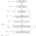
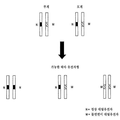
디지탈 PCR은 DNA 샘플 중 두 대립유전자의 벗어난(skewing) 대립유전자 비율의 존재를 검출할 수 있다. 예를 들어, 디지탈 PCR은 종양 DNA 샘플 중 이형접합성 소실 (LOH)을 검출하는데 사용되어 왔다. DNA 샘플 중 두 대립유전자, 즉 A 및 G가 존재하고, A 대립유전자는 LOH가 있는 세포 중에 소실된다고 가정한다. LOH가 종양 샘플 중 50%의 세포에 존재하는 경우, DNA 샘플 중 G:A의 대립유전자 비율은 2:1일 것이다. 그러나, LOH가 종양 샘플에 존재하지 않는 경우, G:A의 대립유전자 비율은 1:1일 것이다.Digital PCR can detect the presence of a skewing allele ratio of two alleles in a DNA sample. For example, digital PCR has been used to detect loss of heterozygosity (LOH) in tumor DNA samples. It is assumed that there are two alleles in the DNA sample, A and G, and that the A allele is lost among cells with LOH. If LOH is present in 50% of the cells in the tumor sample, the allele ratio of G:A in the DNA sample will be 2:1. However, if LOH is not present in the tumor sample, the allele ratio of G:A will be 1:1.
도 1은 디지탈 PCR 실험을 설명하는 흐름도 100이다. 단계 110에서, DNA 샘플을 희석시킨 후, 독립된 웰로 분배한다. 본 발명자들은 일부 혈장 핵산 종이 원래의 샘플에서 이미 상당히 희석되도록 결정했다는 것을 주목한다. 따라서, 필요한 농도로 이미 존재하는 경우, 일부 주형에 대한 희석은 필요하지 않다. 이전의 연구 (예를 들어, 상기 문헌 [Zhou et al 2002])에서는, 특정 "주형 DNA"의 평균 농도가 웰 당 두 주형 중 하나의 대략 0.5 분자가 될 정도로 DNA 샘플을 희석시켰다. 용어 "주형 DNA"는 A 또는 G 대립유전자를 가리키는 것으로 보이고, 이러한 특정 농도에 대한 이론적 근거는 제공되지 않았다는 것을 주목한다.1 is a
단계 120에서, 각각의 웰에서, PCR 과정은 A 및/또는 G 대립유전자를 동시에 검출하기 위해서 실시된다. 단계 130에서, 각각의 웰 중 마커가 식별된다 (예를 들어, 형광으로) (예를 들어 A, G, A 및 G, 또는 어느 쪽도 아님). LOH의 부재 하에, DNA 샘플 중 A 및 G 대립유전자의 과다는 동일하다 (셀 당 각각 한 카피). 따라서, 웰이 A 대립유전자 및 G 대립유전자에 대해 양성일 확률은 동일할 것이다. 이것은 유사한 수의 웰이 A 또는 G 대립유전자에 대해 양성인 것에 의해 반영될 것이다. 그러나, LOH가 종양 샘플 중 50% 또는 그 보다 많은 세포에 존재하는 경우, G 및 A 대립유전자의 대립유전자 비율은 적어도 2:1일 것이다. 이전의 방법들은 단순히 샘플이 적어도 50% 암성인 것으로 추정하였다. 따라서, 웰이 G 대립유전자에 대해 양성일 확률은 A 대립유전자에 대해 양성일 확률보다 높을 것이다. 그 결과, G 대립유전자에 대해 양성인 웰의 수는 A 대립유전자에 대해 양성인 웰의 수보다 더 많을 것이다.In
단계 140에서, 디지탈 PCR 결과를 분류하기 위해, 각각의 대립유전자에 대해 양성이나 다른 대립유전자에 대해서는 양성이 아닌 웰의 수를 계수할 것이다. 상기 예에서, A 대립유전자에 대해 양성이나 G 대립유전자에 대해 음성인 웰의 수, 및 G 대립유전자에 대해 양성이나 A 대립유전자에 대해 음성인 웰의 수를 계수한다. 한 실시양태에서, 덜 양성인 웰을 나타내는 대립유전자는 참조 대립유전자로 간주된다.In
단계 150에서, 정보제공 웰의 총 수는 두 대립유전자 중 어느 하나에 대해 양성인 웰의 수의 합으로 결정된다. 단계 160에서, 더 많은 양성 웰의 대립유전자에 의해 기여되는 정보제공 웰의 비율 (Pr) (파라미터의 한 예)이 계산된다.In
Pr = 더 많은 양성 웰의 대립유전자에 대해서만 양성인 웰의 수/단지 하나의 대립유전자 (A 또는 G)에 대해 양성인 웰의 총 수.P r = number of wells that are positive only for the allele of more positive wells/total number of wells that are positive for only one allele (A or G).
다른 실시양태는 하나의 대립유전자를 갖는 모든 웰을 하나 이상의 대립유전자를 갖는 모든 웰로 나눈 것을 사용할 수 있다.Other embodiments may use all wells with one allele divided by all wells with one or more alleles.
단계 170에서, Pr 값이 대립유전자 불균형을 나타내는지 여부가 결정된다. 정확하고 효율적인 것이 바람직하므로, 이 작업은 간단하지 않다. 불균형을 결정하기 위한 한 방법은 베이지안(Bayesian)-유형 우도 방법, 순차 확률비 검증 (SPRT)을 사용한다. SPRT는 데이타가 축적되면서 두 확률 가설을 비교하도록 하는 방법이다. 즉, SPRT는 디지탈 PCR의 결과를 분류하여 벗어난 대립유전자의 존재 또는 부재를 시사하는 통계적 방법이다. SPRT는 분석될 웰의 수를 최소화하여 소정의 통계적 검증력 및 정확도를 획득하도록 하는 이점을 갖는다.In
예시적 SPRT 분석에서, 실험 결과는 귀무가설 (null hypothesis) 및 대립가설 (alternative hypothesis)에 대해 시험될 것이다. 대립가설은 샘플 중 벗어난 대립유전자 비율이 있는 경우 수용된다. 귀무가설은 샘플 중 벗어난 대립유전자 비율이 없는 경우 수용된다. Pr 값은 귀무가설 또는 대립가설을 수용하기 위해서 두 분별값과 비교될 것이다. 두 가설 모두 수용되지 않는 경우, 샘플은 관찰된 디지탈 PCR 결과가 바람직한 통계적 신뢰도로 샘플을 분류하기에 충분하지 않다는 것을 의미하는 미분류로 표시될 것이다.In an exemplary SPRT analysis, the experimental results will be tested for the null hypothesis and the alternative hypothesis. The allele hypothesis is accepted if there is an outlier allele ratio in the sample. The null hypothesis is accepted if there are no outlier allele proportions in the sample. The values of P r will be compared with the two discrimination values to accommodate the null hypothesis or alternative hypothesis. If neither hypothesis is accepted, the sample will be marked as unclassified, meaning that the observed digital PCR results are not sufficient to classify the sample with the desired statistical confidence.
귀무가설 또는 대립가설을 수용하기 위한 분별값은 전형적으로, 가설에서 언급된 가정 하에 고정된 Pr 값을 기준으로 계산되어 왔다. 귀무가설에서, 샘플은 벗어난 대립유전자 비율을 나타내지 않는 것으로 가정된다. 따라서, 각 웰이 A 및 G 대립유전자에 대해 양성일 확률은 동일할 것이므로, 예상 Pr 값은 1/2일 것이다. 대립가설에서, 예상 Pr 값은 2/3 또는 0.5와 2/3의 대략 중간 정도, 예를 들어 0.585로 여겨진다. 또한, 제한된 수의 실험으로 인해, (.585+3/N)의 상한값 및 (.585-3/N)의 하한값이 선택될 수 있다.The fractional value to accommodate the null hypothesis or alternative hypothesis has typically been calculated on the basis of a fixed P r value under the assumptions mentioned in the hypothesis. In the null hypothesis, it is assumed that the sample does not show an out-of-allele ratio. Thus, the probability that each well is positive for the A and G alleles will be the same, so the expected P r value will be 1/2. In the alternative hypothesis, the expected P r value is considered to be 2/3, or approximately halfway between 0.5 and 2/3, e.g. 0.585. Also, due to the limited number of experiments, an upper limit of (.585+3/N) and a lower limit of (.585-3/N) can be chosen.
B. 다운 증후군의 검출B. Down syndrome detection
본 발명의 한 실시양태에서, 디지탈 SNP는 임신 여성의 혈장으로부터 태아 다운 증후군을 검출하는데 사용된다. 태아/태반 세포에 특이적인 마커를 사용하여, 염색체 21 중 대립유전자의 비율을 측정할 수 있다. 예를 들어, PLAC4 대립유전자의 관찰된 과다표출 정도가 통계적으로 유의한지를 결정하기 위해서, SPRT가 사용된다.In one embodiment of the invention, the digital SNP is used to detect fetal Down syndrome from the plasma of a pregnant woman. Using markers specific for fetal/placental cells, the ratio of alleles in
하나의 예시적 실시양태에 따라, 디지탈 RNA-SNP는 염색체 21로부터 전사되고 태반에 의해 발현되는 PLAC4 mRNA에 위치한 A/G SNP의 다형성 대립유전자, rs8130833의 비율에서의 불균형을 결정한다. 이형접합 정배수체 태아에 대해, A 및 G 대립유전자는 태아 게놈에서 동등하게 표출되어야 하는 한편 (1:1 게놈 비율); 삼염색체 21에서, 삼염색체성 염색체 21은 태아 게놈 중 하나의 SNP 대립유전자의 추가 카피와 회합되어 2:1 비율을 나타낼 것이다. 디지탈 PCR 분석의 목적은 분석된 샘플 중 두 PLAC4 대립유전자의 양이 동등한지 또는 다른지를 결정하는 것이다. 따라서, A 및 G PLAC4 대립유전자는 모두 표적 주형이다. 실시간 PCR 검정은 PLAC4 mRNA를 증폭시키도록 설계되었고, 두 SNP 대립유전자는 태크만(TaqMan) 형광 프로브에 의해 구별되었다. 분석 단계의 도식적 설명이 도 2a에 나타나 있다.According to one exemplary embodiment, the digital RNA-SNP is transcribed from
도 2a는 본 발명의 실시양태에 따른 디지탈 RNA-SNP 방법 200을 설명한다. 단계 210에서, 샘플을 얻는다. 단계 220에서, 핵산 서열, 예를 들어 PLAC4 mRNA를 추출된 RNA 샘플에서 정량화한다. 한 실시양태에서, 이것은 PLAC4 mRNA에 대해 실시간 PCR로 실시된다. 한 측면에서, 이 단계는 조작자에게 표적이 디지탈 PCR 분석의 '영역'에 도달하기 전에 어느 만큼의 희석이 요구되는지에 대한 아이디어를 제공한다.2A illustrates a digital RNA-
단계 230에서, 샘플을 희석시킨다. 단계 240에서, 희석시킨 샘플의 농도를 측정한다. 희석시킨 샘플의 농도가 약 1 주형/웰 (즉, 참조 또는 비-참조 서열 또는 어느 하나의 대립유전자)인지 확인할 수 있다. 일부 실시양태는 이 측정을 위한 IV절에 기술된 기술을 사용한다. 예를 들어, 본 발명자들은 실시간 PCR 분석을 위해서 희석시킨 샘플을 96 웰로 배분하여 알맞게 희석되었는지를 확인하였다. 희석 농도는 또한 모르는 상태로 남겨두어 이 단계가 제거되도록 할 수 있고, 이는 추후 설명될 것이다.In
단계 250에서, 디지탈 PCR을 어레이의 각 웰 상에서 실시하였다. 예를 들어, 동일하게 희석시킨 샘플을 실시간 PCR 분석을 위해서 384-웰에 배분하였다. PCR 결과로부터, 각 핵산 서열에 대한 마커의 양 및 정보제공 웰의 수를 확인한다. 정보제공 웰은 A 또는 G 대립유전자에 대해서만 양성이나, 둘 모두에 대해서 양성은 아닌 것으로 정의된다. 단계 260에서, 예상 Pr 값을 계산한다. 이러한 단계들은 추후 더욱 자세하게 설명될 것이다. 이 계산은 단계 250에서 측정된 값들로부터 파라미터를 결정하는 것을 포함한다. 예를 들어, 웰 당 실제 평균 주형 농도가 계산될 수 있다.In
단계 270에서, SPRT 또는 다른 우도-비율 시험은 불균형이 존재하는지 여부를 결정하기 위해서 실시될 수 있다. 정배수체 사례에 대해, 동일한 수의 A-양성 및 G-양성 웰이 예상된다. 그러나, 삼염색체 21 태아로부터의 주형 분자가 분석되는 경우, 하나의 대립유전자만을 함유하는 웰의 수는 다른 대립유전자만을 함유하는 웰의 수보다 많아야 한다. 요컨대, 삼염색체 21에 대한 대립유전자 불균형이 예상된다.In
상기 언급된 것과 같이, SPRT는 데이타가 축적되면서 두 확률 가설을 비교하도록 하는 베이지안-유형 우도 방법이다. 삼염색체 21 검출을 위한 디지탈 PCR 분석에서, 대립가설은 대립유전자 불균형이 존재하는 경우 (즉, 삼염색체 21이 검출되는 경우) 수용되고; 귀무가설은 대립유전자 불균형이 존재하지 않는 경우 (즉, 삼염색체 21이 검출되지 않는 경우) 수용된다. 더 많은 수의 계수의 대립유전자는 잠재적으로 과다표출된 대립유전자로 지칭되고, 모든 정보제공 웰 중 그의 비율 (Pr)이 계산될 것이다. SPRT는, Pr이 삼염색체 21 샘플에 대해 예상되는 충분한 정도의 대립유전자 불균형을 나타내는지를 결정하기 위해서 적용된다.As mentioned above, SPRT is a Bayesian-type likelihood method that allows two probability hypotheses to be compared as data accumulates. In digital PCR analysis for
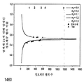
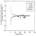
조작 면에 있어서, SPRT는 어느 하나의 가설을 수용하거나 또는 거절하기 위한 확률 경계를 정의하기 위해서 구축된 한 쌍의 SPRT 곡선을 갖는 그래프를 사용하여 적용되고 해석될 수 있다. 도 3은 본 발명의 한 실시양태에 따라 다운 증후군을 결정하기 위한 SPRT 곡선을 갖는 그래프를 설명한다. SPRT 곡선은 신뢰성 있게 분류될 수 있는 경우 주어진 정보제공 웰의 총 수 (x-축)에 대해 잠재적으로 과다표출된 대립유전자에 대해 양성인 정보제공 웰의 요구되는 비율 Pr (y-축)을 플롯팅한다. 도 3에 설명된 것과 같이, 상단 곡선은 대립가설을 수용하기 위한 확률 경계를 설정하고; 하단 곡선은 귀무가설을 수용하기 위한 확률 경계를 설정한다. In terms of operation, SPRT can be applied and interpreted using a graph with a pair of SPRT curves built to define probability boundaries for accepting or rejecting either hypothesis. 3 illustrates a graph with an SPRT curve for determining Down syndrome in accordance with an embodiment of the present invention. The SPRT curve plots the required proportion of informative wells P r (y-axis) that are positive for a potentially overexpressed allele for a given total number of informative wells (x-axis) if they can be classified reliably. Do it. As illustrated in Fig. 3, the top curve establishes a probability boundary to accommodate the alternative hypothesis; The lower curve sets the probability boundary to accept the null hypothesis.
실험적으로 유도된 Pr 값은 어느 하나의 가설을 수용하거나 또는 거절하기 위한 예상 Pr 값과 비교될 것이다. 귀무가설이 수용되는 경우, 샘플은 정배수체 태아를 가진 임신 여성으로부터 수득된 것으로 분류되었다. 대립가설이 수용되는 경우, 샘플은 삼염색체 21 태아를 가진 임신 여성으로부터 수득된 것으로 분류되었다. 별법으로, 주어진 정보제공 계수의 수에 대한 Pr이 질환 분류에 대해 요구되는 통계적 신뢰성 수준에 아직 도달하지 못한 경우, 어느 하나의 가설도 받아들여질 수 없다. 이러한 경우들은 더 많은 데이타가 이용가능하기 전까지는 분류불가능으로 간주된다. 질환 분류가 가능하지 않은 경우, 집계된 데이타가 SPRT에 의해 분류가능해지기 전까지 추가의 384-웰 플레이트를 실시할 수 있다.The experimentally derived P r values will be compared to the expected P r values to accept or reject either hypothesis. If the null hypothesis was accepted, the samples were classified as obtained from pregnant women with euploid fetuses. If the allele hypothesis was accepted, the samples were classified as obtained from pregnant women with
따라서, SPRT는 주어진 수준의 신뢰성을 위해서 다른 통계적 방법보다 더 적은 양의 시험이 요구된다는 이점을 제공한다. 실용적인 측면에서, SPRT는 요구되는 양의 데이타가 축적되어 불필요한 추가적 분석을 최소화하자마자 어느 하나의 가설의 수용 또는 거절을 가능하게 한다. 이 특징은 일반적으로 이용가능한 주형 분자의 수가 제한적인 경우 낮은 농도로 존재하는 혈장 핵산의 분석과 특히 관련이 있다. 엄격한 분류에 추가하여, 분류는 또한 백분율 정확성을 포함할 수 있다. 예를 들어, 분별값과의 비교로 인한 분류는 샘플이 특정 백분율의 핵산 서열 불균형의 우도를 나타내거나, 또는 동등하게는 측정된 불균형이 특정 백분율 또는 다른 값에 정확하다는 것을 제공할 수 있다.Thus, SPRT offers the advantage that fewer tests are required than other statistical methods for a given level of reliability. In practical terms, SPRT allows acceptance or rejection of either hypothesis as soon as the required amount of data has been accumulated to minimize unnecessary further analysis. This feature is of particular relevance to the analysis of plasma nucleic acids, which are present in low concentrations where the number of commonly available template molecules is limited. In addition to strict classification, classification may also include percentage accuracy. For example, classification by comparison with a fractional value may provide that the sample exhibits a likelihood of a certain percentage of nucleic acid sequence imbalance, or equivalently, that the measured imbalance is accurate to a certain percentage or other value.
유사한 접근법을 적용하여, 모체 혈장 또는 혈청 중 태아 핵산을 사용하여 돌연변이 또는 유전자 다형성과 관련한 태아의 유전자형을 결정할 수 있다. 태아는 그의 모계로부터 그의 게놈의 절반을 물려받을 것이라는 것을 상기해야 한다. 설명을 위해서, 두 대립유전자 A 및 B가 있는 특정 유전자좌를 고려한다. 모계가 AB 유전자형의 이형접합체인 경우, 태아는 이론적으로는 AA, BB 또는 AB의 유전자형을 가질 수 있다. 태아가 AB 유전자형, 즉 모계와 같은 유전자형을 갖는 경우, 모체 혈장에는 (모계 및 태아 모두로부터의) AB 유전자형의 핵산만 존재할 것이다. 따라서, 핵산 또는 대립유전자 균형이 모체 혈장에서 보여진다. 반면, 태아가 AA 또는 BB의 유전자형을 갖는 경우, 모체 혈장에서 A 또는 B 대립유전자 각각이 과다표출된 대립유전자 불균형이 존재할 것이다. 이러한 생각은 또한 질환-유발성 돌연변이 (예를 들어, 낭포성 섬유증 또는 베타-지중해빈혈 또는 척수 근위축증을 유발하는 돌연변이)에도 적용가능한데, 각 경우 A는 야생형 대립유전자로, B는 돌연변이 대립유전자로 고려될 수 있다.Applying a similar approach, fetal nucleic acids in maternal plasma or serum can be used to determine the genotype of the fetus associated with mutations or genetic polymorphisms. It should be recalled that the fetus will inherit half of his genome from his mother. For the sake of explanation, consider a specific locus with two alleles A and B. If the maternal line is a heterozygous of the AB genotype, the fetus can theoretically have the genotype of AA, BB or AB. If the fetus has the AB genotype, i.e., the maternal-like genotype, only nucleic acids of the AB genotype (from both maternal and fetal) will be present in maternal plasma. Thus, a nucleic acid or allele balance is seen in maternal plasma. On the other hand, if the fetus has the AA or BB genotype, there will be an allele imbalance in which each A or B allele is overexpressed in maternal plasma. This idea is also applicable to disease-causing mutations (e.g., a mutation that causes cystic fibrosis or beta-thalassemia or spinal muscular dystrophy), in each case A is considered a wild-type allele and B is a mutant allele. Can be.
II. 디지탈 RCDII. Digital RCD
디지탈 RNA-SNP의 단점은 디지탈 RNA-SNP가 분석된 SNP에 대한 이형접합체의 경우에만 적용가능하다는 것이다. 순환성 태아 핵산 분석을 기준으로 한 삼염색체 21 태아 또는 다른 태아 염색체 이수성 (예를 들어, 삼염색체 18, 13 및 성 염색체 이수성)을 검출하기 위한 비침습성 시험이 유전자 다형성의 사용과 무관한 경우 디지탈 RNA-SNP가 이상적이라는 것은 하나의 개선된 점이다. 따라서, 한 실시양태에서, 염색체 양은 참조 염색체, 즉 이 연구에서 염색체 1에 위치한 유전자좌에 상대적인 비-다형성 염색체 21 유전자좌의 디지탈 PCR 분석에 의해 결정된다. 정배수체 태아의 게놈에서 염색체 21 대 염색체 1의 비율의 2:2로부터의 변화는 삼염색체 21의 경우와 구분된다. 삼염색체 21 검출을 위한 디지탈 PCR 분석에서, 비교할 두 가설은 염색체 불균형이 존재하지 않는 (즉, 삼염색체 21이 검출되지 않는) 귀무가설 및 염색체 불균형이 존재하는 (즉, 삼염색체 21이 검출되는) 대립가설일 것이다.The disadvantage of digital RNA-SNP is that digital RNA-SNP is applicable only in the case of heterozygous for the analyzed SNP. Digital if non-invasive tests to detect
이 접근법은 다른 염색체 이수성, 예를 들어 삼염색체 18에서의 염색체 18, 삼염색체 13에서의 염색체 13, 터너 증후군에서의 염색체 X와 연관된 다른 염색체에 일반화될 수 있다. 또한, 염색체 1과는 별개로, 관련 이수성과 연관되지 않은 다른 염색체도 또한 참조 염색체로 사용될 수 있다. 유사한 접근법이 또한 부분적으로, 암에서 통상적으로 결실되는 염색체 대 참조 염색체의 비율 변화를 분석함으로써 암 검출에 적용될 수 있다. 전자의 예로는 결장직장암에서의 염색체 5q, 폐암에서의 염색체 3p 및 비인두 암종에서의 염색체 9p가 포함된다. 도 2b는 서열 불균형으로 나타나는 일부 통상적인 암-관련 염색체 이상을 열거한다.This approach can be generalized to other chromosomes associated with other chromosomal aneuploidy, for
도 2a는 또한 본 발명의 한 실시양태에 따른 디지탈 RCD 방법 205를 설명한다. 단계 220-230에 대한 한 실시양태에서, 추출된 DNA를, 예를 들어, 나노드롭(Nanodrop) 기술로 정량화하고, 웰 당 염색체 21 또는 표준화 염색체 (예컨대, 염색체 1)로부터의 약 하나의 표적 주형의 농도로 희석시킨다. 단계 240의 한 실시양태에서, 384-웰 플레이트 중 태크만 프로브를 모두 사용하는 디지탈 RCD 분석을 진행하기 전에 약 37% 수준의 웰이 음성인지 여부를 확인하기 위해서, 단지 96-웰 포맷에서 염색체 1 프로브를 사용하는 검정에 의해 희석시킨 DNA 샘플을 분석함으로써 확인할 수 있다. 37%의 유의성은 IV절에서 추후 기술될 것이다.2A also illustrates a
단계 240의 시험 및 단계 250의 결과는 한 쌍의 태크만 프로프에 의해 구별되는 파라로그(paralogous) 서열 변이에 의해 식별되는 두 염색체 모두에 존재하는 파라로그 서열을 증폭시키도록 고안된 실시간 PCR 검정으로 수행될 수 있다 (문헌 [Deutsch, S. et al. 2004 J Med Genet 41, 908-915]). 이러한 맥락에서, 정보제공 웰은 염색체 21 또는 염색체 1 유전자좌에 대해 양성인, 그러나 둘 모두에 대해서 양성은 아닌 웰로서 정의된다. 정배수체 태아에 대해, 어느 하나의 유전자좌에 대해 양성인 정보제공 웰의 수는 대략 동등해야 한다. 삼염색체 21 태아에 대해, 염색체 1보다 염색체 21에 대해 양성인 웰의 과다표출이 있어야 한다. 과다표출의 정확한 비율은 하기 절에 기술되어 있다.The test of
III. 태아 서열의 혼입률III. Incorporation rate of fetal sequence
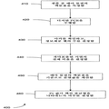
상기 기술된 방법 200 및 205의 실시양태의 단점은 태아 특이적 마커가 요구된다는 것이다. 따라서, 본 발명의 한 실시양태에서, 비-태아 특이적 마커가 사용된다. 비-태아 특이적 마커를 사용하기 위해서, 본 발명의 실시양태는 모체 혈장 (즉, 생물학적 샘플) 중 태아 DNA 분획 농도를 측정한다. 이러한 정보와 함께, 보다 유용한 Pr 값이 하기와 같이 계산될 수 있다.A disadvantage of the embodiments of
모체 혈장 중 태아 DNA의 낮은 분획 백분율로도, 삼염색체 21 태아는 모체 혈장으로 방출된 태아 DNA의 게놈-등가물 (GE) 당 염색체 21 서열의 추가적 양에 기여할 것이다. 예를 들어, 태아에 의해 기여된 5 GE/mL DNA와 50 GE/mL의 총 DNA (즉, 10% 태아 DNA 분획 농도)를 함유하는 정배수체 임신으로부터의 모체 혈장 샘플은 모체 혈장 (밀리리터) 당 염색체 21 서열의 총 100개의 카피 (90개의 모체 카피 + 10개의 태아 카피)를 함유해야 한다. 삼염색체 21 임신에 대해, 태아 GE는 각각 3개 카피의 염색체 21에 기여하여, 모체 혈장 중 염색체 21 서열의 총 105개의 카피/mL (90개의 모체 카피 + 15개의 태아 카피)가 되도록 한다. 따라서, 10% 태아 DNA 농도에서, 삼염색체 임신의 모체 혈장 중 염색체 21 유래 서열의 양은 정배수체 경우의 1.05배일 것이다. 따라서, 분석적 접근법이 이러한 적은 정도의 정량 차이를 결정하도록 개발될 수 있다면, 삼염색체 21 태아의 비침습성 출생 전 진단에 대한 다형성-독립적 시험이 가능할 것이다.Even with a low fractional percentage of fetal DNA in maternal plasma, a
따라서, 과다표출의 정도는 분석된 DNA 샘플 중 태아 DNA 분획 농도에 좌우될 것이다. 예를 들어, 태반 DNA를 분석하는 경우, 태아 게놈 중 이론적 RCD 비율은 3:2, 즉 1.5-배 차이이어야 한다. 그러나, 상기 기술된 것과 같이, 이론적 RCD 비율은 10% 태아 DNA를 함유하는 모체 혈장 샘플을 분석하는 경우 1.05로 감소할 것이다. 실험적으로 유도된 Pr은 단지 염색체 21 유전자좌에 양성인 웰의 수를 총 정보제공 웰의 수로 나누어 계산된다. 실험적으로 유도된 Pr은 계산된 Pr 및 이론적 RCD 비율과 함께 SPRT 분석에 적용된다.Thus, the degree of overexpression will depend on the concentration of the fetal DNA fraction in the analyzed DNA sample. For example, when analyzing placental DNA, the theoretical RCD ratio in the fetal genome should be 3:2, ie 1.5-fold difference. However, as described above, the theoretical RCD ratio will decrease to 1.05 when analyzing maternal plasma samples containing 10% fetal DNA. The experimentally derived P r is calculated by dividing the number of wells positive for only the
도 4는 본 발명의 실시양태에 따라 태아 핵산의 백분율을 사용하여 질환 상태를 결정하는 방법 400을 나타낸다. 단계 410에서, 태아 물질의 분획 백분율이 측정된다. 한 실시양태에서, 분획 백분율은 비-태아-특이적 마커 (즉, 모계 및 태아 모두에 존재하는 유전자 서열)와 관련하여 태아-특이적 마커 (예를 들어, Y-염색체, 유전자 다형성 마커 (예컨대, SNP), 태반 후성 기호 (placental epigenetic signatures))의 양을 측정하여 결정된다. 실시간 PCR, 디지탈 PCR, 서열분석 반응 (대규모 병렬 게놈 서열분석을 포함) 또는 임의의 다른 정량 방법에 의해 실질적 측정을 수행할 수 있다. 한 측면에서, 이 측정에 대해 잠재적으로 대립유전자 불균형이 있을 수 있는 유전자 표적을 사용하지 않는 것이 바람직하다.4 shows a
단계 420에서, 샘플을 희석시키고, 희석시킨 샘플을 웰에 넣고, 각 웰에서 반응을 측정하는 것을 비롯한 디지탈 PCR 또는 다른 측정 방법이 실시된다. 단계 430에서, PCR 결과는 상이한 참조 핵산 서열 (예컨대, 염색체 또는 대립유전자)의 마커를 식별하기 위해서 사용된다. 단계 440에서, 과다표출된 서열의 실제 비율 (Pr)이 계산된다. 단계 450에서, 질환 상태를 결정하기 위한 분별값은 샘플 중 태아 물질의 백분율을 사용하여 계산된다. 단계 460에서, 실제 Pr 및 분별값으로부터, 불균형이 존재하는지 여부가 결정된다.In
한 실시양태에서, 참조 핵산 서열의 분획 백분율은 디지탈 RNA-SNP 방법에 혼입된다. 따라서, 암 세포로 인한 LOH를 연구하는 경우, 50% 미만 암 세포를 갖는 종양 샘플로 실시할 수 있다. 이것은 또한 50% 초과 암 세포를 갖는 샘플에 대해서도 사용되어, 더욱 정확한 Pr을 수득함으로써 부정확한 진단으로 이끌 수 있는 잘못된 양성의 수를 감소시킬 수 있다. 또다른 실시양태에서, 태아 핵산 백분율은 디지탈 PCR 방법에 혼입되어, 태아가 모체 혈장 핵산 분석으로부터 부모의 유전자 돌연변이 (예를 들어, 낭포성 섬유증 또는 베타-지중해빈혈 또는 척수 근위축증을 유발하는 돌연변이) 또는 다형성을 물려받는지 여부를 결정한다.In one embodiment, a fractional percentage of the reference nucleic acid sequence is incorporated into the digital RNA-SNP method. Thus, when studying LOH due to cancer cells, it can be done with tumor samples with less than 50% cancer cells. It can also be used for samples with more than 50% cancer cells, thereby reducing the number of false positives that can lead to incorrect diagnosis by obtaining a more accurate P r. In another embodiment, the fetal nucleic acid percentage is incorporated into a digital PCR method such that the fetus is a parental genetic mutation (e.g., cystic fibrosis or beta-thalassemia or a mutation that causes spinal muscular dystrophy) or Decide whether to inherit polymorphism.
IV. 웰 당 평균 농도의 혼입IV. Incorporation of average concentration per well
이전 방법 (예를 들어, 상기 문헌 [Zhou, W. et al. 2002])의 또다른 단점은 웰 당 주형의 평균 농도 (m)가 웰 당 1로 요구된다는 것이다. 정확한 농도를 얻는 것이 어렵다는 것을 감안할 때, 이것은 부정확해질 수 있다. 또한, 웰 당 1 주형의 정확한 농도로도, 이전 방법은 웰 중 주형의 통계적 분포를 간과하였다. 이전 방법에서, 즉, 기존 알고리즘에서, 대립가설을 수용하기 위한 예상 Pr 값은 대립유전자 비율로 가정되어, 웰 당 주형 DNA의 평균 농도와 무관하였다.Another drawback of the previous method (eg Zhou, W. et al. 2002, supra) is that the average concentration (m) of the template per well is required to be 1 per well. Given that it is difficult to obtain the correct concentration, this can become inaccurate. In addition, even with the exact concentration of 1 template per well, the previous method overlooked the statistical distribution of the template in the wells. In the previous method, that is, in the existing algorithm, the predicted P r value for accepting the allele hypothesis was assumed to be the allele ratio, and was not related to the average concentration of template DNA per well.
그러나, 희석시킨 샘플에서 주형의 자연적인 통계적 편차로 인해, 정확하게 웰 당 1 주형은 아닐 것이다. 본 발명의 실시양태는 하나 이상의 서열의 평균 농도를 측정하여, 분별값, 즉 예상 Pr을 계산하는데 사용된다. 한 측면에서, 이러한 계산은 상이한 핵산 서열을 함유하는 웰의 확률을 측정하여 예상 Pr을 결정하기 위해서 사용되는 통계적 분포를 포함한다.However, due to the natural statistical variation of the template in the diluted sample, it will not be exactly 1 template per well. An embodiment of the present invention is used to determine the average concentration of one or more sequences and to calculate the fractional value, i.e. the expected P r. In one aspect, this calculation includes a statistical distribution used to determine the expected P r by measuring the probability of wells containing different nucleic acid sequences.
한 실시양태에서, 평균 농도는 일례로 DNA 샘플 중 더 낮은 농도의 핵산 서열인 한 참조 핵산 서열로 고려된다. 불균형이 없는 샘플의 경우, 샘플 중 두 서열의 농도는 동일할 것이고, 어느 하나는 참조 대립유전자로 간주될 수 있다. 예를 들어 LOH가 있는 샘플의 경우, 암 세포에서 결실된 대립유전자는 참조 대립유전자로 간주될 것이다. 참조 대립유전자의 평균 농도는 mr로 표시될 것이다. 또다른 실시양태에서, 더 높은 농도의 서열이 참조 서열로 간주될 수 있다.In one embodiment, the average concentration is considered a reference nucleic acid sequence, for example a lower concentration nucleic acid sequence in a DNA sample. In the case of a sample without imbalance, the concentrations of the two sequences in the sample will be the same, and either can be considered a reference allele. For example, for samples with LOH, an allele deleted from cancer cells will be considered a reference allele. The average concentration of the reference allele will be expressed as m r. In another embodiment, a higher concentration of sequence can be considered a reference sequence.
A. A. 디지탈Digital -SNP, -SNP, SPRTSPRT 및 And 디지탈Digital PCR을PCR 사용한 예 Example used
도 5는 본 발명의 한 실시양태에 따라 평균 주형 농도를 사용하여 질환 상태를 결정하는 방법 500을 보여준다. 단계 510에서, 상이한 서열의 양을 측정한다. 이것은, 예를 들어 상기 설명된 것과 같은 디지탈 PCR 실험에서 마커를 계수하여 실시할 수 있다. 그러나, 이것은 증폭 단계를 포함하지 않거나 또는 형광 마커를 사용하지 않지만 다른 성질, 예컨대 질량, 특정 광학적 성질 또는 염기-쌍 성질과 같은 물성을 사용할 수 있는 다른 방법에 의해서도 실시할 수 있다.5 shows a
단계 520에서, 과다표출된 서열의 실제 비율이 결정된다. 이것은 상기 기술된 것과 같이 단지 그 서열만 나타내는 웰의 수를 취하고, 정보제공 웰의 수로 나누어 실시될 수 있다. 단계 530에서, 하나 이상의 서열의 평균 농도가 측정된다 (참조 서열). 한 실시양태에서, 참조 서열은 과다표출된 서열이다. 또다른 실시양태에서, 참조 서열은 과소표출된(underrepresented) 서열이다. 측정은 디지탈 PCR 실험에서 참조 서열에 대해 음성인 웰의 수를 계수하여 실시할 수 있다. 음성 웰의 비율과 평균 주형 농도 사이의 관계는 다음 소절에 기술된 것과 같은 푸아송 (Poisson) 분포에 의해 설명된다.In
단계 540에서, 상이한 서열에 대해 양성인 웰의 예상 양이, 예를 들어 푸아송 분포를 사용하여 계산된다. 예상 양은 웰 당 서열, 웰 당 평균 서열, 서열을 함유하는 웰의 수 또는 임의의 다른 적합한 양의 확률일 수 있다. 단계 550에서, 예상 Pr은 예상 양으로부터 계산된다. 단계 560에서, 분별값은, 예를 들어 SPRT를 사용하여 예상 Pr로부터 계산된다. 단계 570에서, 핵산 서열 불균형의 분류가 결정된다. 방법 500의 특정 측면이 이제 기술된다.In
1. 서열 예상 양의 결정1. Determination of the expected amount of sequence
웰 (반응 또는 반응 혼합물) 당 평균 농도가 단계 530으로부터 알려지면, 서열을 나타내는 웰의 예상 수가 단계 540에서 계산될 수 있다. 이 양은 %, 분수 값, 또는 정수 값으로 표현될 수 있다. 설명을 위해 특정 예를 사용하면서, 웰 당 참조 주형의 평균 농도 (mr)는 웰 당 0.5이고, PLAC4 SNP, rs8130833에서 삼염색체 21 태아의 유전자형은 AGG인 것으로 가정한다. 따라서, 참조 주형은 A 대립유전자일 것이고, 과다표출된 주형은 G 대립유전자일 것이다.If the average concentration per well (reaction or reaction mixture) is known from
한 실시양태에서, 푸아송 분포는 디지탈 PCR과 같은 측정 과정의 웰의 반응 혼합물 중 A 대립유전자의 분포로 가정한다. 다른 실시양태에서는, 이항 분포와 같은 다른 분포 함수가 사용된다.In one embodiment, the Poisson distribution is assumed to be the distribution of the A allele in the reaction mixture of the wells of a measurement procedure such as digital PCR. In other embodiments, other distribution functions such as binomial distributions are used.
푸아송 방정식은 ![]()
![]()
따라서, 0.5의 평균 A-대립유전자 농도에서 A 대립유전자의 임의의 분자를 함유하지 않는 임의의 웰의 확률은 하기와 같을 것이다:Thus, at an average A-allele concentration of 0.5, the probability of any well that does not contain any molecule of the A allele would be:
![]()
![]()
따라서, 하나 이상의 A 대립유전자의 분자를 함유하는 임의의 웰의 확률은: 1 - 0.6065 = 0.3935이다. 그러므로, 약 39%의 웰이 하나 이상의 A 대립유전자의 분자를 함유할 것으로 예상될 것이다.Thus, the probability of any well containing molecules of more than one A allele is: 1-0.6065 = 0.3935. Therefore, it would be expected that about 39% of the wells will contain molecules of one or more A alleles.
비-참조 핵산 서열의 경우, 삼염색체 21 태아의 각 세포에 대해서, A 대 G의 게놈 비율은 1:2일 것이다. 추출된 RNA 또는 DNA 샘플 중 A 대 G 비율이 변화하지 않는 것으로 가정하면, 웰 당 G 대립유전자의 평균 농도는 A 대립유전자의 평균 농도의 2배, 즉 2 × 0.5 = 1일 것이다.For a non-reference nucleic acid sequence, for each cell of a
따라서, 평균 G-대립유전자 농도인 1에서 G 대립유전자의 임의의 분자를 함유하지 않는 임의의 웰의 확률은 하기와 같을 것이다:Thus, at the mean G-allele concentration of 1, the probability of any well not containing any molecule of the G allele would be:
![]()
![]()
따라서, 하나 이상의 G 대립유전자 분자를 함유하는 임의의 웰의 확률은: 1 - 0.3679 = 0.6321일 것이다. 그러므로, 약 63%의 웰이 하나 이상의 G 대립유전자 분자를 함유하는 것으로 예상될 것이다.Thus, the probability of any well containing more than one G allele molecule will be: 1-0.3679 = 0.6321. Therefore, it would be expected that about 63% of the wells contain one or more G allele molecules.
2. 과다표출된 서열 비율의 결정2. Determination of the proportion of overexpressed sequences
예상 양이 계산된 후, 과다표출된 핵산 서열의 비율이 결정될 수 있다. A 대립유전자 및 G 대립유전자로 웰을 채우는 것이 독립적이라고 가정하면, 두 대립유전자를 모두 함유하는 웰의 확률은 0.3935 × 0.6321 = 0.2487일 것이다. 그러므로, 약 25%의 웰이 두 대립유전자를 모두 함유하는 것으로 예상될 것이다.After the expected amount is calculated, the proportion of overexpressed nucleic acid sequences can be determined. Assuming that filling wells with the A and G alleles is independent, the probability of a well containing both alleles would be 0.3935 x 0.6321 = 0.2487. Therefore, it would be expected that about 25% of the wells contain both alleles.
A 대립유전자는 함유하되 G 대립유전자는 함유하지 않는 것으로 예상되는 웰의 비율은 하나 이상의 A 대립유전자를 함유하는 웰의 수에서 A 및 G 대립유전자를 모두 함유하는 웰의 수를 뺀 것이다: 0.3935 - 0.2487 = 0.1448. 유사하게, G 대립유전자는 함유하되 A 대립유전자는 함유하지 않는 것으로 예상되는 웰의 비율은 0.6321 - 0.2487 = 0.3834일 것이다. 정보제공 웰은 A 대립유전자 또는 G 대립유전자에 대해 양성이나, 둘 모두에 대해서는 양성이 아닌 웰로서 정의된다.The proportion of wells that are expected to contain the A allele but not the G allele is the number of wells containing one or more A alleles minus the number of wells containing both A and G alleles: 0.3935- 0.2487 = 0.1448. Similarly, the proportion of wells expected to contain the G allele but not the A allele would be 0.6321-0.2487 = 0.3834. Informational wells are defined as wells that are positive for the A allele or G allele, but not both.
따라서, 디지탈 RNA-SNP 분석에서 G 대립유전자에 상대적으로 A 대립유전자를 함유하는 웰의 예상 비율은 0.1448/0.3834이다. 즉, 단지 G 대립유전자에 대해서만 양성인 웰의 비율은 단지 A 대립유전자에 대해서만 양성인 웰의 2.65배이다. 이것은 과다표출된 대립유전자가 다른 대립유전자의 2배인 태아 게놈 비율과 대조적이다.Thus, the expected proportion of wells containing the A allele relative to the G allele in the digital RNA-SNP assay is 0.1448/0.3834. That is, the proportion of wells that are only positive for the G allele is 2.65 times that of wells that are only positive for the A allele. This is in contrast to the proportion of the fetal genome where the overexpressed allele is twice that of the other alleles.
SPRT 분석에 대해, 과다표출된 대립유전자에 대해 양성인 정보제공 웰의 비율 (Pr)은 SPRT 곡선을 사용하여 계산되고 해석된다. 현 예에서, 정보제공 웰의 비율은 0.1448 + 0.3834 = 0.5282일 것이다. 따라서, mr 0.5에서 삼염색체 21 경우의 예상 Pr은 0.3834/0.5282 = 0.73이다.For SPRT analysis, the proportion of informative wells positive for the overexpressed allele (P r ) is calculated and interpreted using the SPRT curve. In the current example, the proportion of informative wells would be 0.1448 + 0.3834 = 0.5282. Therefore, the expected P r for the case of
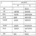
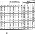
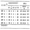
평균 주형 농도 (m)는 푸아송 방정식의 주요 파라미터이므로, Pr은 m에 따라변할 것이다. 도 6은 본 발명의 실시양태에 따라 웰 당 평균 참조 주형 농도 (mr)로 표현된 주형 농도의 범위에 대한 삼염색체 21 샘플의 예상 디지탈 RNA-SNP 대립유전자 비율 및 Pr을 표로 만든 표 600을 나타낸다. 표 600은 일련의 웰 당 평균 참조 주형 농도 (mr)에 대한 예상 대립유전자 비율 및 과다표출된 대립유전자에 대해 양성인 정보제공 웰의 비율 (Pr)을 나타낸다.The average template concentration (m) is the main parameter of the Poisson equation, so P r will vary with m. 6 is a table 600 tabulating the predicted digital RNA-SNP allele ratios and P r of
예상 Pr 값은 비-선형 방식으로 웰 당 참조 대립유전자의 평균 농도 (mr)에 따라 변한다. 표 600에 제시된 것과 같이, 대립가설을 수용하기 위한 예상 Pr 값은 mr과 함께 증가할 것이다. 귀무가설을 수용하기 위한 예상 Pr 값이 0.5로 고정되어 있으므로, 대립유전자 불균형이 있는 샘플 및 대립유전자 불균형이 없는 샘플은 mr이 증가할 경우, Pr 값의 측면에서 보다 광범위하게 나뉘어질 것이다. 다른 실시양태에서는 귀무가설을 수용하기 위한 값이 0.5가 아닐 수 있다는 것을 주목한다. 이것은 정상 비율이 1:1과 다른 경우, 예를 들어, 5:3인 경우 일어날 수 있으며, 따라서 비율이 5:3으로부터 벗어나는 경우 불균형이 발생할 것이다. 두 상이한 핵산 서열 양의 차이는 개별 경우에 따라 결정될 것이다.The expected P r value varies with the average concentration of reference alleles per well (m r ) in a non-linear manner. As shown in Table 600, the expected values of P r to accommodate the alternative hypothesis will increase with m r. Since the predicted P r value to accommodate the null hypothesis is fixed at 0.5, samples with allele imbalance and samples without allele imbalance will be more broadly divided in terms of P r value as m r increases. . Note that in other embodiments the value to accommodate the null hypothesis may not be 0.5. This can happen if the normal ratio is different from 1:1, e.g. 5:3, so an imbalance will occur if the ratio deviates from 5:3. The difference between the amounts of two different nucleic acid sequences will be determined on a separate case basis.
그러나, 이전 방법 (예를 들어, 상기 문헌 [Zhou, W. et al. 2002])은 LOH 샘플에 대해 고정된 예상 Pr 값을 사용하였으므로, LOH가 있는 샘플에 대한 Pr 값을 과소평가하였다 (대립가설 수용). 과소평가의 정도는 mr과 함께 증가할 것이다. 즉, DNA 샘플 중 참조 대립유전자의 평균 농도가 더 높을수록, 이전 방법은 더욱 부정확해질 것이다. 대립가설을 수용하기 위한 Pr의 과소평가는 귀무가설 및 대립가설 모두를 수용하기 위한 분별값의 계산을 부정확하게 할 것이다. However, the previous method (eg, Zhou, W. et al. 2002, supra) used a fixed expected P r value for the LOH sample, thus underestimating the P r value for the sample with LOH. (Accepting the alternative hypothesis). The degree of underestimation will increase with m r. That is, the higher the average concentration of the reference allele in the DNA sample, the more inaccurate the previous method will be. An underestimation of P r to accommodate the alternative hypothesis will make the calculation of the discrimination value to accommodate both the null hypothesis and the alternative hypothesis inaccurate.
3. 예상 P3. Expected P rr 을 기준으로 한 분별값의 계산Calculation of the fractional value based on
SPRT를 사용하는 실시양태에 대해, 문헌 [El Karoui at al. (2006)]으로부터 SPRT 곡선의 상한 및 하한을 계산하기 위한 방정식을 사용할 수 있다. 또한, 귀무가설 또는 대립가설을 수용하기 위해서 바람직한 통계적 신뢰성의 수준은 방정식에서 임계값 우도 비율(threshold likelihood ratio)을 조정하면서 변할 수 있다. 이 연구에서, 임계값 우도 비율 8이 암 검출의 맥락에서 대립유전자 불균형이 있는 샘플과 대립유전자 불균형이 없는 샘플을 구별하는 만족할만한 성능을 제공하는 것으로 나타났기 때문에, 상기 값 8이 사용된다. 따라서, 한 실시양태에서, SPRT 곡선의 상한 및 하한을 계산하기 위한 방정식은 다음과 같다:For embodiments using SPRT, see El Karoui at al. (2006)] can be used to calculate the upper and lower limits of the SPRT curve. In addition, the level of statistical reliability desired to accommodate the null hypothesis or alternative hypothesis can be varied by adjusting the threshold likelihood ratio in the equation. In this study, the value of 8 is used, as the
상한 = [(ln 8)/N - ln δ]/ln γ Upper limit = [(ln 8)/N-ln δ]/ln γ
하한 = [(ln 1/8)/N - ln δ]/ln γLower limit = [(
여기서, δ = (1 - θ1)/(1 - θ0),Here, δ = (1-θ 1 )/(1-θ 0 ),
γ = -(θ1(1 - θ0)/θ0(1 - θ1),γ = -(θ 1 (1-θ 0 )/θ 0 (1-θ 1 ),
θ0 = 귀무가설이 사실인 경우 비-참조 대립유전자를 함유하는 정보제공 웰의 비율 = 0.5 (하기 참조),θ 0 = proportion of informative wells containing non-reference alleles if the null hypothesis is true = 0.5 (see below),
θ1 = 대립가설이 사실인 경우 비-참조 (즉, 과다표출된) 대립유전자를 함유하는 정보제공 웰의 비율,θ 1 = proportion of informative wells containing non-reference (i.e., overexpressed) alleles if the alternative hypothesis is true,
N = 정보제공 웰의 수 = 어느 하나의 대립유전자에만 양성인 웰의 수.N = number of informative wells = number of wells that are positive for only one allele.
(ln은 자연 로그, 즉 loge를 나타내는 수학 기호임)(ln is the natural logarithm, that is, the mathematical symbol for log e)
귀무가설을 수용하기 위한 θ0의 결정을 위해서, 샘플은 정배수체 태아를 갖는 임신 여성으로부터 얻어지는 것으로 가정된다. 이러한 가정 하에, 어느 하나의 주형에 양성인 웰의 예상 수는 1:1일 것이므로, 비-참조 대립유전자를 함유하는 정보제공 웰의 예상 비율은 0.5일 것이다. For the determination of θ 0 to accommodate the null hypothesis, it is assumed that the sample is obtained from a pregnant woman with an euploid fetus. Under this assumption, the expected number of wells positive for either template would be 1:1, so the expected ratio of informative wells containing non-reference alleles would be 0.5.
대립가설을 수용하기 위한 θ1의 결정을 위해서, 샘플은 삼염색체 21 태아를 갖는 임신 여성으로부터 얻어지는 것으로 가정된다. 디지탈 RNA-SNP 분석을 위한 삼염색체 21 경우의 예상 Pr의 계산은 표 600에 상술되어 있다. 따라서, 디지탈 RNA-SNP 분석을 위한 θ1은 표 600의 마지막 열에 제시된 데이타를 참조한다. For the determination of θ 1 to accommodate the alternative hypothesis, the sample is assumed to be obtained from a pregnant woman with
4. 평균 농도의 측정4. Measurement of average concentration
mr의 측정은 당업자에게 공지되거나 또는 공지될 다양한 메커니즘에 의해 실시될 것이다. 한 실시양태에서, mr 값은 디지탈 PCR 분석의 실험 과정 중 결정된다. mr 값과 참조 대립유전자에 대해 양성인 웰의 총 수 사이의 관계는 분포 (예를 들어, 푸아송 분포)에 의해 좌우될 수 있으므로, mr은 다음 식을 사용하여 참조 대립유전자에 대해 양성인 웰의 수로부터 계산될 수 있다:The measurement of m r will be carried out by various mechanisms known or will be known to those skilled in the art. In one embodiment, the m r value is determined during the course of the experiment of a digital PCR analysis. Since the relationship between the m r value and the total number of wells positive for the reference allele can be dictated by the distribution (e.g., Poisson distribution), m r is the well positive for the reference allele using the following equation: It can be calculated from the number of:
mr = - ln (1 - 참조 대립유전자에 대해 양성인 웰의 비율).m r =-ln (1-proportion of wells positive for the reference allele).
ln은 자연 로그, 즉 loge라는 것에 주목한다. 이 접근법은 디지탈 PCR 실험을 위해 사용되는 DNA 샘플 중 mr의 직접적이고 정확한 평가를 제공한다.Note that ln is the natural logarithm, or log e . This approach provides a direct and accurate assessment of m r in DNA samples used for digital PCR experiments.
이 방법은 바람직한 농도를 얻기 위해서 사용될 수 있다. 예를 들어, 샘플의 추출된 핵산은 방법 200의 단계 240에서 실시한 것과 같이 반응 웰 당 하나의 주형 분자와 같은 특정 농도로 희석될 수 있다. 푸아송 분포를 사용하는 한 실시양태에서, 주형이 없는 웰의 예상 비율은 e-m으로 계산될 수 있고, 여기서 m은 웰 당 주형 분자의 평균 농도이다. 예를 들어, 웰 당 하나의 주형 분자의 평균 농도에서, 주형 분자가 없는 웰의 예상 비율은 e-1, 즉 0.37 (37%)로 주어진다. 나머지 63%의 웰은 하나 이상의 주형 분자를 함유할 것이다. 전형적으로, 디지탈 PCR 실행에서 양성 웰 및 정보제공 웰의 수가 계수될 것이다. 정보제공 웰의 정의 및 디지탈 PCR 데이타가 해석되는 방식은 용도에 좌우된다.This method can be used to obtain the desired concentration. For example, the extracted nucleic acid of the sample can be diluted to a specific concentration, such as one template molecule per reaction well, as performed in
다른 실시양태에서, 웰 당 평균 농도 mr은 또다른 정량화 방법, 예를 들어 질량 분광법을 사용하는 정량 실시간 PCR, 반-정량 경쟁적 PCR, 실-경쟁적 PCR 등에 의해 측정된다.In other embodiments, the average concentration m r per well is determined by another method of quantification, such as quantitative real-time PCR using mass spectroscopy, semi-quantitative competitive PCR, real-competitive PCR, and the like.
B. 디지탈 RCDB. Digital RCD
평균 농도를 사용하는 디지탈 RCD를 상기 기재된 디지탈 SNP 방법과 유사한 방식으로 수행할 수 있다. 참조 염색체 (비-염색체 21) 마커, 염색체 21 마커, 및 두 마커 모두에 대해 양성인 웰의 수를 디지탈 PCR에 의해 측정할 수 있다. 디지탈 SNP 분석에 대한 웰 당 참조 마커의 평균 농도 (mr)의 계산에서와 같은 푸아송 확률 함수에 따라, 염색체 21 마커의 양성에 상관없이, 웰 당 참조 마커의 평균 농도 (mr)를 참조 마커에 대해 음성인 웰의 총 수로부터 계산할 수 있다.Digital RCD using the average concentration can be performed in a manner similar to the digital SNP method described above. The number of wells positive for a reference chromosome (non-chromosome 21) marker,
이후, 정배수체 또는 삼염색체 21 태아를 갖는 임신 여성으로부터 얻어진 것과 같은 혈장 샘플을 분류하기 위해 SPRT 분석을 사용할 수 있다. 태아가 정배수체인 경우 귀무가설이 수용될 것이다. 이 시나리오에서, 참조 마커 및 염색체 21 마커에 대해 양성인 웰의 예상 비율은 1:1일 것이며, 이에 따라, 염색체 21 마커에 대해 양성 신호를 갖는 정보제공 웰의 예상 비율은 0.5일 것이다. 태아가 염색체 21에 대해 삼염색체인 경우 대립가설이 수용될 것이다. 이 시나리오에서, 샘플 DNA가 전적으로 태아로부터 유래된 경우, 각 웰에서의 염색체 21 마커의 평균 농도는 참조 마커의 평균 농도 (mr)의 3/2배일 것이다.The SPRT analysis can then be used to classify plasma samples such as those obtained from pregnant women with euploid or
디지탈 RCD를 사용하여 태아-특이적 마커, 예를 들어 태반 후성 기호의 검출을 통해 염색체 양을 측정할 수 있지만 (문헌 [Chim, SSC. et al. 2005 Proc Natl Acad Sci USA 102, 14753-14758]), 디지탈 RCD 분석의 실시양태는 비-태아-특이적 마커를 사용한다. 따라서, 비-태아 특이적 마커를 사용하는 경우 태아 물질의 백분율을 측정하는 추가의 단계가 나타날 것이다. 따라서, 웰 당 염색체 21 마커의 평균 농도는 샘플 중 태아 DNA의 비율에 의존적일 것이며, mr [(200% + 태아 DNA 백분율)/200%]을 사용하여 계산할 수 있다.Although digital RCD can be used to determine the amount of chromosomes through detection of fetal-specific markers such as placental epigenetic symbols (Chim, SSC. et al. 2005 Proc Natl
예시용 특정 실시예를 다시 사용하여, 웰 당 참조 주형인 염색체 1의 평균 농도 (mr)는 0.5인 것으로 가정하며, 50%의 DNA는 태아로부터 유래된 것으로 가정하고, 샘플 중 50%의 DNA는 모체로부터 유래된다.Using the specific examples for illustrative purposes again, the average concentration (m r ) of
따라서, 푸아송 분포를 사용하여, 평균 농도가 웰 당 0.5인 경우 염색체 1 유전자좌의 어떠한 분자도 함유하지 않는 임의의 웰의 확률은 하기와 같을 것이다:Thus, using the Poisson distribution, if the average concentration is 0.5 per well, the probability of any well containing no molecule of the

따라서, 염색체 1 유전자좌 중 하나 이상의 분자를 함유하는 임의의 웰의 확률은 1 - 0.6065 = 0.3935일 것이다. 따라서, 대략 39%의 웰이 유전자좌 중 하나 이상의 분자를 함유하는 것으로 예상될 것이다.Thus, the probability of any well containing one or more molecules of the
이러한 삼염색체 21 태아의 각각의 세포에 대하여, 염색체 21 대 염색체 1의 게놈 비율은 3:2일 것이다. DNA 샘플에서 염색체 21과 염색체 1 사이의 비율은 태아 DNA 분획 농도 (태아 DNA %)에 의존적일 것이며, 3×태아 DNA % + 2(1 - 태아 DNA %) : 2×태아 DNA % + 2×(1 - 태아 DNA %)일 것이다. 따라서, 이 경우에서, 태아 DNA 분획 농도가 50%인 경우, 비는 (3×50% + 2×50%) / (2×50% + 2×50%) = 1.25일 것이다. 디지탈 SNP 방법이 태아 특이적 마커를 사용하지 않은 경우, 이러한 계산을 사용하여 비-참조 서열의 평균 농도를 계산할 수도 있다.For each cell of this
따라서, 웰 당 염색체 1 유전자좌의 평균 농도가 0.5인 경우, 웰 당 염색체 21 유전자좌의 평균 농도는 1.25×0.5 = 0.625이다. 따라서, 평균 농도가 웰 당 0.625인 경우 염색체 21 유전자좌의 어떠한 분자도 함유하지 않는 임의의 웰의 확률은 하기와 같을 것이다:Therefore, when the average concentration of

따라서, 염색체 21 유전자좌 중 하나 이상의 분자를 함유하는 임의의 웰의 확률은 1 - 0.5353 = 0.4647일 것이다. 따라서, 대략 46%의 웰이 유전자좌 중 하나 이상의 분자를 함유하는 것으로 예상될 것이다. 유전자좌 중 하나로의 웰의 충전이 독립적이라고 가정하면, 유전자좌 모두를 함유하는 웰의 확률은 0.3935×0.4647 = 0.1829일 것이다. 따라서, 대략 18%의 웰이 두 유전자좌 모두를 함유하는 것으로 예상될 것이다.Thus, the probability of any well containing one or more molecules of the
염색체 1 유전자좌를 함유하되 염색체 21 유전자좌는 함유하지 않는 것으로 예상되는 웰의 비율은, 하나 이상의 염색체 1 유전자좌를 함유하는 웰의 수에서 유전자좌 모두를 함유하는 웰의 수를 차감한 것, 즉 0.3935 - 0.1829 = 0.2106일 것이다. 유사하게, 염색체 21 유전자좌를 함유하되 유전자좌 모두는 함유하지 않는 것으로 예상되는 웰의 비율은 0.4647 - 0.1829 = 0.2818일 것이다. 정보제공 웰은 염색체 1 유전자좌 또는 염색체 21 유전자좌 중 하나에 대해 양성이되 둘 모두에 대해서는 아닌 웰로서 정의된다.The proportion of wells that are expected to contain the
따라서, 디지탈 RCD 분석에서의 염색체 21 대 염색체 1 예상 비율은 0.2818/0.2106 = 1.34이다. 바꾸어 말하면, 염색체 21 유전자좌에 대해서만 양성인 웰의 비율은 염색체 1 유전자좌에 대해서만 양성인 웰의 비율의 1.34배이다. 이는 DNA 샘플에서의 1.25의 비율과는 대조적이다. Thus, the expected ratio of
SPRT 분석에 대하여, 염색체 21 유전자좌에 대해 양성인 정보제공 웰의 비율 (Pr)을 SPRT 곡선을 사용하여 계산 및 해석하는 것이 필요할 것이다. 현재 예에서, 정보제공 웰의 비율은 0.2106 + 0.2818 = 0.4924일 것이다. 따라서, 0.5의 mr에서 50% 태아 DNA를 갖는 삼염색체 21 경우의 예상 Pr은 0.2818/0.4924 = 0.57이다.For SPRT analysis, it will be necessary to calculate and interpret the proportion of informative wells (P r) that are positive for the chromosome 21 locus using the SPRT curve. In the current example, the proportion of informative wells would be 0.2106 + 0.2818 = 0.4924. Thus, the expected P r for the case of
평균 주형 농도 (m)가 푸아송 방정식에서의 중요 파라미터이기 때문에, Pr은 m에 의해 변할 것이다. 도 7은 본 발명의 실시양태에 따른 웰 당 평균 참조 주형 농도 (mr)로서 표현된 주형 농도의 범위에서 삼염색체 21 샘플 중 10%, 25%, 50% 및 100%의 태아 DNA 분획 농도에 대한 예상 Pr을 표로 만든 표 700을 나타낸다. 디지탈 RCD 분석에 대한 삼염색체 21 경우의 예상 Pr에 대한 계산을 표 700에서 상술한다. 따라서, 표 700에서 상응하는 예상 Pr 값을 나타내는 열로부터 여러 태아 DNA 분획 농도를 갖는 샘플의 디지탈 RCD 분석에 대한 θ1을 얻을 수 있다.Since the mean template concentration (m) is an important parameter in the Poisson equation, P r will vary by m. 7 shows the concentrations of 10%, 25%, 50% and 100% fetal DNA fractions of
C. 결과C. Results
1. 상이한 m1. different m rr 의 비교Comparison of
대립유전자 또는 염색체 불균형의 이론값 (태아 게놈에서와 같음)과 실험적으로 예상되는 정도 사이의 차이, 및 mr 값 범위에 대한 후자의 측정을 위한 계산값에 대한 근거를 표 600 및 700에 나타낸다. 삼염색체 21 샘플의 디지탈 RNA-SNP 분석에서, mr = 0.5인 경우, 참조 대립유전자만을 함유하는 웰에 대한 과다표출된 대립유전자만을 함유하는 웰, 즉 디지탈 RNA-SNP 비율은 2.65이다 (표 600). 100% 태아 DNA로 구성된 표본의 디지탈 RCD 분석에서, mr = 0.5인 경우, 염색체 1 유전자좌에 대해 단독으로 양성인 웰에 대한 염색체 21 유전자좌에 대해 단독으로 양성인 웰, 즉 디지탈 RCD 비율은 1.7이다 (표 700) (Pr = 0.63, 따라서 디지탈 RCD 비율은 0.63/(1 - 0.63) = 1.7임). 태아 DNA 분획 농도가 감소하기 때문에, 디지탈 RCD 비율은 동일한 mr에 대해 감소한다 (표 700).Tables 600 and 700 show the rationale for the difference between the theoretical value of allele or chromosomal imbalance (as in the fetal genome) and the experimentally expected degree, and the calculated values for the latter measurement for the range of m r values. In digital RNA-SNP analysis of
표 600 및 700에서 나타낸 것과 같이, 대립유전자 또는 염색체 과다표출 정도는 mr에 따라 증가한다. 그러나, 정보제공 웰의 백분율은 mr = 0.5 근처에서 그의 최대에 도달하며, mr이 추가로 증가함에 따라 점차 감소한다. 실제로, 표본에 대한 주형 분자의 양이 제한되지 않는 경우 정보제공 웰의 비율의 감소는 분석된 웰의 총 수의 증가에 의해 상쇄될 수 있지만, 추가의 웰은 시약 비용의 증가를 요구할 것이다. 따라서, 최적 디지탈 PCR 수행은 주형 농도와 샘플 당 시험된 웰의 총 수 간에 상호 의존적이다.As shown in Tables 600 and 700, the degree of allele or chromosome overexpression increases with m r. However, the percentage of informative wells reaches their maximum near m r = 0.5 and gradually decreases as m r further increases. In fact, if the amount of template molecule to the sample is not limited, a decrease in the proportion of informative wells can be offset by an increase in the total number of wells analyzed, but additional wells will require an increase in reagent cost. Thus, the optimal digital PCR performance is interdependent between the template concentration and the total number of wells tested per sample.
2. SPRT 곡선을 사용하는 예2. Example of using SPRT curve
상기 논의된 것과 같이, 디지탈 PCR 실험에 대한 예상 대립유전자 또는 염색체 불균형 정도는 반응 혼합물 (예를 들어, 웰) 당 실제 주형 농도에 의존적이다. 본 발명자들은 참조 대립유전자를 기준으로 한 주형 농도, 즉 웰 당 평균 참조 주형 농도 (mr)를 기재한다. 상기 방정식에서 나타낸 것과 같이, 예상 Pr을 사용하여 상부 및 하부 SPRT 곡선의 플롯팅을 결정할 수 있다. 예상 Pr이 또한 mr 값에 의존적이기 때문에, SPRT 곡선의 플롯팅은 본질적으로 mr 값에 의존적일 것이다. 따라서, 실제로, 디지탈 PCR 데이타 세트의 실제 mr에 관련된 한 세트의 SPRT 곡선이 특정 실행으로부터 Pr의 해석을 위해 사용될 필요가 있다.As discussed above, the expected degree of allele or chromosomal imbalance for a digital PCR experiment is dependent on the actual template concentration per reaction mixture (eg, well). We describe the template concentration based on the reference allele, i.e. the average reference template concentration per well (m r ). As shown in the equation above, the expected P r can be used to determine the plotting of the upper and lower SPRT curves. Since the expected P r is also dependent on the m r value, the plotting of the SPRT curve will essentially depend on the m r value. Thus, in practice, a set of SPRT curves related to the actual m r of the digital PCR data set needs to be used for the interpretation of P r from a particular run.
도 8은 본 발명의 실시양태에 따른 디지탈 RNA-SNP 분석에 있어서 0.1, 0.5 및 1.0의 mr 값에 대한 SPRT 곡선에서의 차이의 정도를 예시하는 플롯 800을 나타낸다. 각 세트의 디지탈 PCR 데이타는 특정 실행의 정확한 mr 값에 관련된 특정 곡선으로 해석되어야 한다. 디지탈 RNA-SNP 및 RCD 방법에 대한 예상 대립유전자 또는 염색체 불균형 정도가 상이하기 때문에 (전자: 2:1, 후자: 3:2), 2개 디지탈 PCR 시스템에 대하여 상이한 세트의 SPRT 곡선이 필요하다는 것을 주목한다. 실험적으로 유도된 Pr을 디지탈 PCR 실행의 상응하는 mr에 의해 선택된 관련 SPRT 곡선을 이용하여 해석한다. 이는 고정된 세트의 곡선이 사용된 경우 디지탈 PCR에 의한 LOH의 분자 검출을 위해 앞서 보고된 SPRT를 사용한 것과는 대조적이다.8 shows a
SPRT를 사용하여 디지탈 PCR 데이타를 해석하기 위한 실제적 방법을 가상 디지탈 RNA-SNP 실행을 사용하여 하기 예시한다. 각 경우의 디지탈 RNA-SNP 분석 이후, A 대립유전자에 대해서만 양성인 웰, G 대립유전자에 대해서만 양성인 웰, 또는 두 대립유전자 모두에 대해서 양성인 웰의 수를 계수한다. 참조 대립유전자는 보다 적은 수의 양성 웰을 갖는 대립유전자로서 정의된다. 푸아송 확률 밀도 함수에 따라, 다른 대립유전자가 양성인 지에 관계없이, 참조 대립유전자에 대해 음성인 웰의 총 수를 사용하여 mr 값을 계산한다. 본 발명자의 가설 예의 데이타는 다음과 같다:A practical method for interpreting digital PCR data using SPRT is illustrated below using a virtual digital RNA-SNP run. After digital RNA-SNP analysis in each case, the number of wells positive only for the A allele, wells positive only for the G allele, or wells positive for both alleles is counted. A reference allele is defined as an allele with fewer positive wells. According to the Poisson probability density function, the m r value is calculated using the total number of wells negative for the reference allele, regardless of whether the other allele is positive. The data of the present inventor's hypothesis example are as follows:
96-웰 반응에서, 20개의 웰은 A 대립유전자에 대해서만 양성이고, 24개의 웰은 G 대립유전자에 대해서만 양성이며, 33개의 웰은 두 대립유전자 모두에 대해 양성이다. A 대립유전자가 참조 대립유전자로 간주되는데, A-양성 웰이 G-양성 웰에 비해 적기 때문이다. 참조 대립유전자에 대해 음성인 웰의 수는 96 - 20 - 33 = 43이다. 따라서, 푸아송 방정식을 사용하여 다음과 같이 mr을 계산할 수 있다: -ln(43/96) = 0.80. 이 경우의 실험적으로 결정된 Pr은 24/(20 + 24) = 0.55이다.In a 96-well reaction, 20 wells are positive only for the A allele, 24 wells are positive only for the G allele, and 33 wells are positive for both alleles. The A allele is considered a reference allele because there are fewer A-positive wells compared to G-positive wells. The number of wells negative for the reference allele is 96-20-33 = 43. Thus, using the Poisson equation, we can calculate m r as follows: -ln(43/96) = 0.80. The experimentally determined P r in this case is 24/(20 + 24) = 0.55.
표 600에 따르면, mr = 0.8에서의 삼염색체 21 샘플의 예상 Pr은 0.76이다. 따라서, 이 경우 θ1은 0.76이다. θ1 = 0.76을 기준으로 한 SPRT 곡선을 사용하여 실험적으로 유도된 Pr (이 경우 0.55)을 해석할 것이다. Pr = 0.55를 관련된 SPRT 곡선 상에 피팅하는 경우, 데이타 지점은 하부 곡선 아래에 있다. 따라서, 본 경우는 정배수체로 분류되며, 도 3을 참조한다.According to Table 600, the expected P r of the
3. 기존 방법과의 비교3. Comparison with the existing method
도 9a는 96-웰 디지탈 RNA-SNP 분석에서의 정배수체 및 삼염색체 21 경우를 분류하기 위한 신규 및 기존 SPRT 알고리즘의 효과의 비교 표 900을 나타낸다. 도 9b는 384-웰 디지탈 RNA-SNP 분석에서 정배수체 및 삼염색체 21 경우를 분류하기 위한 신규 및 기존 SPRT 알고리즘의 효과의 비교의 표 950이다. 신규 알고리즘은 디지탈 PCR 데이타로부터 유도된 mr에 특이적인 SPRT 곡선의 선택을 의미한다. 기존 알고리즘은 모든 디지탈 PCR 실행에 대한 고정된 세트의 SPRT 곡선의 사용을 의미한다. 분류의 정확도에 대한 분별값의 부정확한 계산의 효과는 표 900에 나타낸 시뮬레이션 분석에 의해 밝혀진다.9A shows a comparative table 900 of the effects of new and existing SPRT algorithms for classifying euploid and
기존 연구에서 고정된 세트의 SPRT 곡선의 사용과 비교하여, 분류불가능한 데이타의 비율은 표 900 및 950에서 나타난 것과 같이 본 발명의 방법에서 훨씬 적다. 예를 들어, 본 발명의 방법을 사용하여, mr = 0.5에서, 14% 및 0%의 삼염색체 21 샘플이 각각 96-웰 및 384-웰 디지탈 RNA-SNP 분석에 대하여 분류불가능할 것이나, 고정된 곡선의 사용에 대해서는 각각 62% 및 10%가 분류불가능할 것이다 (표 900). 따라서, 본 발명의 방법은 보다 적은 수의 정보제공 웰로 질환 분류를 가능하게 한다.Compared to the use of a fixed set of SPRT curves in previous studies, the proportion of non-classifiable data is much less in the method of the present invention, as shown in Tables 900 and 950. For example, using the method of the present invention, at m r = 0.5, 14% and 0% of
표 900에서 나타낸 것과 같이, 신규 알고리즘은 0.1 내지 2.0의 모든 mr 값에 대한 대립유전자 비율이 벗어나거나 벗어나지 않은 샘플의 분류에서 보다 정확하다. 예를 들어, mr이 1.0과 같고, 96-웰 디지탈 RNA-SNP 실행을 수행하는 경우, 신규 알고리즘은 각각 88% 및 92%의, 대립유전자 비율이 벗어난 샘플 및 벗어나지 않은 샘플을 정확하게 분류하며, 반면 기존 알고리즘을 사용하여 대립유전자 비율이 벗어난 샘플 및 벗어나지 않은 샘플의 정확한 분류의 백분율은 단지 각각 19% 및 36%이다.As shown in Table 900, the new algorithm is more accurate in the classification of samples with or without allele ratios for all m r values of 0.1 to 2.0. For example, if m r is equal to 1.0, and performing a 96-well digital RNA-SNP run, the new algorithm accurately classifies samples with and without allele ratios, of 88% and 92%, respectively, On the other hand, the percentage of correct classification of samples with and without allele ratios using existing algorithms is only 19% and 36%, respectively.
신규 알고리즘을 사용하여, 대립유전자 비율이 벗어난 샘플과 벗어나지 않은 샘플의 분리는 mr에 따라 증가할 것이다. 결과적으로, 분류 정확도는 mr에 따라 증가할 것이다. mr이 2.0 초과로 증가하는 경우 분류 정확도에 대한 2개 샘플 그룹의 분리에서의 증가의 효과는 감소될 것인데, 이는 정보제공 웰의 백분율에서의 감소 때문이다. 반면, 기존 알고리즘의 사용에서, mr이 증가하는 경우 분류 정확도는 유의하게 감소하는데, 이는 예상 Pr 값과 그의 참값의 편차 증가 때문이다.Using the new algorithm, the separation of samples with and without allele ratios will increase with m r. As a result, the classification accuracy will increase with m r. If m r increases above 2.0 the effect of the increase in the separation of the two sample groups on the classification accuracy will be reduced, due to the decrease in the percentage of informative wells. On the other hand, in the use of the existing algorithm, when m r increases, the classification accuracy decreases significantly, because the deviation between the expected P r value and its true value increases.
본 발명자의 실험 및 시뮬레이션 데이타는 디지탈 RNA-SNP가 삼염색체 21 검출에 대해 효과적이며 정확한 방법임을 나타낸다. 모체 혈장에서의 PLAC4 mRNA가 태아로부터 순수하게 유래되기 때문에, 시험된 13개 모체 혈장 샘플 중 12개에 대하여, 오직 1회의 384-웰 디지탈 PCR 실험만이 정확한 분류에 필요하였다 (도 13b의 표 1350). 이에 따라, 상기 균질한 실시간 디지탈 PCR-기반 방법은 RNA-SNP 분석에 대한 질량 분광법-기반 방법의 또다른 형태를 제공한다 (상기 문헌 [Lo, YMD, et al. 2007 Nat Med]). 태반-특이적 mRNA 전사체와는 별개로, 본 발명자들은 또한 모체 혈장에서의 다른 유형의 태아-특이적 핵산 종을 태아 염색체 이수성의 디지탈 PCR-기반 검출에 사용할 수 있다는 것을 구상한다. 한 예는 태아 후성 마커이며 (문헌 [Chim, SSC et al. (2005) Proc Natl Acad Sci USA 102, 14753-14758]; [Chan, KCA et al. (2006) Clin Chem 52, 2211-2218]), 이는 최근에 후성 대립유전자 비율 (EAR) 방법을 사용하여 삼염색체 18의 비-침습적 출생 전 검출에 사용되었다 (문헌 [Tong, YK et al. (2006) Clin Chem 52, 2194-2202]). 따라서, 본 발명자들은 디지탈 EAR이 가능한 분석 기법일 것으로 예상한다.Our experimental and simulation data indicate that digital RNA-SNP is an effective and accurate method for detecting
V. 증가 %, 다중 마커 및 PCR 변형법V.% increase, multiple markers and PCR modification
상기 기재된 것과 같이, 태아 DNA만이 임신 11주 내지 17주 사이에 대략 3%의 평균 분획 농도로 모체 혈장 DNA의 소수 분획을 구성하는 경우, 모체 혈장으로부터 추출된 DNA에 본 발명의 실시양태를 적용하는 것이 복잡해질 수 있다. 그러나, 본원에서 나타낸 것과 같이, 디지탈 RCD는 심지어 이수체 DNA가 소수 집단으로서 존재하는 경우에도 이수성 검출을 가능하게 한다. 임신 초기 동안 존재할 수 있는, 태아 DNA 분획 농도의 감소에 대하여, 보다 많은 수의 정보제공 계수가 디지탈 RCD에 대해 요구된다. 도 12의 표 1200에서 요약한 것과 같이, 본 작업의 의미는 본 발명자들이 한 세트의 기준 파라미터, 예를 들어 요구되는 태아 DNA 분획 및 총 주형 분자를 제공하였다는 것이며, 이들에 대해 진단 검정이 수립될 수 있다. 본 발명자의 견해에서, 25%의 태아 DNA 분획 농도에 대한 7680개의 총 수의 반응은 특히 매력적인 세트의 기준 파라미터이다. 이러한 파라미터는 표 1200에서 나타낸 것과 같이 정배수체 및 삼염색체 21 샘플이 정확하게 97%로 분류가능하게 한다.As described above, when only fetal DNA constitutes a minor fraction of maternal plasma DNA with an average fractional concentration of approximately 3% between 11 and 17 weeks of gestation, applying an embodiment of the present invention to DNA extracted from maternal plasma. Things can get complicated. However, as shown herein, digital RCD enables aneuploidy detection even when aneuploidy DNA is present as a minority population. For the reduction in fetal DNA fraction concentration, which may be present during early pregnancy, a greater number of informative coefficients are required for digital RCD. As summarized in Table 1200 of Figure 12, the meaning of this work is that the inventors have provided a set of reference parameters, e.g. the required fetal DNA fraction and total template molecules, for which diagnostic assays are established. Can be. In our opinion, a total number of responses of 7680 to a 25% fetal DNA fraction concentration is a particularly attractive set of reference parameters. These parameters enable the euploid and
모체 혈장의 단위 부피 당 존재하는 혈장 DNA 분자의 수는 제한된다 (문헌 [Lo, YMD. et al. 1998 Am J Hum Genet 62, 768-7758]). 예를 들어, 임신 초기에서, 태아 및 모체 모두로부터의 기여에 대하여, 상염색체 유전자좌, β-글로빈 유전자의 중앙 모체 혈장 농도는 986 카피/mL인 것으로 나타났다 (문헌 [Lo, YMD. et al. 1998 Am J Hum Genet 62, 768-7758]). 7,680개의 분자를 포획하기 위해, 대략 8 mL의 모체 혈장으로부터 추출된 DNA가 필요할 것이다. 약 15 mL의 모체 혈액으로부터 얻을 수 있는 상기 혈장 부피는 일반적인 실시의 한도이다. 그러나, 본 발명자들은 다중 세트의 chr21 및 참조 염색체 표적을 디지탈 RCD 분석을 위해 합할 수 있다는 것을 구상한다. 5쌍의 chr21 및 참조 염색체 표적에 대하여, 단지 1.6 mL의 모체 혈장이 분석에 필요한 주형 분자의 수를 제공하기 위해 필요할 것이다. 멀티플렉스 단일 분자 PCR을 수행할 수 있다. 이러한 멀티플렉스 단일 분자 분석의 견고성(robustness)은 단일 분자 일배체형화(haplotyping)에 대해 이미 입증되었다 (문헌 [Ding, C. and Cantor, CR. 2003 Proc Natl Acad Sci USA 100, 7449-7453]). The number of plasma DNA molecules present per unit volume of maternal plasma is limited (Lo, YMD. et al. 1998 Am
별법으로, 25%의 태아 DNA 분획 농도를 달성하기 위해, 방법은 모체 혈장에서 태아 DNA의 선택적 풍부화 (문헌 [Li, Y. et al. 2004 Clin Chem 50, 1002-1011]) 또는 모체 DNA 배경의 억제 (문헌 [Dhallan, R et al. 2004 JAMA 291, 1114-1119]) 또는 둘 모두를 가능하게 할 수 있다. 태아 DNA 풍부화 및 모체 DNA 억제를 위한 물리적 방법과는 별개로, 특정 DNA 메틸화 패턴을 나타내는 태아 DNA 분자의 표적화와 같은 분자 풍부화 전략을 사용하는 것이 또한 가능할 것이다 (문헌 [Chim, SSC et al. 2005 Proc Natl Acad Sci USA 102,14753-14758], [Chan, KCA et al. 2006 Clin Chem 52, 2211-2218]; [Chiu, RWK et al. 2007 Am J Pathol 170, 941-950.])Alternatively, in order to achieve a fetal DNA fraction concentration of 25%, the method may be selective enrichment of fetal DNA in maternal plasma (Li, Y. et al. 2004
추가적으로, 디지탈 PCR 수행에 대한 현행 연구에 사용되는 것과 같은 디지탈 실시간 PCR 분석의 수동 설정을 위한 다수의 또다른 방법이 현재 존재한다. 이러한 다른 방법에는 미세유체 디지탈 PCR 칩 (문헌 [Warren, L et al. 2006 Proc Natl Acad Sci USA 103, 17807-17812]; [Ottesen, EA et al. 2006 Science 314, 1464-1467]), 에멀젼 PCR (문헌 [Dressman, D et al. 2003 Proc Natl Acad Sci USA 100, 8817-8822]), 및 예를 들어 로슈(Roche) 454 플랫폼, 일루미나 솔렉사(Illumina Solexa) 플랫폼, 및 어플라이드 바이오시스템즈(Applied Biosystems)의 솔리드(SOLiD)™ 시스템을 사용하는 대규모 병렬 게놈 서열분석 (문헌 [Margulies, M. et al. 2005 Nature 437, 376-380]) 등이 있다. 후자에 대하여, 본 발명자의 방법은 또한 단일 DNA 분자에 대한 대규모 병렬 서열분석 방법에 적용가능하며, 이는 증폭 단계, 예컨대 헬리코스 트루(Helicos True) 단일 분자 DNA 서열분석 기술 (문헌 [Harris TD et al. 2008 Science, 320, 106-109]), 퍼시픽 바이오사이언시즈(Pacific Biosciences)의 단일 분자, 실시간 (SMRT™) 기술, 및 나노공극(nanopore) 서열분석 (문헌 [Soni GV and Meller A. 2007 Clin Chem 53, 1996-2001])을 요구하지 않는다. 이들 방법을 사용하여, 디지탈 RNA-SNP 및 디지탈 RCD를 다수의 샘플에 대하여 빠르게 수행함으로써, 비-침습적 출생 전 진단에 대해 본원에서 제안된 방법의 임상적 실행가능성을 향상시킬 수 있다.Additionally, a number of alternative methods currently exist for the manual setup of digital real-time PCR analysis, such as those used in current studies on digital PCR performance. These other methods include microfluidic digital PCR chips (Warren, L et al. 2006 Proc Natl
도 1은 디지탈 PCR 실험을 설명하는 흐름도이다.
도 2a는 본 발명의 실시양태에 따른 디지탈 RNA-SNP 및 RCD 방법을 예시한다.
도 2b는 암에서 자주 검출되는 염색체 이상의 예에 대한 표를 도시한다.
도 3은 본 발명의 실시양태에 따른 다운 증후군을 결정하는데 사용된 SPRT 곡선을 갖는 그래프를 예시한다.
도 4는 본 발명의 실시양태에 따른 태아 세포의 백분율을 사용하여 질환 상태를 결정하는 방법을 도시한다.
도 5는 본 발명의 실시양태에 따른 평균 농도를 사용하여 질환 상태를 결정하는 방법을 도시한다.
도 6은 본 발명의 실시양태에 따른 웰 당 평균 참조 주형 농도 (mr)로서 표현된 주형 농도 범위에 대해 삼염색체 21 샘플의 예상되는 디지탈 RNA-SNP 대립유전자 비율 및 Pr을 표로 만든 표를 도시한다.
도 7은 본 발명의 실시양태에 따른 웰 당 평균 참조 주형 농도 (mr)로서 표현된 주형 농도 범위에서 삼염색체 21 샘플 중 분획 태아 DNA 농도 10%, 25%, 50% 및 100%에 대해 예상되는 Pr을 표로 만든 표를 도시한다.
도 8은 본 발명의 실시양태에 따른 디지탈 RNA-SNP 분석에서 mr 값 0.1, 0.5 및 1.0에 대해 SPRT 곡선의 차이 정도를 예시하는 플롯을 도시한다.
도 9a는 본 발명의 실시양태에 따른 96-웰 디지탈 RNA-SNP 분석에서 정배수체 및 삼염색체 21 사례를 분류하기 위한 신규/기존 SPRT 알고리즘 효과의 비교에 대한 표를 도시한다.
도 9b는 본 발명의 실시양태에 따른 384-웰 디지탈 RNA-SNP 분석에서 정배수체 및 삼염색체 21 사례를 분류하기 위한 신규/기존 SPRT 알고리즘 효과의 비교에 대한 표를 도시한다.
도 10은 본 발명의 실시양태에 따른 소정의 정보제공 카운트에 대해 정배수체 또는 이수체로서 정확하게 및 부정확하게 분류된 태아 및 분류가능하지 않은 태아의 백분율을 도시하는 표이다.
도 11은 본 발명의 실시양태에 따른 순수 (100%) 태아 DNA 샘플에 대한 디지탈 RCD 분석용 컴퓨터 시뮬레이션을 도시하는 표 1100이다.
도 12는 본 발명의 실시양태에 따른 상이한 분획 농도의 태아 DNA를 갖는 정배수체 또는 삼염색체 21 태아로부터의 샘플을 분류하기 위해 mr=0.5에서 디지탈 RCD 분석의 정확성을 컴퓨터 시뮬레이션한 결과를 도시하는 표 1200이다.
도 13a는 본 발명의 실시양태에 따른 정배수체 및 삼염색체 21 임신 태반 조직에서의 디지탈 RNA-SNP 분석에 대한 표 1300을 도시한다.
도 13b는 본 발명의 실시양태에 따른 정배수체 및 삼염색체 21 임신으로부터의 모체 혈장의 디지탈 RNA-SNP 분석에 대한 표 1350을 도시한다.
도 14a 내지 14c는 본 발명의 실시양태에 따른 RCD 분석으로부터 생성된 분별값 곡선을 예시하는 플롯을 도시한다.
도 15a는 본 발명의 실시양태에 따른 정배수체 및 삼염색체 21 임신 태반 조직에서의 디지탈 RNA-SNP 분석에 대한 표를 도시한다.
도 15b는 본 발명의 실시양태에 따른 한 모체 혈장 샘플로부터의 12개 반응 패널의 디지탈 RNA-SNP 데이타에 대한 표를 도시한다.
도 15c는 본 발명의 실시양태에 따른 정배수체 및 삼염색체 21 임신으로부터의 모체 혈장의 디지탈 RNA-SNP 분석에 대한 표를 도시한다.
도 16a는 본 발명의 실시양태에 따른 정배수체 및 삼염색체 18 태반의 디지탈 RNA-SNP 분석에 대한 표를 도시한다.
도 16b는 본 발명의 실시양태에 따른 정배수체 및 삼염색체 18 태반에 대한 디지탈 RNA-SNP 데이타의 SPRT 해석을 도시한다.
도 17은 본 발명의 실시양태에 따른 정배수체 및 삼염색체 21 임신의 50% 태반/모체 혈액 세포 DNA 혼합물의 디지탈 RCD 분석에 대한 표를 도시한다.
도 18은 본 발명의 실시양태에 따른 정확한 분류에 대한 판정 경계값을 예시하는 SPRT 곡선을 도시한다.
도 19는 본 발명의 실시양태에 따른 정배수체 및 삼염색체 21 임신으로부터의 양수 샘플의 디지탈 RCD 분석에 대한 표를 도시한다.
도 20은 본 발명의 실시양태에 따른 정배수체 및 삼염색체 18 임신 (E = 정배수체; T18 = 삼염색체 18)으로부터의 태반 DNA 샘플의 디지탈 RCD 분석에 대한 표를 도시한다.
도 21은 본 발명의 실시양태에 따른 정배수체 및 삼염색체 21 임신 (E = 정배수체; T21 = 삼염색체 21; U = 미분류)의 50% 태반/모체 혈액 세포 DNA 혼합물의 멀티플렉스 디지탈 RCD 분석에 대한 표를 도시한다.
도 22a 및 22b는 본 발명의 실시양태에 따른 50% 정배수체 또는 삼염색체 21 태반 게놈 DNA/50% 모체 연막(buffy coat) DNA 혼합물의 멀티플렉스 디지탈 RCD 분석에 대한 표를 도시한다. 미분류(Unclass)는 분류가능하지 않음을 나타내고, T21은 삼염색체 21을 나타낸다.
도 23은 남성 및 여성 파트너 둘 다가 동일한 돌연변이를 전달하는 시나리오를 도시한다.
도 24a는 본 발명의 실시양태에 따른 여성/남성 및 남성/남성 DNA 혼합물의 디지탈 RMD 분석에 대한 표를 도시한다.
도 24b는 본 발명의 실시양태에 따른 25% 여성 및 75% 남성 DNA를 갖는 혼합물의 디지탈 RMD 분석에 대한 표를 도시한다.
도 25는 본 발명의 실시양태에 따른 HbE 돌연변이에 대해 모체 혈장 샘플을 모방하는 15% 내지 50% DNA 혼합물의 디지탈 RMD 분석에 대한 표를 도시한다.
도 26a는 본 발명의 실시양태에 따른 CD41/42 돌연변이에 대해 모체 혈장 샘플을 모방하는 5% 내지 50% DNA 혼합물의 디지탈 RMD 분석에 대한 표를 도시한다.
도 26b는 본 발명의 실시양태에 따른 CD41/42 돌연변이에 대해 모체 혈장 샘플을 모방하는 20% DNA 혼합물의 디지탈 RMD 분석에 대한 표를 도시한다.
도 27은 본 발명의 실시양태에 따른 시스템 및 방법으로 이용가능한 예시적 컴퓨터 장치의 블록 다이아그램을 도시한다.1 is a flowchart illustrating a digital PCR experiment.
2A illustrates a digital RNA-SNP and RCD method according to an embodiment of the present invention.
2B shows a table of examples of chromosomal abnormalities frequently detected in cancer.
3 illustrates a graph with an SPRT curve used to determine Down syndrome according to an embodiment of the present invention.
4 shows a method of determining a disease state using the percentage of fetal cells according to an embodiment of the present invention.
5 shows a method of determining a disease state using an average concentration according to an embodiment of the present invention.
Figure 6 is a table tabulated the expected digital RNA-SNP allele ratios and P r of
7 is an estimate for fractional fetal DNA concentrations of 10%, 25%, 50% and 100% in
8 shows a plot illustrating the degree of difference in SPRT curves for m r values of 0.1, 0.5 and 1.0 in a digital RNA-SNP analysis according to an embodiment of the present invention.
9A shows a table for comparison of the effects of a novel/existing SPRT algorithm for classifying euploid and
9B shows a table of the comparison of the effects of the novel/existing SPRT algorithm for classifying euploid and
10 is a table showing the percentage of fetuses that are correctly and incorrectly classified as euploid or aneuploid and non-classifiable fetuses for a given informational count according to an embodiment of the present invention.
11 is Table 1100 showing a computer simulation for digital RCD analysis on pure (100%) fetal DNA samples according to an embodiment of the present invention.
Figure 12 shows the results of computer simulation of the accuracy of digital RCD analysis at m r =0.5 to classify samples from euploid or
13A shows Table 1300 for digital RNA-SNP analysis in euploid and
13B shows Table 1350 for digital RNA-SNP analysis of maternal plasma from euploid and
14A-14C show plots illustrating fractional value curves generated from RCD analysis according to an embodiment of the present invention.
15A shows a table for digital RNA-SNP analysis in euploid and
15B shows a table of digital RNA-SNP data of 12 response panels from one parental plasma sample according to an embodiment of the present invention.
15C shows a table for digital RNA-SNP analysis of maternal plasma from euploid and
16A shows a table for digital RNA-SNP analysis of euploid and
16B shows SPRT interpretation of digital RNA-SNP data for euploid and
FIG. 17 shows a table for digital RCD analysis of a 50% placental/maternal blood cell DNA mixture of euploid and
18 shows an SPRT curve illustrating decision thresholds for correct classification according to an embodiment of the present invention.
19 shows a table for digital RCD analysis of amniotic fluid samples from euploid and
20 shows a table for digital RCD analysis of placental DNA samples from euploid and
21 is a multiplex digital RCD analysis of a 50% placenta/maternal blood cell DNA mixture of euploid and
22A and 22B show tables for multiplex digital RCD analysis of 50% euploid or
23 depicts a scenario in which both male and female partners transmit the same mutation.
24A shows a table for digital RMD analysis of female/male and male/male DNA mixtures according to an embodiment of the present invention.
24B shows a table for digital RMD analysis of mixtures with 25% female and 75% male DNA according to an embodiment of the present invention.
25 shows a table for digital RMD analysis of 15% to 50% DNA mixtures mimicking maternal plasma samples for HbE mutations according to an embodiment of the present invention.
26A shows a table for digital RMD analysis of 5% to 50% DNA mixtures mimicking maternal plasma samples for CD41/42 mutations according to an embodiment of the present invention.
26B shows a table for digital RMD analysis of a 20% DNA mixture mimicking maternal plasma samples for CD41/42 mutations according to an embodiment of the invention.
27 shows a block diagram of an exemplary computer apparatus usable with systems and methods in accordance with an embodiment of the present invention.
실시예Example
하기 실시예는 청구된 발명을 예시하되 제한하지 않으려고 한다.The following examples are intended to illustrate, but not to limit, the claimed invention.
I. 컴퓨터 시뮬레이션I. Computer simulation
SPRT 방법을 사용하여 삼염색체 21 진단의 정확도를 평가하기 위해 컴퓨터 시뮬레이션을 수행하였다. 마이크로소프트 엑셀 2003 소프트웨어 (마이크로소프트 코포레이션(Microsoft Corp.; 미국)) 및 윈도우용 SAS 9.1 소프트웨어 (에스에이에스 인스티튜트 인크.(SAS Institute Inc., 미국 노스 캐롤라이나주))를 사용하여 컴퓨터 시뮬레이션을 수행하였다. 디지탈 PCR의 수행은 참조 주형 농도 (mr), 정보제공 계수의 수, 및 대립유전자 또는 염색체 불균형의 추정 정도 (Pr) 사이의 상호작용이다. 이들 변수 각각의 범위에 대하여 별도의 시뮬레이션을 수행하였다. 디지탈 RNA-SNP 및 디지탈 RCD에 대한 SPRT 곡선의 결정 경계가 상이하기 때문에, 2개 시스템에 대한 시뮬레이션 분석을 별도로 수행하였다. Computer simulation was performed to evaluate the accuracy of
시뮬레이션된 각각의 디지탈 PCR 조건 (즉, mr, 태아 DNA 분획 농도, 총 웰 수)에 대하여, 2회의 시뮬레이션을 수행하였다. 제1회는 시험된 샘플을 정배수체 태아를 갖는 임신 여성으로부터 얻은 시나리오를 시뮬레이션하였다. 제2회는 시험된 샘플을 삼염색체 21 태아를 갖는 임신 여성으로부터 얻은 경우의 시나리오를 시뮬레이션하였다. 각각의 시뮬레이션에서, 5000명의 태아의 시험을 시뮬레이션하였다.For each simulated digital PCR condition (ie, m r , fetal DNA fraction concentration, total number of wells), two simulations were performed. The first simulated a scenario where the tested samples were obtained from pregnant women with euploid fetuses. The second simulated a scenario where the tested samples were obtained from pregnant women with
A. RNA-SNPA. RNA-SNP
디지탈 RNA-SNP에 대하여, mr = 0.1 내지 mr = 2.0으로 384-웰 실험의 시뮬레이션을 수행하였다. 각각의 mr 값에서, 본 발명자들은 5000명의 정배수체 및 5000명의 삼염색체 21 태아를 시험하는 시나리오를 시뮬레이션하였다. 주어진 mr에 적절한 SPRT 곡선을 사용하여 10,000명의 태아를 분류하였다. 도 10은 본 발명의 실시양태에 따른, 정배수체 또는 이수체로서 정확하게 및 부정확하게 분류된 태아, 및 주어진 정보제공 계수에 대해 분류가능하지 않은 태아의 백분율을 나타내는 표 1000이다. 정배수체 및 이수체 경우의 진단에 대한 정확도는 0.5 내지 2.0 사이의 mr에 대해 모두 100%이다. mr = 0.1인 경우, 57% 및 88%의 정배수체 및 삼염색체 21 태아만이 384-웰의 분석 이후 정확하게 분류될 수 있었다.For digital RNA-SNP, a simulation of a 384-well experiment was performed with m r = 0.1 to m r = 2.0. At each m r value, we simulated a scenario testing 5000 euploid and 5000
하기 단계에 기재된 것과 같이 시뮬레이션 데이타를 생성하였다.Simulation data were generated as described in the steps below.
단계 1에서, 각 웰에 대하여, SAS 프로그램의 무작위 (푸아송) 함수 (www.sas.com/technologies/analytics/statistics/index.html)를 사용하여 2개 난수를 생성하여 각각 A 및 G 대립유전자를 나타내었다. 무작위 (푸아송) 함수는 0으로부터 출발하는 양의 정수 (즉, 0, 1, 2, 3,....)를 생성할 것이며, 생성되는 각 정수의 확률은 웰 당 대립유전자의 평균 농도를 나타내는 주어진 평균값에 대한 푸아송 확률 밀도 함수에 따른 상기 수의 확률에 의존하였다. A 대립유전자를 나타내는 난수가 0보다 큰 경우, 즉 A 대립유전자의 1개 이상의 분자를 함유한 경우, 웰은 A 대립유전자에 대해 양성으로서 간주되었다. 유사하게, G 대립유전자를 나타내는 난수가 0보다 큰 경우, 웰은 G 대립유전자에 대해 양성으로서 간주되었다.In
정배수체 태아를 갖는 임신 여성의 시나리오를 시뮬레이션하기 위해, A 대립유전자 및 G 대립유전자에 대한 난수를 생성하는데 동일한 평균 값을 사용하였다. 예를 들어, mr = 0.5에서의 디지탈 RNA-SNP 분석을 시뮬레이션하는 분석에서, A 대립유전자 또는 G 대립유전자 중 하나에 대한 평균 값을 웰 당 0.5 분자의 대립유전자 중 하나에 대한 평균 농도를 의미하는 0.5와 동일하게 설정하였다. 푸아송 방정식을 사용하여, 0.5의 평균 농도에서, A 또는 G 대립유전자에 대해 양성인 웰의 비율은 동일할 것이며, 0.3935였다 (표 600 참조).To simulate the scenario of pregnant women with euploid fetuses, the same mean values were used to generate random numbers for the A and G alleles. For example, in an assay simulating a digital RNA-SNP analysis at m r = 0.5, the average value for either the A allele or the G allele means the average concentration for one of the alleles of 0.5 molecules per well. It was set the same as 0.5. Using Poisson's equation, at an average concentration of 0.5, the proportion of wells that were positive for the A or G allele would be the same and was 0.3935 (see Table 600).
mr = 0.5에서 삼염색체 21 태아를 갖는 임신 여성의 디지탈 RNA-SNP 분석을 시뮬레이션한 경우, 웰 당 과다표출된 대립유전자의 평균 농도는 참조 대립유전자의 평균 농도의 2배, 즉 1이 되는 것으로 예상될 것이다. 이 상황에서, 과다표출된 대립유전자에 대해 양성인 웰의 확률은 0.6321이었다 (표 600 참조).When digital RNA-SNP analysis of pregnant women with trisomy 21 fetuses was simulated at m r = 0.5, the average concentration of overexpressed allele per well was found to be twice the average concentration of the reference allele, i.e. 1. Will be expected. In this situation, the probability of wells positive for the overexpressed allele was 0.6321 (see Table 600).
디지탈 PCR 웰에 대한 난수의 생성 이후, 웰을 하기 상태 중 하나로서 분류할 수 있었다: After generation of random numbers for digital PCR wells, the wells could be classified as one of the following conditions:
a. A 및 G 대립유전자 모두에 대해 음성a. Negative for both A and G alleles
b. A 및 G 대립유전자 모두에 대해 양성b. Positive for both A and G alleles
c. A 대립유전자에 대해 양성이되, G 대립유전자에 대해 음성c. Positive for the A allele, but negative for the G allele
d. G 대립유전자에 대해 양성이되, A 대립유전자에 대해 음성.d. Positive for the G allele, but negative for the A allele.
단계 2에서, 원하는 수의 웰 (현재 시뮬레이션에 대하여 384-웰)을 생성할 때까지 단계 1을 반복하였다. A 대립유전자에 대해서만 양성인 웰의 수 및 G 대립유전자에 대해서만 양성인 웰의 수를 계수하였다. 덜 양성인 웰을 갖는 대립유전자는 참조 대립유전자로서 간주되며, 보다 양성인 웰을 갖는 대립유전자는 잠재적으로 과다표출된 대립유전자로서 간주되었다. 정보제공 웰의 수는 대립유전자 중 하나에 대해 양성이되, 모두에 대해서는 아닌 웰의 총 수였다. 이후, 잠재적으로 과다표출된 대립유전자를 함유하는 정보제공 웰의 비율 (Pr)을 계산하였다. 귀무가설 또는 대립가설을 수용하기 위한 관련 SPRT 곡선에 대한 상한 및 하한을 본 발명의 실시양태에 따라 계산하였다.In
단계 3에서, 정배수체 또는 삼염색체 21 태아를 갖는 임신 여성의 각각의 2개 시나리오에 대해 5000회 시뮬레이션을 수행하였다. 각각의 시뮬레이션은 임신 여성으로부터 얻어진 독립적인 생물학적 샘플로서 간주될 수 있었다. 표 1000에서, 정배수체 경우의 정확한 분류는 귀무가설이 수용되는 정배수체 경우를 의미하며, 정배수체 경우의 부정확한 분류는 대립가설이 수용되는 정배수체 경우를 의미한다. 유사하게, 대립가설이 수용되는 삼염색체 21 경우는 정확하게 분류된 것으로 간주되며, 귀무가설이 수용되는 삼염색체 21 경우는 부정확하게 분류된 것으로 간주된다. 두 군 모두에 대하여, 미리-명시된 웰의 총 수를 시뮬레이션한 이후 귀무가설 또는 대립가설 모두가 수용되지 않는 경우에, 미분류된 것으로 간주되었다.In
단계 4에서, 단계 1 내지 3을 0.1 내지 2.0 범위의 mr에 대해 0.1 증가량으로 수행하였다.In
B. RCDB. RCD
도 11은 본 발명의 실시양태에 따라 0.1 내지 2.0 범위의 mr에서 순수한 (100%) 태아 DNA 샘플에 대한 디지탈 RCD 분석용 컴퓨터 시뮬레이션을 나타내는 표 1100이다. 태아 DNA 분획 농도가 낮아졌기 때문에, 염색체 21 과다표출 정도는 감소되며, 이에 따라 정확한 질환 분류를 위해 보다 많은 수의 정보제공 웰이 요구된다. 따라서, mr = 0.5에서 384개 내지 7680개 웰 범위의 총 웰 수에 대하여 50%, 25% 및 10%의 태아 DNA 농도에 대해 추가로 시뮬레이션을 수행하였다.11 is Table 1100 showing a computer simulation for digital RCD analysis for pure (100%) fetal DNA samples at m r ranging from 0.1 to 2.0 according to an embodiment of the present invention. Since the fetal DNA fraction concentration is lowered, the degree of overexpression of
도 12는 본 발명의 실시양태에 따른 상이한 분획 농도의 태아 DNA를 갖는 정배수체 또는 삼염색체 21 태아로부터의 샘플의 분류에 대하여 mr = 0.5에서 디지탈 RCD 분석의 정확도의 컴퓨터 시뮬레이션 결과를 나타내는 표 1200이다. 디지탈 RCD의 수행은 보다 높은 태아 DNA 분획 농도를 갖는 경우에 대해 보다 양호하다. 25% 태아 DNA 농도 및 7680개 총 수의 PCR 분석에서, 97%의 정배수체 및 이수체 경우 모두가 부정확한 분류 없이 분류가능할 것이다. 나머지 3%의 경우는 분류가 달성될 수 있을 때까지 추가 분석을 요구한다.12 is a table 1200 showing the results of a computer simulation of the accuracy of digital RCD analysis at m r = 0.5 for the classification of samples from euploid or
디지탈 RCD 분석을 시뮬레이션하기 위한 절차는 디지탈 RNA-SNP 분석에 대해 기재된 것과 유사하였다. 시뮬레이션을 위한 단계를 하기 기재한다.The procedure for simulating digital RCD analysis was similar to that described for digital RNA-SNP analysis. The steps for the simulation are described below.
단계 1에서, 푸아송 확률 밀도 함수 하의 2개의 난수를 생성하여 참조 유전자좌, 염색체 1 및 염색체 21 유전자좌를 나타내었다. 정배수체 태아를 갖는 대상체에 대하여, 염색체 1 및 염색체 21 유전자좌 모두의 평균 농도는 동일하였다. 상기 시뮬레이션 분석에서, 웰 당 각 유전자좌에 대한 0.5의 평균 주형 농도를 사용하였다. 삼염색체 21 태아를 갖는 대상체에 대하여, 상기 시뮬레이션에서의 mr은 0.5였으나, 웰 당 염색체 21 유전자좌의 평균 농도는 표 700에서 나타낸 것과 같이 시험된 샘플에서의 태아 DNA 분획 농도에 의존할 것이다. 웰에 대한 참조 및/또는 염색체 21 유전자좌의 분포를 웰 당 유전자좌의 적절한 평균 농도를 갖는 푸아송 확률 밀도 함수에 따라 생성된 각각의 유전자좌를 나타내는 난수에 의해 측정하였다.In
단계 2에서, 원하는 수의 웰, 예를 들어 384-웰 플레이트 실험에 대하여 384개 웰이 생성될 때까지 단계 1을 반복하였다. 염색체 1 단독 및 염색체 21 단독에 대해서만 양성인 웰의 수를 계수하였다. 정보제공 웰의 수는 염색체 중 하나에 대해 양성이되, 모두에 대해서는 아닌 웰의 총 수였다. 이후, 염색체 21에 대해 양성인 정보제공 웰의 비율 (Pr)을 계산하였다. 귀무가설 또는 대립가설을 수용하기 위한 관련 SPRT 곡선의 상한 및 하한을 SPRT 분석에 대해 앞선 섹션에서 기재된 것과 같이 계산하였다.In
단계 3에서, 정배수체 또는 삼염색체 21 태아를 갖는 임신 여성의 2개 시나리오 각각에 대해 5000회의 시뮬레이션을 수행하였다. 각각의 시뮬레이션은 임신 여성으로부터 얻어진 독립적인 생물학적 샘플로서 간주될 수 있었다. 표 1100에서, 정배수체 경우의 정확한 분류는 귀무가설이 수용되는 정배수체 경우를 의미하며, 정배수체 경우의 부정확한 분류는 대립가설이 수용되는 정배수체 경우를 의미한다. 유사하게, 대립가설이 수용되는 삼염색체 21 경우는 정확하게 분류된 것으로 간주되며, 귀무가설이 수용되는 삼염색체 21 경우는 부정확하게 분류된 것으로 간주되었다. 두 군 모두에 대하여, 미리-명시된 웰의 총 수를 시뮬레이션한 이후 귀무가설 또는 대립가설 모두가 수용되지 않는 경우에, 미분류된 것으로 간주되었다.In
단계 4에서, 단계 1 내지 3을 384개 내지 7680개 범위의 총 웰 수에서 10% 25%, 50% 및 100% 태아 DNA를 갖는 샘플에 대해 반복하였다.In
II. 삼염색체 21 검출의 검증II. Verification of detection of
A. PLAC4에 대한 RNA-SNPRNA-SNP to A. PLAC4
염색체 21 상의 PLAC4 유전자에 대한 rs8130833 SNP를 사용하여 디지탈 RNA-SNP의 실제적 실행가능성을 입증하였다 (문헌 [Lo, YMD et al. 2007 Nat Med 13, 218-223]). 2개 정배수체 및 2개 삼염색체 21 이형접합 태반으로부터의 태반 DNA 및 RNA 샘플을 분석하였다. 디지탈 RNA-SNP 프로토콜을 사용하되, 역전사 단계를 생략하였으며, 이에 따라 필수적으로 절차를 디지탈 DNA-SNP 분석으로 전환하여, 태반 DNA 샘플을 분석하였다. 정확한 경우 분류의 가능성과 정보제공 웰의 비율 사이에 균형을 맞추기 위해, 본 발명자들은 웰 당 임의의 유형의 하나의 대립유전자를 목적으로 하는 샘플을 희석하고, 96-웰 디지탈 PCR 분석에 의해 확인하였다. 이후, 384-웰 디지탈 RNA-SNP 실험을 하였다. Pr 및 mr을 계산하고, 상기 mr 값에 대한 SPRT 곡선을 데이타 해석을 위해 사용하였다.The rs8130833 SNP for the PLAC4 gene on
도 13a는 본 발명의 실시양태에 따른 정배수체 및 삼염색체 21 임신의 태반 조직에서의 디지탈 RNA-SNP 분석의 표 1300을 나타낸다. 질량 분광 검정에 의해 유전자형을 측정하였다. 실험적으로 얻어진 Pr이 분류불가능한 영역 아래에 있는 경우 "정배수체"로 지정하였고, 실험적으로 얻어진 Pr이 분류불가능한 영역 위에 있는 경우 삼염색체 21을 나타내는 "T21"로 지정하였다. T21, 삼염색체 21. 1회 384-웰 실험으로 DNA 및 RNA 샘플 모두를 사용하여 각각의 상기 경우를 정확하게 분류하였다.13A shows Table 1300 of digital RNA-SNP analysis in placental tissues of euploid and
추가적으로, 본 발명자들은 정배수체 태아를 갖는 9명의 여성 및 삼염색체 21 태아를 갖는 4명의 여성으로부터의 혈장 RNA 샘플을 시험하였다. 도 13b는 본 발명의 실시양태에 따른 정배수체 및 삼염색체 21 임신 여성으로부터의 모체 혈장의 디지탈 RNA-SNP 분석의 표 1350을 나타낸다. 모든 경우를 정확하게 분류하였다. 한 삼염색체 21 경우 (M2272P)에 대한 초기 결과는 1회 384-웰 실험 이후 SPRT 곡선 사이의 분류불가능한 영역 내에 있었다. 따라서, 추가의 384-웰 실행을 수행하였다. 총 768개 웰로부터의 응집된 데이타로부터 신규 mr 및 Pr 값을 계산하였으며, 상기 mr 값을 기준으로 선택된 신규 세트의 SPRT 곡선을 사용하여 분류를 수행하였다. 이후, 상기 경우를 이수체로서 정확하게 스코어링하였다.Additionally, we tested plasma RNA samples from 9 women with euploid fetuses and 4 women with
본 발명자의 실험 및 시뮬레이션 데이타는 디지탈 RNA-SNP가 삼염색체 21 검출에 대한 효과적이며 정확한 방법임을 나타낸다. 모체 혈장 중 PLAC4 mRNA가 태아로부터 순수하게 유래되기 때문에, 시험된 13개 모체 혈장 샘플 중 12개에 대하여, 오직 1회의 384-웰 디지탈 PCR 실험만이 정확한 분류를 위해 요구되었다. 이에 따라, 상기 균질한 실시간 디지탈 PCR-기반 방법은 RNA-SNP 분석을 위한 질량 분광법-기반 방법과는 또다른 방법을 제공한다. 태반-특이적 mRNA 전사체와는 별개로, 본 발명자들은 또한 모체 혈장에서의 다른 유형의 태아-특이적 핵산 종이 태아 염색체 이수성의 디지탈 PCR-기반 검출에 대해 사용될 수 있다는 것을 구상한다. 한 예는 최근에 후성 대립유전자 비 (EAR) 방법을 사용하여 삼염색체 18의 비-침습적 출생 전 검출에 사용된 태아 후성 마커이다 (문헌 [Tong YK et al. 2006 Clin Chem, 52, 2194-2202]). 따라서, 본 발명자들은 디지탈 EAR이 가능한 분석 기술일 것이라고 예상한다.Our experimental and simulation data indicate that digital RNA-SNP is an effective and accurate method for the detection of
B. RCDB. RCD
또한, 염색체 21 및 1에서의 파라로그 서열을 표적화하는 PCR 분석을 사용하여 삼염색체 21 검출에 대한 디지탈 RCD의 실제적 실행가능성을 연구하였다. 파라로그 유전자좌를 본원에서 예로서 사용하였다. 염색체 21 및 임의의 다른 참조 염색체에 대한 비-파라로그 서열 또한 RCD에 대해 사용할 수 있다. 2개 정배수체 및 2개 삼염색체 21 태반으로부터의 태반 DNA 샘플을 웰 당 염색체 중 하나로부터 대략 1개 표적 주형으로 희석시켰으며, 96-웰 디지탈 PCR 분석에 의해 확인하였다. 각각의 확인된 샘플을 384-웰 디지탈 RCD 실험에 의해 분석하였으며, Pr 및 mr 값을 계산하였다. 디지탈 RCD에 대하여, 염색체 1 파라로그는 참조 주형이었다. 상기 mr 값을 사용하여 데이타 해석을 위한 상응하는 세트의 SPRT 곡선을 선택하였다. 모든 태반 샘플을 도 14a에서 나타낸 것과 같이 정확히 분류하였다.In addition, PCR analysis targeting the paralog sequence on
모체 혈장에서의 태아 DNA의 시나리오와 같은 디지탈 RCD 방법을 사용하여 과량의 정배수체 DNA와 혼합된 삼염색체 21 DNA를 검출할 수 있는지를 입증하기 위해, 정배수체 모체 혈액 세포 DNA의 배경 중 50% 및 25%의 삼염색체 21 태반 DNA를 함유하는 혼합물을 분석하였다. 10개의 삼염색체 21 및 10개의 정배수체 경우로부터의 태반 DNA를 각각 동일한 양의 정배수체 모체 혈액 세포 DNA와 혼합하였으며, 이에 따라 20개의 50% DNA 혼합물을 제조하였다. 도 14b는 본 발명의 실시양태에 따른 50% 태아 DNA 혼합물의 RCD 분석에 대한 SPRT 해석을 예시하는 플롯 (1440)을 나타낸다. 유사하게는, 5개의 삼염색체 21 및 5개의 정배수체 경우로부터의 태반 DNA를 각각 3배 과량의 정배수체 모체 혈액 세포 DNA와 혼합하였으며, 이에 따라 10개의 25% DNA 혼합물을 제조하였다. 도 14c는 25% 태아 DNA 혼합물의 RCD 분석에 대한 SPRT 해석을 예시하는 플롯 1470을 나타낸다. 도 14b 및 14c에서 나타낸 것과 같이, 모든 정배수체 및 이수체 DNA 혼합물을 정확하게 분류하였다.To demonstrate that it is possible to detect
각각의 샘플은 도 14b 및 14c에 표시된 것과 같이 여러 384-웰 디지탈 PCR 분석 이후 분류가능한 지점에 도달하였다. 50% DNA 혼합물에 대하여, 384-웰 플레이트의 수는 1개 내지 5개의 범위가 요구된다. 25% DNA 혼합물에 대하여, 384-웰 플레이트의 수는 1개 내지 7개의 범위가 요구된다. 매 384회의 디지탈 PCR 분석의 추가와 함께 정확하게 분류되는 경우의 누적 비율은 표 1200에서 제시된 컴퓨터 시뮬레이션에 의해 예상된 것과 같았다.Each sample reached a sortable point after several 384-well digital PCR analysis as indicated in FIGS. 14B and 14C. For a 50% DNA mixture, the number of 384-well plates is required in the range of 1-5. For a 25% DNA mixture, the number of 384-well plates is required in the range of 1-7. The cumulative rate when correctly sorted with the addition of every 384 digital PCR analyzes was as expected by the computer simulation presented in Table 1200.
III. 디지탈 PCR을 이용한 방법III. Method using digital PCR
A. 디지탈 RNA-SNPA. Digital RNA-SNP
모든 RNA 샘플을 먼저 써모스크립트(ThermoScript) 역전사효소 (인비트로젠(Invitrogen))를 사용하여 유전자-특이적 역전사 프라이머에 의해 역전사시켰다. 역전사 프라이머의 서열은 ![]()
![]()
![]()
![]()
![]()
![]()
![]()
![]()
앰플리콘이 특정된 HPLC-정제된 단일 가닥 합성 DNA 올리고뉴클레오티드 (싱가포르 소재의 프롤리고(Proligo))의 계열 희석에 의해 보정 곡선을 제작하였다. 서열은 
![]()
![]()
PLAC4 서열 상 rs8130833 SNP의 2개의 대립유전자를 각각 표적으로 하는 2개의 대립유전자-특이적 태크만 프로브를 설계하였다. 그들의 서열은 G 및 A 대립유전자에 대하여 각각 ![]()
![]()
B. 디지탈 RCD 분석B. Digital RCD analysis
본 연구에 사용되는 모든 태반 및 모체 연막 DNA 샘플을 먼저 나노드롭 분광광도계 (미국 델라웨어주 윌밍턴 소재의 나노드롭 테크놀러지(NanoDrop Technology))에 의해 정량화하였다. 6.6 pg/세포의 전환을 사용하여 DNA 농도를 카피/μL로 전환하였다. 웰 당 대략 1개의 주형에 상응하는 DNA의 양을, DNA 샘플을 계열 희석함으로써 결정하고 96-웰 포맷에서 실시간 PCR 검정에 의해 확인하였는데, 여기서 웰의 대략 37%가 음성 증폭을 나타낼 것으로 예측되었다. 확인 플레이트에 대한 PCR 설정은 참조 염색체에 대한 프로브를 추가한 것만 제외하고는 이하에 기재된 것과 동일하였다. 디지탈 RCD 분석에서, 염색체 21 및 1 상의 파라로그 유전자좌 (문헌 [Deutsch, S. et al. 2004 J Med Genet 41, 908-915])를 먼저 전방향 프라이머 ![]()
![]()
![]()
![]()
![]()
![]()
![]()
![]()
IV. 미세유체-기반 디지탈 PCR의 사용IV. Use of microfluidic-based digital PCR
A. 디지탈 RNA-SNPA. Digital RNA-SNP
본 실시예는 미세유체-기반 디지탈 PCR을 사용한 디지탈 PCR 분석의 성능을 증명한다. 플루이다임 바이오마크(Fluidigm BioMark)™ 시스템을 사용하는 이 접근법의 변형이 그에 제한되지 않는 예로서 본원에 예시되었다. 상기 시스템은 실행 1회당 9000회 초과의 디지탈 PCR을 수행할 수 있다.This example demonstrates the performance of digital PCR analysis using microfluidic-based digital PCR. Variations of this approach using the Fluidigm BioMark™ system are exemplified herein by way of example, but not limitation. The system is capable of performing more than 9000 digital PCRs per run.
정배수체 또는 삼염색체 21 태아를 가진 임신 여성으로부터 태반 조직 및 모체 말초 혈액 샘플을 채취하였다. 프라이머 연장에 이은 질량 분광법에 의해 태반 DNA 샘플에서 PLAC4 유전자 상 rs8130833 SNP의 유전자형 결정을 수행하였다. 태반 및 모체 혈장 샘플로부터 RNA를 추출하였다.Placental tissue and maternal peripheral blood samples were collected from pregnant women with euploid or
모든 RNA 샘플을 써모스크립트 역전사효소 (인비트로젠)를 사용하여 유전자-특이적 역전사 프라이머 (![]()
![]()
12.765 디지탈 어레이 (플루이다임)에 의해 바이오마크 시스템™ (플루이다임) 상에서 디지탈 PCR을 수행하였다. 각각의 디지탈 어레이는 12개의 샘플-검정 혼합물을 수용하기 위한 12개의 패널로 이루어져 있다. 각각의 패널을 7 nL 반응물/웰을 수행하기 위한 765개 웰로 추가 분할하였다. PLAC4 유전자 상의 rs8130833 SNP 영역을 전방향 프라이머 (![]()
![]()
![]()
![]()
1개의 정배수체 및 2개의 T21 이형접합체 태반으로부터의 태반 RNA 샘플을 765-웰 반응 패널에서 분석하였다. 각각의 샘플에 대해, A 또는 G 대립유전자 (둘 다는 아님)에 대해 양성인 것을 포함하는 정보제공 웰의 수를 계수하였다. 모든 정보제공 웰 중 과다표출된 대립유전자의 비율 (Pr)을 결정하였다. 이들 실행의 웰 당 정확한 평균 참조 주형 농도 (mr)에 적당한 SPRT 곡선을 적용하여, 실험적으로 얻은 Pr이 정배수체를 지시하는지 T21 샘플을 지시하는지를 결정하였다. 도 15a에 나타낸 바와 같이, 상기 접근법을 사용하여 모든 RNA 샘플이 정확하게 분류되었다.Placental RNA samples from one euploid and two T21 heterozygous placenta were analyzed in a 765-well reaction panel. For each sample, the number of informative wells, including those positive for the A or G allele (but not both), was counted. The proportion (P r ) of overexpressed alleles among all informative wells was determined. Appropriate SPRT curves were applied to the correct mean reference template concentration (m r ) per well of these runs to determine whether the experimentally obtained P r indicates euploid or T21 samples. As shown in Figure 15A, all RNA samples were accurately sorted using this approach.
정배수체 태아를 가진 4명의 여성 및 삼염색체 21 태아를 가진 1명의 여성으로부터 채취한 혈장 RNA 샘플을 추가로 시험하였다. 각각의 샘플을 12개의 765-웰 반응 패널, 즉 혈장 RNA 샘플 당 9180회의 반응으로 분석하였다. 도 15b는 상기 혈장 RNA 샘플의 12개 패널 각각에 대한 정보제공 웰의 수를 나타낸다. 표에 나타낸 바와 같이, 혈장 샘플 중 주형 농도는 임의의 1개의 반응 패널 중의 정보제공 웰의 수로는 SPRT 분류에 충분하지 않을 정도로 묽었다. 상기 샘플을 정배수체 샘플로 분류하기 전에, 3개의 반응 패널로부터의 정보제공 웰을 합해야 했다 (도 15c). 도 15c는 2 내지 12개의 패널로부터 종합한 데이타를 사용하여 모든 혈장 사례가 정확하게 분류될 수 있었음을 나타낸다.Plasma RNA samples taken from 4 women with euploid fetuses and 1 woman with
디지탈 PCR을 수행하기 위한 매뉴얼 방법과 비교해서, 상기 미세유체-기반 방법은 훨씬 더 빠르고 덜 노동 집약적이었다. 전체 과정이 2시간 반 내에 완료될 수 있었다.Compared to the manual method for performing digital PCR, the microfluidic-based method was much faster and less labor intensive. The whole process could be completed in two and a half hours.
B. 삼염색체 18의 출생 전 검출을 위한 디지탈 RNA-SNP 분석B. Digital RNA-SNP analysis for prenatal detection of
본 실시예에서는, 삼염색체 18 태아에 대한 다형성 대립유전자의 비율 불균형을 검출하기 위해, 염색체 18 상의 태반-발현 전사체인 세르핀 펩티다제 억제제 클레이드 B (오브알부민) 멤버 2 (SERPINB2) mRNA에 대한 디지탈 PCR-기반 대립유전자 식별 검정을 사용하였다. QIAamp DNA 미니 키트 (독일 힐덴 소재의 퀴아젠(Qiagen)) 및 TRIzol 시약 (미국 캘리포니아주 칼즈배드 소재의 인비트로젠)을 제조업자의 프로토콜에 기재된 바와 같이 각각 사용하여, 태반 조직 샘플로부터의 DNA 및 RNA 추출을 수행하였다. 추출한 태반 RNA 샘플을 DNase I (인비트로젠) 처리하여 오염된 게놈 DNA를 제거하였다. 앞서 기재된 바와 같은 매스어레이 컴팩트(MassARRAY Compact) (미국 샌디에고 소재의 시쿼놈(Sequenom))를 사용하여 호모지너스 매스익스텐드(Homogenous MassEXTEND) (hME) 검정에 의해 태반 조직 DNA 샘플에서 SERPINB2 유전자 상 rs6098 SNP의 유전자형 결정을 수행하였다.In this example, in order to detect the imbalance of the ratio of polymorphic alleles to the
써모스크립트 역전사효소 (인비트로젠)를 사용하여 유전자-특이적 프라이머 ![]()
![]()
![]()
![]()
![]()
![]()
이형접합 정배수체 태아에 대해, A 및 G 대립유전자는 태아 게놈에서 동등하게 표출되어야 하는 반면 (1:1), 삼염색체 18에 대해서는, 한 대립유전자의 추가 카피가 존재함으로써 태아 게놈 내에 2:1의 비율이 주어질 것이다. 상이한 샘플의 해석에 대한 일련의 SPRT 곡선을 생성시켰다. 이들 곡선은 분류에 필요한 정보제공 웰의 주어진 총 수 (x축)에 대한, 과다표출된 대립유전자에 대해 양성인 정보제공 웰의 예상 비율 Pr (y축)을 나타낸다. 각각의 샘플에 대해, 실험적으로 유도된 Pr을 예상 Pr 값과 비교하였다. 상부 곡선 위쪽의 샘플은 삼염색체 18로 분류되고, 하부 곡선 아래쪽의 샘플은 정배수체로 분류되었다. 두 곡선 사이의 공간은 분류불가능한 영역이다.For heterozygous euploid fetuses, the A and G alleles must be expressed equally in the fetal genome (1:1), whereas for
태아 삼염색체 18의 검출을 위한 디지탈 RNA-SNP 분석의 실행가능성을 SERPINB2 유전자 상 rs6098 SNP를 사용하여 증명하였다. 먼저 이형접합체 사례를 확인하기 위해서, 질량 분광법에 의해 정배수체 및 삼염색체 18 태아를 갖는 대상체로부터의 태반 조직 DNA 샘플의 유전자형을 결정하였다. 9개의 정배수체 및 3개의 삼염색체 18 이형접합체 태반을 발견하여 디지탈 RNA-SNP 분석에 제공하였다. 각각의 샘플에 대해 Pr 및 mr을 계산하고, 상기 mr값에 대한 SPRT 곡선을 질환 분류를 위해 사용하였다. 도 16a에 나타낸 바와 같이, 모든 샘플이 정확하게 분류되었다. 삼염색체 18 태반에 대한 Pr값은 분류불가능한 영역의 위쪽인 반면, 정배수체 태반에 대한 Pr값은 상기 영역의 아래쪽에 속하였다.The feasibility of digital RNA-SNP analysis for the detection of
mr = 0.1, 0.2 및 0.3을 기준으로 한 SPRT 곡선을 갖는 샘플을 도 16b에 나타내었다. 이들 데이타는 디지탈 RNA-SNP 방법이 삼염색체 18 임신에 대한 매우 유용한 진단 도구임을 시사한다. 2개의 곡선은 분류불가능한 영역에 대한 경계선을 나타낸다. 상부 곡선 위쪽의 데이타 지점을 갖는 샘플은 이수체로 분류되고, 하부 곡선 아래쪽의 데이타 지점을 갖는 샘플은 정배수체로 분류되었다.Samples with SPRT curves based on m r = 0.1, 0.2 and 0.3 are shown in FIG. 16B. These data suggest that the digital RNA-SNP method is a very useful diagnostic tool for
C. 디지탈 RCD 분석C. Digital RCD Analysis
본 실시예는 미세유체-기반 디지탈 PCR을 사용한 디지탈 RCD 분석의 성능을 증명한다. 플루이다임 바이오마크™ 시스템을 사용하는 이 접근법의 변형이 그에 제한되지 않는 예로서 본원에 예시되었다. 상기 시스템은 실행 1회당 9000회 초과의 디지탈 PCR을 수행할 수 있다.This example demonstrates the performance of digital RCD analysis using microfluidic-based digital PCR. Variations of this approach using the Fludigm Biomark™ system are exemplified herein by way of example, but not limitation. The system is capable of performing more than 9000 digital PCRs per run.
정배수체 또는 삼염색체 21 (T21) 태아를 가진 임신 여성으로부터 태반 조직, 모체 혈액 세포 및 양수 샘플을 얻었다. 10개의 T21 및 10개의 정배수체 사례로부터의 태반 DNA를 각각 동량의 정배수체 모체 혈액 세포 DNA와 혼합함으로써 20개의 50% DNA 혼합물을 제조하였다. 혼합물 샘플 중 정확한 태아 비율을 확실히 하기 위해, 추출한 DNA를 먼저 260 nm에서 광학 밀도 (OD) 측정에 의해 정량화하였다. 그 다음, 12.765 디지탈 어레이 (플루이다임)를 사용하여 바이오마크™ 시스템 (플루이다임)에 의해 이들을 디지탈 정량화하였다. 샘플의 정량화 검정은 참조 염색체에 대한 프로브를 사용한 것만 제외하고는 이하에 기재된 것과 동일하였다.Placental tissue, maternal blood cells and amniotic fluid samples were obtained from pregnant women with euploid or trisomy 21 (T21) fetuses. Twenty 50% DNA mixtures were prepared by mixing placental DNA from 10 T21 and 10 euploid cases with equal amounts of euploid maternal blood cell DNA, respectively. In order to ensure the correct fetal ratio in the mixture sample, the extracted DNA was first quantified by optical density (OD) measurement at 260 nm. They were then digitally quantified by the Biomark™ system (Fludidime) using a 12.765 digital array (Fludidime). The quantification assay of the sample was the same as described below, except that the probe for the reference chromosome was used.
50% DNA 혼합물 및 양수 샘플 내 염색체 양을, 염색체 1 상에 위치한 유전자좌에 대한 비-다형성 염색체 21 유전자좌의 디지탈 PCR 분석에 의해 결정하였다. 먼저 염색체 21 및 1 상의 1쌍의 파라로그 유전자좌의 101-bp 앰플리콘을 전방향 프라이머 ![]()
![]()
![]()
![]()
![]()
![]()
![]()
![]()
삼염색체 18 (T18)을 검출하기 위한 디지탈 RCD 접근법의 용도를 증명하기 위해, 염색체 21 및 18 상의 파라로그 서열을 표적으로 하는 또다른 검정을 설계하였다. 먼저 염색체 21 및 18 상의 파라로그 유전자좌의 128-bp 앰플리콘을 전방향 프라이머 ![]()
![]()
![]()
![]()
![]()
![]()
![]()
![]()
모든 실험은 12.765 디지탈 어레이 (플루이다임)를 사용하여 바이오마크™ 시스템 (플루이다임) 상에서 수행하였다. 반응 부피 10 μL로 2X 태크만 유니버셜 PCR 마스타 믹스 (어플라이드 바이오시스템즈)를 사용하여 1개의 패널에 대한 반응을 설정하였다. 각각의 반응은 1X 태크만 유니버셜 PCR 마스타 믹스, 각각의 프라이머 900 nM, 각각의 프로브 125 nM 및 50% 태반/모체 혈액 세포 DNA 샘플 3.5 μL를 함유하였다. 샘플/검정 혼합물을 나노플렉스™ IFC 제어기 (플루이다임)에 의해 디지탈 어레이에 로딩하였다. 검출을 위해 바이오마크™ 시스템 상에서 반응을 수행하였다. 반응을 5O℃에서 2분 동안 개시하고, 이어서 95℃에서 10분 동안 진행하고, 95℃에서 15초 및 57℃에서 1분의 40주기를 수행하였다.All experiments were carried out on a Biomark™ system (Fludidime) using a 12.765 digital array (Fludidime). Reactions for one panel were set up using a 2X Taqman Universal PCR Master Mix (Applied Biosystems) with a reaction volume of 10 μL. Each reaction contained 1X Taqman Universal PCR Master Mix, 900 nM of each primer, 125 nM of each probe and 3.5 μL of a 50% placental/maternal blood cell DNA sample. The sample/assay mixture was loaded onto the digital array by means of a NanoPlex™ IFC controller (Fludigm). The reaction was carried out on a Biomark™ system for detection. The reaction was initiated at 50° C. for 2 minutes, followed by 10 minutes at 95° C., followed by 40 cycles of 15 seconds at 95° C. and 1 minute at 57° C.
정배수체 및 T21 50% 태반/모체 혈액 세포 DNA 샘플을 디지탈 어레이 상에서 chr21/chr1 검정에 의해 분석하였다. 각각의 샘플에 대해, chr21 또는 chr1 마커 단독 (둘 다는 아님)에 대해 양성인 웰을 포함하는 정보제공 웰의 수를 계수하였다. 모든 정보제공 웰 중 과다표출된 마커의 비율 (Pr)을 결정하였다. 디지탈 PCR 패널 중 어느 하나에 대한 웰 당 정확한 평균 참조 주형 농도 (mr)에 적당한 SPRT 곡선을 적용하여, 실험적으로 얻은 Pr이 정배수체 샘플을 지시하는지 T21 샘플을 지시하는지를 결정하였다. 결정이 이루어질 수 있을 때까지, 분류되지 않은 채로 남아있는 샘플에 대한 여분의 패널로부터 데이타를 종합하였다. 도 17에 나타낸 바와 같이, 모든 50% 태반/모체 혈액 세포 DNA 샘플이 상기 접근법을 사용하여 필요한 1 내지 4개 패널 범위의 데이타에 의해 정확하게 분류되었다. 도 18에 나타낸 바와 같이, 정확한 분류를 위한 결정 경계선을 나타내기 위해 SPRT 곡선을 또한 플롯팅하였다.Euploid and
정배수체 태아를 가진 23명의 여성 및 T21 태아를 가진 6명의 여성으로부터 얻은 양수 샘플에 대한 RCD 분석을 추가로 적용하였다. 각각의 샘플을 단일 765-웰 반응 패널에서 chr21/chr1 검정에 의해 분석하였다. 도 19는 SPRT 분류 개요를 나타낸다. 도 19에 나타낸 바와 같이, 모든 29개 샘플이 정확하게 분류되었다. 따라서, 디지탈 RCD 방법은 출생 전 진단에 사용되는 다양한 종류의 샘플 유형, 예컨대 양수 및 융모막 융모 생검에서 초위성체 (문헌 [Levett LJ, et al. A large-scale evaluation of amnio-PCR for the rapid prenatal diagnosis of fetal trisomy. Ultrasound Obstet Gynecol 2001; 17: 115-8]) 또는 단일 뉴클레오티드 다형성 (SNP) (문헌 [Tsui NB, et al. Detection of trisomy 21 by quantitative mass spectrometric analysis of single-nucleotide polymorphisms. Clin Chem 2005; 51: 2358-62]) 마커 또는 실시간 비-디지탈 PCR (문헌 [Zimmermann B, et al. Novel real-time quantitative PCR test for trisomy 21. Clin Chem 2002; 48: 362-3])을 사용하는 삼염색체의 검출에 대한 대안적인 접근법이다.RCD analysis was further applied on amniotic fluid samples obtained from 23 women with euploid fetuses and 6 women with T21 fetuses. Each sample was analyzed by the chr21/chr1 assay in a single 765-well reaction panel. 19 shows an overview of SPRT classification. As shown in Fig. 19, all 29 samples were correctly classified. Thus, the digital RCD method is used for prenatal diagnosis in a variety of sample types, such as amniotic fluid and chorionic villus biopsies in supersatellite (Levett LJ, et al. A large-scale evaluation of amnio-PCR for the rapid prenatal diagnosis). of fetal trisomy.Ultrasound Obstet Gynecol 2001; 17: 115-8]) or single nucleotide polymorphism (SNP) (Tsui NB, et al. Detection of
T18 사례를 검출하기 위한 시도로, 3개의 정배수체 및 5개의 T18 태반 DNA 샘플 상에 chr21/chr18 검정을 적용하였다. 모든 정보제공 웰 중 과다표출된 마커의 비율 (Pr)을 계산하였다. 이들 모두는 1개의 T18 사례가 정배수체로 잘못 분류된 것을 제외하고는 정확하게 분류되었다. 결과를 도 20에 요약하였다.In an attempt to detect T18 events, the chr21/chr18 assay was applied on 3 euploid and 5 T18 placental DNA samples. The proportion of overexpressed markers among all informative wells (P r ) was calculated. All of these were correctly classified, except that one T18 case was incorrectly classified as euploid. The results are summarized in Figure 20.
V. 질량 분광법 플랫폼에 대한 멀티플렉스 디지탈 RCD 검정의 이용V. Use of Multiplex Digital RCD Assay for Mass Spectrometry Platform
모체 혈장의 단위 부피 당 존재하는 혈장 DNA 분자의 수는 제한적이다 (문헌 [Lo YMD. et al. 1998 Am J Hum Genet 62, 768-7758]). 예를 들어, 임신 초기에 상염색체 유전자좌, β-글로빈 유전자의 중앙 모체 혈장 농도는 태아 및 모계 모두에 기인하여 986개 카피/mL인 것으로 밝혀진 바 있다 (문헌 [Lo YMD. et al. 1998 Am J Hum Genet 62, 768-7758]). 7,680개의 분자를 포획하기 위해서, 대략 8 mL의 모체 혈장으로부터 추출된 DNA가 필요할 것이다. 약 15 mL의 모체 혈액으로부터 수득가능한 상기 부피의 혈장은 통상적인 실행의 한계이다. 그러나, 본 발명자들은 chr21 및 참조 염색체 표적의 다중 세트를 디지탈 RCD 분석을 위해 합할 수 있는 것으로 생각한다. 5쌍의 chr21 및 참조 염색체 표적에 대해, 단지 1.6 mL의 모체 혈장이 분석에 필요한 주형 분자의 수를 제공하기 위해 필요할 것이다. 멀티플렉스 단일 분자 PCR을 수행할 수 있었다. 이러한 멀티플렉스 단일 분자 분석의 견고성은 단일 분자 일배체형화에 대해 미리 입증된 바 있다 (문헌 [Ding, C. and Cantor, CR. 2003 Proc Natl Acad Sci U S A 100, 7449-7453]).The number of plasma DNA molecules present per unit volume of maternal plasma is limited (Lo YMD. et al. 1998 Am
일례에서, 태반 조직 및 모체 혈액 세포 샘플을 정배수체 또는 삼염색체 21 (T21) 태아를 가진 임신 여성으로부터 수득하였다. 5개의 정배수체 및 5개의 T21 태반 DNA 샘플을 각각 동일한 비율의 모체 혈액 세포 DNA와 혼합하여 50% 태아 DNA를 갖는 혈장 샘플을 모방한 10개의 DNA 혼합물을 생성하였다. 혼합물 샘플 중 정확한 태아 비율을 확실하게 하기 위해, 추출된 DNA를 먼저 260 nm에서의 광학 밀도 (OD) 측정으로 정량화하였다. 이어서, 이를 384-웰 포맷에서 실시간 PCR에 의해 디지탈 방식으로 정량화하였다. 샘플을 정량화하기 위한 검정은 상기 디지탈 RCD 분석의 예에 기재된 바와 동일하였다.In one example, samples of placental tissue and maternal blood cells were obtained from pregnant women with euploid or trisomy 21 (T21) fetuses. Five euploid and five T21 placental DNA samples were each mixed with equal proportions of maternal blood cell DNA to produce 10 DNA mixtures that mimic plasma samples with 50% fetal DNA. In order to ensure the correct proportion of fetuses in the mixture samples, the extracted DNA was first quantified by measuring the optical density (OD) at 260 nm. It was then quantified digitally by real-time PCR in 384-well format. Assays to quantify the samples were the same as described in the example of digital RCD analysis above.
50% 믹스 내 염색체 양을 염색체 1 상에 위치한 유전자좌에 대한 비-다형성 염색체 21 유전자좌의 디지탈 PCR 분석으로 결정하였다. 상기 방법은 디지탈 상대적 염색체 양 (RCD) 분석으로 칭해진다. 염색체 21 및 1 상의 한 쌍의 파라로그 유전자좌의 121-bp 앰플리콘 (각각의 프라이머 상에 10-mer 포함)을 전방향 프라이머 5'-ACGTTGGATGGTTGTTCTGCAAAAAACCTTCGA-3' (서열 29) 및 역방향 프라이머 5'-ACGTTGGATGCTTGGCCAGAAATACTTCATTACCATAT-3' (서열 30)를 사용하여 동시 증폭시켰다. 염색체 21 및 염색체 1 사이의 염기 차이를 표적으로 한 연장 프라이머를 고안하였고, 그의 서열은 5'-CTCATCCTCACTTCGTACCTC-3' (서열 31)이다.The amount of chromosomes in the 50% mix was determined by digital PCR analysis of the
T21 사례를 검출하는 멀티플렉싱 디지탈 PCR 검정의 유용성을 입증하기 위해서, 염색체 21 및 18 상의 파라로그 서열을 표적으로 한 또다른 디지탈 RCD 검정을 고안하였다. 염색체 21 및 18 상의 파라로그 유전자좌의 148-bp 앰플리콘 (각각의 프라이머 상에 10-mer 포함)을 전방향 프라이머 5'- ACGTTGGATGGTACAGAAACCACAAACTGATCGG-3' (서열 32) 및 역방향 프라이머 5'-ACGTTGGATGGTCC AGGCTGTGGGCCT-3' (서열 33)를 사용하여 동시 증폭시켰다. 염색체 21 및 염색체 18 사이의 염기 차이를 표적으로 한 연장 프라이머를 고안하였고, 그의 서열은 5'-ACAAAAGGGGGAAGAGG-3' (서열 34)이다.To demonstrate the usefulness of the multiplexing digital PCR assay to detect T21 events, another digital RCD assay was devised targeting paralog sequences on
멀티플렉스 디지탈 RCD 분석은 프라이머 연장 프로토콜을 이용하여 수행되었다. PCR 반응은 진앰프(GeneAmp) PCR 코어 시약 키트 (어플라이드 바이오시스템즈)를 사용하여 반응 부피 5 μL로 설정하였다. 각각의 반응물은 1X 완충액 II, 2 mM MgCl2, 200 μM dNTP 믹스, 0.2 U 앰플리태크 골드(AmpliTaq Gold), 각각 200 nM의 4종의 프라이머 및 50% DNA 믹스를 함유하였다. 상기 검정/샘플 혼합물을 384-웰 PCR 플레이트에 분배하고, 5O℃에서 2분 동안 반응을 개시한 후, 95℃에서 10분 동안 진행하고, 95℃에서 15초 및 57℃에서 1분의 40회 주기를 수행하였다.Multiplex digital RCD analysis was performed using a primer extension protocol. The PCR reaction was set to a reaction volume of 5 μL using the GeneAmp PCR Core Reagent Kit (Applied Biosystems). Each reaction contained 1X buffer II, 2 mM MgCl 2 , 200 μM dNTP mix, 0.2 U AmpliTaq Gold, 200 nM each of 4 primers and 50% DNA mix. The assay/sample mixture was distributed to a 384-well PCR plate, and the reaction was initiated at 50° C. for 2 minutes, followed by 10 minutes at 95° C., 15 seconds at 95° C. and 40 times of 1 minute at 57° C. The cycle was carried out.
PCR 생성물을 쉬림프 알칼린 포스파타제 (SAP) 처리하여 혼입되지 않은 dNTP를 제거하였다. 상기 혼합물을 37℃에서 40분 동안, 이어서 85℃에서 5분 동안 인큐베이션하였다. 이어서, 프라이머 연장 반응을 수행하였다. 간단히 말해서, 연장 칵테일 중 chr21/chr1 검정으로부터의 771 nM의 연장 프라이머, chr21/chr18 검정으로부터의 1.54 μM의 연장 프라이머, 0.67 U 써모시쿼나제(Thermosequenase) (시쿼놈), 및 각각 64 μM의 ddCTP, ddGTP, dATP 및 dTTP를 SAP-처리된 PCR 생성물에 첨가하였다. 반응 조건은 94℃에서 2분, 이어서 94℃에서 5초, 50℃에서 5초 및 72℃에서 5초의 80회 주기였다. 16 μL의 물 및 3 mg의 클린 레진(Clean Resin) (시쿼놈)을 최종 처리(clean up)를 위해 연장 생성물에 첨가하였다. 상기 혼합물을 회전기에서 20 내지 30분 동안 혼합한 후, 361 g에서 5분 동안 원심 분리단계를 수행하였다. 15 내지 25 nL의 최종 생성물을 매스어레이 나노분산기 S (시쿼놈)로 스펙트로칩(SpectroCHIP) (시쿼놈) 상에 분배하였다. 스펙트로칩으로부터의 데이타 획득을 매스어레이 분석기 컴팩트 질량 분광기 (시쿼놈)에서 수행하였다. 분석을 위해 질량 데이타를 매스어레이 타이퍼(MassARRAY Typer) (시쿼놈) 소프트웨어로 임포트하였다.The PCR product was treated with Shrimp alkaline phosphatase (SAP) to remove unmixed dNTP. The mixture was incubated at 37[deg.] C. for 40 minutes and then at 85[deg.] C. for 5 minutes. Then, a primer extension reaction was performed. Briefly, 771 nM of the extension primer from the chr21/chr1 assay in the extension cocktail, 1.54 μM of the extension primer from the chr21/chr18 assay, 0.67 U Thermosequenase (Sequenome), and 64 μM of ddCTP each. , ddGTP, dATP and dTTP were added to the SAP-treated PCR product. The reaction conditions were 80 cycles of 2 minutes at 94°C, followed by 5 seconds at 94°C, 5 seconds at 50°C and 5 seconds at 72°C. 16 μL of water and 3 mg of Clean Resin (Sequenome) were added to the extension product for clean up. The mixture was mixed for 20 to 30 minutes in a rotary machine, and then centrifuged at 361 g for 5 minutes. 15-25 nL of the final product was distributed on SpectroCHIP (Sequenome) with Mass Array Nano Disperser S (Sequenome). Data acquisition from Spectrochip was performed on a Mass Array Analyzer Compact Mass Spectrometer (Sequenome). Mass data was imported into MassARRAY Typer (Sequenom) software for analysis.
5개의 정배수체 및 5개의 T21 50% 태반/모체 DNA 샘플을 이중 RCD 검정으로 분석하였다. 각 샘플에 대해, 오직 chr21 또는 chr1 또는 chr18 마커에만 양성인 웰을 포함하여 각각의 검정으로부터 정보제공 웰의 수를 계수하였다. 전체 정보제공 웰 중 chr21 마커의 비율 (Pr)을 각각의 RCD 검정에 대해 개별적으로 계산하였다. 이어서, 순차 확률비 검증 (SPRT)을 적용하여 Pr이 정배수체 샘플을 나타내는 지 또는 T21 샘플을 나타내는 지 결정하였다. 이렇게 하여, 각각의 플레이트를 2회 계수함으로써 필요한 웰의 수를 감소시켰다.Five euploid and five
chr21/chr1 검정을 통상 먼저 적용하였다. 샘플이 미분류 상태로 있는 경우, chr21/chr18 검정으로부터의 값을 추가 계산에 포함시킬 것이다. 여분의 플레이트를 결정이 이루어질 수 있을 때까지 미분류 상태로 있는 샘플에 사용하였다. 도 21에 나타낸 바와 같이, 모든 정배수체 50% 믹스 샘플을 단일 384-웰 플레이트를 사용하여 정확히 분류하였다. 여러 T21 사례는 정확한 분류를 위해 2개 이상의 플레이트를 필요로 하였다. 하나의 검정만을 이용한 경우, 분류 달성시 필요한 정보제공 웰의 수에 이르기 위해서 더 많은 플레이트가 필요할 것이다. 예를 들어, RCD 검정 중 어느 하나를 단독 이용한 경우에는 사례 N0230에 대한 데이타가 분류될 수 없었다. 그러나, 2개의 검정으로부터의 데이타를 합한 경우에는 정확한 분류가 이루어졌다. 이중 RCD 검정을 이용하지 않은 경우, 추가의 분석 플레이트가 필요할 것이다. 본 발명자들은 보다 높은 수준의 멀티플렉싱 검정으로 웰 수의 추가 감소를 예상할 것이다.The chr21/chr1 assay was usually applied first. If the sample remains unclassified, the values from the chr21/chr18 test will be included in further calculations. An extra plate was used for samples that remained unsorted until crystals could be made. As shown in Figure 21, all euploid 50% mix samples were accurately sorted using a single 384-well plate. Several T21 cases required two or more plates for accurate classification. If only one assay is used, more plates will be needed to reach the number of informative wells required to achieve classification. For example, data for case N0230 could not be classified when either of the RCD assays were used alone. However, when data from the two assays were combined, an accurate classification was achieved. If the dual RCD assay was not used, an additional assay plate would be required. We would expect a further reduction in the number of wells with higher levels of multiplexing assays.
또다른 예에서, 본 발명자들은 염색체 21, 및 다른 비-염색체-21 상염색체 상에 위치한 그의 상응하는 파라로그 파트너에 대한 4종의 상이한 앰플리콘을 표적으로 하는 4-플렉스 검정을 개발하였다. 이러한 4-플렉스 검정은 정배수체 및 삼염색체 21 임신 여성으로부터의 샘플의 디지탈 RCD 분석, 이어서 SPRT 분류에 사용되었다. 태반 샘플로부터의 DNA 추출은 퀴아앰프 조직 키트 (독일 힐덴 소재의 퀴아젠)를 사용하여 수행되었다.In another example, the inventors have developed a 4-plex assay targeting four different amplicons for
상기 연구에 사용된 모든 태반 및 모체 연막 DNA 샘플을 먼저 나노드롭 분광광도계 (미국 델라웨어주 윌밍턴 소재의 나노드롭 테크놀러지)로 정량화하였다. DNA 농도를 6.6 pg/세포의 전환을 이용하여 게놈 당량 (GE)/μL로 전환하였다. 웰 당 대략 1개의 주형에 해당하는 DNA의 양을 DNA 샘플의 계열 희석으로 결정하였다. 이러한 조건하에, 본 발명자들은 대략 37%의 웰이 음성 증폭을 나타낼 것으로 예상할 것이다. 멀티플렉스 디지탈 RCD 분석에서, 4 세트의 파라로그 서열 표적을 선택하였다: 염색체 21 및 1 상의 파라로그 유전자좌를 전방향 프라이머 5'-ACGTTGGATGTTGATGAAGTCTCATCTCTACTTCG-3' (서열 35) 및 역방향 프라이머 5'-ACGTTGGATGCAATAAGCTTGGCCAGAAATACT-3' (서열 36)를 사용하여 동시 증폭시켜 81 bp의 앰플리콘을 생성하였다. 염색체 21 및 7 상의 파라로그 유전자좌를 전방향 프라이머 5'-ACGTTGGATGGAATTTAAGCTAAATCAGCCTGAACTG-3' (서열 37) 및 역방향 프라이머 5'-ACGTTGGATGGTTTCTCATAGTTCATCGTAGGCTTAT-3' (서열 38)를 사용하여 동시 증폭시켜 82 bp의 앰플리콘을 생성하였다. 염색체 21 및 2 상의 파라로그 유전자좌를 전방향 프라이머 5'-ACGTTGGATGTCAGGCAGGGTTCTATGCAG-3' (서열 39) 및 역방향 프라이머 5'-ACGTTGGATGAGGCGGCTTCCTGGCTCTTG-3' (서열 40)를 사용하여 동시 증폭시켜 101 bp의 앰플리콘을 생성하였다. 염색체 21 및 6 상의 파라로그 유전자좌를 전방향 프라이머 5'-ACGTTGGATGGCTCGTCTCAGGCTCGTAGTT-3' (서열 41) 및 역방향 프라이머 5'-ACGTTGGATGTTTCTTCGAGCCCTTCTTGG-3' (서열 42)를 사용하여 동시 증폭시켜 102 bp의 앰플리콘을 생성하였다. 각각의 반응물은 10X 완충액 II (어플라이드 바이오시스템즈), MgCl2 및 각각 100 nM의 프라이머를 함유하였다. 총 반응 부피는 5 μL/웰이었다. 95℃에서 5분 동안 반응을 개시한 후, 95℃에서 30초, 62℃에서 30초 및 72℃에서 30초의 45회 주기를 수행하고, 72℃에서 7분 동안 최종 연장을 수행하였다. 모든 통상적인 PCR 증폭은 진앰프 PCR 시스템 9700 (어플라이드 바이오시스템즈) 상에서 수행되었다. 혼입되지 않은 뉴클레오티드를 쉬림프 알칼린 포스파타제 (SAP) 처리로 비활성화시켰다. 각각의 반응물은 10X SAP 완충액 (시쿼놈) 및 SAP 효소 (시쿼놈)를 함유하였다. 2 μL의 SAP 믹스를 각각의 PCR에 첨가하였다. SAP 반응물을 37℃에서 40분 동안 및 85℃에서 5분 동안 인큐베이션하였다. SAP 처리 후, iPLEX 골드(Gold) 키트 (시쿼놈)를 사용하여 PCR 생성물 상에서 프라이머 연장 반응을 수행하였다. 염색체 21 및 1 상의 파라로그 유전자좌에 대한 파라로그 서열 미스매치 (PSM)는 연장 프라이머 5'-GTCTCATCTCTACTTCGTACCTC-3' (서열 43)에 의해 검사되었다. 염색체 21 및 7 상의 파파로그 유전자좌에 대한 PSM은 연장 프라이머 5'-TTTTACGCTGTCCCCATTT-3' (서열 44)에 의해 검사되었다. 염색체 21 및 2 상의 파라로그 유전자좌에 대한 PSM은 연장 프라이머 5'-GGTCTATGCAGGAGCCGAC-3' (서열 45)에 의해 검사되었다. 염색체 21 및 6 상의 파라로그 유전자좌에 대한 PSM은 연장 프라이머 5'-TGGGCGCGGGAGCGGACTTCGCTGG-3' (서열 46)에 의해 검사되었다. 각각의 반응물은 10X iPLEX 완충액 (시쿼놈), iPLEX 종결 믹스 (시쿼놈), iPLEX 효소 (시쿼놈), 및 1.03 μM로 사용된 염색체 21 및 6에 대한 PSM용 연장 프라이머를 제외하고는 각각 343 nM의 연장 프라이머를 함유하였다. 2 μL의 iPLEX 믹스를 5 μL의 PCR 생성물에 첨가하였다. 200-쇼트-사이클 프로그램에 따라 iPLEX 반응을 순환시켰다. 간단히 말해서, 샘플을 먼저 94℃에서 35초 동안 변성시킨 후, 52℃에서 5초 동안 어닐링하고, 80℃에서 5초 동안 연장하였다. 어닐링 및 연장 주기를 총 5회의 주기 동안 4회 이상 반복하고, 이어서 94℃, 5초의 변성 단계로 역 순환시킨 후, 다시 5회 주기의 어닐링 및 연장 루프를 수행하였다. 1회의 변성 단계와 함께 5회의 어닐링 및 연장 주기를 39회 반복하여 총 40회에 이르게 하였다. 72℃에서 3분 동안 최종 연장을 수행하였다. iPLEX 반응 생성물을 16 μL의 물로 희석하고, 각각의 PCR에 대해 6 mg의 수지로 탈염시켰다. 384-웰 플레이트를 1600 g에서 3분 동안 원심분리한 후, 스펙트로칩 (시쿼놈) 및 매트릭스-보조 레이저 탈착/이온화 비행시간형 (MALDI-TOF) 질량 분광법 MS 분석기 (시쿼놈) 상에 분배하였다.All placental and maternal smoke film DNA samples used in the study were first quantified with a nanodrop spectrophotometer (Nanodrop Technology, Wilmington, Delaware). The DNA concentration was converted to genomic equivalents (GE)/μL using a conversion of 6.6 pg/cell. The amount of DNA corresponding to approximately 1 template per well was determined by serial dilution of the DNA sample. Under these conditions, we would expect approximately 37% of the wells to exhibit negative amplification. In multiplex digital RCD analysis, four sets of paralog sequence targets were selected: the paralog loci on
염색체 21에 대해서만 또는 4개의 검정 각각에 대한 참조 염색체에 대해서만 양성인 웰의 수를 독립적으로 기록하였다. 각각의 검정에 있어서, 염색체 21 및 참조 염색체에 대한 푸아송 교정된 분자 수를 계산하였다. 염색체 21에 대한 푸아송 교정된 분자 수의 합 뿐만 아니라 전체 4개의 검정으로부터의 푸아송 교정된 참조 염색체 수의 합을 계산하였고, 이는 4-플렉스 검정에 대한 정보제공 계수로 간주되었다. Pr 값은 4-플렉스 검정에 대한 염색체 21 계수를 4-플렉스 검정에 대한 염색체 21 및 참조 염색체 계수의 합으로 나눈 값이었다. 실험적으로 유도된 Pr 값을 SPRT 분석에 적용하였다. SPRT에 의해 질환 분류가 가능해질 때까지 1개 이상의 384-웰 플레이트를 분석할 것이다. 총 2개의 50% 정배수체 태반 게놈 DNA/50% 모체 연막 DNA 믹스 및 2개의 50% 삼염색체 21 태반 게놈 DNA/50% 모체 연막 DNA 믹스를 분석하였다.The number of wells that were positive only for
실험적으로 유도된 Pr 값을 귀무가설 또는 대립가설 시험시의 예상 Pr 값과 비교할 것이다. 다르게는, 주어진 수의 정보제공 계수에 대한 Pr이 질환 분류에 필요한 통계적 신뢰 수준에 도달하지 못한 경우, 귀무가설 또는 대립가설은 모두 수용될 수 없을 것이다. 이러한 경우는 보다 많은 데이타가 이용가능해질 때까지 분류불가능인 것으로 간주되었다.The experimentally derived values of P r will be compared with the expected values of P r in the null hypothesis or alternative hypothesis test. Alternatively, if P r for a given number of informative coefficients did not reach the level of statistical confidence required for disease classification, neither the null hypothesis nor the alternative hypothesis would be acceptable. These cases were considered unclassifiable until more data became available.
각 샘플의 결과 및 SPRT 분류를 도 22a 및 22b에 표로 작성하였다. 2개의 정배수체 샘플은 SPRT 분류에 도달할 수 있기 전에 2개 및 5개의 384-웰 멀티플렉스 디지탈 RCD 분석을 필요로 하였다. 4-플렉스 검정의 개별 구성으로부터의 데이타는 어떤 것도 SPRT에 의한 질환 분류가 가능하지 않았다. 삼염색체 21 샘플은 둘 다 단지 하나의 384-웰 멀티플렉스 디지탈 RCD 분석으로 각각 정확히 분류되었다. 유사하게, 4-플렉스 검정의 개별 구성으로부터의 데이타는 어떤 것도 SPRT에 의한 질환 분류가 가능하지 않았다. 그러나, 4-플렉스 검정으로부터의 혼합 계수는 정확한 SPRT 분류를 가능하게 하였다. 이들 데이타는, 멀티플렉스 디지탈 RCD의 이용에 의해 정보제공 계수의 유효 수가 단일-플렉스 디지탈 RCD 검정의 이용과 비교하여 주어진 수의 수행된 디지탈 PCR 분석에 대해 상당히 증가하였음을 예증하였다.The results and SPRT classification of each sample are tabulated in Figs. 22A and 22B. Two euploid samples required two and five 384-well multiplex digital RCD analyzes before SPRT classification could be reached. None of the data from individual configurations of the 4-plex assay was possible for disease classification by SPRT. Both
VI. 디지탈 후성 상대적 염색체 양의 사용VI. Use of digital epigenetic relative chromosomal quantities
여기서, 본 발명자들은, 염색체 이수성에 관여하는 염색체 (즉, 삼염색체 21에서 염색체 21) 및 참조 염색체 상의 태아-특이적 DNA 메틸화 패턴 또는 다른 후성 변화를 나타내는 후성 마커를 디지탈 PCR 분석에 적용하는, 소위 디지탈 후성 상대적 염색체 양 (디지탈 ERCD)이라 불리는 접근법을 약술한다. 정상 태아를 가진 임신 여성으로부터 추출된 혈장 DNA 내 참조 염색체 후성 마커에 대해 양성인 웰 수에 대한 염색체 21 후성 마커에 대해 양성인 웰 수의 비율은 참조 범위를 제공할 것이다. 상기 비율은 태아가 삼염색체 21을 갖는 경우 증가할 것으로 예상될 것이다. 하나 초과의 염색체 21 마커 및 하나 초과의 참조 염색체 마커를 본 분석에서 사용할 수 있다는 것이 당업자에게 명백하다.Here, the present inventors apply the chromosome involved in chromosome aneuploidy (i.e.,
태아 (태반)-특이적 메틸화 패턴을 나타내는 염색체 21 상의 유전자의 일례는 완전카르복실라제 합성효소(Holocarboxylase synthetase, HLCS) 유전자이다. HLCS는 태반에서 과-메틸화되지만, 모체 혈액 세포에서는 저-메틸화되며, 이는 본원에 참조로 포함된 미국 특허 출원 제11/784499호에 포함된다. 태아 (태반)-특이적 메틸화 패턴을 나타내는 참조 염색체 상의 유전자의 일례는 염색체 3 [10] 상의 RASSF1A 유전자이다. RASSF1A는 태반에서 과-메틸화되지만, 모체 혈액 세포에서는 저-메틸화되며, 본원에 참조로 포함된 미국 특허 출원 제11/784501호를 참조한다.An example of a gene on
모체 혈장을 이용한 태아 내 삼염색체 21의 디지탈 PCR 검출에 대한 과-메틸화된 HLCS 및 과-메틸화된 RASSF1A의 적용에서, 모체 말초 혈액을 먼저 수집한다. 이어서, 상기 혈액을 원심분리하고, 혈장을 수집한다. 이어서, 혈장으로부터의 DNA를 당업계에 널리 공지된 기술, 예컨대 퀴아앰프 블러드(Blood) 키트 (퀴아젠)를 사용하여 추출한다. 이어서, 혈장 DNA를 1종 이상의 메틸화-감수성 제한 효소, 예컨대 HpaII 및 BstUI를 사용하여 소화시킨다. 이들 메틸화-감수성 제한 효소는 태아의 과-메틸화된 서열을 무손상 상태로 두는 반면, 이들 유전자의 모체, 비-메틸화된 버젼은 절단할 것이다. 이어서, 소화된 혈장 DNA 샘플을, 제한 효소 처리되었지만 무손상인 HLCS 또는 RASSF1A 서열의 분자가 반응 웰 당 평균 대략 0.2 내지 1개 검출될 정도로 희석한다. 2개의 실시간 PCR 시스템을 사용하여 희석된 DNA를 증폭시킬 것이며; 하나는 2개의 프라이머, 및 HLCS 유전자에 특이적인 1개의 태크만 프로브 (서열이 비-메틸화된 경우 제한 효소(들)에 의해 절단될 영역 포함)를 사용하고; RASSF1A 유전자에 관한 다른 하나는 유사하게 2개의 프라이머 및 1개의 태크만 프로브를 사용한다. 후자의 RASSF1A 프라이머/프로브 세트와 관련하여, 일례가 문헌 [Chan et al 2006, Clin Chem 52, 2211-2218]에 기재되어 있다. HLCS 및 RASSF1A 표적에 관한 태크만 프로브는 상이한 형광 리포터, 예컨대 각각 FAM 및 VIC를 가질 것이다. 이어서, 384-웰 플레이트를 사용하여 디지탈 PCR 실험을 수행한다. HLCS에 대해서만 양성으로 스코어링된 웰의 수 및 RASSF1A에 대해서만 양성으로 스코어링된 웰의 수를 계수할 것이며, 이들 계수의 비율을 구할 것이다. HLCS:RASSF1A 비율은 정상 정배수체 태아를 가진 임신 여성과 비교하여 삼염색체 21 태아를 가진 임신 여성으로부터 얻은 모체 혈장에 대해 보다 높을 것으로 예상될 것이다. 과다표출의 정도는 디지탈 PCR 수행시 웰 당 평균 참조 주형 농도에 따라 달라질 것이다.In the application of hyper-methylated HLCS and hyper-methylated RASSF1A for digital PCR detection of
이들 결과를 스코어링 하는 다른 방법, 예를 들어 RASSF1A에 대해 동시에 양성인 것에 관계 없이 HLCS에 대해 양성인 웰의 수를 계수하고, 반대로 HLCS에 대해 동시에 양성인 것에 관계 없이 RASSF1A에 대해 양성인 웰의 수를 계수하는 방법이 가능할 것이다. 또한, 비율을 계산하는 대신 HLCS 및 RASSF1A 계수의 총 수 또는 이들의 차이를 이용하여 태아의 삼염색체 21 상태를 나타낼 수 있었다.Other methods of scoring these results, e.g. counting the number of wells positive for HLCS regardless of whether they are simultaneously positive for RASSF1A, and vice versa, counting the number of wells positive for RASSF1A regardless of whether they are simultaneously positive for HLCS. This will be possible. In addition, instead of calculating the ratio, the total number of HLCS and RASSF1A coefficients or their difference could be used to indicate the
플레이트에서 디지탈 PCR을 수행하는 것 외에도, 디지탈 PCR의 다양한 변형, 예를 들어 미세유체 칩, 나노리터 PCR 마이크로플레이트 시스템, 에멀젼 PCR, 폴로니 PCR, 롤링-써클 증폭법, 프라이머 연장법 및 질량 분광법 등을 이용할 수 있다는 것이 당업자에게 명백할 것이다. 디지탈 PCR의 이러한 변형은 단지 예로서 제시되며, 제한적이지 않다.In addition to performing digital PCR on the plate, various modifications of digital PCR, such as microfluidic chip, nanoliter PCR microplate system, emulsion PCR, poloni PCR, rolling-circle amplification method, primer extension method and mass spectrometry, etc. It will be apparent to those skilled in the art that can be used. This modification of digital PCR is presented by way of example only and is not limiting.
실시간 PCR 외에도, 질량 분광법과 같은 방법을 이용하여 디지탈 PCR 결과를 스코어링할 수 있다는 것이 당업자에게 명백할 것이다.In addition to real-time PCR, it will be apparent to those skilled in the art that digital PCR results can be scored using methods such as mass spectroscopy.
HLCS 및 RASSF1A의 태아 및 모체 버젼을 식별하기 위해 메틸화-감수성 제한 효소를 사용하는 것 외에도, 메틸화 상태를 확인하기 위한 다른 방법, 예를 들어 바이술파이트 변형법, 메틸화-특이적 PCR, 메틸화된 시토신에 대한 항체를 사용하는 면역침강법, 질량 분광법 등을 적용할 수도 있음이 당업자에게 명백할 것이다.In addition to using methylation-sensitive restriction enzymes to identify fetal and maternal versions of HLCS and RASSF1A, other methods to confirm methylation status, such as bisulfite modification, methylation-specific PCR, methylated cytosine It will be apparent to those skilled in the art that immunoprecipitation, mass spectroscopy, and the like may be applied using an antibody against.
또한, 본 특허 출원의 상기 예 및 기타 예에서 설명된 접근법을 모체 뇨, 양수, 자궁경관 세척액, 융모막 융모, 모체 타액 등을 비롯한 태아 DNA가 발견될 수 있는 기타 체액에서도 사용할 수 있음이 당업자에게 명백할 것이다.In addition, it is apparent to those skilled in the art that the approaches described in the above and other examples of this patent application can be used in other body fluids where fetal DNA may be found, including maternal urine, amniotic fluid, cervical lavage fluid, chorionic villi, maternal saliva, etc. something to do.
VII. 에멀젼 PCR 및 기타 전략을 이용한 대규모 병렬 게놈 서열분석VII. Large-scale parallel genome sequencing using emulsion PCR and other strategies
여기서, 본 발명자들은, 핵산 분자의 디지탈 판독이 모체 혈장 중 태아 염색체의 이수성, 예를 들어 삼염색체 21을 검출하기 위해 사용될 수 있기 때문에 핵산 분자의 디지탈 판독의 또다른 예를 기재할 것이다. 태아 염색체 이수성은 염색체 또는 염색체 영역의 비정상적 용량(들)으로부터 기인한다. 비침습성 시험법이 감수성 및 특이성이 높기 때문에 오진을 최소화하기에 바람직하다. 그러나, 태아 DNA는 낮은 절대 농도로 존재하고, 모체 혈장 및 혈청 중의 모든 DNA 서열에 대해 소량의 부분으로 나타난다. 따라서, 디지탈 PCR 샘플링 표적화에 특이적인 유전자좌의 갯수는 동일한 생물학적 표본 내에서 무한히 증가될 수 없다. 따라서, 많은 세트의 특이적 표적 유전자좌를 분석함으로써, 디지탈 PCR 샘플링 수행 횟수를 증가시키지 않으면서도 표본으로부터 얻을 수 있는 데이타의 양을 증가시킬 수 있다.Here, the inventors will describe another example of digital reading of nucleic acid molecules since digital reading of nucleic acid molecules can be used to detect aneuploidy of fetal chromosomes in maternal plasma, for
따라서, 본 실시양태는 모체 배경 핵산을 함유한 생물학적 샘플 중에 소량의 집단으로서 존재하는 제한된 양의 태아 핵산으로부터 추론될 수 있는 유전 정보의 양을 최대화함으로써 태아 염색체의 이수성을 비-침습적으로 검출한다. 한 측면에서, 얻어진 유전 정보의 양은 정확한 진단을 위해서는 충분하지만, 요구되는 생물학적 샘플의 투입량 및 비용을 포괄할 정도로 지나치게 과량인 것은 아니다.Thus, this embodiment non-invasively detects aneuploidy of fetal chromosomes by maximizing the amount of genetic information that can be deduced from a limited amount of fetal nucleic acid present as a small population in a biological sample containing maternal background nucleic acids. In one aspect, the amount of genetic information obtained is sufficient for an accurate diagnosis, but not excessively excessive to cover the required input and cost of the biological sample.
454 플랫폼 (로슈) (문헌 [Margulies, M. et al. 2005 Nature 437, 376-380]), 일루미나 게놈 분석기 (또는 솔렉사 플랫폼) 또는 솔리드 시스템 (어플라이드 바이오시스템즈) 또는 헬리코스 트루 싱글 몰레큘 DNA 서열분석 기술 (문헌 [Harris TD et al. 2008 Science, 320, 106-109]), 퍼시픽 바이오사이언시즈의 단일 분자, 실시간 (SMRT™) 기술, 및 나노기공 서열분석 (문헌 [Soni GV and Meller A. 2007 Clin Chem 53: 1996-2001]) 상에서 달성가능한 것과 같은 대규모 병렬 서열분석은 고도로 다중화된 표본으로부터 단리된 많은 핵산 분자를 병렬 방식으로 서열분석한다 (문헌 [Dear Brief Funct Genomic Proteomic 2003; 1: 397-416]). 이들 각각의 플랫폼은, 클로닝에 의해 확장된 또는 비-증폭된 핵산 절편의 단일 분자를 서열분석한다.454 platform (Roche) (Margulies, M. et al. 2005 Nature 437, 376-380), Illumina Genome Analyzer (or Solexa Platform) or Solid System (Applied Biosystems) or Helicos True Single Molecular DNA Sequencing technology (Harris TD et al. 2008 Science, 320, 106-109), Pacific Biosciences single molecule, real-time (SMRT™) technology, and nanopore sequencing (Soni GV and Meller A 2007 Clin Chem 53: 1996-2001]), a large number of nucleic acid molecules isolated from highly multiplexed samples are sequenced in a parallel manner (Dear Brief Funct Genomic Proteomic 2003; 1: 397-416]). Each of these platforms sequence a single molecule of nucleic acid segments that have been expanded or non-amplified by cloning.
대다수의 서열분석 판독이, 수십만 내지 수백만 또는 더 가능하게는 수백만 또는 수백억 정도로, 각각의 진행에서 각각의 샘플로부터 생성되기 때문에, 얻어진 서열분석된 판독은 원래 표본 중 핵산 종 혼합물의 대표적인 프로파일을 형성한다. 예를 들어, 서열분석된 판독의 일배체형, 전사체 및 메틸화 프로파일은 원래 표본의 그것과 유사하다 (문헌 [Brenner et al Nat Biotech 2000; 18: 630-634]; [Taylor et al Cancer Res 2007; 67: 8511-8518]). 각각의 표본으로부터 서열을 대량 샘플링하기 때문에, 핵산 풀(pool)의 서열분석으로부터 몇배의 적용범위 또는 고 중복성으로 생성된 것과 같은 동일한 서열의 수는 또한 원래 샘플 중 특정 핵산 종의 수 또는 유전자위 수에 대한 좋은 정량적 표출이다.Since the majority of sequencing reads, on the order of hundreds of thousands to millions or more possibly millions or billions, are generated from each sample in each run, the sequenced reads obtained form a representative profile of a mixture of nucleic acid species in the original sample. . For example, the haplotype, transcript and methylation profile of the sequenced reads are similar to those of the original sample (Brenner et al Nat Biotech 2000; 18: 630-634); [Taylor et al Cancer Res 2007; 67: 8511-8518]). Since the sequence is largely sampled from each sample, the number of identical sequences, such as those produced with multiplex coverage or high redundancy from sequencing of the nucleic acid pool, is also the number of specific nucleic acid species or loci in the original sample. It is a good quantitative expression for
한 실시양태에서, 랜덤 서열분석은 임신 여성의 혈장 중에 존재하는 DNA 절편에 대해 수행되고, 본래 태아 또는 모계로부터 유래되는 게놈 서열을 얻는다. 랜덤 서열분석은 생물학적 샘플에 존재하는 핵산 분자의 랜덤 부분을 샘플링 (서열분석)하는 것을 포함한다. 서열분석이 랜덤하게 이루어지기 때문에, 핵산 분자 (및 따라서 게놈)의 상이한 하위세트 (분획)를 각각의 분석에서 서열분석할 수 있다. 본 실시양태는 이러한 하위세트가 샘플마다 및 심지어 동일한 샘플을 사용하여 이루어질 수 있는 분석마다 다양한 경우라 해도 작용할 것이다. 분획의 예는 게놈의 약 0.1%, 0.5% 또는 1%이다. 다른 실시양태에서, 분획은 이들 값 중 적어도 하나이다.In one embodiment, random sequencing is performed on DNA fragments present in the plasma of a pregnant woman, and a genomic sequence derived from the original fetus or maternal line is obtained. Random sequencing involves sampling (sequencing) a random portion of a nucleic acid molecule present in a biological sample. Because sequencing is done at random, different subsets (fractions) of nucleic acid molecules (and thus genomes) can be sequenced in each analysis. This embodiment will work even if this subset varies from sample to sample and even from analysis to analysis that can be made using the same sample. Examples of fractions are about 0.1%, 0.5% or 1% of the genome. In other embodiments, the fraction is at least one of these values.
이어서, 바이오인포메틱스 절차를 이용하여 이들 DNA 서열 각각을 인간 게놈에 위치시킬 수 있다. 상기 서열의 비율은 이들이 인간 게놈의 반복 영역 또는 개인간 변이, 예를 들어 카피수 변이(copy number variation)에 적용되는 영역에 존재하기 때문에 추후 분석으로부터 감산될 수 있다. 이에 따라, 관심 염색체 및 하나 이상의 다른 염색체의 양을 측정할 수 있다.Each of these DNA sequences can then be placed in the human genome using bioinformatics procedures. The proportions of these sequences can be subtracted from further analysis because they are present in repetitive regions of the human genome or regions that are subject to interpersonal variation, eg, copy number variation. Accordingly, it is possible to measure the amount of the chromosome of interest and one or more other chromosomes.
한 실시양태에서, 염색체 이수성, 예를 들어 염색체 21 또는 염색체 18 또는 염색체 13에 잠재적으로 포함되는 염색체의 파라미터 (예를 들어, 분획 표출)는 예를 들어 바이오인포메틱스 절차의 결과로부터 계산될 수 있다. 분획 표출은 모든 서열 (예를 들어, 모든 염색체의 일부 측정치) 또는 특정 하위세트의 염색체 (예를 들어, 시험된 것과는 다른 염색체)의 양을 기준으로 얻어질 수 있다.In one embodiment, chromosomal aneuploidy, e.g., a parameter of a chromosome potentially included in
이후, 상기 분획 표출은 정상 태아 (즉, 정배수체)를 포함하는 임신에서 확립된 참조 범위와 비교한다. 일부 변형된 절차에서, 참조 범위를 특정 모계 혈장 샘플 중 태아 DNA의 분획 농도 (f)에 따라 조정하는 것이 가능하다. f 값은 태아가 남성인 경우 Y 염색체에 대해 맵핑가능한 서열을 이용하여 서열분석 데이타세트로부터 측정될 수 있다. f 값은 또한 별도 분석에서, 예를 들어 태아 후성 마커를 사용하여 (문헌 [Chan KCA et al 2006 Clin Chem 52, 2211-8]) 또는 단일 뉴클레오티드 다형성 분석으로부터 측정될 수 있다.Thereafter, the fractional expression is compared with a reference range established in pregnancy including a normal fetus (ie, euploid). In some modified procedures, it is possible to adjust the reference range according to the fractional concentration (f) of fetal DNA in a particular maternal plasma sample. The f value can be determined from sequencing datasets using sequences mappable to the Y chromosome when the fetus is male. The f value can also be determined in a separate assay, for example using a fetal epigenetic marker (Chan KCA et al 2006
한 측면에서, 표본 중의 핵산 풀을 100% 미만의 게놈 적용범위에서 서열분석하고, 포획된 핵산 분자의 비율 중에서, 대부분의 각각의 핵산 종만을 일단 서열분석하는 경우에, 특정 유전자위 또는 염색체의 용량 불균형이 또한 정량적으로 측정될 수 있다. 다시 말하면, 유전자위 또는 염색체의 용량 불균형은 표본의 모든 맵핑가능한 서열분석된 태그 중 상기 유전자위의 표출 백분율로부터 추론된다.In one aspect, when the pool of nucleic acids in the sample is sequenced with less than 100% genomic coverage and, out of the proportion of captured nucleic acid molecules, only the majority of individual nucleic acid species are once sequenced, the dose of a particular locus or chromosome Imbalance can also be measured quantitatively. In other words, the locus or the dose imbalance of the chromosome is deduced from the percentage of expression of the locus among all mappable sequenced tags in the sample.
대규모 병렬 게놈 서열분석 접근법에 대한 한 측면에서, 모든 염색체로부터의 대표적인 데이타는 동시에 생성될 수 있다. 특정 절편의 기원은 해당 시간 전에 선택되지 않는다. 서열분석은 랜덤하게 이루어지고, 이어서 데이타베이스 조사를 수행하여 상기 특정 절편이 어디로부터 유래되었는지 알 수 있다. 이것은 염색체 21로부터의 특정 절편 및 염색체 1로부터의 또다른 절편을 증폭시키는 상황과 대비된다.In one aspect of the large-scale parallel genome sequencing approach, representative data from all chromosomes can be generated simultaneously. The origin of a particular segment is not selected before that time. Sequencing is done randomly, and then a database search can be performed to find out where the particular fragment originated. This contrasts with the situation of amplifying a specific segment from
한 예에서, 이러한 서열의 부분은 상기 설명 예에서 염색체 21과 같은 이수성에 포함되는 염색체로부터 유래할 것이다. 이러한 서열분석 실행으로부터 다른 서열이 다른 염색체로부터 유래될 것이다. 다른 염색체와 비교되는 염색체 21의 상대적 크기를 고려함으로써, 참조 범위 내에서, 상기 서열분석 실행으로부터의 염색체 21-특이적 서열의 정규화 도수를 얻을 수 있다. 태아가 삼염색체 21을 갖는 경우, 서열분석 실행으로부터의 염색체 21-유래된 서열의 정규화 도수는 증가할 것이고, 따라서 삼염색체 21이 검출된다. 정규화 도수의 변화 정도는 분석된 샘플 중 태아 핵산의 분획 농도에 따라 좌우될 것이다.In one example, a portion of this sequence will be derived from a chromosome involved in an aneuploidy, such as
한 실시양태에서, 본 발명자들은 인간 게놈 DNA 및 인간 혈장 DNA 샘플의 단일-말단 서열분석을 위해 일루미나 게놈 분석기를 사용하였다. 일루미나 게놈 분석기는 유동 셀로 명명된 고체 표면 상에서 포획된, 클로닝에 의해 확장된 단일 DNA 분자를 서열분석한다. 각각의 유동 셀은 8개의 개별 표본 또는 표본 풀의 서열분석을 위한 8개 레인을 포함한다. 각각의 레인은 오직 인간 게놈 서열의 30억개의 염기쌍의 분획인 약 200Mb의 서열을 생성할 수 있다. 각각의 게놈 DNA 또는 혈장 DNA 샘플은 유동 셀의 한 레인을 이용하여 서열분석하였다. 생성된 짧은 서열 태그를 인간 참조 게놈에 대해 정렬시키고, 염색체 기원을 기록하였다. 각각의 염색체에 대해 정렬된 서열분석된 개별 태그의 총 갯수를 인간 참조 게놈 또는 비질환 대표 표본으로부터 예상되는 바와 같은 각각의 염색체의 상대적인 크기와 비교하여 표로 만들었다. 이렇게 하여, 염색체 획득 또는 소실을 확인하였다.In one embodiment, we used the Illumina genomic analyzer for single-ended sequencing of human genomic DNA and human plasma DNA samples. The Illumina Genome Analyzer sequences a single DNA molecule expanded by cloning, captured on a solid surface called a flow cell. Each flow cell contains 8 lanes for sequencing of 8 individual samples or pools of samples. Each lane can only produce a sequence of about 200 Mb, which is a fraction of 3 billion base pairs of the human genomic sequence. Each genomic DNA or plasma DNA sample was sequenced using one lane of a flow cell. The resulting short sequence tags were aligned to the human reference genome and the chromosomal origin was recorded. The total number of sequenced individual tags aligned for each chromosome was tabulated compared to the relative size of each chromosome as expected from a human reference genome or non-disease representative sample. In this way, chromosome acquisition or loss was confirmed.
기재된 접근법은 현재 기재된 유전자/염색체 용량 전략의 하나의 예시일 뿐이다. 별법으로, 페어링 말단의 서열분석을 수행할 수 있다. 문헌 [Campbell et al, Nat Genet 2008; 40: 722-729]에 기재된 바와 같이 참조 게놈에서 예상되는 것으로부터 서열분석된 절편의 길이를 비교하는 대신, 정렬된 서열분석된 태그의 갯수를 카운팅하고, 염색체 위치에 따라 분류하였다. 염색체 영역 또는 전체 염색체의 획득 또는 소실은 태그 카운트를 참조 게놈에서의 예상되는 염색체 크기 또는 비질환 대표 표본의 예상되는 염색체 크기와 비교함으로써 측정하였다.The described approach is only one example of the currently described gene/chromosome dose strategy. Alternatively, sequencing of the pairing ends can be performed. See Campbell et al, Nat Genet 2008; 40:722-729], instead of comparing the length of the sequenced fragments from those expected in the reference genome, the number of aligned sequenced tags was counted and sorted according to chromosomal location. Acquisition or loss of chromosomal regions or entire chromosomes was determined by comparing the tag count with the expected chromosomal size in the reference genome or the expected chromosomal size in a non-disease representative sample.
또다른 실시양태에서, 실행 시 서열분석되는 핵산 풀의 분획은 서열분석 전에 추가로 하위 선택된다. 예를 들어, 올리고뉴클레오티드 어레이와 같이 혼성화를 기초로 하는 기술을 이용하여, 특정 염색체, 예를 들어 잠재적 이수체 염색체 및 시험된 이수성에 포함되지 않는 다른 염색체(들)로부터의 핵산 서열을 먼저 하위 선택할 수 있다. 또다른 예는 샘플 풀로부터의 핵산 서열의 특정 하위-집단이 서열분석 이전에 하위 선택되거나 또는 풍부화된다는 것이다. 예를 들어, 모체 혈장 중 태아 DNA 분자가 모체 배경 DNA 분자보다 더 짧은 절편으로 구성되어 있는 것으로 보고되어 있다 (문헌 [Chan et al Clin Chem 2004; 50: 88-92]). 따라서, 분자 크기에 따라 샘플 내 핵산 서열을 분획화하기 위해 당업자들에게 공지된 하나 이상의 방법, 예를 들어 겔 전기영동 또는 크기 배제 컬럼, 또는 미세유체-기반 접근법에 의한 방법을 사용할 수 있다. 그러나, 별법으로, 모체 혈장 중의 무-세포 태아 DNA 분석 예에서는, 태아 핵산 부분이 모체 배경을 억제하는 방법에 의해, 예컨대 포름알데히드의 첨가에 의해 풍부화될 수 있다 (문헌 [Dhallan et al JAMA 2004; 291 : 1114-9]).In another embodiment, the fraction of the nucleic acid pool that is sequenced at run time is further subselected prior to sequencing. Nucleic acid sequences from specific chromosomes, such as potential aneuploidy chromosomes and other chromosome(s) not included in the tested aneuploidy, are first subselected using techniques based on hybridization, e.g., oligonucleotide arrays. I can. Another example is that certain sub-populations of nucleic acid sequences from the sample pool are sub-selected or enriched prior to sequencing. For example, it has been reported that fetal DNA molecules in maternal plasma consist of shorter fragments than maternal background DNA molecules (Chan et al Clin Chem 2004; 50: 88-92). Thus, one or more methods known to those skilled in the art can be used to fractionate nucleic acid sequences in a sample according to molecular size, such as gel electrophoresis or size exclusion columns, or by microfluidic-based approaches. Alternatively, however, in the example of cell-free fetal DNA analysis in maternal plasma, the fetal nucleic acid portion can be enriched by a method that suppresses the maternal background, such as by the addition of formaldehyde (Dhallan et al JAMA 2004; 291: 1114-9]).
로슈 454 플랫폼, 어플라이드 바이오시스템즈 솔리드 플랫폼, 헬리코스 트루 싱글 몰레큘 DNA 서열분석 기술, 퍼시픽 바이오사이언시즈의 단일 분자, 실시간 (SMRT™) 기술 및 나노기공 서열분석에 의해서와 같은 다른 단일 분자 서열분석 전략이 본 출원에서 유사하게 사용될 수 있다.Roche 454 Platform, Applied Biosystems Solid Platform, Helicos True Single Molecular DNA Sequencing Technology, Pacific Biosciences Single Molecule, Real-Time (SMRT™) Technology and Other Single Molecule Sequencing Strategies such as by Nanopore Sequencing It can be used similarly in this application.
결과 및 (예를 들어, 서열분석 및 파라미터 계산을 위한) 추가 논의의 예는 공동 출원된 출원 "게놈 서열분석을 사용한 태아 염색체 이수성 진단" (대리인 관리 번호 016285-005220US)에서 발견될 수 있고, 이것은 본원에 참고로 포함된다. 분별값을 측정하기 위해 본원에 기재된 방법은, 예를 들어 본 섹션에 기재된 바와 같이, 반응물을 서열분석하는 경우 실행될 수 있음을 주목한다.Examples of results and further discussion (eg, for sequencing and parameter calculations) can be found in the co-filed application “Fetal Chromosome Aneuploidy Diagnosis Using Genomic Sequencing” (Agent Accession No. 016285-005220US), which Incorporated herein by reference. It is noted that the methods described herein to determine fractional values can be practiced when sequencing the reactants, for example as described in this section.
모체 혈장 중 태아 DNA의 분획 농도의 측정은 또한 서열분석 진행과 별도로 수행될 수 있다. 예를 들어, Y 염색체 DNA 농도는 실시간 PCR, 미세유체 PCR 또는 질량 분석기를 사용하여 미리 측정할 수 있다. 사실, 태아 DNA 농도는 여성 태아에 적용할 수 있는 Y 염색체 이외의 유전자좌를 사용하여 측정될 수 있다. 예를 들어, 챈 등(Chan et al)은 태아-유래된 메틸화된 RASSF1A 서열이 모체 유래의 비메틸화된 RASSF1A 서열을 배경으로 하여 임신 여성의 혈장에서 검출됨을 밝혀냈다 (문헌 [Chan et al, Clin Chem 2006;52:2211-8]). 따라서, 태아 DNA의 분획 농도는 메틸화된 RASSF1A 서열의 양을 전체 RASSF1A (메틸화 및 비메틸화 RASSF1A) 서열의 양으로 나눔으로써 측정될 수 있다.The determination of the fractional concentration of fetal DNA in maternal plasma can also be performed separately from the sequencing process. For example, the Y chromosomal DNA concentration can be determined in advance using real-time PCR, microfluidic PCR or mass spectrometry. In fact, fetal DNA concentration can be measured using a locus other than the Y chromosome applicable to a female fetus. For example, Chan et al. found that the fetal-derived methylated RASSF1A sequence was detected in the plasma of pregnant women against the background of the mother-derived unmethylated RASSF1A sequence (Chan et al, Clin Chem 2006;52:2211-8]). Thus, the fractional concentration of fetal DNA can be determined by dividing the amount of methylated RASSF1A sequence by the amount of total RASSF1A (methylated and unmethylated RASSF1A) sequence.
혈액 응고 동안 모체 혈액 세포로부터 DNA가 방출되기 때문에, 본 발명을 실행하기 위해서는 모체 혈장이 모체 혈청보다 바람직할 것으로 예상된다. 따라서, 혈청을 사용하는 경우, 태아 DNA의 분획 농도가 모체 혈청에서보다 모체 혈장에서 더 낮을 것으로 예상된다. 다르게 말하면, 모체 혈청을 사용하는 경우, 동일한 임신 여성으로부터 동시에 얻어지는 혈장 샘플과 비교했을 때, 진단하고자 하는 태아 염색체 이수성에 대해 더 많은 서열이 생성될 필요가 있을 것으로 예상된다.Because DNA is released from maternal blood cells during blood coagulation, it is expected that maternal plasma will be preferred over maternal serum for practicing the present invention. Therefore, when using serum, it is expected that the fractional concentration of fetal DNA will be lower in maternal plasma than in maternal serum. In other words, when maternal serum is used, it is expected that more sequences will need to be generated for fetal chromosomal aneuploidy to be diagnosed when compared to plasma samples obtained simultaneously from the same pregnant woman.
그러나, 태아 DNA의 분획 농도를 측정하기 위한 또다른 대안 방법은 임신 여성 및 태아 사이의 다형성 차이의 정량화를 통하는 것일 것이다 (문헌 [Dhallan R, et al. 2007 Lancet, 369, 474-481]). 이러한 방법의 예는 임신 여성이 동형접합성이고 태아가 이형접합성인 다형성 부위를 표적으로 할 것이다. 태아-특이적 대립유전자의 양을 공통 대립유전자의 양과 비교하여 태아 DNA의 분획 농도를 측정할 수 있다. However, another alternative method for determining the fractional concentration of fetal DNA would be through quantification of polymorphic differences between pregnant women and fetuses (Dhallan R, et al. 2007 Lancet, 369, 474-481). An example of this method would target polymorphic sites where the pregnant woman is homozygous and the fetus is heterozygous. The fractional concentration of fetal DNA can be determined by comparing the amount of fetal-specific allele with the amount of common allele.
비교 게놈 혼성화, 마이크로어레이 비교 게놈 혼성화, 정량적 실시간 중합효소 연쇄 반응 (하나 이상의 특이적 서열(들)을 검출하고 정량화함)을 비롯한, 염색체 이상을 검출하기 위한 기존의 기술과 달리, 대규모 병렬 서열분석은 미리 측정되거나 또는 미리 한정된 세트의 DNA 서열의 검출 또는 분석에 따라 좌우되지 않는다. 표본 풀로부터의 DNA 분자의 랜덤한 대표 분획을 서열분석한다. 다양한 염색체 영역에 대해 정렬된 상이한 서열 태그의 갯수는 종양성 DNA를 함유하거나 또는 함유하지 않은 표본 사이에서 비교한다. 염색체 이상은 표본에서 임의의 소정 염색체 영역에 대해 정렬된 서열의 갯수 (또는 백분율)의 차이로 나타낼 것이다.Unlike conventional techniques for detecting chromosomal abnormalities, including comparative genomic hybridization, microarray comparative genomic hybridization, quantitative real-time polymerase chain reaction (detecting and quantifying one or more specific sequence(s)), large-scale parallel sequencing Does not depend on the detection or analysis of a pre-determined or pre-defined set of DNA sequences. A representative random fraction of DNA molecules from the sample pool is sequenced. The number of different sequence tags aligned for various chromosomal regions is compared between samples with or without neoplastic DNA. Chromosomal abnormalities will be expressed as the difference in the number (or percentage) of the sequence aligned for any given chromosomal region in the sample.
또다른 예에서, 혈장 무-세포 DNA에 대한 서열분석 기술을 이용하여 특정 암의 검출을 위해 혈장 DNA에서의 염색체 이상을 검출할 수 있다. 상이한 암은 한 세트의 전형적인 염색체 이상을 갖는다. 다중 염색체 영역의 변화 (증폭 및 결실)가 사용될 수 있다. 따라서, 증폭된 영역에 대해 정렬된 서열의 비율이 증가하고, 감소된 영역에 대해 정렬된 서열의 비율이 감소될 것이다. 염색체 당 표출 백분율은 전체 게놈과 관련하여 임의의 소정 염색체의 게놈 표출의 백분율로서 표현된 참조 게놈 중 각각의 상응하는 염색체에 대한 크기와 비교할 수 있다. 직접 비교 또는 참조 염색체에 대한 비교가 또한 사용될 수 있다.In another example, sequencing techniques for plasma cell-free DNA can be used to detect chromosomal abnormalities in plasma DNA for detection of specific cancers. Different cancers have a set of typical chromosomal abnormalities. Changes (amplification and deletion) of multiple chromosomal regions can be used. Thus, the ratio of aligned sequences to the amplified region will increase, and the ratio of aligned sequences to the reduced region will decrease. The percentage expression per chromosome can be compared to the size for each corresponding chromosome in a reference genome expressed as the percentage of genomic expression of any given chromosome with respect to the whole genome. Direct comparison or comparison to a reference chromosome can also be used.
VIII. 돌연변이 검출VIII. Mutation detection
모체 혈장 중의 태아 DNA는 소량의 집단으로 존재하며, 모체 혈장 DNA 중 평균 3% 내지 6%가 태아에 의한다. 이러한 이유로 인해서, 본 분야에서 행해진 기존 연구의 대부분은 태아가 부계로부터 물려받고 모체 혈장 중의 대다수의 모체 DNA 배경과 구별되는 DNA 표적을 검출하는데 집중되었다. 이와 같이 기존에 검출된 표적의 예는 Y 염색체상의 SRY 유전자 [Lo YMD et al. 1998 Am J Hum Genet, 62, 768-775] 및 모계가 RhD-음성인 경우의 RHD 유전자 [Lo YMD et al. 1998 N Engl J Med, 339, 1734-1738]를 포함한다.Fetal DNA in maternal plasma exists in a small population, and an average of 3% to 6% of maternal plasma DNA is from the fetus. For this reason, most of the existing research done in this field has focused on detecting DNA targets that the fetus inherits from the paternal line and is distinct from the majority of maternal DNA backgrounds in maternal plasma. An example of such a previously detected target is the SRY gene on the Y chromosome [Lo YMD et al. 1998 Am J Hum Genet, 62, 768-775] and the RHD gene when the maternal line is RhD-negative [Lo YMD et al. 1998 N Engl J Med, 339, 1734-1738.
태아 돌연변이 검출을 위해서 모체 혈장을 사용하는 기존의 전략은 부계가 보인자(carrier)인 상염색체 우성 상태로 제한되며, 부계 및 모계가 상이한 돌연변이를 갖는 경우에는 직접적인 돌연변이 검출을 수행하거나 또는 연관 분석 [Ding C. et al 2004 Proc Natl Acad Sci USA 101, 10762-10767]을 실시하여 상염색체 열성 질환은 제외한다. 이러한 기존 전략에는 유의한 한계가 있었다. 예를 들어, 남성 파트너와 여성 파트너가 동일한 돌연변이를 갖는 쌍에서는 모체 혈장 중에서의 직접적인 돌연변이 검출로는 의미있는 태아 진단을 실시할 수 없다. Existing strategies for using maternal plasma for fetal mutation detection are limited to autosomal dominant states, where the paternal is a carrier, and direct mutation detection or association analysis when paternal and maternal have different mutations [ Ding C. et al 2004 Proc Natl
이러한 시나리오는 도 23에 예시되어 있다. 상기 시나리오에서는, 3종의 가능한 태아 유전자형, NN, NM 및 MM이 존재하는데, 여기서 N은 정상적인 대립유전자를 나타내고, M은 돌연변이 대립유전자를 나타낸다. 돌연변이 대립유전자의 예는 낭포성 섬유증, 베타-지중해빈혈, 알파-지중해빈혈, 겸상적혈구 빈혈, 척수 근위축증, 선천성 부신 과형성 등을 일으키는 것을 포함한다. 이러한 장애의 다른 예는 OMIM (Online Mendelian Inheritance in Man) [ www.ncbi.nlm.nih.gov/sites/entrez?db=OMIM&itool=toolbar]에서 찾을 수 있다. 모체 혈장에서, 대부분의 DNA는 모계로부터 유래되며 NM일 것이다. 상기한 3종의 태아 유전자형 중 어느 것에서도, 모체 혈장 중에서의 독특한 검출을 허용하는 임의의 독특한 태아 대립유전자는 존재하지 않는다. 따라서, 통상적인 전략은 여기에 적용될 수 없다. This scenario is illustrated in FIG. 23. In this scenario, there are three possible fetal genotypes, NN, NM and MM, where N represents a normal allele and M represents a mutant allele. Examples of mutant alleles include those that cause cystic fibrosis, beta-thalassemia, alpha-thalassemia, sickle cell anemia, spinal muscular atrophy, congenital adrenal hyperplasia, and the like. Other examples of such disorders can be found in Online Mendelian Inheritance in Man (OMIM) [www.ncbi.nlm.nih.gov/sites/entrez?db=OMIM&itool=toolbar]. In maternal plasma, most of the DNA is derived from the maternal line and will be NM. In any of the three fetal genotypes described above, there are no unique fetal alleles that allow for unique detection in maternal plasma. Therefore, conventional strategies cannot be applied here.
본원에 기재한 실시양태는 이러한 시나리오를 취급할 수 있다. 모계 및 태아가 둘다 NM인 시나리오에서는 N 대립유전자 및 M 대립유전자가 대립유전자 균형을 이루고 있을 것이다. 그러나, 모계가 NM이고 태아가 NN인 경우에는 모체 혈장 중에서 N 대립유전자가 과다표출된 대립유전자 불균형이 일어날 것이다. 한편, 모계가 NM이고 태아가 MM인 경우에는 모체 혈장 중에서 M 대립유전자가 과다표출된 대립유전자 불균형이 일어날 것이다. 따라서, 태아 돌연변이 검출을 위한 귀무가설에서는 태아가 NM 유전자형인 경우에 대립유전자 불균형이 존재하지 않는다고 가정한다. 대립가설에서는, 대립유전자 불균형이 존재하고 N 또는 M 대립유전자 중 어느 것이 과다표출되었는지의 여부에 따라 태아 유전자형은 NN 또는 MM일 수 있다고 가정한다.The embodiments described herein can handle this scenario. In a scenario where both the maternal and fetal are NM, the N allele and the M allele will be in allele balance. However, when the maternal line is NM and the fetus is NN, an allele imbalance in which the N allele is overexpressed in maternal plasma will occur. On the other hand, if the maternal line is NM and the fetus is MM, an allele imbalance in which the M allele is overexpressed in maternal plasma will occur. Therefore, in the null hypothesis for detection of fetal mutations, it is assumed that there is no allelic imbalance when the fetus is an NM genotype. The allele hypothesis assumes that the fetal genotype could be NN or MM, depending on whether an allele imbalance exists and which of the N or M alleles is overexpressed.
대립유전자 불균형의 존재 또는 부재는 본원에 기재한 실시양태를 사용한 디지탈 PCR로 결정될 수 있다. 첫번째 시나리오에서, 특정 부피의 모체 혈장은 모계로부터의 50개 세포 및 태아로부터의 50개 세포의 100개 세포로부터 방출된 DNA를 함유한다. 따라서, 이러한 부피의 혈장 중에서 태아 DNA의 분획 농도는 50%이다. 모계가 유전자형 NM인 경우에는 50개의 N 대립유전자 및 50개의 M 대립유전자가 모계에 의한다. 태아가 유전자형 NM인 경우에는 50개의 N 대립유전자 및 50개의 M 대립유전자가 태아에 의한다. 따라서, N 대립유전자와 M 대립유전자 (각각 총 100개 카피) 사이에는 대립유전자 불균형이 존재하지 않는다. 한편, 태아가 NN 유전자형인 경우에는 이러한 부피의 혈장 중에 100개의 태아-유래의 N 대립유전자가 존재할 것이다. 따라서, 총 150개의 N 대립유전자와 50개의 M 대립유전자가 존재할 것이다. 즉, N과 M 사이에는 N이 M에 대하여 3:1의 비율로 과다표출된 대립유전자 불균형이 존재할 것이다.The presence or absence of an allele imbalance can be determined by digital PCR using the embodiments described herein. In the first scenario, a certain volume of maternal plasma contains DNA released from 100 cells of 50 cells from the maternal line and 50 cells from the fetus. Thus, the fractional concentration of fetal DNA in this volume of plasma is 50%. When the maternal line is genotype NM, 50 N alleles and 50 M alleles are by maternal line. If the fetus is genotype NM, 50 N alleles and 50 M alleles are by the fetus. Thus, there is no allele imbalance between the N allele and the M allele (100 copies each). On the other hand, if the fetus is of the NN genotype, there will be 100 fetal-derived N alleles in this volume of plasma. Thus, there will be a total of 150 N alleles and 50 M alleles. That is, between N and M, there will be an allelic imbalance in which N is overexpressed in a ratio of 3:1 to M.
반대의 상황에서, 태아가 MM 유전자형인 경우에는 이러한 부피의 혈장 중에 100개의 태아-유래의 M 대립유전자가 존재할 것이다. 따라서, 150개의 M 대립유전자와 50개의 N 대립유전자가 존재할 것이다. 즉, N과 M 사이에는 M이 N에 대하여 3:1의 비율로 과다표출된 대립유전자 불균형이 존재할 것이다. 이러한 대립유전자 불균형은 디지탈 PCR로 결정될 수 있다. 양성 웰의 수가 더 적은 대립유전자가 참조 주형으로 간주된다. 디지탈 RNA-SNP 및 디지탈 RCD 분석과 유사하게, 디지탈 PCR 실험에서 대립유전자의 실제 분포는 푸아송 확률 밀도 함수에 의해 좌우된다. 따라서, 본 시나리오에서는 대립유전자 불균형의 이론적인 정도가 3:1이지만, 대립유전자 불균형의 예상 정도는 디지탈 PCR 분석 동안에 웰 당 평균 주형 농도에 따라 달라질 것이다. 따라서, SPRT 분석에서와 같은, 웰 당 평균 참조 주형 농도 (mr)에 적절한 해석 분별값이 사례 분류에 사용될 필요가 있다. In the opposite case, if the fetus is of the MM genotype, there will be 100 fetal-derived M alleles in this volume of plasma. Thus, there will be 150 M alleles and 50 N alleles. That is, between N and M, there will be an allelic imbalance in which M is overexpressed in a ratio of 3:1 to N. This allele imbalance can be determined by digital PCR. Alleles with fewer positive wells are considered reference templates. Similar to digital RNA-SNP and digital RCD analysis, the actual distribution of alleles in digital PCR experiments is dominated by the Poisson probability density function. Thus, although the theoretical degree of allelic imbalance is 3:1 in this scenario, the expected degree of allelic imbalance will depend on the average template concentration per well during digital PCR analysis. Thus, an interpretative fraction appropriate to the average reference template concentration per well (m r ), such as in the SPRT assay, needs to be used for case classification.
추가로, 결정할 필요가 있는 대립유전자 불균형의 정도는 태아 DNA의 분획 농도에 따라 달라진다. 상기한 예와는 반대로, 특정 부피의 모체 혈장이 모계로부터의 90개 세포 및 태아로부터의 10개 세포의 100개 세포로부터 방출된 DNA를 함유하는 경우를 가정한다. 따라서, 이러한 부피의 혈장 중에서 태아 DNA의 분획 농도는 10%이다. 모계가 유전자형 NM인 경우에는 90개의 N 대립유전자 및 90개의 M 대립유전자가 모계에 의한다. 태아가 유전자형 NM인 경우에는 10개의 N 대립유전자 및 10개의 M 대립유전자가 태아에 의한다. 따라서, N 대립유전자와 M 대립유전자 (각각 총 100개 카피) 사이에는 대립유전자 불균형이 존재하지 않는다. 한편, 태아가 NN 유전자형인 경우에는 이러한 부피의 혈장 중에 20개의 태아-유래의 N 대립유전자가 존재할 것이다. 따라서, 총 110개의 N 대립유전자와 90개의 M 대립유전자가 존재할 것이다.Additionally, the degree of allelic imbalance that needs to be determined depends on the fractional concentration of fetal DNA. Contrary to the above example, it is assumed that a certain volume of maternal plasma contains DNA released from 100 cells of 90 cells from the maternal line and 10 cells from the fetus. Thus, the fractional concentration of fetal DNA in this volume of plasma is 10%. When the maternal line is genotype NM, 90 N alleles and 90 M alleles are by maternal line. If the fetus is genotype NM, then 10 N alleles and 10 M alleles are by the fetus. Thus, there is no allele imbalance between the N allele and the M allele (100 copies each). On the other hand, if the fetus is of the NN genotype, there will be 20 fetal-derived N alleles in this volume of plasma. Thus, there will be a total of 110 N alleles and 90 M alleles.
즉, N과 M 사이에는 N이 M에 대하여 과다표출된 대립유전자 불균형이 존재할 것이다. 반대의 상황에서, 태아가 MM 유전자형인 경우에는 이러한 부피의 혈장 중에 20개의 태아-유래의 M 대립유전자가 존재할 것이다. 따라서, 110개의 M 대립유전자와 90개의 N 대립유전자가 존재할 것이다. 즉, N과 M 사이에는 M이 과다표출된 대립유전자 불균형이 존재할 것이다. 태아 DNA의 분획 농도가 10%인 경우에 대립유전자 불균형의 이론적인 정도는 110:90일 것이고, 이는 상기 예에서 나타낸 바와 같이 50% 태아 DNA가 존재하는 경우의 3:1 비율과 상이하다. 따라서, SPRT 분석에서와 같은, 태아 DNA의 분획 농도에 적절한 해석 분별값이 사례 분류에 사용될 필요가 있다. That is, between N and M, there will be an allelic imbalance in which N is overexpressed with respect to M. In the opposite situation, if the fetus is of the MM genotype, there will be 20 fetal-derived M alleles in this volume of plasma. Thus, there will be 110 M alleles and 90 N alleles. That is, between N and M, there will be an allelic imbalance in which M is overexpressed. When the fractional concentration of fetal DNA is 10%, the theoretical degree of allele imbalance will be 110:90, which is different from the 3:1 ratio when 50% fetal DNA is present as shown in the example above. Therefore, an interpreted fractional value appropriate to the fractional concentration of fetal DNA, such as in SPRT analysis, needs to be used for case classification.
따라서, 혈장 DNA를 추출할 것이다. 혈장 샘플 중 모체 및 태아 DNA의 양은 예를 들어 기존에 수립된 실시간 PCR 검정 [Lo, et al. 1998 Am J Hum Genet 62, 768-775] 또는 당업자에게 공지된 다른 유형의 정량화 수단, 예를 들어 SNP 마커 [Dhallan R et al. 2007 Lancet, 369, 474-481] 및 태아 후성 마커 [Chan KCA et al. 2006 Clin Chem, 52, 2211-2218]를 이용하여 정량한다. 태아 DNA의 백분율(%)을 계산한다. 이후, 디지탈 PCR 분석 동안에 각 반응 웰이 평균 1개의 주형 분자 (N 대립유전자일 수도 있고, 또는 M 대립유전자일 수도 있음)를 함유하도록 정량된 혈장 DNA 샘플 (예를 들어 희석하거나 농축된 것)을 제조한다. 한쌍의 프라이머, 및 2개의 태크만 프로브 (1개는 N 대립유전자에 특이적이고, 다른 하나는 M 대립유전자에 특이적임)를 사용하여 디지탈 PCR 분석을 수행한다. 오직 M에만 양성인 웰의 수 및 오직 N에만 양성인 웰의 수를 계수한다. 이들 웰의 비율을 사용하여 대립유전자 불균형의 증거가 있는지 여부를 결정한다. 대립유전자 불균형의 통계적인 증거는 당업자에게 공지된 방법, 예컨대 SPRT를 사용하여 찾을 수 있다. 이러한 분석의 한 변형법에서는 M에만 양성이거나 M 및 N에 양성인 웰의 수를 계수하는 것도 가능하고, N에만 양성이거나 M 및 N에 양성인 웰의 수를 계수하는 것도 가능하며, 이들 계수치의 비율을 도출하는 것도 가능하다. 한번 더, 대립유전자 불균형의 통계적인 증거는 당업자에게 공지된 방법, 예컨대 SPRT를 사용하여 찾을 수 있다. Thus, plasma DNA will be extracted. The amount of maternal and fetal DNA in plasma samples can be determined, for example, in a previously established real-time PCR assay [Lo, et al. 1998 Am
디지탈 RMD (상대적 돌연변이 양(Relative Mutant Dosage))라고 불리는 태아 유전자 돌연변이의 양 결정을 여성/남성 (XX/XY) DNA 혼합물을 사용하여 검증하였다. 남성 및 여성으로부터의 혈액 세포 DNA를 남성 DNA와 각각 혼합하여, 도 24a에 나타낸 바와 같이, XY의 배경에 XX 또는 XY 유전자형을 각각 25% 및 50%의 분획 농도로 갖는 샘플을 수득한다.Determination of the amount of fetal gene mutations called digital RMD (Relative Mutant Dosage) was validated using a female/male (XX/XY) DNA mixture. Blood cell DNAs from males and females were mixed with male DNA, respectively, to obtain samples having XX or XY genotypes at fractional concentrations of 25% and 50%, respectively, on the background of XY, as shown in Fig. 24A.
추가로, 혈액 세포 샘플을 또한 12명의 남성 및 12명의 여성 대상체로부터 수득하였다. 여성 혈액 세포 DNA (유전자형 XX)를 3배 과량의 남성 혈액 세포 DNA (유전자형 XY)와 각각 혼합하여, XY 유전자형을 갖는 DNA 75%의 배경에 XX 유전자형을 갖는 DNA 25%를 함유하는 12개의 DNA 혼합물을 생성하였고, 이 결과는 도 24b에 나타냈다.Additionally, blood cell samples were also obtained from 12 male and 12 female subjects. 12 DNA mixtures containing 25% DNA with XX genotype in the background of 75% DNA with XY genotype by mixing female blood cell DNA (genotype XX) with 3 times excess male blood cell DNA (genotype XY) respectively Was produced, and this result is shown in FIG. 24B.
SPRT 분석의 목적은 배경 DNA에 존재하는 소량의 유전자형을 결정하는 것이었다. 75% XY DNA의 배경에 25% XX DNA를 함유하는 DNA 혼합물에서, 소량의 대립유전자는 DNA 75%에서 유래된 Y이다. 샘플 중의 DNA 25%가 XX 유전자형이기 때문에, 샘플 중에 총 200개의 DNA 분자가 존재하는 경우에는 150개의 분자가 XY 개체에서 유래된 것이다. 따라서, Y 대립유전자의 수는 75개일 것으로 예상된다. 또한, 남성 DNA (유전자형 XY)의 비율에 의한 X 대립유전자의 수 역시 75개이다. 여성 (유전자형 XX)에 의한 X 대립유전자의 수는 50 (= 2×25)이다. 따라서, X/Y 비율은 125/75 = (1 + 25%)/(1 - 25%) = 5/3이다.The purpose of the SPRT analysis was to determine the small amount of genotype present in the background DNA. In a DNA mixture containing 25% XX DNA in the background of 75% XY DNA, the small amount of allele is Y derived from 75% DNA. Since 25% of DNA in the sample is of the XX genotype, if there are a total of 200 DNA molecules in the sample, 150 molecules are derived from XY individuals. Therefore, the number of Y alleles is expected to be 75. In addition, the number of X alleles by the ratio of male DNA (genotype XY) is also 75. The number of X alleles by female (genotype XX) is 50 (= 2×25). Thus, the X/Y ratio is 125/75 = (1 + 25%)/(1-25%) = 5/3.
본 연구의 두번째 부분에서, 혈액 세포 샘플을 베타-글로빈 유전자, 즉, 헤모글로빈, 베타 (HBB) 유전자에 HbE (G→A) 및 CD41/42 (CTTT/-) 돌연변이를 갖는 남성 및 여성 대상체로부터 수득하였다. 모든 가능한 유전자형 (MM, MN 또는 NN)을 갖는 남성 태아를 보유하는 이형접합 모계 (MN (여기서, M = 돌연변이, N = 야생형))로부터 수득한 모체 혈장 샘플을 모방하기 위해서, 야생형 대립유전자에 대해 동형접합 (NN)이거나 2종의 돌연변이 중 하나에 대해 이형접합 (MN)인 남성으로부터의 혈액 세포 DNA를 동일 돌연변이에 대하여 이형접합 (MN)인 여성으로부터 수집한 혈액 세포 DNA 샘플과 각각 혼합하였다. 이로써, 다양한 분획 농도의 남성/돌연변이 DNA를 갖는 DNA 혼합물이 생성되었다. 또한, CD41/42 결실에 대해 동형접합 (MM)인 여성으로부터의 혈액 세포 DNA 샘플도 사용하여 DNA 혼합물을 제조하였다. SPRT 분류를 위한 남성 비율을 정확하게 하기 위해서, 각 DNA 혼합물 중 남성 DNA의 분획 농도를 ZFY/X 검정으로 결정하였다.In the second part of this study, blood cell samples were obtained from male and female subjects with HbE (G→A) and CD41/42 (CTTT/-) mutations in the beta-globin gene, i.e. hemoglobin, beta (HBB) gene. I did. To mimic maternal plasma samples obtained from heterozygous maternal lines (MN (where M = mutant, N = wild type)) carrying male fetuses with all possible genotypes (MM, MN or NN), for wild-type alleles Blood cell DNA from men who were homozygous (NN) or heterozygous (MN) for one of the two mutations were mixed with blood cell DNA samples collected from women who were heterozygous (MN) for the same mutation, respectively. This resulted in DNA mixtures with male/mutant DNA in varying fractional concentrations. In addition, DNA mixtures were prepared using blood cell DNA samples from women who were homozygous (MM) for the CD41/42 deletion. In order to accurately determine the male ratio for SPRT classification, the concentration of the male DNA fraction in each DNA mixture was determined by the ZFY/X assay.
디지탈 ZFY/X 검정을 이용하여 SPRT를 검증하고 또한 DNA 혼합물 중 남성 DNA의 분획 농도를 결정하였다. 염색체 X에서의 아연 핑거(Zinc Finger) 단백질 서열 (ZFX) 및 염색체 Y에서의 아연 핑거 단백질 서열 (ZFY)의 양을 디지탈 PCR 분석으로 결정하였다. 전방향 프라이머 ![]()
![]()
![]()
![]()
![]()
![]()
![]()
![]()
DNA 혼합물 중의 돌연변이 양을 돌연변이 대립유전자에 대한 정상 대립유전자의 디지탈 PCR 분석으로 결정하였다. HbE 돌연변이의 경우, 정상 및 돌연변이 대립유전자의 87-bp 앰플리콘을 전방향 프라이머 ![]()
![]()
![]()
![]()
![]()
![]()
![]()
![]()
CD41/42 결실 돌연변이의 경우, 정상 및 돌연변이 대립유전자의 87-bp 및 83-bp 앰플리콘을 전방향 프라이머 ![]()
![]()
![]()
![]()
![]()
![]()
![]()
![]()
이러한 실험은 12.765 디지탈 어레이즈 (플루이다임)를 사용하는 바이오마크™ 시스템 (플루이다임)에서 수행하였다. 1개의 패널에 대한 반응은 2× 태크만 유니버셜 PCR 마스타 믹스 (어플라이드 바이오시스템즈)를 사용하여 반응 부피 10 ㎕로 설정하였다. CD41/42 및 ZFY/X 검정의 경우, 각 반응물은 1× 태크만 유니버셜 PCR 마스타 믹스, 900 nM씩의 각 프라이머, 125 nM씩의 각 프로브 및 1 ng/㎕의 DNA 혼합물 3.5 ㎕를 함유하였다. HbE 검정의 경우, 정상 (G) 및 돌연변이 (A) 대립유전자를 표적으로 하는, 각각 250 nM 및 125 nM의 프로브를 첨가하였다. 샘플/검정 혼합물을 나노플렉스™ IFC 제어기 (플루이다임)로 디지탈 어레이에 로딩하였다. 반응은 신호 검출을 위해 바이오마크™ 시스템에서 수행하였다. 반응을 50℃에서 2분 동안 개시한 후에 95℃에서 10분 동안 진행하고, 95℃, 15초 및 57℃ (ZFY/X 및 CD41/42의 경우) 또는 56℃ (HbE의 경우), 1분의 50회 주기를 수행하였다. 각 경우마다 적어도 1개의 반응 패널을 사용하였고, 결정이 이루어질 수 있을 때까지 미분류 상태로 있는 샘플을 위한 여분의 패널로부터 데이타를 합하였다. These experiments were carried out on a Biomark™ system (Fluidim) using 12.765 Digital Arrays (Fluidim). The reaction for one panel was set to 10 μl of a reaction volume using 2×Takman Universal PCR Master Mix (Applied Biosystems). For the CD41/42 and ZFY/X assays, each reaction contained 3.5 μl of 1×Takman Universal PCR Master Mix, 900 nM of each primer, 125 nM of each probe and 1 ng/μl of DNA mixture. For the HbE assay, probes of 250 nM and 125 nM, respectively, targeting the normal (G) and mutant (A) alleles were added. The sample/assay mixture was loaded onto the digital array with a NanoPlex™ IFC controller (Fludidime). The reaction was carried out in a Biomark™ system for signal detection. The reaction was initiated at 50° C. for 2 minutes, followed by 10 minutes at 95° C., 95° C., 15 seconds and 57° C. (for ZFY/X and CD41/42) or 56° C. (for HbE), 1
또한, 디지탈 PCR이 당업자에게 공지된 방법, 예를 들어 미세유체 칩, 나노리터 PCR 마이크로플레이트 시스템, 에멀젼 PCR, 폴로니 PCR, 롤링-써클 증폭법, 프라이머 연장법 및 질량 분광법을 이용하여 수행될 수 있다는 것이 당업자에게 명백할 것이다.In addition, digital PCR can be performed using methods known to those skilled in the art, such as microfluidic chip, nanoliter PCR microplate system, emulsion PCR, poloni PCR, rolling-circle amplification method, primer extension method, and mass spectrometry method. It will be apparent to those skilled in the art.
IX. 암을 이용한 예IX. Examples of using cancer
한 실시양태에서, 본 발명을 수행하여 암성 종양에서 발생할 수 있는 것과 같이 대립유전자 비율이 벗어나거나 벗어나지 않은 샘플을 분류할 수 있다. 한 측면에서, 각 경우에 오직 A 대립유전자에 대해서만 양성 신호를 갖는 웰의 수, 오직 G 대립유전자에 대해서만 양성 신호를 갖는 웰의 수, 및 2종의 대립유전자 둘다에 대해서 양성 신호를 갖는 웰의 수를 디지탈 PCR로 결정하였다. 참조 대립유전자는 양성 웰의 수가 더 적은 대립유전자로 정의된다 (2종의 대립유전자가 둘다 동일한 수의 양성 웰을 갖는 시나리오가 있다고 한다면, 둘 중 어느 것이라도 참조 대립유전자로 사용될 수 있음). 참조 대립유전자에 대해 음성인 웰의 총 수를 사용하여, 다른 대립유전자가 양성인지 여부와는 관계 없이 푸아송 확률 밀도 함수에 따라 웰 당 참조 대립유전자의 평균 농도 추론치 (mr)를 계산하였다. 본 연구진은 상기 계산을 예시하는 가정적인 예를 사용한다.In one embodiment, the present invention can be practiced to classify samples with or without allele ratios as may occur in cancerous tumors. In one aspect, the number of wells with a positive signal only for the A allele in each case, the number of wells with a positive signal only for the G allele, and the number of wells with a positive signal for both alleles. The number was determined by digital PCR. A reference allele is defined as an allele with a smaller number of positive wells (if there is a scenario where two alleles both have the same number of positive wells, either can be used as the reference allele). Using the total number of wells negative for the reference allele, the average concentration inference value (m r ) of the reference allele per well was calculated according to the Poisson probability density function, regardless of whether other alleles were positive or not. . We use hypothetical examples to illustrate the above calculations.
96웰 반응에서, 20개 웰은 A 대립유전자 대해 양성이고, 24개 웰은 G 대립유전자에 대해 양성이며, 28개 웰은 이들 2개의 대립유전자 모두에 대해 양성이다. A 대립유전자에 대하여 양성인 웰의 수가 더 적기 때문에, A 대립유전자를 참조 대립유전자로 간주한다. 참조 대립유전자에 대해 음성인 웰의 수는 96 - 20 - 28 = 48개이다. 따라서, mr은 푸아송 분포로 계산될 수 있고, -ln(48/96) = 0.693이다.In a 96 well reaction, 20 wells are positive for the A allele, 24 wells are positive for the G allele, and 28 wells are positive for both of these two alleles. Since there are fewer wells positive for the A allele, the A allele is considered a reference allele. The number of wells negative for the reference allele is 96-20-28 = 48. Thus, m r can be calculated as a Poisson distribution, and -ln(48/96) = 0.693.
LOH 검출과 관련하여, 귀무가설에서는 1개의 대립유전자 결실이 존재하여 샘플에서 대립유전자 비율이 벗어나지 않았다고 가정한다. 이러한 가정하에서, 2종의 대립유전자에 대한 양성 웰의 수의 예상 비율은 1:1이고, 이에 따라 잠재적으로 과다표출된 대립유전자를 함유하는 정보제공 웰 (오직 1종의 대립유전자에 대해서만 양성인 웰)의 예상 비율은 0.5이다.Regarding LOH detection, the null hypothesis assumes that there is one allele deletion and the allele ratio does not deviate in the sample. Under this assumption, the expected ratio of the number of positive wells to two alleles is 1:1, and accordingly, informative wells containing potentially overexpressed alleles (wells that are positive for only one allele) ) Is expected to be 0.5.
299LOH 검출과 관련하여, 대립가설에서는 샘플의 세포 중 50%에서 1개의 대립유전자 결실이 존재하여 샘플에서 대립유전자 비율이 벗어나 있다고 가정한다. 과다표출된 대립유전자와 참조 대립유전자 사이의 대립유전자 비율이 2:1이기 때문에, 웰 당 과다표출된 대립유전자의 평균 농도는 참조 대립유전자의 평균 농도의 2배일 것이다. 그러나, 과다표출된 대립유전자에 양성인 웰의 수는 참조 대립유전자에 양성인 웰의 수의 단순히 2배가 아니라 푸아송 분포에 따를 것이다.Regarding the 299LOH detection, the allele hypothesis assumes that there is one allele deletion in 50% of the cells in the sample, resulting in an allelic proportion out of the sample. Since the allele ratio between the overexpressed allele and the reference allele is 2:1, the average concentration of overexpressed allele per well will be twice the average concentration of the reference allele. However, the number of wells positive for the overexpressed allele will depend on the Poisson distribution, not simply twice the number of wells positive for the reference allele.
정보제공 웰은 A 또는 G 대립유전자에 양성이지만 상기 2종의 대립유전자 둘다에는 양성이 아닌 웰로 정의된다. 대립유전자 비율이 벗어난 샘플의 경우에 과다표출된 대립유전자를 함유하는 웰의 수의 예상 비율의 계산치는 표 600에 나타낸 바와 같다. 상기 예에서, 종양 세포의 50%에서 LOH가 존재하는 경우에는 웰 당 G 대립유전자의 평균 농도가 2×0.693 = 1.386일 것이다. LOH가 종양 세포의 50% 초과에 존재하는 경우에는 웰 당 G 대립유전자의 평균 농도가 하기 식에 따를 것이다: 1/[1 -(LOH를 갖는 비율)] × mr.Informational wells are defined as wells that are positive for the A or G allele but not for both of the two alleles. In the case of the sample out of the allele ratio, the calculated value of the estimated ratio of the number of wells containing the overexpressed allele is shown in Table 600. In this example, if LOH was present in 50% of tumor cells, the average concentration of G allele per well would be 2×0.693 = 1.386. If LOH is present in more than 50% of tumor cells, the average concentration of G allele per well will follow the formula: 1/[1 -(ratio with LOH)] × m r .
G 대립유전자에 양성인 웰의 예상 비율은 1 - e-1.386 = 0.75 (즉, 75% 또는 72개의 웰)일 것이다. A 또는 G 대립유전자에 대한 웰의 양성이 독립적이라고 가정할 때, 0.5 × 0.75 = 0.375의 웰이 A 및 G 대립유전자 둘다에 양성일 것이다. 따라서, 0.5 - 0.375 = 0.125의 웰이 오직 A 대립유전자에만 양성일 것이고, 0.75 - 0.375 = 0.375의 웰이 오직 G 대립유전자에만 양성일 것일 것이다. 따라서, 정보제공 웰의 비율은 0.125 + 0.375 = 0.5일 것이다. G 대립유전자를 갖는 정보제공 웰의 예상 비율은 0.375/0.5 = 0.75이다. 이후, Pr에 대한 이러한 예상 값을 사용하여, 샘플에 대립유전자 비율이 벗어난 경우 (즉, 여기서는 LOH)가 존재하는지의 여부를 결정하기 위한 적절한 SPRT 곡선을 구축할 수 있다. The expected proportion of wells that are positive for the G allele will be 1 -e -1.386 =0.75 (ie, 75% or 72 wells). Assuming that the positivity of the wells for the A or G allele is independent, a well of 0.5 × 0.75 = 0.375 will be positive for both the A and G alleles. Thus, a well of 0.5-0.375 = 0.125 would only be positive for the A allele, and a well of 0.75-0.375 = 0.375 would only be positive for the G allele. Thus, the ratio of informative wells would be 0.125 + 0.375 = 0.5. The expected proportion of informative wells with the G allele is 0.375/0.5 = 0.75. Then, using these expected values for P r , an appropriate SPRT curve can be constructed to determine whether there is an outlier case (i.e., LOH here) in the sample.
이어서, 디지탈 PCR 분석을 통해 실험적으로 결정된, 비-참조 대립유전자를 갖는 정보제공 웰의 실제 비율 (Pr)을 사용하여, 귀무가설 또는 대립가설 중 어느 것이 받아들여질 지, 또는 더 많은 웰을 사용한 추가의 분석이 필요할 지를 결정하였다. 귀무가설 또는 대립가설을 받아들이기 위한 Pr의 결정 한계값은 임계값 우도 비율 8을 기준으로 하여 계산되는데, 이는 상기 값이 대립유전자 불균형이 존재하는 샘플과 존재하지 않는 샘플을 암 검출과 관련하여 식별하는데 만족스러운 성능을 제공하는 것으로 나타났기 때문이다 ([Zhou, W, et al. (2001) Nat Biotechnol 19, 78-81], [Zhou et al 2002 (상기 문헌)]). 상기 예에서, 정보제공 웰의 수는 20 + 24 = 44이고, 실험적으로 수득된 Pr은 24/44 = 0.5455이다. 결정 한계값은 귀무가설을 받아들이기 위해서는 ≤ 0.5879이고, 대립가설을 받아들이기 위해서는 ≥ 0.6739이다. 따라서, 이러한 예에서의 샘플은 대립유전자 비율이 벗어난 NOT으로 분류된다. Then, using the actual ratio (P r ) of informative wells with non-reference alleles, experimentally determined through digital PCR analysis, whether the null hypothesis or the alternative hypothesis will be accepted, or using more wells. It was determined if further analysis was needed. The threshold of determination of P r to accept the null hypothesis or alternative hypothesis is calculated based on a threshold likelihood ratio of 8, which means that the value is for samples with and without an allele imbalance in relation to cancer detection. This is because it has been shown to provide satisfactory performance for identification ([Zhou, W, et al. (2001)
결론적으로, 본 연구진은 샘플 중의 서열 불균형을 검출하는 접근법을 알아냈다. 한 실시양태에서, 본 발명은 태아 염색체 이수성의 비-침습적 검출에 사용될 수 있고, 예를 들어 모체 혈장 중 태아 핵산의 분석을 통해 삼염색체 21을 검출할 수 있다. 이러한 접근법은 또한 양수, 융모막 융모 샘플, 모체 뇨, 자궁경관 샘플, 모체 타액 등을 포함하는, 태아 핵산을 함유하는 다른 생물학적 물질에도 적용될 수 있다. 첫째, 본 연구진은 삼염색체 21 태아를 보유하는 여성의 모체 혈장 중 염색체 21에서의 태반-발현된 전사체인 PLAC4 mRNA에서 SNP의 대립유전자 불균형을 결정하기 위한 본 발명의 용도를 입증하였다. 둘째, 본 연구진은 본 발명이 삼염색체 21의 비-침습적 태아 검출을 위해서 RCD (상대적 염색체 양(Relative Chromosome Dosage)) 분석을 통해 비-다형성 기재의 방법으로 사용될 수 있음을 입증하였다. 이러한 디지탈 RCD-기반의 접근법은 태아 DNA를 함유하는 샘플 중 염색체 21의 전체 카피수가 참조 염색체에 비하여 과다표출되었는지 여부를 직접 평가하는 것을 수반한다. 디지탈 RCD는 정교한 장치 없이도 25% 태아 DNA를 함유하는 샘플에서 삼염색체 21을 검출할 수 있게 한다. 본 연구진은 순차 확률비 검증 (SPRT)을 적용하여 디지탈 PCR 데이타를 해석하였다. 컴퓨터 시뮬레이션 분석을 실시하여 질환 분류 알고리즘의 높은 정확성을 확인하였다.In conclusion, the researchers found an approach to detecting sequence imbalances in samples. In one embodiment, the invention can be used for non-invasive detection of fetal chromosomal aneuploidy, for
추가로, 본 연구진은 상기 접근법이 염색체 이수성 이외의 다른 형태의 핵산 서열 불균형의 결정, 예를 들어 모체 혈장 중 태아 돌연변이의 검출 또는 다형성 검출 및 혈장 중 종양-유래의 핵산 분석에 의한 악성 세포 게놈의 국소적인 획득 및 소실의 검출에 적용될 수 있음을 알아냈다.In addition, the researchers believe that the above approach can be used to determine other types of nucleic acid sequence imbalances other than chromosomal aneuploidy, e.g., detection of fetal mutations or polymorphisms in maternal plasma, and analysis of tumor-derived nucleic acids in plasma. It has been found that it can be applied to the detection of local acquisition and loss.
본 명세서에 기재한 임의의 소프트웨어 성분 또는 기능은 임의의 적합한 컴퓨터 언어, 예를 들어 자바(Java), C++ 또는 펄(Perl) (예를 들어 통상의 기술 또는 객체 지향(object-oriented) 기술을 사용한 것)을 사용한 프로세서에 의해 실행되는 소프트웨어 코드로서 시행될 수 있다. 소프트웨어 코드는 일련의 지시 또는 명령어로서 저장 및/또는 전송을 위한 컴퓨터 판독가능 매체에 저장될 수 있으며, 적합한 매체는 랜덤 액세스 메모리 (RAM, random access memory), 리드 온리 메모리 (ROM, read only memory), 자성 매체, 예컨대 하드 드라이브 또는 플로피 디스크, 또는 광학 매체, 예컨대 콤팩트 디스크 (CD) 또는 DVD (디지탈 버서틀 디스크(digital versatile disk)), 플래쉬 메모리 등을 포함한다. 컴퓨터 판독가능 매체는 이러한 저장 또는 전송 장치의 임의의 조합일 수 있다.Any software component or function described herein may be any suitable computer language, e.g. Java, C++ or Perl (e.g. using conventional or object-oriented techniques). It can be implemented as software code that is executed by the processor using The software code may be stored in a computer-readable medium for storage and/or transmission as a series of instructions or instructions, and suitable media include random access memory (RAM), read only memory (ROM). , Magnetic media, such as hard drives or floppy disks, or optical media, such as compact disks (CDs) or DVDs (digital versatile disks), flash memories, and the like. Computer-readable media can be any combination of such storage or transmission devices.
이러한 프로그램은 또한 다양한 프로토콜, 예를 들어 인터넷에 따라 유선, 광학 및/또는 무선 네트워크를 통해 전송되도록 맞춰진 캐리어 신호를 사용하여 암호화되고 전송될 수도 있다. 이와 같이, 본 발명의 실시양태에 따른 컴퓨터 판독가능 매체가 이러한 프로그램으로 암호화된 데이타 신호를 사용하여 생성될 수 있다. 프로그램 코드로 암호화된 컴퓨터 판독가능 매체는 호환가능한 장치와 함께 패키지될 수도 있고, 또는 다른 장치와 별도로 제공 (예를 들어, 인터넷 다운로드에 의함)될 수도 있다. 임의의 이러한 컴퓨터 판독가능 매체는 1개의 컴퓨터 프로그램 제품 (예를 들어 하드 드라이브 또는 전체 컴퓨터 시스템)상에 있거나 그 내부에 있을 수 있고, 시스템 또는 네트워크 내부의 상이한 컴퓨터 프로그램 제품상에 있거나 그 내부에 있을 수 있다. 컴퓨터 시스템은 모니터, 프린터 또는 본원에서 언급한 임의의 결과를 사용자에게 제공하기 위한 다른 적합한 디스플레이를 포함할 수 있다.Such programs may also be encrypted and transmitted using various protocols, for example carrier signals adapted to be transmitted over wired, optical and/or wireless networks according to the Internet. As such, a computer-readable medium according to an embodiment of the present invention can be generated using a data signal encrypted with such a program. The computer-readable medium encrypted with the program code may be packaged with a compatible device, or may be provided separately from other devices (eg, by Internet download). Any such computer readable medium may be on or within one computer program product (e.g., a hard drive or an entire computer system), and may reside on or within a different computer program product within the system or network. I can. The computer system may include a monitor, printer, or other suitable display for presenting to a user any of the results mentioned herein.
컴퓨터 시스템의 예는 도 27에 나타냈다. 도 27에 나타낸 하위시스템은 시스템 버스 (2775)를 통해 상호연결된다. 디스플레이 어댑터 (2782)에 커플링된 추가의 하위시스템, 예컨대 프린터 (2774), 키보드 (2778), 고정 디스크 (2779), 모니터 (2776) 등을 나타냈다. I/O 제어기 (2771)에 커플링된 주변기기 및 입력/출력 (I/O) 장치는 직렬 포트 (2777)과 같이 당업계에 공지된 임의의 수의 수단을 통해 컴퓨터 시스템에 연결될 수 있다. 예를 들어, 직렬 포트 (2777) 또는 외부 인터페이스 (2781)을 사용하여 컴퓨터 장치를 광범위한 범위의 네트워크, 예를 들어 인터넷, 마우스 입력 장치 또는 스캐너에 연결할 수 있다. 시스템 버스를 통한 상호연결은 중앙 프로세서 (2773)가 각각의 하위시스템과 소통하고 시스템 메모리 (2772) 또는 고정 디스크 (2779)로부터의 지시의 수행 및 하위시스템들 사이의 정보 교환을 제어하도록 한다. 시스템 메모리 (2772) 및/또는 고정 디스크 (2779)는 컴퓨터 판독가능 매체를 포함할 수 있다.An example of a computer system is shown in Fig. 27. The subsystems shown in FIG. 27 are interconnected via a
본 발명의 예시적인 실시양태에 관한 상기 기재는 예시와 설명을 위해서 제공된 것이다. 이는 본 발명을 상기한 형태로만 한정하거나 제한하려는 것이 아니며, 상기한 기술에 비추어 많은 변형과 변화가 가능하다. 본 발명의 이론 및 이것의 실제적인 적용을 최적으로 설명하고 그에 따라 당업자가 본 발명을 고려되는 특별한 용도에 맞게 다양하게 변형시켜 다양한 실시양태에서 최적으로 사용할 수 있도록 하는 실시양태를 선택하여 기재하였다. The above description of exemplary embodiments of the present invention is provided for purposes of illustration and description. This is not intended to limit or limit the present invention only to the above-described form, and many modifications and variations are possible in light of the above-described technology. The theory of the present invention and its practical application have been optimally described, and accordingly, embodiments have been selected and described that allow those skilled in the art to variously modify the present invention to suit the particular application contemplated so that it can be used optimally in various embodiments.
본원에서 언급한 모든 간행물, 특허 및 특허 출원서는 어떠한 목적으로도 그 전문이 본원에 참고로 포함된다.All publications, patents and patent applications mentioned herein are incorporated herein by reference in their entirety for any purpose.


SEQUENCE LISTING <110> Lo, Yuk-Ming Dennis Chiu, Rossa Wai Kwun Chan, Kwan Chee Zee, Benny Chung Ying Chong, Ka Chun The Chinese University of Hong Kong <120> Determining a Nucleic Acid Sequence mbalance <130> IPA161074-CN <140> WO PCT/GB08/02524 <141> 2008-07-23 <150> US 60/951,438 <151> 2007-07-23 <160> 58 <170> FastSEQ for Windows Version 4.0 <210> 1 <211> 27 <212> DNA <213> Artificial Sequence <220> <223> synthetic real-time PCR and digital RNA-SNP analysis PLAC4 gene-specific reverse transcription primer <400> 1 agtatataga accatgttta ggccaga 27 <210> 2 <211> 21 <212> DNA <213> Artificial Sequence <220> <223> synthetic real-time PCR PLAC4 primer <400> 2 ccgctagggt gtcttttaag c 21 <210> 3 <211> 25 <212> DNA <213> Artificial Sequence <220> <223> synthetic real-time PCR PLAC4 primer <400> 3 gtgttgcaat acaaaatgag tttct 25 <210> 4 <211> 14 <212> DNA <213> Artificial Sequence <220> <223> synthetic real-time PCR PLAC4 fluorescent probe <220> <221> modified_base <222> (1)...(1) <223> a modified by 6-carboxyfluorescein (FAM) <220> <221> modified_base <222> (14)...(14) <223> c modified by minor groove binding non-fluorescent quencher (MGBNFQ) <400> 4 attggagcaa attc 14 <210> 5 <211> 104 <212> DNA <213> Artificial Sequence <220> <223> single-stranded synthetic DNA oligonucleotide amplicon <400> 5 cgccgctagg gtgtctttta agctattgga gcaaattcaa atttggctta aagaaaaaga 60 aactcatttt gtattgcaac accaggagta tcccaaggga ctcg 104 <210> 6 <211> 22 <212> DNA <213> Artificial Sequence <220> <223> synthetic digital RNA-SNP analysis non-intron spanning forward primer <400> 6 tttgtattgc aacaccattt gg 22 <210> 7 <211> 15 <212> DNA <213> Artificial Sequence <220> <223> synthetic allele-G-specific TaqMan probe targeting rs8130833 SNP of PLAC4 <220> <221> modified_base <222> (1)...(1) <223> t modified by 6-carboxyfluorescein (FAM) <220> <221> modified_base <222> (15)...(15) <223> g modified by minor groove binding non-fluorescent quencher (MGBNFQ) <400> 7 tcgtcgtcta acttg 15 <210> 8 <211> 17 <212> DNA <213> Artificial Sequence <220> <223> synthetic allele-A-specific TaqMan probe targeting rs8130833 SNP of PLAC4 <220> <221> modified_base <222> (1)...(1) <223> a modified by fluorescent reporter VIC <220> <221> modified_base <222> (17)...(17) <223> g modified by minor groove binding non-fluorescent quencher (MGBNFQ) <400> 8 attcgtcatc taacttg 17 <210> 9 <211> 23 <212> DNA <213> Artificial Sequence <220> <223> synthetic digital RCD analysis co-amplification forward primer for paralogous loci on chromosome 21 and 1 <400> 9 gttgttctgc aaaaaacctt cga 23 <210> 10 <211> 28 <212> DNA <213> Artificial Sequence <220> <223> synthetic digital RCD analysis co-amplification reverse primer for paralogous loci on chromosome 21 and 1 <400> 10 cttggccaga aatacttcat taccatat 28 <210> 11 <211> 19 <212> DNA <213> Artificial Sequence <220> <223> synthetic chromosome-specific TaqMan probe designed to target chromosome 21 paralog <220> <221> modified_base <222> (1)...(1) <223> t modified by 6-carboxyfluorescein (FAM) <220> <221> modified_base <222> (19)...(19) <223> a modified by minor groove binding non-fluorescent quencher (MGBNFQ) <400> 11 tacctccata atgagtaaa 19 <210> 12 <211> 20 <212> DNA <213> Artificial Sequence <220> <223> synthetic chromosome-specific TaqMan probe designed to target chromosome 1 paralog <220> <221> modified_base <222> (1)...(1) <223> c modified by fluorescent reporter VIC <220> <221> modified_base <222> (20)...(20) <223> a modified by minor groove binding non-fluorescent quencher (MGBNFQ) <400> 12 cgtacctctg taatgtgtaa 20 <210> 13 <211> 27 <212> DNA <213> Artificial Sequence <220> <223> synthetic digital PCR PLAC4 gene-specific reverse transcription primer <400> 13 agtatataga accatgttta ggccaga 27 <210> 14 <211> 22 <212> DNA <213> Artificial Sequence <220> <223> synthetic digital PCR amplification forward primer for rs8130833 SNP region of PLAC4 gene <400> 14 tttgtattgc aacaccattt gg 22 <210> 15 <211> 15 <212> DNA <213> Artificial Sequence <220> <223> synthetic allele-G-specific TaqMan probe targeting PLAC4 rs8130833 SNP <220> <221> modified_base <222> (1)...(1) <223> t modified by 6-carboxyfluorescein (FAM) <220> <221> modified_base <222> (15)...(15) <223> g modified by minor groove binding non-fluorescent quencher (MGBNFQ) <400> 15 tcgtcgtcta acttg 15 <210> 16 <211> 17 <212> DNA <213> Artificial Sequence <220> <223> synthetic allele-A-specific TaqMan probe targeting PLAC4 rs8130833 SNP <220> <221> modified_base <222> (1)...(1) <223> a modified by fluorescent reporter VIC <220> <221> modified_base <222> (17)...(17) <223> g modified by minor groove binding non-fluorescent quencher (MGBNFQ) <400> 16 attcgtcatc taacttg 17 <210> 17 <211> 20 <212> DNA <213> Artificial Sequence <220> <223> synthetic digital PCR amplification serpin peptidase inhibitor clade B (ovalbumin) member 2 (SERPINB2) gene-specific reverse transcription primer <400> 17 cgcagacttc tcaccaaaca 20 <210> 18 <211> 20 <212> DNA <213> Artificial Sequence <220> <223> synthetic digital PCR amplification serpin peptidase inhibitor clade B (ovalbumin) member 2 (SERPINB2) forward primer <400> 18 ctcagctctg caatcaatgc 20 <210> 19 <211> 16 <212> DNA <213> Artificial Sequence <220> <223> synthetic allele-A-specific TaqMan probe targeting serpin peptidase inhibitor clade B (ovalbumin) member 2 (SERPINB2) rs6098 SNP <220> <221> modified_base <222> (1)...(1) <223> c modified by 6-carboxyfluorescein (FAM) <220> <221> modified_base <222> (16)...(16) <223> t modified by minor groove binding non-fluorescent quencher (MGBNFQ) <400> 19 ccacagggaa ttattt 16 <210> 20 <211> 16 <212> DNA <213> Artificial Sequence <220> <223> synthetic allele-G-specific TaqMan probe targeting serpin peptidase inhibitor clade B (ovalbumin) member 2 (SERPINB2) rs6098 SNP <220> <221> modified_base <222> (1)...(1) <223> c modified by 6-carboxyfluorescein (FAM) <220> <221> modified_base <222> (16)...(16) <223> t modified by minor groove binding non-fluorescent quencher (MGBNFQ) <400> 20 ccacagggga ttattt 16 <210> 21 <211> 23 <212> DNA <213> Artificial Sequence <220> <223> synthetic digital PCR analysis co-amplification forward primer for 101-bp amplicon for paralogous loci on chromosome 21 and 1 <400> 21 gttgttctgc aaaaaacctt cga 23 <210> 22 <211> 28 <212> DNA <213> Artificial Sequence <220> <223> synthetic digital PCR analysis co-amplification reverse primer for 101-bp amplicon for paralogous loci on chromosome 21 and 1 <400> 22 cttggccaga aatacttcat taccatat 28 <210> 23 <211> 19 <212> DNA <213> Artificial Sequence <220> <223> synthetic chromosome-specific TaqMan probe designed to target chromosome 21 paralog <220> <221> modified_base <222> (1)...(1) <223> t modified by 6-carboxyfluorescein (FAM) <220> <221> modified_base <222> (19)...(19) <223> a modified by minor groove binding non-fluorescent quencher (MGBNFQ) <400> 23 tacctccata atgagtaaa 19 <210> 24 <211> 20 <212> DNA <213> Artificial Sequence <220> <223> synthetic chromosome-specific TaqMan probe designed to target chromosome 1 paralog <220> <221> modified_base <222> (1)...(1) <223> c modified by fluorescent reporter VIC <220> <221> modified_base <222> (20)...(20) <223> a modified by minor groove binding non-fluorescent quencher (MGBNFQ) <400> 24 cgtacctctg taatgtgtaa 20 <210> 25 <211> 24 <212> DNA <213> Artificial Sequence <220> <223> synthetic digital RCD analysis co-amplification forward primer for 128-bp amplicon for paralogous loci on chromosome 21 and 18 <400> 25 gtacagaaac cacaaactga tcgg 24 <210> 26 <211> 17 <212> DNA <213> Artificial Sequence <220> <223> synthetic digital RCD analysis co-amplification reverse primer for 128-bp amplicon for paralogous loci on chromosome 21 and 18 <400> 26 gtccaggctg tgggcct 17 <210> 27 <211> 14 <212> DNA <213> Artificial Sequence <220> <223> synthetic chromosome-specific TaqMan probe designed to target chromosome 21 paralog <220> <221> modified_base <222> (1)...(1) <223> a modified by 6-carboxyfluorescein (FAM) <220> <221> modified_base <222> (14)...(14) <223> a modified by minor groove binding non-fluorescent quencher (MGBNFQ) <400> 27 aagaggcgag gcaa 14 <210> 28 <211> 15 <212> DNA <213> Artificial Sequence <220> <223> synthetic chromosome-specific TaqMan probe designed to target chromosome 18 paralog <220> <221> modified_base <222> (1)...(1) <223> a modified by fluorescent reporter VIC <220> <221> modified_base <222> (15)...(15) <223> c modified by minor groove binding non-fluorescent quencher (MGBNFQ) <400> 28 aagaggacag gcaac 15 <210> 29 <211> 33 <212> DNA <213> Artificial Sequence <220> <223> synthetic digital PCR analysis co-amplification forward primer for 121-bp amplicon for paralogous loci on chromosome 21 and 1 <400> 29 acgttggatg gttgttctgc aaaaaacctt cga 33 <210> 30 <211> 38 <212> DNA <213> Artificial Sequence <220> <223> synthetic digital PCR analysis co-amplification reverse primer for 121-bp amplicon for paralogous loci on chromosome 21 and 1 <400> 30 acgttggatg cttggccaga aatacttcat taccatat 38 <210> 31 <211> 21 <212> DNA <213> Artificial Sequence <220> <223> synthetic digital PCR analysis co-amplification extension primer for 121-bp amplicon for paralogous loci on chromosome 21 and 1 <400> 31 ctcatcctca cttcgtacct c 21 <210> 32 <211> 34 <212> DNA <213> Artificial Sequence <220> <223> synthetic digital RCD analysis co-amplification forward primer for 148-bp amplicon for paralogous loci on chromosome 21 and 18 <400> 32 acgttggatg gtacagaaac cacaaactga tcgg 34 <210> 33 <211> 27 <212> DNA <213> Artificial Sequence <220> <223> synthetic digital RCD analysis co-amplification reverse primer for 148-bp amplicon for paralogous loci on chromosome 21 and 18 <400> 33 acgttggatg gtccaggctg tgggcct 27 <210> 34 <211> 17 <212> DNA <213> Artificial Sequence <220> <223> synthetic digital RCD analysis co-amplification extension primer for 148-bp amplicon for paralogous loci on chromosome 21 and 18 <400> 34 acaaaagggg gaagagg 17 <210> 35 <211> 35 <212> DNA <213> Artificial Sequence <220> <223> synthetic digital RCD analysis co-amplification forward primer for 81 bp amplicon for paralogous loci on chromosome 21 and 1 <400> 35 acgttggatg ttgatgaagt ctcatctcta cttcg 35 <210> 36 <211> 33 <212> DNA <213> Artificial Sequence <220> <223> synthetic digital RCD analysis co-amplification reverse primer for 81 bp amplicon for paralogous loci on chromosome 21 and 1 <400> 36 acgttggatg caataagctt ggccagaaat act 33 <210> 37 <211> 37 <212> DNA <213> Artificial Sequence <220> <223> synthetic digital RCD analysis co-amplification forward primer for 82 bp amplicon for paralogous loci on chromosome 21 and 7 <400> 37 acgttggatg gaatttaagc taaatcagcc tgaactg 37 <210> 38 <211> 37 <212> DNA <213> Artificial Sequence <220> <223> synthetic digital RCD analysis co-amplification reverse primer for 82 bp amplicon for paralogous loci on chromosome 21 and 7 <400> 38 acgttggatg gtttctcata gttcatcgta ggcttat 37 <210> 39 <211> 30 <212> DNA <213> Artificial Sequence <220> <223> synthetic digital RCD analysis co-amplification forward primer for 101 bp amplicon for paralogous loci on chromosome 21 and 2 <400> 39 acgttggatg tcaggcaggg ttctatgcag 30 <210> 40 <211> 30 <212> DNA <213> Artificial Sequence <220> <223> synthetic digital RCD analysis co-amplification reverse primer for 101 bp amplicon for paralogous loci on chromosome 21 and 2 <400> 40 acgttggatg aggcggcttc ctggctcttg 30 <210> 41 <211> 31 <212> DNA <213> Artificial Sequence <220> <223> synthetic digital RCD analysis co-amplification forward primer for 102 bp amplicon for paralogous loci on chromosome 21 and 6 <400> 41 acgttggatg gctcgtctca ggctcgtagt t 31 <210> 42 <211> 30 <212> DNA <213> Artificial Sequence <220> <223> synthetic digital RCD analysis co-amplification reverse primer for 102 bp amplicon for paralogous loci on chromosome 21 and 6 <400> 42 acgttggatg tttcttcgag cccttcttgg 30 <210> 43 <211> 23 <212> DNA <213> Artificial Sequence <220> <223> synthetic extension primer for paralogous sequence mismatches (PSM) on paralogous loci on chromosomes 21 and 1 <400> 43 gtctcatctc tacttcgtac ctc 23 <210> 44 <211> 19 <212> DNA <213> Artificial Sequence <220> <223> synthetic extension primer for paralogous sequence mismatches (PSM) on paralogous loci on chromosomes 21 and 7 <400> 44 ttttacgctg tccccattt 19 <210> 45 <211> 19 <212> DNA <213> Artificial Sequence <220> <223> synthetic extension primer for paralogous sequence mismatches (PSM) on paralogous loci on chromosomes 21 and 2 <400> 45 ggtctatgca ggagccgac 19 <210> 46 <211> 25 <212> DNA <213> Artificial Sequence <220> <223> synthetic extension primer for paralogous sequence mismatches (PSM) on paralogous loci on chromosomes 21 and 6 <400> 46 tgggcgcggg agcggacttc gctgg 25 <210> 47 <211> 25 <212> DNA <213> Artificial Sequence <220> <223> synthetic digital PCR analysis co-amplification forward primer for 87-bp amplicon for zinc finger protein sequences on chromosome X (ZFX) and Y (ZFY) <400> 47 caagtgctgg actcagatgt aactg 25 <210> 48 <211> 28 <212> DNA <213> Artificial Sequence <220> <223> synthetic digital PCR analysis co-amplification reverse primer for 87-bp amplicon for zinc finger protein sequences on chromosome X (ZFX) and Y (ZFY) <400> 48 tgaagtaatg tcagaagcta aaacatca 28 <210> 49 <211> 16 <212> DNA <213> Artificial Sequence <220> <223> synthetic chromosome-specific TaqMan probe designed to target chromosome X paralog <220> <221> modified_base <222> (1)...(1) <223> t modified by fluorescent reporter VIC <220> <221> modified_base <222> (16)...(16) <223> a modified by minor groove binding non-fluorescent quencher (MGBNFQ) <400> 49 tctttagcac attgca 16 <210> 50 <211> 17 <212> DNA <213> Artificial Sequence <220> <223> synthetic chromosome-specific TaqMan probe designed to target chromosome Y paralog <220> <221> modified_base <222> (1)...(1) <223> t modified by 6-carboxyfluorescein (FAM) <220> <221> modified_base <222> (17)...(17) <223> c modified by minor groove binding non-fluorescent quencher (MGBNFQ) <400> 50 tctttaccac actgcac 17 <210> 51 <211> 19 <212> DNA <213> Artificial Sequence <220> <223> synthetic digital PCR co-amplification forward primer for 87-bp amplicon of beta-globin (hemoglobin beta (HBB)) HbE mutation forward primer <400> 51 gggcaaggtg aacgtggat 19 <210> 52 <211> 29 <212> DNA <213> Artificial Sequence <220> <223> synthetic digital PCR co-amplification forward primer for 87-bp amplicon of beta-globin (hemoglobin beta (HBB)) HbE mutation reverse primer <400> 52 ctattggtct ccttaaacct gtcttgtaa 29 <210> 53 <211> 14 <212> DNA <213> Artificial Sequence <220> <223> synthetic allele-G-specific TaqMan probe targeting beta-globin (hemoglobin beta (HBB)) HbE mutation normal allele <220> <221> modified_base <222> (1)...(1) <223> t modified by fluorescent reporter VIC <220> <221> modified_base <222> (14)...(14) <223> c modified by minor groove binding non-fluorescent quencher (MGBNFQ) <400> 53 ttggtggtga ggcc 14 <210> 54 <211> 14 <212> DNA <213> Artificial Sequence <220> <223> synthetic allele-A-specific TaqMan probe targeting beta-globin (hemoglobin beta (HBB)) HbE mutation mutant allele <220> <221> modified_base <222> (1)...(1) <223> t modified by 6-carboxyfluorescein (FAM) <220> <221> modified_base <222> (14)...(14) <223> c modified by minor groove binding non-fluorescent quencher (MGBNFQ) <400> 54 ttggtggtaa ggcc 14 <210> 55 <211> 20 <212> DNA <213> Artificial Sequence <220> <223> synthetic digital PCR co-amplification forward primer for 87-bp and 83-bp amplicon of CD41/42 deletion mutation normal and mutant allele <400> 55 ttttcccacc cttaggctgc 20 <210> 56 <211> 23 <212> DNA <213> Artificial Sequence <220> <223> synthetic digital PCR co-amplification reverse primer for 87-bp and 83-bp amplicon of CD41/42 deletion mutation normal and mutant allele <400> 56 acagcatcag gagtggacag atc 23 <210> 57 <211> 19 <212> DNA <213> Artificial Sequence <220> <223> synthetic allele-specific TaqMan probe targeting CD41/42 deletion mutation normal allele (without deletion) <220> <221> modified_base <222> (1)...(1) <223> c modified by fluorescent reporter VIC <220> <221> modified_base <222> (19)...(19) <223> t modified by minor groove binding non-fluorescent quencher (MGBNFQ) <400> 57 cagaggttct ttgagtcct 19 <210> 58 <211> 15 <212> DNA <213> Artificial Sequence <220> <223> synthetic allele-specific TaqMan probe targeting CD41/42 deletion mutation mutant allele (with deletion) <220> <221> modified_base <222> (1)...(1) <223> a modified by 6-carboxyfluorescein (FAM) <220> <221> modified_base <222> (15)...(15) <223> t modified by minor groove binding non-fluorescent quencher (MGBNFQ) <400> 58 agaggttgag tcctt 15 SEQUENCE LISTING <110> Lo, Yuk-Ming Dennis Chiu, Rossa Wai Kwun Chan, Kwan Chee Zee, Benny Chung Ying Chong, Ka Chun The Chinese University of Hong Kong <120> Determining a Nucleic Acid Sequence mbalance <130> IPA161074-CN <140> WO PCT/GB08/02524 <141> 2008-07-23 <150> US 60/951,438 <151> 2007-07-23 <160> 58 <170> FastSEQ for Windows Version 4.0 <210> 1 <211> 27 <212> DNA <213> Artificial Sequence <220> <223> synthetic real-time PCR and digital RNA-SNP analysis PLAC4 gene-specific reverse transcription primer <400> 1 agtatataga accatgttta ggccaga 27 <210> 2 <211> 21 <212> DNA <213> Artificial Sequence <220> <223> synthetic real-time PCR PLAC4 primer <400> 2 ccgctagggt gtcttttaag c 21 <210> 3 <211> 25 <212> DNA <213> Artificial Sequence <220> <223> synthetic real-time PCR PLAC4 primer <400> 3 gtgttgcaat acaaaatgag tttct 25 <210> 4 <211> 14 <212> DNA <213> Artificial Sequence <220> <223> synthetic real-time PCR PLAC4 fluorescent probe <220> <221> modified_base <222> (1)...(1) <223> a modified by 6-carboxyfluorescein (FAM) <220> <221> modified_base <222> (14)...(14) <223> c modified by minor groove binding non-fluorescent quencher (MGBNFQ) <400> 4 attggagcaa attc 14 <210> 5 <211> 104 <212> DNA <213> Artificial Sequence <220> <223> single-stranded synthetic DNA oligonucleotide amplicon <400> 5 cgccgctagg gtgtctttta agctattgga gcaaattcaa atttggctta aagaaaaaga 60 aactcatttt gtattgcaac accaggagta tcccaaggga ctcg 104 <210> 6 <211> 22 <212> DNA <213> Artificial Sequence <220> <223> synthetic digital RNA-SNP analysis non-intron spanning forward primer <400> 6 tttgtattgc aacaccattt gg 22 <210> 7 <211> 15 <212> DNA <213> Artificial Sequence <220> <223> synthetic allele-G-specific TaqMan probe targeting rs8130833 SNP of PLAC4 <220> <221> modified_base <222> (1)...(1) <223> t modified by 6-carboxyfluorescein (FAM) <220> <221> modified_base <222> (15)...(15) <223> g modified by minor groove binding non-fluorescent quencher (MGBNFQ) <400> 7 tcgtcgtcta acttg 15 <210> 8 <211> 17 <212> DNA <213> Artificial Sequence <220> <223> synthetic allele-A-specific TaqMan probe targeting rs8130833 SNP of PLAC4 <220> <221> modified_base <222> (1)...(1) <223> a modified by fluorescent reporter VIC <220> <221> modified_base <222> (17)...(17) <223> g modified by minor groove binding non-fluorescent quencher (MGBNFQ) <400> 8 attcgtcatc taacttg 17 <210> 9 <211> 23 <212> DNA <213> Artificial Sequence <220> <223> synthetic digital RCD analysis co-amplification forward primer for paralogous loci on chromosome 21 and 1 <400> 9 gttgttctgc aaaaaacctt cga 23 <210> 10 <211> 28 <212> DNA <213> Artificial Sequence <220> <223> synthetic digital RCD analysis co-amplification reverse primer for paralogous loci on chromosome 21 and 1 <400> 10 cttggccaga aatacttcat taccatat 28 <210> 11 <211> 19 <212> DNA <213> Artificial Sequence <220> <223> synthetic chromosome-specific TaqMan probe designed to target chromosome 21 paralog <220> <221> modified_base <222> (1)...(1) <223> t modified by 6-carboxyfluorescein (FAM) <220> <221> modified_base <222> (19)...(19) <223> a modified by minor groove binding non-fluorescent quencher (MGBNFQ) <400> 11 tacctccata atgagtaaa 19 <210> 12 <211> 20 <212> DNA <213> Artificial Sequence <220> <223> synthetic chromosome-specific TaqMan probe designed to target chromosome 1 paralog <220> <221> modified_base <222> (1)...(1) <223> c modified by fluorescent reporter VIC <220> <221> modified_base <222> (20)...(20) <223> a modified by minor groove binding non-fluorescent quencher (MGBNFQ) <400> 12 cgtacctctg taatgtgtaa 20 <210> 13 <211> 27 <212> DNA <213> Artificial Sequence <220> <223> synthetic digital PCR PLAC4 gene-specific reverse transcription primer <400> 13 agtatataga accatgttta ggccaga 27 <210> 14 <211> 22 <212> DNA <213> Artificial Sequence <220> <223> synthetic digital PCR amplification forward primer for rs8130833 SNP region of PLAC4 gene <400> 14 tttgtattgc aacaccattt gg 22 <210> 15 <211> 15 <212> DNA <213> Artificial Sequence <220> <223> synthetic allele-G-specific TaqMan probe targeting PLAC4 rs8130833 SNP <220> <221> modified_base <222> (1)...(1) <223> t modified by 6-carboxyfluorescein (FAM) <220> <221> modified_base <222> (15)...(15) <223> g modified by minor groove binding non-fluorescent quencher (MGBNFQ) <400> 15 tcgtcgtcta acttg 15 <210> 16 <211> 17 <212> DNA <213> Artificial Sequence <220> <223> synthetic allele-A-specific TaqMan probe targeting PLAC4 rs8130833 SNP <220> <221> modified_base <222> (1)...(1) <223> a modified by fluorescent reporter VIC <220> <221> modified_base <222> (17)...(17) <223> g modified by minor groove binding non-fluorescent quencher (MGBNFQ) <400> 16 attcgtcatc taacttg 17 <210> 17 <211> 20 <212> DNA <213> Artificial Sequence <220> <223> synthetic digital PCR amplification serpin peptidase inhibitor clade B (ovalbumin) member 2 (SERPINB2) gene-specific reverse transcription primer <400> 17 cgcagacttc tcaccaaaca 20 <210> 18 <211> 20 <212> DNA <213> Artificial Sequence <220> <223> synthetic digital PCR amplification serpin peptidase inhibitor clade B (ovalbumin) member 2 (SERPINB2) forward primer <400> 18 ctcagctctg caatcaatgc 20 <210> 19 <211> 16 <212> DNA <213> Artificial Sequence <220> <223> synthetic allele-A-specific TaqMan probe targeting serpin peptidase inhibitor clade B (ovalbumin) member 2 (SERPINB2) rs6098 SNP <220> <221> modified_base <222> (1)...(1) <223> c modified by 6-carboxyfluorescein (FAM) <220> <221> modified_base <222> (16)...(16) <223> t modified by minor groove binding non-fluorescent quencher (MGBNFQ) <400> 19 ccacagggaa ttattt 16 <210> 20 <211> 16 <212> DNA <213> Artificial Sequence <220> <223> synthetic allele-G-specific TaqMan probe targeting serpin peptidase inhibitor clade B (ovalbumin) member 2 (SERPINB2) rs6098 SNP <220> <221> modified_base <222> (1)...(1) <223> c modified by 6-carboxyfluorescein (FAM) <220> <221> modified_base <222> (16)...(16) <223> t modified by minor groove binding non-fluorescent quencher (MGBNFQ) <400> 20 ccacagggga ttattt 16 <210> 21 <211> 23 <212> DNA <213> Artificial Sequence <220> <223> synthetic digital PCR analysis co-amplification forward primer for 101-bp amplicon for paralogous loci on chromosome 21 and 1 <400> 21 gttgttctgc aaaaaacctt cga 23 <210> 22 <211> 28 <212> DNA <213> Artificial Sequence <220> <223> synthetic digital PCR analysis co-amplification reverse primer for 101-bp amplicon for paralogous loci on chromosome 21 and 1 <400> 22 cttggccaga aatacttcat taccatat 28 <210> 23 <211> 19 <212> DNA <213> Artificial Sequence <220> <223> synthetic chromosome-specific TaqMan probe designed to target chromosome 21 paralog <220> <221> modified_base <222> (1)...(1) <223> t modified by 6-carboxyfluorescein (FAM) <220> <221> modified_base <222> (19)...(19) <223> a modified by minor groove binding non-fluorescent quencher (MGBNFQ) <400> 23 tacctccata atgagtaaa 19 <210> 24 <211> 20 <212> DNA <213> Artificial Sequence <220> <223> synthetic chromosome-specific TaqMan probe designed to target chromosome 1 paralog <220> <221> modified_base <222> (1)...(1) <223> c modified by fluorescent reporter VIC <220> <221> modified_base <222> (20)...(20) <223> a modified by minor groove binding non-fluorescent quencher (MGBNFQ) <400> 24 cgtacctctg taatgtgtaa 20 <210> 25 <211> 24 <212> DNA <213> Artificial Sequence <220> <223> synthetic digital RCD analysis co-amplification forward primer for 128-bp amplicon for paralogous loci on chromosome 21 and 18 <400> 25 gtacagaaac cacaaactga tcgg 24 <210> 26 <211> 17 <212> DNA <213> Artificial Sequence <220> <223> synthetic digital RCD analysis co-amplification reverse primer for 128-bp amplicon for paralogous loci on chromosome 21 and 18 <400> 26 gtccaggctg tgggcct 17 <210> 27 <211> 14 <212> DNA <213> Artificial Sequence <220> <223> synthetic chromosome-specific TaqMan probe designed to target chromosome 21 paralog <220> <221> modified_base <222> (1)...(1) <223> a modified by 6-carboxyfluorescein (FAM) <220> <221> modified_base <222> (14)...(14) <223> a modified by minor groove binding non-fluorescent quencher (MGBNFQ) <400> 27 aagaggcgag gcaa 14 <210> 28 <211> 15 <212> DNA <213> Artificial Sequence <220> <223> synthetic chromosome-specific TaqMan probe designed to target chromosome 18 paralog <220> <221> modified_base <222> (1)...(1) <223> a modified by fluorescent reporter VIC <220> <221> modified_base <222> (15)...(15) <223> c modified by minor groove binding non-fluorescent quencher (MGBNFQ) <400> 28 aagaggacag gcaac 15 <210> 29 <211> 33 <212> DNA <213> Artificial Sequence <220> <223> synthetic digital PCR analysis co-amplification forward primer for 121-bp amplicon for paralogous loci on chromosome 21 and 1 <400> 29 acgttggatg gttgttctgc aaaaaacctt cga 33 <210> 30 <211> 38 <212> DNA <213> Artificial Sequence <220> <223> synthetic digital PCR analysis co-amplification reverse primer for 121-bp amplicon for paralogous loci on chromosome 21 and 1 <400> 30 acgttggatg cttggccaga aatacttcat taccatat 38 <210> 31 <211> 21 <212> DNA <213> Artificial Sequence <220> <223> synthetic digital PCR analysis co-amplification extension primer for 121-bp amplicon for paralogous loci on chromosome 21 and 1 <400> 31 ctcatcctca cttcgtacct c 21 <210> 32 <211> 34 <212> DNA <213> Artificial Sequence <220> <223> synthetic digital RCD analysis co-amplification forward primer for 148-bp amplicon for paralogous loci on chromosome 21 and 18 <400> 32 acgttggatg gtacagaaac cacaaactga tcgg 34 <210> 33 <211> 27 <212> DNA <213> Artificial Sequence <220> <223> synthetic digital RCD analysis co-amplification reverse primer for 148-bp amplicon for paralogous loci on chromosome 21 and 18 <400> 33 acgttggatg gtccaggctg tgggcct 27 <210> 34 <211> 17 <212> DNA <213> Artificial Sequence <220> <223> synthetic digital RCD analysis co-amplification extension primer for 148-bp amplicon for paralogous loci on chromosome 21 and 18 <400> 34 acaaaagggg gaagagg 17 <210> 35 <211> 35 <212> DNA <213> Artificial Sequence <220> <223> synthetic digital RCD analysis co-amplification forward primer for 81 bp amplicon for paralogous loci on chromosome 21 and 1 <400> 35 acgttggatg ttgatgaagt ctcatctcta cttcg 35 <210> 36 <211> 33 <212> DNA <213> Artificial Sequence <220> <223> synthetic digital RCD analysis co-amplification reverse primer for 81 bp amplicon for paralogous loci on chromosome 21 and 1 <400> 36 acgttggatg caataagctt ggccagaaat act 33 <210> 37 <211> 37 <212> DNA <213> Artificial Sequence <220> <223> synthetic digital RCD analysis co-amplification forward primer for 82 bp amplicon for paralogous loci on chromosome 21 and 7 <400> 37 acgttggatg gaatttaagc taaatcagcc tgaactg 37 <210> 38 <211> 37 <212> DNA <213> Artificial Sequence <220> <223> synthetic digital RCD analysis co-amplification reverse primer for 82 bp amplicon for paralogous loci on chromosome 21 and 7 <400> 38 acgttggatg gtttctcata gttcatcgta ggcttat 37 <210> 39 <211> 30 <212> DNA <213> Artificial Sequence <220> <223> synthetic digital RCD analysis co-amplification forward primer for 101 bp amplicon for paralogous loci on chromosome 21 and 2 <400> 39 acgttggatg tcaggcaggg ttctatgcag 30 <210> 40 <211> 30 <212> DNA <213> Artificial Sequence <220> <223> synthetic digital RCD analysis co-amplification reverse primer for 101 bp amplicon for paralogous loci on chromosome 21 and 2 <400> 40 acgttggatg aggcggcttc ctggctcttg 30 <210> 41 <211> 31 <212> DNA <213> Artificial Sequence <220> <223> synthetic digital RCD analysis co-amplification forward primer for 102 bp amplicon for paralogous loci on chromosome 21 and 6 <400> 41 acgttggatg gctcgtctca ggctcgtagt t 31 <210> 42 <211> 30 <212> DNA <213> Artificial Sequence <220> <223> synthetic digital RCD analysis co-amplification reverse primer for 102 bp amplicon for paralogous loci on chromosome 21 and 6 <400> 42 acgttggatg tttcttcgag cccttcttgg 30 <210> 43 <211> 23 <212> DNA <213> Artificial Sequence <220> <223> synthetic extension primer for paralogous sequence mismatches (PSM) on paralogous loci on chromosomes 21 and 1 <400> 43 gtctcatctc tacttcgtac ctc 23 <210> 44 <211> 19 <212> DNA <213> Artificial Sequence <220> <223> synthetic extension primer for paralogous sequence mismatches (PSM) on paralogous loci on chromosomes 21 and 7 <400> 44 ttttacgctg tccccattt 19 <210> 45 <211> 19 <212> DNA <213> Artificial Sequence <220> <223> synthetic extension primer for paralogous sequence mismatches (PSM) on paralogous loci on chromosomes 21 and 2 <400> 45 ggtctatgca ggagccgac 19 <210> 46 <211> 25 <212> DNA <213> Artificial Sequence <220> <223> synthetic extension primer for paralogous sequence mismatches (PSM) on paralogous loci on chromosomes 21 and 6 <400> 46 tgggcgcggg agcggacttc gctgg 25 <210> 47 <211> 25 <212> DNA <213> Artificial Sequence <220> <223> synthetic digital PCR analysis co-amplification forward primer for 87-bp amplicon for zinc finger protein sequences on chromosome X (ZFX) and Y (ZFY) <400> 47 caagtgctgg actcagatgt aactg 25 <210> 48 <211> 28 <212> DNA <213> Artificial Sequence <220> <223> synthetic digital PCR analysis co-amplification reverse primer for 87-bp amplicon for zinc finger protein sequences on chromosome X (ZFX) and Y (ZFY) <400> 48 tgaagtaatg tcagaagcta aaacatca 28 <210> 49 <211> 16 <212> DNA <213> Artificial Sequence <220> <223> synthetic chromosome-specific TaqMan probe designed to target chromosome X paralog <220> <221> modified_base <222> (1)...(1) <223> t modified by fluorescent reporter VIC <220> <221> modified_base <222> (16)...(16) <223> a modified by minor groove binding non-fluorescent quencher (MGBNFQ) <400> 49 tctttagcac attgca 16 <210> 50 <211> 17 <212> DNA <213> Artificial Sequence <220> <223> synthetic chromosome-specific TaqMan probe designed to target chromosome Y paralog <220> <221> modified_base <222> (1)...(1) <223> t modified by 6-carboxyfluorescein (FAM) <220> <221> modified_base <222> (17)...(17) <223> c modified by minor groove binding non-fluorescent quencher (MGBNFQ) <400> 50 tctttaccac actgcac 17 <210> 51 <211> 19 <212> DNA <213> Artificial Sequence <220> <223> synthetic digital PCR co-amplification forward primer for 87-bp amplicon of beta-globin (hemoglobin beta (HBB)) HbE mutation forward primer <400> 51 gggcaaggtg aacgtggat 19 <210> 52 <211> 29 <212> DNA <213> Artificial Sequence <220> <223> synthetic digital PCR co-amplification forward primer for 87-bp amplicon of beta-globin (hemoglobin beta (HBB)) HbE mutation reverse primer <400> 52 ctattggtct ccttaaacct gtcttgtaa 29 <210> 53 <211> 14 <212> DNA <213> Artificial Sequence <220> <223> synthetic allele-G-specific TaqMan probe targeting beta-globin (hemoglobin beta (HBB)) HbE mutation normal allele <220> <221> modified_base <222> (1)...(1) <223> t modified by fluorescent reporter VIC <220> <221> modified_base <222> (14)...(14) <223> c modified by minor groove binding non-fluorescent quencher (MGBNFQ) <400> 53 ttggtggtga ggcc 14 <210> 54 <211> 14 <212> DNA <213> Artificial Sequence <220> <223> synthetic allele-A-specific TaqMan probe targeting beta-globin (hemoglobin beta (HBB)) HbE mutation mutant allele <220> <221> modified_base <222> (1)...(1) <223> t modified by 6-carboxyfluorescein (FAM) <220> <221> modified_base <222> (14)...(14) <223> c modified by minor groove binding non-fluorescent quencher (MGBNFQ) <400> 54 ttggtggtaa ggcc 14 <210> 55 <211> 20 <212> DNA <213> Artificial Sequence <220> <223> synthetic digital PCR co-amplification forward primer for 87-bp and 83-bp amplicon of CD41/42 deletion mutation normal and mutant allele <400> 55 ttttcccacc cttaggctgc 20 <210> 56 <211> 23 <212> DNA <213> Artificial Sequence <220> <223> synthetic digital PCR co-amplification reverse primer for 87-bp and 83-bp amplicon of CD41/42 deletion mutation normal and mutant allele <400> 56 acagcatcag gagtggacag atc 23 <210> 57 <211> 19 <212> DNA <213> Artificial Sequence <220> <223> synthetic allele-specific TaqMan probe targeting CD41/42 deletion mutation normal allele (without deletion) <220> <221> modified_base <222> (1)...(1) <223> c modified by fluorescent reporter VIC <220> <221> modified_base <222> (19)...(19) <223> t modified by minor groove binding non-fluorescent quencher (MGBNFQ) <400> 57 cagaggttct ttgagtcct 19 <210> 58 <211> 15 <212> DNA <213> Artificial Sequence <220> <223> synthetic allele-specific TaqMan probe targeting CD41/42 deletion mutation mutant allele (with deletion) <220> <221> modified_base <222> (1)...(1) <223> a modified by 6-carboxyfluorescein (FAM) <220> <221> modified_base <222> (15)...(15) <223> t modified by minor groove binding non-fluorescent quencher (MGBNFQ) <400> 58 agaggttgag tcctt 15
Claims (1)
제1 염색체 상의 제1 특이적 표적 유전자좌로부터 DNA를 증폭하고 적어도 하나의 제2 염색체 상의 제2 특이적 표적 유전자좌로부터 DNA를 증폭하는 단계;
증폭된 DNA의 서열을 분석하여 서열 태그를 수득하는 단계;
제1 염색체 상의 제1 특이적 표적 유전자좌로부터의 서열 태그의 제1 양을 결정하는 단계;
적어도 하나의 제2 염색체 상의 제2 특이적 표적 유전자좌로부터의 서열 태그의 제2 양을 결정하는 단계; 및
서열 태그의 제1 양과 제2 양을 비교하여 태아 이수성이 제1 염색체에 대해 존재하는지 여부를 결정하는 단계.
A method for determining the presence or absence of aneuploidy by analyzing a maternal blood sample comprising fetal and maternal acellular genomic DNA, comprising the steps of:
Amplifying DNA from a first specific target locus on the first chromosome and amplifying DNA from a second specific target locus on at least one second chromosome;
Analyzing the sequence of the amplified DNA to obtain a sequence tag;
Determining a first amount of sequence tag from a first specific target locus on a first chromosome;
Determining a second amount of sequence tag from a second specific target locus on at least one second chromosome; And
Comparing the first amount and the second amount of the sequence tag to determine whether fetal acidity is present for the first chromosome.
Applications Claiming Priority (3)
| Application Number | Priority Date | Filing Date | Title |
|---|---|---|---|
| US95143807P | 2007-07-23 | 2007-07-23 | |
| US60/951,438 | 2007-07-23 | ||
| PCT/GB2008/002524 WO2009013492A1 (en) | 2007-07-23 | 2008-07-23 | Determining a nucleic acid sequence imbalance |
Related Parent Applications (1)
| Application Number | Title | Priority Date | Filing Date |
|---|---|---|---|
| KR1020107003906A Division KR101646978B1 (en) | 2007-07-23 | 2008-07-23 | Determining a nucleic acid sequence imbalance |
Publications (1)
| Publication Number | Publication Date |
|---|---|
| KR20160113145A true KR20160113145A (en) | 2016-09-28 |
Family
ID=39798126
Family Applications (23)
| Application Number | Title | Priority Date | Filing Date |
|---|---|---|---|
| KR1020167021214A KR101829565B1 (en) | 2007-07-23 | 2008-07-23 | Determining a nucleic acid sequence imbalance |
| KR1020217005667A KR102443163B1 (en) | 2007-07-23 | 2008-07-23 | Determining a nucleic acid sequence imbalance |
| KR1020187025118A KR102060911B1 (en) | 2007-07-23 | 2008-07-23 | Determining a nucleic acid sequence imbalance |
| KR1020167021213A KR101896167B1 (en) | 2007-07-23 | 2008-07-23 | Determining a nucleic acid sequence imbalance |
| KR1020197037904A KR102197512B1 (en) | 2007-07-23 | 2008-07-23 | Determining a nucleic acid sequence imbalance |
| KR1020167021211A KR20160113145A (en) | 2007-07-23 | 2008-07-23 | Determining a nucleic acid sequence imbalance |
| KR1020107003906A KR101646978B1 (en) | 2007-07-23 | 2008-07-23 | Determining a nucleic acid sequence imbalance |
| KR1020167021212A KR101829564B1 (en) | 2007-07-23 | 2008-07-23 | Determining a nucleic acid sequence imbalance |
| KR1020237010459A KR20230047215A (en) | 2007-07-23 | 2008-07-23 | Determining a nucleic acid sequence imbalance |
| KR1020197028752A KR102128960B1 (en) | 2007-07-23 | 2008-07-23 | Determining a nucleic acid sequence imbalance |
| KR1020177032673A KR101966262B1 (en) | 2007-07-23 | 2008-07-23 | Diagnosing fetal chromosomal aneuploidy using massively parallel genomic sequencing |
| KR1020187029194A KR102076438B1 (en) | 2007-07-23 | 2008-07-23 | Determining a nucleic acid sequence imbalance |
| KR1020207023505A KR102339760B1 (en) | 2007-07-23 | 2008-07-23 | Diagnosing fetal chromosomal aneuploidy using massively parallel genomic sequencing |
| KR1020187031541A KR102112438B1 (en) | 2007-07-23 | 2008-07-23 | Diagnosing fetal chromosomal aneuploidy using massively parallel genomic sequencing |
| KR1020167005386A KR101972994B1 (en) | 2007-07-23 | 2008-07-23 | Diagnosing fetal chromosomal aneuploidy using massively parallel genomic sequencing |
| KR1020217034197A KR102561664B1 (en) | 2007-07-23 | 2008-07-23 | Diagnosing fetal chromosomal aneuploidy using massively parallel genomic sequencing |
| KR1020227030988A KR102516709B1 (en) | 2007-07-23 | 2008-07-23 | Determining a nucleic acid sequence imbalance |
| KR1020227036245A KR20220146689A (en) | 2007-07-23 | 2008-07-23 | Determining a nucleic acid sequence imbalance |
| KR1020207013429A KR102147626B1 (en) | 2007-07-23 | 2008-07-23 | Diagnosing fetal chromosomal aneuploidy using massively parallel genomic sequencing |
| KR1020107003969A KR101916456B1 (en) | 2007-07-23 | 2008-07-23 | Diagnosing fetal chromosomal aneuploidy using massively parallel genomic sequencing |
| KR1020207037417A KR102458210B1 (en) | 2007-07-23 | 2008-07-23 | Determining a nucleic acid sequence imbalance |
| KR1020237025635A KR20230117256A (en) | 2007-07-23 | 2008-07-23 | Diagnosing fetal chromosomal aneuploidy using massively parallel genomic sequencing |
| KR1020197028750A KR102222378B1 (en) | 2007-07-23 | 2008-07-23 | Determining a nucleic acid sequence imbalance |
Family Applications Before (5)
| Application Number | Title | Priority Date | Filing Date |
|---|---|---|---|
| KR1020167021214A KR101829565B1 (en) | 2007-07-23 | 2008-07-23 | Determining a nucleic acid sequence imbalance |
| KR1020217005667A KR102443163B1 (en) | 2007-07-23 | 2008-07-23 | Determining a nucleic acid sequence imbalance |
| KR1020187025118A KR102060911B1 (en) | 2007-07-23 | 2008-07-23 | Determining a nucleic acid sequence imbalance |
| KR1020167021213A KR101896167B1 (en) | 2007-07-23 | 2008-07-23 | Determining a nucleic acid sequence imbalance |
| KR1020197037904A KR102197512B1 (en) | 2007-07-23 | 2008-07-23 | Determining a nucleic acid sequence imbalance |
Family Applications After (17)
| Application Number | Title | Priority Date | Filing Date |
|---|---|---|---|
| KR1020107003906A KR101646978B1 (en) | 2007-07-23 | 2008-07-23 | Determining a nucleic acid sequence imbalance |
| KR1020167021212A KR101829564B1 (en) | 2007-07-23 | 2008-07-23 | Determining a nucleic acid sequence imbalance |
| KR1020237010459A KR20230047215A (en) | 2007-07-23 | 2008-07-23 | Determining a nucleic acid sequence imbalance |
| KR1020197028752A KR102128960B1 (en) | 2007-07-23 | 2008-07-23 | Determining a nucleic acid sequence imbalance |
| KR1020177032673A KR101966262B1 (en) | 2007-07-23 | 2008-07-23 | Diagnosing fetal chromosomal aneuploidy using massively parallel genomic sequencing |
| KR1020187029194A KR102076438B1 (en) | 2007-07-23 | 2008-07-23 | Determining a nucleic acid sequence imbalance |
| KR1020207023505A KR102339760B1 (en) | 2007-07-23 | 2008-07-23 | Diagnosing fetal chromosomal aneuploidy using massively parallel genomic sequencing |
| KR1020187031541A KR102112438B1 (en) | 2007-07-23 | 2008-07-23 | Diagnosing fetal chromosomal aneuploidy using massively parallel genomic sequencing |
| KR1020167005386A KR101972994B1 (en) | 2007-07-23 | 2008-07-23 | Diagnosing fetal chromosomal aneuploidy using massively parallel genomic sequencing |
| KR1020217034197A KR102561664B1 (en) | 2007-07-23 | 2008-07-23 | Diagnosing fetal chromosomal aneuploidy using massively parallel genomic sequencing |
| KR1020227030988A KR102516709B1 (en) | 2007-07-23 | 2008-07-23 | Determining a nucleic acid sequence imbalance |
| KR1020227036245A KR20220146689A (en) | 2007-07-23 | 2008-07-23 | Determining a nucleic acid sequence imbalance |
| KR1020207013429A KR102147626B1 (en) | 2007-07-23 | 2008-07-23 | Diagnosing fetal chromosomal aneuploidy using massively parallel genomic sequencing |
| KR1020107003969A KR101916456B1 (en) | 2007-07-23 | 2008-07-23 | Diagnosing fetal chromosomal aneuploidy using massively parallel genomic sequencing |
| KR1020207037417A KR102458210B1 (en) | 2007-07-23 | 2008-07-23 | Determining a nucleic acid sequence imbalance |
| KR1020237025635A KR20230117256A (en) | 2007-07-23 | 2008-07-23 | Diagnosing fetal chromosomal aneuploidy using massively parallel genomic sequencing |
| KR1020197028750A KR102222378B1 (en) | 2007-07-23 | 2008-07-23 | Determining a nucleic acid sequence imbalance |
Country Status (26)
| Country | Link |
|---|---|
| US (10) | US20090029377A1 (en) |
| EP (15) | EP3540739A1 (en) |
| JP (15) | JP5519500B2 (en) |
| KR (23) | KR101829565B1 (en) |
| CN (11) | CN103849684A (en) |
| AU (1) | AU2008278839B2 (en) |
| BR (1) | BRPI0814670B8 (en) |
| CA (10) | CA2694007C (en) |
| CY (3) | CY1114773T1 (en) |
| DK (6) | DK2557519T3 (en) |
| EA (6) | EA039167B1 (en) |
| ES (6) | ES2571738T3 (en) |
| FI (1) | FI2557517T3 (en) |
| HK (5) | HK1177768A1 (en) |
| HR (4) | HRP20230033T3 (en) |
| HU (3) | HUE030510T2 (en) |
| IL (2) | IL203311A (en) |
| LT (2) | LT2557517T (en) |
| MX (3) | MX341573B (en) |
| NZ (2) | NZ582702A (en) |
| PL (4) | PL2557517T3 (en) |
| PT (3) | PT2557517T (en) |
| SG (1) | SG183062A1 (en) |
| SI (4) | SI2557517T1 (en) |
| WO (2) | WO2009013492A1 (en) |
| ZA (1) | ZA201000524B (en) |
Families Citing this family (268)
| Publication number | Priority date | Publication date | Assignee | Title |
|---|---|---|---|---|
| US8024128B2 (en) | 2004-09-07 | 2011-09-20 | Gene Security Network, Inc. | System and method for improving clinical decisions by aggregating, validating and analysing genetic and phenotypic data |
| DE602005009324D1 (en) | 2005-04-06 | 2008-10-09 | Maurice Stroun | Method for cancer diagnosis by detection of DNA and RNA in the circulation |
| EP2477029A1 (en) * | 2005-06-02 | 2012-07-18 | Fluidigm Corporation | Analysis using microfluidic partitioning devices |
| US20070178501A1 (en) * | 2005-12-06 | 2007-08-02 | Matthew Rabinowitz | System and method for integrating and validating genotypic, phenotypic and medical information into a database according to a standardized ontology |
| US11111544B2 (en) | 2005-07-29 | 2021-09-07 | Natera, Inc. | System and method for cleaning noisy genetic data and determining chromosome copy number |
| US11111543B2 (en) | 2005-07-29 | 2021-09-07 | Natera, Inc. | System and method for cleaning noisy genetic data and determining chromosome copy number |
| US8532930B2 (en) | 2005-11-26 | 2013-09-10 | Natera, Inc. | Method for determining the number of copies of a chromosome in the genome of a target individual using genetic data from genetically related individuals |
| US10083273B2 (en) | 2005-07-29 | 2018-09-25 | Natera, Inc. | System and method for cleaning noisy genetic data and determining chromosome copy number |
| US20070027636A1 (en) * | 2005-07-29 | 2007-02-01 | Matthew Rabinowitz | System and method for using genetic, phentoypic and clinical data to make predictions for clinical or lifestyle decisions |
| US9424392B2 (en) | 2005-11-26 | 2016-08-23 | Natera, Inc. | System and method for cleaning noisy genetic data from target individuals using genetic data from genetically related individuals |
| US8515679B2 (en) * | 2005-12-06 | 2013-08-20 | Natera, Inc. | System and method for cleaning noisy genetic data and determining chromosome copy number |
| US10081839B2 (en) | 2005-07-29 | 2018-09-25 | Natera, Inc | System and method for cleaning noisy genetic data and determining chromosome copy number |
| WO2007092473A2 (en) | 2006-02-02 | 2007-08-16 | The Board Of Trustees Of The Leland Stanford Junior University | Non-invasive fetal genetic screening by digital analysis |
| US9074242B2 (en) * | 2010-02-12 | 2015-07-07 | Raindance Technologies, Inc. | Digital analyte analysis |
| US8137912B2 (en) | 2006-06-14 | 2012-03-20 | The General Hospital Corporation | Methods for the diagnosis of fetal abnormalities |
| EP2029779A4 (en) | 2006-06-14 | 2010-01-20 | Living Microsystems Inc | Use of highly parallel snp genotyping for fetal diagnosis |
| US20080050739A1 (en) | 2006-06-14 | 2008-02-28 | Roland Stoughton | Diagnosis of fetal abnormalities using polymorphisms including short tandem repeats |
| US8372584B2 (en) | 2006-06-14 | 2013-02-12 | The General Hospital Corporation | Rare cell analysis using sample splitting and DNA tags |
| ES2571738T3 (en) * | 2007-07-23 | 2016-05-26 | Univ Hong Kong Chinese | Diagnosis of fetal chromosomal aneuploidy using genomic sequencing |
| US20100112590A1 (en) | 2007-07-23 | 2010-05-06 | The Chinese University Of Hong Kong | Diagnosing Fetal Chromosomal Aneuploidy Using Genomic Sequencing With Enrichment |
| US7888127B2 (en) | 2008-01-15 | 2011-02-15 | Sequenom, Inc. | Methods for reducing adduct formation for mass spectrometry analysis |
| US20110033862A1 (en) * | 2008-02-19 | 2011-02-10 | Gene Security Network, Inc. | Methods for cell genotyping |
| US8709726B2 (en) * | 2008-03-11 | 2014-04-29 | Sequenom, Inc. | Nucleic acid-based tests for prenatal gender determination |
| DE102008019132A1 (en) * | 2008-04-16 | 2009-10-22 | Olympus Life Science Research Europa Gmbh | A method for quantitatively determining the copy number of a predetermined sequence in a sample |
| US20110092763A1 (en) * | 2008-05-27 | 2011-04-21 | Gene Security Network, Inc. | Methods for Embryo Characterization and Comparison |
| EP2315629B1 (en) | 2008-07-18 | 2021-12-15 | Bio-Rad Laboratories, Inc. | Droplet libraries |
| AU2009279734A1 (en) | 2008-08-04 | 2010-02-11 | Natera, Inc. | Methods for allele calling and ploidy calling |
| US8476013B2 (en) | 2008-09-16 | 2013-07-02 | Sequenom, Inc. | Processes and compositions for methylation-based acid enrichment of fetal nucleic acid from a maternal sample useful for non-invasive prenatal diagnoses |
| US8962247B2 (en) * | 2008-09-16 | 2015-02-24 | Sequenom, Inc. | Processes and compositions for methylation-based enrichment of fetal nucleic acid from a maternal sample useful for non invasive prenatal diagnoses |
| AU2015202167B2 (en) * | 2008-09-20 | 2017-12-21 | The Board Of Trustees Of The Leland Stanford Junior University | Noninvasive diagnosis of fetal aneuploidy by sequencing |
| PT2334812T (en) | 2008-09-20 | 2017-03-29 | Univ Leland Stanford Junior | Noninvasive diagnosis of fetal aneuploidy by sequencing |
| US8563242B2 (en) * | 2009-08-11 | 2013-10-22 | The Chinese University Of Hong Kong | Method for detecting chromosomal aneuploidy |
| CA2774252C (en) | 2009-09-30 | 2020-04-14 | Natera, Inc. | Methods for non-invasive prenatal ploidy calling |
| ES2564656T3 (en) | 2009-10-26 | 2016-03-28 | Lifecodexx Ag | Means and methods for the non-invasive diagnosis of chromosomal aneuploidy |
| WO2011053790A2 (en) * | 2009-10-30 | 2011-05-05 | Fluidigm Corporation | Assay of closely linked targets in fetal diagnosis and coincidence detection assay for genetic analysis |
| US8467976B2 (en) * | 2009-11-05 | 2013-06-18 | The Chinese University Of Hong Kong | Fetal genomic analysis from a maternal biological sample |
| PT2496720T (en) | 2009-11-06 | 2020-10-12 | Univ Leland Stanford Junior | Non-invasive diagnosis of graft rejection in organ transplant patients |
| CA2780016C (en) * | 2009-11-06 | 2017-09-19 | The Chinese University Of Hong Kong | Size-based genomic analysis |
| EP2513333B1 (en) * | 2009-12-17 | 2013-10-02 | Keygene N.V. | Restriction enzyme based whole genome sequencing |
| WO2011087760A2 (en) * | 2009-12-22 | 2011-07-21 | Sequenom, Inc. | Processes and kits for identifying aneuploidy |
| EP4074838A1 (en) | 2010-01-19 | 2022-10-19 | Verinata Health, Inc. | Novel protocol for preparing sequencing libraries |
| US9260745B2 (en) | 2010-01-19 | 2016-02-16 | Verinata Health, Inc. | Detecting and classifying copy number variation |
| WO2011090556A1 (en) | 2010-01-19 | 2011-07-28 | Verinata Health, Inc. | Methods for determining fraction of fetal nucleic acid in maternal samples |
| CA2786564A1 (en) | 2010-01-19 | 2011-07-28 | Verinata Health, Inc. | Identification of polymorphic sequences in mixtures of genomic dna by whole genome sequencing |
| WO2011091063A1 (en) | 2010-01-19 | 2011-07-28 | Verinata Health, Inc. | Partition defined detection methods |
| US10388403B2 (en) | 2010-01-19 | 2019-08-20 | Verinata Health, Inc. | Analyzing copy number variation in the detection of cancer |
| AU2015204302B2 (en) * | 2010-01-19 | 2017-10-05 | Verinata Health, Inc. | Method for determining copy number variations |
| US9323888B2 (en) | 2010-01-19 | 2016-04-26 | Verinata Health, Inc. | Detecting and classifying copy number variation |
| US20120100548A1 (en) | 2010-10-26 | 2012-04-26 | Verinata Health, Inc. | Method for determining copy number variations |
| AU2015203579B2 (en) * | 2010-01-19 | 2017-12-21 | Verinata Health, Inc. | Sequencing methods and compositions for prenatal diagnoses |
| US20110312503A1 (en) | 2010-01-23 | 2011-12-22 | Artemis Health, Inc. | Methods of fetal abnormality detection |
| EP2529032B1 (en) * | 2010-01-26 | 2017-01-25 | Nipd Genetics Ltd | Methods and compositions for noninvasive prenatal diagnosis of fetal aneuploidies |
| WO2011130880A1 (en) * | 2010-04-23 | 2011-10-27 | 深圳华大基因科技有限公司 | Detection method of fetal chromosomal aneuploidy |
| US11332785B2 (en) | 2010-05-18 | 2022-05-17 | Natera, Inc. | Methods for non-invasive prenatal ploidy calling |
| US11939634B2 (en) | 2010-05-18 | 2024-03-26 | Natera, Inc. | Methods for simultaneous amplification of target loci |
| US20190010543A1 (en) | 2010-05-18 | 2019-01-10 | Natera, Inc. | Methods for simultaneous amplification of target loci |
| US11408031B2 (en) | 2010-05-18 | 2022-08-09 | Natera, Inc. | Methods for non-invasive prenatal paternity testing |
| US11339429B2 (en) | 2010-05-18 | 2022-05-24 | Natera, Inc. | Methods for non-invasive prenatal ploidy calling |
| US11332793B2 (en) | 2010-05-18 | 2022-05-17 | Natera, Inc. | Methods for simultaneous amplification of target loci |
| US11322224B2 (en) | 2010-05-18 | 2022-05-03 | Natera, Inc. | Methods for non-invasive prenatal ploidy calling |
| WO2013052557A2 (en) * | 2011-10-03 | 2013-04-11 | Natera, Inc. | Methods for preimplantation genetic diagnosis by sequencing |
| AU2011255641A1 (en) | 2010-05-18 | 2012-12-06 | Natera, Inc. | Methods for non-invasive prenatal ploidy calling |
| US11326208B2 (en) | 2010-05-18 | 2022-05-10 | Natera, Inc. | Methods for nested PCR amplification of cell-free DNA |
| US9677118B2 (en) | 2014-04-21 | 2017-06-13 | Natera, Inc. | Methods for simultaneous amplification of target loci |
| US10316362B2 (en) | 2010-05-18 | 2019-06-11 | Natera, Inc. | Methods for simultaneous amplification of target loci |
| SG186787A1 (en) * | 2010-07-23 | 2013-02-28 | Esoterix Genetic Lab Llc | Identification of differentially represented fetal or maternal genomic regions and uses thereof |
| US11031095B2 (en) | 2010-08-06 | 2021-06-08 | Ariosa Diagnostics, Inc. | Assay systems for determination of fetal copy number variation |
| US10533223B2 (en) | 2010-08-06 | 2020-01-14 | Ariosa Diagnostics, Inc. | Detection of target nucleic acids using hybridization |
| US8700338B2 (en) | 2011-01-25 | 2014-04-15 | Ariosa Diagnosis, Inc. | Risk calculation for evaluation of fetal aneuploidy |
| US11203786B2 (en) | 2010-08-06 | 2021-12-21 | Ariosa Diagnostics, Inc. | Detection of target nucleic acids using hybridization |
| US20120077185A1 (en) * | 2010-08-06 | 2012-03-29 | Tandem Diagnostics, Inc. | Detection of genetic abnormalities and infectious disease |
| US10167508B2 (en) | 2010-08-06 | 2019-01-01 | Ariosa Diagnostics, Inc. | Detection of genetic abnormalities |
| US20120034603A1 (en) | 2010-08-06 | 2012-02-09 | Tandem Diagnostics, Inc. | Ligation-based detection of genetic variants |
| US20130040375A1 (en) * | 2011-08-08 | 2013-02-14 | Tandem Diagnotics, Inc. | Assay systems for genetic analysis |
| US20130261003A1 (en) | 2010-08-06 | 2013-10-03 | Ariosa Diagnostics, In. | Ligation-based detection of genetic variants |
| US20140342940A1 (en) | 2011-01-25 | 2014-11-20 | Ariosa Diagnostics, Inc. | Detection of Target Nucleic Acids using Hybridization |
| CN103069004B (en) * | 2010-08-13 | 2015-03-11 | 深圳华大基因医学有限公司 | Method for analyzing cell chromosome |
| US9309556B2 (en) | 2010-09-24 | 2016-04-12 | The Board Of Trustees Of The Leland Stanford Junior University | Direct capture, amplification and sequencing of target DNA using immobilized primers |
| US9267174B2 (en) | 2010-10-26 | 2016-02-23 | Stanford University | Method of simultaneously screening for multiple genotypes and/or mutations |
| TWI642786B (en) * | 2010-11-30 | 2018-12-01 | 香港中文大學 | Detection of genetic or molecular aberrations associated with cancer |
| WO2012078792A2 (en) | 2010-12-07 | 2012-06-14 | Stanford University | Non-invasive determination of fetal inheritance of parental haplotypes at the genome-wide scale |
| CA2821906C (en) | 2010-12-22 | 2020-08-25 | Natera, Inc. | Methods for non-invasive prenatal paternity testing |
| EP3901955A1 (en) * | 2011-01-05 | 2021-10-27 | The Chinese University Of Hong Kong | Noninvasive prenatal genotyping of fetal sex chromosomes |
| US10131947B2 (en) | 2011-01-25 | 2018-11-20 | Ariosa Diagnostics, Inc. | Noninvasive detection of fetal aneuploidy in egg donor pregnancies |
| US9994897B2 (en) | 2013-03-08 | 2018-06-12 | Ariosa Diagnostics, Inc. | Non-invasive fetal sex determination |
| US8756020B2 (en) | 2011-01-25 | 2014-06-17 | Ariosa Diagnostics, Inc. | Enhanced risk probabilities using biomolecule estimations |
| WO2012103031A2 (en) * | 2011-01-25 | 2012-08-02 | Ariosa Diagnostics, Inc. | Detection of genetic abnormalities |
| US11270781B2 (en) | 2011-01-25 | 2022-03-08 | Ariosa Diagnostics, Inc. | Statistical analysis for non-invasive sex chromosome aneuploidy determination |
| EP2902500B1 (en) * | 2011-02-09 | 2017-01-11 | Natera, Inc. | Methods for non-invasive prenatal ploidy calling |
| RU2671980C2 (en) | 2011-02-09 | 2018-11-08 | Натера, Инк. | Methods for non-invasive prenatal ploidy calling |
| EP3736281A1 (en) | 2011-02-18 | 2020-11-11 | Bio-Rad Laboratories, Inc. | Compositions and methods for molecular labeling |
| JP5837622B2 (en) * | 2011-02-24 | 2015-12-24 | ザ チャイニーズ ユニバーシティー オブ ホンコンThe Chinese University Of Hongkong | Molecular testing for multiple pregnancy |
| US20140087962A1 (en) * | 2011-03-22 | 2014-03-27 | Life Technologies Corporation | Identification of Linkage Using Multiplex Digital PCR |
| WO2012129363A2 (en) | 2011-03-24 | 2012-09-27 | President And Fellows Of Harvard College | Single cell nucleic acid detection and analysis |
| AU2012236200B2 (en) * | 2011-03-30 | 2015-05-14 | Verinata Health, Inc. | Method for verifying bioassay samples |
| SI3078752T1 (en) | 2011-04-12 | 2018-12-31 | Verinata Health, Inc | Resolving genome fractions using polymorphism counts |
| GB2484764B (en) | 2011-04-14 | 2012-09-05 | Verinata Health Inc | Normalizing chromosomes for the determination and verification of common and rare chromosomal aneuploidies |
| US9411937B2 (en) | 2011-04-15 | 2016-08-09 | Verinata Health, Inc. | Detecting and classifying copy number variation |
| CN103717750B (en) | 2011-04-29 | 2017-03-08 | 塞昆纳姆股份有限公司 | The quantitation of minority nucleic acid substances |
| US20140235474A1 (en) | 2011-06-24 | 2014-08-21 | Sequenom, Inc. | Methods and processes for non invasive assessment of a genetic variation |
| CA2791118C (en) | 2011-06-29 | 2019-05-07 | Furnan Jiang | Noninvasive detection of fetal genetic abnormality |
| EP2726871A4 (en) * | 2011-06-30 | 2015-03-04 | Univ Singapore | Foetal nucleated red blood cell detection |
| US20130157875A1 (en) * | 2011-07-20 | 2013-06-20 | Anthony P. Shuber | Methods for assessing genomic instabilities |
| EP2563937A1 (en) * | 2011-07-26 | 2013-03-06 | Verinata Health, Inc | Method for determining the presence or absence of different aneuploidies in a sample |
| US8712697B2 (en) | 2011-09-07 | 2014-04-29 | Ariosa Diagnostics, Inc. | Determination of copy number variations using binomial probability calculations |
| WO2013040583A2 (en) * | 2011-09-16 | 2013-03-21 | Complete Genomics, Inc | Determining variants in a genome of a heterogeneous sample |
| JP5964432B2 (en) * | 2011-09-21 | 2016-08-03 | ビージーアイ ダイアグノーシス カンパニー リミテッドBgi Diagnosis Co., Ltd. | Method and system for determining non-ploidy of single cell chromosomes |
| US10196681B2 (en) | 2011-10-06 | 2019-02-05 | Sequenom, Inc. | Methods and processes for non-invasive assessment of genetic variations |
| EP2764459B1 (en) | 2011-10-06 | 2021-06-30 | Sequenom, Inc. | Methods and processes for non-invasive assessment of genetic variations |
| US10424394B2 (en) | 2011-10-06 | 2019-09-24 | Sequenom, Inc. | Methods and processes for non-invasive assessment of genetic variations |
| US9984198B2 (en) | 2011-10-06 | 2018-05-29 | Sequenom, Inc. | Reducing sequence read count error in assessment of complex genetic variations |
| US9367663B2 (en) | 2011-10-06 | 2016-06-14 | Sequenom, Inc. | Methods and processes for non-invasive assessment of genetic variations |
| WO2013052907A2 (en) | 2011-10-06 | 2013-04-11 | Sequenom, Inc. | Methods and processes for non-invasive assessment of genetic variations |
| US8688388B2 (en) | 2011-10-11 | 2014-04-01 | Sequenom, Inc. | Methods and processes for non-invasive assessment of genetic variations |
| WO2013055817A1 (en) | 2011-10-11 | 2013-04-18 | Sequenom, Inc. | Methods and processes for non-invasive assessment of genetic variations |
| CN102329876B (en) * | 2011-10-14 | 2014-04-02 | 深圳华大基因科技有限公司 | Method for measuring nucleotide sequence of disease associated nucleic acid molecules in sample to be detected |
| EP2768978B1 (en) | 2011-10-18 | 2017-11-22 | Multiplicom NV | Fetal chromosomal aneuploidy diagnosis |
| US9845552B2 (en) | 2011-10-27 | 2017-12-19 | Verinata Health, Inc. | Set membership testers for aligning nucleic acid samples |
| EP2602733A3 (en) * | 2011-12-08 | 2013-08-14 | Koninklijke Philips Electronics N.V. | Biological cell assessment using whole genome sequence and oncological therapy planning using same |
| LT2805280T (en) | 2012-01-20 | 2022-12-27 | Sequenom, Inc. | Diagnostic processes that factor experimental conditions |
| WO2013131021A1 (en) | 2012-03-02 | 2013-09-06 | Sequenom Inc. | Methods and processes for non-invasive assessment of genetic variations |
| US9892230B2 (en) * | 2012-03-08 | 2018-02-13 | The Chinese University Of Hong Kong | Size-based analysis of fetal or tumor DNA fraction in plasma |
| CA2868836C (en) * | 2012-03-26 | 2019-08-06 | The Johns Hopkins University | Rapid aneuploidy detection |
| EP3178945B1 (en) * | 2012-04-06 | 2018-10-17 | The Chinese University Of Hong Kong | Method of analyzing a biological sample from a female subject pregnant with a fetus |
| EP3741871A3 (en) * | 2012-04-19 | 2021-02-17 | The Medical College of Wisconsin, Inc. | Highly sensitive surveillance using detection of cell free dna |
| US9920361B2 (en) | 2012-05-21 | 2018-03-20 | Sequenom, Inc. | Methods and compositions for analyzing nucleic acid |
| US10504613B2 (en) | 2012-12-20 | 2019-12-10 | Sequenom, Inc. | Methods and processes for non-invasive assessment of genetic variations |
| US9840732B2 (en) | 2012-05-21 | 2017-12-12 | Fluidigm Corporation | Single-particle analysis of particle populations |
| EP4276194A3 (en) | 2012-05-21 | 2024-03-06 | Sequenom, Inc. | Methods and processes for non-invasive assessment of genetic variations |
| US10289800B2 (en) | 2012-05-21 | 2019-05-14 | Ariosa Diagnostics, Inc. | Processes for calculating phased fetal genomic sequences |
| US11261494B2 (en) | 2012-06-21 | 2022-03-01 | The Chinese University Of Hong Kong | Method of measuring a fractional concentration of tumor DNA |
| US10497461B2 (en) | 2012-06-22 | 2019-12-03 | Sequenom, Inc. | Methods and processes for non-invasive assessment of genetic variations |
| WO2014011928A1 (en) | 2012-07-13 | 2014-01-16 | Sequenom, Inc. | Processes and compositions for methylation-based enrichment of fetal nucleic acid from a maternal sample useful for non-invasive prenatal diagnoses |
| EP2875156A4 (en) | 2012-07-19 | 2016-02-24 | Ariosa Diagnostics Inc | Multiplexed sequential ligation-based detection of genetic variants |
| JP6275145B2 (en) | 2012-09-04 | 2018-02-07 | ガーダント ヘルス, インコーポレイテッド | Systems and methods for detecting rare mutations and copy number polymorphisms |
| US20160040229A1 (en) | 2013-08-16 | 2016-02-11 | Guardant Health, Inc. | Systems and methods to detect rare mutations and copy number variation |
| US10876152B2 (en) | 2012-09-04 | 2020-12-29 | Guardant Health, Inc. | Systems and methods to detect rare mutations and copy number variation |
| US11913065B2 (en) | 2012-09-04 | 2024-02-27 | Guardent Health, Inc. | Systems and methods to detect rare mutations and copy number variation |
| MY173356A (en) | 2012-09-20 | 2020-01-21 | Univ Hong Kong Chinese | Non-invasive determination of methylome of fetus or tumor from plasma |
| US9732390B2 (en) | 2012-09-20 | 2017-08-15 | The Chinese University Of Hong Kong | Non-invasive determination of methylome of fetus or tumor from plasma |
| US10706957B2 (en) | 2012-09-20 | 2020-07-07 | The Chinese University Of Hong Kong | Non-invasive determination of methylome of tumor from plasma |
| WO2014051522A1 (en) * | 2012-09-26 | 2014-04-03 | Agency For Science, Technology And Research | Biomarkers for down syndrome prenatal diagnosis |
| US10482994B2 (en) | 2012-10-04 | 2019-11-19 | Sequenom, Inc. | Methods and processes for non-invasive assessment of genetic variations |
| WO2014055790A2 (en) | 2012-10-04 | 2014-04-10 | Sequenom, Inc. | Methods and processes for non-invasive assessment of genetic variations |
| DK2728014T3 (en) * | 2012-10-31 | 2016-01-25 | Genesupport Sa | A non-invasive method for the detection of fetal chromosomal aneuploidy |
| US10643738B2 (en) | 2013-01-10 | 2020-05-05 | The Chinese University Of Hong Kong | Noninvasive prenatal molecular karyotyping from maternal plasma |
| US20130309666A1 (en) | 2013-01-25 | 2013-11-21 | Sequenom, Inc. | Methods and processes for non-invasive assessment of genetic variations |
| WO2014130589A1 (en) * | 2013-02-20 | 2014-08-28 | Bionano Genomics, Inc. | Characterization of molecules in nanofluidics |
| KR101614471B1 (en) * | 2013-02-28 | 2016-04-21 | 주식회사 테라젠이텍스 | Method and apparatus for diagnosing fetal chromosomal aneuploidy using genomic sequencing |
| ES2670544T3 (en) | 2013-02-28 | 2018-05-30 | The Chinese University Of Hong Kong | Analysis of maternal plasma transcriptome by parallel parallel sequencing of RNA |
| US20130189684A1 (en) | 2013-03-12 | 2013-07-25 | Sequenom, Inc. | Quantification of cell-specific nucleic acid markers |
| EP3597774A1 (en) | 2013-03-13 | 2020-01-22 | Sequenom, Inc. | Primers for dna methylation analysis |
| US9305756B2 (en) | 2013-03-13 | 2016-04-05 | Agena Bioscience, Inc. | Preparation enhancements and methods of use for MALDI mass spectrometry |
| JP2016518811A (en) | 2013-03-15 | 2016-06-30 | ザ チャイニーズ ユニバーシティ オブ ホンコン | Determination of the fetal genome in multiple pregnancy |
| ES2939547T3 (en) | 2013-04-03 | 2023-04-24 | Sequenom Inc | Methods and procedures for the non-invasive evaluation of genetic variations |
| US20140336055A1 (en) | 2013-05-07 | 2014-11-13 | Sequenom, Inc. | Genetic markers for macular degeneration disorder treatment |
| EP3004383B1 (en) | 2013-05-24 | 2019-04-24 | Sequenom, Inc. | Methods for non-invasive assessment of genetic variations using area-under-curve (auc) analysis |
| CN112037860B (en) * | 2013-06-13 | 2024-02-23 | 豪夫迈·罗氏有限公司 | Statistical analysis for non-invasive chromosome aneuploidy determination |
| KR102447079B1 (en) | 2013-06-21 | 2022-09-23 | 시쿼넘, 인코포레이티드 | Methods and processes for non-invasive assessment of genetic variations |
| US20160154931A1 (en) * | 2013-07-17 | 2016-06-02 | Bgi Genomics Co., Limited | Method and device for detecting chromosomal aneuploidy |
| US10174375B2 (en) | 2013-09-20 | 2019-01-08 | The Chinese University Of Hong Kong | Sequencing analysis of circulating DNA to detect and monitor autoimmune diseases |
| US10262755B2 (en) | 2014-04-21 | 2019-04-16 | Natera, Inc. | Detecting cancer mutations and aneuploidy in chromosomal segments |
| US10577655B2 (en) | 2013-09-27 | 2020-03-03 | Natera, Inc. | Cell free DNA diagnostic testing standards |
| WO2015048535A1 (en) | 2013-09-27 | 2015-04-02 | Natera, Inc. | Prenatal diagnostic resting standards |
| CN110176273B (en) | 2013-10-04 | 2024-01-12 | 塞昆纳姆股份有限公司 | Method and process for non-invasive assessment of genetic variation |
| WO2015054080A1 (en) | 2013-10-07 | 2015-04-16 | Sequenom, Inc. | Methods and processes for non-invasive assessment of chromosome alterations |
| EP3061021B1 (en) | 2013-10-21 | 2023-05-10 | Verinata Health, Inc. | Method for improving the sensitivity of detection in determining copy number variations |
| WO2015089726A1 (en) * | 2013-12-17 | 2015-06-25 | 深圳华大基因科技有限公司 | Chromosome aneuploidy detection method and apparatus therefor |
| ES2660989T3 (en) | 2013-12-28 | 2018-03-27 | Guardant Health, Inc. | Methods and systems to detect genetic variants |
| WO2015138774A1 (en) | 2014-03-13 | 2015-09-17 | Sequenom, Inc. | Methods and processes for non-invasive assessment of genetic variations |
| EP3561075A1 (en) | 2014-04-21 | 2019-10-30 | Natera, Inc. | Detecting mutations in tumour biopsies and cell-free samples |
| RU2602366C2 (en) * | 2014-05-21 | 2016-11-20 | Общество С Ограниченной Ответственностью "Тестген" | Method of producing dna primers and probes for minimally invasive prenatal pcr diagnosis of trisomy of 21st chromosome of fetus in blood of pregnant woman and diagnostic kit therefor |
| KR101663171B1 (en) * | 2014-05-27 | 2016-10-14 | 이원 다이애그노믹스 게놈센타(주) | Biomarkers indicative of Down Syndrom and Their uses |
| KR20160003547A (en) * | 2014-07-01 | 2016-01-11 | 바이오코아 주식회사 | Method for analyzing prenatal genetic information from blood or plasma of pregnant woman using digital PCR |
| WO2016010401A1 (en) * | 2014-07-18 | 2016-01-21 | 에스케이텔레콘 주식회사 | Method for expecting fetal single nucleotide polymorphisms using maternal serum dna |
| KR20160010277A (en) * | 2014-07-18 | 2016-01-27 | 에스케이텔레콤 주식회사 | Method for prediction of fetal monogenic genetic variations through next generation sequencing of maternal cell-free dna |
| TWI813141B (en) | 2014-07-18 | 2023-08-21 | 香港中文大學 | Methylation pattern analysis of tissues in a dna mixture |
| US20160026759A1 (en) * | 2014-07-22 | 2016-01-28 | Yourgene Bioscience | Detecting Chromosomal Aneuploidy |
| WO2016015058A2 (en) | 2014-07-25 | 2016-01-28 | University Of Washington | Methods of determining tissues and/or cell types giving rise to cell-free dna, and methods of identifying a disease or disorder using same |
| EP3760739A1 (en) | 2014-07-30 | 2021-01-06 | Sequenom, Inc. | Methods and processes for non-invasive assessment of genetic variations |
| CN107750277B (en) | 2014-12-12 | 2021-11-09 | 维里纳塔健康股份有限公司 | Determination of copy number variation using cell-free DNA fragment size |
| US10364467B2 (en) * | 2015-01-13 | 2019-07-30 | The Chinese University Of Hong Kong | Using size and number aberrations in plasma DNA for detecting cancer |
| US10319463B2 (en) | 2015-01-23 | 2019-06-11 | The Chinese University Of Hong Kong | Combined size- and count-based analysis of maternal plasma for detection of fetal subchromosomal aberrations |
| PT3256605T (en) | 2015-02-10 | 2022-03-17 | Univ Hong Kong Chinese | Detecting mutations for cancer screening and fetal analysis |
| CN104789686B (en) * | 2015-05-06 | 2018-09-07 | 浙江安诺优达生物科技有限公司 | Detect the kit and device of chromosomal aneuploidy |
| CN104789466B (en) * | 2015-05-06 | 2018-03-13 | 安诺优达基因科技(北京)有限公司 | Detect the kit and device of chromosomal aneuploidy |
| EP3294906A1 (en) | 2015-05-11 | 2018-03-21 | Natera, Inc. | Methods and compositions for determining ploidy |
| US10395759B2 (en) | 2015-05-18 | 2019-08-27 | Regeneron Pharmaceuticals, Inc. | Methods and systems for copy number variant detection |
| EA038117B1 (en) | 2015-05-22 | 2021-07-08 | ЭнАйПиДи ДЖЕНЕТИКС ПАБЛИК КОМПАНИ ЛИМИТЕД | Multiplexed parallel analysis of targeted genomic regions for non-invasive prenatal testing |
| CN104951671B (en) * | 2015-06-10 | 2017-09-19 | 东莞博奥木华基因科技有限公司 | The device of fetal chromosomal aneuploidy is detected based on single sample peripheral blood |
| AU2016281776B2 (en) * | 2015-06-24 | 2019-09-05 | Oxford BioDynamics PLC | Detection processes using sites of chromosome interaction |
| EP3118323A1 (en) | 2015-07-13 | 2017-01-18 | Cartagenia N.V. | System and methodology for the analysis of genomic data obtained from a subject |
| EP3322816B1 (en) | 2015-07-13 | 2020-01-01 | Agilent Technologies Belgium NV | System and methodology for the analysis of genomic data obtained from a subject |
| HUE059407T2 (en) | 2015-07-20 | 2022-11-28 | Univ Hong Kong Chinese | Methylation pattern analysis of haplotypes in tissues in dna mixture |
| JP6931236B2 (en) | 2015-07-23 | 2021-09-01 | ザ チャイニーズ ユニバーシティ オブ ホンコン | Analysis of fragmentation patterns of cell-free DNA |
| AU2016305103C1 (en) | 2015-08-12 | 2020-06-04 | The Chinese University Of Hong Kong | Single-molecule sequencing of plasma DNA |
| EP3347466B1 (en) | 2015-09-08 | 2024-01-03 | Cold Spring Harbor Laboratory | Genetic copy number determination using high throughput multiplex sequencing of smashed nucleotides |
| US10774375B2 (en) | 2015-09-18 | 2020-09-15 | Agena Bioscience, Inc. | Methods and compositions for the quantitation of mitochondrial nucleic acid |
| WO2017051996A1 (en) * | 2015-09-24 | 2017-03-30 | 에스케이텔레콤 주식회사 | Non-invasive type fetal chromosomal aneuploidy determination method |
| CN105132572B (en) * | 2015-09-25 | 2018-03-02 | 邯郸市康业生物科技有限公司 | A kind of patau syndrome kit of noninvasive Prenatal Screening 21 |
| KR101848438B1 (en) * | 2015-10-29 | 2018-04-13 | 바이오코아 주식회사 | A method for prenatal diagnosis using digital PCR |
| CN108603228B (en) | 2015-12-17 | 2023-09-01 | 夸登特健康公司 | Method for determining tumor gene copy number by analyzing cell-free DNA |
| GB201522665D0 (en) * | 2015-12-22 | 2016-02-03 | Premaitha Ltd | Detection of chromosome abnormalities |
| KR101817180B1 (en) * | 2016-01-20 | 2018-01-10 | 이원다이애그노믹스(주) | Method of detecting chromosomal abnormalities |
| US10095831B2 (en) | 2016-02-03 | 2018-10-09 | Verinata Health, Inc. | Using cell-free DNA fragment size to determine copy number variations |
| WO2017192589A1 (en) | 2016-05-02 | 2017-11-09 | The United States Of America, As Represented By The Secretary, Department Of Health And Human Services | Neutralizing antibodies to influenza ha and their use and identification |
| KR101879329B1 (en) * | 2016-06-13 | 2018-07-17 | 충북대학교 산학협력단 | RNA-seq expression data simulation method for differential gene expression analysis, and recording medium thereof |
| EP3491560A1 (en) | 2016-07-27 | 2019-06-05 | Sequenom, Inc. | Genetic copy number alteration classifications |
| EP3518974A4 (en) | 2016-09-29 | 2020-05-27 | Myriad Women's Health, Inc. | Noninvasive prenatal screening using dynamic iterative depth optimization |
| JP6560465B1 (en) | 2016-09-30 | 2019-08-21 | ガーダント ヘルス, インコーポレイテッド | Method for multi-resolution analysis of cell-free nucleic acids |
| US9850523B1 (en) | 2016-09-30 | 2017-12-26 | Guardant Health, Inc. | Methods for multi-resolution analysis of cell-free nucleic acids |
| US11485996B2 (en) | 2016-10-04 | 2022-11-01 | Natera, Inc. | Methods for characterizing copy number variation using proximity-litigation sequencing |
| TWI797095B (en) | 2016-10-24 | 2023-04-01 | 美商格瑞爾有限責任公司 | Methods and systems for tumor detection |
| JP2020503003A (en) | 2016-11-30 | 2020-01-30 | ザ チャイニーズ ユニバーシティ オブ ホンコン | Analysis of cell-free DNA in urine and other samples |
| US10011870B2 (en) | 2016-12-07 | 2018-07-03 | Natera, Inc. | Compositions and methods for identifying nucleic acid molecules |
| CN110770341A (en) * | 2017-01-11 | 2020-02-07 | 奎斯特诊断投资有限责任公司 | Aneuploidy noninvasive prenatal screening method |
| US11694768B2 (en) | 2017-01-24 | 2023-07-04 | Sequenom, Inc. | Methods and processes for assessment of genetic variations |
| SG11201906397UA (en) | 2017-01-25 | 2019-08-27 | Univ Hong Kong Chinese | Diagnostic applications using nucleic acid fragments |
| EP3585889A1 (en) | 2017-02-21 | 2020-01-01 | Natera, Inc. | Compositions, methods, and kits for isolating nucleic acids |
| CN110945136A (en) | 2017-06-20 | 2020-03-31 | 威斯康星州立大学医学院 | Assessment of risk of transplantation complications using total cell-free DNA |
| EP3658689B1 (en) | 2017-07-26 | 2021-03-24 | Trisomytest, s.r.o. | A method for non-invasive prenatal detection of fetal chromosome aneuploidy from maternal blood based on bayesian network |
| US11519024B2 (en) | 2017-08-04 | 2022-12-06 | Billiontoone, Inc. | Homologous genomic regions for characterization associated with biological targets |
| EA202090348A1 (en) | 2017-08-04 | 2020-05-06 | Трисомытест, С.Р.О. | METHOD FOR NON-INVASIVE PRENATAL IDENTIFICATION OF ABNORMAL CHROMOSOME ANOMALIES IN THE FETUS AND DETERMINATION OF THE FETAL FLOOR IN THE EVENT OF ONE-FEMALE AND TWO-FILLED PREGNANCY |
| US11646100B2 (en) | 2017-08-04 | 2023-05-09 | Billiontoone, Inc. | Target-associated molecules for characterization associated with biological targets |
| SK862017A3 (en) | 2017-08-24 | 2020-05-04 | Grendar Marian Doc Mgr Phd | Method of using fetal fraction and chromosome representation in the determination of aneuploid state in non-invasive prenatal testing |
| WO2019043656A1 (en) * | 2017-09-01 | 2019-03-07 | Genus Plc | Methods and systems for assessing and/or quantifying sex skewed sperm cell populations |
| CN111526793A (en) | 2017-10-27 | 2020-08-11 | 朱诺诊断学公司 | Apparatus, system and method for ultra low volume liquid biopsy |
| US11168356B2 (en) | 2017-11-02 | 2021-11-09 | The Chinese University Of Hong Kong | Using nucleic acid size range for noninvasive cancer detection |
| CN109979529B (en) * | 2017-12-28 | 2021-01-08 | 北京安诺优达医学检验实验室有限公司 | CNV detection device |
| EP4335928A2 (en) | 2018-01-05 | 2024-03-13 | BillionToOne, Inc. | Quality control templates for ensuring validity of sequencing-based assays |
| CN108282396B (en) * | 2018-02-13 | 2022-02-22 | 湖南快乐阳光互动娱乐传媒有限公司 | Multi-level message broadcasting method and system in IM (instant Messaging) cluster |
| KR102099151B1 (en) * | 2018-03-05 | 2020-04-10 | 서강대학교산학협력단 | Method and apparatus of analyzing digital polymerase chain reaction using microwell array |
| WO2019195225A1 (en) | 2018-04-02 | 2019-10-10 | Illumina, Inc. | Compositions and methods for making controls for sequence-based genetic testing |
| WO2019195346A1 (en) | 2018-04-02 | 2019-10-10 | Progenity, Inc. | Methods, systems, and compositions for counting nucleic acid molecules |
| TW202012636A (en) * | 2018-05-03 | 2020-04-01 | 香港中文大學 | Size-tagged preferred ends and orientation-aware analysis for measuring properties of cell-free mixtures |
| CN112888459B (en) | 2018-06-01 | 2023-05-23 | 格里尔公司 | Convolutional neural network system and data classification method |
| US11525159B2 (en) | 2018-07-03 | 2022-12-13 | Natera, Inc. | Methods for detection of donor-derived cell-free DNA |
| JP2021534803A (en) * | 2018-09-04 | 2021-12-16 | ガーダント ヘルス, インコーポレイテッド | Methods and systems for detecting allelic imbalances in cell-free nucleic acid samples |
| EP3847280A1 (en) | 2018-09-07 | 2021-07-14 | Sequenom, Inc. | Methods, and systems to detect transplant rejection |
| WO2020076474A1 (en) | 2018-10-12 | 2020-04-16 | Nantomics, Llc | Prenatal purity assessments using bambam |
| CA3116176A1 (en) * | 2018-10-31 | 2020-05-07 | Guardant Health, Inc. | Methods, compositions and systems for calibrating epigenetic partitioning assays |
| CN109545379B (en) * | 2018-12-05 | 2021-11-09 | 易必祥 | Treatment system based on gene big data |
| US11581062B2 (en) | 2018-12-10 | 2023-02-14 | Grail, Llc | Systems and methods for classifying patients with respect to multiple cancer classes |
| JP2020108548A (en) * | 2019-01-04 | 2020-07-16 | 株式会社大一商会 | Game machine |
| JP6783437B2 (en) * | 2019-01-04 | 2020-11-11 | 株式会社大一商会 | Game machine |
| JP2022519045A (en) | 2019-01-31 | 2022-03-18 | ガーダント ヘルス, インコーポレイテッド | Compositions and Methods for Isolating Cell-Free DNA |
| CA3128894A1 (en) | 2019-02-19 | 2020-08-27 | Sequenom, Inc. | Compositions, methods, and systems to detect hematopoietic stem cell transplantation status |
| KR20200109544A (en) * | 2019-03-13 | 2020-09-23 | 울산대학교 산학협력단 | Multi-cancer classification method by common significant genes |
| CA3130810A1 (en) | 2019-03-25 | 2020-10-01 | The Chinese University Of Hong Kong | Determining linear and circular forms of circulating nucleic acids |
| EP3947718A4 (en) | 2019-04-02 | 2022-12-21 | Enumera Molecular, Inc. | Methods, systems, and compositions for counting nucleic acid molecules |
| US11931674B2 (en) | 2019-04-04 | 2024-03-19 | Natera, Inc. | Materials and methods for processing blood samples |
| RU2717023C1 (en) * | 2019-04-24 | 2020-03-17 | Общество с ограниченной ответственностью "ГЕНОТЕК ИТ" | Method for determining foetal karyotype of pregnant woman based on sequencing hybrid readings consisting of short fragments of extracellular dna |
| TWI724710B (en) * | 2019-08-16 | 2021-04-11 | 財團法人工業技術研究院 | Method and device for constructing digital disease module |
| GB2615272B (en) | 2019-08-16 | 2024-03-20 | Univ Hong Kong Chinese | Determination of base modifications of nucleic acids |
| CN114728069A (en) * | 2019-09-30 | 2022-07-08 | 迈欧米公司 | Multi-gene risk score for in vitro fertilization |
| WO2021074382A1 (en) * | 2019-10-16 | 2021-04-22 | Stilla Technologies | Determination of nucleic acid sequence concentrations |
| WO2021137770A1 (en) | 2019-12-30 | 2021-07-08 | Geneton S.R.O. | Method for fetal fraction estimation based on detection and interpretation of single nucleotide variants |
| AU2021205853A1 (en) * | 2020-01-08 | 2023-11-23 | Grail, Inc. | Biterminal dna fragment types in cell-free samples and uses thereof |
| US11211147B2 (en) | 2020-02-18 | 2021-12-28 | Tempus Labs, Inc. | Estimation of circulating tumor fraction using off-target reads of targeted-panel sequencing |
| US11211144B2 (en) | 2020-02-18 | 2021-12-28 | Tempus Labs, Inc. | Methods and systems for refining copy number variation in a liquid biopsy assay |
| US11475981B2 (en) | 2020-02-18 | 2022-10-18 | Tempus Labs, Inc. | Methods and systems for dynamic variant thresholding in a liquid biopsy assay |
| JP2023516299A (en) | 2020-02-28 | 2023-04-19 | ラボラトリー コーポレイション オブ アメリカ ホールディングス | Compositions, methods, and systems for paternity determination |
| WO2021229661A1 (en) * | 2020-05-11 | 2021-11-18 | 日本電気株式会社 | Determination device, determination method, and recording medium |
| JP7424475B2 (en) | 2020-05-11 | 2024-01-30 | 日本電気株式会社 | Judgment device, judgment method and program |
| WO2021237105A1 (en) * | 2020-05-22 | 2021-11-25 | Invitae Corporation | Methods for determining a genetic variation |
| CN114645080A (en) * | 2020-12-21 | 2022-06-21 | 高嵩 | Method for detecting fetal genetic variation by sequencing polymorphic sites and target sites |
| WO2022246291A1 (en) * | 2021-05-21 | 2022-11-24 | Invitae Corporation | Methods for determining a genetic variation |
| CN113981062B (en) * | 2021-10-14 | 2024-02-20 | 武汉蓝沙医学检验实验室有限公司 | Method for evaluating fetal DNA concentration by non-maternal and maternal DNA and application |
Family Cites Families (94)
| Publication number | Priority date | Publication date | Assignee | Title |
|---|---|---|---|---|
| US5641628A (en) * | 1989-11-13 | 1997-06-24 | Children's Medical Center Corporation | Non-invasive method for isolation and detection of fetal DNA |
| AU2765992A (en) * | 1991-10-03 | 1993-05-03 | Indiana University Foundation | Method for screening for alzheimer's disease |
| US6100029A (en) * | 1996-08-14 | 2000-08-08 | Exact Laboratories, Inc. | Methods for the detection of chromosomal aberrations |
| US20010051341A1 (en) * | 1997-03-04 | 2001-12-13 | Isis Innovation Limited | Non-invasive prenatal diagnosis |
| GB9704444D0 (en) | 1997-03-04 | 1997-04-23 | Isis Innovation | Non-invasive prenatal diagnosis |
| US6143496A (en) * | 1997-04-17 | 2000-11-07 | Cytonix Corporation | Method of sampling, amplifying and quantifying segment of nucleic acid, polymerase chain reaction assembly having nanoliter-sized sample chambers, and method of filling assembly |
| US6558901B1 (en) * | 1997-05-02 | 2003-05-06 | Biomerieux Vitek | Nucleic acid assays |
| US6566101B1 (en) * | 1997-06-16 | 2003-05-20 | Anthony P. Shuber | Primer extension methods for detecting nucleic acids |
| US20030022207A1 (en) * | 1998-10-16 | 2003-01-30 | Solexa, Ltd. | Arrayed polynucleotides and their use in genome analysis |
| ATE469699T1 (en) * | 1999-02-23 | 2010-06-15 | Caliper Life Sciences Inc | MANIPULATION OF MICROPARTICLES IN MICROFLUID SYSTEMS |
| AUPQ008799A0 (en) * | 1999-04-30 | 1999-05-27 | Tillett, Daniel | Genome sequencing |
| US6818395B1 (en) * | 1999-06-28 | 2004-11-16 | California Institute Of Technology | Methods and apparatus for analyzing polynucleotide sequences |
| US6440706B1 (en) | 1999-08-02 | 2002-08-27 | Johns Hopkins University | Digital amplification |
| EP2088209B1 (en) * | 1999-10-13 | 2017-05-31 | Sequenom, Inc. | Methods for generating databases for identifying polymorphic genetic markers |
| GB0009784D0 (en) * | 2000-04-20 | 2000-06-07 | Simeg Limited | Methods for clinical diagnosis |
| GB0016742D0 (en) * | 2000-07-10 | 2000-08-30 | Simeg Limited | Diagnostic method |
| US6664056B2 (en) * | 2000-10-17 | 2003-12-16 | The Chinese University Of Hong Kong | Non-invasive prenatal monitoring |
| US8898021B2 (en) * | 2001-02-02 | 2014-11-25 | Mark W. Perlin | Method and system for DNA mixture analysis |
| US20020164816A1 (en) * | 2001-04-06 | 2002-11-07 | California Institute Of Technology | Microfluidic sample separation device |
| WO2002081729A2 (en) * | 2001-04-06 | 2002-10-17 | California Institute Of Technology | Nucleic acid amplification utilizing microfluidic devices |
| US7118907B2 (en) * | 2001-06-06 | 2006-10-10 | Li-Cor, Inc. | Single molecule detection systems and methods |
| US20050037388A1 (en) * | 2001-06-22 | 2005-02-17 | University Of Geneva | Method for detecting diseases caused by chromosomal imbalances |
| US6927028B2 (en) * | 2001-08-31 | 2005-08-09 | Chinese University Of Hong Kong | Non-invasive methods for detecting non-host DNA in a host using epigenetic differences between the host and non-host DNA |
| GT200200183A (en) * | 2001-09-28 | 2003-05-23 | PROCEDURE TO PREPARE DERIVATIVES OF HETEROCICLOALQUILSULFONIL PIRAZOL | |
| EP1448205B1 (en) | 2001-10-05 | 2011-03-23 | Zalicus Inc. | Combinations for the treatment of immunoinflammatory disorders |
| EP1456648B8 (en) * | 2001-11-20 | 2009-07-08 | EXACT Sciences Corporation | Automated sample preparation methods and devices |
| WO2003048295A1 (en) | 2001-11-30 | 2003-06-12 | Fluidigm Corporation | Microfluidic device and methods of using same |
| US7691333B2 (en) * | 2001-11-30 | 2010-04-06 | Fluidigm Corporation | Microfluidic device and methods of using same |
| US20030180765A1 (en) | 2002-02-01 | 2003-09-25 | The Johns Hopkins University | Digital amplification for detection of mismatch repair deficient tumor cells |
| US6977162B2 (en) | 2002-03-01 | 2005-12-20 | Ravgen, Inc. | Rapid analysis of variations in a genome |
| MXPA04008472A (en) | 2002-03-01 | 2005-09-20 | Ravgen Inc | Rapid analysis of variations in a genome. |
| US7442506B2 (en) * | 2002-05-08 | 2008-10-28 | Ravgen, Inc. | Methods for detection of genetic disorders |
| US20070178478A1 (en) * | 2002-05-08 | 2007-08-02 | Dhallan Ravinder S | Methods for detection of genetic disorders |
| US7727720B2 (en) | 2002-05-08 | 2010-06-01 | Ravgen, Inc. | Methods for detection of genetic disorders |
| KR100500697B1 (en) | 2002-10-21 | 2005-07-12 | 한국에너지기술연구원 | A multi-stage heat recovery system with the water-fluidized-bed heat exchanger |
| US7704687B2 (en) * | 2002-11-15 | 2010-04-27 | The Johns Hopkins University | Digital karyotyping |
| CA2513292C (en) * | 2003-01-17 | 2016-04-05 | The Chinese University Of Hong Kong | Circulating mrna as diagnostic markers for pregnancy-related disorders |
| CA2517017A1 (en) | 2003-02-28 | 2004-09-16 | Ravgen, Inc. | Methods for detection of genetic disorders |
| WO2004078999A1 (en) | 2003-03-05 | 2004-09-16 | Genetic Technologies Limited | Identification of fetal dna and fetal cell markers in maternal plasma or serum |
| US20040209299A1 (en) * | 2003-03-07 | 2004-10-21 | Rubicon Genomics, Inc. | In vitro DNA immortalization and whole genome amplification using libraries generated from randomly fragmented DNA |
| WO2004083816A2 (en) | 2003-03-14 | 2004-09-30 | John Wayne Cancer Institute | Loss of heterozygosity of the dna markers in the 12q22-23 region |
| WO2004089810A2 (en) * | 2003-04-03 | 2004-10-21 | Fluidigm Corp. | Microfluidic devices and methods of using same |
| US7604965B2 (en) * | 2003-04-03 | 2009-10-20 | Fluidigm Corporation | Thermal reaction device and method for using the same |
| US7476363B2 (en) * | 2003-04-03 | 2009-01-13 | Fluidigm Corporation | Microfluidic devices and methods of using same |
| US20050145496A1 (en) * | 2003-04-03 | 2005-07-07 | Federico Goodsaid | Thermal reaction device and method for using the same |
| US20040197832A1 (en) * | 2003-04-03 | 2004-10-07 | Mor Research Applications Ltd. | Non-invasive prenatal genetic diagnosis using transcervical cells |
| RU2249820C1 (en) * | 2003-08-18 | 2005-04-10 | Лактионов Павел Петрович | Early diagnosis method for detecting diseases related to cellular genetic apparatus disorders |
| EP2354253A3 (en) | 2003-09-05 | 2011-11-16 | Trustees of Boston University | Method for non-invasive prenatal diagnosis |
| US20050282213A1 (en) * | 2003-09-22 | 2005-12-22 | Trisogen Biotechnology Limited Partnership | Methods and kits useful for detecting an alteration in a locus copy number |
| CN101985619B (en) | 2003-10-08 | 2014-08-20 | 波士顿大学信托人 | Methods for prenatal diagnosis of chromosomal abnormalities |
| DE60328193D1 (en) * | 2003-10-16 | 2009-08-13 | Sequenom Inc | Non-invasive detection of fetal genetic traits |
| WO2005039389A2 (en) * | 2003-10-22 | 2005-05-06 | 454 Corporation | Sequence-based karyotyping |
| US20070212689A1 (en) * | 2003-10-30 | 2007-09-13 | Bianchi Diana W | Prenatal Diagnosis Using Cell-Free Fetal DNA in Amniotic Fluid |
| US20060046258A1 (en) * | 2004-02-27 | 2006-03-02 | Lapidus Stanley N | Applications of single molecule sequencing |
| US20100216153A1 (en) * | 2004-02-27 | 2010-08-26 | Helicos Biosciences Corporation | Methods for detecting fetal nucleic acids and diagnosing fetal abnormalities |
| US20100216151A1 (en) * | 2004-02-27 | 2010-08-26 | Helicos Biosciences Corporation | Methods for detecting fetal nucleic acids and diagnosing fetal abnormalities |
| US7709194B2 (en) | 2004-06-04 | 2010-05-04 | The Chinese University Of Hong Kong | Marker for prenatal diagnosis and monitoring |
| DE102004036285A1 (en) * | 2004-07-27 | 2006-02-16 | Advalytix Ag | Method for determining the frequency of sequences of a sample |
| KR20070085275A (en) * | 2004-09-20 | 2007-08-27 | 유니버시티 오브 피츠버그 오브 더 커먼웰쓰 시스템 오브 하이어 에듀케이션 | Multiple mode multiplex reaction quenching method |
| CN1779688A (en) * | 2004-11-22 | 2006-05-31 | 寰硕数码股份有限公司 | Interactive medical information system and method |
| CA2601221C (en) * | 2005-03-18 | 2013-08-06 | The Chinese University Of Hong Kong | A method for the detection of chromosomal aneuploidies |
| CA3147058A1 (en) | 2005-03-18 | 2006-09-21 | The Chinese University Of Hong Kong | Markers for prenatal diagnosis, monitoring or predicting preeclampsia |
| US20070196820A1 (en) | 2005-04-05 | 2007-08-23 | Ravi Kapur | Devices and methods for enrichment and alteration of cells and other particles |
| EP2477029A1 (en) | 2005-06-02 | 2012-07-18 | Fluidigm Corporation | Analysis using microfluidic partitioning devices |
| WO2007001259A1 (en) * | 2005-06-16 | 2007-01-04 | Government Of The United States Of America, Represented By The Secretary, Department Of Health And Human Services | Methods and materials for identifying polymorphic variants, diagnosing susceptibilities, and treating disease |
| US20070059680A1 (en) * | 2005-09-15 | 2007-03-15 | Ravi Kapur | System for cell enrichment |
| US20070122823A1 (en) | 2005-09-01 | 2007-05-31 | Bianchi Diana W | Amniotic fluid cell-free fetal DNA fragment size pattern for prenatal diagnosis |
| WO2007062363A2 (en) * | 2005-11-18 | 2007-05-31 | Large Scale Biology Corporation | Method for diagnosing a person having sjogren's syndrome |
| EP1960929A4 (en) * | 2005-11-26 | 2009-01-28 | Gene Security Network Llc | System and method for cleaning noisy genetic data and using data to make predictions |
| WO2007092473A2 (en) * | 2006-02-02 | 2007-08-16 | The Board Of Trustees Of The Leland Stanford Junior University | Non-invasive fetal genetic screening by digital analysis |
| WO2007100911A2 (en) | 2006-02-28 | 2007-09-07 | University Of Louisville Research Foundation | Detecting fetal chromosomal abnormalities using tandem single nucleotide polymorphisms |
| WO2007112418A2 (en) * | 2006-03-28 | 2007-10-04 | Baylor College Of Medicine | Screening for down syndrome |
| US8058055B2 (en) * | 2006-04-07 | 2011-11-15 | Agilent Technologies, Inc. | High resolution chromosomal mapping |
| US7901884B2 (en) * | 2006-05-03 | 2011-03-08 | The Chinese University Of Hong Kong | Markers for prenatal diagnosis and monitoring |
| US7754428B2 (en) | 2006-05-03 | 2010-07-13 | The Chinese University Of Hong Kong | Fetal methylation markers |
| US20080050739A1 (en) * | 2006-06-14 | 2008-02-28 | Roland Stoughton | Diagnosis of fetal abnormalities using polymorphisms including short tandem repeats |
| EP4170042A1 (en) | 2006-06-14 | 2023-04-26 | Verinata Health, Inc. | Methods for the diagnosis of fetal abnormalities |
| US8372584B2 (en) * | 2006-06-14 | 2013-02-12 | The General Hospital Corporation | Rare cell analysis using sample splitting and DNA tags |
| US8137912B2 (en) * | 2006-06-14 | 2012-03-20 | The General Hospital Corporation | Methods for the diagnosis of fetal abnormalities |
| EP2029778B1 (en) | 2006-06-14 | 2018-05-02 | Verinata Health, Inc | Diagnosis of fetal abnormalities |
| US20080090239A1 (en) * | 2006-06-14 | 2008-04-17 | Daniel Shoemaker | Rare cell analysis using sample splitting and dna tags |
| EP2061801A4 (en) * | 2006-06-14 | 2009-11-11 | Living Microsystems Inc | Diagnosis of fetal abnormalities by comparative genomic hybridization analysis |
| EP2029779A4 (en) * | 2006-06-14 | 2010-01-20 | Living Microsystems Inc | Use of highly parallel snp genotyping for fetal diagnosis |
| WO2007147063A2 (en) * | 2006-06-16 | 2007-12-21 | Sequenom, Inc. | Methods and compositions for the amplification, detection and quantification of nucleic acid from a sample |
| WO2008014516A2 (en) * | 2006-07-28 | 2008-01-31 | Living Microsystems, Inc. | Selection of cells using biomarkers |
| EP2958395B1 (en) * | 2007-01-30 | 2017-12-13 | InterDigital Technology Corporation | Implicit drx cycle length adjustment control in lte_active mode |
| WO2008098142A2 (en) * | 2007-02-08 | 2008-08-14 | Sequenom, Inc. | Nucleic acid-based tests for rhd typing, gender determination and nucleic acid quantification |
| WO2008135986A2 (en) * | 2007-05-04 | 2008-11-13 | Mor Research Applications Ltd | System, method and device for comprehensive individualized genetic information or genetic counseling |
| CA2688032C (en) | 2007-05-24 | 2015-11-03 | Apceth Gmbh & Co. Kg | Cd34 stem cell-related methods and compositions |
| ES2571738T3 (en) | 2007-07-23 | 2016-05-26 | Univ Hong Kong Chinese | Diagnosis of fetal chromosomal aneuploidy using genomic sequencing |
| US20090053719A1 (en) | 2007-08-03 | 2009-02-26 | The Chinese University Of Hong Kong | Analysis of nucleic acids by digital pcr |
| KR20140107677A (en) | 2007-09-19 | 2014-09-04 | 플루리스템 리미티드 | Adherent cells from adipose or placenta tissues and use thereof in therapy |
| PT2334812T (en) * | 2008-09-20 | 2017-03-29 | Univ Leland Stanford Junior | Noninvasive diagnosis of fetal aneuploidy by sequencing |
| CA2780016C (en) | 2009-11-06 | 2017-09-19 | The Chinese University Of Hong Kong | Size-based genomic analysis |
-
2008
- 2008-07-23 ES ES12175754T patent/ES2571738T3/en active Active
- 2008-07-23 EA EA201791612A patent/EA039167B1/en unknown
- 2008-07-23 CA CA2694007A patent/CA2694007C/en active Active
- 2008-07-23 CA CA3200589A patent/CA3200589A1/en active Pending
- 2008-07-23 EA EA201300072A patent/EA028642B1/en unknown
- 2008-07-23 EP EP19153260.5A patent/EP3540739A1/en active Pending
- 2008-07-23 KR KR1020167021214A patent/KR101829565B1/en active IP Right Grant
- 2008-07-23 FI FIEP12180122.9T patent/FI2557517T3/en active
- 2008-07-23 CA CA3076142A patent/CA3076142C/en active Active
- 2008-07-23 CA CA2693081A patent/CA2693081C/en active Active
- 2008-07-23 HR HRP20230033TT patent/HRP20230033T3/en unknown
- 2008-07-23 WO PCT/GB2008/002524 patent/WO2009013492A1/en active Application Filing
- 2008-07-23 CN CN201410052009.7A patent/CN103849684A/en active Pending
- 2008-07-23 SG SG2012054102A patent/SG183062A1/en unknown
- 2008-07-23 CA CA3176319A patent/CA3176319A1/en active Pending
- 2008-07-23 CA CA3076159A patent/CA3076159C/en active Active
- 2008-07-23 CA CA3009992A patent/CA3009992C/en active Active
- 2008-07-23 KR KR1020217005667A patent/KR102443163B1/en active IP Right Grant
- 2008-07-23 DK DK12180133.6T patent/DK2557519T3/en active
- 2008-07-23 CN CN201710089366.4A patent/CN106676188A/en active Pending
- 2008-07-23 AU AU2008278839A patent/AU2008278839B2/en active Active
- 2008-07-23 KR KR1020187025118A patent/KR102060911B1/en active IP Right Grant
- 2008-07-23 US US12/178,181 patent/US20090029377A1/en active Pending
- 2008-07-23 KR KR1020167021213A patent/KR101896167B1/en active IP Right Grant
- 2008-07-23 DK DK12175754.6T patent/DK2514842T3/en active
- 2008-07-23 KR KR1020197037904A patent/KR102197512B1/en active IP Right Grant
- 2008-07-23 EP EP20179672.9A patent/EP3745405A1/en active Pending
- 2008-07-23 CN CN201710089355.6A patent/CN106886688B/en active Active
- 2008-07-23 CN CN201710197441.9A patent/CN107083424A/en active Pending
- 2008-07-23 PT PT121801229T patent/PT2557517T/en unknown
- 2008-07-23 EA EA201201551A patent/EA201201551A1/en unknown
- 2008-07-23 WO PCT/GB2008/002530 patent/WO2009013496A1/en active Application Filing
- 2008-07-23 CN CN200880108377A patent/CN101849236A/en active Pending
- 2008-07-23 EP EP08776043.5A patent/EP2183693B2/en active Active
- 2008-07-23 ES ES12180122T patent/ES2933486T3/en active Active
- 2008-07-23 SI SI200832201T patent/SI2557517T1/en unknown
- 2008-07-23 EA EA202192446A patent/EA202192446A1/en unknown
- 2008-07-23 KR KR1020167021211A patent/KR20160113145A/en not_active Application Discontinuation
- 2008-07-23 KR KR1020107003906A patent/KR101646978B1/en active IP Right Grant
- 2008-07-23 DK DK12180122.9T patent/DK2557517T3/en active
- 2008-07-23 US US12/178,116 patent/US8706422B2/en active Active
- 2008-07-23 KR KR1020167021212A patent/KR101829564B1/en active IP Right Grant
- 2008-07-23 CA CA2900927A patent/CA2900927C/en active Active
- 2008-07-23 EA EA201000231A patent/EA017966B1/en unknown
- 2008-07-23 EP EP19215726.1A patent/EP3656870A1/en active Pending
- 2008-07-23 EP EP12180133.6A patent/EP2557519B1/en active Active
- 2008-07-23 KR KR1020237010459A patent/KR20230047215A/en not_active Application Discontinuation
- 2008-07-23 EP EP12180138.5A patent/EP2557520B1/en active Active
- 2008-07-23 HU HUE12175754A patent/HUE030510T2/en unknown
- 2008-07-23 EP EP21165151.8A patent/EP3892736A1/en active Pending
- 2008-07-23 KR KR1020197028752A patent/KR102128960B1/en active IP Right Grant
- 2008-07-23 KR KR1020177032673A patent/KR101966262B1/en active IP Right Grant
- 2008-07-23 EP EP16150714.0A patent/EP3067807A1/en not_active Ceased
- 2008-07-23 DK DK08776043.5T patent/DK2183693T5/en active
- 2008-07-23 KR KR1020187029194A patent/KR102076438B1/en active IP Right Grant
- 2008-07-23 EA EA201600280A patent/EA035451B9/en not_active IP Right Cessation
- 2008-07-23 EP EP20187420.3A patent/EP3770275A1/en active Pending
- 2008-07-23 MX MX2014006579A patent/MX341573B/en unknown
- 2008-07-23 SI SI200831615A patent/SI2514842T1/en unknown
- 2008-07-23 PT PT121801385T patent/PT2557520T/en unknown
- 2008-07-23 KR KR1020207023505A patent/KR102339760B1/en active IP Right Grant
- 2008-07-23 KR KR1020187031541A patent/KR102112438B1/en active IP Right Review Request
- 2008-07-23 EP EP12180122.9A patent/EP2557517B1/en active Active
- 2008-07-23 CN CN201710089357.5A patent/CN106834474B/en active Active
- 2008-07-23 KR KR1020167005386A patent/KR101972994B1/en active IP Right Grant
- 2008-07-23 HU HUE12180122A patent/HUE061020T2/en unknown
- 2008-07-23 JP JP2010517480A patent/JP5519500B2/en active Active
- 2008-07-23 CN CN201410051950.7A patent/CN103853916B/en active Active
- 2008-07-23 PL PL12180122.9T patent/PL2557517T3/en unknown
- 2008-07-23 KR KR1020217034197A patent/KR102561664B1/en active IP Right Grant
- 2008-07-23 CN CN201710198531.XA patent/CN107083425A/en active Pending
- 2008-07-23 MX MX2014006501A patent/MX346069B/en unknown
- 2008-07-23 SI SI200831133T patent/SI2183693T2/en unknown
- 2008-07-23 EP EP12175754.6A patent/EP2514842B1/en active Active
- 2008-07-23 LT LTEP12180122.9T patent/LT2557517T/en unknown
- 2008-07-23 EP EP22187826.7A patent/EP4134960A1/en active Pending
- 2008-07-23 KR KR1020227030988A patent/KR102516709B1/en active IP Right Grant
- 2008-07-23 KR KR1020227036245A patent/KR20220146689A/en not_active Application Discontinuation
- 2008-07-23 CN CN200880108126.3A patent/CN101971178B/en active Active
- 2008-07-23 CA CA3029497A patent/CA3029497C/en active Active
- 2008-07-23 PL PL08776043T patent/PL2183693T5/en unknown
- 2008-07-23 HU HUE12180138A patent/HUE054639T2/en unknown
- 2008-07-23 KR KR1020207013429A patent/KR102147626B1/en active IP Right Review Request
- 2008-07-23 KR KR1020107003969A patent/KR101916456B1/en active IP Right Grant
- 2008-07-23 EP EP12173422.2A patent/EP2527471B1/en not_active Revoked
- 2008-07-23 EP EP12180129.4A patent/EP2557518B1/en active Active
- 2008-07-23 ES ES08776043T patent/ES2441807T5/en active Active
- 2008-07-23 MX MX2010000846A patent/MX2010000846A/en active IP Right Grant
- 2008-07-23 CN CN201710103299.7A patent/CN106834481A/en active Pending
- 2008-07-23 PL PL12180138T patent/PL2557520T3/en unknown
- 2008-07-23 ES ES12173422T patent/ES2792802T3/en active Active
- 2008-07-23 JP JP2010517481A patent/JP5736170B2/en active Active
- 2008-07-23 DK DK12173422.2T patent/DK2527471T3/en active
- 2008-07-23 KR KR1020207037417A patent/KR102458210B1/en active IP Right Grant
- 2008-07-23 NZ NZ582702A patent/NZ582702A/en unknown
- 2008-07-23 NZ NZ600407A patent/NZ600407A/en unknown
- 2008-07-23 ES ES12180133T patent/ES2820866T3/en active Active
- 2008-07-23 ES ES12180138T patent/ES2869347T3/en active Active
- 2008-07-23 BR BRPI0814670A patent/BRPI0814670B8/en active Search and Examination
- 2008-07-23 KR KR1020237025635A patent/KR20230117256A/en not_active Application Discontinuation
- 2008-07-23 CA CA3127930A patent/CA3127930A1/en active Pending
- 2008-07-23 CN CN201410051659.XA patent/CN103902809B/en active Active
- 2008-07-23 PT PT87760435T patent/PT2183693E/en unknown
- 2008-07-23 KR KR1020197028750A patent/KR102222378B1/en active IP Right Grant
- 2008-07-23 DK DK12180138.5T patent/DK2557520T3/en active
- 2008-07-23 EP EP08776038.5A patent/EP2183692B1/en active Active
- 2008-07-23 SI SI200832168T patent/SI2557520T1/en unknown
- 2008-07-23 LT LTEP12180138.5T patent/LT2557520T/en unknown
- 2008-07-23 PL PL12175754T patent/PL2514842T3/en unknown
-
2010
- 2010-01-14 IL IL203311A patent/IL203311A/en active IP Right Grant
- 2010-01-22 ZA ZA2010/00524A patent/ZA201000524B/en unknown
- 2010-11-12 HK HK13104889.1A patent/HK1177768A1/en unknown
- 2010-11-12 HK HK10110583.0A patent/HK1144024A1/en active IP Right Maintenance
-
2013
- 2013-08-09 HK HK13109380.4A patent/HK1182195A1/en unknown
- 2013-09-18 US US14/030,904 patent/US10208348B2/en active Active
- 2013-10-18 US US14/057,689 patent/US20140256559A1/en not_active Abandoned
- 2013-11-22 US US14/087,525 patent/US20140256560A1/en not_active Abandoned
- 2013-12-25 JP JP2013267526A patent/JP2014073134A/en active Pending
-
2014
- 2014-01-07 HR HRP20140009TT patent/HRP20140009T4/en unknown
- 2014-01-17 CY CY20141100042T patent/CY1114773T1/en unknown
- 2014-06-19 IL IL233261A patent/IL233261A/en active IP Right Grant
- 2014-07-18 US US14/335,477 patent/US9051616B2/en active Active - Reinstated
- 2014-07-18 US US14/335,374 patent/US8972202B2/en active Active
- 2014-12-10 HK HK14112444.1A patent/HK1199067A1/en unknown
-
2015
- 2015-04-20 JP JP2015085723A patent/JP6151739B2/en active Active
-
2016
- 2016-05-09 HR HRP20160493TT patent/HRP20160493T1/en unknown
- 2016-05-18 CY CY20161100429T patent/CY1117525T1/en unknown
- 2016-07-01 JP JP2016131552A patent/JP6522554B2/en active Active
- 2016-10-20 HK HK16112109.5A patent/HK1224033A1/en unknown
-
2017
- 2017-05-25 JP JP2017103772A patent/JP6383837B2/en active Active
-
2018
- 2018-08-06 JP JP2018147642A patent/JP6695392B2/en active Active
- 2018-10-05 JP JP2018190278A patent/JP6629940B2/en active Active
-
2019
- 2019-01-11 US US16/246,420 patent/US11725245B2/en active Active
- 2019-10-31 US US16/670,981 patent/US20200056242A1/en active Pending
- 2019-12-05 JP JP2019220453A patent/JP7026303B2/en active Active
- 2019-12-05 JP JP2019220507A patent/JP7081829B2/en active Active
-
2020
- 2020-04-21 JP JP2020075630A patent/JP2020115887A/en active Pending
-
2021
- 2021-06-23 HR HRP20210983TT patent/HRP20210983T1/en unknown
- 2021-07-02 CY CY20211100590T patent/CY1124357T1/en unknown
-
2022
- 2022-01-19 JP JP2022006135A patent/JP7381116B2/en active Active
- 2022-05-19 JP JP2022082143A patent/JP7457399B2/en active Active
- 2022-09-29 JP JP2022156687A patent/JP2022173465A/en active Pending
-
2023
- 2023-06-16 US US18/336,579 patent/US20230323462A1/en active Pending
- 2023-10-26 JP JP2023184105A patent/JP2023178477A/en active Pending
Also Published As
Similar Documents
| Publication | Publication Date | Title |
|---|---|---|
| JP7026303B2 (en) | Determining the percentage of fetal DNA in a maternal sample | |
| AU2013202132C1 (en) | Determining a nucleic acid sequence imbalance using multiple markers | |
| AU2019283981A1 (en) | Analyzing tumor dna in a cellfree sample |
Legal Events
| Date | Code | Title | Description |
|---|---|---|---|
| A107 | Divisional application of patent | ||
| A201 | Request for examination | ||
| E902 | Notification of reason for refusal | ||
| E601 | Decision to refuse application | ||
| J201 | Request for trial against refusal decision | ||
| J301 | Trial decision |
Free format text: TRIAL NUMBER: 2017101005238; TRIAL DECISION FOR APPEAL AGAINST DECISION TO DECLINE REFUSAL REQUESTED 20171027 Effective date: 20190628 |