KR20150143639A - Polyplexes - Google Patents
Polyplexes Download PDFInfo
- Publication number
- KR20150143639A KR20150143639A KR1020157032241A KR20157032241A KR20150143639A KR 20150143639 A KR20150143639 A KR 20150143639A KR 1020157032241 A KR1020157032241 A KR 1020157032241A KR 20157032241 A KR20157032241 A KR 20157032241A KR 20150143639 A KR20150143639 A KR 20150143639A
- Authority
- KR
- South Korea
- Prior art keywords
- mk2i
- composition
- peptide
- polymer
- predetermined
- Prior art date
Links
Images


Classifications
-
- A—HUMAN NECESSITIES
- A61—MEDICAL OR VETERINARY SCIENCE; HYGIENE
- A61K—PREPARATIONS FOR MEDICAL, DENTAL OR TOILETRY PURPOSES
- A61K31/00—Medicinal preparations containing organic active ingredients
- A61K31/70—Carbohydrates; Sugars; Derivatives thereof
- A61K31/7088—Compounds having three or more nucleosides or nucleotides
- A61K31/7105—Natural ribonucleic acids, i.e. containing only riboses attached to adenine, guanine, cytosine or uracil and having 3'-5' phosphodiester links
-
- A—HUMAN NECESSITIES
- A61—MEDICAL OR VETERINARY SCIENCE; HYGIENE
- A61K—PREPARATIONS FOR MEDICAL, DENTAL OR TOILETRY PURPOSES
- A61K31/00—Medicinal preparations containing organic active ingredients
- A61K31/70—Carbohydrates; Sugars; Derivatives thereof
- A61K31/7088—Compounds having three or more nucleosides or nucleotides
- A61K31/711—Natural deoxyribonucleic acids, i.e. containing only 2'-deoxyriboses attached to adenine, guanine, cytosine or thymine and having 3'-5' phosphodiester links
-
- A—HUMAN NECESSITIES
- A61—MEDICAL OR VETERINARY SCIENCE; HYGIENE
- A61K—PREPARATIONS FOR MEDICAL, DENTAL OR TOILETRY PURPOSES
- A61K38/00—Medicinal preparations containing peptides
- A61K38/005—Enzyme inhibitors
-
- A—HUMAN NECESSITIES
- A61—MEDICAL OR VETERINARY SCIENCE; HYGIENE
- A61K—PREPARATIONS FOR MEDICAL, DENTAL OR TOILETRY PURPOSES
- A61K38/00—Medicinal preparations containing peptides
- A61K38/16—Peptides having more than 20 amino acids; Gastrins; Somatostatins; Melanotropins; Derivatives thereof
-
- A—HUMAN NECESSITIES
- A61—MEDICAL OR VETERINARY SCIENCE; HYGIENE
- A61K—PREPARATIONS FOR MEDICAL, DENTAL OR TOILETRY PURPOSES
- A61K38/00—Medicinal preparations containing peptides
- A61K38/16—Peptides having more than 20 amino acids; Gastrins; Somatostatins; Melanotropins; Derivatives thereof
- A61K38/55—Protease inhibitors
-
- A—HUMAN NECESSITIES
- A61—MEDICAL OR VETERINARY SCIENCE; HYGIENE
- A61K—PREPARATIONS FOR MEDICAL, DENTAL OR TOILETRY PURPOSES
- A61K47/00—Medicinal preparations characterised by the non-active ingredients used, e.g. carriers or inert additives; Targeting or modifying agents chemically bound to the active ingredient
- A61K47/30—Macromolecular organic or inorganic compounds, e.g. inorganic polyphosphates
- A61K47/32—Macromolecular compounds obtained by reactions only involving carbon-to-carbon unsaturated bonds, e.g. carbomers, poly(meth)acrylates, or polyvinyl pyrrolidone
-
- A61K47/48176—
-
- A—HUMAN NECESSITIES
- A61—MEDICAL OR VETERINARY SCIENCE; HYGIENE
- A61K—PREPARATIONS FOR MEDICAL, DENTAL OR TOILETRY PURPOSES
- A61K47/00—Medicinal preparations characterised by the non-active ingredients used, e.g. carriers or inert additives; Targeting or modifying agents chemically bound to the active ingredient
- A61K47/50—Medicinal preparations characterised by the non-active ingredients used, e.g. carriers or inert additives; Targeting or modifying agents chemically bound to the active ingredient the non-active ingredient being chemically bound to the active ingredient, e.g. polymer-drug conjugates
- A61K47/51—Medicinal preparations characterised by the non-active ingredients used, e.g. carriers or inert additives; Targeting or modifying agents chemically bound to the active ingredient the non-active ingredient being chemically bound to the active ingredient, e.g. polymer-drug conjugates the non-active ingredient being a modifying agent
- A61K47/56—Medicinal preparations characterised by the non-active ingredients used, e.g. carriers or inert additives; Targeting or modifying agents chemically bound to the active ingredient the non-active ingredient being chemically bound to the active ingredient, e.g. polymer-drug conjugates the non-active ingredient being a modifying agent the modifying agent being an organic macromolecular compound, e.g. an oligomeric, polymeric or dendrimeric molecule
- A61K47/58—Medicinal preparations characterised by the non-active ingredients used, e.g. carriers or inert additives; Targeting or modifying agents chemically bound to the active ingredient the non-active ingredient being chemically bound to the active ingredient, e.g. polymer-drug conjugates the non-active ingredient being a modifying agent the modifying agent being an organic macromolecular compound, e.g. an oligomeric, polymeric or dendrimeric molecule obtained by reactions only involving carbon-to-carbon unsaturated bonds, e.g. poly[meth]acrylate, polyacrylamide, polystyrene, polyvinylpyrrolidone, polyvinylalcohol or polystyrene sulfonic acid resin
-
- A—HUMAN NECESSITIES
- A61—MEDICAL OR VETERINARY SCIENCE; HYGIENE
- A61K—PREPARATIONS FOR MEDICAL, DENTAL OR TOILETRY PURPOSES
- A61K47/00—Medicinal preparations characterised by the non-active ingredients used, e.g. carriers or inert additives; Targeting or modifying agents chemically bound to the active ingredient
- A61K47/50—Medicinal preparations characterised by the non-active ingredients used, e.g. carriers or inert additives; Targeting or modifying agents chemically bound to the active ingredient the non-active ingredient being chemically bound to the active ingredient, e.g. polymer-drug conjugates
- A61K47/69—Medicinal preparations characterised by the non-active ingredients used, e.g. carriers or inert additives; Targeting or modifying agents chemically bound to the active ingredient the non-active ingredient being chemically bound to the active ingredient, e.g. polymer-drug conjugates the conjugate being characterised by physical or galenical forms, e.g. emulsion, particle, inclusion complex, stent or kit
- A61K47/6921—Medicinal preparations characterised by the non-active ingredients used, e.g. carriers or inert additives; Targeting or modifying agents chemically bound to the active ingredient the non-active ingredient being chemically bound to the active ingredient, e.g. polymer-drug conjugates the conjugate being characterised by physical or galenical forms, e.g. emulsion, particle, inclusion complex, stent or kit the form being a particulate, a powder, an adsorbate, a bead or a sphere
- A61K47/6927—Medicinal preparations characterised by the non-active ingredients used, e.g. carriers or inert additives; Targeting or modifying agents chemically bound to the active ingredient the non-active ingredient being chemically bound to the active ingredient, e.g. polymer-drug conjugates the conjugate being characterised by physical or galenical forms, e.g. emulsion, particle, inclusion complex, stent or kit the form being a particulate, a powder, an adsorbate, a bead or a sphere the form being a solid microparticle having no hollow or gas-filled cores
-
- A—HUMAN NECESSITIES
- A61—MEDICAL OR VETERINARY SCIENCE; HYGIENE
- A61K—PREPARATIONS FOR MEDICAL, DENTAL OR TOILETRY PURPOSES
- A61K48/00—Medicinal preparations containing genetic material which is inserted into cells of the living body to treat genetic diseases; Gene therapy
-
- A—HUMAN NECESSITIES
- A61—MEDICAL OR VETERINARY SCIENCE; HYGIENE
- A61K—PREPARATIONS FOR MEDICAL, DENTAL OR TOILETRY PURPOSES
- A61K9/00—Medicinal preparations characterised by special physical form
- A61K9/0012—Galenical forms characterised by the site of application
- A61K9/0019—Injectable compositions; Intramuscular, intravenous, arterial, subcutaneous administration; Compositions to be administered through the skin in an invasive manner
-
- A—HUMAN NECESSITIES
- A61—MEDICAL OR VETERINARY SCIENCE; HYGIENE
- A61L—METHODS OR APPARATUS FOR STERILISING MATERIALS OR OBJECTS IN GENERAL; DISINFECTION, STERILISATION OR DEODORISATION OF AIR; CHEMICAL ASPECTS OF BANDAGES, DRESSINGS, ABSORBENT PADS OR SURGICAL ARTICLES; MATERIALS FOR BANDAGES, DRESSINGS, ABSORBENT PADS OR SURGICAL ARTICLES
- A61L27/00—Materials for grafts or prostheses or for coating grafts or prostheses
- A61L27/14—Macromolecular materials
- A61L27/26—Mixtures of macromolecular compounds
-
- A—HUMAN NECESSITIES
- A61—MEDICAL OR VETERINARY SCIENCE; HYGIENE
- A61L—METHODS OR APPARATUS FOR STERILISING MATERIALS OR OBJECTS IN GENERAL; DISINFECTION, STERILISATION OR DEODORISATION OF AIR; CHEMICAL ASPECTS OF BANDAGES, DRESSINGS, ABSORBENT PADS OR SURGICAL ARTICLES; MATERIALS FOR BANDAGES, DRESSINGS, ABSORBENT PADS OR SURGICAL ARTICLES
- A61L27/00—Materials for grafts or prostheses or for coating grafts or prostheses
- A61L27/50—Materials characterised by their function or physical properties, e.g. injectable or lubricating compositions, shape-memory materials, surface modified materials
- A61L27/54—Biologically active materials, e.g. therapeutic substances
-
- A—HUMAN NECESSITIES
- A61—MEDICAL OR VETERINARY SCIENCE; HYGIENE
- A61P—SPECIFIC THERAPEUTIC ACTIVITY OF CHEMICAL COMPOUNDS OR MEDICINAL PREPARATIONS
- A61P43/00—Drugs for specific purposes, not provided for in groups A61P1/00-A61P41/00
-
- A—HUMAN NECESSITIES
- A61—MEDICAL OR VETERINARY SCIENCE; HYGIENE
- A61P—SPECIFIC THERAPEUTIC ACTIVITY OF CHEMICAL COMPOUNDS OR MEDICINAL PREPARATIONS
- A61P9/00—Drugs for disorders of the cardiovascular system
-
- A—HUMAN NECESSITIES
- A61—MEDICAL OR VETERINARY SCIENCE; HYGIENE
- A61L—METHODS OR APPARATUS FOR STERILISING MATERIALS OR OBJECTS IN GENERAL; DISINFECTION, STERILISATION OR DEODORISATION OF AIR; CHEMICAL ASPECTS OF BANDAGES, DRESSINGS, ABSORBENT PADS OR SURGICAL ARTICLES; MATERIALS FOR BANDAGES, DRESSINGS, ABSORBENT PADS OR SURGICAL ARTICLES
- A61L2300/00—Biologically active materials used in bandages, wound dressings, absorbent pads or medical devices
- A61L2300/20—Biologically active materials used in bandages, wound dressings, absorbent pads or medical devices containing or releasing organic materials
- A61L2300/252—Polypeptides, proteins, e.g. glycoproteins, lipoproteins, cytokines
Abstract
본 발명은 (i) 활성제, 여기서 상기 활성제는 미리 결정된 pH에서 전하를 포함하고, (ii) 중합체, 여기서 상기 중합체는 미리 결정된 pH에서 활성제와 반대의 전하를 포함하고; 그리고 (iii) 미리 결정된 pH에서 정전으로 함께 결합된 상기 펩티드와 상기 중합체를 포함하는 폴리플렉스를 포함하는 화합물에 관계한다. 일부 구체예에서, 활성제는 펩티드, 예를 들면, MAPKAP 키나아제 II 저해성 펩티드를 포함하는 펩티드이고, 그리고 일부 구체예에서, 상기 펩티드는 세포-투과성 펩티드를 포함한다. 추가 구체예에서, 본 발명은 본 발명에 따른 조성물을 치료가 필요한 개체에 투여함으로써, 질환 또는 장애를 치료하기 위한 방법을 제공한다.(Ii) a polymer, wherein the polymer comprises charge opposite to that of the active at a predetermined pH; and (iii) an active agent, wherein the active agent comprises a charge at a predetermined pH; And (iii) a polyplex comprising said polymer and said peptide bound together electrostatic at a predetermined pH. In some embodiments, the active agent is a peptide, e. G., A peptide comprising a MAPKAP kinase II inhibitory peptide, and in some embodiments, the peptide comprises a cell-permeable peptide. In a further embodiment, the invention provides a method for treating a disease or disorder by administering a composition according to the invention to a subject in need thereof.
Description
관련된 출원Related Application
본 출원은 2013년 4월 11일자 제출된 미국 특허가출원 일련 번호 61/811,078에 우선권을 주장하고, 이의 전체 공개는 본원에 참조로서 편입된다.This application claims priority to U.S. Patent Application Serial No. 61 / 811,078, filed April 11, 2013, the entire disclosure of which is incorporated herein by reference.
정부 권리Government Rights
본 발명의 이러한 요부는 미국 심장 협회에 의해 수여된 보조금 번호 11SDG4890030, 국립보건원에 의해 수여된 보조금 번호 1R21HL110056-01, 그리고 국립 과학 재단에 의해 수여된 장학금 번호 DGE-090966 하에 미국 정부의 뒷받침을 받아 만들어졌다. 미국 정부는 본 발명의 요부에서 일정한 권리를 갖는다. This essential part of the present invention is supported by the US government under Grant No. 11SDG4890030 granted by the American Heart Association, Grant No. 1R21HL110056-01 granted by the National Institutes of Health, and the scholarship number DGE-090966 awarded by the National Science Foundation lost. The United States government has certain rights in the context of this invention.
기술 분야Technical field
현재 개시된 요부는 폴리플렉스에 관계한다. 특히, 현재 개시된 요부는 반대로 하전된 중합체 및 활성제를 포함하는 폴리플렉스를 포함하는 조성물에 관계하고, 여기서 상기 활성제는 펩티드일 수 있다.The presently disclosed lumber relates to polyplex. In particular, the presently disclosed subject matter relates to a composition comprising a polyplex comprising an oppositely charged polymer and an active agent, wherein the active agent may be a peptide.
도입Introduction
펩티드는 합성 소형 분자와 비교할 때 더욱 특정한 및/또는 강력한 약물의 개발을 위한 유의미한 잠재력을 갖는다. 하지만, 전달 장벽은 펩티드-기초된 약물의 변용을 제한하였다. 가령, 펩티드는 전형적으로, 소형 분자 약물보다 더욱 큰 분자량을 갖고 더욱 친수성이며, 세포막을 통하여 직접적으로 확산하는 그들의 능력이 저해된다. 결과적으로, 이들은 리소좀에서 분해를 위해 표적화된 소포에서 구속 또는 세포외배출에 의한 세포의 재활용 제외를 종종 유발하는 엔도솜 경로를 통해 내재화된다. 실제로, 비효율적 세포 침투 및 불량한 세포내 약물동력학은 만약 그렇지 않으면, 특이성, 안전성, 그리고 제조의 용이함에 기초하여 세포내 단백질-단백질 상호작용을 교란하기 위한 바람직한 약물인 펩티드 치료제의 광범위한 임상적 변용에 대한 주요 장애물이다. Peptides have significant potential for the development of more specific and / or potent drugs when compared to synthetic small molecules. However, delivery barriers limited the conversion of peptide-based drugs. For example, peptides typically have higher molecular weights and are more hydrophilic than small molecule drugs, and their ability to diffuse directly through the cell membrane is impaired. As a result, they are internalized via endosomal pathways, often leading to the elimination of recycling of cells by restraint or extracellular release from the vesicles targeted for degradation in lysosomes. Indeed, inefficient cell infiltration and poor intracellular pharmacokinetics can be useful for a wide range of clinical applications of peptide therapeutics, which are otherwise desirable drugs to disturb intracellular protein-protein interactions based on specificity, safety, and ease of manufacture It is a major hurdle.
가령, MAPK 키나아제 II 저해성 펩티드 (MK2i)는 약물로서 유의미한 잠재력을 가질 수 있는 펩티드이다. MAPK 키나아제 II (MK2) 신호전달은 혈관 평활근 세포 (VSMCs)에서 발생한다. MK2 활성화는 혈관수축과 병리학적 VSMC 증식, 이주, 그리고 이식편 폐색을 야기하는 과잉 ECM 생산을 유발한다. MK2i는 이런 이유로, 인간 복재 정맥 (HSV)에서 혈관수축 및 차후 내막 과형성을 이론적으로 감소시키는 것으로 생각된다.For example, MAPK kinase II inhibitory peptide (MK2i) is a peptide that may have significant potential as a drug. MAPK kinase II (MK2) signaling occurs in vascular smooth muscle cells (VSMCs). Activation of MK2 leads to vasoconstriction and pathological VSMC proliferation, migration, and excessive ECM production leading to graft occlusion. For this reason, MK2i is thought to theoretically reduce vasoconstriction and subsequent intimal hyperplasia in human saprolix vein (HSV).
이점에 관해서, MK2의 신호전달은 환경적 스트레스와 기계적 스트레스, 예를 들면, 외과적 이식 동안 이식편을 이식할 때 경험되는 것들에 의해 종종 촉발된다. 따라서, 자가 도관으로 관상 동맥 우회술이 여전히, 다중-맥관 관상 동맥 심장 질환에 대한 표준 치료이긴 하지만, 이들 복재 정맥 이식편 중에서 거의 절반이 내막 과형성으로 인해 첫 번째 18 개월 내에 실패한다. 이식편에 의해 유발된 내막 과형성을 치료하기 위해 MK2i를 전달하기 위한 현재 방법은 성공적이지 않은데, 그 이유는 현재 조성물에서 펩티드가 종종, 세포외배출 또는 리소좀 분해를 위해 수송되는 엔도리소좀 소포 내에 격리되기 때문이다.In this regard, signaling of MK2 is often triggered by environmental stresses and mechanical stresses, such as those experienced when transplanting a graft during surgical implantation. Thus, although coronary artery bypass grafting is still the standard treatment for multi-vessel coronary heart disease, almost half of these saphenous vein grafts fail within the first 18 months due to intimal hyperplasia. Current methods for delivering MK2i to treat intimal hyperplasia induced by grafts are not successful because the peptides in current compositions are often sequestered in endolysomal vesicles transported for extracellular release or lysosomal degradation to be.
따라서, 활성제, 그리고 특히 펩티드를 치료가 필요한 개체에 투여하기 위한 향상된 조성물과 방법이 여전히 요구된다.Thus, there is still a need for improved compositions and methods for administering active agents, and in particular peptides, to individuals in need of treatment.
요약summary
본 요약은 현재 개시된 요부의 여러 구체예를 설명하고, 그리고 많은 경우에, 이들 구체예의 변이와 순열을 열거한다. 본 요약은 다양하고 변하는 구체예를 단지 예시한다. 소정의 구체예의 하나 또는 그 이상의 대표적인 특질의 언급은 유사하게 예시적이다. 이런 구체예는 전형적으로, 언급된 특질(들)을 갖거나 또는 갖지 않으면서 존재할 수 있다; 유사하게, 이들 특질은 본 요약에서 열거되는 지와 상관없이, 현재 개시된 요부의 다른 구체예에 적용될 수 있다. 과도한 반복을 방지하기 위해, 본 요약은 특질의 모든 가능한 조합을 열거하거나 제안하지는 않는다. This summary describes various embodiments of the presently disclosed subject matter and, in many instances, enumerates variations and permutations of these embodiments. The present summary merely illustrates various and varied embodiments. Reference to one or more representative features of certain embodiments is similarly exemplary. Such embodiments may typically be present with or without the stated characteristic (s); Similarly, these features may be applied to other embodiments of the presently disclosed embodiments, whether or not listed in this summary. In order to avoid excessive repetition, this summary does not list or suggest all possible combinations of features.
현재 개시된 요부는 일부 구체예에서, (i) 활성제 (여기서, 활성제는 미리 결정된 p에서 전하를 포함한다), 그리고 (ii) 중합체 (여기서, 중합체는 미리 결정된 pH에서 활성제와 반대의 전하를 포함한다)를 포함하는 화합물을 제공한다. 일부 구체예에서, 정전 결합이 미리 결정된 pH에서 활성제와 중합체 사이에 형성되고, 일부 구체예에서 결합된 활성제와 중합체는 폴리플렉스로서 지칭된다. The presently disclosed backbone, in some embodiments, comprises (i) an active agent, wherein the active agent comprises a charge at a predetermined p, and (ii) a polymer, wherein the polymer comprises charge opposite to the active agent at a predetermined pH ). ≪ / RTI > In some embodiments, electrostatic bonding is formed between the active agent and the polymer at a predetermined pH, and in some embodiments, the combined active agent and polymer is referred to as a polyplex.
일부 구체예에서, 미리 결정된 pH는 약 6.5 내지 약 8이다. 게다가, 일부 구체예에서, 활성제 (가령, 펩티드)와 중합체 사이에 정전 결합은 활성화 pH에서 파괴되고, 이것은 미리 결정된 pH보다 낮거나 또는 약 6.5보다 낮은 pH일 수 있다.In some embodiments, the predetermined pH is from about 6.5 to about 8. In addition, in some embodiments, the electrostatic binding between the active agent (e.g., peptide) and the polymer is broken at the activated pH, which may be a pH lower than or equal to a predetermined pH.
일부 구체예에서, 활성제는 미리 결정된 pH에서 양이온성이고 중합체는 미리 결정된 pH에서 음이온성이고, 그리고 다른 구체예에서 활성제는 미리 결정된 pH에서 음이온성이고 중합체는 미리 결정된 pH에서 양이온성이다. 일부 구체예에서, 활성제는 펩티드, 예를 들면, MAPKAP 키나아제 II 저해성 펩티드를 포함한다. 다른 구체예에서, 펩티드는 서열 번호: 1-4에서 선택되는 하나 또는 그 이상의 서열을 포함한다. 일부 구체예에서, 조성물은 두 번째 활성제, 예를 들면, 일부 구체예에서 siRNA, DNA, 그리고 이들의 조합을 비롯하여, 펩티드, 폴리뉴클레오티드, 그리고 이들의 조합을 더욱 포함할 수 있다.In some embodiments, the active agent is cationic at a predetermined pH and the polymer is anionic at a predetermined pH, and in other embodiments the active agent is anionic at a predetermined pH and the polymer is cationic at a predetermined pH. In some embodiments, the active agent comprises a peptide, e. G., A MAPKAP kinase II inhibitory peptide. In another embodiment, the peptide comprises one or more sequences selected from SEQ ID NOs: 1-4. In some embodiments, the composition may further comprise peptides, polynucleotides, and combinations thereof, including siRNA, DNA, and combinations thereof, in a second active agent, e.g., in some embodiments.
일부 구체예에서, 조성물은 폴리((C1-C6)알킬-아크릴산), 폴리((C1-C6)알킬-메타크릴산), 폴리((C1-C6)알킬-에타크릴산), 또는 이들의 조합을 포함하는 중합체를 포함한다. 일정한 구체예에서, 중합체는 폴리(프로필아크릴산) (PPAA)을 포함한다. 예시적인 중합체는 친수성 블록을 더욱 포함할 수 있고, 이것은 폴리에틸렌 글리콜 ("PEG"), N-(2-히드록시프로필)메타크릴아미드 ("HPMA"), 폴리(N,N-디메틸아크릴아미드) ("pDMA"), 폴리(PEG 메타크릴레이트) ("pPEGMA"), 또는 이들의 조합을 포함할 수 있다.In some embodiments, the composition comprises a poly ((C 1 -C 6) alkylene-acrylic acid), poly ((C 1 -C 6) alkyl-methacrylic acid), poly ((C 1 -C 6) alkyl-ethacrylic Acid), or a combination thereof. In certain embodiments, the polymer comprises poly (propyl acrylic acid) (PPAA). Exemplary polymers may further comprise hydrophilic blocks, which may include polyethyleneglycol ("PEG"), N- (2-hydroxypropyl) methacrylamide ("HPMA"), poly (N, N-dimethylacrylamide) ("pDMA"), poly (PEG methacrylate) ("pPEGMA"), or combinations thereof.
게다가, 본 발명의 일부 구체예는 약 10:1 및 약 1:10 사이에 있는 중합체 대 펩티드의 전하 비율을 포함한다. 게다가, 일정한 구체예에서, 중합체 대 펩티드의 전하 비율은 약 1:3이다. In addition, some embodiments of the present invention include charge ratios of polymer to peptide between about 10: 1 and about 1: 10. In addition, in certain embodiments, the charge ratio of the polymer to the peptide is about 1: 3.
폴리플렉스는 일부 구체예에서, 최소한 하나의 치수, 예를 들면, 직경에서 약 50 nm 내지 약 500 nm의 크기를 가질 수 있다. The polyplex may, in some embodiments, have a size of at least one dimension, for example, from about 50 nm to about 500 nm in diameter.
일정한 구체예에서, 본 발명은 제약학적으로 허용되는 담체와 함께, 본 발명에서 설명된 임의의 조성물을 포함하는 제약학적 조성물을 제공한다. In certain embodiments, the invention provides a pharmaceutical composition comprising any of the compositions described herein, together with a pharmaceutically acceptable carrier.
또 다른 구체예에서, 현재 개시된 요부는 혈관 이식편을 제공하고, 여기서 혈관 이식편은 본원에서 설명된 임의의 구체예에 따른 조성물을 포함한다. In another embodiment, the presently disclosed subject provides a vascular graft wherein the vascular graft comprises a composition according to any of the embodiments described herein.
그리고 또 다른 추가의 구체예에서, 본 발명은 질환 또는 장애, 예를 들면, 혈관 질환을 치료하는 방법을 제공한다. 이들 방법은 본 발명의 임의의 조성물의 효과량을 치료가 필요한 개체에 투여하는 단계를 최소한 포함한다. 일정한 구체예에서, 혈관 질환은 내막 과형성이다. In yet another further embodiment, the invention provides a method of treating a disease or disorder, for example, a vascular disease. These methods include at least the step of administering an effective amount of any of the compositions of the invention to a subject in need of treatment. In certain embodiments, the vascular disease is an intimal hyperplasia.
최종적으로, 일정한 구체예에서, 본 발명은 본원에서 설명된 조성물을 합성하는 방법을 제공한다.Finally, in certain embodiments, the invention provides a method of synthesizing the compositions described herein.
도면의 간단한 설명
도면 1은 양이온성 펩티드, 예를 들면, MAPKAP 키나아제 2 (MK2i)를 포함하는 펩티드, 그리고 음이온성, 엔도소몰리틱 중합체, 예를 들면, PPAA를 포함하는 폴리플렉스의 구체예의 합성을 위한 계통도를 제공한다.
도면 2는 용혈 검정의 결과를 제공하는데, 이것은 폴리플렉스의 구체예가 pH-의존성 막 교란 기전으로 엔도리소좀 경로로부터 탈출을 위해 조율될 수 있다는 것을 증명한다.
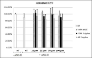
도면 3은 인간 관상 동맥 혈관 평활근 세포 (HCAVSMCs)에서 대조 폴리플렉스 및 유리 MK2i에 비하여 인터류킨-6 (IL-6) 생산을 제거하는 본 발명의 폴리플렉스의 일부 구체예를 보여준다. 도면 3에서 제공된 모든 데이터는 세포 수에 정규화된다. 게다가, "NT"는 처리 없음을 의미한다. *NT+TNFα와 비교하여 p<0.05, *NT+TNFα와 비교하여 p<0.01, #동일한 농도에서 MK2i와 비교하여 p<0.05, ##동일한 농도에서 CPP 폴리플렉스와 비교하여 p<0.05, n=4.
도면 4는 블랭크 폴리플렉스, MK2i 단독, PPAA 단독, 또는 MK2i를 포함하는 폴리플렉스의 구체예로 처리된 인간 복재 정맥 (HSV) 표본의 이완에서 증가 퍼센트를 예증하는 막대 그래프를 제공한다. *대조와 비교하여 p<0.05, **100 μm MK2i와 비교하여 p<0.05, n = 3.
도면 5는 처리되지 않거나, MK2i 단독으로 처리되거나, 또는 MK2i 폴리플렉스의 다양한 구체예로 처리된 HSV 표본의 조직학적 섹션을 보여준다. 어두운 선은 내막 두께의 경계를 정한다. 눈금자는 길이에서 100 μm이다.
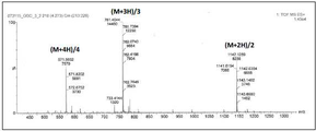
도면 6은 HPLC-정제된 CPP-MK2i 융합 펩티드 (서열 번호 1: YARAAARQARA-KALARQLGVAA)에 대한 전기분무-이온화 질량 분광분석법 (ESI-MS) 질량 스펙트럼을 제공한다. 분자량은 2283.67 g/mol이다. 이러한 질량 스펙트럼은 완전한 펩티드 서열의 단편화에 각각 상응하는 3개의 주요 피크를 보여준다.
도면 7은 D6MSO에서 폴리(아크릴산) (PAA)의 1H NMR 스펙트럼이다. 분자량은 사슬 전달 작용제와 연관된 피크의 구역 (즉, PAA의 경우에 피크 c,d 및 PPAA의 경우에 피크 b)을 아크릴산/프로필아크릴산과 연관된 피크 (즉, PAA의 경우에 피크 및 PPAA의 경우에 피크 c)와 비교함으로써 결정되었다: PAA 중합도 = 106, PPAA 중합도 = 190.
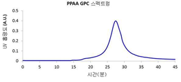
도면 8은 폴리(아크릴산) (PAA)의 GPC 크로마토그램: Mn = 10830 (g/mol), PDI = 1.27, dη/dC = 0.09 (mL/g)이다. 흔적은 중합화에서 활용된 4-시아노-4-(에틸술파닐티오카르보닐) 술파닐펜탄산 (ECT) 사슬 전달 작용제에서 존재하는 트리티오카보네이트 모이어티 (310 nm)의 특징적인 흡수 피크에서 UV 흡광도를 보여준다.
도면 9는 D6MSO에서 폴리(프로필아크릴산) (PPAA) 동종중합체의 1H NMR 스펙트럼을 제공한다. 분자량은 사슬 전달 작용제와 연관된 피크의 구역 (즉, PAA의 경우에 피크 c,d 및 PPAA의 경우에 피크 b)을 아크릴산/프로필아크릴산과 연관된 피크 (즉, PAA의 경우에 피크 a 및 PPAA의 경우에 피크 c)와 비교함으로써 결정되었다: PAA 중합도 = 106 PPAA 중합도 = 190, MW = 21,950 g/mol.
도면 10은 DMF에서 폴리(프로필아크릴산) (PPAA)의 GPC 크로마토그램: Mn = 22010 (g/mol), PDI =1.471, dη/dC = 0.087 (mL/g) 중합체이다. 흔적은 중합화에서 활용된 4-시아노-4-(에틸술파닐티오카르보닐) 술파닐펜탄산 (ECT) 사슬 전달 작용제에서 존재하는 트리티오카보네이트 모이어티 (310 nm)의 특징적인 흡수 피크에서 UV 흡광도를 보여준다.
도면 11은 MK2i 폴리플렉스의 설계와 기능적 특질을 관계시키는 예시를 제공하고, 여기서 MK2iNPs는 엔도솜 탈출을 매개하고 펩티드 치료제를 세포내에 방출하도록 최적화된다.
도면 12는 처리 비교 요약을 제공한다: MK2i-NPs는 엔도소몰리틱 PPAA 중합체로 조제되고, 반면 NE-MK2i-NPs는 PAA 중합체로 조제되는데, 이것은 PPAA와 구조적으로 유사하지만 더욱 낮은 pKa로 인해 엔도소몰리틱이 아니다. MK2i-NPs와 NE-MK2i-NPs 둘 모두 도시된 서열을 갖는 MK2i 펩티드로 만들어진다 (위쪽 열 = 변형된 TAT 모방 세포 투과성 펩티드 서열, 아래쪽 열 = MK2 저해성 서열).
도면 13은 상이한 전하 비율 ([NH3 +]/[COO-])에서 제조된 폴리플렉스의 제타 전위(들)를 보여준다. 영상과 흡수 연구를 위해, NPs는 Alexa®-488 형광단으로 표지화된 MK2i 펩티드로부터 조제되었다. NE-NPs는 비-엔도소몰리틱 (NE) PAA 중합체로 조제된다. 도시된 값은 최소한 3개의 독립된 계측의 평균이다.
도면 14는 119 ± 26 nm의 직경을 갖는 MK2i-NPs의 동적 광 산란 (DLS) 분석을 제공한다.
도면 15는 114 ± 14 nm의 직경을 갖는 NE-MK2i-NPs의 동적 광 산란 분석을 제공한다.
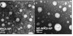
도면 16은 우라닐 아세트산염 대비염색된 MK2i-NPs와 NE-MK2i-NPs의 대표적인 투과 전자 현미경 (TEM) 이미지를 제공한다. 눈금자는 길이에서 100 nm이다.
도면 17은 MK2i-NPs가 DLS 분석에 의해 증명된 바와 같이, 엔도솜 pH 범위에서 pH-촉발된 분해를 겪는다는 것을 보여준다.
도면 18은 형광으로 표지화된 MK2i, MK2i-NPs, 그리고 NE-MK2i-NPs의 세포 흡수와 체류의 정량을 보여주는 그래프를 제공한다. *MK2i와 대비하여 p<0.001, ŦNE-MK2i-NPs와 대비하여 p<0.001, n=3. MK2i-NP 제제는 세포 흡수를 증가시키고, 세포내 체류를 연장하고, 그리고 MK2i의 엔도리소좀 동시국지화를 감소시킨다.
도면 19는 대표적인 흐름 히스토그램을 제공하는데, 이것은 MK2i-NPs를 통해 전달된 형광으로-표지화된 MK2i 펩티드의 증가된 세포 흡수와 더욱 긴 체류를 증명한다.
도면 20은 적혈구 용혈 검정의 결과를 보여주는데, 여기서 MK2i-NPs는 PPAA 중합체와 유사한 pH-의존성 막 교란 활성을 갖지만 NE-MK2i-NPs와 MK2i 펩티드 단독은 그렇지 않다.
도면 21은 완전한 적혈구 용혈 데이터 세트를 제공한다. 적혈구 용혈 검정은 MK2i-NPs는 PPAA 중합체와 유사한 pH-의존성 및 용량 의존성 막 교란 활성을 갖지만, NE-MK2i-NPs와 MK2i 펩티드 단독은 그렇지 않다는 것을 보여준다.
도면 22는 처리 후 24 시간에 LysoTracker® 레드로 Alexa Fluor®-488 표지화된 MK2i 동시국지화의 대표적인 공초점 현미경검사 이미지를 제공한다. 이들 이미지는 MK2i-NPs가 엔도리소좀 동시국지화를 감소시켰다는 것을 증명한다. 눈금자 = 20 μm.
도면 23은 처리 후 0, 12, 그리고 24 시간에 엔도/리소좀 염색제 LysoTracker® 레드로 MK2i 펩티드 동시국지화의 정량을 보여주는 그래프를 제공한다, *MK2i와 대비하여 p<0.01, ŦNE-MK2i-NPs와 대비하여 p<0.01, n ≥ 3 독립된 이미지.
도면 24는 상이한 펩티드 제제로 처리 후 24 시간에 MK2i를 내포하는 세포내 구획의 평균 크기를 전시한다. 구획 구역은 ImageJ 소프트웨어로 정량되었다. *MK2i과 대비하여 p<0.001, ŦNE-MK2i-NPs와 대비하여 p<0.001, 최소한 3개의 상이한 이미지로부터 n = 50개 소포.
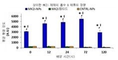
도면 25는 MK2i-NP 제제가 Alexa® 568-MK2i의 HSV 전달을 증가시켰다는 것을 보여준다.
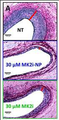
도면 26은 2 시간 동안 처리되고 장기 배양에서 14 일 동안 유지된 Verhoeff Van-Gieson (VVG) 염색된 HSV 섹션의 대표적인 현미경검사 이미지를 제공하고, MK2i-NPs가 신생내막 형성을 효과적으로 차단했다는 것을 보여준다. 적색 막대는 내막 두께의 경계를 정한다. 눈금자는 길이에서 100 μm이다.
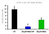
도면 27은 VVG 염색된 조직학적 섹션으로부터 내막 두께의 정량을 제공한다; 수치는 별개의 공여자로부터 최소한 3개의 정맥 고리로부터 6-12개 방사상으로 평행 계측의 평균이다. *NT와 대비하여 p < 0.01, Ŧ동일한 농도에서 MK2i와 대비하여 p < 0.05.
도면 28은 2 시간 동안 처리되고, 그리고 이후, 장기 배양에서 14 일 동안 유지된 HSV 체외이식편의 내막 두께 치수를 제공한다, 최소한 3명의 상이한 공여자로부터 n ≥ 3. *처리 없음 대조 (NT)와 비교하여 p ≤ 0.01, **NT와 비교하여 p ≤ 0.001, Ŧp ≤ 0.05.
도면 29는 MTT 검정을 통해 사정될 때, 2 시간 동안 처리되고 1 또는 장기 배양에서 14 일 동안 유지된 HSV 고리에서 세포 생존력을 보여준다. 최소한 3명의 별개 공여자로부터 n ≥ 3개 정맥 고리.
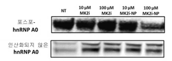
도면 30은 웨스턴 블롯 분석의 결과를 제공하는데, 이들은 MK2i-NPs가 2 시간의 처리 이후에 인간 복재 정맥에서 HnRNP A0 인산화를 감소시킨다는 것을 보여준다, *NT와 대비하여 p < 0.05.
도면 31은 도면 30의 웨스턴 블롯 분석의 추가 결과를 제공하는데, 여기서 MK2i-NPs는 2 시간의 처리 이후에 인간 복재 정맥에서 HnRNP A0 인산화를 감소시켰다, *NT와 대비하여 p < 0.05.
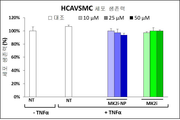
도면 32는 MK2i-NP 처리가 ANG II로 자극된 HCAVSMCs에서 TNFα 생산을 차단한다는 것을 보여준다. 모든 데이터는 세포 수에 정규화된다. "NT"는 처리 없음을 의미하고, *NT + TNFα와 대비하여 p<0.05, Ŧ동일한 농도에서 MK2i와 대비하여 p<0.05, #동일한 농도에서 NE-MK2i-NPs와 대비하여 p<0.05. MK2i-NP 제제는 HCAVSMCs에서 MK2i 생물활성을 증강한다.
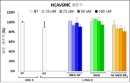
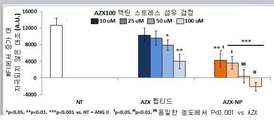
도면 33은 ANG II로 6 시간 동안 자극되고, MK2i-NPs, NE-MK2i-NPs, 또는 MK2i 펩티드 단독으로 2 시간 동안 처리되고, 그리고 새로운 배지에서 24 시간 동안 배양된 HCAVSMCs에서 TNFα 생산을 보여준다. 모든 데이터는 세포 수에 정규화된다. "NT"는 처리 없음을 의미한다. *NT + TNFα 군과 비교하여 p<0.05, Ŧ동일한 농도에서 MK2i와 비교하여 p<0.05, #동일한 농도에서 NE-MK2i-NPs와 비교하여 p<0.05, n = 4.
도면 34는 MK2i-NPs가 HCAVSMCs에서 IL-6 생산에서 TNFα-유도된 증가를 부분적으로 차단한다는 것을 예증한다. 세포는 TNFα로 6 시간 동안 자극되고, MK2i-NPs 또는 MK2i 펩티드 단독으로 2 시간 동안 처리되고, 그리고 새로운 배지에서 24 시간 동안 배양되었다. 모든 데이터는 세포 수에 정규화된다. "NT"는 처리 없음을 의미하고, *NT + TNFα와 대비하여 p<0.05, Ŧ동일한 농도에서 MK2i와 대비하여 p<0.05, #동일한 농도에서 NE-MK2i-NPs와 대비하여 p<0.05.
도면 35는 10 μM ANG II로 6 시간 동안 자극되고, MK2i-NPs, NE-MK2i-NPs, 또는 MK2i 펩티드 단독으로 2 시간 동안 처리되고, 그리고 새로운 배지에서 24 시간 동안 배양된 HCAVSMCs에서 세포 생존력을 보여준다. "NT"는 처리 없음을 의미한다, n = 4.
도면 36은 TNFα로 6 시간 동안 자극되고, MK2i-NPs 또는 MK2i 펩티드 단독으로 2 시간 동안 처리되고, 그리고 새로운 배지에서 24 시간 동안 배양된 HCAVSMCs에서 세포 생존력을 보여주고, 그리고 n = 4.
도면 37은 MK2i-NP 처리가 ANG II 자극에 대한 응답으로 F-액틴 스트레스 섬유 형성을 차단했다는 것을 예증한다. 데이터는 2개의 별개의 실험으로부터 n ≥ 3개 세포를 나타내고, *NT + TNFα와 대비하여 p<0.05, Ŧ동일한 농도에서 MK2i와 대비하여 p<0.05. #동일한 농도에서 NE-MK2i-NPs와 대비하여 p<0.05. 모든 데이터는 세포 수에 정규화된다. "NT"는 처리 없음을 의미하고, *NT + TNFα와 대비하여 p<0.05, Ŧ동일한 농도에서 MK2i와 대비하여 p<0.05. #동일한 농도에서 NE-MK2i-NPs와 대비하여 p<0.05.
도면 38은 MK2i-NPs 또는 대조 (25 μM MK2i)로 1 시간 처리 후, ANG II-자극된 HCAVSMCs에서 F-액틴 스트레스 섬유 형성의 대표적인 형광 현미경검사 이미지를 제공한다.
도면 39는 MK2i-NP 처리가 스크래치 상처의 형성 후 24 시간에 화학유인물질 PDGF-BB (50 ng/mL)로 자극된 HCAVSMCs에서 이주를 차단했다는 것을 보여준다, n≥3: *p<0.05, **NT + PDGF와 대비하여 p<0.01, Ŧ동일한 농도에서 MK2i와 대비하여 p<0.05, #동일한 농도에서 NE-MK2i-NPs와 대비하여 p<0.05. 모든 데이터는 세포 수에 정규화된다. "NT"는 처리 없음을 의미하고, *NT + TNFα와 대비하여 p<0.05, Ŧ동일한 농도에서 MK2i와 대비하여 p<0.05, #동일한 농도에서 NE-MK2i-NPs와 대비하여 p<0.05.
도면 40은 MK2i-NPs가 막 위에 파종 후 8 시간에 보이덴 챔버 검정에서 화학유인물질 PDGF-BB을 향한 세포 이주를 저해했다는 것을 보여준다, 7개 별개의 보이덴 챔버 검정으로부터 n = 4개 이미지. *p<0.05, **NT + PDGF와 대비하여 p<0.01, Ŧ동일한 농도에서 MK2i와 대비하여 p<0.05, #동일한 농도에서 NE-MK2i-NPs와 대비하여 p<0.05. 모든 데이터는 세포 수에 정규화된다. "NT"는 처리 없음을 의미하고, *NT + TNFα와 대비하여 p<0.05, Ŧ동일한 농도에서 MK2i와 대비하여 p<0.05, #동일한 농도에서 NE-MK2i-NPs와 대비하여 p<0.05.
도면 41은 트랜스웰 삽입물을 통하여 이주한 세포의 대표적인 현미경검사 이미지를 제공하는데, 이미지는 10x 배율에서 획득되었다. 처리 분량은 100 μM MK2i, MK2i-NPs, 또는 NE-MK2i-NPs이다; PDGF-BB 분량은 50 ng/mL이다.
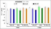
도면 42는 MK2i 펩티드 단독, MK2i-NPs, 또는 NE-MK2i-NPs로 30 분 동안 자극 처리되고, 그리고 50 ng/mL PDGF-BB를 포함하거나 (+) 또는 포함하지 않는 (-) 새로운 배지에서 24 시간 동안 배양된 HCAVSMCs에서 세포 증식을 보여준다. "NT"는 처리 없음을 의미하고, 그리고 n = 4.
도면 43은 MK2i-NP 처리가 정맥 이식편의 VVG 염색된 조직학적 섹션의 대표적인 이미지에 나타나 있는 바와 같이, 신생내막 형성을 감소시켰다는 것을 예증한다. 실제로, MK2i-NPs로 수술중 처리는 이식된 정맥 이식편에서 생체내에서 신생내막 형성과 대식세포 존속을 감소시킨다.
도면 44는 수술 후 28 일에 관류 고정된 경정맥 개재 이식편에서 내막 두께의 정량을 제공한다. *NT와 대비하여 p<0.01, Ŧp<0.05, 처리군마다 n ≥ 7개 이식편.
도면 45는 MK2i-NP 처리가 또한, 정맥 이식편에서 RAM-11 면역조직화학을 이용하여 도시된 바와 같이, 신생내막에서 대식세포의 존속을 감소시켰다는 것을 보여준다. 화살표는 양성으로 염색된 세포의 경계를 정한다. 왼쪽 칼럼 눈금자 = 100 μm, 오른쪽 칼럼 확대된 보기 눈금자 = 50 μm.
도면 46은 각 처리군에 대한 토끼 경정맥 이식편 체외이식편의 대표적인 RAM-11 염색 이미지를 보여준다. 화살표는 양성으로 염색된 세포의 경계를 정한다. 왼쪽 칼럼 눈금자 = 100 μm, 오른쪽 칼럼 확대된 보기 눈금자 = 50 μm.
도면 47은 경정맥 이식편 섹션에서 RAM-11 양성 대식세포 염색의 정량을 제공한다, 4개 정맥 분절로부터 n = 16개 조직학적 이미지, *NT와 대비하여 p<0.05.
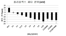
도면 48은 Zetasizer Nano ZS에서 결정된 상이한 전하 비율 ([NH3 +]/[COO-])에서 제조된 폴리플렉스의 ζ-전위를 보여준다. 도시된 값은 최소한 3개의 독립된 계측의 평균이다.
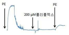
도면 49는 [NH3 +]/[COO-] = 1:3의 전하 비율에서 제조된 폴리플렉스의 pH-의존성 용혈을 예증한다. 유의미한 용혈은 초기 내지 후기 엔도솜 소포의 대표적인 pH 값 (즉, pH < 6.8)에서 증명되었고, 반면 7.4의 생리학적 pH에서 어떤 유의미한 용혈도 관찰되지 않았다. YARA-MK2i 펩티드 단독 또는 AA 폴리플렉스 중에서 어느 것도 검사된 임의의 pH 값에서 임의의 유의미한 용혈을 보여주지 않았고, 그리고 [NH3 +]/[COO-] = 1:3의 전하 비율에서 제조된 폴리플렉스의 pH-의존성 크기 변화는 DLS 분석을 통해 분석되었다.
도면 50은 pH 7.4에서 폴리플렉스가 단일모드 크기 분포를 보여준다는 것을 예증한다. 줄어드는 pH에서, 폴리플렉스는 나타나 있는 바와 같이, 개별 YARA-MK2i 펩티드와 PPAA 중합체 단일체로 해리하기 시작한다.
도면 51은 10 μM ANG II로 6 시간 동안 자극되고, PPAA 폴리플렉스, AA 폴리플렉스, 또는 YARA-MK2i 펩티드 단독으로 2 시간 동안 처리되고, 그리고 새로운 배지에서 24 시간 동안 배양된 HCAVSMCs의 생존력을 보여준다. "NT"는 처리 없음을 의미하고, n = 4.
도면 52는 ANG II로 6 시간 동안 자극되고, PPAA 폴리플렉스, AA 폴리플렉스, 또는 융합 MK2i 펩티드 단독으로 2 시간 동안 처리되고, 그리고 새로운 배지에서 24 시간 동안 배양된 HCAVSMCs에서 TNF-α 생산을 보여준다. 처리는 10, 25, 50, 또는 100 μM의 펩티드 농도에 대해 정규화되었다. 모든 데이터는 LDH 검정에 의해 결정될 때 세포 수에 정규화된다. NT= 처리 없음. *NT + TNFα 군과 비교하여 p<0.05, *동일한 농도에서 MK2i와 비교하여 p<0.05, **동일한 농도에서 AA 폴리플렉스와 비교하여 p<0.05.
도면 53은 Mander의 계수, M1 (본질적으로, 적색 형광과 겹쳐지는 이미지에서 녹색 형광의 %, 다시 말하면, 엔도솜 소포 내에 내포된 펩티드의 %)의 계산을 통해 결정된, 녹색 형광단과 적색 형광단의 동시국지화의 백분율(들)을 보여준다. YARA-MK2i 분량은 모든 표본에 대해 25 μM이다. 도시된 값은 평균 n=3 별개의 이미지 ± SEM이다. *동일한 시점에서 YARA-MK2i와 비교하여 p<0.05, **동일한 시점에서 YARA-MK2i와 비교하여 p<0.01. 이러한 그래프는 HCAVSMC 폴리플렉스 흡수의 현미경적 분석의 결과이고, 그리고 이것은 폴리플렉스가 MK2i 펩티드의 흡수와 엔도솜 탈출을 증강한다는 것을 보여준다.
도면 54는 도면 53에서 데이터에 관계하고, 그리고 동시국지화를 정량하는데 이용된 대표적인 형광 이미지를 제공한다. 왼쪽에 숫자는 세포가 2 시간의 처리 이후에 새로운 배지에서 배양된 시간의 양을 나타내고, 적색과 녹색 통로 둘 모두에 대한 증가가 획득된 모든 이미지에 대해 일정하게 유지되었다.
도면 55는 PPAA 폴리플렉스에 대한 시간의 흐름에서 평균 형광 강도의 플롯을 보여준다.
도면 56은 PPAA 폴리플렉스에 대한 시간의 흐름에서 형광 강도의 히스토그램을 제공한다.
도면 57은 YARA-MK2i 펩티드 단독에 대한 시간의 흐름에서 평균 형광 강도의 플롯을 제공한다.
도면 58은 YARA-MK2i 펩티드 단독에 대한 시간의 흐름에서 형광 강도의 히스토그램이다.
도면 59는 AA 폴리플렉스에 대한 시간의 흐름에서 평균 형광 강도의 플롯이다.
도면 60은 AA 폴리플렉스에 대한 시간의 흐름에서 형광 강도의 히스토그램을 제공한다.
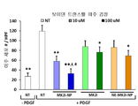
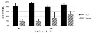
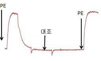
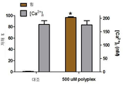
도면 61은 HSV 고리가 페닐에프린 (PE, 10-6 M)으로 수축되고, 그리고 차후에 SNP (10-8-10-6 M)로 이완된 후에, 나트륨 니트로프루시드 (SNP) 이완에서 백분율 증가를 보여주는 막대 그래프를 제공한다. HSV 고리는 이후, 이완에서 처리후 증가를 결정하기 위해, 2 시간 동안 처리되고, PE로 다시 한 번 수축되고, 그리고 SNP로 이완되었다. 처리후 수축 이후에, 모든 고리는 평활근 생존력을 실증하기 위해 KCl로 수축되었다. *대조와 비교하여 p<0.05, **100 μM MK2i와 비교하여 p<0.05, n = 3.
도면 62는 MTT 검정을 통해 사정될 때, 2 시간 동안 처리되고 장기 배양에서 24 시간 동안 유지된 HSV 고리에서 세포 생존력을 보여준다. n = 1.
도면 63은 MTT 검정을 통해 사정될 때, 2 시간 동안 처리되고 장기 배양에서 14 일 동안 유지된 HSV 고리에서 세포 생존력을 보여준다. n = 1.
도면 64는 2 시간 동안 처리되고, 그리고 이후, 장기 배양에서 14 일 동안 유지된 HSV 체외이식편의 내막 두께를 전시한다, n = 3. *대조 (처리되지 않음)와 비교하여 p ≤ 0.01, **대조와 비교하여 p ≤ 0.001, Ŧp ≤ 0.05.
도면 65는 2 시간 동안 처리되고, 그리고 이후, 장기 배양에서 14 일 동안 유지된 HSV 체외이식편의 내막/내측 (I/M) 비율의 플롯을 제공한다, n = 3. *대조 (처리되지 않음)와 비교하여 p ≤ 0.01, **대조와 비교하여 p ≤ 0.001, Ŧp ≤ 0.05.
도면 66은 3:1 전하 비율에서 제조된 AZX-100 폴리플렉스의 DLS 크기 분포를 보여준다.
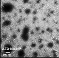
도면 67은 DLS 결과와 일치하는 크기 분포를 보여주는 우라닐 아세트산염 염색된 AZX-100 폴리플렉스의 대표적인 TEM 이미지를 제공한다.
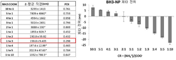
도면 68은 다양한 전하 비율에서 제조된 AZX-100 폴리플렉스의 제타 전위의 요약을 제공한다. 제타 전위는 3:1보다 높은 전하 비율에서 전하 비율에 직접적으로 비례하는 것으로 밝혀졌다. 제타 전위에서 예상치 못한 이동이 아마도 거대분자 재배열로 인해 3:1의 전하 비율에서 관찰되었다.
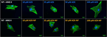
도면 69와 도면 70은 AZX-100 폴리플렉스가 안지오텐신 II 자극된 인간 관상 동맥 혈관 평활근 세포에서 스트레스 섬유 형성의 AZX-100 매개된 저해를 증강한다는 것을 보여준다. 세포는 1 시간 동안 처리되고, 그리고 이후, 안지오텐신 II로 2 시간 동안 차후 자극되었다. 액틴 스트레스 섬유는 팔로이딘 염색되고, 고정된 표본에서 가시화되었고, 그리고 각 처리군으로부터 개별 세포의 상대적 형광 강도가 액틴 스트레스 섬유 형성을 정량하는데 활용되었다.
도면 71은 대조, AZ100 펩티드 또는 AZX 폴리플렉스로 처리된 쥐 대동맥 평활근에서 일어난 저해의 퍼센트를 제공한다.
도면 72는 쥐 대동맥 평활근의 수축을 보여준다.
도면 73은 AZX 폴리플렉스로 처리된 쥐 대동맥 평활근에서 수축의 용량 의존성 저해를 보여준다.
도면 74는 쥐 대동맥 평활근에서 힘의 대표적인 추적 및 칼슘 형광 추적을 전시한다.
도면 75는 쥐 대동맥 평활근에서 일어난 세포내 칼슘에서 변화 및 힘의 저해의 크기를 계측하는 누적 데이터를 제공한다.
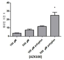
도면 76은 상이한 농도에서 AZX-100 펩티드 또는 AZX 폴리플렉스로 처리 후, HSV에서 % 증강된 이완을 보여준다.
도면 77은 AZX-100 NPs가 인간 세기관지 기도 평활근의 AZX-100 매개된 이완을 증강한다는 것을 예증한다.
도면 78은 RN22-내포 폴리플렉스의 직경, 다분산성 지수 (PDI), 그리고 제타 전위에 대한 상이한 전하 비율의 효과를 보여주는 표와 차트를 제공한다.
도면 79는 페네트라틴-BAK-BH3-내포 폴리플렉스의 직경, 다분산성 지수 (PDI), 그리고 제타 전위에 대한 상이한 전하 비율의 효과를 보여주는 표와 차트를 제공한다. Brief Description of Drawings
1 is a flow diagram for the synthesis of embodiments of polyplexes comprising a cationic peptide, for example, a peptide comprising MAPKAP kinase 2 (MK2i), and an anionic, endosomolytic polymer, such as PPAA, to provide.
Figure 2 provides the results of a hemolysis assay, demonstrating that embodiments of the polyplex can be tuned for escape from the endolysome pathway via a pH-dependent membrane breakage mechanism.
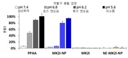
Figure 3 shows some embodiments of the polyplex of the present invention that eliminate interleukin-6 (IL-6) production relative to control polyplex and free MK2i in human coronary artery vascular smooth muscle cells (HCAVSMCs). All data provided in Figure 3 is normalized to the number of cells. In addition, "NT" means no treatment. * Compared to the NT + TNFα p <0.05, * NT + TNFα as compared with p <0.01, # MK2i compared with the same concentration as compared with p <0.05, CPP polyplexes at the same concentration of ## p <0.05, n = 4.
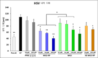
Figure 4 provides a histogram illustrating the percent increase in relaxation of human saprolical vein (HSV) specimens treated with blank polyplex, MK2i alone, PPAA alone, or polyplexes containing MK2i. * Compared with control, p <0.05, ** compared with 100 μm MK2i p <0.05, n = 3.
Figure 5 shows the histological sections of HSV specimens that were untreated, treated with MK2i alone, or treated with various embodiments of the MK2i polyplex. The dark line delimits the thickness of the inner membrane. The ruler is 100 μm in length.
Figure 6 provides an electrospray-ionization mass spectrometry (ESI-MS) mass spectrum for an HPLC-purified CPP-MK2i fusion peptide (SEQ ID NO: 1: YARAAARQARA-KALARQLGVAA). The molecular weight is 2283.67 g / mol. These mass spectra show three major peaks corresponding to fragmentation of the complete peptide sequence, respectively.
7 , D < 6 > is the 1 H NMR spectrum of poly (acrylic acid) (PAA) in MSO. The molecular weight is determined by the area of the peak associated with the chain transfer agent (i.e., peak c in the case of PAA, peak b in the case of PAA, and peak b in the case of PPAA) with peaks associated with acrylic acid / propylacrylic acid (i.e. peak in the case of PAA and PPAA in the case of PPAA) Peak c): PAA degree of polymerization = 106, PPAA degree of polymerization = 190.
8 is a GPC chromatogram of poly (acrylic acid) (PAA): M n = 10830 (g / mol), PDI = 1.27, d? / DC = 0.09 (mL / g). The trace shows that the characteristic absorption peak of the trithiocarbonate moiety (310 nm) present in the 4-cyano-4- (ethylsulfanylthiocarbonyl) sulfanylpentanoic acid (ECT) It shows absorbance.
9 , 1 H NMR spectra of poly (propyl acrylate) (PPAA) homopolymer in D 6 MSO. The molecular weight is determined by the area of the peak associated with the chain transfer agent (i.e., the peak c in the case of PAA, the peak c in the case of PAA and the peak b in the case of PPAA), the peak associated with acrylic acid / C): PAA Polymerization Degree = 106 PPAA Polymerization Degree = 190, MW = 21,950 g / mol.
Figure 10 is a GPC chromatogram of poly (propyl acrylate) (PPAA) in DMF: M n = 22010 (g / mol), PDI = 1.471, d? / DC = 0.087 (mL / g). The trace shows that the characteristic absorption peak of the trithiocarbonate moiety (310 nm) present in the 4-cyano-4- (ethylsulfanylthiocarbonyl) sulfanylpentanoic acid (ECT) It shows absorbance.
Figure 11 provides an example of relating the design and functional properties of MK2i polyplexes, where MK2iNPs are optimized to mediate endosome excretion and release peptide therapeutic agents into cells.
12 , MK2i-NPs are prepared with endomolytic PPAA polymers, whereas NE-MK2i-NPs are prepared with PAA polymers, which are structurally similar to PPAA, but with lower pKa, endo somolytic Is not. Both MK2i-NPs and NE-MK2i-NPs are made of MK2i peptides with the sequence shown (top row = modified TAT mimic cell permeability peptide sequence, bottom row = MK2 inhibition sequence).
Figure 13 shows the zeta potential (s) of the polyplex prepared at different charge ratios ([NH 3 + ] / [COO - ]). For imaging and absorption studies, NPs were prepared from MK2i peptides labeled with Alexa®-488 fluorophore. NE-NPs are prepared with non-endosomolytic (NE) PAA polymers. The values shown are the average of at least three independent measurements.
Figure 14 provides dynamic light scattering (DLS) analysis of MK2i-NPs with a diameter of 119 +/- 26 nm.
Figure 15 provides dynamic light scattering analysis of NE-MK2i-NPs with a diameter of 114 14 nm.
Figure 16 provides a representative transmission electron microscopy (TEM) image of dyed MK2i-NPs and NE-MK2i-NPs versus uranyl acetate. The ruler is 100 nm in length.
In Figure 17 , MK2i-NPs undergo pH-triggered degradation in the endosomal pH range, as evidenced by DLS analysis.
Figure 18 provides a graph showing quantitation of cell uptake and retention of fluorescence-labeled MK2i, MK2i-NPs, and NE-MK2i-NPs. * MK2i as opposed to p <0.001, Ŧ In contrast to the NE-MK2i-NPs p <0.001 , n = 3. The MK2i-NP preparation increases cell uptake, prolongs intracellular retention, and reduces endolysomal localization of MK2i.
Figure 19 provides a representative flow histogram demonstrating increased cell uptake and longer retention of fluorescence-labeled MK2i peptides delivered via MK2i-NPs.
Figure 20 shows the results of an erythrocyte hemolysis assay in which MK2i-NPs have a pH-dependent membrane disruptive activity similar to that of PPAA polymer, whereas NE-MK2i-NPs and MK2i peptides alone do not.
Figure 21 provides a complete set of erythro hemolysis data. Erythrocyte hemolysis assays show that MK2i-NPs have similar pH-dependent and dose-dependent membrane-burst activities to PPAA polymers, whereas NE-MK2i-NPs and MK2i peptides alone do not.
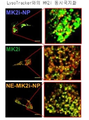
Figure 22 provides a representative confocal microscopy image of Alexa Fluor-488 labeled MK2i simultaneous localization with
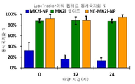
Figure 23 provides a graph showing the amount of peptide co-localized with MK2i endo / lysosomal LysoTracker® red dye to 0, 12, and 24 hours after treatment, - in contrast to MK2i and p <0.01, Ŧ NE-MK2i -NPs In contrast, p <0.01, n ≥ 3 independent images.
Figure 24 shows the average size of intracellular compartment containing MK2i at 24 hours after treatment with different peptide preparations. The compartment was quantified with ImageJ software. * MK2i and to p <0.001, Ŧ In contrast to the NE-MK2i-NPs p <0.001 , n = from at least three
Figure 25 shows that the MK2i-NP preparation increased the HSV delivery of Alexa® 568-MK2i.
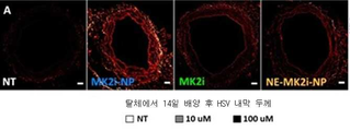
Figure 26 provides a representative microscopic image of the Verhoeff Van-Gieson (VVG) stained HSV section treated for 2 hours and maintained for 14 days in long-term culture, showing that MK2i-NPs effectively blocked neointimal formation. The red bar delimits the thickness of the inner membrane. The ruler is 100 μm in length.
Figure 27 provides a quantification of the intimal thickness from a VVG stained histological section; The figures are the average of 6-12 radial parallel measurements from at least 3 vein rings from separate donors. * NT as opposed to p <0.01, as opposed to Ŧ MK2i at the same concentration p <0.05.
Figure 28 provides intimal thickness measurements of HSV explants treated for 2 hours and then maintained for 14 days in long-term cultures. N ≥ 3. * from no less than 3 different donors Compared with No Control (NT) the p ≤ 0.001 compared with p ≤ 0.01, ** NT, Ρ p ≤ 0.05.
Figure 29 shows cell viability in HSV rings treated for 2 hours and maintained for 14 days in 1 or long-term cultures, as assessed by MTT assay. N ≥ 3 vein rings from at least three separate donors.
30 is a view in contrast to Western provides the results of the blot analysis, which shows that the MK2i-NPs reduces the HnRNP A0 phosphorylation in human saphenous vein after treatment of 2 hours, NT * p <0.05.
Figure 31 is to provide additional results of Western blot analysis of the figure 30, where MK2i-NPs reduced the HnRNP A0 phosphorylation in human saphenous vein after treatment of 2 hours, as opposed to * NT p <0.05.
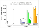
32 , MK2i-NP treatment blocks TNFa production in HCVSMCs stimulated with ANG II. All data is normalized to the number of cells. "NT" means no treatment, and * NT + TNFα in comparison with p <0.05, as opposed to Ŧ MK2i at the same concentration p <0.05, # and in case NE-MK2i-NPs in the same concentration p <0.05. MK2i-NP preparations enhance MK2i bioactivity in HCAVSMCs.
Figure 33 shows TNFa production in HCAVSMCs stimulated with ANG II for 6 hours and treated with MK2i-NPs, NE-MK2i-NPs, or MK2i peptide alone for 2 hours and cultured in fresh medium for 24 hours. All data is normalized to the number of cells. "NT" means no processing. * NT + TNFα as compared with the group p <0.05, compared to the Ŧ MK2i at the same concentration p <0.05, compared with # NE-MK2i-NPs in the same concentration p <0.05, n = 4.
Figure 34 illustrates that MK2i-NPs partially block TNF [alpha] -induced increases in IL-6 production in HCAVSMCs. Cells were stimulated with TNF [alpha] for 6 hours, treated with MK2i-NPs or MK2i peptide alone for 2 hours, and cultured in fresh medium for 24 hours. All data is normalized to the number of cells. "NT" means no treatment, and * NT + TNFα in comparison with p <0.05, as opposed to Ŧ MK2i at the same concentration p <0.05, # and in case NE-MK2i-NPs in the same concentration p <0.05.
Figure 35 shows cell viability in HCAVSMCs stimulated with 10 [mu] M ANG II for 6 hours and treated with MK2i-NPs, NE-MK2i-NPs, or MK2i peptide alone for 2 hours and cultured in fresh medium for 24 hours . "NT" means no treatment, n = 4.
Figure 36 shows cell viability in HCAVSMCs stimulated with TNF [alpha] for 6 hours, treated with MK2i-NPs or MK2i peptide alone for 2 hours, and cultured in fresh medium for 24 hours, and n = 4.
Figure 37 illustrates that MK2i-NP treatment blocked F-actin stress fiber formation in response to ANG II stimulation. Data represents the n ≥ 3 cells from two separate experiments, * NT + TNFα in comparison with p <0.05, p <0.05 compared to Ŧ MK2i and at the same concentration. # P < 0.05 compared to NE-MK2i-NPs at the same concentration. All data is normalized to the number of cells. "NT" means no treatment, and * NT + TNFα in comparison with p <0.05, as opposed to Ŧ MK2i at the same concentration p <0.05. # P < 0.05 compared to NE-MK2i-NPs at the same concentration.
Figure 38 is a representative of F-actin stress fiber formation in ANG II-stimulated HCAVSMCs after 1 hour treatment with MK2i-NPs or control (25 [mu] M MK2i) Fluorescence microscopy images are provided.
Figure 39 shows that block the migration from the MK2i-NP treatment is stimulated with the chemoattractant PDGF-BB (50 ng / mL ) for 24 hours of the formation of scratch wound HCAVSMCs, n≥3: * p <0.05 , * * NT + PDGF preparation and p <0.01, as opposed to Ŧ MK2i at the same concentration p <0.05, # and in case NE-MK2i-NPs in the same concentration p <0.05. All data is normalized to the number of cells. "NT" means no treatment, and * NT + TNFα in comparison with p <0.05, as opposed to Ŧ MK2i at the same concentration p <0.05, # and in case NE-MK2i-NPs in the same concentration p <0.05.
Figure 40 shows that MK2i-NPs inhibited cell migration towards the chemoattractant PDGF-BB in the Voidan chamber assay at 8 hours post seeding on membranes, n = 4 images from seven distinct Vohen chamber probes. * P <0.05, ** NT + PDGF preparation and p <0.01, as opposed to Ŧ MK2i at the same concentration p <0.05, # and in case NE-MK2i-NPs in the same concentration p <0.05. All data is normalized to the number of cells. "NT" means no treatment, and * NT + TNFα in comparison with p <0.05, as opposed to Ŧ MK2i at the same concentration p <0.05, # and in case NE-MK2i-NPs in the same concentration p <0.05.
Figure 41 provides representative microscopic images of cells migrated through the transwell inserts, images were acquired at 10x magnification. Treatments were 100 μM MK2i, MK2i-NPs, or NE-MK2i-NPs; The amount of PDGF-BB is 50 ng / mL.
Figure 42 shows that 24 (n = 24) cells were stimulated with MK2i peptide alone, MK2i-NPs, or NE-MK2i-NPs for 30 min and in fresh medium containing (+) or without (+) 50 ng / Lt; RTI ID = 0.0 > HCAVSMCs < / RTI > cultured for a period of time. "NT" means no treatment, and n = 4.
In Figure 43 , Demonstrate that MK2i-NP treatment reduced neointimal formation as shown in representative images of the VVG stained histological sections of vein grafts. Indeed, intra-operative treatment with MK2i-NPs reduces neointimal formation and macrophage survival in vivo in grafted grafts.
Figure 44 provides a quantification of the intimal thickness in the jugular vein interposed graft fixed perfusion at 28 days post surgery. * In contrast with the NT p <0.01, Ŧ p < 0.05, for each treatment group, n ≥ 7 gae graft.
Figure 45 shows that MK2i-NP treatment also reduced the survival of macrophages in the neointimal lining, as shown using RAM-11 immunohistochemistry in vein grafts. Arrows delimit cells that are stained positively. Left column scale = 100 μm, right column magnified view ruler = 50 μm.
Figure 46 shows a representative RAM-11 staining image of a rabbit jugular vein explant for each treatment group. Arrows delimit cells that are stained positively. Left column scale = 100 μm, right column magnified view ruler = 50 μm.
Figure 47 is a contrast to the RAM-11 positivity for providing a determination of macrophage staining, n = 16 4 from the vein segment gae histological image, * NT from the jugular vein graft section, p <0.05.
48 shows Shows the ζ-potential of the polyplex prepared from the different charge ratios ([NH 3 + ] / [COO - ]) determined in the Zetasizer Nano ZS. The values shown are the average of at least three independent measurements.
Figure 49 Dependent hemolysis of the polyplex prepared at a charge ratio of [NH 3 + ] / [COO - ] = 1: 3. Significant hemolysis was demonstrated at a typical pH value of the endosomal vesicles from early to late (ie pH <6.8), whereas no significant hemolysis was observed at the physiological pH of 7.4. None of the YARA-MK2i peptides alone or the AA polyplexes showed any significant hemolysis at any of the tested pH values, and no polyol produced at charge ratios of [NH 3 + ] / [COO - ] = 1: The pH-dependent size change of the flex was analyzed through DLS analysis.
Figure 50 illustrates that the polyplex at pH 7.4 shows a single mode size distribution. At a reduced pH, polyplexes begin to dissociate into individual YARA-MK2i peptides and PPAA polymer monomers, as shown.
Figure 51 Shows viability of HCAVSMCs stimulated with 10 [mu] M ANG II for 6 hours and treated with PPAA polyplex, AA polyplex, or YARA-MK2i peptide alone for 2 hours and cultured in fresh medium for 24 hours. "NT" means no treatment, n = 4.
52 , Stimulated with ANG II for 6 hours and treated with PPAA polyplex, AA polyplex, or fusion MK2i peptide alone for 2 hours, and TNF-a production in HCAVSMCs cultured in fresh medium for 24 hours. Treatments were normalized to peptide concentrations of 10, 25, 50, or 100 [mu] M. All data are normalized to the number of cells when determined by the LDH assay. NT = No processing. * NT + TNFα as compared with the group p <0.05, * MK2i as compared to the same concentration as compared with p <0.05, ** AA polyplexes at the same concentration of p <0.05.
Figure 53 Simultaneous localization of the green fluorescence and red fluorescence stages, determined through calculation of the Mander's coefficient, M1 (essentially the% of green fluorescence in the overlapping image with red fluorescence, i. E., The percentage of peptides encapsulated in endosomes) It shows the percentage (s). The amount of YARA-MK2i is 25 μM for all samples. The values shown are average n = 3 distinct images +/- SEM. * Compared to the p <0.05, ** YARA-MK2i at the same point in time as compared to YARA-MK2i at the same point p <0.01. These graphs are the result of a microscopic analysis of HCAVSMC polyplex absorption and show that polyplexes enhance absorption of MK2i peptides and endosome excretion.
54 provides a representative fluorescence image used to quantify the data and the simultaneous localization in FIG. The numbers on the left indicate the amount of time the cells were incubated in the new medium after 2 hours of treatment and the increase in both red and green passages was kept constant for all acquired images.
Figure 55 A plot of the mean fluorescence intensity over time for the PPAA polyplex is shown.
Figure 56 provides a histogram of fluorescence intensity over time for the PPAA polyplex.
Figure 57 provides a plot of mean fluorescence intensity over time for the YARA-MK2i peptide alone.
Figure 58 is a histogram of fluorescence intensity over time for the YARA-MK2i peptide alone.
Figure 59 is a plot of mean fluorescence intensity over time for AA polyplex.
Figure 60 provides a histogram of fluorescence intensity in the course of time for the AA polyplex.
Figure 61 is a HSV ring is contracted in the printer (PE, 10 -6 M) on the phenyl, and later SNP (10- 8-10 -6 M) after the relaxation to the increase in the percentage of sodium nitroprusside (SNP) Relaxation Of the bar graph. The HSV loop was then treated for 2 hours, contracted once again to PE, and relaxed to the SNP to determine post-treatment increases in relaxation. After shrinkage after treatment, all rings contracted with KCl to demonstrate smooth muscle viability. * P <0.05 compared with control, ** pK <0.05 compared with 100 μM MK2i, n = 3.
Figure 62 shows cell viability in the HSV loop treated for 2 hours and maintained in the long term culture for 24 hours when assessed by MTT assay. n = 1.
Figure 63 shows cell viability in HSV rings treated for 2 hours and maintained for 14 days in long-term cultures, as assessed by MTT assay. n = 1.
Figure 64 shows the lining thickness of HSV explants treated for 2 hours and then maintained for 14 days in long-term culture, n = 3. * Compared to control (untreated), p ≤ 0.01, ** as compared to control p ≤ 0.001, Ŧ p ≤ 0.05 .
Figure 65 provides a plot of the intimal / medial (I / M) ratio of HSV explants treated for 2 hours and then maintained for 14 days in long-term cultures, n = 3. * Control (untreated) as compared to the p ≤ 0.01, ** as compared to control p ≤ 0.001, Ŧ p ≤ 0.05 .
Figure 66 shows the DLS size distribution of AZX-100 polyplex prepared at a 3: 1 charge ratio.
Figure 67 provides representative TEM images of uranyl acetate dyed AZX-100 polyplexes showing size distributions consistent with DLS results.
Figure 68 provides a summary of the zeta potential of the AZX-100 polyplex prepared at various charge ratios. The zeta potential was found to be directly proportional to the charge ratio at a charge ratio higher than 3: 1. An unexpected shift in zeta potential was observed at a charge ratio of 3: 1, possibly due to macromolecular rearrangement.
Figures 69 and 70 show that AZX-100 polyplexes enhance AZX-100 mediated inhibition of stress fiber formation in angiotensin II-stimulated human coronary artery vascular smooth muscle cells. Cells were treated for 1 hour, and subsequently stimulated with angiotensin II for 2 hours. Actin stress fibers were paloindin stained, visualized in fixed samples, and relative fluorescence intensities of individual cells from each treatment group were used to quantify actin stress fiber formation.
Figure 71 Control, AZ100 peptides or AZX polyplexes in the rat aortic smooth muscle.
72 , Shrinkage of rat aortic smooth muscle.
FIG. 73 shows the dose-dependent inhibition of contraction in rat aortic smooth muscle treated with AZX polyplex.
FIG. 74 exhibits representative tracing of the force and trace of calcium fluorescence in the rat aortic smooth muscle.
Figure 75 provides cumulative data measuring the magnitude of changes in intracellular calcium and inhibition of force in the rat aortic smooth muscle.
Figure 76 shows the% enhanced relaxation in HSV after treatment with AZX-100 peptide or AZX polyplex at different concentrations.
FIG. 77 illustrates that AZX-100 NPs augment AZX-100 mediated relaxation of human bronchoconstrictor smooth muscle.
Figure 78 provides tables and charts showing the effect of different charge ratios on the diameter, polydispersity index (PDI), and zeta potential of the RN22-containing polyplex.
Figure 79 provides tables and charts showing the effect of different charge ratios on the diameter, polydispersity index (PDI), and zeta potential of the Petinalatin-BAK-BH3-containing polyplex.
예시적인 구체예의 상세한 설명DETAILED DESCRIPTION OF EXEMPLARY EMBODIMENTS
현재 개시된 요부의 하나 또는 그 이상의 구체예의 상세가 본 문서에서 진술된다. 본 문서에서 설명된 구체예, 그리고 다른 구체예에 대한 변형은 본 문서에서 제공된 정보의 연구 후 당업자에게 명백할 것이다. 본 문서에서 제공된 정보, 그리고 특히, 설명된 예시적인 구체예의 특정한 상세는 일차적으로 이해의 명료함을 위해 제공되고 불필요하지 않은 한정은 그것으로부터 이해될 것이다. 모순의 경우에, 정의를 비롯한 본 문서의 명세가 우선할 것이다.Details of one or more embodiments of the presently disclosed subject matter are set forth in this document. Variations to the embodiments described herein, and other embodiments, will be apparent to those skilled in the art after study of the information provided herein. The specific details of the information provided herein, and in particular the exemplary embodiments described, are provided primarily for clarity of understanding, and unnecessary limitations will be understood from it. In the case of contradictions, the specification of this document, including definitions, shall prevail.
각 실례는 본 발명의 설명에 의하여 제공되고 이를 한정하지 않는다. 실제로, 다양한 변형과 변이가 발명의 범위로부터 벗어나지 않으면서 본 발명의 교시에 만들어질 수 있다는 것은 당업자에게 명백할 것이다. 가령, 한 구체예의 일부로서 예시되거나 또는 설명된 특질은 또 다른 구체예를 산출하기 위해 다른 구체예에서 이용될 수 있다. Each example is provided by way of explanation of the present invention and is not limited thereto. Indeed, it will be apparent to those skilled in the art that various modifications and variations can be made to the teachings of the invention without departing from the scope of the invention. For example, the features illustrated or described as part of one embodiment may be used in other embodiments to produce another embodiment.
본 발명의 단수 특징 또는 한정에 대한 모든 언급은 달리 명시되지 않으면 또는 언급이 만들어지는 문맥에 의해 명확하게 반대로 암시되지 않으면, 상응하는 복수 특징(들) 또는 한정(들)을 포함할 것이고, 그 반대로도 그러할 것이다. All references to a single feature or limitation of the present invention will include the corresponding plural feature (s) or limitation (s) unless explicitly stated otherwise or explicitly contrary to the context in which the reference is made, That is also true.
본원에서 이용된 바와 같은 방법 또는 과정 단계의 모든 조합은 달리 명시되지 않으면 또는 언급된 조합이 만들어지는 문맥에 의해 명확하게 반대로 암시되지 않으면, 임의의 순서로 수행될 수 있다. All combinations of methods or process steps as used herein may be performed in any order unless otherwise stated or unless the context clearly dictates otherwise.
본 발명의 방법과 조성물, 그리고 이들의 성분은 본원에서 설명된 구체예의 필수 원소와 한정뿐만 아니라 본원에서 설명된 또는 달리 유용한 임의의 추가 또는 임의선택적 성분 또는 한정을 포함할 수 있거나, 이들로 구성되거나, 또는 이들로 본질적으로 구성된다. The methods and compositions of the present invention, and components thereof, may comprise, consist of, or consist essentially of the essential elements and limitations of the embodiments described herein, as well as any additional or optional optional ingredients or limitations set forth herein or otherwise , Or consist essentially of these.
엔도솜 경로를 방지할 수 있고, 세포질 표적에 향상된 접근을 갖고, 증가된 세포내 체류 시간을 갖고, 그리고 세포내-작용 펩티드 약물의 향상된 생물활성을 갖는, 펩티드를 비롯한 활성제를 전달하기 위한 조성물과 방법이 요구된다. 본 발명의 요부는 최소한 이들 각각의 요구에 부합한다. Compositions for delivering active agents, including peptides, which are capable of preventing endosomal pathways, have improved access to cytoplasmic targets, have increased intracellular retention times, and have enhanced biological activity of intracellular-acting peptide drugs, and Method is required. The essentials of the present invention meet at least each of these requirements.
현재 개시된 요부는 펩티드와 중합체를 포함하는 조성물을 포함하고, 여기서 이러한 펩티드와 중합체는 서로에 정전으로 결합되어 미리 결정된 pH에서 폴리플렉스를 형성한다. 용어 "폴리플렉스"는 클러스터, 입자, 집괴, 또는 기타 등등을 형성하는 정전으로-결합된 펩티드와 중합체를 지칭하기 위해 본원에서 이용된다. 따라서, 현재 개시된 요부의 구체예는 펩티드와 중합체의 폴리플렉스를 포함하는 조성물을 포함한다.The presently disclosed subject matter comprises a composition comprising a peptide and a polymer, wherein the peptide and the polymer are electrostatically bound to each other to form a polyplex at a predetermined pH. The term "polyplex" is used herein to refer to electrostatically-coupled peptides and polymers that form clusters, particles, agglomerations, or the like. Thus, embodiments of the presently disclosed subject matter include compositions comprising a polyplex of a peptide and a polymer.
중합체polymer
용어 "중합체"는 동일한 또는 상이한 유형인 지에 상관없이, 단위체를 중합함으로써 제조된 중합성 화합물을 지칭하기 위해 본원에서 이용된다. 일반 용어 "중합체"는 따라서, 용어 동종중합체, 또는 동일한 유형의 단위체 단위로 형성된 중합체, 그리고 용어 공중합체, 또는 2개 또는 그 이상 상이한 유형의 단위체 단위로 형성된 중합체를 포함한다.The term "polymer" is used herein to refer to polymeric compounds made by polymerizing monomers, whether they are the same or different types. The generic term "polymer" thus encompasses the term homopolymer, or polymer formed in units of the same type, and the term copolymer, or polymer formed in units of two or more different types of units.
일부 구체예에서, 중합체는 (C1-C6)알킬-아크릴산, (C1-C6)알킬-메타크릴산, 그리고 (C1-C6)알킬-에타크릴산, 그리고 이들의 조합에서 선택되는 하나 또는 그 이상의 단위체를 포함할 수 있다. 가령, 일부 구체예에서, (C1-C6)알킬-아크릴산 단위체는 프로필 아크릴산 (PAA), 프로필 아크릴산, 부틸 아크릴산, 기타 등등을 포함한다. 결과의 중합체는 폴리((C1-C6)알킬-아크릴산), 폴리((C1-C6)알킬-메타크릴산), 그리고 폴리((C1-C6)알킬-에타크릴산), 그리고 이들의 조합으로 구성되거나 또는 포함할 수 있다. 특정한 구체예에서, 중합체는 폴리(프로필아크릴산) (PPAA) 중합체이다. In some embodiments, the polymer is selected from the group consisting of (C 1 -C 6 ) alkyl-acrylic acid, (C 1 -C 6 ) alkyl-methacrylic acid, and (C 1 -C 6 ) alkyl-ethacrylic acid, And may include one or more unit pieces to be selected. For example, in some embodiments, the (C 1 -C 6 ) alkyl-acrylic acid unit comprises propyl acrylic acid (PAA), propyl acrylic acid, butylacrylic acid, and the like. Polymers of the results of a poly ((C 1 -C 6) alkylene-acrylic acid), poly ((C 1 -C 6) alkyl-methacrylic acid), and poly ((C 1 -C 6) alkyl-ethacrylic acid) , And combinations thereof. In certain embodiments, the polymer is a poly (propyl acrylic acid) (PPAA) polymer.
용어 "알킬"은 일반식 CnH2n+1을 갖는 알킬 기를 지칭하고, 여기서 n = 약 1 내지 약 18 또는 그 이상이다. 이들 기는 직쇄이거나 또는 분지될 수 있다. 알킬은 본원에서 이용될 때, 또한 "저급 알킬"을 포함하는데, 이것은 일반식 CnH2n+1 (여기서 n=1 내지 약 6)을 갖는 알킬 기를 지칭한다. 일부 구체예에서, n = 1 내지 약 3이다. 실시예는 메틸, 에틸, 프로필, 이소프로필, n-부틸, sec-부틸, t-부틸, 이소부틸, n-펜틸, 이소펜틸, 네오펜틸, n-헥실, 기타 등등을 포함한다. 이점에 관해서, 용어 "시클로알킬"은 최소한 3개의 탄소 원자로 구성된 비방향족 탄소-기초된 고리, 예를 들면, 시클로프로필, 시클로헥실, 기타 등등을 지칭한다. 용어 알킬은 시클로알킬을 포괄한다.The term "alkyl" refers to an alkyl group having the general formula C n H 2n + 1 , where n = about 1 to about 18 or more. These groups may be linear or branched. Alkyl, when used herein, also includes "lower alkyl ", which refers to an alkyl group having the general formula C n H 2n + 1 wherein n = 1 to about 6. In some embodiments, n = 1 to about 3. Examples include methyl, ethyl, propyl, isopropyl, n-butyl, sec-butyl, t-butyl, isobutyl, n-pentyl, isopentyl, neopentyl, n-hexyl, In this regard, the term "cycloalkyl" refers to a non-aromatic carbon-based ring consisting of at least three carbon atoms, e.g., cyclopropyl, cyclohexyl, The term alkyl includes cycloalkyl.
일부 구체예에서, 단위체의 기능화된 이형이 본 발명 중합체에서 임의선택적으로 이용된다. 기능화된 단위체는 본원에서 이용된 바와 같이, 가려진 또는 비-가려진 기능기, 예를 들면, 중합화 이후에 다른 모이어티가 부착될 수 있는 기를 포함하는 단위체이다. 이런 기의 무제한적 실례는 일차 아미노 기, 카르복실, 티올, 히드록실, 아지드, 그리고 시아노 기이다. 여러 적합한 가리움 기가 가용하다 (가령, T. W. Greene & P. G. M. Wuts, Protective Groups in Organic Synthesis (2nd edition) J. Wiley & Sons, 1991. P. J. Kocienski, Protecting Groups, Georg Thieme Verlag, 1994를 참조한다). In some embodiments, functionalized variants of the monomers are optionally used in the polymers of the present invention. Functionalized monomers are, as used herein, units that contain a masked or non-masked functional group, for example, a group to which another moiety can be attached after polymerization. Unlimited examples of these groups are primary amino groups, carboxyl, thiol, hydroxyl, azide, and cyano groups. (See, for example, T. W. Greene & P. G. M. Wuts, Protective Groups in Organic Synthesis (2nd edition) J. Wiley & Sons, 1991. P. J. Kocienski, Protecting Groups, Georg Thieme Verlag, 1994).
일부 구체예에서, 중합체는 pH-반응성 중합체이다. 일정한 사례에서, 용어 중합체는 본원에서 이용된 바와 같이, pH-반응 중합체를 포괄한다. pH-반응 중합체는 pH에 따라 전하에서 변화를 경험하는 중합체를 포함한다. 중합체는 미리 결정된 pH에서 양이온성 또는 음이온성일 수 있는데, 이것은 특정한 pH, 일정한 범위의 pH, 일정한 pH 초과, 및/또는 일정한 pH 미만을 포함할 수 있다. 가령, 폴리(알킬 아크릴산)은 카르복실산 기를 포함하고, 그리고 폴리(프로필아크릴산)은 약 6.7의 pKa를 갖는다. 폴리(프로필아크릴산)은 자신의 pKa보다 높은 pH에 있을 때 음이온성 형질을 갖는다. 하지만, 폴리(프로필아크릴산)은 대략 자신의 pKa 또는 그 미만의 pH에 있을 때, 카르복실산 기가 양성화되고, 그리고 더 이상, 음이온성 형질을 갖지 않거나 또는 최소한, 미리 결정된 pH에서 그러했던 것만큼 큰 음이온성 형질을 갖지 않는다. 전하에서 이러한 변화는 폴리(프로필아크릴산) 및 다른 폴리(알킬 아크릴산)을 예시적인 pH-반응성 중합체로 만든다. In some embodiments, the polymer is a pH-reactive polymer. In certain instances, the term polymer encompasses a pH-reactive polymer, as used herein. The pH-responsive polymer comprises a polymer that undergoes a change in charge according to pH. The polymer may be cationic or anionic at a predetermined pH, which may include a specific pH, a range of pH, a constant pH above, and / or below a certain pH. For example, the poly (alkyl acrylic acid) contains a carboxylic acid group and the poly (propyl acrylic acid) has a pKa of about 6.7. Poly (propyl acrylate) has an anionic character when it is at a pH higher than its pKa. However, poly (propyl acrylic acids) are not as large as they were when the carboxylic acid groups are positive and no longer have anionic traits, or at least at a pre-determined pH, It does not have an anionic trait. This change in charge makes poly (propyl acrylate) and other poly (alkyl acrylate) an exemplary pH-reactive polymer.
당업자는 중합체의 환경의 pH에 따라 상이한 전하를 가질 기를 포함하는 다른 중합체를 인지할 것이다. 중합체의 전하가 변화는 pH는 자신의 pKa보다 낮거나, 이와 동등하거나, 또는 이보다 높은 pH일 수 있다. 따라서, 미리 결정된 pH에서 하전된 기를 갖는 중합체는 폴리플렉스를 형성하는데 이용하기 바람직할 수 있고, 그리고 본 발명의 일정한 구체예에서, 중합체는 생리학적 pH에 있을 때 전하를 포함한다. Those skilled in the art will recognize other polymers including groups that have different charges depending on the pH of the environment of the polymer. The pH at which the charge of the polymer changes can be a pH lower than, equal to, or higher than its pKa. Thus, a polymer having a charged group at a predetermined pH may be preferred for use in forming a polyplex, and in certain embodiments of the invention, the polymer comprises a charge when at a physiological pH.
따라서, 예시적인 단위체와 중합체는 대략 생리학적 pH에서 및/또는 약 pH 6.0, 약 pH 7.0, 약 pH 8.0, 대략 엔도솜 pH (가령, 약 pH 5 내지 pH 6), 또는 이들의 조합에서 음이온성 또는 양이온성일 수 있다. 일부 구체예에서, 단위체는 약 pH 7.4 미만, 약 pH 7.0 미만, 약 pH 6.5 미만, 약 pH 6.0 미만, 약 pH 5.0 미만, 약 pH 4.5 미만, 또는 약 pH 4.0 미만의 pH에서 점점 더 양성화된다.Thus, exemplary monomers and polymers can be used at an approximate physiological pH and / or at a pH of about 6.0, about pH 7.0, about pH 8.0, about the endosomal pH (such as about
폴리플렉스의 최소한 부분적으로 분해는 이들 폴리플렉스의 코어에서 결합되는 폴리뉴클레오티드를 주변 환경에 노출시킬 수 있다. 따라서, 폴리플렉스의 최소한 부분적인 분해는 폴리뉴클레오티드가 그들의 최종 표적에 전달되도록 허용할 수 있다. 최소한 부분적인 분해는 또한, 양이온성 단위체 및/또는 소수성 단위체를 주변 환경에 노출시킬 수 있고, 그리고 양이온성 단위체 및/또는 소수성 단위체는 막 교란 형질을 가질 수 있다. 따라서, 이들 단위체의 노출은 폴리플렉스를 내포하는 막의 교란을 유도할 수 있다. 일부 구체예에서, 폴리플렉스의 세포 내로의 흡수 후, 이들 폴리플렉스는 최소한 부분적으로 분해하여 폴리뉴클레오티드를 pH-반응성 방식으로 시토졸에 전달할 수 있다. 일부 구체예에서, 폴리플렉스는 대략 엔도솜 pH 또는 그 미만에서 최소한 부분적으로 분해하고, 그리고 최소한 부분적으로 분해된 폴리플렉스는 폴리뉴클레오티드가 특정 세포의 시토졸에 전달될 수 있도록, 엔도솜 또는 리포솜 막을 교란할 수 있다.At least partial degradation of the polyplex can expose the polynucleotide bound in the core of these polyplexes to the surrounding environment. Thus, at least partial degradation of the polyplex can allow polynucleotides to be delivered to their final target. At least partial degradation may also expose the cationic and / or hydrophobic units to the surrounding environment, and the cationic and / or hydrophobic units may have membrane perturbed traits. Therefore, exposure of these monomer units can lead to disturbance of the membrane containing the polyplex. In some embodiments, after absorption of the polyplex into the cell, these polyplexes can at least partially degrade and deliver the polynucleotide to the cytosol in a pH-responsive manner. In some embodiments, the polyplex is at least partially degraded approximately at or below the endosomal pH, and the at least partially degraded polyplex has an endosomal or liposome membrane, such that the polynucleotide can be delivered to the cytosol of a particular cell It can be disturbed.
이점에 관해서, 본 발명 단위체와 중합체는 막 교란 형질을 가질 수 있다. 따라서, 이들 단위체의 노출은 폴리플렉스를 내포하는 막의 교란을 유도할 수 있다. 일부 구체예에서, 폴리플렉스의 세포 내로의 흡수 후, 이들 폴리플렉스는 최소한 부분적으로 분해하여 폴리뉴클레오티드를 pH-반응성 방식으로 시토졸에 전달할 수 있다. 일부 구체예에서, 폴리플렉스는 미리 결정된 pH (가령, 엔도솜 pH) 또는 그 미만에서 최소한 부분적으로 분해하고, 그리고 최소한 부분적으로 분해된 폴리플렉스는 활성제가 특정 세포의 시토졸에 전달될 수 있도록, 엔도솜 또는 리포솜 막을 교란할 수 있다. In this regard, the monomers and polymers of the present invention may have membrane perturbation traits. Therefore, exposure of these monomer units can lead to disturbance of the membrane containing the polyplex. In some embodiments, after absorption of the polyplex into the cell, these polyplexes can at least partially degrade and deliver the polynucleotide to the cytosol in a pH-responsive manner. In some embodiments, the polyplex is at least partially degraded at or below a predetermined pH (e.g., endosomal pH), and the at least partially degraded polyplex is modified such that the active agent is delivered to the cytosol of a particular cell, Endosomes or liposome membranes can be disturbed.
중합체의 또 다른 구체예는 하나 또는 그 이상의 친수성 블록을 포함하는 공중합체를 포함한다. 용어 "친수성 블록"은 최소한 약 50 mol %의 수용성 및/또는 물-분산가능한 단위체를 포함하는 블록을 의미한다. 이런 구체예에서, 앞서 설명된 나머지 단위체는 본원에서 "pH-반응성 블록"으로서 지칭되는 것을 형성한다. 일부 구체예에서, 친수성 블록을 포함하는 중합체는 친수성 블록을 실제적으로 포함하는 관 및 중합체의 pH-반응성 블록을 실제적으로 포함하는 코어를 포함하는 입자 (가령, 폴리플렉스)를 형성할 수 있다.Another embodiment of the polymer includes a copolymer comprising one or more hydrophilic blocks. The term "hydrophilic block" means a block comprising at least about 50 mol% of a water soluble and / or water-dispersible monomer. In this embodiment, the remaining units described above form what is referred to herein as a "pH-reactive block ". In some embodiments, the polymer comprising hydrophilic blocks can form particles (e.g., polyplexes) that comprise a core that actually contains a pH-responsive block of the tube and polymer that actually contains the hydrophilic block.
따라서, 중합체의 친수성 블록과 나머지 블록(들)은 중합체를, 친수성 중합체 블록으로 만들어진 친수성 표면 기를 포함하는 미셀 (즉, 관)으로 집합시킬 수 있다. 친수성 중합체 블록은 폴리에틸렌 글리콜 (PEG), N-(2-히드록시프로필)메타크릴아미드 (HPMA), 폴리(N,N-디메틸아크릴아미드) (pDMA), 폴리(PEG 메타크릴레이트) (pPEGMA), 이들의 조합, 기타 등등에서 선택되는 단위체를 포함할 수 있다. 중합체 내에 친수성 블록을 포함하는 일부 조성물은 정맥내, 동맥내, 또는 기타 등등에 투여될 때 펩티드의 더욱 높은 안정성 및 증강된 전달을 달성할 수 있다. 일부 구체예에서, 다른 단위체 (즉, pH-반응성 단위체)에 비하여 친수성 단위체의 몰 비율은 약 10 mol%, 15 mol%, 20 mol%, 25 mol%, 30 mol%, 35 mol%, 40 mol%, 45 mol%, 50 mol%, 55 mol%, 60 mol%, 65 mol%, 70 mol%, 75 mol%, 80 mol%, 85 mol%, 및/또는 95 mol%일 수 있다. Thus, the hydrophilic block and the remaining block (s) of the polymer can be assembled into micelles (i.e., tubes) containing hydrophilic surface groups made of hydrophilic polymer blocks. The hydrophilic polymer block may be selected from the group consisting of polyethylene glycol (PEG), N- (2-hydroxypropyl) methacrylamide (HPMA), poly (N, N-dimethylacrylamide) (pDMA), poly (PEG methacrylate) , Combinations thereof, and the like. Some compositions containing hydrophilic blocks in the polymer can achieve higher stability and enhanced delivery of the peptide when administered intravenously, intraarterially, or the like. In some embodiments, the molar ratio of the hydrophilic unit relative to the other unit (i.e., pH-reactive unit) is about 10 mol%, 15 mol%, 20 mol%, 25 mol%, 30 mol%, 35 mol%, 40 mol %, 45 mol%, 50 mol%, 55 mol%, 60 mol%, 65 mol%, 70 mol%, 75 mol%, 80 mol%, 85 mol%, and / or 95 mol%.
본 발명 중합체는 크기에서 변할 수 있다. 크기는 치료되는 개체, 전달되는 활성제, 중합체를 형성하는 단위체, 또는 기타 등등에 의존하거나 또는 의존하지 않을 수도 있다. 예시적인 중합체는 약 10,000 Da, 15,000 Da, 20,000 Da, 25,000 Da, 30,000 Da, 35,000 Da, 40,000 Da, 45,000 Da, 또는 50,000 Da의 크기를 포함할 수 있다. 중합체가 친수성 블록을 포함하는 일정한 구체예에서, 친수성 블록은 약 500 Da, 5,000 Da, 10,000 Da, 15,000 Da, 또는 20,000 Da일 수 있고, 그리고 pH-반응성 블록은 약 5,000 Da, 10,000 Da, 15,000 Da, 20,000 Da, 25,000 Da, 30,000 Da, 35,000 Da, 40,000 Da, 45,000 Da, 또는 50,000 Da일 수 있다.Polymers of the present invention can vary in size. The size may or may not depend on the subject being treated, the active agent delivered, the unit forming the polymer, or the like. Exemplary polymers may include sizes of about 10,000 Da, 15,000 Da, 20,000 Da, 25,000 Da, 30,000 Da, 35,000 Da, 40,000 Da, 45,000 Da, or 50,000 Da. In certain embodiments in which the polymer comprises hydrophilic blocks, the hydrophilic block may be about 500 Da, 5,000 Da, 10,000 Da, 15,000 Da, or 20,000 Da, and the pH-reactive block may be about 5,000 Da, 10,000 Da, , 20,000 Da, 25,000 Da, 30,000 Da, 35,000 Da, 40,000 Da, 45,000 Da, or 50,000 Da.
활성제Activator
현재 개시된 요부는 본 발명 중합체의 구체예와 함께 이용되는 활성제를 더욱 포함한다. 일부 구체예에서, 활성제는 미리 결정된 pH에서, 이러한 pH 미만, 또는 이러한 pH 초과일 때 정전 전하를 포함한다. 용어 "활성제"는 개체에서 생물학적 또는 화학적 이벤트를 변경하거나, 증진하거나, 진척시키거나, 연장하거나, 저해하거나, 활성화시키거나, 제거하거나, 또는 만약 그렇지 않으면 영향을 주는 화합물 또는 실체를 지칭하기 위해 본원에서 이용된다. 일부 구체예에서, 본 발명 폴리플렉스는 두 번째 활성제 또는 추가 활성제를 더욱 포함한다. 일정한 구체예에서, 활성제는 펩티드, 핵산 (가령, DNA, siRNA), 항생제, 또는 기타 등등이다.The presently disclosed backbone further comprises an activator used with embodiments of the polymers of the present invention. In some embodiments, the active agent comprises an electrostatic charge at a predetermined pH, below this pH, or above such a pH. The term "activator" is used herein to refer to a compound or entity that alters, enhances, promotes, prolongs, inhibits, activates, eliminates, or otherwise affects a biological or chemical event in an individual. Lt; / RTI > In some embodiments, the inventive polyplex further comprises a second or additional active agent. In certain embodiments, the active agent is a peptide, a nucleic acid (e.g., DNA, siRNA), an antibiotic, or the like.
용어 "폴리펩티드", "단백질", 그리고 "펩티드"는 크기 또는 기능에 상관없이, 아미노산, 또는 아미노산 유사체의 중합체를 지칭하기 위해 본원에서 교체가능하게 이용된다. 비록 "단백질"이 상대적으로 큰 폴리펩티드에 관하여 종종 이용되고, 그리고 "펩티드"가 작은 폴리펩티드에 관하여 종종 이용되긴 하지만, 이들 용어의 용법은 당분야에서 겹치고 변한다. 용어 "펩티드"는 본원에서 이용된 바와 같이, 달리 언급되지 않으면 펩티드, 폴리펩티드, 그리고 단백질을 지칭한다. 용어 "단백질", "폴리펩티드", 그리고 "펩티드"는 유전자 산물을 지칭할 때 본원에서 교체가능하게 이용된다. 따라서, 예시적인 폴리펩티드는 유전자 산물, 자연발생 단백질, 비자연발생 단백질, 동족체, 오르소로그, 파라로그, 단편, 그리고 이들의 다른 등가물, 변이체와 유사체를 포함한다. 게다가, 용어 "융합 폴리펩티드"는 2개 또는 그 이상의 상이한 폴리펩티드로부터 형성된 폴리펩티드를 전반적으로 지칭하기 위해 본원에서 이용된다.The terms "polypeptide "," protein ", and "peptide" are used interchangeably herein to refer to polymers of amino acids or amino acid analogs, regardless of size or function. Although "protein" is often used for relatively large polypeptides, and "peptides" are often used for small polypeptides, the usage of these terms overlap and vary in the art. The term "peptide" as used herein refers to peptides, polypeptides, and proteins unless otherwise stated. The terms "protein "," polypeptide ", and "peptide" are used interchangeably herein when referring to a gene product. Thus, exemplary polypeptides include gene products, naturally occurring proteins, non-naturally occurring proteins, homologs, orthologs, paralogs, fragments, and other equivalents, variants and analogs thereof. In addition, the term "fusion polypeptide" is used herein to refer generally to polypeptides formed from two or more different polypeptides.
일부 구체예에서, 활성제인 펩티드는 MAPKAP 키나아제 II 저해성 펩티드 (MK2i)를 포함한다. 이론 또는 기전에 한정됨 없이, MK2i 펩티드는 항염증제로서 활성을 갖고, 그리고 평활근 세포 이주를 주동하는 F-액틴 스트레스 섬유 형성을 저해하는 것으로 생각되는데, 이것은 신생내막 형성 및 맥관의 수축을 유발할 수 있다. MK2i는 이런 이유로, 혈관이완을 증강하고 신생내막의 형성을 감소시키는 것으로 생각된다. 따라서, MK2i는 혈관 합체 시술, 특히 복재 정맥 합체와 함께 이용될 때 유익할 수 있다. In some embodiments, the peptide that is an activator comprises a MAPKAP kinase II inhibitory peptide (MK2i). Without being bound by theory or mechanism, it is believed that MK2i peptides are active as anti-inflammatory agents and inhibit the formation of F-actin stress fibers leading to smooth muscle cell migration, which can lead to neointimal formation and vasoconstriction. For this reason, MK2i is thought to enhance vascular relaxation and reduce neointimal formation. Thus, MK2i may be beneficial when used in conjunction with angioplasty, particularly with saphenous vein occlusion.
MK2 펩티드 및/또는 MK2i 펩티드에 관한 추가 정보를 위하여, U.S. 특허 출원 공개 번호 2012/0263680, 2011/0288036, 그리고 2008/0293640을 참조하고, 이들은 전체적으로 본원에 참조로서 편입된다.For additional information regarding MK2 peptides and / or MK2i peptides, see U.S. Pat. Patent application publication numbers 2012/0263680, 2011/0288036, and 2008/0293640, which are incorporated herein by reference in their entireties.
펩티드는 정전으로 하전될 수 있다. 일부 구체예에서, 본 발명의 펩티드는 그들이 미리 결정된 pH에 있을 때 정전으로 하전된다. 가령, 펩티드는 미리 결정된 pH에서, 이러한 pH 미만에서, 또는 이러한 pH 초과에서 양이온성 또는 음이온성일 수 있다. 당업자는 최소한, 펩티드가 pKa보다 낮은, 이에 동등한, 또는 이보다 높은 pH에 있을 때 전하를 갖는 기능기 (가령, 아민 기)를 갖는 다양한 펩티드를 인지할 것이다. 일부 바람직한 구체예는 생리학적 pH에서 하전되는 (가령, 양이온성) 펩티드를 포함한다. Peptides can be charged by blackout. In some embodiments, the peptides of the invention are electrostatically charged when they are at a predetermined pH. For example, the peptide may be cationic or anionic at a predetermined pH, below this pH, or above this pH. One of ordinary skill in the art will recognize, at a minimum, a variety of peptides with functional groups (e.g., amine groups) having a charge when the peptide is at a pH below, equal to, or higher than the pKa. Some preferred embodiments include peptides that are charged (e. G., Cationic) at physiological pH.
본 발명의 조성물의 일부 예시적인 구체예는 MK2i 펩티드를 포함하는데, 이것은 MK2i가 일차 아민의 pKa보다 낮은 pH (즉, 약 pH 9 to 약 pH 12)에 있을 때, 양이온성 형질을 MK2i에 부여하는 일차 아민을 포함한다. 다른 예시적인 펩티드는 Bak의 BH3 모방 저해제를 포함하는데, 이것은 암 세포 아폽토시스를 촉발하는데 이용될 수 있고, 그리고 미리 결정된 pH에서 하전될 수 있다. 다른 예시적인 펩티드는 AZX100 펩티드 (서열 번호: 2)를 포함하는데, 이것은 기도 이완에 활용될 수 있다. 또 다른 구체예에서, 활성제는 RN22 펩티드 (서열 번호: 3) 및 페네트라틴-Bak-BH3 펩티드 (서열 번호: 4)가 포함되지만 이들에 한정되지 않는 친아폽토시스성 펩티드일 수 있다. 따라서, 조성물에서 이용된 펩티드에 따라, 조성물은 다양한 상이한 장애 및/또는 질환을 치료하는데 이용될 수 있다. Some exemplary embodiments of the compositions of the present invention include MK2i peptides, which, when MK2i is at a pH lower than the pKa of the primary amine (i.e., about pH 9 to about pH 12), imparts a cationic trait to MK2i Includes primary amines. Other exemplary peptides include BH3 mimic inhibitors of Bak, which can be used to trigger cancer cell apoptosis and can be charged at a predetermined pH. Another exemplary peptide comprises an AZXlOO peptide (SEQ ID NO: 2), which can be utilized for airway relaxation. In yet another embodiment, the active agent may be a pro-apoptotic peptide including, but not limited to, RN22 peptides (SEQ ID NO: 3) and PheNetatin-Bak-BH3 peptides (SEQ ID NO: 4). Thus, depending on the peptides used in the composition, the compositions may be used to treat a variety of different disorders and / or disorders.
일부 구체예에서, 펩티드는 2개의 상이한 펩티드를 포함하는 융합 펩티드일 수 있다. 융합 펩티드인 펩티드는 활성제를 포함하는 첫 번째 펩티드 및 세포-투과성 펩티드를 포함하는 두 번째 펩티드를 포함할 수 있다. 세포-투과성 펩티드는 일반적으로, 세포-투과성 펩티드 및/또는 거기에 결합된 임의의 분자의 세포 흡수를 촉발하거나, 가속화하거나, 활성화시키거나, 또는 조장하는 펩티드이다. In some embodiments, the peptide may be a fusion peptide comprising two different peptides. The peptide, which is a fusion peptide, may comprise a first peptide comprising an active agent and a second peptide comprising a cell-permeable peptide. Cell-permeable peptides are generally peptides that trigger, accelerate, activate, or stimulate cellular uptake of cell-permeable peptides and / or any molecule bound thereto.
가령, 일부 구체예에서, 세포-투과성 펩티드는 "YARA"이다. YARA는 활성제를 포함하는 첫 번째 펩티드에 결합될 수 있다. 다른 세포-투과성 펩티드는 TAT 펩티드, 안테나페디아 (Antennapedia) (AntP) 펩티드뿐만 아니라 당분야에서 공지된 다른 세포 투과성 펩티드를 포함한다. 일부 구체예에서, 펩티드의 세포-투과성 펩티드와 활성제는 각각, YARA와 MK2i이다. 본원에서 이용된 바와 같이, "YARA-MK2i" (서열 번호: 1)는 세포 투과성 펩티드 (YARA) 및 MAPKAP 키나아제 II 저해제 펩티드 (MK2i) 둘 모두를 포함하는 펩티드를 지칭한다. For example, in some embodiments, the cell-permeable peptide is "YARA ". The YARA can be bound to the first peptide comprising the active agent. Other cell-permeable peptides include TAT peptides, Antennapedia (AntP) peptides, as well as other cell permeable peptides known in the art. In some embodiments, the cell-permeable peptides and activators of the peptides are YARA and MK2i, respectively. As used herein, "YARA-MK2i" (SEQ ID NO: 1) refers to a peptide comprising both a cell permeable peptide (YARA) and a MAPKAP kinase II inhibitor peptide (MK2i).
따라서, 일부 구체예에서, 활성제는 펩티드이다. 일부 구체예에서, 펩티드는 세포 투과성 펩티드를 더욱 포함한다. 세포 투과성 펩티드는 활성제 펩티드와 동일한 펩티드 또는 별개의 펩티드일 수 있다. 게다가, 일부 구체예에서, 활성제는 활성제 펩티드인 부분 및 세포-투과성 펩티드인 다른 부분을 포함하는 융합 펩티드이다. Thus, in some embodiments, the active agent is a peptide. In some embodiments, the peptide further comprises a cell permeable peptide. The cell permeable peptide may be the same peptide as the activator peptide or a separate peptide. In addition, in some embodiments, the active agent is a fusion peptide comprising a moiety that is an activator peptide and another moiety that is a cell-permeable peptide.
폴리플렉스Polyplex
현재 개시된 요부는 본원에서 설명된 중합체와 활성제를 포함하는 폴리플렉스를 더욱 포함한다. 일부 구체예에서, 중합체와 활성제는 미리 결정된 pH에서 반대 전하를 갖고, 그리고 이런 이유로, 미리 결정된 pH에 있을 때 정전으로 결합하여 폴리플렉스를 형성할 수 있다. 미리 결정된 pH는 활성제 (가령, 펩티드) 또는 중합체의 pKa 초과이고, 그리고 활성제 또는 중합체 중에서 다른 하나의 pKa 미만일 수 있다. 가령, 미리 결정된 pH는 약 6.5 내지 약 8.0의 pH일 수 있고, 그리고 더욱 특정하게는, 약 pH 6.5, 약 pH 6.6, 약 pH 6.7, 약 pH 6.8, 약 pH 6.9, 약 pH 7.0, 약 pH 7.1, 약 pH 7.2, 약 pH 7.3, 약 pH 7.4, 약 pH 7.5, 약 pH 7.6, 약 pH 7.7, 약 pH 7.8, 약 pH 7.9, 또는 약 pH 8.0일 수 있다. 미리 결정된 pH는 또한, 개체의 생리학적 pH일 수 있다. 본원에서 이용된 바와 같이, 용어 "미리 결정된 pH에서"는 특정한 pH이거나, 특정 pH 미만이거나, 특정 pH 초과이거나, 또는 일정한 pH 범위 내에 있는 pH를 지칭할 수 있다.The presently disclosed subject matter further comprises a polyplex comprising the polymer and the active agent described herein. In some embodiments, the polymer and the activator have opposite charges at a predetermined pH, and for this reason, they can bind electrostatically to form a polyplex when at a predetermined pH. The predetermined pH may be above the pKa of the active agent (e.g., peptide) or polymer and less than the pKa of the other active agent or polymer. For example, the predetermined pH may be a pH of about 6.5 to about 8.0, and more particularly about pH 6.5, about pH 6.6, about pH 6.7, about pH 6.8, about pH 6.9, about pH 7.0, about pH 7.1 , About pH 7.2, about pH 7.3, about pH 7.4, about pH 7.5, about pH 7.6, about pH 7.7, about pH 7.8, about pH 7.9, or about pH 8.0. The predetermined pH may also be the physiological pH of the individual. As used herein, the term "at a predetermined pH" can refer to a pH that is a particular pH, below a certain pH, above a certain pH, or within a certain pH range.
본 발명 폴리플렉스의 구체예는 미리 결정된 pH에서 반대로 하전되는 특정 중합체와 활성제로 형성할 수 있다. 본 발명 폴리플렉스의 구체예의 형성은 중합체와 활성제 둘 모두 미리 결정된 pH에서 존재할 때 일어날 수 있다. 따라서, 일부 구체예에서, 폴리플렉스는 최소한 정전 상호작용을 통해 결합되는 중합체와 활성제를 포함한다. 본원에서 설명된 바와 같이, 본 발명 중합체 및/또는 활성제의 전하는 pH가 미리 결정된 pH로부터 미리 결정된 pH가 아닌 pH로 변화될 때, 중화되거나, 강화되거나, 또는 양성에서 음성으로 또는 음성에서 양성으로 변할 수 있다. 이것이 발생할 때, 중합체와 활성제는 이들이 더 이상, 반대 전하를 갖지 않고 및/또는 덜한 정도의 반대 전하를 갖고, 따라서 폴리플렉스의 분해를 허용하도록 변할 수 있다. Embodiments of the present polyplex may be formed with specific polymers and activators that are reversely charged at a predetermined pH. Formation of embodiments of the invention polyplexes can occur when both the polymer and the active agent are present at a predetermined pH. Thus, in some embodiments, the polyplex comprises a polymer and an activator that are bound through at least electrostatic interactions. As described herein, the charge of the inventive polymer and / or active agent is neutralized, enhanced, or changed negatively or negatively from positive to negative when the pH is changed from a predetermined pH to a non-predetermined pH . When this occurs, the polymer and the activator may change to allow them to no longer have a counter-charge and / or have a lesser degree of counter-charge, and thus to allow degradation of the polyplex.
이점에 관해서, 폴리플렉스의 최소한 부분적으로 분해는 폴리플렉스에 결합되고 이를 구성하는 활성제 (가령, 폴리뉴클레오티드)를 주변 환경에 노출시킬 수 있다. 따라서, 폴리플렉스의 최소한 부분적인 분해는 활성제가 그들의 최종 표적에 전달되도록 허용할 수 있다. 본원에서 설명된 바와 같이, 최소한 부분적인 분해는 또한, 막 교란 형질을 가질 수 있는 중합체를 노출시킬 수 있다. 일부 구체예에서, 폴리플렉스의 세포 내로의 흡수 후, 이들 폴리플렉스는 최소한 부분적으로 분해하여 활성제를 pH-반응성 방식으로 시토졸에 전달할 수 있다. 일부 구체예에서, 폴리플렉스는 대략 엔도솜 pH 또는 그 미만에서 최소한 부분적으로 분해하고, 그리고 최소한 부분적으로 분해된 폴리플렉스는 활성제가 특정 세포의 시토졸에 전달될 수 있도록, 엔도솜 또는 리포솜 막을 교란할 수 있다. 따라서, 활성제와 폴리플렉스를 형성하는 본 발명 중합체의 구체예는 엔도소몰리틱이고, 그리고 따라서, 폴리플렉스를 거쳐 세포에 들어간 활성제의 세포질 전달을 허용하고 및/또는 증강할 수 있다. In this regard, at least partial degradation of the polyplex can be coupled to the polyplex and the activator (e.g., polynucleotide) constituting it can be exposed to the environment. Thus, at least partial degradation of the polyplex can allow the active agent to be delivered to their final target. As described herein, at least partial degradation can also expose polymers that may have membrane-spoiled traits. In some embodiments, after absorption of the polyplex into cells, these polyplexes can at least partially degrade and deliver the active agent to the cytosol in a pH-responsive manner. In some embodiments, the polyplex is at least partially degraded at about or less than the endosomal pH, and the at least partially degraded polyplex is disrupted with endosomal or liposomal membranes so that the active agent can be delivered to the cytosol of a particular cell can do. Thus, embodiments of the polymers of the present invention that form a polyplex with an active agent are endosomolytic, and thus may allow and / or enhance cytoplasmic delivery of the active agent into the cell via the polyplex.
폴리플렉스의 특정한 구체예는 MK2i와 폴리(프로필아크릴산)의 조성물을 포함한다. 약 6.5 내지 약 8.0의 미리 결정된 pH에서, MK2i는 음이온성이고, 그리고 폴리(프로필아크릴산)은 양이온성이다. 조성물은 이런 이유로, 약 6.5 내지 약 8.0의 미리 결정된 pH에 있을 때, MK2i와 폴리(프로필아크릴산)의 폴리플렉스를 형성하고 및/또는 포함할 수 있다.A particular embodiment of the polyplex comprises a composition of MK2i and poly (propyl acrylic acid). At a predetermined pH of about 6.5 to about 8.0, MK2i is anionic and poly (propyl acrylic acid) is cationic. The composition can thus form and / or contain polyplexes of MK2i and poly (propyl acrylic acid) when at a predetermined pH of from about 6.5 to about 8.0.
조성물은 활성화 pH에 종속될 때 활성화될 수 있다. 일부 구체예에서, 활성화 pH는 미리 결정된 pH보다 낮은 pH이다. 일부 구체예에서, 활성화 pH는 대략, 개체의 세포의 초기 엔도솜에서 발견된 pH이다. 조성물이 활성화 pH에 있을 때, 펩티드와 중합체 사이에 정전 결합이 파괴 (가령, 개열)될 수 있다. 결합은 2개 또는 그 이상 결합된 분자가 서로로부터 해리할 수 있을 정도로 약화되거나 또는 제거될 때에는 언제든지 파괴된다. The composition can be activated when dependent on the activation pH. In some embodiments, the activation pH is a lower pH than the predetermined pH. In some embodiments, the activation pH is approximately the pH found in the initial endosomes of the individual cells. When the composition is at the activation pH, electrostatic bonds between the peptide and the polymer can be broken (e.g., cleaved). Bonds are broken at any time when two or more bound molecules are weakened or removed to such an extent that they can dissociate from each other.
가령, MK2i 펩티드와 폴리(프로필아크릴산) 중합체를 포함하는 조성물의 일부 구체예는 또한, 약 6.5 또는 더욱 낮은 활성화 pH를 갖는다. 따라서, MK2i와 폴리(프로필아크릴산) 폴리플렉스가 6.5 또는 더욱 낮은 pH에 노출될 때, MK2i와 폴리(프로필아크릴산)은 서로로부터 해리할 수 있다. For example, some embodiments of compositions comprising an MK2i peptide and a poly (propyl acrylate) polymer also have an activation pH of about 6.5 or lower. Thus, when MK2i and poly (propyl acrylic acid) polyplex are exposed to 6.5 or lower pH, MK2i and poly (propyl acrylic acid) can dissociate from each other.
활성화 pH에서 활성화됨에 의해서, 활성화 pH에 있는 폴리플렉스의 조성물은 활성제를 비활성 결합된 상태에서 활성 결합되지 않은 상태로 활성화시킬 수 있다. 중합체와 활성제의 해리는 또한, 엔도솜 상호작용 및/또는 교란 및 엔도리소좀 또는 재활용 경로로부터 펩티드의 탈출을 할 수 있게 하는 막-교란 활성을 활성화시킬 수 있는데, 이것은 상기 펩티드가 엔도솜 등으로부터 시토졸 내로 전달되도록 허용할 수 있다. 차후에, 활성제는 세포 내에서 하나 또는 그 이상의 세포질 또는 다른 표적을 표적으로 할 수 있다.By activating at the activation pH, a composition of polyplex at the activation pH can activate the active agent in an inactive binding state and in an inactive binding state. The dissociation of the polymer and the active agent may also activate endosomal interactions and / or perturbations and membrane-perturbative activities that allow the escape of the endosylosome or peptides from the recycling pathway, May be allowed to pass into the sol. Subsequently, the active agent can target one or more cytoplasmic or other targets within the cell.
따라서, 이전 조성물과 달리, 그리고 이론 또는 기전에 한정됨 없이, 현재 개시된 요부의 구체예는 먼저 표적 세포의 엔도솜 경로를 들어갈 수 있고, 그리고 이후, 표적 세포의 엔도솜으로부터 시토졸 내로 최소한 부분적으로 탈출하기 위해 pH-활성화될 수 있다. 이것은 펩티드 활성제의 효력을 증가시키는 이점을 갖는다. 이것은 또한, 단독으로 투여된 또는 단지 세포-투과성 펩티드에만 결합된 펩티드에 비하여 펩티드의 세포내 체류 시간 및/또는 생물활성을 증가시킬 수 있다.Thus, unlike previous compositions, and without limitation to the theory or mechanism, embodiments of the presently disclosed subject matter may first enter the endocomal pathway of the target cell, and then at least partially escape from the endocardium of the target cell into the cytosol Lt; / RTI > This has the advantage of increasing the efficacy of the peptide activator. It can also increase the intracellular residence time and / or biological activity of the peptide compared to peptides that are administered alone or that are only bound to cell-permeable peptides.
더 나아가, 상기 조성물 및/또는 폴리플렉스는 넓은 범위의 상이한 농도의 활성제와 중합체를 포함할 수 있다. 일부 구체예에서, 활성제 및/또는 중합체의 상대적 농도는 전하 비율, 또는 활성제 상에서 하전된 기의 몰 비율 대 중합체 상에서 하전된 기의 몰 비율에 의해 결정된다. 가령, 조성물이 MK2i와 폴리(아크릴산)을 포함하면, 전하 비율은 [NH3 +]:[COO-]의 몰 비율로서 규정될 수 있다. 예시적인 조성물은 약 10:1, 약 9:1, 약 8:1, 약 7:1, 약 6:1, 약 5:1, 약 4:1, 약 3:1, 약 2:1, 약 1:1, 약 1:2, 약 1:3, 약 1:4, 약 1:5, 약 1:6, 약 1:7, 약 1:8, 약 1:9, 또는 약 1:10의 전하 비율을 포함한다.Further, the compositions and / or polyplexes may comprise a wide range of different concentrations of active agent and polymer. In some embodiments, the relative concentration of activator and / or polymer is determined by the charge ratio, or the molar ratio of charged groups on the activator to the molar ratio of charged groups on the polymer. For example, if the composition comprises a MK2i and poly (acrylic acid), the charge ratio [NH 3 +]: - can be defined as the mole ratio of [COO]. Exemplary compositions may comprise about 10: 1, about 9: 1, about 8: 1, about 7: 1, about 6: 1, about 5: 1, about 4: 1, about 3: 1: 3, about 1: 4, about 1: 5, about 1: 6, about 1: 7, about 1: 8, about 1: 9, Charge ratio.
펩티드 생물활성은 일부 구체예, 혈관 평활근 세포에서 병리학적 신호 안지오텐신 II (Ang II)에 대한 응답으로 염증성 신호 생산 (TNF-알파)의 저해의 면에서 확인된다. 인간 복재 정맥 (HSV)에서, PPAA 폴리플렉스는 HSV의 혈관이완을 증강하고 HSV에서 신생내막의 형성을 감소시키는 것으로 확인된다. Peptide bioactivity is found in some embodiments in terms of inhibition of inflammatory signaling (TNF-alpha) in response to pathologic signal angiotensin II (Ang II) in vascular smooth muscle cells. In human carotid artery (HSV), PPAA polyplexes have been shown to enhance vascular relaxation of HSV and reduce the formation of neointima in HSV.
일정한 구체예에서, 미리 결정된 pH는 펩티드 활성제 상에 존재하는 일차 아민 및/또는 중합체 내에 존재하는 카르복실산 기의 pKa 값에 대하여 선별된다. 일정한 구체예에서, 펩티드의 pKa는 약 9와 약 12 사이이고 및/또는 중합체의 pKa는 약 6과 약 7 사이이다. 이러한 혼합은 폴리플렉스의 형성을 유발한다.In certain embodiments, the predetermined pH is selected relative to the pKa value of the carboxylic acid groups present in the primary amine and / or polymer present on the peptide activator. In certain embodiments, the pKa of the peptide is between about 9 and about 12 and / or the pKa of the polymer is between about 6 and about 7. This mixing causes the formation of polyplexes.
일부 구체예에서, 본 발명의 조성물/화합물은 최소한 나노규모 또는 다른 서브마이크론 규모에서 계측될 수 있는 치수를 갖는 폴리플렉스를 포함한다. 폴리플렉스의 크기는 세포 전달, 그리고 특히, 엔도솜 경로를 통한 세포 전달을 위해 최적화될 수 있다. 다른 구체예에서, 조성물은 최소한 하나의 치수에서 약 50 nm, 약 60 nm, 약 70 nm, 약 80 nm, 약 90 nm, 약 100 nm, 약 110 nm, 약 120 nm, 약 130 nm, 약 140 nm, 약 150 nm, 약 160 nm, 약 170 nm, 약 180 nm, 약 190 nm, 약 200 nm, 약 210 nm, 약 220 nm, 약 230 nm, 약 240 nm, 약 250 nm, 약 260 nm, 약 270 nm, 약 280 nm, 약 290 nm, 약 300 nm, 약 310 nm, 약 320 nm, 약 330 nm, 약 340 nm, 약 350 nm, 약 360 nm, 약 370 nm, 약 380 nm, 약 390 nm, 약 400 nm, 약 410 nm, 약 420 nm, 약 430 nm, 약 440 nm, 약 450 nm, 약 460 nm, 약 470 nm, 약 480 nm, 약 490 nm, 또는 약 500 nm인 폴리플렉스를 포함할 수 있다. 일정한 구체예에서, 폴리플렉스는 최소한 하나의 치수, 예를 들면, 직경에서 약 90 nm 내지 약 200 nm의 크기를 갖는다. 일정한 폴리플렉스가 나노규모에서 계측될 수 있기 때문에, 폴리플렉스는 또한, 본원에서 나노입자 또는 나노폴리플렉스로 지칭될 수 있다.In some embodiments, the compositions / compounds of the present invention include polyplexes having dimensions that can be measured at least on a nanoscale or other submicron scale. The size of the polyplex can be optimized for cell delivery, and in particular for cell delivery via the endosome pathway. In other embodiments, the composition comprises at least one of about 50 nm, about 60 nm, about 70 nm, about 80 nm, about 90 nm, about 100 nm, about 110 nm, about 120 nm, about 130 nm, nm, about 150 nm, about 160 nm, about 170 nm, about 180 nm, about 190 nm, about 200 nm, about 210 nm, about 220 nm, about 230 nm, about 240 nm, about 250 nm, About 280 nm, about 280 nm, about 290 nm, about 300 nm, about 310 nm, about 320 nm, about 330 nm, about 340 nm, about 350 nm, about 360 nm, about 370 nm, about 380 nm, about 400 nm, about 410 nm, about 420 nm, about 430 nm, about 440 nm, about 450 nm, about 460 nm, about 470 nm, about 480 nm, about 490 nm, . In certain embodiments, the polyplex has a size of at least one dimension, for example, from about 90 nm to about 200 nm in diameter. Because certain polyplexes can be measured at the nanoscale, polyplexes can also be referred to herein as nanoparticles or nanopiplexes.
일부 구체예에서, 중합체는 폴리플렉스의 외부 껍질 (관)을 형성할 수 있고 폴리플렉스를 보호할 수 있는 친수성 블록을 포함한다. 일부 구체예에서, 중합체 상에서 이런 블록은 폴리플렉스가 단백질을 흡착하는 정도를 감소시키거나 또는 제거할 수 있다. 예시적인 폴리플렉스 상에서 친수성 외부 껍질은 또한, 적혈구의 용혈 또는 응집을 저해할 수 있거나, 면역 자극을 방지할 수 있거나, 순환 시간을 향상시킬 수 있거나, 화물 (가령, 활성제)을 효소적 분해로부터 보호할 수 있거나, 콜로이드 안정성과 '잠행'을 제공할 수 있거나, 또는 이들의 조합일 수 있다. In some embodiments, the polymer comprises a hydrophilic block capable of forming an outer shell (tube) of the polyplex and protecting the polyplex. In some embodiments, such blocks on the polymer can reduce or eliminate the extent to which the polyplexes adsorb the protein. On the exemplary polyplex, the hydrophilic outer shell may also inhibit red blood cell hemolysis or aggregation, prevent immune stimulation, improve circulation time, or protect the cargo (e.g., active agent) from enzymatic degradation Or may provide colloidal stability and " subjugation ", or a combination thereof.
현재 개시된 요부는 더 나아가, 본원에서 설명된 폴리플렉스 및/또는 이들의 제약학적 조성물을 비롯하여, 본원에서 설명된 조성물 중에서 한 가지를 합성하기 위한 방법을 포함한다. 일부 구체예에서, 상기 방법은 중합체와 펩티드가 부분적으로 또는 완전하게 반대로 하전되고 정전으로 결합하여 최소한 하나의 폴리플렉스를 형성하도록, 중합체와 활성제를 미리 결정된 pH에서 혼합하는 것을 포함한다. 조성물을 형성하는데 이용된 중합체와 펩티드의 농도는 특별히 제한되지 않는다. 일부 구체예에서, 중합체와 펩티드는 이들 2가지 성분에 의해 달성된 전하 비율이 약 10:1 내지 약 1:10인 정도의 농도에서 혼합된다. 일부 구체예에서, 중합체와 펩티드는 완충액, 예를 들면, PBS 완충액에서 혼합된다.The presently disclosed subject matter further includes methods for synthesizing one of the compositions described herein, including the polyplexes and / or pharmaceutical compositions thereof described herein. In some embodiments, the method comprises mixing the polymer and the active agent at a predetermined pH such that the polymer and the peptide are partially or completely oppositely charged and electrostatically bound to form at least one polyplex. The concentration of the polymer and peptide used to form the composition is not particularly limited. In some embodiments, the polymers and peptides are mixed at a concentration of about 10: 1 to about 1:10 of the charge ratio achieved by these two components. In some embodiments, the polymer and the peptide are mixed in a buffer, e. G., PBS buffer.
앞서 설명된 바와 같이, 일부 구체예에서, 활성제는 세포-투과성 펩티드를 더욱 포함하는 펩티드를 포함할 수 있다. 이점에 관해서, 세포-투과성 펩티드를 포함하는 폴리플렉스를 합성하기 위한 방법은 활성제와 중합체를 혼합하는 단계에 앞서, 활성제 (가령, 펩티드)를 세포-투과성 펩티드에 융합하는 단계를 포함할 수 있다. 결과의 조성물은 이런 이유로, 중합체 및 활성제 펩티드와 세포-투과성 펩티드의 융합 펩티드를 포함하는 최소한 하나의 폴리플렉스를 포함한다.As described above, in some embodiments, the active agent may comprise a peptide further comprising a cell-permeable peptide. In this regard, a method for synthesizing a polyplex comprising a cell-permeable peptide can include the step of fusing an active agent (e.g., a peptide) to a cell-permeable peptide prior to mixing the active agent and the polymer. The resultant composition for this reason comprises at least one polyplex comprising a polymer and an active agent peptide and a fusion peptide of a cell-permeable peptide.
일부 구체예에서, 본 발명은 폴리플렉스 조성물을 합성하는 방법에 관계하고, 여기서 상기 방법은 활성제, 예를 들면, 펩티드 (가령, YARA-MK2i 융합 펩티드)를 중합체, 예를 들면, pH-반응성, 엔도소몰리틱 중합체 PPAA와 미리 결정된 pH에서 혼합하는 단계를 포함한다.In some embodiments, the invention relates to a method of synthesizing a polyplex composition, wherein the method comprises reacting an active agent, such as a peptide (e.g., a YARA-MK2i fusion peptide) with a polymer, such as a pH- Lt; RTI ID = 0.0 > PPAA < / RTI > at a predetermined pH.
이용 동안, 치료 방법에서, 또는 폴리플렉스를 분해하기 위해, 이들 폴리플렉스는 다양한 pH일 수 있는 미리 결정된 pH로부터 다양한 pH일 수 있는 활성화 pH로 변이될 수 있다. 이러한 변이는 폴리플렉스를 포함하는 중합체와 활성제의 최소한 부분적인 분해를 용이하게 할 수 있다. During use, in the method of treatment, or to disintegrate the polyplex, these polyplexes can be varied from a predetermined pH, which can be various pHs, to an activation pH, which can be various pHs. Such variations can facilitate at least partial degradation of the polymer and the active agent comprising the polyplex.
조성물과 장치Composition and apparatus
현재 개시된 요부는 본원에서 개시된 조성물, 폴리플렉스, 펩티드 및/또는 중합체의 제약학적 조성물을 더욱 포함한다. 게다가, 현재 개시된 요부는 또한, 본원에서 설명된 화합물의 임의의 제약학적으로-허용되는 염, 용매화합물, 생리학적으로 기능적 유도체 및/또는 제약학적으로-허용되는 유도체를 포함한다. The presently disclosed subject matter further comprises a pharmaceutical composition of the compositions, polyplexes, peptides and / or polymers disclosed herein. In addition, the presently disclosed subject matter also includes any pharmaceutically-acceptable salts, solvates, physiologically functional derivatives and / or pharmaceutically-acceptable derivatives of the compounds described herein.
이런 제약학적 조성물은 제약학적으로-허용되는 담체를 포함할 수 있다. 이점에 관해서, 용어 "제약학적으로 허용되는 담체"는 무균 수성 또는 비수성 용액, 분산액, 현탁액 또는 유제뿐만 아니라 이용 직전에 무균 주사가능 용액 또는 분산액으로의 재구성을 위한 무균 분말을 지칭한다. 적절한 유동성은 예로서, 코팅 물질, 예를 들면, 레시틴의 이용에 의해, 분산액의 경우에 필요한 입자 크기의 유지에 의해, 그리고 계면활성제의 이용에 의해 유지될 수 있다. 이들 조성물은 또한, 어쥬번트, 예를 들면, 보존제, 적심제, 유화제 및 분산제를 내포할 수 있다. 미생물의 작용의 예방은 다양한 항균제와 항진균제, 예를 들면, 파라벤, 클로로부탄올, 페놀, 소르브산 등의 포함에 의해 담보될 수 있다. 등장성 작용제, 예를 들면, 당, 염화나트륨 등을 포함하는 것이 또한 바람직할 수 있다. 주사가능 제약학적 형태의 연장된 흡수는 흡수를 지연하는 작용제, 예를 들면, 알루미늄 모노스테아레이트와 젤라틴의 포함에 의해 달성될 수 있다. 주사가능 저장소 형태는 생물분해성 중합체, 예를 들면, 폴리락티드-폴리글리콜리드, 폴리(오르토에스테르) 및 폴리(무수물)에서 약물의 마이크로캡슐 매트릭스를 형성함으로써 만들어진다. 약물 대 중합체의 비율 및 이용된 특정 중합체의 성격에 따라, 약물 방출의 속도가 제어될 수 있다. 저장소 주사가능 제제는 또한, 약물을 신체 조직과 양립하는 리포솜 또는 마이크로유제에 가둠으로써 제조된다. 주사가능 제제는 예로서, 세균-보류 필터를 통한 여과에 의해, 또는 이용 직전에 무균수 또는 다른 무균 주사가능 매체에서 용해되거나 또는 분산될 수 있는 무균 고체 조성물의 형태에서 살균제를 함입함으로써 살균될 수 있다. 적합한 비활성 담체는 당, 예를 들면, 락토오스를 포함할 수 있다.Such pharmaceutical compositions may comprise a pharmaceutically-acceptable carrier. In this regard, the term "pharmaceutically acceptable carrier" refers to sterile aqueous or non-aqueous solutions, dispersions, suspensions or emulsions, as well as sterile powders for reconstitution into a sterile injectable solution or dispersion just prior to use. Proper fluidity can be maintained, for example, by the use of coating materials such as lecithin, by the maintenance of the required particle size in the case of dispersions, and by the use of surfactants. These compositions may also contain adjuvants such as preserving agents, wetting agents, emulsifying agents and dispersing agents. Prevention of the action of microorganisms can be ensured by the inclusion of various antibacterial and antifungal agents, for example, parabens, chlorobutanol, phenol, sorbic acid and the like. It may also be desirable to include isotonic agonists, for example, sugars, sodium chloride, and the like. Prolonged absorption of the injectable pharmaceutical form may be achieved by the inclusion of agents that delay absorption, for example, aluminum monostearate and gelatin. Scannable reservoir forms are made by forming microcapsule matrices of the drug in biodegradable polymers, such as polylactide-polyglycolide, poly (orthoester), and poly (anhydride). Depending on the ratio of drug to polymer and the nature of the particular polymer employed, the rate of drug release can be controlled. Storage injectable formulations are also prepared by entrapping the drug in liposomes or microemulsions compatible with body tissues. Injectable preparations can be sterilized, for example, by incorporation of bactericides in the form of sterile solid compositions that can be dissolved or dispersed in sterile water or other sterile injectable media by filtration through a bacterial- have. Suitable inert carriers may include sugars, for example, lactose.
적합한 제제는 항산화제, 완충액, 세균발육저지제, 살균적 항생제, 제제를 의도된 수용자의 체액과 등장성이 되게 만드는 용질; 그리고 현탁제와 농화제를 포함할 수 있는 수성과 비수성 무균 현탁액을 내포할 수 있는 수성과 비수성 무균 주사 용액을 포함한다. Suitable agents include antioxidants, buffers, bacterial growth inhibitors, bactericidal antibiotics, solutes that render the formulation isotonic with the body fluids of the intended recipient; And aqueous and non-aqueous sterile injection solutions which may contain aqueous and nonaqueous sterile suspensions which may include suspending agents and thickening agents.
조성물은 현탁액, 용액 또는 유성 또는 수성 운반제에서 유제와 같은 형태를 취할 수 있고, 그리고 조제 작용제, 예를 들면, 현탁제, 안정화제 및/또는 분산제를 내포할 수 있다. 대안으로, 활성 성분은 이용 전에, 적합한 운반제, 예를 들면, 무균 발열원 없는 증류수로 구성을 위한 분말 형태일 수 있다.The compositions may take such forms as suspensions, solutions or emulsions in oily or aqueous vehicles, and may contain adjuvants, for example suspending, stabilizing and / or dispersing agents. Alternatively, the active ingredient may be in powder form for constitution with a suitable vehicle, e. G., Distilled water without a sterile pyrogen source, prior to use.
제제는 단위분량 또는 다중분량 용기, 예를 들면, 밀봉된 앰플과 바이알에서 담겨 제공될 수 있고, 그리고 이용 직전에 무균 액체 담체의 첨가만을 필요로 하는 동결된 또는 동결-건조된 (동결건조된) 조건에서 보관될 수 있다.The formulations may be presented in unit-dose or multi-dose containers, such as sealed ampoules and vials, and may be stored in a freeze-dried or freeze-dried (lyophilized) container requiring only the addition of a sterile liquid carrier immediately prior to use, Conditions. ≪ / RTI >
조성물은 또한, 이식 또는 주사를 위한 제조물로서 조제될 수 있다. 따라서, 예로서, 조성물은 적합한 중합성 또는 소수성 물질로 조제될 수 있다 (가령, 허용되는 오일에서 유제로서). 일부 구체예에서, 조성물은 이식을 위한 장치 또는 물질로 또는 이러한 장치 또는 물질에서 제조된다. 구현된 조성물에서 이용될 수 있는 예시적인 장치는 조성물, 예를 들면, MK2i-내포 조성물을 포함하는 혈관 이식편 (가령, 복재 정맥 이식편)을 포함한다. 조성물은 장치를 형성하는 물질 내에, 장치의 표면 상에, 또는 기타 등등에 제공될 수 있다. 조성물을 포함하는 이식편 장치의 구체예는 조성물을 포인트에 직접적으로 전달하는 능력을 가질 수 있고, 여기서 이식편은 내막 과형성 등을 유발할 수 있다. 장치는 개체에서 이식을 위한 광범위한 어레이의 의학적 장치를 포함할 수 있다. 장치는 또한, 개체 내에 또는 개체 상에 이식될 수 있는, 생체재료를 비롯한 물질을 포괄한다.The composition may also be formulated as a preparation for implantation or injection. Thus, by way of example, the composition may be formulated with suitable polymeric or hydrophobic materials (e. G., As an emulsion in an acceptable oil). In some embodiments, the composition is manufactured as an apparatus or material for implantation or in such an apparatus or material. Exemplary devices that may be used in the compositions embodied include compositions, e. G., Vascular grafts (e. G., Saphenous vein grafts) that include an MK2i-encapsulating composition. The composition may be provided in the material forming the device, on the surface of the device, or the like. Embodiments of graft devices that include a composition may have the ability to deliver the composition directly to a point, where the graft may induce intimal hyperplasia and the like. The device may include a wide array of medical devices for implantation in an individual. The device also encompasses materials, including biomaterials, that can be implanted within or on an individual.
현재 개시된 요부는 조성물 또는 제약학적 조성물의 투여에 유용한 장치와 함께 포장된, 본원에서 설명된 바와 같은 조성물 또는 제약학적 조성물을 포함할 수 있는 키트를 더욱 포함한다. 당업자에 의해 인식되는 바와 같이, 적절한 투여-보조 장치는 선별되는 조성물 또는 제약학적 조성물의 제제 및/또는 원하는 투여 부위에 의존할 것이다. 가령, 조성물의 제제가 개체에서 주사에 적절하면, 장치는 주사기일 수 있었다. 다른 실례의 경우에, 원하는 투여 부위가 세포 배양 배지이면, 장치는 무균 피펫일 수 있었다. 또 다른 실례의 경우에, 원하는 투여 부위가 정맥 또는 동맥, 장치는 이식편일 수 있었다.The presently disclosed subject matter further comprises a kit, which may comprise a composition or a pharmaceutical composition as described herein packaged with a device useful for administration of the composition or pharmaceutical composition. As will be appreciated by those skilled in the art, suitable administration-assist devices will depend upon the formulation of the selected composition or pharmaceutical composition and / or the desired site of administration. For example, if the formulation of the composition is suitable for injection in an individual, the device could be a syringe. In other instances, if the desired site of administration was a cell culture medium, the device could be an aseptic pipette. In another example case, the desired site of administration may be a vein or artery, and the device may be a graft.
게다가, 예시적인 조성물이 연관된 펩티드의 효력을 증가시킬 수 있기 때문에, 조성물의 일부 구체예는 펩티드의 분량이 개체에서 활성을 보여주는 기간을 연장할 수 있다. 이것은 투여가 어렵거나 또는 단지 제한된 횟수로만 제공될 수 있는 적용에서 특히 도움이 될 수 있다. 가령, 혈관 이식편은 전형적으로, 활성제로 단지 1회 처리되고 (즉, 수술 및/또는 이식 동안), 그리고 이런 이유로, 혈관 이식편과 함께 투여된 조성물은 연관된 펩티드가 생물학적 활성을 전시하는 기간을 연장할 수 있는 것이 바람직하다.In addition, since an exemplary composition can increase the efficacy of the associated peptide, some embodiments of the composition may prolong the period of time that the portion of the peptide shows activity in the subject. This may be particularly beneficial in applications where administration is difficult or can only be provided a limited number of times. For example, a vascular graft is typically treated only once with the active agent (i. E., During surgery and / or implantation), and for this reason, the composition administered with the vascular graft will prolong the period during which the associated peptides exhibit biological activity .
이용 방법How to use
현재 개시된 요부는 장애 및/또는 질환을 조성물로 치료하기 위한 방법을 더욱 포함한다. 본 발명의 방법은 본원에서 설명된 조성물 (즉, 중합체와 활성제를 포함하는 조성물) 중에서 한 가지의 효과량을 개체에 투여하는 것을 포함한다. 일부 구체예에서, 치료를 위한 질환은 혈관 질환, 예를 들면, 내막 과형성이다. 당업자는 또한, 본 발명에 따른 활성제를 포함하는 조성물로 치료될 수 있는, 혈관계뿐만 아니라 다른 시스템에 영향을 주는 다양한 다른 질환을 인지할 것이다. 일부 구체예에서, 개체는 세포-투과성 펩티드이거나 또는 이들을 포함하는 펩티드를 비롯한 펩티드인 활성제에 의해 치료가능한 질환 또는 장애에 대해 치료된다. 일정한 구체예는 이런 이유로, 일정한 질환을 치료하기 위해 활성제를 투여하기 위한 방법을 제공하고, 여기서 질환을 조성물로 치료하는 방법은 활성제의 효력, 장기 지속, 또는 기타 등등을 증가시킬 수 있다.The presently disclosed subject further comprises a method for treating a disorder and / or disease with a composition. The methods of the invention include administering to the subject an effective amount of one of the compositions described herein (i. E., A composition comprising a polymer and an active agent). In some embodiments, the disease for treatment is vascular disease, e.g., intimal hyperplasia. Those skilled in the art will also recognize various other diseases affecting other systems as well as the vascular system that can be treated with a composition comprising an active agent according to the present invention. In some embodiments, the subject is treated for a disease or disorder treatable by an active agent that is a cell-permeable peptide or a peptide, including a peptide comprising thereof. Certain embodiments therefore provide a method for administering an active agent to treat a disease, wherein the method of treating the disease with the composition may increase the potency of the active agent, prolonged duration, or the like.
용어 "투여하는"은 조성물 및/또는 이의 제약학적 조성물을 개체에 제공하는 임의의 방법을 지칭한다. 이런 방법은 당업자에게 널리 공지되고 경구 투여, 경피 투여, 흡입에 의한 투여, 코 투여, 국소 투여, 질내 투여, 안과 투여, 귀내 투여, 뇌내 투여, 직장 투여, 그리고 주사가능, 예를 들면, 정맥내 투여, 동맥내 투여, 근육내 투여, 그리고 피하 투여를 비롯한 비경구 투여가 포함되지만 이들에 한정되지 않는다. 투여는 치료되는 조직, 예를 들면, 정맥을 조성물을 포함하는 용액에서 침수시킴으로써 조성물을 국소 투여하는 것을 포함할 수 있다. 투여는 또한, 조성물 또는 이의 제약학적 조성물을 포함하는 장치 또는 물질을 제공하고, 그리고 이후 이식함으로써 또는 만약 그렇지 않으면, 상기 장치 또는 물질을 개체에 제공함으로써 달성될 수 있다. 투여는 연속적 또는 간헐적일 수 있다. 다양한 양상에서, 제조물은 치료적으로 투여될 수 있다; 다시 말하면, 현존하는 질환 또는 장애 (가령, 내막 과형성 등)을 치료하기 위해 투여된다. 추가의 다양한 양상에서, 제조물은 예방적으로 투여될 수 있다; 다시 말하면, 질환 또는 장애의 예방을 위해 투여된다. The term "administering " refers to any method of providing a composition and / or a pharmaceutical composition thereof to a subject. Such methods are well known to those skilled in the art and are well known to those skilled in the art and include oral, transdermal, inhalation, nasal, topical, intravaginal, ophthalmic, intra ear, intracerebral, rectal and injectable, Intravenous, intramuscular, and parenteral administration, including subcutaneous administration. Administration may include topical administration of the composition by submerging the tissue to be treated, for example, a vein, in a solution comprising the composition. Administration may also be accomplished by providing an apparatus or material comprising the composition or pharmaceutical composition thereof, and then by implanting or otherwise providing the apparatus or substance to the subject. Administration may be continuous or intermittent. In various aspects, the preparation can be administered therapeutically; In other words, it is administered to treat an existing disease or disorder (e.g., intimal hyperplasia, etc.). In further various aspects, the preparation can be administered prophylactically; In other words, for the prevention of a disease or disorder.
일부 구체예에서, 개체는 조성물의 효과량이 투여될 것이다. 이점에 관하여, 용어 "효과량"은 원하는 결과를 달성하거나 또는 바람직하지 않은 상태에 영향을 주는데 충분한 양을 지칭한다. 가령, "치료 효과량"은 원하는 치료적 결과를 달성하거나 또는 바람직하지 않은 증상에 영향을 주는데 충분하지만, 일반적으로 불리한 부작용을 유발하는데 불충분한 양을 지칭한다. 임의의 특정 환자에 대한 특정한 치료적으로 유효 분량 수준은 치료되는 장애와 장애의 심각도; 이용된 특정한 조성물; 환자의 연령, 체중, 전반적인 건강, 성별과 식이; 투여 시간; 투여 루트; 이용된 특정한 조성물의 배출 속도; 치료의 지속 시간; 이용된 특정한 조성물과 합동으로 또는 동시에 이용된 약물, 그리고 의학 분야에서 널리 공지된 기타 인자를 비롯한 다양한 인자에 의존할 것이다. 가령, 원하는 치료 효과를 달성하는데 필요한 분량보다 낮은 수준에서 조성물의 분량을 시작하고, 그리고 원하는 효과가 달성될 때까지 용량을 점진적으로 증가시키는 것은 당해 분야의 기술 내에 있다. 원하는 경우에, 효과적인 일일량은 투여의 목적으로 복수 분량으로 나눠질 수 있다. 결과적으로, 단일 분량 조성물은 일일량을 구성하기 위해 이런 양 또는 이들의 약수를 내포할 수 있다. 용량은 임의의 금기의 이벤트에서 개별 주치의에 의해 조정될 수 있다. 용량은 변할 수 있다, 그리고 1 일 또는 여러 일 동안 매일 하나 또는 그 이상의 분량 투여에서 투여될 수 있다. 보도는 제약학적 산물의 소정의 부류에 대한 적절한 용량에 대해 기존 문헌에서 발견될 수 있다. 추가의 다양한 양상에서, 제조물은 "예방적 효과량"; 다시 말하면, 질환 또는 장애의 예방에 효과적인 양으로 투여될 수 있다.In some embodiments, the subject will be administered an effective amount of the composition. In this regard, the term "effective amount" refers to an amount sufficient to achieve a desired result or affect an undesirable condition. For example, a "therapeutically effective amount" refers to an amount which is sufficient to achieve the desired therapeutic result or to affect undesirable symptoms, but which is generally insufficient to cause adverse side effects. The specific therapeutically effective dose level for any particular patient will depend on the severity of the disorder and disorder being treated; The specific composition used; Age, weight, overall health, sex and diet of the patient; Time of administration; Route of administration; The rate of excretion of the particular composition employed; Duration of treatment; Will depend on a variety of factors, including drugs used in combination or concurrently with the particular composition employed, and other factors well known in the medical arts. It is within the skill of the art to initiate the amount of the composition at a level lower than that required to achieve the desired therapeutic effect, and gradually increase the dose until the desired effect is achieved. If desired, the effective daily dose can be divided into multiple portions for administration purposes. As a result, a single dose composition can contain such amounts or subdivisions to constitute a daily dose. The dose may be adjusted by an individual physician in any event of the episode. The dosage can vary and can be administered in one or more daily doses daily for a day or several days. Sidewalks can be found in the existing literature for an appropriate dose for a given class of pharmaceutical products. In further various aspects, the article of manufacture comprises a "prophylactically effective amount" In other words, it can be administered in an amount effective to prevent the disease or disorder.
부가적으로, 용어 "개체" 또는 "치료가 필요한 개체"는 투여의 표적을 지칭하는데, 이것은 특정 질환, 병리학적 상태, 장애, 또는 기타 등등에 관련된 증상을 임의선택적으로 전시한다. 본원에서 개시된 방법의 개체는 척추동물, 예를 들면, 포유동물, 어류, 조류, 파충류, 또는 양서류이다. 따라서, 본원에서 개시된 방법의 개체는 인간, 비-인간 영장류, 말, 돼지, 토끼, 개, 양, 염소, 소, 고양이, 기니 피그 또는 설치류일 수 있다. 상기 용어는 특정 연령 또는 성별을 표시하지 않는다. 따라서, 수컷 또는 암컷인 지에 상관없이, 성체와 신생 개체뿐만 아니라 태아가 포함되는 것으로 의도된다. 환자는 질환 또는 장애로 고통받는 개체를 지칭한다. 용어 "개체"는 인간 및 수의학적 개체를 포함한다.Additionally, the term " individual "or" subject in need of treatment "refers to a target of administration, which optionally exhibits symptoms associated with a particular disease, pathological condition, disorder, Individuals of the methods disclosed herein are vertebrates such as mammals, fish, algae, reptiles, or amphibians. Thus, an individual of the methods disclosed herein can be a human, a non-human primate, a horse, a pig, a rabbit, a dog, a sheep, a goat, a cat, a cat, a guinea pig or a rodent. The term does not denote a particular age or sex. Thus, regardless of whether male or female, it is intended to include the fetus as well as the adult and newborn individual. A patient refers to an individual suffering from a disease or disorder. The term "subject" includes human and veterinary subjects.
용어 "치료" 또는 "치료하는"은 질환, 병리학적 상태, 또는 장애를 치유하거나, 개선하거나, 안정시키거나, 또는 예방하는 의도로 개체의 의학적 관리를 지칭한다. 이러한 용어는 적극 치료, 다시 말하면, 질환, 병리학적 상태, 또는 장애의 향상에 특이적으로 관계하는 치료를 포함하고, 그리고 또한, 원인 치료, 다시 말하면, 연관된 질환, 병리학적 상태, 또는 장애의 원인의 제거에 관계하는 치료를 포함한다. 이에 더하여, 이러한 용어는 완화 치료, 다시 말하면, 질환, 병리학적 상태, 또는 장애를 치료하기 보다는 증상을 경감하도록 설계된 치료; 방지적 치료, 다시 말하면, 연관된 질환, 병리학적 상태, 또는 장애의 발달을 최소화하거나 또는 부분적으로 또는 완전하게 저해하는 것에 관련된 치료; 그리고 지지 요법, 다시 말하면, 연관된 질환, 병리학적 상태, 또는 장애의 향상에 관계하는 다른 특정한 요법을 보충하는데 이용된 치료를 포함한다.The term " treating "or" treating "refers to the medical management of an individual with the intention of healing, ameliorating, stabilizing, or preventing a disease, pathological condition, or disorder. These terms include treatments that are specifically directed to an active treatment, i. E., An improvement in a disease, pathological condition, or disorder, and also include a cause treatment, i. E., A cause of the associated disease, pathological condition, ≪ / RTI > In addition, these terms include treatments designed to alleviate symptoms rather than palliative treatment, i. E., Treating a disease, pathological condition, or disorder; Treatment related to cognitive therapy, i. E., Minimizing, partially or completely inhibiting the development of the associated disease, pathological condition, or disorder; And supportive therapies, that is, therapies used to supplement other specific therapies related to the associated disease, pathological condition, or improvement of the disorder.
실시예Example
현재 개시된 요부는 다음의 특정하지만 무제한적 실례에 의해 더욱 예시된다. 이들 실시예는 현재 개시된 요부에 관련된 개발과 실험의 코스 동안 다양한 시점에서 모아진 데이터를 대표하는 데이터의 편집을 포함할 수 있다.The presently disclosed subject matter is further exemplified by the following specific but unlimited example. These embodiments may include editing data representative of data gathered at various points during the course of development and experimentation associated with the presently disclosed lumbar vertebrae.
본 발명의 발명자들은 엔도소몰리틱 나노폴리플렉스 (NPs)의 조제에 의한 치료적 펩티드의 세포질 전달과 체류를 위한 일반화가능한 접근법을 개발하였다. 이러한 기술은 예로서, 혈관 우회로 이식편 내에서 내막 과형성 (IH)을 차단하기 위한 항염증성 세포 투과성 펩티드 (CPP), 예를 들면, MAPKAP 키나아제 2 (MK2i)의 저해제의 전달에 이용될 수 있다. The inventors of the present invention have developed a generalizable approach for the cytoplasmic delivery and retention of therapeutic peptides by the preparation of endosomolytic nano polyplexes (NPs). Such techniques may be used, for example, in the delivery of inhibitors of the anti-inflammatory cytopathic peptide (CPP), such as MAPKAP kinase 2 (MK2i), to block intimal hyperplasia (IH) in the vascular bypass graft.
MK2i, 예를 들면, 많은 CPP의 작용의 효능과 장기 지속은 엔도리소좀 경로 내에서 구속 및 세포질 표적에 대한 제한된 생체이용률을 겪는다. 하지만, MK2i-NP로의 조제는 펩티드 흡수, 엔도솜 탈출, 세포내 반감기, 그리고 시험관내에서 생물활성을 유의미하게 증강하는 것으로 발견되었다. 실제로, MK2i-NP는 염증성 신호전달을 차단하고, 인간 복재 정맥에서 탈체에서 IH를 저해하고, 그리고 토끼 정맥 이식조직 모델에서 생체내에서 IH를 유의미하게 감소시킨다. 따라서, 현재 개시된 NP 기술은 펩티드 치료제의 세포내 전달을 위한 더욱 넓은 이용의 잠재력을 갖는 새로운 전달 플랫폼을 제공하고, 그리고 본원에서 제공된 유망한 MK2i-NP 데이터는 정맥 이식편 실패를 감소시키기 위한 이러한 접근법의 지속된 임상적 변용의 동기를 부여한다.The efficacy and long-term survival of MK2i, for example, the action of many CPPs, undergo limited bioavailability for restraint and cytoplasmic targets within the endolysome pathway. However, preparation with MK2i-NP was found to significantly enhance peptide uptake, endosome excretion, intracellular half-life, and biological activity in vitro. In fact, MK2i-NP blocks inflammatory signal transduction, inhibits IH in transplantation in human saphenous veins, and significantly reduces IH in vivo in rabbit vein graft models. Thus, the presently disclosed NP technology provides a new delivery platform with the potential for wider utilization for intracellular delivery of peptide therapeutics, and the promising MK2i-NP data provided herein provide a continuation of this approach for reducing vein graft failure And to provide the motivation for clinical intervention.
따라서, 주도적인 킬러, 관상 동맥 심장 질환 (CHD)에 대한 요법에 초기 집중으로 신규한 펩티드 전달 시스템이 개발되었다. 자가 복재 정맥과 내유 동맥으로 관상 동맥 우회술은 다중맥관 CHD에 대한 치료의 현재 표준이다; 하지만, 복재 정맥 이식편의 거의 절반이 내막 과형성 (IH)으로 인해 18 개월 내에 실패한다. IH의 근원적인 원인 중에서 한 가지는 수확과 이식후 동맥혈화/심장에서 더욱 빠른 더욱 박동성 혈류에 적응 동안 이식편에 대한 기계적 스트레스와 생화학적 스트레스로 인한 혈관 평활근 세포 (VSMCs)에서 p38 유사분열물질 활성화된 단백질 키나아제 경로의 활성화이다. 활성화된 p38은 MAPKAP 키나아제 II (MK2)를 인산화하고, 인산화된 MK2의 핵에서 시토졸로의 전치를 촉발하고, 여기서 이것은 염증 반응, 혈관수축, 그리고 병리학적 VSMC 표현형을 유도하는 신호를 전파하고, 이들은 합동될 때, IH, 그리고 궁극적으로, 이식편 폐색과 실패를 야기한다. 혈관재생 절차 동안, 자가 정맥은 전형적으로, 이식조직에 앞서 30 분 동안 외식된다. 결과적으로, 외식 동안 치료는 부정확한, 전신 효과를 제한하면서, 표적 조직으로 전달을 증강하는 국소 치료를 위한 환경을 제공한다. 하지만, 이러한 짧은 치료 윈도우는 전체 정맥 이식편 동맥혈화/적응 시기 내내 치료적 효력을 담보하기 위해 흡수와 작용 기간을 최대화하는 치료적 접근법으로부터 이익을 얻는다.Thus, a novel peptide delivery system has been developed with an initial focus on therapy for a leading killer, coronary heart disease (CHD). Coronary artery bypass as an autogenous vein and internal mammary artery is the current standard of care for multivessel CHD; However, almost half of the saphenous vein grafts fail within 18 months due to intimal hyperplasia (IH). One of the root causes of IH is the p38 mitogen-activated protein (VEGF) in vascular smooth muscle cells (VSMCs) due to mechanical stress and biochemical stress on the grafts during arterialization after harvest and transplantation / Activation of the kinase pathway. Activated p38 phosphorylates MAPKAP kinase II (MK2) and triggers transposition of cytosine at the nucleus of phosphorylated MK2, where it propagates signals that induce inflammatory responses, vasoconstriction, and pathological VSMC phenotypes, When jointed, IH, and ultimately, causes graft occlusion and failure. During the vascular regeneration procedure, the autologous vein is typically consumed for 30 minutes prior to implantation. As a result, treatment during eating out provides an environment for topical treatment that enhances delivery to the target tissue, limiting inaccurate, systemic effects. However, these short treatment windows benefit from a therapeutic approach that maximizes absorption and duration of action to ensure therapeutic efficacy throughout the entire vein grafting / adaptation period.
p38 저해제에 대한 임상 시험은 이러한 상류 매개체의 다면발현성 효과를 차단하는 것과 연관된 독성 때문에 실패하였다. 이것은 항염증성 표적으로서 MK2의 추종에 대한 동기를 부여하였지만, 소형 분자 MK2 저해제 역시 특이성의 결여 및 물 용해도로 인해 FDA 승인을 획득하는데 실패하였다. 하지만, 인간 복재 정맥에서 일부 활성을 갖는 효과적인 세포-투과성 펩티드 (CPP) MK2 저해제 (MK2i)가 개발되었다; 유감스럽게도, MK2i 효능은 불량한 세포 흡수 및 후기 엔도솜/초기 리소좀 내에 격리에 의해 방해되고, 세포질에 국부화된 활성화된 MK2에 대한 비효율적 세포내 생체이용률을 유발한다. Clinical trials for p38 inhibitors have failed because of toxicity associated with blocking the multidrug-generating effect of this upstream mediator. This motivated the follow-up of MK2 as an anti-inflammatory target, but small molecule MK2 inhibitors also failed to achieve FDA approval due to lack of specificity and water solubility. However, effective cell-permeable peptide (CPP) MK2 inhibitors (MK2i) with some activity in human saphenous veins have been developed; Unfortunately, MK2i efficacy is hampered by poor cell uptake and sequestration in late endosomes / early lysosomes, leading to inefficient intracellular bioavailability for activated MK2 localized to the cytoplasm.
본원에서, MK2i를 혈관 세포와 조직 내로 효율적으로 전달하고, 시험관내, 탈체, 그리고 생체내에서 펩티드 생물활성을 1 크기 자릿수 증강하는 엔도소몰리틱, 정전-복합화된 폴리플렉스의 조제를 위한 간단한 변용 방법이 개시된다. MK2i-NP는 혈관 우회로 이식편의 성과를 향상시키는 임상적 변용을 위한 강한 잠재력을 갖고, 그리고 이러한 NP 접근법은 생리활성 펩티드에 대한 전달 벡터로서 전신 이용될 태세를 갖춘, 제약학적 기술에서 유의미한 돌파구를 대표한다. Herein, a simple modification for the preparation of endosomolytic, electrostatic-complexed polyplexes which efficiently transfer MK2i into vascular cells and tissues and enhance the peptide biological activity in vivo in vitro, in vivo, and in vivo A method is disclosed. MK2i-NP has a strong potential for clinical alteration to improve the performance of vascular bypass grafts, and this NP approach represents a significant breakthrough in pharmaceutical technology, with poised to be used as a delivery vector for bioactive peptides do.
실시예 1Example 1
폴리(프로필아크릴산) (PPAA) 동종중합체 (Mn = 22,000, PDI = 1.47)는 가역성 부가 단편화 사슬 전달 (RAFT) 중합화에 의해 합성되었다. MK2i 펩티드 (서열: YARAAARQARAKALARQLGVAA)는 표준 FMOC 화학을 통해 합성되고 역상 고성능 액체크로마토그래피 (HPLC)에 의해 정제되었다. MK2i 펩티드와 PPAA 중합체는 10:1에서부터 1:10까지 다양한 전하 비율 (CR, 규정된 [NH3+]/[COO-])에서 혼합되어 폴리플렉스를 형성하였다. Poly (propyl acrylate) (PPAA) homopolymer (Mn = 22,000, PDI = 1.47) was synthesized by reversible addition fragmented chain transfer (RAFT) polymerization. The MK2i peptide (SEQ ID NO: YARAAARQARAKALARQLGVAA) was synthesized via standard FMOC chemistry and purified by reverse phase high performance liquid chromatography (HPLC). MK2i peptides and PPAA polymers were mixed at various charge ratios (CR, defined [NH3 + ] / [COO - ]) from 10: 1 to 1:10 to form polyplexes.
폴리플렉스의 크기, 다분산성, 그리고 제타 전위는 동적 광 산란 (DLS)으로 계측되었다. 1:3의 전하 비율이 추가 연구를 최적 제제로서 선택되었는데, 이것은 약 119 ± 27 nm의 수력학적 직경 및 약 -11.9 ± 3.2 mV)의 제타 전위 (ζ)를 갖는 폴리플렉스를 창출한다. 폴리플렉스의 pH-의존성 펩티드 방출과 막 교란 (즉, 엔도소몰리틱) 행태는 DLS 및 적혈구 용혈 검정을 이용하여 특징화되었다. 시험관내 MK2 저해는 TNFα-자극된 인간 관상 동맥 혈관 평활근 세포 (HCAVSMCs)에서 인터류킨-6 (IL-6) 생산의 하류 저해를 정량하기 위해 ELISA를 통해 사정되었다. 인간 복재 정맥 (HSV) 체외이식편은 동의하는 인간 환자로부터 획득되었고, 그리고 1 mm 두꺼운 정맥 고리 분절의 혈관이완은 근육 배스 / 힘 변환기를 이용하여 탈체에서 검정되었다. The size, polydispersity, and zeta potential of polyplexes were measured by dynamic light scattering (DLS). A charge ratio of 1: 3 was chosen as the optimal formulation for further studies, which creates a polyplex with a hydrodynamic diameter of about 119 ± 27 nm and a zeta potential (ζ) of about -11.9 ± 3.2 mV. The pH-dependent peptide release and membrane disturbance (ie endosomolytic) behavior of polyplexes was characterized using DLS and erythrocyte hemolysis assays. In vitro MK2 inhibition was assessed by ELISA to quantify the downstream inhibition of interleukin-6 (IL-6) production in TNFa-stimulated human coronary artery vascular smooth muscle cells (HCAVSMCs). Human saphenous vein (HSV) explants were obtained from consenting human patients, and vascular relaxation of 1 mm thick venous ring segments was verified in the graft using a muscle bath / force transducer.
고리는 퍼센트 이완을 결정하기 위해, MK2i 폴리플렉스, MK2i 단독, 또는 대조로 2 시간 동안 처리되고, 그리고 차후에, 페닐에프린 (10-6 내지 10-7 M)으로 수축되고, 그리고 이후, 누적 로그 분량의 나트륨 니트로프루시드로 이완되었다. 추가 정맥 고리가 14 일 동안 배양되고, 고정되고, 파라핀에서 포매되고, 절편화되고, Verhoeff-van Gieson 염료로 염색되고, 그리고 이식편 내막 과형성을 제거하는 폴리플렉스 치료의 능력을 사정하기 위해 내막 두께와 내측 두께를 정량하는데 이용되었다.The rings are treated for 2 hours with MK2i polyplex, MK2i alone or as a control to determine percent relaxation and subsequently contracted with phenyl ephrin (10 -6 to 10 -7 M) ≪ / RTI > of sodium nitroprusside. Additional intravenous rings were incubated for 14 days, fixed, embedded in paraffin, sectioned, stained with Verhoeff-van Gieson dye, and assessed for intimal thickness and thickness to assess the ability of the polyplex treatment to remove graft intimal hyperplasia Was used to quantify the medial thickness.
동적 광 산란으로부터 결과는 폴리플렉스 (CR=1:3)가 pH 7.4에서 크기에서 약 119 nm이고, 그리고 pH 6.8에서 또는 그 미만에서 개별 중합체와 펩티드 단일체로의 해리를 증명하고, 엔도솜에서 폴리플렉스로부터 펩티드의 방출을 위한 효과적인 기전을 제공한다는 것이다. 폴리플렉스는 pH 7.4 또는 6.8에서 어떤 용혈 행태도 보여주지 않았지만, pH 6.2와 5.6에서 스위치-유사, 견실한 용혈을 보여준다. 이들 데이터는 폴리플렉스 해리와 펩티드 방출 이후에, 엔도솜 소포의 추가 산성화가 pH-의존성 엔도소몰리틱 활성을 촉발하고 펩티드 세포질 전달을 할 수 있게 한다는 것을 암시한다 (도면 2).The results from dynamic light scattering demonstrate the dissociation of the individual polymers and peptide monomers at polyphase (CR = 1: 3) at about pH 7.4 to about 119 nm and at or below pH 6.8, And provide an effective mechanism for the release of peptides from the flex. Polyplexes did not show any hemolysis behavior at pH 7.4 or 6.8, but show switch-like, robust hemolysis at pH 6.2 and 5.6. These data suggest that further acidification of endosomal vesicles, after polyplex dissociation and peptide release, triggers pH-dependent endomolytic activity and allows peptide cytoplasmic delivery ( FIG . 2 ).
MK2i 폴리플렉스의 생물활성은 시험관내에서 확증되었다. MK2i를 포함하는 폴리플렉스는 MK2i 단독과 비교하여 HCAVSMCs에서 TNFα 유도된 IL-6 분비를 저해하는 것으로 관찰되었다 (도면 3).The biological activity of MK2i polyplex was confirmed in vitro. The polyplexes containing MK2i were observed to inhibit TNF [alpha] induced IL-6 secretion in HCAVSMCs compared to MK2i alone ( FIG . 3 ).
MK2i-폴리플렉스로 처리된 HSV 체외이식편에서 평활근 생리학은 또한, MK2i 단독에 노출된 HSV 체외이식편보다 훨씬 많은 이완을 보여준다. 게다가, 일정한 폴리플렉스는 10-배 낮은 분량에서 MK2i 단독과 동일한 수준의 이완 증강을 달성하였다. 가령, 도면 4에 나타나 있는 바와 같이, 약 10 μM의 MK2i 및 100 μM 분량의 비-폴리플렉스화된 MK2i를 포함하는 폴리플렉스 둘 모두 약 15%-20% 이완을 보여주었다. Smooth muscle physiology in HSV explants treated with MK2i-polyplex also shows much more relaxation than HSV explants exposed to MK2i alone. In addition, constant polyplexes achieved the same level of relaxation enhancement as MK2i alone at 10-fold lower doses. For example, as shown in FIG. 4 , both polyplexes containing about 10 μM of MK2i and 100 μM of non-polyplexed MK2i showed about 15% -20% relaxation.
도면 5는 MK2i 폴리플렉스가 MK2i 단독과 비교하여 내막 과형성을 예방하는 증강된 능력을 어떻게 나타내는 지를 보여주고, 줄어든 내막 두께를 보여준다. 14 일의 장기 배양 후, MK2i 폴리플렉스로 처리된 HSV 섹션은 63.8 ± 8 nm의 내막 두께를 갖고, 반면 비-폴리플렉스화된 MK2i로 처리된 HSV 섹션은 10 μM에서 89.5 ± 13.9 nm의 내막 두께를 가졌다 (p = 0.05). MK2i 폴리플렉스 처리된 HSV에 대한 내막/내측 비율은 0.459 ± 0.058이고, 반면 MK2i 단독으로 처리된 HSV 섹션은 14 일의 장기 배양 후, 10 μM에서 0.736 ± 0.132의 내막/내측 비율을 가졌다 (p = 0.03). Figure 5 shows how MK2i polyplex exhibits enhanced ability to prevent intimal hyperplasia compared to MK2i alone and shows reduced intimal thickness. After 14 days of long-term culture, HSV sections treated with MK2i polyplex had an intimal thickness of 63.8 8 nm, while HSV sections treated with non-polyplexed MK2i exhibited an intimal thickness of 89.5 13.9 nm at 10 μM (P = 0.05), respectively. The intimal / medial ratio for MK2i polyplex treated HSV was 0.459 ± 0.058, while the HSV sections treated with MK2i alone had an intimal / medial ratio of 0.736 ± 0.132 at 10 μM after 14 days of long term culture (p = 0.03).
실시예 2Example 2 : MK2i-NPs의 합성과 물리화학적 특성화: Synthesis and Physicochemical Characterization of MK2i-NPs
MK2i 펩티드 (YARAAARQARAKALARQLGVAA)는 고체상 합성을 통해 합성되었고, 그리고 순도가 전기분무-이온화 질량 분광분석법을 통해 실증된다 (도면 6). 가역성 부가 단편화 사슬 전달 (RAFT) 중합화가 폴리(아크릴산) (PAA) [Mn = 10,830 (GPC), Mn = 7,640 (H1 NMR), PDI = 1.27 (GPC) (도면 7과 도면 8)] 및 폴리(프로필아크릴산) (PPAA) [Mn = 22,010 (GPC), Mn = 21,950 (H1 NMR), PDI = 1.47 (GPC) (도면 9와 도면 10)]를 합성하는데 활용되었다. 나노폴리플렉스 (NPs)는 pH 8.0에서 PBS에서 MK2i 펩티드와 PAA 또는 PPAA 동종중합체의 단순한 혼합에 의해 형성되었는데, 상기 pH는 MK2i 펩티드 상에 존재하는 일차 아민 및 PPAA 중합체에서 카르복실산 모이어티의 pKa 값 사이이었다. PAA는 벡터 대조로서 활용되는데, 그 이유는 이것이 PPAA에 구조적 유사성을 갖는 음이온성 중합체이긴 하지만, 더욱 낮은 pKa (pKa~4.3)로 인해 생리학적으로 유관한 범위에서 pH-반응도를 결여하기 때문이다.The MK2i peptide (YARAAARQARAKALARQLGVAA) was synthesized via solid phase synthesis, and purity was demonstrated by electrospray-ionization mass spectrometry ( FIG . 6 ). Reversible addition fragmentation chain transfer (RAFT) polymerization is poly (acrylic acid) (PAA) [M n = 10,830 (GPC), M n = 7,640 (
최적 폴리플렉스 조제 조건을 결정하기 위해, MK2i-NPs의 라이브러리가 다양한 전하 비율 [즉, CR = ([NH3 +]MK2i:[COO-]PPAA)]에서 제조되고, 그리고 크기 분포와 입자 표면 전하가 각각, 동적 광 산란 (DLS) 및 ζ-전위 분석을 통해 특징화되었다. MK2i-NP ζ-전위는 CR ~ 2:1에서 겉보기 등전점으로, CR에 직접적으로 비례하였다 (도면 13). CR은 또한, MK2i-NP 크기에 유의미하게 영향을 주었는데, 단지 좁은 범위의 CR만 단일모드 크기 분포를 산출하였다 (즉, CR = 1:2 및 1:3, 표 1). To determine the optimal polyplex preparation conditions, the library of MK2i-NPs contained various charge ratios [i.e., CR = ([NH 3 +] MK2i : [COO -] PPAA)] is prepared, and was characterized by a particle size distribution and surface charge, respectively, dynamic light scattering (DLS) and ζ- potential analysis at. The MK2i-NP ζ-potential was directly proportional to CR, from CR 2: 1 to apparent isoelectric point ( Figure 13 ). CR also significantly affected the MK2i-NP size, only a narrow range of CRs yielded a single-mode size distribution (ie, CR = 1: 2 and 1: 3, Table 1 ).
표 1. DLS 분석에 의해 결정될 때 상이한 전하 비율 ([NH3 +]/[COO-])에서 제조된 MK2i-NPs의 크기 요약. 별표 (*)는 다중모드 크기 분포 (복수 피크 존재)를 지시한다. 1:3 (Alexa) 폴리플렉스는 세포 흡수 연구에서 이용을 위해 Alexa488-접합된 MK2i 펩티드로 조제되었다. 1:3 (NE) 폴리플렉스는 운반제 대조로서, 엔도솜 pH 범위에서 pH-의존성 막 교란 활성을 전시하지 않는 비-엔도소몰리틱 (NE) 폴리(아크릴산) 중합체로 조제되었다. Table 1. Summary of sizes of MK2i-NPs prepared from different charge ratios ([NH 3 + ] / [COO - ]) as determined by DLS analysis. An asterisk ( * ) indicates a multimodal size distribution (multiple peaks present). The 1: 3 (Alexa) polyplex was formulated with Alexa488-conjugated MK2i peptide for use in cell resorption studies. 1: 3 (NE) polyplexes were prepared as non-endosomolytic (NE) poly (acrylic acid) polymers that do not exhibit pH-dependent membrane disruptive activity in the endosomal pH range as vehicle control.

1:3의 CR이 본 실시예에서 선택되었는데, 그 이유는 이러한 비율이 최소 입자 크기 및 다분산성을 갖는 단일모드 크기 분포를 일관되게 산출하였기 때문이다 (d h =119 ± 28 nm, ζ = -11.9 ± 3.2 mV). 비-엔도소몰리틱 MK2i 나노폴리플렉스 (NE-MK2i-NPs)가 생물학적 연구를 위한 운반제 대조로서 PAA로 조제되었다. PAA로 CR=1:3에서 제조된 NE-MK2i-NPs는 엔도소몰리틱 MK2i-NPs에 통계학적으로 동등한 크기와 ζ-전위를 가졌다 (d h =114 ± 38 nm, ζ = -16.4 ± 5.1 mV). 형광 MK2i-NPs와 NE-MK2i-NPs는 세포내 추적을 할 수 있게 하기 위해 1:3의 CR에서 Alexa®-488 접합된 MK2i 펩티드로 제조되고 표지되지 않은 NPs와 유사한 크기와 전하를 산출하였다. CR = 1:3에서 제조된 NPs는 TEM 영상을 통해 더욱 특징화되었는데 (도면 14, 도면 15, 도면 16), 이것은 DLS 결과와 일치하였다. A CR of 1: 3 was chosen in this example because this ratio consistently yielded a single mode size distribution with minimum particle size and polydispersity ( d h = 119 ± 28 nm, ζ = - 11.9 ± 3.2 mV). Non-endosomolytic MK2i nanopiplex (NE-MK2i-NPs) was prepared as PAA as a vehicle control for biological studies. NE-MK2i-NPs prepared at CR = 1: 3 with PAA had statistically equivalent size and zeta-potential to endomolytic MK2i-NPs ( d h = 114 ± 38 nm, ζ = -16.4 ± 5.1 mV). Fluorescent MK2i-NPs and NE-MK2i-NPs were generated from Alexa®-488 conjugated MK2i peptides at 1: 3 CR and yielded similar size and charge to unlabeled NPs to allow intracellular tracing. NPs prepared at CR = 1: 3 were further characterized via TEM imaging ( FIG. 14, FIG. 15, FIG. 16 ), consistent with the DLS results.
엔도리소좀 조건 하에 MK2i-NP 포장해체는 다양한 pH에서 DLS를 이용하여 사정되었고, 그리고 MK2i-NPs가 pH가 PPAA 상에서 카르복실산의 pKa (pH~6.7)를 향하여 세포외 pH로부터 낮아짐에 따라서 해리한다는 것을 드러내는데, 이것은 또한, 초기 엔도솜 조건에 상관한다 (도면 17). 이론에 한정됨 없이, 더욱 낮은 pH에서 PPAA 중합체는 양성화/탈이온화되고, 그리고 펩티드에서 순 양성 전하는 MK2i-NPs의 정전 반발과 분해를 유발한다. 초기 엔도솜-유사 조건 하에 NP 분해는 펩티드 생물활성 및/또는 PPAA 엔도솜 막 교란 기능이 중합체-펩티드 상호작용에 의해 입체적으로 방해되는 가능성을 감소시킨다.The disassembly of MK2i-NP under endolysosomal conditions was assessed using DLS at various pHs and MK2i-NPs dissociated as the pH decreased from the extracellular pH towards the pKa (pH ~ 6.7) of the carboxylic acid on the PPAA , Which also correlates to the initial endosomal condition ( Figure 17 ). Without being bound by theory, PPAA polymers are biotinylated / deionized at lower pH, and net positive charges in peptides cause electrostatic repulsion and degradation of MK2i-NPs. NP degradation under early endosomal-like conditions reduces the possibility that the peptide bioactivity and / or the PPAA endosomal membrane perturbing function is sterically hindered by polymer-peptide interactions.
실시예 3Example 3 : MK2i-NP 세포 내재화, 엔도솜 탈출, 그리고 세포내 체류: MK2i-NP cell internalization, endosomal escape, and intracellular retention
시간의 흐름에서 MK2i-NP 흡수와 세포내 체류의 양이 2 시간 동안 처리되고, 세척되고, 그리고 새로운 배지에서 5 일 동안 유지된 HCAVSMCs의 유세포분석을 통해 사정되었다. NE-MK2i-NPs와 MK2i와 비교하여 MK2i-NP 처리된 세포에서 펩티드 흡수에서 1 크기 자릿수 이상 증가가 계측된다. (도면 18) NE-MK2i-NPs 흡수가 유리 펩티드에 동등하였기 때문에, 이들 데이터는 세포 내재화에서 차이가 NP 조성에 기인하고 입자 형태와 전하와는 관계없다는 것을 지시한다. 부가적으로, MK2i-NPs로 처리된 HCAVSMCs는 펩티드의 더욱 안정된 세포내 체류를 증명하고, 반면 NE-MK2i-NP와 MK2i 처리된 세포는 아마도, 세포의 세포외배출을 위한 엔도리소좀 경로 또는 트래피킹에서 펩티드 분해로 인해 세포내 펩티드를 더욱 급속히 상실하였다 (도면 19). MK2i-NPs는 처리/세척 이후에 첫 72 시간의 배양에 걸쳐 형광에서 증가를 보여주었다. 이러한 효과는 세포의 외막에 결합된 MK2i-NPs의 지체된 내재화에 기인하지 않는다. 형광에서 이러한 증가는 NPs로부터 MK2i의 점진적인 세포내 포장해체 동안 축소되는 Alexa®-488 자가-퀀칭 기전에 기인하는 것으로 가정된다. Over time, amounts of MK2i-NP uptake and intracellular retention were assessed by flow cytometry analysis of HCAVSMCs treated for 2 hours, washed, and maintained for 5 days in fresh medium. A one-magnitude increase in peptide uptake is measured in MK2i-NP treated cells compared to NE-MK2i-NPs and MK2i. ( Fig. 18 ). Since NE-MK2i-NPs absorption was equivalent to free peptide, these data indicate that the differences in cellular internalization are due to the NP composition and are independent of particle shape and charge. In addition, HCAVSMCs treated with MK2i-NPs demonstrate a more stable intracellular retention of the peptide, while NE-MK2i-NP and MK2i treated cells may presumably result in an endolysosome pathway or trafficking Lt; RTI ID = 0.0 > ( Figure 19 ). ≪ / RTI > MK2i-NPs showed an increase in fluorescence over the first 72 hours of culture after treatment / washing. This effect is not due to the delayed internalization of MK2i-NPs bound to the outer membrane of the cell. This increase in fluorescence is assumed to be due to the Alexa®-488 self-quenching mechanism, which is reduced during the progressive intracellular packaging breakdown of MK2i from NPs.
적혈구 용혈 검정은 엔도솜 탈출 기능의 지표로서 MK2i-NPs의 pH-의존성 막 교란 활성을 사정하는데 활용되었다. PPAA는 자신의 pKa (~6.7)에서 또는 그 미만에서 pH에서 적혈구 막을 교란하였다 (도면 20). 세포외 (7.4)와 초기 엔도솜 (6.8) pH에서, MK2i-NPs는 막 교란 활성을 거의 보여주지 않았다. 하지만, 후기 엔도솜 (6.2)과 리소좀 (5.6)의 대표적인 pH에서, 용혈에서 유의미한 증가가 관찰되었다. 후기 엔도솜 / 리소좀 pH에서 MK2i-NPs의 용혈 행태는 중합체 농도에 직접적으로 비례하였는데 (도면 21), >90% 적혈구 용해가 pH 5.6에서 40 μg/mL MK2i-NPs에서 발생하였다. MK2i-NPs는 비록 NPs로의 조제가 pH 6.8에서 유리 PPAA에 비하여 막 교란 활성을 약간 가리긴 했지만, PPAA 중합체의 내재하는 막 교란 활성을 유지한다. MK2i 펩티드 단독과 비-엔도소몰리틱 NE-MK2i-NP 제제 중에서 어느 것도 엔도리소좀 pH 범위에서 막 교란 활성을 전시하지 않았다.Erythrocyte hemolysis assay was used to assess the pH-dependent membrane perturbation activity of MK2i-NPs as an index of endosomal escape function. PPAA disturbed the erythrocyte membrane at pH at or below its pKa (~ 6.7) ( Figure 20 ). At extracellular (7.4) and early endosomal (6.8) pH, MK2i-NPs showed little membrane perturbation activity. However, at the representative pH of late endosomes (6.2) and lysosomes (5.6), a significant increase in hemolysis was observed. At the late endosomal / lysosomal pH, the hemolytic behavior of MK2i-NPs was directly proportional to polymer concentration ( Fig. 21 ),> 90% erythrocyte dissolution occurred at 40 μg / mL MK2i-NPs at pH 5.6. MK2i-NPs retain the intrinsic membrane-mediating activity of the PPAA polymer, although the preparation to NPs slightly inhibited membrane-damaging activity at pH 6.8 compared to free PPAA. Neither the MK2i peptide alone nor the non-endosomolytic NE-MK2i-NP formulation exhibited membrane perturbation activity in the endolysome pH range.
MK2i-NP의 엔도솜 탈출은 인간 관상 동맥 혈관 평활근 세포 (HCAVSMCs)에서 영상화되고 시험관내에서 정량되었다 (도면 22). 거의 모든 (~90%) MK2i는 유리 펩티드로서 또는 LysoTracker® 염색제와 동시국지화된 NE-MK2i-NPs를 통해 전달되었다. 하지만, MK2i-NP 제제는 MK2i 엔도리소좀 동시국지화를 유의미하게 감소시켰다. 2-시간 처리와 세척 이후에 MK2i/LysoTracker® 동시국지화의 종적 정량은 모든 시점에서 MK2i-NP 제제에 대한 유의미하게 감소된 MK2i/LysoTracker® 동시국지화를 드러냈고, 그리고 LysoTracker®와 NP 제제를 통해 전달된 MK2i의 동시국지화는 시간의 흐름에서 줄어들었다 (도면 23). 구획 크기의 정량은 NE-MK2i-NP 또는 MK2i 처리된 세포는 엔도솜을 나타내는 더욱 작은 소포 내에서 MK2i 국지화를 보여주고, 반면 MK2i-NPs를 통해 전달된 MK2i는 누출성, 종창성의 엔도솜 마크로피노솜, 또는 세포질 영역을 나타낼 수 있는 더욱 큰 구획 내에서 발견된다는 것을 드러냈다 (도면 24). Endosomal escape of MK2i-NP was visualized in human coronary artery vascular smooth muscle cells (HCAVSMCs) and quantitated in vitro ( FIG . 22 ). Nearly all (~ 90%) MK2i was delivered as free peptide or NE-MK2i-NPs co-localized with LysoTracker® stain. However, the MK2i-NP formulation significantly reduced the simultaneous localization of MK2i endolysosomes. The longitudinal quantification of MK2i / LysoTracker® co-localization after 2-hour treatment and washing revealed a significantly reduced MK2i / LysoTracker® co-localization of MK2i-NP preparation at all time points, and the delivery through LysoTracker® and NP preparations Simultaneous localization of MK2i was reduced over time ( Figure 23 ). Quantification of compartment size showed that NE-MK2i-NP or MK2i-treated cells showed localization of MK2i in smaller vesicles showing endosomes, while MK2i delivered via MK2i-NPs showed endosomes of leukocytes, , Or in larger compartments that can represent cytoplasmic regions ( Figure 24 ).
실시예 4Example 4 : 인간 복재 정맥에서 내막 과형성의 저해: Inhibition of intimal hyperplasia in human saphenous vein
정맥 IH의 탈체 장기 배양 모델에서, 인간 복재 정맥 (HSV) 고리는 2 시간 동안 처리되고, 그리고 차후에, 신생내막 형성을 가속화하기 위해 높은 혈청 조건에서 유지되었다. Alexa*r(-568 접합된 MK2i 펩티드가 맥관 벽에 MK2i 전달을 사정하는데 이용되었고, 그리고, 시험관내 결과와 유사하게, MK2i-NPs는 유리 MK2i에 비하여 증강된 흡수를 일관되게 나타냈다 (도면 25). 배양액에서 14 일 후, 탄력성 판의 Verhoeff-Van Gieson (VVG) 염색이 조직 섹션에서 수행되었다 (도면 26). 복수 공여자로부터 내막 두께의 정량은 MK2i-NPs가 용량 의존성 방식에서 및 유리 MK2i보다 1 크기 자릿수 낮은 펩티드 분량에서 IH를 유의미하게 저해한다는 것을 드러냈다 (도면 27, 도면 28에서 완전한 데이터 세트). 100 μM MK2i에서 MK2i-NP 요법은 IH를 완전히 제거하고, 수확 직후에 조직학을 위해 준비된 대조 조직에 통계학적으로 동등한 내막 두께를 산출하는 유일한 처리이었다 (p=0.49). MTT 검정이 처리후 1과 14 일에 수행되었고, 그리고 장기 배양 결과가 조직 세포독성에 의해 영향을 받지 않았다는 것을 실증한다 (도면 29). In the ternary long term culture model of intravenous IH, the human recipient vein (HSV) ring was treated for 2 hours and subsequently maintained in high serum conditions to accelerate neointimal formation. Alexa * r (-568 conjugated MK2i peptide was used to assess MK2i transfer to the vascular wall and, similar to the in vitro results, MK2i-NPs consistently exhibited enhanced absorption over free MK2i ( Figure 25 ) After 14 days in culture, Verhoeff-Van Gieson (VVG) staining of a resilient plate was performed in the tissue section ( Figure 26 ) .The quantification of intimal thickness from multiple donors was done in a dose- MK2i-NP treatment at 100 [mu] M MK2i completely removed IH and the control tissues prepared for histology immediately after harvesting ( Fig. 27, complete data set in Figure 28 ) (P = 0.49) .The MTT assay was performed on
실시예 5Example 5 : MK2i-NP 생물활성의 기계적인 석명: Mechanism of MK2i-NP Biological Activity
MK2의 염증성 작용은 하류 작동체, 다시 말하면, 전사후 유전자 조절인자 트리스테트라프롤린 (TTP) 및 비균질성 핵 리보핵산단백질 A0 (hnRNPA0)을 통해 행동하는데, 이것은 염증성 사이토킨 mRNA을 안정시키고 이들의 발현을 증강한다. MK2i-NPs가 이러한 기전을 차단함으로써 행동한다는 것을 확증하기 위해, HnRNP A0의 인산화가 사정되었다. 웨스턴 블롯은 MK2i-NPs가 HSV에서 HnRNP A0 인산화를 유의미하게 저해한다는 것을 확증하였다 (도면 30, 도면 31). 안지오텐신-II 자극된 HCAVSMCs에서 사이토킨 생산의 시험관내 ELISA 분석은 이러한 기전을 확증하였고, 그리고 MK2i-NPs는 일차성 hnRNPA0 표적 TNFα의 분비를 효율적으로 저해하였다 (도면 32, 도면 33). MK2i-NPs는 1 크기 자릿수 낮은 분량에서 NE-MK2i-NP와 MK2i에 동등한 TNFα 저해를 달성하였고 (즉, 10 μM MK2i가 100 μM MK2i에 동등한 효과를 생산하였다), 그리고 100 μM MK2i-NPs는 안지오텐신 II-자극된 TNFα 생산을 완전히 제거한다. TNFα-자극된 HCAVSMCs에서 인터류킨-6 생산의 저해는 또한, 유리 펩티드 단독과 비교하여 MK2i-NPs에 대한 생물활성에서 유의미한 증가를 보여주었다 (도면 34). 이들 처리 중에서 어느 것도 유의미한 독성을 유발하지 않았다 (도면 35, 도면 36).The inflammatory action of MK2 acts through downstream agents, in other words, through the posttranscriptional gene regulator, trisetetroproline (TTP) and inhomogeneous nuclear ribonucleoprotein protein A0 (hnRNPA0), which stabilizes inflammatory cytokine mRNA and enhances its expression do. In order to confirm that MK2i-NPs act by blocking this mechanism, phosphorylation of HnRNP A0 was assessed. Western blot confirmed that MK2i-NPs significantly inhibited HnRNP A0 phosphorylation in HSV ( FIG. 30, FIG . 31 ). In vitro ELISA analysis of cytokine production in angiotensin-II stimulated HCAVSMCs confirmed this mechanism and MK2i-NPs efficiently inhibited the secretion of primary hnRNPA0-targeted TNFa ( FIG. 32, FIG . 33) . MK2i-NPs achieved TNFα inhibition equivalent to NE-MK2i-NP and MK2i (ie, 10 μM MK2i produced an equivalent effect to 100 μM MK2i) at a low order of magnitude, and 100 μM MK2i-NPs inhibited angiotensin II-stimulated TNF [alpha] production. Inhibition of interleukin-6 production in TNFa-stimulated HCAVSMCs also showed a significant increase in biological activity against MK2i-NPs compared to the free peptide alone ( Figure 34 ). None of these treatments resulted in significant toxicity ( FIG. 35, FIG . 36 ).
MK2 활성은 또한, Lim 키나아제 (LIM-K)와 열 쇼크 단백질 27 (HSP-27)의 하류 인산화를 통해 스트레스 섬유 형성 및 세포 이주를 촉발하였다. 스트레스 섬유 형성은 안지오텐신-II 자극된 HCAVSMCs에서 MK2i-NPs에 의해 유의미하게 저해되고, NE-MK2i-NPs와 MK2i와 비교하여 유의미하게 증강된 생물활성을 증명하였다 (도면 37). MK2i-NP 처리된 세포는 자극되지 않은 대조 세포와 유사한 피질성 액틴 염색을 전시하고, 반면 NE-MK2i-NPs와 MK2i 처리는 스트레스 섬유 형성을 완전히 차단하지 못하였다 (도면 38). MK2i-NPs가 IH의 특징적인 병리학적 VSMC 이주를 예방한다는 것을 실증하기 위해, 스크래치 상처와 보이덴 챔버 이주 검정 둘 모두 PDGF-BB의 존재에서 HCAVSMCs에서 수행되었다 (도면 39, 도면 40, 도면 41). 양쪽 검정에서, MK2i-NPs는 유리 MK2i 펩티드보다 1 크기 자릿수 낮은 분량에서 세포 이주를 저해하였다. 증식 검정은 이들 결과가 세포 증식에 대한 치료 효과에 기인하지 않았다는 것을 확증하였다 (도면 42).MK2 activity also triggered stress fiber formation and cell migration through downstream phosphorylation of Lim kinase (LIM-K) and heat shock protein 27 (HSP-27). Stress fiber formation was significantly inhibited by MK2i-NPs in angiotensin-II stimulated HCAVSMCs and demonstrated significantly enhanced bioactivity compared to NE-MK2i-NPs and MK2i ( FIG . 37 ). MK2i-NP treated cells exhibited cortical actin staining similar to unstimulated control cells, whereas NE-MK2i-NPs and MK2i treatment did not completely block stress fiber formation ( Figure 38 ). To demonstrate that MK2i-NPs prevent the characteristic pathologic VSMC migration of IH, both scratch wounds and Voyden chamber migration assays were performed in HCAVSMCs in the presence of PDGF-BB ( Figure 39, Figure 40, Figure 41 ) . In both assays, MK2i-NPs inhibited cell migration by one order of magnitude lower than the free MK2i peptide. Proliferation assays confirmed that these results were not due to therapeutic effects on cell proliferation ( Figure 42 ).
실시예 6Example 6 : : 토끼 정맥 이식편 개재 모델에서 생체내 생물활성In vivo bioactivity in rabbit vein graft intervening model
생체내에서 MK2i-NPs의 치료적 이익은 격동 혈류를 유도하고 이식편 IH를 가속화하기 위해 중합성 커프 방법을 이용하는 토끼 양측성 경정맥 이식편 개재성 이식조직 모델에서 사정되었다. 이러한 모델에서, 경정맥 이식편은 탈체 30 분 동안 처리되었는데, 이것은 이식편이 전형적인 혈관재생 절차 동안 외식되는 최소 시간을 나타낸다. 각 토끼에 대해, 1개의 이식편이 처리되었고, 그리고 대측의 이식편은 운반제 대조를 투여 받았다. 이식편은 수술후 28 일에 수확되었고, 그리고 VVG 염색된 조직학적 섹션이 내막 두께 정량에 이용되었다 (도면 43). 30 μM MK2i-NPs로 처리는 단독으로 신생내막 형성에서 임의의 유의미한 변화를 생산하지 못한 처리되지 않은 대조 및 유리 MK2i 펩티드 둘 모두와 비교하여 신생내막 성장을 유의미하게 저해하였다 (도면 44).Therapeutic benefits of MK2i-NPs in vivo have been assessed in a rabbit bilateral jugular vein graft reconstructive graft model using a polymeric cuff technique to induce catheter flow and accelerate graft IH. In this model, the jugular vein grafts were treated for 30 minutes of decalcification, which represents the minimum time that grafts are consumed during a typical vascular regeneration procedure. For each rabbit, one graft was treated, and the grafts of the majority received the vehicle control. The grafts were harvested 28 days postoperatively, and VVG stained histological sections were used for intimal thickness determination ( Figure 43 ). Treatment with 30 [mu] M MK2i-NPs significantly inhibited neointimal growth compared to both untreated control and free MK2i peptides that did not produce any significant change in neointimal formation ( Fig . 44 ).
이식된 정맥 이식편에서 MK2i-NPs의 항염증성 효과를 확증하기 위해, 조직 섹션은 토끼 대식세포 특이적 항체, RAM-11로 염색되었다 (도면 45, 도면 46). MK2는 염증성 사이토킨, 예를 들면, TNFα의 생산을 상향조절하는 전사후 유전자 조절인자 hnRNP A0을 활성화시켰고, 그리고 TNFα 생산은 정맥 이식편 내에 존재하는 평활근 세포, 내피 세포, 섬유모세포, 그리고 백혈구에서 단핵구 화학유인물질 단백질-1 (MCP-1)의 발현을 유도한다. MCP-1은 순환하는 염증 세포에 대한 강력한 화학유인물질이고, 그리고 MCP-1 mRNA 수준은 정맥 이식편에서 심지어 합체후 8 주에서 상승되고, 단핵구와 조직 대식세포의 정맥 벽으로의 모집을 유발하고 IH의 병인을 야기하는 것으로 나타났다. 이를 위해, 이식후 4 주에 수행된 대식세포 분석은 MK2i-NP 처리된 이식편에서 내막성 대식세포의 존속에서 유의미한 감소를 증명하고, MCP-1을 통한 TNFα-유도된 대식세포 모집의 치료적 저해를 암시한다 (도면 47). To confirm the anti-inflammatory effect of MK2i-NPs in grafted vein grafts, tissue sections were stained with rabbit macrophage specific antibody, RAM-11 ( Figure 45, Figure 46 ). MK2 activates the transcriptional post-transcriptional regulatory factor hnRNP A0, which upregulates the production of inflammatory cytokines, such as TNFa, and TNFa production is induced in smooth muscle cells, endothelial cells, fibroblasts, and monocytes Induce the expression of attractant protein-1 (MCP-1). MCP-1 is a potent chemoattractant to circulating inflammatory cells, and MCP-1 mRNA levels are elevated in venous grafts even at 8 weeks after coalescence, resulting in recruitment of monocytes and macrophages to the intravenous wall, Of the patients. To this end, the macrophage assay performed at 4 weeks post-transplantation demonstrated a significant reduction in survival of endothelial macrophages in MK2i-NP treated grafts and a therapeutic inhibition of TNFα-induced macrophage recruitment via MCP-1 ( Fig . 47 ).
논의Argument
MK2i 펩티드의 세포 흡수와 생물활성은 치료적 펩티드의 선천적 구조를 변경하지 않으면서 1 크기 자릿수 증강되었고, 표적 결합과 생물활성에 대한 잠재적으로 유해한 효과를 회피하였다. 이들 결과는 또한, p38 MAPK / MK2 경로가 IH와 이식편 실패를 야기하는 활성화된, 병리학적 표현형으로 VSMCs의 전이에서 수행하는 역할을 강조한다. The cellular uptake and bioactivity of MK2i peptides were enhanced by one order of magnitude without altering the conformational structure of the therapeutic peptide and avoided potentially harmful effects on target binding and biological activity. These results also underscore the role that the p38 MAPK / MK2 pathway plays in the migration of VSMCs into an activated, pathologic phenotype that causes IH and graft failure.
게다가, CPP가 세포 흡수를 증가시키기 위해 치료적 펩티드에 접합될 수 있지만, CPP 정체가 표적 특이성에 유의미하게 영향을 주는 것으로 나타났고, 그리고 대부분의 CPP는 엔도리소좀 트래피킹 경로를 탈출할 수 없다. 핵산의 전달을 위해 양성으로 하전된 CPP 서열 또는 중합성 형질감염 작용제를 이용하여 패러다임에 반하여, 양성으로 하전된, CPP-기초된 MK2i 펩티드의 순 음성으로 하전된 폴리플렉스 제제로의 조제는 VSMCs에서 흡수를 유의미하게 증강시키는 것으로 밝혀졌다. In addition, although CPP can be conjugated to therapeutic peptides to increase cellular uptake, CPP stagnation has been shown to significantly affect target specificity, and most CPPs are unable to escape the endolysomal trafficking pathway. In contrast to paradigms using positively charged CPP sequences or polymeric transfection agents for the delivery of nucleic acids, the preparation of a positively charged CPP-based MK2i peptide as a net negative-charged polyplex product was observed in VSMCs Lt; RTI ID = 0.0 > absorption. ≪ / RTI >
VSMCs는 음성으로 하전된 입자 (가령, LDL)를 흡수하는 다양한 스캐빈저 수용체를 발현하고, 그리고 혈관 스트레스는 이들 수용체의 발현을 상향조절한다. 시험관내 결과는 높은 수준의 MK2i 세포 내재화 및 MK2i-NPs로 달성된 엔도리소좀 트래피킹 경로로부터 탈출이 폴리플렉스 형태와 전하에 의해 전적으로 지배되기 보다는 PPAA의 특정한 조성에 의존하였다는 것을 암시한다. 이것은 MK2i-NPs와 유사한 크기와 전하의 NE-MK2i-NPs가 MK2i 펩티드의 세포 흡수를 증가시키는 것으로 발견되지 않았다는 관찰로부터 추론될 수 있다. VSMCs express a variety of scavenger receptors that absorb negatively charged particles (such as LDL), and vascular stress upregulates the expression of these receptors. In vitro results suggest that high levels of MK2i cell internalization and escape from the endolysomal trafficking pathway achieved with MK2i-NPs were dependent on the specific composition of PPAA rather than being entirely dominated by polyplex type and charge. This can be deduced from the observation that NE-MK2i-NPs of similar size and charge to MK2i-NPs were not found to increase the cellular uptake of MK2i peptides.
이론에 의해 한정됨 없이, MK2i-NPs의 가속된 세포 내재화는 세포 표면 상호작용 및/또는 흡수 기전에서 변화에 기인할 수 있다. 이론에 의해 한정됨 없이, MK2i-NPs가 복수 세포내섭취 루트를 통해 평활근 세포에 들어가고, 그리고 대음세포작용이 MK2i-NPs의 내재화에서 작동할 수 있지만 NE-MK2i-NPs 또는 MK2i에서는 그렇지 않은 것으로 제안된다. 본원에서, PPAA-기초된 폴리플렉스는 대음세포작용과 엔도솜 누출의 조합을 촉발하고, 증강된 세포질 접근을 야기하는 것으로 보고된 아데노바이러스 내재화 기전을 생체모방할 수 있는 것으로 제안된다.Without being bound by theory, the accelerated cellular internalization of MK2i-NPs may be due to changes in cell surface interactions and / or mechanisms of uptake. Without being bound by theory, it is proposed that MK2i-NPs enter smooth muscle cells through a multicellular ingestion route and that ovarian cell action may work in the internalization of MK2i-NPs, but not in NE-MK2i-NPs or MK2i . In the present application, PPAA-based polyplexes are suggested to mimic adenoviral internalization mechanisms that have been reported to trigger a combination of endoplasmic reticulum action and endosome leukocytosis, resulting in an enhanced cytoplasmic approach.
마크로피노솜은 산성화된 구획으로 수송되고, 그리고 다른 단부 소포보다 누출성일 수 있다. 이러한 내재화 루트는 PPAA의 pH-의존성 막 교란 활성과 합동으로, MK2i-NPs를 통해 전달될 때 MK2i 세포내 반감기의 유의미한 증가를 설명할 수 있다. The macropinosomes are transported to acidified compartments and may be more leachable than the other end vesicles. This internalization route, coupled with the pH-dependent membrane perturbation activity of PPAA, can account for a significant increase in the half-life in MK2i cells when delivered via MK2i-NPs.
MK2i의 세포내 반감기 (Τ1/2)는 MK2i-NPs 내로 함입에 의해 14-배 증가되었다 (MK2i-NP Τ1/2 = 57.8 일 대 MK2i Τ1/2 = 4.1 일; 처리 이후에 0과 5 일에 세포내 펩티드 형광에 기초하여 계산됨). Τ1/2에서 이러한 증가는 정맥 합체 적용 또는 기타 유사한 것에 적용가능한데, 그 이유는 이것이 잠재적으로, 급성 염증 시기와 치유 시기 둘 모두의 완전한 지속 기간 내내 연장된 치료 효과를 가질, 합체의 시점에서 단일 치료의 적용을 할 수 있게 하기 때문이다. The intracellular half-life (Τ 1/2 ) of MK2i was increased 14-fold by intramuscular injection into MK2i-NPs (MK2i-NP Τ 1/2 = 57.8 vs. MK2i Τ 1/2 = 4.1 days; Calculated on the basis of intracellular peptide fluorescence at 5 days). In Τ 1/2 , this increase is applicable to vein coalescence applications or other similar applications, because this potentially has a single therapeutic effect at the time of coalescence, which has an extended therapeutic effect over the complete duration of both acute inflammation and healing periods Because it allows the application of treatment.
MK2i-NPs가 대략 8 주의 추정된 세포내 반감기를 산출했기 때문에, 이식에 앞서 단일 처리는 정맥 이식편 적응의 지속 기간 동안 IH를 저해하는데 충분하고, 유의미하게 향상된 장기간 성과를 산출할 수 있다. 탈체에서, 이식편의 수술중 처리는 표적 조직에 전달을 증강하고 부정확한 효과 또는 전신 독성에 대한 잠재력을 방지하는데 유용한 치료적 전략이다. Since MK2i-NPs yielded an estimated intracellular half-life of approximately 8 weeks, a single treatment prior to transplantation is sufficient to inhibit IH during the duration of venous graft adaptation and can yield significantly improved long-term outcomes. In the transplant, the intraoperative treatment of the graft is a therapeutic strategy that augments transmission to the target tissue and is useful in preventing the potential for inaccurate effects or systemic toxicity.
본 발명의 요부는 PPAA를 이용한 폴리플렉스 제제가 MK2i 펩티드의 세포내 전달과 생물활성을 유의미하게 증강하시키고, 그리고 이러한 시스템이 IH를 저해하기 위해 혈관 이식편 이식 동안 적용된 예방적 요법으로서 유의미한 임상적 잠재력을 갖는다는 것을 확립한다. A major part of the present invention is that the polyplex formulation using PPAA significantly enhances intracellular delivery and biological activity of MK2i peptides and that this system has significant clinical potential as prophylactic therapy applied during vascular graft transplantation to inhibit IH Lt; / RTI >
재료와 방법Materials and methods
세포 침투물 MK2 저해성 펩티드의 합성 Synthesis of Cell Penetrate MK2 Inhibitory Peptides
서열 YARAAARQARA-KALARQLGVAA를 갖는 MK2 저해성 펩티드 (MK2i)는 표준 FMOC 화학을 활용하여 PS3 펩티드 합성장치 (Protein Technologies, Inc. Tucson, AZ)에서 합성되었다. N-메틸피롤리돈 (NMP, Fischer Scientific)이 펩티드 합성에서 용매로서 활용되었다. HCTU가 N-메틸모르폴린의 존재에서 활성체 (Chempep, Wellington, FL)로서 이용되었다. 모든 아미노산은 수율과 순도를 최대화하기 위해 이중 연계되었다. 펩티드는 TFA/페놀/H2O/트리이소프로필실란 (88/5/5/2)에서 개열되거나/탈보호되었다. 펩티드는 이후, 연장된 흐름 키트, Waters 2489 UV/가시광선 검출기, 그리고 phenomenex Luna C18(2) AXIA 충전된 칼럼 (100A, 250 x 21.2 mm, 5 마이크론)으로 장비된 Waters 1525 이진 HPLC 펌프에서 역상 HPLC에 의해 더욱 정제되었다. 0.05% 포름산을 포함하는 HPLC 등급 물 및 HPLC 등급 아세토니트릴이 이동상으로서 이용되었고, 그리고 펩티드가 25 분에 걸쳐 90% A 내지 90% B 구배를 활용하여 정제되었다 (16 mL/분). 아세토니트릴은 정제된 분획물로부터 회전성 증발기로 제거되고, 그리고 정제된 분획물은 이후 동결건조되었다. 펩티드 순도는 Waters Synapt ESI-MS에서 전기분무 이온화 질량 분광분석법 (ESI-MS)을 통해 실증되었다.The MK2 inhibitory peptide (MK2i) with the sequence YARAAARQARA-KALARQLGVAA was synthesized on a PS3 peptide synthesizer (Protein Technologies, Inc. Tucson, AZ) utilizing standard FMOC chemistry. N-methylpyrrolidone (NMP, Fischer Scientific) was utilized as a solvent in the peptide synthesis. HCTU was used as an activator (Chempep, Wellington, FL) in the presence of N-methylmorpholine. All amino acids were double-linked to maximize yield and purity. The peptide was cleaved / deprotected in TFA / phenol / H 2 O / triisopropylsilane (88/5/5/2). The peptides were then purified on a Waters 1525 binary HPLC pump equipped with an extended flow kit, a Waters 2489 UV / visible light detector, and a phenomenex Luna C18 (2) AXIA packed column (100 A, 250 x 21.2 mm, 5 micron) Lt; / RTI > HPLC grade water containing 0.05% formic acid and HPLC grade acetonitrile was used as the mobile phase and the peptide was purified (25 mL / min) utilizing a 90% A to 90% B gradient over 25 minutes. Acetonitrile was removed from the purified fractions with a rotary evaporator, and the purified fractions were then lyophilized. Peptide purity was demonstrated by electrospray ionization mass spectrometry (ESI-MS) on Waters Synapt ESI-MS.
단위체와 중합체 합성Monomer and polymer synthesis
모든 시약은 Sigma로부터 구입되고, 그리고 달리 명시되지 않으면 분석적 등급이었다. 2-프로필아크릴산은 디에틸 프로필말로네이트 (Alfa Aesar)를 전구체로서 활용하여, Ferrito 등에 의해 개설된 절차에 따라 합성되었다. 4-시아노-4- (에틸술파닐티오카르보닐) 술파닐펜탄산 (ECT) 사슬 전달 작용제 (CTA)는 앞서 설명된 바와 같이 합성되었다. PPAA 동종중합체의 RAFT 중합화는 2,2'-아조-비스- 이소부티릴니트릴 (AIBN)을 유리 라디칼 개시자로서 이용하여, 70 ℃에서 48 시간 동안 질소 대기 하에 대량으로 수행되었다. All reagents were purchased from Sigma and were analytical grade unless otherwise specified. 2-Propyl acrylate was synthesized according to the procedure set forth by Ferrito et al. Utilizing diethyl propyl malonate (Alfa Aesar) as a precursor. The 4-cyano-4- (ethylsulfanylthiocarbonyl) sulfanylpentanoic acid (ECT) chain transfer agent (CTA) was synthesized as described above. RAFT polymerisation of PPAA homopolymers was carried out using 2,2'-azo-bis-isobutyrylnitrile (AIBN) as a free radical initiator at 70 < 0 > C for 48 hours under a nitrogen atmosphere.
반응 혼합물은 3회 동결-진공-해동 주기를 겪고 중합화에 앞서 30 분 동안 질소로 일소되었다. CTA 대 AIBN의 몰 비율은 1 대 1이고, 그리고 단위체 대 CTA 비율은 25,000 g/mol의 분자량이 100% 전환에서 달성되도록 설정되었다. 중합화 이후에, 결과의 중합체는 DMF에서 용해되고 진공에서 하룻밤 동안 건조에 앞서 에테르 내로 5 회 침전되었다. PAA 동종중합체의 RAFT 중합화는 AIBN을 유리 라디칼 개시자로서 이용하여 70 ℃에서 18 시간 동안 질소 대기 하에 증류된 디옥산에서 수행되었다. 반응 혼합물은 중합화에 앞서 30 분 동안 질소로 일소되었다. CTA 대 AIBN의 몰 비율은 5 대 1이고, 그리고 단위체 대 CTA 비율은 8,000 g/mol의 분자량이 100% 전환에서 달성되도록 설정되었다. 중합화 이후에, 결과의 중합체는 디옥산에서 용해되고 진공에서 하룻밤 동안 건조에 앞서 에테르 내로 5 회 침전되었다. 겔 투과 크로마토그래피 (GPC, Agilent)는 60 ℃에서 0.1% LiBr을 이동상으로서 내포하는 HPLC-등급 DMF를 이용하여 PPAA와 PAA 동종중합체의 분자량과 다분산성 (Mw/Mn, PDI)을 결정하는데 이용되었다. 분자량 계산은 ASTRA V 소프트웨어 (Wyatt Technology)로 수행되고, 그리고 굴절률 검출기를 통해 중합체의 오프라인 주사를 통해 결정된 실험적으로-결정된 dn/dc 값에 기초되었다 (계산된 PPAA dn/dc = 0.087 mL/g, 계산된 PAA dn/dc = 0.09 mL/g). The reaction mixture underwent 3 freeze-vacuum-thaw cycles and was desorbed with nitrogen for 30 minutes prior to polymerization. The molar ratio of CTA to AIBN is 1 to 1 and the unit to CTA ratio is set to achieve a molecular weight of 25,000 g / mol at 100% conversion. After polymerization, the resulting polymer was dissolved in DMF and precipitated five times into the ether prior to drying overnight in vacuo. RAFT polymerization of PAA homopolymer was carried out in distilled dioxane under nitrogen atmosphere at 70 캜 for 18 hours using AIBN as a free radical initiator. The reaction mixture was desorbed with nitrogen for 30 minutes prior to polymerization. The molar ratio of CTA to AIBN is 5 to 1 and the unit to CTA ratio is set to achieve a molecular weight of 8,000 g / mol at 100% conversion. After polymerization, the resulting polymer was dissolved in dioxane and precipitated five times into the ether prior to drying overnight in vacuo. Gel permeation chromatography (GPC, Agilent) determines the molecular weight and polydispersity (M w / M n , PDI) of PPAA and PAA homopolymers using HPLC-grade DMF containing 0.1% LiBr as mobile phase at 60 ° C Respectively. Molecular weight calculations were performed with ASTRA V software (Wyatt Technology) and based on an experimentally-determined dn / dc value determined through offline scanning of the polymer through a refractive index detector (calculated PPAA dn / dc = 0.087 mL / g, Calculated PAA dn / dc = 0.09 mL / g).
MK2i 나노폴리플렉스 (MK2i-NP) 합성과 특성화Synthesis and Characterization of MK2i Nanopolyplex (MK2i-NP)
PPAA는 1 M NaOH에서 용해되고 인산염 완충액 (pH 8) 내로 희석되어 원액이 획득되었다. 정제된 MK2i 펩티드는 인산염 완충액 (pH 8)에서 용해되었다. MK2i 펩티드와 PPAA 중합체는 [NH3 +]:[COO-] = 10:1 내지 1:10로부터 다양한 CR에서 혼합되어 MK2i-NPs를 형성하였다. 결과의 폴리플렉스는 0.45 μm PTFE 필터를 통해 주사기 여과되었고, 그리고 수력학적 직경과 ζ-전위는 재활용가능 딥 (dip) 세포 키트 (Malvern Instruments Ltd., Worcestershire, U.K.)로 Malvern Zetasizer Nano-ZS에서 특징화되었다.PPAA was dissolved in 1 M NaOH and diluted in phosphate buffer (pH 8) to obtain the stock solution. The purified MK2i peptide was dissolved in phosphate buffer (pH 8). MK2i peptides and PPAA polymers were mixed at various CRs from [NH 3 + ]: [COO - ] = 10: 1 to 1:10 to form MK2i-NPs. The resultant polyplex was syringe filtered through a 0.45 μm PTFE filter and the hydrodynamic diameter and ζ-potential were measured on a Malvern Zetasizer Nano-ZS with a recyclable dip cell kit (Malvern Instruments Ltd., Worcestershire, UK) .
1:3의 CR이 이후 선택되고 차후 시험관내, 탈체, 그리고 생체내 연구에서 이용되었다. 비-엔도소몰리틱 중합체 PAA로 동일한 CR에서 조제된 나노폴리플렉스 (즉, NE-MK2i-NPs)는 DLS에 의해 분석되고 운반제 대조로서 이용되었다. DLS 분석에 의해 지시된 크기를 실증하기 위해, 1:3의 전하 비율에서 MK2i-NPs와 NE-MK2i-NPs가 전파 전자 현미경검사 (TEM) 영상을 통해 가시화되었다. TEM 표본은 탄소 필름-지지된 구리 격자 (Ted Pella)를 수성 폴리플렉스 현탁액의 20 μL 비말 (1 mg/mL) 위에 반전시킴으로써 제조되고 건조 블롯팅되었다. 모든 표본은 이후, 3% 우라닐 아세트산염의 20 μL 비말 위에 반전되고 2 분 동안 염색되었다. 표본을 건조 블로팅한 후에, 표본은 200 kV에서 작동하는 Philips CM20 시스템에서 영상화에 앞서 2 시간 동안 진공에서 건조되었다. 이미지는 AMT 이미지 포착 엔진 소프트웨어가 설치된 CCD 카메라 (Advanced Microscopy Techniques, Danvers, MA)를 이용하여 수집되었다. 1:3의 CR에서 폴리플렉스의 pH-의존성 크기 변화는 이후, PBS -/-에서 다양한 pH 값 (즉, pH 7.4, 6.8, 6.2, 그리고 5.6)에서 DLS 분석에 의해 정량되었다. A 1: 3 CR was subsequently selected and used in subsequent in vitro, in vivo, and in vivo studies. Nanopolyplexes (i.e., NE-MK2i-NPs) prepared with the same CR as non-endosomolytic polymer PAA were analyzed by DLS and used as vehicle control. To demonstrate the size indicated by the DLS analysis, MK2i-NPs and NE-MK2i-NPs at 1: 3 charge ratios were visualized by scanning electron microscopy (TEM) imaging. TEM specimens were prepared by inversion of a carbon film-supported copper lattice (Ted Pella) onto a 20 μL droplet of aqueous polyplex suspension (1 mg / mL) and dried blotted. All specimens were then inverted over a 20 μL droplet of 3% uranyl acetate and stained for 2 minutes. After drying the blots of the specimens, the specimens were dried in a vacuum for 2 hours prior to imaging in a Philips CM20 system operating at 200 kV. Images were collected using a CCD camera (Advanced Microscopy Techniques, Danvers, MA) equipped with AMT image acquisition engine software. The pH-dependent size change of the polyplex at 1: 3 CR was then quantified by DLS analysis at various pH values (ie, pH 7.4, 6.8, 6.2, and 5.6) in PBS - / -.
pH-의존성 막 교란 용혈 검정pH-dependent membrane disturbance hemolytic assay
MK2i-NPs의 엔도솜 교란 잠재력을 사정하기 위해, 적혈구 용혈 검정이 지질 이중층의 MK2i-NP pH-의존성 교란을 계측하는데 활용되었다. 전체 인간 혈액이 익명 공여자로부터 뽑혀졌고, 그리고 혈장이 원심분리 및 식염수 세척을 통해 제거되었다. 나머지 적혈구는 150 mM NaCl로 3회 세척되고, 그리고 생리학적 (pH 7.4), 초기 엔도솜 (pH 6.8), 초기/후기 엔도솜 (pH 6.2) 및 후기 엔도솜/리소좀 (pH 5.8) 환경에 상응하는 인산염 완충액 내로 재현탁되었다. MK2i-NPs, NE-MK2i-NPs, MK2i 펩티드 단독 (1-40 μg/mL), PBS (음성 대조), 또는 1% Triton X-100 (양성 대조)이 적혈구 현탁액에 첨가되고 37 ℃에서 1 시간 동안 배양되었다. 무손상 적혈구는 원심분리를 통해 펠렛화되었고, 그리고 상층액이 새로운 96-웰 평판으로 이전되었다. 상층액 내에 헤모글로빈 함량은 이후, 541 nm에서 흡광도에 의해 계측되었다. 퍼센트 용혈은 Triton X-100 및 PBS 대조에 비하여 결정되었다.In order to assess the endosomal disturbance potential of MK2i-NPs, erythrocyte hemolysis assays were used to measure MK2i-NP pH-dependent disturbances in lipid bilayers. Whole human blood was drawn from an anonymous donor, and plasma was removed by centrifugation and saline washing. The remaining erythrocytes were washed three times with 150 mM NaCl and were incubated at 37 ° C for 1 h with a pH of 7.4 and a pH of 6.8, an endosomal pH of 6.8, an endosomal pH of 6.2 and an endosomal / Lt; / RTI > phosphate buffer. MK2i-NPs, NE-MK2i-NPs, MK2i peptide alone (1-40 μg / mL), PBS (negative control), or 1% Triton X-100 (positive control) were added to the red cell suspension and incubated at 37 ° C. for 1 hour Lt; / RTI > The intact erythrocytes were pelleted by centrifugation and the supernatant transferred to a new 96-well plate. The hemoglobin content in the supernatant was then measured by absorbance at 541 nm. Percent hemolysis was determined relative to Triton X-100 and PBS control.
세포 배양Cell culture
일차성 HCAVSMCs는 Lonza로부터 획득되었다; HCAVSMCs는 5% FBS, 인간 염기성 섬유모세포 성장 인자 (bFGF, 5 ng/mL), 인간 인슐린 (5μg/mL), 아스코르빈산 (50 μg/mL), L-글루타민 (10 mM), 인간 표피 성장 인자 (EGF, 5 ng/mL), 그리고 1% 페니실린-스트렙토마이신]으로 보충된 완전한 성장 배지 혈관 세포 기초 배지 (ATCC)에서 배양되었다. Primary HCAVSMCs were obtained from Lonza; HCAVSMCs were grown in the presence of 5% FBS, human basic fibroblast growth factor (bFGF, 5 ng / mL), human insulin (5 μg / mL), ascorbic acid (50 μg / mL), L- glutamine (ATCC) supplemented with factor (EGF, 5 ng / mL), and 1% penicillin-streptomycin].
모든 배양액은 37 ℃와 5% CO2 환경에서 75cm2 폴리스티렌 조직 배양 플라스크에서 유지되고 세포 배양 배지가 격일로 다시 채워졌다. 세포는 수확되고 계대되기에 앞서 80-90% 합류점까지 성장되었다. 모든 세포는 각 특정한 실험을 위해 필요하면, 20,000-30,000 세포/cm2의 밀도에서 파종되었다. 단지 초기 계대 (번호 3-8)로부터 세포만 실험에 이용되었다.All cultures were maintained in 75 cm 2 polystyrene tissue culture flasks at 37 < 0 > C and 5% CO 2 environment and cell culture medium was replenished every other day. Cells were grown to 80-90% confluence prior to harvesting and passage. All cells were sown at a density of 20,000-30,000 cells / cm 2 , if necessary for each particular experiment. Only cells from the early passage (Nos. 3-8) were used for the experiment.
염증성 사이토킨 분석Inflammatory cytokine assay
200 μL의 세포 현탁액 (10,000 세포/웰에서)이 웰마다 근사 70% 합류점을 산출하기 위해 96-웰 평판 위에 파종되었다. 세포는 하룻밤 동안 평판에 부착되도록 허용되었다.200 μL of cell suspension (at 10,000 cells / well) was inoculated on a 96-well plate to yield an approximate 70% confluence per well. Cells were allowed to attach to the plate overnight.
종양 괴사 인자-α ELISATumor Necrosis Factor-α ELISA
세포는 낮은 혈청 배지 (DMEM, 1% FBS, 그리고 1% P/S, 달성하다 세포 무활동을 달성하기 위해)에서 10 μM ANG-II로 4 시간 동안 처리되고, 그 이후에 MK2i-NPs, MK2i, 또는 NE-MK2i-NPs로 2 시간 동안 처리되었다. 처리 이후에, 각 웰은 흡인되고 새로운 배지로 보충되었다. 24 시간 후, 100 μL의 상층액이 수집되고, 그리고 사이토킨 분석이 수행될 때까지 -80 ℃에서 동결되었다. 인간 TNF-α (cat#900-K25) ELISA 발달 키트 (Peprotech; Rocky Hill, NJ)가 제조업체의 프로토콜에 따라, 처리된 세포로부터 수집된 상층액에서 사이토킨 수준을 계측하는데 이용되었다. 모든 데이터는 이후, 제조업체의 프로토콜에 따라 CytoTox-ONE 균질한 막 완전성 검정 (Promega)에 의해 결정된 세포 생존력에 정규화되었다.Cells were treated with 10 μM ANG-II for 4 hours in low serum medium (DMEM, 1% FBS, and 1% P / S, to achieve attained cellular deactivation), after which MK2i-NPs, MK2i , Or NE-MK2i-NPs for 2 hours. After treatment, each well was aspirated and replenished with fresh medium. After 24 hours, 100 μL of supernatant was collected and frozen at -80 ° C. until cytokine analysis was performed. Human TNF-a (cat # 900-K25) ELISA development kit (Peprotech; Rocky Hill, NJ) was used to measure cytokine levels in supernatants collected from treated cells according to manufacturer's protocol. All data were then normalized to the cell viability determined by the CytoTox-ONE homogeneous membrane integrity assay (Promega) according to the manufacturer's protocol.
인터류킨-6 ELISAInterleukin-6 ELISA
세포는 낮은 혈청 배지에서 20 ng/mL TNF-α로 4 시간 동안 처리되고, 그 이후에 MK2i-NPs, MK2i, 또는 NE-MK2i-NPs로 2 시간 동안 처리되었다. 처리 이후에, 각 웰은 흡인되고 새로운 배지로 보충되었다. 24 시간 후, 100 μL의 상층액이 수집되고, 그리고 사이토킨 분석이 수행될 수 있을 때까지 -80 ℃에서 동결되었다. 인간 TNF-α (cat#900-K16) ELISA 발달 키트 (Peprotech; Rocky Hill, NJ)가 제조업체의 프로토콜에 따라, 처리된 세포로부터 수집된 상층액에서 사이토킨 수준을 계측하는데 이용되었다. 모든 데이터는 이후, 제조업체의 프로토콜에 따라 CytoTox-ONE 균질한 막 완전성 검정 (Promega)에 의해 결정된 세포 생존력에 정규화되었다.Cells were treated with 20 ng / mL TNF-α for 4 h in low serum medium and then treated with MK2i-NPs, MK2i, or NE-MK2i-NPs for 2 h. After treatment, each well was aspirated and replenished with fresh medium. After 24 hours, 100 μL of supernatant was collected and frozen at -80 ° C. until cytokine analysis could be performed. Human TNF-a (cat # 900-K16) ELISA development kit (Peprotech; Rocky Hill, NJ) was used to measure cytokine levels in supernatants collected from treated cells according to manufacturer's protocol. All data were then normalized to the cell viability determined by the CytoTox-ONE homogeneous membrane integrity assay (Promega) according to the manufacturer's protocol.
F-액틴 스트레스 섬유 검정F-actin stress fiber black
HCAVSMCs는 Lab-Tek II 8-웰 챔버 덮개유리 (Thermo Scientific Nunc)에서 15,000 세포/웰에서 파종되고 하룻밤 동안 부착되도록 허용되었다. 세포는 이후, 낮은 혈청 배지에서 10, 25, 그리고 50 μM의 농도에서 MK2i-NPs, NE-MK2i-NPs, 또는 MK2i 펩티드 단독으로 1 시간 동안 처리되었다. 처리 이후에, 세포는 PBS -/-로 2x 세척되고, 그리고 차후에, 1 μM 안지오텐신 II (Sigma Aldrich) 또는 PBS -/- (음성 대조)로 2 시간 동안 처리되었다. ANG-II 자극 후, 세포는 PBS로 2x 세척되고, 4% 파라포름알데히드에서 5 분 동안 고정되고, 0.4% Triton-X 100으로 10 분 동안 투과화되고, 그리고 PBS -/-에서 1% BSA로 15 분 동안 차단되었다. 세포는 이후, Hoechst 용액 (PBS -/-에서 1/5000 희석)으로 10 분 동안 염색되고, 그 이후에 Alexa-488-팔로이딘으로 30 분 동안 염색되었다. 염색된 커버슬립은 이후, ProLong Gold 안티페이드 봉입제 (Invitrogen)로 유리 덮개 슬라이드 위에 반전되었다. 슬라이드는 밀봉과 영상에 앞서 24 시간 동안 건조되었다. 처리된 세포는 NIS Elements 영상 소프트웨어가 설치된 Nikon Eclipse Ti 도립 형광 현미경을 이용하여 영상화되었다. 획득 세팅과 노출 시간은 촬영된 모든 이미지에 대해 일정하게 유지되었다. 스트레스 섬유 형성은 개별 세포를 자유재량으로 선별하고, 그리고 각 처리군에 대한 2가지 독립된 실험으로부터 n ≥ 5 세포의 상대적 형광 강도를 계산하기 위해 imageJ 소프트웨어를 이용하여 정량되었다. HCAVSMCs were seeded at 15,000 cells / well in a Lab-Tek II 8-well chamber cover glass (Thermo Scientific Nunc) and allowed to attach overnight. Cells were then treated with MK2i-NPs, NE-MK2i-NPs, or MK2i peptide alone for 1 h at a concentration of 10, 25, and 50 μM in low serum medium. After treatment, cells were washed 2x with PBS - / - and subsequently treated with 1 μM angiotensin II (Sigma Aldrich) or PBS - / - (negative control) for 2 hours. After ANG-II stimulation, the cells were washed 2x with PBS, fixed in 4% paraformaldehyde for 5 minutes, permeabilized with 0.4% Triton-X100 for 10 minutes, and washed in PBS- / - with 1% BSA It was blocked for 15 minutes. Cells were then stained with Hoechst solution (1/5000 dilution in PBS - / -) for 10 min, and then stained with Alexa-488-Paloidin for 30 min. The dyed cover slip was then reversed onto a glass cover slide with ProLong Gold antifade encapsulant (Invitrogen). Slides were dried for 24 hours prior to sealing and imaging. The treated cells were imaged using a Nikon Eclipse Ti forward fluorescence microscope equipped with NIS Elements imaging software. Acquisition settings and exposure times were kept constant for all images taken. Stress fiber formation was quantified using imageJ software to select individual cells at discretion and to calculate the relative fluorescence intensities of n ≥ 5 cells from two independent experiments for each treatment group.
화학주성 이주 검정: 스크래치 상처 검정Chemosomatic Migration Test: Scratch Wound Test
HCAVSMCs는 250 μl 낮은 혈청 성장 배지에서 20,000 세포/웰의 밀도에서 Lab-TEK II 8-웰 챔버 덮개유리에 파종되고, 그리고 거의 합류성 (90-95%) 단층을 달성하기 위해 하룻밤 동안 부착되도록 허용되었다. 세포는 MK2i-NPs, NE-MK2i-NPs, MK2i 펩티드 또는 PBS -/-로 30 분 동안 처리되었다. 처리 이후에, 스크래치 상처가 각 세포 단층의 중앙을 통하여 10 uL 피펫 첨부로 만들어졌다. 배지는 이후, 이주성 세포의 가시화를 위해 세포질을 염색하기 위해, 제조업체의 프로토콜에 따라 30 분 동안 CellTrackerTM Green BODIPY® 염색제 (Invitrogen)를 내포하는 낮은 혈청 성장 배지로 대체되었다. 상기 염색제로 처리 이후에, 배지는 50 ng/ml PDGF-BB를 내포하는 낮은 혈청 성장 배지 (또는 음성 대조의 경우에 PBS -/-)로 대체되었다. 스크래치 상처 구역은 이후, NIS Elements 영상 소프트웨어가 설치된 Nikon Eclipse Ti 도립 형광 현미경 (Nikon Instruments Inc, Melville, NY)을 이용하여 0, 3, 6, 12와 24 시간에 영상화되었다. 상처 봉합은 본래 스크래치 상처 구역에 정규화된 이주성 세포의 주변에서 스크래치 상처 구역을 정량함으로써, imageJ 소프트웨어로 계산되었다. 각 처리군에 대한 스크래치 상처 검정이 3가지 독립된 실험에서 수행되었다.HCAVSMCs are seeded in Lab-TEK II 8-well chamber cover glass at a density of 20,000 cells / well in 250 μl low serum growth medium and allowed to attach overnight to achieve near confluent (90-95%) faults . Cells were treated with MK2i-NPs, NE-MK2i-NPs, MK2i peptides or PBS - / - for 30 min. After treatment, scratch wounds were made with a 10 uL pipette through the center of each cell monolayer. The medium was then replaced with a low serum growth medium containing the CellTracker ™ Green BODIPY® stain (Invitrogen) for 30 minutes according to the manufacturer's protocol to stain the cytoplasm for visualization of the migrating cells. After treatment with the stain, the medium was replaced with a low serum growth medium (or PBS - / - in the case of negative control) containing 50 ng / ml PDGF-BB. Scratch wound areas were then imaged at 0, 3, 6, 12 and 24 hours using a Nikon Eclipse Ti forward fluorescent microscope equipped with NIS Elements imaging software (Nikon Instruments Inc, Melville, NY). The wound suture was originally calculated with imageJ software by quantifying the scratch wound area in the vicinity of the normalized migrating cells in the scratch wound area. Scratch wound tests for each treatment group were performed in three independent experiments.
화학주성 이주 검정: 보이덴 챔버 검정Chemotoxicity Migration Assay: Voyden Chamber Assay
HCAVSMCs는 24 웰 평판에서 낮은 혈청 배지에서 30,000 세포/웰의 밀도에서 파종되고 하룻밤 동안 부착되도록 허용되었다. 세포는 MK2i-NPs, NE-MK2i-NPs, MK2i 펩티드 또는 PBS -/-로 30 분 동안 처리되었다. 처리 이후에, 각 웰은 PBS -/-로 2x 세척되고, 트립신처리되고, 100 μl 낮은 혈청 성장 배지에서 재현탁되고, 그리고 하위 챔버에서 24 웰 평판에서 50 ng/ml PDGF-BB (또는 음성 대조의 경우에 PBS-/-)를 내포하는 600 μl 낮은 혈청 성장 배지로 6.5 mm, 8 μm 구멍 폴리카보네이트 삽입물 (Corning) 위에 도말되었다. 세포는 8 시간 동안 이주하도록 허용되었고, 그리고 이후, 각 삽입물의 위쪽에서 세포가 면봉으로 부드럽게 제거되었다. 각 삽입물의 아래쪽에서 세포는 이후, 고정되고 Modified Giemsa Differential Quik Stain 키트 (Polysciences)를 이용하여 염색되었다. 간단히 말하면, 삽입물은 용액 A에서 최소한 10 초 동안 고정되고, 용액 B에서 5 회 담가지고, 그리고 이후, 용액 C에서 5 회 담가졌다. 4개 이미지가 각 삽입물의 4개 사분면으로부터 촬영되었고, 그리고 세포의 숫자 / 고출력 장이 각 이미지를 임계화하고 세포를 수동으로 계수함으로써 imageJ에서 정량되었다. 각 처리는 삼중으로 수행되었고, 그리고 평균 세포 수/ HPF가 계산된다.HCAVSMCs were seeded at a density of 30,000 cells / well in low serum medium on a 24 well plate and allowed to adhere overnight. Cells were treated with MK2i-NPs, NE-MK2i-NPs, MK2i peptides or PBS - / - for 30 min. After treatment, each well was washed 2x with PBS - / -, trypsinized, resuspended in 100 [mu] l low serum growth medium, and resuspended at 50 ng / ml PDGF-BB (or negative control (Corning) with 600 μl low serum growth medium containing PBS - / - in the case of the control group. Cells were allowed to migrate for 8 hours, and then cells were gently removed from the top of each insert with a swab. At the bottom of each insert, cells were then fixed and stained using Modified Giemsa Differential Quik Stain Kits (Polysciences). Briefly, the insert was immobilized in solution A for at least 10 seconds, immersed 5 times in solution B, and then immersed 5 times in solution C. Four images were taken from four quadrants of each insert, and the number / high power fields of the cells were quantified in imageJ by thresholding each image and manually counting the cells. Each treatment was performed in triplicate, and the average cell number / HPF was calculated.
세포 증식 검정Cell proliferation assay
HCAVSMCs는 96 웰 평판에서 낮은 혈청 배지에서 10,000 세포/웰에서 파종되고 하룻밤 동안 부착되도록 허용되었다. 세포는 MK2i-NPs, NE-MK2i-NPs, MK2i 펩티드 또는 PBS -/- (양성과 음성 대조의 경우)로 30 분 동안 처리되었다. 각 처리물은 이후, 흡인되고 100 μl 낮은 혈청 성장 배지 ± 50 ng/mL PDGF-BB로 대체되어다. 24 시간의 배양 후, CellTiter 96® 수성 비-방사성 세포 증식 검정 (Promega)이 제조업체의 프로토콜에 따라 수행되었다. 간단히 말하면, 100 μl 페나진 메토술페이트 (PMS) 용액이 2.0 ml MTS 용액에 첨가되고 혼합되었다. 20 μl의 PMS/MTS 용액이 이후, 100 μl 배지를 내포하는 96 웰 평판의 각 웰에 첨가되고, 그리고 상기 평판은 가습된, 5% CO2 대기에서 37 ℃에서 4 시간 배양되었다. 배양 이후에, 각 웰의 흡광도가 처리군 사이에 상대적 증식 비율을 결정하기 위해 TECAN Infinite M1000 Pro 평판 판독기로 490 nm에서 기록되었다.HCAVSMCs were seeded at 10,000 cells / well in low serum medium in a 96 well plate and allowed to adhere overnight. Cells were treated with MK2i-NPs, NE-MK2i-NPs, MK2i peptides or PBS - / - (for positive and negative controls) for 30 min. Each treatment is then aspirated and replaced with 100 μl low serum growth medium ± 50 ng / mL PDGF-BB. After 24 hours of culture, the CellTiter 96® aqueous non-radioactive cell proliferation assay (Promega) was performed according to the manufacturer's protocol. Briefly, 100 μl phenazine methosulphate (PMS) solution was added to and mixed with 2.0 ml MTS solution. 20 μl of PMS / MTS solution was then added to each well of a 96 well plate containing 100 μl medium, and the plate was incubated for 4 hours at 37 ° C. in a humidified, 5% CO 2 atmosphere. After incubation, the absorbance of each well was recorded at 490 nm with a TECAN Infinite M1000 Pro plate reader to determine the relative proliferation rate between the treatment groups.
세포 흡수와 세포내 트래피킹의 현미경적 분석Microscopic Analysis of Cellular Absorption and Intracellular Trafficking
아민-반응성 Alexa-488 숙신이미딜 에스테르가 DMSO에서 용해되고, 그리고 100 mM 중탄산나트륨 완충액 (pH = 8.3)에서 MK2i 펩티드와 1 대 3 몰 비율에서 혼합되었다. 반응하지 않은 형광단 및 유기 용매는 PD-10 miditrap G-10 탈염 칼럼을 이용하여 제거되고, 그리고 형광으로 표지화된 펩티드는 동결건조되었다. PPAA와 PAA 중합체는 형광으로 표지화된 MK2i 펩티드와 [NH3 +]/[COO-] = 1:3의 CR에서 혼합되고 0.45 μm PTFE 필터를 통해 주사기 여과되어 각각, 형광 MK2i-NPs 및 대조 NE-MK2i-NPs를 형성하였다. 형광 MK2i-NP와 NE-MK2i-NP 수력학적 직경과 표면 전하는 각각, DLS와 제타 전위 분석에 의해 계측되었다. 형광 MK2i-NPs, NE-MK2i-NPs, 또는 MK2i 펩티드 단독이 1% FBS와 1% P/S로 보충된 DMEM 배지에서 10 μM MK2i 펩티드의 농도에서 Lab-Tek II 8-웰 챔버 덮개유리 (Thermo scientific Nunc)에서 성장된 HCAVSMCs에 적용되었다. 세포는 2 시간 동안 처리되고, PBS -/-로 2x 세척되고, 그리고 배지가 대체되었다. 세포는 이후, 새로운 배지에서 추가 0, 2, 4, 10, 또는 22 시간 동안 배양되었다. 최종 2 시간의 배양을 위해, 50 nM LysoTracker® Red DND-99 (Invitrogen)가 세포 내에서 산성 엔도/리소좀 소포를 시각화하기 위해 각 웰에 첨가되었다. 배양 후에, 세포는 세포외 형광을 퀀칭하기 위해 0.1% 트리판 블루로 1 분 동안 세척되고, 그 이후에 PBS -/-로 2회 추가 세척되었다. 세포는 이후, ZEN 영상 소프트웨어가 설치된 LSM 710 META 형광 현미경 (Carl Zeiss Thornwood, NY)을 이용하여 영상화되었다. 획득 세팅은 모든 처리군에 대해 획득된 모든 이미지에 대해 일정하게 유지되었다.The amine-reactive Alexa-488 succinimidyl ester was dissolved in DMSO and mixed with MK2i peptide in 100 mM sodium bicarbonate buffer (pH = 8.3) in a 1: 3 molar ratio. Unreacted fluorophore and organic solvent were removed using a PD-10 miditrap G-10 desalting column, and the fluorescence-labeled peptides were lyophilized. PPAA and PAA polymers were mixed with fluorescently labeled MK2i peptides and CR at [NH 3 + ] / [COO - ] = 1: 3 and syringe filtered through a 0.45 μm PTFE filter to obtain fluorescent MK2i-NPs and control NE- MK2i-NPs. Fluorescent MK2i-NP and NE-MK2i-NP hydrodynamic diameters and surface charge were measured by DLS and zeta potential analysis, respectively. Cells were incubated with Lab-Tek II 8-well chamber cover glass (Thermo) at a concentration of 10 μM MK2i peptide in DMEM medium supplemented with 1% FBS and 1% P / S, alone or with fluorescent MK2i-NPs, NE-MK2i-NPs, or MK2i peptides Scientific Nunc). Cells were treated for 2 hours, washed 2x with PBS - / -, and medium replaced. The cells were then incubated for an additional 0, 2, 4, 10, or 22 hours in fresh medium. For the final 2 hours of incubation, 50 nM LysoTracker® Red DND-99 (Invitrogen) was added to each well to visualize acidic endo / lysosomal vesicles within the cells. After incubation, the cells were washed with 0.1% trypan blue for one minute to quench the extracellular fluorescence, and then washed twice with PBS - / -. Cells were then imaged using an LSM 710 META fluorescence microscope (Carl Zeiss Thornwood, NY) equipped with ZEN imaging software. Acquisition settings were kept constant for all images acquired for all treatment groups.
모든 이미지는 imageJ 소프트웨어를 이용하여 처리되었고, 그리고 동시국지화는 Just Another Colocalization Plugin (JACoP)을 이용하여 분석된다. Mander의 중복 계수 (양쪽 형광 통로에서 양성 픽셀 값을 갖는 픽셀의 분율)가 이후, 동시국지화를 정량하기 위해 각 처리군에 대한 n ≥ 3 별개의 이미지에 대해 계산되었다. 펩티드가 발견된 구획의 크기에 대한 처리 효과를 결정하기 위해, imageJ에서 자유재량 선별 도구가 각 처리군에 대해 ≥ 50 개별 세포내 구획을 개설하는데 이용되었고, 그리고 각각의 구역이 정량되고 평균화되었다.All images were processed using imageJ software, and simultaneous localization is analyzed using Just Another Colocalization Plugin (JACoP). The redundancy factor of Mander (fraction of pixels with positive pixel values in both fluorescence passages) was then calculated for n > = 3 distinct images for each treatment group to quantify simultaneous localization. To determine the effect of treatment on the size of the compartment in which the peptides were found, a discretionary selection tool at image J was used to establish ≥50 individual cell compartments for each treatment group, and each area was quantified and averaged.
세포내 흡수와 체류의 유세포 계측 정량Flow cytometric determination of intracellular uptake and retention
HCAVSMCs는 80-90% 합류점까지 성장되고, 수확되고, 그리고 24-웰 평판에서 웰마다 20,000 세포에서 파종되고 낮은 혈청 배지에서 하룻밤 동안 부착되도록 허용되었다. 형광 MK2i 펩티드, MK2i-NPs, 그리고 NE-MK2i-NPs가 현미경검사 분석을 위해 전술한 바와 같이 합성되었고, 그리고 HCAVSMCs가 10 μM MK2i의 농도에서 2 시간 동안 처리되었다. 처리 이후에, 세포는 PBS -/-로 세척되고, 세포외 폴리플렉스 및/또는 펩티드를 제거하기 위해 실온에서 10 분 동안 CellScrub 완충액 (Genlantis)으로 세척되고, PBS -/-에서 2x 세척되고, 그리고 배지가 완전한 성장 배지로 다시 채워졌다. 세포는 이후, 추가 0, 12, 24, 72, 또는 120 시간 동안 배양되었다. 세포는 이후, PBS -/-로 세척되고, 트립신처리되고, 그리고 BD CellQuestTM Pro 소프트웨어 (V 5.2)가 설치된 FACSCalibur 유세포분석기 (Becton Dickinson)에서 분석을 위해 PBS (-/-)에서 0.1% 트리판 블루에서 재현탁되었다. 데이터는 이출되고 FlowJo 소프트웨어 (V 7.6.4)로 분석되었다. 모든 표본은 삼중으로 실행된다.HCAVSMCs were grown to 80-90% confluence, harvested and seeded at 20,000 cells per well on a 24-well plate and allowed to adhere overnight in low serum medium. Fluorescent MK2i peptides, MK2i-NPs, and NE-MK2i-NPs were synthesized as described above for microscopy analysis and HCAVSMCs were treated for 2 hours at a concentration of 10 μM MK2i. After treatment, the cells were washed with PBS - / -, washed with CellScrub buffer (Genlantis) at room temperature for 10 minutes to remove extracellular polyplexes and / or peptides, washed 2x with PBS - / - The medium was replenished with complete growth medium. The cells were then incubated for an additional 0, 12, 24, 72, or 120 hours. Cells were then washed with PBS - / -, trypsinized, and resuspended in PBS (- / -) for analysis in a FACSCalibur flow cytometer (Becton Dickinson) equipped with BD CellQuest TM Pro software (V 5.2) It was resurrected in blue. Data was exported and analyzed with FlowJo software (V 7.6.4). All samples are run in triplicate.
인간 복재 정맥Human saphenous vein
HSV의 익명화되고, 폐기된 분절이 관상 또는 말초 혈관 우회로 수술을 받은 동의 환자로부터 수집되었다. 외과적 적출 이후에, HSV 분절은 외과 시술의 종결 때까지 식염수에서 보관되었는데, 이 시점에서 이들은 차가운 이식조직 수확 완충액 (100 mM 칼륨 락토비오네이트, 25 mM KH2PO4, 5 mM MgSO4, 30 mM 라피노오스, 5 mM 아데노신, 3 mM 글루타티온, 1 mM 알로퓨리놀, 50 g/L 히드록시에틸 전분, pH 7.4)에서 배치되었다. 모든 HSV 분절이 수확의 24 시간 이내에 이용되었다. 무균 배양 후드에서 무균 기술을 활용하여, HSV 분절이 60 mm 페트리 접시로 이전되었다. 각 분절 (0.5 mm)의 끝이 블레이드로 제거되고, 그리고 과잉 외막과 지방 조직이 최소 조작으로 제거되었다. HSV 분절은 장기 배양 또는 근육 배스 실험에서 활용되는 1.0 mm의 근사 너비를 갖는 연속 고리로 절단되었다. 각 분절로부터 2개 고리는 배양전 내막 두께 치수를 획득하기 위해 37 ℃에서 30 분 동안 10% 포르말린에서 즉시 고정되었다.Anonymized, discarded segments of HSV were collected from consent patients who underwent coronary or peripheral vascular bypass surgery. After surgical excision, HSV segments were stored in saline until the conclusion of the surgical procedure, at this point they are cold graft harvest buffer (100
HSV 장기 배양 및 탈체 IH에 대한 검정HSV Long-term culture and assay for IH
정맥 분절 기능적 생존력을 시험하기 위한 준비에서, HSV 고리는 계량되었다, 그리고 그들의 길이가 기록되었다. HSV 고리는 이후, 37 ℃에서 95% O2와 5% CO2으로 평형화된 중탄산염 완충액 (120mM NaCl, 4.7 mM KCl, 1.0 mM MgSO4, 1.0 mM NaH2PO4, 10 mM 글루코오스, 1.5 mM CaCl2, 그리고 25 mM Na2HCO3, pH 7.4)을 내포하는 근육 배스에서 현탁되었다. 이들 고리는 신장되고, 그리고 최대 장력이 획득될 때까지 길이가 점진적으로 조정되었다. 정규화된 반응성은 각 맥관 분절에 대한 수동 길이-장력 관계를 결정함으로써 획득되었다. 고리는 수축성 효현제에 대한 최대 반응을 생산하는 1 g의 휴지기 장력에서 유지되고, 그리고 완충액에서 2 시간 동안 평형화되었다. 힘 치수는 Powerlab 자료 획득 시스템 및 Chart 소프트웨어 (AD Instruments, Colorado Springs, CO)으로 접속된 Radnoti Glass Technology (Monrovia, CA) 힘 변환기 (159901A)를 이용하여 획득되었다.In preparation for testing intravenous segment functional viability, HSV rings were weighed, and their length recorded. The HSV loop was then incubated at 37 ° C in bicarbonate buffer (120 mM NaCl, 4.7 mM KCl, 1.0 mM MgSO 4 , 1.0 mM NaH 2 PO 4 , 10 mM glucose, 1.5 mM CaCl 2 and 25 mM Na 2 HCO 3 , pH 7.4). These rings were elongated and their length was gradually adjusted until the maximum tension was obtained. Normalized reactivity was obtained by determining the manual length-tension relationship for each vessel segment. The rings were maintained at a resting tension of 1 g to produce maximal response to shrinkage agonists and equilibrated in buffer for 2 hours. Force dimensions were obtained using a Radnoti Glass Technology (Monrovia, CA) force transducer (159901A) connected to a Powerlab data acquisition system and charting software (AD Instruments, Colorado Springs, CO).
HSV 고리는 초기에 110 mM KCl (중탄산염 완충액에서 NaCl의 동몰 대체)로 수축되었고, 그리고 산출된 힘이 계측되었다. 110 mM KCl은 막 탈분극을 유발하고, 기능적으로 실행가능한 평활근을 내포하는 맥관의 수축을 야기하였다. 맥관 생존력이 복수 KCl 과제로 실증된 후에, 추가 고리가 절단되고, 24 웰 평판에서 배치되고, 그리고 공기 중에 5% CO2의 대기에서 37 ℃에서 14 일 동안, 30% FBS, 1% L-글루타민 및 1% 페니실린/스트렙토마이신으로 보충된 RPMI 1640 배지에서 유지되었다. 이들 고리는 처리되지 않고, MK2i-NPs, NE-MK2i-NPs, MK2i 펩티드, 또는 완충액 단독으로 2 시간 동안 처리되고, 세척되고, 그리고 새로운 배지가 제공되었다. 처리가 없는 배양 배지는 14 일 동안 격일로 대체되었다.The HSV loop was initially contracted with 110 mM KCl (in-situ replacement of NaCl in bicarbonate buffer) and the calculated force was measured. 110 mM KCl caused membrane depolarization and resulted in contraction of the vasculature, which contained a functionally viable smooth muscle. After vascular viability was demonstrated with multiple KCl challenges, the additional loop was cut, placed in a 24 well plate, and incubated at 37 < 0 > C for 14 days in air with 5% CO2 in air with 30% FBS, 1% L- And maintained in RPMI 1640 medium supplemented with 1% penicillin / streptomycin. These rings were not treated and treated with MK2i-NPs, NE-MK2i-NPs, MK2i peptides, or buffer alone for 2 hours, washed, and fresh medium was provided. The culture medium without treatment was replaced every other day for 14 days.
HSV 생존력HSV viability
MTT 검정은 치료 후 1과 14 일에 HSV 고리에서 수행된다. HSV 고리는 전술한 바와 같이 준비되고 처리되고, 그리고 1 또는 14 일의 장기 배양 이후에, HSV 고리는 계량되고, 그리고 이후, DPBS에서 용해된 250 μL의 0.01% 메틸 테트라졸륨에서 배치되었다. 이들 고리는 37 ℃ 인큐베이터에서 1 시간 동안 배치되었다. 반응은 고리를 증류수 내로 배치함으로써 중지되었다. 이들 고리는 이후, 1 mL의 CelloSolve 내로 배치되고 37 ℃에서 하룻밤 동안 배양되었다. 배양 이후에, 고리는 용해 상태에서 혼합되었고, 그리고 CelloSolve는 추출되고 큐벳 내로 배치되었고, 여기서 570 nm에서 흡광도가 결정된다. 상대적 생존력 계산은 고리의 습성 중량에 정규화된 흡광도에 기초되었다.MTT assays are performed on HSV rings at 1 and 14 days after treatment. The HSV rings were prepared and treated as described above, and after 1 or 14 days of long-term culture, the HSV rings were weighed and then placed in 250 μL of 0.01% methyltetrazolium dissolved in DPBS. These rings were placed in a 37 < 0 > C incubator for 1 hour. The reaction was stopped by placing the ring in distilled water. These rings were then placed into 1 mL of CelloSolve and incubated overnight at < RTI ID = 0.0 > 37 C. < / RTI > After incubation, the rings were mixed in solution, and CelloSolve was extracted and placed into the cuvette, where absorbance was determined at 570 nm. Relative viability calculations were based on the absorbance normalized to the wet weight of the rings.
맥관 형태계측Measurement of pulse shape
14 일의 장기 배양 후, 정맥 분절은 37 ℃에서 30 분 동안 0.5 ml의 10% 포르말린에서 고정되고 섹션화를 위해 파라핀에서 포매되었다. 각 고리의 중간부분에서 시작하여, 5 μm 이격된 5개 횡단 섹션이 각 검체로부터 절단되었다. 이들 섹션은 이후, Verhoeff-van Gieson 염료로 염색되었다. 조직학 섹션은 Nikon Eclipse Ti 도립 형광 현미경 (Nikon Instruments Inc, Melville, NY)을 이용하여 영상화되었고, 그리고 내막과 내측 두께의 6개 방사상으로 평행 계측이 NIS 원소 영상 소프트웨어를 이용하여 각 섹션으로부터 무작위로 채취되었다 (고리마다 총 6-12 계측, 별개의 공여자로부터 처리군마다 n≥3 고리). 혈관내막은 내부 탄력성 판의 내강 쪽 또는 그 내에 내포된 세포의 무질서한 편성에서 조직으로서 규정되고, 반면 내측 층은 내막 층 및 외부 탄력성 판 사이에 내포되었다. 내막과 내측 농화가 현미경의 컴퓨터 영상 분석 소프트웨어로 10x 배율에서 각 섹션에 대해 계측되었다.After 14 days of long-term culture, the vein segments were fixed in 0.5 ml of 10% formalin for 30 minutes at 37 ° C and embedded in paraffin for sectioning. Beginning at the middle of each loop, 5 cross sections with 5 μm spacing were cut from each specimen. These sections were then dyed with Verhoeff-van Gieson dyes. The histology section was imaged using a Nikon Eclipse Ti forward fluorescence microscope (Nikon Instruments Inc, Melville, NY), and six radially parallel measurements of intima and medial thickness were taken randomly from each section using NIS elemental imaging software (Total 6-12 measurements per ring, n? 3 rings per treatment group from separate donors). The vascular endothelium was defined as tissue in the disordered knitting of cells contained within or within the lumen of the inner resilient plate while the medial layer was nested between the inner membrane layer and the outer resilient plate. The intima and medial thickening were measured for each section at 10x magnification with a computerized image analysis software on a microscope.
MK2i 맥관 침투MK2i invasion
생존력을 실증한 후에, HSV 고리는 Alexa-568 표지화된 MK2i 펩티드, MK2i-NPs, 또는 NE-MK2i-NPs로 30 분 동안 처리되고, PBS -/-에서 2x 세척되고, 그리고 OCT 화합물에서 즉시 포매되고 드라이아이스 위에서 동결되었다. 5 μm 동결절편은 각 처리된 맥관의 중앙으로부터 절단되고 맥관 벽 내로 펩티드 전달의 분석을 위해 현미경 슬라이드 상에 적재되었다. 맥관 침투는 각 섹션으로부터 평균 내막 형광을 계산하고 내막 구역에 정규화 (각 처리군에 대해 n=3 별개의 공여자)함으로써 imageJ에서 정량되었다.After demonstrating viability, the HSV loop was treated with Alexa-568 labeled MK2i peptide, MK2i-NPs, or NE-MK2i-NPs for 30 min, washed 2x in PBS - / - It was frozen on dry ice. 5 [mu] M frozen sections were cut from the center of each treated vessel and loaded onto a microscope slide for analysis of peptide transfer into the vessel wall. Vessel infiltration was quantified in image J by calculating the mean endocardial fluorescence from each section and normalizing to the intimal area (n = 3 separate donors for each treatment group).
웨스턴 블롯 분석Western blot analysis
새로운 배지에서 2 시간의 처리 및 24 시간의 장기 배양 이후에, 처리된 HSV 고리의 부분이 액체 질소로 즉시 동결되고, 분말화되고, 그리고 요소-DTT-CHAPS 완충액을 이용하여 균질화되었다. 용해물은 원심분리되고 (6000 g, 20 분), 그리고 상층액은 HnRNP A0 인산화의 평가를 위해 수집되었다. 동등한 양의 단백질 (레인마다 20 μg)이 15, 10, 또는 4-20% SDS-PAGE 겔 위에 적하되었다; 단백질은 전기영동적으로 분리되고, 그리고 이후, Immobilon 막 (Millipore, Billerica, MA)으로 이전되었다. 막은 포스포-hnRNP A0 (Millipore) 및 탈인산화된 hnRNP A0 (Santa Cruz)에 대한 일차 항체로 4 ℃에서 하룻밤 동안 탐침되었다. 세척 후, 이들 막은 실온에서 1 시간 동안 적절한 이차 항체 (Li-Cor)와 함께 배양되었다. 두 번째 항체는 Odyssey 직접적인 적외선 형광 영상 시스템 (Li-Cor)을 이용하여 영상화되고 800과 680 nm 파장에서 LiCor Odyssey 소프트웨어 v2.1로 농도계측에 의해 정량되었다.After 2 hours of treatment in the fresh medium and 24 hours of long-term culture, the portion of the treated HSV loop was immediately frozen in liquid nitrogen, pulverized, and homogenized using urea-DTT-CHAPS buffer. The lysate was centrifuged (6000 g, 20 min), and the supernatant was collected for evaluation of HnRNP A0 phosphorylation. An equivalent amount of protein (20 μg per lane) was loaded onto a 15, 10, or 4-20% SDS-PAGE gel; Proteins were electrophoretically separated and then transferred to Immobilon membrane (Millipore, Billerica, MA). The membranes were probed overnight at 4 째 C with primary antibodies against phospho-hNRNP A0 (Millipore) and dephosphorylated hnRNP A0 (Santa Cruz). After washing, these membranes were incubated with the appropriate secondary antibody (Li-Cor) for 1 hour at room temperature. The second antibody was imaged using an Odyssey direct infrared fluorescence imaging system (Li-Cor) and quantitated by density measurement with LiCor Odyssey software v2.1 at 800 and 680 nm wavelengths.
토끼 양측성 경정맥 이식편 개재 모델Rabbit bilateral jugular vein graft interposition model
수컷 뉴질랜드 백색 토끼 (3.0-3.5 kg; n = 24)는 케타민 염산염 (1.4 mg/kg) 및 자일라진 (0.2 mg/kg)으로 근육내 주사를 통해 마취되었다. 마취는 기관내 삽관 및 흡입된 이소플루란 (2.0-5.0%)으로 유지되었다. 고분량 IV 헤파린 일시 주사 (250 U/kg)가 경동맥 교차 클램프 직전에 투여되었다. 이러한 수술 절차는 광학적 배율 (배율 x2.5) 하에 무균 기술로 수행되었다. Male New Zealand white rabbits (3.0-3.5 kg; n = 24) were anesthetized with intramuscular injection with ketamine hydrochloride (1.4 mg / kg) and xylazine (0.2 mg / kg). Anesthesia was maintained with intubation and inhaled isoflurane (2.0-5.0%). A high dose IV heparin transient injection (250 U / kg) was administered just before the carotid cross clamp. These surgical procedures were performed with aseptic technique under optical magnification (magnification x2.5).
정맥 우회로 이식편은 앞서 설명된 바와 같은 문합 커프 기술로 작제되었다. 간단히 말하면, 2.0-mm 바디 루프로 구성되는 중합체 커프가 4-Fr 유도계 덮개 (Terumo Medical, Elkton, MD)로부터 장식되었다. 더욱 작은 지류 맥관의 결찰 이후에, 외경 정맥이 총경동맥 내로 개재 이식편의 창출을 위해 수확되었다 (길이에서 3.0-4.0 cm). 경정맥 단부는 커프에 통과되고, 외번되고, 그리고 6-0 실크로 고정되었다. 정맥 이식편은 차후에, 30 μM MK2i-NP, 30 μM MK2i 펩티드, 또는 PBS (처리 없음)를 내포하는 2 mL의 헤파린 혈장-Lyte 용액에서 30 분 동안 처리되었다. 처리 이후에, 경동맥 내강은 2.0-cm 동맥절개술로 노출되었고, 그리고 커프트, 역전된 정맥 단부가 삽입되었다. 3-0 실크가 커프 주변에서 동맥을 확보하는데 이용되었다. 최종적으로, 1.0 cm의 경동맥 뒷벽이 정맥 이식편 연장을 허용하기 위해 커프 사이에서 잘라내졌다.The vein bypass graft was constructed using the anastomotic cuff technique as described above. Briefly, a polymeric cuff consisting of a 2.0-mm body loop was decorated from a 4-Fr induction cover (Terumo Medical, Elkton, Md.). After ligation of the smaller branch vessels, the external jugular vein was harvested (3.0-4.0 cm in length) to create an implanted graft into the common carotid artery. The jugular vein end was passed through the cuff, externally, and fixed with 6-0 silk. The vein grafts were subsequently treated for 30 minutes in 2 mL of heparin plasma-Lyte solution containing 30 μM MK2i-NP, 30 μM MK2i peptide, or PBS (no treatment). After treatment, the carotid lumen was exposed by a 2.0-cm arteriotomy, and a cuffed, inverted vein end was inserted. 3-0 silk was used to secure the artery around the cuff. Finally, a 1.0 cm carotid back wall was cut between the cuffs to allow for venous graft extension.
토끼는 수술후 28 일에 안락사되었고, 그리고 정맥 이식편이 롤러 펌프로 ~50 mm Hg 압력 하에 10% 중성 완충된 포르말린으로 원지에서 관류 고정되었다. 정맥 이식편은 차후에, 절제되고 4개의 분절로 절개되어, 이식편의 길이를 따라서 형태학적 변이의 평가를 허용하기 위해 조직이 커프 위에 가로놓이는 것을 방지하였다. 조직학적 섹션이 준비되었고, 그리고 내막과 내측 두께가 각 맥관 섹션의 각 사분면으로부터 3 계측을 수행함으로써 정량되었다 (12 계측/분절 = 48 계측/이식편). 별개의 섹션이 각 이식편의 혈관내막 내로 면역 세포의 침윤에 대한 치료 효과를 평가하기 위해 토끼 대식세포 항체 RAM-11 (Dako)로 염색되었다. 혈관내막에서 대식세포 양성 염색은 염색된 이식편 섹션의 혈관내막에서 양성으로 염색된 세포의 숫자를 수동으로 계수함으로써 정량되었다. 상이한 이식편 섹션으로부터 조직학적 이미지가 각 처리군에 대해 분석되었다.The rabbits were euthanized 28 days postoperatively, and the vein grafts were perfused with paper impregnated with 10% neutral buffered formalin at ~ 50 mm Hg pressure with a roller pump. The vein graft was later resected and incised in four segments to prevent tissue from laying on the cuff to permit evaluation of morphological variation along the length of the graft. Histologic sections were prepared and intimal and medial thickness were quantified by performing three measurements from each quadrant of each section of the vessel (12 instruments / segment = 48 instruments / grafts). Separate sections were stained with rabbit macrophage antibody RAM-11 (Dako) to assess the therapeutic effect of immune cell infiltration into the endothelial of each graft. Macrophage-positive staining in the endothelium was quantified by manually counting the number of cells stained positively in the endothelium of the stained section of the graft. Histological images were analyzed for each treatment group from different graft sections.
통계statistics
통계학적 분석은 실험군을 비교하기 위해, 일원 변량분석 (ANOVA), 그 이후에 Tukey의 사후 검사로 수행되었다. 분석은 OriginPro 8 소프트웨어 (Originlab, Northampton, MA) 또는 Minitab 16 소프트웨어 (State College, PA)로 행위되었다. 통계학적 유의성은 95% 신뢰 한계 내에서 인정되었다. 결과는 산술 평균 ± SEM으로서 그래픽적으로 제공되었고, 그리고 p-값은 도면 내에서 또는 도면 범례에서 포함된다. Statistical analysis was performed by one-way analysis of variance (ANOVA) followed by Tukey post-test to compare the study groups. Analysis was performed with
추가적인 뒷받침 데이터Additional backed up data
도면 48은 Zetasizer Nano ZS에서 결정된 상이한 전하 비율 ([NH3 +]/[COO-])에서 제조된 폴리플렉스의 ζ-전위를 보여준다. 도시된 값은 최소한 3개의 독립된 계측의 평균이다. Figure 48 shows the zeta-potential of the polyplex prepared at different charge ratios ([NH 3 + ] / [COO - ]) determined in the Zetasizer Nano ZS. The values shown are the average of at least three independent measurements.
폴리플렉스 pH-의존성 막 교란 행태는 엔도솜 탈출이 세포질 펩티드 전달과 체류를 증진하도록 조율되었다. 도면 49는 [NH3 +]/[COO-] = 1:3의 전하 비율에서 제조된 폴리플렉스의 pH-의존성 용혈을 예증한다. 유의미한 용혈은 초기 내지 후기 엔도솜 소포의 대표적인 pH 값 (즉, pH < 6.8)에서 증명되었고, 반면 7.4의 생리학적 pH에서 어떤 유의미한 용혈도 관찰되지 않았다. YARA-MK2i 펩티드 단독 또는 AA 폴리플렉스 중에서 어느 것도 검사된 임의의 pH 값에서 임의의 유의미한 용혈을 보여주지 않았고, 그리고 [NH3 +]/[COO-] = 1:3의 전하 비율에서 제조된 폴리플렉스의 pH-의존성 크기 변화는 DLS 분석을 통해 분석되었다. The polyplex pH-dependent membrane perturbation behavior was coordinated so that endosome excretion promoted cytoplasmic peptide delivery and retention. Figure 49 illustrates the pH-dependent hemolysis of a polyplex prepared at a charge ratio of [NH 3 + ] / [COO - ] = 1: 3. Significant hemolysis was demonstrated at a typical pH value of the endosomal vesicles from early to late (ie pH <6.8), whereas no significant hemolysis was observed at the physiological pH of 7.4. None of the YARA-MK2i peptides alone or the AA polyplexes showed any significant hemolysis at any of the tested pH values, and no polyol produced at charge ratios of [NH 3 + ] / [COO - ] = 1: The pH-dependent size change of the flex was analyzed through DLS analysis.
도면 50은 pH 7.4에서 폴리플렉스가 단일모드 크기 분포를 보여준다는 것을 예증한다. 줄어드는 pH에서, 폴리플렉스는 개별 YARA-MK2i 펩티드와 PPAA 중합체 단일체로 해리하기 시작하였다. Figure 50 illustrates that the polyplex at pH 7.4 shows a single mode size distribution. At reduced pH, polyplexes began to dissociate into individual YARA-MK2i peptides and PPAA polymer monolayers.
폴리플렉스 처리는 인간 관상 동맥 혈관 평활근 세포 (HCAVSMCs)에서 세포 생존력에 대한 유의미한 효과를 갖지 않았다. 도면 51은 10 μM ANG II로 6 시간 동안 자극되고, PPAA 폴리플렉스, AA 폴리플렉스, 또는 YARA-MK2i 펩티드 단독으로 2 시간 동안 처리되고, 그리고 새로운 배지에서 24 시간 동안 배양된 HCAVSMCs의 생존력을 보여준다. NT = 처리 없음, n = 4.Polyflex treatment did not have a significant effect on cell viability in human coronary artery vascular smooth muscle cells (HCAVSMCs). Figure 51 shows the viability of HCAVSMCs stimulated with 10 [mu] M ANG II for 6 hours and treated with PPAA polyplex, AA polyplex, or YARA-MK2i peptide alone for 2 hours and cultured in fresh medium for 24 hours. NT = No processing, n = 4.
도면 52는 ANG II로 6 시간 동안 자극되고, PPAA 폴리플렉스, AA 폴리플렉스, 또는 융합 MK2i 펩티드 단독으로 2 시간 동안 처리되고, 그리고 새로운 배지에서 24 시간 동안 배양된 HCAVSMCs에서 TNF-α 생산을 보여준다. 처리는 10, 25, 50, 또는 100 μM의 펩티드 농도에 대해 정규화되었다. 모든 데이터는 LDH 검정에 의해 결정될 때 세포 수에 정규화되었다. NT= 처리 없음. *NT + TNFα 군과 비교하여 p<0.05, *동일한 농도에서 MK2i와 비교하여 p<0.05, **동일한 농도에서 AA 폴리플렉스와 비교하여 p<0.05. Figure 52 shows TNF-a production in HCAVSMCs stimulated with ANG II for 6 hours and treated with PPAA polyplex, AA polyplex, or fusion MK2i peptide alone for 2 hours and cultured in fresh medium for 24 hours. Treatments were normalized to peptide concentrations of 10, 25, 50, or 100 [mu] M. All data were normalized to cell number when determined by LDH assay. NT = No processing. * NT + TNFα as compared with the group p <0.05, * MK2i as compared to the same concentration as compared with p <0.05, ** AA polyplexes at the same concentration of p <0.05.
HCAVSMCs 폴리플렉스 흡수의 현미경적 분석은 이들 폴리플렉스가 MK2i 펩티드의 흡수와 엔도솜 탈출을 증강한다는 것을 보여준다. 펩티드는 추적 목적을 위해 녹색 형광단으로 표지화되었다. 세포 영상에 앞서, 이들 세포는 엔도리소좀 경로에서 세포내 소포를 표시하는 적색 LysoTracker® 염색제로 처리되었다. 이러한 분석은 유리 펩티드로 처리된 세포에 비하여 엔도리소좀 트래피킹 경로로부터 탈출을 증강하는 폴리플렉스의 능력을 사정하기 위해 행위되었다. 도면 53은 Mander의 계수, M1 (본질적으로, 적색 형광과 겹쳐지는 이미지에서 녹색 형광의 %, 다시 말하면, 엔도솜 소포 내에 내포된 펩티드의 %)의 계산을 통해 결정된, 녹색 형광단과 적색 형광단의 동시국지화의 백분율을 보여준다. YARA-MK2i 분량 = 모든 표본에 대해 25 μM. 도시된 값은 평균 n=3 별개의 이미지 ± SEM이다. *동일한 시점에서 YARA-MK2i와 비교하여 p<0.05, **동일한 시점에서 YARA-MK2i와 비교하여 p<0.01. 이러한 그래프는 HCAVSMC 폴리플렉스 흡수의 현미경적 분석의 결과이고, 그리고 이것은 폴리플렉스가 MK2i 펩티드의 흡수와 엔도솜 탈출을 증강한다는 것을 보여준다. Microscopic analysis of HCAVSMCs polyplex uptake shows that these polyplexes enhance the uptake of MK2i peptides and endosome excretion. The peptides were labeled with green fluorescent moieties for tracking purposes. Prior to cell imaging, these cells were treated with a red LysoTracker® stain indicating intracellular vesicles in the endolysome pathway. This assay was performed to assess the ability of the polyplex to enhance escape from the endolysomal trafficking pathway relative to cells treated with free peptide. Figure 53 shows the relationship between the green fluorescence and the red fluorescence end, determined through calculation of the Mander's coefficient, M1 (essentially the% of green fluorescence in the image overlapping the red fluorescence, i.e., the percentage of peptides encapsulated in endosomes) It shows the percentage of concurrent localization. YARA-MK2i dose = 25 μM for all specimens. The values shown are average n = 3 distinct images +/- SEM. * Compared to the p <0.05, ** YARA-MK2i at the same point in time as compared to the YARA-MK2i at the same point p <0.01. These graphs are the result of a microscopic analysis of HCAVSMC polyplex absorption and show that polyplexes enhance absorption of MK2i peptides and endosome excretion.
펩티드 흡수와 세포내 반감기는 신규한 폴리플렉스 제제를 통해 전달될 때 HCAVSMCs에서 유의미하게 증강되었다. 연구는 형광으로 표지화된 펩티드 및 유세포분석법을 이용하여 행위되었다. PPAA 폴리플렉스 (도면 55, 도면 56), YARA-MK2i 펩티드 단독 (도면 57, 도면 58), 그리고 AA 폴리플렉스 (도면 59, 도면 60)에 대한 시간의 흐름에서 평균 형광 강도의 플롯 및 시간의 흐름에서 형광 강도의 히스토그램이 작성되었다. 지수 라인은 각 처리군에 대한 형광 반감기를 결정하기 위해 각 데이터 세트에 적합되었다. 평균 형광 강도 값은 처리되지 않은 대조와 비교하여 MFI에서 증가로서 보고되었다, n=3.Peptide uptake and intracellular half-life were significantly enhanced in HCAVSMCs when delivered via the new polyplex. Studies were performed using fluorescently labeled peptides and flow cytometry. Plots and time trends of mean fluorescence intensity over time for PPAA polyplex (Figure 55, Figure 56) , YARA-MK2i peptide alone (Figure 57, Figure 58) , and AA polyplex (Figure 59, A histogram of fluorescence intensity was prepared. Exponential lines were fitted for each data set to determine the fluorescence half-life for each treatment group. Mean fluorescence intensity values were reported as an increase in MFI compared to untreated controls, n = 3.
폴리플렉스는 유리 펩티드로 처리와 비교하여 HSV 혈관이완을 더욱 강력하게 증강한다. 폴리플렉스는 인간 복재 정맥 체외이식편의 나트륨 니트로프루시드 (SNP) 유도된 이완을 유의미하게 증강시켰다. HSV 고리는 페닐에프린 (PE, 10-6 M)으로 수축되고, 그리고 차후에, SNP (10-8-10-6 M)으로 이완되었다. HSV 고리는 이후, 이완에서 처리후 증가를 결정하기 위해 2 시간 동안 처리되고, PE로 다시 한 번 수축되고, 그리고 SNP로 이완되었다. 처리후 수축 이후에, 모든 고리는 평활근 생존력을 실증하기 위해 KCl로 수축되었다. 도면 61에 나타나 있는 바와 같이, *대조와 비교하여 p<0.05, **100 uM MK2i와 비교하여 p<0.05, n = 3. Polyflexes more strongly enhance HSV vascular relaxation compared to treatment with free peptides. Polyplex significantly enhanced the sodium nitroprusside (SNP) induced relaxation of human saprol vein explants. HSV ring was relaxed printer (PE, 10 -6 M) a, and subsequently, the SNP (10- 8-10 -6 M) on contractions phenyl. The HSV loop was then treated for 2 hours to determine post-treatment increase in relaxation, retracted once again to PE, and relaxed to SNP. After shrinkage after treatment, all rings contracted with KCl to demonstrate smooth muscle viability. As it is shown in figure 61, as compared to the control * p <0.05, compared to the 100 uM MK2i ** p <0.05, n = 3.
도면 62는 MTT 검정을 통해 사정될 때, 2 시간 동안 처리되고 장기 배양에서 24 시간 동안 유지된 HSV 고리에서 세포 생존력을 보여준다. n = 1. 도면 63은 MTT 검정을 통해 사정될 때, 2 시간 동안 처리되고 장기 배양에서 14 일 동안 유지된 HSV 고리에서 세포 생존력을 보여준다. n = 1. Figure 62 shows cell viability in the HSV loop treated for 2 hours and maintained in the long term culture for 24 hours when assessed by MTT assay. n = 1. Figure 63 shows cell viability in the HSV loop treated for 2 hours and maintained for 14 days in long-term culture, as assessed by MTT assay. n = 1.
도면 64는 2 시간 동안 처리되고, 그리고 이후, 장기 배양에서 14 일 동안 유지된 HSV 체외이식편의 내막 두께를 전시한다, n = 3. *대조 (처리되지 않음)와 비교하여 p ≤ 0.01, **대조와 비교하여 p ≤ 0.001, Ŧp ≤ 0.05. Figure 64 shows the lining thickness of HSV explants treated for 2 hours and then maintained for 14 days in long-term culture, n = 3. * Compared to control (untreated), p ≤ 0.01, ** as compared to control p ≤ 0.001, Ŧ p ≤ 0.05 .
도면 65는 2 시간 동안 처리되고, 그리고 이후, 장기 배양에서 14 일 동안 유지된 HSV 체외이식편의 내막/내측 (I/M) 비율의 플롯을 제공한다, n = 3. *대조 (처리되지 않음)와 비교하여 p ≤ 0.01, **p ≤ 0.001. Figure 65 provides a plot of the intimal / medial (I / M) ratio of HSV explants treated for 2 hours and then maintained for 14 days in long-term cultures, n = 3. * Control (untreated) P < = 0.01, ** p < = 0.001.
실시예 7Example 7 : AZX-100 폴리플렉스 특성화 요약: AZX-100 Polyplex Characterization Summary
도면 66은 3:1 전하 비율에서 제조된 AZX-100 폴리플렉스의 DLS 크기 분포를 제공한다. 도면 67은 DLS 결과와 일치하는 크기 분포를 보여주는 우라닐 아세트산염 염색된 AZX-100 폴리플렉스의 대표적인 TEM 이미지이다. 도면 68은 다양한 전하 비율에서 제조된 AZX-100 폴리플렉스의 제타 전위 요약이다. 제타 전위는 3:1보다 높은 전하 비율에서 전하 비율에 직접적으로 비례하는 것으로 밝혀졌다. 제타 전위에서 예상치 못한 이동이 아마도 거대분자 재배열로 인해 3:1의 전하 비율에서 관찰된다. Figure 66 provides the DLS size distribution of AZX-100 polyplexes prepared at 3: 1 charge ratios. Figure 67 is a representative TEM image of the uranyl acetate dyed AZX-100 polyplex, showing the size distribution consistent with the DLS results. Figure 68 is a summary of the zeta potential of AZX-100 polyplex prepared at various charge ratios. The zeta potential was found to be directly proportional to the charge ratio at a charge ratio higher than 3: 1. An unexpected shift in zeta potential is observed at a charge ratio of 3: 1, possibly due to macromolecular rearrangement.
AZX-100 폴리플렉스는 안지오텐신 II 자극된 인간 관상 동맥 혈관 평활근 세포에서 스트레스 섬유 형성의 AZX-100 매개된 저해를 증강시켰다. 세포는 1 시간 동안 처리되고, 그리고 이후, 안지오텐신 II으로 2 시간 동안 차후 자극되었다. 액틴 스트레스 섬유가 팔로이딘 염색된, 고정된 표본에서 가시화되었고, 그리고 각 처리군으로부터 개별 세포의 상대적 형광 강도가 액틴 스트레스 섬유 형성을 정량하는데 활용되었다. 도면 69와 도면 70은 AZX-100 폴리플렉스가 안지오텐신 II 자극된 인간 관상 동맥 혈관 평활근 세포에서 스트레스 섬유 형성의 AZX-100 매개된 저해를 증강한다는 것을 보여준다. AZX-100 polyplexes enhanced AZX-100 mediated inhibition of stress fiber formation in angiotensin II-stimulated human coronary artery vascular smooth muscle cells. Cells were treated for 1 hour, and subsequently stimulated with angiotensin II for 2 hours. Actin stress fibers were visualized in Paloidin-stained, fixed specimens and the relative fluorescence intensities of individual cells from each treatment group were used to quantify actin stress fiber formation. Figure 69 and figure 70 shows that AZX-100 polyplexes have an enhanced AZX-100-mediated inhibition of stress fiber formation in angiotensin II human coronary artery smooth muscle cells stimulated.
도면 71-73은 쥐 대동맥 평활근이 근육 배스 기구에서 현탁된 실례의 결과를 보여준다. 조직은 중탄산염 완충액에서 평형화되고, 그리고 상기 조직의 생존력을 확증하기 위해 KCl (110 mM)으로 공격되었다. 조직을 기저 장력으로 복귀시킨 후에, 평활근은 5*10-8 M 페닐에프린 (5*10-8 M)으로 공격되고, 그리고 이후, 중탄산염 완충액으로 세척되었다. 조직은 이후, 중탄산염 완충액의 차후 세척을 통해 기저 장력으로 복귀되고, 그리고 이후, 대조, AZX 100 펩티드, 또는 AZX 폴리플렉스로 처리되었다. 30 분의 배양 후, 이들 조직은 페닐에프린으로 공격되었고, 그리고 발생하는 퍼센트 저해를 결정하기 위해 수축력이 초기 수축과 비교되었다. AZX 100 펩티드는 쥐 대동맥 평활근에 첨가될 때, 수축의 용량 의존성 저해를 보여주었다. AZX 폴리플렉스는 또한, 수축의 용량 의존성 저해를 가졌지만, 필적하는 분량에 대해 수축의 더욱 큰 저해 (대략 5-배 증가)를 가졌다. (n=6) Figures 71-73 show the results of an example in which the rat aortic smooth muscle was suspended in a muscle bath apparatus. Tissues were equilibrated in bicarbonate buffer and challenged with KCl (110 mM) to confirm viability of the tissue. After returning the tissue to baseline tension, the smooth muscle was attacked with 5 * 10 -8 M phenylphosphine (5 * 10 -8 M) and then washed with bicarbonate buffer. The tissue was then returned to baseline tension through subsequent washes of bicarbonate buffer and then treated with control,
도면 74와 도면 75는 대동맥 평활근이 Fluoroplex에서 현탁된 실례의 결과를 보여준다. 조직은 중탄산염 완충액에서 평형화되었고 실온에서 4 시간 동안 5μM Fura 2-AM으로 적하되었다. AZX 폴리플렉스와 함께 전배양은 페닐에프린으로 공격될 때, 세포내 칼슘에서 상승을 저해하지 않는다. 도면 74는 힘의 대표적인 추적 및 칼슘 형광 추적을 전시한다. 도면 75는 일어난 세포내 칼슘에서 변화 및 힘의 저해의 크기를 계측하는 누적 데이터를 제공한다. Figures 74 and 75 show the results of an example of aortic smooth muscle suspended in Fluoroplex. Tissues were equilibrated in bicarbonate buffer and dripped with 5 μM Fura 2-AM for 4 hours at room temperature. Preincubation with AZX polyplexes does not inhibit elevation in intracellular calcium when attacked with phenylphrine. 74 shows a representative trace of power and calcium fluorescence tracking. Figure 75 provides cumulative data measuring changes in intracellular calcium and the magnitude of the inhibition of force.
도면 76은 인간 복재 정맥이 근육 배스 기구에서 현탁된 실례의 결과를 제공한다. 조직은 중탄산염 완충액에서 평형화되고, 그리고 상기 조직의 생존력을 확증하기 위해 KCl (110 mM)로 공격되었다. 조직을 기저 장력으로 복귀시킨 후에, 평활근은 10-6 M 페닐에프린으로 공격되고, 그리고 이후, 나트륨 니트로프루시드 (10-7 M)로 이완되었다. 조직은 이후, 중탄산염 완충액의 차후 세척을 통해 기저 장력으로 복귀되고, 그리고 이후, 대조, AZX 100 펩티드, 또는 AZX 폴리플렉스로 처리되었다. 30 분의 배양 후, 조직은 페닐에프린으로 공격되고, 그리고 이후, 나트륨 니트로프루시드로 이완되었다. 퍼센트 이완은 퍼센트 증강된 이완을 결정하기 위해 초기 이완과 비교되었다. AZX 100 펩티드는 대조와 비교하여 이완의 용량 의존성 증강을 보여주었다. AZX 폴리플렉스는 또한, 이완의 용량 의존성 증강을 가졌지만, 펩티드 단독과 비교하여 더욱 큰 이완 (거의 5-배 증가)을 나타냈다. (n=3). Figure 76 provides the results of an example in which the human saphenous vein was suspended in a muscle bath apparatus. Tissues were equilibrated in bicarbonate buffer and challenged with KCl (110 mM) to confirm viability of the tissue. After returning the tissue to baseline tension, the smooth muscle was attacked with 10 -6 M phenylphosphine and then relaxed with sodium nitroprusside (10 -7 M). The tissue was then returned to baseline tension through subsequent washes of bicarbonate buffer and then treated with control,
최종적으로, 도면 77과 표 2는 합쳐, AZX-100 NPs가 인간 세기관지 기도 평활근 (HASM)의 AZX-100 매개된 이완을 증강한다는 것을 예증한다. Finally, Figure 77 and Table 2 together demonstrate that AZX-100 NPs augment AZX-100 mediated relaxation of human bronchoconstrictor smooth muscle (HASM).
표 2. HASM의 이완. Table 2. Relaxation of HASM.

본원에서 이용된 다음의 용어는 당업자에 의해 널리 이해되는 것으로 생각되지만, 현재 개시된 요부의 설명을 용이하게 하기 위해 정의가 진술된다. The following terms used herein are considered to be well understood by those skilled in the art, but definitions are set forth to facilitate the description of the presently disclosed subject matter.
달리 정의되지 않으면, 본원에서 이용된 모든 기술 용어와 과학 용어는 현재 개시된 요부가 속하는 당해 분야의 평균적 기술자에 의해 통상적으로 이해되는 바와 동일한 의미를 갖는다. 비록 본원에서 설명된 것들과 유사하거나 동등한 임의의 방법, 장치와 재료가 현재 개시된 요부의 실시 또는 시험에 이용될 수 있지만, 대표적인 방법, 장치와 재료가 하기에 설명된다.Unless otherwise defined, all technical and scientific terms used herein have the same meanings as commonly understood by one of ordinary skill in the art to which the presently disclosed subject matter pertains. Although any method, apparatus and material similar or equivalent to those described herein can be used in the practice or testing of the presently disclosed subject matter, representative methods, apparatus and materials are described below.
오래된 특허 법 규약에 따라서, 용어 단수 ("a", "an", 그리고 "the")는 청구항을 비롯하여, 본 출원에서 이용될 때 "하나 또는 그 이상"을 지칭한다. 따라서, 예로서, "조성물"에 대한 언급은 복수의 이런 조성물, 기타 등등을 포함한다.In accordance with old patent law conventions, the terms "a", "an", and "the" refer to "one or more" when used in this application, including claims. Thus, by way of example, reference to "composition" includes a plurality of such compositions, and the like.
달리 지시되지 않으면, 명세서 및 청구항 내에서 이용된 양, 성질, 기타 등등을 표시하는 모든 숫자는 모든 경우에서 용어 "약"에 의해 수식되는 것으로 이해된다. 따라서, 반대로 지시되지 않으면, 명세서 및 첨부된 청구항에서 진술된 수치 파라미터는 현재 개시된 요부에 의해 획득이 추구되는 원하는 성질에 따라 변할 수 있는 근사이다.Unless otherwise indicated, all numbers expressing quantities, qualities, etc. used in the specification and claims are to be understood as being modified in all instances by the term "about ". Accordingly, unless indicated to the contrary, the numerical parameters set forth in the specification and the appended claims are approximations that may vary depending on the desired properties sought to be acquired by the presently disclosed lumbar region.
본원에서 이용된 바와 같이, 용어 "약"은 질량, 중량, 시간, 부피, 농도 또는 백분율의 값 또는 양을 지칭할 때, 특정된 양으로부터 일부 구체예에서 ±20%, 일부 구체예에서 ±10%, 일부 구체예에서 ±5%, 일부 구체예에서 ±1%, 일부 구체예에서 ±0.5%, 그리고 일부 구체예에서 ±0.1%의 변이를 포괄하는 것으로 의미되는데, 그 이유는 이런 변형이 개시된 방법을 수행하는데 적절하기 때문이다.As used herein, the term "about" when referring to a value or amount of mass, weight, time, volume, concentration, or percentage refers to a value in a range of from about 20% %, In some embodiments +/- 5%, in some embodiments +/- 1%, in some embodiments +/- 0.5%, and in some embodiments +/- 0.1% Because it is appropriate to carry out the method.
본원에서 이용된 바와 같이, 범위가 "약"특정 값에서부터, 및/또는 "약" 다른 특정 값까지로서 표현될 수 있다. 다수의 값이 본원에서 개시되고, 그리고 각 값이 또한, 특정 값 그 자체에 더하여 "약" 상기 값으로서 본원에서 개시되는 것으로 또한 이해된다. 가령, 값 "10"이 개시되면, "약 10" 역시 개시된다. 두 특정 단위 사이에 각 단위 또한 개시되는 것으로 또한 이해된다. 가령, 10과 15가 개시되면, 11, 12, 13, 그리고 14 역시 개시된다.As used herein, a range may be expressed as from "about" a particular value, and / or to a " about "other specified value. It is also understood that a number of values are disclosed herein, and that each value is also disclosed herein as the "about" value in addition to the particular value itself. For example, when the value "10" is started, "about 10" It is also understood that each unit between two specific units is also disclosed. For example, when 10 and 15 are started, 11, 12, 13, and 14 are also started.
참고문헌references
본 문서 전반에서, 다양한 참고문헌이 언급된다. 아래에 열거된 것들을 비롯하여, 모든 이런 참고문헌은 본원에 참조로서 편입된다.Throughout this document, various references are cited. All such references, including those listed below, are incorporated herein by reference.
1. Go, A.S. et al. Heart disease and stroke statistics--2013 update: a report from the American Heart Association. Circulation 127, e6-e245 (2013).1. Go, AS et al. Heart disease and stroke statistics - 2013 update: a report from the American Heart Association. Circulation 127, e6-e245 (2013).
2. Alexander, J.H. et al. Efficacy and safety of edifoligide, an E2F transcription factor decoy, for prevention of vein graft failure following coronary artery bypass graft surgery: PREVENT IV: a randomized controlled trial. JAMA 294, 2446-2454 (2005).2. Alexander, JH et al. Efficacy and safety of edifoligide, an E2F transcription factor decoy, for prevention of vein graft failure following coronary artery bypass graft surgery: a randomized controlled trial. JAMA 294, 2446-2454 (2005).
3. Saunders, P.C. et al. Vein graft arterialization causes differential activation of mitogen-activated protein kinases. J Thorac Cardiovasc Surg 127, 1276-1284 (2004).3. Saunders, PC et al. Differential activation of mitogen-activated protein kinases leading to venous graft arterialization. J Thorac Cardiovasc Surg 127, 1276-1284 (2004).
4. Raingeaud, J. et al. Pro-inflammatory cytokines and environmental stress cause p38 mitogen-activated protein kinase activation by dual phosphorylation on tyrosine and threonine. J Biol Chem 270, 7420-7426 (1995).4. Raingeaud, J. et al. Pro-inflammatory cytokines and environmental stress cause p38 mitogen-activated protein kinase activation by dual phosphorylation on tyrosine and threonine. J Biol Chem 270, 7420-7426 (1995).
5.
Zarubin, T. & Han, J. Activation and signaling of the p38 MAP kinase pathway. Cell Res 15, 11-18 (2005).5. Zarubin, T. & Han, J. Activation and signaling of the p38 MAP kinase pathway.
6.
Engel, K., Kotlyarov, A. & Gaestel, M. Leptomycin B-sensitive nuclear export of MAPKAP kinase 2 is regulated by phosphorylation. EMBO J 17, 3363-3371 (1998).6. Engel, K., Kotlyarov, A. & Gaestel, M. Leptomycin B-sensitive nuclear export of
7. Xu, J.J., Hendriks, B.S., Zhao, J. & de Graaf, D. Multiple effects of acetaminophen and p38 inhibitors: towards pathway toxicology. FEBS Lett 582, 1276-1282 (2008).7. Xu, JJ, Hendriks, BS, Zhao, J. & de Graaf, D. Multiple effects of acetaminophen and p38 inhibitors: towards pathway toxicology. FEBS Lett 582, 1276-1282 (2008).
8.
Dambach, D.M. Potential adverse effects associated with inhibition of p38alpha/beta MAP kinases. Curr Top Med Chem 5, 929-939 (2005).8. Dambach, DM Potential adverse effects associated with inhibition of p38alpha / beta MAP kinases. Curr
9.
Ward, B., Seal, B.L., Brophy, C.M. & Panitch, A. Design of a bioactive cell-penetrating peptide: when a transduction domain does more than transduce. Journal of Peptide Science 15, 668-674 (2009).9. Ward, B., Seal, BL, Brophy, CM & Panitch, A. Design of a bioactive cell-penetrating peptide: a transduction domain does more than transduce. Journal of
10. Hayess, K. & Benndorf, R. Effect of protein kinase inhibitors on activity of mammalian small heat-shock protein (HSP25) kinase. Biochem Pharmacol 53, 1239-1247 (1997).10. Hayess, K. & Benndorf, R. Effect of protein kinase inhibitors on mammalian small heat-shock protein (HSP25) kinase. Biochem Pharmacol 53, 1239-1247 (1997).
11. Lopes, L.B. et al. A novel cell permeant peptide inhibitor of MAPKAP kinase II inhibits intimal hyperplasia in a human saphenous vein organ culture model. J Vasc Surg 52, 1596-1607 (2010).11. Lopes, LB et al. A novel cell permeant peptide inhibitor of MAPKAP kinase II inhibits intimal hyperplasia in a human saphenous vein organ culture model. J Vasc Surg 52, 1596-1607 (2010).
12. Flynn, C.R. et al. Internalization and intracellular trafficking of a PTD-conjugated anti-fibrotic peptide, AZX100, in human dermal keloid fibroblasts. J Pharm Sci 99, 3100-3121 (2010).12. Flynn, CR et al. Internalization and intracellular trafficking of a PTD-conjugated anti-fibrotic peptide, AZX100, in human dermal keloid fibroblasts. J Pharm Sci 99, 3100-3121 (2010).
13. Jones, R.A. et al. Poly(2-alkylacrylic acid) polymers deliver molecules to the cytosol by pH-sensitive disruption of endosomal vesicles. Biochem J 372, 65-75 (2003).13. Jones, RA et al. Poly (2-alkylacrylic acid) polymers deliver molecules to the cytosol by pH-sensitive disruption of endosomal vesicles. Biochem J 372, 65-75 (2003).
14. Lackey, C.A., Press, O.W., Hoffman, A.S. & Stayton, P.S. A biomimetic pH-responsive polymer directs endosomal release and intracellular delivery of an endocytosed antibody complex. Bioconjugate Chemistry 13, 996-1001 (2002).14. Lackey, CA, Press, OW, Hoffman, AS & Stayton, PSA biomimetic pH-responsive polymer directs endosomal release and intracellular delivery of an endocytosed antibody complex. Bioconjugate Chemistry 13, 996-1001 (2002).
15. Murthy, N., Robichaud, J.R., Tirrell, D.A., Stayton, P.S. & Hoffman, A.S. The design and synthesis of polymers for eukaryotic membrane disruption. J Control Release 61, 137-143 (1999).15. Murthy, N., Robichaud, JR, Tirrell, DA, Stayton, PS & Hoffman, AS The design and synthesis of polymers for eukaryotic membrane disruption. J Control Release 61, 137-143 (1999).
16. Foster, S., Duvall, C.L., Crownover, E.F., Hoffman, A.S. & Stayton, P.S. Intracellular delivery of a protein antigen with an endosomal-releasing polymer enhances CD8 T-cell production and prophylactic vaccine efficacy. Bioconjug Chem 21, 2205-2212 (2010).16. Foster, S., Duvall, CL, Crownover, EF, Hoffman, AS & Stayton, PS Intracellular delivery of a protein antigen with an endosomal-releasing polymer enhancer CD8 T-cell production and prophylactic vaccine efficacy. Bioconjug Chem 21, 2205-2212 (2010).
17. Crownover, E., Duvall, C.L., Convertine, A., Hoffman, A.S. & Stayton, P.S. RAFT-synthesized graft copolymers that enhance pH-dependent membrane destabilization and protein circulation times. J Control Release 155, 167-174 (2011).17. Crownover, E., Duvall, CL, Convertine, A., Hoffman, AS & Stayton, PS RAFT-synthesized graft copolymers that enhance pH-dependent membrane destabilization and protein circulation times. J Control Release 155, 167-174 (2011).
18.
Sorkin, A. & Von Zastrow, M. Signal transduction and endocytosis: close encounters of many kinds. Nat Rev Mol Cell Biol 3, 600-614 (2002).18. Sorkin, A. & Von Zastrow, M. Signal transduction and endocytosis: close encounters of many kinds. Nat Rev
19. Evans, B.C. et al. Ex vivo red blood cell hemolysis assay for the evaluation of pH-responsive endosomolytic agents for cytosolic delivery of biomacromolecular drugs. J Vis Exp, e50166 (2013).19. Evans, BC et al. Ex vivo red blood cell hemolysis assay for the evaluation of pH-responsive endosomolytic agents for cytosolic delivery of biomacromolecular drugs. J Vis Exp , e50166 (2013).
20. Humphries, W.H.t. & Payne, C.K. Imaging lysosomal enzyme activity in live cells using self-quenched substrates. Anal Biochem 424, 178-183 (2012).20. Humphries, WHT & Payne, CK Imaging lysosomal enzyme activity in live cells using self-quenched substrates. Anal Biochem 424, 178-183 (2012).
21. Rousseau, S. et al. Inhibition of SAPK2a/p38 prevents hnRNP A0 phosphorylation by MAPKAP-K2 and its interaction with cytokine mRNAs. EMBO J 21, 6505-6514 (2002).21. Rousseau, S. et al. Inhibition of SAPK2a / p38 prevents hnRNP A0 phosphorylation by MAPKAP-K2 and its interaction with cytokine mRNAs. EMBO J 21, 6505-6514 (2002).
22.
Hitti, E. et al. Mitogen-activated protein kinase-activated protein kinase 2 regulates tumor necrosis factor mRNA stability and translation mainly by altering tristetraprolin expression, stability, and binding to adenine/uridine-rich element. Mol Cell Biol 26, 2399-2407 (2006).22. Hitti, E. et al. Mitogen-activated protein kinase-activated
23.
Ronkina, N. et al. MAPKAP kinases MK2 and MK3 in inflammation: complex regulation of TNF biosynthesis via expression and phosphorylation of tristetraprolin. Biochem Pharmacol 80, 1915-1920 (2010).23. Ronkina, N. et al. MAPKAP kinases MK2 and MK3 in inflammation: complex regulation of TNF biosynthesis via expression and phosphorylation of tristetraprolin.
24. Chen, H.F., Xie, L.D. & Xu, C.S. Role of heat shock protein 27 phosphorylation in migration of vascular smooth muscle cells. Mol Cell Biochem 327, 1-6 (2009).24. Chen, HF, Xie, LD & Xu, CS Role of heat shock protein 27 phosphorylation in migration of vascular smooth muscle cells. Mol Cell Biochem 327, 1-6 (2009).
25. Schleimer, K. et al. Training a sophisticated microsurgical technique: interposition of external jugular vein graft in the common carotid artery in rats. J Vis Exp (2012).25. Schleimer, K. et al. In this study, we compared the clinical outcomes of the two types of carotid artery in rats. J Vis Exp (2012).
26. Mueller, L. et al. TNF-alpha similarly induces IL-6 and MCP-1 in fibroblasts from colorectal liver metastases and normal liver fibroblasts. Biochem Biophys Res Commun 397, 586-591 (2010).26. Mueller, L. et al. TNF-alpha like induces IL-6 and MCP-1 in fibroblasts from colorectal liver metastases and normal liver fibroblasts. Biochem Biophys Res Commun 397, 586-591 (2010).
27.
Mitchell, R.N. & Libby, P. Vascular remodeling in transplant vasculopathy. Circ Res 100, 967-978 (2007).27. Mitchell, RN & Libby, P. Vascular remodeling in transplant vasculopathy.
28. Stark, V.K., Hoch, J.R., Warner, T.F. & Hullett, D.A. Monocyte chemotactic protein-1 expression is associated with the development of vein graft intimal hyperplasia. Arterioscl Throm Vas 17, 1614-1621 (1997).28. Stark, VK, Hoch, JR, Warner, TF & Hullett, DA Monocyte chemotactic protein-1 expression associated with the development of vein graft intimal hyperplasia. Arterioscl Throm Vas 17, 1614-1621 (1997).
29. Walensky, L.D. et al. Activation of apoptosis in vivo by a hydrocarbon-stapled BH3 helix. Science 305, 1466-1470 (2004).29. Walensky, LD et al. Activation of apoptosis in vivo by a hydrocarbon-stapled BH3 helix. Science 305, 1466-1470 (2004).
30. Heitz, F., Morris, M.C. & Divita, G. Twenty years of cell-penetrating peptides: from molecular mechanisms to therapeutics. British Journal of Pharmacology 157, 195-206 (2009).30. Heitz, F., Morris, MC & Divita, G. Twenty years of cell-penetrating peptides: from molecular mechanisms to therapeutics. British Journal of Pharmacology 157, 195-206 (2009).
31. LaBelle, J.L. et al. A stapled BIM peptide overcomes apoptotic resistance in hematologic cancers. J Clin Invest 122, 2018-2031 (2012).31. LaBelle, JL et al. A stapled BIM peptide overcomes apoptotic resistance in hematologic cancers. J Clin Invest 122, 2018-2031 (2012).
32.
Walensky, L.D. et al. A stapled BID BH3 helix directly binds and activates BAX. Mol Cell 24, 199-210 (2006).32. Walensky, LD et al. A stapled BID BH3 helix directly binds and activates BAX.
33.
Okamoto, T. et al. Stabilizing the pro-apoptotic BimBH3 helix (BimSAHB) does not necessarily enhance affinity or biological activity. ACS Chem Biol 8, 297-302 (2013).33. Okamoto, T. et al. Stabilizing the pro-apoptotic BimBH3 helix (BimSAHB) does not necessarily enhance affinity or biological activity.
34. Mislick, K.A. & Baldeschwieler, J.D. Evidence for the role of proteoglycans in cation-mediated gene transfer. Proc Natl Acad Sci U S A 93, 12349-12354 (1996).34. Mislick, KA & Baldeschwieler, JD Evidence for the role of proteoglycans in cation-mediated gene transfer. Proc Natl Acad Sci USA 93, 12349-12354 (1996).
35. Richard, J.P. et al. Cellular uptake of unconjugated TAT peptide involves clathrin-dependent endocytosis and heparan sulfate receptors. J Biol Chem 280, 15300-15306 (2005).35. Richard, JP et al. Cellular uptake of unconjugated TAT peptides with clathrin-dependent endocytosis and heparan sulfate receptors. J Biol Chem 280, 15300-15306 (2005).
36. Mietus-Snyder, M., Friera, A., Glass, C.K. & Pitas, R.E. Regulation of scavenger receptor expression in smooth muscle cells by protein kinase C: a role for oxidative stress. Arterioscler Thromb Vasc Biol 17, 969-978 (1997).36. Mietus-Snyder, M., Friera, A., Glass, CK & Pitas, RE Regulation of scavenger receptor expression in smooth muscle cells by protein kinase C: a role for oxidative stress. Arterioscler Thromb Vasc Biol 17, 969-978 (1997).
37. Li, H., Freeman, M.W. & Libby, P. Regulation of smooth muscle cell scavenger receptor expression in vivo by atherogenic diets and in vitro by cytokines. J Clin Invest 95, 122-133 (1995).37. Li, H., Freeman, MW & Libby, P. Regulation of smooth muscle cell scavenger receptor expression in vivo by atherogenic diets and in vitro by cytokines. J Clin Invest 95, 122-133 (1995).
38. Voigt, J., Christensen, J. & Shastri, V.P. Differential uptake of nanoparticles by endothelial cells through polyelectrolytes with affinity for caveolae. Proc Natl Acad Sci U S A 111, 2942-2947 (2014).38. Voigt, J., Christensen, J. & Shastri, VP Differential uptake of nanoparticles by endothelial cells through polyelectrolytes with affinity for caveolae. Proc Natl Acad Sci USA 111, 2942-2947 (2014).
39.
Alam, M.R. et al. The biological effect of an antisense oligonucleotide depends on its route of endocytosis and trafficking. Oligonucleotides 20, 103-109 (2010).39. Alam, MR et al. The biological effect of an antisense oligonucleotide depends on its route of endocytosis and trafficking.
40. Meier, O. et al. Adenovirus triggers macropinocytosis and endosomal leakage together with its clathrin-mediated uptake. J Cell Biol 158, 1119-1131 (2002).40. Meier, O. et al. Adenovirus triggers macropinocytosis and endosomal leakage together with its clathrin-mediated uptake. J Cell Biol 158,1119-1131 (2002).
41. Rossman, J.S., Leser, G.P. & Lamb, R.A. Filamentous influenza virus enters cells via macropinocytosis. J Virol 86, 10950-10960 (2012).41. Rossman, JS, Leser, GP & Lamb, RA Filamentous influenza virus enters cells via macropinocytosis. J Virol 86, 10950-10960 (2012).
42. Hewlett, L.J., Prescott, A.R. & Watts, C. The coated pit and macropinocytic pathways serve distinct endosome populations. J Cell Biol 124, 689-703 (1994).42. Hewlett, LJ, Prescott, AR & Watts, C. The coated pit and macropinocytic pathways serve distinct endosome populations. J Cell Biol 124, 689-703 (1994).
43.
Wadia, J.S., Stan, R.V. & Dowdy, S.F. Transducible TAT-HA fusogenic peptide enhances escape of TAT-fusion proteins after lipid raft macropinocytosis. Nat Med 10, 310-315 (2004).43. Wadia, JS, Stan, RV & Dowdy, SF Transducible TAT-HA fusogenic peptide enhances escape of TAT-fusion proteins after lipid raft macropinocytosis.
44. Muto, A. et al. Inhibition of Mitogen Activated Protein Kinase Activated Protein Kinase II with MMI-0100 reduces intimal hyperplasia ex vivo and in vivo. Vascul Pharmacol 56, 47-55 (2012).44. Muto, A. et al. Inhibition of Mitogen Activated Protein Kinase Activated Kinase II with MMI-0100 reduces intimal hyperplasia ex vivo and in vivo. Vascul Pharmacol 56, 47-55 (2012).
45. Kalra, M. & Miller, V.M. Early remodeling of saphenous vein grafts: proliferation, migration and apoptosis of adventitial and medial cells occur simultaneously with changes in graft diameter and blood flow. J Vasc Res 37, 576-584 (2000).45. Kalra, M. & Miller, VM Early remodeling of saphenous vein grafts: proliferation, migration and apoptosis of adventitial and medial cells occur simultaneously with changes in graft diameter and blood flow. J Vasc Res 37, 576-584 (2000).
46.
Zwolak, R.M., Adams, M.C. & Clowes, A.W. Kinetics of vein graft hyperplasia: association with tangential stress. J Vasc Surg 5, 126-136 (1987).46. Zwolak, RM, Adams, MC & Clowes, AW Kinetics of vein graft hyperplasia: association with tangential stress.
47.
Alexander, J.H. et al. The PRoject of Ex-vivo Vein graft ENgineering via Transfection IV (PREVENT IV) trial: study rationale, design, and baseline patient characteristics. Am Heart J 150, 643-649 (2005).47. Alexander, JH et al. The PRoject of ex-vivo Vein graft was performed using the following procedure.
48. Goldberg, M., Langer, R. & Jia, X. Nanostructured materials for applications in drug delivery and tissue engineering. J Biomater Sci Polym Ed 18, 241-268 (2007).48. Goldberg, M., Langer, R. & Jia, X. Nanostructured materials for applications in drug delivery and tissue engineering. J Biomater Sci Polym Ed 18, 241-268 (2007).
49. Li, H., Nelson, C.E., Evans, B.C. & Duvall, C.L. Delivery of intracellular-acting biologics in pro-apoptotic therapies. Curr Pharm Des 17, 293-319 (2011).49. Li, H., Nelson, CE, Evans, BC & Duvall, CL Delivery of intracellular-acting biologics in pro-apoptotic therapies. Curr Pharm Des 17, 293-319 (2011).
50. Ferrito, M.a.T., D. A. Poly(2-ethylacrylic acid). Macromolecular Syntheses 11, 59-62 (1992).50. Ferrite, MaT, DA Poly (2-ethylacrylic acid). Macromolecular Syntheses 11, 59-62 (1992).
51. Convertine, A.J., Benoit, D.S., Duvall, C.L., Hoffman, A.S. & Stayton, P.S. Development of a novel endosomolytic diblock copolymer for siRNA delivery. J Control Release 133, 221-229 (2009).51. Convertine, AJ, Benoit, DS, Duvall, CL, Hoffman, AS & Stayton, PS Development of a novel endosomolytic diblock copolymer for siRNA delivery. J Control Release 133, 221-229 (2009).
52.
Henry, S.M., El-Sayed, M.E., Pirie, C.M., Hoffman, A.S. & Stayton, P.S. pH-responsive poly(styrene-alt-maleic anhydride) alkylamide copolymers for intracellular drug delivery. Biomacromolecules 7, 2407-2414 (2006).52. Henry, SM, El-Sayed, ME, Pirie, CM, Hoffman, AS and Stayton, PS pH-responsive poly (styrene-alt-maleic anhydride) alkylamide copolymers for intracellular drug delivery.
53. Bolte, S. & Cordelieres, F.P. A guided tour into subcellular colocalization analysis in light microscopy. J Microsc-Oxford 224, 213-232 (2006).53. Bolte, S. & Cordelieres, FPA guided tour into subcellular colocalization analysis in light microscopy. J Microsc -Oxford 224, 213-232 (2006).
54. Jiang, Z. et al. A novel vein graft model: adaptation to differential flow environments. Am J Physiol Heart Circ Physiol 286, H240-245 (2004).54. Jiang, Z. et al. A novel vein graft model: adaptation to differential flow environments. Am J Physiol Heart Circ Physiol 286, H240-245 (2004).
55. Duvall et al. Mol Pharm. 2010;7(2):468-476.55. Duvall et al. Mol Pharm. 2010; 7 (2): 468-476.
참조로서 편입 Transfer as a reference
본 명세서에서 언급된 모든 간행물, 특허, 그리고 특허 출원은 마치 각 개별 간행물, 특허 또는 특허 출원이 참조로서 편입되는 것으로 구체적으로 및 개별적으로 지시되는 것처럼 본원에 참조로서 편입된다. 현재 개시된 요부의 다양한 상세는 본원에서 개시된 요부의 범위로부터 벗어나지 않으면서 변화될 수 있는 것으로 이해될 것이다. 게다가, 전술한 설명은 단지 예시를 목적으로 하고, 그리고 한정을 목적으로 하지 않는다.All publications, patents, and patent applications mentioned in this specification are herein incorporated by reference as if each individual publication, patent or patent application were specifically and individually indicated to be incorporated by reference. It will be appreciated that the various details of the presently disclosed subject matter may be changed without departing from the scope of the disclosure herein. Moreover, the foregoing description is for the purpose of illustration only and is not intended to be limiting.
SEQUENCE LISTING <110> Vanderbilt University Duvall, Craig Evans, Brian C Brophy, Colleen Hocking, Kyle <120> Polyplexes <130> 11672N/13109W <150> US 61/811,078 <151> 2013-04-11 <160> 4 <170> Patentln version 3.5 <210> 1 <211> 22 <212> PRT <213> Artificial Sequence <220> <223> MK2i Sequence <400> 1 Tyr Ala Arg Ala Ala Ala Arg Gln Ala Arg Ala Lys Ala Leu Ala Arg 1 5 10 15 Gln Leu Gly Val Ala Ala 20 <210> 2 <211> 24 <212> PRT <213> Artificial Sequence <220> <223> AZX-100 Sequence <400> 2 Tyr Ala Arg Ala Ala Ala Arg Gln Ala Arg Ala Trp Leu Arg Arg Ala 1 5 10 15 Ser Ala Pro Leu Pro Gly Leu Lys 20 <210> 3 <211> 22 <212> PRT <213> Artificial Sequence <220> <223> RN22 Sequence <400> 3 Arg Arg Arg Lys Phe Val Arg Ser Arg Arg Pro Arg Thr Ala Ser Cys 1 5 10 15 Ala Leu Ala Phe Val Asn 20 <210> 4 <21.1> 35 <212> PRT <213> Artificial Sequence <220> <223> Penetratin-Bak-BH3 Sequence <400> 4 Arg Gln Ile Lys Ile Trp Phe Gln Asn Arg Arg Met Lys Trp Lys Lys 1 5 10 15 Met Gly Gln Val Gly Arg Gln Leu Ala Ile Ile Gly Asp Asp Ile Asn 20 25 30 Arg Arg Tyr 35 SEQUENCE LISTING <110> Vanderbilt University Duvall, Craig Evans, Brian C Brophy, Colleen Hocking, Kyle <120> Polyplexes <130> 11672N / 13109W ≪ 150 > US 61 / 811,078 <151> 2013-04-11 <160> 4 <170> Patentln version 3.5 <210> 1 <211> 22 <212> PRT <213> Artificial Sequence <220> <223> MK2i Sequence <400> 1 Tyr Ala Arg Ala Ala Ala Arg Gln Ala Arg Ala Lys Ala Leu Ala Arg 1 5 10 15 Gln Leu Gly Val Ala Ala 20 <210> 2 <211> 24 <212> PRT <213> Artificial Sequence <220> <223> AZX-100 Sequence <400> 2 Tyr Ala Arg Ala Ala Ala Arg Gln Ala Arg Ala Trp Leu Arg Arg Ala 1 5 10 15 Ser Ala Pro Leu Pro Gly Leu Lys 20 <210> 3 <211> 22 <212> PRT <213> Artificial Sequence <220> <223> RN22 Sequence <400> 3 Arg Arg Arg Lys Phe Val Arg Ser Ser Arg Pro Arg Thr Ala Ser Cys 1 5 10 15 Ala Leu Ala Phe Val Asn 20 <210> 4 <21.1> 35 <212> PRT <213> Artificial Sequence <220> <223> Penetratin-Bak-BH3 Sequence <400> 4 Arg Gln Ile Lys Ile Trp Phe Gln Asn Arg Arg Met Lys Trp Lys Lys 1 5 10 15 Met Gly Gln Val Gly Arg Gln Leu Ala Ile Ile Gly Asp Asp Ile Asn 20 25 30 Arg Arg Tyr 35
Claims (29)
미리 결정된 pH에서 전하를 포함하는 활성제; 그리고
미리 결정된 pH에서 활성제와 반대의 전하를 포함하는 중합체,
여기서 정전 결합이 미리 결정된 pH에서 활성제와 중합체 사이에 형성됨.A composition comprising:
An active agent comprising a charge at a predetermined pH; And
A polymer comprising an opposite charge to the activator at a predetermined pH,
Wherein an electrostatic bond is formed between the activator and the polymer at a predetermined pH.
미리 결정된 pH에서 전하를 포함하는 복수의 활성제; 그리고
미리 결정된 pH에서 활성제와 반대의 전하를 포함하는 복수의 중합체,
여기서 복수의 펩티드 및 복수의 중합체는 미리 결정된 pH에서, 복수의 활성제에 정전 결합된 복수의 펩티드를 포함하는 폴리플렉스를 형성함.A composition comprising:
A plurality of active agents comprising charge at a predetermined pH; And
A plurality of polymers comprising an opposite charge to the activator at a predetermined pH,
Wherein the plurality of peptides and the plurality of polymers form a polyplex comprising a plurality of peptides electrostatically bound to a plurality of active agents at a predetermined pH.
청구항 1 내지 23 중에서 어느 한 항의 조성물; 그리고
제약학적으로 허용되는 담체.A pharmaceutical composition comprising:
A composition according to any one of claims 1 to 23; And
A pharmaceutically acceptable carrier.
A method for synthesizing the composition of any one of claims 1 to 23.
Applications Claiming Priority (3)
| Application Number | Priority Date | Filing Date | Title |
|---|---|---|---|
| US201361811078P | 2013-04-11 | 2013-04-11 | |
| US61/811,078 | 2013-04-11 | ||
| PCT/US2014/033873 WO2014169256A2 (en) | 2013-04-11 | 2014-04-11 | Polyplexes |
Publications (1)
| Publication Number | Publication Date |
|---|---|
| KR20150143639A true KR20150143639A (en) | 2015-12-23 |
Family
ID=51690135
Family Applications (1)
| Application Number | Title | Priority Date | Filing Date |
|---|---|---|---|
| KR1020157032241A KR20150143639A (en) | 2013-04-11 | 2014-04-11 | Polyplexes |
Country Status (12)
| Country | Link |
|---|---|
| US (1) | US10835549B2 (en) |
| EP (1) | EP2991616A4 (en) |
| JP (1) | JP2016518358A (en) |
| KR (1) | KR20150143639A (en) |
| CN (1) | CN105431134A (en) |
| AU (1) | AU2014250830A1 (en) |
| BR (1) | BR112015025968A2 (en) |
| CA (1) | CA2911315A1 (en) |
| HK (1) | HK1216152A1 (en) |
| MX (1) | MX2015014354A (en) |
| RU (1) | RU2015148055A (en) |
| WO (1) | WO2014169256A2 (en) |
Families Citing this family (3)
| Publication number | Priority date | Publication date | Assignee | Title |
|---|---|---|---|---|
| BR112017014737A2 (en) | 2015-01-08 | 2018-01-16 | Moerae Matrix Inc | formulation of mk2 inhibitor peptides |
| CN104815333B (en) * | 2015-04-05 | 2017-10-31 | 北京工业大学 | A kind of preparation method and applications of polyion micelle nano-particle |
| US11147827B2 (en) | 2016-08-05 | 2021-10-19 | Vanderbilt University | Conjugation of lipophilic albumin-binding moiety to RNA for improved carrier-free in vivo pharmacokinetics and gene silencing |
Family Cites Families (11)
| Publication number | Priority date | Publication date | Assignee | Title |
|---|---|---|---|---|
| EP1383480A4 (en) * | 2001-04-30 | 2006-05-24 | Targeted Genetics Corp | Lipid-comprising drug delivery complexes and methods for their production |
| EP1585817B1 (en) * | 2002-02-20 | 2009-09-02 | The General Hospital Corporation | Conjugates comprising a biodegradable polymer and uses therefor |
| WO2004063342A2 (en) * | 2003-01-09 | 2004-07-29 | Invitrogen Corporation | Cellular delivery and activation polypeptide-nucleic acid complexes |
| US9492400B2 (en) | 2004-11-04 | 2016-11-15 | Massachusetts Institute Of Technology | Coated controlled release polymer particles as efficient oral delivery vehicles for biopharmaceuticals |
| DK2185698T3 (en) * | 2007-08-07 | 2015-07-27 | Purdue Research Foundation | Kinase Inhibitors and Uses thereof |
| US9271933B2 (en) * | 2007-11-27 | 2016-03-01 | Rutgers, The State University Of New Jersey | Graft copolymer polyelectrolyte complexes for drug delivery |
| WO2009133968A1 (en) * | 2008-04-30 | 2009-11-05 | 国立大学法人東京大学 | Charge conversional ternary polyplex |
| US20100150952A1 (en) * | 2008-11-07 | 2010-06-17 | University Of Washington | pH-RESPONSIVE POLYMER CARRIER COMPOSITIONS FOR CYTOSOLIC PROTEIN DELIVERY |
| US9327008B2 (en) * | 2008-12-10 | 2016-05-03 | Purdue Research Foundation | Cell-permeant peptide-based inhibitor of kinases |
| US20130115256A1 (en) | 2010-05-24 | 2013-05-09 | Cynthia Lander | Methods for treating or preventing vascular graft failure |
| DK2970389T3 (en) * | 2013-03-15 | 2020-10-26 | Rhythm Pharmaceuticals Inc | Pharmaceutical compounds |
-
2014
- 2014-04-11 JP JP2016507694A patent/JP2016518358A/en active Pending
- 2014-04-11 US US14/784,017 patent/US10835549B2/en active Active
- 2014-04-11 CA CA2911315A patent/CA2911315A1/en not_active Abandoned
- 2014-04-11 RU RU2015148055A patent/RU2015148055A/en not_active Application Discontinuation
- 2014-04-11 CN CN201480033617.1A patent/CN105431134A/en active Pending
- 2014-04-11 WO PCT/US2014/033873 patent/WO2014169256A2/en active Application Filing
- 2014-04-11 BR BR112015025968A patent/BR112015025968A2/en not_active Application Discontinuation
- 2014-04-11 AU AU2014250830A patent/AU2014250830A1/en not_active Abandoned
- 2014-04-11 MX MX2015014354A patent/MX2015014354A/en unknown
- 2014-04-11 EP EP14783124.2A patent/EP2991616A4/en not_active Withdrawn
- 2014-04-11 KR KR1020157032241A patent/KR20150143639A/en not_active Application Discontinuation
-
2016
- 2016-04-13 HK HK16104189.5A patent/HK1216152A1/en unknown
Also Published As
| Publication number | Publication date |
|---|---|
| MX2015014354A (en) | 2016-06-07 |
| JP2016518358A (en) | 2016-06-23 |
| EP2991616A4 (en) | 2016-11-09 |
| AU2014250830A1 (en) | 2015-11-12 |
| BR112015025968A2 (en) | 2018-01-09 |
| EP2991616A2 (en) | 2016-03-09 |
| US20160058876A1 (en) | 2016-03-03 |
| RU2015148055A (en) | 2017-05-16 |
| CN105431134A (en) | 2016-03-23 |
| WO2014169256A2 (en) | 2014-10-16 |
| WO2014169256A3 (en) | 2014-12-11 |
| CA2911315A1 (en) | 2014-10-16 |
| US10835549B2 (en) | 2020-11-17 |
| HK1216152A1 (en) | 2016-10-21 |
Similar Documents
| Publication | Publication Date | Title |
|---|---|---|
| Wang et al. | Lacritin-mediated regeneration of the corneal epithelia by protein polymer nanoparticles | |
| Schellinger et al. | Melittin-grafted HPMA-oligolysine based copolymers for gene delivery | |
| AU2018273958B2 (en) | PD-1 and CTLA-4 dual inhibitor peptides | |
| JP2008526749A (en) | Sustained release delivery of PDGF using self-assembling peptide nanofibers | |
| KR101447901B1 (en) | Adipocyte targeted non-viral gene delivery system | |
| Li et al. | Oligohistidine and targeting peptide functionalized TAT-NLS for enhancing cellular uptake and promoting angiogenesis in vivo | |
| Zeng et al. | Self-assembled ternary complexes of plasmid DNA, low molecular weight polyethylenimine and targeting peptide for nonviral gene delivery into neurons | |
| Hong et al. | Cardiac RNAi therapy using RAGE siRNA/deoxycholic acid-modified polyethylenimine complexes for myocardial infarction | |
| Luo et al. | Acid-activated melittin for targeted and safe antitumor therapy | |
| Yi et al. | Sequentially targeting cancer‐associated fibroblast and mitochondria alleviates tumor hypoxia and inhibits cancer metastasis by preventing “soil” formation and “seed” dissemination | |
| Zahir-Jouzdani et al. | Corneal chemical burn treatment through a delivery system consisting of TGF-β 1 siRNA: in vitro and in vivo | |
| WO2019028469A1 (en) | Polymer-functionalized mitochondrial compositions and methods of use in cellular transplantation and for altering metabolic phenotype | |
| KR20150143639A (en) | Polyplexes | |
| WO2008121349A1 (en) | Tag and target delivery system | |
| EP2488165A1 (en) | Nanoparticle of the core-shell type suitable for delivering therapeutic oligonucleotides to target tissues and the use thereof for the preparation of a medicament for treating duchenne muscular dystrophy | |
| Rose et al. | Gelatin coating to stabilize the transfection ability of nucleic acid polyplexes | |
| KR20210052473A (en) | Peptides with immunomodulatory properties | |
| Okada | Targeted siRNA therapy using cytoplasm-responsive nanocarriers and cell-penetrating peptides | |
| EP3484521A1 (en) | Poly(l-lysine isophthalamide) (plp) polymers with hydrophobic pendant chains | |
| Bang et al. | Targeted delivery of self-assembled nanocomplex between fusion peptides and siRNAs for breast cancer treatment | |
| US9457101B2 (en) | Hydrogels for delivery of therapeutic polypeptides | |
| Yang et al. | A Versatile Platform for the Tumor‐Targeted Intracellular Delivery of Peptides, Proteins, and siRNA | |
| KR101497668B1 (en) | A siRNA gene Delivery System for Systemic Circulation | |
| Evans | Development of pH-responsive nano-polyplexes for intracellular delivery of therapeutic biomacromolecules | |
| Mumcuoğlu | Self-assembled peptide nanostructure delivery sytems for oligonucleotide therapy |
Legal Events
| Date | Code | Title | Description |
|---|---|---|---|
| WITN | Withdrawal due to no request for examination |