KR20060002033A - Lineage-restricted neuronal precursors - Google Patents
Lineage-restricted neuronal precursors Download PDFInfo
- Publication number
- KR20060002033A KR20060002033A KR1020057024112A KR20057024112A KR20060002033A KR 20060002033 A KR20060002033 A KR 20060002033A KR 1020057024112 A KR1020057024112 A KR 1020057024112A KR 20057024112 A KR20057024112 A KR 20057024112A KR 20060002033 A KR20060002033 A KR 20060002033A
- Authority
- KR
- South Korea
- Prior art keywords
- cells
- neurons
- ncam
- neuronal
- cell
- Prior art date
Links
- 239000002243 precursor Substances 0.000 title claims abstract description 134
- 230000001537 neural effect Effects 0.000 title claims abstract description 65
- 210000004027 cell Anatomy 0.000 claims abstract description 664
- 210000002569 neuron Anatomy 0.000 claims abstract description 208
- 238000000034 method Methods 0.000 claims abstract description 101
- 210000003169 central nervous system Anatomy 0.000 claims abstract description 50
- 102000018233 Fibroblast Growth Factor Human genes 0.000 claims abstract description 43
- 108050007372 Fibroblast Growth Factor Proteins 0.000 claims abstract description 43
- 229940126864 fibroblast growth factor Drugs 0.000 claims abstract description 43
- 210000000130 stem cell Anatomy 0.000 claims abstract description 31
- 108090000742 Neurotrophin 3 Proteins 0.000 claims abstract description 24
- 210000000276 neural tube Anatomy 0.000 claims abstract description 16
- 239000002609 medium Substances 0.000 claims description 75
- 230000004069 differentiation Effects 0.000 claims description 72
- 239000002858 neurotransmitter agent Substances 0.000 claims description 53
- 210000004498 neuroglial cell Anatomy 0.000 claims description 47
- 210000002257 embryonic structure Anatomy 0.000 claims description 35
- 241000124008 Mammalia Species 0.000 claims description 30
- 210000003494 hepatocyte Anatomy 0.000 claims description 28
- 239000000203 mixture Substances 0.000 claims description 26
- 210000001519 tissue Anatomy 0.000 claims description 25
- 239000000427 antigen Substances 0.000 claims description 24
- 108091007433 antigens Proteins 0.000 claims description 24
- 102000036639 antigens Human genes 0.000 claims description 24
- SHGAZHPCJJPHSC-YCNIQYBTSA-N all-trans-retinoic acid Chemical compound OC(=O)\C=C(/C)\C=C\C=C(/C)\C=C\C1=C(C)CCCC1(C)C SHGAZHPCJJPHSC-YCNIQYBTSA-N 0.000 claims description 23
- 230000035755 proliferation Effects 0.000 claims description 23
- 229930002330 retinoic acid Natural products 0.000 claims description 23
- 229960001727 tretinoin Drugs 0.000 claims description 23
- 241000282412 Homo Species 0.000 claims description 19
- 230000012010 growth Effects 0.000 claims description 18
- 210000005055 nestin Anatomy 0.000 claims description 17
- 108010049931 Bone Morphogenetic Protein 2 Proteins 0.000 claims description 16
- 102100024506 Bone morphogenetic protein 2 Human genes 0.000 claims description 16
- 102000008730 Nestin Human genes 0.000 claims description 16
- 108010088225 Nestin Proteins 0.000 claims description 16
- 239000003102 growth factor Substances 0.000 claims description 16
- 230000000394 mitotic effect Effects 0.000 claims description 14
- 108090000189 Neuropeptides Proteins 0.000 claims description 12
- 230000006378 damage Effects 0.000 claims description 12
- 208000037265 diseases, disorders, signs and symptoms Diseases 0.000 claims description 12
- 230000000694 effects Effects 0.000 claims description 11
- 102000004190 Enzymes Human genes 0.000 claims description 10
- 108090000790 Enzymes Proteins 0.000 claims description 10
- 238000012258 culturing Methods 0.000 claims description 10
- 230000001404 mediated effect Effects 0.000 claims description 10
- 239000000126 substance Substances 0.000 claims description 9
- 102000003797 Neuropeptides Human genes 0.000 claims description 8
- 241000283973 Oryctolagus cuniculus Species 0.000 claims description 8
- 239000007640 basal medium Substances 0.000 claims description 8
- 230000024245 cell differentiation Effects 0.000 claims description 8
- 201000010099 disease Diseases 0.000 claims description 8
- 108010049955 Bone Morphogenetic Protein 4 Proteins 0.000 claims description 7
- 102100024505 Bone morphogenetic protein 4 Human genes 0.000 claims description 7
- 230000001413 cellular effect Effects 0.000 claims description 7
- 102000003960 Ligases Human genes 0.000 claims description 6
- 108090000364 Ligases Proteins 0.000 claims description 6
- 241000282887 Suidae Species 0.000 claims description 6
- 210000004556 brain Anatomy 0.000 claims description 6
- 238000002955 isolation Methods 0.000 claims description 6
- 239000008194 pharmaceutical composition Substances 0.000 claims description 6
- 241000283690 Bos taurus Species 0.000 claims description 5
- 241000282465 Canis Species 0.000 claims description 5
- 108010005939 Ciliary Neurotrophic Factor Proteins 0.000 claims description 5
- 102100031614 Ciliary neurotrophic factor Human genes 0.000 claims description 5
- 241000283086 Equidae Species 0.000 claims description 5
- 241000282326 Felis catus Species 0.000 claims description 5
- 241001494479 Pecora Species 0.000 claims description 5
- 241000283984 Rodentia Species 0.000 claims description 5
- 239000012298 atmosphere Substances 0.000 claims description 5
- 230000008859 change Effects 0.000 claims description 5
- 238000011282 treatment Methods 0.000 claims description 5
- 210000000225 synapse Anatomy 0.000 claims description 4
- 102100032352 Leukemia inhibitory factor Human genes 0.000 claims description 3
- 108090000581 Leukemia inhibitory factor Proteins 0.000 claims description 3
- 102000003683 Neurotrophin-4 Human genes 0.000 claims description 3
- 108090000099 Neurotrophin-4 Proteins 0.000 claims description 3
- 239000003795 chemical substances by application Substances 0.000 claims description 3
- 230000002559 cytogenic effect Effects 0.000 claims description 3
- 239000003937 drug carrier Substances 0.000 claims description 3
- 150000002270 gangliosides Chemical class 0.000 claims description 3
- 230000000626 neurodegenerative effect Effects 0.000 claims description 3
- 238000000746 purification Methods 0.000 claims description 3
- 238000000926 separation method Methods 0.000 claims description 3
- 230000001228 trophic effect Effects 0.000 claims description 3
- 102000005755 Intercellular Signaling Peptides and Proteins Human genes 0.000 claims description 2
- 108010070716 Intercellular Signaling Peptides and Proteins Proteins 0.000 claims description 2
- 241000288906 Primates Species 0.000 claims description 2
- 238000007918 intramuscular administration Methods 0.000 claims description 2
- 238000007912 intraperitoneal administration Methods 0.000 claims description 2
- 238000001990 intravenous administration Methods 0.000 claims description 2
- 230000009207 neuronal maturation Effects 0.000 claims description 2
- 208000011580 syndromic disease Diseases 0.000 claims description 2
- 108090000715 Brain-derived neurotrophic factor Proteins 0.000 claims 1
- 102000004219 Brain-derived neurotrophic factor Human genes 0.000 claims 1
- 102100029268 Neurotrophin-3 Human genes 0.000 claims 1
- 230000002276 neurotropic effect Effects 0.000 claims 1
- 210000000278 spinal cord Anatomy 0.000 abstract description 28
- 102000004230 Neurotrophin 3 Human genes 0.000 abstract description 23
- 229940032018 neurotrophin 3 Drugs 0.000 abstract description 23
- 230000002518 glial effect Effects 0.000 abstract description 16
- 108010069196 Neural Cell Adhesion Molecules Proteins 0.000 abstract description 12
- 238000011509 clonal analysis Methods 0.000 abstract description 4
- 230000001713 cholinergic effect Effects 0.000 abstract description 3
- 208000012902 Nervous system disease Diseases 0.000 abstract description 2
- 208000025966 Neurological disease Diseases 0.000 abstract description 2
- 102000001068 Neural Cell Adhesion Molecules Human genes 0.000 abstract 1
- 230000003371 gabaergic effect Effects 0.000 abstract 1
- 230000000285 glutaminergic effect Effects 0.000 abstract 1
- 101000974015 Homo sapiens Nucleosome assembly protein 1-like 1 Proteins 0.000 description 68
- 101000992283 Homo sapiens Optineurin Proteins 0.000 description 68
- 102100028762 Neuropilin-1 Human genes 0.000 description 68
- 230000014509 gene expression Effects 0.000 description 43
- DHMQDGOQFOQNFH-UHFFFAOYSA-N Glycine Chemical compound NCC(O)=O DHMQDGOQFOQNFH-UHFFFAOYSA-N 0.000 description 36
- 108090000623 proteins and genes Proteins 0.000 description 32
- 210000001130 astrocyte Anatomy 0.000 description 27
- 241000700159 Rattus Species 0.000 description 26
- 102000004243 Tubulin Human genes 0.000 description 26
- 108090000704 Tubulin Proteins 0.000 description 26
- 210000004248 oligodendroglia Anatomy 0.000 description 25
- WHUUTDBJXJRKMK-VKHMYHEASA-N L-glutamic acid Chemical compound OC(=O)[C@@H](N)CCC(O)=O WHUUTDBJXJRKMK-VKHMYHEASA-N 0.000 description 24
- BTCSSZJGUNDROE-UHFFFAOYSA-N gamma-aminobutyric acid Chemical compound NCCCC(O)=O BTCSSZJGUNDROE-UHFFFAOYSA-N 0.000 description 24
- 229930195712 glutamate Natural products 0.000 description 22
- 229940049906 glutamate Drugs 0.000 description 22
- 210000001161 mammalian embryo Anatomy 0.000 description 22
- 239000011575 calcium Substances 0.000 description 20
- 238000002054 transplantation Methods 0.000 description 20
- 238000000338 in vitro Methods 0.000 description 19
- 102100023460 Choline O-acetyltransferase Human genes 0.000 description 18
- 108010058699 Choline O-acetyltransferase Proteins 0.000 description 18
- VYFYYTLLBUKUHU-UHFFFAOYSA-N dopamine Chemical compound NCCC1=CC=C(O)C(O)=C1 VYFYYTLLBUKUHU-UHFFFAOYSA-N 0.000 description 18
- 239000004471 Glycine Substances 0.000 description 17
- 241000699666 Mus <mouse, genus> Species 0.000 description 17
- 108020003175 receptors Proteins 0.000 description 17
- 238000011161 development Methods 0.000 description 16
- 230000018109 developmental process Effects 0.000 description 16
- 102000005962 receptors Human genes 0.000 description 16
- WOVKYSAHUYNSMH-RRKCRQDMSA-N 5-bromodeoxyuridine Chemical compound C1[C@H](O)[C@@H](CO)O[C@H]1N1C(=O)NC(=O)C(Br)=C1 WOVKYSAHUYNSMH-RRKCRQDMSA-N 0.000 description 15
- 102100036123 Far upstream element-binding protein 2 Human genes 0.000 description 15
- 241000700157 Rattus norvegicus Species 0.000 description 15
- 101710187830 Tumor necrosis factor receptor superfamily member 1B Proteins 0.000 description 15
- 150000001875 compounds Chemical class 0.000 description 15
- 102000004169 proteins and genes Human genes 0.000 description 15
- 230000004044 response Effects 0.000 description 15
- 108020004414 DNA Proteins 0.000 description 14
- 102100039289 Glial fibrillary acidic protein Human genes 0.000 description 14
- 101710193519 Glial fibrillary acidic protein Proteins 0.000 description 14
- 210000005046 glial fibrillary acidic protein Anatomy 0.000 description 14
- 239000000243 solution Substances 0.000 description 14
- 102000008214 Glutamate decarboxylase Human genes 0.000 description 13
- 108091022930 Glutamate decarboxylase Proteins 0.000 description 13
- 108010085895 Laminin Proteins 0.000 description 13
- 235000018102 proteins Nutrition 0.000 description 13
- 102100037362 Fibronectin Human genes 0.000 description 12
- 108010067306 Fibronectins Proteins 0.000 description 12
- 238000004458 analytical method Methods 0.000 description 12
- 230000004083 survival effect Effects 0.000 description 12
- 102100027347 Neural cell adhesion molecule 1 Human genes 0.000 description 11
- 238000006243 chemical reaction Methods 0.000 description 11
- 102100024392 Insulin gene enhancer protein ISL-1 Human genes 0.000 description 10
- OIPILFWXSMYKGL-UHFFFAOYSA-N acetylcholine Chemical compound CC(=O)OCC[N+](C)(C)C OIPILFWXSMYKGL-UHFFFAOYSA-N 0.000 description 10
- 238000002474 experimental method Methods 0.000 description 10
- 238000001727 in vivo Methods 0.000 description 10
- 238000001890 transfection Methods 0.000 description 10
- 229960003638 dopamine Drugs 0.000 description 9
- 229940088598 enzyme Drugs 0.000 description 9
- 239000003550 marker Substances 0.000 description 9
- 210000002161 motor neuron Anatomy 0.000 description 9
- 210000003757 neuroblast Anatomy 0.000 description 9
- 230000008569 process Effects 0.000 description 9
- 108091032973 (ribonucleotides)n+m Proteins 0.000 description 8
- 102000009127 Glutaminase Human genes 0.000 description 8
- 108010073324 Glutaminase Proteins 0.000 description 8
- 241000699670 Mus sp. Species 0.000 description 8
- 238000003365 immunocytochemistry Methods 0.000 description 8
- 238000002372 labelling Methods 0.000 description 8
- 210000001428 peripheral nervous system Anatomy 0.000 description 8
- 230000001737 promoting effect Effects 0.000 description 8
- 238000003757 reverse transcription PCR Methods 0.000 description 8
- 230000001225 therapeutic effect Effects 0.000 description 8
- 108091003079 Bovine Serum Albumin Proteins 0.000 description 7
- 239000012981 Hank's balanced salt solution Substances 0.000 description 7
- 102000004142 Trypsin Human genes 0.000 description 7
- 108090000631 Trypsin Proteins 0.000 description 7
- 229960004373 acetylcholine Drugs 0.000 description 7
- 238000003556 assay Methods 0.000 description 7
- 230000011278 mitosis Effects 0.000 description 7
- 239000000047 product Substances 0.000 description 7
- 238000010186 staining Methods 0.000 description 7
- 239000012588 trypsin Substances 0.000 description 7
- OKKJLVBELUTLKV-UHFFFAOYSA-N Methanol Chemical compound OC OKKJLVBELUTLKV-UHFFFAOYSA-N 0.000 description 6
- 229910052791 calcium Inorganic materials 0.000 description 6
- 230000032823 cell division Effects 0.000 description 6
- 210000002932 cholinergic neuron Anatomy 0.000 description 6
- 230000006870 function Effects 0.000 description 6
- 230000002401 inhibitory effect Effects 0.000 description 6
- 208000014674 injury Diseases 0.000 description 6
- 238000012360 testing method Methods 0.000 description 6
- OYPRJOBELJOOCE-UHFFFAOYSA-N Calcium Chemical compound [Ca] OYPRJOBELJOOCE-UHFFFAOYSA-N 0.000 description 5
- 101001053263 Homo sapiens Insulin gene enhancer protein ISL-1 Proteins 0.000 description 5
- 102100023174 Methionine aminopeptidase 2 Human genes 0.000 description 5
- 108091000117 Tyrosine 3-Monooxygenase Proteins 0.000 description 5
- 102000048218 Tyrosine 3-monooxygenases Human genes 0.000 description 5
- 208000027418 Wounds and injury Diseases 0.000 description 5
- 239000000654 additive Substances 0.000 description 5
- 102000014823 calbindin Human genes 0.000 description 5
- 108060001061 calbindin Proteins 0.000 description 5
- 239000012091 fetal bovine serum Substances 0.000 description 5
- 239000007924 injection Substances 0.000 description 5
- 238000002347 injection Methods 0.000 description 5
- 108010090448 insulin gene enhancer binding protein Isl-1 Proteins 0.000 description 5
- 210000005036 nerve Anatomy 0.000 description 5
- 230000000926 neurological effect Effects 0.000 description 5
- 238000012216 screening Methods 0.000 description 5
- 230000007651 self-proliferation Effects 0.000 description 5
- VBEQCZHXXJYVRD-GACYYNSASA-N uroanthelone Chemical compound C([C@@H](C(=O)N[C@H](C(=O)N[C@@H](CS)C(=O)N[C@@H](CC(N)=O)C(=O)N[C@@H](CS)C(=O)N[C@H](C(=O)N[C@@H]([C@@H](C)CC)C(=O)NCC(=O)N[C@@H](CC=1C=CC(O)=CC=1)C(=O)N[C@@H](CO)C(=O)NCC(=O)N[C@@H](CC(O)=O)C(=O)N[C@@H](CCCNC(N)=N)C(=O)N[C@@H](CS)C(=O)N[C@@H](CCC(N)=O)C(=O)N[C@@H]([C@@H](C)O)C(=O)N[C@@H](CCCNC(N)=N)C(=O)N[C@@H](CC(O)=O)C(=O)N[C@@H](CC(C)C)C(=O)N[C@@H](CCCNC(N)=N)C(=O)N[C@@H](CC=1C2=CC=CC=C2NC=1)C(=O)N[C@@H](CC=1C2=CC=CC=C2NC=1)C(=O)N[C@@H](CCC(O)=O)C(=O)N[C@@H](CC(C)C)C(=O)N[C@@H](CCCNC(N)=N)C(O)=O)C(C)C)[C@@H](C)O)NC(=O)[C@H](CO)NC(=O)[C@H](CC(O)=O)NC(=O)[C@H](CC(C)C)NC(=O)[C@H](CO)NC(=O)[C@H](CCC(O)=O)NC(=O)[C@@H](NC(=O)[C@H](CC=1NC=NC=1)NC(=O)[C@H](CCSC)NC(=O)[C@H](CS)NC(=O)[C@@H](NC(=O)CNC(=O)CNC(=O)[C@H](CC(N)=O)NC(=O)[C@H](CC(C)C)NC(=O)[C@H](CS)NC(=O)[C@H](CC=1C=CC(O)=CC=1)NC(=O)CNC(=O)[C@H](CC(O)=O)NC(=O)[C@H](CC=1C=CC(O)=CC=1)NC(=O)[C@H](CO)NC(=O)[C@H](CO)NC(=O)[C@H]1N(CCC1)C(=O)[C@H](CS)NC(=O)CNC(=O)[C@H]1N(CCC1)C(=O)[C@H](CC=1C=CC(O)=CC=1)NC(=O)[C@H](CO)NC(=O)[C@@H](N)CC(N)=O)C(C)C)[C@@H](C)CC)C1=CC=C(O)C=C1 VBEQCZHXXJYVRD-GACYYNSASA-N 0.000 description 5
- FWBHETKCLVMNFS-UHFFFAOYSA-N 4',6-Diamino-2-phenylindol Chemical compound C1=CC(C(=N)N)=CC=C1C1=CC2=CC=C(C(N)=N)C=C2N1 FWBHETKCLVMNFS-UHFFFAOYSA-N 0.000 description 4
- CIWBSHSKHKDKBQ-JLAZNSOCSA-N Ascorbic acid Chemical compound OC[C@H](O)[C@H]1OC(=O)C(O)=C1O CIWBSHSKHKDKBQ-JLAZNSOCSA-N 0.000 description 4
- 102400001368 Epidermal growth factor Human genes 0.000 description 4
- 101800003838 Epidermal growth factor Proteins 0.000 description 4
- 241001465754 Metazoa Species 0.000 description 4
- 108090000192 Methionyl aminopeptidases Proteins 0.000 description 4
- 230000003321 amplification Effects 0.000 description 4
- 230000015572 biosynthetic process Effects 0.000 description 4
- 230000010261 cell growth Effects 0.000 description 4
- 230000002490 cerebral effect Effects 0.000 description 4
- 238000005516 engineering process Methods 0.000 description 4
- 229940116977 epidermal growth factor Drugs 0.000 description 4
- 230000002964 excitative effect Effects 0.000 description 4
- 210000002950 fibroblast Anatomy 0.000 description 4
- 238000003384 imaging method Methods 0.000 description 4
- 230000035800 maturation Effects 0.000 description 4
- 108020004999 messenger RNA Proteins 0.000 description 4
- 210000000653 nervous system Anatomy 0.000 description 4
- 108010091047 neurofilament protein H Proteins 0.000 description 4
- 230000004766 neurogenesis Effects 0.000 description 4
- 238000003199 nucleic acid amplification method Methods 0.000 description 4
- 230000002062 proliferating effect Effects 0.000 description 4
- 230000002829 reductive effect Effects 0.000 description 4
- 239000013589 supplement Substances 0.000 description 4
- 229920002307 Dextran Polymers 0.000 description 3
- 239000006144 Dulbecco’s modified Eagle's medium Substances 0.000 description 3
- KCXVZYZYPLLWCC-UHFFFAOYSA-N EDTA Chemical compound OC(=O)CN(CC(O)=O)CCN(CC(O)=O)CC(O)=O KCXVZYZYPLLWCC-UHFFFAOYSA-N 0.000 description 3
- 229920000209 Hexadimethrine bromide Polymers 0.000 description 3
- 102100034343 Integrase Human genes 0.000 description 3
- 238000012408 PCR amplification Methods 0.000 description 3
- 108091000080 Phosphotransferase Proteins 0.000 description 3
- 108010092799 RNA-directed DNA polymerase Proteins 0.000 description 3
- FAPWRFPIFSIZLT-UHFFFAOYSA-M Sodium chloride Chemical compound [Na+].[Cl-] FAPWRFPIFSIZLT-UHFFFAOYSA-M 0.000 description 3
- HEMHJVSKTPXQMS-UHFFFAOYSA-M Sodium hydroxide Chemical compound [OH-].[Na+] HEMHJVSKTPXQMS-UHFFFAOYSA-M 0.000 description 3
- 208000036142 Viral infection Diseases 0.000 description 3
- 241000700605 Viruses Species 0.000 description 3
- 239000012620 biological material Substances 0.000 description 3
- 230000000903 blocking effect Effects 0.000 description 3
- 239000001506 calcium phosphate Substances 0.000 description 3
- 229910000389 calcium phosphate Inorganic materials 0.000 description 3
- 235000011010 calcium phosphates Nutrition 0.000 description 3
- -1 calvindine Proteins 0.000 description 3
- 239000006285 cell suspension Substances 0.000 description 3
- 210000003710 cerebral cortex Anatomy 0.000 description 3
- 239000002299 complementary DNA Substances 0.000 description 3
- 210000004748 cultured cell Anatomy 0.000 description 3
- 230000034994 death Effects 0.000 description 3
- 239000002552 dosage form Substances 0.000 description 3
- 230000004064 dysfunction Effects 0.000 description 3
- 238000004520 electroporation Methods 0.000 description 3
- 230000002255 enzymatic effect Effects 0.000 description 3
- 239000013604 expression vector Substances 0.000 description 3
- 239000012634 fragment Substances 0.000 description 3
- YFHXZQPUBCBNIP-UHFFFAOYSA-N fura-2 Chemical compound CC1=CC=C(N(CC(O)=O)CC(O)=O)C(OCCOC=2C(=CC=3OC(=CC=3C=2)C=2OC(=CN=2)C(O)=O)N(CC(O)=O)CC(O)=O)=C1 YFHXZQPUBCBNIP-UHFFFAOYSA-N 0.000 description 3
- 210000001362 glutamatergic neuron Anatomy 0.000 description 3
- 210000004408 hybridoma Anatomy 0.000 description 3
- 230000004054 inflammatory process Effects 0.000 description 3
- 238000003780 insertion Methods 0.000 description 3
- 230000037431 insertion Effects 0.000 description 3
- 230000000670 limiting effect Effects 0.000 description 3
- 238000001638 lipofection Methods 0.000 description 3
- 239000000463 material Substances 0.000 description 3
- 230000028161 membrane depolarization Effects 0.000 description 3
- 210000002894 multi-fate stem cell Anatomy 0.000 description 3
- 210000005044 neurofilament Anatomy 0.000 description 3
- 210000000056 organ Anatomy 0.000 description 3
- 102000020233 phosphotransferase Human genes 0.000 description 3
- 210000001778 pluripotent stem cell Anatomy 0.000 description 3
- 229920000729 poly(L-lysine) polymer Polymers 0.000 description 3
- 230000002035 prolonged effect Effects 0.000 description 3
- 238000011160 research Methods 0.000 description 3
- 239000006228 supernatant Substances 0.000 description 3
- 238000001356 surgical procedure Methods 0.000 description 3
- 210000002504 synaptic vesicle Anatomy 0.000 description 3
- 238000003786 synthesis reaction Methods 0.000 description 3
- 238000010361 transduction Methods 0.000 description 3
- 230000026683 transduction Effects 0.000 description 3
- QORWJWZARLRLPR-UHFFFAOYSA-H tricalcium bis(phosphate) Chemical compound [Ca+2].[Ca+2].[Ca+2].[O-]P([O-])([O-])=O.[O-]P([O-])([O-])=O QORWJWZARLRLPR-UHFFFAOYSA-H 0.000 description 3
- 230000009385 viral infection Effects 0.000 description 3
- 208000024827 Alzheimer disease Diseases 0.000 description 2
- WOVKYSAHUYNSMH-UHFFFAOYSA-N BROMODEOXYURIDINE Natural products C1C(O)C(CO)OC1N1C(=O)NC(=O)C(Br)=C1 WOVKYSAHUYNSMH-UHFFFAOYSA-N 0.000 description 2
- BHPQYMZQTOCNFJ-UHFFFAOYSA-N Calcium cation Chemical compound [Ca+2] BHPQYMZQTOCNFJ-UHFFFAOYSA-N 0.000 description 2
- VEXZGXHMUGYJMC-UHFFFAOYSA-M Chloride anion Chemical compound [Cl-] VEXZGXHMUGYJMC-UHFFFAOYSA-M 0.000 description 2
- 108010068682 Cyclophilins Proteins 0.000 description 2
- 102000001493 Cyclophilins Human genes 0.000 description 2
- 241000701022 Cytomegalovirus Species 0.000 description 2
- 102000001301 EGF receptor Human genes 0.000 description 2
- 108060006698 EGF receptor Proteins 0.000 description 2
- 102000003974 Fibroblast growth factor 2 Human genes 0.000 description 2
- 108090000379 Fibroblast growth factor 2 Proteins 0.000 description 2
- WHUUTDBJXJRKMK-UHFFFAOYSA-N Glutamic acid Natural products OC(=O)C(N)CCC(O)=O WHUUTDBJXJRKMK-UHFFFAOYSA-N 0.000 description 2
- 102000003693 Hedgehog Proteins Human genes 0.000 description 2
- 108090000031 Hedgehog Proteins Proteins 0.000 description 2
- 241000701044 Human gammaherpesvirus 4 Species 0.000 description 2
- 108010088373 Neurofilament Proteins Proteins 0.000 description 2
- 102000008763 Neurofilament Proteins Human genes 0.000 description 2
- 102000004108 Neurotransmitter Receptors Human genes 0.000 description 2
- 108090000590 Neurotransmitter Receptors Proteins 0.000 description 2
- 238000000636 Northern blotting Methods 0.000 description 2
- 208000018737 Parkinson disease Diseases 0.000 description 2
- 102000004590 Peripherins Human genes 0.000 description 2
- 108010003081 Peripherins Proteins 0.000 description 2
- RJKFOVLPORLFTN-LEKSSAKUSA-N Progesterone Chemical compound C1CC2=CC(=O)CC[C@]2(C)[C@@H]2[C@@H]1[C@@H]1CC[C@H](C(=O)C)[C@@]1(C)CC2 RJKFOVLPORLFTN-LEKSSAKUSA-N 0.000 description 2
- 102000001253 Protein Kinase Human genes 0.000 description 2
- 102000004874 Synaptophysin Human genes 0.000 description 2
- 108090001076 Synaptophysin Proteins 0.000 description 2
- 241000251539 Vertebrata <Metazoa> Species 0.000 description 2
- 230000002159 abnormal effect Effects 0.000 description 2
- 239000000853 adhesive Substances 0.000 description 2
- 230000001070 adhesive effect Effects 0.000 description 2
- 230000000735 allogeneic effect Effects 0.000 description 2
- VFMMPHCGEFXGIP-UHFFFAOYSA-N alpha-Naphthoflavone Natural products O1C2=C3C=CC=CC3=CC=C2C(=O)C=C1C1=CC=CC=C1 VFMMPHCGEFXGIP-UHFFFAOYSA-N 0.000 description 2
- 150000001412 amines Chemical class 0.000 description 2
- 229940024606 amino acid Drugs 0.000 description 2
- 235000001014 amino acid Nutrition 0.000 description 2
- 150000001413 amino acids Chemical class 0.000 description 2
- 206010002026 amyotrophic lateral sclerosis Diseases 0.000 description 2
- 229960005070 ascorbic acid Drugs 0.000 description 2
- 235000010323 ascorbic acid Nutrition 0.000 description 2
- 239000011668 ascorbic acid Substances 0.000 description 2
- 239000011324 bead Substances 0.000 description 2
- 230000000035 biogenic effect Effects 0.000 description 2
- 229950004398 broxuridine Drugs 0.000 description 2
- 239000000872 buffer Substances 0.000 description 2
- 229910001424 calcium ion Inorganic materials 0.000 description 2
- 238000004113 cell culture Methods 0.000 description 2
- 238000012512 characterization method Methods 0.000 description 2
- 230000004186 co-expression Effects 0.000 description 2
- 210000002808 connective tissue Anatomy 0.000 description 2
- 230000009089 cytolysis Effects 0.000 description 2
- 230000007547 defect Effects 0.000 description 2
- 230000002999 depolarising effect Effects 0.000 description 2
- 208000035475 disorder Diseases 0.000 description 2
- 238000010494 dissociation reaction Methods 0.000 description 2
- 230000005593 dissociations Effects 0.000 description 2
- 235000013601 eggs Nutrition 0.000 description 2
- 210000001671 embryonic stem cell Anatomy 0.000 description 2
- 238000005538 encapsulation Methods 0.000 description 2
- 239000000284 extract Substances 0.000 description 2
- 101150022753 galc gene Proteins 0.000 description 2
- 230000002068 genetic effect Effects 0.000 description 2
- 229960002449 glycine Drugs 0.000 description 2
- 239000001963 growth medium Substances 0.000 description 2
- 230000003394 haemopoietic effect Effects 0.000 description 2
- 238000004128 high performance liquid chromatography Methods 0.000 description 2
- 230000001900 immune effect Effects 0.000 description 2
- 230000028993 immune response Effects 0.000 description 2
- 239000007943 implant Substances 0.000 description 2
- 230000006698 induction Effects 0.000 description 2
- 230000003834 intracellular effect Effects 0.000 description 2
- 230000004807 localization Effects 0.000 description 2
- 238000005259 measurement Methods 0.000 description 2
- 239000003226 mitogen Substances 0.000 description 2
- 230000004048 modification Effects 0.000 description 2
- 238000012986 modification Methods 0.000 description 2
- 210000000933 neural crest Anatomy 0.000 description 2
- 230000004031 neuronal differentiation Effects 0.000 description 2
- 230000007514 neuronal growth Effects 0.000 description 2
- 239000003076 neurotropic agent Substances 0.000 description 2
- 238000007826 nucleic acid assay Methods 0.000 description 2
- 238000004091 panning Methods 0.000 description 2
- 210000005047 peripherin Anatomy 0.000 description 2
- 229910001414 potassium ion Inorganic materials 0.000 description 2
- 108060006633 protein kinase Proteins 0.000 description 2
- KIDHWZJUCRJVML-UHFFFAOYSA-N putrescine Chemical compound NCCCCN KIDHWZJUCRJVML-UHFFFAOYSA-N 0.000 description 2
- NGVDGCNFYWLIFO-UHFFFAOYSA-N pyridoxal 5'-phosphate Chemical compound CC1=NC=C(COP(O)(O)=O)C(C=O)=C1O NGVDGCNFYWLIFO-UHFFFAOYSA-N 0.000 description 2
- 230000001172 regenerating effect Effects 0.000 description 2
- 230000001105 regulatory effect Effects 0.000 description 2
- 230000002207 retinal effect Effects 0.000 description 2
- 150000003384 small molecules Chemical class 0.000 description 2
- 229910001415 sodium ion Inorganic materials 0.000 description 2
- 241000894007 species Species 0.000 description 2
- 210000002536 stromal cell Anatomy 0.000 description 2
- 239000000725 suspension Substances 0.000 description 2
- 238000012546 transfer Methods 0.000 description 2
- 230000009466 transformation Effects 0.000 description 2
- 230000007704 transition Effects 0.000 description 2
- WFKWXMTUELFFGS-UHFFFAOYSA-N tungsten Chemical compound [W] WFKWXMTUELFFGS-UHFFFAOYSA-N 0.000 description 2
- 229910052721 tungsten Inorganic materials 0.000 description 2
- 239000010937 tungsten Substances 0.000 description 2
- 241001430294 unidentified retrovirus Species 0.000 description 2
- 239000013598 vector Substances 0.000 description 2
- 230000002861 ventricular Effects 0.000 description 2
- XLYOFNOQVPJJNP-UHFFFAOYSA-N water Chemical compound O XLYOFNOQVPJJNP-UHFFFAOYSA-N 0.000 description 2
- 238000001262 western blot Methods 0.000 description 2
- OGNSCSPNOLGXSM-UHFFFAOYSA-N (+/-)-DABA Natural products NCCC(N)C(O)=O OGNSCSPNOLGXSM-UHFFFAOYSA-N 0.000 description 1
- IQFYYKKMVGJFEH-OFKYTIFKSA-N 1-[(2r,4s,5r)-4-hydroxy-5-(tritiooxymethyl)oxolan-2-yl]-5-methylpyrimidine-2,4-dione Chemical compound C1[C@H](O)[C@@H](CO[3H])O[C@H]1N1C(=O)NC(=O)C(C)=C1 IQFYYKKMVGJFEH-OFKYTIFKSA-N 0.000 description 1
- JKMHFZQWWAIEOD-UHFFFAOYSA-N 2-[4-(2-hydroxyethyl)piperazin-1-yl]ethanesulfonic acid Chemical compound OCC[NH+]1CCN(CCS([O-])(=O)=O)CC1 JKMHFZQWWAIEOD-UHFFFAOYSA-N 0.000 description 1
- LHYQAEFVHIZFLR-UHFFFAOYSA-L 4-(4-diazonio-3-methoxyphenyl)-2-methoxybenzenediazonium;dichloride Chemical compound [Cl-].[Cl-].C1=C([N+]#N)C(OC)=CC(C=2C=C(OC)C([N+]#N)=CC=2)=C1 LHYQAEFVHIZFLR-UHFFFAOYSA-L 0.000 description 1
- 101800000263 Acidic protein Proteins 0.000 description 1
- 102000002260 Alkaline Phosphatase Human genes 0.000 description 1
- 108020004774 Alkaline Phosphatase Proteins 0.000 description 1
- 101100455868 Arabidopsis thaliana MKK2 gene Proteins 0.000 description 1
- IYMAXBFPHPZYIK-BQBZGAKWSA-N Arg-Gly-Asp Chemical class NC(N)=NCCC[C@H](N)C(=O)NCC(=O)N[C@@H](CC(O)=O)C(O)=O IYMAXBFPHPZYIK-BQBZGAKWSA-N 0.000 description 1
- 206010003571 Astrocytoma Diseases 0.000 description 1
- 108010027344 Basic Helix-Loop-Helix Transcription Factors Proteins 0.000 description 1
- 102000018720 Basic Helix-Loop-Helix Transcription Factors Human genes 0.000 description 1
- 102100026189 Beta-galactosidase Human genes 0.000 description 1
- 241000701822 Bovine papillomavirus Species 0.000 description 1
- 206010006187 Breast cancer Diseases 0.000 description 1
- 208000026310 Breast neoplasm Diseases 0.000 description 1
- 101100400452 Caenorhabditis elegans map-2 gene Proteins 0.000 description 1
- 102000005701 Calcium-Binding Proteins Human genes 0.000 description 1
- 108010045403 Calcium-Binding Proteins Proteins 0.000 description 1
- 241000283707 Capra Species 0.000 description 1
- 108010078791 Carrier Proteins Proteins 0.000 description 1
- 102100035882 Catalase Human genes 0.000 description 1
- 108010053835 Catalase Proteins 0.000 description 1
- 241000700199 Cavia porcellus Species 0.000 description 1
- 108091006146 Channels Proteins 0.000 description 1
- 108010035532 Collagen Proteins 0.000 description 1
- 102000008186 Collagen Human genes 0.000 description 1
- 102000004127 Cytokines Human genes 0.000 description 1
- 108090000695 Cytokines Proteins 0.000 description 1
- 230000006820 DNA synthesis Effects 0.000 description 1
- 238000002965 ELISA Methods 0.000 description 1
- 108700039887 Essential Genes Proteins 0.000 description 1
- 101150021185 FGF gene Proteins 0.000 description 1
- 241000027355 Ferocactus setispinus Species 0.000 description 1
- 102000053171 Glial Fibrillary Acidic Human genes 0.000 description 1
- 108700005000 Glial Fibrillary Acidic Proteins 0.000 description 1
- WQZGKKKJIJFFOK-GASJEMHNSA-N Glucose Natural products OC[C@H]1OC(O)[C@H](O)[C@@H](O)[C@@H]1O WQZGKKKJIJFFOK-GASJEMHNSA-N 0.000 description 1
- 102000011714 Glycine Receptors Human genes 0.000 description 1
- 108010076533 Glycine Receptors Proteins 0.000 description 1
- 102000004327 Glycine dehydrogenase (decarboxylating) Human genes 0.000 description 1
- 108090000826 Glycine dehydrogenase (decarboxylating) Proteins 0.000 description 1
- 239000007995 HEPES buffer Substances 0.000 description 1
- 108700005087 Homeobox Genes Proteins 0.000 description 1
- 101000979001 Homo sapiens Methionine aminopeptidase 2 Proteins 0.000 description 1
- 101000969087 Homo sapiens Microtubule-associated protein 2 Proteins 0.000 description 1
- 208000023105 Huntington disease Diseases 0.000 description 1
- 108010003272 Hyaluronate lyase Proteins 0.000 description 1
- 102000001974 Hyaluronidases Human genes 0.000 description 1
- 108090000723 Insulin-Like Growth Factor I Proteins 0.000 description 1
- 102100037852 Insulin-like growth factor I Human genes 0.000 description 1
- 102000012411 Intermediate Filament Proteins Human genes 0.000 description 1
- 108010061998 Intermediate Filament Proteins Proteins 0.000 description 1
- 102000007547 Laminin Human genes 0.000 description 1
- 241000713666 Lentivirus Species 0.000 description 1
- 241000829100 Macaca mulatta polyomavirus 1 Species 0.000 description 1
- 206010027476 Metastases Diseases 0.000 description 1
- 102000029749 Microtubule Human genes 0.000 description 1
- 108091022875 Microtubule Proteins 0.000 description 1
- 241000713333 Mouse mammary tumor virus Species 0.000 description 1
- 241000714177 Murine leukemia virus Species 0.000 description 1
- 101100163882 Mus musculus Ascl1 gene Proteins 0.000 description 1
- 102000006386 Myelin Proteins Human genes 0.000 description 1
- 108010083674 Myelin Proteins Proteins 0.000 description 1
- RHEXWAQFMZCXIM-UHFFFAOYSA-N NC(N)=N.N=C=S.OC1=CC=CC=C1.ClC(Cl)Cl Chemical compound NC(N)=N.N=C=S.OC1=CC=CC=C1.ClC(Cl)Cl RHEXWAQFMZCXIM-UHFFFAOYSA-N 0.000 description 1
- 206010028980 Neoplasm Diseases 0.000 description 1
- 108010025020 Nerve Growth Factor Proteins 0.000 description 1
- 102000007072 Nerve Growth Factors Human genes 0.000 description 1
- 206010029260 Neuroblastoma Diseases 0.000 description 1
- 206010030113 Oedema Diseases 0.000 description 1
- 101000981993 Oncorhynchus mykiss Myelin proteolipid protein Proteins 0.000 description 1
- 238000010222 PCR analysis Methods 0.000 description 1
- 229930040373 Paraformaldehyde Natural products 0.000 description 1
- 239000005700 Putrescine Substances 0.000 description 1
- 238000010240 RT-PCR analysis Methods 0.000 description 1
- 241000700584 Simplexvirus Species 0.000 description 1
- 102000019197 Superoxide Dismutase Human genes 0.000 description 1
- 108010012715 Superoxide dismutase Proteins 0.000 description 1
- 102100035596 Synaptoporin Human genes 0.000 description 1
- 101710146449 Synaptoporin Proteins 0.000 description 1
- 241000255588 Tephritidae Species 0.000 description 1
- ATJFFYVFTNAWJD-UHFFFAOYSA-N Tin Chemical compound [Sn] ATJFFYVFTNAWJD-UHFFFAOYSA-N 0.000 description 1
- 108091023040 Transcription factor Proteins 0.000 description 1
- 102000040945 Transcription factor Human genes 0.000 description 1
- 102000004338 Transferrin Human genes 0.000 description 1
- 108090000901 Transferrin Proteins 0.000 description 1
- 102100031988 Tumor necrosis factor ligand superfamily member 6 Human genes 0.000 description 1
- 108050002568 Tumor necrosis factor ligand superfamily member 6 Proteins 0.000 description 1
- 241000700618 Vaccinia virus Species 0.000 description 1
- 102100035140 Vitronectin Human genes 0.000 description 1
- 108010031318 Vitronectin Proteins 0.000 description 1
- LEBBDRXHHNYZIA-LDUWYPJVSA-N [(2s,3r,4s,5r,6r)-3,4,5-trihydroxy-6-(hydroxymethyl)oxan-2-yl] n-[(z)-1,3-dihydroxyoctadec-4-en-2-yl]carbamate Chemical compound CCCCCCCCCCCCC\C=C/C(O)C(CO)NC(=O)O[C@@H]1O[C@H](CO)[C@H](O)[C@H](O)[C@H]1O LEBBDRXHHNYZIA-LDUWYPJVSA-N 0.000 description 1
- 230000005856 abnormality Effects 0.000 description 1
- 108020002494 acetyltransferase Proteins 0.000 description 1
- 102000005421 acetyltransferase Human genes 0.000 description 1
- 230000003213 activating effect Effects 0.000 description 1
- 230000000996 additive effect Effects 0.000 description 1
- 238000004115 adherent culture Methods 0.000 description 1
- 230000032683 aging Effects 0.000 description 1
- 239000000556 agonist Substances 0.000 description 1
- 230000002927 anti-mitotic effect Effects 0.000 description 1
- 230000000692 anti-sense effect Effects 0.000 description 1
- 230000000890 antigenic effect Effects 0.000 description 1
- 239000003080 antimitotic agent Substances 0.000 description 1
- 108010072041 arginyl-glycyl-aspartic acid Proteins 0.000 description 1
- 230000003140 astrocytic effect Effects 0.000 description 1
- 208000036815 beta tubulin Diseases 0.000 description 1
- 108010005774 beta-Galactosidase Proteins 0.000 description 1
- 230000002146 bilateral effect Effects 0.000 description 1
- 210000003969 blast cell Anatomy 0.000 description 1
- 230000000740 bleeding effect Effects 0.000 description 1
- 230000008499 blood brain barrier function Effects 0.000 description 1
- 210000001218 blood-brain barrier Anatomy 0.000 description 1
- 210000001185 bone marrow Anatomy 0.000 description 1
- 229940098773 bovine serum albumin Drugs 0.000 description 1
- 238000010804 cDNA synthesis Methods 0.000 description 1
- 239000000969 carrier Substances 0.000 description 1
- 210000005056 cell body Anatomy 0.000 description 1
- 230000005779 cell damage Effects 0.000 description 1
- 230000030833 cell death Effects 0.000 description 1
- 230000003915 cell function Effects 0.000 description 1
- 208000037887 cell injury Diseases 0.000 description 1
- 230000006037 cell lysis Effects 0.000 description 1
- 239000002771 cell marker Substances 0.000 description 1
- 210000003855 cell nucleus Anatomy 0.000 description 1
- 230000004663 cell proliferation Effects 0.000 description 1
- 238000002659 cell therapy Methods 0.000 description 1
- 230000006800 cellular catabolic process Effects 0.000 description 1
- 210000001638 cerebellum Anatomy 0.000 description 1
- 239000003153 chemical reaction reagent Substances 0.000 description 1
- 229960001231 choline Drugs 0.000 description 1
- OEYIOHPDSNJKLS-UHFFFAOYSA-N choline Chemical compound C[N+](C)(C)CCO OEYIOHPDSNJKLS-UHFFFAOYSA-N 0.000 description 1
- 238000003776 cleavage reaction Methods 0.000 description 1
- 238000010367 cloning Methods 0.000 description 1
- 230000008045 co-localization Effects 0.000 description 1
- 239000011248 coating agent Substances 0.000 description 1
- 238000000576 coating method Methods 0.000 description 1
- 229920001436 collagen Polymers 0.000 description 1
- 239000000084 colloidal system Substances 0.000 description 1
- 238000002591 computed tomography Methods 0.000 description 1
- 230000001054 cortical effect Effects 0.000 description 1
- 210000004714 cranial suture Anatomy 0.000 description 1
- 239000000287 crude extract Substances 0.000 description 1
- UQHKFADEQIVWID-UHFFFAOYSA-N cytokinin Natural products C1=NC=2C(NCC=C(CO)C)=NC=NC=2N1C1CC(O)C(CO)O1 UQHKFADEQIVWID-UHFFFAOYSA-N 0.000 description 1
- 239000004062 cytokinin Substances 0.000 description 1
- 230000003247 decreasing effect Effects 0.000 description 1
- 230000007850 degeneration Effects 0.000 description 1
- 230000003412 degenerative effect Effects 0.000 description 1
- 239000003085 diluting agent Substances 0.000 description 1
- 238000002224 dissection Methods 0.000 description 1
- 239000012153 distilled water Substances 0.000 description 1
- 210000005064 dopaminergic neuron Anatomy 0.000 description 1
- 230000003291 dopaminomimetic effect Effects 0.000 description 1
- 239000003814 drug Substances 0.000 description 1
- 238000001962 electrophoresis Methods 0.000 description 1
- 238000007823 electrophoretic assay Methods 0.000 description 1
- 230000013020 embryo development Effects 0.000 description 1
- 210000002308 embryonic cell Anatomy 0.000 description 1
- 230000007613 environmental effect Effects 0.000 description 1
- 210000002919 epithelial cell Anatomy 0.000 description 1
- 235000020774 essential nutrients Nutrition 0.000 description 1
- 238000011156 evaluation Methods 0.000 description 1
- 230000001747 exhibiting effect Effects 0.000 description 1
- 238000000605 extraction Methods 0.000 description 1
- 239000012894 fetal calf serum Substances 0.000 description 1
- 230000001605 fetal effect Effects 0.000 description 1
- 210000003754 fetus Anatomy 0.000 description 1
- 230000004907 flux Effects 0.000 description 1
- 229960003692 gamma aminobutyric acid Drugs 0.000 description 1
- 238000012239 gene modification Methods 0.000 description 1
- 238000001415 gene therapy Methods 0.000 description 1
- 238000007429 general method Methods 0.000 description 1
- 230000005017 genetic modification Effects 0.000 description 1
- 235000013617 genetically modified food Nutrition 0.000 description 1
- 210000004602 germ cell Anatomy 0.000 description 1
- 239000011521 glass Substances 0.000 description 1
- 239000008103 glucose Substances 0.000 description 1
- ZDXPYRJPNDTMRX-UHFFFAOYSA-N glutamine Natural products OC(=O)C(N)CCC(N)=O ZDXPYRJPNDTMRX-UHFFFAOYSA-N 0.000 description 1
- 210000004565 granule cell Anatomy 0.000 description 1
- 239000004519 grease Substances 0.000 description 1
- 238000003306 harvesting Methods 0.000 description 1
- 230000036541 health Effects 0.000 description 1
- 210000000777 hematopoietic system Anatomy 0.000 description 1
- 238000012203 high throughput assay Methods 0.000 description 1
- 230000000971 hippocampal effect Effects 0.000 description 1
- 229960002773 hyaluronidase Drugs 0.000 description 1
- 230000002102 hyperpolarization Effects 0.000 description 1
- 210000002865 immune cell Anatomy 0.000 description 1
- 210000000987 immune system Anatomy 0.000 description 1
- 230000036039 immunity Effects 0.000 description 1
- 229940124541 immunological agent Drugs 0.000 description 1
- 239000003547 immunosorbent Substances 0.000 description 1
- 238000012744 immunostaining Methods 0.000 description 1
- 238000002513 implantation Methods 0.000 description 1
- 230000001976 improved effect Effects 0.000 description 1
- 230000001939 inductive effect Effects 0.000 description 1
- 208000015181 infectious disease Diseases 0.000 description 1
- 230000000977 initiatory effect Effects 0.000 description 1
- 210000003963 intermediate filament Anatomy 0.000 description 1
- 230000003902 lesion Effects 0.000 description 1
- 239000003446 ligand Substances 0.000 description 1
- 238000000670 ligand binding assay Methods 0.000 description 1
- 238000009630 liquid culture Methods 0.000 description 1
- 230000007774 longterm Effects 0.000 description 1
- 210000004698 lymphocyte Anatomy 0.000 description 1
- 238000004519 manufacturing process Methods 0.000 description 1
- 230000002503 metabolic effect Effects 0.000 description 1
- 230000037323 metabolic rate Effects 0.000 description 1
- 230000009401 metastasis Effects 0.000 description 1
- 238000000386 microscopy Methods 0.000 description 1
- 239000004005 microsphere Substances 0.000 description 1
- 210000004688 microtubule Anatomy 0.000 description 1
- 230000000877 morphologic effect Effects 0.000 description 1
- 210000000663 muscle cell Anatomy 0.000 description 1
- 230000035772 mutation Effects 0.000 description 1
- 210000005012 myelin Anatomy 0.000 description 1
- 230000032405 negative regulation of neuron apoptotic process Effects 0.000 description 1
- 210000003061 neural cell Anatomy 0.000 description 1
- 230000004770 neurodegeneration Effects 0.000 description 1
- 208000015122 neurodegenerative disease Diseases 0.000 description 1
- 230000007472 neurodevelopment Effects 0.000 description 1
- 210000000461 neuroepithelial cell Anatomy 0.000 description 1
- 230000007971 neurological deficit Effects 0.000 description 1
- 231100000189 neurotoxic Toxicity 0.000 description 1
- 230000002887 neurotoxic effect Effects 0.000 description 1
- 231100000252 nontoxic Toxicity 0.000 description 1
- 230000003000 nontoxic effect Effects 0.000 description 1
- 238000013421 nuclear magnetic resonance imaging Methods 0.000 description 1
- 235000015097 nutrients Nutrition 0.000 description 1
- 235000016709 nutrition Nutrition 0.000 description 1
- 230000008520 organization Effects 0.000 description 1
- 230000002611 ovarian Effects 0.000 description 1
- 230000003647 oxidation Effects 0.000 description 1
- 238000007254 oxidation reaction Methods 0.000 description 1
- 229920002866 paraformaldehyde Polymers 0.000 description 1
- 230000036961 partial effect Effects 0.000 description 1
- 239000002245 particle Substances 0.000 description 1
- 230000037361 pathway Effects 0.000 description 1
- 238000000059 patterning Methods 0.000 description 1
- 230000002093 peripheral effect Effects 0.000 description 1
- 238000002205 phenol-chloroform extraction Methods 0.000 description 1
- 239000002504 physiological saline solution Substances 0.000 description 1
- 238000007747 plating Methods 0.000 description 1
- 229920001992 poloxamer 407 Polymers 0.000 description 1
- 230000008488 polyadenylation Effects 0.000 description 1
- 229920000642 polymer Polymers 0.000 description 1
- 239000013641 positive control Substances 0.000 description 1
- 230000009237 prenatal development Effects 0.000 description 1
- 238000002360 preparation method Methods 0.000 description 1
- 102000004196 processed proteins & peptides Human genes 0.000 description 1
- 108090000765 processed proteins & peptides Proteins 0.000 description 1
- 229960003387 progesterone Drugs 0.000 description 1
- 239000000186 progesterone Substances 0.000 description 1
- 230000000750 progressive effect Effects 0.000 description 1
- 230000004850 protein–protein interaction Effects 0.000 description 1
- 239000000700 radioactive tracer Substances 0.000 description 1
- 238000003127 radioimmunoassay Methods 0.000 description 1
- 108700019148 rat Ncl-ps1 Proteins 0.000 description 1
- 239000002994 raw material Substances 0.000 description 1
- 238000001525 receptor binding assay Methods 0.000 description 1
- 238000011084 recovery Methods 0.000 description 1
- 230000008439 repair process Effects 0.000 description 1
- 238000009256 replacement therapy Methods 0.000 description 1
- 230000001177 retroviral effect Effects 0.000 description 1
- PYWVYCXTNDRMGF-UHFFFAOYSA-N rhodamine B Chemical compound [Cl-].C=12C=CC(=[N+](CC)CC)C=C2OC2=CC(N(CC)CC)=CC=C2C=1C1=CC=CC=C1C(O)=O PYWVYCXTNDRMGF-UHFFFAOYSA-N 0.000 description 1
- 150000003839 salts Chemical class 0.000 description 1
- 230000007017 scission Effects 0.000 description 1
- 238000007423 screening assay Methods 0.000 description 1
- 238000010187 selection method Methods 0.000 description 1
- 229940000207 selenious acid Drugs 0.000 description 1
- MCAHWIHFGHIESP-UHFFFAOYSA-N selenous acid Chemical compound O[Se](O)=O MCAHWIHFGHIESP-UHFFFAOYSA-N 0.000 description 1
- 210000001044 sensory neuron Anatomy 0.000 description 1
- 210000002966 serum Anatomy 0.000 description 1
- 210000000329 smooth muscle myocyte Anatomy 0.000 description 1
- 239000011734 sodium Substances 0.000 description 1
- 239000011780 sodium chloride Substances 0.000 description 1
- 230000000638 stimulation Effects 0.000 description 1
- 239000000758 substrate Substances 0.000 description 1
- 235000000346 sugar Nutrition 0.000 description 1
- 150000008163 sugars Chemical class 0.000 description 1
- 238000011477 surgical intervention Methods 0.000 description 1
- 208000024891 symptom Diseases 0.000 description 1
- 230000002194 synthesizing effect Effects 0.000 description 1
- 230000002123 temporal effect Effects 0.000 description 1
- 238000003325 tomography Methods 0.000 description 1
- 239000012581 transferrin Substances 0.000 description 1
- 230000008733 trauma Effects 0.000 description 1
- 210000004881 tumor cell Anatomy 0.000 description 1
- 241000701161 unidentified adenovirus Species 0.000 description 1
- 230000003612 virological effect Effects 0.000 description 1
- 238000012800 visualization Methods 0.000 description 1
- 238000005406 washing Methods 0.000 description 1
Images


Classifications
-
- C—CHEMISTRY; METALLURGY
- C12—BIOCHEMISTRY; BEER; SPIRITS; WINE; VINEGAR; MICROBIOLOGY; ENZYMOLOGY; MUTATION OR GENETIC ENGINEERING
- C12N—MICROORGANISMS OR ENZYMES; COMPOSITIONS THEREOF; PROPAGATING, PRESERVING, OR MAINTAINING MICROORGANISMS; MUTATION OR GENETIC ENGINEERING; CULTURE MEDIA
- C12N5/00—Undifferentiated human, animal or plant cells, e.g. cell lines; Tissues; Cultivation or maintenance thereof; Culture media therefor
- C12N5/06—Animal cells or tissues; Human cells or tissues
- C12N5/0602—Vertebrate cells
- C12N5/0618—Cells of the nervous system
- C12N5/0623—Stem cells
-
- A—HUMAN NECESSITIES
- A61—MEDICAL OR VETERINARY SCIENCE; HYGIENE
- A61P—SPECIFIC THERAPEUTIC ACTIVITY OF CHEMICAL COMPOUNDS OR MEDICINAL PREPARATIONS
- A61P25/00—Drugs for disorders of the nervous system
-
- A—HUMAN NECESSITIES
- A61—MEDICAL OR VETERINARY SCIENCE; HYGIENE
- A61P—SPECIFIC THERAPEUTIC ACTIVITY OF CHEMICAL COMPOUNDS OR MEDICINAL PREPARATIONS
- A61P25/00—Drugs for disorders of the nervous system
- A61P25/28—Drugs for disorders of the nervous system for treating neurodegenerative disorders of the central nervous system, e.g. nootropic agents, cognition enhancers, drugs for treating Alzheimer's disease or other forms of dementia
-
- C—CHEMISTRY; METALLURGY
- C12—BIOCHEMISTRY; BEER; SPIRITS; WINE; VINEGAR; MICROBIOLOGY; ENZYMOLOGY; MUTATION OR GENETIC ENGINEERING
- C12N—MICROORGANISMS OR ENZYMES; COMPOSITIONS THEREOF; PROPAGATING, PRESERVING, OR MAINTAINING MICROORGANISMS; MUTATION OR GENETIC ENGINEERING; CULTURE MEDIA
- C12N5/00—Undifferentiated human, animal or plant cells, e.g. cell lines; Tissues; Cultivation or maintenance thereof; Culture media therefor
- C12N5/06—Animal cells or tissues; Human cells or tissues
- C12N5/0602—Vertebrate cells
- C12N5/0603—Embryonic cells ; Embryoid bodies
- C12N5/0606—Pluripotent embryonic cells, e.g. embryonic stem cells [ES]
-
- A—HUMAN NECESSITIES
- A61—MEDICAL OR VETERINARY SCIENCE; HYGIENE
- A61K—PREPARATIONS FOR MEDICAL, DENTAL OR TOILETRY PURPOSES
- A61K35/00—Medicinal preparations containing materials or reaction products thereof with undetermined constitution
- A61K35/12—Materials from mammals; Compositions comprising non-specified tissues or cells; Compositions comprising non-embryonic stem cells; Genetically modified cells
-
- A—HUMAN NECESSITIES
- A61—MEDICAL OR VETERINARY SCIENCE; HYGIENE
- A61K—PREPARATIONS FOR MEDICAL, DENTAL OR TOILETRY PURPOSES
- A61K48/00—Medicinal preparations containing genetic material which is inserted into cells of the living body to treat genetic diseases; Gene therapy
-
- C—CHEMISTRY; METALLURGY
- C12—BIOCHEMISTRY; BEER; SPIRITS; WINE; VINEGAR; MICROBIOLOGY; ENZYMOLOGY; MUTATION OR GENETIC ENGINEERING
- C12N—MICROORGANISMS OR ENZYMES; COMPOSITIONS THEREOF; PROPAGATING, PRESERVING, OR MAINTAINING MICROORGANISMS; MUTATION OR GENETIC ENGINEERING; CULTURE MEDIA
- C12N2501/00—Active agents used in cell culture processes, e.g. differentation
- C12N2501/10—Growth factors
- C12N2501/115—Basic fibroblast growth factor (bFGF, FGF-2)
-
- C—CHEMISTRY; METALLURGY
- C12—BIOCHEMISTRY; BEER; SPIRITS; WINE; VINEGAR; MICROBIOLOGY; ENZYMOLOGY; MUTATION OR GENETIC ENGINEERING
- C12N—MICROORGANISMS OR ENZYMES; COMPOSITIONS THEREOF; PROPAGATING, PRESERVING, OR MAINTAINING MICROORGANISMS; MUTATION OR GENETIC ENGINEERING; CULTURE MEDIA
- C12N2501/00—Active agents used in cell culture processes, e.g. differentation
- C12N2501/10—Growth factors
- C12N2501/13—Nerve growth factor [NGF]; Brain-derived neurotrophic factor [BDNF]; Cilliary neurotrophic factor [CNTF]; Glial-derived neurotrophic factor [GDNF]; Neurotrophins [NT]; Neuregulins
-
- C—CHEMISTRY; METALLURGY
- C12—BIOCHEMISTRY; BEER; SPIRITS; WINE; VINEGAR; MICROBIOLOGY; ENZYMOLOGY; MUTATION OR GENETIC ENGINEERING
- C12N—MICROORGANISMS OR ENZYMES; COMPOSITIONS THEREOF; PROPAGATING, PRESERVING, OR MAINTAINING MICROORGANISMS; MUTATION OR GENETIC ENGINEERING; CULTURE MEDIA
- C12N2501/00—Active agents used in cell culture processes, e.g. differentation
- C12N2501/30—Hormones
- C12N2501/38—Hormones with nuclear receptors
- C12N2501/385—Hormones with nuclear receptors of the family of the retinoic acid recptor, e.g. RAR, RXR; Peroxisome proliferator-activated receptor [PPAR]
Abstract
Description
도 1은 NRP 세포를 포함한 NEP 세포 및 그 프로제니(progeny)의 면역반응성의 요약도를 나타낸 것이다.1 shows a summary of the immunoreactivity of NEP cells, including NRP cells, and their progeny.
도 2는 콜린 아세틸 트랜스퍼라아제(ChAT), p75, islet-1(Isl-1), 캘빈딘(calbindin), 글루탐산 디카르복실라아제(GAD), 글루타미나아제 및 시클로필린(하우스키핑 유전자)의 발현을 검정하기 위한 쥐(rat)의 E-NCAM+ 세포들로부터 분리된 총 RNA의 RT-PCR 증폭 결과를 나타낸 것이다.Figure 2 shows choline acetyl transferase (ChAT), p75, islet-1 (Isl-1), calvindine (calbindin), glutamic acid decarboxylase (GAD), glutaminase and cyclophylline (housekeeping genes) RT-PCR amplification results of total RNA isolated from rat E-NCAM + cells to assay expression.
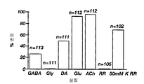
도 3은 퓨라-2(fura-2) 칼슘 이온 이미지에 의해 측정된 격렬하게 해리되고(빈 막대) 분화된(빗금친 막대) E-NCAM+ 세포의 신경전달물질에 대해 반응을 하는 세포수를 막대 그래프로 나타낸 것이다: GABA(Y-아미노 부틸산), Gly(글리신), DA(도파민), Glu(글루타메이트), Ach(아세틸 콜린), RR(쥐(rat)의 링거액), 및 50㎜ K RR(나트륨 이온을 칼륨 이온으로 치환한 쥐(rat)의 링거액).FIG. 3 shows the number of cells responding to neurotransmitters of wildly dissociated (empty bars) and differentiated (hatched bars) E-NCAM + cells measured by fura-2 calcium ion images. Bar graphs are shown: GABA (Y-amino butyric acid), Gly (glycine), DA (dopamine), Glu (glutamate), Ach (acetylcholine), RR (rat Ringer's solution), and 50 mm K RR (Ringer's solution of rats with sodium ions replaced with potassium ions).
도 4는 격렬하게 해리된 E-NCAM+ 세포로부터 Ca2 +의 I340/I380의 비율을 나타낸 것이다.Figure 4 shows the ratio of I 340 / I 380 of Ca 2 + from violently dissociated E-NCAM + cells.
도 5는 분화된 E-NCAM+ 세포로부터 Ca2 +의 I340/I380의 비율을 나타낸 것이다.Figure 5 shows the ratio of I 340 / I 380 of the Ca 2 + from the E-NCAM + cell differentiation.
도 6은 성숙된 신경세포의 마커(marker)가 발현되는 것 대한 단일의 E-NCAM+ 클론을 PCR 분석한 결과를 나타낸 것이다.Figure 6 shows the results of PCR analysis of a single E-NCAM + clone for the expression of markers of mature neurons.
도 7은 퓨라-2(fura-2) 칼슘 이온 이미징에 의한 신경전달물질에 대해 반응을 하는 네 개의 E-NCAM+ 클론에 대한 세포의 백분율을 막대 그래프로 나타낸 것이다: GABA(Y-아미노 부틸산), Gly(글리신), DA(도파민), Glu(글루타메이트), Ach(아세틸 콜린), RR(쥐(rat)의 링거액), 및 50㎜ K RR(나트륨 이온을 칼륨 이온으로 치환한 쥐(rat)의 링거액).FIG. 7 shows a bar graph of the percentage of cells for four E-NCAM + clones that respond to neurotransmitters by fura-2 calcium ion imaging: GABA (Y-amino butyric acid) ), Gly (glycine), DA (dopamine), Glu (glutamate), Ach (acetylcholine), RR (Ringer's solution in rats), and 50 mm K RR (rat with sodium ions replaced with potassium ions) Ringer's solution).
도 8 및 9는 하나의 E-NCAM+ 클론으로부터 기록된 두 가지 세포들로부터 Ca2 +의 I340/I380의 비율을 나타낸 것이다.8 and 9 shows the ratio of I 340 / I 380 of the Ca 2 + from the one of the E-NCAM + two kinds of cells recorded from clone.
도 10은 BRDU를 결합시켜 측정한 것으로 E-NCAM+ 세포의 세포 분열에 있어서 뼈 모포제닉(morphogenetic) 단백질 2(BMP-2)의 효과를 나타낸 것이다.10 shows the effect of bone morphogenetic protein 2 (BMP-2) on cell division of E-NCAM + cells as measured by binding BRDU.
도 11은 BRDU를 결합시켜 측정한 것으로 E-NCAM+ 세포의 세포 분열에 있어서 소닉 헤지호그(sonic hedgehog)(Shh)의 효과를 나타낸 것이다.FIG. 11 shows the effect of sonic hedgehog (Shh) on cell division of E-NCAM + cells as measured by binding BRDU.
도 12는 좌로부터 p75, Isl-1, ChAT, 캘빈딘(calbindin), GAD 및 글루타미나아제의 발현을 검정하기 위하여 생쥐(mouse)의 E-NCAM+ 세포로부터 분리된 총 RNA를 RT-PCR을 통하여 증폭한 결과를 나타낸 것이다.12 shows RT-PCR of total RNA isolated from E-NCAM + cells of mice to assay expression of p75, Isl-1, ChAT, calbindin, GAD and glutaminase from the left. It shows the result of amplification through.
도 13은 좌로부터 네스틴, N-CAM, 뉴로필라멘트-M(NF-M), 마이크로튜블 관련 단백질 2(Map-2), GFAP 및 DM-20/PLP의 발현을 검정하기 위하여 분화된 생쥐(mouse)의 ES 세포로부터 분리된 총 RNA를 RT-PCR을 통하여 증폭한 결과를 나타낸 것이다.Figure 13 shows mice differentiated from the left to differentiate expression of nestin, N-CAM, neurofilament-M (NF-M), microtubule related protein 2 (Map-2), GFAP and DM-20 / PLP. Total RNA isolated from ES cells of mouse) is shown by amplification by RT-PCR.
도 14는 좌로부터 ChAT, p75, islet-1, 캘빈딘, GAD 및 글루타미나아제의 발현을 검정하기 위하여 분화된 생쥐(mouse)의 ES 세포로부터 분리된 총 RNA를 RT-PCR을 통하여 증폭한 결과를 나타낸 것이다.Figure 14 shows amplification of total RNA isolated from ES cells of differentiated mice via RT-PCR to assay the expression of ChAT, p75, islet-1, calvindine, GAD and glutaminase from the left. The results are shown.
기술분야Technical Field
관련 출원에 대한 설명Description of related application
본 출원은 1997. 7. 4일자 미국특허출원 제08/909,435호의 일부계속출원이다.This application is partly filed in US Patent Application Serial No. 08 / 909,435, dated July 4, 1997.
연구 또는 개발을 위한 연합적 후원Combined Sponsorship for Research or Development
본 발명은 정부적인 지원으로 FIRST 장학금과 국립 보건원의 기초 암 연구를 위한 대학원 지원금의 도움을 받아 이루어졌다. 정부는 본 발명에 대한 특정한 권리를 갖는다.The present invention was made possible with the help of government funded FIRST scholarships and graduate grants for basic cancer research at the National Institutes of Health. The government has certain rights in the invention.
배경기술 Background
본 발명은 계통이 예정된 전구체 세포 및 그 물질의 제조 및 이용방법에 관한 것이다. 특히 본 발명은 포유류의 배아로부터 분리된 신경세포로 예정된 전구체(NRP), 신경상피 간세포(NEP cell) 또는 배아 간세포(ES)에 관한 것이다. 상기 NRP들은 자가 증식(self-renewal)을 할 수 있고 신경세포(neuron)로 분화될 수 있는 것이지만, 성상아교세포 및 희돌기아교세포로 이루어진 교질세포(glia)로는 분화되지는 않는다. 또한 상기 NRP 세포들을 발생, 분리, 배양, 감염 및 이식하는 방법을 개시한다.The present invention relates to precursor cells intended for lineage and methods of making and using the materials. In particular, the present invention relates to precursors (NRP), neuroepithelial hepatocytes (NEP cells) or embryonic hepatocytes (ES) that are intended as neurons isolated from mammalian embryos. The NRPs can self-renewal and differentiate into neurons, but they do not differentiate into glia composed of astroglia and oligodendrocytes. Also disclosed are methods of generating, isolating, culturing, infecting and transplanting NRP cells.
간세포들의 특성을 지닌 다분화능(multipotent) 세포들은 중추신경계의 여러 부위 및 여러 분화 단계에서 동정되어왔다(F.H. Gage 외, CNS로부터 간세포의 분리, 특성화 및 이용, 18 Ann. Rev. Neurosci. 159-92(1995); M. Marvin 및 R. McKay, 척추 동물 CNS의 다분화능 간세포, 3 Semin. Cell. Biol. 401-11(1992); 및 R.P. Skoff, 신경아교질 세포(neuroglial cell)의 계통, 2 The Neuroscentist 335-44(1996)). 상기에서 언급한 NEP 세포들은 자가 증식 기능을 지니고 신경세포, 희돌기아교세포 및 성상아교세포으로의 분화 기능을 지니고 있으며, 이러한 점들로 인하여 상기 세포들은 다분화능 간세포라는 것을 의미한다(A.A. Davis 및 Temple, 쥐(rat) 배아의 대뇌 피질에 있는 자가 증식 다분화능 간세포, 362 Nature 363-72(1994); A.G. Gritti 외, 기초 섬유아세포의 성장 인자에 반응하는 성숙한 쥐(mouse) 뇌의 다분화능 간세포의 증식 및 자가 증식, 16 J. Neurosci. 1091- 1100(1996); B.A. Reynolds 외, 신경세포와 성상아교세포를 만드는 다분화능 EGF에 반응하는 스트리아탈 배아 프로제니터(striatal embryonic progenitor), 12 J. Neurosci. 4565-74(1992); B.A. Reynolds & S. Weiss, EGF-반응 포유류 배아의 CNS 전구체가 간세포라는 것을 증명하는 클로날과 모집단 분석, 175 Developmental Biol. 1-13(1996); B.P. Williams 외, 일반적인 전구체 세포로부터 신경세포 및 희돌기아교세포의 발생, 7 Neuron 685-93(1991)).Multipotent cells with characteristics of hepatocytes have been identified at various sites and differentiation stages of the central nervous system (FH Gage et al., Separation, Characterization and Use of Hepatocytes from the CNS, 18 Ann. Rev. Neurosci. 159-92 (1995); M. Marvin and R. McKay, Multipotent Hepatocytes of Vertebrate CNS, 3 Semin. Cell. Biol. 401-11 (1992); and RP Skoff, Lineage of Neuroglial Cells, 2 The Neuroscentist 335-44 (1996). The NEP cells mentioned above have autologous proliferation and differentiation into neurons, oligodendrocytes and astroglia, which means that the cells are multipotent hepatocytes (AA Davis and Temple). , Autologous multipotent hepatocytes in the cerebral cortex of rat embryos, 362 Nature 363-72 (1994); AG Gritti et al., Of multipotent hepatocytes of mature mouse brain in response to growth factors of basal fibroblasts Proliferation and Self-Proliferation, 16 J. Neurosci. 1091-1100 (1996); BA Reynolds et al., Striatal embryonic progenitors in response to multipotent EGF producing neurons and astroglia, 12 J. Neurosci. 4565-74 (1992); BA Reynolds & S. Weiss, clonal and population analysis demonstrating that the CNS precursors of EGF-responsive mammalian embryos are hepatocytes, 175 Developmental Biol. 1-13 (1996); BP Williams et al. , Normal From the precursor cells and neurons diluent protrusion occurs in glial cells, 7 Neuron 685-93 (1991)).
신경계는 정해진 분화 가능성을 갖고 있는 전구체 세포들을 포함하고 있다(T.J. Kilpatric 및 P.F. Bratlett, 생쥐(mouse) 대뇌로부터 클론된 다분화능 전구체는 FGF-2를 요구하는 반면, 교질 전구체들은 FGF-2 또는 EGF로 자극을 받음, 15 J. Neurosci. 3653-61(1995); J. Price 외, 레트로바이러스에 매개된 유전자 전이에 의한 대뇌 신경계의 계통 분석, 84 Developmental Biol. 156-60(1987); B.A. Reynolds 외, 상기서; B.A. Reynold 및 S. Weiss, 상기서; B. Williams, 대뇌 피질의 난핵 부분에 있어서의 전구 세포 타입, 17 BioEssays 391-93(1995); 및 B.P. Williams 외, 상기서). 다분화능 간세포 및 계통이 예정된 전구체 세포들간의 관계는 아직 불명확하다. 계통이 정해진 세포들은 다분화능 세포들로부터 유래될 수 있지만 직접적인 실험적 증거가 없는 신경계에서는 여전히 가설적인 가능성이 있을 수 있을 뿐이다. 또한 다분화능 세포로부터 상기 전구체들을 정제하는 방법도 소개된 바 없다. The nervous system contains precursor cells with defined differentiation potential (TJ Kilpatric and PF Bratlett, multipotent precursors cloned from mouse cerebral cells require FGF-2, while glial precursors are referred to as FGF-2 or EGF). Stimulated, 15 J. Neurosci. 3653-61 (1995); J. Price et al., Lineage Analysis of the Cerebral Nervous System by Retrovirus Mediated Gene Transfer, 84 Developmental Biol. 156-60 (1987); BA Reynolds et al. BA Reynold and S. Weiss, B. Williams, Progenitor Cell Types in the Ovarian Portion of the Cerebral Cortex, 17 BioEssays 391-93 (1995); and BP Williams, et al. The relationship between pluripotent stem cells and progenitor cells scheduled for lineage is still unclear. Lined cells can be derived from pluripotent cells, but there may still be hypothetical possibilities in the nervous system without direct experimental evidence. In addition, a method of purifying the precursors from multipotent cells has not been introduced.
여기에서 참고로 인용된 발명의 명칭이 「NEP(Neuroepithelial Stem) 세포의 발생, 특성화 및 분리」인 미국특허출원 제08/852,744호(1997. 5. 7 출원)에 의하 면, NEP 세포들은 피브로넥틴(fibronectin)위에서 성장하고 섬유아세포 성장인자(FGF)를 필요로 하고, 계배 추출물(CEE)에 아직 특징이 부여되지 않은 성분이 존재하기 때문에 배지에서 미분화된 표현형(phenotype)을 유지하고 증식한다. NEP 세포들의 성장 요소들은 배아 14.5일째의 피질 난할대(ventricular) 부분 세포로부터 분리된 뉴로스피어(neurophere)와는 다르다(B.A. Reynolds 외, 상기서; B.A. Reynolds 및 S. Weiss, 상기서; 국제출원공개 제9615226호; 국제출원공개 제9615224호; 국제출원공개 제9609543호; 국제출원공개 제9513364호; 국제출원공개 제9416718호; 국제출원공개 제9410292호; 및 국제출원공개 제9409119호). 뉴로스피어는 현탁 배지에서 배양되고 CEE 또는 FGF를 요구하지는 않으나 생존을 위해서는 상피 성장인자(EGF)에 의해 영향을 받는다. 비록 FGF가 뉴로스피어의 성장을 일시적으로 지지할 수는 있긴 하나, 상기 뉴로스피어를 장기간 성장시키게 하는 데는 적합하지는 않다. 접착성 배지에서 자라는 NEP 세포들은 FGF에 영향을 받지 않으며, EGF 수용체가 검출 가능한 수준을 표현하지는 않고, 그리고 뉴로스피어가 분리될 수 있는 전 단계인 배아 성장 단계에서 분리된다. 이와 같이, NEP 세포들은 뇌 골수 및 척추의 다분화능 전구체로 나타날 수 있으며, 반면에 뉴로스피어는 피질의 간세포로 나타날 수 있다. 그럼에도 불구하고, NEP 세포들은 중추신경계의 다분화능 간세포 또는 전구세포로부터 계통 제한 원리를 연구하기 위한 시범적인 시스템으로 제공된다. NEP 세포들에 대한 연구로 인하여 명백해진 위와 같은 원리들은 신경세포 및 교질세포의 발생을 위하여 충분히 다분화될 수 있는 모든 CNS 전구체 세포들에 대해 광범위하게 적용될 것이라고 예상된다. 이와 같이, 본 발명은 신경세 포 및 교질세포로 분화될 수 있는 동안 CNS가 유도되는 부위에 관계없이 모든 CNS 전구 세포들에 적용될 수 있다.According to US patent application Ser. No. 08 / 852,744 (filed May 5, 1997), entitled `` Generation, Characterization and Isolation of Neuropepithelial Stem Cells '' (NEP), the NEP cells are fibronectin ( It grows on fibronectin, requires fibroblast growth factor (FGF), and maintains and propagates an undifferentiated phenotype in the medium because of the presence of components that have not yet been characterized in CEE. Growth factors in NEP cells are different from neurospheres isolated from ventricular partial cells at day 14.5 of embryos (BA Reynolds et al., Supra; BA Reynolds and S. Weiss, supra; 9615226; International Application Publication No. 9615224; International Application Publication No. 9609543; International Application Publication No. 9513364; International Application Publication No. 9416718; International Application Publication No. 9410292; and International Application Publication No. 9409119). Neurospheres are cultured in suspension medium and do not require CEE or FGF but are affected by epidermal growth factor (EGF) for survival. Although FGFs may temporarily support the growth of neurospheres, they are not suitable for long-term growth of neurospheres. NEP cells growing in adhesive medium are not affected by FGF, do not express detectable levels of EGF receptors, and are isolated at the embryonic growth stage, which is the stage where neurospheres can be isolated. As such, NEP cells may appear as multipotent precursors of cerebral bone marrow and spine, whereas neurospheres may appear as cortical hepatocytes. Nevertheless, NEP cells serve as a pilot system to study the principle of lineage restriction from pluripotent stem cells or progenitor cells of the central nervous system. The above principles, which are evident from the study of NEP cells, are expected to apply broadly to all CNS precursor cells that can be sufficiently differentiated for the generation of neurons and glial cells. As such, the invention can be applied to all CNS progenitor cells regardless of the site from which the CNS is induced while they can differentiate into neuronal cells and glial cells.
Anderson 및 D.L. Stemple의 미국특허 제5,589,376호는 포유류의 신경분(neural crest) 간세포, 이의 분리 및 클로날 증식에 대한 방법을 개시하고 있으나, 배양된 NEP 세포, 배양된 계통이 예정된 전구체 세포, 및 이를 발생, 분리 및 배양하는 방법에 대해 개시하지는 않고 있다. 신경분 세포들은 말초신경계(PNS)의 신경세포 및 교질세포로 분화되는 반면에, NEP 세포들은 중추신경계(CNS)의 신경세포 및 교질세포로 분화된다.Anderson and D.L. US Pat. No. 5,589,376 to Stemple discloses a method for neural crest hepatocytes, isolation and clonal proliferation of mammals, but cultured NEP cells, cultured lineage precursor cells, and generating, isolating them And a method of culturing is not disclosed. Neuroblasts differentiate into neurons and glial cells of the peripheral nervous system (PNS), while NEP cells differentiate into neurons and glial cells of the central nervous system (CNS).
「계통이 예정된 신경세포 전구체의 분리」에 대한 미국특허출원 제08/909,435호(1997. 7. 4 출원)는 신경세포로 분화될 수는 있지만 교질세포로는 분화되지 않는 신경세포로 예정된 전구(NRP) 세포에 대해 개시하고 있다. 상기 특허출원은 NRP 세포들이 NEP 세포들로부터 분리될 수 있으며, 또한 배아 척수로부터 직접적으로 분리될 수 있다는 것을 개시하였다.U.S. Patent Application Serial No. 08 / 909,435 (filed Jul. 4, 1997) on Separating Neural Cell Precursors Scheduled to Progenitors is a precursor to neurons that can differentiate into neurons but not into glial cells. NRP) cells are disclosed. The patent application discloses that NRP cells can be isolated from NEP cells and also directly from the embryonic spinal cord.
「중추신경계로부터 계통이 예정된 교질세포의 전구체」에 대한 미국특허출원 제08/980,850호(1997. 11. 29 출원)는 희돌기아교세포, A2B5+ 프로세스-베어링(process-bearing) 성상아교세포 및 A2B5- 섬유아세포와 같은 성상아교세포로 분화될 수 있으며, 신경세포로 분화되지는 않는 교질세포로 예정된 전구체(GRP) 세포에 대하여 개시하고 있다. GRP 세포들은 분화된 NEP 세포로부터 분리될 수 있으며, 또한 CNS 조직으로부터 분리될 수 있고, 그리고 성장인자 조건, 형태, 프로제니 (progeny)의 희소돌기교질세포-타입-2 성상아교세포(O-2A) 프로제니터(progenitor) 세포들과는 다르다.U.S. Patent Application No. 08 / 980,850 (filed Nov. 29, 1997) for `` Precursors of Glial Cells Scheduled from the Central Nervous System '' discloses oligodendrocytes, A2B5 + process-bearing astrocytes and A2B5 - discloses precursor (GRP) cells destined for glial cells that can differentiate into astroglia, such as fibroblasts, but do not differentiate into neurons. GRP cells can be isolated from differentiated NEP cells, can also be isolated from CNS tissue, and growth factor conditions, morphology, progeny's oligodendrocyte-type-2 astroglia (O-2A) Different from progenitor cells.
「CNS 및 PNS에 있어서 일반적인 신경세포의 프로제니터」에 대한 미국특허출원 제09/073,881호(1998. 5. 6 출원)는 CNS 및 PNS의 다른 세포들과 마찬가지로 신경분 세포로 분화되는 것으로 유도될 수 있는 NEP 세포에 대하여 개시하고 있다.U.S. Patent Application Serial No. 09 / 073,881 (filed May 5, 1998) on "Progenerators for Neuronal Cells Common in CNS and PNS" induces differentiation into neuronal cells like other cells of CNS and PNS Disclosed are NEP cells that can be.
신경세포로 예정된 전구 세포들은 다른 곳에 나타나는 NEP 세포, GRP 세포, 뉴로스피어 및 신경분 간세포들과는 구별된다. NEP 세포들은 신경세포 또는 교질세포로 분화될 수 있으며, 반면에 NRP는 신경세포로 분화될 수 있지만 교질세포로는 분화되지 않고, 그리고 NEP 및 NRP 세포들은 별개의 세포 마커(cell marker)를 드러내고 있다. GRP 세포들는 교질세포로 분화될 수 있지만 신경세포로 분화되지는 않는다. 상기에서 언급한 바와 같이, 뉴로스피어는 현탁 배지에서 배양되고 CEE 또는 FGF를 필요로 하지는 않지만 생존을 위해서는 EGF에 영향을 받으나, 반면에 NRP 세포들은 접착성 배지에서 배양되고 EGF 수용체가 검출될 수 있는 수준을 나타내지 않는다. 또한 신경분 세포들은 말초신경계의 신경세포 및 교질세포로 분화되지만, NRP 세포들은 중추신경계의 신경세포로 분화된다. NRP 세포들은 폴리시알레이트된(polysialated) 또는 배아 신경세포 접착 분자(E-NCAM)로 나타날 수도 있으나, NEP 세포, 뉴로스피어, GRP 세포 및 신경분 세포들은 그렇지 않다. 그러므로, NRP 세포들은 다른 세포 타입과는 다른 증식 가능성(proliferative potential), 세포 마커(cell marker)의 표현 및 영양 조건을 갖고 있다.Progenitor cells destined for neurons are distinguished from NEP cells, GRP cells, neurospheres and neurostem cells appearing elsewhere. NEP cells can differentiate into neurons or glial cells, whereas NRP can differentiate into neurons but not into glial cells, and NEP and NRP cells reveal distinct cell markers. . GRP cells can differentiate into glial cells but do not differentiate into neurons. As mentioned above, neurospheres are cultured in suspension medium and do not require CEE or FGF but are affected by EGF for survival, whereas NRP cells can be cultured in adhesive medium and EGF receptors can be detected. It does not indicate a level. Neuroblasts also differentiate into neurons and glial cells of the peripheral nervous system, while NRP cells differentiate into neurons of the central nervous system. NRP cells may appear as polysialated or embryonic neuronal adhesion molecules (E-NCAMs), while NEP cells, neurospheres, GRP cells and neuroblast cells are not. Therefore, NRP cells have proliferative potential, expression of cell markers and nutritional conditions that are different from other cell types.
시험관내에서(in vitro) 포유류의 신경세포로 예정된 전구체 세포들을 분리 하고 성장시킴으로써, 이식을 위한 순수한 신경세포 집단의 사용, 특이적으로 선택된 발육 단계에 있어서의 유전자 발견, 및 표적 유전자 치료 등과 같은 치료적·진단적 사용에 대한 세포-특이적 항체의 발생 등을 가능하게 한다. 또한 NRP 세포들은 특이한 특성을 갖는 신경세포의 부차집단 즉, 높은 처리량 분석(throughtout assay)으로 신경전달물질(neurotransmitter) 기능 및 작은 분자들을 분석하는 모토뉴런(motoneuron) 및 다른 신경세포를 생성하는데 사용될 수 있다. 더욱이, NEP 세포 또는 배아 간세포(ES cell)들로부터 NRP 세포를 수득하는 방법은 많은 수의 후-유사분열단계의 신경세포의 준비 원료를 제공하는 것이다. 종양 세포주로부터 수득된 후-유사분열단계의 세포들은 이미 상업적으로 입수 가능하다(예를 들어, Clontech, Palo Alto, CA). 또한 본 발명은 다분화능 NEP 세포들이 어떻게 다양하게 NEP 유도체로 정해지는지를 제시한다. 특히, 포유류의 신경세포로 예정된 전구체 세포들의 성장 및 자가 증식을 가능하게 하는 배양 조건이 적합하기 때문에 상기 포유류의 간세포를 발육시킬 수 있다. 이러한 점은 NEP 유도체의 많은 수의 종양들이 포유류, 특히 사람내에 존재하기 때문에 바람직하다. 포유류의 NEP 세포의 성장에 대한 인식은 사람의 질병(disorder)을 이해하는 데 필수적인 것이다.Treatments such as the use of pure neuronal populations for transplantation, gene discovery at specifically selected developmental stages, and targeted gene therapy, by isolating and growing precursor cells destined for mammalian neurons in vitro Generation of cell-specific antibodies for red and diagnostic use. NRP cells can also be used to generate motoneurons and other neurons that analyze neurotransmitter function and small molecules in a subpopulation of neurons with specific characteristics, ie, high throughput assays. have. Moreover, a method of obtaining NRP cells from NEP cells or ES cells is to provide a raw material for the preparation of a large number of post-mitotic stage neurons. Cells of the post-mitotic stage obtained from the tumor cell line are already commercially available (eg Clontech, Palo Alto, CA). The present invention also suggests how multipotent NEP cells are defined as various NEP derivatives. In particular, it is possible to develop the hepatocytes of the mammal because the culture conditions that allow the growth and self-proliferation of the precursor cells destined for the mammalian neurons are suitable. This is desirable because a large number of tumors of NEP derivatives are present in mammals, especially humans. Recognition of the growth of mammalian NEP cells is essential to understanding human disorder.
본 발명은 앞서 언급한 견지에서 보더라도, 포유류의 신경계로 분화가 예정된 전구체 세포들의 분리된 집단 및 상기 세포들을 발생, 분리, 배양, 감염 및 이식하는 방법에 있어서 상당히 진척된 것이다.In view of the foregoing, the present invention is a significant advance in the isolated population of precursor cells intended to differentiate into the mammalian nervous system and methods of generating, isolating, culturing, infecting and transplanting such cells.
본 발명의 목적은 분리된 포유류의 신경세포로 예정된 전구체 세포들의 순수한 집단 및 그 프로제니(progeny)를 제공하기 위한 것이다. It is an object of the present invention to provide a pure population of precursor cells destined for isolated mammalian neurons and their progeny.
본 발명의 다른 목적은 포유류의 신경세포로 예정된 전구체 세포들을 발생, 분리, 배양 및 재생하는 방법 및 그 프로제니를 제공하기 위한 것이다.Another object of the present invention is to provide a method for generating, isolating, culturing and regenerating precursor cells destined for mammalian neurons and progeny thereof.
본 발명의 또 다른 목적은 신경세포 및 교질세포 둘 다를 발생시킬 수 있는 CNS 다분화능 전구체 세포로부터 신경세포로 예정된 전구체 세포들을 발생시키는 방법을 제공하기 위한 것이다.Another object of the present invention is to provide a method for generating precursor cells destined for neurons from CNS multipotent precursor cells capable of generating both neurons and glial cells.
본 발명의 또 다른 목적은 신경세포로 예정된 전구체 세포들로부터 유도되는 신경세포의 순수한 분화 집단을 제공하기 위한 것이다.Another object of the present invention is to provide a pure differentiation population of neurons derived from precursor cells destined for neurons.
본 발명의 또 다른 목적은 상기 신경세포로 예정된 전구체 세포들을 형질감염시키고 이식하는 방법을 제공하기 위한 것이다.Still another object of the present invention is to provide a method for transfecting and transplanting predetermined precursor cells into the neurons.
이러한 목적들은 포유류의 CNS 신경세포로 예정된 전구체 세포들의 분리된 순수한 집단들을 제공함으로써 달성될 수 있다. 바람직하게 상기 신경세포로 예정된 전구체 세포들은 자가 증식이 가능하고 CNS 신경세포로 분화가 가능하지만 CNS 교질세포로는 분화될 수 없고, 그리고 배아 신경세포 접착 분자(E-NCAM)를 발현하면서도 A2B5 항체에 의해 인식되는 갱글리오시드(ganglioside)를 발현하지는 못한다. 상기 신경세포로 예정된 전구체 세포들은 네스틴(nestin) 또는 β-Ⅲ 튜블린(tubulin)을 발현하게 할 수도 있고, 그렇지 않을 수도 있다. 이와 같이 배아 신경세포 접착 분자(E-NCAM)는 상기 세포들에 대한 명백한 항원이다. 상기 NRP 세포들은 신경전달물질을 방출시키고 상기 물질과 반응하는 신경세포들로 분화될 수 있 다. 상기 신경세포들은 이러한 신경전달물질들에 대한 수용체를 가진다고 할 수 있으며, 상기 세포들은 신경전달물질-합성 효소를 발현하는 능력을 지닌다. 상기 NRP 세포들은 기능 시냅스(synapse)의 형성 및/또는 전기적 활성을 증가시키게 하는 신경세포로 분화될 수 있다. 또한 상기 NRP 세포들은 상기 세포들에 대한 성장인자, 상기 세포들에 대한 분화인자, 상기 세포들에 대한 성숙인자 및 이들의 조합으로 이루어진 군으로부터 선택된 하나 이상의 물질을 안정적으로 발현하게 할 수 있다. 또한 본 발명의 신경세포로 예정된 전구체 세포들은 사람, 사람을 제외한 영장류, 말, 개과, 고양이, 소과, 돼지, 양, 토끼목 및 설치류로부터 선택 및 분리될 수 있다.These objectives can be achieved by providing isolated pure populations of precursor cells destined for mammalian CNS neurons. Preferably, the progenitor cells destined for the neurons are capable of self proliferation and differentiation into CNS neurons but cannot differentiate into CNS glial cells, and express an embryonic neuronal adhesion molecule (E-NCAM) to A2B5 antibodies. It does not express gangliosides as recognized by. Precursor cells destined for the neuron may or may not express nestin or β-III tubulin. As such, embryonic neuronal adhesion molecules (E-NCAMs) are obvious antigens for these cells. The NRP cells can differentiate into neurons that release and react with neurotransmitters. The neurons may be said to have receptors for these neurotransmitters, and the cells have the ability to express neurotransmitter-synthesizing enzymes. The NRP cells can be differentiated into neurons, which increase the formation of functional synapses and / or electrical activity. In addition, the NRP cells can stably express one or more substances selected from the group consisting of growth factors for the cells, differentiation factors for the cells, mature factors for the cells, and combinations thereof. Precursor cells destined for the neurons of the present invention may also be selected and isolated from humans, primates other than humans, horses, canines, cats, bovines, pigs, sheep, rabbits and rodents.
본 발명의 포유류의 CNS 신경세포로 예정된 전구체 세포들의 순수한 집단(population)을 분리하는 방법은 하기 단계로 이루어진다:The method for isolating a pure population of precursor cells destined for CNS neurons of the mammal of the invention consists of the following steps:
(a) 신경세포 및 교질세포를 발생시키는 포유류의 다분화능 CNS 간세포 집단을 분리하고;(a) isolating a multipotent CNS hepatocyte population of mammals that gives rise to neurons and glial cells;
(b) 상기 세포들을 주입하기 위해 배치된 배지내에서 상기 다분화능 CNS 간세포를 배양하여, 분화가 개시되도록 하고;(b) culturing said multipotent CNS hepatocytes in a medium arranged to inject said cells to initiate differentiation;
(c) 신경세포로 예정된 전구체 세포를 한정하는 선택된 항원을 발현하게 하는 세포 부차집단(subpopulation)들을 분화된 세포들로부터 정제하고; 그리고(c) purifying cell subpopulations from differentiated cells to express selected antigens that define precursor cells destined for neurons; And
(d) 고착 성장이 가능하도록 배치된 배지에서 정제된 세포 부차집단들을 배양함.(d) incubating the purified cell subpopulations in a medium arranged to allow fixation growth.
신경세포로 예정된 전구체 세포들을 한정하는 바람직한 선택된 항원은 배아 신경세포 접착 분자(E-NCAM)이다. 바람직하게 상기 NRP 세포들을 정제하는 단계는 특이적 항원 캡춰(capture), 플루어레신, 활성화된 세포 소팅(sorting) 및 자석 구슬 캡춰(magnetic bead capture)로 이루어진 군으로부터 선택된 절차를 포함한다. 특이적 항체 캡춰는 매우 바람직하다. 바람직한 구체예에서, 포유류의 다분화능 CNS 간세포는 신경상피 간세포이다. CNS의 신경상피 간세포의 집단을 분리하기 위한 바람직한 절차는 하기 단계로 이루어진다:Preferred selected antigens that define precursor cells destined for neurons are embryonic neuronal adhesion molecules (E-NCAMs). Preferably, purifying the NRP cells includes a procedure selected from the group consisting of specific antigen capture, flueresin, activated cell sorting and magnetic bead capture. Specific antibody capture is very desirable. In a preferred embodiment, the mammalian multipotent CNS stem cells are neuroepithelial stem cells. A preferred procedure for separating the population of neuroepithelial stem cells of the CNS consists of the following steps:
(a) 신경관 폐쇄(closure) 후, 그러나 신경관의 세포 분화 전 배아 발육 단계에 있는 포유류의 배아로부터 CNS 조직을 제거하고;(a) removing CNS tissue from a mammalian embryo after neural tube closure but before embryonic cell differentiation;
(b) 상기 포유류의 배아로부터 제거된 상기 신경관으로 이루어진 세포들을 분리하고;(b) isolating cells consisting of the neural tube removed from the mammalian embryo;
(c) 섬유아세포 성장인자 및 계배 추출물의 효과적인 양으로 이루어진 신경상피 간세포를 토대(substratum)상 공급-세포-독립(feeder-cell-infddependent) 배지 및 고착·성장을 위한 배지에 평판하고; 그리고(c) Neural epithelial hepatocytes consisting of an effective amount of fibroblast growth factor and germline extract are plated on a substratum feeder-cell-infddependent medium and medium for fixation and growth; And
(d) 신경상피 간세포를 성장시키기 위한 온도 및 대기에서 상기 평판된 세포들을 배양함.(d) incubating the plated cells at temperature and atmosphere to grow neuroepithelial stem cells.
바람직하게 상기 포유류의 배아는 사람, 사람을 제외한 영장류, 말, 개과, 고양이, 소과, 돼지, 양, 토끼목 및 설치류로 이루어진 군으로부터 선택된다. 또한 상기 토대(substratum)는 피브로엑틴(fibronectin), 비트로넥틴(vitronectin), 라미닌 및 RGD 펩티드로 이루어진 군으로부터 선택된다. 바람직한 구체예에서, 상기 배지는 섬유아세포성장인자 및 뉴로트로핀 3(NT-3)의 효율적인 양으로 이루어진다.Preferably the embryo of the mammal is selected from the group consisting of humans, non-human primates, horses, canines, cats, bovines, pigs, sheep, rabbits and rodents. The substratum is also selected from the group consisting of fibronectin, vitronectin, laminin and RGD peptides. In a preferred embodiment, the medium consists of an efficient amount of fibroblast growth factor and neurotrophin 3 (NT-3).
포유류의 CNS 신경세포로 예정된 전구체 세포의 순수한 집단을 분리하기 위한 방법은 하기 단계로 이루어진다:The method for isolating a pure population of progenitor cells destined for mammalian CNS neurons consists of the following steps:
(a) 신경관 폐쇄(closure) 후, 그러나 신경관의 교질세포 및 신경세포 분화 전의 배아 발생 단계에 있는 포유류의 배아로부터 CNS 조직의 샘플을 제거하고;(a) removing a sample of CNS tissue from a mammalian embryo that is in the embryonic development stage after neural tube closure but prior to neuronal glia and neuronal differentiation;
(b) 상기 포유류의 배아로부터 제거된 CNS 조직의 샘플로 이루어진 세포들을 해리시키고;(b) dissociating cells consisting of a sample of CNS tissue removed from the mammalian embryo;
(c) 신경세포로 예정된 전구체 세포를 명확하게 하도록 선택된 항원을 발현하는 부차집단을 상기 해리된 세포로부터 정제하고;(c) purifying subpopulations expressing selected antigens from said dissociated cells to clarify progenitor cells destined for neurons;
(d) 신경세포로 예정된 전구체 세포의 토대(substratum)상 공급-세포-독립(feeder-cell-independent) 배지 및 고착·성장을 위한 배지에서 상기 정제된 세포 부차집단을 평판배양하고; 그리고(d) plate the purified cell subpopulations in a feeder-cell-independent medium on a substratum of the precursor cells destined for neurons and a medium for fixation and growth; And
(e) 상기 신경세포로 예정된 전구체 세포를 성장시키기 위한 온도 및 대기에서 상기 평판된 세포들을 배양함.(e) incubating the plated cells at a temperature and atmosphere for growing precursor cells destined for the neurons.
바람직하게 상기 신경세포로 예정된 전구체 세포를 한정하는 선택된 항원은 배아 신경세포 접착 분자(E-NCAM)이다. 또한 바람직한 정제 단계는 특이적 항원 캡춰(capture), 플루어레신, 활성화된 세포 소팅(sorting) 및 자석 구슬 캡춰(magnetic bead capture)로 이루어진 군으로부터 선택된 절차를 포함한다. 특이적 항체 캡춰가 매우 바람직하다. 상기 포유류의 배아는 사람, 사람을 제외한 영장류, 말, 개과, 고양이, 소과, 돼지, 양, 토끼목 및 설치류로 이루어진 군으로부터 선택된다.Preferably the selected antigen that defines the precursor cell destined for the neuron is an embryonic neuronal adhesion molecule (E-NCAM). Preferred purification steps also include a procedure selected from the group consisting of specific antigen capture, fluresin, activated cell sorting, and magnetic bead capture. Specific antibody capture is very desirable. The mammalian embryo is selected from the group consisting of humans, non-human primates, horses, canines, cats, bovines, pigs, sheep, rabbits and rodents.
후-유사분열단계의 신경세포를 수득하는 방법은 하기 단계로 이루어진다:The method for obtaining neurons in the post-mitotic stage consists of the following steps:
(a) 신경세포로 예정된 전구체 세포들을 공급하고, 증식 조건내에서 신경세포로 예정된 전구체 세포들을 배양하고; 그리고(a) feeding progenitor cells destined for neurons and culturing progenitor cells destined for neurons in proliferative conditions; And
(b) 상기 신경세포로 예정된 전구체 세포의 증식 조건을 배양 조건으로 변화시키고, 그럼으로써 후-유사분열단계의 신경세포로 분화될 수 있는 신경세포로 예정된 전구체 세포들을 수득함.(b) changing the proliferative conditions of the progenitor cells destined for the neurons to culture conditions, thereby obtaining precursor cells destined for the neurons capable of differentiating into neurons in the post-mitotic stage.
바람직한 상기 배양 조건의 변화는 기초배지에 레티노산을 첨가하거나 기초배지에 유사분열인자를 회수하는 것을 포함한다. 상기 유사분열인자는 섬유아세포성장인자이다. 또한 배양조건의 변화는 기초배지에 신경세포의 성숙인자를 첨가하는 것을 포함할 수 있다. 바람직한 신경세포의 성숙인자는 소닉 헤지호그(sonic hedgehog), BMP-2, BMP-4, NT-3, NT-4, CNTF, LIF, 레티노산, 뇌-유도된 뉴로트로픽 인자(BDNF) 및 이들의 조합으로 이루어진 군으로부터 선택된다.Preferred changes in the culture conditions include adding retinoic acid to the basal medium or recovering the mitotic factor to the basal medium. The mitotic factor is a fibroblast growth factor. In addition, the change in culture conditions may include the addition of mature factors of neurons to the basal medium. Preferred neuronal maturation factors are sonic hedgehog, BMP-2, BMP-4, NT-3, NT-4, CNTF, LIF, retinoic acid, brain-derived neurotropic factors (BDNF) and their Is selected from the group consisting of
본 발명의 다른 바람직한 구체예는 여기에서 언급되는 상기 포유류의 CNS 신경세포로 예정된 세포들로 이루어진 분리된 세포성 조성물을 포함한다. 본 발명의 다른 바람직한 구체예는 상기 조성물의 치료적으로 효과적인 양 및 제약학적으로 수용가능한 담체를 포함한다.Another preferred embodiment of the present invention includes an isolated cellular composition consisting of cells destined for the CNS neurons of the mammal mentioned herein. Another preferred embodiment of the present invention includes a therapeutically effective amount of the composition and a pharmaceutically acceptable carrier.
포유류에 있어서 신경세포성 질환(disorder)을 치료하는 방법은 여기에서 언급되는 상기 포유류의 CNS 신경세포로 예정된 세포들로 이루어진 분리된 세포성 조성물의 치료적으로 효과적인 양을 상기 포유류에게 투여하는 것을 포함한다. 포유류의 신경적으로 가벼운 질병을 치료하는 다른 방법은 상기 제약학적 조성물 및 제 약학적으로 수용 가능한 담체를 치료적으로 효과적인 양으로 상기 포유류에게 투여하는 것을 포함한다. 상기 조성물은 근육내 투여, 수막강내 투여, 복강내 투여, 정맥내 투여 및 이들의 조합으로부터 선택된 일반적인 방법으로 투여된다. 이와 같은 방법은 분화인자, 성장인자, 세포성숙인자 및 이들의 조합으로 이루어진 군으로부터 선택된 것들을 투여하는 것을 포함한다. 상기 분화인자들은 레티노산, BMP-2, BMP-4 및 이들의 조합으로 이루어진 군으로부터 선택되는 것이 바람직하다.A method of treating neuronal disorders in a mammal comprises administering to the mammal a therapeutically effective amount of an isolated cellular composition of cells destined for the mammalian CNS neurons referred to herein. do. Another method of treating a neurologically mild disease of a mammal comprises administering the pharmaceutical composition and a pharmaceutically acceptable carrier to the mammal in a therapeutically effective amount. The composition is administered by a general method selected from intramuscular administration, intramedullary administration, intraperitoneal administration, intravenous administration and combinations thereof. Such methods include administering those selected from the group consisting of differentiation factor, growth factor, cytogenic factor and combinations thereof. The differentiation factor is preferably selected from the group consisting of retinoic acid, BMP-2, BMP-4 and combinations thereof.
신경 퇴행성 증후군(neurodegenerative symptom)들을 치료하기 위한 방법은 하기 단계로 이루어진다:The method for treating neurodegenerative symptoms consists of the following steps:
(a) 신경세포로 예정된 전구체 세포의 순수한 집단을 마련하고;(a) preparing a pure population of precursor cells destined for neurons;
(b) 상기 신경세포로 예정된 전구체 세포를 성장인자, 신경전달물질, 신경전달물질 합성효소, 뉴로펩티드, 뉴로펩티드 합성 효소, 또는 자유-라디칼 매개된 손상에 대해 보호를 할 수 있는 물질로 유전적으로 형질전환시키고, 그럼으로써 상기 성장인자, 신경전달물질, 신경전달물질 합성효소, 뉴로펩티드, 뉴로펩티드 합성 효소, 또는 자유-라디칼 매개된 손상에 대해 보호를 할 수 있는 물질을 발현하는 교질세포로 예정된 전구체 세포의 형질전환된 부차집단을 생성시키고; 그리고(b) The precursor cells destined for the neurons are genetically protected with growth factors, neurotransmitters, neurotransmitter synthetases, neuropeptides, neuropeptide synthetases, or substances capable of protecting against free-radical mediated damage. Glial cells that are transformed and thereby express the growth factor, neurotransmitter, neurotransmitter synthase, neuropeptide, neuropeptide synthase, or a substance capable of protecting against free-radical mediated damage Generating transformed subpopulations of precursor cells; And
(c) 상기 포유류에 신경세포로 예정된 전구체 세포의 상기 형질전환된 집단의 효과적인 양을 투여함.(c) administering to said mammal an effective amount of said transformed population of precursor cells destined for neurons.
신경학적 활성에 대한 방법 또는 스크리닝 화합물은 하기 단계로 이루어진다:Methods or screening compounds for neurological activity consist of the following steps:
(a) 시험관내에서(in vitro) 배양된 신경세포로 예정된 전구체 세포나 그 유 도체, 또는 이들의 혼합물의 순수한 집단을 마련하고;(a) preparing a pure population of precursor cells or their derivatives, or mixtures thereof, that are intended to be neurons cultured in vitro ;
(b) 다양한 투약량으로 선택된 화합물을 상기 세포나 그 유도체, 또는 이들의 혼합물에 노출시키고; 그리고(b) exposing the selected compound at various dosages to the cells or derivatives thereof, or mixtures thereof; And
(c) 선택된 시간 동안 상기 선택된 화합물에 대한 상기 세포나 그 유도체, 또는 이들의 혼합물의 반응을 점검함.(c) checking the response of said cell or derivative thereof, or mixtures thereof to said selected compound for a selected time.
신경세포성 또는 신경 퇴행성 질병을 치료하는 방법은 신경세포로 예정된 전구체 세포나 그 유도체, 또는 이들의 혼합물의 효과적인 양을 상기 치료가 필요한 포유류에게 투여하는 것으로 이루어진다. 상기 신경세포로 예정된 전구체 세포나 그 유도체, 또는 이들의 혼합물은 이종 공여체(heterologous donor) 또는 자기 공여체(autologous donor)로부터 선택될 수 있다. 상기 공여체는 태아, 청소년 또는 성인의 것일 수 있다.The method of treating neuronal or neurodegenerative diseases consists of administering to a mammal in need of such treatment an effective amount of a precursor cell or derivative thereof, or a mixture thereof, destined for a neuron. Precursor cells destined for the neurons, derivatives thereof, or mixtures thereof may be selected from heterologous donors or autologous donors. The donor may be of fetus, adolescent or adult.
포유류의 CNS 신경세포로 예정된 전구체 세포의 순수한 집단을 분리해내는 방법은 하기 단계로 이루어진다:The method of isolating a pure population of predetermined precursor cells into mammalian CNS neurons consists of the following steps:
(a) 포유류의 배아 간세포의 샘플을 마련하고;(a) preparing a sample of mammalian embryonic stem cells;
(b) 상기 포유류의 배아 간세포로부터 신경세포로 예정된 전구체 세포를 한정하는 선택된 항원을 발현하는 부차집단을 정제하고;(b) purifying a subpopulation expressing selected antigens defining precursor cells destined for neurons from embryonic stem cells of said mammal;
(c) 상기 정제된 부차집단을 신경세포로 예정된 전구체 세포의 토대(substratum)상 공급-세포-독립(feeder-cell-independent) 배지 및 고착·성장을 위한 배지에서 평판배양하고; 그리고(c) the purified subpopulations are plated in a feeder-cell-independent medium on a substratum of precursor cells destined for neurons and a medium for fixation and growth; And
(d) 상기 신경세포로 예정된 세포를 성장시키기 위한 온도 및 대기에서 상기 평판된 세포들을 배양함.(d) incubating the plated cells at a temperature and atmosphere for growing cells destined for the neurons.
본 발명의 신경세포로 예정된 전구체 세포 및 이들의 제조 및 사용 방법을 기술하기에 앞서, 본 발명은 특정한 배열, 공정 과정 및 물질들에 국한되는 것은 아니며, 상기 배열, 공정 과정 및 물질들은 다양할 수 있다. 또한 본 발명의 보호 범위는 첨부된 특허청구범위 및 그것의 균등한 것에까지 미치기 때문에, 여기에서 이용되는 기술은 단지 특정 실시예를 기술하기 위한 목적으로 사용되는 것이며 이에 제한되는 것은 아니다. Prior to describing precursor cells destined for the neurons of the present invention and methods of making and using them, the present invention is not limited to particular arrangements, processes and materials, which arrangements, processes and materials may vary. have. Furthermore, since the protection scope of the present invention extends to the appended claims and their equivalents, the technology used herein is for the purpose of describing particular embodiments only and is not limited thereto.
본 명세서 및 첨부된 특허청구범위에서 사용된 대상물을 지칭하는 용어는 본 발명의 문맥상에서 특별히 언급하지 않는 이상 복수의 지시 대상물을 포함하는 것이다. 이와 같이, 예를 들어, "배아"라는 용어는 둘 이상의 배아를 지칭할 수도 있으며, "유사분열 촉진인자(mitogen)"는 둘 이상의 유사분열 촉진인자의 혼합물을 지칭하는 것일 수도 있고, 그리고 "인자"란 둘 이상의 인자의 혼합물을 지칭하는 것일 수도 있다.The term designating an object used in the specification and the appended claims includes a plurality of indicating objects unless specifically stated in the context of the present invention. As such, for example, the term “embryo” may refer to two or more embryos, and “mitogen” may refer to a mixture of two or more mitotic promoters, and “factor” May also refer to a mixture of two or more factors.
본 발명을 개시하고 청구함에 있어서 하기의 전문 용어는 다음에서 정의한 바와 같이 사용될 것이다.In describing and claiming the present invention, the following terminology will be used as defined below.
여기에서 사용되는 "자가 증식"이란 예를 들어 신경상피 간세포(neuroepithelial stem cell)가 두 개의 딸세포를 생산하기 위하여 분할되어 상기 딸세포 중 적어도 하나가 다분화능 신경상피 간세포로 될 수 있는 기능을 의미하거 나, 또는 신경세포로 예정된 전구체 세포(NRP cell)가 분할되어 두 개의 딸세포를 생산하여 적어도 하나가 신경세포로 예정된 전구체 세포로 될 수 있는 기능을 의미하는 것이다.As used herein, "self-proliferating" means, for example, the ability of neuroepithelial stem cells to be split to produce two daughter cells such that at least one of the daughter cells becomes a multipotent neuroepithelial stem cell. In other words, a neuron-predetermined precursor cell (NRP cell) is divided to produce two daughter cells to mean a function that can be at least one precursor cell to the neuron.
여기에서 사용되는 "클론 밀도(clonal density)" 및 이와 유사한 용어는 선택된 배양 디쉬에 평판배양되는 경우 단일 및 비-침해(non-impinging) 세포가 분리될 만큼 낮은 밀도를 의미한다. 대표적인 상기 클론 밀도는 약 225 세포/100㎜ 배양 디쉬이다.As used herein, “clonal density” and similar terms mean a density low enough to separate single and non-impinging cells when plated in selected culture dishes. Representative clone density is about 225 cells / 100 mm culture dish.
여기에서 사용되는 "공급-세포-독립 고착 배양(feeder-cell-independent adherent culture)" 및 이와 유사한 용어가 의미하는 바는 대상 조직으로부터 취한 세포가 첨가되는 배양 디쉬에 우선 평판하여, 다른 세포 층이 존재하지 않는 상태의 시험관내에서(in vitro) 세포를 성장시키는 것을 말한다. 공급 세포 배양에 있어서, 상기 공급 세포는 대상 조직으로부터 취한 세포들을 부착시키기 위한 토대(substratum)를 제공하고, 추가적으로 유사분열 촉진인자 및 생존인자로써 공급된다. 여기에서 사용되는 공급-세포-독립 고착 배양은 화학적으로 정의된 토대, 예를 들어 피브로넥틴을 사용하고, 유사분열 촉진인자 또는 생존인자는 정제된 인자 또는 다른 세포 또는 조직으로부터 취해진 추출물(crude extracts)을 함유한 액체 배양 배지를 보충하기 위하여 제공된다. 그러므로 공급-세포-독립 배양에 있어서, 상기 배양 디쉬내의 세포들은 대상 조직으로부터 유도된 초기 세포(primarily cell)들이고, 대상 조직으로부터 유도된 세포들의 성장을 지지하도록 요구된 다른 세포 형태들을 포함하지는 않는다.As used herein, " feeder-cell-independent adherent culture " and similar terms mean that the other cell layer is preferentially plated in a culture dish to which cells taken from the target tissue are added. It refers to growing cells in vitro in a nonexistent state. In feeder cell culture, the feeder cell provides a substratum for attaching cells taken from the subject tissue and is additionally supplied as a mitotic promoter and survival factor. The feed-cell-independent fixation culture used herein uses a chemically defined foundation, for example fibronectin, and the mitotic promoter or survivor may be purified from purified factors or crude extracts taken from other cells or tissues. It is provided to replenish the containing liquid culture medium. Thus, in feed-cell-independent cultures, the cells in the culture dish are primarily cells derived from the subject tissue and do not contain other cell types required to support the growth of cells derived from the subject tissue.
여기에서 사용되는 "효과적인 양"이란 비독성이며, 바람직한 효과 및 기능을 수행하기 위해 제공될 수 있을 만큼의 성장인자, 생존인자 또는 다른 인자의 효과적인 양을 의미한다. 예를 들어, 여기에서 사용되는 FGF의 효과적인 양이란 다른 본질적인 영양원 및 인자 등이 조합되어 사용되는 경우 NEP 세포의 자가 재생 및 증식을 위하여 선택된 양을 의미한다. 이식을 위한 NRP 세포나 그 유도체, 또는 이들의 혼합물의 효과적인 양은 선택된 효과를 얻기 위하여 충분한 세포의 양 또는 수를 의미한다. NRP 세포들은 일반적으로 약 5∼50,000 세포/㎕의 농도로 투약될 것이다. 이식은 일반적으로 주사 부위당 약 15㎕까지로 이루어질 것이다. 그러나 중추신경계의 외과적 수술과 함께 이루어지는 이식은 상기와 같은 농도에서 여러 번 행하여질 수 있다. 이와 같이, 이식에 사용되는 세포 수는 단지 실용적인 면에서만 제한되고, 상기 수들은 본 발명의 기술분야의 통상의 지식을 가진 자에 의하여 과도한 실험을 하지 않고도 결정될 수 있다.As used herein, an "effective amount" is nontoxic and refers to an effective amount of growth factor, survival factor or other factor that can be provided to perform the desired effect and function. For example, an effective amount of FGF as used herein means an amount selected for self-renewal and proliferation of NEP cells when used in combination with other essential nutrients, factors, and the like. An effective amount of NRP cells or derivatives thereof, or mixtures thereof for transplantation means an amount or number of cells sufficient to achieve the selected effect. NRP cells will generally be administered at a concentration of about 5-50,000 cells / μl. Implantation will generally consist of up to about 15 μl per injection site. However, transplantation with surgical intervention of the central nervous system may be performed several times at such concentrations. As such, the number of cells used for transplantation is limited only in practical terms and the numbers can be determined without undue experimentation by one of ordinary skill in the art.
여기에서 사용되는 NRP 세포의 "유도체"란 유전자 형질도입, 분화 또는 이와 유사한 공정에 의하여 시험관내에서(in vitro) NRP 세포로부터 유도된 세포를 의미하는 것이다.As used herein, “derivative” of NRP cells refers to cells derived from NRP cells in vitro by gene transduction, differentiation or similar processes.
여기에서 사용되는 "NRP 세포를 포유류에게 투여하는 것"이란 수용체(recipient)의 CNS 조직 가까이 또는 CNS 조직 내로 이식 또는 삽입한다는 의미이다. 상기 투여는 외과적 수술 즉, 본 발명의 기술분야의 통상의 지식을 가진 자에게 알려진 주입 캐뉼라 및 바늘 등을 수단으로 하는 모든 방법에 의해 이루어진다.As used herein, "administering NRP cells to a mammal" means transplanting or inserting into or near the CNS tissue of a recipient. The administration is by any method by means of surgical operations, i.e., injection cannula, needles, and the like, which are known to those skilled in the art.
여기에서 사용되는 "이종(heterologous)"이란 이식 수용체와는 다른 개체, 조직 또는 세포를 의미하는 것이다. 이식 공여체는 이식 수용체와 같이 동종 또는 이종으로부터 취하여질 수 있다. 예를 들면, 이식을 위한 NRP 세포의 이종 공여체는 이식 수용체와 같이 다른 종으로부터 취하여질 수 있다.As used herein, "heterologous" refers to an individual, tissue or cell that is different from the transplant receptor. Transplant donors can be taken from allogeneic or heterologous, such as transplantation receptors. For example, heterologous donors of NRP cells for transplantation can be taken from other species, such as transplantation receptors.
여기에서 사용되는 "자기(autologous)"란 신체내에서 자가 발생적인 것을 의미한다. 예를 들어, 이식을 위한 조직 또는 세포의 자기 공여체는 이식을 받는 동일 개체이다. 또 다른 예로는, 자기 세포는 개체내에서 기인하며, 전이되고, 이식되는 세포들이다. 생체내(in vitro) 조작은 세포를 회수하고 상기 세포 또는 유도체를 이식하는 그 사이에서 이루어질 수 있으나, 이식 전에 필수적으로 요구되는 것은 아니다.As used herein, "autologous" means autogenous in the body. For example, the magnetic donor of tissue or cell for transplantation is the same individual undergoing transplantation. In another example, magnetic cells are cells that originate from, metastasize, and transplant in an individual. In vitro manipulations can be made between harvesting cells and transplanting the cells or derivatives, but are not required prior to transplantation.
여기에서 사용되는 "형질전환", "형질도입" 및 "형질감염" 등은 삽입 또는 전이의 방법에 관계없이, 유전자 또는 유전자들을 NRP 세포내로 삽입 또는 전이시키는 것을 의미한다. 그러므로 형질전환은 칼슘 인산 형질감염, DEAE-덱스트란 형질감염, 폴리브렌 형질감염, 일렉트로포레이션(electroporation), 리포펙션(lipofection) 및 바이러스 감염 등과 같이 당업계에 알려진 모든 다른 방법에 의하여 이루어질 수 있다.As used herein, "transformation", "transduction", "transfection" and the like refer to the insertion or transfer of a gene or genes into NRP cells, regardless of the method of insertion or metastasis. Therefore, transformation can be accomplished by any other method known in the art, such as calcium phosphate transfection, DEAE-dextran transfection, polybrene transfection, electroporation, lipofection and viral infections. .
본 발명은 쥐 및 사람들로부터 분리된 신경세포로 예정된 전구체 세포들을 이용하는 것에 관한 것이다. 그러나 본 발명은 모든 포유류의 신경세포로 예정된 전구체 세포들을 포함하는 것이며, 쥐나 사람의 신경세포로 예정된 전구체 세포들에 한정되는 것은 아니다. 포유류의 신경세포로 예정된 전구체 세포들은 사람, 사람을 제외한 영장류, 말, 개과, 고양이, 소과, 돼지, 양 및 토끼류 등으로부터 선 택 및 분리될 수 있다.The present invention relates to the use of precursor cells destined for neurons isolated from mice and humans. However, the present invention includes precursor cells destined for all mammalian neurons, and is not limited to precursor cells destined for rat or human neurons. Precursor cells destined for mammalian neurons may be selected and isolated from humans, non-human primates, horses, canines, cats, bovines, pigs, sheep and rabbits.
중추신경계의 다분화능 간세포는 직접적으로 또는 계통이 예정된 중간 전구체의 발생을 통하여 분화된 신경세포 및 교질세포를 발생하게 할 수 있다. 망막이 분화됨에 있어서, 다분화능 망막 전구체는 중간 전구체가 존재하지 않도록 최종 분열단계에서 조차도 분화된 세포들의 모든 조합들을 발생시킬 수 있다. 중추신경계의 다른 부분에서는, 이와는 반대로 레트로바이러스를 표지시켜 연구한 결과 하나의 세포 타입 또는 제한된 수의 세포 타입을 발생시키는 계통 예정된 전구체의 존재가 암시적으로 확인되었다. 상기 이분화능(bipotential) 희돌기아교세포-타입-2-성상아교세포(O-2A) 및 신경세포 전구체와 같은 중간 단계 전구체는 조직 배양 연구에 개시된 바 있다. 그러나 아직은 신경세포 및 교질세포로 분화가 가능한 다분화능 배아 또는 성인 간세포, 또는 다른 간세포로부터 취해진 중간 계통 예정된 전구체의 발생이 최근에 출원된 미국특허출원 제08/909,435호(1997. 7. 4 출원)까지만 보더라도 발견되지는 않았다. 이와 같이 배지에서 동정된 다분화능 간세포, 및 생체내(in vivo) 및 시험관내(in vitro)에서 동정된 감작 전구체(committed precursor)와의 계통 관계는 지금까지 발견되지 않았다. 발육(development)될 수 있는 모델들은 (1) 계통적으로 관련된 세포를 보여주는 다분화능 및 더욱 감작된 간세포(committed stem cell) 또는 (2) 분화의 독립 통로를 나타내는 세포들을 포함하였다.Multipotent hepatocytes of the central nervous system can allow the generation of differentiated neurons and glial cells either directly or through the generation of predetermined intermediate precursors. In retinal differentiation, multipotent retinal precursors can give rise to all combinations of differentiated cells even in the final dividing stage such that no intermediate precursor is present. In other parts of the central nervous system, on the contrary, retroviral labeling studies have implicitly identified the presence of lineage predetermined precursors that give rise to one cell type or a limited number of cell types. Intermediate precursors such as bipotential oligodendrocyte-type-2-astroglia (O-2A) and neuronal precursors have been disclosed in tissue culture studies. However, U.S. Patent Application No. 08 / 909,435 (filed Jul. 4, 1997), which has recently been filed for the generation of intermediate lined precursors taken from multipotent embryos or adult hepatocytes or other hepatocytes capable of differentiating into neurons and glial cells. It wasn't found until I saw it. Thus no phylogenetic relationship with the multipotent hepatocytes identified in the medium and the committed precursors identified in vivo and in vitro have not been found to date. Models that can be developed included (1) pluripotents showing systematically related cells and more sensitized stem cells or (2) cells showing independent pathways of differentiation.
쥐(rat) 척수를 발육시키는 것은 상기 분화를 연구하는 것에 대한 이상적인 모델을 보여준다. 배아 10.5일째에서는, 미방(caudal) 신경관이 생체내(in vivo) 및 시험관내(in vitro)에서 네스틴 면역반응성 분할 세포의 동종 집단으로 나타난다. 상기 초기 동종 세포들은 특징적인 공간적 그리고 시간적 프로필(profile)에 있어서 신경세포, 희소돌기세포 및 성상돌기세포를 발생시키기 위하여 며칠에 걸쳐서 형태를 갖추어간다(patterned). 신경조직발생(neurogenesis)은 등배 경사(ventro-dorsal gradient)를 유도하여 쥐(rat)에 있어서 배아 13.5일째에 후유사분열을 진행시키고 초기 신경세포를 제공한다. 신경조직발생은 이틀 동안 더 진행되어 희돌기아교세포 전구체의 분화를 유도하고 성상돌기아교세포의 분화를 유도한다.Developing a rat spinal cord represents an ideal model for studying the differentiation. At embryonic day 10.5, the caaudal neural tube appears as a homogeneous population of nestin immunoreactive dividing cells in vivo and in vitro . The early allogeneic cells are patterned over several days to generate neurons, oligodendrocytes, and astroids in a characteristic spatial and temporal profile. Neurogenesis induces a ventro-dorsal gradient to progress posterior mitosis at 13.5 days of embryos in rats and provide early neurons. Neurogenesis progresses for two more days to induce differentiation of oligodendrocyte precursors and to induce differentiation of astrocytes.
시험관내에서(in vitro) 더 늘어난 기간 동안 분화된 세포로써 쥐(rat)의 배아 10.5일째로부터 분리된 신경상피 간세포(NEP cell)를 성장시키는 방법은 미국특허출원 제08/852,744호에서 개시하고 있으며, 더 나아가 CNS내의 세가지 주요 세포 타입을 발생시킬 수 있는 집단들을 제시하였다. 이와 같은 NEP 세포들은 매개 신경모세포(intermediate neuroblast)를 통해 또는 직접적으로 종결 분화의 한 부분과 같이 신경세포로 분화될 수 있는 분열되는 다분화능 간세포로 나타난다. 신경세포들이 매개체 또는 더욱 예정된 전구체를 통하여 NEP 세포들로부터 분화되었는지에 대하여 측정하기 위해, NEP 세포들의 분화 배지로부터 취한 면역학적으로 한정된 집단의 종류를 분리하여 특화하였다. 여기에서는 형태학적 그리고 표현형적으로 NRP들과 동일성이 있는 세포들을 NEP 세포 배지로부터 분리할 수 있다는 것을 보여주었다. 클로날 분석(clonal analysis)은 각각의 NEP 세포들이 신경세포 전구체의 발생을 통하여 신경세포를 발생시킨다는 것을 보여주고, 그리고 신경세포로 예정되 고 교질세포로 예정된 전구체를 발생시킨다는 것을 보여준다. 더 나아가 E-NCAM+(양성 배아 신경세포 접착 분자) 세포들이 배아 13.5일째에 신경관 배지에서 나타나는 것을 보여주었으며, 상기 세포들이 유사분열을 한다는 것과, 성상아교세포 또는 희돌기아교세포와 같은 것은 아니지만 다양한 신경세포 표현형을 발생시킬 수 있는 자가 증식성 간세포라는 것을 보여주었다. 이와 같이 신경세포로 예정된 전구체(NRP)들은 생체내(in vivo) 신경세포의 분화의 단계에서 동정될 수 있다. 더욱이 NRP는 생쥐(mouse)의 배아, 생쥐 배아 간세포(ES cell) 및 사람의 배아 척수로부터 분리되고 배양될 수 있다는 것을 제시한다. 이와 같은 결과들은 다분화능 및 신경세포로 예정된 간세포들 사이에 직접적인 계통 관계가 존재한다는 것을 나타내는 것이며, 신경세포의 분화가 발생 운명에 있어서 커다란 제한요소를 포함한다는 것을 제시한다.A method for growing neural epithelial cells isolated from 10.5 days of rat embryos as differentiated cells for an extended period of time in vitro is disclosed in US patent application Ser. No. 08 / 852,744. Furthermore, we have proposed populations that can generate three major cell types in the CNS. Such NEP cells appear as dividing multipotent hepatocytes that can differentiate into neurons either through mediated neuroblasts or directly as part of termination differentiation. In order to determine if neurons differentiated from NEP cells via mediators or more predetermined precursors, a special type of immunologically defined population was taken from the differentiation medium of NEP cells. Here we have shown that cells that are morphologically and phenotypicly identical to NRPs can be isolated from NEP cell medium. Clonal analysis shows that each NEP cell generates neurons through the generation of neuronal precursors, and shows that they produce precursors that are intended to be neurons and are intended to be glial cells. Furthermore, E-NCAM + (positive embryonic neuronal adhesion molecule) cells have been shown to appear in neural tube media on day 13.5 of the embryo, and the cells are mitotic, but not as astrocytes or oligodendrocytes. It has been shown to be autologous hepatocytes capable of generating neuronal phenotypes. As such, precursors (NRPs) destined for neurons may be identified at the stage of differentiation of neurons in vivo . Moreover, NRP suggests that it can be isolated and cultured from mouse embryos, mouse embryonic hepatocytes (ES cells) and human embryonic spinal cord. These results indicate that there is a direct lineage relationship between pluripotency and hepatocytes destined for neurons, suggesting that differentiation of neurons includes a large limiting factor in their fate.
도 1은 척수 분화에 대한 모델을 보여주는 것이다. 상기 모델은 신경분(neural crest)의 조혈 및 분화에 대하여 제시된 것과 유사하다(D.J. Anderson의 「신경분 계통 문제: 조혈과정이란?」, 3 Neuron 1-12(1989)). 상기 모델에 의하면, NEP 세포(10)는 미방 신경분의 동종 세포 집단으로 나타나며, 상기 신경분은 네스틴(즉, 네트틴+(nestin+))을 발현시키지만 다른 계통 표지(lin-)를 발현시키지는 않는다. 상기 세포들은 배지내에서 분열되고 자가 증식되고, 그리고 분화된 표현형을 발생시킨다. 종전의 결과들은 더욱 제한된 분화능을 지닌 즉시 분할되는 전구체를 제시하였다. 상기 전구체들은 교질세포로 예정된 전구체(14)를 포함하며, 상기 전구체(14)는 희돌기아교세포(18) 및 성상아교세포(22)를 발생시키고, 또한 다양한 종류의 신경세포(30 및 34)들을 발생시키는 신경세포 프로제니터(progenitor)를 발생시킨다. 또한 상기 모델은 PNS 신경세포(42), 시반세포(46) 및 유연근(smooth muscle) 세포(50)로 분화될 수 있는 신경분 세포(38)도 NEP 세포로부터 유래한다는 것을 나타내고 있다. 그러므로 상기 모델은 다분화능 전구체(NEP 세포)들이 매개 전구체를 통하여, 분화된 세포(즉, 희돌기아교세포, 모토뉴런(motoneuron), PNS 신경세포, 시반세포 및 유연근 세포)를 발생시키는 것을 제시한다. 상기 모델은 결과적으로 신경세포로 예정된 증식 잠재성을 갖는 세포들이 존재한다는 것을 나타낸다.1 shows a model for spinal cord differentiation. The model is similar to that presented for hematopoietic and differentiation of neural crest (DJ Anderson's "Nerve System Problem: What is Hematopoietic Process?", 3 Neuron 1-12 (1989)). According to the model,
NEP 세포 배지는 분화된 세포 타입을 얻기 위하여 분류될 수 있는 순간적으로 나타나는 많은 세포들의 근원이 되는 세포들을 제공한다. 본능적 영향하의 발육에 대한 잠재성이 CNS내 다양한 표현형을 발생시키게 되고 그러한 발육 잠재성으로 인하여, 상기에 언급한 결과들은 진보적인 제한요소를 만나는 초기 다분화능 세포들을 묘사하는 모델을 지지하는 직접적인 증거이다. 증거는 초기 다분화능 NEP 세포들이 시험관내(in vitro) 신경세포로 예정된 전구체를 발생시키고, 그리고 상기 신경세포로 예정된 전구체들이 생체내(in vivo)에 존재한다는 것을 말해준다. 또한 NRP는 아세포(blast cell)의 특징을 가지고 있다는 것을 보여주며, 다분화능 간세포 및 더욱 예정된 NEP 세포들 사이에 직접적인 계통 관계가 존재하는 것을 보여준다.NEP cell medium provides cells that are the source of many instantaneous cells that can be sorted to obtain differentiated cell types. The potential for development under instinctive influence gives rise to various phenotypes in the CNS, and because of its development potential, the above-mentioned results are direct evidence to support the model depicting early multipotent cells that meet progressive limitations. . Evidence suggests that early multipotent NEP cells generate precursors destined for in vitro neurons, and that precursors destined for neurons are present in vivo . It also shows that NRP has the characteristics of blast cells, indicating that there is a direct lineage relationship between pluripotent stem cells and more scheduled NEP cells.
이러한 결과들은 E-NCAM 면역반응성 세포들이 그것들의 분화능에 있어서는 제한적이라는 것을 제시하였다. E-NCAM+ 세포들은 테스트된 모든 배양 조건하에서 희돌기아교세포 또는 성상아교세포로 분화되는 데에 실패하였다. 반대로, 동일한 조건하에서 NEP 세포들은 신경세포, 성상아교세포 및 희돌기아교세포들로 분화되었으며, A2B5 면역반응 세포들은 희돌기아교세포로 분화되었다. 이러한 이유로 인하여, E-NCAM 면역반응성 세포들은 신경세포로 예정된 전구체로 여기에서 설명하고 있다.These results suggested that E-NCAM immunoreactive cells are limited in their differentiation capacity. E-NCAM + cells failed to differentiate into oligodendrocytes or astroglia under all cultured conditions tested. In contrast, under the same conditions, NEP cells differentiated into neurons, astroglia and oligodendrocytes, while A2B5 immune-responsive cells differentiated into oligodendrocytes. For this reason, E-NCAM immunoreactive cells are described herein as precursors destined for neurons.
면역 및 이중-표지 결과들은 E-NCAM이 다분화능 NEP 세포들로부터 발생된 특수한 신경세포의 하부계통을 동정하는데에 이용될 수 있다는 것을 증명한다. 조혈 시스템 및 신경분의 매개 전구체에 대한 마커와 같이, E-NCAM 및 A2B5 교질세포 전구체 마커는 매개 전구체에 비하여 독특한 것은 아니다. 이와 유사하게, A2B5는 동일한 종류의 신경세포를 인지하는 것으로 나타났고, 동일한 배양 조건에서 성상아교세포에 의하여 일시적으로 발현된다. 그럼에도 불구하고, 특정한 배양 조건하에서는 여기에서 정의된 상기 마커들이 매개 전구체를 선택하는데 사용될 수 있으며, 그럼으로써 분화능에 제한성을 갖는 공발현되는 초기 세포 표면 에피토프를 나타내는데에 이용될 수 있다.Immune and double-labeled results demonstrate that E-NCAM can be used to identify specific neuronal subsystems generated from multipotent NEP cells. Like markers for hematopoietic system and neuronal mediator precursors, E-NCAM and A2B5 glial progenitor markers are not unique compared to mediator precursors. Similarly, A2B5 has been shown to recognize neurons of the same type and is transiently expressed by astrocytes under the same culture conditions. Nevertheless, under certain culture conditions, the markers defined herein can be used to select mediator precursors, thereby indicating coexpressed early cell surface epitopes that have limited capacity to differentiate.
실시예Example
여기에서 나열되는 실험들에 사용되는 기초 배지(NEP 배지)는 DMEM-F12(GIBCO/BRL, Gaithersburg, MD)에 100㎕/㎖의 트랜스페린(Calbiochem, San Diego, CA), 5㎕/㎖의 인슐린(Sigma Chemical Co., St. Louis, MO), 16㎕/㎖의 푸트레신(putrescine)(Sigma), 20nM의 프로게스테론(progesterone)(Sigma), 30nM 셀렌산(selenious acid)(Sigma), 1㎎/㎖ 소 혈청 알부민(GIBCO/BRL), 플러스 B27 첨가제(GIBCO/BRL), 20ng/㎖의 기초 섬유아세포성장인자(bFGF) 및 10% 계배 추출물(CEE)로 보충하여 이루어졌다. 일반적으로 상기 첨가제들은 사용할 때까지 -20℃에서 100×의 농도로 보관되었다. 정상적으로 200㎖의 NEP 배지를 CEE를 제외한 모든 첨가제들과 함께 준비하였고, 2주 내에 사용하였다. CEE를 배양된 세포들을 공급할 때 NEP 배지에 첨가하였다.Basal medium (NEP medium) used in the experiments listed here was 100 μl / ml transferrin (Calbiochem, San Diego, Calif.), 5 μl / ml in DMEM-F12 (GIBCO / BRL, Gaithersburg, MD). (Sigma Chemical Co., St. Louis, MO), 16 μl / ml putrescine (Sigma), 20 nM progesterone (Sigma), 30 nM selenious acid (Sigma), 1 Mg / ml bovine serum albumin (GIBCO / BRL), plus B27 additive (GIBCO / BRL), 20 ng / ml basal fibroblast growth factor (bFGF) and 10% lineage extract (CEE). In general, the additives were stored at a concentration of 100 × at −20 ° C. until use. Normally 200 ml of NEP medium was prepared with all additives except CEE and used within two weeks. CEE was added to NEP medium when feeding cultured cells.
FGF 및 CEE는 D.L. Stemple 및 D.J. Anderson의 상기서; M.S. Rao 및 D.J. Anderson의 상기서; 및 L. Sommers외의 포유류의 신경조직발생에 있어서 bHLH 전사인자 MASH1의 세포기능, 15 Neuron 1245-58(1995)에서 개시된 바와 같이 마련되었다. FGF도 또한 상업적으로 입수 가능하였다(UBI).FGF and CEE are described in D.L. Stemple and D.J. Anderson's Remembrance; M.S. Rao and D.J. Anderson's Remembrance; And the cellular function of the bHLH transcription factor MASH1 in neuronal development in mammals other than L. Sommers, 15 Neuron 1245-58 (1995). FGF was also commercially available (UBI).
간단히 말하면, CEE는 하기와 같이 마련되었다. 병아리 알(chick egg)들을 습기가 많은 상태에서 38℃로 11일 동안 배양하였다. 알들을 세척하고 배아들을 제거하여 4℃의 무균 최소 에센셜 배지(글루타민 및 Earle 염을 함유한 MEM)가 들어있는 페트리디쉬에 두었다. 약 10개의 배아들을 30㎖ 주사기를 이용하여 50㎖의 테스트 튜브내로 담가두었다. 상기 절차들은 일반적으로 약 25㎖의 배지에서 이루어진다. 25㎖의 각각의 배지에 MEM 25㎖를 첨가하였다. 상기 튜브를 4℃에서 1시간 동안 흔들어주었다. 무균의 하이알루로니다아제(hyaluronidase)(배아 1㎎/25g)(Sigma)를 첨가하였고, 그리고 상기 혼합물을 30,000g로 6시간 동안 원심분리 하였다. 상층액을 회수하여 0.45㎛의 필터에 통과시킨 후 0.22㎛ 필터로 통과시켜 -80℃에서 사용할 때까지 보관하였다.In short, the CEE was prepared as follows. Chick eggs were incubated at 38 ° C. for 11 days in a humid condition. The eggs were washed and embryos were removed and placed in Petri dishes containing sterile minimal essential medium (MEM containing glutamine and Earle salts) at 4 ° C. About 10 embryos were soaked into 50 ml test tubes using a 30 ml syringe. The procedures are generally carried out in about 25 ml of medium. 25 ml of MEM was added to 25 ml of each medium. The tube was shaken at 4 ° C. for 1 hour. Sterile hyaluronidase (embryonic 1 mg / 25 g) (Sigma) was added and the mixture was centrifuged at 30,000 g for 6 hours. The supernatant was collected, passed through a 0.45 μm filter, passed through a 0.22 μm filter, and stored until use at -80 ° C.
라미닌(Laminin)(Biomedical Technologies Inc.)은 증류수에 용해시켜 20㎎/㎖의 농도로 맞추고 조직 배양 플레이트(Falcon)에 처리하였다. 피브로넥틴(fibronectin)(Sigma)은 10㎎/㎖의 스톡 농도로 재현탁시키고, -80℃에서 보관하였고, 그리고 D-PBS(GIBCO/BRL)내에서 250㎍/㎖의 농도로 희석시켰다. 상기 피브로넥틴 용액을 조직 배양 디쉬에 처리하고 즉시 회수하였다. 그 다음, 상기 라미닌 용액으로 처리하고 플레이트를 5시간 동안 배양하였다. 과량의 라미닌을 회수하였고, 상기 플레이트를 건조한 상태로 두었다. 코팅된 플레이트(coating plate)를 물로 세척한 후에 다시 건조시켰다. 피브로넥틴은 NEP 세포들이 콜라겐 또는 폴리-L-리신(PLL)에 고착되지 않고 라미닌에는 불완전하게 고착되기 때문에, 피브로넥틴은 NEP 세포들을 위한 성장 기질로 선택되었다. 이와 같이 배지내의 NEP 세포들을 유지시키기 위한 다음의 실험들은 피브로넥틴으로 코팅된 디쉬에서 이루어졌다. 라미닌으로 코팅된 디쉬들이 사용되었으며, 이는 NEP 간세포의 분화를 촉진하기 위해서였다.Laminin (Biomedical Technologies Inc.) was dissolved in distilled water to a concentration of 20 mg / ml and treated in tissue culture plates (Falcon). Fibronectin (Sigma) was resuspended at a stock concentration of 10 mg / ml, stored at -80 ° C, and diluted to a concentration of 250 μg / ml in D-PBS (GIBCO / BRL). The fibronectin solution was treated in a tissue culture dish and recovered immediately. Then, the laminin solution was treated and the plate was incubated for 5 hours. Excess laminin was recovered and the plate was left dry. The coated plate was washed with water and then dried again. Fibronectin was chosen as a growth substrate for NEP cells because NEP cells do not adhere to collagen or poly-L-lysine (PLL) but incompletely adhere to laminin. As such, the following experiments for maintaining NEP cells in the medium were done in dishes coated with fibronectin. Dishes coated with laminin were used to promote differentiation of NEP hepatocytes.
클로날 분석(clonal analysis)을 위하여, 트립신화에 의하여 추출된 세포들은 35㎜ 디쉬당 50∼100개의 세포 밀도로 평판되었다. 개개의 세포들이 동정되었고 그리스 펜슬(grease pencil)로 위치를 디쉬 위에 표시하였다. 세포들을 상기 언급한 대로 10∼15일에 걸쳐서 첨가제와 함께 DMEM/F12 내에서 성장시켰다.For clonal analysis, cells extracted by trypsinization were plated at 50-100 cell densities per 35 mm dish. Individual cells were identified and placed on a dish with a grease pencil. Cells were grown in DMEM / F12 with additives over 10-15 days as mentioned above.
본 발명의 세포들은 제약학적 조성물을 포함하는 조성물의 제조에 이용될 수 있으며, 상기 조성물은 적절하게 조제되고 투여됨으로써 관련 신경 조직의 상해, 질병 또는 퇴행성으로 야기된 결함, 쇠약 및 다른 기능 장애의 치료를 가능하게 한다. 제한되지 않는 실시예에 의하여, 본 발명에 의하여 마련된 바람직한 세포들을 예를 들어, 세포 치환 치료법을 효과적이게 하는 수단과 같은 이식을 통하여 투여함으로써 세포 상해 또는 쇠약이 이루어지는 경우 이를 치료할 수 있다. 이와 같이 예를 들어, 상기 세포들은 성장 및 분화의 촉진을 위한 성장 배지내에서 마련될 수 있다. 바람직한 배지는 예를 들어, 성장 또는 분화 인자, 예를 들어 레티노산, BMP-2, BMP-4 또는 NT-3, NT-4, CNTF 및 BDNF 등과 같은 하나 이상의 뉴로트로핀(neurotrophin)을 포함할 수 있다. 그러므로 상기 배지내에서 바람직하게 자란 세포들은 수막강내적으로 I.V., I.P. 또는 표적 부위에 세포를 주입하는 최선의 수단이면 무엇이든 상관없이 이용하여 주입될 수 있다. 이러한 타입의 특정한 투여 형태들은 의사나 전문가에게 잘 알려져 있다.The cells of the present invention can be used in the manufacture of a composition comprising a pharmaceutical composition, wherein the composition is appropriately formulated and administered to treat defects, weaknesses and other dysfunctions caused by injury, disease or degeneration of the associated nervous tissue. To make it possible. By way of non-limiting example, preferred cells prepared by the present invention can be treated if cell injury or weakness occurs by administering, for example, through transplantation, such as a means to effect cell replacement therapy. As such, for example, the cells may be prepared in growth media for the promotion of growth and differentiation. Preferred media will include, for example, one or more neurotrophins such as growth or differentiation factors such as retinoic acid, BMP-2, BMP-4 or NT-3, NT-4, CNTF and BDNF, etc. Can be. Therefore, cells that are preferably grown in the medium may be treated with I.V., I.P. Alternatively, it can be injected using whatever means are the best means of injecting the cells into the target site. Certain dosage forms of this type are well known to doctors or professionals.
본 발명의 세포들은 진단적으로 적용함에 있어서 유용하고, 스크리닝 분석과 같은 예를 들어 신경세포 표지 및 다른 결합 파트너 또는 리간드, 모듈레이터 또는 세포 성장 및/또는 분화의 모듈레이터와 같은 기능을 할 수 있는 인자들을 동정하는 데에 사용하기 위하여 마련될 수 있다. 본 발명의 세포들은 예를 들어 세포성장 및 분화에 있어서 결함을 밝혀내기 위한 분석법에 있어서 양성 대조군으로써, 그리고 세포성장 및 분화을 야기시키는 인자로써 사용될 수 있다.Cells of the invention are useful in diagnostic applications and identify factors that can function such as screening assays, such as, for example, neuronal labeling and other binding partners or ligands, modulators or modulators of cell growth and / or differentiation. Can be prepared for use. Cells of the invention can be used, for example, as a positive control in assays to identify defects in cell growth and differentiation, and as factors causing cell growth and differentiation.
본 발명의 세포들은 여러 가지 치료적 목적에 이용될 수 있으며, 이러한 치료적 목적은 제약학적 조성물 및 바람직한 담체들을 마련하는 것을 포함하고, 이는 치료를 필요로 하는 개체에게 투여함으로써 상해, 질병 또는 유전적으로 야기된 신경적인 결손과 관련한 다양한 세포성 쇠약, 기능장애 또는 다른 변칙 또는 비정상적인 것을 치료할 수 있다. 본 발명에서 숙지되는 파킨슨 병, 훈팅톤 병, 알츠하이머 병, 및 질병 또는 조건들은 상해, 외상, ALS 병(또는 Lou Gehrig 병)으로 야기되는 기능장애를 포함한다.The cells of the present invention can be used for a variety of therapeutic purposes, which include preparing a pharmaceutical composition and the desired carriers, which can be administered to an individual in need thereof for injury, disease or genetics. Various cellular breakdowns, dysfunctions or other anomalies or abnormalities associated with the resulting neurological deficits can be treated. Parkinson's disease, Huntington's disease, Alzheimer's disease, and diseases or conditions as taught herein include dysfunction caused by injury, trauma, ALS disease (or Lou Gehrig disease).
실시예Example 1 One
신경세포로 예정된 전구체 분할이 생체내에서(in vivo) 정상적으로 발견되는 지에 대하여 측정하기 위하여, 쥐(rat) 척추 배아 13.5일째의 단편들을 초기 신경세포 표지의 패널(panel)로 분석하였다. 단편들은 배아 13.5일째에 신선하게 냉동된 배아의 잘라진 부분들이었고, 이뮤노사이토캐미스트리(immunocytochemistry)에 의하여 표지되었다. 염색절차를 당업계에 알려진 방법대로 수행하였다. E-NCAM(Developmental Studies Hybridoma Bank, Iowa) 및 β-Ⅲ 튜뷸린(Sigma Chemical Co., St. Louis, Missouri)에 대한 항체로 세포들을 이중-표지시키거나, 또는 세포들을 E-NCAM로 염색하고 모든 세포를 동정하는 뉴클리어 마커(nuclear marker)인 DAPI로 카운터스테인하였다. 모든 2차 모노클로날 항체는 Southern Biothechnology로부터 입수하였다.In order to determine whether precursor division scheduled as neurons is normally found in vivo , fragments at day 13.5 of rat spinal embryos were analyzed by a panel of early neuronal markers. The fragments were cut sections of freshly frozen embryos on day 13.5 of the embryo and were labeled by immunocytochemistry. Staining procedure was performed according to methods known in the art. Double-label cells with antibodies against Developmental Studies Hybridoma Bank, Iowa and E-NCAM (Sigma Chemical Co., St. Louis, Missouri), or stain cells with E-NCAM and All cells were counterstained with DAPI, a nuclear marker that identifies. All secondary monoclonal antibodies were obtained from Southern Biothechnology.
폴리시알레이트된(polysialate) 또는 배아의 N-CAM(E-NCAM)은 신경세포 전구체에 대한 적당한 마커(marker)가 된다는 것을 밝혀냈다. E-NCAM 면역활성 세포들은 배아 13.5일째에 첫 번째로 검출되었다. E-NCAM 면역활성 세포들은 신경관의 변 방대(margin)에서 발견될 수 있었으나 증식되는 난할대(ventricular)에서는 발견되지 않았다. β-Ⅲ 튜뷸린으로 이중표지된 것은 대부분의 E-NCAM 면역반응성 세포들이 상기 신경세포 마커를 공발현하는 것을 제시하였다. 더욱 중간쯤에 나타나는 세포 소집단은 E-NACM+였으나, β-Ⅲ 튜뷸린 면역반응성을 나타내지는 않았고, E-NCAM이 β-Ⅲ 튜뷸린에 앞서서 발현되는 신경세포 전구체로 분화된 초기 및 특이적 마커일 수 있다는 것을 제시하였다.Polysialate or embryonic N-CAM (E-NCAM) has been found to be a suitable marker for neuronal precursors. E-NCAM immunoactive cells were detected first on day 13.5 of the embryo. E-NCAM immunoactive cells could be found in the margin of the neural tube but not in the ventricular proliferation. Double labeling with β-III tubulin suggested that most E-NCAM immunoreactive cells co-express the neuronal marker. The more intermediate subpopulation was E-NACM +, but did not show β-III tubulin immunoreactivity, and early and specific markers where E-NCAM differentiated into neuronal precursors expressed prior to β-III tubulin Suggested that it may be.
실시예Example 2 2
E-NCAM 면역반응성 세포들을 특정화하기 위하여, 배아 13.5일째의 척수를 해리시키고, E-NCAM 면역반응성 세포들을 항체의 패널(panel)로 염색하였다(표 1). Sprague-Dawley 쥐(rat) 배아를 배아 13.5일째에 제거하여 Hanks 발란스된 염 용액(HBSS, Gibco)이 들어 있는 페트리디쉬에 두었다. 상기 배아의 신경 절편(trunk segment)을 텅스텐 바늘을 이용하여 절개하였고, 헹군 후 신선한 HBSS로 옮겼다. 척수들을 가느다란 5번 핀셋(forceps)을 이용하여 주변 연결조직으로부터 기계적으로 절개하였다. 분리된 척수들을 0.05% 트립신/EDTA 용액에서 20분간 배양하였다. 상기 트립신 용액을 10% 우 태아 혈청(FBS)을 함유하는 신선한 HBSS로 대치시켰다. 파스테르 피펫으로 세포들을 해리시키기 위하여 상기 절편들을 서서히 부수었다. 그 후 부서진 세포들을 높은 밀도로 PLL/라미닌으로 코팅된 35㎜ 디쉬에 평판하였고 24시간 후에 염색하였다.To characterize E-NCAM immunoreactive cells, the spinal cord at day 13.5 of the embryo was dissociated and E-NCAM immunoreactive cells were stained with a panel of antibodies (Table 1). Sprague-Dawley rat embryos were removed on day 13.5 and placed in Petri dishes containing Hanks balanced salt solution (HBSS, Gibco). The nerve segments of the embryos were cut using a tungsten needle, rinsed and transferred to fresh HBSS. Spinal cords were mechanically incised from peripheral connective tissue using thin tweezers (forceps). The isolated spinal cords were incubated for 20 min in 0.05% trypsin / EDTA solution. The trypsin solution was replaced with fresh HBSS containing 10% fetal bovine serum (FBS). The sections were slowly broken up to dissociate the cells with a Paster pipette. The broken cells were then plated in 35 mm dishes coated with high density PLL / laminin and stained after 24 hours.
A2B5 및 α-GalC와 같은 세포 표면 마커를 염색하면서 살아있는 세포를 배양한다. 내부항원에 대한 항체로 세포를 염색하기 위하여 배지들을 냉각된 메탄올에서 고정시켰다. 여기에서 상기 내부항원들은 다음과 같다: 성상아교세포를 특이하게 인지하는 GFAP(A. Bignami 외, 이뮤노플루어레신(immunofluorescene)에 의한 성상아교세포내 교질섬유세포 산성 단백질의 자리화(localization), 102 Neurosci. Lett. 137-41(1989)); 신경세포들을 염색하는 β-Ⅲ 튜블린(DAKO) 및 RT-97(E. Geisert 및 A. Frankfurter, 쥐(rat)의 β-튜블린 족의 면역염색에 의해 가시화시킴으로써 상해에 대해 신경세포의 반응 관찰, 102 Neurosci. Lett. 137-41(1989)); 분화된 간세포에 대한 마커인 네스틴(U. Lendahl 외, 중간 필라멘트 단백질의 새로운 족(class)을 발현시키는 CNS 간세포, 60 Cell, 585-95(1990)); 또는 분할된 세포들의 수를 측정하기 위한 마커인 5-브로모데옥시우리딘(BrdU, Sigma). 이중 또는 삼중으로 표지하는 실험들은 배양되는 세포들과 일차 항체들을 적절하게 결합시키면서 동시에 배양함으로써 이루어졌고, 그 다음 비-크로스-반응적(non-cross-reactive) 이차 항체로도 결합시키면서 이루어졌다(M. Mayer 외, 희돌기아교세포의 발생, 돌연변이 및 생존을 위한 섬모 뉴트로핀 인자 및 루케미아(ciliary neurotrophic factor and leukemia) 저해 인자, 120 Development 142-53(1994)). 삼중 표지 실험에 있어서, 배지들을 1시간 동안 차단 완충제 내에서 일차 항체로 배양하었고, PBS로 헹군 후, 1시간 동안 차단 완충제 내에서 종-특이적 이차 항체와 함께 배양하였다. 배지들은 PBS로 3번 세척되었고 플로어레신 현미경으로 검사하였다. 동시에 4개의 항체로 표지하기 위하여, 살아있는 세포들은 표면 항체 A2B5 및 α-GalC과 함께 이중층이 생기지 않도록 우선 배양되었다. 그 후 세포들을 냉각된 메탄올로 10분 동안 고정시켰고 α-β-Ⅲ 튜블린 및 적당한 이차 항체로 염색하였다. 상기 염색 결과를 관찰한 후에(거의 음성으로 관찰됨), 클론들을 GFAP 및 표면 항체의 first set에 대한 두 번째 층으로 염색하였다. 마지막으로, GFAP에 대한 이차 항체가 첨가되었다. 이러한 절차는 세 개의 플루어레신-칼라 컨쥬게이트된 이차 항체를 이용하여 4개의 항체들로 염색하는 것을 가능하게 하는 것이었다.Live cells are cultured while staining cell surface markers such as A2B5 and α-GalC. Medium was fixed in cold methanol to stain cells with antibodies to internal antigen. Herein, the internal antigens are as follows: Localization of glial fibroblast acidic proteins in astrocytes by GFAP (A. Bignami et al., Immunofluorescene) that specifically recognizes astrocytes. 102 Neurosci. Lett. 137-41 (1989); Response of neurons to injury by visualization by immunostaining of β-III tubulin (DAKO) and RT-97 (E. Geisert and A. Frankfurter, rat, β-tubulin families that stain neurons) Observation, 102 Neurosci. Lett. 137-41 (1989)); Nestin, a marker for differentiated hepatocytes (U. Lendahl et al., CNS hepatocytes expressing a new class of intermediate filament proteins, 60 Cell, 585-95 (1990)); Or 5-bromodeoxyuridine (BrdU, Sigma), a marker for measuring the number of divided cells. Double or triple labeling experiments were performed by simultaneously culturing the cells to be cultured and the primary antibodies as appropriately bound, followed by binding to non-cross-reactive secondary antibodies as well ( M. Mayer et al., Ciliary neurotrophic factor and leukemia inhibitory factor for the development, mutation and survival of oligodendrocytes, 120 Development 142-53 (1994)). For triple label experiments, media were incubated with primary antibody in blocking buffer for 1 hour, rinsed with PBS, and incubated with species-specific secondary antibody in blocking buffer for 1 hour. The media were washed three times with PBS and examined by floorresin microscopy. In order to label with four antibodies at the same time, the living cells were first incubated with the surface antibodies A2B5 and α-GalC so that no bilayer was formed. Cells were then fixed with chilled methanol for 10 minutes and stained with α-β-III tubulin and appropriate secondary antibody. After observing the staining results (nearly negative), clones were stained with a second layer for the first set of GFAP and surface antibodies. Finally, secondary antibodies against GFAP were added. This procedure was to enable staining with four antibodies using three flueresin-color conjugated secondary antibodies.
전체 세포들의 60±3%로 구성된 E-NCAM 면역반응성 세포들은 평판 후 24시간에서 분리된 배지에서 나타났다. 잔류하는 세포들의 대부분은 A2B5+였다. 미국특허출원 제08/852,744호는 발육단계에서 A2B5 면역반응성 세포들은 A2B5를 공발현시키지 않는다는 것을 개시한 바 있다. 상기한 바와 같이, β-Ⅲ 튜블린 또는 E-NCAM 면역반응성 세포는 A2B5를 공발현시키지 않았다. 배양된 E-NCAM 면역반응성 세포들(85±8%)은 네스틴 면역반응성과 마찬가지로 β-Ⅲ 튜블린 면역반응성을 공발현시켰지만, 교질세포 전구체 면역반응성을 특정화시키는 마커들을 발현시키지는 않았다. 약 20%의 E-NCAM+ 세포들은 24시간 동안 분할되었다. 분할된 E-NCAM+ 세포들 대부분은 β-Ⅲ 튜블린을 공발현시켰으며, 상기 세포 집단은 분할되는 신경아세포(neuroblast)라는 것을 나타내었다. 아직도 상기 높은 비율의 상기세포들이 긴 표지 기간과 함께 상기와 같은 조건하에서 분할된다고 하는 것은 알려지지 않는다. 그러나 이러한 집단들이 비록 신경세포로 충분히 감작된 세포들의 부분들을 포함할지라도 이렇게 감작된 신경세포(committed neuron)들은 조직 배지내에서 확장과 분 열과 함께 상기 집단으로부터 제거될 수 있다. 표 2는 이러한 세포들의 항원 프로필에 대한 결과를 요약한 것이며, 여러 가지 다른 항원들을 발현하는 배아 13.5일째의 배아로부터 취하여진 E-NCAM+ 세포의 백분율을 보여준다. 이러한 결과들은 배아 13.5일째의 척수로부터 취하여진 E-NCAM+ 세포들이 신경세포를 발현시킨다는 것을 나타내며, 교질세포나 마커를 발현시키지는 않는다는 것을 나타낸다.E-NCAM immunoreactive cells, consisting of 60 ± 3% of total cells, appeared in medium isolated at 24 hours after plate. Most of the remaining cells were A2B5 + . US patent application Ser. No. 08 / 852,744 discloses that A2B5 immunoreactive cells do not co-express A2B5 in the developmental stage. As mentioned above, β-III tubulin or E-NCAM immunoreactive cells did not co-express A2B5. Cultured E-NCAM immunoreactive cells (85 ± 8%) co-expressed β-III tubulin immunoreactivity as well as nestin immunoreactivity, but did not express markers to characterize glia precursor immunoreactivity. About 20% of E-NCAM + cells were split for 24 hours. Most of the divided E-NCAM + cells co-expressed β-III tubulin, indicating that the cell population is neuroblasts that are divided. It is still unknown that the high proportion of the cells divide under such conditions with a long labeling period. However, even though these populations include portions of cells sufficiently sensitized by neurons, these committed neurons can be removed from the population with expansion and division in tissue medium. Table 2 summarizes the results for the antigen profile of these cells and shows the percentage of E-NCAM + cells taken from embryo at day 13.5 of the embryo expressing several different antigens. These results indicate that E-NCAM + cells taken from spinal cord at day 13.5 of the embryo express neurons and do not express glial cells or markers.
a : 미국 Iowa 주, Developmental Studies Hybridoma Banka: Developmental Studies Hybridoma Bank, Iowa, USA
b : 미국 MO 주, St. Louis, Sigma Chemical Co.b: US MO, Louis, Sigma Chemical Co.
c : 미국 MD 주, Gaithersburg, Boehringer Mannheim Biochemicals c: Gaithersburg, Boehringer Mannheim Biochemicals, MD, USA
d : 미국 NY 주, Westbury, Accurated: Accurate, Westbury, NY, USA
e : 미국 CA 주, Temecula, Chemicone: Temecula, Chemicon, CA
실시예Example 3 3
E-NCAM 면역반응성 세포들의 분화 가능성을 측정하기 위하여, E-NCAM+ 세포들을 이뮤노패닝(immunopanning)에 의하여 정제하였고 격자로된 디쉬에 클론 밀도로 평판하였다. 배아 13.5일째 세포들을 실시예 2에 의하여 마련하였다. E-NCAM+ 세포 집단들을 Wysocki 및 V. Sato, 림프구 "패닝(panning)": 세포 분리(selection) 방법, 75 Proc. Nat'l Acad. Sci. USA 2844-48(1978); 및 M. Mayer 외, 상기서에 의해 개시된 방법대로 항체-캡춰(antibody-capture) 기술을 이용하여 배아 13.5일째 되는 세포들로부터 정제하였다. 간단하게, 세포들을 트립신처리하고 그 결과로 얻어진 세포 현탁액을 A2B5-항체-코팅된 디쉬에 평판하여 상기 플레이트에 A2B5+ 세포들을 결합시켰다. 상기 상층액을 제거하였고, 그 플레이트를 J. Bottenstein 및 G. Sato, 무혈청 보충 배지내에서의 쥐(rat) 뉴로블라스토마 세포주의 성장, 76 Proc. Nat'l Acad. Sci. USA 514-17(1979)에 언급되어 있는 바와 같이 첨가제가 보충되어 있는 DMEM(DMEM-BS)으로 세척하였다. 상기 상층액을 E-NCAM-항체-코팅된 디쉬에 평판하고 E-NCAM 면역반응성 세포들과 결합하도록 방치하였다. 여기에서 결합된 세포들을 플레이트로부터 긁어모았고, 하나의 웰당 5,000개의 세포로 300㎖ DMEM-BS ± 성장인자의 피브로넥틴/라미닌-코팅된 글라스 커버슬립(glass coverslip)상에 다시 플래이트하였다. To measure the differentiation potential of E-NCAM immunoreactive cells, E-NCAM + cells were purified by immunopananning and plated with clone density in lattice dishes. Embryos 13.5 days were prepared by Example 2. E-NCAM + cell populations were Wysocki and V. Sato, Lymphocyte “panning”: cell selection method, 75 Proc. Nat'l Acad. Sci. USA 2844-48 (1978); And M. Mayer et al., Purified from cells 13.5 days old using antibody-capture techniques as described by the above. Briefly, the cells were trypsinized and the resulting cell suspension was plated on A2B5-antibody-coated dishes to bind A2B5 + cells to the plate. The supernatant was removed and the plates were plated with J. Bottenstein and G. Sato, growth of rat neuroblastoma cell lines in serum-free supplement medium, 76 Proc. Nat'l Acad. Sci. Washing with DMEM supplemented with additives (DMEM-BS) as mentioned in USA 514-17 (1979). The supernatants were plated in E-NCAM-antibody-coated dishes and left to bind with E-NCAM immunoreactive cells. The bound cells were scraped from the plate and plated again on a fibronectin / laminin-coated glass coverslip of 300 ml DMEM-BS ± growth factor at 5,000 cells per well.
플레이트를 코팅하는 A2B5 및 E-NCAM 항체들은 5㎍/㎕의 농도로 이용되었다. 세포들을 37℃ 배양기내에서 20∼30분 동안 플레이트에 결합되도록 방치하였다. 성장인자들은 하루 걸러서 10ng/㎖의 농도로 첨가되었다. 재조합된 bFGF 및 뉴로트로핀 3(NT-3)은 PreproTech로부터 구입하였고 레티노산(RA)은 Sigma에서 구입하였다.A2B5 and E-NCAM antibodies coating the plate were used at a concentration of 5 μg / μl. The cells were left to bind to the plate for 20-30 minutes in a 37 ° C. incubator. Growth factors were added at a concentration of 10 ng / ml every other day. Recombinant bFGF and neurotropin 3 (NT-3) were purchased from PreproTech and retinoic acid (RA) was purchased from Sigma.
24시간 후에, 여러개의 이뮤노패닝된 E-NCAM+ 세포들은 실시예 2에서 기재된 방법에 따라서 이뮤노사이토캐미스트리에 의하여 분석되었다. 바로 그때 상기 세포들의 95% 이상이 E-NCAM+였다. 정제된 그리고 염색된 세포들을 격자 클로날 디쉬에 평판되었고, 개개의 E-NCAM+ 세포들을 동정하여 실시예 2의 이뮤노사이토캐미스티리에 의하여 분석되었다.After 24 hours, several immunopanned E-NCAM + cells were analyzed by immunocytochemistry according to the method described in Example 2. Just then at least 95% of the cells were E-NCAM + . Purified and stained cells were plated in lattice clonal dishes and analyzed by the immunocytochemistry of Example 2 identifying individual E-NCAM + cells.
테스트된 모든 사이토키닌의 최적 성장은 세포가 FGF(10ng/㎖) 및 NT-3(10ng/㎖)에서 배양되었을 때에 관찰되었다. FGF 및 NT-3의 존재하에서, 단일 E-NCAM+ 세포들은 배지에서 분할되어 한 개에서 몇 백개의 세포들로 클로니들이 생성되었다. 5일째에 이르러서, 대부분의 콜로니들은 E-NCAM 면역반응성을 발현하도록 유지된 20∼50개의 딸세포들을 포함하였다. 딸세포들은 밝은 상(phase)으로 나타났고 짧은 진행과정(processes)을 갖는다. 이 단계에서는 대부분의 E-NCAM+ 세포들은 β-Ⅲ 튜블린 또는 뉴로필라멘트-M 면역반응성을 발현하지는 않는다.Optimal growth of all cytokinins tested was observed when cells were incubated in FGF (10 ng / ml) and NT-3 (10 ng / ml). In the presence of FGF and NT-3, single E-NCAM + cells were split in the medium resulting in clonies from one to several hundred cells. By
E-NCAM+ 클론들의 분화를 촉진하기 위하여, 상기 FGF- 및 NT-3-함유 배지를 레티노산(RA), 및 유사분열 촉진인자(mitogen)인 bFGF 함유 배지로 치환하였다. 이와 같은 분화 배지에서, E-NCAM+ 세포들은 분할 및 정교하고 광범위한 과정들을 멈추고 신경 마커가 발현되기 시작하였다. 모든 세포들을 동정하기 위한 신경 및 교질세포 마커 및 DAPI 히스토캐미스트리로 클론을 4중 표지함으로써 모든 클론들이 β-Ⅲ 튜뷸린 면역반응성 세포들 및 뉴로필라멘트-M(NF-M) 면역반응성 세포들을 포함하고 있다는 것과 E-NCAM+ 클론들은 희돌기아교세포 또는 성상아교세포들로 분화되지 않는다는 것을 알 수 있었다.To promote differentiation of E-NCAM + clones, the FGF- and NT-3-containing media were replaced with retinoic acid (RA) and bFGF containing media, which are mitogens. In this differentiation medium, E-NCAM + cells stopped dividing and elaborating and extensive processes and neural markers began to be expressed. By cloning clones with neuronal and glial markers and DAPI histochemistry to identify all cells, all clones were able to generate β-III tubulin immunoreactive cells and neurofilament-M (NF-M) immunoreactive cells. And E-NCAM + clones did not differentiate into oligodendrocytes or astroglia.
표 3은 DAPI, α-β-Ⅲ 튜뷸린, A2B5 및 α-GFAP로 124 E-NCAM+ 클론들을 4중으로 표지함으로써 얻어진 결과를 나타낸 것이다.Table 3 shows the results obtained by quadruple labeling 124 E-NCAM + clones with DAPI, α-β-III tubulin, A2B5 and α-GFAP.
실시예Example 4 4
본 실시예에서는, 실시예 2과 동일한 방법으로 분리된 배아 13.5일째 척수로부터 유도된 이뮤노패닝된 A2B5+ 세포들을 기초배지에 FGF 및 NT-3이 첨가된 신경세포 촉진된 배지에서 배양하였다. 배양주를 5일 동안 발육시켰고, 그리고 실시예 3에서와 같이 RA-함유 배지로 교체하였으며, 시스터 플레이트를 E-NCAM 또는 A2B5 면역반응성을 위하여 염색하였다.In this example, immunopanned A2B5 + cells derived from spinal cord at day 13.5 of embryos isolated in the same manner as in Example 2 were cultured in neuron promoted medium to which FGF and NT-3 were added to basal medium. Cultures were developed for 5 days and replaced with RA-containing medium as in Example 3, and the sister plates were stained for E-NCAM or A2B5 immunoreactivity.
어떠한 A2B5 이뮤노패닝된 세포도 신경세포의 성장을 진척시키는 조건하에서 성장시켰을 경우 E-NCAM 면역반응성이 나타나지 않았다. 그러나 모든 A2B5 이뮤노패닝된 세포들은 A2B5 면역반응성을 발현하도록 유지하여, 교질세포 전구체 세포의 생존 및 증식에 영향을 미치지 않는 신경세포 촉진 상태를 나타내었다. A2B5 교질세포 전구체 세포가 FGF 및 NT-3의 존재하에서 뚜렷한 세포 사망 없이 A2B5를 발현하고, 배양 10일 후 건강한 희돌기아교세포 및 성상돌기세포를 발생시키면서 평행적으로 정제하고 성장시켰기 때문에, E-NCAM+ 전구체로부터 분화될 수 있는 희돌기아교세포 및 성상아교세포의 신경세포 배양주의 사망이 초래되어 실시예 3의 희돌기아교세포 및 성상아교세포의 분화가 검출되지 않았다. 또한 A2B5+ 세포들은 결코 FGF 및 NT-3의 존재하에서 신경세포를 발생시키지 않으며, 테스트가 이루어지는 어느 때이고 E-NCAM의 발현은 관찰되지 않았다. 이와 같이, E-NCAM 면역반응적 세포들은 A2B5 면역반응성 교질세포로 예정된 전구체들이며, 이러한 것들은 희돌기아교세포로 분화될 수는 없으며, 다분화능 배아 10.5일째의 신경상피세포와 비교하여 볼 때 신경세포로 분화가 될 것이었다.No A2B5 immunopanned cells showed E-NCAM immunoreactivity when grown under conditions that promote neuronal growth. However, all A2B5 immunopanned cells maintained to express A2B5 immunoreactivity, exhibiting neuronal promoting status that did not affect survival and proliferation of glial progenitor cells. E-A2B5 glial progenitor cells expressed A2B5 without pronounced cell death in the presence of FGF and NT-3, and were purified and grown in parallel, generating healthy oligodendrocytes and
실시예Example 5 5
본 발명의 시스템에서는 E-NCAM이 신경세포로 예정된 전구체 세포들을 동정하는것을 명확하게 나타내며 동시에, 발생의 마지막 단계에 있는 특정한 교질세포 전구체가 E-NCAM 면역반응성을 발현할 수 있는 것으로 알려져 있다. 이러한 발견은 본 발명의 방법에 의해 동정된 여러 E-NCAM+ 세포들이 이분화능(bipotential)일 수 있다는 가능성을 제시한다. 이러한 가능성을 테스트하기 위하여, E-NCAM+ 세포들을 신경세포 촉진 배지(FGF + NT-3) 또는 교질세포 촉진 배지(FGF + 10% 우 태아 혈청)에서 클로날로 평판되었고 그 세포들의 발생에 대하여 비교하였다. 10% 우태아 혈청을 포함한 FGF를 함유하는 배지는 교질세포를 분화시키기 위하여 선택되었으며, 그 이유는 상기 배지가 A2B5 면역반응성 교질세포 전구체 세포뿐만 아니라 척수 NEP 세포들의 성상아교세포의 분화를 촉진하기 때문이다(미국특허출원 제08/852,744호). 모든 E-NCAM+ 클론(24/24)을 8일 후 β-Ⅲ 튜뷸린+ 세포들만을 함유하는 신경세포 촉진 배지내에서 성장시켰으며, 이와 동시에 상기 클론들은 성상아교세포를 발생시키거나 증식시키지 않는 혈청 함유 배지 내에서 성장시켰다. 교질 촉진 조건내에서 자란 총 97개의 E-NCAM+ 세포들로부터, 90개의 클론(92%)들은 24시간 후에 단일 사망 세포로 이루어져 있으며, 나머지 7개의 클론(8%)들은 48시간 후에 하나 또는 두 개의 사망 세포들을 포함하였다. 이와 같이, E-NCAM 면역반응성 세포들은 교질세포 전구체 세포들과는 대조적으로 성상아교세포 촉진 조건에서 증식거나 분화되는데에 실패하였다.The system of the present invention clearly shows that E-NCAM identifies progenitor cells destined for neurons, while at the same time it is known that certain glial progenitors in the final stage of development can express E-NCAM immunoreactivity. This finding suggests the possibility that several E-NCAM + cells identified by the methods of the present invention may be bipotential. To test this possibility, E-NCAM + cells were plated with clonal in neuronal cell promoting medium (FGF + NT-3) or glial cell promoting medium (FGF + 10% fetal bovine serum) and compared for their development. It was. A medium containing FGF containing 10% fetal calf serum was chosen to differentiate glial cells because the medium promotes the differentiation of astroglia of A2B5 immunoreactive glial progenitor cells as well as spinal cord NEP cells. (US Patent Application No. 08 / 852,744). After 8 days all E-NCAM + clones (24/24) were grown in neuronal facilitation medium containing only β-III tubulin + cells, at the same time the clones did not develop or proliferate astrocytomas. Grown in serum-containing medium. From a total of 97 E-NCAM + cells grown in glial facilitation conditions, 90 clones (92%) consisted of single death cells after 24 hours, and the remaining 7 clones (8%) had one or two after 48 hours. Dead cells were included. As such, E-NCAM immunoreactive cells failed to proliferate or differentiate under astroglia promoting conditions as opposed to glial progenitor cells.
실시예Example 6 6
신경세포의 발생에 대한 E-NCAM+ 세포의 제한은 신경세포의 특정한 하부타입을 발생시키는데 대한 제한을 포함하는지를 알아보기 위하여, FGF가 결여된 상태의 RA 및 NT-3에서 성장된 E-NCAM+ 클론이 다른 신경전달물질을 발현하는지를 측정해 보았다. 본 실시예에 사용되는 항체들은 표 4에서 나타내었다.To determine if the restriction of E-NCAM + cells on the development of neurons includes the restriction on generating specific subtypes of neurons, E-NCAM + grown in RA and NT-3 lacking FGF We determined whether the clones expressed other neurotransmitters. The antibodies used in this example are shown in Table 4.
이러한 결과들은 개개의 클론들이 GABA성, 글루타메이트성 및 콜린성 신경세포들을 발생시킬 수도 있다는 것을 의미한다. 테스트된 10개의 클론들은 모두 GABA성, 글루타메이트성 및 콜린성 신경세포들을 포함하였다. 이와 같이 분화되는 신경세포에 한정되지 않는 E-NCAM 면역반응성 세포들은 흥분성, 저해성 그리고 콜린성 신경세포를 발생시킬 수 있는 능력이 있다.These results indicate that individual clones may generate GABA, glutamate and cholinergic neurons. All 10 clones tested included GABA, glutamate and cholinergic neurons. E-NCAM immunoreactive cells that are not limited to these differentiated neurons have the ability to generate excitatory, inhibitory and cholinergic neurons.
실시예Example 7 7
실시예 5와 동일한 방법으로 E-NCAM+ 세포들을 FGF 및 NT-3 내에서 성장시켰고 그 세포들의 주된 클론(primary clone)들을 배양 7∼10일 후에 수 백만 세포들로 성장시켜, 몇몇의 자가 증식 정도가 나타났다. E-NCAM+ 집단의 연장된 자가 증식을 증명하기 위하여, 선택된 클론들을 이차적 그리고 삼차적으로 서브클로닝하였다. 배아 13.5일째의 척수로부터 취하여진 개개의 E-NCAM+ 세포들을 피브로넥틴/라미닌에 평판하였고 FGF 및 NT-3의 존재하에서 7일 동안 배양하였다. 5개의 개개의 클론들을 무작위적으로 선별하여 동일한 익스팬션 조건을 이용하여 클로날 밀도에서 재평판하였다. 이차 클론들의 수를 세었고, 그리고 커다란 클론들이 선별되어 재평판되었다. 여기에서 얻어진 삼차 클론들의 수를 세었고, 그리고 나서 클론들을 FGF 및 RA를 교환함으로써 후유사분열 신경세포로 분화하도록 유도하였다.In the same manner as in Example 5, E-NCAM + cells were grown in FGF and NT-3 and the primary clones of the cells were grown to millions of cells after 7-10 days of culture, leading to several autologous growth. Degree appeared. To demonstrate prolonged self proliferation of the E-NCAM + population, selected clones were subcloned second and third. Individual E-NCAM + cells taken from the spinal cord at day 13.5 of the embryos were plated in fibronectin / laminin and incubated for 7 days in the presence of FGF and NT-3. Five individual clones were randomly selected and replated at clonal density using the same expansion conditions. Secondary clones were counted, and large clones were screened and retested. The tertiary clones obtained here were counted and then clones were induced to differentiate into posterior mitotic neurons by exchanging FGF and RA.
모든 클론들은 많은 딸 클론(daughter clone)들을 생성시켰고 상기 딸 클론들은 삼차 클론들을 동시에 발생시켰다. 작은 클론들과 매우 큰 클론들은 유사 자가 증식성을 나타내었다. 삼차 클론들을 RA를 함유하고 FGF가 결여된 배지에 접촉시켰을 때, 클론의 다수의 세포들은 β-Ⅲ 튜블린을 발현하는 후유사분열 신경세포로 분화되었다. 이와 같이, E-NCAM+ 세포들은 연장된 자가 증식성을 갖고 있으며, 신경세포로 발생될 수 있는 능력을 가진 다수의 딸세포로 발생될 수 있다.All clones generated many daughter clones and the daughter clones generated tertiary clones simultaneously. Small clones and very large clones showed similar autologous proliferation. When tertiary clones were contacted with medium containing RA and lacking FGF, many of the cells of the clone differentiated into postmitotic neurons expressing β-III tubulin. As such, E-NCAM + cells have prolonged self proliferation and can develop into multiple daughter cells with the ability to develop into neurons.
이와 같은 결과들은 여러 단계(multiple passage) 후라도, E-NCAM 면역반응성이 배지내의 다수의 신경세포 표현형으로 분화할 수 있는 신경아세포를 동정할 수 있다는 것을 제시하였다. NT-3 및 FGF는 증식 단계에 있는 아세포들을 유지시키기 위한 요건이며, 동시에 RA는 분화를 촉진하였다.These results suggested that even after multiple passages, E-NCAM immunoreactivity can identify neuroblasts capable of differentiating into multiple neuronal phenotypes in the medium. NT-3 and FGF are requirements for maintaining blasts in the proliferation stage, while RA promoted differentiation.
실시예Example 8 8
배아 10.5일째의 척수로부터 취하여진 개개의 NEP 세포들은 신경세포, 성상 아교세포 및 희돌기아교세포를 발생시킬 수 있는 E-NCAM 면역 반응성, 다분화능 및 세포의 자가 증식 집단임이 이미 밝혀진 바 있다(미국특허출원 제08/852,744호). NEP 전구체로부터 신경세포의 분화가 E-NCAM+ 중간 신경세포 전구체의 발생을 포함하는지를 알아보기 위하여, 시험관내에서 분화되도록 유도된 NEP 세포 배지에 E-NCAM+ 면역반응성 세포가 존재하는지 실험하였다.Individual NEP cells taken from the spinal cord at day 10.5 of the embryo have already been shown to be E-NCAM immune responsive, multipotent and self-proliferating populations of cells capable of generating neurons, astroglia and oligodendrocytes (US Patent Application No. 08 / 852,744). To determine if differentiation of neurons from NEP precursors involves the generation of E-NCAM + intermediate neuronal precursors, it was tested for the presence of E-NCAM + immunoreactive cells in NEP cell medium induced to differentiate in vitro.
NEP 세포들을 미국특허출원 제08/852,744호에 기재된 방법에 따라 마련하였다. 간단하게, Spargue Dawley 쥐(rat)의 배아를 10.5일째 배아(13∼22개의 체절(somites))에서 제거하여 Ca 및 Mg이 결여된 Hanks 안정된 염용액(HBSS, GIBCO/BRL)을 포함하는 페트리 디쉬에 두었다. 배아(마지막 10개의 체절)의 트렁크(trunk) 절편을 텅스텐 바늘을 이용하여 분리하고, 헹구고, 그리고 신선한 HBSS로 옮겼다. 크렁크 절편을 10분에서 12분 동안 1%의 트립신 용액(GIBCO/BRL)내에서 4℃로 배양하였다. 상기 트립신 용액을 10% 우 태아 혈청(FBS, GIBCO/BRL)을 함유한 신선한 HBSS로 대치하였다. 상기 절편을 파스테르 피펫으로 부드럽게 갈아서 체절과 연결조직으로 둘러싸이지 않은 신경관을 분리시켰다. 분리된 신경관을 10분 동안 0.05% 트립신/EDTA 용액으로 옮겼다. 세포들을 갈아서 해리시켰고 피브로넥틴으로 코팅된 35mm 디쉬에 NEP 배지로 평판하였다. 세포들을 5% CO2/95% 대기하에서 보관하였다. 세포들을 낮은 밀도(약 35mm 플레이트당 5,000 세포 이하)로 평판한 후 1∼3일 후에 재평판하였다. 여러 개의 디쉬에서 얻은 세포에 트립신 처리(2분 동안 0.05% 트립신/EDTA 용액)를 하여 추출하였다. 그 다음 세포들을 펠렛팅하였고, 소량으로 재현탁하였고, 그리고 수를 세었다. 약 5,000개의 세포들이 35mm 디쉬(Corning 또는 Nunc)에 평판되었다.NEP cells were prepared according to the method described in US patent application Ser. No. 08 / 852,744. Briefly, Spargue Dawley rat embryos were removed from day 10.5 embryos (13-22 segments) and Petri dishes containing Hanks stabilized saline solution (HBSS, GIBCO / BRL) lacking Ca and Mg. Put on. Trunk sections of the embryos (last 10 segments) were separated using tungsten needles, rinsed, and transferred to fresh HBSS. Krunk sections were incubated at 4 ° C. in 1% trypsin solution (GIBCO / BRL) for 10-12 minutes. The trypsin solution was replaced with fresh HBSS containing 10% fetal bovine serum (FBS, GIBCO / BRL). The sections were gently ground with a Paster pipette to separate neural tubes not surrounded by segment and connective tissue. The isolated neural tube was transferred to 0.05% trypsin / EDTA solution for 10 minutes. The cells were ground and dissociated and plated with NEP medium in a 35 mm dish coated with fibronectin. Cells were stored under 5% CO 2 /95% atmosphere. Cells were plated at low density (up to about 5,000 cells per 35 mm plate) and re- plated after 1-3 days. Cells from several dishes were extracted with trypsin treatment (0.05% trypsin / EDTA solution for 2 minutes). Cells were then pelleted, resuspended in small amounts, and counted. About 5,000 cells were plated in 35 mm dishes (Corning or Nunc).
배아 10.5일째의 배아로부터 얻은 NEP 세포들을 5일 동안 FGF 및 CEE가 존재하는 상태에서 배양하였고, CEE가 존재하는 라미닌 위에서 재평판함으로써 분화시켰다. 분화된 NEP 세포들을 E-NCAM, GFAP 및 GalC에 대한 항체로 삼중 표지하였다. 이러한 것은 E-NCAM 면역 세포들이 성상아교세포(GFAP) 또는 희돌기아교세포(GalC) 마커를 발현하는 NEP 세포들로부터 분화되었다는 것을 나타내었다. 시스터 플레이트(sister plate)를 E-NCAM 및 네스틴에 대한 항체로 이중 표지하였다. 이러한 것은 E-NCAM 면역반응 세포들이 네스틴을 공발현시키는 NEP 세포들로부터 분화되었다는 것을 알 수 있었다. NEP 세포들을 BrdU로 24시간 동안 배양하였고 BrdU 및 E-NCAM에 대한 항체로 이중 표지하였다. 이것은 대부분의 E-NCAM 면역반응성 세포들이 24시간 내에 분할된 것을 나타내었다. 상기와 같은 높은 표지 비율(labelling rate)은 이전의 실시예와 비교하여 분리 절차에 있어서 다른점을 나타낼 수도 있다. 표 5는 배아 10.5일째의 세포로부터 취아여진 E-NCAM+ 세포들의 항원 프로필을 요약한 것이다. NEP-유도된 E-NCAM+ 세포들은 배아 13.5일째의 E-NCAM+ 세포들과 항원적으로 유사하였으며, 실험된 교질세포의 마커를 발현하지는 않았다. 하기 표 5에서 세포들의 작은 서브셋(subset)이 해당 마커를 발현하는 것은 ' * '로 나타내었다. NEP cells from embryos at day 10.5 were cultured in the presence of FGF and CEE for 5 days and differentiated by re-plate on laminin with CEE. Differentiated NEP cells were triple labeled with antibodies to E-NCAM, GFAP and GalC. This indicated that E-NCAM immune cells were differentiated from NEP cells expressing astroglia (GFAP) or oligodendrocyte (GalC) markers. Sister plates were double labeled with antibodies to E-NCAM and Nestin. This indicated that E-NCAM immune response cells were differentiated from NEP cells coexpressing Nestin. NEP cells were incubated for 24 hours with BrdU and double labeled with antibodies to BrdU and E-NCAM. This indicated that most of the E-NCAM immunoreactive cells split within 24 hours. Such high labeling rates may indicate differences in separation procedures compared to previous embodiments. Table 5 summarizes the antigenic profiles of E-NCAM + cells taken from cells at day 10.5 of embryos. NEP-induced E-NCAM + cells were antigenically similar to E-NCAM + cells on day 13.5 of the embryo and did not express markers of the tested glial cells. In Table 5 below, a small subset of cells expresses the markers as indicated by '*'.
이와 같이, 유도된 NEP 배지에는 E-NCAM+ 세포들을 포함한 다수의 표현형이 존재하였다. 배아 13.5일째의 E-NCAM+ 세포들과 같이 NEP 유도 E-NCAM+ 세포들은 교질세포 마커를 발현시키지 않았지만 β-Ⅲ 튜블린(20∼30%) 및 네스틴(70∼80%) 면역반응성을 공발현시켰다. 패닝(panning)된 E-NCAM+ 세포 90%가 BrdU에 결합되었고, 배지내에 RA 또는 NT-3을 첨가시킨 후에 신경세포를 발생시켰고, 배아 13.5일째의 E-NCAM 면역반응성 세포들과 유사하게 나타났다.As such, there were a number of phenotypes in the induced NEP medium, including E-NCAM + cells. NEP-induced E-NCAM + cells, like E-NCAM + cells at day 13.5 of the embryo, did not express glial markers, but immunoreactive with β-III tubulin (20-30%) and nestin (70-80%). Co-expressed. 90% of panning E-NCAM + cells bound to BrdU, developed neurons after addition of RA or NT-3 in medium, and appeared similar to E-NCAM immunoreactive cells at day 13.5 of embryos .
실시예Example 9 9
단일 NEP 유도된 E-NCAM+ 세포들이 분화 가능성에 있어서 신경세포에 제한을 가하는지에 대한 여부를 알기 위하여, 세포들을 클로날 배지에서 조사하였다. NEP 세포들은 미국특허출원 08/852,744호에 기재된 대로 CEE가 결여된 라미닌 위에서 평판함으로써 분화가 유도되었다. 배아 10.5일째의 배아로부터 유도된 NEP 세포들은 FGF 및 CEE의 존재하에서 5일 동안 배양되었고, CEE가 결여된 상태하에서 파미닌 위에 재평판함으로써 분화되었다. 그 후 이뮤노패닝된 E-NCAM 면역반응성 세포들은 피브로넥틴/라미닌으로 코팅된 클로날-그리드 디쉬(Greiner Laboretechnik) 위에서 평판되었고, 단일 세포들을 배양하였다. 5일 후에, 클론들을 RA로 바꾸고, FGF를 회수하였다. 클론들을 3일 동안 자라게 한 후, 파라포름알데히드로 고정시키고, A2B5, 및 GFAP와 β-Ⅲ 튜블린에 대한 항체로 표지하였다. 또한, 세포들을 DAPI로 카운터스테인하여 각각의 세포핵을 관찰하였다. 표 6은 조사된 47개의 클론들을 염색한 결과를 요약한 것이다(47개의 클론 중 8개의 클론은 재평판시 생존하지 못하였다). 어떠한 클론도 성상아교세포(GFAP+), 또는 교질세포 전구체(A2B5+)를 포함하지 않았다.To determine whether single NEP induced E-NCAM + cells impose limitations on neurons in differentiation potential, cells were examined in clonal medium. NEP cells were differentiated by plating on laminin lacking CEE as described in US patent application 08 / 852,744. NEP cells derived from embryos at day 10.5 of the embryos were cultured for 5 days in the presence of FGF and CEE and differentiated by repanning on paminein in the absence of CEE. The immunopanned E-NCAM immunoreactive cells were then plated on a clonal-grid dish (Greiner Laboretechnik) coated with fibronectin / laminin and cultured single cells. After 5 days, clones were changed to RA and FGF was recovered. Clones were allowed to grow for 3 days, then paraformaldehyde fixed and labeled with A2B5 and antibodies against GFAP and β-III tubulin. In addition, the cells were counterstained with DAPI to observe each cell nucleus. Table 6 summarizes the results of staining the 47 clones examined (8 of 47 clones did not survive resale). None of the clones contained astroglia (GFAP + ), or glial precursors (A2B5 + ).
세포들의 분화가 유도된 48시간 후에, 10∼30%의 세포들은 E-NCAM 면역반응성이 발현되기 시작하였다. NEP-세포-유도된 E-NCAM+ 세포들을 실시예 3과 같은 절차에 따라서 이뮤노패닝하여 선별하였고, 개개의 E-NCAM+ 세포들을 FGF 및 NT-3을 포함하는 배지에 평판하였고, 그리고 클론들을 10일 후에 분석하였다.48 hours after the differentiation of the cells, 10-30% of the cells began to express E-NCAM immunoreactivity. NEP-cell-derived E-NCAM + cells were selected by immunopanning following the same procedure as in Example 3, individual E-NCAM + cells were plated in a medium containing FGF and NT-3, and clones Were analyzed after 10 days.
모든 클론들이 E-NCAM+/β-Ⅲ-튜블린* 세포들을 포함하였지만, GFAP 또는 A2B5 면역반응성 세포들을 포함하지는 않았다. 또한 개개의 E-NCAM+ 세포들은 패런트 NEP 세포 집단(parent NEP cell population)으로부터 성상아교세포 및 희돌기아교세포를 촉진하는 배양 조건하에서, 희돌기아교세포 또는 성상아교세포로 분화하는데 실패하였다. E-NCAM+ 세포들은 높은 농도의 FGF(10ng/㎖) 및 NT-3(10ng/㎖)이 존재하는 상태하에서 규정된 배지의 분할 전구체 세포들로 유지될 수 있었다. 3개월까지 유지된 E-NCAM+ 세포들은 β-Ⅲ-튜블린* 성숙된 신경세포로 빠르게 분화될 수 있었으며, 상기 신경세포들은 라미닌 위의 성숙된 RA에 노출되었을 경우 다양한 신경전달물질 표현형으로 나타났다. 이와 같이 E-NCAM+ 세포들은 분화능 및 항원 프로필에 있어서, 또한 분할된 전구체 세포 집단의 연장된 성장을 위한 최적 조건에 있어서도 배아 13.5일째의 신경세포 전달물질과 유사하였다.All clones included E-NCAM + / β-III-tubulin * cells, but not GFAP or A2B5 immunoreactive cells. Individual E-NCAM + cells also failed to differentiate into oligodendrocytes or astrocytes under culture conditions that promote astrocytes and oligodendrocytes from a parent NEP cell population. E-NCAM + cells could be maintained as divided precursor cells in defined medium in the presence of high concentrations of FGF (10 ng / ml) and NT-3 (10 ng / ml). E-NCAM + cells maintained up to 3 months were able to rapidly differentiate into β-III-tubulin * mature neurons, which showed various neurotransmitter phenotypes when exposed to mature RA on laminin. . As such, E-NCAM + cells were similar to neuronal transporters at day 13.5 in embryonic capacity and antigen profile, and also in optimal conditions for prolonged growth of divided precursor cell populations.
실시예Example 10 10
뚜렷이 균일한 네스틴+/E-NCAM+ NEP 세포 집단으로부터 E-NCAM+ 집단의 분화는 발생 운명이 정해져 있다고 제시하였다. 개개의 NEP 세포들은 신경아세포 또는 교질아세포를 발생시키기 위해 미리 예정된 것일 수 있다는 것은 믿기 어렵긴 하지만 가능하다고 판단되었다. 이러한 가능성을 확인하기 위하여, E-NCAM 면역반응성 세포 및 A2B5 면역반응성 세포들을 발생시키는 것에 대한 개개의 E-NCAM 클론의 능력을 측정하였다. A2B5 면역반응성이 발생 단계에 있는 희돌기아교세포-성상아교세포 전구체와 구별된다는 것을 미리 보여주었기 때문에 A2B5 및 E-NCAM을 선별하였다. 10.5일째의 배아로부터 얻은 NEP 세포들을 5일 동안 FGF 및 CEE의 존재하에서 배양하였고, 트립신처리를 하여 추출하였으며, 격자 클로날 디쉬에서 클로날 밀도로 재평판되었다. 배양 7일 후에, 개개의 클론들은 실시예 2와 같은 방법에 의하여 E-NCAM 및 A2B5에 대한 항체로 이중-표지되었다. 배양된 112개의 NEP 클론들 중에서, 83%가 A2B5 및 E-NCAM 면역반응성 세포를 발생시켰다. 상기 클론들 중 5%만이 A2B5 면역반응성 세포를 구성하였고, 클론들 중 12%가 A2B5 또는 E-NCAM 면역반응성을 확인할 수 있는 염색이 관찰되지 않았다. 테스트된 모든 클론들에 있어서, E-NCAM 및 A2B5는 비-오버래핑(non-overlapping) 집단으로 발현되었다. 즉, 어느 세포도 두 마커 모두를 공발현하지 못했다. 표 7은 112개의 클론들로 얻어진 결과들을 요약한 것이다.Differentiation of the E-NCAM + population from the clearly homogeneous Nestin + / E-NCAM + NEP cell population suggested that developmental fate was determined. It was difficult to believe that it was possible to believe that individual NEP cells could be pre-determined to generate neuroblasts or glial cells. To confirm this possibility, the ability of individual E-NCAM clones to generate E-NCAM immunoreactive cells and A2B5 immunoreactive cells was measured. A2B5 and E-NCAM were selected because A2B5 immunoreactivity was previously shown to be distinct from oligodendrocyte-astroglia precursors in the developmental stage. NEP cells from day 10.5 embryos were cultured in the presence of FGF and CEE for 5 days, extracted by trypsinization, and re- plated with clonal density in lattice clonal dishes. After 7 days of culture, individual clones were double-labeled with antibodies to E-NCAM and A2B5 by the same method as in Example 2. Of the 112 NEP clones cultured, 83% developed A2B5 and E-NCAM immunoreactive cells. Only 5% of the clones constituted A2B5 immunoreactive cells and no staining was observed in which 12% of the clones could confirm A2B5 or E-NCAM immunoreactivity. For all clones tested, E-NCAM and A2B5 were expressed in non-overlapping populations. That is, neither cell coexpressed both markers. Table 7 summarizes the results obtained with 112 clones.
이와 같이, NEP 세포들의 대다수가 신경세포로 예정된 전구체와 마찬가지로 교질세포로 예정된 세포들에 대한 전구체를 발생시킬 수 있다는 것을 나타내고 있다.As such, it has been shown that the majority of NEP cells can generate precursors for cells destined for glial cells as well as precursors destined for neurons.
실시예Example 11 11
대부분의 신경세포들이 E-NCAM+ 중간 신경아세포를 통해 발생되었는지를 테스트하기 위하여, 보충-매개된 세포 용해가 E-NCAM+ 세포들을 선택적으로 사멸시키는데 이용되었다. 분화를 위한 상태에서 NEP 세포들을 재평판한 후 24시간 후에, E-NCAM 면역반응성 세포들을 E-NCAM에 대한 IgG 항체 및 기니픽(guinea pig) 보충물을 이용하여 사멸시켰다. 시스터 플레이트에서, 교질세포 전구체는 항-A2B5 IgM 항체 및 보충물을 이용하여 사멸되었다. 발생 단계에서, 대부분의 E-NCAM+ 세포들은 β-Ⅲ 튜블린을 발현하지 않았다. 처리된 플레이트를 3일 동안 분화되도록 방치하였고, 신경세포의 발달을 관찰하였다. E-NCAM 매개된 용해(lysis)로 인하여 β-Ⅲ 튜블린-면역반응성 세포들의 수가 상당히 감소되었고, 여기에서 상기 세포들은 A2B5로 처리된 배지와 비교하여 볼 때 발육(development)된 것이었고(각각 219±35 : 879±63), 그리고 시험관내(in vitro) NEP 세포로부터 신경세포의 분화는 E-NCAM 면역반응 단계를 통한 전이(transition)을 요구한다는 것을 제시하였다.To test whether most neurons occurred through E-NCAM + intermediate neuroblasts, supplement-mediated cell lysis was used to selectively kill E-NCAM + cells. Twenty four hours after re-platening NEP cells in the state for differentiation, E-NCAM immunoreactive cells were killed using IgG antibody and guinea pig supplement to E-NCAM. In sister plates, glial progenitors were killed using anti-A2B5 IgM antibodies and supplements. In the developmental stage, most E-NCAM + cells did not express β-III tubulin. The treated plates were allowed to differentiate for 3 days and the development of neurons was observed. E-NCAM mediated lysis significantly reduced the number of β-III tubulin-immunoreactive cells, where the cells were developed as compared to A2B5 treated media (each 219 ± 35: 879 ± 63), and differentiation of neurons from in vitro NEP cells suggests a transition through the E-NCAM immune response step.
실시예Example 12 12
격렬하게 해리된 Violently dissociated NRPNRP 세포들과 구별될 수 있는 분화된 E- Differentiated E- that can be distinguished from cells NCAMNCAM ++ 세포들 Cells
E-NCAM+ 세포들을 실시예 3의 절차에 따라서 이뮤노패닝함으로써 분리하여, 35mm 디쉬에 평판하였고, 그리고 24시간(격렬하게 해리된) 또는 10일(분화된) 동안 성숙하도록 방치하였다. 그 후 실시예 2의 방법에 의하여, 배양된 세포들은 BRDU 결합, E-NCAM 발현, NF-M 발현 및 시냅토피신(synaptophysin) 발현에 의한 세포 분할에 대하여 분석되었다. 격렬하게 해리된 E-NCAM+ 세포들의 약 70%가 BDRU에 결합되었으며 배지에서 E-NCAM+ 세포들이 분할되어 나타나는 반면, 10일 후에 분화 촉진 배지내에서는 세포가 BRDU에 약간 결합되거나 결합되지 않고, 그리고 분할이 정지되었다. E-NCAM 및 NF-M 면역반응성에 대한 이중-표지는 격렬하게 해리된 매우 소수의 세포들이 NF-M을 발현하는 것을 보여주었고, 거의 대부분의 분화된 세포들은 그 단백질을 발현하는 것으로 나타났다. 이와 유사하게, 시냅토피신은 시냅틱 소포(vesicle) 및 기능적인 시냅스와 특이적으로 관련된 단백질이며(T.C. Sudhof, 시냅틱 소포 순환(Synaptic Vesicle Cycle): 단백질-단백질 상호작용의 캐스케이드, 375 Nature 645-653(1995)), 상기 시냅토피신은 격렬하게 해리된 E-NCAM+ 세포들에 의하여는 아니지만 분화된 세포에 의하여 발현되었다. 비록 시냅토피신 단백질 발현이 시냅틱 소포와 관련이 되어있다 하더라도, 초기 발현은 세포체 내에서 발견될 수 있으며 신경조직발생(neurogenesis)이 이루어지는 동안 초기에 발현되었다(M. Fujita 외, 쥐(rat)의 소뇌의 과립 세포내에서의 시냅토피신의 발생 프로필: 이뮤노사이토캐미스트리 연구, 45 J. Electron Microsc. Tokyo 185-194(1996); 및 D. Grabs 외, 쥐(rat)의 해마(hippocampal) 네트워크의 출생전후 발달이 일어나는 동안 시냅토피신 및 시냅토포린의 분화적 발현, 6 Eur. J. Neurosci. 1765-1771(1994)). 이러한 결과들은 격렬하게 해리된 E-NCAM+ 세포들이 성숙되었고, 배지에서 성숙된 분할 세포라는 것을 보여주는 것이다. 또한 이러한 결과들은 NRP 세포들이 RA에 의하여, 그리고 유사분열 촉진인자의 제거에 의하여 분화를 유도하였는지를 제시하였으며, 상기 세포들은 성숙한 신경세포의 많은 형태적(morphological) 및 면역적 특성을 획득한다.E-NCAM + cells were isolated by immunopanning according to the procedure of Example 3, plated in 35 mm dishes, and left to mature for 24 hours (violently dissociated) or 10 days (differentiated). By the method of Example 2, the cultured cells were then analyzed for cell division by BRDU binding, E-NCAM expression, NF-M expression and synaptophysin expression. About 70% of the vigorously dissociated E-NCAM + cells bound to BDRU and the E-NCAM + cells split up in the medium, whereas after 10 days the cells did not bind or bind slightly to BRDU in the differentiation promoting medium, The division was stopped. Double-labels for E-NCAM and NF-M immunoreactivity showed that very few cells that were severely dissociated expressed NF-M, and almost all differentiated cells expressed the protein. Similarly, synaptopycin is a protein specifically associated with synaptic vesicles and functional synapses (TC Sudhof, Synaptic Vesicle Cycle: Cascade of protein-protein interactions, 375 Nature 645-653 ( 1995)), the synaptopycin was expressed by differentiated cells but not by violently dissociated E-NCAM + cells. Although synaptophycin protein expression is associated with synaptic vesicles, early expression can be found in cell bodies and was expressed early during neurogenesis (M. Fujita et al., Rat). Developmental profile of synaptophycin in granule cells of cerebellum: immunocytochemistry study, 45 J. Electron Microsc. Tokyo 185-194 (1996); and D. Grabs et al, rat hippocampal network Differentiated expression of synaptophycin and synaptoporin during the prenatal development of the human body, 6 Eur. J. Neurosci. 1765-1771 (1994)). These results show that wildly dissociated E-NCAM + cells have matured and are divided cells matured in the medium. These results also suggest whether NRP cells induced differentiation by RA and by removal of mitotic promoters, which acquire many of the morphological and immunological properties of mature neurons.
실시예Example 13 13
분화된 그러나 격렬하게 해리되지는 않은 E-E-differentiated but not strongly dissociated NCAMNCAM ++ 세포들에서 검출될 수 있는 That can be detected in cells 신경세포의 많은A lot of nerve cells 표현형들(phenotypes) Phenotypes
NRP 세포들은 희돌기아교세포 또는 성상아교세포가 아닌 후유사분열의 신경세포로 분화될 수 있다고 상기에서 나타낸 바 있다. NRP 세포들이 척수에서 존재하는 주요한 신경세포의 모든 표현형으로 분화될 수 있는지를 알아보기 위하여, 또는 상기 NRP 세포들이 분화능에 있어서 더욱 더 많은 예정성을 갖는지에 대하여 알아보기 위하여 성숙한 신경세포에 대한 효소 및 세포 타입 특이적 마커를 합성하는 신경전달물질의 발현을 분화하기 위한 NRP를 유도한 후 시험하였다. 또한 p75(Q. Yan & E.J. Johnson, 발육하는 쥐(rat)에 있어서 신경세포 성장인자의 이뮤노사이토캐미스트리 연구, 8 J. Neurosci. 3481-3498(1988)의 발현); 척수의 모토뉴런(motoneuron)을 특정짓는 Islet-1(T. Tsuchida 외, LIM 호메오박스(homeobox) 유전자가 발현되는 것으로 한정된 배아의 모터 신경세포의 국부 조직(topographic organization), 79 Cell 957-970(1994)); 및 GABA로 공발현되는 칼빈딘(C. Batini, 단백질-D28K를 결합시키는 GABA 및 칼슘 결합의 소뇌 국재(localization) 및 공국재(colocalization), 128 Arch. Ital. Biol. 127-149(1990))이 시험되었다.It has been shown above that NRP cells can differentiate into neuronal cells of posterior mitosis rather than oligodendrocytes or astroglia. Enzymes for mature neurons to see if NRP cells can differentiate into all phenotypes of major neurons present in the spinal cord, or to see if the NRP cells have more prevalence in differentiation capacity. NRP was induced and tested to differentiate expression of neurotransmitters that synthesize cell type specific markers. P75 (Q. Yan & E. J. Johnson, immunocytochemistry study of neuronal growth factor in developing rats, 8 J. Neurosci. 3481-3498 (1988) expression); Islet-1 (T. Tsuchida et al., Topographic organization of embryonic motor neurons limited to the expression of the LIM homeobox gene, which specifies the motoneuron of the spinal cord, 79 Cell 957-970 (1994)); And calvin co-expressed with GABA (C. Batini, cerebellar localization and colocalization of GABA and calcium binding that bind protein-D28K, 128 Arch. Ital. Biol. 127-149 (1990)) This was tested.
쥐(rat)의 13.5일째 신경관 배아로부터 취하여진 E-NCAM+ 세포들을 실시예 3과 동일한 방법에 의하여 이뮤노패닝하여 분리하였고, 35mm 디쉬에 평판하였고, 그리고 분화 촉진 배지에서 배양하였다. 배양 10일 후, 총 RNA를 상기 세포들로부터 분리하였고 신경전달물질 아세틸콜린(Ach), GABA 및 글루타메이트를 합성하기 위한 능력은 RT-PCR에 의하여 합성된 효소를 발현시켜 획득되었다. 총 RNA는 구아니딘 이소티오시아네이트-페놀-클로로포름 추출 방법(TRIZOL, Gibco/BRL)의 변형에 의하여 세포들 또는 전체 조직으로부터 분리되었다. cDNA 합성을 위하여, 총 RNA 1∼5㎍을 Gibco/BRL 프로토콜에 따라서 SUPERSCRIPT Ⅱ(Gibco/BRL), 변형된 말로니 뮤린 루케미아 바이러스(Maloney murine leukemia virus) 역전사효소(RT), 및 올리고(dT)12 -18 프라이머를 이용하여 20㎕로 반응시켜 사용되었다.E-NCAM + cells taken from rat neural tube embryos at day 13.5 were isolated by immunopanning by the same method as Example 3, plated in 35 mm dishes, and cultured in differentiation promoting media. After 10 days of culture, total RNA was isolated from the cells and the ability to synthesize the neurotransmitter acetylcholine (Ach), GABA and glutamate was obtained by expressing enzymes synthesized by RT-PCR. Total RNA was isolated from cells or whole tissues by modification of the guanidine isothiocyanate-phenol-chloroform extraction method (TRIZOL, Gibco / BRL). For cDNA synthesis, 1-5 μg total RNA was transferred to SUPERSCRIPT II (Gibco / BRL), modified Malonyy murine leukemia virus reverse transcriptase (RT), and oligo (dT) according to Gibco / BRL protocol. ) it was used to react with 20㎕ using 12-18 primer.
cDNA의 PCR 증폭을 위하여, cDNA를 역전사효소 반응의 1/20 만큼의 양으로 50㎕의 반응량으로 분주하면서 이용하였다. PCR 증폭은 ELONGASE 폴리머라아제(Gibco/BRL)를 이용하여 행하여졌다. 수용체의 PCR 증폭에 사용되는 프라이머 염기서열 및 순환 온도는 표 8과 같다. 상기 반응은 35번 순환되고, 완전한 신장(extension)이 이루어지는 것이 확실한 마지막 부분에서 10분 동안 72℃로 배양하였다. 여기에서 얻어진 PCR 생성물은 AVANTAGE PCR-PURE 키트(Clontech, Palo Alto, CA)를 이용하여 정제되었고, 그것들의 동일성을 확인하기 위하여 서열분석되었다.For PCR amplification of cDNA, cDNA was used in a 50 μl reaction volume in an amount equal to 1/20 of reverse transcriptase reaction. PCR amplification was performed using ELONGASE polymerase (Gibco / BRL). Primer sequences and circulation temperatures used for PCR amplification of the receptor are shown in Table 8. The reaction was cycled 35 times and incubated at 72 ° C. for 10 minutes in the last portion where complete extension was assured. The PCR products obtained here were purified using AVANTAGE PCR-PURE kit (Clontech, Palo Alto, Calif.) And sequenced to confirm their identity.
도 2에서 보여진 바와 같이, 상기 모든 유전자들은 분화된 세포(" D ")에 나타났다. 반대로 신경전달물질 표현형의 상기 마커들 중 어느 것도 분리 후 24시간 내에 검사된 세포들로부터 검출될 수 없었으며(도 2에서 " AD " 란 "격렬하게 해리된"이라는 의미임), 그러한 것은 비록 하우스키핑 유전자, 즉 시클로필린(cyclophilin)의 발현이 두 세포 집단 모두에서 신속하게 검출될 수 있다 하더라도 그러하다. 상기와 같은 결과들은 배지내에서 NRP 세포들이 성숙된다는 것과 NCAM 발현 및 신경세포의 운명 결정이 신경전달물질의 합성에 우선한다는 것을 보여준다.As shown in FIG. 2, all of these genes appeared in differentiated cells (“D”). In contrast, none of the markers of the neurotransmitter phenotype could be detected from the cells examined within 24 hours after isolation (“AD” in FIG. 2 means “violously dissociated”), although such Even if the expression of the keeping gene, cyclophilin, can be detected quickly in both cell populations. These results show that NRP cells mature in the medium and that NCAM expression and neuronal fate determination take precedence over the synthesis of neurotransmitters.
모든 세포들 또는 분화된 세포들의 서브셋(subset)이 상기와 같은 마커들을 발현시키는지를 결정하기 위하여, 효소를 합성하는 신경전달물질의 발현을 이뮤노사이토캐미스트리에 의하여 시험하였다. 세포들을 10일 동안 배양하였고, 분화하도록하여, 고정시켰으며, 그리고 콜린, 아세틸트랜스퍼라아제(ChAT), 글루탐산 데카르복실라아제(GAD), 티로신 하이드록실라아제(TH), 글리신 및 글루타메이트의 발현을 검출하기 위하여 실시예 2와 같은 방법의 이뮤노사이토캐미스트리에 의해 시험되었다. ChAT, TH 및 GAD에 대한 항체를 Chemicon으로부터 입수하였고, 글루타메이트 및 글리신에 대한 항체들은 Signature Immunologicals로부터 입수하였다. 실제적으로 모든 분화된 세포들이 검출 가능한 글루타메이트 수준을 발현하였다. 매우 작은 백분율로 글리신 및 GAD가 발현되었다. 추출 백분율은 10∼50%로 다양하게 나타났다. ChAT 및 TH+ 세포 백분율은 매우 작고, 1∼5%의 범위 있다. 그러나 변경된 배양 조건은 실질적으로 많은 수로 보여졌다. 실제로 100%의 세포들이 글루타메이트를 합성하기 때문에, 최소한 몇 개의 세포들은 하나 이상의 신경전달물질을 합성할 것이다. 그럼에도 불구하고, 상기와 같은 결과들은 분화에 있어서, E-NCAM+ 세포들이 신경전달물질 표현형이 다른 집단에서 성숙될 수 있다는 것을 분명히 보여주고 있다.To determine if all cells or a subset of differentiated cells express such markers, expression of neurotransmitters synthesizing enzymes was tested by immunocytochemistry. Cells were incubated for 10 days, allowed to differentiate, fixed and expressed expression of choline, acetyltransferase (ChAT), glutamic acid decarboxylase (GAD), tyrosine hydroxylase (TH), glycine and glutamate Was tested by immunocytochemistry in the same manner as in Example 2 to detect. Antibodies against ChAT, TH and GAD were obtained from Chemicon, and antibodies against glutamate and glycine were obtained from Signature Immunologicals. Practically all differentiated cells expressed detectable glutamate levels. Glycine and GAD were expressed in very small percentages. The percentage of extraction varied from 10 to 50%. ChAT and TH + cell percentages are very small and range from 1 to 5%. However, altered culture conditions have been shown to be substantially large. In fact, because 100% of the cells synthesize glutamate, at least some cells will synthesize one or more neurotransmitters. Nevertheless, these results clearly show that in differentiation, E-NCAM + cells can mature in a population with different neurotransmitter phenotypes.
분화된 세포로 얻어진 결과와는 대조적으로, ChAT, GAD, TH 또는 글리신 중 어느 하나도 격렬하게 해리된 세포들에서 검출될 수 없었다. 글루타메이트는 그러한 세포들의 작은 서브셋(subset)에서 검출되었다(10% 이하). 그러나 글루타미나아제는 RT-PCT에 의한 상기 세포들 내에서는 검출될 수 없었으며(도 2), 그것은 배지로부터 상기 세포들에 의하여 글루타메이트가 흡수되었다는 것을 나타냈다.In contrast to the results obtained with differentiated cells, none of ChAT, GAD, TH or glycine could be detected in vigorously dissociated cells. Glutamate was detected in a small subset of such cells (up to 10%). However, glutaminase could not be detected in the cells by RT-PCT (FIG. 2), indicating that glutamate was taken up by the cells from the medium.
실시예Example 14 14
성숙(maturation)에 의해 변화된 E-E- changed by maturation NCAMNCAM ++ 세포들의 신경전달물질 수용체 Neurotransmitter receptors in cells
성숙한 신경세포는 상기의 표면위의 적절한 신경전달물질 수용체를 발현함으로써 다수의 신경전달물질에 응답할 수 있는 능력을 지니고 있으며, 이와 같은 능력은 상기 세포의 또 다른 중요한 특성이다. 글루타메이트, 글리신, 도파민 및 아세틸콜린에 반응하는 분화된 E-NCAM+ 세포들의 능력을 측정하기 위하여, 퓨라-2 Ca2+ 이미지 기술이 이용되었다. 배아 13.5일째의 E-NCAM+ 세포들을 10일 동안 배지에서 성장시켰고 분화되도록 두었다. 상기 세포들을 퓨라-2와 함께 로딩하였고, 신경전달물질의 적용에 대한 탈분극 반응(depolarizing response)을 점검하였다.Mature neurons have the ability to respond to multiple neurotransmitters by expressing appropriate neurotransmitter receptors on the surface, which is another important property of the cells. To measure the ability of differentiated E-NCAM + cells to respond to glutamate, glycine, dopamine and acetylcholine, fura-2 Ca 2+ imaging technology was used. E-NCAM + cells at day 13.5 of the embryos were grown in medium for 10 days and allowed to differentiate. The cells were loaded with Pura-2 and the depolarizing response to the application of neurotransmitters was checked.
세포들을 5μM 퓨라-2/AM(D. Grynkiewicz 외, 매우 향상된 플루어레신 특성을 지닌 칼슘 인디케이터의 새로운 발생, 260 J. Biol. Chem. 3440-3450(1985))으로, 그리고 23℃의 암실에서 20분간 쥐의 링거(RR)내에 PLURONIC F127(80㎍/㎖)를 더 첨가하여 로딩되었고, 그리고 RR로 3번 세척하고 10분간 디에스테르화하였다. 세포내 칼슘 농도의 상대적인 변화는 340/380nm의 자극에 의한 플루어레신 강도의 배경-교정 비율(background-corrected ratio)로부터 측정되었다. 반응은 비율이 정해진 한계선의 가치의 10%의 최소 린스(rinse)로써 정의되었다. Zeiss-Attofluor 이미징 시스템 및 소프트웨어(Atto Instruments Inc., Rockville, MD)는 상기 데이터를 이용하여 분석하였다. 데이터 점들은 1Hz에서 표본화되었다. 신경전달물질은 RR 내에서 만들어졌으며, 작은 양의 루프 인젝터(200㎕)를 이용하여 배스 교환(bath exchange)에 의해 운반되었다. RR은 140mM NaCl, 3mM KCl, 1mM MgCl2, 2mM CaCl2, 10mM HEPES 및 10mM 글루코오스를 함유하였다. 또한 산화를 예방하기 위하여 도파민 용액에 500μM의 아스코르브산을 첨가하였다. 500μM의 아스코르브산의 대조군을 적용한 것에는 어떠한 효과도 없었다. 모든 용액의 pH는 NaOH를 이용하여 7.4로 맞추었다. 또한 50mM의 K+ RR은 정상적인 RR내 Na+를 같은 molar의 K+를 치환하여 만들었다.Cells were transferred to 5 μM Pura-2 / AM (D. Grynkiewicz et al., New generation of calcium indicators with highly improved fluuresin properties, 260 J. Biol. Chem. 3440-3450 (1985)), and in the dark at 23 ° C. It was loaded by further addition of PLURONIC F127 (80 μg / ml) in Ringer (RR) of rats for 20 minutes, washed three times with RR and diesterized for 10 minutes. The relative change in intracellular calcium concentration was measured from the background-corrected ratio of fluerresin intensity by stimulation of 340/380 nm. The response was defined as a minimum rinse of 10% of the value of the rate limit line. Zeiss-Attofluor imaging system and software (Atto Instruments Inc., Rockville, MD) were analyzed using this data. Data points were sampled at 1 Hz. Neurotransmitters were made in the RR and transported by bath exchange using a small amount of loop injector (200 μl). RR contained 140 mM NaCl, 3 mM KCl, 1 mM MgCl 2 , 2 mM CaCl 2 , 10 mM HEPES and 10 mM glucose. In addition, 500 μM of ascorbic acid was added to the dopamine solution to prevent oxidation. There was no effect on the application of a control of 500 μM ascorbic acid. The pH of all solutions was adjusted to 7.4 with NaOH. In addition, 50 mM K + RR was made by substituting the same molar K + for Na + in normal RR.
도 3은 격렬하게 해리된 세포 및 분화된 세포에서 유도된 신경전달물질에 반응하는 세포수를 막대 그래프로 나타낸 것이다. 일반적으로, 신경전달물질에 반응하는 세포의 수 및 신경전달물질-유도된 Ca2 + 반응의 크기는 분화된 세포에서 증가되었다. 가장 두드러진 실시예는 도파민이었으며, 여기에서는 격렬하게 해리된 세포의 10%가 내부의 Ca2 + 에 있어서 증가된 500μM의 도파민에 반응하였으며, 분화된 세포의 76%가 반응하는 것과 비교해보면 최종적으로 66%가 증가한 것이나 마찬가지였다. 이러한 점에 있어서의 예외는 GABA 및 글리신에 대한 Ca2 +의 반응이었다. 흥미롭게도, 격렬하게 해리된 세포는 46%가 GABA에 반응하는데 비하여, 분화된 세포는 8%만이 GABA에 반응하였다. 이와 유사하게, 글리신에 반응하는 Ca2 +의 대사속도는 격렬하게 해리된 세포에 있어서는 20%, 그리고 분화된 세포에서는 0%로 감소하였다. 바람직한 저해적 신경전달물질의 프로필에 있어서, 이러한 변화는 성숙과 관련한 내부 염소 이온 농도의 감소를 나타내며, GABA 및 글리신 반응은 탈분극 반응(depolarizing)으로부터 과분극 반응(hyperpolarizing)으로 변위된다(W. Wu 외, 쥐(rat)의 척수에서의 글리신 및 GABA-매개된 시냅스의 초기 발육, 12 J. Neurosci. 3935-3945(1992)). 그러나 염소 이온 수준이 증가되어 남고, 그리고 적은 GABA 및 글리신 수용체들이 분화되는 세포로 발현될 수 있다는 가능성이 제외될 수도 있다. 도 4 및 도 5는 각각 경과된 시간 동안 격렬하게 해리된 그리고 분화된 세포들로부터 (I340/I380) Ca2 +의 반응비를 나타낸 것이다. 격렬하게 해리된 세포들은 GABA 및 글루타메이트에 반응하였지만, 동일한 배아로부터 분화된 세포들은 도파민, 글루타메이트 및 아세틸콜린에 반응하였고 GABA 또는 글리신에는 반응하지 않았다. 인접한 세포들의 다양한 신경전달물질과 Ca2 +의 반응 비교는 세포 사이의 반응 프로필에 있어서 서로 이종임을 밝혀내었으며, 신경전달물질을 합성하기 위한 능력에 있어서 이종의 E-NCAM+ 세포들만은 아니라는 것을 제시하면서, 전달 수용체 발현이라는 용어가 선택되었다. 신경전달물질뿐 아니라, 쥐(rat) 링거의 증가된 K+(50mM K+ RR)는 세포를 탈분극하도록 적용되었고, 그리고 전압-개폐 채널을 통하여 Ca2 +의 출입이 허용되도록 적용되었다. 격렬하게 해리된 세포들에 있어서, 50mM K+ RR에 반응하는 49%가 85%의 분화된 세포와 비교하여 볼 때, 더욱 분화된 세포들은 격렬하게 해리된 세포들보다 전기적인 능력이 있다는 것을 알 수 있었다.Figure 3 shows a bar graph of the number of cells responding to neurotransmitters induced in violently dissociated and differentiated cells. In general, the number and neurotransmitters in the cells that respond to the neurotransmitter-size of the induced Ca 2 + response was increased in the differentiated cells. The most prominent examples was dopamine, in which compared to that of 10% of a vigorously dissociated cells are reacted in dopamine in the increased 500μM in the interior of the Ca 2 +, 76% of the differentiated cells to react finally 66 It was as if the percentage increased. Exceptions in this regard was the reaction of Ca + 2 to GABA and glycine. Interestingly, only 46% of violated cells responded to GABA, whereas only 8% of differentiated cells responded to GABA. Similarly, in the metabolic rate of Ca 2 + in response to glycine is a vigorously dissociated cells by 20%, and in the differentiated cells was decreased to 0%. In the preferred inhibitory neurotransmitter profile, this change represents a decrease in the internal chlorine ion concentration associated with maturation, and the GABA and glycine responses are shifted from depolarizing to hyperpolarizing (W. Wu et al. , Early development of glycine and GABA-mediated synapses in rat spinal cord, 12 J. Neurosci. 3935-3945 (1992)). However, chlorine ion levels remain elevated, and the possibility that fewer GABA and glycine receptors can be expressed in differentiated cells may be excluded. 4 and 5 show the (I 340 / I 380) The reaction ratio of Ca 2 + from the respective time elapsed during a vigorously dissociation and the differentiated cells. Violently dissociated cells responded to GABA and glutamate, but cells differentiated from the same embryo responded to dopamine, glutamate and acetylcholine and not to GABA or glycine. To various neurotransmitters and reaction comparison of Ca 2 + of the adjacent cells according to the reaction profile between cells were revealed to yijongim each other, only the E-NCAM + cells of heterogeneous in their ability to synthesize the neurotransmitter is not In presenting, the term delivery receptor expression was chosen. Neurotransmitter, as well as rat (rat) with increased K + (50mM K + RR) of the stringer has been applied to depolarize the cell, and a voltage-through the open channel has been applied to permit the entry of Ca + 2. In violently dissociated cells, 49% responding to 50mM K + RR showed that the more differentiated cells had more electrical capability than the violently dissociated cells compared to 85% differentiated cells. Could.
이와 같이, 격렬하게 해리된 E-NCAM+ 세포들 및 완전하게 분화된 E-NCAM+ 세포들의 여러 가지 특성들에 대한 비교는 표 9에 요약하여 나타내었으며, 상기 세포들 사이에는 각기 두드러진 특성이 존재한다. 미성숙한 세포는 유사분열적으로 활성이 있으나, 분화된 세포는 그렇지 않다. 미성숙한 세포는 모든 성숙한 신경세포의 단백질인 NF-M, 시냅토피신 또는 신경전달물질을 발현하지 않으나, 반면에 이와 같은 단백질들은 분화된 세포들에서 검출될 수 있다. 더욱이, 신경전달물질로 유도된 Ca2 + 반응에 있어서 격렬하게 해리된 세포들은 분화된 세포들 보다 모든 전체적으로 반응성이 적다.As such, a comparison of the various properties of vigorously dissociated E-NCAM + cells and fully differentiated E-NCAM + cells is summarized in Table 9, and there are distinct characteristics among the cells. Immature cells are mitotically active, but differentiated cells are not. Immature cells do not express NF-M, synaptophycin or neurotransmitter proteins of all mature neurons, while such proteins can be detected in differentiated cells. Moreover, the vigorous dissociation of Ca 2 + in the reaction leading to neurotransmitter cells are all entirely less reactive than differentiated cells.
실시예Example 15 15
다수의 신경전달물질의 표현형을 발생시킬 수 있는 개개의 E-Individual E- to Generate Phenotypes of Multiple Neurotransmitters NCAMNCAM + + 세포cell
상기에서 언급한 매스 컬쳐(mass culture) 실험은 E-NCAM+ 집단이 다수의 신경전달물질의 표현형을 발생시킬 수 있다는 것을 보여주었다. 그러나 개개의 세포들이 특이적 신경세포의 표현형을 발생시키기 위하여 미리 감작될 수 있다는 가능성은 존재하였다. 매스 컬쳐에서 NRP들의 분화능이 개개의 NRP의 잠재적인 능력을 반영하는지에 대한 여부를 알아보기 위하여, E-NCAM+ 세포들의 클로날 분석이 수행되었다. E-NCAM+ 세포들을 실시예 3과 동일한 방법으로 면역선별하였으며(immunoselected), 클로날 밀도로 평판되었고, FGF 및 NT-3에서 그리고 증식을 촉진시킬 수 있는 조건에서 배양하였다. 클론들은 배지내에서 10일 후에 몇 백개의 세포들로 성장하였고, 그 후 그 세포들의 분화능은 배지에서 FGF를 회수하고 RA를 첨가함으로써 촉진되었다.The mass culture experiments mentioned above showed that E-NCAM + populations can generate phenotypes of many neurotransmitters. However, the possibility exists that individual cells can be sensitized in advance to generate specific neuronal phenotypes. In order to determine whether the differentiation capacity of NRPs in the mass culture reflects the potential ability of individual NRPs, a clonal analysis of E-NCAM + cells was performed. E-NCAM + cells were immunoselected in the same manner as in Example 3, plated with clonal density, and cultured in FGF and NT-3 and under conditions capable of promoting proliferation. The clones grew to several hundred cells after 10 days in the medium, and then the differentiation capacity of the cells was promoted by recovering FGF from the medium and adding RA.
세 가지 다른 기술들이 개개의 NRP 세포들로부터 발생된 클론들이 이종의 신경세포 집단으로 구성되었는지를 알아보기 위하여 이용되었다: 실시예 13에 의한 RT-PCR, 실시예 2에 의한 방법에 따른 이뮤노사이토캐미스트리, 및 실시예 14에 의한 칼슘 이미징(calcium imaging). 6개의 클론들이 RT-PCR 분석에 의하여 실험되었다. 상기 6개의 클론 중 5개의 클론은 다양한 신경전달물질의 표현형을 나타내었다: 1개의 클론은 테스트된 6개의 마커들을 나타내었고, 3개의 클론들은 4가지 마커들을 나타내었고, 그리고 나머지 1개의 클론은 3가지 마커들을 나타내었다. 그러므로 1개의 클론을 제외한 모든 클론들이 세포의 이종 집단으로 구성되어 있었다. 하나의 클론은 단지 p75 및 Isl-1의 검출가능한 수준으로 나타났지만, ChAT는 그렇지 않았다. 상기와 같은 클론은 완전하게 분화되지 않은 미성숙한 클론을 의미하였다. 도 6은 테스트된 모든 신경전달물질을 발현하는 대표적인 클론들로부터 얻은 결과를 나타낸 것이다. 상기 결과들은 개개의 클론들이 다수의 신경전달물질 합성 효소 또는 다른 표현 마커(phenotypic marker)를 발현한다는 것과, 대부분의 클론들이 이종의 집단으로 구성되어 있다는 것을 증명하는 것이다.Three different techniques were used to determine whether clones generated from individual NRP cells were composed of heterogeneous neuronal populations: RT-PCR according to Example 13, immunocytos according to the method according to Example 2 Chemistry, and calcium imaging by Example 14. Six clones were tested by RT-PCR analysis. Five of the six clones represented the phenotypes of various neurotransmitters: one clone represented six markers tested, three clones represented four markers, and the
PCR 결과를 확인하기 위하여, 그리고 단백질 수준에서의 이종성을 보여주기 위하여, 클론들은 p75의 발현에 대하여 분석되었다. 17개의 클론 중 어떠한 클론도 p75 면역반응성 세포들이 독점적으로 구성되지는 않으나 17개의 클론 모두는 다른 신경세포와 마찬가지로 p75에 면역반응적인 세포들을 포함하였다. 이와 마찬가지로 글루타메이트 또는 글리신 면역반응성을 위한 염색은 각각의 전달물질(transmitter)이 동일한 클로날 집단에 있어서 세포의 서브셋(subset)에 의하여만 발현된다는 것을 나타내었고, 클론들은 이종의 집단임을 가르켰다.To confirm the PCR results and to show heterogeneity at the protein level, clones were analyzed for the expression of p75. None of the 17 clones consisted exclusively of p75 immunoreactive cells, but all 17 clones contained cells that were immunoreactive to p75 like other neurons. Similarly, staining for glutamate or glycine immunoreactivity indicated that each transmitter was expressed only by a subset of cells in the same clonal population and the clones were heterogeneous.
이종성은 다른 신경전달물질의 합성에 의하여 증명되었을 뿐 아니라 세포에 의하여 발현된 수용체의 이종성에 의하여도 증명되었다. 증가된 세포내 칼슘 농도에 의한 증거로써, GABA, 글리신, 도파민, 글루타메이트, 아세틸콜린 및 50mM K+ RR의 적용에 대한 분화된 클로날 세포의 반응 프로필을 조사하였다. Ca2 + 측정은 4개의 다른 클론으로부터 113개의 세포에서 이루어졌다. 상기 4개의 클론들 모두는 그 반응 프로필에 있어서 이종성이 나타났으며, 개개의 클론들간에는 다소 다른점이 있었다. 도 7은 적용된 신경전달물질 각각에 반응하는 4개의 모든 클론들로부터 얻어진 세포들을 백분율로 계산하여 막대 그래프로 나타낸 것이다.Heterogeneity has been demonstrated not only by the synthesis of other neurotransmitters but also by the heterogeneity of receptors expressed by cells. As evidenced by increased intracellular calcium concentrations, the response profiles of differentiated clonal cells for the application of GABA, glycine, dopamine, glutamate, acetylcholine and 50 mM K + RR were investigated. Ca 2 + measurements were made in 113 cells from four different clones. All four clones showed heterogeneity in their response profile, with some differences between the individual clones. FIG. 7 is a bar graph showing the percentage of cells obtained from all four clones responding to each of the applied neurotransmitters.
분화된 E-NCAM+ 세포의 매스 컬쳐와 함께, 클로날 세포들은 클루타메이트(93%), 아세틸콜린(96%), 50mM K+ RR(70%) 및 도파민(50%)에 비교적 높게 반응하였으며, 반면에 몇몇 세포들은 GABA(27%) 및 글리신(1%)에 반응하였다. 도 8 및 9는 하나의 클론으로부터 측정된 두 개의 세포들로부터의 Ca2 + 반응의 (I340/I380) 비율을 나타낸 것이다. 이러한 수용체의 이종성 발현은 개개의 NRP 세포들의 다분화능 특성을 제시한 것이다. 이와 같이, 세포들의 클로날 집단의 성숙은 매스 컬쳐에서의 세포의 성숙과 밀접하게 유사한 점이 있다.With mass cultures of differentiated E-NCAM + cells, clonal cells respond relatively high to glutamate (93%), acetylcholine (96%), 50mM K + RR (70%) and dopamine (50%). While some cells responded to GABA (27%) and glycine (1%). 8 and 9 shows the (I 340 / I 380) ratio of Ca 2 + in the reaction from the two measurement cells from one clone. Heterologous expression of these receptors suggests the multipotent properties of individual NRP cells. As such, the maturation of a clonal population of cells is closely similar to the maturation of cells in a mass culture.
다수의 독립적 방법에 의하여, 상기 클로날 분석은 개개의 NRP 세포들의 다분화능 특성을 증명하는 것이다. 이러한 분석은 매스 컬쳐 결과, NRP 세포들이 발육적인 측면의 잠재성을 갖고 있다는 것을 확인시켜주었다. 신경세포들을 발생시키도록 예정되었다 하더라도, 프로제니(progeny)의 특별한 표현형은 발생의 나중 단계에서 지시되었다. 이와 같이, 신경세포 전구체는 계통이 예정된 신경세포 전구체 및 분화된 신경세포 프로제니 사이의 전이를 확정하기 위하여 정제될 수 있고, 동시에 조작될 수 있다는 사실이 증명되었다.By a number of independent methods, the clonal assay is to demonstrate the multipotent properties of individual NRP cells. These analyzes confirmed that the mass culture revealed that NRP cells have a developmental potential. Although intended to generate neurons, a particular phenotype of progeny was indicated at later stages of development. As such, it has been demonstrated that neuronal precursors can be purified and manipulated simultaneously to confirm the transition between predetermined neuronal precursors and differentiated neuronal progeny.
실시예Example 16 16
NRPNRP 세포들의 운명에 영향을 미치는 Affecting the fate of cells 세포외Extracellular 신호 signal
여기에서 개시된 결과들은 신경세포 전구체가 매스 및 클로날 컬쳐에서 다양한 표현형을 갖는 성숙한 신경세포로 시험관내에서(in vitro) 발달될 수 있는지를 보여주는 것이고, 그리고 RA의 적용 또는 FGF의 제거가 다양한 표현형으로의 분화를 촉진시킬 수 있는지에 대하여 보여주는 것이다. 그러나 정상적인 발육 상태에서, 분화는 공간적으로 그리고 시간적으로 조절되어, 모토뉴런은 배쪽으로 발생되고, 감각(sensory) 신경세포는 등쪽으로 발생하며, 이러한 것은 특정한 환경적 신호가 신경세포 전구체의 바이어스 분화(bias differentiation)일 수 있다는 것을 제시한 것이다. 이러한 예로서, 두 개의 잠재적인 조절 분자의 영향이 신경조직발생 시기에 척수로 발현되고, 등(BMP-2/4; J.M. Graff, 배의 형성(patterning): BMP인지 또는 BMP가 아닌지, 그것이 문제이다, 89 Cell 171-174(1997)) 또는 배(Shh; M.J. Fietz 외, 초파리 및 척추동물의 발육(development)에 있어서 Hedgehog 유전자 족, Development(Suppl.) 43-51(1994))의 표현형의 바이어스 세포(bias cell)에서 나타내는 것이다.The results disclosed herein show whether neuronal precursors can be developed in vitro into mature neurons with various phenotypes in mass and clonal cultures, and the application of RA or removal of FGF can be achieved in various phenotypes. To show if it can promote differentiation. In normal development, however, differentiation is regulated spatially and temporally, so that motor neurons develop on the ventral side, sensory neurons develop on the dorsal side, and certain environmental signals may cause bias differentiation of neuronal precursors. bias differentiation). As an example, whether the influence of two potential regulatory molecules is expressed in the spinal cord at the time of neurogenesis, and so forth (BMP-2 / 4; JM Graff, patterning: BMP or not BMP, is it a problem? 89 Cell 171-174 (1997)) or embryonic (Shh; MJ Fietz et al., Phenotype of Hedgehog gene family, Development (Suppl.) 43-51 (1994)) in the development of fruit flies and vertebrates. It is shown in a bias cell.
BMP-2가 E-NCAM+ 세포들의 배지에 첨가되었을 때에, 세포 분열은 급격히 감소되었다. BMP-2으로 인하여 유사분열 촉진인자 및 FGF를 과도하게 진행시켰으며, FGF가 존재하는 상태에서 세포분열이 60% 감소되었다(도 10). 동일한 효과들은 BMP-4에서도 찾아볼 수 있다. BMP-2는 생존 인자가 아니며, 그 이유는 BMP-2 단독으로 존재하는 상태에서 자란 세포들이 생존하지 못하기 때문이다. 유사분열이 감소되면서 분화된 세포가 출현되었다. 세포의 크기가 증가되었고 세포들은 광범위한 과정들을 만들어냈다. 48시간 동안 BMP-2에서 자란 세포들은 신경전달물질을 발현하는지에 대하여 측정되었다. GABA성, 글루타메이트성, 도파민성 및 콜린성 신경세포들이 검출되었다. 상기 콜린성 신경세포의 수는 비처리 대조군보다는 훨씬 크지만(5∼10% : 0∼1%), 모든 다른 표현형들이 매우 촉진되어 나타나기 때문에 배(ventral)쪽의 표현형으로 치우치지는 않았다. 이와 같이, BMP-2는 항유사분열제의 역할을 하였고, E-NCAM+ NRP 세포들의 분화를 촉진하기는 하지만, 배의 운명(ventral fate)을 저해하는 것으로 나타나지는 않았다.When BMP-2 was added to the medium of E-NCAM + cells, cell division was drastically reduced. Due to BMP-2, mitosis promoter and FGF were excessively advanced, and cell division was reduced by 60% in the presence of FGF (FIG. 10). The same effects can be found in BMP-4. BMP-2 is not a survival factor because cells grown in the presence of BMP-2 alone do not survive. As mitosis was reduced, differentiated cells emerged. The cell size was increased and the cells produced a wide range of processes. Cells grown in BMP-2 for 48 hours were measured to express neurotransmitters. GABA, glutamate, dopaminergic and cholinergic neurons were detected. The number of cholinergic neurons was much larger than the untreated control (5-10%: 0-1%), but was not biased towards the ventral phenotype because all other phenotypes were highly promoted. As such, BMP-2 served as an antimitotic agent and promoted differentiation of E-NCAM + NRP cells, but did not appear to inhibit ventral fate.
BMP의 영향을 촉진하는 항유사분열 및 분화와는 대조적으로, Shh는 유사분열인자로 나타났다. 100ng/㎖의 Shh의 유사분열의 결과(최대 반응)는 대조군보다 세배 높게 나타났으나, 10ng/㎖의 FGF 결과보다는 적게 나타났다(도 11). Shh로 한 실험은 NT-3의 존재하에서 이루어졌으며, 상기 NT-3는 생존제였으며(Y.A. Barde, 뉴로트로핀: 신경세포를 생존시키는 단백질 족(protein family), 390 Prog. Clin. Biol. Res. 45-56(1994)), 유사분열 촉진인자는 아니었고, 이것은 Shh 그 자체가 E-NCAM+ 세포에 대한 생존 인자로 나타날 수 없기 때문이며, 즉 Shh 단독으로 있는 배지에서 자란 E-NCAM+ 세포들은 생존하기 못했기 때문이다. 유사분열상의 Shh는 노출된 이틀 후에 단지 외견상으로 그 영향이 나타날 뿐이었고, 그리고 5일 이상 분석이 이루어졌다. 세포 분열의 분화는 처음 24시간 동안은 발견되지 않았다.In contrast to antimitotic and differentiation that promotes the effects of BMP, Shh appeared to be a mitotic factor. The result of mitosis of 100 ng / ml Shh (maximum response) was three times higher than that of the control, but less than that of 10 ng / ml FGF (FIG. 11). The experiment with Shh was carried out in the presence of NT-3, which was a survivor (YA Barde, Neurotropin: protein family for neuron survival, 390 Prog. Clin. Biol. Res 45-56 (1994)), but not mitotic promoters, because Shh itself cannot appear as a survival factor for E-NCAM + cells, ie E-NCAM + cells grown in Shh alone medium. They did not survive. Shh in mitotic phase was only apparently affected two days after exposure, and analyzed for more than five days. Differentiation of cell division was not found during the first 24 hours.
Shh는 5일 이상 분석을 통하여 모토뉴런 분화를 촉진한다는 것이 밝혀지지 않았다. 세포들은 증식이 되었으며, p75 또는 ChAT 면역반응성 신경세포들이 검출되지 않았다. 콜린성 신경세포들을 관찰하는데에 실패한 것은 p75 또는 ChAT 양성 세포들로 분화를 하는 것에 대해서 E-NCAM+ 세포들이 무능하기 때문이 아니었으며, BMP-2 또는 RA와 같은 분화제로 처리하였을 때 ChAT 및 p75 면역반응성 세포를 분화시킬 수 있는 것과 같이 시스터 컬쳐(sister culture)에서는 E-NCAM+ 세포들이 증식으로 인하여 Shh에 반응하였다. 최소한 테스트 기간 동안 Shh는 예상외로 모토뉴런 분화를 촉진하지 못하였다.Shh has not been shown to promote motoneuron differentiation through analysis for more than 5 days. Cells proliferated and no p75 or ChAT immunoreactive neurons were detected. The failure to observe cholinergic neurons was not due to the inability of E-NCAM + cells to differentiate into p75 or ChAT positive cells, and ChAT and p75 immunity when treated with differentiation agents such as BMP-2 or RA In sister cultures, E-NCAM + cells responded to Shh due to proliferation, as is possible to differentiate reactive cells. At least during the test period, Shh unexpectedly failed to promote motoneuron differentiation.
상기와 같은 결과들은 세포외 신호 분자인 Shh 및 BMP가 E-NCAM+ 세포들의 표현형을 조절한다는 것을 제시하였다. BMP-2는 세포 증식을 억제하고 분화를 촉진시키며 배 표현형(ventral phenotyes)으로의 분화를 저해하지는 않는다. 반대로, Shh는 증식을 촉진하고, p75 및 ChAT 면역반응성 신경세포를 포함한 모든 신경세포의 표현형으로의 분화를 저해한다.These results suggested that the extracellular signal molecules Shh and BMP regulate the phenotype of E-NCAM + cells. BMP-2 inhibits cell proliferation, promotes differentiation and does not inhibit differentiation into ventral phenotyes. In contrast, Shh promotes proliferation and inhibits the differentiation of all neurons into the phenotype, including p75 and ChAT immunoreactive neurons.
실시예Example 17 17
E-E- NCAMNCAM 면역반응성 신경세포 전구체를 포함하는 생쥐(mouse)의 신경관 Neural tube of mouse containing immunoreactive neuron precursor
NRP가 생쥐(mouse)의 신경관에 존재하는지를 알아보기 위하여, 실시예 2와 같은 방법에 의해 배아 11일째의 생쥐의 척수를 해리시켜 E-NCAM 면역반응성 세포들의 특성에 대하여 시험하였다. 다수의 E-NCAM 면역반응성 세포들이 배아 11일째에 발견되었고, 상기 세포들은 세포의 총 집단의 약 60%를 이루고 있었다. E-NCAM 양성 세포들은 다양한 방법들로 신경세포와 형태적으로 유사하다는 것이 발견되었다. 발육(development) 마지막 단계에서, Gal-C 또는 GFAP와 E-NCAM의 공발현은 이중-표지 실험에서 발견되었으며, E-NCAM 면역반응성이 신경세포 전구체를 동정할 수 있다는 것을 제시하였다.To determine whether NRP is present in the neural tube of the mouse, the spinal cord of the mouse on day 11 of the embryo was dissociated by the same method as in Example 2 to test the characteristics of E-NCAM immunoreactive cells. A large number of E-NCAM immunoreactive cells were found on day 11 of the embryos, which made up about 60% of the total population of cells. E-NCAM positive cells have been found to be morphologically similar to neurons in a variety of ways. At the end of development, co-expression of Gal-C or GFAP and E-NCAM was found in double-label experiments, suggesting that E-NCAM immunoreactivity could identify neuronal precursors.
생쥐(mouse)의 E-NCAM-양성 세포가 세포 분열을 진행시키는지에 대하여 알아보기 위하여, 세포들을 BRDU로 이중 표지하였고, 그 후 BRDU 및 E_NCAM 면역반응성을 공발현시키는 세포들을 검출하기 위한 이중 표지를 하였다. 그 결과, E-NCAM 양성 세포들은 배지내에서 최소한 3일 동안 분할되었다. 이와 같이, E-NCAM 양성 세포들은 쥐(rat)에서 이전에 언급했던 NRP와 유사하게 나타났다. E-NCAM 양성 세포들이 다양한 신경세포의 표현형을 발생시키는지를 확인하기 위하여, 실시예 3과 동일한 방법으로 준비한 면역적으로 선별된 E-NCAM 세포들을 10일 동안 배지내에서 분화시켰다. 그 다음 플레이트를 추출하였고, cDNA를 실시예 13과 동일한 방법으로 준비하여 신경전달물질 합성을 평가하였다. 도 12에서와 같이, p75, islet-1, ChAT, calbindin, GAD 및 글루타미나아제들을 분화된 집단내에서 신속하게 검출할 수 있었다. 이와 같이, 생쥐의 E-NCAM 면역반응성 세포들은 콜린성, 흥분성 및 저해성 표현형을 발현하는 신경세포를 발생시킬 수 있었다.To determine if mouse E-NCAM-positive cells progress cell division, cells were double labeled with BRDU and then double labeled to detect cells that co-express BRDU and E_NCAM immunoreactivity. It was. As a result, E-NCAM positive cells were split for at least 3 days in the medium. As such, E-NCAM positive cells appeared similar to the NRP previously mentioned in rats. To confirm that E-NCAM positive cells give rise to various neuronal phenotypes, immunoselectively selected E-NCAM cells prepared in the same manner as in Example 3 were differentiated in medium for 10 days. Plates were then extracted and cDNA was prepared in the same manner as in Example 13 to assess neurotransmitter synthesis. As in FIG. 12, p75, islet-1, ChAT, calbindin, GAD and glutaminase could be detected quickly in the differentiated population. As such, mouse E-NCAM immunoreactive cells were able to generate neurons expressing the cholinergic, excitatory and inhibitory phenotypes.
실시예Example 18 18
ESES 세포로부터 From the cell 발생될Be generated 수 있는 E- E- can NCAMNCAM 면역반응성 신경아세포 Immune Responsive Neuroblasts
실시예 17에서는 생쥐(mouse)의 척수가 쥐(rat)의 NRP 세포와 유사한 E-NCAM 면역반응성 NRP를 포함하고 있다는 것을 보여주었다. 유사한 계통이 예정된 전구체들이 ES 세포들로부터 발생될 수 있는지를 알아보기 위하여, 생쥐(mouse)의 ES 세포들을 Developmental Studies Hybridoma Bank(DSHB; University of Iowa, Iowa City, Iowa)로부터 입수하였고, 배지에서 자라게 한 다음 E-NCAM, A2B5 및 다른 신경교질세포 마커들의 발현에 대해 특정하였다. 앞서 기술한 바와 같이, 미분화된 ES 세포들은 테스트된 모든 마커들에 대한 검출 가능한 면역반응성을 나타내지 못하였다. 반대로, ES 세포들이 신경세포의 분화 조건하에서 평판되었을 경우, ES 세포들은 자신들의 형태(morphology)를 바꾸고, 다양한 신경세포 및 교질세포의 마커를 발현하기 시작하였다(도 13). 분화된 ES 세포들을 추출하였고, 총 RNA를 실시예 13과 동일한 방법을 이용하여 RT-PCR에 의하여 마련하였다. 특히 E-NCAM의 초기 발현(초기 신경세포 마커) 및 PLP/DM20 유전자(배아 교질 전구체에 의해서 발현되는 것으로 알려짐)는 주목할 만하다. PCR에 의하여 초기 신경세포 및 교질세포 마커를 검출함에 있어서 시종 일관, 세포를 발현하는 높은 폴리시알레이트된 NCAM(polysialiated NCAM)은 총 세포에 대하여 작은 비율을 차지아였다. 배지내의 세포들 중 5% 이하가 배지내에서 5일 후에 E-NCAM 면역반응성을 발현하였다. A2B5 면역반응성 세포들의 비율이 상당히 증가하였고, 분화된 세포들의 약 10%가 상기 마커를 발현하였다.Example 17 showed that the spinal cord of mice contained E-NCAM immunoreactive NRP similar to rat NRP cells. To see if similar lineages could be generated from ES cells, mouse ES cells were obtained from Developmental Studies Hybridoma Bank (DSHB; University of Iowa, Iowa City, Iowa) and grown in media. One was then specified for expression of E-NCAM, A2B5 and other glial cell markers. As described above, undifferentiated ES cells did not exhibit detectable immunoreactivity to all markers tested. Conversely, when ES cells were plated under neuronal differentiation conditions, ES cells changed their morphology and began to express markers of various neurons and glial cells (FIG. 13). Differentiated ES cells were extracted and total RNA was prepared by RT-PCR using the same method as in Example 13. In particular, the initial expression of E-NCAM (early neuronal marker) and the PLP / DM20 gene (known to be expressed by embryonic colloid precursors) are noteworthy. In consistently detecting early neuronal and glial cell markers by PCR, high polysialiated NCAMs expressing cells consistently accounted for a small percentage of total cells. Up to 5% of the cells in the medium expressed E-NCAM immunoreactivity after 5 days in the medium. The proportion of A2B5 immunoreactive cells increased significantly and about 10% of differentiated cells expressed the marker.
E-NCAM 면역반응성 세포들이 신경세포 전구체를 표현하는지를 알아보기 위하여, 신경세포 및 교질세포 마커들의 공발현이 시험되었다. E-NCAM 면역반응성 세포들은 MAP-2 및 β-Ⅲ 튜블린 면역반응성을 공발현하였으나, GFAP 및 네스틴을 공발현하지는 않았다. E-NCAM 양성 세포들은 Gal-C 또는 다른 희돌기아교세포의 마커들을 발현하지 않았다. 이와 같이, E-NCAM 면역반응성 세포들은 척수에서 유도된 E-NCAM 양성 NRP와 유사하게 나타나는 생쥐(mouse)의 ES 세포들로부터 유도되었다.To see if E-NCAM immunoreactive cells express neuronal precursors, coexpression of neuronal and glial markers was tested. E-NCAM immunoreactive cells co-expressed MAP-2 and β-III tubulin immunoreactivity but did not co-express GFAP and nestin. E-NCAM positive cells did not express markers of Gal-C or other oligodendrocytes. As such, E-NCAM immunoreactive cells were derived from mouse ES cells that appear similar to E-NCAM positive NRP derived from the spinal cord.
ES 세포에서 유도된 신경세포 전구체가 다양한 종류의 신경세포로 발생되는지를 확인하기 위하여, E-NCAM 면역반응성 세포들은 실시예 3과 동일한 방법으로 면역적으로 선별되었고, 그리고 상기에서 정제된 세포들은 10일 동안 분화되도록 놔두었다. 그 후 세포들을 추출하였으며, 표현형 마커(phenotypic marker)의 발현에 대한 이뮤노사이토캐미스트리 및 RT-PCR에 의하여 분석하였다. 도 14는 ChAT, p75, islet-1, calbindin, GAD 및 글루타미나아제의 PCR 실험 결과를 나타낸 것이며, 상기와 같은 것들은 분화된 집단에서 신속하게 검출되었다. 이와 같이, ES 세포에서 유도된 E-NCAM면역반응성 세포들은 다양한 신경전달물질들을 발현하는 후유사분열의 신경세포로 분화되었고, 상기 신경전달물질은 콜린성, 흥분성 및 저해성 표현형을 포함하였다. 그러므로, ES 세포들은 계통이 예정된 NRP 세포들의 원천(source)으로써 사용될 수 있다.In order to confirm whether neuronal precursors derived from ES cells are generated from various kinds of neurons, E-NCAM immunoreactive cells were immunoselectively selected in the same manner as in Example 3, and the purified cells were It was allowed to erupt for days. Cells were then extracted and analyzed by immunocytochemistry and RT-PCR for expression of phenotypic markers. Figure 14 shows the results of PCR experiments of ChAT, p75, islet-1, calbindin, GAD and glutaminase, which were detected quickly in the differentiated population. As such, E-NCAM immunoreactive cells derived from ES cells were differentiated into postmitotic neurons expressing various neurotransmitters, which included cholinergic, excitatory and inhibitory phenotypes. Therefore, ES cells can be used as a source of scheduled NRP cells.
실시예Example 19 19
사람의 신경관에 존재하는 Present in the neural tube of a person NRPNRP
NRP가 사람의 신경관에 존재하는지를 알아보기 위하여, 사람 배아의 척수를 해리시켰고, 높은 농도의 FGF를 함유하는 DMEM/F12에서 자란 세포들을 실시예 2와 동일한 방법으로 시험하였다.To determine if NRP is present in human neural tubes, spinal cords of human embryos were dissociated and cells grown in DMEM / F12 containing high concentrations of FGF were tested in the same manner as in Example 2.
사람의 척수 세포(HSC)들은 초기에는 쥐(rat) 및 생쥐(mouse)의 척수와 형태적으로 유사하게 나타나지만, 신경세포의 형태를 갖는 상당한 비율의 세포들을 갖는 섬유아세포로 신속하게 분화되었다. HSC를 빠르게 분할되도록 하였으며, 대부분의 세포들(95%)이 네스틴에 면역반응성을 나타내었다. 상기 단계에서, 배지는 GFAP 또는 O4/Gal-C 면역반응성 세포들의 발현에 의해 검출될 수 있는 성상아교세포, 희소도기아교세포 또는 그것들의 전구체를 포함하지는 않았다. E-NCAM 면역반응성 세포들이 상당한 수로 존재하지만, 총 집단의 약 40%를 이루었다. 비록 몇몇의 단조로운(flat) E-NCAM 면역반응성 세포들이 나타나기는 했지만, E-NCAM 면역반응성 세포들은 신경세포와 형태적으로 유사하게 나타났다. E-NCAM 양성 세포들의 두 집단 모두가 MAP2K에 면역반응성을 나타내었고, 다양한 초기 신경세포 마커들을 발현하였으며, 이를 표 10에 요약하여 나타내었다.Human spinal cord cells (HSCs) initially appeared morphologically similar to the spinal cords of rats and mice, but rapidly differentiated into fibroblasts with a significant proportion of cells in the form of neurons. The HSCs were allowed to divide rapidly, and most of the cells (95%) were immunoreactive with Nestin. In this step, the medium did not contain astroglia, rare glial cells or their precursors that could be detected by expression of GFAP or O4 / Gal-C immunoreactive cells. A significant number of E-NCAM immunoreactive cells are present, but make up about 40% of the total population. Although several flat E-NCAM immunoreactive cells appeared, E-NCAM immunoreactive cells appeared morphologically similar to neurons. Both populations of E-NCAM positive cells showed immunoreactivity to MAP2K and expressed various early neuronal markers, summarized in Table 10.
분화 단계에서는, 이중 표지에 의하여 Gal-C 또는 GFAP와 함께 E-NCAM이 공발현되는 것이 관찰되지 않았으며, E-NCAM 면역반응성이 신경세포 전구체를 동정하는 것을 암시하였다. 즉, E-NCAM 면역반응성 사람의 척수 세포는 신경세포를 발현하지만 비 신경세포의 항원을 발현하지는 않는다. 사람의 E-NCAM+ 세포가 쥐(rat)의 그것과 같이 세포 분열을 하는지에 관하여 알아보기 위하여, HSC의 혼합 배지를 BRDU로 표지하였고, 그 다음 BRDU 및 E-NCAM 면역반응성을 공발현시키는 세포를 검출하기 위하여 이중 표지하였다. 그 결과 E-NCAM+ 세포들은 배지 내에서 최소 3일 동안 분할되었다. E-NCAM 면역반응성 세포들이 NF-H를 발현한다는 표 10의 결과와 일관성 있게, NF-H 및 BRDU 결합 세포들이 NF-H를 공발현하였다. 이와 같이 설치류의 태아 척수 배지에서처럼, 사람으로부터 분할된 네스틴 면역반응성 전구체 세포들이 존재하였고, E-NCAM 면역반응성 세포들이 이 세대(age)에서 총 전구체 집단의 상당한 절편을 나타내었다. E-NCAM+ 세포들은 쥐나 생쥐에 대해 앞에서 언급한 NRP와 유사하게 나타났다.In the differentiation step, E-NCAM co-expression with Gal-C or GFAP was not observed by double labeling, suggesting that E-NCAM immunoreactivity identifies neuronal precursors. That is, E-NCAM immunoreactive human spinal cord cells express neurons but do not express non-neuronal antigens. To see if human E-NCAM + cells divide cells like that of rats, the mixed medium of HSC was labeled with BRDU, and then the cells co-expressing BRDU and E-NCAM immunoreactivity Double labeled to detect. As a result, E-NCAM + cells were split for at least 3 days in the medium. Consistent with the results in Table 10 that E-NCAM immunoreactive cells express NF-H, NF-H and BRDU binding cells coexpressed NF-H. As such, in the fetal spinal cord medium of rodents, there were nested immunostable precursor cells from humans, and E-NCAM immunoreactive cells showed a significant slice of the total precursor population in this age. E-NCAM + cells appeared similar to the aforementioned NRP for mice and mice.
이식된 세포들을 화학적 및 전기적으로 상해시킨 결과을 포함하여, 신경세포의 실질적 파괴, 또는 노화 과정을 포함한 모든 방식으로 얻어진 비정상적 신경학적 또는 퇴행성 신경 장애를 갖고 있는 사람을 포함한 동물에게 투여할 수 있다. 양측(bilateral), 또는 예를 들어 파킨슨병으로 고생하는 환자의 경우에서처럼 가장 효과적인 쪽의 반대측(contralateral)에서 이식될 수 있다. 외과적 처치는 두개골 봉합술에 관한 것처럼, 특정한 뇌의 지역이 위치하도록 바라직하게 수행되며, 정위 기술(stereotactic techniques)에 의하여 외과적으로 수행된다. 선택적으로 세포들은 정위적인 외과적 처치가 결여된 상태에서 이식될 수 있다. 세포들을 본 발명의 기술분야에서 알려진 모든 세포 주입 방법 또는 이식 방법을 이용하여 영향을 받는 모든 신경세포 지역에 전달할 수 있다.The transplanted cells can be administered to animals, including those with abnormal neurological or degenerative neurological disorders obtained in any manner, including the substantial destruction of neurons, or the aging process, including the results of chemical and electrical injury. It may be implanted bilateral, or contralateral to the most effective side, for example in patients suffering from Parkinson's disease. Surgical treatment is preferably performed to locate a particular brain region, as is the case for cranial sutures, and is performed surgically by stereotactic techniques. Optionally, the cells can be transplanted in the absence of stereotactic surgical procedures. Cells can be delivered to all affected nerve cell regions using any cell injection method or transplantation method known in the art.
본 발명의 다른 구체예에서, NRP 세포들을 숙주내로 이식하고, 숙주에서의 증식 및/또는 분화가 유도될 수 있는데, 그것은 하기에 의하여 이루어진다: (1) 투여 전 시험관내(in vitro)에서의 증식 및/또는 분화; (2) 투여 전 시험관내(in vitro) 분화, 및 투여 후 생체내(in vivo) 증식 및 분화; (3) 투여 전 시험관내(in vitro) 증식, 및 투여 후 생체내(in vivo) 증식없이 분화; 또는 (4) 신선하게 분리된 다음 직접적으로 주입한 후에 생체내(in vivo)에서 증식 및 분화.In another embodiment of the invention, NRP cells can be transplanted into a host and proliferation and / or differentiation in the host can be induced, which is achieved by: (1) proliferation in vitro prior to administration And / or differentiation; (2) in vitro differentiation before administration, and in vivo proliferation and differentiation after administration; (3) in vitro proliferation before administration, and differentiation without administration in vivo after administration; Or (4) proliferation and differentiation in vivo following fresh isolation followed by direct injection.
NRP 세포들은 치료적 전달 또는 다른 화합물로 이용될 수 있다. 치료적 화합물의 전달을 위하여 혈액-뇌 장벽을 통과시키기 위한 방법에는 본 발명의 기술분야에 알려진 캡슐화 장치(encapsulation device)로 세포를 이식하는 방법이 있으며, 또는 유전적으로 조작된 치료적 화합물을 자체적으로 생산하는 세포들을 직접적으로 이식하는 방법이 있다. 그러한 화합물들은 작은 분자, 펩티드, 단백질 또는 바이러스 입자일 수 있다. 세포들은 칼슘 포스페이트 형질감염, DEAE-덱스트란 형질감염, 폴리브렌 형질감염, 일렉트로포레이션(electroporation), 리포펙션(lipofection) 및 바이러스 감염 등을 포함하는 본 발명의 기술분야에서 알려진 모든 수단에 의하여 유전적으로 형질도입될 수 있다. 세포들은 치료적인 물질을 발현시키기 위하여 유전적으로 우선 조작되었고, 그리고 확산될 수 있는 자유 세포로 이식되었으며, CNS 연조직(parenchyma)내로 결합되거나 캡슐화 장치내로 포함된다(R.P. Lanza 및 W.L. Chick, 캡슐화된 세포 치료학, Sci. Amer.: Sci & Med., July/Aug, 16-25(1995); P.M. Galletti, 생체인공 기관, 16 Artificial Organs 55-60(1992); A.S. Hoffman, 1990년대의 생물학적 물질의 분자공학: A Growing Liaison of Polymers with Molecular Biology, 16 Artificial Organs 43-49(1992); B.D. Ratner, 생체물질과학에 있어서의 새로운 아이디어 - 공학적인 생물학적물질에 대한 경로, 27 J. Biomed. Mat. Res. 837-850(1993); 및 M.J. Lysaght 외, 최근 면역분리된 세포 치료법의 발전, 56 J. Cell Biochem. 196-203(1994)).NRP cells can be used for therapeutic delivery or other compounds. Methods for passing the blood-brain barrier for delivery of therapeutic compounds include methods of transplanting cells into encapsulation devices known in the art, or by themselves genetically engineered therapeutic compounds There is a way to directly transplant the producing cells. Such compounds may be small molecules, peptides, proteins or viral particles. Cells are cultured by any means known in the art, including calcium phosphate transfection, DEAE-dextran transfection, polybrene transfection, electroporation, lipofection, viral infection, and the like. Can be transduced entirely. Cells were genetically engineered first to express therapeutic substances, then transplanted into free cells that could be diffused, incorporated into CNS parenchyma or incorporated into encapsulation devices (RP Lanza and WL Chick, Encapsulated Cell Therapeutics). , Sci.Amer .: Sci & Med., July / Aug, 16-25 (1995); PM Galletti, Bioartificial Organs, 16 Artificial Organs 55-60 (1992); AS Hoffman, Molecular Engineering of Biological Materials in the 1990s : A Growing Liaison of Polymers with Molecular Biology, 16 Artificial Organs 43-49 (1992); BD Ratner, A New Idea in Biomaterials Science-A Route to Engineering Biological Materials, 27 J. Biomed.Mat.Res. 837 -850 (1993) and MJ Lysaght et al., Recent advances in immuno isolated cell therapy, 56 J. Cell Biochem. 196-203 (1994)).
이식된 세포들은 로다민 또는 플루어레신으로 표지된 마이크로스피어(microsphere), 패스트 블루(fast blue), 비스-벤자민, 또는 β-갈락토시다아제 또는 알칼라인 포스파타아제(alkaline phosphatase)와 같은 효소적 표지를 발현시키기 위한 본 발명의 기술분야에서 이미 알려진 유전적인 형질도입 절차에 의하여 결합된 유전적인 마커(marker)와 같은 트레이서 다이(tracer dye)로 결합시킴으로써 동정될 수 있다. Transplanted cells are enzymatic, such as microspheres, fast blue, bis-benzamine, or β-galactosidase or alkaline phosphatase labeled with rhodamine or flueresin. It can be identified by binding to a tracer dye such as a genetic marker bound by genetic transduction procedures already known in the art for expressing a label.
본 발명의 기술분야에서 이미 알려진 발현 시스템은 세포내에서 활동적인 프로모터를 갖는 동안 치료적 화합물을 발현시키는 데에 적절하며, 개시, 종결 및 폴리아데닐화를 위한 내부 신호를 사용하기 위하여 이용될 수 있다. 바람직한 발현 벡터(vector)의 예들은 pSC11을 포함한 재조합 백시니아 바이러스 벡터, 시미안 바이러스 40(SV 40), 라우스 사코마 바이러스(RSV), 쥐의 유방암 바이러스(MMTV), 아데노바이러스, 헤르페스 심플렉스 바이러스(HSV), 소의 파필로마 바이러스, 엡스테인-바 바이러스(EBV) 또는 렌티바이러스와 같은 바이러스로부터 유도된 벡터, 또는 본 발명의 기술분야에서 알려진 모든 다른 진핵생물의 발현 벡터들을 포함한다. 상기 발현 벡터들 대다수가 상업적으로 입수가능하다.Expression systems already known in the art are suitable for expressing therapeutic compounds while having an active promoter in the cell and can be used to use internal signals for initiation, termination and polyadenylation. . Examples of preferred expression vectors include recombinant vaccinia virus vectors, including pSC11, Simian virus 40 (SV 40), Raus sacoma virus (RSV), mouse breast cancer virus (MMTV), adenovirus, herpes simplex virus. (HSV), bovine papilloma virus, Epstein-Barr virus (EBV) or vectors derived from viruses such as lentiviruses, or all other eukaryotic expression vectors known in the art. Many of these expression vectors are commercially available.
세포들은 숙주 내에서 발현이 요구되는 신경전달물질, 뉴로펩티드, 신경전달물질 합성 효소 또는 뉴로펩티드 합성 효소를 암호화하는 모든 유전자를 발현하기 위하여 형질 도입될 수 있다.Cells can be transduced to express any gene encoding a neurotransmitter, neuropeptide, neurotransmitter synthetase or neuropeptide synthase that is required for expression in the host.
시험관내에서(in vitro)에서 배양된 NRP 세포 및/또는 그 유도체들은 잠재적인 신경학적으로 치료적 조성물을 스크리닝하기 위하여 이용될 수 있다. 이러한 조성물들은 다양한 투약형태에서 배지내의 세포들에게 적용될 수 있으며, 세포들의 투약은 다양한 시간으로 점검되었다. 효소, 수용체 및 다른 세포 표면 분자와 같은 증가된 수준의 단백질의 발현을 유도하는 것, 또는 신경전달물질, 아미노산, 뉴로펩티드 및 바아오제닉 아민(biogenic amine)의 발현은 단백질 분석, 효소적 분석, 수용체 결합 분석, 효소-연결된 이뮤노소르벤트(immunosorbent) 분석, 전기영동적 분석, 고성능 액체 크로마토그래피를 이용한 분석, 웨스턴 블랏 및 방사선면역 분석을 포함하여, 상기 분자들의 수준이 변경된 것을 확인할 수 있는 모든 당업계의 기술로 분석될 수 있다. 노던 블랏과 같은 핵산 분석법은 상기 분자에 대한 mRNA 암호화의 수준을 측정하기 위한 것이거나 상기 분자들을 합성하는 효소들에 대한 mRNA 암호화 수준을 측정하기 위한 것이다. 선택적으로, 상기와 같은 제약학적 조성물로 처리된 세포들은 동물 및 그 생존물(survival)에게 이식될 수 있고, 그리고 신경세포를 형성할 수 있는 능력 및 상기 세포 타입의 모든 기능을 발현할 수 있는 능력은 본 발명의 기술분야에서 적용가능한 모든 방법에 의하여 분석될 수 있다.NRP cells and / or derivatives thereof cultured in vitro can be used to screen for potential neurologically therapeutic compositions. These compositions can be applied to the cells in the medium in various dosage forms, and the dosing of the cells was checked at various times. Inducing the expression of increased levels of proteins such as enzymes, receptors and other cell surface molecules, or the expression of neurotransmitters, amino acids, neuropeptides and biogenic amines may be determined by protein analysis, enzymatic analysis, All sugars that can be seen to change levels of these molecules, including receptor binding assays, enzyme-linked immunosorbent assays, electrophoretic assays, assays using high performance liquid chromatography, western blot and radioimmunoassay Can be analyzed by industry technology. Nucleic acid assays, such as Northern blots, are for measuring the level of mRNA coding for the molecule or for measuring the level of mRNA coding for the enzymes that synthesize the molecules. Optionally, cells treated with such pharmaceutical compositions can be transplanted into animals and their survival, and the ability to form neurons and the ability to express all the functions of the cell type. Can be analyzed by any method applicable in the art.
NRP 세포들은 본 발명의 분야에서 모든 알려진 방법들에 의하여 저온 보존될 수 있다.NRP cells can be cryopreserved by all known methods in the art.
실시예Example 20 20
비정상적 신경학적 또는 신경 퇴행성 증후군의 치료를 위한 For the treatment of abnormal neurological or neurodegenerative syndromes NRPNRP 세포들 및/또는 그 유도체들의 이용 Use of cells and / or derivatives thereof
NRP 세포들은 실시예 2, 3, 8, 18 또는 19의 방법에 의하여 분리된다. 세포들을 사람의 배아 또는 성인의 CNS로부터 얻거나, 또는 세포들의 면역거부가 임상적으로 문제가 없는, 즉 사람의 면역 체계를 외부의 자극으로부터 보호하지 않기 위해서 유전적으로 조작된 피그(pig)와 같은 엑소그라픽 소스(xenographic source)로부터 얻었다. 일상적인 절차로 CNS 조직을 절개하였고, 무균의 수집 장치내에서 조직을 수집한 후에 CNS 조직을 절개함으로써 배아로부터 수집된 세포들을 얻었다. 일반적인 해부를 통하여 조직을 분해함으로써 출생 후의 CNS로부터 유래한 세포들을 얻었다. 조직을 마련하여, 세포들을 면역정제하였고, 그리고 결과적으로 정제된 세포들을 실시예 2와 같은 방법으로 배양하였다.NRP cells are isolated by the method of Example 2, 3, 8, 18 or 19. Cells obtained from human embryos or adult CNS, or immunorejection of cells is clinically trouble free, i.e. genetically engineered pigs that do not protect the human immune system from external stimuli. Obtained from xenographic source. CNS tissue was excised by routine procedures and cells collected from embryos were obtained by dissecting the CNS tissue after collecting the tissue in a sterile collection device. Cells derived from the postnatal CNS were obtained by digesting the tissue through general dissection. Tissues were prepared, cells were immunopurified, and the resulting purified cells were cultured in the same manner as in Example 2.
세포들은 직접적으로 이식될 수 있거나 또는 이식 전에 시험관내에서(in vitro) 우선 배양될 수도 있다. 시험관내에서(in vitro) 배양된 집단들은 신경세포 및 신경세포로 발생될 것이 예정된(committed) 세포의 발생을 향상시킬 수 있는 조건하에서 더 배양될 수 있다.The cells may be transplanted directly or may be first cultured in vitro prior to transplantation. Populations cultured in vitro can be further cultured under conditions that can enhance the development of neurons and cells committed to develop into neurons.
일반적으로 이식은 PBS와 같은 생리적인 염용액내에서 5∼50,000 세포/㎕의 세포 현탁도로 이루어졌다. 세포들은 질병 또는 상태에 의해 영향을 받는 모든 CNS 의 영역에 또는 그 근처에 이식될 수 있다. 이식 절차는 사람 환자에게 이용하기 위하여 적절하게 변형될 수도 있으며, 상기 이식 절차는 본질적으로 O-2A 프로제니터 세포 이식 분야에서 당업자에게 알려진 것과 유사한 절차이다(A.K. Groves 외, 정제된 O-2A 프로제니터 세포들의 이식에 의해 이루어지는 신경의 수초가 파괴된 정신적 상해(demyelinated lesion)의 복구, 362 Nature 453-455(1993).Transplantation generally consisted of cell suspensions of 5-50,000 cells / μL in physiological saline solution such as PBS. Cells may be implanted in or near any area of the CNS that is affected by the disease or condition. The transplant procedure may be appropriately modified for use in human patients, which is essentially a procedure similar to that known to those skilled in the art of O-2A progenitor cell transplantation (AK Groves et al, Purified O-2A Pro). Repair of demyelinated lesions of nerve myelin caused by transplantation of generator cells, 362 Nature 453-455 (1993).
더욱 상세하게는, 이식은 컴퓨터 X선 단층 촬영 스테레오탁식 가이드(stereotaxic guide)를 이용하여 행한다. 본 발명의 기술분야에 알려진 모든 방법들을 이용하여 환자를 수술한다. 정확하게 배치된 이식이 바람직한 경우에, 환자는 이식을 받을 부위의 좌표를 확실하게 하기 위하여 CT 스캐닝을 받는다. 주입 캐뉼라는 관련된 분야의 당업자에 의하여 이용되는 모든 형태의 것이 이용될 수 있다. 상기 캐뉼라는 그 후에 올바른 좌표의 뇌로 삽입시킨 다음 제거되었고, 19-게이지(gauge) 용해 캐뉼라로 대치시킨 후 소량의 세포 현탁액으로 재로딩하였다. 상기 세포들을 상기 캐뉼라로 1분당 1∼10㎖의 비율로 천천히 용해시킨 다음, 캐뉼라를 회수하였다. 제일 큰 가능한 지역에 걸쳐서 확산된 세포들로 인하여 바람직할 수 있는 몇몇의 질병들에 있어서, 다수의 스테레오탁틱 바늘의 통로가 전지역에 걸쳐서 만들어질 수 있다. 출혈 또는 부종(edema)에 대하여 환자들을 후-수술적으로 검사하였다. 신경학적 평가들은 여러 가지 후-수술적 간격에서 행하여졌으며, 또한 이식된 세포들의 대사적 활성을 측정하기 위하여 이용될 수 있다면 PEP 스캔(scan)도 역시 마찬가지로 행해질 수 있다. 이러한 그리고 이와 유사한 방법들은 본 발명이 제시하는 모든 목적들에 대한 NRP 세포들의 모든 이식에 이용될 수 있다.More specifically, transplantation is performed using a computer x-ray tomography stereotaxic guide. Surgery is performed on the patient using all methods known in the art. If a correctly positioned implant is desired, the patient undergoes CT scanning to ensure the coordinates of the site to receive the implant. The injection cannula may be any type used by those skilled in the art. The cannula was then inserted into the brain at the correct coordinates and then removed, replaced with a 19-gauge lysis cannula and reloaded with a small amount of cell suspension. The cells were slowly lysed with the cannula at a rate of 1-10 ml per minute, and then the cannula was recovered. In some diseases that may be desirable due to cells spread over the largest possible area, multiple stereotactic needle passages may be made throughout the area. Patients were post-surgically examined for bleeding or edema. Neurological evaluations were made at various post-surgical intervals, and a PEP scan can be done as well if it can be used to measure the metabolic activity of transplanted cells. These and similar methods can be used for all transplantation of NRP cells for all purposes presented by the present invention.
방법의 성공은 비공격적 분석, 예를 들어 핵 자기 공명 영상 스캐너에 의하여 측정되며, 본 발명의 기술분야에 알려진 방법들에 따라서 기능적으로 회복되었는지를 측정한다.The success of the method is measured by non-aggressive analysis, for example by nuclear magnetic resonance imaging scanners, to determine whether it has recovered functionally according to methods known in the art.
실시예Example 21 21
이식을 위해 유전적으로 조작된 Genetically engineered for transplantation NRPNRP 세포들 및/또는 그 유도체들의 이용 Use of cells and / or derivatives thereof
본 실시예에서 NRP 세포들은 생체내(in vivo) 파괴에 대한 이식된 세포의 내성을 만들어 낼 유전자 생성물의 발현, 숙주 세포에 대해 영양 작용의 지지체를 제공할 유전자 생성물의 발현, 및/또는 숙주내에서 발생되는 파괴적인 방법들을 제한하는 유전자 생성물의 발현을 위한 질병의 지역에 또는 지역 근처에 삽입되기 전에 생체외에서(ex vivo) 유전적으로 변형된다. 유전적인 변형은 본 발명의 기술분야에서 알려진 모든 기술에 의하여 이루어질 수 있으며, 단지 칼슘 포스페이트 형질감염, DEAE-덱스트란 형질감염, 폴리브렌 형질감염, 일렉트로포레이션, 리포펙션 및 바이러스의 감염 등에 제한되는 것은 아니다. 유전자는 생체내(in vivo) 파괴에 대한 세포 내성을 만들어 내는 것, 숙주 세포에 영양 작용의 지지체를 제공하는 유전자 생성물을 발현시키는 것 및/또는 숙주내에서 발생하는 파괴적인 방법을 제한하는 유전자 생성물을 발현시키는 것은 하기를 포함하며, 이에 제한되는 것은 아니다: 인슐린-유사 성장 인자-Ⅰ; 퇴화-가속 인자(decay accelarating factor); 카탈라아제(catalase); 슈퍼옥사이드 디스뮤타아제(superoxide dismutase); 뉴트로핀 족을 이루는 것들; 교질세포-유도 뉴트로픽 인자; 섬모 신경영양성(ciliary neurotrophic) 인자; 루케미아 저해 인자; 파스(fas) 리간드; 염증성 프로세스를 저해하는 사이토킨(cytokine); 염증성 프로세스를 저해하는 수용체 절편; 및 염증성 프로세스를 저해하는 항체 등.In this example, NRP cells express expression of a gene product that will produce resistance of the transplanted cell to in vivo destruction, expression of the gene product that will provide a support for trophic action against the host cell, and / or in the host. It is genetically modified ex vivo before insertion into or near the region of the disease for expression of the gene product, limiting the disruptive methods that occur in the region. Genetic modifications can be made by any technique known in the art and are limited to only calcium phosphate transfection, DEAE-dextran transfection, polybrene transfection, electroporation, lipofection and viral infection, etc. It is not. Genes may be gene products that create cellular resistance to in vivo destruction, express gene products that provide nutrient support to host cells, and / or limit disruptive methods occurring in the host. Expressing include, but are not limited to: insulin-like growth factor-I; Decay accelarating factor; Catalase; Superoxide dismutase; Neutropin family; Glial cell-derived neurotropic factor; Ciliary neurotrophic factor; Leucemia inhibitory factor; Fas ligand; Cytokines that inhibit inflammatory processes; Receptor fragments that inhibit inflammatory processes; And antibodies that inhibit inflammatory processes.
실시예Example 22 22
잠재적으로 신경학적 치료 조성물의 스크리닝에 대한 Potentially for screening neurological therapeutic compositions NRPNRP 세포들 및/또는 유도체들의 이용 Use of Cells and / or Derivatives
시험관내에서(in vitro) 배양된 NRP 세포들 또는 그 유도체들이나 혼합물들은 다양한 투약형태로 조성물에 노출될 수 있고, 상기 세포들의 반응은 다양한 시간대로 점검되었다. 효소, 수용체 및 다른 세포 표면 분자들과 같은 단백질의 새로운 또는 증가된 수준의 발현에 대한 유도, 또는 신경전달물질, 아미노산, 뉴로펩티드 및 바이오제닉 아민의 유도는 본 발명의 기술분야에서 알려진 모든 기술들로 분석될 수 있고, 단백질 분석, 효소적 분석, 수용체 결합 분석, 효소-결합된 이뮤노소르벤트 분석, 전기영동적 분석, 고성능 액체 크로마토그래피에 의한 분석, 웨스턴 블랏 및 방사선면역 분석(radioimmune assay)을 포함하여, 상기 분자들의 수준이 변경된 것을 확인할 수 있는 모든 당업계의 기술로 분석될 수 있다. 노던 블랏과 같은 핵산 분석법은 상기 분자에 대한 mRNA 암호화의 수준을 측정하기 위한 것이거나 상기 분자들을 합성하는 효소들에 대한 mRNA 암호화 수준을 측정하기 위한 것이다. 또한 예를 들면 브로모데옥시우리딘 또는 트리티에이트된(tritiated) 티미딘의 결합에 의해 측정된 것과 같이, NRP 세포들 및/또는 그 유도체들의 분할을 촉진할 수 있는 화합물에 대하여 NRP 세포 수를 증가시키는 것을 유도하거나 DNA 합성을 촉진하는 화합물의 능력을 측정하는 것에 의한 스크리닝을 하는데 세포들이 이용될 수 있다. 또한 사멸이 예상되는 상태(예를 들어, 신경독성 시약에 노출 및 모든 영양 작용 인자의 회수)에서 세포들을 화합물에 적용하여, 그리고 본 발명의 실무자들에게 알려진 모든 기술을 이용한 세포 생존 실험에 의하여, 세포들은 NRP 세포들 및/또는 상기 유도체들의 생존을 증가시키는 화합물에 대한 스크리닝에 이용될 수 있다. 세포들을 특정한 수용체에 결합하는 것을 특이적으로 방해하는 화합물에 대한 스크리닝하는데 이용될 수 있으며, 이와 같은 것들은 상기 수용체에 대한 작용물질의 결합에 의하여 도출된 반응을 막기 위한 차단 화합물의 능력을 봄으로써 가능한 것이다. 또한 세포들은 본 발명의 당업자에게 알려진 리간드 결합 분석을 이용하여 특정한 수용체를 활성화시킬 수 있는 화합물에 대한 스크리닝에 이용될 수 있으며, 이러한 것은 칼슘 수준의 흐름(flux)과 같은 수용체의 발현과 관련한 생리적인 변형을 관찰함으로써, 또는 당업자에게 알려진 다른 변화를 관찰함으로써 이루어질 수 있다. 선택적으로, 상기 제약학적 조성물로 처리된 세포들을 동물 및 그 생존물(survival)에 이식할 수 있으며, 신경세포를 형성할 수 있는 능력 및 이러한 세포 타입의 모든 기능을 발현하는 능력은 본 발명의 기술분야에서 수용 가능한 방법에 의하여 분석될 수 있다.NRP cells or derivatives or mixtures thereof cultured in vitro can be exposed to the composition in various dosage forms, and the response of the cells was checked at various times. Induction of new or increased levels of expression of proteins such as enzymes, receptors, and other cell surface molecules, or induction of neurotransmitters, amino acids, neuropeptides, and biogenic amines are known in the art. Protein analysis, enzymatic analysis, receptor binding analysis, enzyme-linked immunosorbent analysis, electrophoretic analysis, analysis by high performance liquid chromatography, Western blot and radioimmune assay Including, can be analyzed by any skill in the art to confirm that the level of the molecule has changed. Nucleic acid assays, such as Northern blots, are for measuring the level of mRNA coding for the molecule or for measuring the level of mRNA coding for the enzymes that synthesize the molecules. NRP cell numbers may also be compared to compounds capable of promoting the cleavage of NRP cells and / or derivatives thereof, as measured, for example, by binding of bromodeoxyuridine or tritiated thymidine. Cells can be used for screening by measuring the ability of a compound to induce an increase or promote DNA synthesis. In addition, by applying cells to the compound in a state where death is expected (e.g. exposure to neurotoxic reagents and recovery of all trophic factors), and by cell survival experiments using any technique known to practitioners of the present invention, The cells can be used for screening for compounds that increase the survival of NRP cells and / or the derivatives. It can be used to screen for compounds that specifically interfere with binding of cells to specific receptors, such as possible by looking at the ability of blocking compounds to block responses elicited by the binding of agonists to the receptors. will be. Cells can also be used for screening for compounds capable of activating specific receptors using ligand binding assays known to those of skill in the art, which are physiological in relation to expression of receptors such as calcium levels of flux. By observing modifications or by observing other changes known to those skilled in the art. Optionally, cells treated with the pharmaceutical composition can be transplanted into animals and their survival, and the ability to form neurons and the ability to express all the functions of these cell types are known in the art. Analysis may be by any method acceptable in the art.
실시예Example 23 23
본 실시예에 있어서, 세포들은 배아 13.5일째의 쥐(rat)의 척수로부터 추출되었고, E-NCAM 면역반응성 신경세포로 예정된 전구체 세포들은 실시예 3의 방법에 따라서 이뮤노패닝에 의하여 분리되었다. 이러한 세포들을 세포 트래커(cell tracker)로 표지하였고, 유리 마이크로일렉트로드(microelectrode)를 이용하여 다른 피질 지역에 이식되었다. 동물들은 3.5, 10 또는 21일 후에 희생되었고, 본 발명의 기술분야에서 잘 알려진 방법에 따라서 뇌를 절개하였다. 상기 이식된 세포들은 생존하였고, 세번 모두 분화되었다.In this example, cells were extracted from the spinal cord of rats at day 13.5 of the embryo, and precursor cells destined for E-NCAM immunoreactive neurons were isolated by immunopanning according to the method of Example 3. These cells were labeled with a cell tracker and transplanted to other cortical regions using free microelectrodes. Animals were sacrificed after 3.5, 10 or 21 days and their brains were dissected according to methods well known in the art. The transplanted cells survived and differentiated all three times.
실시예Example 24 24
본 실시예에서 세포들을 추출하였으며, 분리하였고, 실시예 3에 기재된 대로 35mm 디쉬에 평판하였다. 세포들을 그린 플루어레센트 단백질(GFP) 수용체 유전자를 포함한 레트로바이러스의 구조와 함께 사이토메갈로바이러스(CMV) 프로모터하에서 배양되었다. 세포들을 8시간 동안 재생시키고, 그런 후에 GFP 발현을 위하여 분석하였다. GFP 발현은 감염 후 초기 24시간 동안 검출되었으며, 상기 실험이 종결된 2주 동안까지 GFP 발현이 지속되었다. 이와 같은 결과들은 엑토픽(ectopic) 유전자는 이종의 프로모터가 존재하는 상태하의 NRP에서 발현될 수 있다는 것과 감염된 세포들이 몇 주 동안 엑토픽 단백질들을 안정적으로 발현하도록 유지된다는 것을 보여준다. Cells were extracted in this example, isolated and plated in 35 mm dishes as described in Example 3. The cells were cultured under the cytomegalovirus (CMV) promoter with the structure of the retrovirus containing the Green Flurecent Protein (GFP) receptor gene. Cells were regenerated for 8 hours and then analyzed for GFP expression. GFP expression was detected for the first 24 hours post infection and GFP expression continued until 2 weeks after the experiment was terminated. These results show that ectopic genes can be expressed in NRP with heterologous promoters and infected cells remain stable to express ectopic proteins for several weeks.
본 발명은 분리된 포유류의 신경세포로 예정된 전구체 세포들의 순수한 집단 및 그 프로제니(progeny), 포유류의 신경세포로 예정된 전구체 세포들을 발생, 분 리, 배양 및 재생하는 방법 및 그 프로제니, 신경세포 및 교질세포 둘 다를 발생시킬 수 있는 CNS 다분화능 전구체 세포로부터 신경세포로 예정된 전구체 세포들을 발생시키는 방법, 신경세포로 예정된 전구체 세포들로부터 유도되는 신경세포의 순수한 분화 집단, 및 상기 신경세포로 예정된 전구체 세포들을 형질감염시키고 이식하는 방법을 제공하는 발명의 효과를 갖는다.The present invention provides a pure population of progeny cells destined for isolated mammalian neurons and progeny thereof, a method for generating, isolating, culturing and regenerating progeny cells destined for mammalian neurons and progeny, neurons thereof. And a method for generating precursor cells destined for neurons from CNS multipotent precursor cells capable of generating both glial cells, a purely differentiated population of neurons derived from precursor cells destined for neurons, and precursors destined for said neurons The invention has the effect of providing a method for transfecting and transplanting cells.
<110> University of Utah Research Foundation <120> Lineage-Restricted Neuronal Precursors <150> US 08/909,435 <151> 1997-07-04 <150> US 09/109,858 <151> 1998-07-02 <160> 14 <170> KOPATIN 1.5 <210> 1 <211> 21 <212> DNA <213> Rattus norvegicus <400> 1 gcacatactc agacgaagcc a 21 <210> 2 <211> 24 <212> DNA <213> Rattus norvegicus <400> 2 agcagccaag atggagcaat agac 24 <210> 3 <211> 23 <212> DNA <213> Rattus norvegicus <400> 3 ctgaatactg gctgaatgac atg 23 <210> 4 <211> 23 <212> DNA <213> Rattus norvegicus <400> 4 aaattaatga caacatccaa gac 23 <210> 5 <211> 20 <212> DNA <213> Rattus norvegicus <400> 5 gcagcatagg cttcagcaag 20 <210> 6 <211> 19 <212> DNA <213> Rattus norvegicus <400> 6 gtagcaggtc cgcaaggtg 19 <210> 7 <211> 21 <212> DNA <213> Rattus norvegicus <400> 7 gaatcttttc tcctggtggt g 21 <210> 8 <211> 21 <212> DNA <213> Rattus norvegicus <400> 8 gatcaaaagc cccgtacaca g 21 <210> 9 <211> 18 <212> DNA <213> Rattus norvegicus <400> 9 gcagaatccc acctgcag 18 <210> 10 <211> 19 <212> DNA <213> Rattus norvegicus <400> 10 gttgctggca tcgaaagag 19 <210> 11 <211> 24 <212> DNA <213> Rattus norvegicus <400> 11 gcacagacat ggttgggata ctag 24 <210> 12 <211> 20 <212> DNA <213> Rattus norvegicus <400> 12 gcagggctgt tctggagtcg 20 <210> 13 <211> 20 <212> DNA <213> Rattus norvegicus <400> 13 ccaccgtgtt cttcgacatc 20 <210> 14 <211> 19 <212> DNA <213> Rattus norvegicus <400> 14 ggtccagcat ttgccatgg 19 <110> University of Utah Research Foundation <120> Lineage-Restricted Neuronal Precursors <150> US 08 / 909,435 <151> 1997-07-04 <150> US 09 / 109,858 <151> 1998-07-02 <160> 14 <170> KOPATIN 1.5 <210> 1 <211> 21 <212> DNA <213> Rattus norvegicus <400> 1 gcacatactc agacgaagcc a 21 <210> 2 <211> 24 <212> DNA <213> Rattus norvegicus <400> 2 agcagccaag atggagcaat agac 24 <210> 3 <211> 23 <212> DNA <213> Rattus norvegicus <400> 3 ctgaatactg gctgaatgac atg 23 <210> 4 <211> 23 <212> DNA <213> Rattus norvegicus <400> 4 aaattaatga caacatccaa gac 23 <210> 5 <211> 20 <212> DNA <213> Rattus norvegicus <400> 5 gcagcatagg cttcagcaag 20 <210> 6 <211> 19 <212> DNA <213> Rattus norvegicus <400> 6 gtagcaggtc cgcaaggtg 19 <210> 7 <211> 21 <212> DNA <213> Rattus norvegicus <400> 7 gaatcttttc tcctggtggt g 21 <210> 8 <211> 21 <212> DNA <213> Rattus norvegicus <400> 8 gatcaaaagc cccgtacaca g 21 <210> 9 <211> 18 <212> DNA <213> Rattus norvegicus <400> 9 gcagaatccc acctgcag 18 <210> 10 <211> 19 <212> DNA <213> Rattus norvegicus <400> 10 gttgctggca tcgaaagag 19 <210> 11 <211> 24 <212> DNA <213> Rattus norvegicus <400> 11 gcacagacat ggttgggata ctag 24 <210> 12 <211> 20 <212> DNA <213> Rattus norvegicus <400> 12 gcagggctgt tctggagtcg 20 <210> 13 <211> 20 <212> DNA <213> Rattus norvegicus <400> 13 ccaccgtgtt cttcgacatc 20 <210> 14 <211> 19 <212> DNA <213> Rattus norvegicus <400> 14 ggtccagcat ttgccatgg 19
Claims (25)
Applications Claiming Priority (4)
| Application Number | Priority Date | Filing Date | Title |
|---|---|---|---|
| US08/909,435 US6734015B1 (en) | 1997-07-04 | 1997-07-04 | Isolation of lineage-restricted neuronal precursors |
| US08/909,435 | 1997-07-04 | ||
| US09/109,858 | 1998-07-02 | ||
| US09/109,858 US6787353B1 (en) | 1997-07-04 | 1998-07-02 | Lineage-restricted neuronal precursors and methods of isolation |
Related Parent Applications (1)
| Application Number | Title | Priority Date | Filing Date |
|---|---|---|---|
| KR1020007000022A Division KR20010021499A (en) | 1997-07-04 | 1998-07-03 | Lineage-Restricted Neuronal Precursors |
Publications (1)
| Publication Number | Publication Date |
|---|---|
| KR20060002033A true KR20060002033A (en) | 2006-01-06 |
Family
ID=26807446
Family Applications (1)
| Application Number | Title | Priority Date | Filing Date |
|---|---|---|---|
| KR1020057024112A KR20060002033A (en) | 1997-07-04 | 1998-07-03 | Lineage-restricted neuronal precursors |
Country Status (8)
| Country | Link |
|---|---|
| US (1) | US20050003531A1 (en) |
| EP (1) | EP1011732A4 (en) |
| JP (1) | JP4371179B2 (en) |
| KR (1) | KR20060002033A (en) |
| AU (1) | AU755657B2 (en) |
| CA (1) | CA2294737A1 (en) |
| IL (1) | IL133799A0 (en) |
| WO (1) | WO1999001159A1 (en) |
Families Citing this family (36)
| Publication number | Priority date | Publication date | Assignee | Title |
|---|---|---|---|---|
| US7544511B2 (en) | 1996-09-25 | 2009-06-09 | Neuralstem Biopharmaceuticals Ltd. | Stable neural stem cell line methods |
| WO2000068359A1 (en) * | 1999-05-07 | 2000-11-16 | University Of Utah Research Foundation | Lineage-restricted precursor cells isolated from mouse neural tube and mouse embryonic stem cells |
| US6667176B1 (en) | 2000-01-11 | 2003-12-23 | Geron Corporation | cDNA libraries reflecting gene expression during growth and differentiation of human pluripotent stem cells |
| US7410798B2 (en) | 2001-01-10 | 2008-08-12 | Geron Corporation | Culture system for rapid expansion of human embryonic stem cells |
| DE60044655D1 (en) * | 1999-02-12 | 2010-08-19 | Stemcells California Inc | Enriched cell populations of the central nervous system |
| DE19928210B4 (en) * | 1999-06-19 | 2005-08-18 | Neuroprogen Gmbh Leipzig | Neuronal cell material and method for its production |
| AU7355500A (en) * | 1999-09-08 | 2001-04-10 | Genetics Institute Inc. | Bmp-9 compositions and methods for inducing differentiation of cholinergic neurons |
| EP1762244A1 (en) * | 1999-09-08 | 2007-03-14 | Genetics Institute, LLC | Bmp-9 compositions for inducing differentiation of cholinergic neurons |
| US20050042749A1 (en) | 2001-05-16 | 2005-02-24 | Carpenter Melissa K. | Dopaminergic neurons and proliferation-competent precursor cells for treating Parkinson's disease |
| US20020009743A1 (en) * | 2000-05-17 | 2002-01-24 | Carpenter Melissa K. | Neural progenitor cell populations |
| US7250294B2 (en) * | 2000-05-17 | 2007-07-31 | Geron Corporation | Screening small molecule drugs using neural cells differentiated from human embryonic stem cells |
| AU2001293586A1 (en) * | 2000-09-29 | 2002-04-08 | Vincent Tropepe | Primitive neural stem cells and method for differentiation of stem cells to neural cells |
| CN101029302B (en) * | 2001-06-21 | 2011-03-30 | 杰龙公司 | Dopaminergic neurons and proliferation-competent precursor cells for treating parkinson's disease |
| US7285415B2 (en) | 2002-07-11 | 2007-10-23 | The Regents Of The University Of California | Oligodendrocytes derived from human embryonic stem cells for remyelination and treatment of spinal cord injury |
| US20050101014A1 (en) | 2002-07-11 | 2005-05-12 | Keirstead Hans S. | Oligodendrocytes derived from human embryonic stem cells for remyelination and treatment of spinal cord injury |
| AU2003293409A1 (en) | 2002-12-09 | 2004-06-30 | Karl K. Johe | Method for discovering neurogenic agents |
| US8293488B2 (en) | 2002-12-09 | 2012-10-23 | Neuralstem, Inc. | Method for screening neurogenic agents |
| IN2014CN03629A (en) | 2004-11-17 | 2015-09-04 | Neuralstem Inc | |
| JP2010500047A (en) | 2006-08-15 | 2010-01-07 | エージェンシー フォー サイエンス,テクノロジー アンド リサーチ | Mesenchymal stem cell conditioned medium |
| AU2008242987A1 (en) * | 2007-04-17 | 2008-10-30 | University Of Rochester | Telencephalic glial-restricted cell populations and related compositions and methods |
| EP2014768A1 (en) * | 2007-07-10 | 2009-01-14 | Innovalor AG | Neurons and methods for preparing them |
| US8513009B2 (en) | 2008-01-30 | 2013-08-20 | Geron Corporation | Synthetic surfaces for culturing stem cell derived oligodendrocyte progenitor cells |
| EP2479260B1 (en) | 2008-03-17 | 2016-01-06 | Agency For Science, Technology And Research | Microcarriers for stem cell culture |
| WO2010008486A2 (en) | 2008-06-24 | 2010-01-21 | Parkinsons Institute | Pluripotent cell lines and methods of use thereof |
| US20110020293A1 (en) | 2009-07-21 | 2011-01-27 | Abt Holding Company | Use of Stem Cells to Reduce Leukocyte Extravasation |
| SG10201404281YA (en) | 2009-07-21 | 2014-09-26 | Abt Holding Co | Use of stem cells to reduce leukocyte extravasation |
| SG10201914001QA (en) | 2010-02-25 | 2020-03-30 | Abt Holding Co | Modulation of macrophage activation |
| ES2571991T3 (en) | 2010-07-28 | 2016-05-27 | Neuralstem Inc | Procedures for treating and / or reversing diseases and / or neurodegenerative disorders |
| US9725689B2 (en) | 2010-10-08 | 2017-08-08 | Terumo Bct, Inc. | Configurable methods and systems of growing and harvesting cells in a hollow fiber bioreactor system |
| DK2718416T3 (en) | 2011-06-06 | 2020-02-24 | ReGenesys BVBA | Expansion of stem cells in hollow fiber bioreactors |
| US9896656B2 (en) | 2013-03-15 | 2018-02-20 | Wake Forest University Health Sciences | Neural progenitor cell differentiation |
| CN115177637A (en) | 2013-04-12 | 2022-10-14 | 休斯顿卫理公会医院 | Improvements in organs for transplantation |
| AU2014266935B2 (en) | 2013-04-30 | 2020-06-11 | Katholieke Universiteit Leuven | Cell therapy for myelodysplastic syndromes |
| CN105992816B (en) | 2013-11-16 | 2018-04-17 | 泰尔茂比司特公司 | Cell amplification in bioreactor |
| CN107148275A (en) | 2014-10-20 | 2017-09-08 | 神经干细胞公司 | The NSC of the stabilization of exogenous polynucleotide comprising encoding growth hormone and its application method |
| US10593576B2 (en) * | 2015-03-03 | 2020-03-17 | Brooks Automation (Germany) Gmbh | Inspection system |
Family Cites Families (7)
| Publication number | Priority date | Publication date | Assignee | Title |
|---|---|---|---|---|
| US5087570A (en) * | 1988-05-10 | 1992-02-11 | Weissman Irving L | Homogeneous mammalian hematopoietic stem cell composition |
| US5411883A (en) * | 1989-12-26 | 1995-05-02 | Somatix Therapy Corporation | Proliferated neuron progenitor cell product and process |
| CA2113118C (en) * | 1991-07-08 | 2002-09-17 | Samuel Weiss | Novel growth factor-responsive progenitor cells which can be proliferated in vitro |
| US5175103A (en) * | 1991-10-21 | 1992-12-29 | Trustees Of University Of Pennsylvania | Preparation of pure cultures of post-mitotic human neurons |
| US5589376A (en) * | 1992-07-27 | 1996-12-31 | California Institute Of Technology | Mammalian neural crest stem cells |
| ATE234353T1 (en) * | 1992-10-28 | 2003-03-15 | Neurospheres Holdings Ltd | BIOLOGICAL FACTORS AND NEURONAL STEM CELLS |
| US5753505A (en) * | 1995-07-06 | 1998-05-19 | Emory University | Neuronal progenitor cells and uses thereof |
-
1998
- 1998-07-03 AU AU83823/98A patent/AU755657B2/en not_active Ceased
- 1998-07-03 KR KR1020057024112A patent/KR20060002033A/en not_active Application Discontinuation
- 1998-07-03 EP EP98934255A patent/EP1011732A4/en not_active Withdrawn
- 1998-07-03 CA CA002294737A patent/CA2294737A1/en not_active Abandoned
- 1998-07-03 IL IL13379998A patent/IL133799A0/en active IP Right Grant
- 1998-07-03 WO PCT/US1998/013875 patent/WO1999001159A1/en not_active Application Discontinuation
- 1998-07-03 JP JP50743099A patent/JP4371179B2/en not_active Expired - Fee Related
-
2004
- 2004-08-04 US US10/911,374 patent/US20050003531A1/en not_active Abandoned
Also Published As
| Publication number | Publication date |
|---|---|
| US20050003531A1 (en) | 2005-01-06 |
| WO1999001159A1 (en) | 1999-01-14 |
| AU755657B2 (en) | 2002-12-19 |
| JP4371179B2 (en) | 2009-11-25 |
| EP1011732A4 (en) | 2002-10-23 |
| IL133799A0 (en) | 2001-04-30 |
| CA2294737A1 (en) | 1999-01-14 |
| JP2002515071A (en) | 2002-05-21 |
| AU8382398A (en) | 1999-01-25 |
| EP1011732A1 (en) | 2000-06-28 |
Similar Documents
| Publication | Publication Date | Title |
|---|---|---|
| AU755657B2 (en) | Lineage-restricted neuronal precursors | |
| US20130017179A1 (en) | Lineage-Restricted Neuronal Precursors | |
| US7795021B2 (en) | Lineage restricted glial precursors from the central nervous system | |
| US7651853B2 (en) | Cultures of GFAP+ nestin+ cells that differentiate to neurons | |
| US8168174B2 (en) | Method of isolating human neuroepithelial precursor cells from human fetal tissue | |
| EP1173548B1 (en) | Materials and methods for the production of dopaminergic neurons | |
| KR20040079669A (en) | Method for dopaminergic neuronal differentiation from rat embryonic neural precursors by Nurr1 overexpression | |
| EP1179046B1 (en) | Lineage-restricted precursor cells isolated from mouse neural tube and mouse embryonic stem cells | |
| MXPA00000225A (en) | Lineage-restricted neuronal precursors | |
| US20040115807A1 (en) | O-2a progenitors multipotent cells from neurohypophysis | |
| US20050186184A1 (en) | Mammalian pluripotent neural cells and uses thereof | |
| AU4826200A (en) | Lineage-restricted precursor cells isolated from mouse neural tube and mouse embryonic stem cells |
Legal Events
| Date | Code | Title | Description |
|---|---|---|---|
| A107 | Divisional application of patent | ||
| A201 | Request for examination | ||
| E902 | Notification of reason for refusal | ||
| E601 | Decision to refuse application |