JP6885517B1 - Diagnostic support device and model generation device - Google Patents
Diagnostic support device and model generation device Download PDFInfo
- Publication number
- JP6885517B1 JP6885517B1 JP2020545815A JP2020545815A JP6885517B1 JP 6885517 B1 JP6885517 B1 JP 6885517B1 JP 2020545815 A JP2020545815 A JP 2020545815A JP 2020545815 A JP2020545815 A JP 2020545815A JP 6885517 B1 JP6885517 B1 JP 6885517B1
- Authority
- JP
- Japan
- Prior art keywords
- learning
- medical image
- result
- classification model
- body part
- Prior art date
- Legal status (The legal status is an assumption and is not a legal conclusion. Google has not performed a legal analysis and makes no representation as to the accuracy of the status listed.)
- Active
Links
Images


Classifications
-
- G—PHYSICS
- G06—COMPUTING; CALCULATING OR COUNTING
- G06V—IMAGE OR VIDEO RECOGNITION OR UNDERSTANDING
- G06V10/00—Arrangements for image or video recognition or understanding
- G06V10/70—Arrangements for image or video recognition or understanding using pattern recognition or machine learning
- G06V10/77—Processing image or video features in feature spaces; using data integration or data reduction, e.g. principal component analysis [PCA] or independent component analysis [ICA] or self-organising maps [SOM]; Blind source separation
- G06V10/774—Generating sets of training patterns; Bootstrap methods, e.g. bagging or boosting
- G06V10/7747—Organisation of the process, e.g. bagging or boosting
-
- G—PHYSICS
- G06—COMPUTING; CALCULATING OR COUNTING
- G06N—COMPUTING ARRANGEMENTS BASED ON SPECIFIC COMPUTATIONAL MODELS
- G06N20/00—Machine learning
-
- G—PHYSICS
- G06—COMPUTING; CALCULATING OR COUNTING
- G06N—COMPUTING ARRANGEMENTS BASED ON SPECIFIC COMPUTATIONAL MODELS
- G06N3/00—Computing arrangements based on biological models
- G06N3/02—Neural networks
- G06N3/08—Learning methods
-
- G—PHYSICS
- G06—COMPUTING; CALCULATING OR COUNTING
- G06T—IMAGE DATA PROCESSING OR GENERATION, IN GENERAL
- G06T7/00—Image analysis
- G06T7/0002—Inspection of images, e.g. flaw detection
- G06T7/0012—Biomedical image inspection
-
- G—PHYSICS
- G06—COMPUTING; CALCULATING OR COUNTING
- G06T—IMAGE DATA PROCESSING OR GENERATION, IN GENERAL
- G06T7/00—Image analysis
- G06T7/0002—Inspection of images, e.g. flaw detection
- G06T7/0012—Biomedical image inspection
- G06T7/0014—Biomedical image inspection using an image reference approach
-
- G—PHYSICS
- G06—COMPUTING; CALCULATING OR COUNTING
- G06V—IMAGE OR VIDEO RECOGNITION OR UNDERSTANDING
- G06V10/00—Arrangements for image or video recognition or understanding
- G06V10/70—Arrangements for image or video recognition or understanding using pattern recognition or machine learning
- G06V10/82—Arrangements for image or video recognition or understanding using pattern recognition or machine learning using neural networks
-
- G—PHYSICS
- G16—INFORMATION AND COMMUNICATION TECHNOLOGY [ICT] SPECIALLY ADAPTED FOR SPECIFIC APPLICATION FIELDS
- G16H—HEALTHCARE INFORMATICS, i.e. INFORMATION AND COMMUNICATION TECHNOLOGY [ICT] SPECIALLY ADAPTED FOR THE HANDLING OR PROCESSING OF MEDICAL OR HEALTHCARE DATA
- G16H30/00—ICT specially adapted for the handling or processing of medical images
- G16H30/40—ICT specially adapted for the handling or processing of medical images for processing medical images, e.g. editing
-
- G—PHYSICS
- G16—INFORMATION AND COMMUNICATION TECHNOLOGY [ICT] SPECIALLY ADAPTED FOR SPECIFIC APPLICATION FIELDS
- G16H—HEALTHCARE INFORMATICS, i.e. INFORMATION AND COMMUNICATION TECHNOLOGY [ICT] SPECIALLY ADAPTED FOR THE HANDLING OR PROCESSING OF MEDICAL OR HEALTHCARE DATA
- G16H50/00—ICT specially adapted for medical diagnosis, medical simulation or medical data mining; ICT specially adapted for detecting, monitoring or modelling epidemics or pandemics
- G16H50/20—ICT specially adapted for medical diagnosis, medical simulation or medical data mining; ICT specially adapted for detecting, monitoring or modelling epidemics or pandemics for computer-aided diagnosis, e.g. based on medical expert systems
-
- G—PHYSICS
- G16—INFORMATION AND COMMUNICATION TECHNOLOGY [ICT] SPECIALLY ADAPTED FOR SPECIFIC APPLICATION FIELDS
- G16H—HEALTHCARE INFORMATICS, i.e. INFORMATION AND COMMUNICATION TECHNOLOGY [ICT] SPECIALLY ADAPTED FOR THE HANDLING OR PROCESSING OF MEDICAL OR HEALTHCARE DATA
- G16H50/00—ICT specially adapted for medical diagnosis, medical simulation or medical data mining; ICT specially adapted for detecting, monitoring or modelling epidemics or pandemics
- G16H50/70—ICT specially adapted for medical diagnosis, medical simulation or medical data mining; ICT specially adapted for detecting, monitoring or modelling epidemics or pandemics for mining of medical data, e.g. analysing previous cases of other patients
-
- G—PHYSICS
- G06—COMPUTING; CALCULATING OR COUNTING
- G06T—IMAGE DATA PROCESSING OR GENERATION, IN GENERAL
- G06T2207/00—Indexing scheme for image analysis or image enhancement
- G06T2207/10—Image acquisition modality
- G06T2207/10116—X-ray image
-
- G—PHYSICS
- G06—COMPUTING; CALCULATING OR COUNTING
- G06T—IMAGE DATA PROCESSING OR GENERATION, IN GENERAL
- G06T2207/00—Indexing scheme for image analysis or image enhancement
- G06T2207/20—Special algorithmic details
- G06T2207/20081—Training; Learning
-
- G—PHYSICS
- G06—COMPUTING; CALCULATING OR COUNTING
- G06T—IMAGE DATA PROCESSING OR GENERATION, IN GENERAL
- G06T2207/00—Indexing scheme for image analysis or image enhancement
- G06T2207/20—Special algorithmic details
- G06T2207/20084—Artificial neural networks [ANN]
-
- G—PHYSICS
- G06—COMPUTING; CALCULATING OR COUNTING
- G06T—IMAGE DATA PROCESSING OR GENERATION, IN GENERAL
- G06T2207/00—Indexing scheme for image analysis or image enhancement
- G06T2207/30—Subject of image; Context of image processing
- G06T2207/30004—Biomedical image processing
- G06T2207/30061—Lung
-
- G—PHYSICS
- G06—COMPUTING; CALCULATING OR COUNTING
- G06V—IMAGE OR VIDEO RECOGNITION OR UNDERSTANDING
- G06V2201/00—Indexing scheme relating to image or video recognition or understanding
- G06V2201/03—Recognition of patterns in medical or anatomical images
-
- G—PHYSICS
- G16—INFORMATION AND COMMUNICATION TECHNOLOGY [ICT] SPECIALLY ADAPTED FOR SPECIFIC APPLICATION FIELDS
- G16H—HEALTHCARE INFORMATICS, i.e. INFORMATION AND COMMUNICATION TECHNOLOGY [ICT] SPECIALLY ADAPTED FOR THE HANDLING OR PROCESSING OF MEDICAL OR HEALTHCARE DATA
- G16H30/00—ICT specially adapted for the handling or processing of medical images
- G16H30/20—ICT specially adapted for the handling or processing of medical images for handling medical images, e.g. DICOM, HL7 or PACS
Abstract
本発明の一側面に係る診断支援装置は、正常な症例の複数の第1学習医用画像を使用した教師なし学習により生成された訓練済みの第1分類モデル、並びに正常な症例及び異常な症例を含む複数の学習データセットを使用した教師あり学習により生成された訓練済みの第2分類モデルを使用して、対象医用画像に写る検査対象者の身体部位が正常であるか否かを判定する。The diagnostic support device according to one aspect of the present invention includes a trained first classification model generated by unsupervised learning using a plurality of first learning medical images of normal cases, and normal cases and abnormal cases. A trained second classification model generated by supervised learning using multiple training datasets including is used to determine whether the body part of the subject to be examined in the subject medical image is normal.
Description
本発明は、診断支援装置及びモデル生成装置に関する。 The present invention relates to a diagnostic support device and a model generation device.
近年、人工知能を用いて、医用画像に対する診断を支援する技術の開発が進んでいる。例えば、特許文献1には、病変領域の所見分類結果を示す正解ラベルの付与された画像を使用して、多層ニューラルネットワークの教師あり学習を実施することが提案されている。この教師あり学習では、多層ニューラルネットワークは、訓練データである画像の入力に対して、入力された画像内の病変領域を分類した結果が対応する正解ラベルに適合するように訓練される。この教師あり学習により、訓練された多層ニューラルネットワークは、与えられた画像に対して病変領域を分類する能力を獲得することができる。
In recent years, the development of technology for supporting diagnosis of medical images using artificial intelligence has been progressing. For example,
上記特許文献1等に例示される教師あり学習により訓練された分類モデルによれば、病変の種別を分類する、病変領域の位置を特定する等の多様な分類タスクを遂行することができる。しかしながら、本件発明者らは、この方法には、次のような問題点があることを見出した。
According to the classification model trained by supervised learning exemplified in the above-mentioned
すなわち、教師あり学習とは、機械学習の手法の一つであり、端的には、訓練データ及び正解ラベルの間の対応関係を学習モデルに習得させる学習方法である。医用画像に対する分類タスクを遂行可能な訓練済み分類モデルを構築するためには、教師あり学習には、上記のとおり、訓練データである医用画像、及び当該医用画像に対する所見結果を示す正解ラベルの組み合わせにより構成される学習データセットが使用される。 That is, supervised learning is one of the methods of machine learning, and in short, it is a learning method in which a learning model learns the correspondence between training data and correct answer labels. In order to build a trained classification model capable of performing a classification task on a medical image, supervised learning is a combination of the medical image, which is training data, and a correct label indicating the finding result for the medical image, as described above. A training dataset composed of is used.
教師あり学習により構築される訓練済み分類モデルの分類精度に影響を及ぼす一つの要因は、使用される学習データセットの数である。つまり、教師あり学習に使用する学習データセットの数を多くすればするほど、構築される訓練済み分類モデルの分類精度が高くなることを期待することができる。特に、訓練データが多様でかつ大量であることにより、訓練済み分類モデルの分類精度の向上を期待することができる。 One factor that affects the classification accuracy of trained classification models constructed by supervised learning is the number of training datasets used. In other words, it can be expected that the greater the number of training data sets used for supervised learning, the higher the classification accuracy of the trained classification model to be constructed. In particular, the variety and large amount of training data can be expected to improve the classification accuracy of the trained classification model.
定期的な健康診断等により、大量の医用画像を比較的に低コストでかつ容易に収集することができる。ところが、定期的な健康診断を受診する受診者の多くは健常である。そのため、異常な症例の訓練データとして利用可能な、被験者の正常でない身体部位の写る医用画像を大量に収集するのは困難で、かつ収集するためには時間及びコストもかかる。したがって、時間及びコストの面から、正常な症例及び異常な症例の混在した大量の学習データセットを用意するのは困難であり、これに起因して、教師あり学習により分類精度の高い訓練済み分類モデルを構築するのは困難である。 A large amount of medical images can be easily collected at a relatively low cost by regular medical examinations and the like. However, many of the examinees who undergo regular medical examinations are healthy. Therefore, it is difficult to collect a large amount of medical images showing abnormal body parts of a subject, which can be used as training data for abnormal cases, and it takes time and cost to collect them. Therefore, in terms of time and cost, it is difficult to prepare a large training data set in which normal cases and abnormal cases are mixed, and due to this, it is a trained classification with high classification accuracy by supervised learning. It is difficult to build a model.
本発明は、一側面では、このような事情を鑑みてなされたものであり、その目的は、比較的に低コストで、医用画像に対する分類精度の向上を図るための技術を提供することである。 The present invention has been made in view of such circumstances on one aspect, and an object of the present invention is to provide a technique for improving the classification accuracy of medical images at a relatively low cost. ..
本発明は、上述した課題を解決するために、以下の構成を採用する。 The present invention employs the following configuration in order to solve the above-mentioned problems.
すなわち、本発明の一側面に係る診断支援装置は、データ取得部、第1分類モデルを備える第1モデル演算部、第2分類モデルを備える第2モデル演算部、判定部、及び出力部、を備える。データ取得部は、検査対象者の身体部位の写る対象医用画像を取得する。前記第1分類モデルは、正常な身体部位の写る複数の第1学習医用画像を使用した教師なし学習により、与えられた医用画像に写る身体部位の正常の度合いを1クラス分類で評価するように訓練されている。前記第1モデル演算部は、取得された前記対象医用画像を訓練された前記第1分類モデルに与えて、訓練された前記第1分類モデルの演算処理を実行することにより、前記対象医用画像に写る前記検査対象者の身体部位に対して1クラス分類で評価した正常の度合いを第1結果として取得する。一方、前記第2分類モデルは、第2学習医用画像及び当該第2学習医用画像に写る身体部位が正常か否かを示す正解ラベルの組み合わせによりそれぞれ構成された複数の学習データセットを使用した教師あり学習により、与えられた医用画像に写る身体部位の正常の度合いを評価するように訓練されており、前記複数の学習データセットの第2学習医用画像は、正常な身体部位の写る正常医用画像、及び異常な身体部位の写る異常医用画像を含む。前記第2モデル演算部は、取得された前記対象医用画像を訓練された前記第2分類モデルに与えて、訓練された前記第2分類モデルの演算処理を実行することにより、前記対象医用画像に写る前記検査対象者の身体部位に対して評価した正常の度合いを第2結果として取得する。判定部は、前記第1結果及び前記第2結果に基づいて、前記対象医用画像に写る前記検査対象者の身体部位が正常であるか否かを判定する。出力部は、前記判定の結果を出力する。 That is, the diagnostic support device according to one aspect of the present invention includes a data acquisition unit, a first model calculation unit including a first classification model, a second model calculation unit including a second classification model, a determination unit, and an output unit. Be prepared. The data acquisition unit acquires a target medical image showing the body part of the subject to be examined. The first classification model is such that the degree of normality of a body part reflected in a given medical image is evaluated by one class classification by unsupervised learning using a plurality of first learning medical images showing a normal body part. Have been trained. The first model calculation unit gives the acquired target medical image to the trained first classification model and executes the calculation processing of the trained first classification model to obtain the target medical image. The degree of normality evaluated by one class classification for the body part of the subject to be inspected is obtained as the first result. On the other hand, the second classification model is a teacher using a plurality of learning data sets each composed of a combination of a second learning medical image and a correct answer label indicating whether or not the body part shown in the second learning medical image is normal. By learning, it is trained to evaluate the degree of normality of the body part reflected in the given medical image, and the second learning medical image of the plurality of learning datasets is a normal medical image showing the normal body part. , And an abnormal medical image showing an abnormal body part. The second model calculation unit gives the acquired target medical image to the trained second classification model and executes the calculation processing of the trained second classification model to obtain the target medical image. The degree of normality evaluated for the body part of the subject to be inspected is obtained as the second result. Based on the first result and the second result, the determination unit determines whether or not the body part of the test subject shown in the target medical image is normal. The output unit outputs the result of the determination.
上記のとおり、定期的な健康診断等により、正常な症例の学習医用画像は比較的に低コストでかつ容易に収集可能であるのに対して、正常でない(異常な)症例の学習医用画像を含む学習データセットを収集するのは困難でかつコストがかかる。そこで、当該構成に係る診断支援装置は、医用画像に写る検査対象者の身体部位が正常であるか否かを分類するのに、教師なし学習により生成される訓練済みの第1分類モデル及び教師あり学習により生成される訓練済みの第2分類モデルの2つの分類モデルを利用する。これにより、比較的に低コストでかつ容易に収集可能な正常な症例の学習医用画像を使用した教師なし学習で訓練された第1分類モデルにより、異常な症例の訓練データを含む少量の学習データセットを使用した教師あり学習で訓練された第2分類モデルの性能を補うことができる。したがって、当該構成によれば、比較的に低コストで、医用画像に対する分類精度の向上を図ることができる。なお、検査対象者は、人の身体を模した模型物(例えば、X線撮影用ファントム等)を含んでよい。 As described above, the images for learning doctors of normal cases can be easily collected at a relatively low cost by regular medical examinations, etc., whereas the images for learning doctors of abnormal (abnormal) cases can be collected. Collecting the training datasets it contains is difficult and costly. Therefore, the diagnostic support device according to the configuration is a trained first classification model and a teacher generated by unsupervised learning to classify whether or not the body part of the test subject shown in the medical image is normal. We use two classification models of the trained second classification model generated by unsupervised learning. As a result, a small amount of training data including training data of abnormal cases is obtained by the first classification model trained by unsupervised learning using learning medical images of normal cases that can be easily collected at a relatively low cost. It can supplement the performance of the second classification model trained in supervised learning using sets. Therefore, according to this configuration, it is possible to improve the classification accuracy for medical images at a relatively low cost. The person to be inspected may include a model object (for example, a phantom for X-ray photography) that imitates a human body.
上記一側面に係る診断支援装置において、前記第1結果及び前記第2結果は、前記身体部位の正常の度合いを数値で示すように構成されてよい。前記判定部は、前記第1結果を優先する程度を規定する第1パラメータ及び前記第2結果を優先する程度を規定する第2パラメータを含む結合器を備えてよい。前記判定部の判定は、取得された前記第1結果及び前記第2結果を前記結合器に与えることで、前記第1パラメータ及び前記第2パラメータの各値を使用して、前記第1結果及び前記第2結果それぞれを重み付けすること、重み付けされた前記第1結果及び第2結果を結合すること、並びに前記結合により得られた数値(判定値)を閾値と比較することにより、前記検査対象者の身体部位が正常であるか否かを判定すること、により構成されてよい。当該構成によれば、各パラメータの値の調整により、分類精度の向上を適切に達成することができる。 In the diagnostic support device according to the one aspect, the first result and the second result may be configured to numerically indicate the degree of normality of the body part. The determination unit may include a coupler including a first parameter that defines the degree to which the first result is prioritized and a second parameter that defines the degree to which the second result is prioritized. The determination of the determination unit is performed by giving the acquired first result and the second result to the coupler, and using the respective values of the first parameter and the second parameter, the first result and the determination. By weighting each of the second results, combining the weighted first and second results, and comparing the numerical value (judgment value) obtained by the combination with the threshold value, the test subject It may consist of determining whether or not the body part of the body is normal. According to this configuration, improvement of classification accuracy can be appropriately achieved by adjusting the value of each parameter.
上記一側面に係る診断支援装置において、前記第1パラメータ及び前記第2パラメータの各値は、それぞれに写る身体部位が正常であるか否か特定された複数の第3学習医用画像に対する前記判定の精度が最適化されるように調整されてよい。当該構成によれば、各パラメータの値の最適化により、分類精度の更なる向上を期待することができる。 In the diagnostic support device according to the one aspect, the values of the first parameter and the second parameter are the determinations for the plurality of third learning medical images in which it is specified whether or not the body parts reflected in the respective values are normal. It may be adjusted to optimize accuracy. According to this configuration, further improvement in classification accuracy can be expected by optimizing the values of each parameter.
上記一側面に係る診断支援装置において、前記第1パラメータ、前記第2パラメータ、及び前記閾値の少なくともいずれか1つは、オペレータの入力により指定されてよい。当該構成によれば、分類精度の向上を図るために、各パラメータの値を容易に調整することができる。なお、オペレータは、診断支援装置を直接的又は間接的に操作する医師、その他のユーザを含んでよい。間接的に操作することは、診断支援装置に端末からアクセスして、診断支援装置の処理結果を端末により受信することを含んでよい。 In the diagnostic support device according to the one aspect, at least one of the first parameter, the second parameter, and the threshold value may be specified by the input of the operator. According to this configuration, the value of each parameter can be easily adjusted in order to improve the classification accuracy. The operator may include a doctor or other user who directly or indirectly operates the diagnosis support device. The indirect operation may include accessing the diagnostic support device from the terminal and receiving the processing result of the diagnostic support device by the terminal.
上記一側面に係る診断支援装置において、前記第1分類モデルは、与えられた医用画像を特徴量に変換するように構成される符号器、及び前記特徴量から前記医用画像を復号化するように構成される復号器を含んでよい。前記教師なし学習は、前記各第1学習医用画像を前記符号器に与えることで前記復号器により生成される復号化画像が前記各第1学習医用画像に適合するように、前記符号器及び前記復号器を訓練することを含んでよい。当該構成によれば、医用画像に写る身体部位の正常の度合いを1クラス分類で評価すると共に、正常な身体部位の写る医用画像を再構成可能な第1分類モデルを提供することができる。 In the diagnostic support device according to the one aspect, the first classification model has a encoder configured to convert a given medical image into a feature amount, and decodes the medical image from the feature amount. It may include a decoder that is configured. In the unsupervised learning, the encoder and the encoder are provided so that the decoded image generated by the decoder is adapted to each of the first learning medical images by giving each first learning medical image to the encoder. It may include training the decoder. According to this configuration, it is possible to evaluate the degree of normality of a body part appearing in a medical image by one class classification and provide a first classification model capable of reconstructing a medical image showing a normal body part.
上記一側面に係る診断支援装置において、前記第1分類モデルは、前記教師なし学習により、前記符号器により得られる特徴量に基づいて前記1クラス分類の評価を行うように訓練された1クラス分類器を更に含んでもよい。前記第1分類モデルの演算処理は、前記変換により得られた前記対象特徴量を訓練された前記1クラス分類器に与えることで、訓練された前記1クラス分類器から前記第1結果を取得すること、を含んでよい。当該構成によれば、医用画像に写る身体部位の正常の度合いを適切に評価可能な第1分類モデルを提供することができる。なお、前記1クラス分類器は、機械学習可能な任意のモデルにより構成されてよい。例えば、前記1クラス分類器は、ニューラルネットワークにより構成されてよい。 In the diagnostic support device according to the above aspect, the first classification model is a one-class classification trained to evaluate the one-class classification based on the feature amount obtained by the encoder by the unsupervised learning. It may further include a vessel. The arithmetic processing of the first classification model obtains the first result from the trained one-class classifier by giving the target feature amount obtained by the conversion to the trained one-class classifier. That may be included. According to this configuration, it is possible to provide a first classification model capable of appropriately evaluating the degree of normality of a body part shown in a medical image. The one-class classifier may be configured by any machine-learnable model. For example, the one-class classifier may be configured by a neural network.
上記一側面に係る診断支援装置において、前記第1分類モデルの演算処理は、前記検査対象者の身体部位が正常ではないと判定された場合に、取得された前記対象医用画像を訓練された前記符号器に与えることで、前記対象医用画像を対象特徴量に変換すること、前記変換により得られた前記対象特徴量を訓練された前記復号器に与えることで、前記対象特徴量から対象復号化画像を生成すること、前記対象医用画像及び生成された前記対象復号化画像の間の差分を算出すること、並びに算出される前記差分に基づいて、前記検査対象者の身体部位が正常ではない(すなわち、異常である)と判定されたことに関与する関連領域を前記対象医用画像内で特定すること、を含んでよい。前記判定の結果を出力することは、特定された前記関連領域を示す情報を出力することを含んでよい。当該構成によれば、検査対象者の身体部位が正常であるか否かを分類すると共に、検査対象者の身体部位が正常ではないと判定された場合に、その判定に関与する関連領域を抽出することができる。なお、関連領域は、病変候補領域と読み替えられてよい。 In the diagnostic support device according to the one aspect, the arithmetic processing of the first classification model is performed by training the acquired medical image of the target medical person when it is determined that the body part of the subject to be inspected is not normal. By giving the target medical image to the encoder, the target medical image is converted into the target feature amount, and by giving the target feature amount obtained by the conversion to the trained decoder, the target decoding is performed from the target feature amount. Based on the generation of the image, the calculation of the difference between the subject medical image and the generated target decoded image, and the calculated difference, the body part of the subject to be examined is not normal ( That is, it may include identifying the related region related to the determination of (abnormal) in the target medical image. Outputting the result of the determination may include outputting information indicating the identified related region. According to this configuration, whether or not the body part of the test subject is normal is classified, and when it is judged that the body part of the test subject is not normal, the related area involved in the judgment is extracted. can do. The related region may be read as a lesion candidate region.
上記一側面に係る診断支援装置において、前記各学習データセットは、学習属性情報を更に備えてよい。前記第2分類モデルは、前記学習属性情報を更に使用した前記教師あり学習により、与えられた属性情報を考慮して、与えられた医用画像に写る身体部位の正常の度合いを評価するように訓練されていてもよい。前記データ取得部は、前記検査対象者の属性を示す対象属性情報を更に取得してもよい。前記第2モデル演算部は、取得された前記対象属性情報を訓練された前記第2分類モデルに更に与えて、訓練された前記第2分類モデルの演算処理を実行することにより、前記第2結果を取得してもよい。当該構成によれば、属性情報を更に考慮することにより、分類精度の向上を期待することができる。なお、属性は、例えば、年齢、性別、身長、体重、腹囲、胸囲等の人物の任意の特徴を含んでよい。 In the diagnostic support device according to the above aspect, each learning data set may further include learning attribute information. The second classification model is trained to evaluate the degree of normality of a body part reflected in a given medical image in consideration of the given attribute information by the supervised learning that further uses the learning attribute information. It may have been done. The data acquisition unit may further acquire target attribute information indicating the attributes of the inspection target person. The second model calculation unit further gives the acquired target attribute information to the trained second classification model, and executes the calculation processing of the trained second classification model to obtain the second result. May be obtained. According to this configuration, it can be expected that the classification accuracy will be improved by further considering the attribute information. The attributes may include any characteristics of the person such as age, gender, height, weight, abdominal circumference, and chest circumference.
上記一側面に係る診断支援装置において、前記第2分類モデルは、畳み込みニューラルネットワークにより構成されてよい。当該構成によれば、医用画像に写る身体部位が正常か否かを適切に分類可能な第2分類モデルを提供することができる。なお、第2分類モデルの構成は、このような例に限定されなくてよい。第2分類モデルは、機械学習可能な任意のモデルにより構成されてよい。 In the diagnostic support device according to the above aspect, the second classification model may be configured by a convolutional neural network. According to this configuration, it is possible to provide a second classification model capable of appropriately classifying whether or not the body part shown in the medical image is normal. The configuration of the second classification model does not have to be limited to such an example. The second classification model may be composed of any machine-learnable model.
上記一側面に係る診断支援装置において、前記判定の結果を出力することは、前記判定の結果を示す結果情報を前記対象医用画像に関連付けることを含んでよい。当該構成によれば、得られた対象医用画像の利便性を高めることができる。例えば、関連付けられた結果情報に基づいて、正常でないと判定された身体部位の写る対象医用画像のみを抽出することができる。これにより、正常でない身体部位の写る対象医用画像のみを表示装置に表示することができる。その結果、表示装置の表示効率を高めることができる。なお、結果情報のデータ形式は、特に限定されなくてよく、実施の形態に応じて適宜選択されてよい。例えば、結果情報は、DICOM(Digital Imaging and Communications in Medicine)タグにより構成されてよい。 In the diagnostic support device according to the one aspect, outputting the result of the determination may include associating the result information indicating the result of the determination with the target medical image. According to this configuration, the convenience of the obtained target medical image can be enhanced. For example, based on the associated result information, only the target medical image showing the body part determined to be abnormal can be extracted. As a result, only the target medical image showing the abnormal body part can be displayed on the display device. As a result, the display efficiency of the display device can be improved. The data format of the result information is not particularly limited and may be appropriately selected according to the embodiment. For example, the result information may be composed of DICOM (Digital Imaging and Communications in Medicine) tags.
上記一側面に係る診断支援装置において、前記判定の結果を出力することは、前記判定の結果を示す情報を前記対象医用画像に合成することを含んでよい。当該構成によれば、得られた対象医用画像の利便性を高めることができる。 In the diagnostic support device according to the one aspect, outputting the result of the determination may include synthesizing the information indicating the result of the determination with the target medical image. According to this configuration, the convenience of the obtained target medical image can be enhanced.
本発明の形態は、上記診断支援装置の形態に限られなくてよい。本発明の一側面は、上記診断支援装置で使用可能な訓練済みの第1分類モデル及び第2分類モデルを生成するモデル生成装置であってよい。なお、モデル生成装置は、学習装置と読み替えられてよい。 The form of the present invention is not limited to the form of the diagnostic support device. One aspect of the present invention may be a model generator that generates the trained first and second classification models that can be used in the diagnostic support device. The model generation device may be read as a learning device.
例えば、本発明の一側面に係るモデル生成装置は、第1取得部、第1学習部、第2取得部、第2学習部、第3取得部、判定部、及び調整部を備える。第1取得部は、正常な身体部位の写る複数の第1学習医用画像を取得するする。第1学習部は、取得された前記複数の第1学習医用画像を使用して、第1分類モデルの教師なし学習を実施する。前記第1分類モデルは、医用画像の入力を受け付け、入力された当該医用画像に写る身体部位の正常の度合いを1クラス分類で評価するように構成される。前記教師なし学習は、入力された医用画像が前記複数の第1学習医用画像のクラスに属する場合に入力された当該医用画像に写る身体部位は正常であると評価し、入力された医用画像が前記複数の第1学習医用画像のクラスに属さない場合に入力された当該医用画像に写る身体部位は正常ではないと評価するように前記第1分類モデルを訓練することを含む。第2取得部は、第2学習医用画像及び当該第2学習医用画像に写る身体部位が正常か否かを示す正解ラベルの組み合わせによりそれぞれ構成された複数の学習データセットを取得する。前記複数の学習データセットの第2学習医用画像は、正常な身体部位の写る正常医用画像、及び異常な身体部位の写る異常医用画像を含む。第2学習部は、取得された前記複数の学習データセットを使用して、第2分類モデルの教師あり学習を実施する。前記第2分類モデルは、医用画像の入力を受け付け、入力された当該医用画像に写る身体部位の正常の度合いを評価するように構成される。前記教師あり学習は、前記各学習データセットについて、前記第2学習医用画像の入力に対して、入力された前記第2学習医用画像に写る身体部位に対して正常の度合いを評価した結果が対応する前記正解ラベルに適合するように前記第2分類モデルを訓練することを含む。第3取得部は、それぞれに写る身体部位が正常か否か特定された複数の第3学習医用画像を取得する。判定部は、訓練された前記第1分類モデル及び訓練された前記第2分類モデルを使用して、取得された前記各第3学習医用画像に写る身体部位が正常か否かを判定する。前記判定部は、前記各第3学習医用画像を訓練された前記第1分類モデルに与えることで、前記各第3学習医用画像に写る身体部位に対して1クラス分類で評価した正常の度合いを第1結果として取得する。前記判定部は、前記各第3学習医用画像を訓練された前記第2分類モデルに与えることで、前記各第3学習医用画像に写る身体部位に対して評価した正常の度合いを第2結果として取得する。前記第1結果及び前記第2結果は、前記身体部位の正常の度合いを数値で示すように構成される。前記判定部は、前記第1結果を優先する程度を規定する第1パラメータ及び前記第2結果を優先する程度を規定する第2パラメータを含む結合器を備える。前記判定部は、取得された前記第1結果及び前記第2結果を前記結合器に与えることで、前記第1パラメータ及び前記第2パラメータの各値を使用して、前記第1結果及び前記第2結果それぞれを重み付けする。前記判定部は、重み付けされた前記第1結果及び第2結果を結合する。前記判定部は、前記結合により得られた数値(判定値)を閾値と比較することにより、前記各第3学習医用画像に写る身体部位が正常であるか否かを判定する。調整部は、前記各第3学習医用画像に対する前記判定の精度が最適化されるように、前記第1パラメータ及び前記第2パラメータの各値を調整する。当該構成によれば、比較的に低コストで、医用画像に対する分類精度の比較的に良い訓練済みの機械学習モデル(第1分類モデル及び第2分類モデル)を生成することができる。 For example, the model generation device according to one aspect of the present invention includes a first acquisition unit, a first learning unit, a second acquisition unit, a second learning unit, a third acquisition unit, a determination unit, and an adjustment unit. The first acquisition unit acquires a plurality of first learning medical images showing a normal body part. The first learning unit uses the acquired plurality of first learning medical images to perform unsupervised learning of the first classification model. The first classification model is configured to accept input of a medical image and evaluate the degree of normality of a body part reflected in the input medical image in one class classification. In the unsupervised learning, when the input medical image belongs to the plurality of first learning medical image classes, the body part reflected in the input medical image is evaluated as normal, and the input medical image is obtained. This includes training the first classification model to evaluate that the body part shown in the medical image input when it does not belong to the plurality of classes of the first learning medical image is not normal. The second acquisition unit acquires a plurality of learning data sets each composed of a combination of a second learning doctor image and a correct answer label indicating whether or not the body part shown in the second learning doctor image is normal. The second learning medical image of the plurality of learning data sets includes a normal medical image showing a normal body part and an abnormal medical image showing an abnormal body part. The second learning unit uses the acquired plurality of training data sets to perform supervised learning of the second classification model. The second classification model is configured to accept input of a medical image and evaluate the degree of normality of a body part appearing in the input medical image. The supervised learning corresponds to the result of evaluating the degree of normality of each of the learning data sets with respect to the input of the second learning doctor image with respect to the body part reflected in the input second learning doctor image. Includes training the second classification model to fit the correct label. The third acquisition unit acquires a plurality of third learning medical images in which it is specified whether or not the body parts reflected in each are normal. The determination unit uses the trained first classification model and the trained second classification model to determine whether or not the body part shown in each of the acquired third learning medical images is normal. By giving each of the third learning medical images to the trained first classification model, the determination unit determines the degree of normality evaluated by one class classification for the body parts reflected in each of the third learning medical images. Obtained as the first result. The determination unit gives each of the third learning medical images to the trained second classification model, and the degree of normality evaluated for the body part reflected in each of the third learning medical images is used as the second result. get. The first result and the second result are configured to numerically indicate the degree of normality of the body part. The determination unit includes a coupler including a first parameter that defines the degree to which the first result is prioritized and a second parameter that defines the degree to which the second result is prioritized. By giving the acquired first result and the second result to the coupler, the determination unit uses the respective values of the first parameter and the second parameter to obtain the first result and the second result. 2 Weight each result. The determination unit combines the weighted first and second results. The determination unit determines whether or not the body part shown in each of the third learning medical images is normal by comparing the numerical value (determination value) obtained by the combination with the threshold value. The adjusting unit adjusts the values of the first parameter and the second parameter so that the accuracy of the determination for each of the third learning medical images is optimized. According to this configuration, it is possible to generate a trained machine learning model (first classification model and second classification model) having relatively good classification accuracy for medical images at a relatively low cost.
上記一側面に係るモデル生成装置において、前記複数の第3学習医用画像は、1又は複数の限度見本を含んでよく、前記調整部は、前記第1パラメータ及び前記第2パラメータの各値を、全ての前記1又は複数の限度見本に対する前記判定が誤らないように調整してもよい。当該構成によれば、限度見本及びこれに類似する被検体に対する分類精度の向上を期待することができる。また、忌避すべき症例を限度見本に設定することで、その症例及び類似の症例に対して誤分類する確率を低減することができる。 In the model generation device according to the one aspect, the plurality of third learning medical images may include one or a plurality of limit samples, and the adjusting unit sets the values of the first parameter and the second parameter. Adjustments may be made so that the determination for all the one or more limit samples is correct. According to this configuration, it can be expected that the classification accuracy for the limit sample and the subject similar thereto will be improved. In addition, by setting a case to be avoided as a limit sample, it is possible to reduce the probability of misclassifying that case and similar cases.
上記一側面に係るモデル生成装置は、身体部位の写る原初の医用画像に対して拡大処理を適用することで、前記複数の第1学習医用画像及び前記複数の学習データセットの前記第2学習医用画像の少なくとも一部を生成する拡大処理部を更に備えてもよい。当該構成によれば、コストを殆どかけることなく、学習医用画像の数を容易に増やすことができ、これによって、生成される訓練済みの第1分類モデル及び第2分類モデルによる分類の精度の向上を図ることができる。なお、拡大処理は、平行移動等の画像処理により、原初の医用画像に写る特徴の少なくとも一部を維持したまま、原初の医用画像と異なる新たな医用画像を生成する処理である。例えば、拡大処理は、前記原初の医用画像に対する、平行移動、回転、旋回、反転、切り抜き、コントラストの変更、拡大、縮小、又はこれらの組み合わせにより構成されてよい。 The model generation device according to the above aspect applies the enlargement processing to the original medical image in which the body part is captured, so that the plurality of first learning medical images and the plurality of learning data sets for the second learning doctor An enlargement processing unit that generates at least a part of the image may be further provided. According to this configuration, the number of learning medical images can be easily increased at almost no cost, thereby improving the accuracy of classification by the trained first and second classification models generated. Can be planned. The enlargement process is a process of generating a new medical image different from the original medical image by performing image processing such as translation while maintaining at least a part of the features appearing in the original medical image. For example, the enlargement process may be composed of translation, rotation, rotation, inversion, cropping, contrast change, enlargement, reduction, or a combination thereof with respect to the original medical image.
上記一側面に係る診断支援装置及びモデル生成装置では、第1分類モデルの第1結果及び第2分類モデルの第2結果は並列に取り扱われている。しかしながら、各装置の構成は、このような例に限定されなくてよい。一例として、各装置は、第1分類モデルの第1結果が第2分類モデルに入力されるように構成されてよい。 In the diagnostic support device and the model generation device according to the above aspect, the first result of the first classification model and the second result of the second classification model are handled in parallel. However, the configuration of each device does not have to be limited to such an example. As an example, each device may be configured such that the first result of the first classification model is input to the second classification model.
例えば、本発明の一側面に係るモデル生成装置は、第1取得部、第1学習部、第2取得部、及び第2学習部を備える。第1取得部は、正常な身体部位の写る複数の第1学習医用画像を取得する。第1学習部は、取得された前記複数の第1学習医用画像を使用して、第1分類モデルの教師なし学習を実施する。前記第1分類モデルは、医用画像の入力を受け付け、入力された当該医用画像に写る身体部位の正常の度合いを1クラス分類で評価するように構成される。前記教師なし学習は、入力された医用画像が前記複数の第1学習医用画像のクラスに属する場合に入力された当該医用画像に写る身体部位は正常であると評価し、入力された医用画像が前記複数の第1学習医用画像のクラスに属さない場合に入力された当該医用画像に写る身体部位は正常ではないと評価するように前記第1分類モデルを訓練することを含む。第2取得部は、第2学習医用画像及び当該第2学習医用画像に写る身体部位が正常か否かを示す正解ラベルの組み合わせによりそれぞれ構成された複数の学習データセットを取得する。前記複数の学習データセットの第2学習医用画像は、正常な身体部位の写る正常医用画像、及び異常な身体部位の写る異常医用画像を含む。第2学習部は、取得された前記複数の学習データセット及び訓練済みの前記第1分類モデルを使用して、第2分類モデルの教師あり学習を実施する。前記第2分類モデルは、医用画像及び当該医用画像に対する前記第1分類モデルの評価の結果の入力を受け付け、入力された当該医用画像に写る身体部位の正常の度合いを評価するように構成される。前記教師あり学習は、前記各学習データセットについて、前記第2学習医用画像及び前記第2学習医用画像に対する前記第1分類モデルの評価の結果の入力に対して、入力された前記第2学習医用画像に写る身体部位に対して正常の度合いを評価した結果が対応する前記正解ラベルに適合するように前記第2分類モデルを訓練することを含む。当該構成によれば、比較的に低コストで、医用画像に対する分類精度の比較的に良い訓練済みの機械学習モデル(第1分類モデル及び第2分類モデル)を生成することができる。 For example, the model generation device according to one aspect of the present invention includes a first acquisition unit, a first learning unit, a second acquisition unit, and a second learning unit. The first acquisition unit acquires a plurality of first learning medical images showing a normal body part. The first learning unit uses the acquired plurality of first learning medical images to perform unsupervised learning of the first classification model. The first classification model is configured to accept input of a medical image and evaluate the degree of normality of a body part reflected in the input medical image in one class classification. In the unsupervised learning, when the input medical image belongs to the plurality of first learning medical image classes, the body part reflected in the input medical image is evaluated as normal, and the input medical image is obtained. This includes training the first classification model to evaluate that the body part shown in the medical image input when it does not belong to the plurality of classes of the first learning medical image is not normal. The second acquisition unit acquires a plurality of learning data sets each composed of a combination of a second learning doctor image and a correct answer label indicating whether or not the body part shown in the second learning doctor image is normal. The second learning medical image of the plurality of learning data sets includes a normal medical image showing a normal body part and an abnormal medical image showing an abnormal body part. The second learning unit uses the acquired plurality of learning data sets and the trained first classification model to perform supervised learning of the second classification model. The second classification model is configured to accept input of the medical image and the evaluation result of the first classification model for the medical image, and evaluate the degree of normality of the body part reflected in the input medical image. .. In the supervised learning, for each of the learning data sets, the input for the second learning doctor image and the input of the evaluation result of the first classification model for the second learning doctor image is performed. This includes training the second classification model so that the result of evaluating the degree of normality for the body part shown in the image matches the corresponding correct answer label. According to this configuration, it is possible to generate a trained machine learning model (first classification model and second classification model) having relatively good classification accuracy for medical images at a relatively low cost.
上記一側面に係るモデル生成装置において、前記第1分類モデルは、疑似医用画像を生成するように構成された生成器、及び医用画像の入力を受け付け、入力された医用画像の由来を識別するように構成された識別器、を備えてもよい。前記第1分類モデルを訓練することは、入力された医用画像の由来が前記生成器か前記複数の第1学習医用画像かを識別するように前記識別器を訓練する第1ステップ、及び前記識別器の識別性能を低下させる疑似医用画像を生成するように前記生成器を訓練する第2ステップ、を交互に繰り返し実行することを含んでよい。前記第1分類モデルの評価の結果は、入力された医用画像及び前記生成器により生成された疑似医用画像の間の差異により構成されてよい。当該構成によれば、医用画像に写る身体部位が正常か否かを適切に分類可能な訓練済みの機械学習モデルを提供することができる。 In the model generator according to the above aspect, the first classification model receives an input of a generator configured to generate a pseudo-medical image and a medical image, and identifies the origin of the input medical image. A discriminator configured in may be provided. Training the first classification model is a first step of training the classifier to discriminate whether the input medical image is derived from the generator or the plurality of first learning medical images, and the identification. The second step of training the generator to generate a pseudo-medical image that reduces the identification performance of the device may be included in alternating and repeated executions. The evaluation result of the first classification model may consist of the difference between the input medical image and the pseudo-medical image generated by the generator. According to this configuration, it is possible to provide a trained machine learning model capable of appropriately classifying whether or not a body part shown in a medical image is normal.
また、上記一側面に係るモデル生成装置において、前記第1分類モデルは、医用画像の入力を受け付け、入力された医用画像を前記生成器により生成するために前記生成器に与える入力値を推定するように構成された推定器を更に備えてよい。前記第2学習部は、更に、前記各学習データセットについて、前記第2学習医用画像と前記第2学習医用画像に対する前記推定器の推定値から訓練済みの前記生成器が生成した疑似医用画像との間の差異を最小化するように前記推定器を訓練してもよい。当該構成によれば、医用画像に写る身体部位が正常か否かを適切に分類可能な訓練済みの機械学習モデルを提供することができる。 Further, in the model generator according to the one aspect, the first classification model accepts the input of the medical image and estimates the input value given to the generator in order to generate the input medical image by the generator. An estimator configured as described above may be further provided. Further, for each of the learning data sets, the second learning unit includes the second learning medical image and the pseudo-medical image generated by the trained generator from the estimated values of the estimator with respect to the second learning medical image. The estimator may be trained to minimize the difference between. According to this configuration, it is possible to provide a trained machine learning model capable of appropriately classifying whether or not a body part shown in a medical image is normal.
また、本発明の一側面に係る診断支援装置は、検査対象者の身体部位の写る対象医用画像を取得するデータ取得部と、上記いずれかの側面に係るモデル生成装置により訓練された前記第1分類モデル及び前記第2分類モデルを使用して、取得された前記対象医用画像に写る前記検査対象者の身体部位が正常であるか否かを判定する判定部と、前記判定の結果を出力する出力部と、を備える。当該構成によれば、比較的に低コストで、医用画像に対する分類精度の向上を図ることができる。 Further, the diagnostic support device according to one aspect of the present invention is the first one trained by a data acquisition unit that acquires a target medical image showing a body part of an examination subject and a model generation device according to any one of the above aspects. Using the classification model and the second classification model, a determination unit for determining whether or not the body part of the examination subject shown in the acquired target medical image is normal, and the result of the determination are output. It includes an output unit. According to this configuration, it is possible to improve the classification accuracy for medical images at a relatively low cost.
上記各形態に係るモデル生成装置及び診断支援装置それぞれの別の態様として、本発明の一側面は、以上の各構成の全部又はその一部を実現する情報処理方法であってもよいし、プログラムであってもよいし、このようなプログラムを記憶した、コンピュータその他装置、機械等が読み取り可能な記憶媒体であってもよい。ここで、コンピュータ等が読み取り可能な記憶媒体とは、プログラム等の情報を、電気的、磁気的、光学的、機械的、又は、化学的作用によって蓄積する媒体である。また、本発明の一側面は、上記いずれかの形態に係るモデル生成装置及び診断支援装置により構成される診断支援システムであってもよい。 As another aspect of each of the model generation device and the diagnosis support device according to each of the above modes, one aspect of the present invention may be an information processing method that realizes all or a part of each of the above configurations, or a program. It may be a storage medium that stores such a program and can be read by a computer or other device, a machine, or the like. Here, the storage medium that can be read by a computer or the like is a medium that stores information such as a program by electrical, magnetic, optical, mechanical, or chemical action. Further, one aspect of the present invention may be a diagnostic support system composed of a model generation device and a diagnostic support device according to any one of the above forms.
本発明の一側面に係る診断支援方法は、コンピュータが、検査対象者の身体部位の写る対象医用画像を取得するステップと、取得された前記対象医用画像を訓練された第1分類モデルに与えて、訓練された当該第1分類モデルの演算処理を実行することにより、前記対象医用画像に写る前記検査対象者の身体部位に対して1クラス分類で評価した正常の度合いを第1結果として取得するステップであって、前記第1分類モデルは、正常な身体部位の写る複数の第1学習医用画像を使用した教師なし学習により、与えられた医用画像に写る身体部位の正常の度合いを1クラス分類で評価するように訓練されている、ステップと、取得された前記対象医用画像を訓練された第2分類モデルに与えて、訓練された当該第2分類モデルの演算処理を実行することにより、前記対象医用画像に写る前記検査対象者の身体部位に対して評価した正常の度合いを第2結果として取得するステップであって、前記第2分類モデルは、第2学習医用画像及び当該第2学習医用画像に写る身体部位が正常か否かを示す正解ラベルの組み合わせによりそれぞれ構成された複数の学習データセットを使用した教師あり学習により、与えられた医用画像に写る身体部位の正常の度合いを評価するように訓練されており、前記複数の学習データセットの第2学習医用画像は、正常な身体部位の写る正常医用画像、及び異常な身体部位の写る異常医用画像を含む、ステップと、前記第1結果及び前記第2結果に基づいて、前記対象医用画像に写る前記検査対象者の身体部位が正常であるか否かを判定するステップと、前記判定の結果を出力するステップと、を実行する、情報処理方法である。 In the diagnostic support method according to one aspect of the present invention, a computer gives a step of acquiring a target medical image showing a body part of a subject to be examined and the acquired target medical image to a trained first classification model. By executing the arithmetic processing of the trained first classification model, the degree of normality evaluated by one class classification for the body part of the examination subject shown in the target medical image is acquired as the first result. As a step, the first classification model classifies the degree of normality of a body part appearing in a given medical image into one class by unsupervised learning using a plurality of first learning medical images showing a normal body part. By giving the step and the acquired medical image of the subject to the trained second classification model and performing the arithmetic processing of the trained second classification model. The second classification model is a step of acquiring the degree of normality evaluated for the body part of the test subject shown in the target medical image as the second result, and the second classification model is the second learning medical image and the second learning doctor. Evaluate the degree of normality of a given medical image by supervised learning using multiple learning datasets, each composed of a combination of correct labels indicating whether the body part in the image is normal. The second learning medical image of the plurality of training datasets includes a normal medical image showing a normal body part and an abnormal medical image showing an abnormal body part, and the first step. Based on the result and the second result, a step of determining whether or not the body part of the test subject shown in the target medical image is normal and a step of outputting the result of the determination are executed. It is an information processing method.
本発明の一側面に係るモデル生成方法は、正常な身体部位の写る複数の第1学習医用画像を取得するステップと、取得された前記複数の第1学習医用画像を使用して、第1分類モデルの教師なし学習を実施するステップであって、前記第1分類モデルは、医用画像の入力を受け付け、入力された当該医用画像に写る身体部位の正常の度合いを1クラス分類で評価するように構成され、前記教師なし学習は、入力された医用画像が前記複数の第1学習医用画像のクラスに属する場合に入力された当該医用画像に写る身体部位は正常であると評価し、入力された医用画像が前記複数の第1学習医用画像のクラスに属さない場合に入力された当該医用画像に写る身体部位は正常ではないと評価するように前記第1分類モデルを訓練することを含む、ステップと、第2学習医用画像及び当該第2学習医用画像に写る身体部位が正常か否かを示す正解ラベルの組み合わせによりそれぞれ構成された複数の学習データセットを取得するステップであって、前記複数の学習データセットの第2学習医用画像は、正常な身体部位の写る正常医用画像、及び異常な身体部位の写る異常医用画像を含む、ステップと、取得された前記複数の学習データセット及び訓練済みの前記第1分類モデルを使用して、第2分類モデルの教師あり学習を実施するステップであって、前記第2分類モデルは、医用画像及び当該医用画像に対する前記第1分類モデルの評価の結果の入力を受け付け、入力された当該医用画像に写る身体部位の正常の度合いを評価するように構成され、前記教師あり学習は、前記各学習データセットについて、前記第2学習医用画像及び前記第2学習医用画像に対する前記第1分類モデルの評価の結果の入力に対して、入力された前記第2学習医用画像に写る身体部位に対して正常の度合いを評価した結果が対応する前記正解ラベルに適合するように前記第2分類モデルを訓練することを含む、ステップと、を実行する、情報処理方法である。 The model generation method according to one aspect of the present invention uses the steps of acquiring a plurality of first learning medical images showing a normal body part and the acquired plurality of first learning medical images to classify first. In the step of performing unsupervised learning of the model, the first classification model accepts the input of the medical image and evaluates the degree of normality of the body part reflected in the input medical image in one class classification. The unsupervised learning is configured, and when the input medical image belongs to the plurality of classes of the first learning medical image, the body part reflected in the input medical image is evaluated as normal and input. A step comprising training the first classification model to evaluate that the body parts appearing in the medical image entered when the medical image does not belong to the class of the plurality of first learning medical images are not normal. This is a step of acquiring a plurality of learning data sets each composed of a combination of a second learning doctor's image and a correct answer label indicating whether or not the body part shown in the second learning doctor's image is normal. The second learning medical image of the training data set includes the steps, the acquired plurality of learning data sets, and the trained image including the normal medical image showing the normal body part and the abnormal medical image showing the abnormal body part. It is a step of carrying out supervised learning of the second classification model using the first classification model, and the second classification model is a result of evaluation of the medical image and the first classification model for the medical image. It is configured to accept input and evaluate the degree of normality of the body part reflected in the input medical image, and the supervised learning is the second learning medical image and the second learning for each learning data set. For the input of the evaluation result of the first classification model for the medical image, the result of evaluating the degree of normality for the body part reflected in the input second learning medical image conforms to the corresponding correct answer label. A method of information processing that performs steps, including training the second classification model.
本発明によれば、比較的に低コストで、医用画像に対する分類精度の向上を図ることができる。 According to the present invention, it is possible to improve the classification accuracy of medical images at a relatively low cost.
以下、本発明の一側面に係る実施の形態(以下、「本実施形態」とも表記する)を、図面に基づいて説明する。ただし、以下で説明する本実施形態は、あらゆる点において本発明の例示に過ぎない。本発明の範囲を逸脱することなく種々の改良や変形を行うことができることは言うまでもない。つまり、本発明の実施にあたって、実施形態に応じた具体的構成が適宜採用されてもよい。なお、本実施形態において登場するデータを自然言語により説明しているが、より具体的には、コンピュータが認識可能な疑似言語、コマンド、パラメータ、マシン語等で指定される。 Hereinafter, embodiments according to one aspect of the present invention (hereinafter, also referred to as “the present embodiment”) will be described with reference to the drawings. However, the embodiments described below are merely examples of the present invention in all respects. Needless to say, various improvements and modifications can be made without departing from the scope of the present invention. That is, in carrying out the present invention, a specific configuration according to the embodiment may be appropriately adopted. Although the data appearing in the present embodiment is described in natural language, more specifically, it is specified in a pseudo language, a command, a parameter, a machine language, etc. that can be recognized by a computer.
§1 適用例
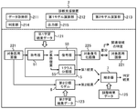
図1は、本発明を適用した場面の一例を模式的に例示する。本実施形態に係る診断支援システム100は、モデル生成装置1及び診断支援装置2を備えている。§1 Application example FIG. 1 schematically illustrates an example of a situation in which the present invention is applied. The
本実施形態に係るモデル生成装置1は、機械学習を実施することで、訓練済みのモデルを生成するように構成されたコンピュータである。具体的に、モデル生成装置1は、被験者の正常な身体部位の写る複数の第1学習医用画像31を取得し、取得された複数の第1学習医用画像31を使用して、第1分類モデル5の教師なし学習を実施する。第1分類モデル5は、医用画像の入力を受け付け、入力された医用画像に写る身体部位の正常の度合いを1クラス分類(one-class classification)で評価するように構成される。一例では、1クラス分類で正常の度合いを評価するとは、学習用データを特徴空間に写像し、線形又は非線形の超平面からの距離を評価することである。これにより、例えば、与えられた医用画像に対する評価値が大きい場合にその医用画像に写る身体部位を異常、評価値が小さい場合に身体部位を正常とすることで、医用画像に写る身体部位が正常であるか否かの判別を行うことが可能となる。教師なし学習は、入力された医用画像が複数の第1学習医用画像31のクラスに属する場合に入力された当該医用画像に写る身体部位は正常であると評価し、入力された医用画像が複数の第1学習医用画像31のクラスに属さない場合に入力された当該医用画像に写る身体部位は正常ではないと評価するように第1分類モデル5を訓練することを含む。この訓練により、第1分類モデル5は、与えられた医用画像に写る身体部位の正常の度合いを1クラス分類で評価する能力を獲得する。
The
また、モデル生成装置1は、第2学習医用画像331及び当該第2学習医用画像331に写る身体部位が正常か否かを示す正解ラベル333の組み合わせによりそれぞれ構成された複数の学習データセット33を取得する。複数の学習データセット33の第2学習医用画像331は、正常な身体部位の写る正常医用画像、及び異常な身体部位の写る異常医用画像を含む。モデル生成装置1は、取得された複数の学習データセット33を使用して、第2分類モデル6の教師あり学習を実施する。第2分類モデル6は、医用画像の入力を受け付け、入力された当該医用画像に写る身体部位の正常の度合いを評価するように構成される。教師あり学習は、各学習データセット33について、第2学習医用画像331の入力に対して、入力された第2学習医用画像331に写る身体部位に対して正常の度合いを評価した結果が対応する正解ラベル333に適合するように第2分類モデル6を訓練することを含む。この訓練により、第2分類モデル6は、与えられた医用画像に写る身体部位の正常の度合いを評価する能力を獲得する。
Further, the
各機械学習を実施した結果、モデル生成装置1は、訓練済みの第1分類モデル5及び第2分類モデル6を生成することができる。
As a result of performing each machine learning, the
一方、本実施形態に係る診断支援装置2は、訓練済みの第1分類モデル5及び第2分類モデル6を使用して、検査対象者の身体部位が正常であるか否かを分類するように構成されたコンピュータである。具体的に、診断支援装置2は、検査対象者の身体部位の写る対象医用画像221を取得する。診断支援装置2は、取得された対象医用画像221を訓練された第1分類モデル5に与えて、訓練された第1分類モデル5の演算処理を実行する。これにより、診断支援装置2は、対象医用画像221に写る検査対象者の身体部位に対して1クラス分類で評価した正常の度合いを第1結果として取得する。また、診断支援装置2は、取得された対象医用画像221を訓練された第2分類モデル6に与えて、訓練された第2分類モデル6の演算処理を実行する。これにより、診断支援装置2は、対象医用画像221に写る検査対象者の身体部位に対して評価した正常の度合いを第2結果として取得する。診断支援装置2は、第1結果及び第2結果に基づいて、対象医用画像221に写る検査対象者の身体部位が正常であるか否かを判定する。そして、診断支援装置2は、当該判定の結果を出力する。
On the other hand, the
以上のとおり、本実施形態では、モデル生成装置1において、正常な症例の第1学習医用画像31を使用した教師なし学習により訓練済みの第1分類モデル5を生成すると共に、正常な症例及び異常な症例を含む学習データセット33を使用した教師あり学習により訓練済みの第2分類モデル6を生成する。診断支援装置2では、生成された訓練済みの第1分類モデル5及び第2分類モデル6の2つの分類モデルを使用して、対象医用画像221に写る検査対象者の身体部位が正常であるか否かを推論する。異常な症例の第2学習医用画像331を含む学習データセット33を大量に収集するのは困難でかつコストがかかるのに対して、正常な症例の第1学習医用画像31は比較的に低コストでかつ容易に収集可能である。そのため、学習データセット33の件数が少ないことで訓練済みの第2分類モデル6の分類精度が低くなる可能性があるが、比較的に低コストでかつ容易に収集された大量の第1学習医用画像31を使用して訓練された第1分類モデル5によりその分類の性能を補うことができる。したがって、本実施形態によれば、比較的に低コストで、医用画像に写る検査対象者の身体部位が正常であるか否かについての分類精度の向上を図ることができる。
As described above, in the present embodiment, in the
なお、医用画像は、診断の支援に利用可能であれば、その種類は、特に限定されなくてよく、実施の形態に応じて適宜選択されてよい。医用画像は、例えば、X線写真、CT(Computed Tomography)画像、MRI(Magnetic Resonance Imaging)画像、超音波画像等であってよい。身体部位は、例えば、頭部、胸部、腹部等の身体のある範囲を占める部分の全部又は一部であってよい。或いは、身体部位は、その身体のある範囲を占める部分に存在する器官(臓器)又は組織の全部又は一部であってよい。 The type of the medical image is not particularly limited as long as it can be used to support the diagnosis, and may be appropriately selected according to the embodiment. The medical image may be, for example, an X-ray photograph, a CT (Computed Tomography) image, an MRI (Magnetic Resonance Imaging) image, an ultrasonic image, or the like. The body part may be all or part of a part of the body such as the head, chest, abdomen, etc. Alternatively, the body part may be all or part of an organ or tissue that exists in a part of the body that occupies a certain area.
身体部位が正常とは、例えば、病変領域が存在しない又は存在しない蓋然性が高いと人又は機械により診断されることであってよく、真実には病変領域が存在することを含んでもよい。一方、身体部位が異常とは、例えば、病変領域が存在する又は存在する蓋然性が高いと人又は機械により診断されることであってよく、真実には病変領域が存在しないことを含んでもよい。 A normal body part may be, for example, diagnosed by a person or machine as having no or highly probable absence of a lesion area, and may also include the presence of a lesion area in truth. On the other hand, an abnormality in a body part may mean, for example, being diagnosed by a person or a machine that a lesion area exists or is likely to exist, and may include the fact that the lesion area does not exist.
教師なし学習は、基本的には、正解ラベルを含まない学習データを使用した機械学習の手法である。教師なし学習は、自己教師あり学習及び敵対的学習を含んでもよい。一方、教師あり学習は、正解ラベルを含む学習データ(上記学習データセット33)を使用した機械学習の手法である。 Unsupervised learning is basically a machine learning method that uses learning data that does not include correct labels. Unsupervised learning may include self-supervised learning and hostile learning. On the other hand, supervised learning is a machine learning method using learning data including a correct label (the learning data set 33).
第1結果及び第2結果はそれぞれ、第1分類モデル5及び第2分類モデル6それぞれから直接的又は間接的に取得されてよい。すなわち、第1分類モデル5及び第2分類モデル6はそれぞれ、第1結果及び第2結果それぞれに対応する出力値を直接的に出力するように構成されてよい。或いは、第1結果及び第2結果はそれぞれ、第1分類モデル5及び第2分類モデル6それぞれの出力値に対して所定の演算(例えば、閾値判定、複数の数値を合計する等)を実行することにより得られてもよい。
The first result and the second result may be obtained directly or indirectly from the
図1の例では、モデル生成装置1及び診断支援装置2は、ネットワークを介して互いに接続されている。ネットワークの種類は、例えば、インターネット、無線通信網、移動通信網、電話網、専用網等から適宜選択されてよい。ただし、モデル生成装置1及び診断支援装置2の間でデータをやりとりする方法は、このような例に限定されなくてもよく、実施の形態に応じて適宜選択されてよい。例えば、モデル生成装置1及び診断支援装置2の間では、記憶媒体を利用して、データがやりとりされてよい。
In the example of FIG. 1, the
また、図1の例では、モデル生成装置1及び診断支援装置2は、それぞれ別個のコンピュータにより構成されている。しかしながら、本実施形態に係る診断支援システム100の構成は、このような例に限定されなくてもよく、実施の形態に応じて適宜決定されてよい。例えば、モデル生成装置1及び診断支援装置2は一体のコンピュータであってもよい。また、例えば、モデル生成装置1及び診断支援装置2のうちの少なくとも一方は、複数台のコンピュータにより構成されてもよい。
Further, in the example of FIG. 1, the
§2 構成例
[ハードウェア構成]
<モデル生成装置>
図2は、本実施形態に係るモデル生成装置1のハードウェア構成の一例を模式的に例示する。図2に示されるとおり、本実施形態に係るモデル生成装置1は、制御部11、記憶部12、通信インタフェース13、入力装置14、出力装置15、及びドライブ16が電気的に接続されたコンピュータである。なお、図2では、通信インタフェースを「通信I/F」と記載している。後述の図3でも同様の表記を用いる。§2 Configuration example [Hardware configuration]
<Model generator>
FIG. 2 schematically illustrates an example of the hardware configuration of the
制御部11は、ハードウェアプロセッサであるCPU(Central Processing Unit)、RAM(Random Access Memory)、ROM(Read Only Memory)等を含み、プログラム及び各種データに基づいて情報処理を実行するように構成される。CPUは、プロセッサ・リソースの一例である。記憶部12は、メモリ・リソースの一例であり、例えば、ハードディスクドライブ、ソリッドステートドライブ等で構成される。本実施形態では、記憶部12は、モデル生成プログラム81、第1学習医用画像31、学習データセット33、調整用データセット35、第1学習結果データ121、第2学習結果データ123、調整結果データ125等の各種情報を記憶する。
The
モデル生成プログラム81は、各分類モデル(5、6)の機械学習に関する後述の情報処理(図7〜図10)をモデル生成装置1に実行させるためのプログラムである。モデル生成プログラム81は、当該情報処理の一連の命令を含む。複数の第1学習医用画像31は、第1分類モデル5の機械学習に使用される。複数の学習データセット33は、第2分類モデル6の機械学習に使用される。複数の調整用データセット35は、第1結果を優先する程度を規定する第1パラメータ及び第2結果を優先する程度を規定する第2パラメータそれぞれの値を調整するのに使用される。第1学習結果データ121は、第1分類モデル5の機械学習の結果に関する情報を示す。第2学習結果データ123は、第2分類モデル6の機械学習の結果に関する情報を示す。調整結果データ125は、各パラメータの値の調整結果に関する情報を示す。本実施形態では、第1学習結果データ121、第2学習結果データ123、及び調整結果データ125は、モデル生成プログラム81を実行した結果として生成される。
The
通信インタフェース13は、例えば、有線LAN(Local Area Network)モジュール、無線LANモジュール等であり、ネットワークを介した有線又は無線通信を行うためのインタフェースである。モデル生成装置1は、この通信インタフェース13を利用して、他の情報処理装置との間で、ネットワークを介したデータ通信を実行してもよい。
The
入力装置14は、例えば、マウス、キーボード等の入力を行うための装置である。また、出力装置15は、例えば、ディスプレイ(表示装置)、スピーカ等の出力を行うための装置である。入力装置14及び出力装置15は、例えば、タッチパネルディスプレイ等により、一体的に構成されてもよい。ユーザ等のオペレータは、入力装置14及び出力装置15を利用することで、モデル生成装置1を操作することができる。
The input device 14 is, for example, a device for inputting a mouse, a keyboard, or the like. Further, the
ドライブ16は、例えば、CDドライブ、DVDドライブ等であり、記憶媒体91に記憶されたプログラム等の各種情報を読み込むためのドライブ装置である。記憶媒体91は、コンピュータその他装置、機械等が、記憶されたプログラム等の各種情報を読み取り可能なように、当該プログラム等の情報を、電気的、磁気的、光学的、機械的又は化学的作用によって蓄積する媒体である。上記モデル生成プログラム81、第1学習医用画像31、学習データセット33、及び調整用データセット35の少なくともいずれかは、記憶媒体91に記憶されていてもよい。この場合、モデル生成装置1は、記憶媒体91から、これらのうちの少なくともいずれかを取得してもよい。なお、図2では、記憶媒体91の一例として、CD、DVD等のディスク型の記憶媒体を例示している。しかしながら、記憶媒体91の種類は、ディスク型に限られなくてもよく、ディスク型以外であってもよい。ディスク型以外の記憶媒体として、例えば、フラッシュメモリ等の半導体メモリを挙げることができる。ドライブ16の種類は、記憶媒体91の種類に応じて任意に選択されてよい。
The
なお、モデル生成装置1の具体的なハードウェア構成に関して、実施形態に応じて、適宜、構成要素の省略、置換及び追加が可能である。例えば、プロセッサ・リソースは、複数のハードウェアプロセッサを含んでもよい。ハードウェアプロセッサは、マイクロプロセッサ、FPGA(field-programmable gate array)、GPU(Graphics Processing Unit)等で構成されてよい。記憶部12は、制御部11に含まれるRAM及びROMにより構成されてもよい。通信インタフェース13、入力装置14、出力装置15及びドライブ16の少なくともいずれかは省略されてもよい。モデル生成装置1は、複数台のコンピュータで構成されてもよい。この場合、各コンピュータのハードウェア構成は、一致していてもよいし、一致していなくてもよい。また、モデル生成装置1は、提供されるサービス専用に設計された情報処理装置の他、汎用のサーバ装置、PC(Personal Computer)等であってもよい。
Regarding the specific hardware configuration of the
<診断支援装置>
図3は、本実施形態に係る診断支援装置2のハードウェア構成の一例を模式的に例示する。図3に示されるとおり、本実施形態に係る診断支援装置2は、制御部21、記憶部22、通信インタフェース23、入力装置24、出力装置25、及びドライブ26が電気的に接続されたコンピュータである。<Diagnosis support device>
FIG. 3 schematically illustrates an example of the hardware configuration of the
診断支援装置2の制御部21〜ドライブ26及び記憶媒体92はそれぞれ、上記モデル生成装置1の制御部11〜ドライブ16及び記憶媒体91それぞれと同様に構成されてよい。制御部21は、ハードウェアプロセッサであるCPU、RAM、ROM等を含み、プログラム及びデータに基づいて各種情報処理を実行するように構成される。記憶部22は、例えば、ハードディスクドライブ、ソリッドステートドライブ等で構成される。記憶部22は、診断支援プログラム82、第1学習結果データ121、第2学習結果データ123、調整結果データ125等の各種情報を記憶する。
The
診断支援プログラム82は、訓練済みの第1分類モデル5及び第2分類モデル6を使用して、検査対象者の身体部位が正常であるか否かを分類する後述の情報処理(図11)を診断支援装置2に実行させるためのプログラムである。診断支援プログラム82は、当該情報処理の一連の命令を含む。診断支援プログラム82、第1学習結果データ121、第2学習結果データ123、及び調整結果データ125の少なくともいずれかは、記憶媒体92に記憶されていてもよい。これに応じて、診断支援装置2は、これらのうちの少なくともいずれかを記憶媒体92から取得してもよい。
The
なお、診断支援装置2の具体的なハードウェア構成に関して、実施形態に応じて、適宜、構成要素の省略、置換及び追加が可能である。例えば、診断支援装置2のプロセッサ・リソースは、複数のハードウェアプロセッサを含んでもよい。ハードウェアプロセッサは、マイクロプロセッサ、FPGA、GPU等で構成されてよい。記憶部22は、制御部21に含まれるRAM及びROMにより構成されてもよい。通信インタフェース23、入力装置24、出力装置25、及びドライブ26の少なくともいずれかは省略されてもよい。診断支援装置2は、複数台のコンピュータで構成されてもよい。この場合、各コンピュータのハードウェア構成は、一致していてもよいし、一致していなくてもよい。また、診断支援装置2は、提供されるサービス専用に設計された情報処理装置の他、汎用のサーバ装置、汎用のPC等であってもよい。
Regarding the specific hardware configuration of the
[ソフトウェア構成]
<モデル生成装置>
図4は、本実施形態に係るモデル生成装置1のソフトウェア構成の一例を模式的に例示する。図5Aは、第1分類モデル5の教師なし学習の処理過程の一例を模式的に例示する。図5Bは、第2分類モデル6の教師あり学習の処理過程の一例を模式的に例示する。図5Cは、第1パラメータ及び第2パラメータの各値を調整する処理過程の一例を模式的に例示する。[Software configuration]
<Model generator>
FIG. 4 schematically illustrates an example of the software configuration of the
モデル生成装置1の制御部11は、記憶部12に記憶されたモデル生成プログラム81をRAMに展開する。そして、制御部11は、CPUにより、RAMに展開されたモデル生成プログラム81に含まれる命令を解釈及び実行して、各構成要素を制御する。これにより、図4に示されるとおり、本実施形態に係るモデル生成装置1は、第1取得部110、第1学習部111、第2取得部112、第2学習部113、第3取得部114、判定部115、調整部116、保存処理部117、原初データ取得部118、及び拡大処理部119をソフトウェアモジュールとして備えるコンピュータとして動作する。すなわち、本実施形態では、各ソフトウェアモジュールは、制御部11(CPU)により実現される。
The
第1取得部110は、被験者の正常な身体部位の写る複数の第1学習医用画像31を取得する。第1学習部111は、取得された複数の第1学習医用画像31を使用して、第1分類モデル5の教師なし学習を実施する。第1分類モデル5は、医用画像の入力を受け付け、入力された当該医用画像に写る身体部位が正常であるか否かを1クラス分類で分類した結果に対応する出力値を出力するように構成される。教師なし学習は、入力された医用画像が複数の第1学習医用画像31のクラスに属する場合に入力された当該医用画像に写る身体部位は正常であると分類し、入力された医用画像が複数の第1学習医用画像31のクラスに属さない場合に入力された当該医用画像に写る身体部位は正常ではないと分類するように第1分類モデル5を訓練することを含む。このように訓練可能であれば、第1分類モデル5の構成及び教師なし学習の方法はそれぞれ、特に限定されなくてよく、実施の形態に応じて適宜選択されてよい。
The first acquisition unit 110 acquires a plurality of first learning
図4に示されるとおり、本実施形態では、第1分類モデル5は、符号器51及び復号器53を含む。符号器51は、与えられた医用画像を特徴量に変換(医用画像から特徴量を抽出)するように構成される。換言すると、符号器51は、医用画像の入力を受け付け、入力された医用画像を特徴量に変換した結果に対応する出力値を出力するように構成される。復号器53は、符号器51により得られた特徴量から医用画像を復号化するように構成される。換言すると、復号器53は、特徴量の入力を受け付け、入力された特徴量から元の医用画像を復号化した結果(復号化画像)を出力するように構成される。教師なし学習は、各第1学習医用画像31を符号器51に与えることで復号器53により生成される復号化画像が各第1学習医用画像31に適合するように、符号器51及び復号器53を訓練することを含む。すなわち、教師なし学習では、符号器51及び復号器53は、各第1学習医用画像31及び対応する復号化画像の間の再構成誤差を最小化するように訓練される。
As shown in FIG. 4, in this embodiment, the
本実施形態では、第1分類モデル5は、1クラス分類器55を更に含む。1クラス分類器55は、符号器51により得られる特徴量の入力を受け付け、入力された特徴量に基づいて医用画像に写る身体部位の正常の度合いを1クラス分類で評価した結果に対応する出力値を出力するように構成される。教師なし学習は、符号器51により得られる特徴量に基づいて当該1クラス分類の評価を行うように1クラス分類器55を訓練することを更に含む。換言すると、教師なし学習は、各第1学習医用画像31を訓練された符号器51に入力することで得られる各学習特徴量のクラスに入力された特徴量が属する場合に、与えられた医用画像に写る身体部位は正常であると評価し、各学習特徴量のクラスに入力された特徴量が属さない場合、与えられた医用画像に写る身体部位は正常ではないと評価するように、1クラス分類器55を訓練することを含む。
In this embodiment, the
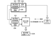
第2取得部112は、第2学習医用画像331及び当該第2学習医用画像331に写る身体部位が正常か否かを示す正解ラベル333の組み合わせによりそれぞれ構成される複数の学習データセット33を取得する。正解ラベル333のデータ形式は、実施の形態に応じて適宜決定されてよい。例えば、正解ラベル333は、正常であるか否かを2値で示すように構成されてもよいし、或いは、異常のカテゴリ(正常であることを含む)を2値以上で示すように構成されてもよい。図5Bに示されるとおり、複数の学習データセット33の第2学習医用画像331は、被験者の正常な身体部位の写る正常医用画像3311、及び被験者の異常な身体部位の写る異常医用画像3312を含む。第2分類モデル6は、医用画像の入力を受け付け、入力された当該医用画像に写る身体部位の正常の度合いを評価した結果に対応する出力値を出力するように構成される。第2学習部113は、取得された複数の学習データセット33を使用して、第2分類モデル6の教師あり学習を実施する。教師あり学習は、各学習データセット33について、第2学習医用画像331の入力に対して、入力された第2学習医用画像331に写る身体部位に対して正常の度合いを評価した結果が対応する正解ラベル333に適合するように第2分類モデル6を訓練することを含む。
The second acquisition unit 112 acquires a plurality of learning
第3取得部114は、それぞれに写る身体部位が正常か否か特定された複数の第3学習医用画像351を取得する。第3学習医用画像351に写る身体部位が正常か否かを特定する形式は、実施の形態に応じて適宜決定されてよい。本実施形態では、第2学習医用画像331と同様に、各第3学習医用画像351には、正解ラベル353が付与される。各正解ラベル353は、対応する第3学習医用画像351に写る身体部位が正常か否かを示すように構成される。すなわち、第3取得部114は、第3学習医用画像351及び正解ラベル353の組み合わせによりそれぞれ構成される複数の調整用データセット35を取得する。
The third acquisition unit 114 acquires a plurality of third learning
判定部115は、訓練された第1分類モデル5及び訓練された第2分類モデル6を使用して、取得された各第3学習医用画像351に写る身体部位が正常か否かを判定する。具体的には、判定部115は、各第3学習医用画像351を訓練された第1分類モデル5に与えることで、各第3学習医用画像351に写る身体部位に対して1クラス分類で評価した正常の度合いを第1結果として取得する。また、判定部115は、各第3学習医用画像351を訓練された第2分類モデル6に与えることで、各第3学習医用画像351に写る身体部位に対して評価した正常の度合いを第2結果として取得する。本実施形態では、第1結果及び第2結果は、身体部位の正常の度合いを数値で示すように構成される。正常の度合いを数値で示すことは、正常である程度を直接的に示すこと、及び異常である程度を示すことにより正常である程度を間接的に示すことの少なくとも一方により構成されてよい。すなわち、第1結果及び第2結果はそれぞれ、正常である程度を示す数値及び異常である程度を示す数値の少なくとも一方により構成されてよい。度合いは、例えば、確率、指数等により表現されてよい。判定部115は、第1結果を優先する程度を規定する第1パラメータ及び第2結果を優先する程度を規定する第2パラメータを含む結合器7を備える。判定部115は、取得された第1結果及び第2結果を結合器7に与えることで、第1パラメータ及び第2パラメータの各値を使用して、第1結果及び第2結果それぞれを重み付けする。判定部115は、重み付けされた第1結果及び第2結果を結合する。判定部115は、結合により得られた数値(判定値)を閾値と比較することにより、各第3学習医用画像351に写る身体部位が正常であるか否かを判定する。調整部116は、各第3学習医用画像351に対する判定部115の判定の精度が最適化されるように、第1パラメータ及び第2パラメータの各値を調整する。図5Cに示されるとおり、複数の第3学習医用画像351は、1又は複数の限度見本(「限度医用画像」と称してもよい)352を含んでよい。複数の第3学習医用画像351の全てが限度見本352であってもよい。この場合、調整部116は、全ての1又は複数の限度見本352に対する判定部115の判定が誤らないように、第1パラメータ及び第2パラメータの各値を調整してもよい。
The determination unit 115 uses the trained
図5A〜図5Cに示されるとおり、保存処理部117は、各機械学習及びパラメータ調整の結果に関する情報を生成する。保存処理部117は、訓練済みの第1分類モデル5に関する情報を第1学習結果データ121として生成する。保存処理部117は、訓練済みの第2分類モデル6に関する情報を第2学習結果データ123として生成する。保存処理部117は、調整された結合器7に関する情報を調整結果データ125として生成する。保存処理部117は、生成された第1学習結果データ121、第2学習結果データ123、及び調整結果データ125を所定の記憶領域に保存する。
As shown in FIGS. 5A-5C, the storage processing unit 117 generates information regarding the results of each machine learning and parameter adjustment. The storage processing unit 117 generates information about the trained
原初データ取得部118は、被験者の身体部位の写る1又は複数の原初の医用画像390を取得する。拡大処理部119は、原初の医用画像390に対して拡大処理を適用することで、新たな医用画像395を生成する。拡大処理は、平行移動等の画像処理により、原初の医用画像390に写る特徴の少なくとも一部を維持したまま、原初の医用画像390と異なる新たな医用画像395を生成する処理である。
The primordial data acquisition unit 118 acquires one or more primordial
各学習医用画像(31、331、351)の少なくとも一部には、原初の医用画像390及び新たな医用画像395の少なくともいずれかが用いられてよい。その他、各学習医用画像(31、331、351)には、原初の医用画像390及び新たな医用画像395以外で個別に取得された医用画像が用いられてよい。なお、原初の医用画像390及び各学習医用画像(31、331、351)は、例えば、X線撮影装置、コンピュータ断層撮影装置、磁気共鳴断層撮影装置等の撮影装置により被験者の身体部位を撮影することで得られた医用画像である。それぞれの収集に係る被験者の数は、任意に決定されてよい。被験者は、人の身体を模した模型物(例えば、X線撮影用ファントム等)を含んでよい。
At least a portion of each learning medical image (31, 331, 351) may be at least one of the original
(第1分類モデルの構成の一例)
符号器51、復号器53、及び1クラス分類器55は、演算パラメータを有する機械学習可能なモデルにより構成される。それぞれに利用する機械学習モデルは、それぞれの演算処理を実行可能であれば、その種類は、特に限定されなくてよく、実施の形態に応じて適宜選択されてよい。(Example of configuration of the first classification model)
The
図5Aに示されるとおり、本実施形態では、符号器51、復号器53、及び1クラス分類器55には、全結合型ニューラルネットワークが用いられる。符号器51、復号器53、及び1クラス分類器55はそれぞれ、入力層(511、531、551)、中間(隠れ)層(512、532、552)、及び出力層(513、533、553)を備えている。各中間層(512、532、552)の数は、実施の形態に応じて適宜決定されてよい。ただし、それぞれの構造は、このような例に限定されなくてよく、実施の形態に応じて適宜決定されてよい。例えば、各中間層(512、532、552)は省略されてもよい。また、例えば、畳み込み層等の他の種類の層がそれぞれに含まれてもよい。
As shown in FIG. 5A, in this embodiment, a fully connected neural network is used for the
各層(511〜513、531〜533、551〜553)は、1又は複数のニューロン(ノード)を備えている。それぞれに含まれるニューロンの数は、実施の形態に応じて適宜決定されてよい。例えば、入力層(511、531、551)及び出力層(513、533、553)に含まれるニューロンの数は、入力及び出力のデータの次元に応じて決定されてよい。具体例として、符号器51の入力層511及び復号器53の出力層533に含まれるニューロンの数は、医用画像の画素数に応じて決定されてよい。復号器53及び1クラス分類器55の各入力層(531、551)に含まれるニューロンの数は、符号器51の出力層513に含まれるニューロンの数に応じて決定されてよい。1クラス分類器55の出力層553に含まれるニューロンの数は、1クラス分類の結果の形式に応じて決定されてよい(例えば、1つ)。
Each layer (511-513, 531-533, 551-553) comprises one or more neurons (nodes). The number of neurons contained in each may be appropriately determined according to the embodiment. For example, the number of neurons contained in the input layer (511, 513, 551) and the output layer (513, 533, 535) may be determined according to the dimensions of the input and output data. As a specific example, the number of neurons included in the
隣接する層のニューロン同士は適宜結合される。図5Aの例では、各ニューロンは、隣接する層の全てのニューロンと結合されている。しかしながら、各ニューロンの結合関係は、このような例に限定されなくてもよく、実施の形態に応じて適宜設定されてよい。各結合には、重み(結合荷重)が設定されている。各ニューロンには閾値が設定されており、基本的には、各入力と各重みとの積の和が閾値を超えているか否かによって各ニューロンの出力が決定される。閾値は、活性化関数により表現されてもよい。この場合、各入力及び各重みの積の和を活性化関数に入力し、活性化関数の演算を実行することで、各ニューロンの出力が決定される。活性化関数の種類は、特に限定されなくてもよく、実施の形態に応じて適宜選択されてよい。それぞれに含まれる各ニューロン間の結合の重み及び各ニューロンの閾値は、それぞれの演算処理に利用される演算パラメータの一例である。 Neurons in adjacent layers are appropriately connected. In the example of FIG. 5A, each neuron is connected to all neurons in adjacent layers. However, the connection relationship of each neuron does not have to be limited to such an example, and may be appropriately set according to the embodiment. A weight (bonding load) is set for each coupling. A threshold is set for each neuron, and basically, the output of each neuron is determined by whether or not the sum of the products of each input and each weight exceeds the threshold. The threshold value may be expressed by an activation function. In this case, the output of each neuron is determined by inputting the sum of the products of each input and each weight into the activation function and executing the operation of the activation function. The type of activation function does not have to be particularly limited and may be appropriately selected depending on the embodiment. The weight of the connection between each neuron and the threshold value of each neuron included in each are examples of arithmetic parameters used in each arithmetic processing.
第1分類モデル5の各構成要素の各演算パラメータの値は、上記教師なし学習の過程で所望の能力を獲得するように調整される。まず、第1学習部111は、各第1学習医用画像31を符号器51の入力層511に入力し、符号器51及び復号器53の順伝播の演算処理を実行する。順伝播の演算処理は、入力側から順に各層に含まれる各ニューロンの発火判定を行うことである。符号器51の順伝播の演算処理により、各第1学習医用画像31から抽出された特徴量(に対応する出力値)を符号器51の出力層513から得ることができる。次いで、復号器53の順伝播の演算処理により、各第1学習医用画像31に対応して特徴量から生成された復号化画像(に対応する出力値)を復号器53の出力層533から得ることができる。第1学習部111は、各第1学習医用画像31及び対応する復号画像の間の誤差(再構成誤差)が小さくなるように、符号器51及び復号器53の各演算パラメータの値を調整する。これにより、訓練済みの符号器51及び復号器53が生成される。
The value of each arithmetic parameter of each component of the
次に、第1学習部111は、各第1学習医用画像31を訓練された符号器51の入力層511に入力し、符号器51及び1クラス分類器55の順伝播の演算処理を実行する。1クラス分類器55の順伝播の演算処理により、各第1学習医用画像31に写る身体部位に対して正常の度合いを評価した結果に対応する出力値を1クラス分類器55の出力層553から得ることができる。一例では、出力値は、特徴空間の原点からの距離を示す。第1学習部111は、この原点からの距離を最大化するように1クラス分類器55の演算パラメータの値を調整する。これにより、識別境界のマージンが最大化された訓練済みの1クラス分類器55が生成される。
Next, the first learning unit 111 inputs each first learning
保存処理部117は、上記教師なし学習により生成された訓練済みの第1分類モデル5の各構成要素の構造及び演算パラメータの値を示す情報を第1学習結果データ121として生成する。構造は、例えば、ニューラルネットワークにおける入力層から出力層までの層の数、各層の種類、各層に含まれるニューロンの数、隣接する層のニューロン同士の結合関係等により特定されてよい。システム内でモデルの構造が共通化される場合、この構造に関する情報は第1学習結果データ121から省略されてもよい。保存処理部117は、生成された第1学習結果データ121を所定の記憶領域に保存する。
The storage processing unit 117 generates information indicating the structure of each component of the trained
(第2分類モデルの構成の一例)
第2分類モデル6も、第1分類モデル5と同様に、演算パラメータを有する機械学習可能なモデルにより構成される。第2分類モデル6に利用する機械学習モデルは、医用画像に写る身体部位の正常の度合いを評価する演算処理を実行可能であれば、その種類は、特に限定されなくてよく、実施の形態に応じて適宜選択されてよい。(Example of the configuration of the second classification model)
Like the
図5Bに示されるとおり、本実施形態では、第2分類モデル6には、畳み込みニューラルネットワークが用いられる。第2分類モデル6は、畳み込み層61、プーリング層62、及び全結合層(63、64)を備えている。畳み込み層61は、与えられたデータに対する畳み込み演算を行うように構成される。畳み込み演算とは、与えられたデータと所定のフィルタとの相関を算出する処理に相当する。例えば、画像の畳み込みを行うことで、フィルタの濃淡パターンと類似する濃淡パターンを入力される画像から検出することができる。畳み込み層61は、この畳み込み演算に対応するニューロン(ノード)であって、入力又は自分の層よりも前(入力側)に配置された層の出力の一部の領域に結合するニューロン(ノード)を備えている。プーリング層62は、プーリング処理を行うように構成される。プーリング処理は、与えられたデータのフィルタに対する応答の強かった位置の情報を一部捨て、当該データ内に現れる特徴の微小な位置変化に対する応答の不変性を実現する。例えば、プーリング処理では、フィルタ内の最も大きな値が抽出され、それ以外の値が削除されてよい。全結合層(63、64)は、上記全結合型ニューラルネットワークの各層(511〜513、531〜533、551〜553)と同様である。
As shown in FIG. 5B, in this embodiment, a convolutional neural network is used for the
第2分類モデル6に含まれる各層61〜64の数は、実施の形態に応じて適宜決定されてよい。また、畳み込み層61及びプーリング層62の配置も、実施の形態に応じて適宜決定されてよい。図5Bの例では、最も入力側(図の左側)には、畳み込み層61が配置され、最も出力側(図の右側)には全結合層(63、64)が配置され、全結合層63の直前には、プーリング層62が配置されている。これにより、最も入力側の畳み込み層61が入力層を構成し、全結合層64が出力層を構成している。畳み込み層61及びプーリング層62は交互に配置されてよい。ただし、第2分類モデル6の構造は、このような例に限定されなくてよい。例えば、複数の畳み込み層61が連続して配置された後に、1又は複数のプーリング層62が配置されてもよい。また、第2分類モデル6に含まれる層の種類は、これらに限られなくてよい。第2分類モデル6は、例えば、正規化層、ドロップアウト層等の他の種類の層を含んでもよい。
The number of each
各全結合層(63、64)に含まれるニューロンの数は、実施の形態に応じて適宜決定されてよい。例えば、出力層を構成する全結合層64に含まれるニューロンの数は、第2分類モデル6による評価の形式に応じて決定されてよい。第2分類モデル6による評価は、例えば、正常であるか否かを分類すること、異常のカテゴリ(すなわち、種別)を識別すること等を含んでもよい。一例として、全結合層64の出力値が、身体部位の正常の度合いを直接的に示す場合、全結合層64に含まれるニューロンの数は、1つであってよい。或いは、全結合層64の出力値が、正常及び異常それぞれの度合いを示す場合、全結合層64に含まれるニューロンの数は、2つであってよい。或いは、全結合層64の出力値が、異常のカテゴリ(正常を含む)に属する程度を示す場合、全結合層64に含まれるニューロンの数は、カテゴリに応じて決定されてよい。この場合、異常の度合いは、異常の各カテゴリの出力値の合計値により与えられてよい。
The number of neurons contained in each fully connected layer (63, 64) may be appropriately determined depending on the embodiment. For example, the number of neurons contained in the fully connected
畳み込み層61及び全結合層(63、64)の各結合には、上記第1分類モデル5と同様に、重み(結合荷重)が設定される。各ニューロンの閾値(又は活性化関数)は適宜与えられてよい。畳み込み層61及び全結合層(63、64)に含まれる各ニューロン間の結合の重み及び各ニューロンの閾値は、第2分類モデル6の演算処理に利用される演算パラメータの一例である。
A weight (bonding load) is set for each bond of the
第2分類モデル6の各演算パラメータの値は、上記教師あり学習の過程で所望の能力を獲得するように調整される。第2学習部113は、各学習データセット33の第2学習医用画像331を第2分類モデル6の入力層(最も入力側の畳み込み層61)に入力し、第2分類モデル6の順伝播の演算処理を実行する。この演算処理により、各第2学習医用画像331に写る身体部位に対して正常の度合いを評価した結果に対応する出力値を出力層(全結合層64)から得ることができる。第2学習部113は、各学習データセット33について、出力層から得られる評価結果及び正解ラベル333により示される正解の間の誤差を算出し、算出される誤差が小さくなるように、第2分類モデル6の各演算パラメータの値を調整する。これにより、訓練済みの第2分類モデル6が生成される。
The value of each arithmetic parameter of the
保存処理部117は、上記教師あり学習により生成された訓練済みの第2分類モデル6の構造及び演算パラメータの値を示す情報を第2学習結果データ123として生成する。上記第1学習結果データ121と同様に、システム内でモデルの構造が共通化される場合、第2分類モデル6の構造に関する情報は第2学習結果データ123から省略されてもよい。保存処理部117は、生成された第2学習結果データ123を所定の記憶領域に保存する。
The storage processing unit 117 generates information indicating the values of the structure and the calculation parameters of the trained
(結合器)
結合器7の構成は、第1結果及び第2結果を重み付けして結合し、医用画像に写る身体部位が正常であるか否かの最終的な判定結果を得るための演算処理を実行可能であれば、特に限定されなくてよく、実施の形態に応じて適宜決定されてよい。(Coupler)
The configuration of the
図5Cに示されるとおり、結合器7には、2層の全結合型ニューラルネットワークが用いられる。結合器7は、入力層71及び出力層72を備える。ただし、結合器7の構造は、このような例に限定されなくてよく、実施の形態に応じて適宜決定されてよい。例えば、結合器7は、3層以上の全結合型ニューラルネットワークにより構成されてもよい。
As shown in FIG. 5C, a two-layer fully connected neural network is used for the
各層(71、72)は、1又は複数のニューロン(ノード)を備える。各層(71、72)に含まれるニューロンの数は、実施の形態に応じて適宜決定されてよい。例えば、入力層71に含まれるニューロンの数は、各結果のデータの次元に応じて決定されてよい。出力層72に含まれるニューロンの数は、最終的な判定結果のデータの次元に応じて決定されてよい。出力層72の出力形式は、実施の形態に応じて適宜決定されてよい。例えば、出力層72は、閾値と比較して判定結果を導出するための判定値(すなわち、重み付けされた第1結果及び第2結果を結合した結果)又は直接的に判定結果を示す出力値を出力するように構成されてよい。なお、出力層72の出力値が判定値を示す場合、結合器7の順伝播の演算処理は、第1結果及び第2結果それぞれを重み付けすること、並びに重み付けされた第1結果及び第2結果を結合することそれぞれに相当する処理を含んでいると解釈されてよい。一方、出力層72の出力値が判定結果を直接的に示す場合、結合器7の順伝播の演算処理は、第1結果及び第2結果それぞれを重み付けすること、重み付けされた第1結果及び第2結果を結合すること、並びに結合により得られた判定値を閾値と比較することに相当する処理を含んでいると解釈されてよい。
Each layer (71, 72) comprises one or more neurons (nodes). The number of neurons contained in each layer (71, 72) may be appropriately determined according to the embodiment. For example, the number of neurons contained in the
各ニューロンの結合関係は、実施の形態に応じて適宜設定されてよい。各層(71、72)の各結合には、上記第1分類モデル5と同様に、重み(結合荷重)が設定される。各ニューロンの閾値(又は活性化関数)は適宜与えられてよい。第1結果の演算に関与する各ニューロン間の結合の重み及び各ニューロンの閾値は、第1パラメータの一例である。第2結果の演算に関与する各ニューロン間の結合の重み及び各ニューロンの閾値は、第2パラメータの一例である。
The connection relationship of each neuron may be appropriately set according to the embodiment. A weight (bonding load) is set for each bond of each layer (71, 72) in the same manner as in the
本実施形態では、各パラメータの値は、複数の調整用データセット35を使用した機械学習により調整される。具体的に、判定部115は、各調整用データセット35の第3学習医用画像351を訓練された第1分類モデル5の符号器51に入力し、訓練された第1分類モデル5の符号器51及び1クラス分類器55の順伝播の演算処理を実行する。この演算処理により、判定部115は、各第3学習医用画像351に対する第1結果を1クラス分類器55から取得する。また、判定部115は、各調整用データセット35の第3学習医用画像351を訓練された第2分類モデル6に入力し、訓練された第2分類モデル6の順伝播の演算処理を実行する。この演算処理により、判定部115は、各第3学習医用画像351に対する第2結果を第2分類モデル6から取得する。そして、判定部115は、取得された第1結果及び第2結果を結合器7の入力層71に入力し、結合器7の順伝播の演算処理を実行する。この演算処理により、判定値又は判定結果に対応する出力値を結合器7の出力層72から取得することができる。調整部116は、各調整用データセット35について、判定値により導出される又は出力値により示される判定結果及び正解ラベル353により示される正解の間の誤差を算出し、算出される誤差が小さくなるように、結合器7の各パラメータの値を調整する。複数の第3学習医用画像351に1又は複数の限度見本352が含まれる場合には、調整部116は、限度見本352に対する判定結果に誤りが生じないように、結合器7の各演算パラメータの値を調整する。これにより、調整済みの結合器7が生成される。
In this embodiment, the value of each parameter is adjusted by machine learning using a plurality of adjustment data sets 35. Specifically, the determination unit 115 inputs the third learning
保存処理部117は、上記機械学習により生成された調整済みの結合器7の構造及び各パラメータの値を示す情報を調整結果データ125として生成する。上記第1学習結果データ121等と同様に、システム内でモデルの構造が共通化される場合、結合器7の構造に関する情報は調整結果データ125から省略されてもよい。保存処理部117は、生成された調整結果データ125を所定の記憶領域に保存する。
The storage processing unit 117 generates information indicating the structure of the adjusted
<診断支援装置>
図6は、本実施形態に係る診断支援装置2のソフトウェア構成の一例を模式的に例示する。診断支援装置2の制御部21は、記憶部22に記憶された診断支援プログラム82をRAMに展開する。そして、制御部21は、CPUにより、RAMに展開された診断支援プログラム82に含まれる命令を解釈及び実行して、各構成要素を制御する。これにより、図6に示されるとおり、本実施形態に係る診断支援装置2は、データ取得部211、第1モデル演算部212、第2モデル演算部213、判定部214、及び出力部215をソフトウェアモジュールとして備えるコンピュータとして動作する。すなわち、本実施形態では、診断支援装置2の各ソフトウェアモジュールは、上記モデル生成装置1と同様に、制御部21(CPU)により実現される。<Diagnosis support device>
FIG. 6 schematically illustrates an example of the software configuration of the
データ取得部211は、検査対象者の身体部位の写る対象医用画像221を取得する。対象医用画像221は、撮影装置により検査対象者の身体部位を撮影することで得られた医用画像である。検査対象者は、上記被験者と同様に、人の身体を模した模型物を含んでよい。
The data acquisition unit 211 acquires the target
第1モデル演算部212は、第1学習結果データ121を保持することで、訓練済みの第1分類モデル5を備える。第1モデル演算部212は、取得された対象医用画像221を訓練済みの第1分類モデル5に与えて、訓練済みの第1分類モデル5の演算処理を実行する。本実施形態では、第1分類モデル5の演算処理は、取得された対象医用画像221を訓練された符号器51に与えることで、対象医用画像221を対象特徴量に変換すること、及び変換により得られた対象特徴量を訓練された1クラス分類器55に与えることで、対象医用画像221に写る検査対象者の身体部位に対する第1結果に対応する出力値を訓練された1クラス分類器55から取得すること、を含む。すなわち、第1モデル演算部212は、取得された対象医用画像221を訓練済みの符号器51に入力し、訓練済みの符号器51及び1クラス分類器55の順伝播の演算処理を実行する。これにより、第1モデル演算部212は、当該第1結果に対応する出力値を訓練済みの第1分類モデル5から取得する。
The first model calculation unit 212 includes the trained
第2モデル演算部213は、第2学習結果データ123を保持することで、訓練済みの第2分類モデル6を備える。第2モデル演算部213は、取得された対象医用画像221を訓練済みの第2分類モデル6に与えて、訓練済みの第2分類モデル6の演算処理を実行する。本実施形態では、第2モデル演算部213は、取得された対象医用画像221を訓練済みの第2分類モデル6の入力層に入力し、訓練済みの第2分類モデル6の順伝播の演算処理を実行する。これにより、第2モデル演算部213は、対象医用画像221に写る検査対象者の身体部位に対する第2結果に対応する出力値を訓練済みの第2分類モデル6の出力層から取得する。
The second model calculation unit 213 includes the trained
判定部214は、第1結果及び第2結果に基づいて、対象医用画像221に写る検査対象者の身体部位が正常であるか否かを判定する。本実施形態では、第1結果及び第2結果は、身体部位の正常の度合いを数値で示すように構成される。判定部214は、調整結果データ125を保持することで、複数の第3学習医用画像351に対する判定精度が最適化されるように各値の調整された各パラメータを含む結合器7を備える。第1結果及び第2結果に基づいて判定することは、取得された第1結果及び第2結果を結合器7に与えることで、各パラメータの値を使用して、第1結果及び第2結果それぞれを重み付けすること、重み付けされた第1結果及び第2結果を結合すること、並びに結合により得られた判定値を閾値と比較することにより、検査対象者の身体部位が正常であるか否かを判定すること、により構成される。具体的には、判定部214は、各結果の値を結合器7の入力層71に入力し、結合器7の順伝播の演算処理を実行する。これにより、判定部214は、判定値又は判定結果に対応する出力値を出力層72から取得する。結合器7が判定値を出力するように構成される場合、判定部214は、得られた判定値を閾値と比較することで判定結果を導出する。出力部215は、判定部214による判定の結果を出力する。
Based on the first result and the second result, the determination unit 214 determines whether or not the body part of the examination subject shown in the target
なお、本実施形態では、第1分類モデル5の演算処理は、検査対象者の身体部位が正常ではないと判定される場合に、取得された対象医用画像221を訓練された符号器51に与えることで、対象医用画像221を対象特徴量に変換すること、変換により得られた対象特徴量を訓練された復号器53に与えることで、対象特徴量から対象復号化画像225を生成すること、対象医用画像221及び生成された対象復号化画像225の間の差分を算出すること、並びに算出された差分に基づいて、検査対象者の身体部位が正常ではないと判定されたことに関与する関連領域を対象医用画像221内で特定すること、を含んでもよい。この場合、判定の結果を出力することは、特定された関連領域を示す情報を出力することを含んでもよい。
In the present embodiment, the arithmetic processing of the
具体的に、第1モデル演算部212は、対象医用画像221を訓練済みの符号器51に入力し、訓練済みの符号器51の順伝播の演算処理を実行することで、対象特徴量に対応する出力値を訓練済みの符号器51から取得してもよい。この演算処理は、上記第1結果を得る処理と共通化されてよい。続いて、第1モデル演算部212は、得られた対象特徴量を訓練済みの復号器53に入力し、訓練済みの復号器53の順伝播の演算処理を実行することで、対象復号化画像225に対応する出力を訓練済みの復号器53から取得してもよい。そして、第1モデル演算部212は、対象医用画像221及び生成された対象復号化画像225の間の差分を算出し、算出された差分に基づいて、対象医用画像221内で関連領域を特定してもよい。出力部215は、特定された関連領域を示す情報を上記判定結果として出力してもよい。
Specifically, the first model calculation unit 212 corresponds to the target feature amount by inputting the target
訓練済みの符号器51及び復号器53によれば、機械学習に使用された第1学習医用画像31及びこれに類似する医用画像は精度よく再構成されるのに対して、それ以外の医用画像の再構成の精度は下がり得る。第1学習医用画像31には、正常な身体部位の写る医用画像が用いられているため、異常な身体部位の写る医用画像に対する訓練済みの符号器51及び復号器53の再構成の精度は低くなり得る。つまり、異常な身体部位の写る部分に関して、訓練された符号器51及び復号器53は精度よく再構成することができない可能性が高くなる。したがって、上記演算処理において、対象医用画像221及び対象復号化画像225の間の差分(再構成誤差)に基づいて、病変の蓋然性の高い領域(病変候補領域)を関連領域として抽出することができる。なお、この関連領域を特定(抽出)する処理は、検査対象者の身体部位が正常と判定される場合にも実行されてよい。すなわち、関連領域を特定する処理は、検査対象者の身体部位が正常であるか否かの判定に関わらずに実行されてよい。
According to the trained
<その他>
モデル生成装置1及び診断支援装置2の各ソフトウェアモジュールに関しては後述する動作例で詳細に説明する。なお、本実施形態では、モデル生成装置1及び診断支援装置2の各ソフトウェアモジュールがいずれも汎用のCPUによって実現される例について説明している。しかしながら、上記ソフトウェアモジュールの一部又は全部が、1又は複数の専用のプロセッサにより実現されてもよい。また、モデル生成装置1及び診断支援装置2それぞれのソフトウェア構成に関して、実施形態に応じて、適宜、ソフトウェアモジュールの省略、置換及び追加が行われてもよい。<Others>
Each software module of the
§3 動作例
[モデル生成装置]
<拡大処理>
図7は、本実施形態に係るモデル生成装置1のデータ拡大処理に関する処理手順の一例を示すフローチャートである。ただし、以下のデータ拡大の処理手順は一例に過ぎず、各ステップは可能な限り変更されてよい。更に、以下のデータ拡大の処理手順について、実施の形態に応じて、適宜、ステップの省略、置換、及び追加が可能である。§3 Operation example [Model generator]
<Enlargement processing>
FIG. 7 is a flowchart showing an example of a processing procedure related to the data enlargement processing of the
(ステップS101)
ステップS101では、制御部11は、原初データ取得部118として動作し、被験者の身体部位の写る1又は複数の原初の医用画像390を取得する。(Step S101)
In step S101, the
原初の医用画像390は適宜生成されてよい。例えば、被験者の正常又は異常な身体部位を撮影装置により撮影することで、原初の医用画像390を生成することができる。生成された原初の医用画像390は、撮影により得られた生の医用画像又は何らかの画像処理により加工済みの医用画像であってよい。或いは、原初の医用画像390は、他の原初の医用画像に対して後述するステップS102の拡大処理を適用することで生成された新たな医用画像であってもよい。生成された原初の医用画像390には、当該原初の医用画像390に写る身体部位が正常であるか否かを示す正解ラベルが関連付けられてよい。身体部位が正常であるか否かの推論は、医師等の人手又は訓練済みの機械学習モデル等の機械により行われてよい。
The original
原初の医用画像390は、コンピュータの動作により自動的に生成されてもよいし、少なくとも部分的にオペレータの操作を含むことで手動的に生成されてもよい。また、原初の医用画像390の生成は、モデル生成装置1により行われてもよいし、モデル生成装置1以外の他のコンピュータにより行われてもよい。原初の医用画像390をモデル生成装置1が生成する場合、制御部11は、自動的に又は入力装置14を介したオペレータの操作により手動的に上記一連の生成処理を実行することで、1又は複数の原初の医用画像390を取得することができる。一方、原初の医用画像390を他のコンピュータが生成する場合、制御部11は、例えば、ネットワーク、記憶媒体91等を介して、他のコンピュータにより生成された1又は複数の原初の医用画像390を取得することができる。複数の原初の医用画像390を取得する場合、一部の原初の医用画像390がモデル生成装置1により生成され、その他の原初の医用画像390が1又は複数の他のコンピュータにより生成されてもよい。
The original
取得する原初の医用画像390の数は、実施の形態に応じて適宜決定されてよい。原初の医用画像390を取得すると、制御部11は、次のステップS102に処理を進める。
The number of primordial
(ステップS102)
ステップS102では、制御部11は、拡大処理部119として動作し、取得された原初の医用画像390に対して拡大処理を適用することで、1又は複数の新たな医用画像395を生成する。拡大処理は、原初の医用画像390に対する、平行移動、回転、旋回、反転、切り抜き、コントラストの変更、拡大、縮小、又はこれらの組み合わせにより構成されてよい。なお、回転は、任意の点を中心に設定し、設定された中心を軸に画像を回す画像処理である。旋回は、任意の辺を固定し、固定された辺から上下左右に回す画像処理である。各画像処理の量は、例えば、オペレータの指定、プログラム内の設定値等により適宜決定されてよい。1つの原初の医用画像390から生成される新たな医用画像395の数は、特に限定されなくてよく、実施の形態に応じて適宜決定されてよい。(Step S102)
In step S102, the
原初の医用画像390に正解ラベルが関連付けられている場合、新たな医用画像395にも正解ラベルが関連付けられてよい。拡大処理後も正解ラベルの内容の変更が不要な場合には、原初の医用画像390に関連付けられた正解ラベルが、新たな医用画像395にも関連付けられてよい。一方、拡大処理により正解ラベルの内容の変更を要する場合には、原初の医用画像390に関連付けられた正解ラベルを修正し又は新たな正解ラベルを生成し、修正された又は生成された正解ラベルが新たな医用画像395に関連付けられてよい。正解ラベルの修正又は生成は、人手又は機械的に行われてよい。正解ラベルの修正を機械的に行う場合、正解ラベルの修正内容は、拡大処理に応じて推定されてよい。新たな医用画像395を生成すると、制御部11は、次のステップS103に処理を進める。
If the original
(ステップS103)
ステップS103では、制御部11は、生成された1又は複数の新たな医用画像395を所定の記憶領域に保存する。所定の記憶領域は、例えば、制御部11内のRAM、記憶部12、外部記憶装置、記憶メディア又はこれらの組み合わせであってよい。記憶メディアは、例えば、CD、DVD等であってよく、制御部11は、ドライブ16を介して記憶メディアに新たな医用画像395を格納してもよい。外部記憶装置は、例えば、NAS(Network Attached Storage)等のデータサーバであってよい。この場合、制御部11は、通信インタフェース13を利用して、ネットワークを介してデータサーバに新たな医用画像395を格納してもよい。また、外部記憶装置は、例えば、モデル生成装置1に接続された外付けの記憶装置であってもよい。生成された新たな医用画像395の保存が完了すると、制御部11は、本動作例に係るデータ拡大に関する処理手順を終了する。(Step S103)
In step S103, the
<第1分類モデルの機械学習>
図8は、本実施形態に係るモデル生成装置1の第1分類モデル5の教師なし学習に関する処理手順の一例を示すフローチャートである。ただし、以下の第1分類モデル5の教師なし学習に関する処理手順は一例に過ぎず、各ステップは可能な限り変更されてよい。更に、以下の第1分類モデル5の教師なし学習に関する処理手順について、実施の形態に応じて、適宜、ステップの省略、置換、及び追加が可能である。<Machine learning of the first classification model>
FIG. 8 is a flowchart showing an example of a processing procedure related to unsupervised learning of the
(ステップS111)
ステップS111では、制御部11は、第1取得部110として動作し、被験者の正常な身体部位の写る複数の第1学習医用画像31を取得する。(Step S111)
In step S111, the
複数の第1学習医用画像31の少なくとも一部には、原初の医用画像390及び新たな医用画像395の少なくともいずれかが用いられてよい。原初の医用画像390及び新たな医用画像395に正解ラベルが関連付けられている場合、制御部11は、原初の医用画像390及び新たな医用画像395のデータベースから、正解ラベルに基づいて、第1学習医用画像31として使用する原初の医用画像390及び新たな医用画像395の少なくともいずれかを抽出してよい。また、複数の第1学習医用画像31の少なくとも一部は、原初の医用画像390及び新たな医用画像395以外から個別に取得されてもよい。この場合、第1学習医用画像31を生成する方法は、上記原初の医用画像390を生成する方法と同様であってよい。なお、複数の第1学習医用画像31の全てが、被験者の正常な身体部位の写る医用画像であることが望ましい。しかしながら、複数の第1学習医用画像31の一部には、被験者の異常な身体部位の写る医用画像が含まれていてもよい。
At least one of the original
取得する第1学習医用画像31の数は、実施の形態に応じて適宜決定されてよい。第1学習医用画像31を取得すると、制御部11は、次のステップS112に処理を進める。
The number of first learning
(ステップS112)
ステップS112では、制御部11は、第1学習部111として動作し、取得された複数の第1学習医用画像31を使用して、第1分類モデル5の教師なし学習を実施する。(Step S112)
In step S112, the
本実施形態では、第1分類モデル5は、符号器51、復号器53、及び1クラス分類器55を含む。制御部11は、各第1学習医用画像31を符号器51に与えることで復号器53により生成される復号化画像が各第1学習医用画像31に適合する(すなわち、各第1学習医用画像31及び対応する復号化画像の間の再構成誤差を最小化する)ように、符号器51及び復号器53を訓練する。その後、制御部11は、複数の第1学習医用画像31を使用して、訓練された符号器51により得られる特徴量に基づいて、与えられた医用画像に写る身体部位の正常の度合いを1クラス分類で評価するように、1クラス分類器55を訓練する。各訓練処理には、例えば、バッチ勾配降下法、確率的勾配降下法、ミニバッチ勾配降下法等が用いられてよい。
In this embodiment, the
なお、機械学習の処理対象となる符号器51、復号器53、及び1クラス分類器55を構成するニューラルネットワークは適宜用意されてよい。それぞれの構造(例えば、層の数、各層に含まれるニューロンの数、隣接する層のニューロン同士の結合関係等)、各ニューロン間の結合の重みの初期値、及び各ニューロンの閾値の初期値は、テンプレートにより与えられてもよいし、オペレータの入力により与えられてもよい。また、再学習を行う場合には、制御部11は、過去の機械学習を行うことで得られた学習結果データに基づいて、符号器51、復号器53、及び1クラス分類器55を用意してもよい。
The neural network constituting the
符号器51及び復号器53の訓練処理の一例として、まず、制御部11は、各第1学習医用画像31を符号器51の入力層511に入力し、符号器51及び復号器53の順伝播の演算処理を実行する。制御部11は、この演算処理により、各第1学習医用画像31から特徴量を抽出し、抽出された特徴量から生成された復号化画像に対応する出力値を復号器53の出力層533から取得する。
As an example of the training process of the
制御部11は、各第1学習医用画像31及び復号器53から得られる対応する復号化画像の間の誤差(再構成誤差)を算出する。誤差の算出には、損失関数が用いられてよい。損失関数は、機械学習モデルの出力と正解との差分(すなわち、相違の程度)を評価する関数であり、出力値と正解(所望の値)との差分値が大きいほど、損失関数により算出される誤差の値は大きくなる。誤差の計算に利用する損失関数の種類は、特に限定されなくてもよく、実施の形態に応じて適宜選択されてよい。損失関数には、例えば、平均二乗誤差、交差エントロピー誤差等の公知の損失関数が用いられてよい。
The
制御部11は、誤差逆伝播(Back propagation)法により、算出された誤差の勾配を用いて、復号器53及び符号器51の各演算パラメータ(各ニューロン間の結合の重み、各ニューロンの閾値等)の値の誤差を算出する。制御部11は、算出された各誤差に基づいて、符号器51及び復号器53の各演算パラメータの値を更新する。各演算パラメータの値を更新する程度は、学習率により調節されてよい。学習率は、オペレータの指定により与えられてもよいし、プログラム内の設定値として与えられてもよい。
The
制御部11は、上記一連の更新処理により、算出される誤差の和が小さくなるように、符号器51及び復号器53の各演算パラメータの値を調整する。例えば、規定回数実行する、算出される誤差の和が閾値以下になる等の所定の条件を満たすまで、制御部11は、上記一連の処理による各演算パラメータの値の調整を繰り返してもよい。これにより、制御部11は、各第1学習医用画像31を符号器51に与えることで復号器53により生成される復号化画像が各第1学習医用画像31に適合するように、符号器51及び復号器53を訓練することができる。
The
なお、符号器51及び復号器53の訓練処理において、上記更新処理の他に、制御部11は、符号器51から得られる特徴量と所定の確率分布(例えば、ガウス分布等)から得られる値との誤差を算出し、当該誤差の和が小さくなるように符号器51の各演算パラメータの値を更に調整してもよい。これにより、制御部11は、符号器51の出力値(特徴量)を正規化してもよい。
In the training process of the
次に、1クラス分類器55の訓練処理の一例として、制御部11は、各第1学習医用画像31を訓練された符号器51の入力層511に入力し、符号器51及び1クラス分類器55の順伝播の演算処理を実行する。制御部11は、この演算処理により、各第1学習医用画像31に写る身体部位の正常の度合いを1クラス分類で評価した結果に対応する出力値を1クラス分類器55の出力層553から得ることができる。
Next, as an example of the training process of the one-
1クラス分類器55から得られる出力値は、正常な身体部位を特徴空間に写像した結果に対応する。制御部11は、特徴空間上で、得られた出力値に対応する点と原点との距離を誤差として算出する。制御部11は、誤差逆伝播法により、算出された誤差の勾配を用いて、1クラス分類器55の各演算パラメータの値の誤差を算出する。制御部11は、算出された各誤差に基づいて、1クラス分類器55の各演算パラメータの値を更新する。1クラス分類器55の各演算パラメータの値を更新する程度は、学習率により調節されてよい。
The output value obtained from the 1-
制御部11は、上記一連の更新処理により、算出される誤差が小さくなるように、1クラス分類器55の各演算パラメータの値を調整する。上記符号器51及び復号器53と同様に、制御部11は、所定の条件を満たすまで、上記一連の処理による1クラス分類器55の各演算パラメータの値の調整を繰り返してもよい。これにより、制御部11は、訓練された符号器51により得られる特徴量に基づいて、与えられた医用画像が複数の第1学習医用画像31のクラスに属する場合に当該医用画像に写る身体部位は正常であると評価し、与えられた医用画像が複数の第1学習医用画像31のクラスに属さない場合に当該医用画像に写る身体部位は正常ではないと評価するように、1クラス分類器55を訓練することができる。
The
以上の教師なし学習により、第1分類モデル5の符号器51及び1クラス分類器55は、複数の第1学習医用画像31から正常な身体部位の写る医用画像の分布を学習し、与えられた医用画像に写る身体部位の正常の度合いを1クラス分類で評価する能力を獲得する。また、復号器53は、複数の第1学習医用画像31の分布に含まれる医用画像に関して、符号器51により得られる特徴量から与えられた医用画像を適切に復号化する能力を獲得する。第1分類モデル5の訓練処理が完了すると、制御部11は、次のステップS113に処理を進める。
Through the above unsupervised learning, the
(ステップS113)
ステップS113では、制御部11は、保存処理部117として動作し、ステップS112の教師なし学習の結果に関する情報を第1学習結果データ121として生成する。本実施形態では、制御部11は、上記教師なし学習により構築された訓練済みの符号器51、復号器53、及び1クラス分類器55の構造及び各演算パラメータの値を示す情報を第1学習結果データ121として生成する。そして、制御部11は、生成された第1学習結果データ121を所定の記憶領域に保存する。(Step S113)
In step S113, the
所定の記憶領域は、例えば、制御部11内のRAM、記憶部12、外部記憶装置、記憶メディア又はこれらの組み合わせであってよい。記憶メディアは、例えば、CD、DVD等であってよく、制御部11は、ドライブ16を介して記憶メディアに第1学習結果データ121を格納してもよい。外部記憶装置は、例えば、NAS(Network Attached Storage)等のデータサーバであってよい。この場合、制御部11は、通信インタフェース13を利用して、ネットワークを介してデータサーバに第1学習結果データ121を格納してもよい。また、外部記憶装置は、例えば、モデル生成装置1に接続された外付けの記憶装置であってもよい。第1学習結果データ121の保存が完了すると、制御部11は、本動作例に係る第1分類モデル5の教師なし学習に関する処理手順を終了する。
The predetermined storage area may be, for example, a RAM in the
なお、生成された第1学習結果データ121は、任意のタイミングで診断支援装置2に提供されてよい。例えば、制御部11は、ステップS113の処理として又はステップS113の処理とは別に、第1学習結果データ121を診断支援装置2に転送してもよい。診断支援装置2は、この転送を受信することで、第1学習結果データ121を取得してもよい。また、診断支援装置2は、通信インタフェース23を利用して、モデル生成装置1又はデータサーバにネットワークを介してアクセスすることで、第1学習結果データ121を取得してもよい。また、例えば、診断支援装置2は、記憶媒体92を介して、第1学習結果データ121を取得してもよい。また、例えば、第1学習結果データ121は、診断支援装置2に予め組み込まれてもよい。
The generated first learning
更に、制御部11は、上記ステップS111〜ステップS113の処理を定期又は不定期に繰り返すことで、第1学習結果データ121を更新又は新たに生成してもよい。この繰り返しの際に、複数の第1学習医用画像31の少なくとも一部の変更、修正、追加、削除等が適宜実行されてよい。そして、制御部11は、更新した又は新たに生成した第1学習結果データ121を任意の方法で診断支援装置2に提供することで、診断支援装置2の保持する第1学習結果データ121を更新してもよい。
Further, the
<第2分類モデルの機械学習>
図9は、本実施形態に係るモデル生成装置1の第2分類モデル6の教師あり学習に関する処理手順の一例を示すフローチャートである。ただし、以下の第2分類モデル6の教師あり学習に関する処理手順は一例に過ぎず、各ステップは可能な限り変更されてよい。更に、以下の第2分類モデル6の教師あり学習に関する処理手順について、実施の形態に応じて、適宜、ステップの省略、置換、及び追加が可能である。<Machine learning of the second classification model>
FIG. 9 is a flowchart showing an example of a processing procedure related to supervised learning of the
(ステップS121)
ステップS121では、制御部11は、第2取得部112として動作し、複数の学習データセット33を取得する。(Step S121)
In step S121, the
複数の学習データセット33の第2学習医用画像331の少なくとも一部には、原初の医用画像390及び新たな医用画像395の少なくともいずれかが用いられてよい。この場合、制御部11は、原初の医用画像390及び新たな医用画像395のデータベースから、正常医用画像3311及び異常医用画像3312を含むように、第2学習医用画像331として使用する原初の医用画像390及び新たな医用画像395の少なくともいずれかを適宜抽出してよい。また、複数の学習データセット33の第2学習医用画像331の少なくとも一部は、原初の医用画像390及び新たな医用画像395以外から個別に取得されてもよい。この場合、学習データセット33を生成する方法は、上記原初の医用画像390を生成する方法と同様であってよい。更には、複数の学習データセット33の第2学習医用画像331の一部は、複数の第1学習医用画像31と重複してもよい。
At least a portion of the second learning
取得する学習データセット33の数は、実施の形態に応じて適宜決定されてよい。学習データセット33を取得すると、制御部11は、次のステップS122に処理を進める。
The number of learning
(ステップS122)
ステップS122では、制御部11は、第2学習部113として動作し、取得された複数の学習データセット33を使用して、第2分類モデル6の教師あり学習を実施する。制御部11は、各学習データセット33について、第2学習医用画像331の入力に対して、入力された第2学習医用画像331に写る身体部位に対して正常の度合いを評価した結果が対応する正解ラベル333に適合するように、第2分類モデル6を訓練する。すなわち、制御部11は、第2学習医用画像331を訓練データとして使用し、正解ラベル333を教師信号として使用して、第2分類モデル6の教師あり学習を実行する。上記ステップS121と同様に、この訓練処理には、例えば、バッチ勾配降下法、確率的勾配降下法、ミニバッチ勾配降下法等が用いられてよい。(Step S122)
In step S122, the
なお、上記第1分類モデル5と同様に、機械学習の処理対象となる第2分類モデル6を構成する畳み込みニューラルネットワークは適宜用意されてよい。第2分類モデル6の構造、各ニューロン間の結合の重みの初期値、及び各ニューロンの閾値の初期値は、テンプレートにより与えられてもよいし、オペレータの入力により与えられてもよい。また、再学習を行う場合には、制御部11は、過去の機械学習を行うことで得られた学習結果データに基づいて、第2分類モデル6を用意してもよい。
As in the case of the
第2分類モデル6の訓練処理の一例として、まず、制御部11は、各学習データセット33の第2学習医用画像331を第2分類モデル6の入力層に入力し、第2分類モデル6の順伝播の演算処理を実行する。制御部11は、この演算処理により、各第2学習医用画像331に写る身体部位に対して正常の度合いを評価した結果に対応する出力値を出力層から取得する。制御部11は、各学習データセット33について、当該演算処理で出力層から得られる出力値と正解ラベル333との間の誤差を算出する。上記ステップS121と同様に、当該誤差の算出には、損失関数が適宜用いられてよい。
As an example of the training process of the
制御部11は、誤差逆伝播法により、算出された誤差の勾配を用いて、第2分類モデル6の各演算パラメータの値の誤差を算出する。制御部11は、算出された各誤差に基づいて、第2分類モデル6の各演算パラメータの値を更新する。第2分類モデル6の各演算パラメータの値を更新する程度は、学習率により調節されてよい。
The
制御部11は、上記一連の更新処理により、算出される誤差の和が小さくなるように、第2分類モデル6の各演算パラメータの値を調整する。上記ステップS121と同様に、制御部11は、所定の条件を満たすまで、上記一連の処理による第2分類モデル6の各演算パラメータの値の調整を繰り返してもよい。これにより、制御部11は、各学習データセット33について、第2学習医用画像331の入力に対して、入力された第2学習医用画像331に写る身体部位に対して正常の度合いを評価した結果が対応する正解ラベル333に適合するように、第2分類モデル6を訓練することができる。
The
以上の教師あり学習により、第2分類モデル6は、学習データセット33に現れる症例に関して、与えられた医用画像に写る身体部位の正常の度合いを適切に評価する能力を獲得する。第2分類モデル6の訓練処理が完了すると、制御部11は、次のステップS123に処理を進める。
Through the above supervised learning, the
(ステップS123)
ステップS123では、制御部11は、保存処理部117として動作し、ステップS122の教師あり学習の結果に関する情報を第2学習結果データ123として生成する。本実施形態では、制御部11は、上記教師あり学習により構築された訓練済みの第2分類モデル6の構造及び各演算パラメータの値を示す情報を第2学習結果データ123として生成する。そして、制御部11は、生成された第2学習結果データ123を所定の記憶領域に保存する。所定の記憶領域は、例えば、制御部11内のRAM、記憶部12、外部記憶装置、記憶メディア又はこれらの組み合わせであってよい。第2学習結果データ123の保存先は、第1学習結果データ121と同じであってもよいし、異なっていてもよい。(Step S123)
In step S123, the
第2学習結果データ123の保存が完了すると、制御部11は、本動作例に係る第2分類モデル6の教師あり学習に関する処理手順を終了する。なお、上記第1学習結果データ121と同様に、第2学習結果データ123は、任意のタイミングで診断支援装置2に提供されてよい。また、上記ステップS121〜ステップS123の処理が定期又は不定期に繰り返されてよい。この繰り返しの際に、複数の学習データセット33の少なくとも一部の変更、修正、追加、削除等が適宜実行されてよい。そして、更新又は新たに生成された第2学習結果データ123が任意の方法で診断支援装置2に提供されることで、診断支援装置2の保持する第2学習結果データ123が更新されてもよい。
When the storage of the second
<結合器のパラメータ調整>
図10は、本実施形態に係るモデル生成装置1のパラメータ調整に関する処理手順の一例を示すフローチャートである。上記データ拡大、第1分類モデル5の教師なし学習、第2分類モデル6の教師あり学習、及び以下のパラメータ調整に関する処理手順を含むモデル生成装置1の情報処理方法は、「モデル生成方法」の一例である。ただし、以下のパラメータ調整に関する処理手順は一例に過ぎず、各ステップは可能な限り変更されてよい。更に、以下のパラメータ調整に関する処理手順について、実施の形態に応じて、適宜、ステップの省略、置換、及び追加が可能である。<Adjustment of coupler parameters>
FIG. 10 is a flowchart showing an example of a processing procedure related to parameter adjustment of the
(ステップS131)
ステップS131では、制御部11は、第3取得部114として動作し、複数の調整用データセット35を取得する。(Step S131)
In step S131, the
複数の調整用データセット35の第3学習医用画像351の少なくとも一部には、原初の医用画像390及び新たな医用画像395の少なくともいずれかが用いられてよい。また、複数の調整用データセット35の第3学習医用画像351の少なくとも一部は、原初の医用画像390及び新たな医用画像395以外から個別に取得されてもよい。この場合、調整用データセット35を生成する方法は、上記原初の医用画像390を生成する方法と同様であってよい。更には、複数の調整用データセット35の第3学習医用画像351の一部は、第1学習医用画像31又は第2学習医用画像331と重複してもよい。なお、複数の調整用データセット35の第3学習医用画像351は、1又は複数の限度見本352を含んでもよい。限度見本352は適宜選択されてよい。例えば、限度見本352は、オペレータ等により手動的に指定されてもよい。
At least a portion of the third learning
取得する調整用データセット35の数は、実施の形態に応じて適宜決定されてよい。調整用データセット35を取得すると、制御部11は、次のステップS132に処理を進める。
The number of adjustment data sets 35 to be acquired may be appropriately determined according to the embodiment. When the
(ステップS132及びステップS133)
ステップS132では、制御部11は、判定部115として動作し、訓練された第1分類モデル5及び訓練された第2分類モデル6を使用して、各調整用データセット35の第3学習医用画像351に写る身体部位が正常であるか否かを判定する。ステップS133では、制御部11は、調整部116として動作し、各第3学習医用画像351に対する判定部115の判定の精度が最適化されるように、結合器7の各パラメータの値を調整する。(Step S132 and Step S133)
In step S132, the
本実施形態では、制御部11は、ステップS132及びステップS133により、複数の調整用データセット35を使用して、結合器7の教師あり学習を実施する。なお、上記ステップS121と同様に、結合器7の訓練処理には、例えば、バッチ勾配降下法、確率的勾配降下法、ミニバッチ勾配降下法等が用いられてよい。また、上記第1分類モデル5等と同様に、機械学習の処理対象となる結合器7を構成するニューラルネットワークは適宜用意されてよい。結合器7の構造、各ニューロン間の結合の重みの初期値、及び各ニューロンの閾値の初期値は、テンプレートにより与えられてもよいし、オペレータの入力により与えられてもよい。また、再学習を行う場合には、制御部11は、過去の機械学習を行うことで得られた学習結果データに基づいて、結合器7を用意してもよい。
In the present embodiment, the
具体的に、ステップS132では、制御部11は、各調整用データセット35の第3学習医用画像351を訓練された第1分類モデル5の符号器51に入力し、訓練された第1分類モデル5の符号器51及び1クラス分類器55の順伝播の演算処理を実行する。制御部11は、この演算処理により、各第3学習医用画像351に対する第1結果を訓練済みの1クラス分類器55から取得する。また、制御部11は、各調整用データセット35の第3学習医用画像351を訓練された第2分類モデル6に入力し、訓練された第2分類モデル6の順伝播の演算処理を実行する。制御部11は、この演算処理により、各第3学習医用画像351に対する第2結果を訓練済みの第2分類モデル6から取得する。そして、制御部11は、取得された第1結果及び第2結果を結合器7の入力層71に入力し、結合器7の順伝播の演算処理を実行する。制御部11は、この演算処理により、各第3学習医用画像351について、閾値と比較して判定結果を導出するための判定値又は直接的に判定結果を示す出力値を出力層72から取得する。閾値は適宜与えられてよい。
Specifically, in step S132, the
続いて、ステップS133では、制御部11は、各調整用データセット35について、判定値により導出される又は出力値により示される判定結果及び正解ラベル353により示される正解の間の誤差を算出する。上記ステップS121等と同様に、当該誤差の算出には、損失関数が適宜用いられてよい。制御部11は、誤差逆伝播法により、算出された誤差の勾配を用いて、結合器7の各パラメータの値の誤差を算出する。制御部11は、算出された各誤差に基づいて、結合器7の各パラメータの値を更新する。結合器7の各パラメータの値を更新する程度は、学習率により調節されてよい。
Subsequently, in step S133, the
制御部11は、上記一連の更新処理により、算出される誤差の和が小さくなるように、結合器7の各パラメータの値を調整する。上記ステップS121等と同様に、制御部11は、所定の条件を満たすまで、ステップS132及びステップS133の一連の処理による結合器7の各パラメータの値の調整を繰り返してもよい。更には、複数の第3学習医用画像351に1又は複数の限度見本352が含まれる場合には、制御部11は、限度見本352に対する判定結果に誤りが生じなくなるまで、結合器7の各演算パラメータの値の調整を繰り返してもよい。これにより、制御部11は、各第3学習医用画像351に対する判定の精度が最適化されるように、結合器7の各パラメータの値を調整することができる。結合器7の各パラメータの値の調整が完了すると、制御部11は、次のステップS134に処理を進める。
The
(ステップS134)
ステップS134では、制御部11は、保存処理部117として動作し、ステップS133のパラメータの調整結果に関する情報を調整結果データ125として生成する。本実施形態では、制御部11は、上記教師あり学習により構築された訓練済みの結合器7の構造及び各パラメータの値を示す情報を調整結果データ125として生成する。そして、制御部11は、生成された調整結果データ125を所定の記憶領域に保存する。所定の記憶領域は、例えば、制御部11内のRAM、記憶部12、外部記憶装置、記憶メディア又はこれらの組み合わせであってよい。調整結果データ125の保存先は、第1学習結果データ121及び第2学習結果データ123の少なくとも一方と同じであってもよいし、或いは、第1学習結果データ121及び第2学習結果データ123と異なっていてもよい。(Step S134)
In step S134, the
調整結果データ125の保存が完了すると、制御部11は、本動作例に係る結合器7の各パラメータ調整に関する処理手順を終了する。なお、上記第1学習結果データ121等と同様に、調整結果データ125は、任意のタイミングで診断支援装置2に提供されてよい。また、上記ステップS131〜ステップS134の処理が定期又は不定期に繰り返されてよい。この繰り返しの際に、複数の調整用データセット35の少なくとも一部の変更、修正、追加、削除等が適宜実行されてよい。同様に、1又は複数の限度見本352の少なくとも一部の変更、修正、追加、削除等が適宜実行されてよい。そして、更新又は新たに生成された調整結果データ125が任意の方法で診断支援装置2に提供されることで、診断支援装置2の保持する調整結果データ125が更新されてもよい。
When the storage of the
[診断支援装置]
図11は、本実施形態に係る診断支援装置2の処理手順の一例を示すフローチャートである。以下の診断支援装置2の処理手順は、「診断支援方法」の一例である。ただし、以下の診断支援装置2の処理手順は一例に過ぎず、各ステップは可能な限り変更されてよい。更に、以下の診断支援装置2の処理手順について、実施の形態に応じて、適宜、ステップの省略、置換、及び追加が可能である。[Diagnosis support device]
FIG. 11 is a flowchart showing an example of the processing procedure of the
(ステップS201)
ステップS201では、制御部21は、データ取得部211として動作し、検査対象者の身体部位の写る対象医用画像221を取得する。対象医用画像221を取得する方法は、実施の形態に応じて適宜選択されてよい。例えば、制御部21は、撮影装置から直接的に対象医用画像221を取得してもよい。また、例えば、制御部21は、他のコンピュータから対象医用画像221を取得してもよい。取得する対象医用画像221の数は、実施の形態に応じて適宜決定されてよい。対象医用画像221を取得すると、制御部21は、次のステップS202に処理を進める。なお、複数の対象医用画像221を取得した場合、制御部21は、対象医用画像221毎にステップS202以降の処理を実行する。(Step S201)
In step S201, the
(ステップS202)
ステップS202では、制御部21は、第1モデル演算部212として動作し、訓練された第1分類モデル5を使用することで、取得された対象医用画像221に写る対象者の身体部位に対する第1結果を取得する。すなわち、制御部21は、取得された対象医用画像221を訓練された第1分類モデル5に与えて、訓練された第1分類モデル5の演算処理を実行することで、第1結果に対応する出力値を訓練された第1分類モデル5から取得する。(Step S202)
In step S202, the
本実施形態では、制御部21は、第1学習結果データ121を参照して、訓練済みの符号器51及び1クラス分類器55の設定を行う。制御部21は、対象医用画像221を訓練済みの符号器51の入力層511に入力し、訓練済みの符号器51の順伝播の演算処理を実行する。この演算処理により、制御部21は、対象特徴量に対応する出力値を出力層513から取得する。続いて、制御部21は、取得された対象特徴量を訓練済みの1クラス分類器55の入力層551に入力し、訓練済みの1クラス分類器55の順伝播の演算処理を実行する。この演算処理により、制御部21は、第1結果に対応する出力値を出力層553から取得する。第1結果を取得すると、制御部21は、次のステップS203に処理を進める。
In the present embodiment, the
(ステップS203)
ステップS203では、制御部21は、第2モデル演算部213として動作し、訓練された第2分類モデル6を使用することで、取得された対象医用画像221に写る対象者の身体部位に対する第2結果を取得する。すなわち、制御部21は、取得された対象医用画像221を訓練された第2分類モデル6に与えて、訓練された第2分類モデル6の演算処理を実行することで、第2結果に対応する出力値を訓練された第2分類モデル6から取得する。(Step S203)
In step S203, the
本実施形態では、制御部21は、第2学習結果データ123を参照して、訓練済みの第2分類モデル6の設定を行う。制御部21は、対象医用画像221を訓練済みの第2分類モデル6の入力層に入力し、訓練済みの第2分類モデル6の順伝播の演算処理を実行する。この演算処理により、制御部21は、第2結果に対応する出力値を出力層から取得する。第2結果を取得すると、制御部21は、次のステップS204に処理を進める。
In the present embodiment, the
なお、ステップS203の処理を実行するタイミングは、このような例に限定されなくてよく、実施の形態に応じて適宜変更されてよい。例えば、ステップS203の処理は、ステップS202の前、又はステップS202と並列に実行されてもよい。 The timing of executing the process of step S203 is not limited to such an example, and may be appropriately changed according to the embodiment. For example, the process of step S203 may be executed before step S202 or in parallel with step S202.
(ステップS204)
ステップ204では、制御部21は、判定部214として動作し、第1結果及び第2結果に基づいて、対象医用画像221に写る検査対象者の身体部位が正常であるか否かを最終的に判定する。(Step S204)
In step 204, the
本実施形態では、制御部21は、調整結果データ125を参照して、訓練済みの結合器7の設定を行う。制御部21は、取得された第1結果及び第2結果を訓練済みの結合器7の入力層71に入力し、訓練済みの結合器7の順伝播の演算処理を実行する。この演算処理により、制御部21は、判定値又は判定結果に対応する出力値を出力層72から取得する。出力層72が判定値を出力するように構成される場合、制御部21は、得られた判定値を閾値と比較することで判定結果を導出する。閾値は適宜与えられてよい。判定結果を取得すると、制御部21は、次のステップS205に処理を進める。
In the present embodiment, the
(ステップS205)
ステップS205では、制御部21は、ステップS204の判定結果に基づいて、処理の分岐先を決定する。ステップS204において、検査対象者の身体部位は正常ではないと判定した場合、制御部21は、ステップS206に処理を進める。他方、検査対象者の身体部位は正常であると判定した場合、制御部21は、ステップS206〜ステップS208の処理を省略し、ステップS209に処理を進める。(Step S205)
In step S205, the
(ステップS206)
ステップS206では、制御部21は、第1モデル演算部212として動作し、訓練された復号器53を使用して、対象特徴量から対象復号化画像225を生成する。(Step S206)
In step S206, the
本実施形態では、制御部21は、第1学習結果データ121を参照して、訓練済みの符号器51及び復号器53の設定を行う。制御部21は、対象医用画像221を訓練済みの符号器51の入力層511に入力し、訓練済みの符号器51の順伝播の演算処理を実行する。この演算処理により、制御部21は、対象特徴量に対応する出力値を出力層513から取得する。続いて、制御部21は、取得された対象特徴量を訓練済みの復号器53の入力層531に入力し、訓練済みの復号器53の順伝播の演算処理を実行する。この演算処理により、制御部21は、対象復号化画像225に対応する出力値を出力層533から取得する。なお、ステップS202の処理結果を利用する場合、制御部21は、ステップS206において符号器51に関する処理を省略してよい。対象復号化画像225を取得すると、制御部21は、次のステップS207に処理を進める。
In the present embodiment, the
(ステップS207及びステップS208)
ステップS207では、制御部21は、第1モデル演算部212として動作し、対象医用画像221及び生成された対象復号化画像225の間の差分を算出する。次のステップS208では、制御部21は、第1モデル演算部212として動作し、算出された差分に基づいて、検査対象者の身体部位が正常ではないと判定されたことに関与する関連領域を対象医用画像221内で特定する。(Step S207 and Step S208)
In step S207, the
上記のとおり、対象復号化画像225では、異常の蓋然性の高い部分が精度よく再構成されず、対象医用画像221の対応する部分との差が大きくなる。そのため、制御部21は、対象医用画像221及び対象復号化画像225の対応する画素同士の差分を算出し、各画素の差分値を閾値と比較する。閾値は適宜与えられてよい。この処理により、制御部21は、画素の差分値が閾値以上である(又は閾値を超える)部分を関連領域として抽出することができる。関連領域を特定すると、制御部21は、次のステップS209に処理を進める。
As described above, in the target decoded
なお、ステップS206〜ステップS209の処理は、検査対象者の身体部位は正常であると判定した場合にも実行されてよい。この場合、ステップS205の処理は省略されてよい。ステップS206〜ステップS208の処理の少なくとも一部(特に、ステップS206)は、ステップS202において実行されてよい。また、この場合、関連領域の特定結果は、無視されてもよいし、或いは検査対象者の身体部位が正常ではないと判定した場合と同様に取り扱われてもよい。 The processes of steps S206 to S209 may be executed even when it is determined that the body part of the test subject is normal. In this case, the process of step S205 may be omitted. At least a part of the processing of steps S206 to S208 (particularly, step S206) may be performed in step S202. Further, in this case, the specific result of the related region may be ignored, or may be treated in the same manner as when it is determined that the body part of the test subject is not normal.
(ステップS209)
ステップS209では、制御部21は、出力部215として動作し、検査対象者の身体部位が正常であるか否かを判定した結果に関する情報を出力する。(Step S209)
In step S209, the
出力先及び出力する情報の内容はそれぞれ、実施の形態に応じて適宜決定されてよい。出力先は、例えば、出力装置25、他のコンピュータの出力装置、RAM、記憶部22、データサーバ、外部記憶装置、他のコンピュータの記憶装置等であってよい。一方、出力情報の内容に関して、制御部21は、例えば、判定結果を示す結果情報をそのまま出力してもよい。また、例えば、制御部21は、判定結果に基づいて、所定の情報処理を実行してもよい。そして、制御部21は、所定の情報処理の結果を示す情報を結果情報として出力してもよい。一例として、制御部21は、判定結果に応じて特定のメッセージ(例えば、身体部位が正常ではない蓋然性が高いことを知らせるための警告)を出力してもよい。結果情報は、対象医用画像221と共に出力されてよいし、或いは単独で出力されてもよい。ステップS201において、複数の対象医用画像221を取得している場合、制御部21は、複数の対象医用画像221を表示装置に一覧表示してもよい。この場合、制御部21は、正常ではないと判定された対象医用画像221を優先的に又は区別して表示してもよい。
The output destination and the content of the information to be output may be appropriately determined according to the embodiment. The output destination may be, for example, an
また、判定の結果を出力することは、判定の結果を示す結果情報を対象医用画像221に関連付けることを含んでもよい。結果情報のデータ形式は、実施の形態に応じて適宜選択されてよい。一例として、結果情報は、DICOMタグにより構成されてよい。また、判定の結果を出力することは、ステップS208により特定された関連領域を示す情報を出力することを含んでもよい。この場合、関連領域は、対象医用画像221と共に出力されてよいし、或いは単独で出力されてもよい。
Further, outputting the determination result may include associating the result information indicating the determination result with the target
図12A及び図12Bは、判定の結果を示す情報を対象医用画像221と共に出力する形態の一例を模式的に例示する。図12Aの例では、制御部21は、判定の結果を示す文字表記251を対象医用画像221に合成し、得られた合成画像を出力装置25等に出力する。文字表記251の内容及び配置は、実施の形態に応じて適宜決定されてよい。関連領域が特定されている場合、文字表記251は、関連領域以外の領域に配置されてよい。この例のとおり、判定の結果を出力することは、判定の結果を示す情報を対象医用画像221に合成することを含んでもよい。なお、この場合に、判定の結果を示す情報は、文字表記251の例に限られなくてもよい。判定の結果を示す情報には、文字以外に、例えば、記号、印、図形等が用いられてよい。
12A and 12B schematically illustrate an example of a form in which information indicating the result of determination is output together with the target
図12Bの例では、制御部21は、対象医用画像221を出力装置25等に出力する際に、対象医用画像221の外枠252の色調(色相、明度、彩度)を判定結果に応じて変更する。すなわち、制御部21は、正常であると判定された対象医用画像221及び正常ではないと判定された対象医用画像221の間で外枠252の色調が異なるように各対象医用画像221を出力してもよい。外枠252の色調は、視覚効果の一例である。この例のとおり、制御部21は、対象医用画像221の視覚効果により判定の結果を表現してもよい。なお、視覚効果は、外枠252の色調の例に限られなくてよい。視覚効果には、外枠252の色調以外に、例えば、テクスチャ、パターン等が用いられてよい。
In the example of FIG. 12B, when the
図13A〜図13Cは、対象医用画像221と共に関連領域を示す情報を出力する形態の一例を模式的に例示する。図13Aの例では、制御部21は、ボックス255の表示により、対象医用画像221上で関連領域を示す。ボックス255は、関連領域を示す情報の一例である。ボックス255の形状は、特定された関連領域の形状と一致していてもよい。或いは、ボックス255の形状には、或いは関連領域を含むような所定の形状が採用されてもよい。所定の形状は、例えば、矩形、円形、楕円形等であってよい。また、ボックス255の視覚効果は、実施の形態に応じて適宜決定されてよい。
13A to 13C schematically illustrate an example of a form in which information indicating a related region is output together with the target
図13Bの例では、制御部21は、強調領域256の表示により、対象医用画像221上で関連領域を示す。強調領域256は、関連領域を示す情報の一例である。強調領域256は、視覚効果が他の領域(強調領域256以外の領域)と異なるように構成される。視覚効果は、例えば、色調等により規定されてよい。上記ボックス255と同様に、強調領域256の形状は、特定された関連領域の形状と一致していてもよいし、或いは強調領域256の形状には、所定の形状が採用されてもよい。
In the example of FIG. 13B, the
図13Cの例では、制御部21は、ヒートマップ257の表示により、対象医用画像221上で関連領域を示す。ヒートマップ257は、関連領域を示す情報の一例である。ヒートマップ257では、対象医用画像221及び対象復号化画像225の間の差分の大きい画素ほど大きな値(例えば、濃い色)で表現される。ヒートマップ257は、各画素の差分値に基づいて適宜生成されてよい。
In the example of FIG. 13C, the
なお、図12A、図12B、及び図13A〜図13Cでは、説明の便宜上、対象医用画像221の一例として、胸部のX線画像を用いた。このX線画像は、Xiaosong Wang, Yifan Peng, Le Lu, Zhiyong Lu, Mohammadhadi Bagheri, Ronald Summers, ChestX-ray8: Hospital-scale Chest X-ray Database and Benchmarks on Weakly-Supervised Classification and Localization of Common Thorax Diseases, IEEE CVPR, pp. 3462-3471, 2017(参考URL:https://nihcc.app.box.com/v/ChestXray-NIHCC)で公開されているものである。ただし、対象医用画像221は、各図の例に限定されるものではない。判定結果に関する情報の出力が完了すると、制御部21は、本動作例に係る診断支援装置2の処理手順を終了する。
In FIGS. 12A, 12B, and 13A to 13C, an X-ray image of the chest was used as an example of the target
[特徴]
以上のとおり、本実施形態では、モデル生成装置1において、上記ステップS111及びステップS112により、正常な症例の第1学習医用画像31を使用した教師なし学習により訓練済みの第1分類モデル5を生成する。これと共に、上記ステップS121及びステップS122により、正常な症例及び異常な症例を含む学習データセット33を使用した教師あり学習により訓練済みの第2分類モデル6を生成する。異常な症例の第2学習医用画像331を含む学習データセット33を大量に収集するのは困難でかつコストがかかるのに対して、正常な症例の第1学習医用画像31は比較的に低コストでかつ容易に収集可能である。そのため、ステップS122で使用する学習データセット33の件数が少ないことで訓練済みの第2分類モデル6の分類精度が低くなる可能性がある。これに対して、ステップS112において、比較的に低コストでかつ容易に収集された大量の第1学習医用画像31を使用することで、訓練済みの第1分類モデル5の分類精度を高めることができる。この訓練済みの第1分類モデル5により、訓練済みの第2分類モデル6の分類精度を補うことができる。したがって、本実施形態によれば、比較的に低コストで、医用画像に写る検査対象者の身体部位が正常であるか否かについての分類精度の向上を図ることができる。診断支援装置2では、上記ステップS201〜ステップS204において、生成された訓練済みの第1分類モデル5及び第2分類モデル6の2つの分類モデルを使用することで、対象医用画像221に写る検査対象者の身体部位が正常であるか否かを高精度に判定可能であることを期待することができる。[Features]
As described above, in the present embodiment, in the
また、本実施形態では、診断支援装置2は、訓練済みの第1分類モデル5による第1結果及び訓練済みの第2分類モデル6による第2結果を結合するように構成された結合器7を備えている。ステップS131〜ステップS133により、結合器7の各パラメータの値は、複数の第3学習医用画像351に対する判定の精度が最適化されるように調整されている。これにより、対象医用画像221に写る検査対象者の身体部位が正常であるか否かを分類する精度の更なる向上を図ることができる。更に、本実施形態では、複数の第3学習医用画像351は限度見本352を含んでもよい。これにより、限度見本352及びこれに類似する被検体に対する分類精度の向上を期待することができる。また、忌避すべき症例を限度見本352に設定することで、その症例及び類似の症例に対して誤分類する確率を低減することができる。
Further, in the present embodiment, the
また、本実施形態では、ステップS209において、判定の結果を示す結果情報を対象医用画像221に対してアノテーションすることができる。これにより、対象医用画像221の利便性を高めることができる。例えば、図12A及び図12Bのように、対象医用画像221と共に判定結果を視覚的に表示することで、医師の読影業務における異常の見落としを抑制し、かつ当該読影業務の効率化を図ることができる。また、例えば、関連付けられた結果情報に基づいて、正常でないと判定された身体部位の写る対象医用画像221のみを抽出することができる。これにより、正常でない身体部位の写る対象医用画像221を優先的に表示装置(例えば、出力装置25)に表示することができため、読影業務を行う医師の負担を減らすことができる。
Further, in the present embodiment, in step S209, result information indicating the result of the determination can be annotated with respect to the target
また、本実施形態では、モデル生成装置1により生成される訓練済みの符号器51及び復号器53により、異常である又はその蓋然性の高い領域を関連領域として抽出することができる。診断支援装置2では、ステップS209において、この関連領域を示す情報を対象医用画像221に対してアノテーションすることができる。これにより、対象医用画像221の利便性を高めることができる。例えば、図13A〜図13Cのように、対象医用画像221上で関連領域を明示することで、医師の読影業務の効率化を図ることができる。
Further, in the present embodiment, the region that is abnormal or has a high probability of being abnormal can be extracted as a related region by the trained
なお、教師あり学習により、アノテーションする箇所を特定する能力を機械学習モデルに獲得させるためには、学習データセットは、学習医用画像内のアノテーションする箇所を示すラベルを更に含まなければならない。健康診断等における一般的な読影業務では、目視により医用画像を確認するに留まり、アノテーションする箇所を入力することは少ない。そのため、この場合には、アノテーションする箇所を示すラベルを入力する作業を追加する分だけ、学習データセットを収集するコストが更に高くなる。更に、この作業は専門性が高く、大量の学習データセットを収集するのは極めて困難である。これに対して、本実施形態では、アノテーションする箇所を示すラベルを用いることなく、大量の第1学習医用画像31を使用して、比較的に高精度に関連領域を特定する能力を第1分類モデル5に獲得させることができる。したがって、本実施形態によれば、比較的に低コストで、医用画像内で関連領域を特定する能力を機械学習モデルに獲得させることができる。
In addition, in order for the machine learning model to acquire the ability to identify the part to be annotated by supervised learning, the learning data set must further include a label indicating the part to be annotated in the learning doctor's image. In general image interpretation work such as health examinations, only the medical image is visually confirmed, and the part to be annotated is rarely input. Therefore, in this case, the cost of collecting the training data set is further increased by the additional work of inputting the label indicating the part to be annotated. Moreover, this task is highly specialized and it is extremely difficult to collect large training data sets. On the other hand, in the present embodiment, the ability to identify the related region with relatively high accuracy by using a large amount of the first learning
また、本実施形態では、ステップS101及びステップS102により、原初の医用画像390から1又は複数の新たな医用画像395を生成することができる。生成された新たな医用画像395は、各学習医用画像(31、331、351)として利用可能である。したがって、本実施形態によれば、コストを殆どかけることなく、各学習医用画像(31、331、351)の数を容易に増やすことができ、これによって、生成される訓練済みの第1分類モデル5及び第2分類モデル6による分類の精度の向上を図ることができる。
Further, in the present embodiment, one or a plurality of new
また、本実施形態では、第1分類モデル5は、1クラス分類器55を含む。1クラス分類器55は、ニューラルネットワークにより構成される。第2分類モデル6は、畳み込みニューラルネットワークにより構成される。これらにより、検査対象者の身体部位が正常か否かを適切に分類可能な第1分類モデル5及び第2分類モデル6を提供することができる。
Further, in the present embodiment, the
§4 変形例
以上、本発明の実施の形態を詳細に説明してきたが、前述までの説明はあらゆる点において本発明の例示に過ぎない。本発明の範囲を逸脱することなく種々の改良又は変形を行うことができることは言うまでもない。例えば、以下のような変更が可能である。なお、以下では、上記実施形態と同様の構成要素に関しては同様の符号を用い、上記実施形態と同様の点については、適宜説明を省略した。以下の変形例は適宜組み合わせ可能である。§4 Modifications Although the embodiments of the present invention have been described in detail above, the above description is merely an example of the present invention in all respects. Needless to say, various improvements or modifications can be made without departing from the scope of the present invention. For example, the following changes can be made. In the following, the same reference numerals will be used for the same components as those in the above embodiment, and the same points as in the above embodiment will be omitted as appropriate. The following modifications can be combined as appropriate.
<4.1>
上記実施形態では、符号器51、復号器53、1クラス分類器55、及び結合器7には、全結合型ニューラルネットワークが用いられている。また、第2分類モデル6には、畳み込みニューラルネットワークが用いられている。しかしながら、それぞれに用いられるニューラルネットワークの種類は、このような例に限定されなくてよく、実施の形態に応じて適宜選択されてよい。例えば、符号器51には、畳み込みニューラルネットワークが用いられてよい。また、例えば、第2分類モデル6には、全結合型ニューラルネットワークが用いられてよい。<4.1>
In the above embodiment, a fully connected neural network is used for the
また、第1分類モデル5及び第2分類モデル6には、ニューラルネットワーク以外の機械学習モデルが用いられてよい。例えば、符号器51及び復号器53は、主成分分析により導出された固有ベクトルを用いた直交射影行列により構成されてよい。この場合、教師なし学習は、主成分分析を行うことを含んでよい。また、例えば、1クラス分類器55は、1クラスサポートベクタマシン等により構成されてよい。また、例えば、異常な身体部位の写る医用画像等の、第1学習医用画像31とは大きく異なる医用画像が訓練済みの符号器51に与えられた場合、基本的には、この医用画像に対して得られる特徴量は、各第1学習医用画像31に対して得られる特徴量から外れた値になる。そのため、1クラス分類器55は、訓練済みの符号器51により得られる各第1学習医用画像31の特徴量の分布及び識別境界(例えば、閾値)を示す情報により構成されてよい。この場合、1クラス分類器55は、与えられた医用画像の特徴量及び各第1学習医用画像31の特徴量の間の距離に基づいて、与えられた医用画像の特徴量が外れ値か否かを判定することで、与えられた医用画像に写る身体部位の正常の度合いを評価してもよい。同様に、第1分類モデル5は、複数の第1学習医用画像31を含むデータ群及び識別境界を示す情報により構成されてよい。この場合、第1分類モデル5は、与えられた医用画像及び各第1学習医用画像31の間の距離に基づいて、与えられた医用画像が外れ値か否かを判定することで、与えられた医用画像に写る身体部位の正常の度合いを評価してもよい。また、例えば、第2分類モデル6は、回帰モデル、サポートベクタマシン等により構成されてよい。各機械学習の方法は、各モデルの構成に応じて適宜選択されてよい。
Further, a machine learning model other than the neural network may be used for the
<4.2>
上記実施形態において、第1分類モデル5及び第2分類モデル6それぞれの入力及び出力の形式は、上記の例に限定されなくてよく、実施の形態に応じて適宜変更されてよい。例えば、第2分類モデル6による分類は、対象医用画像221内の関連領域を推定することが含まれてよい。すなわち、第2分類モデル6の出力値には、関連領域を示す情報が含まれてよい。この場合、正解ラベル333には、第2学習医用画像331内の関連領域を示す情報が含まれてよい。また、例えば、第1分類モデル5及び第2分類モデル6の少なくとも一方は、人物の属性を示す属性情報の入力を更に受け付けるように構成されてもよい。<4.2>
In the above embodiment, the input and output formats of the
図14Aは、属性情報の入力を更に受け付けるように構成される本変形例に係る第2分類モデル6の教師あり学習の処理過程の一例を模式的に例示する。図14Bは、本変形例に係る結合器7のパラメータ調整の処理過程の一例を模式的に例示する。図14Cは、本変形例に係る診断支援装置2の処理過程の一例を模式的に例示する。
FIG. 14A schematically illustrates an example of the processing process of supervised learning of the
図14Aに示されるとおり、第2分類モデル6Aが属性情報の入力を更に受け付けるように構成される場合、各学習データセット33Aは、被験者の属性を示す学習属性情報339を更に備える。学習属性情報339は、教師あり学習の訓練データとして使用される学習用の属性情報である。この点を除き、各学習データセット33Aは、上記各学習データセット33と同様に構成されてよい。上記ステップS121では、制御部11は、第2学習医用画像331、正解ラベル333、及び学習属性情報339の組み合わせによりそれぞれ構成される複数の学習データセット33Aを取得する。
As shown in FIG. 14A, when the
第2分類モデル6Aは、属性情報の入力を更に受け付ける点を除き、上記第2分類モデル6と同様に構成されてよい。属性情報の入力を受け付ける構成は、実施の形態に応じて適宜決定されてよい。一例として、第2分類モデル6Aは、全結合層63に属性情報が入力されるように構成されてよい。他の例として、第2分類モデル6Aは、畳み込み層61及びプーリング層62に対して並列に配置される1つ以上の層を更に備えてもよい。この場合、第2分類モデル6Aは、最も入力側に配置される層に属性情報が入力され、最も出力側に配置される層の出力が全結合層63に入力されるように構成されてよい。
The
上記ステップS122では、第2分類モデル6Aは、学習属性情報339を更に使用した教師あり学習により、与えられた属性情報を考慮して、与えられた医用画像に写る身体部位の正常の度合いを評価するように訓練される。すなわち、制御部11は、各学習データセット33について、第2学習医用画像331及び学習属性情報339の入力に対して、入力された第2学習医用画像331に写る身体部位に対して正常の度合いを評価した結果が対応する正解ラベル333に適合するように、第2分類モデル6Aを訓練する。訓練方法は、上記実施形態と同様であってよい。ステップS123では、制御部11は、訓練済みの第2分類モデル6Aに関する情報を第2学習結果データ123Aとして生成する。第2学習結果データ123Aは、上記第2学習結果データ123と同様であってよい。上記実施形態と同様に、生成された第2学習結果データ123Aは、任意の方法で診断支援装置2に提供されてよい。
In step S122, the
また、図14Bに示されるとおり、上記学習データセット33Aと同様に、各調整用データセット35Aは、被験者の属性を示す学習属性情報359を更に備える。学習属性情報359は、学習属性情報339と同様である。この点を除き、各調整用データセット35Aは、上記各調整用データセット35と同様であってよい。上記ステップS131では、制御部11は、第3学習医用画像351、正解ラベル353、及び学習属性情報359の組み合わせによりそれぞれ構成される複数の調整用データセット35Aを取得する。
Further, as shown in FIG. 14B, similarly to the learning data set 33A, each
ステップS132では、制御部11は、上記実施形態と同様に、各調整用データセット35Aの第3学習医用画像351を訓練済みの第1分類モデル5に与えて、訓練済みの第1分類モデル5の演算処理を実行する。これにより、制御部11は、各第3学習医用画像351に対する第1結果を訓練済みの第1分類モデル5から取得する。また、制御部11は、各調整用データセット35Aの第3学習医用画像351及び学習属性情報359を訓練済みの第2分類モデル6Aに与えて、訓練済みの第2分類モデル6Aの演算処理を実行する。これにより、制御部11は、各第3学習医用画像351に対する第2結果を訓練済みの第2分類モデル6Aから取得する。そして、制御部11は、取得された第1結果及び第2結果を結合器7に与えて、結合器7の演算処理を実行する。これにより、制御部11は、各第3学習医用画像351について、判定値又は判定結果を示す出力値を結合器7から取得する。ステップS133では、制御部11は、上記実施形態と同様に、各調整用データセット35Aについて、判定値により導出される又は出力値により示される判定結果及び正解ラベル353の間の誤差が小さくなるように、結合器7の各パラメータの値を調整する。これにより、制御部11は、各調整用データセット35Aの第3学習医用画像351に対する判定の精度が最適化されるように、結合器7の各パラメータの値を調整する。
In step S132, the
一方、図14Cに示されるとおり、本変形例に係る診断支援装置2(第2モデル演算部213)は、第2学習結果データ123Aを保持することで、訓練済みの第2分類モデル6Aを備える。上記ステップS203を実行する前までに、制御部21は、データ取得部211として動作し、検査対象者の属性を示す対象属性情報229を取得する。つまり、本変形例では、データ取得部211は、対象属性情報229を更に取得するように構成される。対象属性情報229は、診断支援の処理に使用される属性情報である。上記ステップS203では、制御部21は、取得された対象属性情報229を訓練済みの第2分類モデル6Aに更に与えて(すなわち、対象医用画像221及び対象属性情報229を訓練済みの第2分類モデル6Aに入力し)、訓練済みの第2分類モデル6Aの演算処理を実行する。この演算処理により、制御部21は、第2結果に対応する出力値を訓練済みの第2分類モデル6Aから取得する。これらの点を除き、本変形例に係る診断支援装置2は、上記実施形態と同様の処理により、対象医用画像221に写る身体部位が正常であるか否かを判定し、その判定の結果を出力することができる。
On the other hand, as shown in FIG. 14C, the diagnostic support device 2 (second model calculation unit 213) according to this modified example includes the trained
本変形例によれば、人物の属性を更に考慮して、身体部位が正常であるか否かの判定を遂行することができる。そのため、当該判定精度の更なる向上を期待することができる。なお、属性は、例えば、年齢、性別、身長、体重、腹囲、胸囲等の人物の何らかの特徴に関する。属性情報(学習属性情報、対象属性情報)は、入力装置を介したオペレータの入力等により適宜取得されてよい。また、第1分類モデル5も、属性情報の入力を更に受け付けるように構成されてよい。一例として、訓練済みの第1分類モデル5は、属性情報により示される属性のクラス毎に生成されてもよい。この場合、属性のクラス分けは、オペレータの指定、クラスタリング等により行われてよい。
According to this modification, it is possible to determine whether or not the body part is normal by further considering the attributes of the person. Therefore, further improvement in the determination accuracy can be expected. The attributes relate to some characteristics of a person such as age, gender, height, weight, abdominal circumference, and chest circumference. Attribute information (learning attribute information, target attribute information) may be appropriately acquired by input by an operator via an input device or the like. Further, the
<4.3>
上記実施形態において、ステップS205〜ステップS208の処理は省略されてもよい。診断支援装置2において訓練済みの復号器53を使用しない場合、第1学習結果データ121から訓練済みの復号器53に関する情報は省略されてよい。<4.3>
In the above embodiment, the processing of steps S205 to S208 may be omitted. When the trained
また、上記実施形態において、第1分類モデル5の構成から1クラス分類器55が省略されてよい。上記のとおり、対象復号化画像225では、異常の蓋然性の高い部分が精度よく再構成されず、対象医用画像221の対応する部分との差が大きくなる。そのため、この場合に、訓練済みの第1分類モデル5は、対象医用画像221及び対象復号化画像225の間の差分値(例えば、対応する画素同士の値の差の合計)に基づいて、検査対象者の身体部位の正常の度合いを評価してもよい。
Further, in the above embodiment, the one-
また、上記実施形態では、第1分類モデル5は、符号器51、復号器53、及び1クラス分類器55を備えている。しかしながら、第1分類モデル5の構成は、教師なし学習により医用画像に写る身体部位の正常の度合いを評価する能力を獲得可能であれば、このような例に限定されなくてよく、実施の形態に応じて適宜決定されてよい。
Further, in the above embodiment, the
図15は、本変形例に係る第1分類モデルの教師なし学習の処理過程の一例を模式的に例示する。本変形例に係る第1分類モデルは、推定器51B、生成器53B、及び識別器57を備える。生成器53Bは、ノイズ(潜在変数)の入力を受け付け、入力されたノイズから生成した疑似画像を出力するように構成される。識別器57は、画像の入力を受け付け、入力された画像の由来を識別する、すなわち、入力された画像が学習データの画像か生成器53Bにより生成された疑似画像かを識別するように構成される。推定器51Bは、医用画像の入力を受け付け、入力された医用画像を生成器53Bにより生成するために生成器53Bに与える入力値(ノイズ)を推定するように構成される。換言すると、推定器51Bは、生成器53Bの出力(疑似画像)から入力(ノイズ)を推定するように構成される。推定器51Bは、符号器51に対応し、生成器53Bは、復号器53に対応する。推定器51B、生成器53B、及び識別器57はそれぞれ、複数の演算パラメータを備える。生成器53B及び識別器57は、上記符号器51等と同様に、ニューラルネットワークにより構成されてよい。推定器51Bは、ニューラルネットワークにより構成されてもよいし、或いは、回帰モデル等の関数式により構成されてもよい。
FIG. 15 schematically illustrates an example of the processing process of unsupervised learning of the first classification model according to this modified example. The first classification model according to this modification includes an
本変形例では、第1分類モデルの教師なし学習は、生成器53B及び識別器57の間の敵対的学習により構成される。すなわち、ステップS112では、制御部11は、所定の確率分布(例えば、ガウス分布)から複数のノイズ371を取得する。制御部11は、取得された各ノイズ371を生成器53Bに入力して、生成器53Bの演算処理を実行する。これにより、制御部11は、各ノイズ371から生成された疑似医用画像373を生成器53Bから取得する。制御部11は、各疑似医用画像373を識別器57に入力し、識別器57の演算処理を実行することで、各疑似医用画像373に対する識別結果に対応する出力値を識別器57から取得する。この場合、医用画像の由来は生成器53Bであると識別する(図の「偽」に対応)ことが正解である。制御部11は、識別器57から得られる出力値とこの正解との間の誤差を算出する。同様に、制御部11は、各第1学習医用画像31を識別器57に入力し、識別器57の演算処理を実行することで、各第1学習医用画像31に対する識別結果に対応する出力値を識別器57から取得する。この場合、医用画像の由来は複数の第1学習医用画像31であると識別する(図の「真」に対応)ことが正解である。制御部11は、識別器57から得られる出力値とこの正解との間の誤差を算出する。制御部11は、生成器53Bの各演算パラメータの値を固定した上で、例えば、誤差逆伝播法により、算出される誤差の和が小さくなるように、識別器57の各演算パラメータの値を調整する。これにより、制御部11は、入力された医用画像の由来が生成器53Bであるか複数の第1学習医用画像31であるか(すなわち、入力された医用画像が疑似医用画像373か第1学習医用画像31か)を識別するように、識別器57を訓練する。
In this variant, the unsupervised learning of the first classification model consists of hostile learning between the
また、制御部11は、識別器57の識別性能を低下させる疑似医用画像373を生成するように生成器53Bを訓練する。具体的に、制御部11は、各疑似医用画像373を識別器57に入力し、識別器57の演算処理を実行することで、各疑似医用画像373に対する識別結果に対応する出力値を識別器57から取得する。生成器53Bの訓練では、医用画像の由来が複数の第1学習医用画像31であると誤る(図の「真」に対応)ことが正解である。制御部11は、識別器57から得られる出力値とこの正解との間の誤差を算出する。制御部11は、識別器57の各演算パラメータの値を固定した上で、例えば、誤差逆伝播法により、算出される誤差の和が小さくなるように、生成器53Bの各演算パラメータの値を調整する。これにより、制御部11は、識別器57の識別性能を低下させる疑似医用画像373を生成するように生成器53Bを訓練することができる。
In addition, the
制御部11は、上記識別器57の訓練及び生成器53Bの訓練を交互に繰り返し実行する。繰り返す回数は、実施の形態に応じて適宜決定されてよい。これにより、訓練済みの生成器53B及び識別器57を生成することができる。識別器57の訓練によれば、生成器53Bの性能に応じて、入力された医用画像の由来を識別する能力を識別器57に獲得させることができる。一方、生成器53Bの訓練によれば、識別器57の識別性能に応じて、識別器57が第1学習医用画像31と誤るような疑似医用画像373を生成する能力を生成器53Bに獲得させることができる。したがって、識別器57及び生成器53Bの訓練を交互に繰り返すことで、識別器57の識別性能の向上に応じて、生成器53Bは、第1学習医用画像31に類似する(すなわち、正常な身体部位の写る)疑似医用画像373を生成する能力を獲得することができる。
The
次に、制御部11は、訓練済みの生成器53Bの出力から入力を推定する推定器51Bを生成する。推定器51Bを生成する方法は、実施の形態に応じて適宜選択されてよい。一例として、制御部11は、複数のノイズ371を所定の確率分布から取得する。制御部11は、訓練済みの生成器53Bを使用して、各ノイズ371から疑似医用画像373を生成する。これにより、制御部11は、ノイズ371及び疑似医用画像373の組み合わせによりそれぞれ構成される複数のデータセットを生成する。制御部11は、任意の方法により、疑似医用画像373からノイズ371を逆演算する関数式を推定器51Bとして導出してもよい。
Next, the
その他の例として、推定器51Bがニューラルネットワーク等の機械学習モデルにより構成される場合、制御部11は、各データセットについて、疑似医用画像373を推定器51Bに入力することで推定器51Bから得られる推定の結果が対応するノイズ371に適合するように、推定器51Bを訓練してもよい。これにより、訓練済みの生成器53Bの出力から入力を適切に推定可能な訓練済みの推定器51Bを生成することができる。
As another example, when the
また、その他の例として、制御部11は、第1学習医用画像31及び第2学習医用画像331の少なくとも一方(以下、単に「学習医用画像」と記載する)を使用して、訓練済みの推定器51Bを生成してもよい。具体的には、制御部11は、学習医用画像を推定器51Bに入力し、推定器51Bの演算処理を実行する。この演算処理により、制御部11は、学習医用画像に対応するノイズを推定した結果に対応する出力値を推定器51Bから取得する。続いて、制御部11は、推定器51Bにより推定されたノイズを訓練済みの生成器53Bに入力して、訓練済みの生成器53Bの演算処理を実行する。この演算処理により、制御部11は、推定されたノイズから生成された疑似医用画像を訓練済みの生成器53Bから取得する。そして、制御部11は、生成された疑似医用画像及び学習医用画像の間の誤差(再構成誤差)を算出する。このとき、制御部11は、学習医用画像を訓練済みの識別器57に入力し、訓練済みの識別器57の演算処理を実行してもよい。そして、制御部11は、訓練済みの識別器57から得られる識別結果の誤差を更に算出してもよい。制御部11は、例えば、誤差逆伝播法により、算出される誤差の和が小さくなるように、推定器51Bの各演算パラメータの値を調整する。これにより、制御部11は、学習医用画像及び学習医用画像に対する推定器51Bの推定値から訓練済みの生成器53Bが生成した疑似医用画像の間の差異、並びに学習医用画像に対する訓練済みの識別器57の識別結果の誤差を最小化するように、推定器51Bを訓練する。この訓練処理の結果、訓練済みの生成器53Bの出力から入力を適切に推定可能な訓練済みの推定器51Bを生成することができる。
Further, as another example, the
以上により、訓練済みの第1分類モデルを生成することができる。本変形例では、診断支援装置2は、訓練済みの推定器51B、訓練済みの生成器53B、及び訓練済みの識別器57を使用して、対象医用画像221に写る検査対象者の身体部位の正常の度合いを評価する。具体的に、ステップS202では、制御部21は、対象医用画像221を訓練済みの推定器51Bに入力し、訓練済みの推定器51Bの演算処理を実行する。この演算処理により、制御部21は、対象医用画像221に対応するノイズを推定した結果を訓練済みの推定器51Bから取得する。続いて、制御部21は、推定されたノイズを訓練済みの生成器53Bに入力し、訓練済みの生成器53Bの演算処理を実行する。この演算処理により、制御部21は、対象医用画像221に対応する疑似医用画像を生成した結果を訓練済みの生成器53Bから取得する。また、制御部21は、対象医用画像221を訓練済みの識別器57に入力し、訓練済みの識別器57の演算処理を実行する。この演算処理により、制御部21は、対象医用画像221に対する識別結果を取得する。
From the above, the trained first classification model can be generated. In this modification, the
第1分類モデルの教師なし学習には、正常な身体部位の写る第1学習医用画像31が用いられているため、対象医用画像221に写る検査対象者の身体部位が正常である可能性が高いほど、訓練済みの生成器53Bにより生成される疑似医用画像は対象医用画像221に類似する。すなわち、疑似医用画像及び対象医用画像221の間の差分値が小さくなる。また、対象医用画像221に写る検査対象者の身体部位が正常である可能性が高いほど、訓練済みの識別器57は、対象医用画像221の由来が複数の第1学習医用画像31であると識別する可能性が高くなる。そのため、制御部21は、疑似医用画像及び対象医用画像221の間の差分値、並びに訓練済みの識別器57の出力値(識別結果)に基づいて、対象医用画像221に写る検査対象者の身体部位の正常の度合いを評価することができる。本変形例によれば、上記実施形態と同様に、与えられた医用画像に写る身体部位の正常の度合いを1クラス分類で評価可能な第1分類モデルを提供することができる。
Since the first learning
なお、本変形例では、上記2つの指標のうちのいずれか一方の指標のみでも上記分類を行うことができる。そのため、上記2つの指標のうちのいずれか一方は省略されてよい。訓練済みの識別器57による識別結果を省略する場合、訓練済みの第1分類モデルは、訓練済みの推定器51B及び生成器53Bにより構成されてよい。すなわち、識別器57は生成器53Bの訓練にのみ使用され、第1分類モデルの構成から識別器57は省略されてよい。また、疑似医用画像及び対象医用画像221の間の差分値を省略する場合、訓練済みの第1分類モデルは、訓練済みの識別器57のみで構成されてよい。すなわち、訓練済みの識別器57は、1クラス分類器として利用されてよい。
In this modification, the above classification can be performed using only one of the above two indexes. Therefore, one of the above two indicators may be omitted. If the discriminant result by the trained
<4.4>
上記実施形態では、結合器7は、ニューラルネットワークにより構成されている。しかしながら、結合器7の構成は、このような例に限定されなくてもよい。例えば、結合器7は、第1パラメータ及び第2パラメータを有する単純な関数式等により構成されてよい。重み付けされた第1結果及び第2結果を結合することは、重み付けされた第1結果及び第2結果を単純に合計又は平均することであってよい。上記実施形態において、第1パラメータ及び第2パラメータの値を調整する際に、制御部11は、閾値も適宜調整してもよい。<4.4>
In the above embodiment, the
また、上記実施形態では、結合器7の各パラメータの値は、機械学習により調整されている。しかしながら、結合器7の各パラメータの値を調整する方法は、このような例に限定されなくてもよい。一例として、第1パラメータ及び第2パラメータの各値は、例えば、差分進化(Differential evolution)、ベイズ最適化等の公知の最適化法により最適化されてよい。その他の例として、モデル生成装置1は、第1パラメータ及び第2パラメータに適当な候補値を割り当て、各第3学習医用画像351に対する判定を行ってもよい。そして、モデル生成装置1は、各第3学習医用画像351に対する判定の精度が最も高い候補値を各パラメータの値に採用してもよい。この場合、割り当てる候補値は、予め決定されていてよい。或いは、割り当てる候補値は、ランダム等の方法で機械的に決定されてもよい。その他の例として、第1パラメータ、第2パラメータ、及び閾値の少なくともいずれか1つは、オペレータの入力により指定されてもよい。オペレータは、例えば、モデル生成装置1又は診断支援装置2を直接操作する又はユーザ端末から間接的に操作する医師、その他のユーザ等であってよい。オペレータは、第3学習医用画像351に対する判定結果を確認しながら、第1パラメータの値、第2パラメータの値、及び閾値を適宜調整してもよい。この例の場合、第1パラメータの値、第2パラメータの値、及び閾値を容易に調整することができる。
Further, in the above embodiment, the value of each parameter of the
また、上記実施形態において、結合器7の各パラメータの値の調整は、診断支援装置2で実行されてもよい。この場合、モデル生成装置1のソフトウェア構成から第3取得部114、判定部115、及び調整部116が省略されてもよい。診断支援装置2のソフトウェア構成は、第3取得部114及び調整部116を更に備えてもよい。第1モデル演算部212、第2モデル演算部213、及び判定部214は、判定部115と同様の処理を実行するように構成されてよい。これにより、診断支援装置2は、上記ステップS131〜ステップS134を実行するように構成されてよい。
Further, in the above embodiment, the adjustment of the value of each parameter of the
また、上記実施形態において、結合器7は、省略されてもよい。この場合、モデル生成装置1のソフトウェア構成から第3取得部114、判定部115、及び調整部116は省略されてよい。モデル生成装置1の処理手順からステップS131〜ステップS134が省略されてよい。また、ステップS204では、制御部21は、第1結果及び第2結果に基づいて、検査対象者の身体部位が正常であるか否かを適宜判定してよい。一例として、異常の見落としを防止する観点から、第1結果及び第2結果のうちの少なくとも一方において異常と評価した場合、制御部21は、検査対象者の身体部位が正常ではないと判定してもよい。その他の例として、第1結果及び第2結果の両方において異常と評価した場合にのみ、制御部21は、検査対象者の身体部位が正常ではないと判定してもよい。その他の例として、制御部21は、重み付けせずに第1結果及び第2結果を結合し、結合により得られた判定値を閾値と比較することで、検査対象者の身体部位が正常であるか否かを判定してもよい。
Further, in the above embodiment, the
<4.5>
上記実施形態では、第1分類モデル5及び第2分類モデル6は、並列的に配置されている。しかしながら、第1分類モデル5及び第2分類モデル6の構成は、このような例に限定されなくてもよい。例えば、上記第2分類モデル6は、医用画像及び当該医用画像に対する第1分類モデル5の評価の結果の入力を受け付け、入力された当該医用画像に写る身体部位の正常の度合いを評価するように構成されてよい。この場合、上記ステップS122では、制御部11は、取得された複数の学習データセット33及び訓練済みの第1分類モデル5を使用して、第2分類モデル6の教師あり学習を実施してもよい。教師あり学習は、各学習データセット33について、第2学習医用画像331及び当該第2学習医用画像331に対する第1分類モデル5の評価の結果の入力に対して、入力された第2学習医用画像331に写る身体部位に対して正常の度合いを評価した結果が対応する正解ラベル333に適合するように第2分類モデルを訓練することを含んでよい。結合器7は、省略されてよい。また、この場合、診断支援装置2の制御部21は、ステップS202〜ステップS204の代わりに、判定部214として動作し、訓練された第1分類モデル5及び第2分類モデル6を使用して、取得された対象医用画像221に写る検査対象者の身体部位が正常であるか否かを判定してもよい。第1モデル演算部212及び第2モデル演算部213は、判定部214に含まれてよい。具体的には、制御部21は、対象医用画像221を訓練済みの第1分類モデル5に入力し、訓練された第1分類モデル5の演算処理を実行する。この演算処理により、制御部21は、対象医用画像221に対する訓練された第1分類モデル5の評価結果を取得する。制御部21は、対象医用画像221及び第1分類モデル5の評価結果を訓練済みの第2分類モデル6に入力し、訓練された第2分類モデル6の演算処理を実行する。この演算処理により、制御部21は、対象医用画像221に写る検査対象者の身体部位に対して正常の度合いを評価(この場合は、身体部位が正常であるか否かを判定)した結果を訓練された第2分類モデル6から取得することができる。<4.5>
In the above embodiment, the
図16Aは、本変形例に係る第1分類モデルの教師なし学習の処理過程の一例を模式的に例示する。図16Bは、本変形例に係る第2分類モデル6Cの教師なし学習の処理過程の一例を模式的に例示する。図16Cは、本変形例に係る診断支援装置2の処理過程の一例を模式的に例示する。
FIG. 16A schematically illustrates an example of the processing process of unsupervised learning of the first classification model according to this modification. FIG. 16B schematically illustrates an example of the processing process of unsupervised learning of the
図16Aに示されるとおり、本変形例に係る第1分類モデルは、上記図15の変形例と同様に、生成器53C及び識別器59を備える。生成器53Cは、ノイズ(潜在変数)381から疑似医用画像383を生成するように構成される。識別器59は、医用画像の入力を受け付け、入力された医用画像の由来を識別するように構成される。生成器53Cの構成は、上記生成器53Bと同様であってよく、識別器59の構成は、上記識別器57と同様であってよい。
As shown in FIG. 16A, the first classification model according to this modification includes a generator 53C and a
上記ステップS112において、第1分類モデルを訓練することは、入力された医用画像の由来が生成器53Cか複数の第1学習医用画像31かを識別するように識別器59を訓練する第1ステップ、及び識別器59の識別性能を低下させる疑似医用画像383を生成するように生成器53Cを訓練する第2ステップを交互に繰り返し実行することを含む。各訓練ステップは、上記図15の変形例と同様であってよい。本変形例では、第1分類モデルの分類の結果は、入力された医用画像及び生成器53Cにより生成される疑似医用画像の間の差異(差分値)、並びに入力された医用画像に対する識別器59の識別の結果により構成される。
In step S112, training the first classification model is a first step of training the
図16Bに示されるとおり、本変形例に係る第1分類モデルは、医用画像の入力を受け付け、入力された医用画像を生成器53Cにより生成するために生成器53Cに与える入力値(ノイズ)を推定するように構成される推定器51Cを更に備える。推定器51Cの構成は、上記推定器51Bと同様であってよい。また、推定器51Cは、上記推定器51Bと同様の方法により生成されてよい。本変形例では、制御部11は、各学習データセット33Cの第2学習医用画像331を推定器51Cに入力し、推定器51Cの演算処理を実行する。各学習データセット33Cは、上記各学習データセット33Aと同様である。この演算処理により、制御部11は、各第2学習医用画像331に対応するノイズを推定した結果に対応する出力値(推定値382)を推定器51Cから取得する。続いて、制御部11は、推定器51Cにより得られた推定値382を訓練済みの生成器53Cに入力して、訓練済みの生成器53Cの演算処理を実行する。この演算処理により、制御部11は、ノイズの推定値382から生成された疑似医用画像384を訓練済みの生成器53Cから取得する。そして、制御部11は、生成された疑似医用画像384及び対応する第2学習医用画像331の間の誤差(差分値387)を算出する。また、制御部11は、各学習データセット33Cの第2学習医用画像331を訓練済みの識別器59に入力し、訓練済みの識別器59の演算処理を実行する。この演算処理により、制御部11は、各第2学習医用画像に対する識別結果に対応する出力値385を訓練済みの識別器59から取得する。制御部11は、差分値387及び出力値385の誤差の和が小さくなるように、推定器51Cの各演算パラメータの値を調整する。これにより、制御部11は、各学習データセット33Cについて、第2学習医用画像331及び疑似医用画像384の間の差異、並びに第2学習医用画像331に対する訓練済みの識別器59の識別結果の誤差を最小化するように、推定器51Cを訓練する。この訓練処理の結果、訓練済みの生成器53Cの出力から入力を適切に推定可能な訓練済みの推定器51Cを生成することができる。
As shown in FIG. 16B, the first classification model according to the present modification accepts the input of the medical image and gives an input value (noise) to the generator 53C in order to generate the input medical image by the generator 53C. It further comprises an
一方、本変形例に係る第2分類モデル6Cは、畳み込み層66、プーリング層67、全結合層(68、69)を備える。畳み込み層66、プーリング層67、及び全結合層(68、69)は、上記第2分類モデル6の畳み込み層61、プーリング層62、及び全結合層(63、64)と同様に構成されてよい。本変形例では、差分値387、出力値385及び属性情報は、全結合層68に入力される。すなわち、本変形例では、全結合層68が、差分値387、出力値385及び属性情報についての入力層である。ただし、第2分類モデル6Cの構成は、このような例に限定されなくてもよい。上記変形例と同様に、第2分類モデル6Cは、畳み込み層66及びプーリング層67に対して並列に配置される1つ以上の層を更に備えてもよい。この場合、差分値387、出力値385及び属性情報の少なくともいずれかは、最も入力側に配置される層に入力されてよい。
On the other hand, the
ステップS122において、制御部11は、各学習データセット33Cについて、上記推定器51Cの訓練処理と同様に、差分値387及び出力値385を算出する。続いて、制御部11は、各学習データセット33Cの第2学習医用画像331、学習属性情報339、差分値387、及び出力値385を第2分類モデル6Cの各入力層に入力し、第2分類モデル6Cの演算処理を実行する。これにより、制御部11は、各第2学習医用画像331に対する第2分類モデル6Cの分類結果に対応する出力値を出力層から取得する。そして、制御部11は、算出される誤差の和が小さくなるように、第2分類モデル6Cの各演算パラメータの値を調整する。これにより、制御部11は、各学習データセット33Cについて、第2学習医用画像331、学習属性情報339、差分値387、及び出力値385の入力に対して、入力された第2学習医用画像331に写る身体部位の正常の度合いを評価した結果が対応する正解ラベル333に適合するように、第2分類モデル6Cを訓練する。これにより、訓練済みの第2分類モデル6Cを生成することができる。
In step S122, the
他方、図16Cに示されるとおり、本変形例に係る診断支援装置2の制御部21は、上記ステップS202〜ステップS204の代わりに、訓練された第1分類モデル及び第2分類モデル6Cを使用して、取得された対象医用画像221に写る検査対象者の身体部位が正常であるか否かを判定する。具体的には、制御部21は、取得された対象医用画像221を訓練済みの推定器51Cに入力し、訓練済みの推定器51Cの演算処理を実行する。この演算処理により、制御部21は、ノイズの推定値382を訓練済みの推定器51Cから取得する。制御部21は、ノイズの推定値382を訓練済みの生成器53Cに入力し、訓練済みの生成器53Cの演算処理を実行する。この演算処理により、制御部21は、推定値382から生成された疑似医用画像384を訓練済みの生成器53Cから取得する。制御部21は、対象医用画像221及び疑似医用画像384の間の差異(差分値387)を算出する。また、制御部21は、対象医用画像221を訓練済みの識別器59に入力し、訓練済みの識別器59の演算処理を実行する。この演算処理により、制御部21は、対象医用画像221に対する識別結果に対応する出力値385を訓練済みの識別器59から取得する。上記変形例と同様に、制御部21は、検査対象者の属性を示す対象属性情報229を適宜取得する。制御部21は、対象医用画像221、対象属性情報229、差分値387、及び出力値385を訓練済みの第2分類モデル6Cの各入力層に入力し、訓練済みの第2分類モデル6Cの演算処理を実行する。この演算処理により、制御部21は、対象医用画像221に写る検査対象者の身体部位が正常であるか否かを判定した結果に対応する出力値を訓練済みの第2分類モデル6Cの出力層(全結合層69)から取得することができる。
On the other hand, as shown in FIG. 16C, the
本変形例によれば、上記実施形態と同様に、教師なし学習により訓練された第1分類モデル及び教師あり学習により訓練された第2分類モデルを提供することができる。これによって、比較的に低コストで、医用画像に対する分類精度の向上を図ることができる。また、本変形例によれば、推定器51C及び第2分類モデル6Cの訓練処理において、差分値387及び出力値385の算出まで共通化可能である。そのため、推定器51C及び第2分類モデル6Cの訓練処理を同時に実行することができる。これにより、機械学習の処理の効率化を図ることができる。
According to this modification, it is possible to provide a first classification model trained by unsupervised learning and a second classification model trained by supervised learning, as in the above embodiment. As a result, it is possible to improve the classification accuracy for medical images at a relatively low cost. Further, according to this modification, in the training process of the
なお、本変形例において、入力された医用画像に対する識別器59の識別の結果は第1分類モデルの評価の結果から省略されてよい。この場合、識別器59は生成器53Bの訓練にのみ使用され、第1分類モデルの構成から識別器59は省略されてよい。また、第2分類モデル6Cの入力データから属性情報は省略されてよい。この場合、各学習データセット33Cから学習属性情報339は省略されてよい。診断支援装置2の処理手順において、対象属性情報229に関する処理は省略されてよい。更に、差分値387及び出力値385は、合計された後に第2分類モデル6Cに入力されてもよい。
In this modification, the result of identification of the
<4.6>
上記実施形態では、診断支援システム100は、ネットワークを介して接続されるモデル生成装置1及び診断支援装置2により構成されている。しかしながら、診断支援システム100の構成は、このような例に限定されなくてもよく、実施の形態に応じて適宜変更されてよい。<4.6>
In the above embodiment, the
図17は、本変形例に係るシステム構成の一例を模式的に例示する。本変形例に係るシステムは、上記実施形態に係る診断支援システム100に加えて、データサーバ40及びユーザ端末41を含む。データサーバ40及びユーザ端末41は、上記モデル生成装置1等と同様に、プロセッサ・リソース及びメモリ・リソースを備えるように適宜構成されてよい。データサーバ40は、例えば、汎用のサーバ装置、NAS等により構成されてよい。ユーザ端末41は、例えば、汎用のPC、タブレット端末、スマートフォンを含む携帯電話等により構成されてよい。
FIG. 17 schematically illustrates an example of the system configuration according to this modified example. The system according to this modification includes a
本変形例では、対象医用画像221及び当該対象医用画像221に対する診断支援装置2による判定の結果を示す結果情報が任意の方法でデータサーバ40に提供される。データサーバ40は、対象医用画像221及び結果情報を蓄積する。上記実施形態と同様に、結果情報は、対象医用画像221に関連付け又は合成されてよい。本変形例では、医師等のユーザは、ユーザ端末41を介して、データサーバ40にアクセスし、対象医用画像221及び結果情報を閲覧することができる。本変形例のように、対象医用画像221及び結果情報を蓄積するコンピュータ及びそれらを閲覧するコンピュータは別個に用意されてよい。なお、本変形例において、ユーザは、ユーザ端末41を介して、診断支援装置2に直接的にアクセスすることで、対象医用画像221及び結果情報を閲覧してもよい。
In this modification, the result information indicating the result of the determination by the
1…モデル生成装置、
11…制御部、12…記憶部、13…通信インタフェース、
14…入力装置、15…出力装置、16…ドライブ、
91…記憶媒体、81…モデル生成プログラム、
110…第1取得部、111…第1学習部、
112…第2取得部、113…第2学習部、
114…第3取得部、115…判定部、116…調整部、
117…保存処理部、118…原初データ取得部、
119…拡大処理部、
121…第1学習結果データ、123…第2学習結果データ、
125…調整結果データ、
2…診断支援装置、
21…制御部、22…記憶部、23…通信インタフェース、
24…入力装置、25…出力装置、26…ドライブ、
92…記憶媒体、82…診断支援プログラム、
211…データ取得部、212…第1モデル演算部、
213…第2モデル演算部、214…判定部、
215…出力部、
221…対象医用画像、225…対象復号化画像、
251…文字表記、252…外枠、
255…ボックス、256…強調領域、257…ヒートマップ、
31…第1学習医用画像、
33…学習データセット、
331…第2学習医用画像、333…正解ラベル、
35…調整用データセット、
351…第3学習医用画像、353…正解ラベル、
390…原初の医用画像、395…新たな医用画像、
5…第1分類モデル、
51…符号器、
511…入力層、512…中間(隠れ)層、513…出力層、
53…復号器、
531…入力層、532…中間(隠れ)層、533…出力層、
55…第1クラス分類器、
551…入力層、552…中間(隠れ)層、553…出力層、
6…第2分類モデル、
61…畳み込み層、62…プーリング層、
63・64…全結合層、
7…結合器、
71…入力層、72…出力層1 ... Model generator,
11 ... Control unit, 12 ... Storage unit, 13 ... Communication interface,
14 ... input device, 15 ... output device, 16 ... drive,
91 ... storage medium, 81 ... model generation program,
110 ... 1st acquisition department, 111 ... 1st learning department,
112 ... 2nd acquisition department, 113 ... 2nd learning department,
114 ... Third acquisition unit, 115 ... Judgment unit, 116 ... Adjustment unit,
117 ... Preservation processing unit, 118 ... Original data acquisition unit,
119 ... Enlargement processing unit,
121 ... 1st learning result data, 123 ... 2nd learning result data,
125 ... Adjustment result data,
2 ... Diagnosis support device,
21 ... Control unit, 22 ... Storage unit, 23 ... Communication interface,
24 ... Input device, 25 ... Output device, 26 ... Drive,
92 ... storage medium, 82 ... diagnostic support program,
211 ... Data acquisition unit, 212 ... First model calculation unit,
213 ... Second model calculation unit, 214 ... Judgment unit,
215 ... Output section,
221 ... Target medical image, 225 ... Target decoded image,
251 ... Character notation, 252 ... Outer frame,
255 ... box, 256 ... emphasized area, 257 ... heat map,
31 ... 1st learning medical image,
33 ... Learning dataset,
331 ... Second learning medical image, 333 ... Correct label,
35 ... Adjustment data set,
351 ... 3rd learning medical image, 353 ... correct label,
390 ... Primitive medical image, 395 ... New medical image,
5 ... First classification model,
51 ... Coder,
511 ... Input layer, 512 ... Intermediate (hidden) layer, 513 ... Output layer,
53 ... Decoder,
531 ... Input layer, 532 ... Intermediate (hidden) layer, 533 ... Output layer,
55 ... First class classifier,
551 ... Input layer, 552 ... Intermediate (hidden) layer, 535 ... Output layer,
6 ... Second classification model,
61 ... Convolution layer, 62 ... Pooling layer,
63.64 ... Fully connected layer,
7 ... Coupler,
71 ... Input layer, 72 ... Output layer
Claims (21)
第1分類モデルを備える第1モデル演算部であって、
前記第1分類モデルは、正常な身体部位の写る複数の第1学習医用画像を使用した教師なし学習により、与えられた医用画像に写る身体部位の正常の度合いを1クラス分類で評価するように訓練されており、
前記第1モデル演算部は、取得された前記対象医用画像を訓練された前記第1分類モデルに与えて、訓練された前記第1分類モデルの演算処理を実行することにより、前記対象医用画像に写る前記検査対象者の身体部位に対して前記1クラス分類で評価した正常の度合いを第1結果として取得する、
第1モデル演算部と、
第2分類モデルを備える第2モデル演算部であって、
前記第2分類モデルは、第2学習医用画像及び当該第2学習医用画像に写る身体部位が正常か否かを示す正解ラベルの組み合わせによりそれぞれ構成された複数の学習データセットを使用した教師あり学習により、与えられた医用画像に写る身体部位の正常の度合いを評価するように訓練されており、
前記複数の学習データセットの第2学習医用画像は、正常な身体部位の写る正常医用画像、及び異常な身体部位の写る異常医用画像を含み、
前記第2モデル演算部は、取得された前記対象医用画像を訓練された前記第2分類モデルに与えて、訓練された前記第2分類モデルの演算処理を実行することにより、前記対象医用画像に写る前記検査対象者の身体部位に対して評価した正常の度合いを第2結果として取得する、
第2モデル演算部と、
前記第1結果及び前記第2結果に基づいて、前記対象医用画像に写る前記検査対象者の身体部位が正常であるか否かを判定する判定部と、
前記判定の結果を出力する出力部と、
を備える、
診断支援装置。 A data acquisition unit that acquires images of the target medical image of the body part of the subject to be examined,
A first model calculation unit provided with a first classification model.
The first classification model is such that the degree of normality of a body part reflected in a given medical image is evaluated by one class classification by unsupervised learning using a plurality of first learning medical images showing a normal body part. Trained and
The first model calculation unit gives the acquired target medical image to the trained first classification model and executes the calculation processing of the trained first classification model to obtain the target medical image. The degree of normality evaluated by the above-mentioned one class classification for the body part of the person to be inspected is acquired as the first result.
1st model calculation unit and
A second model calculation unit provided with a second classification model.
The second classification model is supervised learning using a plurality of learning data sets each composed of a combination of a second learning doctor image and a correct answer label indicating whether or not the body part shown in the second learning doctor image is normal. Is trained to assess the degree of normality of body parts in a given medical image.
The second learning medical image of the plurality of learning data sets includes a normal medical image showing a normal body part and an abnormal medical image showing an abnormal body part.
The second model calculation unit gives the acquired target medical image to the trained second classification model and executes the calculation processing of the trained second classification model to obtain the target medical image. The degree of normality evaluated for the body part of the subject to be inspected is obtained as the second result.
The second model calculation unit and
Based on the first result and the second result, a determination unit for determining whether or not the body part of the test subject shown in the target medical image is normal, and a determination unit.
An output unit that outputs the result of the determination
To prepare
Diagnostic support device.
前記判定部は、前記第1結果を優先する程度を規定する第1パラメータ及び前記第2結果を優先する程度を規定する第2パラメータを含む結合器を備え、
前記判定部の判定は、
取得された前記第1結果及び前記第2結果を前記結合器に与えることで、前記第1パラメータ及び前記第2パラメータの各値を使用して、前記第1結果及び前記第2結果それぞれを重み付けすること、
重み付けされた前記第1結果及び第2結果を結合すること、並びに
前記結合により得られた数値を閾値と比較することにより、前記検査対象者の身体部位が正常であるか否かを判定すること、
により構成される、
請求項1に記載の診断支援装置。 The first result and the second result are configured to numerically indicate the degree of normality of the body part.
The determination unit includes a coupler including a first parameter that defines the degree to which the first result is prioritized and a second parameter that defines the degree to which the second result is prioritized.
The determination of the determination unit is
By giving the acquired first result and the second result to the coupler, the first result and the second result are weighted by using the respective values of the first parameter and the second parameter. To do,
By combining the weighted first and second results and comparing the numerical value obtained by the combination with the threshold value, it is determined whether or not the body part of the test subject is normal. ,
Consists of
The diagnostic support device according to claim 1.
請求項2に記載の診断支援装置。 The values of the first parameter and the second parameter are adjusted so that the accuracy of the determination is optimized for a plurality of third learning medical images in which it is specified whether or not the body part reflected in each is normal. ing,
The diagnostic support device according to claim 2.
請求項2に記載の診断支援装置。 At least one of the first parameter, the second parameter, and the threshold value is specified by operator input.
The diagnostic support device according to claim 2.
前記教師なし学習は、前記各第1学習医用画像を前記符号器に与えることで前記復号器により生成される復号化画像が前記各第1学習医用画像に適合するように、前記符号器及び前記復号器を訓練することを含む、
請求項1から4のいずれか1項に記載の診断支援装置。 The first classification model includes a encoder configured to convert a given medical image into a feature quantity, and a decoder configured to decode the medical image from the feature quantity.
In the unsupervised learning, the encoder and the encoder are provided so that the decoded image generated by the decoder is adapted to each of the first learning medical images by giving each first learning medical image to the encoder. Including training the decoder,
The diagnostic support device according to any one of claims 1 to 4.
前記第1分類モデルの演算処理は、取得された前記対象医用画像を訓練された前記符号器に与えることで、前記対象医用画像を対象特徴量に変換すること、及び前記変換により得られた前記対象特徴量を訓練された前記1クラス分類器に与えることで、訓練された前記1クラス分類器から前記第1結果を取得すること、を含む、
請求項5に記載の診断支援装置。 The first classification model includes a one-class classifier trained by the unsupervised learning to evaluate the one-class classification based on the features obtained by the encoder.
The arithmetic processing of the first classification model is to convert the target medical image into a target feature amount by giving the acquired target medical image to the trained coder, and the above-mentioned obtained by the conversion. The first result is obtained from the trained one-class classifier by giving the target feature quantity to the trained one-class classifier.
The diagnostic support device according to claim 5.
請求項6に記載の診断支援装置。 The one-class classifier is composed of a neural network.
The diagnostic support device according to claim 6.
取得された前記対象医用画像を訓練された前記符号器に与えることで、前記対象医用画像を対象特徴量に変換すること、
前記変換により得られた前記対象特徴量を訓練された前記復号器に与えることで、前記対象特徴量から対象復号化画像を生成すること、
前記対象医用画像及び生成された前記対象復号化画像の間の差分を算出すること、並びに
算出される前記差分に基づいて、前記検査対象者の身体部位が正常ではないと判定されたことに関与する関連領域を前記対象医用画像内で特定すること、
を含み、
前記判定の結果を出力することは、特定された前記関連領域を示す情報を出力することを含む、
請求項5から7のいずれか1項に記載の診断支援装置。 The arithmetic processing of the first classification model is performed when it is determined that the body part of the person to be inspected is not normal.
By giving the acquired target medical image to the trained encoder, the target medical image can be converted into a target feature amount.
By giving the target feature amount obtained by the conversion to the trained decoder, a target decoded image can be generated from the target feature amount.
Involved in calculating the difference between the target medical image and the generated target decoded image, and determining that the body part of the test subject is not normal based on the calculated difference. Identifying the relevant area in the target medical image,
Including
Outputting the result of the determination includes outputting information indicating the identified related region.
The diagnostic support device according to any one of claims 5 to 7.
前記第2分類モデルは、前記学習属性情報を更に使用した前記教師あり学習により、与えられた属性情報を考慮して、与えられた医用画像に写る身体部位の正常の度合いを評価するように訓練されており、
前記データ取得部は、前記検査対象者の属性を示す対象属性情報を更に取得し、
前記第2モデル演算部は、取得された前記対象属性情報を訓練された前記第2分類モデルに更に与えて、訓練された前記第2分類モデルの演算処理を実行することにより、前記第2結果を取得する、
請求項1から8のいずれか1項に記載の診断支援装置。 Each of the training datasets further includes learning attribute information.
The second classification model is trained to evaluate the degree of normality of a body part reflected in a given medical image in consideration of the given attribute information by the supervised learning that further uses the learning attribute information. Has been
The data acquisition unit further acquires target attribute information indicating the attributes of the inspection target person, and further acquires the target attribute information.
The second model calculation unit further gives the acquired target attribute information to the trained second classification model, and executes the calculation processing of the trained second classification model to obtain the second result. To get,
The diagnostic support device according to any one of claims 1 to 8.
請求項1から9のいずれか1項に記載の診断支援装置。 The second classification model is composed of a convolutional neural network.
The diagnostic support device according to any one of claims 1 to 9.
請求項1から10のいずれか1項に記載の診断支援装置。 Outputting the result of the determination includes associating the result information indicating the result of the determination with the target medical image.
The diagnostic support device according to any one of claims 1 to 10.
請求項11に記載の診断支援装置。 The result information is composed of DICOM tags.
The diagnostic support device according to claim 11.
請求項1から10のいずれか1項に記載の診断支援装置。 Outputting the result of the determination includes synthesizing information indicating the result of the determination with the target medical image.
The diagnostic support device according to any one of claims 1 to 10.
取得された前記複数の第1学習医用画像を使用して、第1分類モデルの教師なし学習を実施する第1学習部であって、
前記第1分類モデルは、医用画像の入力を受け付け、入力された当該医用画像に写る身体部位の正常の度合いを1クラス分類で評価するように構成され、
前記教師なし学習は、入力された医用画像が前記複数の第1学習医用画像のクラスに属する場合に入力された当該医用画像に写る身体部位は正常であると評価し、入力された医用画像が前記複数の第1学習医用画像のクラスに属さない場合に入力された当該医用画像に写る身体部位は正常ではないと評価するように前記第1分類モデルを訓練することを含む、
第1学習部と、
第2学習医用画像及び当該第2学習医用画像に写る身体部位が正常か否かを示す正解ラベルの組み合わせによりそれぞれ構成された複数の学習データセットを取得する第2取得部であって、前記複数の学習データセットの第2学習医用画像は、正常な身体部位の写る正常医用画像、及び異常な身体部位の写る異常医用画像を含む、第2取得部と、
取得された前記複数の学習データセットを使用して、第2分類モデルの教師あり学習を実施する第2学習部であって、
前記第2分類モデルは、医用画像の入力を受け付け、入力された当該医用画像に写る身体部位の正常の度合いを評価するように構成され、
前記教師あり学習は、前記各学習データセットについて、前記第2学習医用画像の入力に対して、入力された前記第2学習医用画像に写る身体部位に対して正常の度合いを評価した結果が対応する前記正解ラベルに適合するように前記第2分類モデルを訓練することを含む、
第2学習部と、
それぞれに写る身体部位が正常か否か特定された複数の第3学習医用画像を取得する第3取得部と、
訓練された前記第1分類モデル及び訓練された前記第2分類モデルを使用して、取得された前記各第3学習医用画像に写る身体部位が正常か否かを判定する判定部であって、
前記判定部は、前記各第3学習医用画像を訓練された前記第1分類モデルに与えることで、前記各第3学習医用画像に写る身体部位に対して1クラス分類で評価した正常の度合いを第1結果として取得し、
前記判定部は、前記各第3学習医用画像を訓練された前記第2分類モデルに与えることで、前記各第3学習医用画像に写る身体部位に対して評価した正常の度合いを第2結果として取得し、
前記第1結果及び前記第2結果は、前記身体部位の正常の度合いを数値で示すように構成され、
前記判定部は、前記第1結果を優先する程度を規定する第1パラメータ及び前記第2結果を優先する程度を規定する第2パラメータを含む結合器を備え、
前記判定部は、取得された前記第1結果及び前記第2結果を前記結合器に与えることで、前記第1パラメータ及び前記第2パラメータの各値を使用して、前記第1結果及び前記第2結果それぞれを重み付けし、
前記判定部は、重み付けされた前記第1結果及び第2結果を結合し、
前記判定部は、前記結合により得られた数値を閾値と比較することにより、前記各第3学習医用画像に写る身体部位が正常であるか否かを判定する、
判定部と、
前記各第3学習医用画像に対する前記判定の精度が最適化されるように、前記第1パラメータ及び前記第2パラメータの各値を調整する調整部と、
を備える、
モデル生成装置。 The first acquisition unit that acquires a plurality of first learning medical images showing normal body parts, and
A first learning unit that performs unsupervised learning of the first classification model using the obtained plurality of first learning medical images.
The first classification model is configured to accept input of a medical image and evaluate the degree of normality of a body part reflected in the input medical image in one class classification.
In the unsupervised learning, when the input medical image belongs to the plurality of first learning medical image classes, the body part reflected in the input medical image is evaluated as normal, and the input medical image is obtained. The first classification model is trained to evaluate that the body part shown in the medical image input when it does not belong to the class of the plurality of first learning medical images is not normal.
1st learning department and
A second acquisition unit that acquires a plurality of learning data sets each composed of a combination of a second learning medical image and a correct answer label indicating whether or not the body part shown in the second learning medical image is normal. The second learning medical image of the learning data set of the second acquisition unit includes a normal medical image showing a normal body part and an abnormal medical image showing an abnormal body part.
It is a second learning unit that performs supervised learning of the second classification model using the acquired plurality of training data sets.
The second classification model is configured to accept the input of a medical image and evaluate the degree of normality of the body part reflected in the input medical image.
The supervised learning corresponds to the result of evaluating the degree of normality of each of the learning data sets with respect to the input of the second learning doctor image with respect to the body part reflected in the input second learning doctor image. Including training the second classification model to fit the correct label.
Second learning department and
A third acquisition unit that acquires a plurality of third learning medical images that identify whether or not the body parts reflected in each are normal, and
It is a determination unit that determines whether or not the body part shown in each of the acquired third learning medical images is normal by using the trained first classification model and the trained second classification model.
By giving each of the third learning medical images to the trained first classification model, the determination unit determines the degree of normality evaluated by one class classification for the body parts reflected in each of the third learning medical images. Obtained as the first result,
The determination unit gives each of the third learning medical images to the trained second classification model, and the degree of normality evaluated for the body part reflected in each of the third learning medical images is used as the second result. Acquired,
The first result and the second result are configured to numerically indicate the degree of normality of the body part.
The determination unit includes a coupler including a first parameter that defines the degree to which the first result is prioritized and a second parameter that defines the degree to which the second result is prioritized.
By giving the acquired first result and the second result to the coupler, the determination unit uses the respective values of the first parameter and the second parameter to obtain the first result and the second result. Weight each of the two results
The determination unit combines the weighted first and second results.
The determination unit determines whether or not the body part shown in each of the third learning medical images is normal by comparing the numerical value obtained by the coupling with the threshold value.
Judgment part and
An adjusting unit that adjusts the values of the first parameter and the second parameter so that the accuracy of the determination for each of the third learning medical images is optimized.
To prepare
Model generator.
前記調整部は、前記第1パラメータ及び前記第2パラメータの各値を、全ての前記1又は複数の限度見本に対する前記判定が誤らないように調整する、
請求項14に記載のモデル生成装置。 The plurality of third-learning medical images include one or more limit samples.
The adjusting unit adjusts each value of the first parameter and the second parameter so that the determination for all the one or a plurality of limit samples is not erroneous.
The model generator according to claim 14.
請求項14又は15に記載のモデル生成装置。 Enlargement processing to generate at least a part of the plurality of first learning medical images and the second learning medical image of the plurality of learning data sets by applying the enlargement processing to the original medical image showing a body part. With more parts,
The model generator according to claim 14 or 15.
請求項16に記載のモデル生成装置。 The enlargement process comprises translation, rotation, rotation, inversion, cropping, contrast change, enlargement, reduction, or a combination thereof with respect to the original medical image.
The model generator according to claim 16.
取得された前記複数の第1学習医用画像を使用して、第1分類モデルの教師なし学習を実施する第1学習部であって、
前記第1分類モデルは、医用画像の入力を受け付け、入力された当該医用画像に写る身体部位の正常の度合いを1クラス分類で評価するように構成され、
前記教師なし学習は、入力された医用画像が前記複数の第1学習医用画像のクラスに属する場合に入力された当該医用画像に写る身体部位は正常であると評価し、入力された医用画像が前記複数の第1学習医用画像のクラスに属さない場合に入力された当該医用画像に写る身体部位は正常ではないと評価するように前記第1分類モデルを訓練することを含む、
第1学習部と、
第2学習医用画像及び当該第2学習医用画像に写る身体部位が正常か否かを示す正解ラベルの組み合わせによりそれぞれ構成された複数の学習データセットを取得する第2取得部であって、前記複数の学習データセットの第2学習医用画像は、正常な身体部位の写る正常医用画像、及び異常な身体部位の写る異常医用画像を含む、第2取得部と、
取得された前記複数の学習データセット及び訓練済みの前記第1分類モデルを使用して、第2分類モデルの教師あり学習を実施する第2学習部であって、
前記第2分類モデルは、医用画像及び当該医用画像に対する前記第1分類モデルの評価の結果の入力を受け付け、入力された当該医用画像に写る身体部位の正常の度合いを評価するように構成され、
前記教師あり学習は、前記各学習データセットについて、前記第2学習医用画像及び前記第2学習医用画像に対する前記第1分類モデルの評価の結果の入力に対して、入力された前記第2学習医用画像に写る身体部位に対して正常の度合いを評価した結果が対応する前記正解ラベルに適合するように前記第2分類モデルを訓練することを含む、
第2学習部と、
を備える、
モデル生成装置。 The first acquisition unit that acquires a plurality of first learning medical images showing normal body parts, and
A first learning unit that performs unsupervised learning of the first classification model using the obtained plurality of first learning medical images.
The first classification model is configured to accept input of a medical image and evaluate the degree of normality of a body part reflected in the input medical image in one class classification.
In the unsupervised learning, when the input medical image belongs to the plurality of first learning medical image classes, the body part reflected in the input medical image is evaluated as normal, and the input medical image is obtained. The first classification model is trained to evaluate that the body part shown in the medical image input when it does not belong to the class of the plurality of first learning medical images is not normal.
1st learning department and
A second acquisition unit that acquires a plurality of learning data sets each composed of a combination of a second learning medical image and a correct answer label indicating whether or not the body part shown in the second learning medical image is normal. The second learning medical image of the learning data set of the second acquisition unit includes a normal medical image showing a normal body part and an abnormal medical image showing an abnormal body part.
A second learning unit that performs supervised learning of the second classification model using the acquired plurality of training data sets and the trained first classification model.
The second classification model is configured to accept input of the medical image and the evaluation result of the first classification model for the medical image, and evaluate the degree of normality of the body part reflected in the input medical image.
In the supervised learning, for each of the learning data sets, the input for the second learning doctor image and the input of the evaluation result of the first classification model for the second learning doctor image is performed. The second classification model is trained so that the result of evaluating the degree of normality for the body part shown in the image matches the corresponding correct answer label.
Second learning department and
To prepare
Model generator.
疑似医用画像を生成するように構成された生成器、及び
医用画像の入力を受け付け、入力された医用画像の由来を識別するように構成された識別器、
を備え、
前記第1分類モデルを訓練することは、
入力された医用画像の由来が前記生成器か前記複数の第1学習医用画像かを識別するように前記識別器を訓練する第1ステップ、及び
前記識別器の識別性能を低下させる疑似医用画像を生成するように前記生成器を訓練する第2ステップ、
を交互に繰り返し実行することを含み、
前記第1分類モデルの評価の結果は、入力された医用画像及び前記生成器により生成された疑似医用画像の間の差異により構成される、
請求項18に記載のモデル生成装置。 The first classification model is
A generator configured to generate a pseudo-medical image, and a classifier configured to accept input of a medical image and identify the origin of the input medical image.
With
Training the first classification model
The first step of training the discriminator to discriminate between the generator or the plurality of first learning medical images from which the input medical image is derived, and the pseudo-medical image that reduces the discriminating performance of the discriminator. The second step of training the generator to produce,
Including repeating the execution alternately
The result of the evaluation of the first classification model is composed of the difference between the input medical image and the pseudo-medical image generated by the generator.
The model generator according to claim 18.
前記第2学習部は、更に、前記各学習データセットについて、前記第2学習医用画像と前記第2学習医用画像に対する前記推定器の推定値から訓練済みの前記生成器が生成した疑似医用画像との間の差異を最小化するように前記推定器を訓練する、
請求項19に記載のモデル生成装置。 The first classification model further comprises an estimator configured to accept input of a medical image and estimate an input value given to the generator to generate the input medical image by the generator.
Further, for each of the learning data sets, the second learning unit includes the second learning medical image and the pseudo-medical image generated by the trained generator from the estimated value of the estimator with respect to the second learning medical image. Train the estimator to minimize the difference between
The model generator according to claim 19.
請求項18から20のいずれか1項に記載のモデル生成装置により訓練された前記第1分類モデル及び前記第2分類モデルを使用して、取得された前記対象医用画像に写る前記検査対象者の身体部位が正常であるか否かを判定する判定部と、
前記判定の結果を出力する出力部と、
を備える、
診断支援装置。
A data acquisition unit that acquires images of the target medical image of the body part of the subject to be examined,
Using the first classification model and the second classification model trained by the model generator according to any one of claims 18 to 20, the test subject shown in the subject medical image acquired. A judgment unit that determines whether or not the body part is normal, and
An output unit that outputs the result of the determination
To prepare
Diagnostic support device.
Applications Claiming Priority (1)
| Application Number | Priority Date | Filing Date | Title |
|---|---|---|---|
| PCT/JP2020/011839 WO2021186592A1 (en) | 2020-03-17 | 2020-03-17 | Diagnosis assistance device and model generation device |
Publications (2)
| Publication Number | Publication Date |
|---|---|
| JP6885517B1 true JP6885517B1 (en) | 2021-06-16 |
| JPWO2021186592A1 JPWO2021186592A1 (en) | 2021-09-23 |
Family
ID=76310125
Family Applications (1)
| Application Number | Title | Priority Date | Filing Date |
|---|---|---|---|
| JP2020545815A Active JP6885517B1 (en) | 2020-03-17 | 2020-03-17 | Diagnostic support device and model generation device |
Country Status (3)
| Country | Link |
|---|---|
| US (1) | US20230005251A1 (en) |
| JP (1) | JP6885517B1 (en) |
| WO (1) | WO2021186592A1 (en) |
Cited By (3)
| Publication number | Priority date | Publication date | Assignee | Title |
|---|---|---|---|---|
| JP7036272B1 (en) * | 2021-10-06 | 2022-03-15 | トヨタ自動車株式会社 | Data collection system, data collection method and data collection device |
| WO2022264757A1 (en) * | 2021-06-17 | 2022-12-22 | 富士フイルム株式会社 | Medical image diagnostic system, medical image diagnostic method, and program |
| WO2022264755A1 (en) * | 2021-06-17 | 2022-12-22 | 富士フイルム株式会社 | Medical image diagnosis system, medical image diagnosis method, and program |
Families Citing this family (4)
| Publication number | Priority date | Publication date | Assignee | Title |
|---|---|---|---|---|
| US20220108434A1 (en) * | 2020-10-07 | 2022-04-07 | National Technology & Engineering Solutions Of Sandia, Llc | Deep learning for defect detection in high-reliability components |
| US20220351367A1 (en) * | 2021-04-30 | 2022-11-03 | Avicenna.Ai | Continuous update of hybrid models for multiple tasks learning from medical images |
| TWI806220B (en) * | 2021-11-04 | 2023-06-21 | 財團法人資訊工業策進會 | System and method to assess abnormality |
| WO2023188417A1 (en) * | 2022-03-31 | 2023-10-05 | ファナック株式会社 | Work analysis device |
Citations (4)
| Publication number | Priority date | Publication date | Assignee | Title |
|---|---|---|---|---|
| JP2018120300A (en) * | 2017-01-23 | 2018-08-02 | 株式会社リコー | Information processing apparatus, information processing method, and program |
| JP2018529134A (en) * | 2015-06-02 | 2018-10-04 | 寛 陳 | Medical data analysis method based on deep learning and its intelligent analyzer |
| JP2019087181A (en) * | 2017-11-10 | 2019-06-06 | アズビル株式会社 | Device and method for image inspection |
| US20200035350A1 (en) * | 2017-02-21 | 2020-01-30 | Koh Young Technology Inc. | Method and apparatus for processing histological image captured by medical imaging device |
Family Cites Families (1)
| Publication number | Priority date | Publication date | Assignee | Title |
|---|---|---|---|---|
| US10127659B2 (en) * | 2016-11-23 | 2018-11-13 | General Electric Company | Deep learning medical systems and methods for image acquisition |
-
2020
- 2020-03-17 WO PCT/JP2020/011839 patent/WO2021186592A1/en active Application Filing
- 2020-03-17 JP JP2020545815A patent/JP6885517B1/en active Active
-
2022
- 2022-08-24 US US17/821,850 patent/US20230005251A1/en active Pending
Patent Citations (4)
| Publication number | Priority date | Publication date | Assignee | Title |
|---|---|---|---|---|
| JP2018529134A (en) * | 2015-06-02 | 2018-10-04 | 寛 陳 | Medical data analysis method based on deep learning and its intelligent analyzer |
| JP2018120300A (en) * | 2017-01-23 | 2018-08-02 | 株式会社リコー | Information processing apparatus, information processing method, and program |
| US20200035350A1 (en) * | 2017-02-21 | 2020-01-30 | Koh Young Technology Inc. | Method and apparatus for processing histological image captured by medical imaging device |
| JP2019087181A (en) * | 2017-11-10 | 2019-06-06 | アズビル株式会社 | Device and method for image inspection |
Cited By (3)
| Publication number | Priority date | Publication date | Assignee | Title |
|---|---|---|---|---|
| WO2022264757A1 (en) * | 2021-06-17 | 2022-12-22 | 富士フイルム株式会社 | Medical image diagnostic system, medical image diagnostic method, and program |
| WO2022264755A1 (en) * | 2021-06-17 | 2022-12-22 | 富士フイルム株式会社 | Medical image diagnosis system, medical image diagnosis method, and program |
| JP7036272B1 (en) * | 2021-10-06 | 2022-03-15 | トヨタ自動車株式会社 | Data collection system, data collection method and data collection device |
Also Published As
| Publication number | Publication date |
|---|---|
| WO2021186592A1 (en) | 2021-09-23 |
| JPWO2021186592A1 (en) | 2021-09-23 |
| US20230005251A1 (en) | 2023-01-05 |
Similar Documents
| Publication | Publication Date | Title |
|---|---|---|
| JP6885517B1 (en) | Diagnostic support device and model generation device | |
| JP7053541B2 (en) | Patient-specific deep learning image denoising methods and systems | |
| CN110121749B (en) | Deep learning medical system and method for image acquisition | |
| US11443428B2 (en) | Systems and methods for probablistic segmentation in anatomical image processing | |
| JP7069359B2 (en) | Methods and systems for improving cancer detection using deep learning | |
| US11069056B2 (en) | Multi-modal computer-aided diagnosis systems and methods for prostate cancer | |
| CN111563523B (en) | COPD classification using machine-trained anomaly detection | |
| JP2020500378A (en) | Deep learning medical systems and methods for medical procedures | |
| US11893729B2 (en) | Multi-modal computer-aided diagnosis systems and methods for prostate cancer | |
| EP3570288A1 (en) | Method for obtaining at least one feature of interest | |
| CN110400617A (en) | The combination of imaging and report in medical imaging | |
| Gündel et al. | Robust classification from noisy labels: Integrating additional knowledge for chest radiography abnormality assessment | |
| Chagas et al. | A new approach for the detection of pneumonia in children using CXR images based on an real-time IoT system | |
| WO2022056297A1 (en) | Method and apparatus for analyzing medical image data in a latent space representation | |
| JP7334900B2 (en) | Discriminator, learning device, method, program, trained model and storage medium | |
| JP7456928B2 (en) | Abnormal display control method of chest X-ray image, abnormal display control program, abnormal display control device, and server device | |
| EP3965117A1 (en) | Multi-modal computer-aided diagnosis systems and methods for prostate cancer | |
| Chaisangmongkon et al. | External validation of deep learning algorithms for cardiothoracic ratio measurement | |
| CN109191425A (en) | medical image analysis method | |
| CN113614788A (en) | Deep reinforcement learning for computer-aided reading and analysis | |
| Cid et al. | Development and validation of open-source deep neural networks for comprehensive chest x-ray reading: a retrospective, multicentre study | |
| CN113223104B (en) | Cardiac MR image interpolation method and system based on causal relationship | |
| US20240127436A1 (en) | Multi-modal computer-aided diagnosis systems and methods for prostate cancer | |
| Mithila et al. | U-net Based Autonomous Fetal Segmentation From 2D and 3D Ultrasound Images | |
| CN112862787B (en) | CTA image data processing method, device and storage medium |
Legal Events
| Date | Code | Title | Description |
|---|---|---|---|
| A621 | Written request for application examination |
Free format text: JAPANESE INTERMEDIATE CODE: A621 Effective date: 20200902 |
|
| A871 | Explanation of circumstances concerning accelerated examination |
Free format text: JAPANESE INTERMEDIATE CODE: A871 Effective date: 20200902 |
|
| A975 | Report on accelerated examination |
Free format text: JAPANESE INTERMEDIATE CODE: A971005 Effective date: 20201022 |
|
| A131 | Notification of reasons for refusal |
Free format text: JAPANESE INTERMEDIATE CODE: A131 Effective date: 20201208 |
|
| A521 | Request for written amendment filed |
Free format text: JAPANESE INTERMEDIATE CODE: A523 Effective date: 20210202 |
|
| TRDD | Decision of grant or rejection written | ||
| A01 | Written decision to grant a patent or to grant a registration (utility model) |
Free format text: JAPANESE INTERMEDIATE CODE: A01 Effective date: 20210413 |
|
| A61 | First payment of annual fees (during grant procedure) |
Free format text: JAPANESE INTERMEDIATE CODE: A61 Effective date: 20210426 |
|
| R150 | Certificate of patent or registration of utility model |
Ref document number: 6885517 Country of ref document: JP Free format text: JAPANESE INTERMEDIATE CODE: R150 |